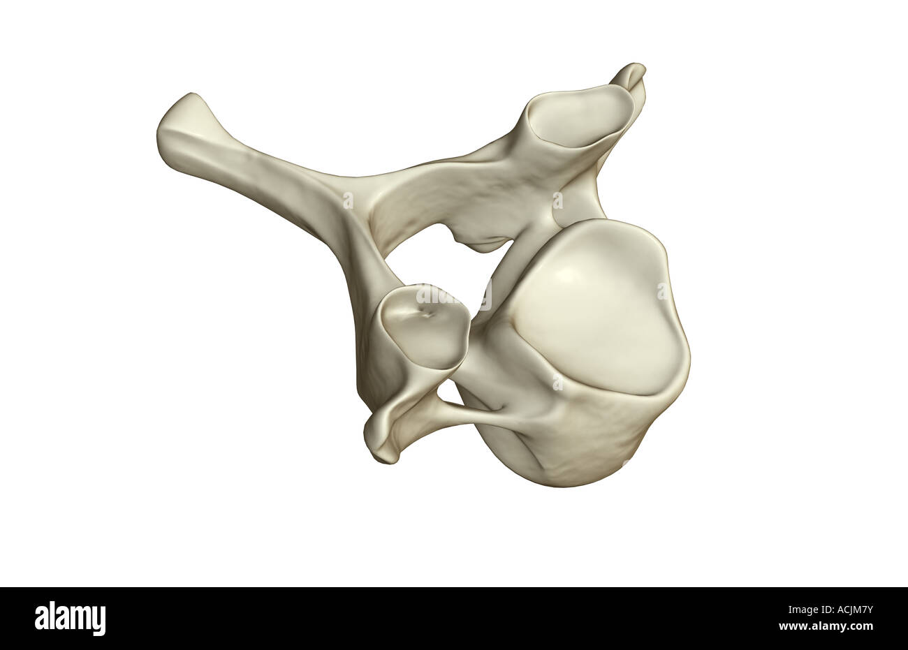
Quick filters:
Cervical vertebra Stock Photos and Images
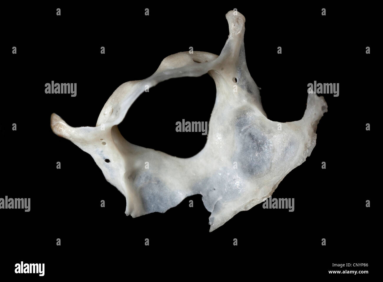 Barn owl (Tyto alba), cervical vertebra of a mouse, undigested food residue from a pellet Stock Photohttps://www.alamy.com/image-license-details/?v=1https://www.alamy.com/stock-photo-barn-owl-tyto-alba-cervical-vertebra-of-a-mouse-undigested-food-residue-47938694.html
RMCNYP86–Barn owl (Tyto alba), cervical vertebra of a mouse, undigested food residue from a pellet
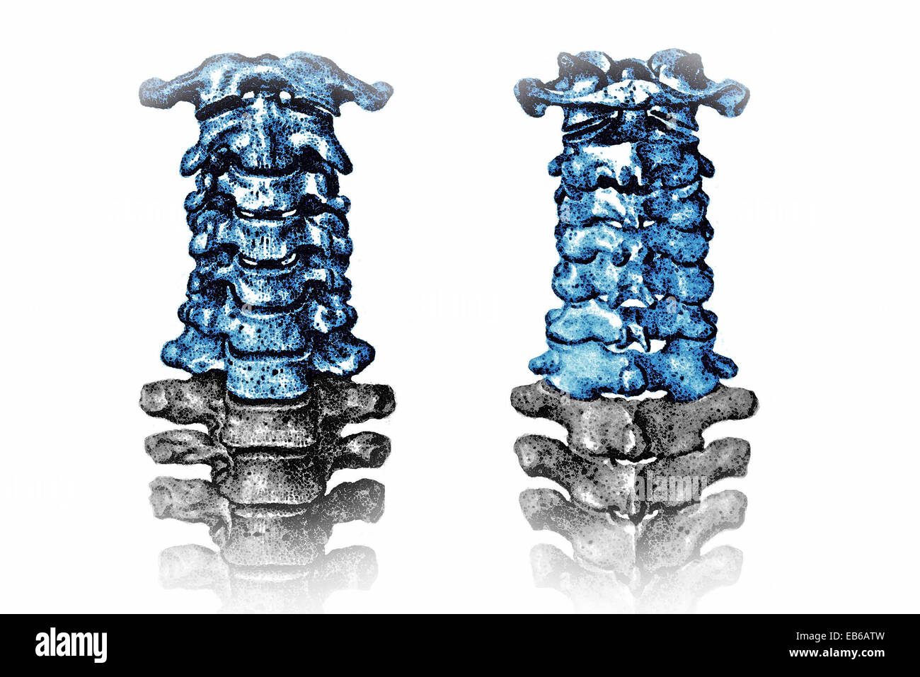 CERVICAL VERTEBRA, ILLUSTRATION Stock Photohttps://www.alamy.com/image-license-details/?v=1https://www.alamy.com/stock-photo-cervical-vertebra-illustration-75742937.html
RMEB6ATW–CERVICAL VERTEBRA, ILLUSTRATION
 Woman rehab with her physiotherapist. cervical vertebra treatment with exercise Stock Photohttps://www.alamy.com/image-license-details/?v=1https://www.alamy.com/woman-rehab-with-her-physiotherapist-cervical-vertebra-treatment-with-exercise-image354794020.html
RF2BH67B0–Woman rehab with her physiotherapist. cervical vertebra treatment with exercise
 Human Cervical Vertebra isolated on white background Stock Photohttps://www.alamy.com/image-license-details/?v=1https://www.alamy.com/human-cervical-vertebra-isolated-on-white-background-image610656101.html
RF2XDDP2D–Human Cervical Vertebra isolated on white background
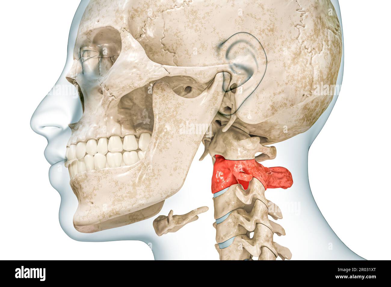 Axis second cervical vertebra in red color with body 3D rendering illustration isolated on white with copy space. Human skeleton ans spine anatomy, me Stock Photohttps://www.alamy.com/image-license-details/?v=1https://www.alamy.com/axis-second-cervical-vertebra-in-red-color-with-body-3d-rendering-illustration-isolated-on-white-with-copy-space-human-skeleton-ans-spine-anatomy-me-image550799168.html
RF2R031XT–Axis second cervical vertebra in red color with body 3D rendering illustration isolated on white with copy space. Human skeleton ans spine anatomy, me
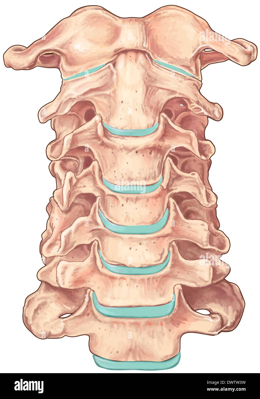 Cervical vertebra drawing Stock Photohttps://www.alamy.com/image-license-details/?v=1https://www.alamy.com/cervical-vertebra-drawing-image67544061.html
RMDWTW3W–Cervical vertebra drawing
 Backache. Back pain. Pain in the cervical vertebra. Stock Photohttps://www.alamy.com/image-license-details/?v=1https://www.alamy.com/backache-back-pain-pain-in-the-cervical-vertebra-image344408537.html
RF2B094GW–Backache. Back pain. Pain in the cervical vertebra.
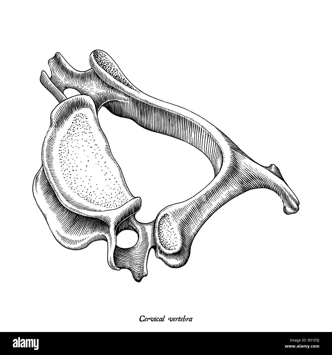 Cervical vertebra human anatomy superior lateral view hand draw vintage clip art isolated on white background Stock Vectorhttps://www.alamy.com/image-license-details/?v=1https://www.alamy.com/cervical-vertebra-human-anatomy-superior-lateral-view-hand-draw-vintage-clip-art-isolated-on-white-background-image226841262.html
RFR51E9J–Cervical vertebra human anatomy superior lateral view hand draw vintage clip art isolated on white background
 Toothed Whale. This object is part of the Education and Outreach collection, some of which are in the Q?rius science education center and available to see.Cenozoic - Neogene - MioceneThis fossilized cervical vertebra is from the spine of an aquatic porpoise-like toothed whale. Stock Photohttps://www.alamy.com/image-license-details/?v=1https://www.alamy.com/toothed-whale-this-object-is-part-of-the-education-and-outreach-collection-some-of-which-are-in-the-qrius-science-education-center-and-available-to-seecenozoic-neogene-miocenethis-fossilized-cervical-vertebra-is-from-the-spine-of-an-aquatic-porpoise-like-toothed-whale-image353466153.html
RM2BF1NK5–Toothed Whale. This object is part of the Education and Outreach collection, some of which are in the Q?rius science education center and available to see.Cenozoic - Neogene - MioceneThis fossilized cervical vertebra is from the spine of an aquatic porpoise-like toothed whale.
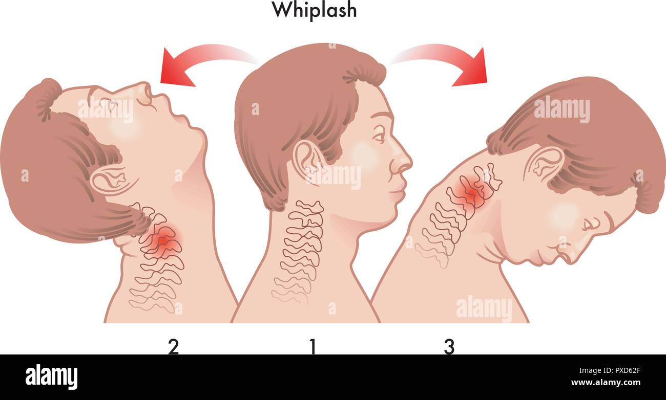 vector medical illustration of the dynamics of the whiplash injury Stock Vectorhttps://www.alamy.com/image-license-details/?v=1https://www.alamy.com/vector-medical-illustration-of-the-dynamics-of-the-whiplash-injury-image222795623.html
RFPXD62F–vector medical illustration of the dynamics of the whiplash injury
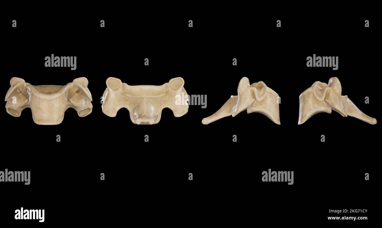 Cervical Vertebra -Multiple Views Stock Photohttps://www.alamy.com/image-license-details/?v=1https://www.alamy.com/cervical-vertebra-multiple-views-image491879611.html
RF2KG71CY–Cervical Vertebra -Multiple Views
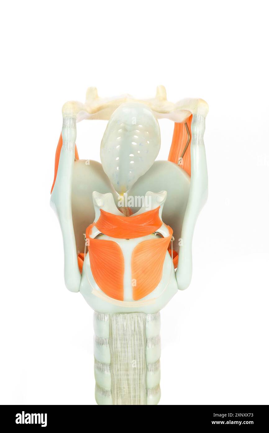 Artificial model of human larynx or voice box isolated on white background Stock Photohttps://www.alamy.com/image-license-details/?v=1https://www.alamy.com/artificial-model-of-human-larynx-or-voice-box-isolated-on-white-background-image615861991.html
RF2XNXX73–Artificial model of human larynx or voice box isolated on white background
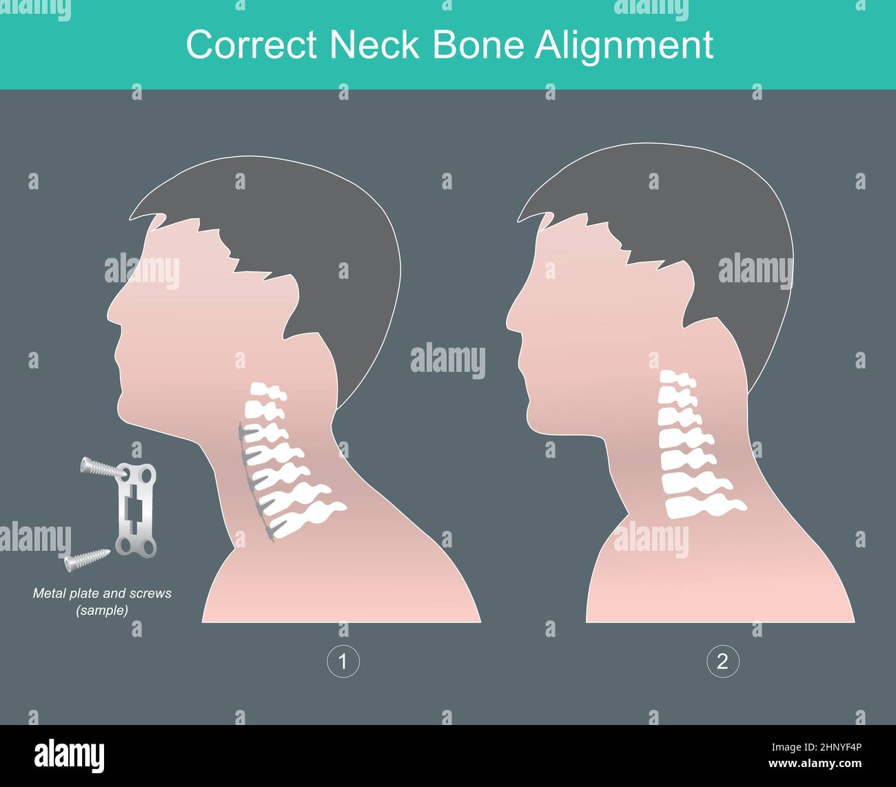 Correct Neck Bone Alignment. Showing human sideview the correct neck bone alignment. Stock Vectorhttps://www.alamy.com/image-license-details/?v=1https://www.alamy.com/correct-neck-bone-alignment-showing-human-sideview-the-correct-neck-bone-alignment-image460981942.html
RF2HNYF4P–Correct Neck Bone Alignment. Showing human sideview the correct neck bone alignment.
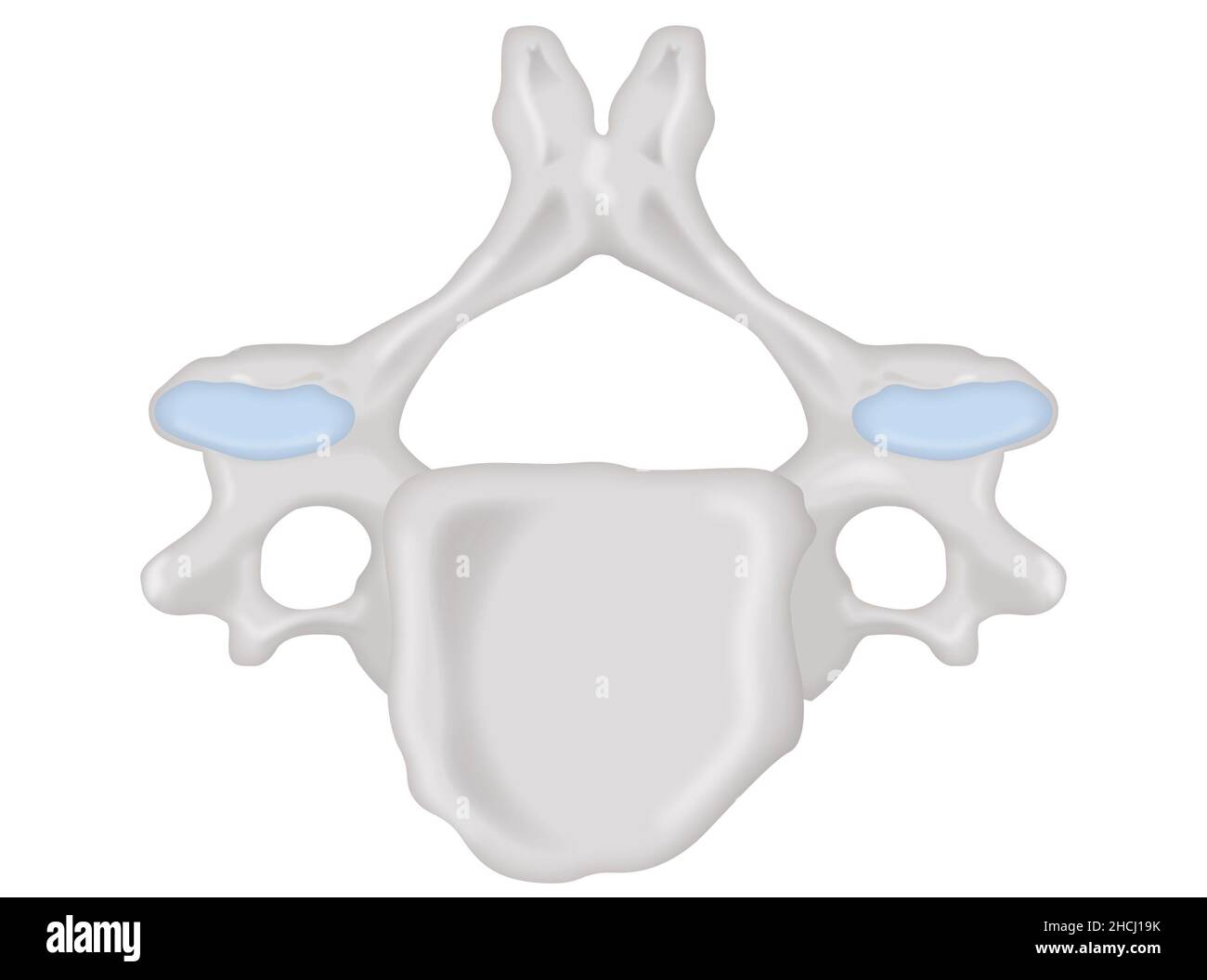 Cervical vertebra superior view, unlabeled anatomical structure Stock Photohttps://www.alamy.com/image-license-details/?v=1https://www.alamy.com/cervical-vertebra-superior-view-unlabeled-anatomical-structure-image455241631.html
RF2HCJ19K–Cervical vertebra superior view, unlabeled anatomical structure
 neck of a young girl, pain in the spine on the neck, osteochondrosis and scaleo of the cervical vertebra, medicine and health Stock Photohttps://www.alamy.com/image-license-details/?v=1https://www.alamy.com/neck-of-a-young-girl-pain-in-the-spine-on-the-neck-osteochondrosis-and-scaleo-of-the-cervical-vertebra-medicine-and-health-image477560831.html
RF2JMXNKY–neck of a young girl, pain in the spine on the neck, osteochondrosis and scaleo of the cervical vertebra, medicine and health
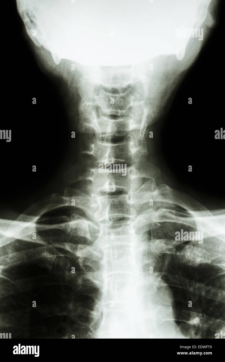 film x-ray cervical spine AP : show normal thai man's cervical spine Stock Photohttps://www.alamy.com/image-license-details/?v=1https://www.alamy.com/stock-photo-film-x-ray-cervical-spine-ap-show-normal-thai-mans-cervical-spine-77393232.html
RFEDWFT0–film x-ray cervical spine AP : show normal thai man's cervical spine
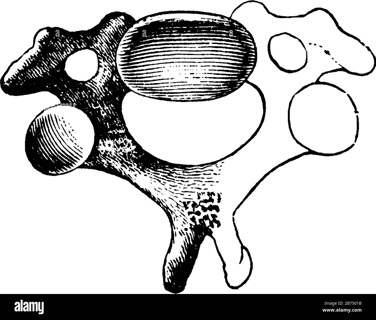 This illustration represents A Cervical Vertebra, vintage line drawing or engraving illustration. Stock Vectorhttps://www.alamy.com/image-license-details/?v=1https://www.alamy.com/this-illustration-represents-a-cervical-vertebra-vintage-line-drawing-or-engraving-illustration-image348619751.html
RF2B7501B–This illustration represents A Cervical Vertebra, vintage line drawing or engraving illustration.
 Doctor, patient, ruff, nurse, invest, centrifugal trauma, HWS syndrome, cervical vertebra pillar trauma, treatment, lash syndrome, immobilise, cut out Stock Photohttps://www.alamy.com/image-license-details/?v=1https://www.alamy.com/stock-photo-doctor-patient-ruff-nurse-invest-centrifugal-trauma-hws-syndrome-cervical-122937632.html
RMH4085M–Doctor, patient, ruff, nurse, invest, centrifugal trauma, HWS syndrome, cervical vertebra pillar trauma, treatment, lash syndrome, immobilise, cut out
 Doctor looking at a of the cervical spine x-ray of her patient. Stock Photohttps://www.alamy.com/image-license-details/?v=1https://www.alamy.com/stock-photo-doctor-looking-at-a-of-the-cervical-spine-x-ray-of-her-patient-72441562.html
RME5RYXJ–Doctor looking at a of the cervical spine x-ray of her patient.
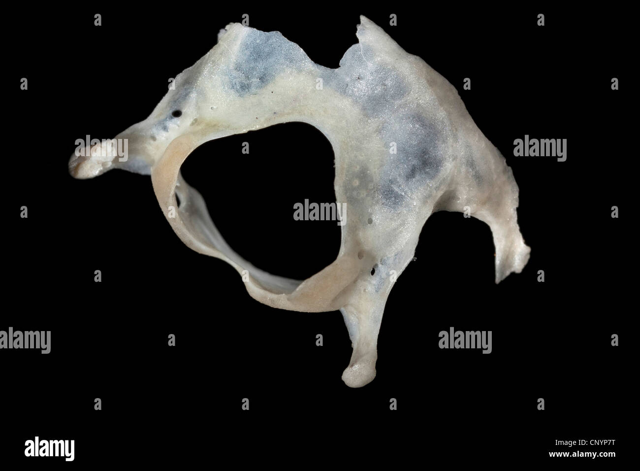 Barn owl (Tyto alba), cervical vertebra of a mouse, undigested food residue from a pellet Stock Photohttps://www.alamy.com/image-license-details/?v=1https://www.alamy.com/stock-photo-barn-owl-tyto-alba-cervical-vertebra-of-a-mouse-undigested-food-residue-47938684.html
RMCNYP7T–Barn owl (Tyto alba), cervical vertebra of a mouse, undigested food residue from a pellet
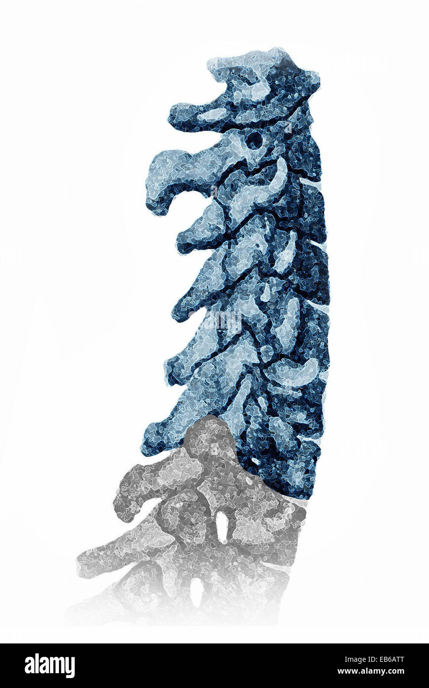 CERVICAL VERTEBRA, ILLUSTRATION Stock Photohttps://www.alamy.com/image-license-details/?v=1https://www.alamy.com/stock-photo-cervical-vertebra-illustration-75742936.html
RMEB6ATT–CERVICAL VERTEBRA, ILLUSTRATION
 Woman rehab with her physiotherapist. cervical vertebra treatment with exercise Stock Photohttps://www.alamy.com/image-license-details/?v=1https://www.alamy.com/woman-rehab-with-her-physiotherapist-cervical-vertebra-treatment-with-exercise-image354794022.html
RF2BH67B2–Woman rehab with her physiotherapist. cervical vertebra treatment with exercise
 Cervical vertebra Stock Photohttps://www.alamy.com/image-license-details/?v=1https://www.alamy.com/stock-photo-cervical-vertebra-13172510.html
RFACJPFY–Cervical vertebra
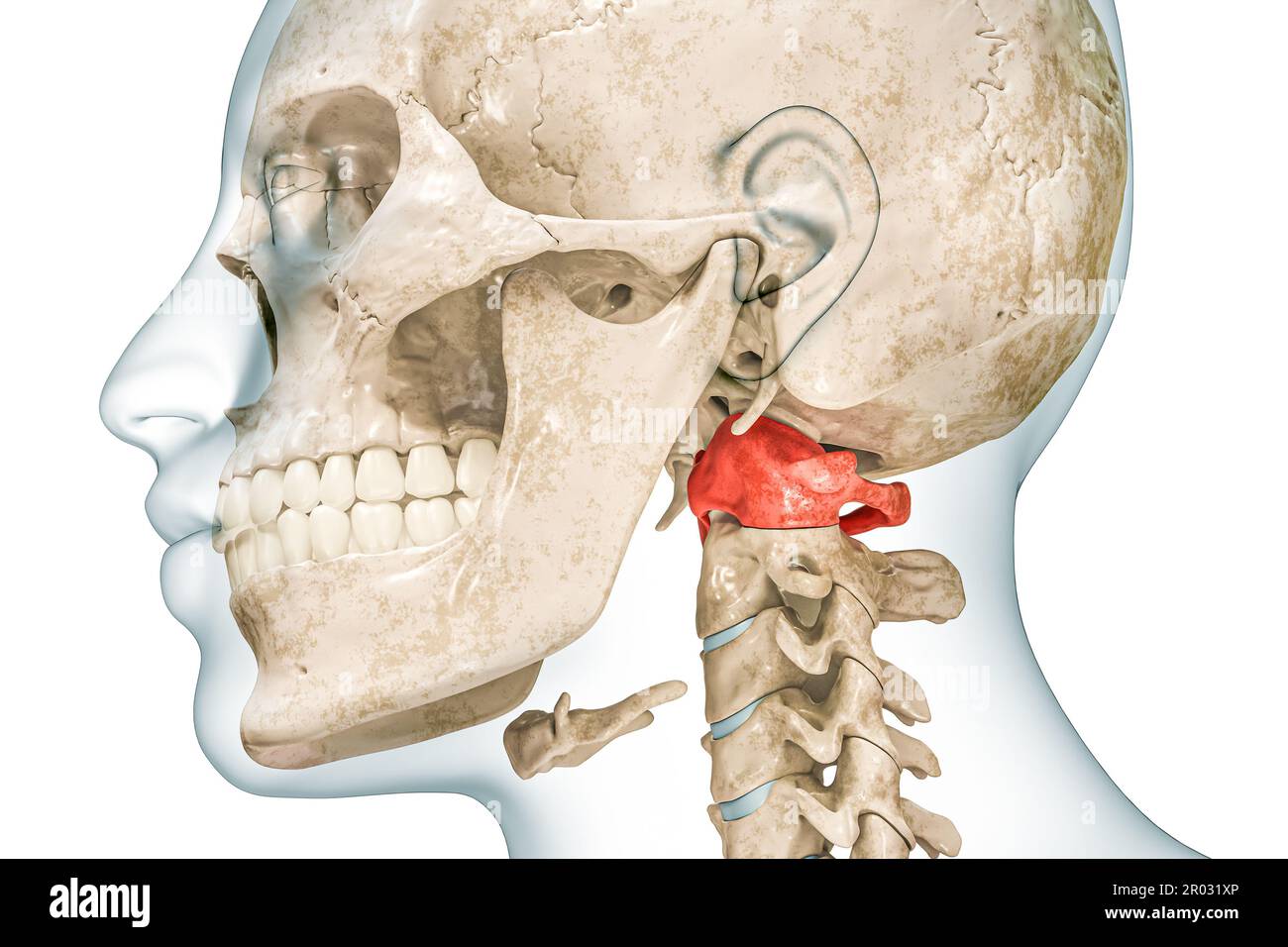 Atlas first cervical vertebra in red color with body 3D rendering illustration isolated on white with copy space. Human skeleton ans spine anatomy, me Stock Photohttps://www.alamy.com/image-license-details/?v=1https://www.alamy.com/atlas-first-cervical-vertebra-in-red-color-with-body-3d-rendering-illustration-isolated-on-white-with-copy-space-human-skeleton-ans-spine-anatomy-me-image550799166.html
RF2R031XP–Atlas first cervical vertebra in red color with body 3D rendering illustration isolated on white with copy space. Human skeleton ans spine anatomy, me
 A normal human vertebral column (lateral view). Shown (from top to bottom) are the atlas (cervical vertebra 1: C1), axis (C2), cervical vertebrae 3-7 Stock Photohttps://www.alamy.com/image-license-details/?v=1https://www.alamy.com/stock-photo-a-normal-human-vertebral-column-lateral-view-shown-from-top-to-bottom-130806568.html
RFHGPN34–A normal human vertebral column (lateral view). Shown (from top to bottom) are the atlas (cervical vertebra 1: C1), axis (C2), cervical vertebrae 3-7
 Backache. Back pain. Pain in the cervical vertebra. Stock Photohttps://www.alamy.com/image-license-details/?v=1https://www.alamy.com/backache-back-pain-pain-in-the-cervical-vertebra-image344408461.html
RF2B094E5–Backache. Back pain. Pain in the cervical vertebra.
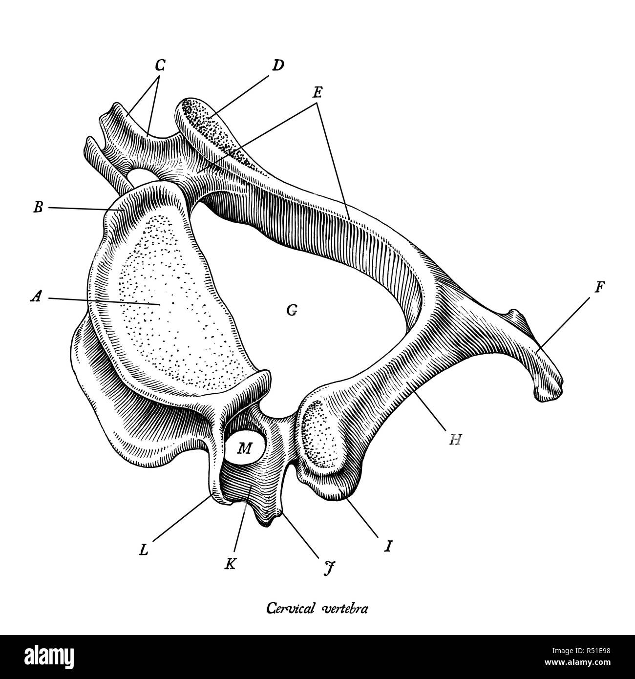 Cervical vertebra human anatomy superior lateral view hand draw vintage clip art isolated on white background with description Stock Vectorhttps://www.alamy.com/image-license-details/?v=1https://www.alamy.com/cervical-vertebra-human-anatomy-superior-lateral-view-hand-draw-vintage-clip-art-isolated-on-white-background-with-description-image226841252.html
RFR51E98–Cervical vertebra human anatomy superior lateral view hand draw vintage clip art isolated on white background with description
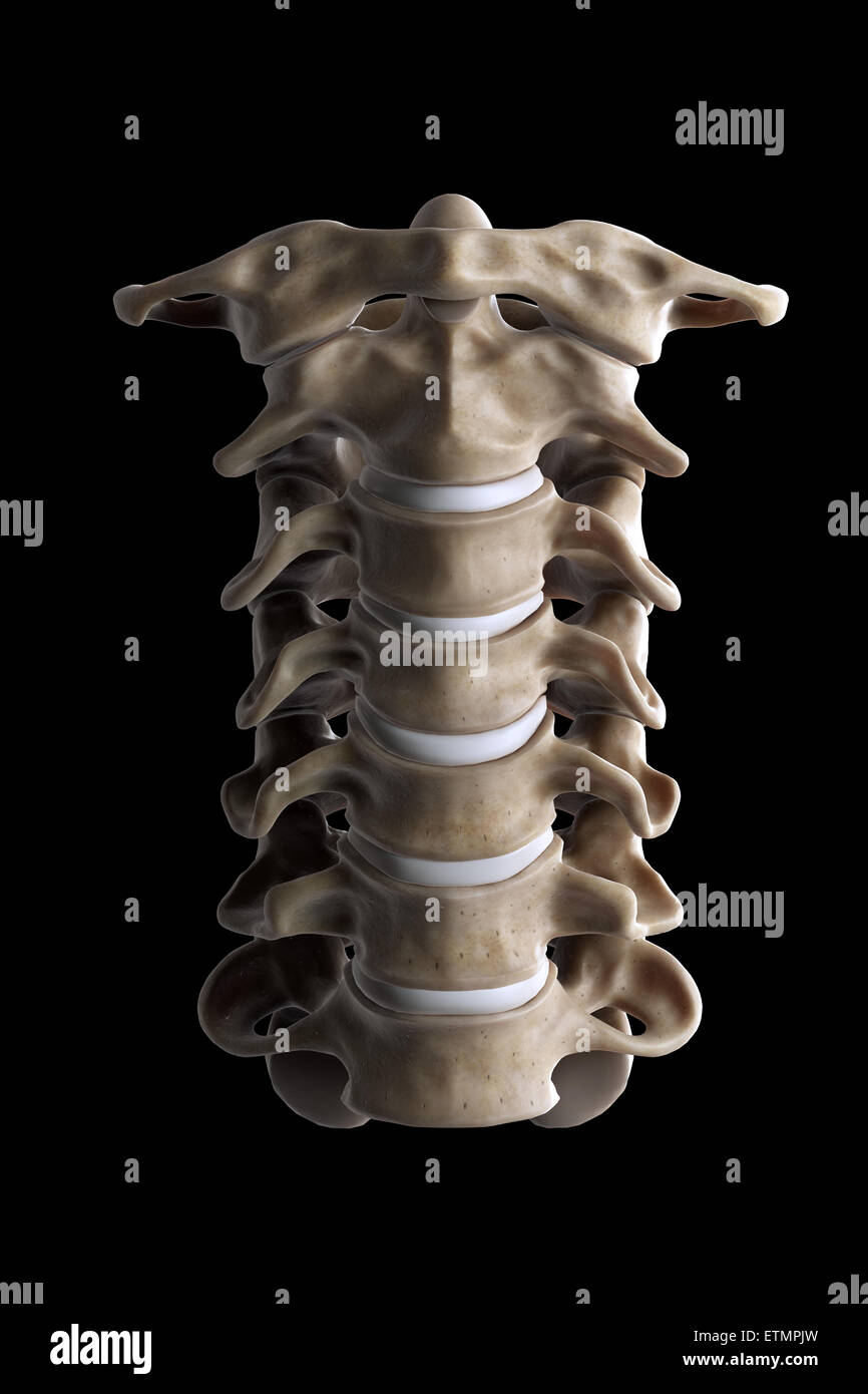 Illustration showing all seven cervical vertebrae. Stock Photohttps://www.alamy.com/image-license-details/?v=1https://www.alamy.com/stock-photo-illustration-showing-all-seven-cervical-vertebrae-84050033.html
RMETMPJW–Illustration showing all seven cervical vertebrae.
 X-ray C-spine or x-ray image of Cervical spine open mount view for fracture of cervical vertebra 2nd ( axis ). Stock Photohttps://www.alamy.com/image-license-details/?v=1https://www.alamy.com/x-ray-c-spine-or-x-ray-image-of-cervical-spine-open-mount-view-for-fracture-of-cervical-vertebra-2nd-axis-image546004992.html
RF2PM8JX8–X-ray C-spine or x-ray image of Cervical spine open mount view for fracture of cervical vertebra 2nd ( axis ).
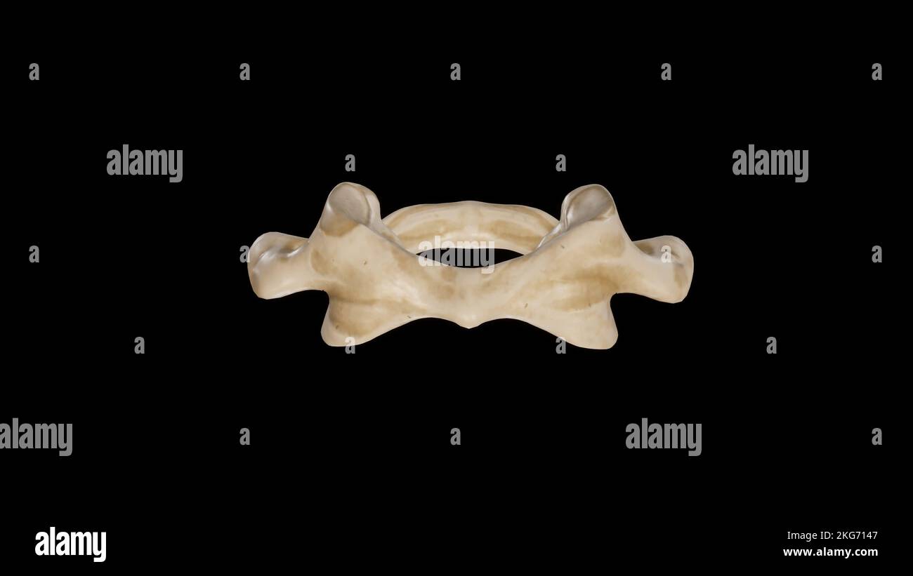 Anterior view of First Cervical Vertebra (Atlas) Stock Photohttps://www.alamy.com/image-license-details/?v=1https://www.alamy.com/anterior-view-of-first-cervical-vertebra-atlas-image491879367.html
RF2KG7147–Anterior view of First Cervical Vertebra (Atlas)
 Doctor studying ill patients mri, finds damage in cervical vertebra, diagnostics Stock Photohttps://www.alamy.com/image-license-details/?v=1https://www.alamy.com/doctor-studying-ill-patients-mri-finds-damage-in-cervical-vertebra-diagnostics-image339789896.html
RF2AMPNDC–Doctor studying ill patients mri, finds damage in cervical vertebra, diagnostics
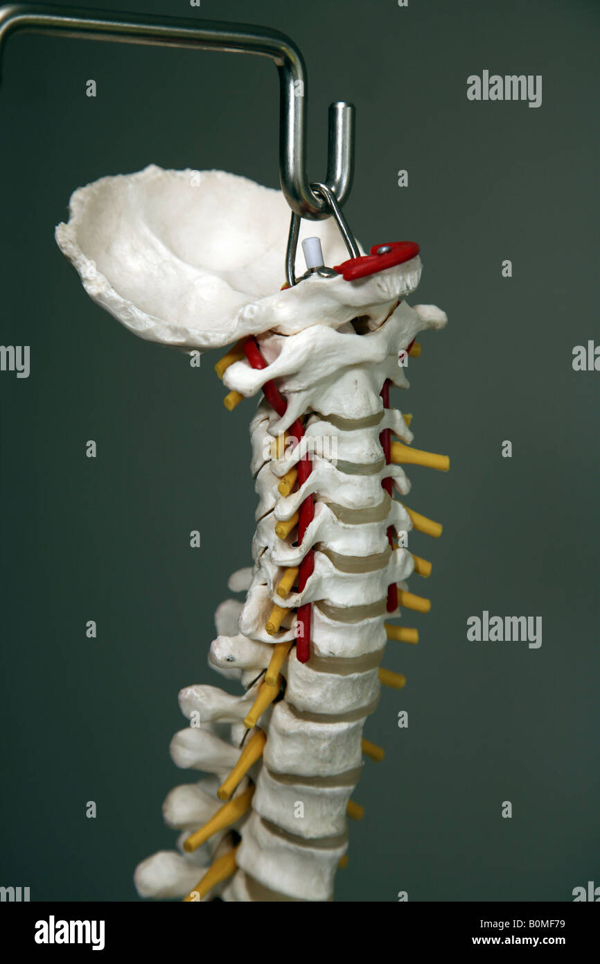 A model of the cervical spine (neck) of the human body including the Occiput Stock Photohttps://www.alamy.com/image-license-details/?v=1https://www.alamy.com/stock-photo-a-model-of-the-cervical-spine-neck-of-the-human-body-including-the-17661373.html
RMB0MF79–A model of the cervical spine (neck) of the human body including the Occiput
 Cervical Vertebra of Crymocetus (formerly Plesiosaurus) Stock Photohttps://www.alamy.com/image-license-details/?v=1https://www.alamy.com/cervical-vertebra-of-crymocetus-formerly-plesiosaurus-image532103075.html
RM2NWKAW7–Cervical Vertebra of Crymocetus (formerly Plesiosaurus)
 neck of a young girl, pain in the spine on the neck, osteochondrosis and scaleo of the cervical vertebra, medicine and health Stock Photohttps://www.alamy.com/image-license-details/?v=1https://www.alamy.com/neck-of-a-young-girl-pain-in-the-spine-on-the-neck-osteochondrosis-and-scaleo-of-the-cervical-vertebra-medicine-and-health-image477560858.html
RF2JMXNMX–neck of a young girl, pain in the spine on the neck, osteochondrosis and scaleo of the cervical vertebra, medicine and health
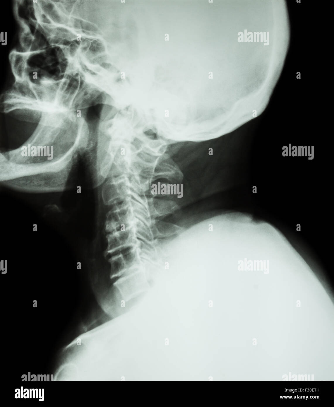 Cervical spondylosis . Film x-ray of cervical spine ( lateral position ) ( side view ) Stock Photohttps://www.alamy.com/image-license-details/?v=1https://www.alamy.com/stock-photo-cervical-spondylosis-film-x-ray-of-cervical-spine-lateral-position-87907473.html
RFF30ETH–Cervical spondylosis . Film x-ray of cervical spine ( lateral position ) ( side view )
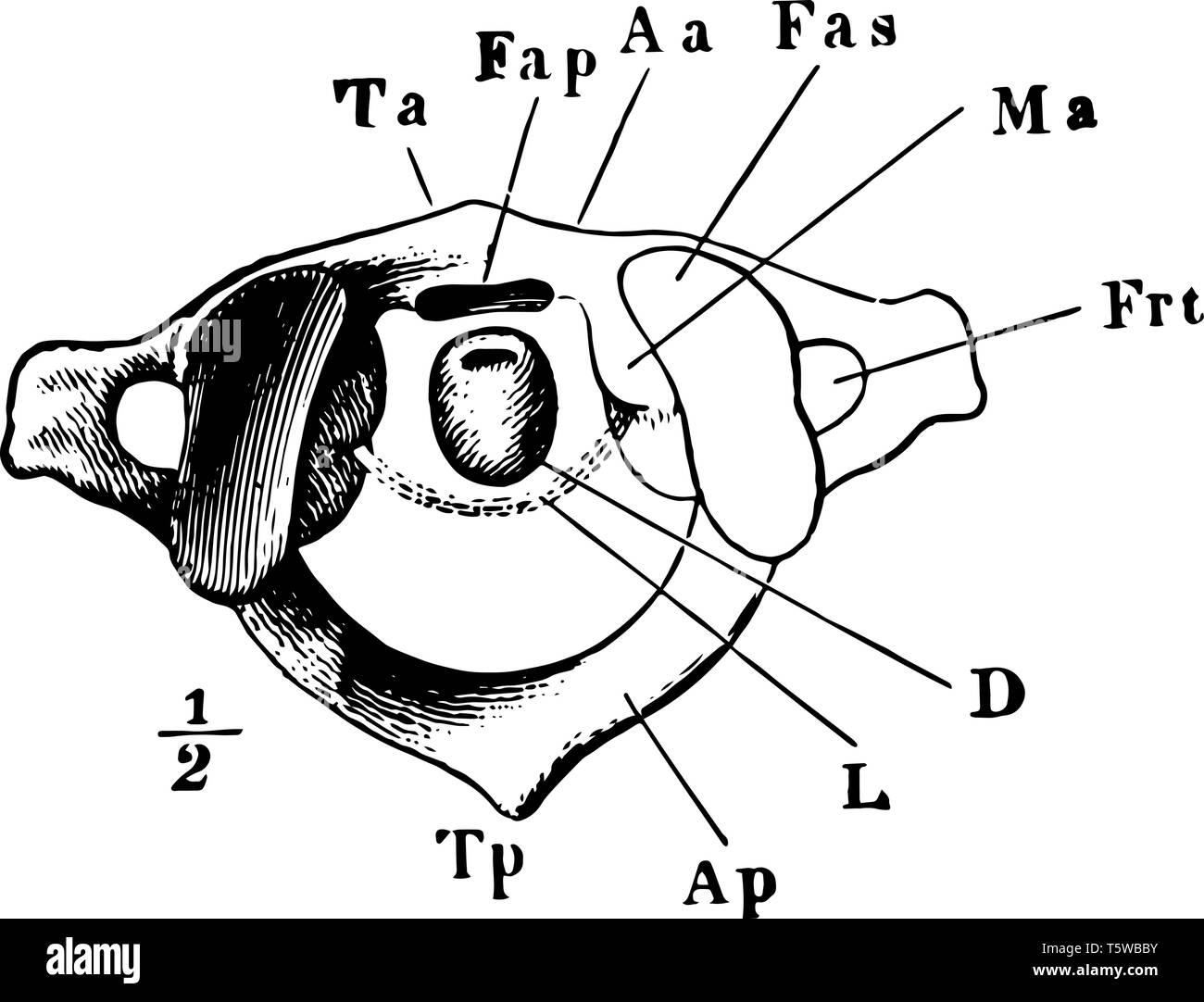 This illustration represents The Atlas which is 1st Cervical Vertebra vintage line drawing or engraving illustration. Stock Vectorhttps://www.alamy.com/image-license-details/?v=1https://www.alamy.com/this-illustration-represents-the-atlas-which-is-1st-cervical-vertebra-vintage-line-drawing-or-engraving-illustration-image244576191.html
RFT5WBBY–This illustration represents The Atlas which is 1st Cervical Vertebra vintage line drawing or engraving illustration.
 Doctor, patient, complaints, injury, nape, ruff, invest centrifugal trauma, cervical vertebra pillar, cervical vertebra, trauma, neck tie, Schanz tie, Schanz-Verband Stock Photohttps://www.alamy.com/image-license-details/?v=1https://www.alamy.com/stock-photo-doctor-patient-complaints-injury-nape-ruff-invest-centrifugal-trauma-122854853.html
RMH3TEH9–Doctor, patient, complaints, injury, nape, ruff, invest centrifugal trauma, cervical vertebra pillar, cervical vertebra, trauma, neck tie, Schanz tie, Schanz-Verband
 Doctor looking at a of the cervical spine x-ray of her patient. Stock Photohttps://www.alamy.com/image-license-details/?v=1https://www.alamy.com/stock-photo-doctor-looking-at-a-of-the-cervical-spine-x-ray-of-her-patient-72441564.html
RME5RYXM–Doctor looking at a of the cervical spine x-ray of her patient.
 Portrait view of Cervical vertebra.This is part of bird skeletal system. Bird anatomy. Bird skeletal system. Stock Photohttps://www.alamy.com/image-license-details/?v=1https://www.alamy.com/portrait-view-of-cervical-vertebrathis-is-part-of-bird-skeletal-system-bird-anatomy-bird-skeletal-system-image518243511.html
RF2N340TR–Portrait view of Cervical vertebra.This is part of bird skeletal system. Bird anatomy. Bird skeletal system.
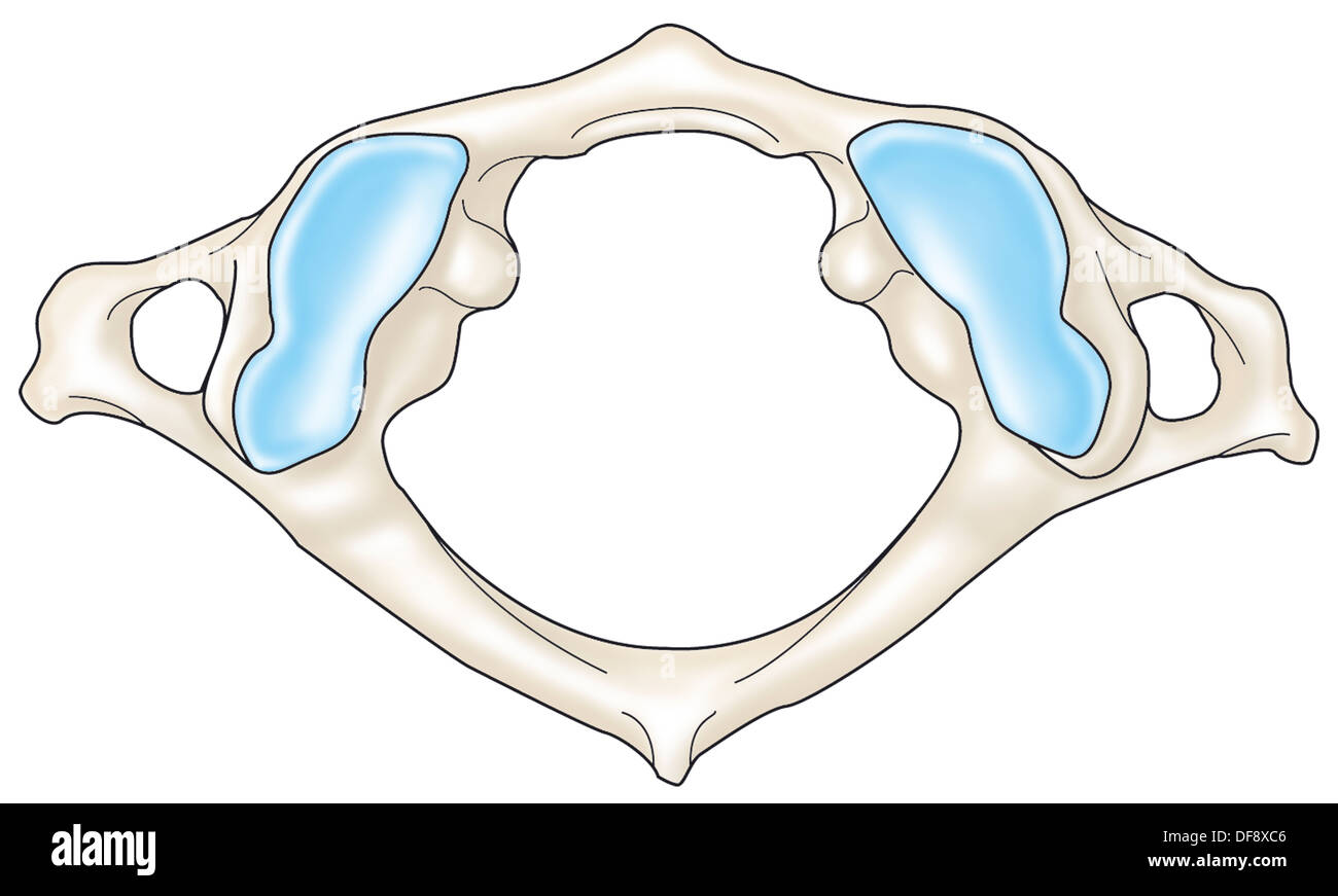 CERVICAL VERTEBRA, DRAWING Stock Photohttps://www.alamy.com/image-license-details/?v=1https://www.alamy.com/cervical-vertebra-drawing-image61047286.html
RMDF8XC6–CERVICAL VERTEBRA, DRAWING
 The family and friends of of Nicolas Chauvin, a rugby player from Stade Francais who died of a fracture of a cervical vertebra caused by a double tackle during a match in Begles Stock Photohttps://www.alamy.com/image-license-details/?v=1https://www.alamy.com/the-family-and-friends-of-of-nicolas-chauvin-a-rugby-player-from-stade-francais-who-died-of-a-fracture-of-a-cervical-vertebra-caused-by-a-double-tackle-during-a-match-in-begles-image486376791.html
RM2K78AFK–The family and friends of of Nicolas Chauvin, a rugby player from Stade Francais who died of a fracture of a cervical vertebra caused by a double tackle during a match in Begles
 Cervical vertebra Stock Photohttps://www.alamy.com/image-license-details/?v=1https://www.alamy.com/stock-photo-cervical-vertebra-13169574.html
RFACJDRK–Cervical vertebra
 Bildnummer: 55674349 Datum: 30.07.2011 Copyright: imago/Xinhua (110802) -- LANZHOU, Aug. 2, 2011 (Xinhua) -- A patient receives cervical vertebra traction treatment in a clinic of Tongyi Village, Liangzhou District, Wuwei City, northwest China s Gansu Province, July 30, 2011. On July 29 in a village clinic, Chang Faguo, 70, of the Luzhou village, receives herbal drugs from doctor Zhang Yuwen of Ma er Village neighbouring Luzhou and then rides home. Luzhou, located in the Wuwei City s Liangzhou District in northwest China s Gansu Province, is one of the impoverished villages in the poor provi Stock Photohttps://www.alamy.com/image-license-details/?v=1https://www.alamy.com/bildnummer-55674349-datum-30072011-copyright-imagoxinhua-110802-lanzhou-aug-2-2011-xinhua-a-patient-receives-cervical-vertebra-traction-treatment-in-a-clinic-of-tongyi-village-liangzhou-district-wuwei-city-northwest-china-s-gansu-province-july-30-2011-on-july-29-in-a-village-clinic-chang-faguo-70-of-the-luzhou-village-receives-herbal-drugs-from-doctor-zhang-yuwen-of-ma-er-village-neighbouring-luzhou-and-then-rides-home-luzhou-located-in-the-wuwei-city-s-liangzhou-district-in-northwest-china-s-gansu-province-is-one-of-the-impoverished-villages-in-the-poor-provi-image562784284.html
RM2RKH12M–Bildnummer: 55674349 Datum: 30.07.2011 Copyright: imago/Xinhua (110802) -- LANZHOU, Aug. 2, 2011 (Xinhua) -- A patient receives cervical vertebra traction treatment in a clinic of Tongyi Village, Liangzhou District, Wuwei City, northwest China s Gansu Province, July 30, 2011. On July 29 in a village clinic, Chang Faguo, 70, of the Luzhou village, receives herbal drugs from doctor Zhang Yuwen of Ma er Village neighbouring Luzhou and then rides home. Luzhou, located in the Wuwei City s Liangzhou District in northwest China s Gansu Province, is one of the impoverished villages in the poor provi
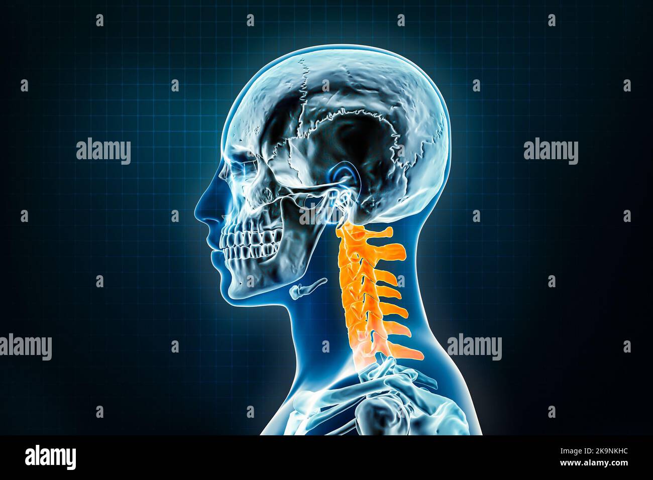 Cervical vertebrae x-ray lateral or profile view. Osteology of the human skeleton, spine 3D rendering illustration. Anatomy, medical, science, biology Stock Photohttps://www.alamy.com/image-license-details/?v=1https://www.alamy.com/cervical-vertebrae-x-ray-lateral-or-profile-view-osteology-of-the-human-skeleton-spine-3d-rendering-illustration-anatomy-medical-science-biology-image487898584.html
RF2K9NKHC–Cervical vertebrae x-ray lateral or profile view. Osteology of the human skeleton, spine 3D rendering illustration. Anatomy, medical, science, biology
 Backache. Back pain. Pain in the cervical vertebra. Stock Photohttps://www.alamy.com/image-license-details/?v=1https://www.alamy.com/backache-back-pain-pain-in-the-cervical-vertebra-image344408429.html
RF2B094D1–Backache. Back pain. Pain in the cervical vertebra.
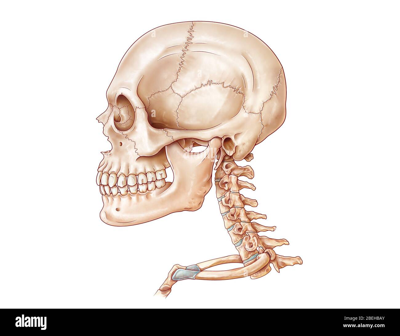 Skull and Cervical Vertebrae, Illustration Stock Photohttps://www.alamy.com/image-license-details/?v=1https://www.alamy.com/skull-and-cervical-vertebrae-illustration-image353194659.html
RM2BEHBAY–Skull and Cervical Vertebrae, Illustration
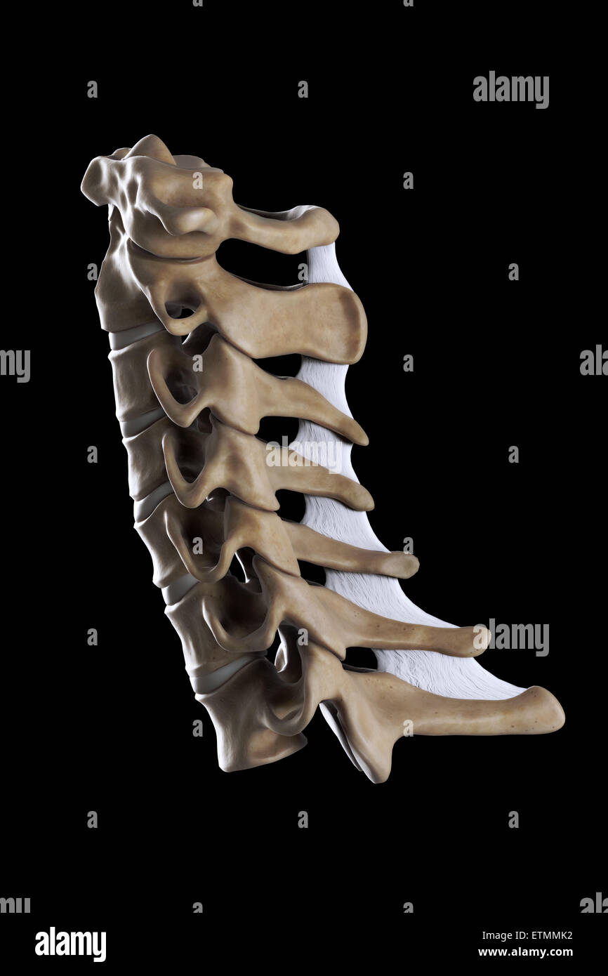 Illustration showing all seven cervical vertebrae. Stock Photohttps://www.alamy.com/image-license-details/?v=1https://www.alamy.com/stock-photo-illustration-showing-all-seven-cervical-vertebrae-84048470.html
RMETMMK2–Illustration showing all seven cervical vertebrae.
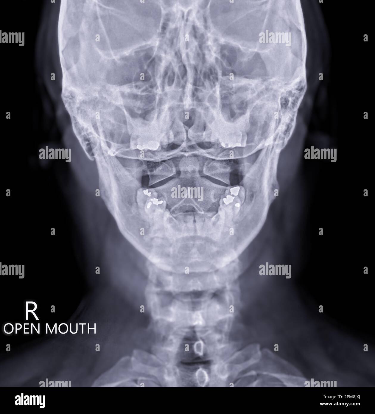 X-ray C-spine or x-ray image of Cervical spine open mount view for fracture of cervical vertebra 2nd ( axis ). Stock Photohttps://www.alamy.com/image-license-details/?v=1https://www.alamy.com/x-ray-c-spine-or-x-ray-image-of-cervical-spine-open-mount-view-for-fracture-of-cervical-vertebra-2nd-axis-image546005002.html
RF2PM8JXJ–X-ray C-spine or x-ray image of Cervical spine open mount view for fracture of cervical vertebra 2nd ( axis ).
 Lateral view of First Cervical Vertebra (Atlas) Stock Photohttps://www.alamy.com/image-license-details/?v=1https://www.alamy.com/lateral-view-of-first-cervical-vertebra-atlas-image491879350.html
RF2KG713J–Lateral view of First Cervical Vertebra (Atlas)
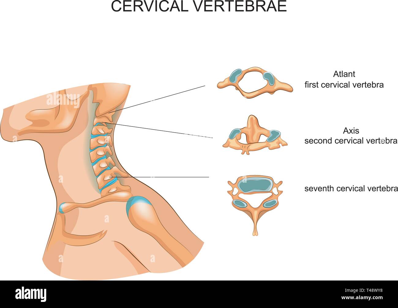 vector illustration of the structure of the cervical vertebrae Stock Vectorhttps://www.alamy.com/image-license-details/?v=1https://www.alamy.com/vector-illustration-of-the-structure-of-the-cervical-vertebrae-image243599756.html
RFT48WY8–vector illustration of the structure of the cervical vertebrae
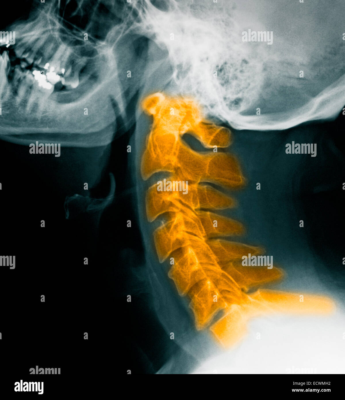 Normal cervical spine x-rays. Stock Photohttps://www.alamy.com/image-license-details/?v=1https://www.alamy.com/stock-photo-normal-cervical-spine-x-rays-76782302.html
RMECWMH2–Normal cervical spine x-rays.
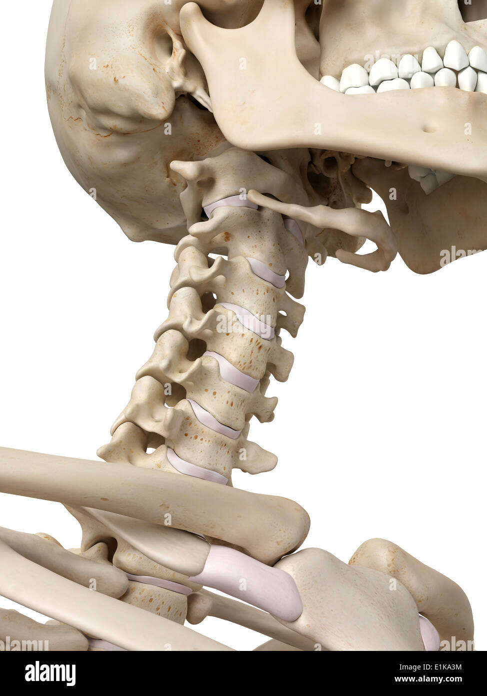 Human cervical spine computer artwork. Stock Photohttps://www.alamy.com/image-license-details/?v=1https://www.alamy.com/human-cervical-spine-computer-artwork-image69881160.html
RFE1KA3M–Human cervical spine computer artwork.
 neck of a young girl, pain in the spine on the neck, osteochondrosis and scaleo of the cervical vertebra, medicine and health Stock Photohttps://www.alamy.com/image-license-details/?v=1https://www.alamy.com/neck-of-a-young-girl-pain-in-the-spine-on-the-neck-osteochondrosis-and-scaleo-of-the-cervical-vertebra-medicine-and-health-image477560897.html
RF2JMXNP9–neck of a young girl, pain in the spine on the neck, osteochondrosis and scaleo of the cervical vertebra, medicine and health
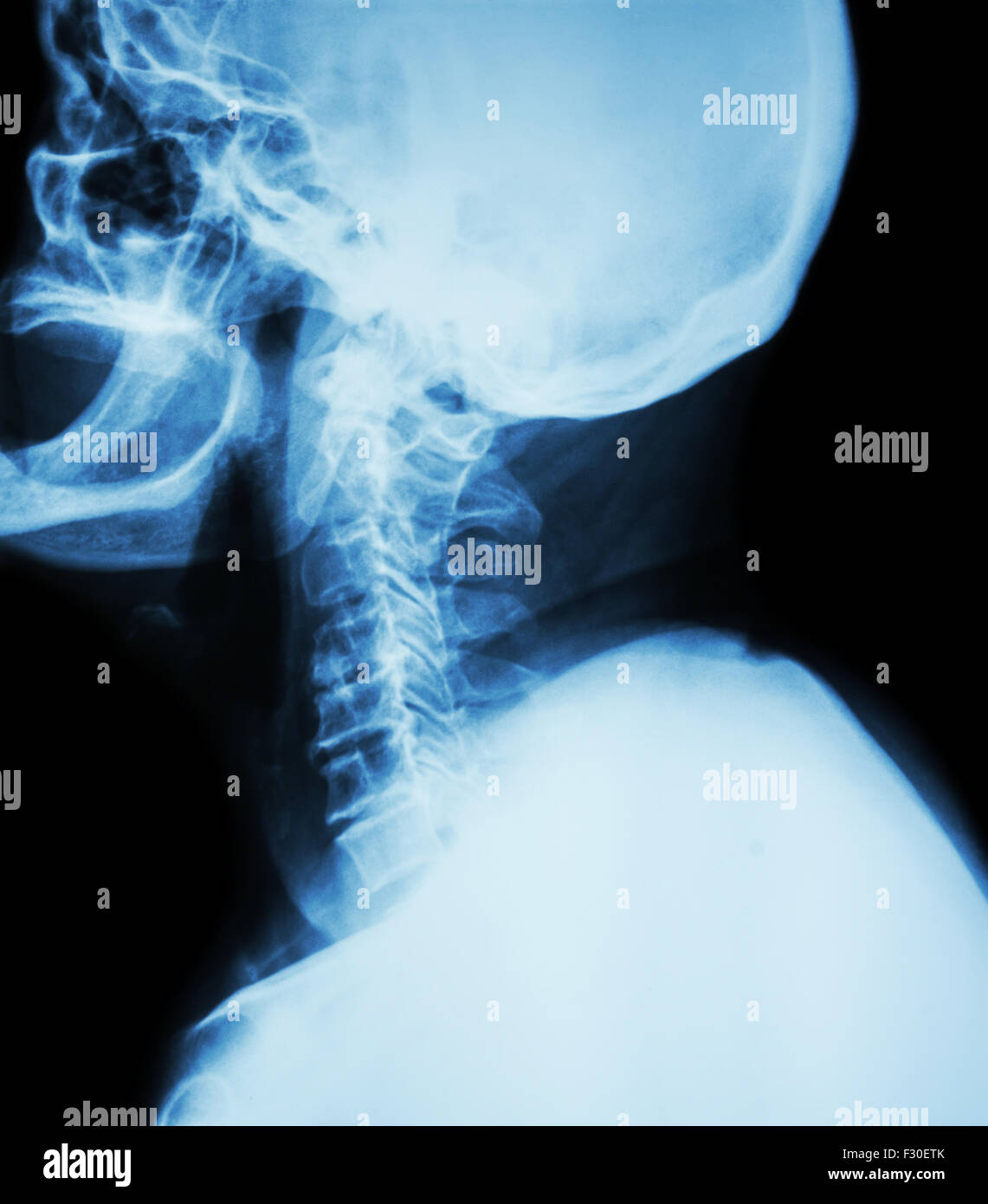 Cervical spondylosis . Film x-ray of cervical spine ( lateral position ) ( side view ) Stock Photohttps://www.alamy.com/image-license-details/?v=1https://www.alamy.com/stock-photo-cervical-spondylosis-film-x-ray-of-cervical-spine-lateral-position-87907475.html
RFF30ETK–Cervical spondylosis . Film x-ray of cervical spine ( lateral position ) ( side view )
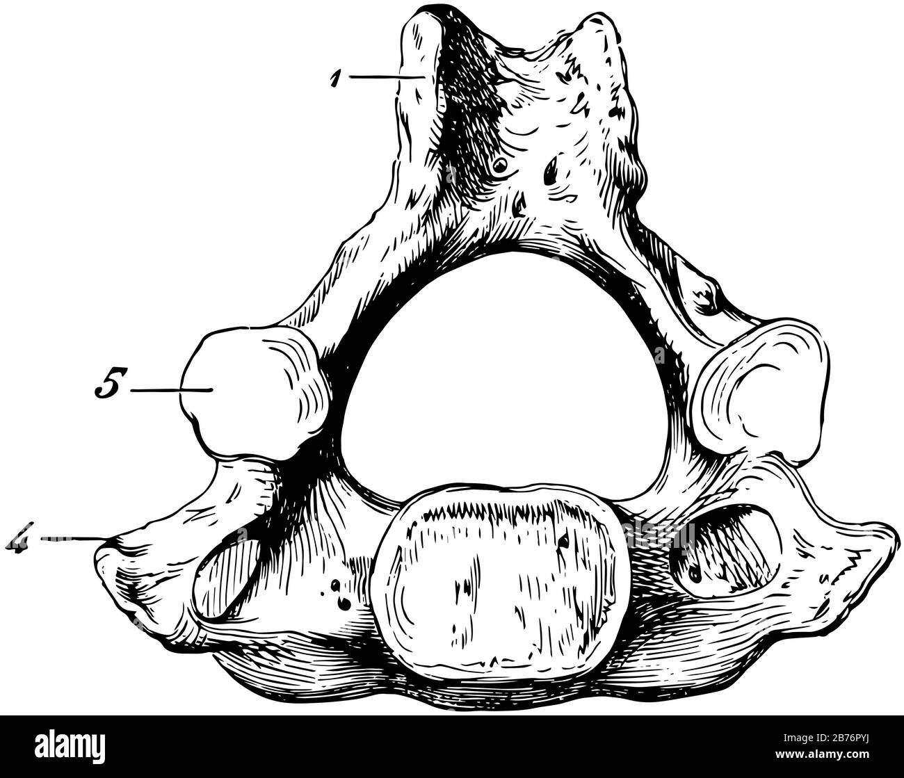 This illustration represents Human Cervical Vertebra Bone, vintage line drawing or engraving illustration. Stock Vectorhttps://www.alamy.com/image-license-details/?v=1https://www.alamy.com/this-illustration-represents-human-cervical-vertebra-bone-vintage-line-drawing-or-engraving-illustration-image348659686.html
RF2B76PYJ–This illustration represents Human Cervical Vertebra Bone, vintage line drawing or engraving illustration.
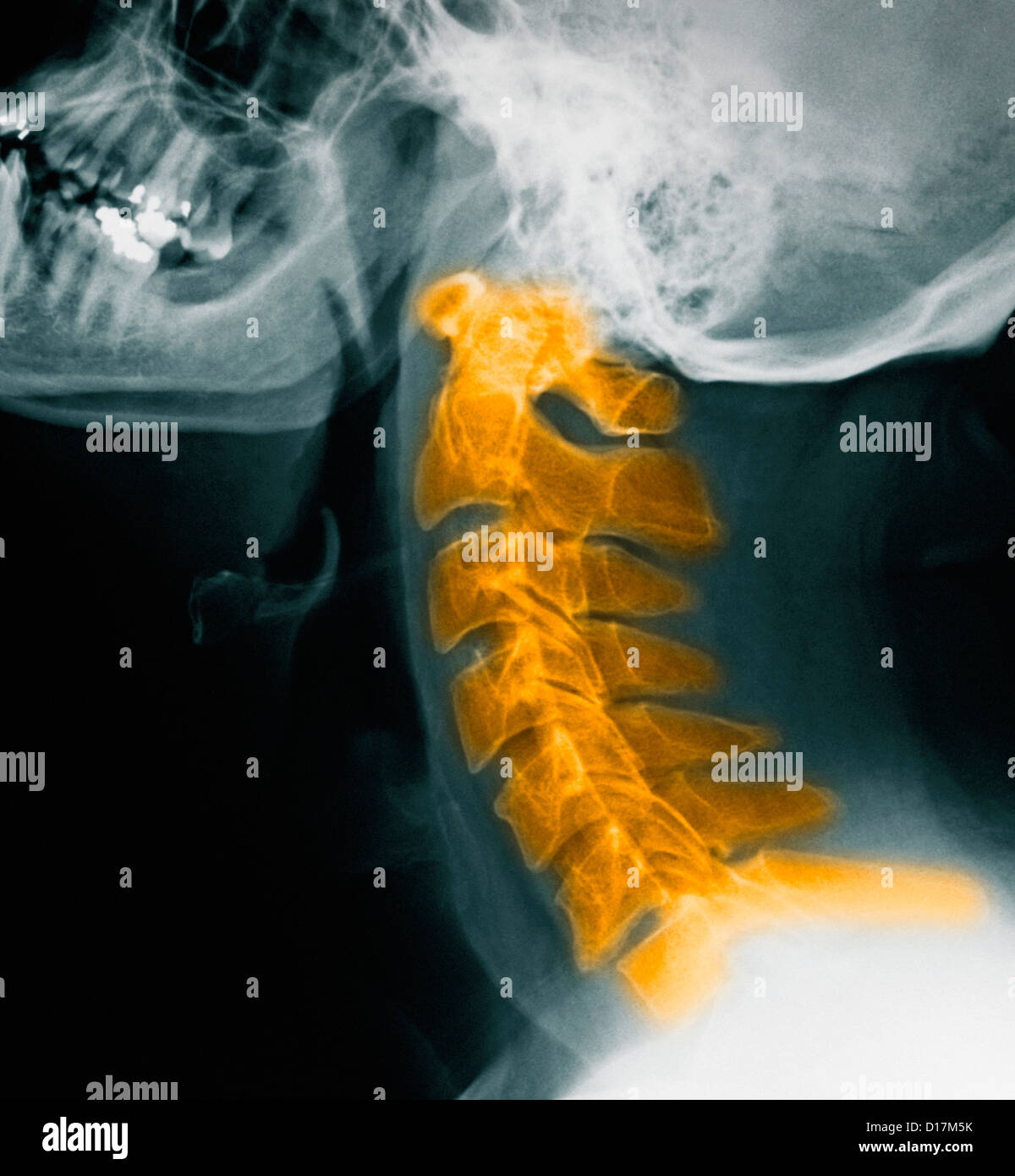 CerviCal spine X-rays, normal Stock Photohttps://www.alamy.com/image-license-details/?v=1https://www.alamy.com/stock-photo-cervical-spine-x-rays-normal-52415263.html
RFD17M5K–CerviCal spine X-rays, normal
 Doctor looking at a of the cervical spine x-ray of her patient. Stock Photohttps://www.alamy.com/image-license-details/?v=1https://www.alamy.com/stock-photo-doctor-looking-at-a-of-the-cervical-spine-x-ray-of-her-patient-72441563.html
RME5RYXK–Doctor looking at a of the cervical spine x-ray of her patient.
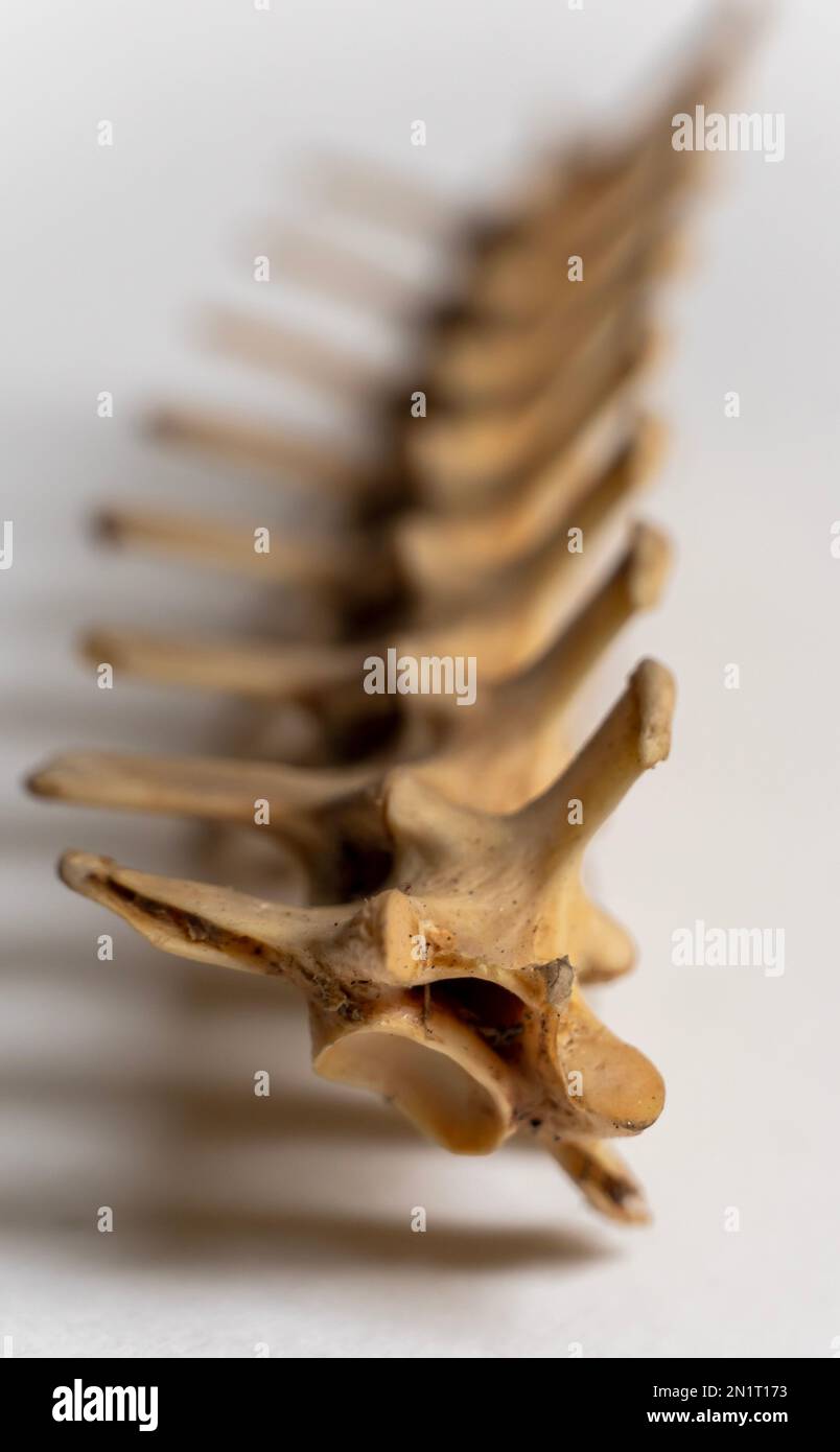 Portrait view of Cervical vertebra is a part of bird skeletal system. Bird anatomy. Bird skeletal system. Stock Photohttps://www.alamy.com/image-license-details/?v=1https://www.alamy.com/portrait-view-of-cervical-vertebra-is-a-part-of-bird-skeletal-system-bird-anatomy-bird-skeletal-system-image517453527.html
RF2N1T173–Portrait view of Cervical vertebra is a part of bird skeletal system. Bird anatomy. Bird skeletal system.
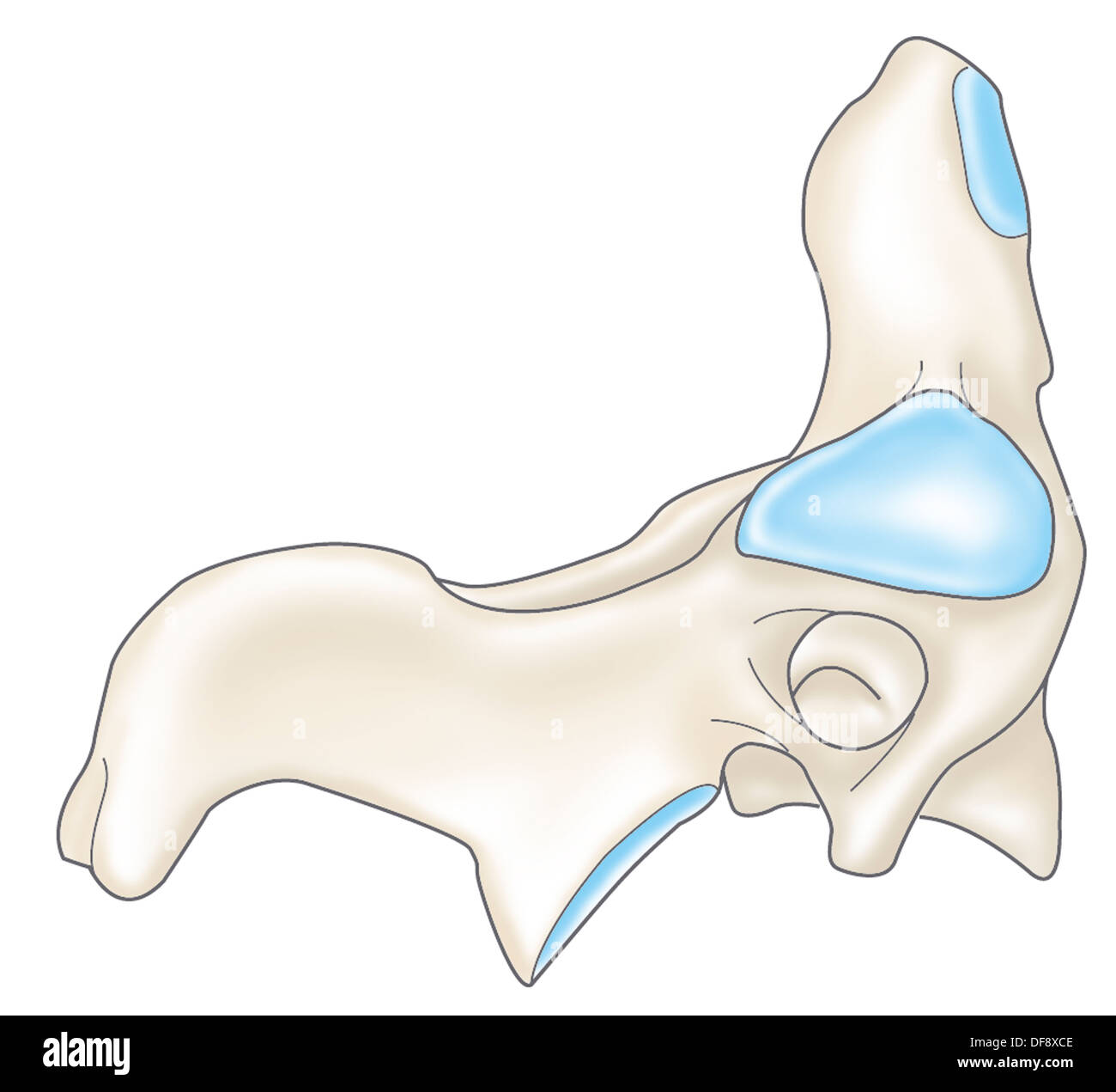 CERVICAL VERTEBRA, DRAWING Stock Photohttps://www.alamy.com/image-license-details/?v=1https://www.alamy.com/cervical-vertebra-drawing-image61047294.html
RMDF8XCE–CERVICAL VERTEBRA, DRAWING
 A view of the funeral of Nicolas Chauvin, a rugby player from Stade Francais who died of a fracture of a cervical vertebra caused by a double tackle during a match in Begles Stock Photohttps://www.alamy.com/image-license-details/?v=1https://www.alamy.com/a-view-of-the-funeral-of-nicolas-chauvin-a-rugby-player-from-stade-francais-who-died-of-a-fracture-of-a-cervical-vertebra-caused-by-a-double-tackle-during-a-match-in-begles-image486376789.html
RM2K78AFH–A view of the funeral of Nicolas Chauvin, a rugby player from Stade Francais who died of a fracture of a cervical vertebra caused by a double tackle during a match in Begles
 Cervical vertebra Stock Photohttps://www.alamy.com/image-license-details/?v=1https://www.alamy.com/stock-photo-cervical-vertebra-13207045.html
RFACPD9X–Cervical vertebra
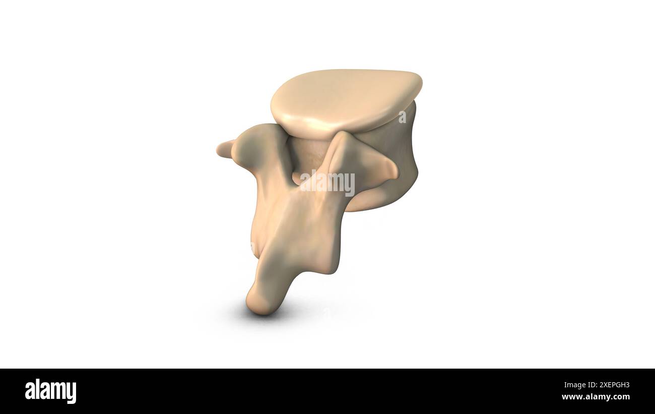 Human Lumbar Vertebra isolated on white background Stock Photohttps://www.alamy.com/image-license-details/?v=1https://www.alamy.com/human-lumbar-vertebra-isolated-on-white-background-image611464031.html
RF2XEPGH3–Human Lumbar Vertebra isolated on white background
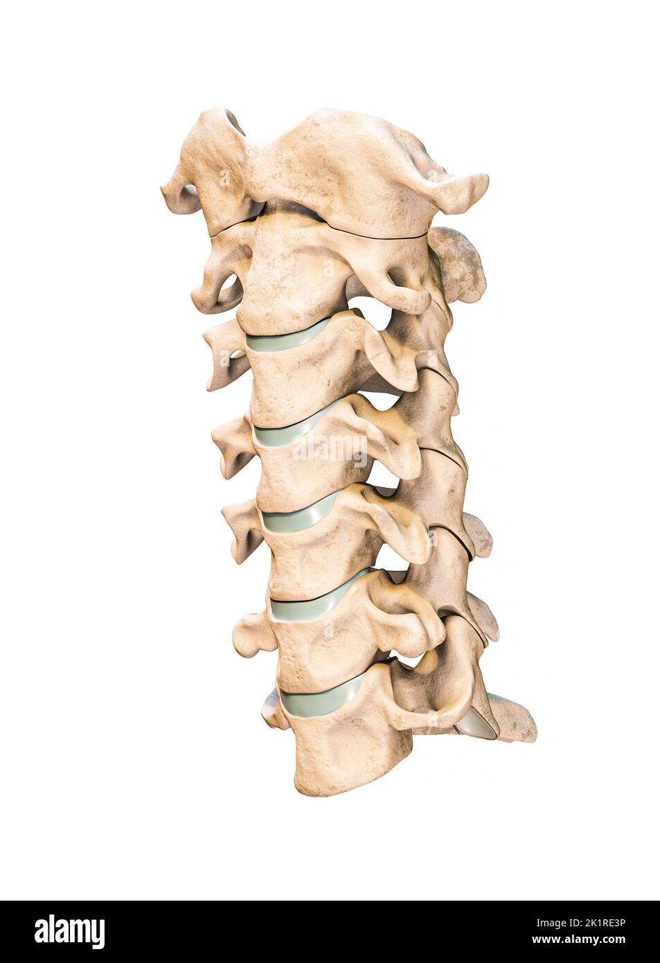 Three-quarter anterior or front view of the seven human cervical vertebrae isolated on white background 3D rendering illustration. Anatomy, osteology, Stock Photohttps://www.alamy.com/image-license-details/?v=1https://www.alamy.com/three-quarter-anterior-or-front-view-of-the-seven-human-cervical-vertebrae-isolated-on-white-background-3d-rendering-illustration-anatomy-osteology-image483020938.html
RF2K1RE3P–Three-quarter anterior or front view of the seven human cervical vertebrae isolated on white background 3D rendering illustration. Anatomy, osteology,
 Backache. Back pain. Pain in the cervical vertebra. Stock Photohttps://www.alamy.com/image-license-details/?v=1https://www.alamy.com/backache-back-pain-pain-in-the-cervical-vertebra-image344408448.html
RF2B094DM–Backache. Back pain. Pain in the cervical vertebra.
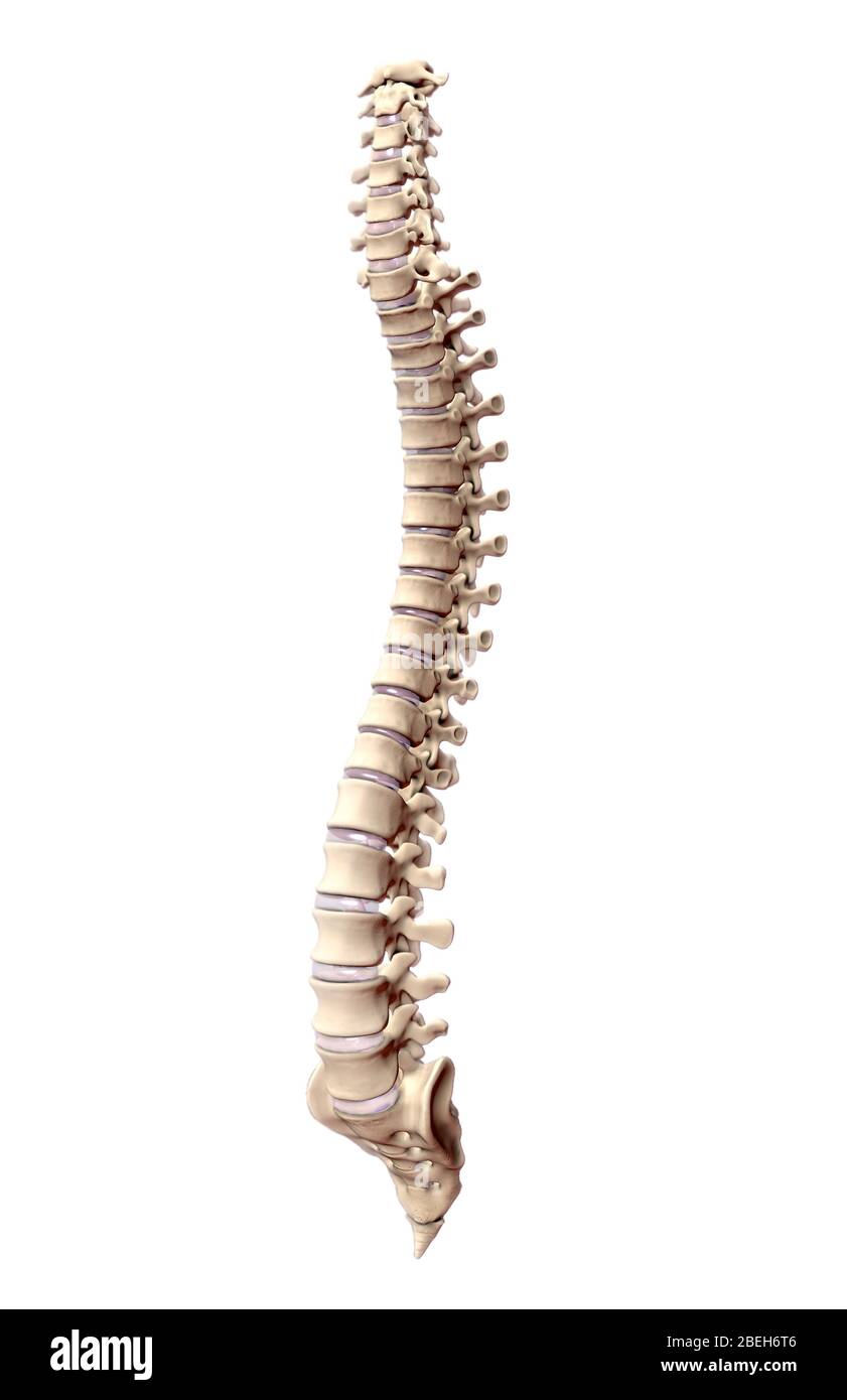 An illustration of the human spine depicting the cervical, thoracic, lumbar, and sacral vertebrae. Stock Photohttps://www.alamy.com/image-license-details/?v=1https://www.alamy.com/an-illustration-of-the-human-spine-depicting-the-cervical-thoracic-lumbar-and-sacral-vertebrae-image353191110.html
RM2BEH6T6–An illustration of the human spine depicting the cervical, thoracic, lumbar, and sacral vertebrae.
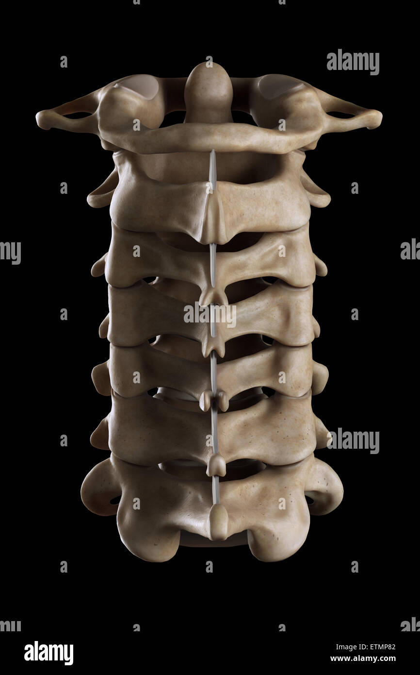 Illustration showing all seven cervical vertebrae. Stock Photohttps://www.alamy.com/image-license-details/?v=1https://www.alamy.com/stock-photo-illustration-showing-all-seven-cervical-vertebrae-84049730.html
RMETMP82–Illustration showing all seven cervical vertebrae.
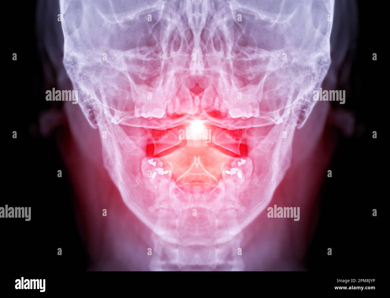 X-ray C-spine or x-ray image of Cervical spine open mount view for fracture of cervical vertebra 2nd ( axis ). Stock Photohttps://www.alamy.com/image-license-details/?v=1https://www.alamy.com/x-ray-c-spine-or-x-ray-image-of-cervical-spine-open-mount-view-for-fracture-of-cervical-vertebra-2nd-axis-image546005034.html
RF2PM8JYP–X-ray C-spine or x-ray image of Cervical spine open mount view for fracture of cervical vertebra 2nd ( axis ).
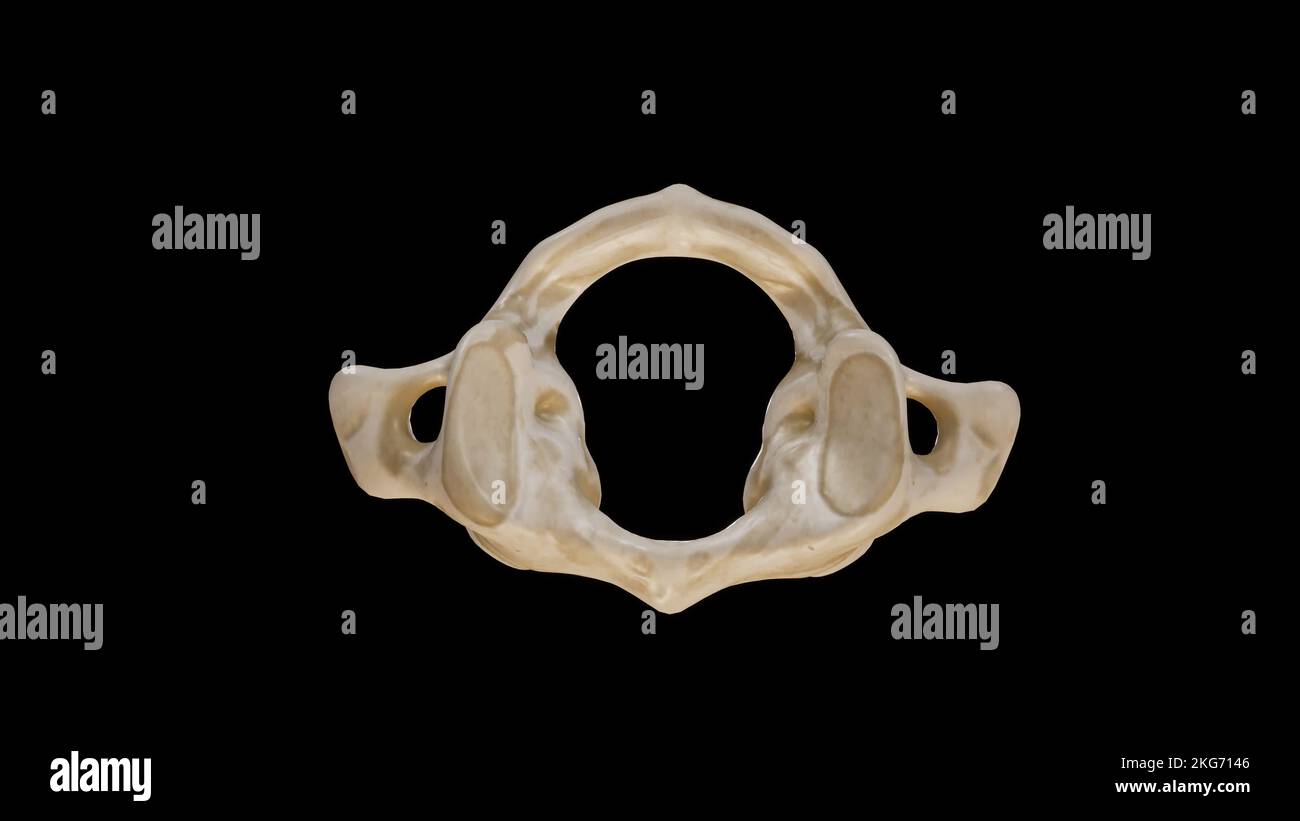 Superior view of First Cervical Vertebra (Atlas) Stock Photohttps://www.alamy.com/image-license-details/?v=1https://www.alamy.com/superior-view-of-first-cervical-vertebra-atlas-image491879366.html
RF2KG7146–Superior view of First Cervical Vertebra (Atlas)
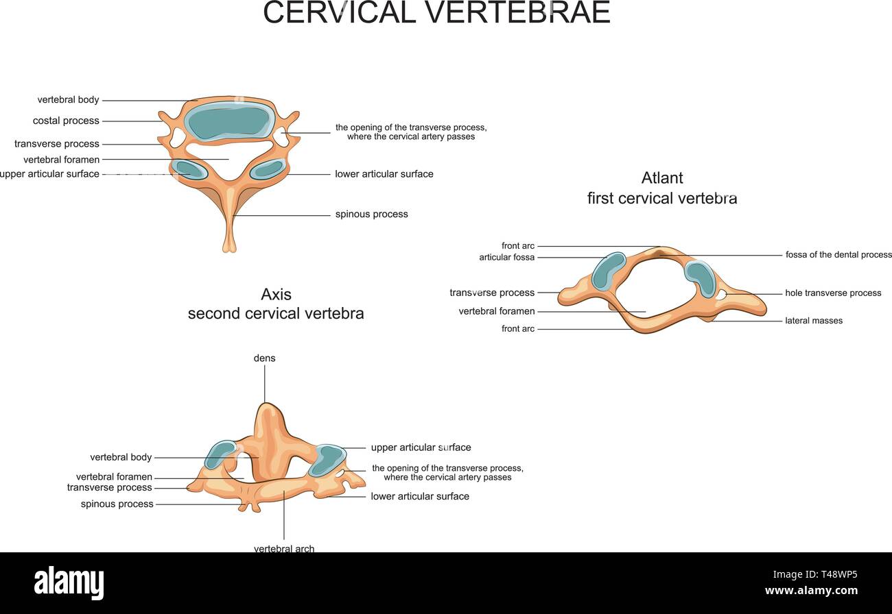 vector illustration of anatomy of cervical vertebrae Stock Vectorhttps://www.alamy.com/image-license-details/?v=1https://www.alamy.com/vector-illustration-of-anatomy-of-cervical-vertebrae-image243599613.html
RFT48WP5–vector illustration of anatomy of cervical vertebrae
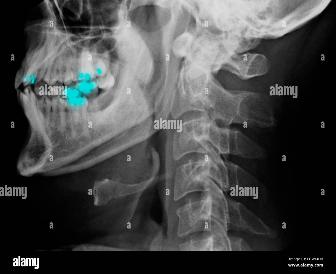 Normal cervical spine x-rays. Stock Photohttps://www.alamy.com/image-license-details/?v=1https://www.alamy.com/stock-photo-normal-cervical-spine-x-rays-76782311.html
RMECWMHB–Normal cervical spine x-rays.
 Human cervical spine computer artwork. Stock Photohttps://www.alamy.com/image-license-details/?v=1https://www.alamy.com/human-cervical-spine-computer-artwork-image69881127.html
RFE1KA2F–Human cervical spine computer artwork.
 neck of a young girl, pain in the spine on the neck, osteochondrosis and scaleo of the cervical vertebra, medicine and health Stock Photohttps://www.alamy.com/image-license-details/?v=1https://www.alamy.com/neck-of-a-young-girl-pain-in-the-spine-on-the-neck-osteochondrosis-and-scaleo-of-the-cervical-vertebra-medicine-and-health-image471630400.html
RF2JB8HAT–neck of a young girl, pain in the spine on the neck, osteochondrosis and scaleo of the cervical vertebra, medicine and health
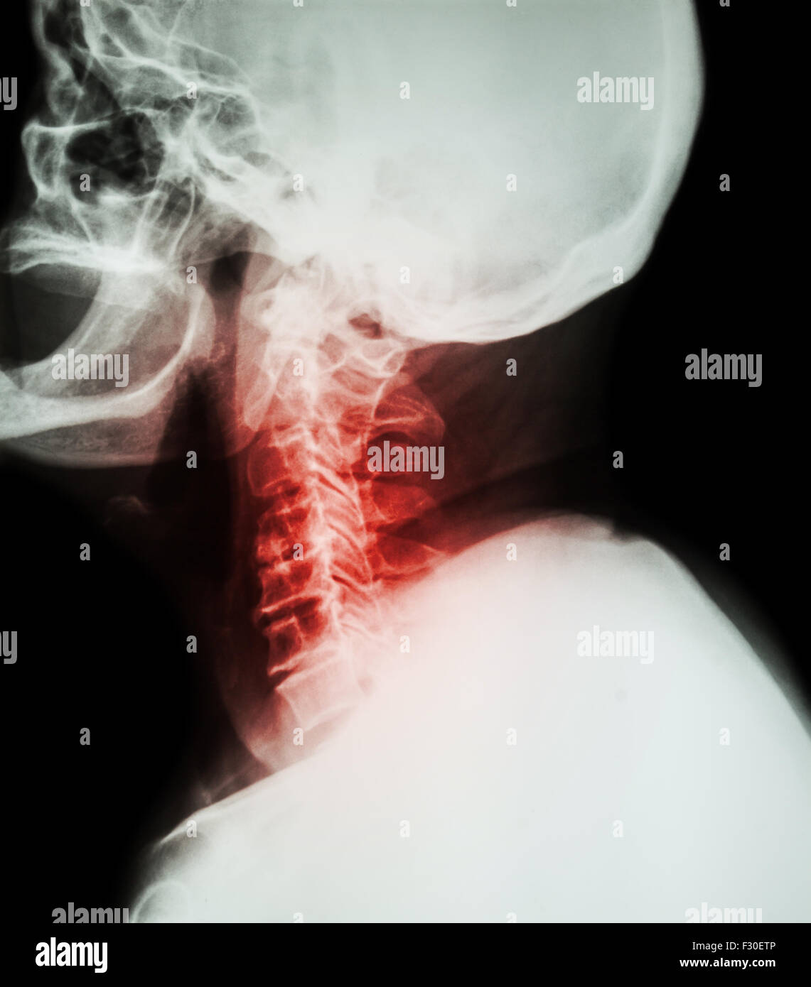 Cervical spondylosis . Film x-ray of cervical spine ( lateral position ) ( side view ) Stock Photohttps://www.alamy.com/image-license-details/?v=1https://www.alamy.com/stock-photo-cervical-spondylosis-film-x-ray-of-cervical-spine-lateral-position-87907478.html
RFF30ETP–Cervical spondylosis . Film x-ray of cervical spine ( lateral position ) ( side view )
 Figure of Atlas, it is the most superior cervical vertebra of the spine, vintage line drawing or engraving illustration. Stock Vectorhttps://www.alamy.com/image-license-details/?v=1https://www.alamy.com/figure-of-atlas-it-is-the-most-superior-cervical-vertebra-of-the-spine-vintage-line-drawing-or-engraving-illustration-image348663753.html
RF2B7704W–Figure of Atlas, it is the most superior cervical vertebra of the spine, vintage line drawing or engraving illustration.
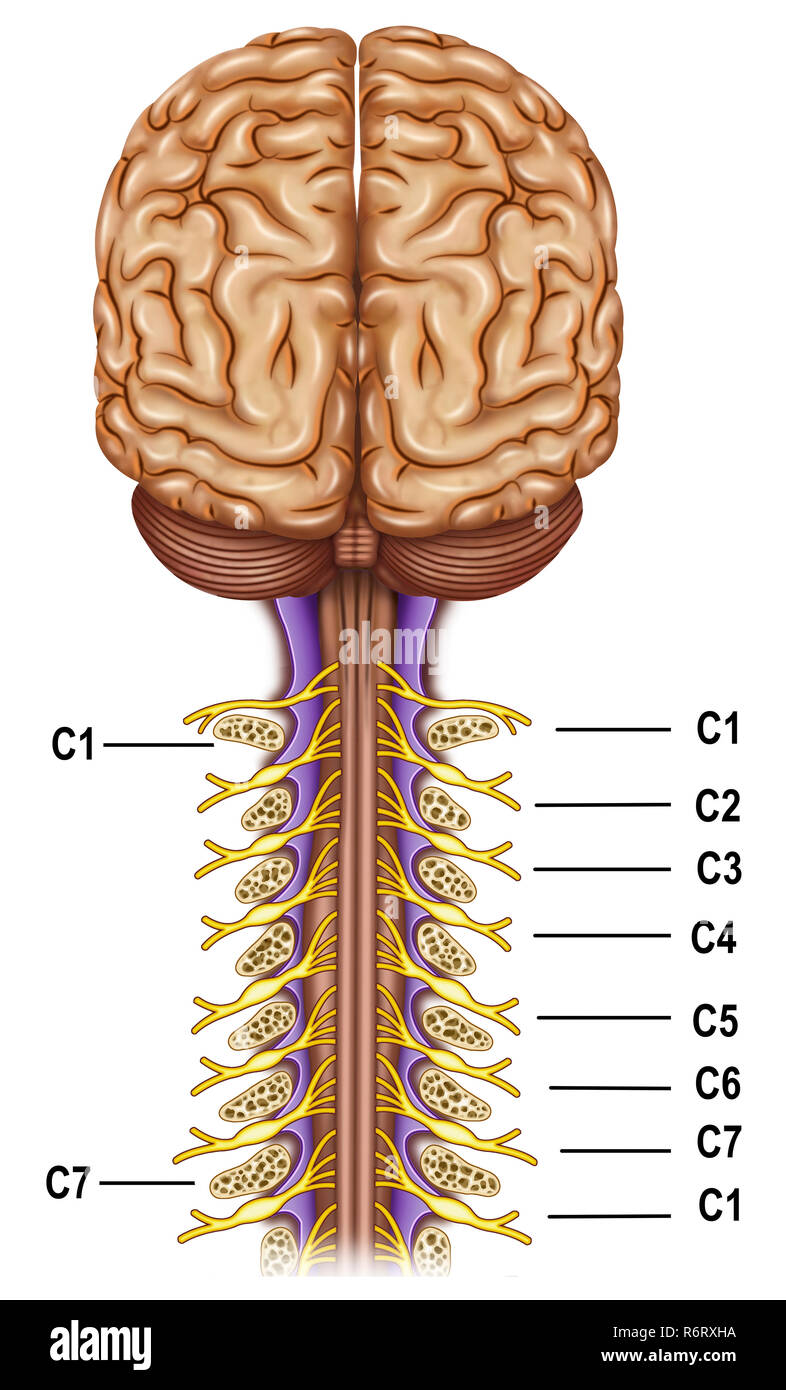 The cervical plexus. It controls the motor functions of the neck, it is the superior nervous plexus of the peripheral nervous system. Stock Photohttps://www.alamy.com/image-license-details/?v=1https://www.alamy.com/the-cervical-plexus-it-controls-the-motor-functions-of-the-neck-it-is-the-superior-nervous-plexus-of-the-peripheral-nervous-system-image227948486.html
RFR6RXHA–The cervical plexus. It controls the motor functions of the neck, it is the superior nervous plexus of the peripheral nervous system.
 Doctor looking at a of the cervical spine x-ray of her patient. Stock Photohttps://www.alamy.com/image-license-details/?v=1https://www.alamy.com/stock-photo-doctor-looking-at-a-of-the-cervical-spine-x-ray-of-her-patient-72441884.html
RME5T0A4–Doctor looking at a of the cervical spine x-ray of her patient.
 Cervical vertebra is a part of bird skeletal system. Bird anatomy. Bird skeletal system. Avian skeletal systems are modified according to their usage. Stock Photohttps://www.alamy.com/image-license-details/?v=1https://www.alamy.com/cervical-vertebra-is-a-part-of-bird-skeletal-system-bird-anatomy-bird-skeletal-system-avian-skeletal-systems-are-modified-according-to-their-usage-image517453497.html
RF2N1T161–Cervical vertebra is a part of bird skeletal system. Bird anatomy. Bird skeletal system. Avian skeletal systems are modified according to their usage.
 CERVICAL VERTEBRA, X-RAY Stock Photohttps://www.alamy.com/image-license-details/?v=1https://www.alamy.com/stock-photo-cervical-vertebra-x-ray-49247948.html
RMCT3C78–CERVICAL VERTEBRA, X-RAY
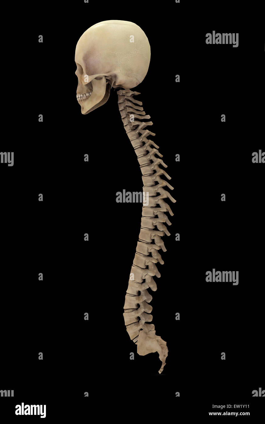 3D rendering of human vertebral column, side view. Stock Photohttps://www.alamy.com/image-license-details/?v=1https://www.alamy.com/stock-photo-3d-rendering-of-human-vertebral-column-side-view-84251021.html
RFEW1Y11–3D rendering of human vertebral column, side view.
 Cervical vertebra Stock Photohttps://www.alamy.com/image-license-details/?v=1https://www.alamy.com/stock-photo-cervical-vertebra-13220324.html
RFACRTTN–Cervical vertebra
 x-ray of cervical fusion surgery Stock Photohttps://www.alamy.com/image-license-details/?v=1https://www.alamy.com/x-ray-of-cervical-fusion-surgery-image65494873.html
RFDPFFAH–x-ray of cervical fusion surgery
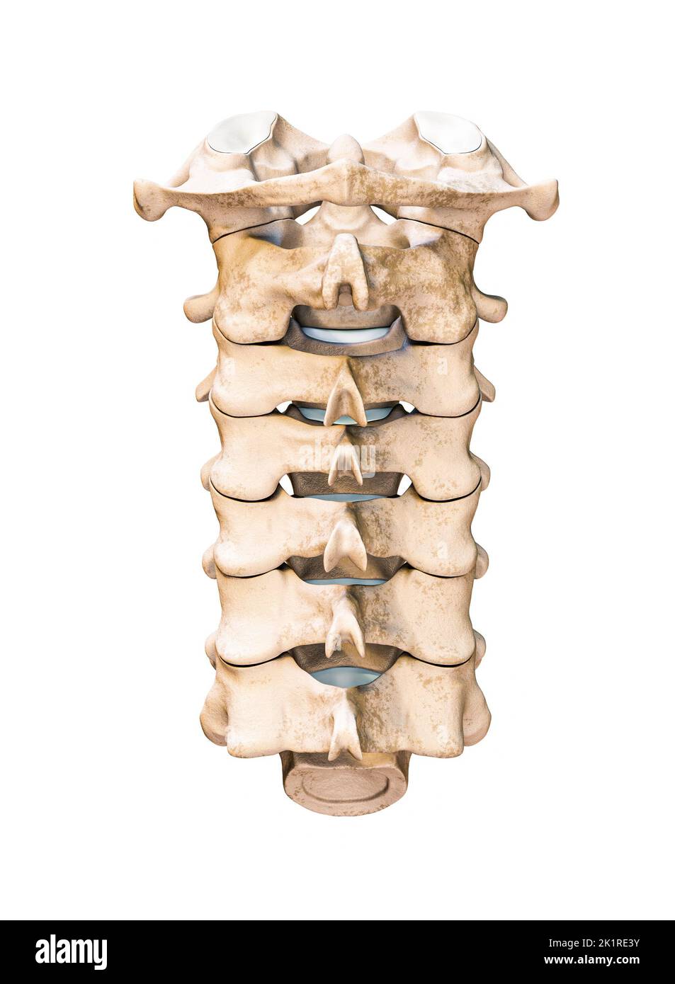 Posterior or rear or back view of the seven human cervical vertebrae isolated on white background 3D rendering illustration. Anatomy, osteology, blank Stock Photohttps://www.alamy.com/image-license-details/?v=1https://www.alamy.com/posterior-or-rear-or-back-view-of-the-seven-human-cervical-vertebrae-isolated-on-white-background-3d-rendering-illustration-anatomy-osteology-blank-image483020943.html
RF2K1RE3Y–Posterior or rear or back view of the seven human cervical vertebrae isolated on white background 3D rendering illustration. Anatomy, osteology, blank
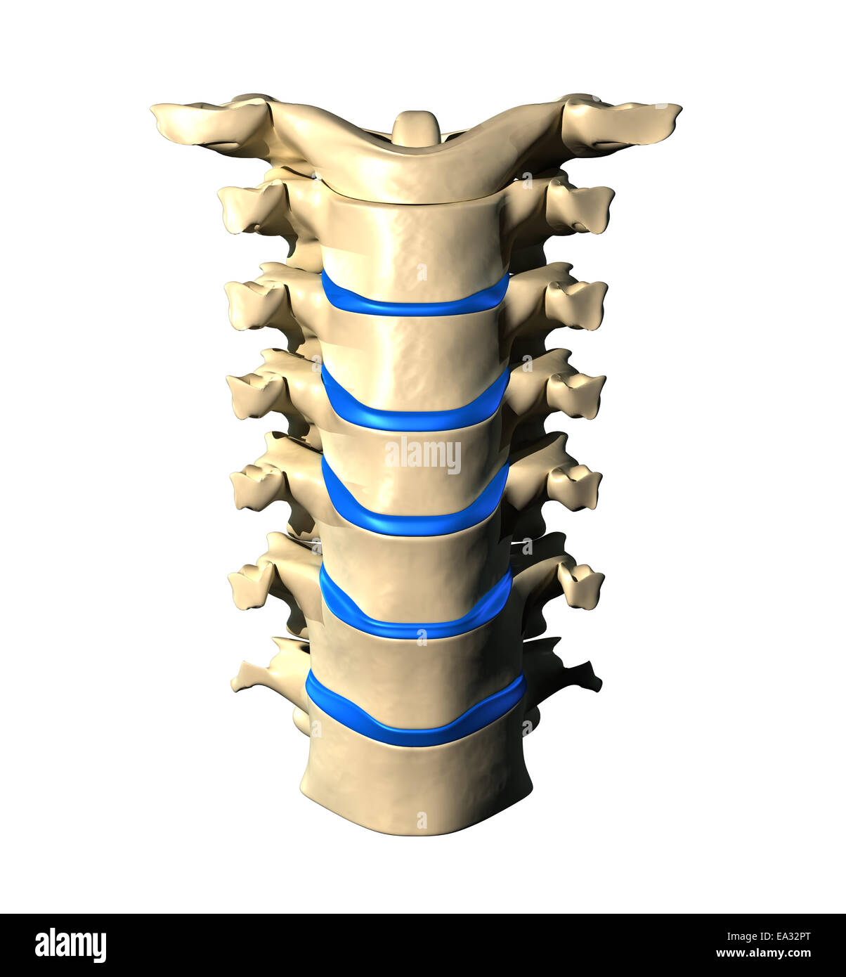 Cervical Spine - Anterior / Front view Stock Photohttps://www.alamy.com/image-license-details/?v=1https://www.alamy.com/stock-photo-cervical-spine-anterior-front-view-75056096.html
RFEA32PT–Cervical Spine - Anterior / Front view
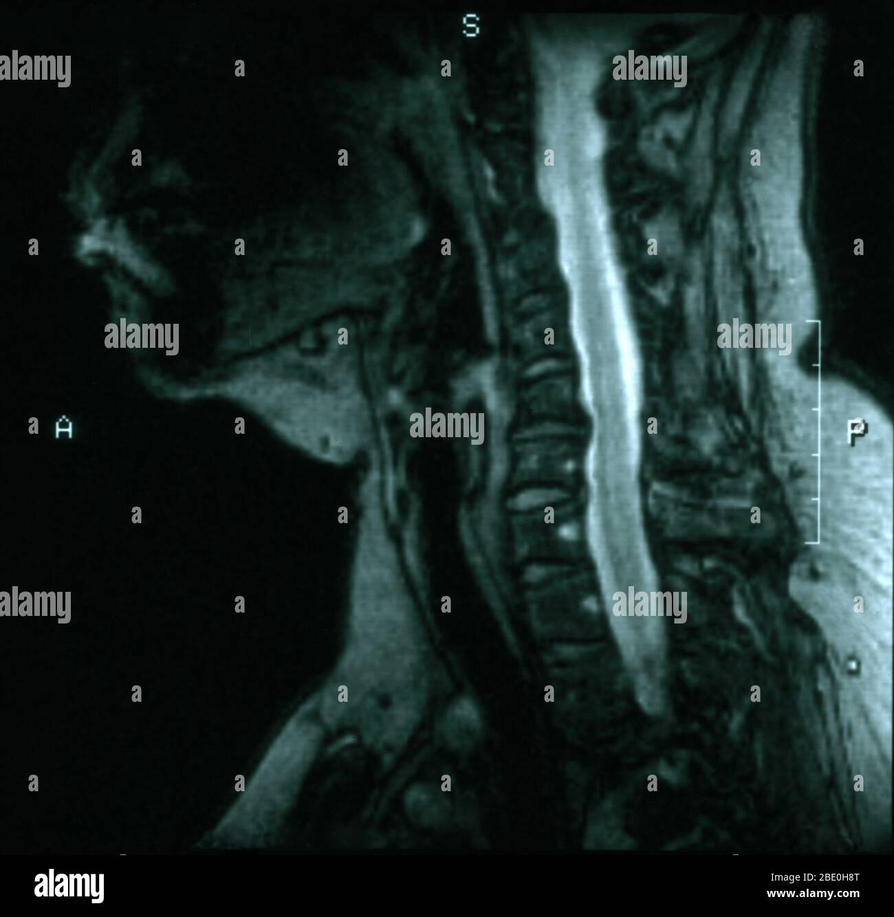 MRI of the cervical spine from the craniovertebral junction down T3-T4 level. MRI slice thickness is 5.0MM. The MRI shows degenerative disc disease at C5-6 and C6-7 with posterior osteophytes at both levels causing a slight narrowing of the spinal canal particularly at C5-6. Stock Photohttps://www.alamy.com/image-license-details/?v=1https://www.alamy.com/mri-of-the-cervical-spine-from-the-craniovertebral-junction-down-t3-t4-level-mri-slice-thickness-is-50mm-the-mri-shows-degenerative-disc-disease-at-c5-6-and-c6-7-with-posterior-osteophytes-at-both-levels-causing-a-slight-narrowing-of-the-spinal-canal-particularly-at-c5-6-image352826120.html
RM2BE0H8T–MRI of the cervical spine from the craniovertebral junction down T3-T4 level. MRI slice thickness is 5.0MM. The MRI shows degenerative disc disease at C5-6 and C6-7 with posterior osteophytes at both levels causing a slight narrowing of the spinal canal particularly at C5-6.
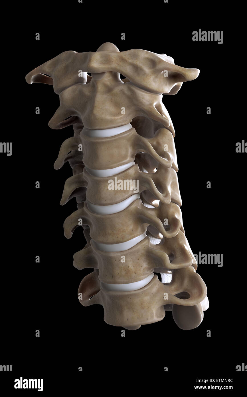 Illustration showing all seven cervical vertebrae. Stock Photohttps://www.alamy.com/image-license-details/?v=1https://www.alamy.com/stock-photo-illustration-showing-all-seven-cervical-vertebrae-84049376.html
RMETMNRC–Illustration showing all seven cervical vertebrae.
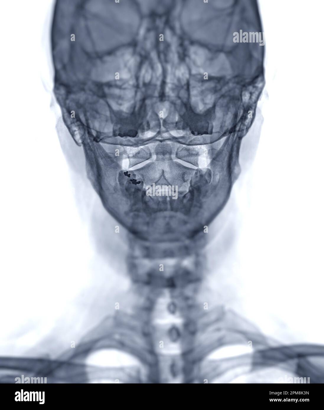 X-ray C-spine or x-ray image of Cervical spine open mount view for fracture of cervical vertebra 2nd ( axis ). Stock Photohttps://www.alamy.com/image-license-details/?v=1https://www.alamy.com/x-ray-c-spine-or-x-ray-image-of-cervical-spine-open-mount-view-for-fracture-of-cervical-vertebra-2nd-axis-image546005145.html
RF2PM8K3N–X-ray C-spine or x-ray image of Cervical spine open mount view for fracture of cervical vertebra 2nd ( axis ).
 First Cervical Vertebra (Atlas) -Multiple Views Stock Photohttps://www.alamy.com/image-license-details/?v=1https://www.alamy.com/first-cervical-vertebra-atlas-multiple-views-image491879617.html
RF2KG71D5–First Cervical Vertebra (Atlas) -Multiple Views
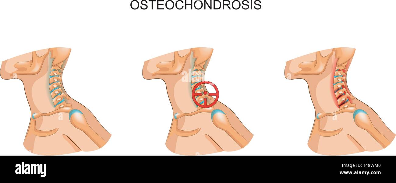 vector illustration of osteochondrosis of the cervical vertebrae Stock Vectorhttps://www.alamy.com/image-license-details/?v=1https://www.alamy.com/vector-illustration-of-osteochondrosis-of-the-cervical-vertebrae-image243599552.html
RFT48WM0–vector illustration of osteochondrosis of the cervical vertebrae
 Normal cervical spine x-rays. Stock Photohttps://www.alamy.com/image-license-details/?v=1https://www.alamy.com/stock-photo-normal-cervical-spine-x-rays-76782308.html
RMECWMH8–Normal cervical spine x-rays.
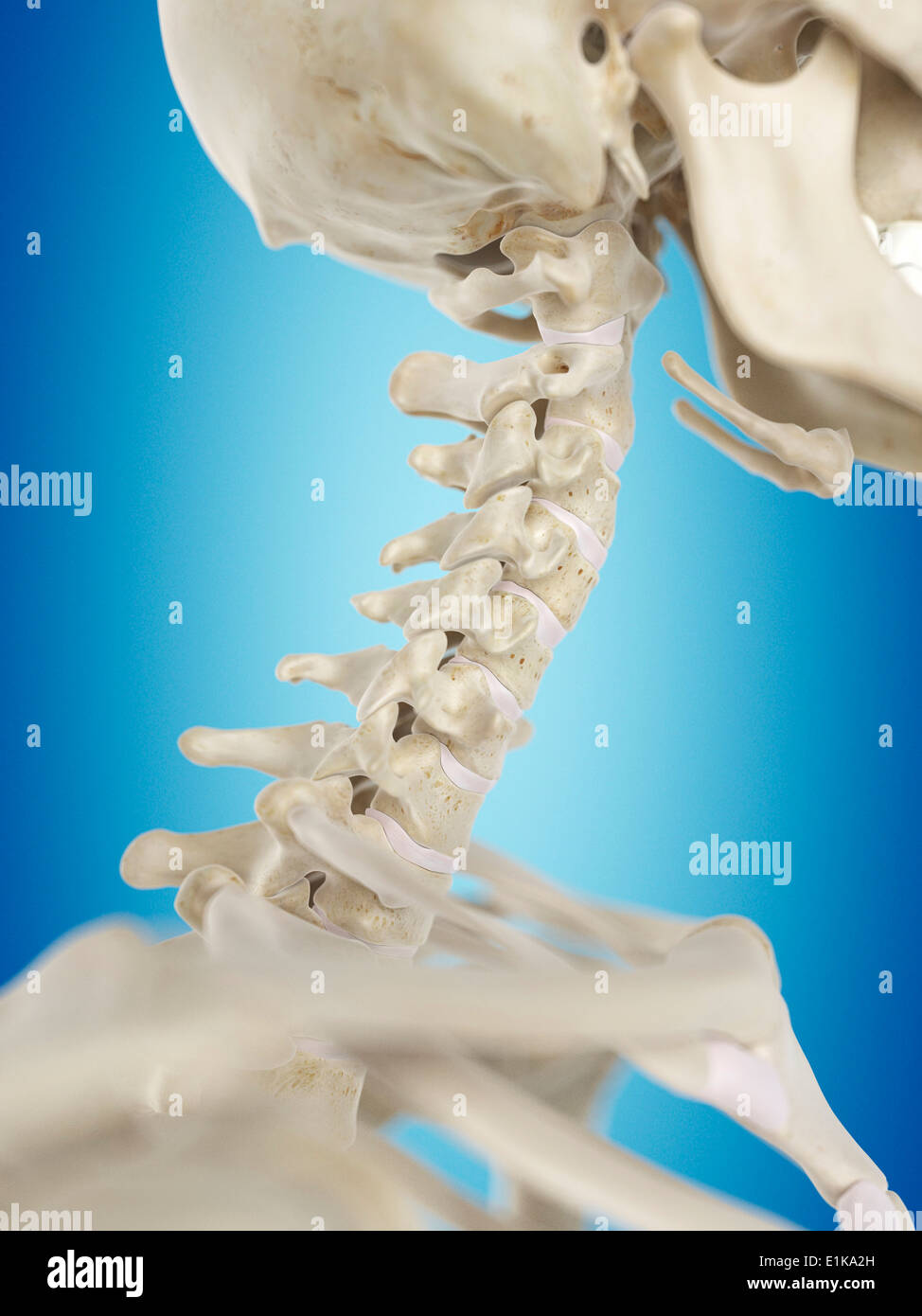 Human cervical spine computer artwork. Stock Photohttps://www.alamy.com/image-license-details/?v=1https://www.alamy.com/human-cervical-spine-computer-artwork-image69881129.html
RFE1KA2H–Human cervical spine computer artwork.
 neck of a young girl, pain in the spine on the neck, osteochondrosis and scaleo of the cervical vertebra, medicine and health Stock Photohttps://www.alamy.com/image-license-details/?v=1https://www.alamy.com/neck-of-a-young-girl-pain-in-the-spine-on-the-neck-osteochondrosis-and-scaleo-of-the-cervical-vertebra-medicine-and-health-image471630414.html
RF2JB8HBA–neck of a young girl, pain in the spine on the neck, osteochondrosis and scaleo of the cervical vertebra, medicine and health
 Cervical spondylosis . Film x-ray of cervical spine ( lateral position ) ( side view ) Stock Photohttps://www.alamy.com/image-license-details/?v=1https://www.alamy.com/stock-photo-cervical-spondylosis-film-x-ray-of-cervical-spine-lateral-position-87907477.html
RFF30ETN–Cervical spondylosis . Film x-ray of cervical spine ( lateral position ) ( side view )
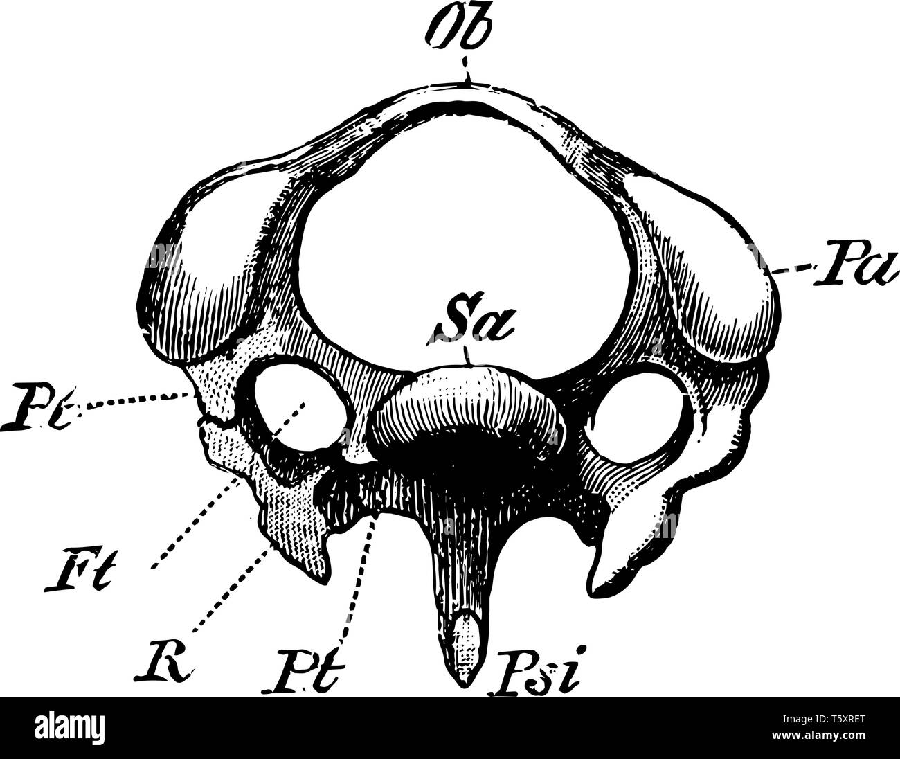 The Diagram of a Third Cervical Vertebra of a Woodpecker, vintage line drawing or engraving illustration. Stock Vectorhttps://www.alamy.com/image-license-details/?v=1https://www.alamy.com/the-diagram-of-a-third-cervical-vertebra-of-a-woodpecker-vintage-line-drawing-or-engraving-illustration-image244607632.html
RFT5XRET–The Diagram of a Third Cervical Vertebra of a Woodpecker, vintage line drawing or engraving illustration.
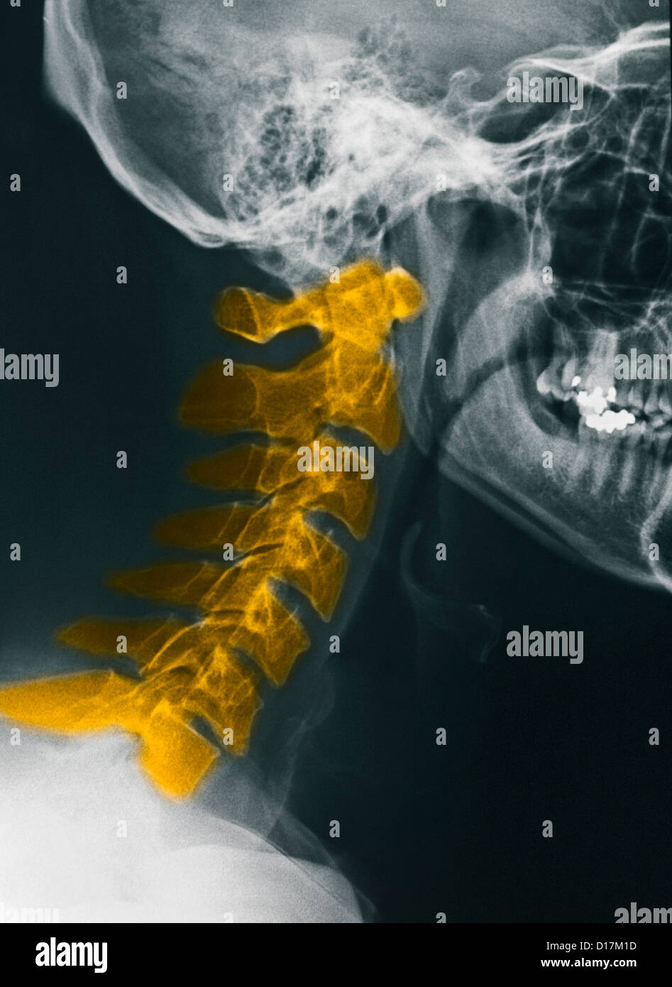 CerviCal spine X-rays, normal Stock Photohttps://www.alamy.com/image-license-details/?v=1https://www.alamy.com/stock-photo-cervical-spine-x-rays-normal-52415145.html
RFD17M1D–CerviCal spine X-rays, normal
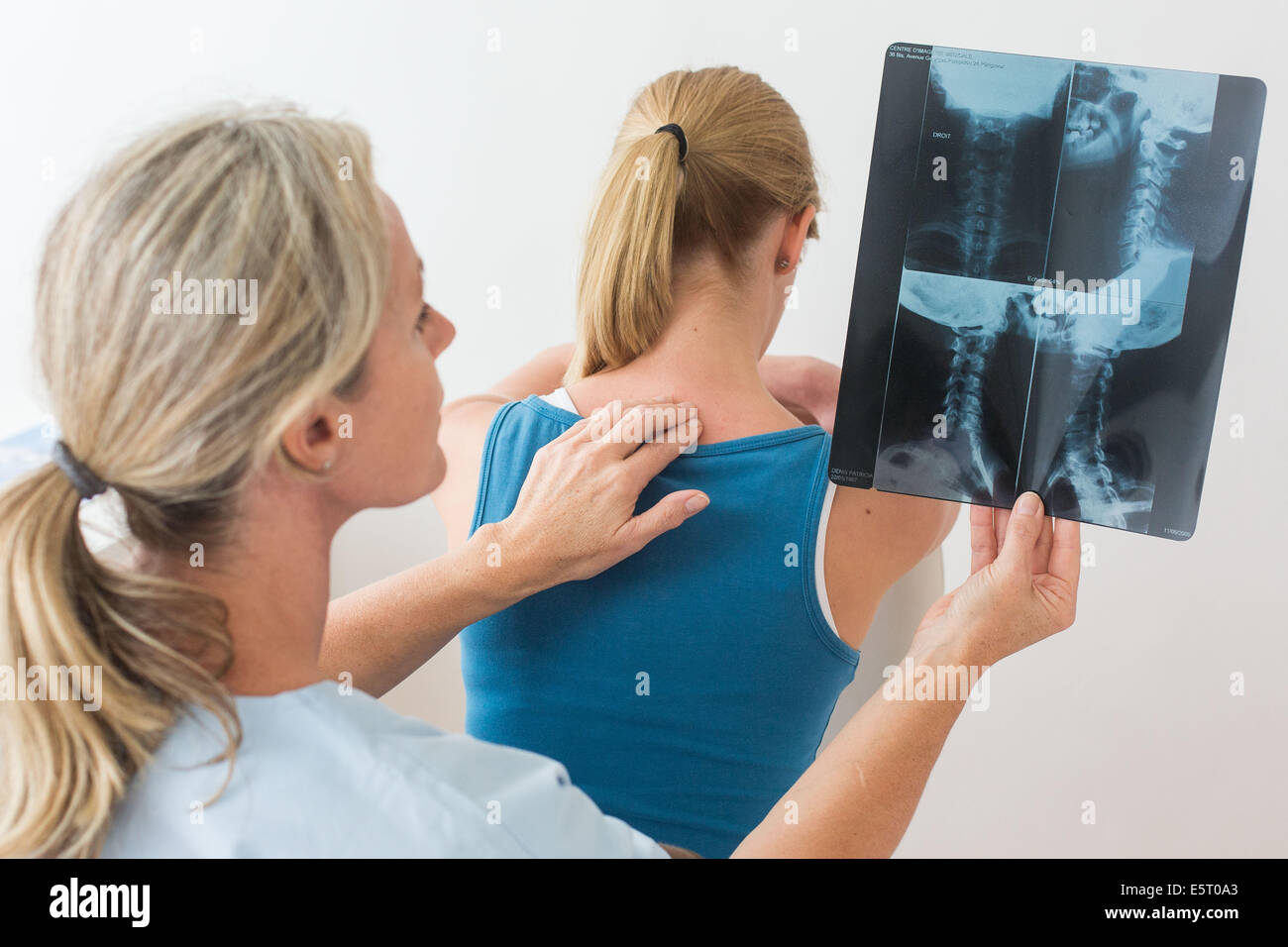 Doctor looking at a of the cervical spine x-ray of her patient. Stock Photohttps://www.alamy.com/image-license-details/?v=1https://www.alamy.com/stock-photo-doctor-looking-at-a-of-the-cervical-spine-x-ray-of-her-patient-72441883.html
RME5T0A3–Doctor looking at a of the cervical spine x-ray of her patient.
 Cervical spine Stock Photohttps://www.alamy.com/image-license-details/?v=1https://www.alamy.com/cervical-spine-image360869835.html
RM2BY314B–Cervical spine
 CERVICAL VERTEBRA, X-RAY Stock Photohttps://www.alamy.com/image-license-details/?v=1https://www.alamy.com/stock-photo-cervical-vertebra-x-ray-49247951.html
RMCT3C7B–CERVICAL VERTEBRA, X-RAY
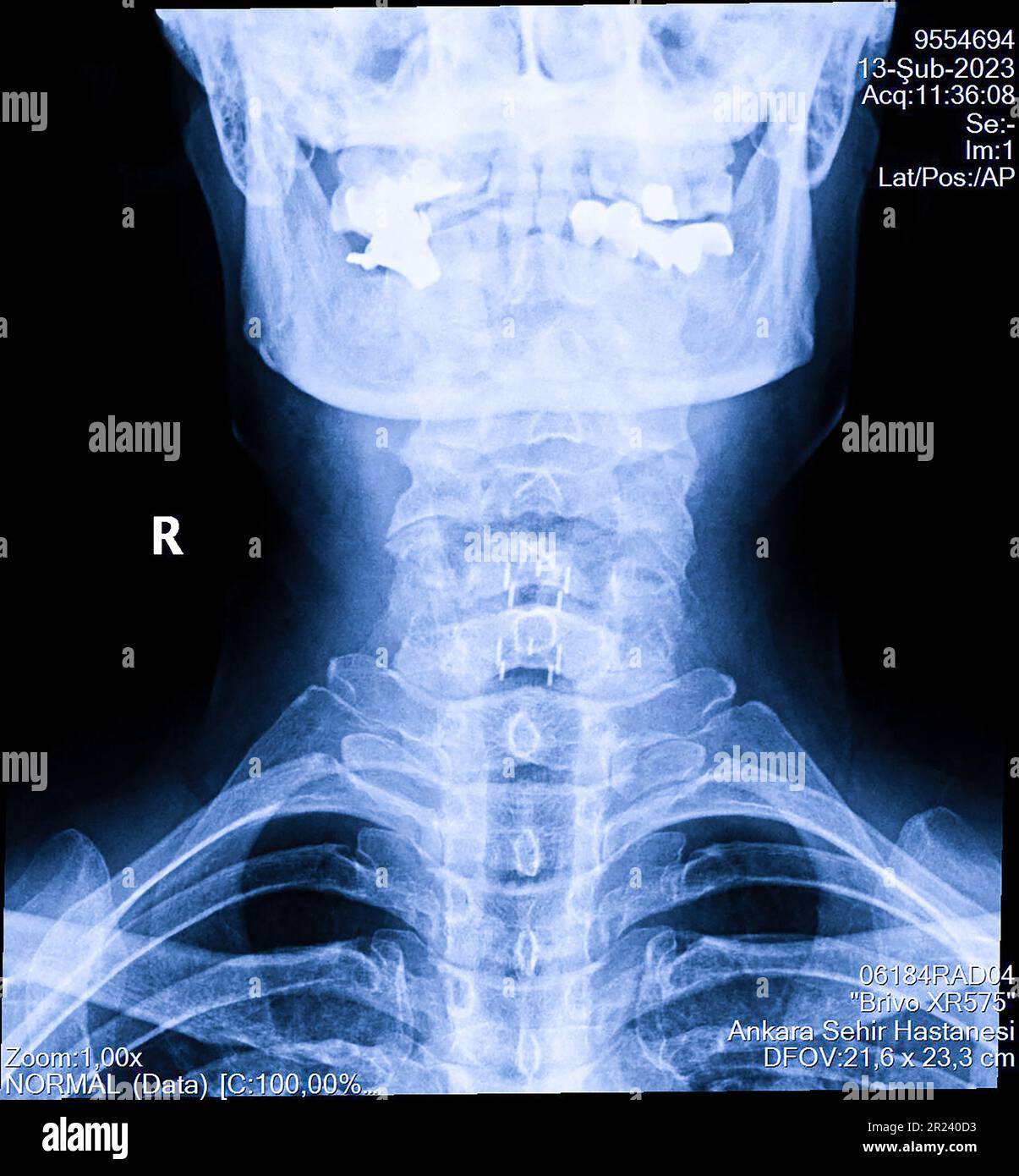 Human cervical spine x-ray, neck radiography Stock Photohttps://www.alamy.com/image-license-details/?v=1https://www.alamy.com/human-cervical-spine-x-ray-neck-radiography-image552049263.html
RF2R240D3–Human cervical spine x-ray, neck radiography
