Quick filters:
Cervical vertebrae 7 Stock Photos and Images
 A normal human vertebral column (lateral view). Shown (from top to bottom) are the atlas (cervical vertebra 1: C1), axis (C2), cervical vertebrae 3-7 Stock Photohttps://www.alamy.com/image-license-details/?v=1https://www.alamy.com/stock-photo-a-normal-human-vertebral-column-lateral-view-shown-from-top-to-bottom-130806568.html
RFHGPN34–A normal human vertebral column (lateral view). Shown (from top to bottom) are the atlas (cervical vertebra 1: C1), axis (C2), cervical vertebrae 3-7
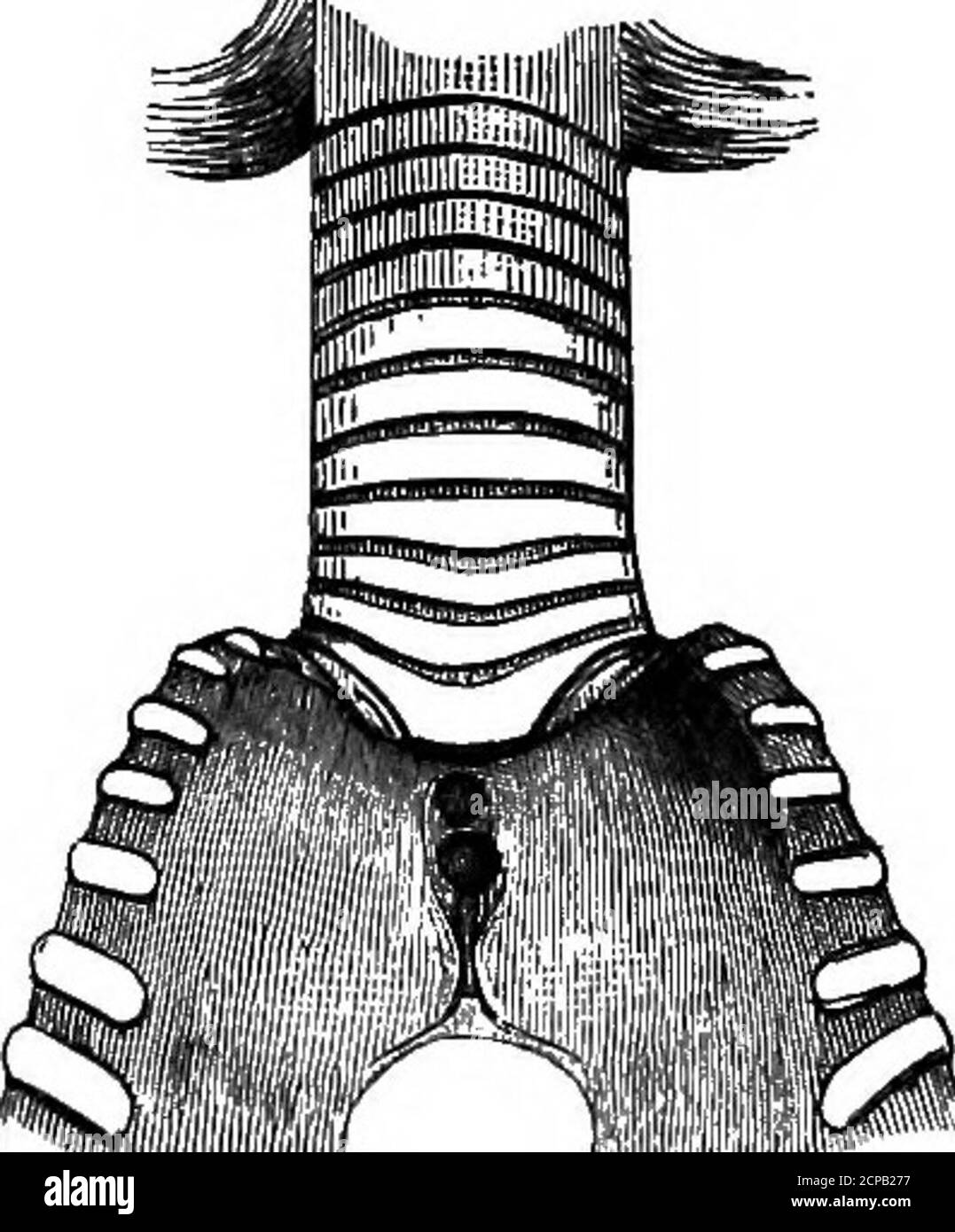 . The structure and classification of birds . s fuse4anteriorly with the walls of the.skull. The nostrils are continuedforward by a groove precisely likethat of Scopus and Gancroma. Inthe palatine bones the fusion of theinternal laminae to form a mediankeel behind the interparietal spaceis precisely like Scopus; so, too, isthe lateral angle of these bones (see p. 422). There is a firmsynostosis between the furcula and the carina sterni. Cervical vertebrae 7-13 have, as in most other Herodiones(excluding, however, the supposed ally of BalcBniceps, Scopus),a ventral catapophysial canal. The fami Stock Photohttps://www.alamy.com/image-license-details/?v=1https://www.alamy.com/the-structure-and-classification-of-birds-s-fuse4anteriorly-with-the-walls-of-theskull-the-nostrils-are-continuedforward-by-a-groove-precisely-likethat-of-scopus-and-gancroma-inthe-palatine-bones-the-fusion-of-theinternal-laminae-to-form-a-mediankeel-behind-the-interparietal-spaceis-precisely-like-scopus-so-too-isthe-lateral-angle-of-these-bones-see-p-422-there-is-a-firmsynostosis-between-the-furcula-and-the-carina-sterni-cervical-vertebrae-7-13-have-as-in-most-other-herodionesexcluding-however-the-supposed-ally-of-balcbniceps-scopusa-ventral-catapophysial-canal-the-fami-image375183403.html
RM2CPB277–. The structure and classification of birds . s fuse4anteriorly with the walls of the.skull. The nostrils are continuedforward by a groove precisely likethat of Scopus and Gancroma. Inthe palatine bones the fusion of theinternal laminae to form a mediankeel behind the interparietal spaceis precisely like Scopus; so, too, isthe lateral angle of these bones (see p. 422). There is a firmsynostosis between the furcula and the carina sterni. Cervical vertebrae 7-13 have, as in most other Herodiones(excluding, however, the supposed ally of BalcBniceps, Scopus),a ventral catapophysial canal. The fami
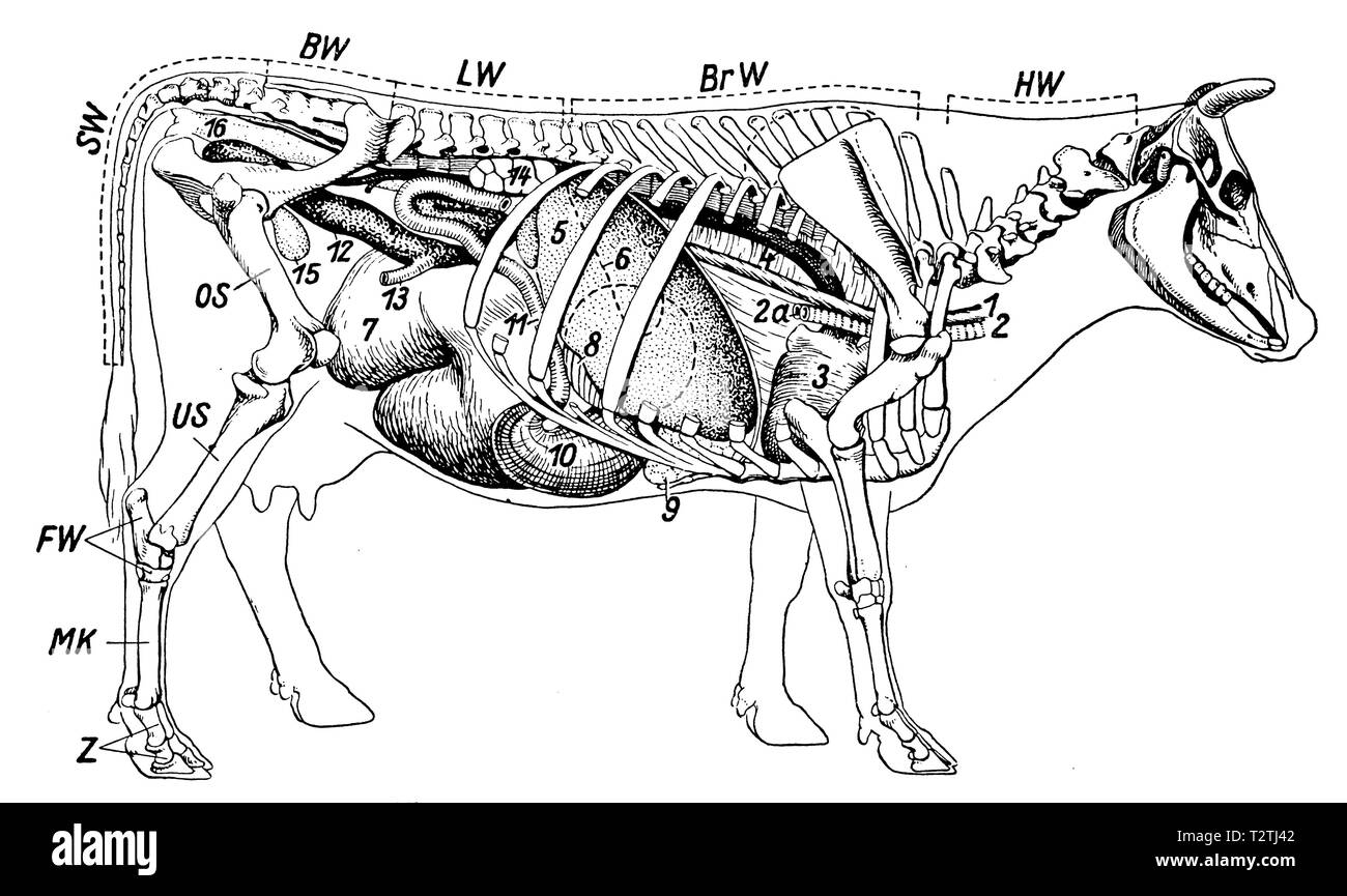 Skeleton and internal organs of a cow. HW Cervical Vertebrae, BrW Thoracic Vertebrae, LW Lumbar Vertebrae, BW Pelvic Vertebrae, SW Cervical Vertebrae, OS Femoral Bones, US Lower Limb, FW Tarsal Bones, MK Metatarsal Bones, Z Toe Bones, 1 Esophagus, 2 Trachea, 2a Tracheal Fibula, 3 Heart, 4 Body Artery, 5 Liver, 6 Spleen (Outline dashed, obscured by the liver), 7 rumen, 8 leaves (mostly obscured), 9 reticulum, 10 abomasum, 11 small intestine, 12 cecum, 13 small intestine into the colon, 14 kidney, 15 bladder, 16 rectum. (Lungs not shown), 1941 Stock Photohttps://www.alamy.com/image-license-details/?v=1https://www.alamy.com/skeleton-and-internal-organs-of-a-cow-hw-cervical-vertebrae-brw-thoracic-vertebrae-lw-lumbar-vertebrae-bw-pelvic-vertebrae-sw-cervical-vertebrae-os-femoral-bones-us-lower-limb-fw-tarsal-bones-mk-metatarsal-bones-z-toe-bones-1-esophagus-2-trachea-2a-tracheal-fibula-3-heart-4-body-artery-5-liver-6-spleen-outline-dashed-obscured-by-the-liver-7-rumen-8-leaves-mostly-obscured-9-reticulum-10-abomasum-11-small-intestine-12-cecum-13-small-intestine-into-the-colon-14-kidney-15-bladder-16-rectum-lungs-not-shown-1941-image242715538.html
RMT2TJ42–Skeleton and internal organs of a cow. HW Cervical Vertebrae, BrW Thoracic Vertebrae, LW Lumbar Vertebrae, BW Pelvic Vertebrae, SW Cervical Vertebrae, OS Femoral Bones, US Lower Limb, FW Tarsal Bones, MK Metatarsal Bones, Z Toe Bones, 1 Esophagus, 2 Trachea, 2a Tracheal Fibula, 3 Heart, 4 Body Artery, 5 Liver, 6 Spleen (Outline dashed, obscured by the liver), 7 rumen, 8 leaves (mostly obscured), 9 reticulum, 10 abomasum, 11 small intestine, 12 cecum, 13 small intestine into the colon, 14 kidney, 15 bladder, 16 rectum. (Lungs not shown), 1941
 . Fio. 1.—Skkleton of the Doa—Carntvora—(Strangkway). Axial Skeleton. The Skull. Cranial Bones.a. Occipital, 1: 6, Parietal, 2; c. Frontal, 2; /.% Temporal, 2; Sphenoid, 1; Ethmoid, 2; Auditory ossicles, 8. Facial Bones.—f, Nasal, 2; e. Lachrymal, 2; d. Malar, 2; /(, Maxilla, 2; gr, Premaxilla, 2; t, Inferior maxilla, 2; Palatine, 2; Pterygoid, 2; Vomer, 1; Turbinals, 4; Hyoid (segments), 9, Teef/i.—Incisors, 12; Canines, 4; Molars. 26. The Trunk.—Z I, Cervical vertebrae, 7; m m. Dorsal vertebrae, 13; ?i n, Lumbar vertebrae, 7; o. Sacrum (three segments), 1; p p. Coccygeal vertebrae (variable Stock Photohttps://www.alamy.com/image-license-details/?v=1https://www.alamy.com/fio-1skkleton-of-the-doacarntvorastrangkway-axial-skeleton-the-skull-cranial-bonesa-occipital-1-6-parietal-2-c-frontal-2-temporal-2-sphenoid-1-ethmoid-2-auditory-ossicles-8-facial-bonesf-nasal-2-e-lachrymal-2-d-malar-2-maxilla-2-gr-premaxilla-2-t-inferior-maxilla-2-palatine-2-pterygoid-2-vomer-1-turbinals-4-hyoid-segments-9-teefiincisors-12-canines-4-molars-26-the-trunkz-i-cervical-vertebrae-7-m-m-dorsal-vertebrae-13-i-n-lumbar-vertebrae-7-o-sacrum-three-segments-1-p-p-coccygeal-vertebrae-variable-image179906866.html
RMMCKD16–. Fio. 1.—Skkleton of the Doa—Carntvora—(Strangkway). Axial Skeleton. The Skull. Cranial Bones.a. Occipital, 1: 6, Parietal, 2; c. Frontal, 2; /.% Temporal, 2; Sphenoid, 1; Ethmoid, 2; Auditory ossicles, 8. Facial Bones.—f, Nasal, 2; e. Lachrymal, 2; d. Malar, 2; /(, Maxilla, 2; gr, Premaxilla, 2; t, Inferior maxilla, 2; Palatine, 2; Pterygoid, 2; Vomer, 1; Turbinals, 4; Hyoid (segments), 9, Teef/i.—Incisors, 12; Canines, 4; Molars. 26. The Trunk.—Z I, Cervical vertebrae, 7; m m. Dorsal vertebrae, 13; ?i n, Lumbar vertebrae, 7; o. Sacrum (three segments), 1; p p. Coccygeal vertebrae (variable
 . A diapsid reptile from the Pennsylvanian of Kansas. Reptiles, Fossil -- Kansas; Paleontology -- Pennsylvanian; Paleontology -- Kansas. 18 SPECIAL PUBLICATION MUSEUM OF NATURAL HISTORY NO. 7. Fig. 12.—Petrolacosaurus kansenxis Lane. Mature skull and lower jaws with articulated cervical vertebrae, and an isolated right clethnim, KUVP 33607, X 2. Tlie component .skull elements are disarticulated, and com- pressed into a single plane. The riglit mandible and the ertel)rac are exposed in lateral view. See Fig. 13 for opposite view of same specimen. See Fig. 2 for key to abbreviations.. Please no Stock Photohttps://www.alamy.com/image-license-details/?v=1https://www.alamy.com/a-diapsid-reptile-from-the-pennsylvanian-of-kansas-reptiles-fossil-kansas-paleontology-pennsylvanian-paleontology-kansas-18-special-publication-museum-of-natural-history-no-7-fig-12petrolacosaurus-kansenxis-lane-mature-skull-and-lower-jaws-with-articulated-cervical-vertebrae-and-an-isolated-right-clethnim-kuvp-33607-x-2-tlie-component-skull-elements-are-disarticulated-and-com-pressed-into-a-single-plane-the-riglit-mandible-and-the-ertelrac-are-exposed-in-lateral-view-see-fig-13-for-opposite-view-of-same-specimen-see-fig-2-for-key-to-abbreviations-please-no-image215967129.html
RMPFA47N–. A diapsid reptile from the Pennsylvanian of Kansas. Reptiles, Fossil -- Kansas; Paleontology -- Pennsylvanian; Paleontology -- Kansas. 18 SPECIAL PUBLICATION MUSEUM OF NATURAL HISTORY NO. 7. Fig. 12.—Petrolacosaurus kansenxis Lane. Mature skull and lower jaws with articulated cervical vertebrae, and an isolated right clethnim, KUVP 33607, X 2. Tlie component .skull elements are disarticulated, and com- pressed into a single plane. The riglit mandible and the ertel)rac are exposed in lateral view. See Fig. 13 for opposite view of same specimen. See Fig. 2 for key to abbreviations.. Please no
 The diseases and disorders of The diseases and disorders of the ox, with some account of the diseases of the sheep diseasesdisorderox00gres Year: 1889 32 THE DISEASES AND DISORDERS OF THE OX. speedy pace. Hence the shell of the booes is thicker, and thereby, of course, greater strength is afforded without much increase in size. The Vertebral Column.—The ox has 7 cervical vertebrae, 13 dorsal, 6 lumbar, 5 sacral, and from 16 to 20 coccygeal ver- tebrae. The sheep differs by having 6 or 7 lumbar, 4 sacral, and also from 16 to 24 coccygeal vertebr. Taking first the cervical vertebrae into consi Stock Photohttps://www.alamy.com/image-license-details/?v=1https://www.alamy.com/the-diseases-and-disorders-of-the-diseases-and-disorders-of-the-ox-with-some-account-of-the-diseases-of-the-sheep-diseasesdisorderox00gres-year-1889-32-the-diseases-and-disorders-of-the-ox-speedy-pace-hence-the-shell-of-the-booes-is-thicker-and-thereby-of-course-greater-strength-is-afforded-without-much-increase-in-size-the-vertebral-columnthe-ox-has-7-cervical-vertebrae-13-dorsal-6-lumbar-5-sacral-and-from-16-to-20-coccygeal-ver-tebrae-the-sheep-differs-by-having-6-or-7-lumbar-4-sacral-and-also-from-16-to-24-coccygeal-vertebr-taking-first-the-cervical-vertebrae-into-consi-image241939163.html
RMT1H7TB–The diseases and disorders of The diseases and disorders of the ox, with some account of the diseases of the sheep diseasesdisorderox00gres Year: 1889 32 THE DISEASES AND DISORDERS OF THE OX. speedy pace. Hence the shell of the booes is thicker, and thereby, of course, greater strength is afforded without much increase in size. The Vertebral Column.—The ox has 7 cervical vertebrae, 13 dorsal, 6 lumbar, 5 sacral, and from 16 to 20 coccygeal ver- tebrae. The sheep differs by having 6 or 7 lumbar, 4 sacral, and also from 16 to 24 coccygeal vertebr. Taking first the cervical vertebrae into consi
 A manual of practical obstetrics . inches—exactly the same as the depth of the symphysis pubis.. Diameters of fetal head. 1-2. Occipitofrontal. 3-4. Occipito-mental.5-6. Cervico-bregmatic (or vertical). 7-8. Fronto-mental. Articulation and Movements of the Head.—The motions offlexion and extension are provided for, in part, by the articu-lation of the occipital condyles with the atlas, and, in part, bythe articulations of the cervical vertebrae. The motion ofrotation (which cannot be forced beyond the fourth of a circlewithout danger) is provided for by the articulation of theatlas with the ax Stock Photohttps://www.alamy.com/image-license-details/?v=1https://www.alamy.com/a-manual-of-practical-obstetrics-inchesexactly-the-same-as-the-depth-of-the-symphysis-pubis-diameters-of-fetal-head-1-2-occipitofrontal-3-4-occipito-mental5-6-cervico-bregmatic-or-vertical-7-8-fronto-mental-articulation-and-movements-of-the-headthe-motions-offlexion-and-extension-are-provided-for-in-part-by-the-articu-lation-of-the-occipital-condyles-with-the-atlas-and-in-part-bythe-articulations-of-the-cervical-vertebrae-the-motion-ofrotation-which-cannot-be-forced-beyond-the-fourth-of-a-circlewithout-danger-is-provided-for-by-the-articulation-of-theatlas-with-the-ax-image338494643.html
RM2AJKNAB–A manual of practical obstetrics . inches—exactly the same as the depth of the symphysis pubis.. Diameters of fetal head. 1-2. Occipitofrontal. 3-4. Occipito-mental.5-6. Cervico-bregmatic (or vertical). 7-8. Fronto-mental. Articulation and Movements of the Head.—The motions offlexion and extension are provided for, in part, by the articu-lation of the occipital condyles with the atlas, and, in part, bythe articulations of the cervical vertebrae. The motion ofrotation (which cannot be forced beyond the fourth of a circlewithout danger) is provided for by the articulation of theatlas with the ax
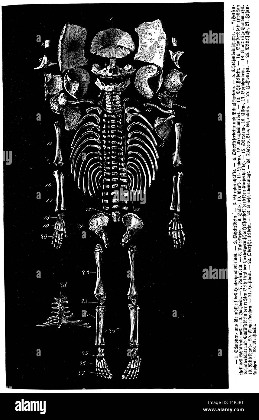 Skeleton. 1) scalp part and base of the occipital bone, 2) parietal bone, 3) frontal half, 4) upper jawbone and Pflugscharbein, 5) temporal bone scales, * rock part of the temporal bone, 6) zygomatic bone, 7) nasal bone, 8) lower jaw, 9) cervical vertebrae, 10) thoracic vertebrae , 11) lumbar vertebrae, 12) sacral vertebrae, 13) clavicle, 14) scapula, 15) humeral bone, 16) elbow, 17) spinal bone, 18) cartilaginous carpal, 19) metacarpal bone, 20) bony bone, 21) hip bone, 22) femur, 23) Patellar attachment, 24) fibula, 24a) tibia, 25) tarsal root, 26) metatarsal bones, 28) sternum, anonym 1887 Stock Photohttps://www.alamy.com/image-license-details/?v=1https://www.alamy.com/skeleton-1-scalp-part-and-base-of-the-occipital-bone-2-parietal-bone-3-frontal-half-4-upper-jawbone-and-pflugscharbein-5-temporal-bone-scales-rock-part-of-the-temporal-bone-6-zygomatic-bone-7-nasal-bone-8-lower-jaw-9-cervical-vertebrae-10-thoracic-vertebrae-11-lumbar-vertebrae-12-sacral-vertebrae-13-clavicle-14-scapula-15-humeral-bone-16-elbow-17-spinal-bone-18-cartilaginous-carpal-19-metacarpal-bone-20-bony-bone-21-hip-bone-22-femur-23-patellar-attachment-24-fibula-24a-tibia-25-tarsal-root-26-metatarsal-bones-28-sternum-anonym-1887-image243890972.html
RMT4P5BT–Skeleton. 1) scalp part and base of the occipital bone, 2) parietal bone, 3) frontal half, 4) upper jawbone and Pflugscharbein, 5) temporal bone scales, * rock part of the temporal bone, 6) zygomatic bone, 7) nasal bone, 8) lower jaw, 9) cervical vertebrae, 10) thoracic vertebrae , 11) lumbar vertebrae, 12) sacral vertebrae, 13) clavicle, 14) scapula, 15) humeral bone, 16) elbow, 17) spinal bone, 18) cartilaginous carpal, 19) metacarpal bone, 20) bony bone, 21) hip bone, 22) femur, 23) Patellar attachment, 24) fibula, 24a) tibia, 25) tarsal root, 26) metatarsal bones, 28) sternum, anonym 1887
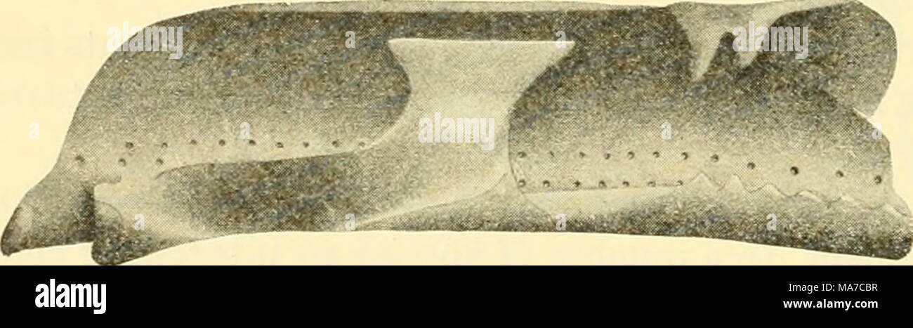 . The elasmobranch fishes . B. Ehinobatis productus. (C'liester tStock, orig.) Fig. 7(5. Cervical vertebrae. hd., dorsal basal plate (basidorsal) ; hv., ventral basal plate; c, centrum; f.d., foramen for dorsal nerve; id., dorsal interealarv (interdorsal plate) ; r., rib; shd., neural si^ine. Stock Photohttps://www.alamy.com/image-license-details/?v=1https://www.alamy.com/the-elasmobranch-fishes-b-ehinobatis-productus-cliester-tstock-orig-fig-75-cervical-vertebrae-hd-dorsal-basal-plate-basidorsal-hv-ventral-basal-plate-c-centrum-fd-foramen-for-dorsal-nerve-id-dorsal-interealarv-interdorsal-plate-r-rib-shd-neural-siine-image178413643.html
RMMA7CBR–. The elasmobranch fishes . B. Ehinobatis productus. (C'liester tStock, orig.) Fig. 7(5. Cervical vertebrae. hd., dorsal basal plate (basidorsal) ; hv., ventral basal plate; c, centrum; f.d., foramen for dorsal nerve; id., dorsal interealarv (interdorsal plate) ; r., rib; shd., neural si^ine.
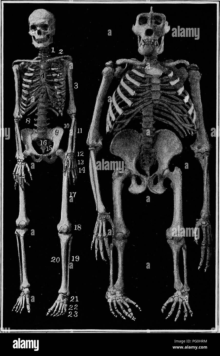 . The American natural history; a foundation of useful knowledge of the higher animals of North America. Natural history. 12 APES AND MONKEYS ful canine teeth are alone sufficient to proclaim the savage wild beast. To many persons it seems strange that, notwithstanding the seemingly wide differences between the various races of. By permission of J. F. G. Umlauff. SKELETONS OP MAN AND GORILLA. 1, cervical vertebrae, 2, collar bone, 3, humerus, 4, sternum, 5, ribs, 6, rib cartilages, 7, dorsal vertebrae, 8, lumbar vertebrae, 9, pelvis, 10, radius, 11, ulna, 12, carpals, 13, metacarpals, 14, phal Stock Photohttps://www.alamy.com/image-license-details/?v=1https://www.alamy.com/the-american-natural-history-a-foundation-of-useful-knowledge-of-the-higher-animals-of-north-america-natural-history-12-apes-and-monkeys-ful-canine-teeth-are-alone-sufficient-to-proclaim-the-savage-wild-beast-to-many-persons-it-seems-strange-that-notwithstanding-the-seemingly-wide-differences-between-the-various-races-of-by-permission-of-j-f-g-umlauff-skeletons-op-man-and-gorilla-1-cervical-vertebrae-2-collar-bone-3-humerus-4-sternum-5-ribs-6-rib-cartilages-7-dorsal-vertebrae-8-lumbar-vertebrae-9-pelvis-10-radius-11-ulna-12-carpals-13-metacarpals-14-phal-image216372904.html
RMPG0HRM–. The American natural history; a foundation of useful knowledge of the higher animals of North America. Natural history. 12 APES AND MONKEYS ful canine teeth are alone sufficient to proclaim the savage wild beast. To many persons it seems strange that, notwithstanding the seemingly wide differences between the various races of. By permission of J. F. G. Umlauff. SKELETONS OP MAN AND GORILLA. 1, cervical vertebrae, 2, collar bone, 3, humerus, 4, sternum, 5, ribs, 6, rib cartilages, 7, dorsal vertebrae, 8, lumbar vertebrae, 9, pelvis, 10, radius, 11, ulna, 12, carpals, 13, metacarpals, 14, phal
 Catalogue of the fossil Reptilia and Amphibia in the British Museum (Natural history) ..By Richard Lydekker .. . al and distal aspects of the right humerus ;from the Kimeridge Clay of the Isle of Portland. ^. a, trochantericridge ; r, radial, u, ulnar facet. with strongly developed trochanteric ridges. Cervical and ante-rior dorsal vertebrae with pentagonal, subcordiform centra (fig. 7);and the superior costal articulation of the former in great part orentirely on the neural arch (fig. 7); late posterior dorsal and an-terior caudal centra narrowed superiorly. ICHTHYOSJlTTKID.E. 15 This subgrou Stock Photohttps://www.alamy.com/image-license-details/?v=1https://www.alamy.com/catalogue-of-the-fossil-reptilia-and-amphibia-in-the-british-museum-natural-history-by-richard-lydekker-al-and-distal-aspects-of-the-right-humerus-from-the-kimeridge-clay-of-the-isle-of-portland-a-trochantericridge-r-radial-u-ulnar-facet-with-strongly-developed-trochanteric-ridges-cervical-and-ante-rior-dorsal-vertebrae-with-pentagonal-subcordiform-centra-fig-7and-the-superior-costal-articulation-of-the-former-in-great-part-orentirely-on-the-neural-arch-fig-7-late-posterior-dorsal-and-an-terior-caudal-centra-narrowed-superiorly-ichthyosjlttkide-15-this-subgrou-image342730348.html
RM2AWGM1G–Catalogue of the fossil Reptilia and Amphibia in the British Museum (Natural history) ..By Richard Lydekker .. . al and distal aspects of the right humerus ;from the Kimeridge Clay of the Isle of Portland. ^. a, trochantericridge ; r, radial, u, ulnar facet. with strongly developed trochanteric ridges. Cervical and ante-rior dorsal vertebrae with pentagonal, subcordiform centra (fig. 7);and the superior costal articulation of the former in great part orentirely on the neural arch (fig. 7); late posterior dorsal and an-terior caudal centra narrowed superiorly. ICHTHYOSJlTTKID.E. 15 This subgrou
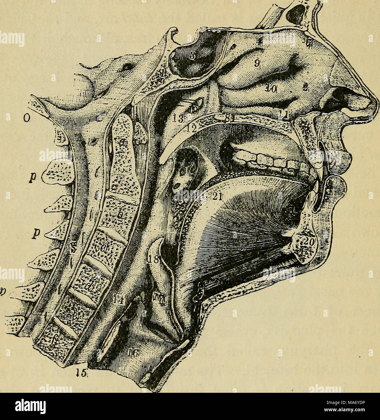 . Elementary physiology . Fig. 91.—Medial section of the face and neck, sphenoid bone; 2, nasal cavity ; 3, brain cavity; 4, ethmoid bone ; 5, frontal bone; 6, nasal bone; 7, superior maxillary bone ; 8, palatal bone ; 9, superior turbinated bone ; 10, middle turbinated bone ; 11, inferior turbinated bone ; 12, soft palate ; 13, upper part of pharynx ; 14, lower part of pharynx ; 15, oesophagus ; 16, larynx ; 17, glottis ; 18, epiglottis ; 19, opening of Eustachian tube ; 20, inferior maxillary bone ; 21, tongue ; 22, tonsil; a to f, bodies of cervical vertebrae ; s, spinal cord ; /, pro- cess Stock Photohttps://www.alamy.com/image-license-details/?v=1https://www.alamy.com/elementary-physiology-fig-91medial-section-of-the-face-and-neck-sphenoid-bone-2-nasal-cavity-3-brain-cavity-4-ethmoid-bone-5-frontal-bone-6-nasal-bone-7-superior-maxillary-bone-8-palatal-bone-9-superior-turbinated-bone-10-middle-turbinated-bone-11-inferior-turbinated-bone-12-soft-palate-13-upper-part-of-pharynx-14-lower-part-of-pharynx-15-oesophagus-16-larynx-17-glottis-18-epiglottis-19-opening-of-eustachian-tube-20-inferior-maxillary-bone-21-tongue-22-tonsil-a-to-f-bodies-of-cervical-vertebrae-s-spinal-cord-pro-cess-image178403506.html
RMMA6YDP–. Elementary physiology . Fig. 91.—Medial section of the face and neck, sphenoid bone; 2, nasal cavity ; 3, brain cavity; 4, ethmoid bone ; 5, frontal bone; 6, nasal bone; 7, superior maxillary bone ; 8, palatal bone ; 9, superior turbinated bone ; 10, middle turbinated bone ; 11, inferior turbinated bone ; 12, soft palate ; 13, upper part of pharynx ; 14, lower part of pharynx ; 15, oesophagus ; 16, larynx ; 17, glottis ; 18, epiglottis ; 19, opening of Eustachian tube ; 20, inferior maxillary bone ; 21, tongue ; 22, tonsil; a to f, bodies of cervical vertebrae ; s, spinal cord ; /, pro- cess
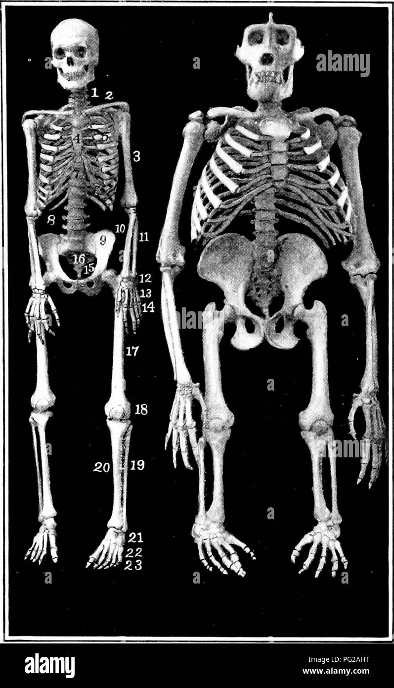 . The American natural history; a foundation of useful knowledge of the higher animals of North America. Natural history. ORDEES OF MAM.MALS—APES AND MOXKEYS The Apes.—The three great man-like (or an'thro-poid) apes — gorilla, chimpanzee and orang-utan—are so much like human beings that, to most persons, they are the most won-. By permission of J. F. G. Umla SKELETONS OF MAN AND GORILLA. 1, cervical vertebrae I'-*, carpals, 2, ci)llar bone. 13, metacarpals, 3, humerus, 14, phalanges, 4, sternuiu, 15, cavity of pelvis 5, rihs, 16, sacrum, 6, rib cartilages, 17, femur. 7, dorsal vertebrae, 18, p Stock Photohttps://www.alamy.com/image-license-details/?v=1https://www.alamy.com/the-american-natural-history-a-foundation-of-useful-knowledge-of-the-higher-animals-of-north-america-natural-history-ordees-of-mammalsapes-and-moxkeys-the-apesthe-three-great-man-like-or-anthro-poid-apes-gorilla-chimpanzee-and-orang-utanare-so-much-like-human-beings-that-to-most-persons-they-are-the-most-won-by-permission-of-j-f-g-umla-skeletons-of-man-and-gorilla-1-cervical-vertebrae-i-carpals-2-cillar-bone-13-metacarpals-3-humerus-14-phalanges-4-sternuiu-15-cavity-of-pelvis-5-rihs-16-sacrum-6-rib-cartilages-17-femur-7-dorsal-vertebrae-18-p-image216411156.html
RMPG2AHT–. The American natural history; a foundation of useful knowledge of the higher animals of North America. Natural history. ORDEES OF MAM.MALS—APES AND MOXKEYS The Apes.—The three great man-like (or an'thro-poid) apes — gorilla, chimpanzee and orang-utan—are so much like human beings that, to most persons, they are the most won-. By permission of J. F. G. Umla SKELETONS OF MAN AND GORILLA. 1, cervical vertebrae I'-*, carpals, 2, ci)llar bone. 13, metacarpals, 3, humerus, 14, phalanges, 4, sternuiu, 15, cavity of pelvis 5, rihs, 16, sacrum, 6, rib cartilages, 17, femur. 7, dorsal vertebrae, 18, p
 The science and practice of medicine . bC 5 Ph be 5 DESCRIPTION OF REGIONS OF THE THORAX. 541 ber, and run as follows : (1.) Along the middle of the sternum,from its upper to its lower end; (2.) From the acromial end of theclavicle to the external tubercle of the pubes (right and left); (4.)Along the spinous processes of the cervical and dorsal vertebrae;(6.) Along the posterior or spinal border of the scapulse, from theclavicular transverse line to the mammary transverse line. (SeeFigs. 5, 6, and 7.) The horizontal or transverse lines are four in number, and areas follows : (1.) Around the lo Stock Photohttps://www.alamy.com/image-license-details/?v=1https://www.alamy.com/the-science-and-practice-of-medicine-bc-5-ph-be-5-description-of-regions-of-the-thorax-541-ber-and-run-as-follows-1-along-the-middle-of-the-sternumfrom-its-upper-to-its-lower-end-2-from-the-acromial-end-of-theclavicle-to-the-external-tubercle-of-the-pubes-right-and-left-4along-the-spinous-processes-of-the-cervical-and-dorsal-vertebrae6-along-the-posterior-or-spinal-border-of-the-scapulse-from-theclavicular-transverse-line-to-the-mammary-transverse-line-seefigs-5-6-and-7-the-horizontal-or-transverse-lines-are-four-in-number-and-areas-follows-1-around-the-lo-image339314659.html
RM2AM138K–The science and practice of medicine . bC 5 Ph be 5 DESCRIPTION OF REGIONS OF THE THORAX. 541 ber, and run as follows : (1.) Along the middle of the sternum,from its upper to its lower end; (2.) From the acromial end of theclavicle to the external tubercle of the pubes (right and left); (4.)Along the spinous processes of the cervical and dorsal vertebrae;(6.) Along the posterior or spinal border of the scapulse, from theclavicular transverse line to the mammary transverse line. (SeeFigs. 5, 6, and 7.) The horizontal or transverse lines are four in number, and areas follows : (1.) Around the lo
 . A text-book of agricultural zoology. Zoology, Economic. Fig. 175.—Skeleton of Fowl. A-B, cervical vertebrae; 1, spinous procens of third vertebra; 2, inferior ridge on same; 8, styloid process ; 4, vertebral foramen; B-0, dorsal vertebrae; 6, spinous process of first; 7, crest formed by union of other spinous processes; D-B, coccygeal vertebrae; F, G, head; 8, interorbital septum; 9, interorbital foramen; 10, premaxillary bone; 10', anterior nares; 11, man- dible ; 12, quadrate; 13, maxilla ; 14, keel of sternum (H); IS, episternum; 16, posterior lateral process; 17, obiic[ue lateral process Stock Photohttps://www.alamy.com/image-license-details/?v=1https://www.alamy.com/a-text-book-of-agricultural-zoology-zoology-economic-fig-175skeleton-of-fowl-a-b-cervical-vertebrae-1-spinous-procens-of-third-vertebra-2-inferior-ridge-on-same-8-styloid-process-4-vertebral-foramen-b-0-dorsal-vertebrae-6-spinous-process-of-first-7-crest-formed-by-union-of-other-spinous-processes-d-b-coccygeal-vertebrae-f-g-head-8-interorbital-septum-9-interorbital-foramen-10-premaxillary-bone-10-anterior-nares-11-man-dible-12-quadrate-13-maxilla-14-keel-of-sternum-h-is-episternum-16-posterior-lateral-process-17-obiic-ue-lateral-process-image216446845.html
RMPG404D–. A text-book of agricultural zoology. Zoology, Economic. Fig. 175.—Skeleton of Fowl. A-B, cervical vertebrae; 1, spinous procens of third vertebra; 2, inferior ridge on same; 8, styloid process ; 4, vertebral foramen; B-0, dorsal vertebrae; 6, spinous process of first; 7, crest formed by union of other spinous processes; D-B, coccygeal vertebrae; F, G, head; 8, interorbital septum; 9, interorbital foramen; 10, premaxillary bone; 10', anterior nares; 11, man- dible ; 12, quadrate; 13, maxilla ; 14, keel of sternum (H); IS, episternum; 16, posterior lateral process; 17, obiic[ue lateral process
 . The health guide : aiming at a higher science of life and the life-forces, giving nature's simple and beautiful laws of cure, the science of magnetic manipulation, bathing, electricity, food, sleep, exercise, marriage, and the treatment for one hundred diseases : thus, constituting a home doctor far superior to drugs. ly vitalizingfocus. The Spine has 33 vertebrae, 7 of which are cervical, belong-ing to the neck ; 12 dorsal, to which the ribs are attached,reaching just below the small of the back; 5 lumbar; 5 sacral,and 4 coccygeal. The coccyx forms the lower point of thespine. (GraysAnatomy Stock Photohttps://www.alamy.com/image-license-details/?v=1https://www.alamy.com/the-health-guide-aiming-at-a-higher-science-of-life-and-the-life-forces-giving-natures-simple-and-beautiful-laws-of-cure-the-science-of-magnetic-manipulation-bathing-electricity-food-sleep-exercise-marriage-and-the-treatment-for-one-hundred-diseases-thus-constituting-a-home-doctor-far-superior-to-drugs-ly-vitalizingfocus-the-spine-has-33-vertebrae-7-of-which-are-cervical-belong-ing-to-the-neck-12-dorsal-to-which-the-ribs-are-attachedreaching-just-below-the-small-of-the-back-5-lumbar-5-sacraland-4-coccygeal-the-coccyx-forms-the-lower-point-of-thespine-graysanatomy-image336835052.html
RM2AG04F8–. The health guide : aiming at a higher science of life and the life-forces, giving nature's simple and beautiful laws of cure, the science of magnetic manipulation, bathing, electricity, food, sleep, exercise, marriage, and the treatment for one hundred diseases : thus, constituting a home doctor far superior to drugs. ly vitalizingfocus. The Spine has 33 vertebrae, 7 of which are cervical, belong-ing to the neck ; 12 dorsal, to which the ribs are attached,reaching just below the small of the back; 5 lumbar; 5 sacral,and 4 coccygeal. The coccyx forms the lower point of thespine. (GraysAnatomy
 Catalogue of the fossil Reptilia and Amphibia in the British Museum (Natural history) ..By Richard Lydekker .. . Soc. for 1853, p. 7 (1854).—Plesiosaurus. See Blake inTate and Blakes Yorkshire Lias, p. 249 (1870).—P. zetlandL 3 In this description it appears that V. megacephalua has been confused withPlesiosaurus macrocephalus. 168 SAUEOPTERYGIA. Thaumatosaurus carinatus (Cuvier1). Syn. Plesiosaurus carinatus, Cuvier2. Imperfectly known, but apparently of the approximate size ofT. indicus. The cervical vertebrae have a very prominent haemalcarina, with a deep pit on either side. Hah. Europe (F Stock Photohttps://www.alamy.com/image-license-details/?v=1https://www.alamy.com/catalogue-of-the-fossil-reptilia-and-amphibia-in-the-british-museum-natural-history-by-richard-lydekker-soc-for-1853-p-7-1854plesiosaurus-see-blake-intate-and-blakes-yorkshire-lias-p-249-1870p-zetlandl-3-in-this-description-it-appears-that-v-megacephalua-has-been-confused-withplesiosaurus-macrocephalus-168-saueopterygia-thaumatosaurus-carinatus-cuvier1-syn-plesiosaurus-carinatus-cuvier2-imperfectly-known-but-apparently-of-the-approximate-size-oft-indicus-the-cervical-vertebrae-have-a-very-prominent-haemalcarina-with-a-deep-pit-on-either-side-hah-europe-f-image342715056.html
RM2AWG0FC–Catalogue of the fossil Reptilia and Amphibia in the British Museum (Natural history) ..By Richard Lydekker .. . Soc. for 1853, p. 7 (1854).—Plesiosaurus. See Blake inTate and Blakes Yorkshire Lias, p. 249 (1870).—P. zetlandL 3 In this description it appears that V. megacephalua has been confused withPlesiosaurus macrocephalus. 168 SAUEOPTERYGIA. Thaumatosaurus carinatus (Cuvier1). Syn. Plesiosaurus carinatus, Cuvier2. Imperfectly known, but apparently of the approximate size ofT. indicus. The cervical vertebrae have a very prominent haemalcarina, with a deep pit on either side. Hah. Europe (F
 Elements of animal physiology, chiefly human . n thus contains in all thirty-threevertebrse, consisting ofseven cervical (neck),I 7 Cervical twelve clorsal (back), fiveC Vertebrae. ^^^^^5^^ (loius) Vertebralbones, togetherwith theOS sacrum and os coccygis,which are all clearly shownin fig. 9. It will be observed on examining the diagram of the vertebral column, that I 12 Dorsal tl^® vcrtehrce increase in vertebr5e.g{;j;g ^^^(j strength doionivard,because of the greater bur-den they have to bear,thus affording additionalstructural proof that the6rec^ is the position naturalto man. It will also Stock Photohttps://www.alamy.com/image-license-details/?v=1https://www.alamy.com/elements-of-animal-physiology-chiefly-human-n-thus-contains-in-all-thirty-threevertebrse-consisting-ofseven-cervical-necki-7-cervical-twelve-clorsal-back-fivec-vertebrae-5-loius-vertebralbones-togetherwith-theos-sacrum-and-os-coccygiswhich-are-all-clearly-shownin-fig-9-it-will-be-observed-on-examining-the-diagram-of-the-vertebral-column-that-i-12-dorsal-tl-vcrtehrce-increase-in-vertebr5egjg-j-strength-doionivardbecause-of-the-greater-bur-den-they-have-to-bearthus-affording-additionalstructural-proof-that-the6rec-is-the-position-naturalto-man-it-will-also-image339020699.html
RM2AKFMA3–Elements of animal physiology, chiefly human . n thus contains in all thirty-threevertebrse, consisting ofseven cervical (neck),I 7 Cervical twelve clorsal (back), fiveC Vertebrae. ^^^^^5^^ (loius) Vertebralbones, togetherwith theOS sacrum and os coccygis,which are all clearly shownin fig. 9. It will be observed on examining the diagram of the vertebral column, that I 12 Dorsal tl^® vcrtehrce increase in vertebr5e.g{;j;g ^^^(j strength doionivard,because of the greater bur-den they have to bear,thus affording additionalstructural proof that the6rec^ is the position naturalto man. It will also
 Monograph of the Palaeontographical Society . TAB. III.Crocodilus champsoides, half the nat. size. Fig. 1. Upper view of the skull. 2. Under view of the fore part of ditto. 3. Side view of ditto. 4. The left half of the back part of the skull of ditto, nat. size. 5. The point of an anterior young tooth coming into place, nat. size. 6. Half of the crown of one of the large conical teeth, nat. size. 7. One of the shorter and more obtuse posterior teeth, nat. size. T. 111.. TAB. IV.Vertebrae of the Crocodilus toliapicus, nat. size. Fig. 1. Fore part of a mutilated seventh cervical vertebra. 2. Un Stock Photohttps://www.alamy.com/image-license-details/?v=1https://www.alamy.com/monograph-of-the-palaeontographical-society-tab-iiicrocodilus-champsoides-half-the-nat-size-fig-1-upper-view-of-the-skull-2-under-view-of-the-fore-part-of-ditto-3-side-view-of-ditto-4-the-left-half-of-the-back-part-of-the-skull-of-ditto-nat-size-5-the-point-of-an-anterior-young-tooth-coming-into-place-nat-size-6-half-of-the-crown-of-one-of-the-large-conical-teeth-nat-size-7-one-of-the-shorter-and-more-obtuse-posterior-teeth-nat-size-t-111-tab-ivvertebrae-of-the-crocodilus-toliapicus-nat-size-fig-1-fore-part-of-a-mutilated-seventh-cervical-vertebra-2-un-image338145576.html
RM2AJ3T3M–Monograph of the Palaeontographical Society . TAB. III.Crocodilus champsoides, half the nat. size. Fig. 1. Upper view of the skull. 2. Under view of the fore part of ditto. 3. Side view of ditto. 4. The left half of the back part of the skull of ditto, nat. size. 5. The point of an anterior young tooth coming into place, nat. size. 6. Half of the crown of one of the large conical teeth, nat. size. 7. One of the shorter and more obtuse posterior teeth, nat. size. T. 111.. TAB. IV.Vertebrae of the Crocodilus toliapicus, nat. size. Fig. 1. Fore part of a mutilated seventh cervical vertebra. 2. Un
 Monograph of the Palaeontographical Society . TAB. IV.Vertebrae of the Crocodilus toliapicus, nat. size. Fig. 1. Fore part of a mutilated seventh cervical vertebra. 2. Under pa.t of ditto. 3. Side view of a mutilated eighth cervical vertebra. 4. Front view of the same vertebra. 5. Under view of the centrum of a lumbar vertebra. 6. Under view of the centrum of the fourth or fifth dorsal vertebra. 7. Side view of the first caudal vertebra. 8. Side view of a middle caudal vertebra. 9. Under view of the same vertebra. T. TV,. TAB. V. Fig. 1. Side view of the fourth cervical vertebra (mutilated) of Stock Photohttps://www.alamy.com/image-license-details/?v=1https://www.alamy.com/monograph-of-the-palaeontographical-society-tab-ivvertebrae-of-the-crocodilus-toliapicus-nat-size-fig-1-fore-part-of-a-mutilated-seventh-cervical-vertebra-2-under-pat-of-ditto-3-side-view-of-a-mutilated-eighth-cervical-vertebra-4-front-view-of-the-same-vertebra-5-under-view-of-the-centrum-of-a-lumbar-vertebra-6-under-view-of-the-centrum-of-the-fourth-or-fifth-dorsal-vertebra-7-side-view-of-the-first-caudal-vertebra-8-side-view-of-a-middle-caudal-vertebra-9-under-view-of-the-same-vertebra-t-tv-tab-v-fig-1-side-view-of-the-fourth-cervical-vertebra-mutilated-of-image338145242.html
RM2AJ3RKP–Monograph of the Palaeontographical Society . TAB. IV.Vertebrae of the Crocodilus toliapicus, nat. size. Fig. 1. Fore part of a mutilated seventh cervical vertebra. 2. Under pa.t of ditto. 3. Side view of a mutilated eighth cervical vertebra. 4. Front view of the same vertebra. 5. Under view of the centrum of a lumbar vertebra. 6. Under view of the centrum of the fourth or fifth dorsal vertebra. 7. Side view of the first caudal vertebra. 8. Side view of a middle caudal vertebra. 9. Under view of the same vertebra. T. TV,. TAB. V. Fig. 1. Side view of the fourth cervical vertebra (mutilated) of
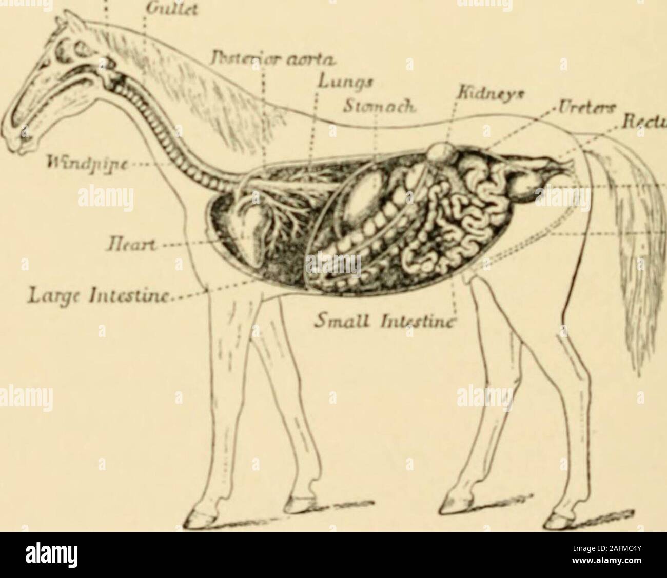 . Riding and driving. PLATE XVI —SKELETON OF THE HORSE J.arvnx .RteUuiV -r-norln. Slimtath : -•rrrf/rr v V - ^ ^^ // (in. PLATE XVIL —INTERNAL PARTS OF THE HORSE SKELETON OF THE HORSE 1. Eye cavity 21. Great trochanter 2. Face bones 22. Thigh bone 3. Incisor teeth 23. Ischium 4. Molar teeth 24. Radius, or forearm bone 5. Lower jaw 25. Carpal, or knee bones 6. First vertebra of neck 26. Trapezium 7. Second vertebra of neck 27. Cannon bones 8. Cervical vertebrae 28. Pastern bones 9. Spinal processes of back 29. Sesamoid bone 10. Dorsal and lumbar vertebrae 30. Small pastern bone 11. Sacrum 31. Stock Photohttps://www.alamy.com/image-license-details/?v=1https://www.alamy.com/riding-and-driving-plate-xvi-skeleton-of-the-horse-jarvnx-rteuuiv-r-norln-slimtath-rrrfrr-v-v-in-plate-xvil-internal-parts-of-the-horse-skeleton-of-the-horse-1-eye-cavity-21-great-trochanter-2-face-bones-22-thigh-bone-3-incisor-teeth-23-ischium-4-molar-teeth-24-radius-or-forearm-bone-5-lower-jaw-25-carpal-or-knee-bones-6-first-vertebra-of-neck-26-trapezium-7-second-vertebra-of-neck-27-cannon-bones-8-cervical-vertebrae-28-pastern-bones-9-spinal-processes-of-back-29-sesamoid-bone-10-dorsal-and-lumbar-vertebrae-30-small-pastern-bone-11-sacrum-31-image336665419.html
RM2AFMC4Y–. Riding and driving. PLATE XVI —SKELETON OF THE HORSE J.arvnx .RteUuiV -r-norln. Slimtath : -•rrrf/rr v V - ^ ^^ // (in. PLATE XVIL —INTERNAL PARTS OF THE HORSE SKELETON OF THE HORSE 1. Eye cavity 21. Great trochanter 2. Face bones 22. Thigh bone 3. Incisor teeth 23. Ischium 4. Molar teeth 24. Radius, or forearm bone 5. Lower jaw 25. Carpal, or knee bones 6. First vertebra of neck 26. Trapezium 7. Second vertebra of neck 27. Cannon bones 8. Cervical vertebrae 28. Pastern bones 9. Spinal processes of back 29. Sesamoid bone 10. Dorsal and lumbar vertebrae 30. Small pastern bone 11. Sacrum 31.
 Memoirs of the Connecticut Academy of Arts and Sciences . 13 14. Plate VI. Figures 1-6.—Seven anterior cervical vertebrae of the type of P. ingens Marsh, No. 1175;seen from the left side; slightly restored, x 0.50 Figure 1.—Atlas and axis.Figure 2.—Third cervical.Figure 3.—Fourth cervical.Figure 4.—Fifth cervical.Figure 5.—Sixth cervical.Figure 6.—Seventh cervical. Figure 7.—Seventh cervical vertebra of Pteranodon sp., No. 2692; seen trom the left side.x0.66 Figure 8.—Eighth cervical vertebra of the same individual; seen from the left side;showing rib. x 0.66 Figure 9.—Ninth cervical vertebra Stock Photohttps://www.alamy.com/image-license-details/?v=1https://www.alamy.com/memoirs-of-the-connecticut-academy-of-arts-and-sciences-13-14-plate-vi-figures-1-6seven-anterior-cervical-vertebrae-of-the-type-of-p-ingens-marsh-no-1175seen-from-the-left-side-slightly-restored-x-050-figure-1atlas-and-axisfigure-2third-cervicalfigure-3fourth-cervicalfigure-4fifth-cervicalfigure-5sixth-cervicalfigure-6seventh-cervical-figure-7seventh-cervical-vertebra-of-pteranodon-sp-no-2692-seen-trom-the-left-sidex066-figure-8eighth-cervical-vertebra-of-the-same-individual-seen-from-the-left-sideshowing-rib-x-066-figure-9ninth-cervical-vertebra-image340041722.html
RM2AN66K6–Memoirs of the Connecticut Academy of Arts and Sciences . 13 14. Plate VI. Figures 1-6.—Seven anterior cervical vertebrae of the type of P. ingens Marsh, No. 1175;seen from the left side; slightly restored, x 0.50 Figure 1.—Atlas and axis.Figure 2.—Third cervical.Figure 3.—Fourth cervical.Figure 4.—Fifth cervical.Figure 5.—Sixth cervical.Figure 6.—Seventh cervical. Figure 7.—Seventh cervical vertebra of Pteranodon sp., No. 2692; seen trom the left side.x0.66 Figure 8.—Eighth cervical vertebra of the same individual; seen from the left side;showing rib. x 0.66 Figure 9.—Ninth cervical vertebra
 The Airedale terrier standard simplified . 1. Fossa on lower Jaw 19. The Fibula 7 Branch of lower Jaw 20. Stifle Joint 3. Superior Maxilla 21. Hock Joint 4. Temporal Fossa 22. Metatarsal Bone 5. Parietal Bone 23. Phalanges 6. Sagiittal Crest 24. Shoulder Blade 7. Malar Bone 25. Shoulder Joint 8. Nasal Bone 26. Humerus or Arm 9. Occipital Crest 27. Elbow Joint 10. Cervical Vertebrae 28. Radius of Forearm 11. Dorsal Vertehne 29. Ulnar 12. Lumbar Vertebra* 30. Wrist or Carpal Bones 13. Front part of Sacrum 31. Metatarsal Bones 14. The Pelvis 32. Phalanges 15. Bones of Tail 33. Ribs 16. Femur of 1 Stock Photohttps://www.alamy.com/image-license-details/?v=1https://www.alamy.com/the-airedale-terrier-standard-simplified-1-fossa-on-lower-jaw-19-the-fibula-7-branch-of-lower-jaw-20-stifle-joint-3-superior-maxilla-21-hock-joint-4-temporal-fossa-22-metatarsal-bone-5-parietal-bone-23-phalanges-6-sagiittal-crest-24-shoulder-blade-7-malar-bone-25-shoulder-joint-8-nasal-bone-26-humerus-or-arm-9-occipital-crest-27-elbow-joint-10-cervical-vertebrae-28-radius-of-forearm-11-dorsal-vertehne-29-ulnar-12-lumbar-vertebra-30-wrist-or-carpal-bones-13-front-part-of-sacrum-31-metatarsal-bones-14-the-pelvis-32-phalanges-15-bones-of-tail-33-ribs-16-femur-of-1-image338177502.html
RM2AJ58RX–The Airedale terrier standard simplified . 1. Fossa on lower Jaw 19. The Fibula 7 Branch of lower Jaw 20. Stifle Joint 3. Superior Maxilla 21. Hock Joint 4. Temporal Fossa 22. Metatarsal Bone 5. Parietal Bone 23. Phalanges 6. Sagiittal Crest 24. Shoulder Blade 7. Malar Bone 25. Shoulder Joint 8. Nasal Bone 26. Humerus or Arm 9. Occipital Crest 27. Elbow Joint 10. Cervical Vertebrae 28. Radius of Forearm 11. Dorsal Vertehne 29. Ulnar 12. Lumbar Vertebra* 30. Wrist or Carpal Bones 13. Front part of Sacrum 31. Metatarsal Bones 14. The Pelvis 32. Phalanges 15. Bones of Tail 33. Ribs 16. Femur of 1
 . The horse, its treatment in health and disease with a complete guide to breeding, training and management . o 7, Cervical Vertebrae. The articular processes of the vertebrae throughout are connected bymeans of a capsular ligament, and the same may be said of the articulationson the transverse processes of the two last lumbar and first sacral vertebrae. ARTICULATIONS OF THE HEAD It has elsewhere been pointed out that these are for the most partimmovable, and the mode of formation has been described. The Tempero-Maxillary Articulation (fig. 348) or joint formedbetween the lower jaw and the tem Stock Photohttps://www.alamy.com/image-license-details/?v=1https://www.alamy.com/the-horse-its-treatment-in-health-and-disease-with-a-complete-guide-to-breeding-training-and-management-o-7-cervical-vertebrae-the-articular-processes-of-the-vertebrae-throughout-are-connected-bymeans-of-a-capsular-ligament-and-the-same-may-be-said-of-the-articulationson-the-transverse-processes-of-the-two-last-lumbar-and-first-sacral-vertebrae-articulations-of-the-head-it-has-elsewhere-been-pointed-out-that-these-are-for-the-most-partimmovable-and-the-mode-of-formation-has-been-described-the-tempero-maxillary-articulation-fig-348-or-joint-formedbetween-the-lower-jaw-and-the-tem-image370067982.html
RM2CE21DJ–. The horse, its treatment in health and disease with a complete guide to breeding, training and management . o 7, Cervical Vertebrae. The articular processes of the vertebrae throughout are connected bymeans of a capsular ligament, and the same may be said of the articulationson the transverse processes of the two last lumbar and first sacral vertebrae. ARTICULATIONS OF THE HEAD It has elsewhere been pointed out that these are for the most partimmovable, and the mode of formation has been described. The Tempero-Maxillary Articulation (fig. 348) or joint formedbetween the lower jaw and the tem
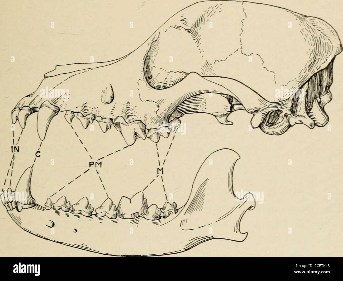 . Guide leaflet. equivalent of the columella, the incus is derived from the quadrate, and the malleus from the articular In almost all mammals there are seven cervical vertebrae. In the giraffethese are excessively elongated and in the whales excessively com-pressed. Only in the manatee (21) and a species of two-toed sloth (16)is the number reduced to six and only in the three-toed sloths is thenumber increased, here to nine. The mammals develop bony ends or epiphyses on the limb bones(rare in reptiles) and on the vertebrae. It is between these epiphyses and BIOLOGY OF MAMMALS 7 the shafts of Stock Photohttps://www.alamy.com/image-license-details/?v=1https://www.alamy.com/guide-leaflet-equivalent-of-the-columella-the-incus-is-derived-from-the-quadrate-and-the-malleus-from-the-articular-in-almost-all-mammals-there-are-seven-cervical-vertebrae-in-the-giraffethese-are-excessively-elongated-and-in-the-whales-excessively-com-pressed-only-in-the-manatee-21-and-a-species-of-two-toed-sloth-16is-the-number-reduced-to-six-and-only-in-the-three-toed-sloths-is-thenumber-increased-here-to-nine-the-mammals-develop-bony-ends-or-epiphyses-on-the-limb-bonesrare-in-reptiles-and-on-the-vertebrae-it-is-between-these-epiphyses-and-biology-of-mammals-7-the-shafts-of-image370564771.html
RM2CETK43–. Guide leaflet. equivalent of the columella, the incus is derived from the quadrate, and the malleus from the articular In almost all mammals there are seven cervical vertebrae. In the giraffethese are excessively elongated and in the whales excessively com-pressed. Only in the manatee (21) and a species of two-toed sloth (16)is the number reduced to six and only in the three-toed sloths is thenumber increased, here to nine. The mammals develop bony ends or epiphyses on the limb bones(rare in reptiles) and on the vertebrae. It is between these epiphyses and BIOLOGY OF MAMMALS 7 the shafts of
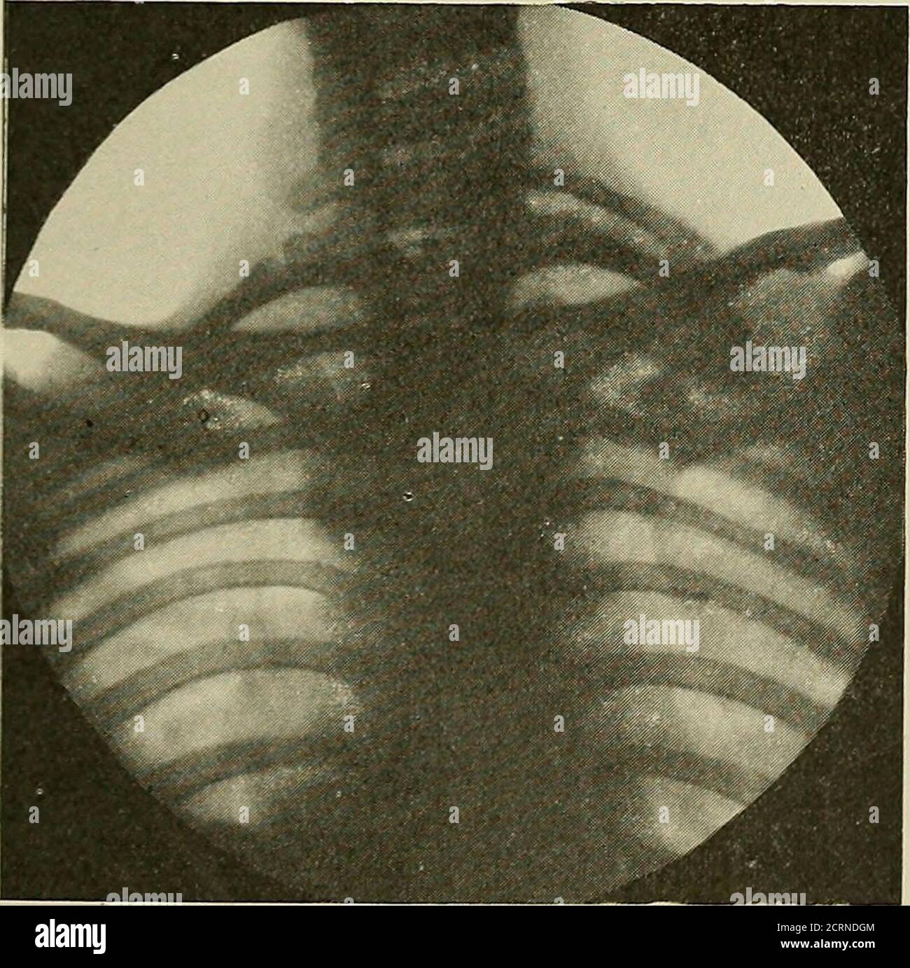 . Radiography and radio-therapeutics . a.6.7 Cervical vertebrae l°lo6D Dorsal vertebrs Fig. 184.- -Diagram to illustrate the anatomical points inFig. 183. where it is desirable to show the atlas and axis, and the articulation between the former and theoccipital bone, it is necessaryto take the skiagram throughthe open mouth, as describedin detail above. The resultingpicture is necessarily small,but large enough to includethe parts desired. The position usually takenis the lateral one, with thehead rotated towards theplate. It is then possible toget a fairly good outline ofthe seven cervical ve Stock Photohttps://www.alamy.com/image-license-details/?v=1https://www.alamy.com/radiography-and-radio-therapeutics-a67-cervical-vertebrae-llo6d-dorsal-vertebrs-fig-184-diagram-to-illustrate-the-anatomical-points-infig-183-where-it-is-desirable-to-show-the-atlas-and-axis-and-the-articulation-between-the-former-and-theoccipital-bone-it-is-necessaryto-take-the-skiagram-throughthe-open-mouth-as-describedin-detail-above-the-resultingpicture-is-necessarily-smallbut-large-enough-to-includethe-parts-desired-the-position-usually-takenis-the-lateral-one-with-thehead-rotated-towards-theplate-it-is-then-possible-toget-a-fairly-good-outline-ofthe-seven-cervical-ve-image376026468.html
RM2CRNDGM–. Radiography and radio-therapeutics . a.6.7 Cervical vertebrae l°lo6D Dorsal vertebrs Fig. 184.- -Diagram to illustrate the anatomical points inFig. 183. where it is desirable to show the atlas and axis, and the articulation between the former and theoccipital bone, it is necessaryto take the skiagram throughthe open mouth, as describedin detail above. The resultingpicture is necessarily small,but large enough to includethe parts desired. The position usually takenis the lateral one, with thehead rotated towards theplate. It is then possible toget a fairly good outline ofthe seven cervical ve
 . An introduction to the osteology of the mammalia . e of Greenland RightWhale (Balteua mysticetus), . a articular surface for occipital condyle ; e epi-physis on posterior end of body of seventh cervical vertebra ; sn foramen in archof atlas for first spinal nerve ; i arch of atlas ,23456 conjoined arches of theaxis and four following vertebrae ; 7 arch of seventh vertebra. In the common large Fin Whale of our coasts (13.musculus) the atlas (Fig. 16) has short, stout, conical, imper-forate transverse processes. The axis (Fig. 17) has a broadoval body, high massive arch, very short odontoid p Stock Photohttps://www.alamy.com/image-license-details/?v=1https://www.alamy.com/an-introduction-to-the-osteology-of-the-mammalia-e-of-greenland-rightwhale-balteua-mysticetus-a-articular-surface-for-occipital-condyle-e-epi-physis-on-posterior-end-of-body-of-seventh-cervical-vertebra-sn-foramen-in-archof-atlas-for-first-spinal-nerve-i-arch-of-atlas-23456-conjoined-arches-of-theaxis-and-four-following-vertebrae-7-arch-of-seventh-vertebra-in-the-common-large-fin-whale-of-our-coasts-13musculus-the-atlas-fig-16-has-short-stout-conical-imper-forate-transverse-processes-the-axis-fig-17-has-a-broadoval-body-high-massive-arch-very-short-odontoid-p-image369745785.html
RM2CDFAEH–. An introduction to the osteology of the mammalia . e of Greenland RightWhale (Balteua mysticetus), . a articular surface for occipital condyle ; e epi-physis on posterior end of body of seventh cervical vertebra ; sn foramen in archof atlas for first spinal nerve ; i arch of atlas ,23456 conjoined arches of theaxis and four following vertebrae ; 7 arch of seventh vertebra. In the common large Fin Whale of our coasts (13.musculus) the atlas (Fig. 16) has short, stout, conical, imper-forate transverse processes. The axis (Fig. 17) has a broadoval body, high massive arch, very short odontoid p
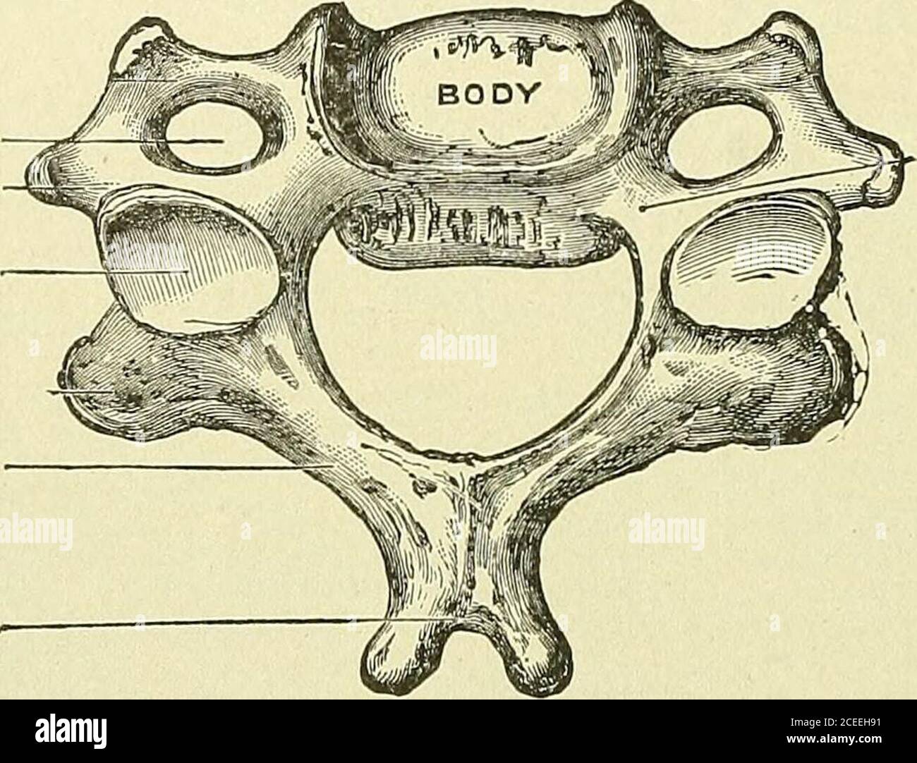 . Text-book of anatomy and physiology for nurses. Fig. 30.—VertebralColumn, Lateral Aspect. 1-7, Cervical vertebrae;8-19, dorsal vertebrae; 20-24, lumbar vertebrae; A, A,spinous processes; B, B,articular facets of trans-verse processes of first tendorsal vertebrae; C, auricu-lar surface of sacrum; D,I), foramina in transverseprocesses of cervical verte-brae.— (Goulds Illus. Dic-tionary.) 36 anatomy and physiology for nurses. Points of Special Interest, The cervical vertebras present a foramen at the base of the transverseprocess, the transverse foramen, through which an artery runs to the brai Stock Photohttps://www.alamy.com/image-license-details/?v=1https://www.alamy.com/text-book-of-anatomy-and-physiology-for-nurses-fig-30vertebralcolumn-lateral-aspect-1-7-cervical-vertebrae8-19-dorsal-vertebrae-20-24-lumbar-vertebrae-a-aspinous-processes-b-barticular-facets-of-trans-verse-processes-of-first-tendorsal-vertebrae-c-auricu-lar-surface-of-sacrum-di-foramina-in-transverseprocesses-of-cervical-verte-brae-goulds-illus-dic-tionary-36-anatomy-and-physiology-for-nurses-points-of-special-interest-the-cervical-vertebras-present-a-foramen-at-the-base-of-the-transverseprocess-the-transverse-foramen-through-which-an-artery-runs-to-the-brai-image370343821.html
RM2CEEH91–. Text-book of anatomy and physiology for nurses. Fig. 30.—VertebralColumn, Lateral Aspect. 1-7, Cervical vertebrae;8-19, dorsal vertebrae; 20-24, lumbar vertebrae; A, A,spinous processes; B, B,articular facets of trans-verse processes of first tendorsal vertebrae; C, auricu-lar surface of sacrum; D,I), foramina in transverseprocesses of cervical verte-brae.— (Goulds Illus. Dic-tionary.) 36 anatomy and physiology for nurses. Points of Special Interest, The cervical vertebras present a foramen at the base of the transverseprocess, the transverse foramen, through which an artery runs to the brai
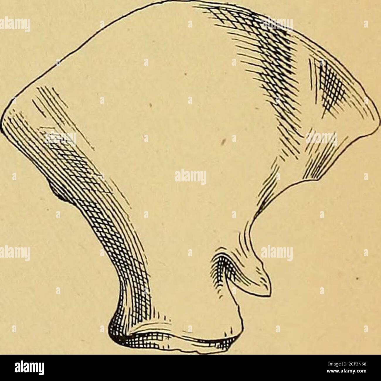 . Transactions and proceedings of the New Zealand Institute . Cervical vefieirce ofJl^adecaiTis azisiraZie7isis.(fro7ti view) Iig.4f.. jSctzpiolco of3facleaj/ius ausir/zlzensis.iinside.) Illiosf:ra2inq Faj^er iy IJ-JS. Crra-y. Gray.—On Macleayius australiensis. 91 but over the curve of the uose 10 feet. The length of the vertebrae, 23 feet;of the lower jaws, 7 feet 8 inches ; of the firat rib, 3 feet 6 inches ; and of themiddle rib, 7 feet 4 inches, as measured by Mr. E. Gerrard, jun., whoobserves that the last small bone of the tail is wanting. There are eightchevron bones present, but I shou Stock Photohttps://www.alamy.com/image-license-details/?v=1https://www.alamy.com/transactions-and-proceedings-of-the-new-zealand-institute-cervical-vefieirce-ofjladecaitis-azisirazie7isisfro7ti-view-iig4f-jsctzpiolco-of3facleajius-ausirzlzensisiinside-illiosfra2inq-fajer-iy-ij-js-crra-y-grayon-macleayius-australiensis-91-but-over-the-curve-of-the-uose-10-feet-the-length-of-the-vertebrae-23-feetof-the-lower-jaws-7-feet-8-inches-of-the-firat-rib-3-feet-6-inches-and-of-themiddle-rib-7-feet-4-inches-as-measured-by-mr-e-gerrard-jun-whoobserves-that-the-last-small-bone-of-the-tail-is-wanting-there-are-eightchevron-bones-present-but-i-shou-image375022656.html
RM2CP3N68–. Transactions and proceedings of the New Zealand Institute . Cervical vefieirce ofJl^adecaiTis azisiraZie7isis.(fro7ti view) Iig.4f.. jSctzpiolco of3facleaj/ius ausir/zlzensis.iinside.) Illiosf:ra2inq Faj^er iy IJ-JS. Crra-y. Gray.—On Macleayius australiensis. 91 but over the curve of the uose 10 feet. The length of the vertebrae, 23 feet;of the lower jaws, 7 feet 8 inches ; of the firat rib, 3 feet 6 inches ; and of themiddle rib, 7 feet 4 inches, as measured by Mr. E. Gerrard, jun., whoobserves that the last small bone of the tail is wanting. There are eightchevron bones present, but I shou
 . Lateral curvature of the spine and round shoulders . .4^L 4THL. .4^R. Fig. 16.—Tracings of Physiological Curves of Normal Children, on the Left ofA Girl of One and a Half Years, on the Right of a Girl of Eleven. In the adult, the part played by the bodies of the vertebrae and the discs inproducing the physiological curves is shown by the following table: DiiTERENCE Between the Sums of the Anterior and Posterior Borders. VERTEBR.3E. DiSCS. Cervical region 1.3 mm. 7.8 mm. Dorsal region 13.3 mm. 9.2 mm. Lumbar region 6.7 mm. 21.i mm. PELVIC INCLINATION. 17 The cervical curve is formed principal Stock Photohttps://www.alamy.com/image-license-details/?v=1https://www.alamy.com/lateral-curvature-of-the-spine-and-round-shoulders-4l-4thl-4r-fig-16tracings-of-physiological-curves-of-normal-children-on-the-left-ofa-girl-of-one-and-a-half-years-on-the-right-of-a-girl-of-eleven-in-the-adult-the-part-played-by-the-bodies-of-the-vertebrae-and-the-discs-inproducing-the-physiological-curves-is-shown-by-the-following-table-diiterence-between-the-sums-of-the-anterior-and-posterior-borders-vertebr3e-discs-cervical-region-13-mm-78-mm-dorsal-region-133-mm-92-mm-lumbar-region-67-mm-21i-mm-pelvic-inclination-17-the-cervical-curve-is-formed-principal-image372124135.html
RM2CHBM3K–. Lateral curvature of the spine and round shoulders . .4^L 4THL. .4^R. Fig. 16.—Tracings of Physiological Curves of Normal Children, on the Left ofA Girl of One and a Half Years, on the Right of a Girl of Eleven. In the adult, the part played by the bodies of the vertebrae and the discs inproducing the physiological curves is shown by the following table: DiiTERENCE Between the Sums of the Anterior and Posterior Borders. VERTEBR.3E. DiSCS. Cervical region 1.3 mm. 7.8 mm. Dorsal region 13.3 mm. 9.2 mm. Lumbar region 6.7 mm. 21.i mm. PELVIC INCLINATION. 17 The cervical curve is formed principal
 . Riding and driving. Molar teeth 24. 5. Lower jaw 25. 6. First vertebra of neck 26. Trapezium Cannon bones ^r^cHy 7. Second vertebra of neck 27. 8. Cervical vertebrae 28. Pastern bones 9. Spinal processes of back 29. Sesamoid bone .;.i/.el 10. Dorsal and lumbar vertebrae 30. Small pastern bone-^^^^jj.^ J^- Sacrum 31. Upper end of leg bone Stifle joint Leg bone, or tibia -^i-d 12. Tail bones 32. P.i. Shoulder blade 33. ??44^ Hollow of shoulder blade 15. Upper end of arm bone 16. Arm bone, or, huijf>erus . 34. Point of hock i S: Wash 35. Hock joint 36. Head of small metatarsal bone leansed Stock Photohttps://www.alamy.com/image-license-details/?v=1https://www.alamy.com/riding-and-driving-molar-teeth-24-5-lower-jaw-25-6-first-vertebra-of-neck-26-trapezium-cannon-bones-rchy-7-second-vertebra-of-neck-27-8-cervical-vertebrae-28-pastern-bones-9-spinal-processes-of-back-29-sesamoid-bone-iel-10-dorsal-and-lumbar-vertebrae-30-small-pastern-bone-jj-j-sacrum-31-upper-end-of-leg-bone-stifle-joint-leg-bone-or-tibia-i-d-12-tail-bones-32-pi-shoulder-blade-33-44-hollow-of-shoulder-blade-15-upper-end-of-arm-bone-16-arm-bone-or-huijfgterus-34-point-of-hock-i-s-wash-35-hock-joint-36-head-of-small-metatarsal-bone-leansed-image370703683.html
RM2CF3097–. Riding and driving. Molar teeth 24. 5. Lower jaw 25. 6. First vertebra of neck 26. Trapezium Cannon bones ^r^cHy 7. Second vertebra of neck 27. 8. Cervical vertebrae 28. Pastern bones 9. Spinal processes of back 29. Sesamoid bone .;.i/.el 10. Dorsal and lumbar vertebrae 30. Small pastern bone-^^^^jj.^ J^- Sacrum 31. Upper end of leg bone Stifle joint Leg bone, or tibia -^i-d 12. Tail bones 32. P.i. Shoulder blade 33. ??44^ Hollow of shoulder blade 15. Upper end of arm bone 16. Arm bone, or, huijf>erus . 34. Point of hock i S: Wash 35. Hock joint 36. Head of small metatarsal bone leansed
 . Chamber's scientific reader : illustrated with wood engravings. Readers. 46 PHYSIOLOGY OF THE HUMAN BODY. column there are 33 vertebrae. At the top are 7 cervical1 vertebrae, or vertebrae of the neck The topmost, or first cer- vical, is termed atlas, because by it the head is properly borne up, as the earth was by the fabled god Atlas. The second cervical is called ^iPk the axis, because it is the proper axis or joint of the neck, the joint between it and the atlas, which is a jnvot-joint, being that which enables the head to turn round. Next to the cervical are the 12 dorsal2 vertebrae, or Stock Photohttps://www.alamy.com/image-license-details/?v=1https://www.alamy.com/chambers-scientific-reader-illustrated-with-wood-engravings-readers-46-physiology-of-the-human-body-column-there-are-33-vertebrae-at-the-top-are-7-cervical1-vertebrae-or-vertebrae-of-the-neck-the-topmost-or-first-cer-vical-is-termed-atlas-because-by-it-the-head-is-properly-borne-up-as-the-earth-was-by-the-fabled-god-atlas-the-second-cervical-is-called-ipk-the-axis-because-it-is-the-proper-axis-or-joint-of-the-neck-the-joint-between-it-and-the-atlas-which-is-a-jnvot-joint-being-that-which-enables-the-head-to-turn-round-next-to-the-cervical-are-the-12-dorsal2-vertebrae-or-image235093451.html
RMRJDC2K–. Chamber's scientific reader : illustrated with wood engravings. Readers. 46 PHYSIOLOGY OF THE HUMAN BODY. column there are 33 vertebrae. At the top are 7 cervical1 vertebrae, or vertebrae of the neck The topmost, or first cer- vical, is termed atlas, because by it the head is properly borne up, as the earth was by the fabled god Atlas. The second cervical is called ^iPk the axis, because it is the proper axis or joint of the neck, the joint between it and the atlas, which is a jnvot-joint, being that which enables the head to turn round. Next to the cervical are the 12 dorsal2 vertebrae, or
 . Breviora. 1981 THE DROMASAURS 13 by the transverse flange of the pterygoid, so the presence of a coronoid is uncertain. Vertebrae: The first fourteen vertebrae of Galeops are present (Figs. 6, 7), although many of these are incompletely preserved. Cox (1959) differentiated the cervical from the trunk vertebrae of the dicynodont Kingoria on the basis of the size of the parapophysis and diapophysis and the thickness of the associated ribs, the cervical vertebrae having more poorly developed parapophyses and more slender ribs. In Galeops, a well-developed parapophysis is first seen in the seven Stock Photohttps://www.alamy.com/image-license-details/?v=1https://www.alamy.com/breviora-1981-the-dromasaurs-13-by-the-transverse-flange-of-the-pterygoid-so-the-presence-of-a-coronoid-is-uncertain-vertebrae-the-first-fourteen-vertebrae-of-galeops-are-present-figs-6-7-although-many-of-these-are-incompletely-preserved-cox-1959-differentiated-the-cervical-from-the-trunk-vertebrae-of-the-dicynodont-kingoria-on-the-basis-of-the-size-of-the-parapophysis-and-diapophysis-and-the-thickness-of-the-associated-ribs-the-cervical-vertebrae-having-more-poorly-developed-parapophyses-and-more-slender-ribs-in-galeops-a-well-developed-parapophysis-is-first-seen-in-the-seven-image234271188.html
RMRH3Y84–. Breviora. 1981 THE DROMASAURS 13 by the transverse flange of the pterygoid, so the presence of a coronoid is uncertain. Vertebrae: The first fourteen vertebrae of Galeops are present (Figs. 6, 7), although many of these are incompletely preserved. Cox (1959) differentiated the cervical from the trunk vertebrae of the dicynodont Kingoria on the basis of the size of the parapophysis and diapophysis and the thickness of the associated ribs, the cervical vertebrae having more poorly developed parapophyses and more slender ribs. In Galeops, a well-developed parapophysis is first seen in the seven
 . Bulletin of the Natural History Museum Zoology. p.vert. Vertebrae An articulated series of thirteen vertebrae (here referred to as vertebrae 1-13) is preserved, along with an isolated element on the lower left (vertebra 14). All vertebrae are exposed ventrally only; the surfaces of vertebrae 1-7 are weathered, while that of vertebra 11 is broken. The series 1-13 represents the anterior presacral part of the column. Vertebra 1, the anteriormost, is the smallest; size then increases gradually along the series such that the last is approxi- mately twice the dimension of the first. The cervical- Stock Photohttps://www.alamy.com/image-license-details/?v=1https://www.alamy.com/bulletin-of-the-natural-history-museum-zoology-pvert-vertebrae-an-articulated-series-of-thirteen-vertebrae-here-referred-to-as-vertebrae-1-13-is-preserved-along-with-an-isolated-element-on-the-lower-left-vertebra-14-all-vertebrae-are-exposed-ventrally-only-the-surfaces-of-vertebrae-1-7-are-weathered-while-that-of-vertebra-11-is-broken-the-series-1-13-represents-the-anterior-presacral-part-of-the-column-vertebra-1-the-anteriormost-is-the-smallest-size-then-increases-gradually-along-the-series-such-that-the-last-is-approxi-mately-twice-the-dimension-of-the-first-the-cervical-image233851659.html
RMRGCT4Y–. Bulletin of the Natural History Museum Zoology. p.vert. Vertebrae An articulated series of thirteen vertebrae (here referred to as vertebrae 1-13) is preserved, along with an isolated element on the lower left (vertebra 14). All vertebrae are exposed ventrally only; the surfaces of vertebrae 1-7 are weathered, while that of vertebra 11 is broken. The series 1-13 represents the anterior presacral part of the column. Vertebra 1, the anteriormost, is the smallest; size then increases gradually along the series such that the last is approxi- mately twice the dimension of the first. The cervical-
 . Bulletin - United States National Museum. Science. Figure 95.—Anterior view of third cervical vertebra, USNM 11535, of Parietobalaena palmeri Kellogg. Abbrs.: d.a., diapo- physis; p.a., parapophysis. of the fifth (63 mm.) exceeds that of the preceding cervicals, and the fourth and third were widened transversely. The length of the 7 cervical vertebrae, including the cartilaginous intervertebral disks, is approximately 235 mm. (9K inches). The distance (124 mm.) between the outer edges of the anterior articular facets of the atlas (fig. 94) of another physically iinmature specimen (USNM 11535 Stock Photohttps://www.alamy.com/image-license-details/?v=1https://www.alamy.com/bulletin-united-states-national-museum-science-figure-95anterior-view-of-third-cervical-vertebra-usnm-11535-of-parietobalaena-palmeri-kellogg-abbrs-da-diapo-physis-pa-parapophysis-of-the-fifth-63-mm-exceeds-that-of-the-preceding-cervicals-and-the-fourth-and-third-were-widened-transversely-the-length-of-the-7-cervical-vertebrae-including-the-cartilaginous-intervertebral-disks-is-approximately-235-mm-9k-inches-the-distance-124-mm-between-the-outer-edges-of-the-anterior-articular-facets-of-the-atlas-fig-94-of-another-physically-iinmature-specimen-usnm-11535-image233751960.html
RMRG8908–. Bulletin - United States National Museum. Science. Figure 95.—Anterior view of third cervical vertebra, USNM 11535, of Parietobalaena palmeri Kellogg. Abbrs.: d.a., diapo- physis; p.a., parapophysis. of the fifth (63 mm.) exceeds that of the preceding cervicals, and the fourth and third were widened transversely. The length of the 7 cervical vertebrae, including the cartilaginous intervertebral disks, is approximately 235 mm. (9K inches). The distance (124 mm.) between the outer edges of the anterior articular facets of the atlas (fig. 94) of another physically iinmature specimen (USNM 11535
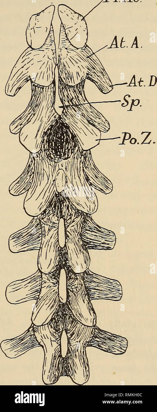 . Annals of the South African Museum = Annale van die Suid-Afrikaanse Museum. Natural history. A Contribution to the Morphology of the Gorgonopsia. 149 Pr.Ai/. xii., 1934. The excellent material now at my disposal fully confirms the views expressed in this paper. In Hipposaurus, Aelurognathus, and Arctognathoides the neck consists of 7 vertebrae. In Hipposaurus and Aelurognathus the shoulder- girdle is preserved in natural relation to the vertebral column so that this important point in the determina- tion of the cervical series is known. In addition the presence of inter- centra in the first Stock Photohttps://www.alamy.com/image-license-details/?v=1https://www.alamy.com/annals-of-the-south-african-museum-=-annale-van-die-suid-afrikaanse-museum-natural-history-a-contribution-to-the-morphology-of-the-gorgonopsia-149-prai-xii-1934-the-excellent-material-now-at-my-disposal-fully-confirms-the-views-expressed-in-this-paper-in-hipposaurus-aelurognathus-and-arctognathoides-the-neck-consists-of-7-vertebrae-in-hipposaurus-and-aelurognathus-the-shoulder-girdle-is-preserved-in-natural-relation-to-the-vertebral-column-so-that-this-important-point-in-the-determina-tion-of-the-cervical-series-is-known-in-addition-the-presence-of-inter-centra-in-the-first-image236458332.html
RMRMKH0C–. Annals of the South African Museum = Annale van die Suid-Afrikaanse Museum. Natural history. A Contribution to the Morphology of the Gorgonopsia. 149 Pr.Ai/. xii., 1934. The excellent material now at my disposal fully confirms the views expressed in this paper. In Hipposaurus, Aelurognathus, and Arctognathoides the neck consists of 7 vertebrae. In Hipposaurus and Aelurognathus the shoulder- girdle is preserved in natural relation to the vertebral column so that this important point in the determina- tion of the cervical series is known. In addition the presence of inter- centra in the first
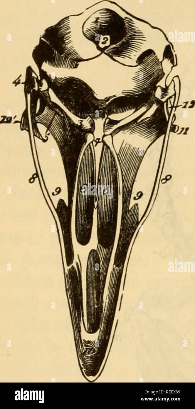 . The comparative anatomy of the domesticated animals. Horses; Veterinary anatomy. 160 THE BONES. Fig. 110.. support in the violent efforts that flight demands. The two or three last are often even covered by the wiag-bones, and joined to them. The inferior crest of the body forms a very long spine, especially in the first vertebrae. The spinous processes—flat, wide, short, and consolidated with each other by their opposite borders—constitute a long crest extending from the last cervical vertebra to the bones of the wings (Fig. 109, 7). The trans- verse processes widen to their summit; in the Stock Photohttps://www.alamy.com/image-license-details/?v=1https://www.alamy.com/the-comparative-anatomy-of-the-domesticated-animals-horses-veterinary-anatomy-160-the-bones-fig-110-support-in-the-violent-efforts-that-flight-demands-the-two-or-three-last-are-often-even-covered-by-the-wiag-bones-and-joined-to-them-the-inferior-crest-of-the-body-forms-a-very-long-spine-especially-in-the-first-vertebrae-the-spinous-processesflat-wide-short-and-consolidated-with-each-other-by-their-opposite-bordersconstitute-a-long-crest-extending-from-the-last-cervical-vertebra-to-the-bones-of-the-wings-fig-109-7-the-trans-verse-processes-widen-to-their-summit-in-the-image232667913.html
RMREEX89–. The comparative anatomy of the domesticated animals. Horses; Veterinary anatomy. 160 THE BONES. Fig. 110.. support in the violent efforts that flight demands. The two or three last are often even covered by the wiag-bones, and joined to them. The inferior crest of the body forms a very long spine, especially in the first vertebrae. The spinous processes—flat, wide, short, and consolidated with each other by their opposite borders—constitute a long crest extending from the last cervical vertebra to the bones of the wings (Fig. 109, 7). The trans- verse processes widen to their summit; in the
 . Bulletin. Ethnology. BUREAU OF AMERICAN ETHNOLOGY BULLETIN 185 PLATE 40. «, Articulated bison cervical vertebrae associated with scattered deer bones and perforated frontals in bison skull as found in Section 7 on the 8-foot level, b, Pocket of miscellaneous bison bones on 7-foot level, Section 6.. Please note that these images are extracted from scanned page images that may have been digitally enhanced for readability - coloration and appearance of these illustrations may not perfectly resemble the original work.. Smithsonian Institution. Bureau of American Ethnology. Washington : G. P. O. Stock Photohttps://www.alamy.com/image-license-details/?v=1https://www.alamy.com/bulletin-ethnology-bureau-of-american-ethnology-bulletin-185-plate-40-articulated-bison-cervical-vertebrae-associated-with-scattered-deer-bones-and-perforated-frontals-in-bison-skull-as-found-in-section-7-on-the-8-foot-level-b-pocket-of-miscellaneous-bison-bones-on-7-foot-level-section-6-please-note-that-these-images-are-extracted-from-scanned-page-images-that-may-have-been-digitally-enhanced-for-readability-coloration-and-appearance-of-these-illustrations-may-not-perfectly-resemble-the-original-work-smithsonian-institution-bureau-of-american-ethnology-washington-g-p-o-image234142167.html
RMRGX2M7–. Bulletin. Ethnology. BUREAU OF AMERICAN ETHNOLOGY BULLETIN 185 PLATE 40. «, Articulated bison cervical vertebrae associated with scattered deer bones and perforated frontals in bison skull as found in Section 7 on the 8-foot level, b, Pocket of miscellaneous bison bones on 7-foot level, Section 6.. Please note that these images are extracted from scanned page images that may have been digitally enhanced for readability - coloration and appearance of these illustrations may not perfectly resemble the original work.. Smithsonian Institution. Bureau of American Ethnology. Washington : G. P. O.
 . Advances in herpetology and evolutionary biology : essays in honor of Ernest E. Williams. Williams, Ernest E. (Ernest Edward); Herpetology; Evolution. Figure 3. Trunk vertebrae of Chamaelinorops (MCZ 154450). A. Dorsal view of the presacral vertebrae, minus the atlas. B. Ventral view of the presacral vertebrae, minus the atlas. C. Dorsal view of the sacral and anterior caudal area. ancles examined have the parapophyses of cervical vertebra no. 8 enlarged like those of nos. 5 to 7. In the parapophysial region of the axis, in both Chamaelinor- ops and A. garmani, are broad, round, anterolatera Stock Photohttps://www.alamy.com/image-license-details/?v=1https://www.alamy.com/advances-in-herpetology-and-evolutionary-biology-essays-in-honor-of-ernest-e-williams-williams-ernest-e-ernest-edward-herpetology-evolution-figure-3-trunk-vertebrae-of-chamaelinorops-mcz-154450-a-dorsal-view-of-the-presacral-vertebrae-minus-the-atlas-b-ventral-view-of-the-presacral-vertebrae-minus-the-atlas-c-dorsal-view-of-the-sacral-and-anterior-caudal-area-ancles-examined-have-the-parapophyses-of-cervical-vertebra-no-8-enlarged-like-those-of-nos-5-to-7-in-the-parapophysial-region-of-the-axis-in-both-chamaelinor-ops-and-a-garmani-are-broad-round-anterolatera-image237921319.html
RMRR271Y–. Advances in herpetology and evolutionary biology : essays in honor of Ernest E. Williams. Williams, Ernest E. (Ernest Edward); Herpetology; Evolution. Figure 3. Trunk vertebrae of Chamaelinorops (MCZ 154450). A. Dorsal view of the presacral vertebrae, minus the atlas. B. Ventral view of the presacral vertebrae, minus the atlas. C. Dorsal view of the sacral and anterior caudal area. ancles examined have the parapophyses of cervical vertebra no. 8 enlarged like those of nos. 5 to 7. In the parapophysial region of the axis, in both Chamaelinor- ops and A. garmani, are broad, round, anterolatera
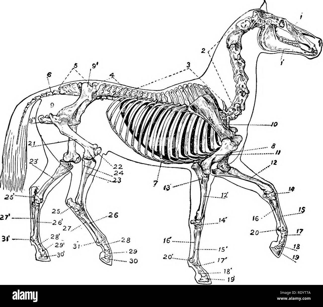 . Productive horse husbandry. Horses; Horses. STRUCTURE AND FUNCTION pulsive effort of the hindquarters is met by the forehand in such a manner as to maintain the equilibrium as the body is advanced. Locomotion is accomplished by the supporting columns being. Fig. 2.—Skeleton of the horse, showing the vertebral arch and the bone columns, one pair of legs supporting, the alternate pair, partially flexed, in a stride. 1, bones of the head; 1', lower jaw; 2, cervical vertebrge; 3, dorsal vertebrae; 4, lumbar vertebra;; 5, sacral vertebrse (sacrum); 6, coccygeal vertebree; 7, ribs; 8, sternum (bre Stock Photohttps://www.alamy.com/image-license-details/?v=1https://www.alamy.com/productive-horse-husbandry-horses-horses-structure-and-function-pulsive-effort-of-the-hindquarters-is-met-by-the-forehand-in-such-a-manner-as-to-maintain-the-equilibrium-as-the-body-is-advanced-locomotion-is-accomplished-by-the-supporting-columns-being-fig-2skeleton-of-the-horse-showing-the-vertebral-arch-and-the-bone-columns-one-pair-of-legs-supporting-the-alternate-pair-partially-flexed-in-a-stride-1-bones-of-the-head-1-lower-jaw-2-cervical-vertebrge-3-dorsal-vertebrae-4-lumbar-vertebra-5-sacral-vertebrse-sacrum-6-coccygeal-vertebree-7-ribs-8-sternum-bre-image232337038.html
RMRDYT7A–. Productive horse husbandry. Horses; Horses. STRUCTURE AND FUNCTION pulsive effort of the hindquarters is met by the forehand in such a manner as to maintain the equilibrium as the body is advanced. Locomotion is accomplished by the supporting columns being. Fig. 2.—Skeleton of the horse, showing the vertebral arch and the bone columns, one pair of legs supporting, the alternate pair, partially flexed, in a stride. 1, bones of the head; 1', lower jaw; 2, cervical vertebrge; 3, dorsal vertebrae; 4, lumbar vertebra;; 5, sacral vertebrse (sacrum); 6, coccygeal vertebree; 7, ribs; 8, sternum (bre
 . Catalogue of the fossil Reptilia and Amphibia in the British Museum (Natural history) ... By Richard Lydekker ... Reptiles, Fossil; Amphibians, Fossil. CROCODILT DJE. 61 Fig. 9.. Crocodilus spenceri.—Ee3tored cranium; from the London Clay of Sheppey. About . 38988. Part of the maxillary region of the cranium. Same history. The following vertebrae were referred by Owen to C. champsoides ; tliey all belong to thz Bowerbank Collection. 38979. The third cervical vertebra, Figured by Owen, op. cit. {Fig.) pi. v. figs. 7, 8. 38978. The first dorsal vertebra, Figured by Owen, op. cit. pi. v. (Fig. Stock Photohttps://www.alamy.com/image-license-details/?v=1https://www.alamy.com/catalogue-of-the-fossil-reptilia-and-amphibia-in-the-british-museum-natural-history-by-richard-lydekker-reptiles-fossil-amphibians-fossil-crocodilt-dje-61-fig-9-crocodilus-spenceriee3tored-cranium-from-the-london-clay-of-sheppey-about-38988-part-of-the-maxillary-region-of-the-cranium-same-history-the-following-vertebrae-were-referred-by-owen-to-c-champsoides-tliey-all-belong-to-thz-bowerbank-collection-38979-the-third-cervical-vertebra-figured-by-owen-op-cit-fig-pi-v-figs-7-8-38978-the-first-dorsal-vertebra-figured-by-owen-op-cit-pi-v-fig-image233176761.html
RMRFA39D–. Catalogue of the fossil Reptilia and Amphibia in the British Museum (Natural history) ... By Richard Lydekker ... Reptiles, Fossil; Amphibians, Fossil. CROCODILT DJE. 61 Fig. 9.. Crocodilus spenceri.—Ee3tored cranium; from the London Clay of Sheppey. About . 38988. Part of the maxillary region of the cranium. Same history. The following vertebrae were referred by Owen to C. champsoides ; tliey all belong to thz Bowerbank Collection. 38979. The third cervical vertebra, Figured by Owen, op. cit. {Fig.) pi. v. figs. 7, 8. 38978. The first dorsal vertebra, Figured by Owen, op. cit. pi. v. (Fig.
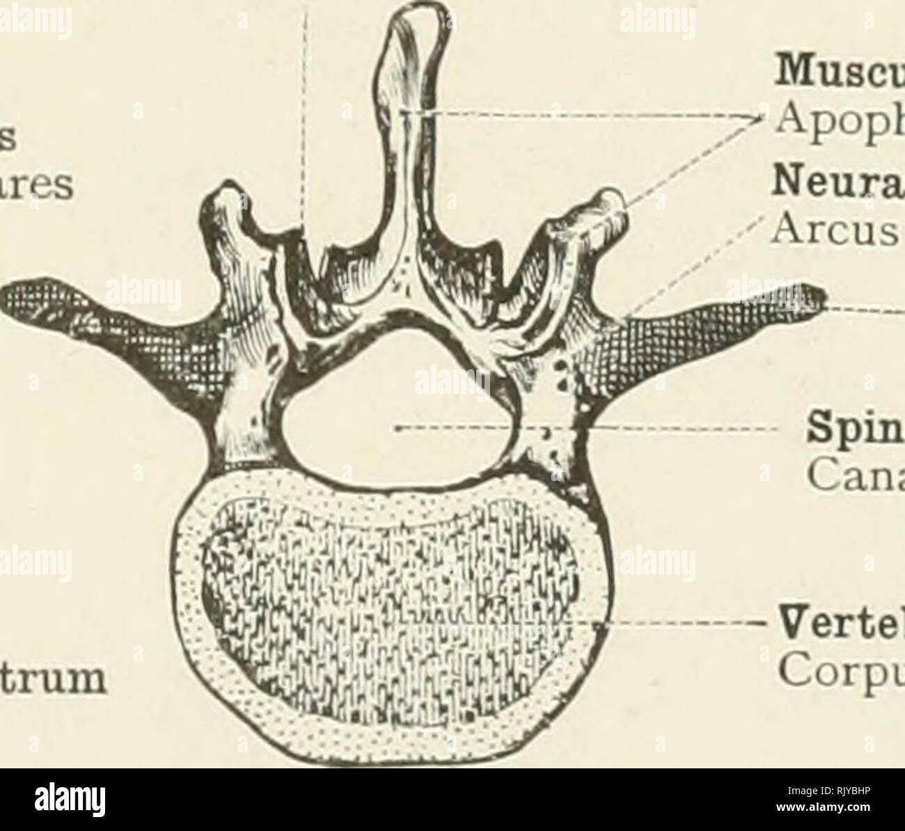 . An atlas of human anatomy for students and physicians. Anatomy. / Muscular apophyses Apophyses musculares Vertebral body or centrum xij Corpus vertebrae. Muscular apophysis Apophyses musculares Neural arch Arcus vertebrae Costal apophysis Apophysis costalis Spinal canal Canalis vertebralis Vertebral body or centrum Corpus vertebrae Fig. 99.—Skeleton of a Cervical Segment. Fig. 100.—Skeleton of a Lumbar Segment. Spinal canal Canalis vertebralis Muscular apophyses "7 Apophyses musculares Articular apophysis Apophysis articulari Neural arch Arcus vertebrae Vertebral body or centrum Co Stock Photohttps://www.alamy.com/image-license-details/?v=1https://www.alamy.com/an-atlas-of-human-anatomy-for-students-and-physicians-anatomy-muscular-apophyses-apophyses-musculares-vertebral-body-or-centrum-xij-corpus-vertebrae-muscular-apophysis-apophyses-musculares-neural-arch-arcus-vertebrae-costal-apophysis-apophysis-costalis-spinal-canal-canalis-vertebralis-vertebral-body-or-centrum-corpus-vertebrae-fig-99skeleton-of-a-cervical-segment-fig-100skeleton-of-a-lumbar-segment-spinal-canal-canalis-vertebralis-muscular-apophyses-quot7-apophyses-musculares-articular-apophysis-apophysis-articulari-neural-arch-arcus-vertebrae-vertebral-body-or-centrum-co-image235400418.html
RMRJYBHP–. An atlas of human anatomy for students and physicians. Anatomy. / Muscular apophyses Apophyses musculares Vertebral body or centrum xij Corpus vertebrae. Muscular apophysis Apophyses musculares Neural arch Arcus vertebrae Costal apophysis Apophysis costalis Spinal canal Canalis vertebralis Vertebral body or centrum Corpus vertebrae Fig. 99.—Skeleton of a Cervical Segment. Fig. 100.—Skeleton of a Lumbar Segment. Spinal canal Canalis vertebralis Muscular apophyses "7 Apophyses musculares Articular apophysis Apophysis articulari Neural arch Arcus vertebrae Vertebral body or centrum Co
 . An introduction to the study of mammals living and extinct. Mammals. PHYSETERID.-E 249 to the apex. Upper edge of the mesethmoid forming a roughened irregular projection between the narial apertures, inclining to the left side. Mandible exceedingly long and narrow, the symphysis being more than half the length of the ramus. Vertebras: C 7, D 11, L 8, C 24 ; total 50. Atlas free; all the other cervical vertebrae united by their bodies and spines into a single mass. Eleventh pair of ribs rudimentary. Head about one-third the length of the body; very massive, high and truncated, and rather comp Stock Photohttps://www.alamy.com/image-license-details/?v=1https://www.alamy.com/an-introduction-to-the-study-of-mammals-living-and-extinct-mammals-physeterid-e-249-to-the-apex-upper-edge-of-the-mesethmoid-forming-a-roughened-irregular-projection-between-the-narial-apertures-inclining-to-the-left-side-mandible-exceedingly-long-and-narrow-the-symphysis-being-more-than-half-the-length-of-the-ramus-vertebras-c-7-d-11-l-8-c-24-total-50-atlas-free-all-the-other-cervical-vertebrae-united-by-their-bodies-and-spines-into-a-single-mass-eleventh-pair-of-ribs-rudimentary-head-about-one-third-the-length-of-the-body-very-massive-high-and-truncated-and-rather-comp-image232347787.html
RMRE09Y7–. An introduction to the study of mammals living and extinct. Mammals. PHYSETERID.-E 249 to the apex. Upper edge of the mesethmoid forming a roughened irregular projection between the narial apertures, inclining to the left side. Mandible exceedingly long and narrow, the symphysis being more than half the length of the ramus. Vertebras: C 7, D 11, L 8, C 24 ; total 50. Atlas free; all the other cervical vertebrae united by their bodies and spines into a single mass. Eleventh pair of ribs rudimentary. Head about one-third the length of the body; very massive, high and truncated, and rather comp
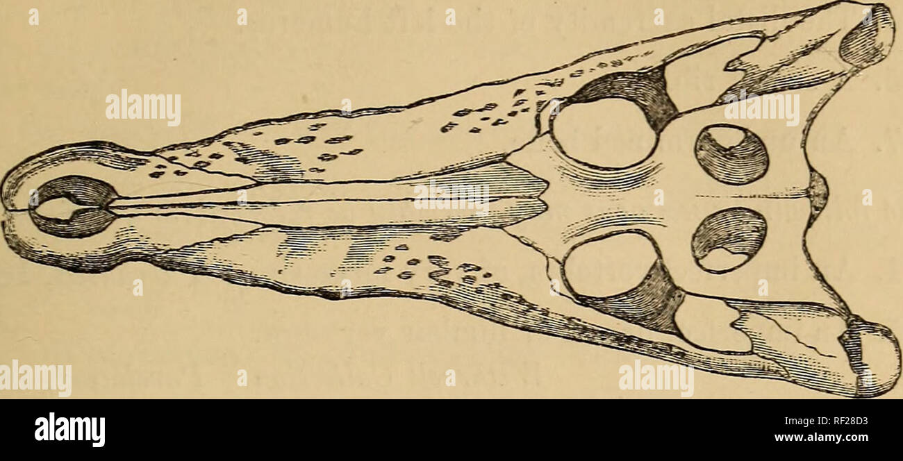 . Catalogue of the fossil Reptilia and Amphibia in the British Museum (Natural history) ... By Richard Lydekker ... Reptiles, Fossil; Amphibians, Fossil. CROCODILT DJE. 61 Fig. 9.. Crocodilus spenceri.—Ee3tored cranium; from the London Clay of Sheppey. About . 38988. Part of the maxillary region of the cranium. Same history. The following vertebrae were referred by Owen to C. champsoides ; tliey all belong to thz Bowerbank Collection. 38979. The third cervical vertebra, Figured by Owen, op. cit. {Fig.) pi. v. figs. 7, 8. 38978. The first dorsal vertebra, Figured by Owen, op. cit. pi. v. (Fig. Stock Photohttps://www.alamy.com/image-license-details/?v=1https://www.alamy.com/catalogue-of-the-fossil-reptilia-and-amphibia-in-the-british-museum-natural-history-by-richard-lydekker-reptiles-fossil-amphibians-fossil-crocodilt-dje-61-fig-9-crocodilus-spenceriee3tored-cranium-from-the-london-clay-of-sheppey-about-38988-part-of-the-maxillary-region-of-the-cranium-same-history-the-following-vertebrae-were-referred-by-owen-to-c-champsoides-tliey-all-belong-to-thz-bowerbank-collection-38979-the-third-cervical-vertebra-figured-by-owen-op-cit-fig-pi-v-figs-7-8-38978-the-first-dorsal-vertebra-figured-by-owen-op-cit-pi-v-fig-image233005167.html
RMRF28D3–. Catalogue of the fossil Reptilia and Amphibia in the British Museum (Natural history) ... By Richard Lydekker ... Reptiles, Fossil; Amphibians, Fossil. CROCODILT DJE. 61 Fig. 9.. Crocodilus spenceri.—Ee3tored cranium; from the London Clay of Sheppey. About . 38988. Part of the maxillary region of the cranium. Same history. The following vertebrae were referred by Owen to C. champsoides ; tliey all belong to thz Bowerbank Collection. 38979. The third cervical vertebra, Figured by Owen, op. cit. {Fig.) pi. v. figs. 7, 8. 38978. The first dorsal vertebra, Figured by Owen, op. cit. pi. v. (Fig.
 . Bulletin of the British Museum (Natural History), Geology. . Figs. 6-8. Three vertebrae from a complete set of cervicals. 6, atlas in ventral view 7, axis, in lateral view ; 8, fifth cervical in anterior view, a = vertebrarterial foramen. A second and less complete atlas is similar to the first but more indented at the front of the dorsal side. A third atlas has its transverse processes projecting less backwards, and more prominent ridges behind the ventral hollows, which may be correlated characters. Its front articular facets are less wide than in the first two atlases mentioned, and its d Stock Photohttps://www.alamy.com/image-license-details/?v=1https://www.alamy.com/bulletin-of-the-british-museum-natural-history-geology-figs-6-8-three-vertebrae-from-a-complete-set-of-cervicals-6-atlas-in-ventral-view-7-axis-in-lateral-view-8-fifth-cervical-in-anterior-view-a-=-vertebrarterial-foramen-a-second-and-less-complete-atlas-is-similar-to-the-first-but-more-indented-at-the-front-of-the-dorsal-side-a-third-atlas-has-its-transverse-processes-projecting-less-backwards-and-more-prominent-ridges-behind-the-ventral-hollows-which-may-be-correlated-characters-its-front-articular-facets-are-less-wide-than-in-the-first-two-atlases-mentioned-and-its-d-image233994050.html
RMRGK9PA–. Bulletin of the British Museum (Natural History), Geology. . Figs. 6-8. Three vertebrae from a complete set of cervicals. 6, atlas in ventral view 7, axis, in lateral view ; 8, fifth cervical in anterior view, a = vertebrarterial foramen. A second and less complete atlas is similar to the first but more indented at the front of the dorsal side. A third atlas has its transverse processes projecting less backwards, and more prominent ridges behind the ventral hollows, which may be correlated characters. Its front articular facets are less wide than in the first two atlases mentioned, and its d
 . Bulletin of the Museum of Comparative Zoology at Harvard College. Zoology. 300 bulletin: museum of comparative zoology 24.6 and varies between 23 (4 cases) and 26 (5 cases). The total num- ber of vertebrae varies between 52 and 55, averaging 53.7. The typical vertebral formula of Nasalis reads: 7 cervical, 12 thoracic, 7 lumbar, 3 sacral, and 25 caudal vertebrae. The average vertebral formulae of nearly all other lower catarrhines are exactly the same, except of course in regard to the caudal region. This formula, disregarding the caudal region, occurred in 85 per cent of the proboscis monke Stock Photohttps://www.alamy.com/image-license-details/?v=1https://www.alamy.com/bulletin-of-the-museum-of-comparative-zoology-at-harvard-college-zoology-300-bulletin-museum-of-comparative-zoology-246-and-varies-between-23-4-cases-and-26-5-cases-the-total-num-ber-of-vertebrae-varies-between-52-and-55-averaging-537-the-typical-vertebral-formula-of-nasalis-reads-7-cervical-12-thoracic-7-lumbar-3-sacral-and-25-caudal-vertebrae-the-average-vertebral-formulae-of-nearly-all-other-lower-catarrhines-are-exactly-the-same-except-of-course-in-regard-to-the-caudal-region-this-formula-disregarding-the-caudal-region-occurred-in-85-per-cent-of-the-proboscis-monke-image233914551.html
RMRGFMB3–. Bulletin of the Museum of Comparative Zoology at Harvard College. Zoology. 300 bulletin: museum of comparative zoology 24.6 and varies between 23 (4 cases) and 26 (5 cases). The total num- ber of vertebrae varies between 52 and 55, averaging 53.7. The typical vertebral formula of Nasalis reads: 7 cervical, 12 thoracic, 7 lumbar, 3 sacral, and 25 caudal vertebrae. The average vertebral formulae of nearly all other lower catarrhines are exactly the same, except of course in regard to the caudal region. This formula, disregarding the caudal region, occurred in 85 per cent of the proboscis monke
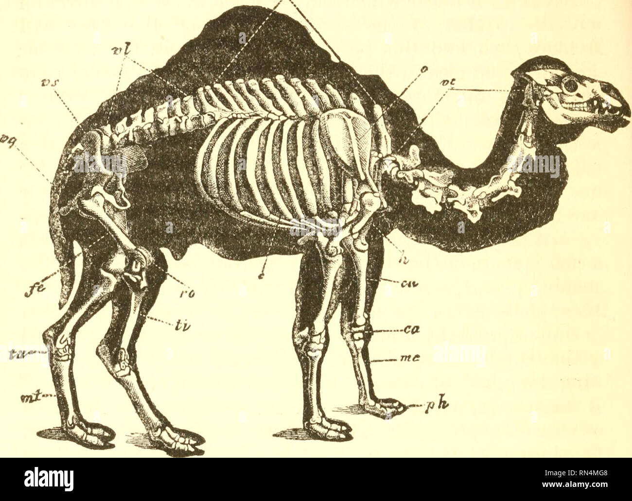 . Animal physiology. Physiology, Comparative. 484 EXTREMITIES OF LOWER ANIMALS. Stances, we find the number of bones in the hand increased, but all of them enclosed in one envelope, so that the fingers are not separate. This is the case with many aquatic animals. Fig. 229.—Skeleton of the Camel. t'c, cervical vertebrae ; vd, dorsal vertebrae ; vl, lumbar vertebrae ; vs. sacral vertebrae; rg, caudal vertebrae; s, scapula; h, humerus; cit, ulna; ca, carpus; 7«c, meta- carpus ; ph, phalanges ; fe, femur; ra, patella; ti, tibia; ia, tarsus; mt, metatarsus. —such as the Whale tribe among Mammals, T Stock Photohttps://www.alamy.com/image-license-details/?v=1https://www.alamy.com/animal-physiology-physiology-comparative-484-extremities-of-lower-animals-stances-we-find-the-number-of-bones-in-the-hand-increased-but-all-of-them-enclosed-in-one-envelope-so-that-the-fingers-are-not-separate-this-is-the-case-with-many-aquatic-animals-fig-229skeleton-of-the-camel-tc-cervical-vertebrae-vd-dorsal-vertebrae-vl-lumbar-vertebrae-vs-sacral-vertebrae-rg-caudal-vertebrae-s-scapula-h-humerus-cit-ulna-ca-carpus-7c-meta-carpus-ph-phalanges-fe-femur-ra-patella-ti-tibia-ia-tarsus-mt-metatarsus-such-as-the-whale-tribe-among-mammals-t-image236746504.html
RMRN4MG8–. Animal physiology. Physiology, Comparative. 484 EXTREMITIES OF LOWER ANIMALS. Stances, we find the number of bones in the hand increased, but all of them enclosed in one envelope, so that the fingers are not separate. This is the case with many aquatic animals. Fig. 229.—Skeleton of the Camel. t'c, cervical vertebrae ; vd, dorsal vertebrae ; vl, lumbar vertebrae ; vs. sacral vertebrae; rg, caudal vertebrae; s, scapula; h, humerus; cit, ulna; ca, carpus; 7«c, meta- carpus ; ph, phalanges ; fe, femur; ra, patella; ti, tibia; ia, tarsus; mt, metatarsus. —such as the Whale tribe among Mammals, T
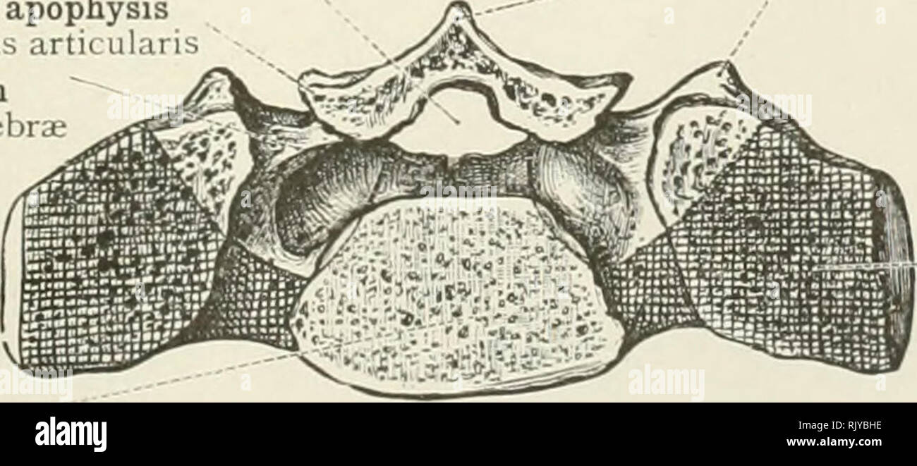 . An atlas of human anatomy for students and physicians. Anatomy. Muscular apophysis Apophyses musculares Neural arch Arcus vertebrae Costal apophysis Apophysis costalis Spinal canal Canalis vertebralis Vertebral body or centrum Corpus vertebrae Fig. 99.—Skeleton of a Cervical Segment. Fig. 100.—Skeleton of a Lumbar Segment. Spinal canal Canalis vertebralis Muscular apophyses "7 Apophyses musculares Articular apophysis Apophysis articulari Neural arch Arcus vertebrae Vertebral body or centrum Corpus vertebrae. Costal epiphysis Epiphysis costalis Fig. igi.—Skeleton of a Sacral Segment Th Stock Photohttps://www.alamy.com/image-license-details/?v=1https://www.alamy.com/an-atlas-of-human-anatomy-for-students-and-physicians-anatomy-muscular-apophysis-apophyses-musculares-neural-arch-arcus-vertebrae-costal-apophysis-apophysis-costalis-spinal-canal-canalis-vertebralis-vertebral-body-or-centrum-corpus-vertebrae-fig-99skeleton-of-a-cervical-segment-fig-100skeleton-of-a-lumbar-segment-spinal-canal-canalis-vertebralis-muscular-apophyses-quot7-apophyses-musculares-articular-apophysis-apophysis-articulari-neural-arch-arcus-vertebrae-vertebral-body-or-centrum-corpus-vertebrae-costal-epiphysis-epiphysis-costalis-fig-igiskeleton-of-a-sacral-segment-th-image235400410.html
RMRJYBHE–. An atlas of human anatomy for students and physicians. Anatomy. Muscular apophysis Apophyses musculares Neural arch Arcus vertebrae Costal apophysis Apophysis costalis Spinal canal Canalis vertebralis Vertebral body or centrum Corpus vertebrae Fig. 99.—Skeleton of a Cervical Segment. Fig. 100.—Skeleton of a Lumbar Segment. Spinal canal Canalis vertebralis Muscular apophyses "7 Apophyses musculares Articular apophysis Apophysis articulari Neural arch Arcus vertebrae Vertebral body or centrum Corpus vertebrae. Costal epiphysis Epiphysis costalis Fig. igi.—Skeleton of a Sacral Segment Th
 . Bulletin of the Museum of Comparative Zoology at Harvard College. Zoology. Fig. 5. Vertebrae of Heptodon posticus (MCZ 17670) and Tapirus pinchaque (AMNH:M 149424). A, E, dorsal atlases of H. posticus and 7, pinchaque, respectively. B, C, D, lateral views of axis and tfiird and fourth vertebrae of H. posticus. F, G, lateral and anterior vie/zs of sixth cervical vertebra of H. posticus. H, I, J, views of seventh cervical and first and second thoracic vertebrae of H. posticus. K, L, lateral views of last lum tebra of H. posticus and T. pinchaque, respectively. All X Vj. views of cervical later Stock Photohttps://www.alamy.com/image-license-details/?v=1https://www.alamy.com/bulletin-of-the-museum-of-comparative-zoology-at-harvard-college-zoology-fig-5-vertebrae-of-heptodon-posticus-mcz-17670-and-tapirus-pinchaque-amnhm-149424-a-e-dorsal-atlases-of-h-posticus-and-7-pinchaque-respectively-b-c-d-lateral-views-of-axis-and-tfiird-and-fourth-vertebrae-of-h-posticus-f-g-lateral-and-anterior-viezs-of-sixth-cervical-vertebra-of-h-posticus-h-i-j-views-of-seventh-cervical-and-first-and-second-thoracic-vertebrae-of-h-posticus-k-l-lateral-views-of-last-lum-tebra-of-h-posticus-and-t-pinchaque-respectively-all-x-vj-views-of-cervical-later-image233903080.html
RMRGF5NC–. Bulletin of the Museum of Comparative Zoology at Harvard College. Zoology. Fig. 5. Vertebrae of Heptodon posticus (MCZ 17670) and Tapirus pinchaque (AMNH:M 149424). A, E, dorsal atlases of H. posticus and 7, pinchaque, respectively. B, C, D, lateral views of axis and tfiird and fourth vertebrae of H. posticus. F, G, lateral and anterior vie/zs of sixth cervical vertebra of H. posticus. H, I, J, views of seventh cervical and first and second thoracic vertebrae of H. posticus. K, L, lateral views of last lum tebra of H. posticus and T. pinchaque, respectively. All X Vj. views of cervical later
 . Human embryology and morphology. Embryology, Human; Morphology. THE SPINAL COLUMN AND BACK. 153 present in every vertebra. In the cervical vertebrae the anterior part of the transverse processes represents a costal process, but only in the 6 th (sometimes) and 7 th is the costal process formed spine trans, proa costal facet centrum. trans, proa dors, head rib vent, head Flo. 123.—The Bicipital Rib of a Lower Vertebrate (crocodile). by a separate- centre of ossification. The costal process of the 7 th may develop into a rudiment or even a fully formed rib which reaches the sternum. In the lum Stock Photohttps://www.alamy.com/image-license-details/?v=1https://www.alamy.com/human-embryology-and-morphology-embryology-human-morphology-the-spinal-column-and-back-153-present-in-every-vertebra-in-the-cervical-vertebrae-the-anterior-part-of-the-transverse-processes-represents-a-costal-process-but-only-in-the-6-th-sometimes-and-7-th-is-the-costal-process-formed-spine-trans-proa-costal-facet-centrum-trans-proa-dors-head-rib-vent-head-flo-123the-bicipital-rib-of-a-lower-vertebrate-crocodile-by-a-separate-centre-of-ossification-the-costal-process-of-the-7-th-may-develop-into-a-rudiment-or-even-a-fully-formed-rib-which-reaches-the-sternum-in-the-lum-image232345619.html
RMRE075R–. Human embryology and morphology. Embryology, Human; Morphology. THE SPINAL COLUMN AND BACK. 153 present in every vertebra. In the cervical vertebrae the anterior part of the transverse processes represents a costal process, but only in the 6 th (sometimes) and 7 th is the costal process formed spine trans, proa costal facet centrum. trans, proa dors, head rib vent, head Flo. 123.—The Bicipital Rib of a Lower Vertebrate (crocodile). by a separate- centre of ossification. The costal process of the 7 th may develop into a rudiment or even a fully formed rib which reaches the sternum. In the lum
 . Bulletin - United States National Museum. Science. 10 Cervical Vertebrae, USNM 11976, Pelocetus calvertensis 1-5, Anterior views: i, Atlas; 2, axis; 3, fourth cervical; 4, sixth cervical; 5, seventh cervical. 6-10, Lateral views: 6, Atlas; 7, axis and third cervical, ankylosed; 8, fourth cervical; 9, sixth cervical; 10, seventh cervical.. Please note that these images are extracted from scanned page images that may have been digitally enhanced for readability - coloration and appearance of these illustrations may not perfectly resemble the original work.. United States National Museum; Smith Stock Photohttps://www.alamy.com/image-license-details/?v=1https://www.alamy.com/bulletin-united-states-national-museum-science-10-cervical-vertebrae-usnm-11976-pelocetus-calvertensis-1-5-anterior-views-i-atlas-2-axis-3-fourth-cervical-4-sixth-cervical-5-seventh-cervical-6-10-lateral-views-6-atlas-7-axis-and-third-cervical-ankylosed-8-fourth-cervical-9-sixth-cervical-10-seventh-cervical-please-note-that-these-images-are-extracted-from-scanned-page-images-that-may-have-been-digitally-enhanced-for-readability-coloration-and-appearance-of-these-illustrations-may-not-perfectly-resemble-the-original-work-united-states-national-museum-smith-image233765002.html
RMRG8WJ2–. Bulletin - United States National Museum. Science. 10 Cervical Vertebrae, USNM 11976, Pelocetus calvertensis 1-5, Anterior views: i, Atlas; 2, axis; 3, fourth cervical; 4, sixth cervical; 5, seventh cervical. 6-10, Lateral views: 6, Atlas; 7, axis and third cervical, ankylosed; 8, fourth cervical; 9, sixth cervical; 10, seventh cervical.. Please note that these images are extracted from scanned page images that may have been digitally enhanced for readability - coloration and appearance of these illustrations may not perfectly resemble the original work.. United States National Museum; Smith
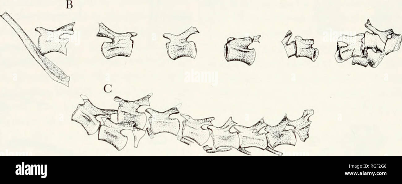 . Bulletin of the Museum of Comparative Zoology at Harvard College. Zoology. D ^ Figure 7. Archaeolbyris f/orensis. A, proximal caudal vertebrae, one cervical rib, and two isolated presacral ribs, MCZ 4081; B, mid-caudal vertebrae, not in articulation, and an isolated presacral rib, MCZ 4083; C, mid-caudal vertebrae, in articulation, MCZ 4084; D, posterior caudal vertebrae, MCZ 4081. All X 1- the body of the eentrum, in close proximitv' to the transverse process. The two articulat- ing facets are separated only by a small groove. The capitular facet is triangular in shape, with its tip pointin Stock Photohttps://www.alamy.com/image-license-details/?v=1https://www.alamy.com/bulletin-of-the-museum-of-comparative-zoology-at-harvard-college-zoology-d-figure-7-archaeolbyris-forensis-a-proximal-caudal-vertebrae-one-cervical-rib-and-two-isolated-presacral-ribs-mcz-4081-b-mid-caudal-vertebrae-not-in-articulation-and-an-isolated-presacral-rib-mcz-4083-c-mid-caudal-vertebrae-in-articulation-mcz-4084-d-posterior-caudal-vertebrae-mcz-4081-all-x-1-the-body-of-the-eentrum-in-close-proximitv-to-the-transverse-process-the-two-articulat-ing-facets-are-separated-only-by-a-small-groove-the-capitular-facet-is-triangular-in-shape-with-its-tip-pointin-image233900584.html
RMRGF2G8–. Bulletin of the Museum of Comparative Zoology at Harvard College. Zoology. D ^ Figure 7. Archaeolbyris f/orensis. A, proximal caudal vertebrae, one cervical rib, and two isolated presacral ribs, MCZ 4081; B, mid-caudal vertebrae, not in articulation, and an isolated presacral rib, MCZ 4083; C, mid-caudal vertebrae, in articulation, MCZ 4084; D, posterior caudal vertebrae, MCZ 4081. All X 1- the body of the eentrum, in close proximitv' to the transverse process. The two articulat- ing facets are separated only by a small groove. The capitular facet is triangular in shape, with its tip pointin
 . The American natural history; a foundation of useful knowledge of the higher animals of North America. Natural history. 12 APES AND MONKEYS ful canine teeth are alone sufficient to proclaim the savage wild beast. To many persons it seems strange that, notwithstanding the seemingly wide differences between the various races of. By permission of J. F. G. Umlauff. SKELETONS OP MAN AND GORILLA. 1, cervical vertebrae, 2, collar bone, 3, humerus, 4, sternum, 5, ribs, 6, rib cartilages, 7, dorsal vertebrae, 8, lumbar vertebrae, 9, pelvis, 10, radius, 11, ulna, 12, carpals, 13, metacarpals, 14, phal Stock Photohttps://www.alamy.com/image-license-details/?v=1https://www.alamy.com/the-american-natural-history-a-foundation-of-useful-knowledge-of-the-higher-animals-of-north-america-natural-history-12-apes-and-monkeys-ful-canine-teeth-are-alone-sufficient-to-proclaim-the-savage-wild-beast-to-many-persons-it-seems-strange-that-notwithstanding-the-seemingly-wide-differences-between-the-various-races-of-by-permission-of-j-f-g-umlauff-skeletons-op-man-and-gorilla-1-cervical-vertebrae-2-collar-bone-3-humerus-4-sternum-5-ribs-6-rib-cartilages-7-dorsal-vertebrae-8-lumbar-vertebrae-9-pelvis-10-radius-11-ulna-12-carpals-13-metacarpals-14-phal-image232010955.html
RMRDD09F–. The American natural history; a foundation of useful knowledge of the higher animals of North America. Natural history. 12 APES AND MONKEYS ful canine teeth are alone sufficient to proclaim the savage wild beast. To many persons it seems strange that, notwithstanding the seemingly wide differences between the various races of. By permission of J. F. G. Umlauff. SKELETONS OP MAN AND GORILLA. 1, cervical vertebrae, 2, collar bone, 3, humerus, 4, sternum, 5, ribs, 6, rib cartilages, 7, dorsal vertebrae, 8, lumbar vertebrae, 9, pelvis, 10, radius, 11, ulna, 12, carpals, 13, metacarpals, 14, phal
 . The American natural history; a foundation of useful knowledge of the higher animals of North America. Natural history. ORDEES OF MAM.MALS—APES AND MOXKEYS The Apes.—The three great man-like (or an'thro-poid) apes — gorilla, chimpanzee and orang-utan—are so much like human beings that, to most persons, they are the most won-. By permission of J. F. G. Umla SKELETONS OF MAN AND GORILLA. 1, cervical vertebrae I'-*, carpals, 2, ci)llar bone. 13, metacarpals, 3, humerus, 14, phalanges, 4, sternuiu, 15, cavity of pelvis 5, rihs, 16, sacrum, 6, rib cartilages, 17, femur. 7, dorsal vertebrae, 18, p Stock Photohttps://www.alamy.com/image-license-details/?v=1https://www.alamy.com/the-american-natural-history-a-foundation-of-useful-knowledge-of-the-higher-animals-of-north-america-natural-history-ordees-of-mammalsapes-and-moxkeys-the-apesthe-three-great-man-like-or-anthro-poid-apes-gorilla-chimpanzee-and-orang-utanare-so-much-like-human-beings-that-to-most-persons-they-are-the-most-won-by-permission-of-j-f-g-umla-skeletons-of-man-and-gorilla-1-cervical-vertebrae-i-carpals-2-cillar-bone-13-metacarpals-3-humerus-14-phalanges-4-sternuiu-15-cavity-of-pelvis-5-rihs-16-sacrum-6-rib-cartilages-17-femur-7-dorsal-vertebrae-18-p-image232039827.html
RMRDE94K–. The American natural history; a foundation of useful knowledge of the higher animals of North America. Natural history. ORDEES OF MAM.MALS—APES AND MOXKEYS The Apes.—The three great man-like (or an'thro-poid) apes — gorilla, chimpanzee and orang-utan—are so much like human beings that, to most persons, they are the most won-. By permission of J. F. G. Umla SKELETONS OF MAN AND GORILLA. 1, cervical vertebrae I'-*, carpals, 2, ci)llar bone. 13, metacarpals, 3, humerus, 14, phalanges, 4, sternuiu, 15, cavity of pelvis 5, rihs, 16, sacrum, 6, rib cartilages, 17, femur. 7, dorsal vertebrae, 18, p
 . Bulletin of the Museum of Comparative Zoology at Harvard College. Zoology. H. Figure 10. Elements of the vertebral column of Caerorhachis bairdi. A, 18th, 19th and 20th vertebrae, lateral view; B, restoration of 18th, 19th and 20th vertebrae, lateral view; C, ic 3; D, pc 3; E, pc 6; F, pc 7; G, pc 10; H, restoration of pc, anterior view; I, restoration of pc, posterior view; J, restoration of vertebra, anterior view. All X 2. vertebrae represents the third cervical. The configuration of the associated ribs reveals that the first several vertebrae in the block are cervicals, and unless the an Stock Photohttps://www.alamy.com/image-license-details/?v=1https://www.alamy.com/bulletin-of-the-museum-of-comparative-zoology-at-harvard-college-zoology-h-figure-10-elements-of-the-vertebral-column-of-caerorhachis-bairdi-a-18th-19th-and-20th-vertebrae-lateral-view-b-restoration-of-18th-19th-and-20th-vertebrae-lateral-view-c-ic-3-d-pc-3-e-pc-6-f-pc-7-g-pc-10-h-restoration-of-pc-anterior-view-i-restoration-of-pc-posterior-view-j-restoration-of-vertebra-anterior-view-all-x-2-vertebrae-represents-the-third-cervical-the-configuration-of-the-associated-ribs-reveals-that-the-first-several-vertebrae-in-the-block-are-cervicals-and-unless-the-an-image233893785.html
RMRGENWD–. Bulletin of the Museum of Comparative Zoology at Harvard College. Zoology. H. Figure 10. Elements of the vertebral column of Caerorhachis bairdi. A, 18th, 19th and 20th vertebrae, lateral view; B, restoration of 18th, 19th and 20th vertebrae, lateral view; C, ic 3; D, pc 3; E, pc 6; F, pc 7; G, pc 10; H, restoration of pc, anterior view; I, restoration of pc, posterior view; J, restoration of vertebra, anterior view. All X 2. vertebrae represents the third cervical. The configuration of the associated ribs reveals that the first several vertebrae in the block are cervicals, and unless the an
 . The anatomy of the domestic animals . Veterinary anatomy. THE VERTEBRAL COLUMN ,33 The Vertebral Column The vertebral formula of the horse is CTTigLeSsCyu^i. Anterior artic- ular process of axis Spinous proc- ess of axis Transverse process of axis. Fig. 7.—Cervical Vertebh^e of Hohse; Dorsal View. Fig. 8.—Cervical Vertebrae of Horse; Ventral a, Articular processes; b, transverse processes; 1, View. dorsal arch of atlas; S, wing of atlas; 3, intervertebral a, Transverse processes; 1, ventral tubercle of atlas; foramen of atlas; 4, alar foramen of atlas; 6, foramen S, anterior articular caviti Stock Photohttps://www.alamy.com/image-license-details/?v=1https://www.alamy.com/the-anatomy-of-the-domestic-animals-veterinary-anatomy-the-vertebral-column-33-the-vertebral-column-the-vertebral-formula-of-the-horse-is-cttiglesscyui-anterior-artic-ular-process-of-axis-spinous-proc-ess-of-axis-transverse-process-of-axis-fig-7cervical-vertebhe-of-hohse-dorsal-view-fig-8cervical-vertebrae-of-horse-ventral-a-articular-processes-b-transverse-processes-1-view-dorsal-arch-of-atlas-s-wing-of-atlas-3-intervertebral-a-transverse-processes-1-ventral-tubercle-of-atlas-foramen-of-atlas-4-alar-foramen-of-atlas-6-foramen-s-anterior-articular-caviti-image232327914.html
RMRDYCHE–. The anatomy of the domestic animals . Veterinary anatomy. THE VERTEBRAL COLUMN ,33 The Vertebral Column The vertebral formula of the horse is CTTigLeSsCyu^i. Anterior artic- ular process of axis Spinous proc- ess of axis Transverse process of axis. Fig. 7.—Cervical Vertebh^e of Hohse; Dorsal View. Fig. 8.—Cervical Vertebrae of Horse; Ventral a, Articular processes; b, transverse processes; 1, View. dorsal arch of atlas; S, wing of atlas; 3, intervertebral a, Transverse processes; 1, ventral tubercle of atlas; foramen of atlas; 4, alar foramen of atlas; 6, foramen S, anterior articular caviti
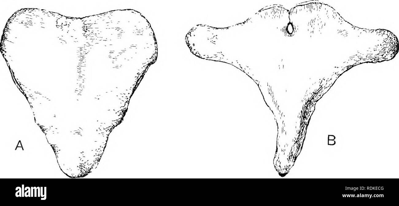 . The Cambridge natural history. Zoology. Fig. 186.âSection tlirougli iiiiddlB line of united cervical vertebrae of Greenland liight 'liale {Bal- aena â mysticetus). x -^. a. Arti- cular surface for occipital condyle; e, ejiipli3'sis on posterior end of body of seventh cervical vertebia ; 571, foramen in aich of atlas ior first spinal nerve ; 1, nrcli of atlas ; 2, 3, 4, 5, 6, conjoined arches of the axis and four followin"; verte- brae ; 7, arch of seventh vertebra. (From Flower's Oxteulagy.) as there is only a rudimentary pelvis, not attached to the vertebral column, no sacral region Stock Photohttps://www.alamy.com/image-license-details/?v=1https://www.alamy.com/the-cambridge-natural-history-zoology-fig-186section-tlirougli-iiiiddlb-line-of-united-cervical-vertebrae-of-greenland-liight-liale-bal-aena-mysticetus-x-a-arti-cular-surface-for-occipital-condyle-e-ejiipli3sis-on-posterior-end-of-body-of-seventh-cervical-vertebia-571-foramen-in-aich-of-atlas-ior-first-spinal-nerve-1-nrcli-of-atlas-2-3-4-5-6-conjoined-arches-of-the-axis-and-four-followinquot-verte-brae-7-arch-of-seventh-vertebra-from-flowers-oxteulagy-as-there-is-only-a-rudimentary-pelvis-not-attached-to-the-vertebral-column-no-sacral-region-image232153728.html
RMRDKECG–. The Cambridge natural history. Zoology. Fig. 186.âSection tlirougli iiiiddlB line of united cervical vertebrae of Greenland liight 'liale {Bal- aena â mysticetus). x -^. a. Arti- cular surface for occipital condyle; e, ejiipli3'sis on posterior end of body of seventh cervical vertebia ; 571, foramen in aich of atlas ior first spinal nerve ; 1, nrcli of atlas ; 2, 3, 4, 5, 6, conjoined arches of the axis and four followin"; verte- brae ; 7, arch of seventh vertebra. (From Flower's Oxteulagy.) as there is only a rudimentary pelvis, not attached to the vertebral column, no sacral region
 . Carnegie Institution of Washington publication. . FIGS. 5 and 6. Two specimens of the atlas in which there is incomplete dorsal arch formation. Fig. 5, specimen No. 258637 F2, U. S. National Museum. Fig. 6, specimen No. 262993, U. S. National Museum. FIG. 7. Specimen showing symmetry of the dorsal laminae. Specimen No. 256457, U. S. National Museum. FIG. 8. Specimen showing asymmetry of the dorsal laminae. Specimen No. 271782, U. S. National Museum. FIG. 9. Posterior view of the cervical vertebrae, of which the fifth lacks dorsal laminae. Specimen No. 227471, U. S. Nat. Mus. FIG. 10. Specime Stock Photohttps://www.alamy.com/image-license-details/?v=1https://www.alamy.com/carnegie-institution-of-washington-publication-figs-5-and-6-two-specimens-of-the-atlas-in-which-there-is-incomplete-dorsal-arch-formation-fig-5-specimen-no-258637-f2-u-s-national-museum-fig-6-specimen-no-262993-u-s-national-museum-fig-7-specimen-showing-symmetry-of-the-dorsal-laminae-specimen-no-256457-u-s-national-museum-fig-8-specimen-showing-asymmetry-of-the-dorsal-laminae-specimen-no-271782-u-s-national-museum-fig-9-posterior-view-of-the-cervical-vertebrae-of-which-the-fifth-lacks-dorsal-laminae-specimen-no-227471-u-s-nat-mus-fig-10-specime-image233473666.html
RMRFRJ16–. Carnegie Institution of Washington publication. . FIGS. 5 and 6. Two specimens of the atlas in which there is incomplete dorsal arch formation. Fig. 5, specimen No. 258637 F2, U. S. National Museum. Fig. 6, specimen No. 262993, U. S. National Museum. FIG. 7. Specimen showing symmetry of the dorsal laminae. Specimen No. 256457, U. S. National Museum. FIG. 8. Specimen showing asymmetry of the dorsal laminae. Specimen No. 271782, U. S. National Museum. FIG. 9. Posterior view of the cervical vertebrae, of which the fifth lacks dorsal laminae. Specimen No. 227471, U. S. Nat. Mus. FIG. 10. Specime
RMRN78EY–. The anatomy of the domestic animals. Veterinary anatomy. Fig; 199.—Skeleton- of Dog; Lateral View. o. Cranium; 6, face; c, mandible; 1H-7H, cervical vertebrae; 13B, last thoracic vertebra; 1L-7L, lumbar verte- bne; K, sacrum; S, coccygeal vertebrse; 1R13R, ribs; R.kn., costal cartilages; St., sternum; d, scapula; d', supia- spinous fossa; d". inf raspinous fossa; i, spine of scapula; ^.acromion; 3, tuberosity of scapula; 5', articular end of scapula; e, humerus; 4. head of humerus; o, lateral tuberosity of humerus; o', deltoid ridge; 6, 6', epicondyles of humerus; 7, lateral condyloid
![. The vet. book, or, Animal doctor [microform]. Horses; Bétail; Chevaux; Veterinary medicine; Livestock; Médecine vétérinaire. « It yomhor.. looks as thin «i. dieletcn, give him Si. John's ConditionPowtes. The Skeleton of a Horse. (I) Head or Cranium. (81 Oibit. (3) Molar Teeth. (4) Canine Teeth. (5) Canine Teeth. (6) Inciaora. (7) Incieors, (8) Atlaa. (9) Lower Jaw. (10) Cervical Vertebrae. (lltol2| Dorsal Vertebrae. (12 to 13) Lumbar Vertebrae. (14 to 15, Sacrum or Sacral Vertebrae. (latoiri Caudal Vertebrae or Coccy- geal Vertebrae. (18-19-20) Haunch Bone, Flank Bone, Hip Bone, Os Coxae or Stock Photo . The vet. book, or, Animal doctor [microform]. Horses; Bétail; Chevaux; Veterinary medicine; Livestock; Médecine vétérinaire. « It yomhor.. looks as thin «i. dieletcn, give him Si. John's ConditionPowtes. The Skeleton of a Horse. (I) Head or Cranium. (81 Oibit. (3) Molar Teeth. (4) Canine Teeth. (5) Canine Teeth. (6) Inciaora. (7) Incieors, (8) Atlaa. (9) Lower Jaw. (10) Cervical Vertebrae. (lltol2| Dorsal Vertebrae. (12 to 13) Lumbar Vertebrae. (14 to 15, Sacrum or Sacral Vertebrae. (latoiri Caudal Vertebrae or Coccy- geal Vertebrae. (18-19-20) Haunch Bone, Flank Bone, Hip Bone, Os Coxae or Stock Photo](https://c8.alamy.com/comp/RENT0W/the-vet-book-or-animal-doctor-microform-horses-btail-chevaux-veterinary-medicine-livestock-mdecine-vtrinaire-it-yomhor-looks-as-thin-i-dieletcn-give-him-si-johns-conditionpowtes-the-skeleton-of-a-horse-i-head-or-cranium-81-oibit-3-molar-teeth-4-canine-teeth-5-canine-teeth-6-inciaora-7-incieors-8-atlaa-9-lower-jaw-10-cervical-vertebrae-lltol2-dorsal-vertebrae-12-to-13-lumbar-vertebrae-14-to-15-sacrum-or-sacral-vertebrae-latoiri-caudal-vertebrae-or-coccy-geal-vertebrae-18-19-20-haunch-bone-flank-bone-hip-bone-os-coxae-or-RENT0W.jpg) . The vet. book, or, Animal doctor [microform]. Horses; Bétail; Chevaux; Veterinary medicine; Livestock; Médecine vétérinaire. « It yomhor.. looks as thin «i. dieletcn, give him Si. John's ConditionPowtes. The Skeleton of a Horse. (I) Head or Cranium. (81 Oibit. (3) Molar Teeth. (4) Canine Teeth. (5) Canine Teeth. (6) Inciaora. (7) Incieors, (8) Atlaa. (9) Lower Jaw. (10) Cervical Vertebrae. (lltol2| Dorsal Vertebrae. (12 to 13) Lumbar Vertebrae. (14 to 15, Sacrum or Sacral Vertebrae. (latoiri Caudal Vertebrae or Coccy- geal Vertebrae. (18-19-20) Haunch Bone, Flank Bone, Hip Bone, Os Coxae or Stock Photohttps://www.alamy.com/image-license-details/?v=1https://www.alamy.com/the-vet-book-or-animal-doctor-microform-horses-btail-chevaux-veterinary-medicine-livestock-mdecine-vtrinaire-it-yomhor-looks-as-thin-i-dieletcn-give-him-si-johns-conditionpowtes-the-skeleton-of-a-horse-i-head-or-cranium-81-oibit-3-molar-teeth-4-canine-teeth-5-canine-teeth-6-inciaora-7-incieors-8-atlaa-9-lower-jaw-10-cervical-vertebrae-lltol2-dorsal-vertebrae-12-to-13-lumbar-vertebrae-14-to-15-sacrum-or-sacral-vertebrae-latoiri-caudal-vertebrae-or-coccy-geal-vertebrae-18-19-20-haunch-bone-flank-bone-hip-bone-os-coxae-or-image232819801.html
. The vet. book, or, Animal doctor [microform]. Horses; Bétail; Chevaux; Veterinary medicine; Livestock; Médecine vétérinaire. « It yomhor.. looks as thin «i. dieletcn, give him Si. John's ConditionPowtes. The Skeleton of a Horse. (I) Head or Cranium. (81 Oibit. (3) Molar Teeth. (4) Canine Teeth. (5) Canine Teeth. (6) Inciaora. (7) Incieors, (8) Atlaa. (9) Lower Jaw. (10) Cervical Vertebrae. (lltol2| Dorsal Vertebrae. (12 to 13) Lumbar Vertebrae. (14 to 15, Sacrum or Sacral Vertebrae. (latoiri Caudal Vertebrae or Coccy- geal Vertebrae. (18-19-20) Haunch Bone, Flank Bone, Hip Bone, Os Coxae or Stock Photohttps://www.alamy.com/image-license-details/?v=1https://www.alamy.com/the-vet-book-or-animal-doctor-microform-horses-btail-chevaux-veterinary-medicine-livestock-mdecine-vtrinaire-it-yomhor-looks-as-thin-i-dieletcn-give-him-si-johns-conditionpowtes-the-skeleton-of-a-horse-i-head-or-cranium-81-oibit-3-molar-teeth-4-canine-teeth-5-canine-teeth-6-inciaora-7-incieors-8-atlaa-9-lower-jaw-10-cervical-vertebrae-lltol2-dorsal-vertebrae-12-to-13-lumbar-vertebrae-14-to-15-sacrum-or-sacral-vertebrae-latoiri-caudal-vertebrae-or-coccy-geal-vertebrae-18-19-20-haunch-bone-flank-bone-hip-bone-os-coxae-or-image232819801.htmlRMRENT0W–. The vet. book, or, Animal doctor [microform]. Horses; Bétail; Chevaux; Veterinary medicine; Livestock; Médecine vétérinaire. « It yomhor.. looks as thin «i. dieletcn, give him Si. John's ConditionPowtes. The Skeleton of a Horse. (I) Head or Cranium. (81 Oibit. (3) Molar Teeth. (4) Canine Teeth. (5) Canine Teeth. (6) Inciaora. (7) Incieors, (8) Atlaa. (9) Lower Jaw. (10) Cervical Vertebrae. (lltol2| Dorsal Vertebrae. (12 to 13) Lumbar Vertebrae. (14 to 15, Sacrum or Sacral Vertebrae. (latoiri Caudal Vertebrae or Coccy- geal Vertebrae. (18-19-20) Haunch Bone, Flank Bone, Hip Bone, Os Coxae or
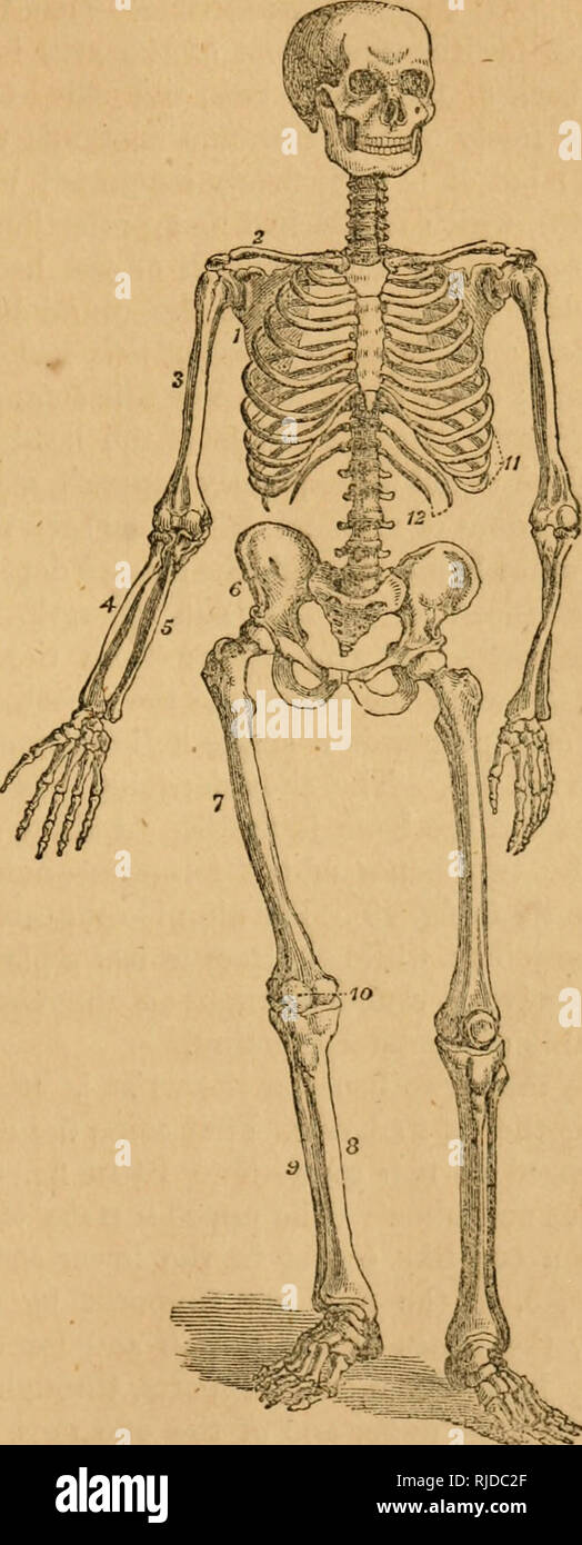 . Chamber's scientific reader : illustrated with wood engravings. Readers. THE EONY SKELETON. 47 cartilaginous band ; the other two are unattached to anything in front, and are therefore called floating ribs. The ribs, along with the sternum. THE HEAD. Tie Cranium— 1 Frontal bone. 2 Parietal bones. 1 Occipital bone. 2 Temporal bones. 1 Sphenoid bone. 1 Ethmoid bone. The Face— 2 Upper maxillary bones. 2 Malar bones. 2 Palate bones. 2 Lachrymal bones. 2 Nasal bones. The vomer. 2 Turbinated bones. 1 Lower maxillary bone. THE TRUNK. The Spine— 7 Cervical vertebrae. 12 Dorsal vertebras. 5 Lumbar ve Stock Photohttps://www.alamy.com/image-license-details/?v=1https://www.alamy.com/chambers-scientific-reader-illustrated-with-wood-engravings-readers-the-eony-skeleton-47-cartilaginous-band-the-other-two-are-unattached-to-anything-in-front-and-are-therefore-called-floating-ribs-the-ribs-along-with-the-sternum-the-head-tie-cranium-1-frontal-bone-2-parietal-bones-1-occipital-bone-2-temporal-bones-1-sphenoid-bone-1-ethmoid-bone-the-face-2-upper-maxillary-bones-2-malar-bones-2-palate-bones-2-lachrymal-bones-2-nasal-bones-the-vomer-2-turbinated-bones-1-lower-maxillary-bone-the-trunk-the-spine-7-cervical-vertebrae-12-dorsal-vertebras-5-lumbar-ve-image235093447.html
RMRJDC2F–. Chamber's scientific reader : illustrated with wood engravings. Readers. THE EONY SKELETON. 47 cartilaginous band ; the other two are unattached to anything in front, and are therefore called floating ribs. The ribs, along with the sternum. THE HEAD. Tie Cranium— 1 Frontal bone. 2 Parietal bones. 1 Occipital bone. 2 Temporal bones. 1 Sphenoid bone. 1 Ethmoid bone. The Face— 2 Upper maxillary bones. 2 Malar bones. 2 Palate bones. 2 Lachrymal bones. 2 Nasal bones. The vomer. 2 Turbinated bones. 1 Lower maxillary bone. THE TRUNK. The Spine— 7 Cervical vertebrae. 12 Dorsal vertebras. 5 Lumbar ve
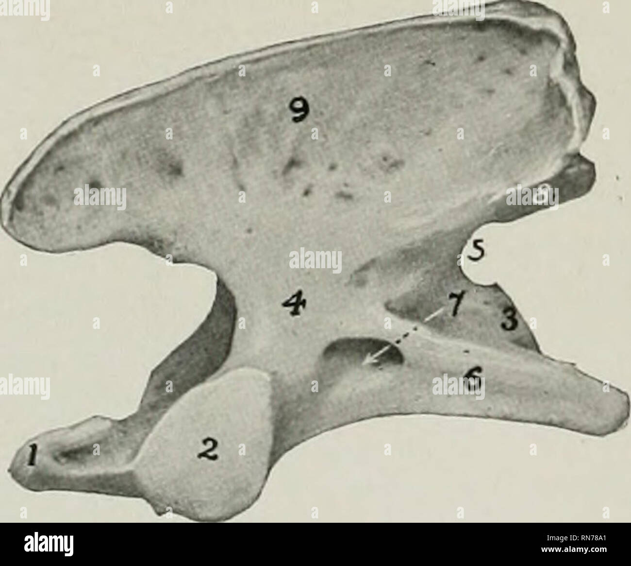 . The anatomy of the domestic animals. Veterinary anatomy. Fig. 202.—Seventh Cervical ^'ERTEB^A of Dog; Posterior View. 1, Body; 2, costal facet; 3, transverse process; 4, notch; 5, 5', articular processes; 6, spinous process. Fig. 203.—Atlas of Dog; Dorsal View. 1, Dorsal arch; 2, 2, posterior articular cavities; 3, ventral tubercle: 4, 4' intervertebral foramina; 5, 5', wings; 6, 6', alar notches; 7, T, foramina transve The bodies of the thirteen thoracic vertebrae are wide and compressed dorso- ventrally, especially at each end of the region. Their con- vex anterior surfaces are depressed i Stock Photohttps://www.alamy.com/image-license-details/?v=1https://www.alamy.com/the-anatomy-of-the-domestic-animals-veterinary-anatomy-fig-202seventh-cervical-erteba-of-dog-posterior-view-1-body-2-costal-facet-3-transverse-process-4-notch-5-5-articular-processes-6-spinous-process-fig-203atlas-of-dog-dorsal-view-1-dorsal-arch-2-2-posterior-articular-cavities-3-ventral-tubercle-4-4-intervertebral-foramina-5-5-wings-6-6-alar-notches-7-t-foramina-transve-the-bodies-of-the-thirteen-thoracic-vertebrae-are-wide-and-compressed-dorso-ventrally-especially-at-each-end-of-the-region-their-con-vex-anterior-surfaces-are-depressed-i-image236802777.html
RMRN78A1–. The anatomy of the domestic animals. Veterinary anatomy. Fig. 202.—Seventh Cervical ^'ERTEB^A of Dog; Posterior View. 1, Body; 2, costal facet; 3, transverse process; 4, notch; 5, 5', articular processes; 6, spinous process. Fig. 203.—Atlas of Dog; Dorsal View. 1, Dorsal arch; 2, 2, posterior articular cavities; 3, ventral tubercle: 4, 4' intervertebral foramina; 5, 5', wings; 6, 6', alar notches; 7, T, foramina transve The bodies of the thirteen thoracic vertebrae are wide and compressed dorso- ventrally, especially at each end of the region. Their con- vex anterior surfaces are depressed i
 . A text-book of agricultural zoology. Zoology, Economic. Fig. 175.—Skeleton of Fowl. A-B, cervical vertebrae; 1, spinous procens of third vertebra; 2, inferior ridge on same; 8, styloid process ; 4, vertebral foramen; B-0, dorsal vertebrae; 6, spinous process of first; 7, crest formed by union of other spinous processes; D-B, coccygeal vertebrae; F, G, head; 8, interorbital septum; 9, interorbital foramen; 10, premaxillary bone; 10', anterior nares; 11, man- dible ; 12, quadrate; 13, maxilla ; 14, keel of sternum (H); IS, episternum; 16, posterior lateral process; 17, obiic[ue lateral process Stock Photohttps://www.alamy.com/image-license-details/?v=1https://www.alamy.com/a-text-book-of-agricultural-zoology-zoology-economic-fig-175skeleton-of-fowl-a-b-cervical-vertebrae-1-spinous-procens-of-third-vertebra-2-inferior-ridge-on-same-8-styloid-process-4-vertebral-foramen-b-0-dorsal-vertebrae-6-spinous-process-of-first-7-crest-formed-by-union-of-other-spinous-processes-d-b-coccygeal-vertebrae-f-g-head-8-interorbital-septum-9-interorbital-foramen-10-premaxillary-bone-10-anterior-nares-11-man-dible-12-quadrate-13-maxilla-14-keel-of-sternum-h-is-episternum-16-posterior-lateral-process-17-obiic-ue-lateral-process-image232132992.html
RMRDJG00–. A text-book of agricultural zoology. Zoology, Economic. Fig. 175.—Skeleton of Fowl. A-B, cervical vertebrae; 1, spinous procens of third vertebra; 2, inferior ridge on same; 8, styloid process ; 4, vertebral foramen; B-0, dorsal vertebrae; 6, spinous process of first; 7, crest formed by union of other spinous processes; D-B, coccygeal vertebrae; F, G, head; 8, interorbital septum; 9, interorbital foramen; 10, premaxillary bone; 10', anterior nares; 11, man- dible ; 12, quadrate; 13, maxilla ; 14, keel of sternum (H); IS, episternum; 16, posterior lateral process; 17, obiic[ue lateral process
 . Brimleyana. Zoology; Ecology; Natural history. Turtles 11 humerus (.114.1); 1 radius (.114.2); 1 phalange (.114.7). 2 mixed specimens both with partial fragmented carapaces and plastra (.115); 3 cervical vertebrae (.115.5-. 115.6); 2 humeri (.115.1-.115.2); 2 scapulo-acromial processes (.115.3- .113.4); 2 coracoids (. 115.8-. 115.9); 2 tibiae (.115.10-.115.il); 1 ilium (.115.12); 1 phalange (.115.13). Four individual specimens with fragmented carapace and plastron (.116-. 119). Two mixed specimens with badly fragmented carapaces and plastra (.120). Isolated elements: 16 nuchals (.123-.132)(3 Stock Photohttps://www.alamy.com/image-license-details/?v=1https://www.alamy.com/brimleyana-zoology-ecology-natural-history-turtles-11-humerus-1141-1-radius-1142-1-phalange-1147-2-mixed-specimens-both-with-partial-fragmented-carapaces-and-plastra-115-3-cervical-vertebrae-1155-1156-2-humeri-1151-1152-2-scapulo-acromial-processes-1153-1134-2-coracoids-1158-1159-2-tibiae-11510-115il-1-ilium-11512-1-phalange-11513-four-individual-specimens-with-fragmented-carapace-and-plastron-116-119-two-mixed-specimens-with-badly-fragmented-carapaces-and-plastra-120-isolated-elements-16-nuchals-123-1323-image234284614.html
RMRH4GBJ–. Brimleyana. Zoology; Ecology; Natural history. Turtles 11 humerus (.114.1); 1 radius (.114.2); 1 phalange (.114.7). 2 mixed specimens both with partial fragmented carapaces and plastra (.115); 3 cervical vertebrae (.115.5-. 115.6); 2 humeri (.115.1-.115.2); 2 scapulo-acromial processes (.115.3- .113.4); 2 coracoids (. 115.8-. 115.9); 2 tibiae (.115.10-.115.il); 1 ilium (.115.12); 1 phalange (.115.13). Four individual specimens with fragmented carapace and plastron (.116-. 119). Two mixed specimens with badly fragmented carapaces and plastra (.120). Isolated elements: 16 nuchals (.123-.132)(3
 . Brimleyana. Zoology; Ecology; Natural history. 3 cm (.110.13-.110.14); 2 dorsal vertebrae (.110.9-. 110.10); complete pelvic girdle (.110.15); 1 caudal vertebra (.110.8). An individual specimen consisting of a nearly complete carapace and plastron (.111). An individual specimen consist- ing of a complete carapace (missing left 8-9th peripherals and 6th neural) and plastron (.112); partial skull and mandible (.112.1); partial hyoid process (.112.2); 7 cervical vertebrae (.112.20-. 112.26); 2 humeri (.112.5-. 112.6); 2 ulnae (.112.7-.112.8); 2 radii (.112.9-. 112.10); 2 scapulo-acromial proces Stock Photohttps://www.alamy.com/image-license-details/?v=1https://www.alamy.com/brimleyana-zoology-ecology-natural-history-3-cm-11013-11014-2-dorsal-vertebrae-1109-11010-complete-pelvic-girdle-11015-1-caudal-vertebra-1108-an-individual-specimen-consisting-of-a-nearly-complete-carapace-and-plastron-111-an-individual-specimen-consist-ing-of-a-complete-carapace-missing-left-8-9th-peripherals-and-6th-neural-and-plastron-112-partial-skull-and-mandible-1121-partial-hyoid-process-1122-7-cervical-vertebrae-11220-11226-2-humeri-1125-1126-2-ulnae-1127-1128-2-radii-1129-11210-2-scapulo-acromial-proces-image234284624.html
RMRH4GC0–. Brimleyana. Zoology; Ecology; Natural history. 3 cm (.110.13-.110.14); 2 dorsal vertebrae (.110.9-. 110.10); complete pelvic girdle (.110.15); 1 caudal vertebra (.110.8). An individual specimen consisting of a nearly complete carapace and plastron (.111). An individual specimen consist- ing of a complete carapace (missing left 8-9th peripherals and 6th neural) and plastron (.112); partial skull and mandible (.112.1); partial hyoid process (.112.2); 7 cervical vertebrae (.112.20-. 112.26); 2 humeri (.112.5-. 112.6); 2 ulnae (.112.7-.112.8); 2 radii (.112.9-. 112.10); 2 scapulo-acromial proces
 . The animals and man; an elementary textbook of zoology and human physiology. Zoology; Physiology. cu Fig. Co) Fig. 166 B. Side view of spinal column. C-1 to 7, cervical ver- tebrae; D-1 to 12, dor- sal vertebrje; L-1 to 5, lumbar vertebrae; S. 1. sacrum; Co. 1-4 coccyx. (After Mar- tin.) 166 A. Bony and cartilaginous skeleton, a, left parietal bone of cran- ium; b, frontal bone; c, cervical vertebrae; d, sternum; e, lumbar verte- bras; /, ulna; g, radius; h, carpals; /, meta-carpals; k, phalanges; I, tibia; m, fibula; », tarsal bones; o, metatarsals; p, phalanges; g, pa- tella (knee-pan); r, Stock Photohttps://www.alamy.com/image-license-details/?v=1https://www.alamy.com/the-animals-and-man-an-elementary-textbook-of-zoology-and-human-physiology-zoology-physiology-cu-fig-co-fig-166-b-side-view-of-spinal-column-c-1-to-7-cervical-ver-tebrae-d-1-to-12-dor-sal-vertebrje-l-1-to-5-lumbar-vertebrae-s-1-sacrum-co-1-4-coccyx-after-mar-tin-166-a-bony-and-cartilaginous-skeleton-a-left-parietal-bone-of-cran-ium-b-frontal-bone-c-cervical-vertebrae-d-sternum-e-lumbar-verte-bras-ulna-g-radius-h-carpals-meta-carpals-k-phalanges-i-tibia-m-fibula-tarsal-bones-o-metatarsals-p-phalanges-g-pa-tella-knee-pan-r-image232254736.html
RMRDT380–. The animals and man; an elementary textbook of zoology and human physiology. Zoology; Physiology. cu Fig. Co) Fig. 166 B. Side view of spinal column. C-1 to 7, cervical ver- tebrae; D-1 to 12, dor- sal vertebrje; L-1 to 5, lumbar vertebrae; S. 1. sacrum; Co. 1-4 coccyx. (After Mar- tin.) 166 A. Bony and cartilaginous skeleton, a, left parietal bone of cran- ium; b, frontal bone; c, cervical vertebrae; d, sternum; e, lumbar verte- bras; /, ulna; g, radius; h, carpals; /, meta-carpals; k, phalanges; I, tibia; m, fibula; », tarsal bones; o, metatarsals; p, phalanges; g, pa- tella (knee-pan); r,
 . Bulletin of the Museum of Comparative Zoology at Harvard College. Zoology. Fig. 7. Alligator jn-enasalis (Loomis). Vertebra, Dorsal 3; A, anterior view; B, lateral view, left side; C, posterior view. One-half natural size. Mus. Comp. Zool. No. 1,014.. Fig. 8. Alligalor prenasalis (Loomis). Vertebrae (1), Cervical 8; (2), Dorsal 1; (3), Dorsals 2, 3; (4), Dorsal 4; (5), Dorsals 5-6. A, Anterior views; B, lateral views, left side; C, posterior views. One-half natural size. Mus. Comp. Zool. No. 1,015, (1, 3, 4, 5); and No. 1,014, (2).. Please note that these images are extracted from scanned pa Stock Photohttps://www.alamy.com/image-license-details/?v=1https://www.alamy.com/bulletin-of-the-museum-of-comparative-zoology-at-harvard-college-zoology-fig-7-alligator-jn-enasalis-loomis-vertebra-dorsal-3-a-anterior-view-b-lateral-view-left-side-c-posterior-view-one-half-natural-size-mus-comp-zool-no-1014-fig-8-alligalor-prenasalis-loomis-vertebrae-1-cervical-8-2-dorsal-1-3-dorsals-2-3-4-dorsal-4-5-dorsals-5-6-a-anterior-views-b-lateral-views-left-side-c-posterior-views-one-half-natural-size-mus-comp-zool-no-1015-1-3-4-5-and-no-1014-2-please-note-that-these-images-are-extracted-from-scanned-pa-image233915667.html
RMRGFNPY–. Bulletin of the Museum of Comparative Zoology at Harvard College. Zoology. Fig. 7. Alligator jn-enasalis (Loomis). Vertebra, Dorsal 3; A, anterior view; B, lateral view, left side; C, posterior view. One-half natural size. Mus. Comp. Zool. No. 1,014.. Fig. 8. Alligalor prenasalis (Loomis). Vertebrae (1), Cervical 8; (2), Dorsal 1; (3), Dorsals 2, 3; (4), Dorsal 4; (5), Dorsals 5-6. A, Anterior views; B, lateral views, left side; C, posterior views. One-half natural size. Mus. Comp. Zool. No. 1,015, (1, 3, 4, 5); and No. 1,014, (2).. Please note that these images are extracted from scanned pa
 . Bulletin. Natural history; Natuurlijke historie. OSTEOLOGY OF DEINONYCHUS ANTIRRHOPUS 49 TABLE 3. Morphology of the cervical vertebrae of Deinonychus antirrhopus and Felis leo (attitude of centra faces relative to the floor of the neural canal) ANTERIOR CENTRUM FACE POSTERIOR CENTRUM FACE D. antirrhopus F. leo D. antirrhopus F. leo YPM 5204 YPMOC YPM 5204 YPMOC Vertebra number YPM 5210 1050 YPM 5210 1050 Atlas — — Axis — — 75° 80° 3 ? 58° ? 63° 4 51° 55° 58° 58° 5 41° 58» 58° 61° 6 ? 65° ? 65° 7 45° 75° 73° 70° 8 ? — ? — 9 ? — ? — First dorsal 85° 80° 85° 85° Mid-dorsal 90° 85° 89° 85°. FIG. Stock Photohttps://www.alamy.com/image-license-details/?v=1https://www.alamy.com/bulletin-natural-history-natuurlijke-historie-osteology-of-deinonychus-antirrhopus-49-table-3-morphology-of-the-cervical-vertebrae-of-deinonychus-antirrhopus-and-felis-leo-attitude-of-centra-faces-relative-to-the-floor-of-the-neural-canal-anterior-centrum-face-posterior-centrum-face-d-antirrhopus-f-leo-d-antirrhopus-f-leo-ypm-5204-ypmoc-ypm-5204-ypmoc-vertebra-number-ypm-5210-1050-ypm-5210-1050-atlas-axis-75-80-3-58-63-4-51-55-58-58-5-41-58-58-61-6-65-65-7-45-75-73-70-8-9-first-dorsal-85-80-85-85-mid-dorsal-90-85-89-85-fig-image234210834.html
RMRH168J–. Bulletin. Natural history; Natuurlijke historie. OSTEOLOGY OF DEINONYCHUS ANTIRRHOPUS 49 TABLE 3. Morphology of the cervical vertebrae of Deinonychus antirrhopus and Felis leo (attitude of centra faces relative to the floor of the neural canal) ANTERIOR CENTRUM FACE POSTERIOR CENTRUM FACE D. antirrhopus F. leo D. antirrhopus F. leo YPM 5204 YPMOC YPM 5204 YPMOC Vertebra number YPM 5210 1050 YPM 5210 1050 Atlas — — Axis — — 75° 80° 3 ? 58° ? 63° 4 51° 55° 58° 58° 5 41° 58» 58° 61° 6 ? 65° ? 65° 7 45° 75° 73° 70° 8 ? — ? — 9 ? — ? — First dorsal 85° 80° 85° 85° Mid-dorsal 90° 85° 89° 85°. FIG.
 . The diseases and disorders of the ox, with some account of the diseases of the sheep. 32 THE DISEASES AND DISORDERS OF THE OX. speedy pace. Hence the shell of the booes is thicker, and thereby, of course, greater strength is afforded without much increase in size. The Vertebral Column.—The ox has 7 cervical vertebrae, 13 dorsal, 6 lumbar, 5 sacral, and from 16 to 20 coccygeal ver- tebrae. The sheep differs by having 6 or 7 lumbar, 4 sacral, and also from 16 to 24 coccygeal vertebr©. Taking first the cervical vertebrae into consideration, we find that these vertebrae in the case of the ox dif Stock Photohttps://www.alamy.com/image-license-details/?v=1https://www.alamy.com/the-diseases-and-disorders-of-the-ox-with-some-account-of-the-diseases-of-the-sheep-32-the-diseases-and-disorders-of-the-ox-speedy-pace-hence-the-shell-of-the-booes-is-thicker-and-thereby-of-course-greater-strength-is-afforded-without-much-increase-in-size-the-vertebral-columnthe-ox-has-7-cervical-vertebrae-13-dorsal-6-lumbar-5-sacral-and-from-16-to-20-coccygeal-ver-tebrae-the-sheep-differs-by-having-6-or-7-lumbar-4-sacral-and-also-from-16-to-24-coccygeal-vertebr-taking-first-the-cervical-vertebrae-into-consideration-we-find-that-these-vertebrae-in-the-case-of-the-ox-dif-image231404219.html
RMRCDACB–. The diseases and disorders of the ox, with some account of the diseases of the sheep. 32 THE DISEASES AND DISORDERS OF THE OX. speedy pace. Hence the shell of the booes is thicker, and thereby, of course, greater strength is afforded without much increase in size. The Vertebral Column.—The ox has 7 cervical vertebrae, 13 dorsal, 6 lumbar, 5 sacral, and from 16 to 20 coccygeal ver- tebrae. The sheep differs by having 6 or 7 lumbar, 4 sacral, and also from 16 to 24 coccygeal vertebr©. Taking first the cervical vertebrae into consideration, we find that these vertebrae in the case of the ox dif
 . A diapsid reptile from the Pennsylvanian of Kansas. Reptiles, Fossil -- Kansas; Paleontology -- Pennsylvanian; Paleontology -- Kansas. 18 SPECIAL PUBLICATION MUSEUM OF NATURAL HISTORY NO. 7. Fig. 12.—Petrolacosaurus kansenxis Lane. Mature skull and lower jaws with articulated cervical vertebrae, and an isolated right clethnim, KUVP 33607, X 2. Tlie component .skull elements are disarticulated, and com- pressed into a single plane. The riglit mandible and the ertel)rac are exposed in lateral view. See Fig. 13 for opposite view of same specimen. See Fig. 2 for key to abbreviations.. Please no Stock Photohttps://www.alamy.com/image-license-details/?v=1https://www.alamy.com/a-diapsid-reptile-from-the-pennsylvanian-of-kansas-reptiles-fossil-kansas-paleontology-pennsylvanian-paleontology-kansas-18-special-publication-museum-of-natural-history-no-7-fig-12petrolacosaurus-kansenxis-lane-mature-skull-and-lower-jaws-with-articulated-cervical-vertebrae-and-an-isolated-right-clethnim-kuvp-33607-x-2-tlie-component-skull-elements-are-disarticulated-and-com-pressed-into-a-single-plane-the-riglit-mandible-and-the-ertelrac-are-exposed-in-lateral-view-see-fig-13-for-opposite-view-of-same-specimen-see-fig-2-for-key-to-abbreviations-please-no-image231645637.html
RMRCTAAD–. A diapsid reptile from the Pennsylvanian of Kansas. Reptiles, Fossil -- Kansas; Paleontology -- Pennsylvanian; Paleontology -- Kansas. 18 SPECIAL PUBLICATION MUSEUM OF NATURAL HISTORY NO. 7. Fig. 12.—Petrolacosaurus kansenxis Lane. Mature skull and lower jaws with articulated cervical vertebrae, and an isolated right clethnim, KUVP 33607, X 2. Tlie component .skull elements are disarticulated, and com- pressed into a single plane. The riglit mandible and the ertel)rac are exposed in lateral view. See Fig. 13 for opposite view of same specimen. See Fig. 2 for key to abbreviations.. Please no