Quick filters:
Ciliary ring Stock Photos and Images
 Dark brown human eye, cornea and outer layer of the eyeball, with ciliary muscle; ring of smooth muscle Stock Photohttps://www.alamy.com/image-license-details/?v=1https://www.alamy.com/dark-brown-human-eye-cornea-and-outer-layer-of-the-eyeball-with-ciliary-muscle-ring-of-smooth-muscle-image364711473.html
RM2C5A15N–Dark brown human eye, cornea and outer layer of the eyeball, with ciliary muscle; ring of smooth muscle
 Eyelash spiral Stock Photohttps://www.alamy.com/image-license-details/?v=1https://www.alamy.com/stock-photo-eyelash-spiral-27632544.html
RMBGXNGG–Eyelash spiral
 Human Eye Close UP In The Eye Of The Beholder Stock Photohttps://www.alamy.com/image-license-details/?v=1https://www.alamy.com/human-eye-close-up-in-the-eye-of-the-beholder-image616345936.html
RF2XPMYET–Human Eye Close UP In The Eye Of The Beholder
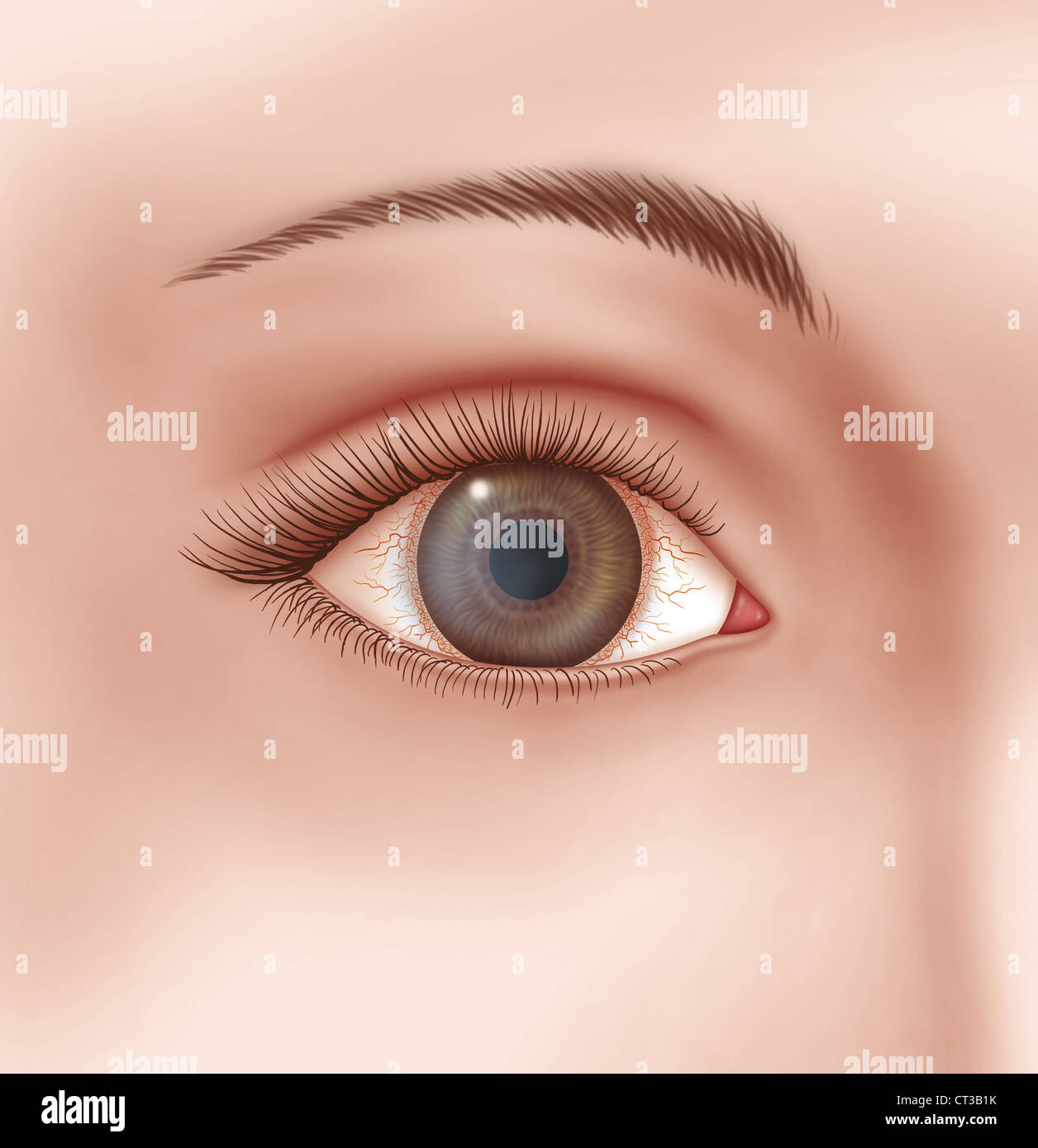 KERATITIS, ILLUSTRATION Stock Photohttps://www.alamy.com/image-license-details/?v=1https://www.alamy.com/stock-photo-keratitis-illustration-49247007.html
RMCT3B1K–KERATITIS, ILLUSTRATION
 Textbook of normal histology: including an account of the development of the tissues and of the organs . d the ciliary or outer margin of the iris in front.Within this important territory three areas may be distinguished : 1, the ciliary ring; 2, the cili-Fig. 371. ary processes; 3, the ciliary muscle. The ciliary ring, or orbicu-lus ciliaris, is a circular tractabout 4 mm. in breadth, situatedimmediately in front of the oraserrata and extending to theposterior ends of the ciliary pro-cesses. This zone differs fromthe choroid in the absence ofthe choriocapillaris and in thepresence of muscular Stock Photohttps://www.alamy.com/image-license-details/?v=1https://www.alamy.com/textbook-of-normal-histology-including-an-account-of-the-development-of-the-tissues-and-of-the-organs-d-the-ciliary-or-outer-margin-of-the-iris-in-frontwithin-this-important-territory-three-areas-may-be-distinguished-1-the-ciliary-ring-2-the-cili-fig-371-ary-processes-3-the-ciliary-muscle-the-ciliary-ring-or-orbicu-lus-ciliaris-is-a-circular-tractabout-4-mm-in-breadth-situatedimmediately-in-front-of-the-oraserrata-and-extending-to-theposterior-ends-of-the-ciliary-pro-cesses-this-zone-differs-fromthe-choroid-in-the-absence-ofthe-choriocapillaris-and-in-thepresence-of-muscular-image338903940.html
RM2AKABC4–Textbook of normal histology: including an account of the development of the tissues and of the organs . d the ciliary or outer margin of the iris in front.Within this important territory three areas may be distinguished : 1, the ciliary ring; 2, the cili-Fig. 371. ary processes; 3, the ciliary muscle. The ciliary ring, or orbicu-lus ciliaris, is a circular tractabout 4 mm. in breadth, situatedimmediately in front of the oraserrata and extending to theposterior ends of the ciliary pro-cesses. This zone differs fromthe choroid in the absence ofthe choriocapillaris and in thepresence of muscular
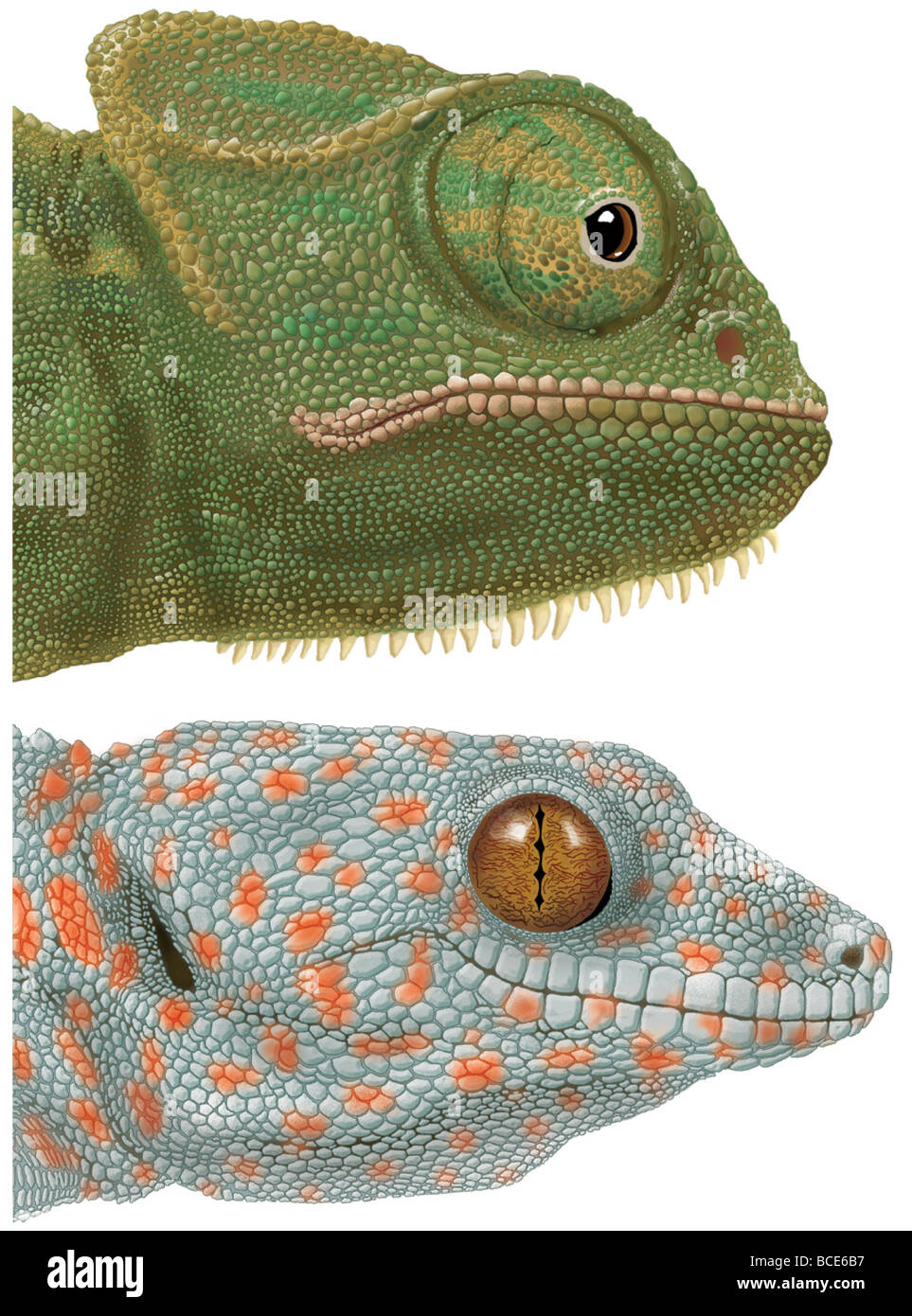 Specialized eyes of the chameleon (Chamaeleo) and the gecko (Gekko). Stock Photohttps://www.alamy.com/image-license-details/?v=1https://www.alamy.com/stock-photo-specialized-eyes-of-the-chameleon-chamaeleo-and-the-gecko-gekko-24898587.html
RMBCE6B7–Specialized eyes of the chameleon (Chamaeleo) and the gecko (Gekko).
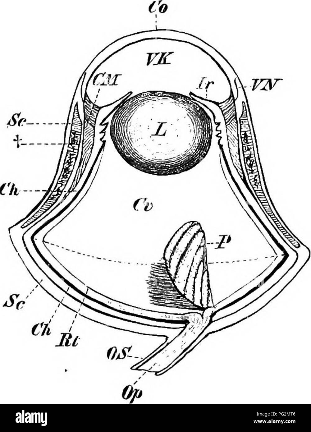 . Elements of the comparative anatomy of vertebrates. Anatomy, Comparative. Fig. 170. — Eye OP Lacerta niu- ralis, SHOWING THE Ring of Bony Sclero- tic Plates.. Fig. 171.—Eye of an Owl. Rt, retina ; Ch, Choroid; Sc, sclerotic, with its bony ring at t : CM, ciliary muscle; Co, cornea; VN, point of jvmction between sclerotic and cornea ; Jr, iris; VK, anterior chamber; L, lens ; Cr, vitreous humour ; P, pecten; Op, OS, optic nerve and sheath. The dotted line passing across the broadest portion of the circumference of the eye divides the latter into an inner and an outer segment. outer portion is Stock Photohttps://www.alamy.com/image-license-details/?v=1https://www.alamy.com/elements-of-the-comparative-anatomy-of-vertebrates-anatomy-comparative-fig-170-eye-op-lacerta-niu-ralis-showing-the-ring-of-bony-sclero-tic-plates-fig-171eye-of-an-owl-rt-retina-ch-choroid-sc-sclerotic-with-its-bony-ring-at-t-cm-ciliary-muscle-co-cornea-vn-point-of-jvmction-between-sclerotic-and-cornea-jr-iris-vk-anterior-chamber-l-lens-cr-vitreous-humour-p-pecten-op-os-optic-nerve-and-sheath-the-dotted-line-passing-across-the-broadest-portion-of-the-circumference-of-the-eye-divides-the-latter-into-an-inner-and-an-outer-segment-outer-portion-is-image216419174.html
RMPG2MT6–. Elements of the comparative anatomy of vertebrates. Anatomy, Comparative. Fig. 170. — Eye OP Lacerta niu- ralis, SHOWING THE Ring of Bony Sclero- tic Plates.. Fig. 171.—Eye of an Owl. Rt, retina ; Ch, Choroid; Sc, sclerotic, with its bony ring at t : CM, ciliary muscle; Co, cornea; VN, point of jvmction between sclerotic and cornea ; Jr, iris; VK, anterior chamber; L, lens ; Cr, vitreous humour ; P, pecten; Op, OS, optic nerve and sheath. The dotted line passing across the broadest portion of the circumference of the eye divides the latter into an inner and an outer segment. outer portion is
 Elementary text-book of zoology (1884) Elementary text-book of zoology elementarytextbo0101clau Year: 1884 392 ANNELIDA. organs, which give rise to the a mil vesicles (fig. 315 a, AS). The rudiments both of the cerebral ganglion and of the ventral cord are derived from growths of the ectoderm,—the former from the apical plate, the latter as a paired thickening of the ventral ectoderm. The two are connected by the cesophageal ring, which is also provided with ganglion cells. In older stages, after the disappearance of the segments, the ciliary apparatus begins to degenerate and finally vanishe Stock Photohttps://www.alamy.com/image-license-details/?v=1https://www.alamy.com/elementary-text-book-of-zoology-1884-elementary-text-book-of-zoology-elementarytextbo0101clau-year-1884-392-annelida-organs-which-give-rise-to-the-a-mil-vesicles-fig-315-a-as-the-rudiments-both-of-the-cerebral-ganglion-and-of-the-ventral-cord-are-derived-from-growths-of-the-ectodermthe-former-from-the-apical-plate-the-latter-as-a-paired-thickening-of-the-ventral-ectoderm-the-two-are-connected-by-the-cesophageal-ring-which-is-also-provided-with-ganglion-cells-in-older-stages-after-the-disappearance-of-the-segments-the-ciliary-apparatus-begins-to-degenerate-and-finally-vanishe-image239661410.html
RMRWWEG2–Elementary text-book of zoology (1884) Elementary text-book of zoology elementarytextbo0101clau Year: 1884 392 ANNELIDA. organs, which give rise to the a mil vesicles (fig. 315 a, AS). The rudiments both of the cerebral ganglion and of the ventral cord are derived from growths of the ectoderm,—the former from the apical plate, the latter as a paired thickening of the ventral ectoderm. The two are connected by the cesophageal ring, which is also provided with ganglion cells. In older stages, after the disappearance of the segments, the ciliary apparatus begins to degenerate and finally vanishe
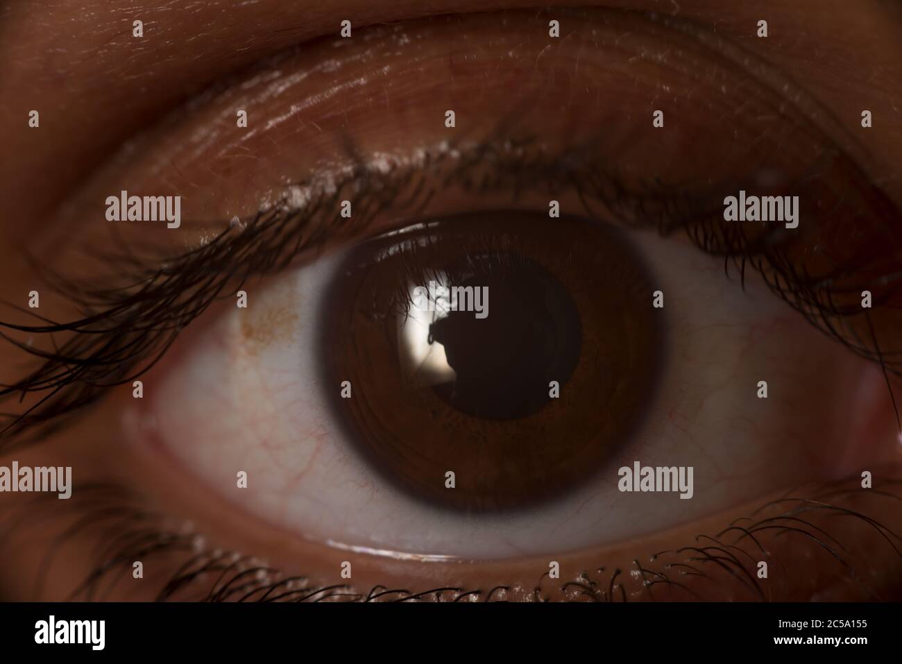 Dark brown human eye, cornea and outer layer of the eyeball, with ciliary muscle; ring of smooth muscle Stock Photohttps://www.alamy.com/image-license-details/?v=1https://www.alamy.com/dark-brown-human-eye-cornea-and-outer-layer-of-the-eyeball-with-ciliary-muscle-ring-of-smooth-muscle-image364711457.html
RM2C5A155–Dark brown human eye, cornea and outer layer of the eyeball, with ciliary muscle; ring of smooth muscle
 Lashes mascara Stock Photohttps://www.alamy.com/image-license-details/?v=1https://www.alamy.com/stock-photo-lashes-mascara-27632536.html
RMBGXNG8–Lashes mascara
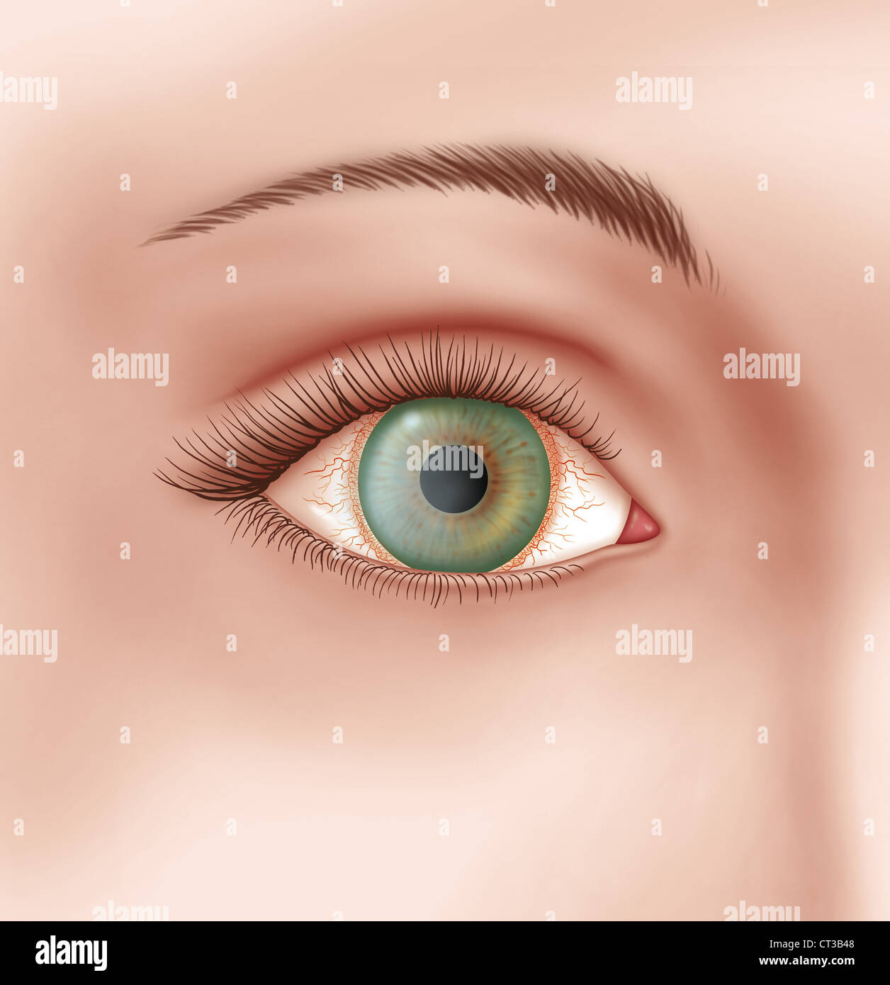 KERATITIS, ILLUSTRATION Stock Photohttps://www.alamy.com/image-license-details/?v=1https://www.alamy.com/stock-photo-keratitis-illustration-49247080.html
RMCT3B48–KERATITIS, ILLUSTRATION
 . Text-book of normal histology: including an account of the development of the tissues and of the organs. he ciliary or outer margin of the iris in front.Within this important territory three areas may be distinguished : I, the ciliary ring; 2, the cili-FiG. 371. ary processes; 3, the ciliary muscle. The ciliary ring, or orbicu-lus ciliaris, is a circular tractabout 4 mm. in breadth, situatedimmediately in front of the eraserrata and extending to theposterior ends of the ciliary pro-cesses. This zone differs fromthe choroid in the absence ofthe choriocapillaris and in thepresence of muscular Stock Photohttps://www.alamy.com/image-license-details/?v=1https://www.alamy.com/text-book-of-normal-histology-including-an-account-of-the-development-of-the-tissues-and-of-the-organs-he-ciliary-or-outer-margin-of-the-iris-in-frontwithin-this-important-territory-three-areas-may-be-distinguished-i-the-ciliary-ring-2-the-cili-fig-371-ary-processes-3-the-ciliary-muscle-the-ciliary-ring-or-orbicu-lus-ciliaris-is-a-circular-tractabout-4-mm-in-breadth-situatedimmediately-in-front-of-the-eraserrata-and-extending-to-theposterior-ends-of-the-ciliary-pro-cesses-this-zone-differs-fromthe-choroid-in-the-absence-ofthe-choriocapillaris-and-in-thepresence-of-muscular-image370367133.html
RM2CEFK1H–. Text-book of normal histology: including an account of the development of the tissues and of the organs. he ciliary or outer margin of the iris in front.Within this important territory three areas may be distinguished : I, the ciliary ring; 2, the cili-FiG. 371. ary processes; 3, the ciliary muscle. The ciliary ring, or orbicu-lus ciliaris, is a circular tractabout 4 mm. in breadth, situatedimmediately in front of the eraserrata and extending to theposterior ends of the ciliary pro-cesses. This zone differs fromthe choroid in the absence ofthe choriocapillaris and in thepresence of muscular
 Elements of comparative anatomy (1878) Elements of comparative anatomy elementsofcompar00gege Year: 1878 530 COMPAEATIVE ANATOMY. In tlie Saurii, Chelonii, and Aves, the anterior portion of the sclerotic, which abuts on the cornea, is supported (Fig. 296, s) by a circlet of flat pieces of bone (sclerotic ring). In all Mammals except the Monotremata the sclerotic is formed of connective tissue; it is very thick in the Cetacea (Fig. 299, s). The choroid is made up of several layers, which, as a rule, have the same characters as in Man. Anteriorly it gives rise to the folded ciliary processes ; Stock Photohttps://www.alamy.com/image-license-details/?v=1https://www.alamy.com/elements-of-comparative-anatomy-1878-elements-of-comparative-anatomy-elementsofcompar00gege-year-1878-530-compaeative-anatomy-in-tlie-saurii-chelonii-and-aves-the-anterior-portion-of-the-sclerotic-which-abuts-on-the-cornea-is-supported-fig-296-s-by-a-circlet-of-flat-pieces-of-bone-sclerotic-ring-in-all-mammals-except-the-monotremata-the-sclerotic-is-formed-of-connective-tissue-it-is-very-thick-in-the-cetacea-fig-299-s-the-choroid-is-made-up-of-several-layers-which-as-a-rule-have-the-same-characters-as-in-man-anteriorly-it-gives-rise-to-the-folded-ciliary-processes-image239674078.html
RMRWX2ME–Elements of comparative anatomy (1878) Elements of comparative anatomy elementsofcompar00gege Year: 1878 530 COMPAEATIVE ANATOMY. In tlie Saurii, Chelonii, and Aves, the anterior portion of the sclerotic, which abuts on the cornea, is supported (Fig. 296, s) by a circlet of flat pieces of bone (sclerotic ring). In all Mammals except the Monotremata the sclerotic is formed of connective tissue; it is very thick in the Cetacea (Fig. 299, s). The choroid is made up of several layers, which, as a rule, have the same characters as in Man. Anteriorly it gives rise to the folded ciliary processes ;
 Eyelash Mirror Stock Photohttps://www.alamy.com/image-license-details/?v=1https://www.alamy.com/stock-photo-eyelash-mirror-27634138.html
RFBGXRHE–Eyelash Mirror
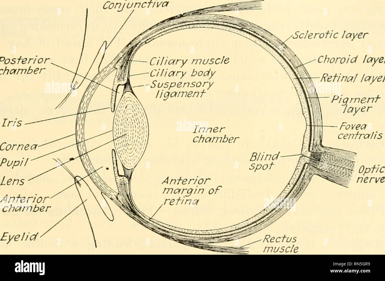 . Animal biology. Zoology; Biology. SUBPHYLUM VERTEBRATA 333 or more convex. It is inclosed in a thin fibrous capsule which is attached all around by a suspensory ligament to a body known as the ciliary body, to which also the iris is attached and which contains a muscle known as the ciliary muscle, in the form of a complete ring. All of these belong to the choroid layer. The tendency of the eyeball to maintain a globular form causes a tension on the lens capsule which serves to flatten the lens and to focus the eye for distance. When it is desired to focus on a near object, the ciliary muscle Stock Photohttps://www.alamy.com/image-license-details/?v=1https://www.alamy.com/animal-biology-zoology-biology-subphylum-vertebrata-333-or-more-convex-it-is-inclosed-in-a-thin-fibrous-capsule-which-is-attached-all-around-by-a-suspensory-ligament-to-a-body-known-as-the-ciliary-body-to-which-also-the-iris-is-attached-and-which-contains-a-muscle-known-as-the-ciliary-muscle-in-the-form-of-a-complete-ring-all-of-these-belong-to-the-choroid-layer-the-tendency-of-the-eyeball-to-maintain-a-globular-form-causes-a-tension-on-the-lens-capsule-which-serves-to-flatten-the-lens-and-to-focus-the-eye-for-distance-when-it-is-desired-to-focus-on-a-near-object-the-ciliary-muscle-image236765517.html
RMRN5GR9–. Animal biology. Zoology; Biology. SUBPHYLUM VERTEBRATA 333 or more convex. It is inclosed in a thin fibrous capsule which is attached all around by a suspensory ligament to a body known as the ciliary body, to which also the iris is attached and which contains a muscle known as the ciliary muscle, in the form of a complete ring. All of these belong to the choroid layer. The tendency of the eyeball to maintain a globular form causes a tension on the lens capsule which serves to flatten the lens and to focus the eye for distance. When it is desired to focus on a near object, the ciliary muscle
 Mascara Stock Photohttps://www.alamy.com/image-license-details/?v=1https://www.alamy.com/stock-photo-mascara-27630333.html
RMBGXJNH–Mascara
RMRJ5KAH–. Chordate anatomy. Chordata; Anatomy, Comparative. 4IO CHORDATE ANATOMY lens, vitreous humor, and retina. The curved cornea serves to converge rays of Ught, and the lens increases the convergence. The lens is a biconvex translucent and elastic body surrounded by an elastic capsule. Fibers which extend from the periphery of the lens to the ciliary body form a suspensory ligament which holds the lens in position back of the iris. The ciliary body is a ring of vascular and muscular tissue, wedge-shaped in cross section, which projects into the cavity of the eye-ball, just back of the iris. The c
 Mascara Stock Photohttps://www.alamy.com/image-license-details/?v=1https://www.alamy.com/stock-photo-mascara-27634134.html
RMBGXRHA–Mascara
 . Biology of the vertebrates : a comparative study of man and his animal allies. Vertebrates; Vertebrates -- Anatomy; Anatomy, Comparative. il2 Biology of the Vertebrates mediate neurons, and ganglionic neurons (Fig. 710). The retina terminates abruptly near the periphery of the ciliary ring, although derivatives of the optic cup continue along the inner portion of the ciliary body and iris. The irregular anterior margin of the retina is known as the or a serrata (Fig. 709).. Fig. 711. Diagram showing detail of structure of the retina. a. 1, pigment epithelium; 2, processes of pigment epitheli Stock Photohttps://www.alamy.com/image-license-details/?v=1https://www.alamy.com/biology-of-the-vertebrates-a-comparative-study-of-man-and-his-animal-allies-vertebrates-vertebrates-anatomy-anatomy-comparative-il2-biology-of-the-vertebrates-mediate-neurons-and-ganglionic-neurons-fig-710-the-retina-terminates-abruptly-near-the-periphery-of-the-ciliary-ring-although-derivatives-of-the-optic-cup-continue-along-the-inner-portion-of-the-ciliary-body-and-iris-the-irregular-anterior-margin-of-the-retina-is-known-as-the-or-a-serrata-fig-709-fig-711-diagram-showing-detail-of-structure-of-the-retina-a-1-pigment-epithelium-2-processes-of-pigment-epitheli-image234592937.html
RMRHJHK5–. Biology of the vertebrates : a comparative study of man and his animal allies. Vertebrates; Vertebrates -- Anatomy; Anatomy, Comparative. il2 Biology of the Vertebrates mediate neurons, and ganglionic neurons (Fig. 710). The retina terminates abruptly near the periphery of the ciliary ring, although derivatives of the optic cup continue along the inner portion of the ciliary body and iris. The irregular anterior margin of the retina is known as the or a serrata (Fig. 709).. Fig. 711. Diagram showing detail of structure of the retina. a. 1, pigment epithelium; 2, processes of pigment epitheli
 Eyelash total Stock Photohttps://www.alamy.com/image-license-details/?v=1https://www.alamy.com/stock-photo-eyelash-total-27632541.html
RMBGXNGD–Eyelash total
 . The Biological bulletin. Biology; Zoology; Biology; Marine Biology. 268 Q. BONE AND G. O. MACKIE dnc. en c m FIGURE 1. Diagram showing organization of trunk and anterior tail region in Oikopleura. The tail has been twisted for clarity, b represents brain; eg, caudal ganglion; en, caudal nerve; cm, caudal muscle cells; cr, ciliary ring; dnc, dorsal nerve cord; f, fin; g, gonad; Lr, Langer- hans receptor; m, mouth; n, notochord; o, oikoplastic epithelium; r, rectum; and s, stomach. paired bristle-bearing receptor cells on either side of the trunk which were dis- covered by Langerhans (1877). S Stock Photohttps://www.alamy.com/image-license-details/?v=1https://www.alamy.com/the-biological-bulletin-biology-zoology-biology-marine-biology-268-q-bone-and-g-o-mackie-dnc-en-c-m-figure-1-diagram-showing-organization-of-trunk-and-anterior-tail-region-in-oikopleura-the-tail-has-been-twisted-for-clarity-b-represents-brain-eg-caudal-ganglion-en-caudal-nerve-cm-caudal-muscle-cells-cr-ciliary-ring-dnc-dorsal-nerve-cord-f-fin-g-gonad-lr-langer-hans-receptor-m-mouth-n-notochord-o-oikoplastic-epithelium-r-rectum-and-s-stomach-paired-bristle-bearing-receptor-cells-on-either-side-of-the-trunk-which-were-dis-covered-by-langerhans-1877-s-image234637652.html
RMRHMJM4–. The Biological bulletin. Biology; Zoology; Biology; Marine Biology. 268 Q. BONE AND G. O. MACKIE dnc. en c m FIGURE 1. Diagram showing organization of trunk and anterior tail region in Oikopleura. The tail has been twisted for clarity, b represents brain; eg, caudal ganglion; en, caudal nerve; cm, caudal muscle cells; cr, ciliary ring; dnc, dorsal nerve cord; f, fin; g, gonad; Lr, Langer- hans receptor; m, mouth; n, notochord; o, oikoplastic epithelium; r, rectum; and s, stomach. paired bristle-bearing receptor cells on either side of the trunk which were dis- covered by Langerhans (1877). S
 Eyelash close Stock Photohttps://www.alamy.com/image-license-details/?v=1https://www.alamy.com/stock-photo-eyelash-close-27632548.html
RFBGXNGM–Eyelash close
 Transactions . -are attached posteriorly to the choroid. The fibres of Miilleiform the angular ring beneath those of Bowman. The physiologic action which follows would almost seemobvious. A contraction of the long fibres relaxes the zonule.Coincidentally with this, the circular fibres surrounding the mar-gin of the iris contract, impeding the free venous flow and caus-ing the ciliary processes to become turgid, with blood; they inttxrn pressing, by their bulk, on the anterior part of the sus-pensory ligament of necessity flatten the edges and protrude 187 the center of the lens in exactly the Stock Photohttps://www.alamy.com/image-license-details/?v=1https://www.alamy.com/transactions-are-attached-posteriorly-to-the-choroid-the-fibres-of-miilleiform-the-angular-ring-beneath-those-of-bowman-the-physiologic-action-which-follows-would-almost-seemobvious-a-contraction-of-the-long-fibres-relaxes-the-zonulecoincidentally-with-this-the-circular-fibres-surrounding-the-mar-gin-of-the-iris-contract-impeding-the-free-venous-flow-and-caus-ing-the-ciliary-processes-to-become-turgid-with-blood-they-inttxrn-pressing-by-their-bulk-on-the-anterior-part-of-the-sus-pensory-ligament-of-necessity-flatten-the-edges-and-protrude-187-the-center-of-the-lens-in-exactly-the-image342784840.html
RM2AWK5FM–Transactions . -are attached posteriorly to the choroid. The fibres of Miilleiform the angular ring beneath those of Bowman. The physiologic action which follows would almost seemobvious. A contraction of the long fibres relaxes the zonule.Coincidentally with this, the circular fibres surrounding the mar-gin of the iris contract, impeding the free venous flow and caus-ing the ciliary processes to become turgid, with blood; they inttxrn pressing, by their bulk, on the anterior part of the sus-pensory ligament of necessity flatten the edges and protrude 187 the center of the lens in exactly the
 mascara eyelash Stock Photohttps://www.alamy.com/image-license-details/?v=1https://www.alamy.com/stock-photo-mascara-eyelash-27630332.html
RFBGXJNG–mascara eyelash
 Human physiology : designed for colleges and the higher classes in schools and for general reading . 292 HUMAN PHYSIOLOGY. Object of the apparatus to form images of objects on the retina. FIG. 161.. CILIARY PROCESSES. which stops at the beginning of these processes, presenting, asyou see, a scalloped appearance. The processes, however, donot arise from the retina, but come from the choroid coat, andare united at their origin by a ring of ligamentous substanceto the sclerotic coat. The exact operation of this beautifularrangement is not known, but it is pretty well ascertained,that muscular fib Stock Photohttps://www.alamy.com/image-license-details/?v=1https://www.alamy.com/human-physiology-designed-for-colleges-and-the-higher-classes-in-schools-and-for-general-reading-292-human-physiology-object-of-the-apparatus-to-form-images-of-objects-on-the-retina-fig-161-ciliary-processes-which-stops-at-the-beginning-of-these-processes-presenting-asyou-see-a-scalloped-appearance-the-processes-however-donot-arise-from-the-retina-but-come-from-the-choroid-coat-andare-united-at-their-origin-by-a-ring-of-ligamentous-substanceto-the-sclerotic-coat-the-exact-operation-of-this-beautifularrangement-is-not-known-but-it-is-pretty-well-ascertainedthat-muscular-fib-image343363187.html
RM2AXHF6Y–Human physiology : designed for colleges and the higher classes in schools and for general reading . 292 HUMAN PHYSIOLOGY. Object of the apparatus to form images of objects on the retina. FIG. 161.. CILIARY PROCESSES. which stops at the beginning of these processes, presenting, asyou see, a scalloped appearance. The processes, however, donot arise from the retina, but come from the choroid coat, andare united at their origin by a ring of ligamentous substanceto the sclerotic coat. The exact operation of this beautifularrangement is not known, but it is pretty well ascertained,that muscular fib
 Lashes in the mirror Stock Photohttps://www.alamy.com/image-license-details/?v=1https://www.alamy.com/stock-photo-lashes-in-the-mirror-27626152.html
RFBGXDC8–Lashes in the mirror
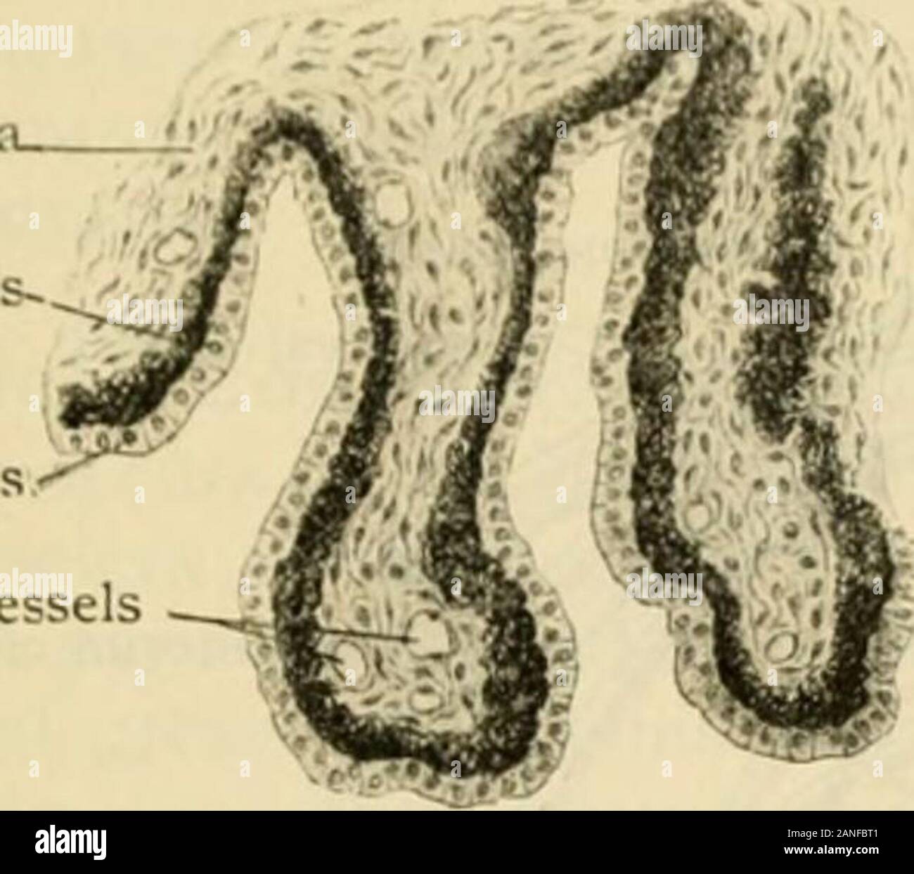 Human anatomy, including structure and development and practical considerations . Ciliary- ringOra scrrata Retina Anterior part of sagittally sectioned eye-ball,showing iris, ciliary processes and ring and oraserrata. X 3. THE VASCULAR TUNIC. 1457 the veins, wliich begin between the meshes of the choriocapillaris, the two systemsbeing separate. The nerves of the choroid arise from the long and short ciHary nerves duringtheir course on the inner surface of the sclera. They form a ple.xus within thelamina suprachorioidea, which contains groups of ganglion cells, and sends numerousnonmcdullated f Stock Photohttps://www.alamy.com/image-license-details/?v=1https://www.alamy.com/human-anatomy-including-structure-and-development-and-practical-considerations-ciliary-ringora-scrrata-retina-anterior-part-of-sagittally-sectioned-eye-ballshowing-iris-ciliary-processes-and-ring-and-oraserrata-x-3-the-vascular-tunic-1457-the-veins-wliich-begin-between-the-meshes-of-the-choriocapillaris-the-two-systemsbeing-separate-the-nerves-of-the-choroid-arise-from-the-long-and-short-cihary-nerves-duringtheir-course-on-the-inner-surface-of-the-sclera-they-form-a-plexus-within-thelamina-suprachorioidea-which-contains-groups-of-ganglion-cells-and-sends-numerousnonmcdullated-f-image340243345.html
RM2ANFBT1–Human anatomy, including structure and development and practical considerations . Ciliary- ringOra scrrata Retina Anterior part of sagittally sectioned eye-ball,showing iris, ciliary processes and ring and oraserrata. X 3. THE VASCULAR TUNIC. 1457 the veins, wliich begin between the meshes of the choriocapillaris, the two systemsbeing separate. The nerves of the choroid arise from the long and short ciHary nerves duringtheir course on the inner surface of the sclera. They form a ple.xus within thelamina suprachorioidea, which contains groups of ganglion cells, and sends numerousnonmcdullated f
 Mirror Eyelashes Stock Photohttps://www.alamy.com/image-license-details/?v=1https://www.alamy.com/stock-photo-mirror-eyelashes-29870194.html
RMBMGKMJ–Mirror Eyelashes
 Text-book of comparative anatomy . FIG. 16.— Vorticella microstoma, after Stein (from Clauss Text-book of Zoology), a, Dividinglongitudinally ; N, nucleus ; 6, after complete division one part severs itself after having formeda ring of cilia behind ; w, oral ciliary organ ; c, vorticellas in conjugation; A, the adhering bud-likeindividuals. PROTOZOA 11 Order 4. Peritricha. Body globular or cylindrical, only partially ciliated, either near the mouth in aspiral, or in a belt. VorticcUa (Fig. 16), Carchesium, Epistylis, Trichodina, Strom-liidium, Tintinnus, Ophrydium. N. FIG. 17.—Podophrya gemmip Stock Photohttps://www.alamy.com/image-license-details/?v=1https://www.alamy.com/text-book-of-comparative-anatomy-fig-16-vorticella-microstoma-after-stein-from-clauss-text-book-of-zoology-a-dividinglongitudinally-n-nucleus-6-after-complete-division-one-part-severs-itself-after-having-formeda-ring-of-cilia-behind-w-oral-ciliary-organ-c-vorticellas-in-conjugation-a-the-adhering-bud-likeindividuals-protozoa-11-order-4-peritricha-body-globular-or-cylindrical-only-partially-ciliated-either-near-the-mouth-in-aspiral-or-in-a-belt-vorticcua-fig-16-carchesium-epistylis-trichodina-strom-liidium-tintinnus-ophrydium-n-fig-17podophrya-gemmip-image342783141.html
RM2AWK3B1–Text-book of comparative anatomy . FIG. 16.— Vorticella microstoma, after Stein (from Clauss Text-book of Zoology), a, Dividinglongitudinally ; N, nucleus ; 6, after complete division one part severs itself after having formeda ring of cilia behind ; w, oral ciliary organ ; c, vorticellas in conjugation; A, the adhering bud-likeindividuals. PROTOZOA 11 Order 4. Peritricha. Body globular or cylindrical, only partially ciliated, either near the mouth in aspiral, or in a belt. VorticcUa (Fig. 16), Carchesium, Epistylis, Trichodina, Strom-liidium, Tintinnus, Ophrydium. N. FIG. 17.—Podophrya gemmip
 mascara complete Stock Photohttps://www.alamy.com/image-license-details/?v=1https://www.alamy.com/stock-photo-mascara-complete-27632550.html
RFBGXNGP–mascara complete
 Text-book of comparative anatomy . FIG. 14.—Stylonychia mytilus, after Stein FIG. 15. — Stentor Roeselii, after Stein (from Clauss Zoology), seen from the ventral (Clauss Zoology). 0, Oral opening with oeso^ surface. Ws, Adoral ciliated zone; C, contractile phagus ; PV, contractile vacuole N, nucleus,vacuole ; AT, nucleus ; N, rnicronucleus; A, anus.. FIG. 16.— Vorticella microstoma, after Stein (from Clauss Text-book of Zoology), a, Dividinglongitudinally ; N, nucleus ; 6, after complete division one part severs itself after having formeda ring of cilia behind ; w, oral ciliary organ ; c, vor Stock Photohttps://www.alamy.com/image-license-details/?v=1https://www.alamy.com/text-book-of-comparative-anatomy-fig-14stylonychia-mytilus-after-stein-fig-15-stentor-roeselii-after-stein-from-clauss-zoology-seen-from-the-ventral-clauss-zoology-0-oral-opening-with-oeso-surface-ws-adoral-ciliated-zone-c-contractile-phagus-pv-contractile-vacuole-n-nucleusvacuole-at-nucleus-n-rnicronucleus-a-anus-fig-16-vorticella-microstoma-after-stein-from-clauss-text-book-of-zoology-a-dividinglongitudinally-n-nucleus-6-after-complete-division-one-part-severs-itself-after-having-formeda-ring-of-cilia-behind-w-oral-ciliary-organ-c-vor-image342783427.html
RM2AWK3N7–Text-book of comparative anatomy . FIG. 14.—Stylonychia mytilus, after Stein FIG. 15. — Stentor Roeselii, after Stein (from Clauss Zoology), seen from the ventral (Clauss Zoology). 0, Oral opening with oeso^ surface. Ws, Adoral ciliated zone; C, contractile phagus ; PV, contractile vacuole N, nucleus,vacuole ; AT, nucleus ; N, rnicronucleus; A, anus.. FIG. 16.— Vorticella microstoma, after Stein (from Clauss Text-book of Zoology), a, Dividinglongitudinally ; N, nucleus ; 6, after complete division one part severs itself after having formeda ring of cilia behind ; w, oral ciliary organ ; c, vor
 Eyelash very close Stock Photohttps://www.alamy.com/image-license-details/?v=1https://www.alamy.com/stock-photo-eyelash-very-close-27630335.html
RFBGXJNK–Eyelash very close
 . A text-book of physiology : for medical students and physicians . Ciliary Muscleand the Muscles of the Iris.—From an optical point of view theiris plays the part of a diaphragm. It is, moreover, an adjustablediaphragm the aperture of which—that is, the size of the pupil—is varied reflexly according to the conditions of illumination. Itsadjustments are made possible by the fact that it contains withinits substance two bands of muscular tissue, one, the sphinctermuscle, forming a circular ring whose contraction diminishes theaperture of the pupil, and the other a dilator muscle whose contrac-t Stock Photohttps://www.alamy.com/image-license-details/?v=1https://www.alamy.com/a-text-book-of-physiology-for-medical-students-and-physicians-ciliary-muscleand-the-muscles-of-the-irisfrom-an-optical-point-of-view-theiris-plays-the-part-of-a-diaphragm-it-is-moreover-an-adjustablediaphragm-the-aperture-of-whichthat-is-the-size-of-the-pupilis-varied-reflexly-according-to-the-conditions-of-illumination-itsadjustments-are-made-possible-by-the-fact-that-it-contains-withinits-substance-two-bands-of-muscular-tissue-one-the-sphinctermuscle-forming-a-circular-ring-whose-contraction-diminishes-theaperture-of-the-pupil-and-the-other-a-dilator-muscle-whose-contrac-t-image370186082.html
RM2CE7C3E–. A text-book of physiology : for medical students and physicians . Ciliary Muscleand the Muscles of the Iris.—From an optical point of view theiris plays the part of a diaphragm. It is, moreover, an adjustablediaphragm the aperture of which—that is, the size of the pupil—is varied reflexly according to the conditions of illumination. Itsadjustments are made possible by the fact that it contains withinits substance two bands of muscular tissue, one, the sphinctermuscle, forming a circular ring whose contraction diminishes theaperture of the pupil, and the other a dilator muscle whose contrac-t
 . Human physiology : designed for colleges and the higher classes in schools, and for general reading. 292 HUMAN PHYSIOLOGY. Object of the apparatus to form images of objects on the retina. FIG. 161.. CILIARY PROCESSES. which stops at the beginning of these processes, presenting, asyou see, a scalloped appearance. The processes, however, donot arise from the retina, but come from the choroid coat, andare united at their origin by a ring of ligamentous substanceto the sclerotic coat. The exact operation of this beautifularrangement is not known, but it is pretty well ascertained,that muscular f Stock Photohttps://www.alamy.com/image-license-details/?v=1https://www.alamy.com/human-physiology-designed-for-colleges-and-the-higher-classes-in-schools-and-for-general-reading-292-human-physiology-object-of-the-apparatus-to-form-images-of-objects-on-the-retina-fig-161-ciliary-processes-which-stops-at-the-beginning-of-these-processes-presenting-asyou-see-a-scalloped-appearance-the-processes-however-donot-arise-from-the-retina-but-come-from-the-choroid-coat-andare-united-at-their-origin-by-a-ring-of-ligamentous-substanceto-the-sclerotic-coat-the-exact-operation-of-this-beautifularrangement-is-not-known-but-it-is-pretty-well-ascertainedthat-muscular-f-image370413865.html
RM2CEHPJH–. Human physiology : designed for colleges and the higher classes in schools, and for general reading. 292 HUMAN PHYSIOLOGY. Object of the apparatus to form images of objects on the retina. FIG. 161.. CILIARY PROCESSES. which stops at the beginning of these processes, presenting, asyou see, a scalloped appearance. The processes, however, donot arise from the retina, but come from the choroid coat, andare united at their origin by a ring of ligamentous substanceto the sclerotic coat. The exact operation of this beautifularrangement is not known, but it is pretty well ascertained,that muscular f
 . A treatise on diseases of the eye . ate processes, which project froma thickened ring of mesodermic tissue, the future ciliari/ body, becomehighly vascular, forming the ciliary processes. A little farther back onthe optic cuj), lying close to the pigment layer, numerous capillary blood- VITREOUS BODY 27 vessels develop which form the choriocapillaris of the chorioid. Largervessels appear in the tissue just outside of the choriocapillaris, whicheventually form the layer of large vessels of the chorioid. A coiiflensa-tion and proliferation of the cells of the mesoderm, continuous with thesubst Stock Photohttps://www.alamy.com/image-license-details/?v=1https://www.alamy.com/a-treatise-on-diseases-of-the-eye-ate-processes-which-project-froma-thickened-ring-of-mesodermic-tissue-the-future-ciliari-body-becomehighly-vascular-forming-the-ciliary-processes-a-little-farther-back-onthe-optic-cuj-lying-close-to-the-pigment-layer-numerous-capillary-blood-vitreous-body-27-vessels-develop-which-form-the-choriocapillaris-of-the-chorioid-largervessels-appear-in-the-tissue-just-outside-of-the-choriocapillaris-whicheventually-form-the-layer-of-large-vessels-of-the-chorioid-a-coiiflensa-tion-and-proliferation-of-the-cells-of-the-mesoderm-continuous-with-thesubst-image369613240.html
RM2CD99CT–. A treatise on diseases of the eye . ate processes, which project froma thickened ring of mesodermic tissue, the future ciliari/ body, becomehighly vascular, forming the ciliary processes. A little farther back onthe optic cuj), lying close to the pigment layer, numerous capillary blood- VITREOUS BODY 27 vessels develop which form the choriocapillaris of the chorioid. Largervessels appear in the tissue just outside of the choriocapillaris, whicheventually form the layer of large vessels of the chorioid. A coiiflensa-tion and proliferation of the cells of the mesoderm, continuous with thesubst
 . The Biological bulletin. Biology; Zoology; Biology; Marine Biology. Axons from these cells pass anteriorly and enter the preoral ciliary band forming the circumoral nerve ring. In addition, a tract of axons passes backward in the ventral midline. These axons enter the postoral ciliary band. In the stomach wall near the entrance to the intestine, the axons of a group of about ten fluorescent cells form a ring (Fig. 23). Several axons connect this ring with the axonal tract in the postoral band. Fluorescent cells in the epithelium of the telotroch all send axons into the postoral tract too (Fi Stock Photohttps://www.alamy.com/image-license-details/?v=1https://www.alamy.com/the-biological-bulletin-biology-zoology-biology-marine-biology-axons-from-these-cells-pass-anteriorly-and-enter-the-preoral-ciliary-band-forming-the-circumoral-nerve-ring-in-addition-a-tract-of-axons-passes-backward-in-the-ventral-midline-these-axons-enter-the-postoral-ciliary-band-in-the-stomach-wall-near-the-entrance-to-the-intestine-the-axons-of-a-group-of-about-ten-fluorescent-cells-form-a-ring-fig-23-several-axons-connect-this-ring-with-the-axonal-tract-in-the-postoral-band-fluorescent-cells-in-the-epithelium-of-the-telotroch-all-send-axons-into-the-postoral-tract-too-fi-image234631802.html
RMRHMB76–. The Biological bulletin. Biology; Zoology; Biology; Marine Biology. Axons from these cells pass anteriorly and enter the preoral ciliary band forming the circumoral nerve ring. In addition, a tract of axons passes backward in the ventral midline. These axons enter the postoral ciliary band. In the stomach wall near the entrance to the intestine, the axons of a group of about ten fluorescent cells form a ring (Fig. 23). Several axons connect this ring with the axonal tract in the postoral band. Fluorescent cells in the epithelium of the telotroch all send axons into the postoral tract too (Fi
 . Bulletin : report of Agricultural Experiment Station, Agricultural and Mechanical College, Auburn, Ala. Agriculture -- Alabama. 8 By the contractions of this muscle, it plays an important part in accommodating or adjusting the eye to the perception of objects at different distances. The ciliary body forms a ring which overlaps before and behind the ciliary muscle and lies between the choroid and iris, or rather it connects the choroid to the iris. The ciliary processes consist of 110 to 120 radiating folds formed by the plaiting and folding inward of the choroid at its anterior margin; these Stock Photohttps://www.alamy.com/image-license-details/?v=1https://www.alamy.com/bulletin-report-of-agricultural-experiment-station-agricultural-and-mechanical-college-auburn-ala-agriculture-alabama-8-by-the-contractions-of-this-muscle-it-plays-an-important-part-in-accommodating-or-adjusting-the-eye-to-the-perception-of-objects-at-different-distances-the-ciliary-body-forms-a-ring-which-overlaps-before-and-behind-the-ciliary-muscle-and-lies-between-the-choroid-and-iris-or-rather-it-connects-the-choroid-to-the-iris-the-ciliary-processes-consist-of-110-to-120-radiating-folds-formed-by-the-plaiting-and-folding-inward-of-the-choroid-at-its-anterior-margin-these-image233813415.html
RMRGB3B3–. Bulletin : report of Agricultural Experiment Station, Agricultural and Mechanical College, Auburn, Ala. Agriculture -- Alabama. 8 By the contractions of this muscle, it plays an important part in accommodating or adjusting the eye to the perception of objects at different distances. The ciliary body forms a ring which overlaps before and behind the ciliary muscle and lies between the choroid and iris, or rather it connects the choroid to the iris. The ciliary processes consist of 110 to 120 radiating folds formed by the plaiting and folding inward of the choroid at its anterior margin; these
 . The anatomy of the domestic animals. Veterinary anatomy. 866 THE SENSE ORGANS AND SKIN OF THE HORSE ring (Orliiciilus ciliaris) is the posterior zone, which is distinguished from the cho- rioid mainly by its greater thickness and the absence of the ciiorio-caiHllaris. Its inner face presents numerous fine meri(Honal ridges, 1j>- the union of which the ciliary processes are formed. The ciliary processes (Processus ciliares), more than a hundred in number, form a circle of radial folds which surround the lens and fur- nish attachment to the zonula ciliaris (or suspensory ligament of the len Stock Photohttps://www.alamy.com/image-license-details/?v=1https://www.alamy.com/the-anatomy-of-the-domestic-animals-veterinary-anatomy-866-the-sense-organs-and-skin-of-the-horse-ring-orliiciilus-ciliaris-is-the-posterior-zone-which-is-distinguished-from-the-cho-rioid-mainly-by-its-greater-thickness-and-the-absence-of-the-ciiorio-caihllaris-its-inner-face-presents-numerous-fine-merihonal-ridges-1jgt-the-union-of-which-the-ciliary-processes-are-formed-the-ciliary-processes-processus-ciliares-more-than-a-hundred-in-number-form-a-circle-of-radial-folds-which-surround-the-lens-and-fur-nish-attachment-to-the-zonula-ciliaris-or-suspensory-ligament-of-the-len-image236772975.html
RMRN5X9K–. The anatomy of the domestic animals. Veterinary anatomy. 866 THE SENSE ORGANS AND SKIN OF THE HORSE ring (Orliiciilus ciliaris) is the posterior zone, which is distinguished from the cho- rioid mainly by its greater thickness and the absence of the ciiorio-caiHllaris. Its inner face presents numerous fine meri(Honal ridges, 1j>- the union of which the ciliary processes are formed. The ciliary processes (Processus ciliares), more than a hundred in number, form a circle of radial folds which surround the lens and fur- nish attachment to the zonula ciliaris (or suspensory ligament of the len
 . The anatomy of the domestic animals . Veterinary anatomy. 866 THE SENSE ORGANS AND SKIN OF THE HORSE ring (Orbiculus ciliaris) is the posterior zone, which is distinguished from the cho- rioid mainly by its greater thickness and the absence of the chorio-capillaris. Its inner face presents numerous fine meridional ridges, by the union of which the ciliary processes are formed. The ciliary processes (Processus ciliares), more than a hundred in number, form a circle of radial folds which surround the lens and fur- nish attachment to the zonula ciliaris (or suspensory ligament of the lens). The Stock Photohttps://www.alamy.com/image-license-details/?v=1https://www.alamy.com/the-anatomy-of-the-domestic-animals-veterinary-anatomy-866-the-sense-organs-and-skin-of-the-horse-ring-orbiculus-ciliaris-is-the-posterior-zone-which-is-distinguished-from-the-cho-rioid-mainly-by-its-greater-thickness-and-the-absence-of-the-chorio-capillaris-its-inner-face-presents-numerous-fine-meridional-ridges-by-the-union-of-which-the-ciliary-processes-are-formed-the-ciliary-processes-processus-ciliares-more-than-a-hundred-in-number-form-a-circle-of-radial-folds-which-surround-the-lens-and-fur-nish-attachment-to-the-zonula-ciliaris-or-suspensory-ligament-of-the-lens-the-image232322615.html
RMRDY5T7–. The anatomy of the domestic animals . Veterinary anatomy. 866 THE SENSE ORGANS AND SKIN OF THE HORSE ring (Orbiculus ciliaris) is the posterior zone, which is distinguished from the cho- rioid mainly by its greater thickness and the absence of the chorio-capillaris. Its inner face presents numerous fine meridional ridges, by the union of which the ciliary processes are formed. The ciliary processes (Processus ciliares), more than a hundred in number, form a circle of radial folds which surround the lens and fur- nish attachment to the zonula ciliaris (or suspensory ligament of the lens). The
 . The anatomy of the domestic animals. Veterinary anatomy. 866 THE SENSE ORGANS AND SKIN OF THE HORSE ring (Orbiculus ciliaris) is the posterior zone, which is distinguished from the cho- rioid mainly by its greater thiclcness and the absence of the chorio-capillaris. Its inner face presents numerous fine meridional ridges, by the union of which the ciliary processes are formed. The ciliary processes (Processus ciliares), more than a hundred in numlier, form a circle of ratiial fokls which surround the lens and fur- nish attachment to the zonula ciliaris (or suspensory ligament of the lens). T Stock Photohttps://www.alamy.com/image-license-details/?v=1https://www.alamy.com/the-anatomy-of-the-domestic-animals-veterinary-anatomy-866-the-sense-organs-and-skin-of-the-horse-ring-orbiculus-ciliaris-is-the-posterior-zone-which-is-distinguished-from-the-cho-rioid-mainly-by-its-greater-thiclcness-and-the-absence-of-the-chorio-capillaris-its-inner-face-presents-numerous-fine-meridional-ridges-by-the-union-of-which-the-ciliary-processes-are-formed-the-ciliary-processes-processus-ciliares-more-than-a-hundred-in-numlier-form-a-circle-of-ratiial-fokls-which-surround-the-lens-and-fur-nish-attachment-to-the-zonula-ciliaris-or-suspensory-ligament-of-the-lens-t-image236773059.html
RMRN5XCK–. The anatomy of the domestic animals. Veterinary anatomy. 866 THE SENSE ORGANS AND SKIN OF THE HORSE ring (Orbiculus ciliaris) is the posterior zone, which is distinguished from the cho- rioid mainly by its greater thiclcness and the absence of the chorio-capillaris. Its inner face presents numerous fine meridional ridges, by the union of which the ciliary processes are formed. The ciliary processes (Processus ciliares), more than a hundred in numlier, form a circle of ratiial fokls which surround the lens and fur- nish attachment to the zonula ciliaris (or suspensory ligament of the lens). T
 . The cat : an introduction to the study of backboned animals, especially mammals. Cats; Anatomy, Comparative. 292 THE CAT. [CHAP. IX. opening contracts m-ore and more till it is reduced to an extremely narrow vertical chink. This is probably due (as has been sug- gested to me by my friend Mr. Henry Power) to the greater con- traction of the superior and inferior radiating fibres than of those which radiate outwards and inwards. A muscle called the ciliary muscle consists of a ring of radiating organic fibres which take origin in front from the inner surface of the sclerotic, close to the corn Stock Photohttps://www.alamy.com/image-license-details/?v=1https://www.alamy.com/the-cat-an-introduction-to-the-study-of-backboned-animals-especially-mammals-cats-anatomy-comparative-292-the-cat-chap-ix-opening-contracts-m-ore-and-more-till-it-is-reduced-to-an-extremely-narrow-vertical-chink-this-is-probably-due-as-has-been-sug-gested-to-me-by-my-friend-mr-henry-power-to-the-greater-con-traction-of-the-superior-and-inferior-radiating-fibres-than-of-those-which-radiate-outwards-and-inwards-a-muscle-called-the-ciliary-muscle-consists-of-a-ring-of-radiating-organic-fibres-which-take-origin-in-front-from-the-inner-surface-of-the-sclerotic-close-to-the-corn-image235096327.html
RMRJDFNB–. The cat : an introduction to the study of backboned animals, especially mammals. Cats; Anatomy, Comparative. 292 THE CAT. [CHAP. IX. opening contracts m-ore and more till it is reduced to an extremely narrow vertical chink. This is probably due (as has been sug- gested to me by my friend Mr. Henry Power) to the greater con- traction of the superior and inferior radiating fibres than of those which radiate outwards and inwards. A muscle called the ciliary muscle consists of a ring of radiating organic fibres which take origin in front from the inner surface of the sclerotic, close to the corn
RMREF70J–. Comparative anatomy. Anatomy, Comparative. THE SENSE ORGANS :)"D humor, and retina. The curved cornea serves to converge rays of light, and the lens increases the convergence. The lens is a biconvex translucent and elastic body surrounded by an elastic capsule. Fibers which extend from the periphery of the lens to the ciliary body form a suspensory ligament which holds the lens in position back of the iris. The ciliary body is a ring of vascular and muscular tissue, wedge-shaped in cross section, which projects into the cavity of the eye-ball, just back of the iris. The ciliary muscles,
 . Anatomy, descriptive and applied. Anatomy. Fig. S30.—Ophthalmoscopic appearance of healthy fundus in a person of very fair complexion. Scleral ring well marked. Left eye, inverted image. (Wecker and Jaeger.) Fig. 831.—Ophthalmoscopic appearance of severe recent papillitis. Several elongated patches of blood near border of disk. (After Hughlings Jackson.) SO that its curvature can only be altered to a limited extent by the Ciliary muscle. And, finally, the lens may be dislocated or displaced by blows upon the eyeball, and its relations to surround- ing structures altered by adhesions or the p Stock Photohttps://www.alamy.com/image-license-details/?v=1https://www.alamy.com/anatomy-descriptive-and-applied-anatomy-fig-s30ophthalmoscopic-appearance-of-healthy-fundus-in-a-person-of-very-fair-complexion-scleral-ring-well-marked-left-eye-inverted-image-wecker-and-jaeger-fig-831ophthalmoscopic-appearance-of-severe-recent-papillitis-several-elongated-patches-of-blood-near-border-of-disk-after-hughlings-jackson-so-that-its-curvature-can-only-be-altered-to-a-limited-extent-by-the-ciliary-muscle-and-finally-the-lens-may-be-dislocated-or-displaced-by-blows-upon-the-eyeball-and-its-relations-to-surround-ing-structures-altered-by-adhesions-or-the-p-image236768574.html
RMRN5MME–. Anatomy, descriptive and applied. Anatomy. Fig. S30.—Ophthalmoscopic appearance of healthy fundus in a person of very fair complexion. Scleral ring well marked. Left eye, inverted image. (Wecker and Jaeger.) Fig. 831.—Ophthalmoscopic appearance of severe recent papillitis. Several elongated patches of blood near border of disk. (After Hughlings Jackson.) SO that its curvature can only be altered to a limited extent by the Ciliary muscle. And, finally, the lens may be dislocated or displaced by blows upon the eyeball, and its relations to surround- ing structures altered by adhesions or the p
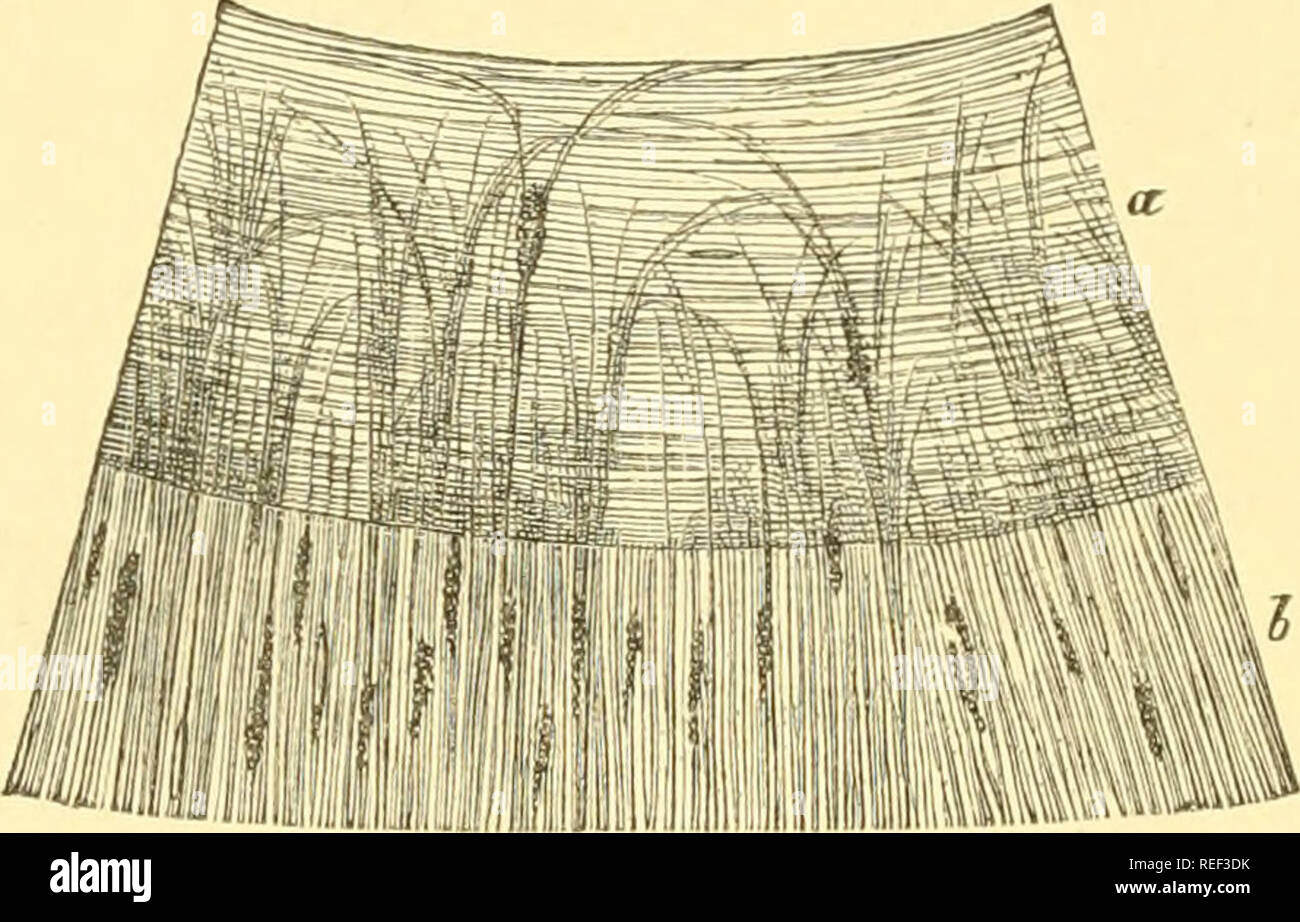 . Compendium of histology. Histology. 250 TWENTY-FOURTH LECTURE. boundary region of the cornea and sclerotica. Meridional bundles of the former radiate in a posterior direction into the ciliary body. Below and inwards occur interwoven filaments, and still further inwards, circular bundles (Mueller's ring muscle). We meet with colorless connective-tissue cells in the con- nective-tissue substratum of the iris of light eyes, and pig- mented cells in that of dark ones. Besides these, smooth muscular elements occur. Annular bundles (Fig. 201, a). Fig. 201. —Surface of the human iris ; a, the sphin Stock Photohttps://www.alamy.com/image-license-details/?v=1https://www.alamy.com/compendium-of-histology-histology-250-twenty-fourth-lecture-boundary-region-of-the-cornea-and-sclerotica-meridional-bundles-of-the-former-radiate-in-a-posterior-direction-into-the-ciliary-body-below-and-inwards-occur-interwoven-filaments-and-still-further-inwards-circular-bundles-muellers-ring-muscle-we-meet-with-colorless-connective-tissue-cells-in-the-con-nective-tissue-substratum-of-the-iris-of-light-eyes-and-pig-mented-cells-in-that-of-dark-ones-besides-these-smooth-muscular-elements-occur-annular-bundles-fig-201-a-fig-201-surface-of-the-human-iris-a-the-sphin-image232671983.html
RMREF3DK–. Compendium of histology. Histology. 250 TWENTY-FOURTH LECTURE. boundary region of the cornea and sclerotica. Meridional bundles of the former radiate in a posterior direction into the ciliary body. Below and inwards occur interwoven filaments, and still further inwards, circular bundles (Mueller's ring muscle). We meet with colorless connective-tissue cells in the con- nective-tissue substratum of the iris of light eyes, and pig- mented cells in that of dark ones. Besides these, smooth muscular elements occur. Annular bundles (Fig. 201, a). Fig. 201. —Surface of the human iris ; a, the sphin
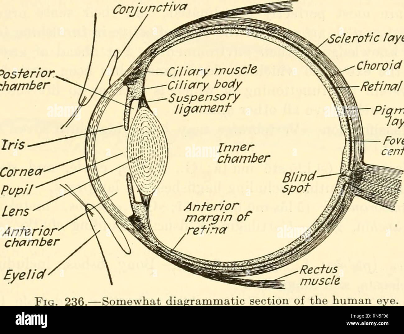 . Animal biology. Zoology; Biology. SUBPHYLUM VERTEBRATA 339 or more convex. It is inclosed in a thin fibrous capsule which is attached all around by a suspensory ligament to a body known as the ciliary body, from which also the iris extends and which contains a muscle known as the ciliary muscle, in the form of a complete ring. All of these belong to the choroid layer. The tendency of the eyeball to maintain a globular form causes a tension on the lens capsule which serves to flatten the lens and to focus the eye for distance. When it is desired to focus on a near object, the ciliary muscle c Stock Photohttps://www.alamy.com/image-license-details/?v=1https://www.alamy.com/animal-biology-zoology-biology-subphylum-vertebrata-339-or-more-convex-it-is-inclosed-in-a-thin-fibrous-capsule-which-is-attached-all-around-by-a-suspensory-ligament-to-a-body-known-as-the-ciliary-body-from-which-also-the-iris-extends-and-which-contains-a-muscle-known-as-the-ciliary-muscle-in-the-form-of-a-complete-ring-all-of-these-belong-to-the-choroid-layer-the-tendency-of-the-eyeball-to-maintain-a-globular-form-causes-a-tension-on-the-lens-capsule-which-serves-to-flatten-the-lens-and-to-focus-the-eye-for-distance-when-it-is-desired-to-focus-on-a-near-object-the-ciliary-muscle-c-image236764340.html
RMRN5F98–. Animal biology. Zoology; Biology. SUBPHYLUM VERTEBRATA 339 or more convex. It is inclosed in a thin fibrous capsule which is attached all around by a suspensory ligament to a body known as the ciliary body, from which also the iris extends and which contains a muscle known as the ciliary muscle, in the form of a complete ring. All of these belong to the choroid layer. The tendency of the eyeball to maintain a globular form causes a tension on the lens capsule which serves to flatten the lens and to focus the eye for distance. When it is desired to focus on a near object, the ciliary muscle c
 . Biological structure and function; proceedings. Biochemistry; Cytology. Figs. 9 and 10. Cross-sections through a small portion of a swimming-plate from the ctenophore, Mnemiopsis leidyi. The filament arrangement is 9 + 3 rather than 9 4- 2, as there is a compact centre filament close to the two tubular ones. Two of the nine filaments in the outer ring are connected to the ciliary membrane by a ridge, visible as a line from these lateral filaments to the ciliary surface. The arrows in P'ig. 9 point to places between the ciliary membranes where there can be seen a bridging substance joining th Stock Photohttps://www.alamy.com/image-license-details/?v=1https://www.alamy.com/biological-structure-and-function-proceedings-biochemistry-cytology-figs-9-and-10-cross-sections-through-a-small-portion-of-a-swimming-plate-from-the-ctenophore-mnemiopsis-leidyi-the-filament-arrangement-is-9-3-rather-than-9-4-2-as-there-is-a-compact-centre-filament-close-to-the-two-tubular-ones-two-of-the-nine-filaments-in-the-outer-ring-are-connected-to-the-ciliary-membrane-by-a-ridge-visible-as-a-line-from-these-lateral-filaments-to-the-ciliary-surface-the-arrows-in-pig-9-point-to-places-between-the-ciliary-membranes-where-there-can-be-seen-a-bridging-substance-joining-th-image234612967.html
RMRHKF6F–. Biological structure and function; proceedings. Biochemistry; Cytology. Figs. 9 and 10. Cross-sections through a small portion of a swimming-plate from the ctenophore, Mnemiopsis leidyi. The filament arrangement is 9 + 3 rather than 9 4- 2, as there is a compact centre filament close to the two tubular ones. Two of the nine filaments in the outer ring are connected to the ciliary membrane by a ridge, visible as a line from these lateral filaments to the ciliary surface. The arrows in P'ig. 9 point to places between the ciliary membranes where there can be seen a bridging substance joining th
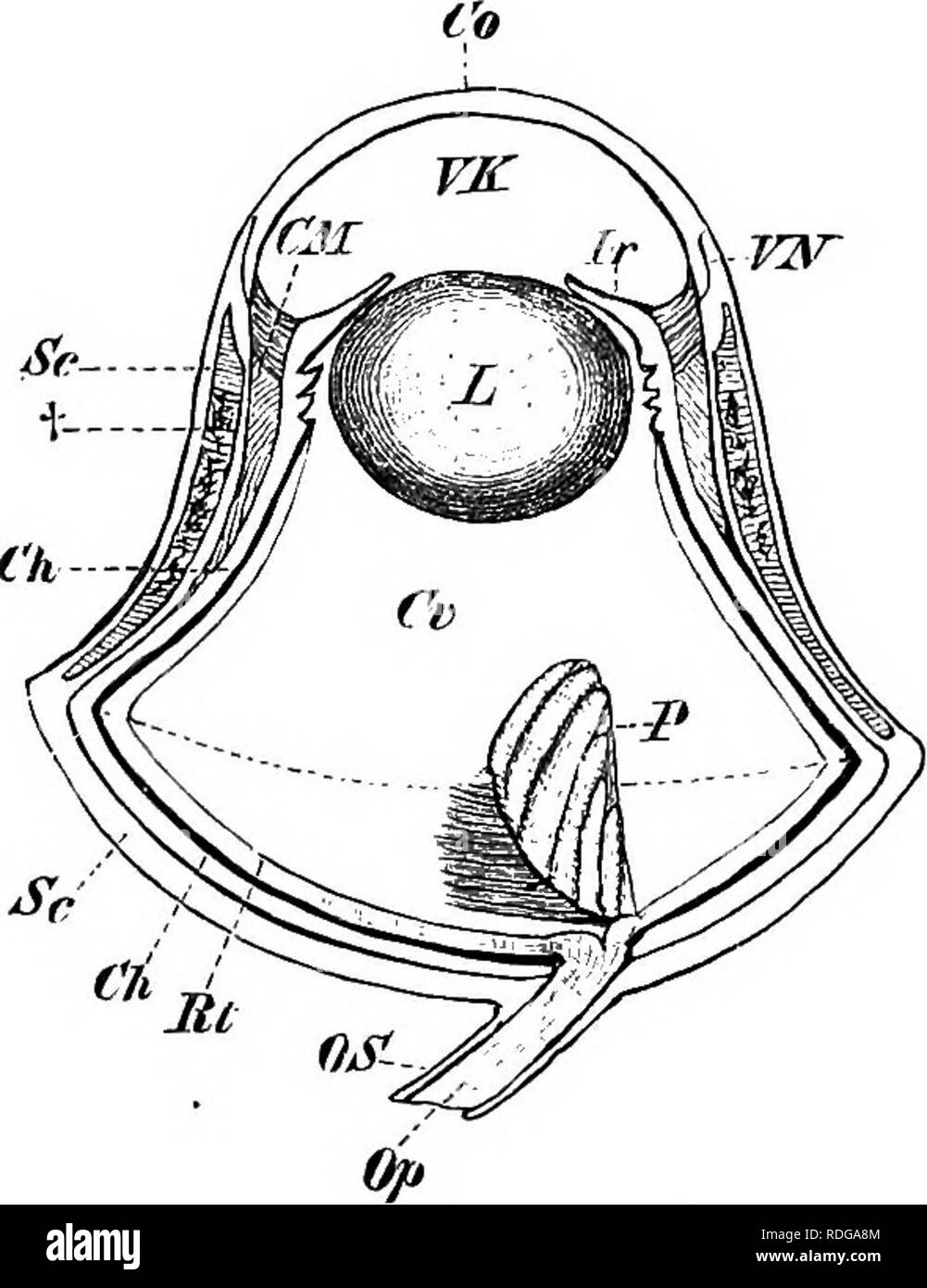 . Elements of the comparative anatomy of vertebrates. Anatomy, Comparative. Fig. 170. — Eye OP Lacerta niu- ralis, SHOWING THE Ring of Bony Sclero- tic Plates.. Fig. 171.—Eye of an Owl. Rt, retina ; Ch, Choroid; Sc, sclerotic, with its bony ring at t : CM, ciliary muscle; Co, cornea; VN, point of jvmction between sclerotic and cornea ; Jr, iris; VK, anterior chamber; L, lens ; Cr, vitreous humour ; P, pecten; Op, OS, optic nerve and sheath. The dotted line passing across the broadest portion of the circumference of the eye divides the latter into an inner and an outer segment. outer portion is Stock Photohttps://www.alamy.com/image-license-details/?v=1https://www.alamy.com/elements-of-the-comparative-anatomy-of-vertebrates-anatomy-comparative-fig-170-eye-op-lacerta-niu-ralis-showing-the-ring-of-bony-sclero-tic-plates-fig-171eye-of-an-owl-rt-retina-ch-choroid-sc-sclerotic-with-its-bony-ring-at-t-cm-ciliary-muscle-co-cornea-vn-point-of-jvmction-between-sclerotic-and-cornea-jr-iris-vk-anterior-chamber-l-lens-cr-vitreous-humour-p-pecten-op-os-optic-nerve-and-sheath-the-dotted-line-passing-across-the-broadest-portion-of-the-circumference-of-the-eye-divides-the-latter-into-an-inner-and-an-outer-segment-outer-portion-is-image232084628.html
RMRDGA8M–. Elements of the comparative anatomy of vertebrates. Anatomy, Comparative. Fig. 170. — Eye OP Lacerta niu- ralis, SHOWING THE Ring of Bony Sclero- tic Plates.. Fig. 171.—Eye of an Owl. Rt, retina ; Ch, Choroid; Sc, sclerotic, with its bony ring at t : CM, ciliary muscle; Co, cornea; VN, point of jvmction between sclerotic and cornea ; Jr, iris; VK, anterior chamber; L, lens ; Cr, vitreous humour ; P, pecten; Op, OS, optic nerve and sheath. The dotted line passing across the broadest portion of the circumference of the eye divides the latter into an inner and an outer segment. outer portion is
 . Elementary text-book of zoology. Zoology. 392 ANNELIDA. organs, which give rise to the a mil vesicles (fig. 315 a, AS). The rudiments both of the cerebral ganglion and of the ventral cord are derived from growths of the ectoderm,—the former from the apical plate, the latter as a paired thickening of the ventral ectoderm. The two are connected by the cesophageal ring, which is also provided with ganglion cells. In older stages, after the disappearance of the segments, the ciliary apparatus begins to degenerate and finally vanishes; after which two strong hooked seta? make their appear- ance a Stock Photohttps://www.alamy.com/image-license-details/?v=1https://www.alamy.com/elementary-text-book-of-zoology-zoology-392-annelida-organs-which-give-rise-to-the-a-mil-vesicles-fig-315-a-as-the-rudiments-both-of-the-cerebral-ganglion-and-of-the-ventral-cord-are-derived-from-growths-of-the-ectodermthe-former-from-the-apical-plate-the-latter-as-a-paired-thickening-of-the-ventral-ectoderm-the-two-are-connected-by-the-cesophageal-ring-which-is-also-provided-with-ganglion-cells-in-older-stages-after-the-disappearance-of-the-segments-the-ciliary-apparatus-begins-to-degenerate-and-finally-vanishes-after-which-two-strong-hooked-seta-make-their-appear-ance-a-image231687276.html
RMRCX7DG–. Elementary text-book of zoology. Zoology. 392 ANNELIDA. organs, which give rise to the a mil vesicles (fig. 315 a, AS). The rudiments both of the cerebral ganglion and of the ventral cord are derived from growths of the ectoderm,—the former from the apical plate, the latter as a paired thickening of the ventral ectoderm. The two are connected by the cesophageal ring, which is also provided with ganglion cells. In older stages, after the disappearance of the segments, the ciliary apparatus begins to degenerate and finally vanishes; after which two strong hooked seta? make their appear- ance a
 . Elements of comparative anatomy. Anatomy, Comparative. 530 COMPAEATIVE ANATOMY. In tlie Saurii, Chelonii, and Aves, the anterior portion of the sclerotic, which abuts on the cornea, is supported (Fig. 296, s) by a circlet of flat pieces of bone (sclerotic ring). In all Mammals except the Monotremata the sclerotic is formed of connective tissue; it is very thick in the Cetacea (Fig. 299, s). The choroid is made up of several layers, which, as a rule, have the same characters as in Man. Anteriorly it gives rise to the folded ciliary processes ; these are feebly developed in the Selachii and &l Stock Photohttps://www.alamy.com/image-license-details/?v=1https://www.alamy.com/elements-of-comparative-anatomy-anatomy-comparative-530-compaeative-anatomy-in-tlie-saurii-chelonii-and-aves-the-anterior-portion-of-the-sclerotic-which-abuts-on-the-cornea-is-supported-fig-296-s-by-a-circlet-of-flat-pieces-of-bone-sclerotic-ring-in-all-mammals-except-the-monotremata-the-sclerotic-is-formed-of-connective-tissue-it-is-very-thick-in-the-cetacea-fig-299-s-the-choroid-is-made-up-of-several-layers-which-as-a-rule-have-the-same-characters-as-in-man-anteriorly-it-gives-rise-to-the-folded-ciliary-processes-these-are-feebly-developed-in-the-selachii-and-l-image231599647.html
RMRCP7KY–. Elements of comparative anatomy. Anatomy, Comparative. 530 COMPAEATIVE ANATOMY. In tlie Saurii, Chelonii, and Aves, the anterior portion of the sclerotic, which abuts on the cornea, is supported (Fig. 296, s) by a circlet of flat pieces of bone (sclerotic ring). In all Mammals except the Monotremata the sclerotic is formed of connective tissue; it is very thick in the Cetacea (Fig. 299, s). The choroid is made up of several layers, which, as a rule, have the same characters as in Man. Anteriorly it gives rise to the folded ciliary processes ; these are feebly developed in the Selachii and &l
 . Elements of Comparative Anatomy. . 530 COMPAEATIVE ANATOMY. In the Saurii, Cheloniij and Aves, the anterior portion of the sclerotic, which abuts on the cornea, is supported (Fig. 296, &•') by a circlet of flat pieces of bone (sclerotic ring). In all Mammals except the Monotremata the sclerotic is formed of connective tissue; it is very thick in the Cetacea (Fig. 299, .v). The choroid is made up of several layers, which, as a rule, have the same characters as in Man. Anteriorly it gives rise to the folded ciliary processes; these are feebly developed in the Selachii and * Gano'idei (Stur Stock Photohttps://www.alamy.com/image-license-details/?v=1https://www.alamy.com/elements-of-comparative-anatomy-530-compaeative-anatomy-in-the-saurii-cheloniij-and-aves-the-anterior-portion-of-the-sclerotic-which-abuts-on-the-cornea-is-supported-fig-296-amp-by-a-circlet-of-flat-pieces-of-bone-sclerotic-ring-in-all-mammals-except-the-monotremata-the-sclerotic-is-formed-of-connective-tissue-it-is-very-thick-in-the-cetacea-fig-299-v-the-choroid-is-made-up-of-several-layers-which-as-a-rule-have-the-same-characters-as-in-man-anteriorly-it-gives-rise-to-the-folded-ciliary-processes-these-are-feebly-developed-in-the-selachii-and-ganoidei-stur-image231569453.html
RMRCMW5H–. Elements of Comparative Anatomy. . 530 COMPAEATIVE ANATOMY. In the Saurii, Cheloniij and Aves, the anterior portion of the sclerotic, which abuts on the cornea, is supported (Fig. 296, &•') by a circlet of flat pieces of bone (sclerotic ring). In all Mammals except the Monotremata the sclerotic is formed of connective tissue; it is very thick in the Cetacea (Fig. 299, .v). The choroid is made up of several layers, which, as a rule, have the same characters as in Man. Anteriorly it gives rise to the folded ciliary processes; these are feebly developed in the Selachii and * Gano'idei (Stur
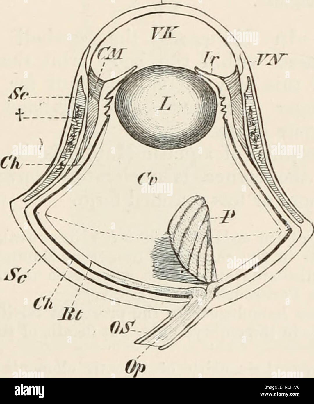 . Elements of the comparative anatomy of vertebrates. Anatomy, Comparative; Vertebrates -- Anatomy. FIG. 154.—EYE OF Lacerta muralis, SHOWING THE RING OF BONY SCLEROTIC PLATES. While the eyeball of Reptiles has a globular form (Fig. 154), that of Birds, and especially nocturnal Birds of prey (Owls), is more elongated and tubular, an external larger segment being sharply marked off from an internal smaller one (Fig. 155). The. FIG. 155.—EYE OF AN OWL. Et, retina ; Oh, choroid ; Sc, sclerotic, with its bony ring at t ; CM, ciliary muscle ; Go, cornea ; VN, point of junction between sclerotic and Stock Photohttps://www.alamy.com/image-license-details/?v=1https://www.alamy.com/elements-of-the-comparative-anatomy-of-vertebrates-anatomy-comparative-vertebrates-anatomy-fig-154eye-of-lacerta-muralis-showing-the-ring-of-bony-sclerotic-plates-while-the-eyeball-of-reptiles-has-a-globular-form-fig-154-that-of-birds-and-especially-nocturnal-birds-of-prey-owls-is-more-elongated-and-tubular-an-external-larger-segment-being-sharply-marked-off-from-an-internal-smaller-one-fig-155-the-fig-155eye-of-an-owl-et-retina-oh-choroid-sc-sclerotic-with-its-bony-ring-at-t-cm-ciliary-muscle-go-cornea-vn-point-of-junction-between-sclerotic-and-image231611050.html
RMRCPP76–. Elements of the comparative anatomy of vertebrates. Anatomy, Comparative; Vertebrates -- Anatomy. FIG. 154.—EYE OF Lacerta muralis, SHOWING THE RING OF BONY SCLEROTIC PLATES. While the eyeball of Reptiles has a globular form (Fig. 154), that of Birds, and especially nocturnal Birds of prey (Owls), is more elongated and tubular, an external larger segment being sharply marked off from an internal smaller one (Fig. 155). The. FIG. 155.—EYE OF AN OWL. Et, retina ; Oh, choroid ; Sc, sclerotic, with its bony ring at t ; CM, ciliary muscle ; Go, cornea ; VN, point of junction between sclerotic and