Communicating vessels Black & White Stock Photos
RF2R6X4JA–Communicating Vessels icon vector image.
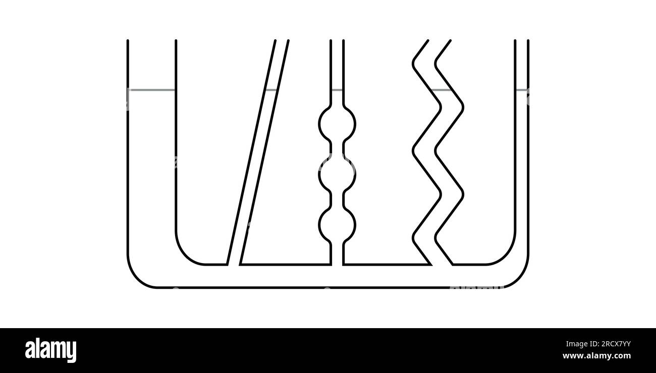
 Communicating vessels experiment. Connected vessels diagram. Vector illustration isolated on white background. Stock Vectorhttps://www.alamy.com/image-license-details/?v=1https://www.alamy.com/communicating-vessels-experiment-connected-vessels-diagram-vector-illustration-isolated-on-white-background-image558684671.html
RF2RCX7YY–Communicating vessels experiment. Connected vessels diagram. Vector illustration isolated on white background.
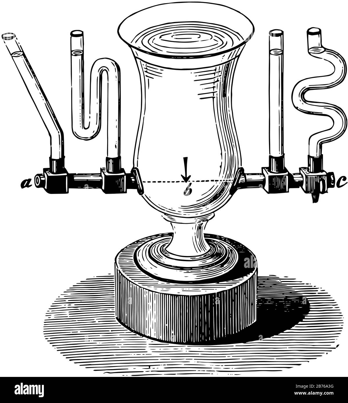
 This diagram represents Water Level in Multiple Connected Vessels, vintage line drawing or engraving illustration. Stock Vectorhttps://www.alamy.com/image-license-details/?v=1https://www.alamy.com/this-diagram-represents-water-level-in-multiple-connected-vessels-vintage-line-drawing-or-engraving-illustration-image348649604.html
RF2B76A3G–This diagram represents Water Level in Multiple Connected Vessels, vintage line drawing or engraving illustration.
RF2S0E1HC–Physics icons High-Quality Vector Icons Collection with Editable Stroke. Ideal for Professional and Creative Projects.
 . The topographical anatomy of the limbs of the horse. Horses; Physiology. 64 TOPOGRAPHICAL ANATOMY OF Very slender dorsal metacarpal arteries (aa. metacarpea dorsalis medialis et lateralis) will be found in the shallow grooves at the line of junction of the second and third and third and fourth metacarpal bones. These vessels arise from the rete carpi dorsalis, and are con- nected with the volar metacarpal arteries by small communicating vessels that cross the surface of the second and fourth metacarpal bones close to their bases. The dorsal metacarpal arteries can often be followed to a unio Stock Photohttps://www.alamy.com/image-license-details/?v=1https://www.alamy.com/the-topographical-anatomy-of-the-limbs-of-the-horse-horses-physiology-64-topographical-anatomy-of-very-slender-dorsal-metacarpal-arteries-aa-metacarpea-dorsalis-medialis-et-lateralis-will-be-found-in-the-shallow-grooves-at-the-line-of-junction-of-the-second-and-third-and-third-and-fourth-metacarpal-bones-these-vessels-arise-from-the-rete-carpi-dorsalis-and-are-con-nected-with-the-volar-metacarpal-arteries-by-small-communicating-vessels-that-cross-the-surface-of-the-second-and-fourth-metacarpal-bones-close-to-their-bases-the-dorsal-metacarpal-arteries-can-often-be-followed-to-a-unio-image232418172.html
RMRE3FN0–. The topographical anatomy of the limbs of the horse. Horses; Physiology. 64 TOPOGRAPHICAL ANATOMY OF Very slender dorsal metacarpal arteries (aa. metacarpea dorsalis medialis et lateralis) will be found in the shallow grooves at the line of junction of the second and third and third and fourth metacarpal bones. These vessels arise from the rete carpi dorsalis, and are con- nected with the volar metacarpal arteries by small communicating vessels that cross the surface of the second and fourth metacarpal bones close to their bases. The dorsal metacarpal arteries can often be followed to a unio
 Men with surveillance equipment on boat Stock Photohttps://www.alamy.com/image-license-details/?v=1https://www.alamy.com/stock-photo-men-with-surveillance-equipment-on-boat-19113837.html
RFB32KW1–Men with surveillance equipment on boat
 PHOTOGRAPHIC NOTES. Possibilities of Vessels Communicating with Each Other at Sea., scientific american, 1886-01-09 Stock Photohttps://www.alamy.com/image-license-details/?v=1https://www.alamy.com/photographic-notes-possibilities-of-vessels-communicating-with-each-other-at-sea-scientific-american-1886-01-09-image334329180.html
RM2ABX07T–PHOTOGRAPHIC NOTES. Possibilities of Vessels Communicating with Each Other at Sea., scientific american, 1886-01-09
RF2R28HJT–Communicating Vessels icon vector image.
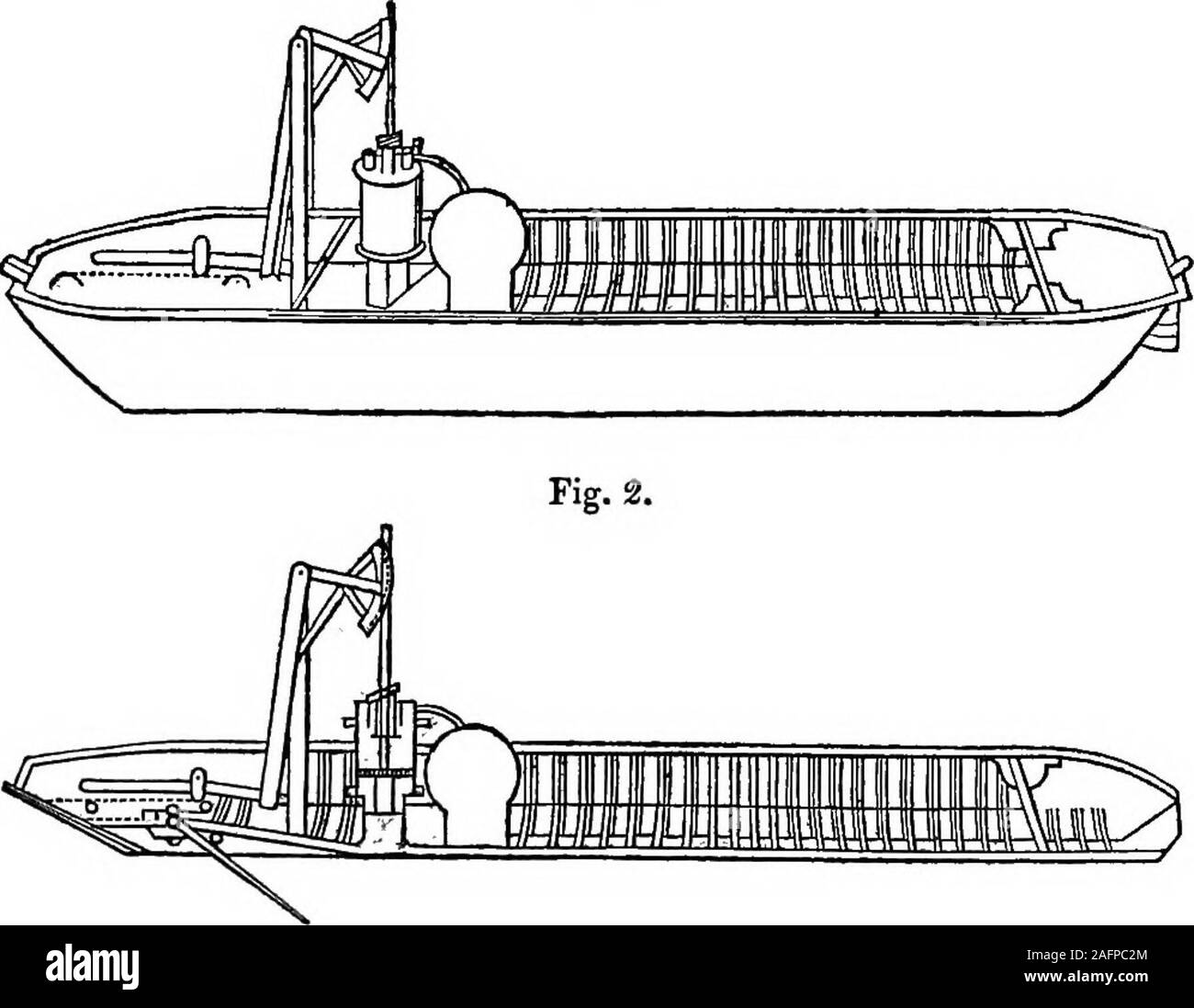
 . A sketch of the origin and progress of steam navigation from authentic documents. at, 6th Nov. 1788. which has a valve C communicating with the water that floats her.M and N is a cylinder screwed on the trunk A B, and has its pistonL connected with the bolt I, to which I apply one of my boilers, bywhich means water is drawn through the valve C and trunk A B,and is discharged through the valve D, which causes the boat tomove forward. In 1790, James Rumsey obtained another English patent for 1790.propelling vessels, the title of which is for his new invented ^^^ ^^?methods of applying Power of Stock Photohttps://www.alamy.com/image-license-details/?v=1https://www.alamy.com/a-sketch-of-the-origin-and-progress-of-steam-navigation-from-authentic-documents-at-6th-nov-1788-which-has-a-valve-c-communicating-with-the-water-that-floats-herm-and-n-is-a-cylinder-screwed-on-the-trunk-a-b-and-has-its-pistonl-connected-with-the-bolt-i-to-which-i-apply-one-of-my-boilers-bywhich-means-water-is-drawn-through-the-valve-c-and-trunk-a-band-is-discharged-through-the-valve-d-which-causes-the-boat-tomove-forward-in-1790-james-rumsey-obtained-another-english-patent-for-1790propelling-vessels-the-title-of-which-is-for-his-new-invented-methods-of-applying-power-of-image336709260.html
RM2AFPC2M–. A sketch of the origin and progress of steam navigation from authentic documents. at, 6th Nov. 1788. which has a valve C communicating with the water that floats her.M and N is a cylinder screwed on the trunk A B, and has its pistonL connected with the bolt I, to which I apply one of my boilers, bywhich means water is drawn through the valve C and trunk A B,and is discharged through the valve D, which causes the boat tomove forward. In 1790, James Rumsey obtained another English patent for 1790.propelling vessels, the title of which is for his new invented ^^^ ^^?methods of applying Power of
 DYNAMIC ELECTRICITY. BY GEO. R. HOPKINS. GRENET BATTERY WITH AIR TUBES. CHLORIDE OF SODIUM BATTERY. Communicating with Wrecked Vessels. The Citadel Park of Barcelona. DEPOLARI ZATION OF ELECTRODES BY MECHANICAL AGITATION. PLAN OF DEPOLARIZING APPARATUS., scientific american, 1881-12-24 Stock Photohttps://www.alamy.com/image-license-details/?v=1https://www.alamy.com/dynamic-electricity-by-geo-r-hopkins-grenet-battery-with-air-tubes-chloride-of-sodium-battery-communicating-with-wrecked-vessels-the-citadel-park-of-barcelona-depolari-zation-of-electrodes-by-mechanical-agitation-plan-of-depolarizing-apparatus-scientific-american-1881-12-24-image334325073.html
RM2ABWR15–DYNAMIC ELECTRICITY. BY GEO. R. HOPKINS. GRENET BATTERY WITH AIR TUBES. CHLORIDE OF SODIUM BATTERY. Communicating with Wrecked Vessels. The Citadel Park of Barcelona. DEPOLARI ZATION OF ELECTRODES BY MECHANICAL AGITATION. PLAN OF DEPOLARIZING APPARATUS., scientific american, 1881-12-24
RF2R24AR5–Communicating Vessels icon vector image. Suitable for mobile application web application and print media.
 . Human physiology. Fig. 154.—Vertical Section through a small portion of a Nail.Highly magnified. a, dermis; b, rete mucosum; c, the nail, composed of thickened epithelium. vessels and nerves. It generally consists of a central medullaryportion ox pith, surrounded by a fibrous cortical part. In somehairs the medullary portion is wanting. Each hair is provided with small glands which secrete an oilyfluid to lubricate the hair and the surrounding skin. These are THE SKIN 167 called the sebaceous glands (Lat. sebum, suet). They consist oflittle saccules communicating with a common duct which ope Stock Photohttps://www.alamy.com/image-license-details/?v=1https://www.alamy.com/human-physiology-fig-154vertical-section-through-a-small-portion-of-a-nailhighly-magnified-a-dermis-b-rete-mucosum-c-the-nail-composed-of-thickened-epithelium-vessels-and-nerves-it-generally-consists-of-a-central-medullaryportion-ox-pith-surrounded-by-a-fibrous-cortical-part-in-somehairs-the-medullary-portion-is-wanting-each-hair-is-provided-with-small-glands-which-secrete-an-oilyfluid-to-lubricate-the-hair-and-the-surrounding-skin-these-are-the-skin-167-called-the-sebaceous-glands-lat-sebum-suet-they-consist-oflittle-saccules-communicating-with-a-common-duct-which-ope-image370640530.html
RM2CF03NP–. Human physiology. Fig. 154.—Vertical Section through a small portion of a Nail.Highly magnified. a, dermis; b, rete mucosum; c, the nail, composed of thickened epithelium. vessels and nerves. It generally consists of a central medullaryportion ox pith, surrounded by a fibrous cortical part. In somehairs the medullary portion is wanting. Each hair is provided with small glands which secrete an oilyfluid to lubricate the hair and the surrounding skin. These are THE SKIN 167 called the sebaceous glands (Lat. sebum, suet). They consist oflittle saccules communicating with a common duct which ope
 . Human physiology. Fig. 154.—Vertical Section through a small portion of a Nail.Highly magnified. a, dermis; b, rete mucosum; c, the nail, composed of thickened epithelium. vessels and nerves. It generally consists of a central medullaryportion ox pith, surrounded by a fibrous cortical part. In somehairs the medullary portion is wanting. Each hair is provided with small glands which secrete an oilyfluid to lubricate the hair and the surrounding skin. These are THE SKIN 167 called the sebaceous glands (Lat. sebum, suet). They consist oflittle saccules communicating with a common duct which ope Stock Photohttps://www.alamy.com/image-license-details/?v=1https://www.alamy.com/human-physiology-fig-154vertical-section-through-a-small-portion-of-a-nailhighly-magnified-a-dermis-b-rete-mucosum-c-the-nail-composed-of-thickened-epithelium-vessels-and-nerves-it-generally-consists-of-a-central-medullaryportion-ox-pith-surrounded-by-a-fibrous-cortical-part-in-somehairs-the-medullary-portion-is-wanting-each-hair-is-provided-with-small-glands-which-secrete-an-oilyfluid-to-lubricate-the-hair-and-the-surrounding-skin-these-are-the-skin-167-called-the-sebaceous-glands-lat-sebum-suet-they-consist-oflittle-saccules-communicating-with-a-common-duct-which-ope-image370637213.html
RM2CEYYF9–. Human physiology. Fig. 154.—Vertical Section through a small portion of a Nail.Highly magnified. a, dermis; b, rete mucosum; c, the nail, composed of thickened epithelium. vessels and nerves. It generally consists of a central medullaryportion ox pith, surrounded by a fibrous cortical part. In somehairs the medullary portion is wanting. Each hair is provided with small glands which secrete an oilyfluid to lubricate the hair and the surrounding skin. These are THE SKIN 167 called the sebaceous glands (Lat. sebum, suet). They consist oflittle saccules communicating with a common duct which ope
 . A Manual of botany : being an introduction to the study of the structure, physiology, and classification of plants . Botany. CIRCULATION OF THE SAP. 147 through the woody tissue, porous vessels, and cells, dissolving starch and other matters, and appropriating various new substances. Pro- ceeding upwards and outwards, this sap reaches the leaves, where it is exposed to the air, and is elaborated by the function of respiration. It then returns, or descends chiefly through the bark, either directly or in a circuitous manner, communicating with the central parts by the medullary rays, depositin Stock Photohttps://www.alamy.com/image-license-details/?v=1https://www.alamy.com/a-manual-of-botany-being-an-introduction-to-the-study-of-the-structure-physiology-and-classification-of-plants-botany-circulation-of-the-sap-147-through-the-woody-tissue-porous-vessels-and-cells-dissolving-starch-and-other-matters-and-appropriating-various-new-substances-pro-ceeding-upwards-and-outwards-this-sap-reaches-the-leaves-where-it-is-exposed-to-the-air-and-is-elaborated-by-the-function-of-respiration-it-then-returns-or-descends-chiefly-through-the-bark-either-directly-or-in-a-circuitous-manner-communicating-with-the-central-parts-by-the-medullary-rays-depositin-image232114949.html
RMRDHMYH–. A Manual of botany : being an introduction to the study of the structure, physiology, and classification of plants . Botany. CIRCULATION OF THE SAP. 147 through the woody tissue, porous vessels, and cells, dissolving starch and other matters, and appropriating various new substances. Pro- ceeding upwards and outwards, this sap reaches the leaves, where it is exposed to the air, and is elaborated by the function of respiration. It then returns, or descends chiefly through the bark, either directly or in a circuitous manner, communicating with the central parts by the medullary rays, depositin
 . The comparative anatomy of the domesticated animals. Veterinary anatomy. HORIZONTAL SECTION THROUGH THE MIDDLE PLANE OP THREE PETERIAN GLANDS, SHOWING THE DISTRIBUTION OF THE BLOOD-VESSELS IN THEIR INTEBIOR. the central lacteal from Fig. 203. DIAGRAMMATIC REPRESENTA- TION OF THE ORIGIN OF THE LACTEALS IN A VILLUS. e, Central lacteal; d, Connec- tive-tissue corpuscles with communicating branches; c, Ciliated columnar epithe- lial cells, the attached ex- tremities of which are di- rectly contiguous with -the connective tissue corpuscles. each villus; the second is placed between the glandular Stock Photohttps://www.alamy.com/image-license-details/?v=1https://www.alamy.com/the-comparative-anatomy-of-the-domesticated-animals-veterinary-anatomy-horizontal-section-through-the-middle-plane-op-three-peterian-glands-showing-the-distribution-of-the-blood-vessels-in-their-intebior-the-central-lacteal-from-fig-203-diagrammatic-representa-tion-of-the-origin-of-the-lacteals-in-a-villus-e-central-lacteal-d-connec-tive-tissue-corpuscles-with-communicating-branches-c-ciliated-columnar-epithe-lial-cells-the-attached-ex-tremities-of-which-are-di-rectly-contiguous-with-the-connective-tissue-corpuscles-each-villus-the-second-is-placed-between-the-glandular-image232452832.html
RMRE53XT–. The comparative anatomy of the domesticated animals. Veterinary anatomy. HORIZONTAL SECTION THROUGH THE MIDDLE PLANE OP THREE PETERIAN GLANDS, SHOWING THE DISTRIBUTION OF THE BLOOD-VESSELS IN THEIR INTEBIOR. the central lacteal from Fig. 203. DIAGRAMMATIC REPRESENTA- TION OF THE ORIGIN OF THE LACTEALS IN A VILLUS. e, Central lacteal; d, Connec- tive-tissue corpuscles with communicating branches; c, Ciliated columnar epithe- lial cells, the attached ex- tremities of which are di- rectly contiguous with -the connective tissue corpuscles. each villus; the second is placed between the glandular
 . The comparative anatomy of the domesticated animals. Veterinary anatomy. HORIZONTAL SECTION THROUGH THE MIDDLE PLANE OF THREE PEYERIAN GLANDS, SHOWING THE DISTRIBUTION OF THE BLOOD-VESSELS IN THEIR INTERIOR. the central lacteal from Fig. 203. DIAGRAMMATIC REPRESENTA- TION OF THE ORIGIN OP THE LACTEAI^ IN A VILLUS. e, Central lacteal; c?, Connec- tive-tissue corpuscles with communicating branches; c, Ciliated columnar epithe- lial cells, the attached ex- tremities of which are di- rectly contiguous with the cojanectiv-e tissue corpuscles. each villus; the second is placed between the glandula Stock Photohttps://www.alamy.com/image-license-details/?v=1https://www.alamy.com/the-comparative-anatomy-of-the-domesticated-animals-veterinary-anatomy-horizontal-section-through-the-middle-plane-of-three-peyerian-glands-showing-the-distribution-of-the-blood-vessels-in-their-interior-the-central-lacteal-from-fig-203-diagrammatic-representa-tion-of-the-origin-op-the-lacteai-in-a-villus-e-central-lacteal-c-connec-tive-tissue-corpuscles-with-communicating-branches-c-ciliated-columnar-epithe-lial-cells-the-attached-ex-tremities-of-which-are-di-rectly-contiguous-with-the-cojanectiv-e-tissue-corpuscles-each-villus-the-second-is-placed-between-the-glandula-image237847594.html
RMRPXW0X–. The comparative anatomy of the domesticated animals. Veterinary anatomy. HORIZONTAL SECTION THROUGH THE MIDDLE PLANE OF THREE PEYERIAN GLANDS, SHOWING THE DISTRIBUTION OF THE BLOOD-VESSELS IN THEIR INTERIOR. the central lacteal from Fig. 203. DIAGRAMMATIC REPRESENTA- TION OF THE ORIGIN OP THE LACTEAI^ IN A VILLUS. e, Central lacteal; c?, Connec- tive-tissue corpuscles with communicating branches; c, Ciliated columnar epithe- lial cells, the attached ex- tremities of which are di- rectly contiguous with the cojanectiv-e tissue corpuscles. each villus; the second is placed between the glandula
 . A text-book of horseshoeing for horseshoers and veterinarians. Horseshoeing. 36 HORSESHOEING. thick-walled, very elastic tubes, without valves, and carry bright-red blood, which flows in spurts, as can be seen when an artery is cut. If a finger be pressed lightly over an artery lying Fig. 21.. side view of forefoot, showing blood-vessels and nerves: a, digital artery; b, anterior artery of tlie OS suifraginis; d, anterior coronary artery, or circumflex artery of the coronet; e' pre- plantar ungual artery; /, inferior communicating arteries passing out from the semilunar artery of the os pedi Stock Photohttps://www.alamy.com/image-license-details/?v=1https://www.alamy.com/a-text-book-of-horseshoeing-for-horseshoers-and-veterinarians-horseshoeing-36-horseshoeing-thick-walled-very-elastic-tubes-without-valves-and-carry-bright-red-blood-which-flows-in-spurts-as-can-be-seen-when-an-artery-is-cut-if-a-finger-be-pressed-lightly-over-an-artery-lying-fig-21-side-view-of-forefoot-showing-blood-vessels-and-nerves-a-digital-artery-b-anterior-artery-of-tlie-os-suifraginis-d-anterior-coronary-artery-or-circumflex-artery-of-the-coronet-e-pre-plantar-ungual-artery-inferior-communicating-arteries-passing-out-from-the-semilunar-artery-of-the-os-pedi-image232341327.html
RMRE01MF–. A text-book of horseshoeing for horseshoers and veterinarians. Horseshoeing. 36 HORSESHOEING. thick-walled, very elastic tubes, without valves, and carry bright-red blood, which flows in spurts, as can be seen when an artery is cut. If a finger be pressed lightly over an artery lying Fig. 21.. side view of forefoot, showing blood-vessels and nerves: a, digital artery; b, anterior artery of tlie OS suifraginis; d, anterior coronary artery, or circumflex artery of the coronet; e' pre- plantar ungual artery; /, inferior communicating arteries passing out from the semilunar artery of the os pedi
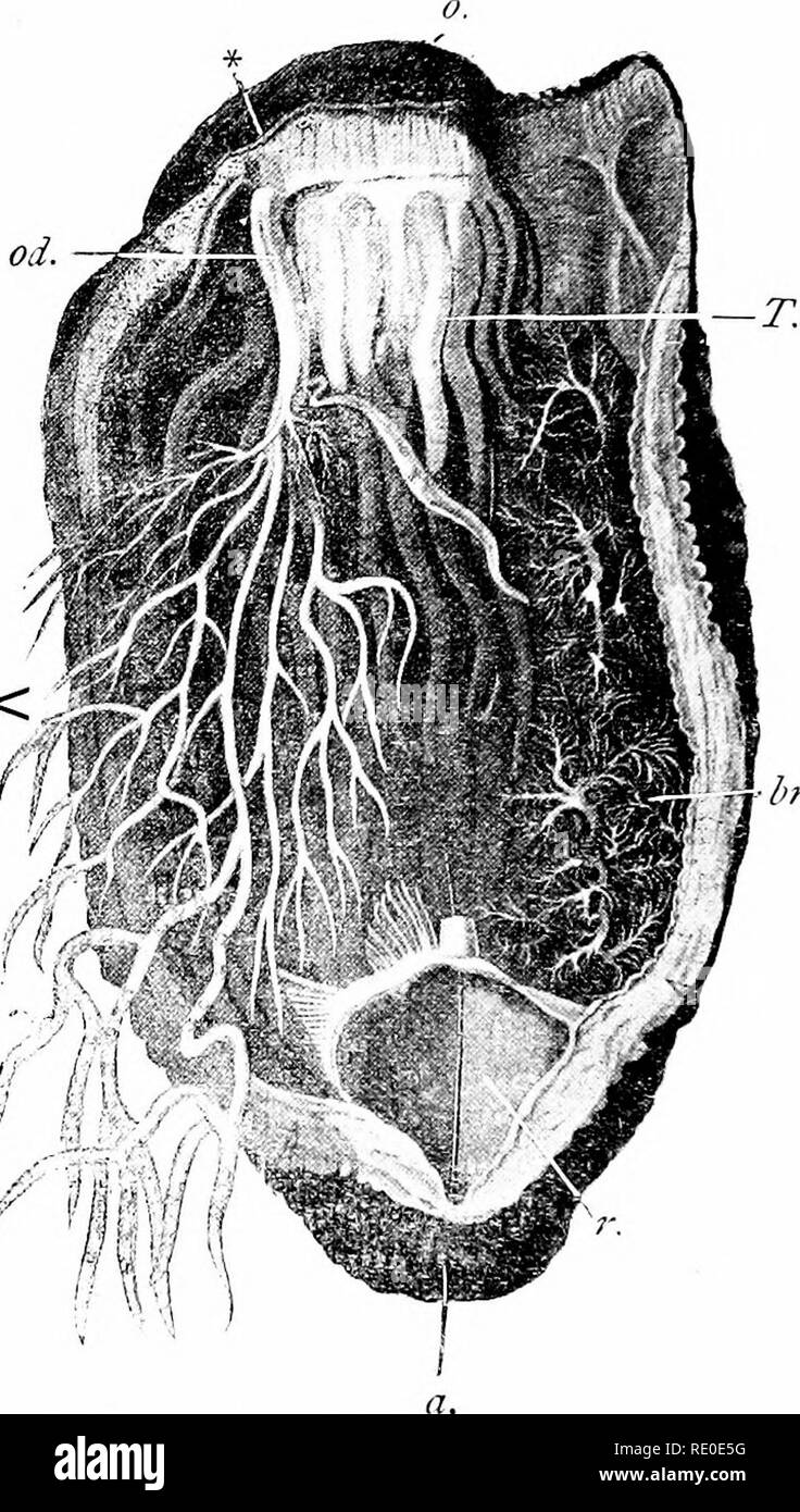
 . Outlines of zoology. Zoology. 242 ECHINODERMA. the mouth communicating id) with the tentacles, (b) with five radial vessels, one for each ambulacral area, (c) with a " Polian vesicle " or more than one pendent in the body ov.<^. h Fig. yS.—Dissection of Holoth The gut has been removed ; there is a bristle throtigh anus (a) and cloaca (r); o, mouth ; 'J the retracted tentacles ; ^>?-, a respiratory tree ; i>7', the ovary ; otf, the oviduct; *, a bristle at genital aperture. cavity, (d) with a " stone canal " which usually hangs freely in the body cavity, and open Stock Photohttps://www.alamy.com/image-license-details/?v=1https://www.alamy.com/outlines-of-zoology-zoology-242-echinoderma-the-mouth-communicating-id-with-the-tentacles-b-with-five-radial-vessels-one-for-each-ambulacral-area-c-with-a-quot-polian-vesicle-quot-or-more-than-one-pendent-in-the-body-ovlt-h-fig-ysdissection-of-holoth-the-gut-has-been-removed-there-is-a-bristle-throtigh-anus-a-and-cloaca-r-o-mouth-j-the-retracted-tentacles-gt-a-respiratory-tree-igt7-the-ovary-otf-the-oviduct-a-bristle-at-genital-aperture-cavity-d-with-a-quot-stone-canal-quot-which-usually-hangs-freely-in-the-body-cavity-and-open-image232351100.html
RMRE0E5G–. Outlines of zoology. Zoology. 242 ECHINODERMA. the mouth communicating id) with the tentacles, (b) with five radial vessels, one for each ambulacral area, (c) with a " Polian vesicle " or more than one pendent in the body ov.<^. h Fig. yS.—Dissection of Holoth The gut has been removed ; there is a bristle throtigh anus (a) and cloaca (r); o, mouth ; 'J the retracted tentacles ; ^>?-, a respiratory tree ; i>7', the ovary ; otf, the oviduct; *, a bristle at genital aperture. cavity, (d) with a " stone canal " which usually hangs freely in the body cavity, and open
![. Correspondence relative to the seizure of British vessels in Behring Sea by the United States' authorities in 1886-91 [microform]. Bering Sea controversy; Fisheries; Béring, Affaire des pêcheries de; Pêche commerciale. ZZVIU CONTENTS. ;*.;i. 200 201 Lord Stanley of Preston to Lord Kefcrring to Nos. 189 and 197 lirecediiiK: Communicating a 391 Knutsford, Kith Aug., 1890. â telegram stilting the aniencied claim of schooner " Path- I finder '" was in eoiirse of transmission. I Lord Stanley of Preston to Lord Amended claim re "Pathfinder Knutsford, 20th Aug., 18!)0. will be for Stock Photo . Correspondence relative to the seizure of British vessels in Behring Sea by the United States' authorities in 1886-91 [microform]. Bering Sea controversy; Fisheries; Béring, Affaire des pêcheries de; Pêche commerciale. ZZVIU CONTENTS. ;*.;i. 200 201 Lord Stanley of Preston to Lord Kefcrring to Nos. 189 and 197 lirecediiiK: Communicating a 391 Knutsford, Kith Aug., 1890. â telegram stilting the aniencied claim of schooner " Path- I finder '" was in eoiirse of transmission. I Lord Stanley of Preston to Lord Amended claim re "Pathfinder Knutsford, 20th Aug., 18!)0. will be for Stock Photo](https://c8.alamy.com/comp/RJ3MY0/correspondence-relative-to-the-seizure-of-british-vessels-in-behring-sea-by-the-united-states-authorities-in-1886-91-microform-bering-sea-controversy-fisheries-bring-affaire-des-pcheries-de-pche-commerciale-zzviu-contents-i-200-201-lord-stanley-of-preston-to-lord-kefcrring-to-nos-189-and-197-lirecediiik-communicating-a-391-knutsford-kith-aug-1890-telegram-stilting-the-aniencied-claim-of-schooner-quot-path-i-finder-quot-was-in-eoiirse-of-transmission-i-lord-stanley-of-preston-to-lord-amended-claim-re-quotpathfinder-knutsford-20th-aug-18!0-will-be-for-RJ3MY0.jpg) . Correspondence relative to the seizure of British vessels in Behring Sea by the United States' authorities in 1886-91 [microform]. Bering Sea controversy; Fisheries; Béring, Affaire des pêcheries de; Pêche commerciale. ZZVIU CONTENTS. ;*.;i. 200 201 Lord Stanley of Preston to Lord Kefcrring to Nos. 189 and 197 lirecediiiK: Communicating a 391 Knutsford, Kith Aug., 1890. â telegram stilting the aniencied claim of schooner " Path- I finder '" was in eoiirse of transmission. I Lord Stanley of Preston to Lord Amended claim re "Pathfinder Knutsford, 20th Aug., 18!)0. will be for Stock Photohttps://www.alamy.com/image-license-details/?v=1https://www.alamy.com/correspondence-relative-to-the-seizure-of-british-vessels-in-behring-sea-by-the-united-states-authorities-in-1886-91-microform-bering-sea-controversy-fisheries-bring-affaire-des-pcheries-de-pche-commerciale-zzviu-contents-i-200-201-lord-stanley-of-preston-to-lord-kefcrring-to-nos-189-and-197-lirecediiik-communicating-a-391-knutsford-kith-aug-1890-telegram-stilting-the-aniencied-claim-of-schooner-quot-path-i-finder-quot-was-in-eoiirse-of-transmission-i-lord-stanley-of-preston-to-lord-amended-claim-re-quotpathfinder-knutsford-20th-aug-18!0-will-be-for-image234880884.html
. Correspondence relative to the seizure of British vessels in Behring Sea by the United States' authorities in 1886-91 [microform]. Bering Sea controversy; Fisheries; Béring, Affaire des pêcheries de; Pêche commerciale. ZZVIU CONTENTS. ;*.;i. 200 201 Lord Stanley of Preston to Lord Kefcrring to Nos. 189 and 197 lirecediiiK: Communicating a 391 Knutsford, Kith Aug., 1890. â telegram stilting the aniencied claim of schooner " Path- I finder '" was in eoiirse of transmission. I Lord Stanley of Preston to Lord Amended claim re "Pathfinder Knutsford, 20th Aug., 18!)0. will be for Stock Photohttps://www.alamy.com/image-license-details/?v=1https://www.alamy.com/correspondence-relative-to-the-seizure-of-british-vessels-in-behring-sea-by-the-united-states-authorities-in-1886-91-microform-bering-sea-controversy-fisheries-bring-affaire-des-pcheries-de-pche-commerciale-zzviu-contents-i-200-201-lord-stanley-of-preston-to-lord-kefcrring-to-nos-189-and-197-lirecediiik-communicating-a-391-knutsford-kith-aug-1890-telegram-stilting-the-aniencied-claim-of-schooner-quot-path-i-finder-quot-was-in-eoiirse-of-transmission-i-lord-stanley-of-preston-to-lord-amended-claim-re-quotpathfinder-knutsford-20th-aug-18!0-will-be-for-image234880884.htmlRMRJ3MY0–. Correspondence relative to the seizure of British vessels in Behring Sea by the United States' authorities in 1886-91 [microform]. Bering Sea controversy; Fisheries; Béring, Affaire des pêcheries de; Pêche commerciale. ZZVIU CONTENTS. ;*.;i. 200 201 Lord Stanley of Preston to Lord Kefcrring to Nos. 189 and 197 lirecediiiK: Communicating a 391 Knutsford, Kith Aug., 1890. â telegram stilting the aniencied claim of schooner " Path- I finder '" was in eoiirse of transmission. I Lord Stanley of Preston to Lord Amended claim re "Pathfinder Knutsford, 20th Aug., 18!)0. will be for