Compensatory emphysema Stock Photos and Images
 . The American journal of roentgenology, radium therapy and nuclear medicine . Fig. ioa. Fig. 10b.. Fir,, ioc. Fig. iorf. ig. to. F. 15. No. N--;. This patient was unusualh good :mcl breathed according to instruction. Note that in aat lull inspiration, aeration >>l the two lungs is nearly equal, whereas in (o) the evidence l obstructiveemphysema ol the right lung is striking at the end ol full expiration. Compensatory emphysema of left lungis quite evident. (< and d All signs disappeai after removal of foreign body. The Roentgen-Ray Diagnosis of Non-opaque Foreign Bodies 303 unless al Stock Photohttps://www.alamy.com/image-license-details/?v=1https://www.alamy.com/the-american-journal-of-roentgenology-radium-therapy-and-nuclear-medicine-fig-ioa-fig-10b-fir-ioc-fig-iorf-ig-to-f-15-no-n-this-patient-was-unusualh-good-mcl-breathed-according-to-instruction-note-that-in-aat-lull-inspiration-aeration-gtgtl-the-two-lungs-is-nearly-equal-whereas-in-o-the-evidence-l-obstructiveemphysema-ol-the-right-lung-is-striking-at-the-end-ol-full-expiration-compensatory-emphysema-of-left-lungis-quite-evident-lt-and-d-all-signs-disappeai-after-removal-of-foreign-body-the-roentgen-ray-diagnosis-of-non-opaque-foreign-bodies-303-unless-al-image376063216.html
RM2CRR4D4–. The American journal of roentgenology, radium therapy and nuclear medicine . Fig. ioa. Fig. 10b.. Fir,, ioc. Fig. iorf. ig. to. F. 15. No. N--;. This patient was unusualh good :mcl breathed according to instruction. Note that in aat lull inspiration, aeration >>l the two lungs is nearly equal, whereas in (o) the evidence l obstructiveemphysema ol the right lung is striking at the end ol full expiration. Compensatory emphysema of left lungis quite evident. (< and d All signs disappeai after removal of foreign body. The Roentgen-Ray Diagnosis of Non-opaque Foreign Bodies 303 unless al
 . The diseases of children : medical and surgical. sed, a result no doubt due to in-flammatory softening of their walls. Emphysema is also constantly presentin association with dilated bronchial tubes. The chest walls during an acuteattack assume the position of inspiration, and, particularly the infraclavicular A . 354 Diseases of tJie Respiratory Apparatus regions, become hyper-resonant, while the expiratory murmur is prolonged.As already remarked, compensatory emphysema is constantly present inassociation with broncho-pneumonia and collapse. Bronchiectasis takes placein association with ch Stock Photohttps://www.alamy.com/image-license-details/?v=1https://www.alamy.com/the-diseases-of-children-medical-and-surgical-sed-a-result-no-doubt-due-to-in-flammatory-softening-of-their-walls-emphysema-is-also-constantly-presentin-association-with-dilated-bronchial-tubes-the-chest-walls-during-an-acuteattack-assume-the-position-of-inspiration-and-particularly-the-infraclavicular-a-354-diseases-of-tjie-respiratory-apparatus-regions-become-hyper-resonant-while-the-expiratory-murmur-is-prolongedas-already-remarked-compensatory-emphysema-is-constantly-present-inassociation-with-broncho-pneumonia-and-collapse-bronchiectasis-takes-placein-association-with-ch-image337034235.html
RM2AG96GY–. The diseases of children : medical and surgical. sed, a result no doubt due to in-flammatory softening of their walls. Emphysema is also constantly presentin association with dilated bronchial tubes. The chest walls during an acuteattack assume the position of inspiration, and, particularly the infraclavicular A . 354 Diseases of tJie Respiratory Apparatus regions, become hyper-resonant, while the expiratory murmur is prolonged.As already remarked, compensatory emphysema is constantly present inassociation with broncho-pneumonia and collapse. Bronchiectasis takes placein association with ch
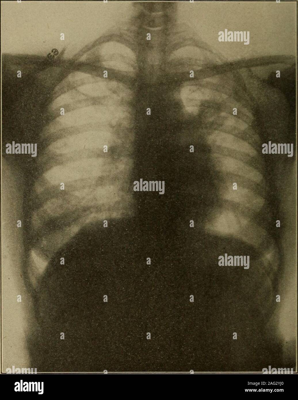
 . Medical diagnosis for the student and practitioner. Fig. 127.—Apical Tuberculosis—Healed or Latent. Note that lesions are closely cir-cumscribed by apparently normal lung. {Dr. Frank S. Bissell.) This is true because the more characteristic changes tend to becomemasked by the effects of fibrosis and mixed infections. Usually, however,the distribution of the lesions points the way to a correct diagnosis or somearea of slight involvement is found where the changes are more typical. Compensatory emphysema frequently exists in some degree, manefesting Emphysema. 3i8 MKDICAL I)IA(;OSIS itself ch Stock Photohttps://www.alamy.com/image-license-details/?v=1https://www.alamy.com/medical-diagnosis-for-the-student-and-practitioner-fig-127apical-tuberculosishealed-or-latent-note-that-lesions-are-closely-cir-cumscribed-by-apparently-normal-lung-dr-frank-s-bissell-this-is-true-because-the-more-characteristic-changes-tend-to-becomemasked-by-the-effects-of-fibrosis-and-mixed-infections-usually-howeverthe-distribution-of-the-lesions-points-the-way-to-a-correct-diagnosis-or-somearea-of-slight-involvement-is-found-where-the-changes-are-more-typical-compensatory-emphysema-frequently-exists-in-some-degree-manefesting-emphysema-3i8-mkdical-iiaosis-itself-ch-image336897064.html
RM2AG2YJ0–. Medical diagnosis for the student and practitioner. Fig. 127.—Apical Tuberculosis—Healed or Latent. Note that lesions are closely cir-cumscribed by apparently normal lung. {Dr. Frank S. Bissell.) This is true because the more characteristic changes tend to becomemasked by the effects of fibrosis and mixed infections. Usually, however,the distribution of the lesions points the way to a correct diagnosis or somearea of slight involvement is found where the changes are more typical. Compensatory emphysema frequently exists in some degree, manefesting Emphysema. 3i8 MKDICAL I)IA(;OSIS itself ch
 Diseases of the chest and the principles of physical diagnosis . cted, may bemarkedly diseased in the upper lobe, but the lower will be free, or at mostcontain scattered tubercles. During the terminal months of the diseasethe lower part of one or the other lung, which undergoes a certain amountof compensatory emphysema, supplies the patient with most of hisbreathing space (Fig. 236; see also Fig. 101 J. 320 DISEASES OF THE BRONCHI, LUNGS, PLEURA, AND DIAPHRAGM Another change, and one that probably follows the pulmonary in-fection very quickly, is a low-grade inflammation of the apical pleurawh Stock Photohttps://www.alamy.com/image-license-details/?v=1https://www.alamy.com/diseases-of-the-chest-and-the-principles-of-physical-diagnosis-cted-may-bemarkedly-diseased-in-the-upper-lobe-but-the-lower-will-be-free-or-at-mostcontain-scattered-tubercles-during-the-terminal-months-of-the-diseasethe-lower-part-of-one-or-the-other-lung-which-undergoes-a-certain-amountof-compensatory-emphysema-supplies-the-patient-with-most-of-hisbreathing-space-fig-236-see-also-fig-101-j-320-diseases-of-the-bronchi-lungs-pleura-and-diaphragm-another-change-and-one-that-probably-follows-the-pulmonary-in-fection-very-quickly-is-a-low-grade-inflammation-of-the-apical-pleurawh-image340248302.html
RM2ANFJ52–Diseases of the chest and the principles of physical diagnosis . cted, may bemarkedly diseased in the upper lobe, but the lower will be free, or at mostcontain scattered tubercles. During the terminal months of the diseasethe lower part of one or the other lung, which undergoes a certain amountof compensatory emphysema, supplies the patient with most of hisbreathing space (Fig. 236; see also Fig. 101 J. 320 DISEASES OF THE BRONCHI, LUNGS, PLEURA, AND DIAPHRAGM Another change, and one that probably follows the pulmonary in-fection very quickly, is a low-grade inflammation of the apical pleurawh
 Diseases of the chest and the principles of physical diagnosis . , especially of the chronic type with ex-tensive fibroid changes, also leads to a marked degree of compensatory DISEASES OF THE LUXGS 497 emphysema of the opposite side. And this is true of any clironic inflam-matory|disease affecting one lung alone. In other words the presenceof a pathological lesion invohing all or a considerable portion of one lungis practically always accomiDanied by a compensatory- emphysema ofthe opposite side. Physical Signs.—In some instances the hypertrophy of the unaffectedlung is extraordinarily great. Stock Photohttps://www.alamy.com/image-license-details/?v=1https://www.alamy.com/diseases-of-the-chest-and-the-principles-of-physical-diagnosis-especially-of-the-chronic-type-with-ex-tensive-fibroid-changes-also-leads-to-a-marked-degree-of-compensatory-diseases-of-the-luxgs-497-emphysema-of-the-opposite-side-and-this-is-true-of-any-clironic-inflam-matorydisease-affecting-one-lung-alone-in-other-words-the-presenceof-a-pathological-lesion-invohing-all-or-a-considerable-portion-of-one-lungis-practically-always-accomidanied-by-a-compensatory-emphysema-ofthe-opposite-side-physical-signsin-some-instances-the-hypertrophy-of-the-unaffectedlung-is-extraordinarily-great-image340227466.html
RM2ANEKGX–Diseases of the chest and the principles of physical diagnosis . , especially of the chronic type with ex-tensive fibroid changes, also leads to a marked degree of compensatory DISEASES OF THE LUXGS 497 emphysema of the opposite side. And this is true of any clironic inflam-matory|disease affecting one lung alone. In other words the presenceof a pathological lesion invohing all or a considerable portion of one lungis practically always accomiDanied by a compensatory- emphysema ofthe opposite side. Physical Signs.—In some instances the hypertrophy of the unaffectedlung is extraordinarily great.
 A treatise on the principles and practice of medicine . Figs. 33, 34, and 35.—Cuts from Dieulafoy, showing: Fig. 33, the posterior surface ofthe lungs and their interlobar fissures; Fig. 34, the lateral aspect of the left lung; and Fig.35, that of the right lung. adhesions is of no consequence. Compensatory emphysema, pain,thoracic oppression, obliteration of the complementary pleural space,dulness, decreased vocal fremitus and breathing, failure of Littensphenomenon and stagnation of bronchial secretion may occur when theadhesions are thick, and stasis may ensue. Peritonitis, mediastinitis 46 Stock Photohttps://www.alamy.com/image-license-details/?v=1https://www.alamy.com/a-treatise-on-the-principles-and-practice-of-medicine-figs-33-34-and-35cuts-from-dieulafoy-showing-fig-33-the-posterior-surface-ofthe-lungs-and-their-interlobar-fissures-fig-34-the-lateral-aspect-of-the-left-lung-and-fig35-that-of-the-right-lung-adhesions-is-of-no-consequence-compensatory-emphysema-painthoracic-oppression-obliteration-of-the-complementary-pleural-spacedulness-decreased-vocal-fremitus-and-breathing-failure-of-littensphenomenon-and-stagnation-of-bronchial-secretion-may-occur-when-theadhesions-are-thick-and-stasis-may-ensue-peritonitis-mediastinitis-46-image339153165.html
RM2AKNN91–A treatise on the principles and practice of medicine . Figs. 33, 34, and 35.—Cuts from Dieulafoy, showing: Fig. 33, the posterior surface ofthe lungs and their interlobar fissures; Fig. 34, the lateral aspect of the left lung; and Fig.35, that of the right lung. adhesions is of no consequence. Compensatory emphysema, pain,thoracic oppression, obliteration of the complementary pleural space,dulness, decreased vocal fremitus and breathing, failure of Littensphenomenon and stagnation of bronchial secretion may occur when theadhesions are thick, and stasis may ensue. Peritonitis, mediastinitis 46
 Peroral endoscopy and laryngeal surgery . Fk;. 37(>.—RadioLsrapli sliowinj; slmc Imllon dmly iiictal part dense cnoiigli l<show) in left main 1)roni-hus of a girl of seven years. Distortion gives appearanceof median position. J-eft Inng atelectatic. Compensatory emphysema of right hmg.Bntton was aspirated three months previonslv. Removal hy oral hronchoscopywithout anesthesia, a hook being inserted in the eye of the button. ( Radiograplby Dr. George C. Johnstin. .iithors r:uc ) 4J0 ILLUSTRATIVE CASES OF ENCOSCOPY FOR FOREIGN BODIES.. Fig. 377.—Radiograph showing pclil)le in right bronch Stock Photohttps://www.alamy.com/image-license-details/?v=1https://www.alamy.com/peroral-endoscopy-and-laryngeal-surgery-fk-37gtradiolsrapli-sliowinj-slmc-imllon-dmly-iiictal-part-dense-cnoiigli-lltshow-in-left-main-1roni-hus-of-a-girl-of-seven-years-distortion-gives-appearanceof-median-position-j-eft-inng-atelectatic-compensatory-emphysema-of-right-hmgbntton-was-aspirated-three-months-previonslv-removal-hy-oral-hronchoscopywithout-anesthesia-a-hook-being-inserted-in-the-eye-of-the-button-radiograplby-dr-george-c-johnstin-iithors-ruc-4j0-illustrative-cases-of-encoscopy-for-foreign-bodies-fig-377radiograph-showing-pclille-in-right-bronch-image340254777.html
RM2ANFXC9–Peroral endoscopy and laryngeal surgery . Fk;. 37(>.—RadioLsrapli sliowinj; slmc Imllon dmly iiictal part dense cnoiigli l<show) in left main 1)roni-hus of a girl of seven years. Distortion gives appearanceof median position. J-eft Inng atelectatic. Compensatory emphysema of right hmg.Bntton was aspirated three months previonslv. Removal hy oral hronchoscopywithout anesthesia, a hook being inserted in the eye of the button. ( Radiograplby Dr. George C. Johnstin. .iithors r:uc ) 4J0 ILLUSTRATIVE CASES OF ENCOSCOPY FOR FOREIGN BODIES.. Fig. 377.—Radiograph showing pclil)le in right bronch
 Peroral endoscopy and laryngeal surgery . Flu. ,^75.—Radiograiihs, showing pin in rij;lit iironchiis of a boy aged fouryears. Pin removed by oral bronclioscopy. (Radiograph by Ur. Russell H. Boggs)Pin shadow strengthened for photo-engraving. (Authors case.) ILLUSTKATI-|-; CASKS ni KM.OSCOPV KOK I-HKICIGN BOUIKS. 4UU. Fk;. 37(>.—RadioLsrapli sliowinj; slmc Imllon dmly iiictal part dense cnoiigli l<show) in left main 1)roni-hus of a girl of seven years. Distortion gives appearanceof median position. J-eft Inng atelectatic. Compensatory emphysema of right hmg.Bntton was aspirated three mon Stock Photohttps://www.alamy.com/image-license-details/?v=1https://www.alamy.com/peroral-endoscopy-and-laryngeal-surgery-flu-75radiograiihs-showing-pin-in-rijlit-iironchiis-of-a-boy-aged-fouryears-pin-removed-by-oral-bronclioscopy-radiograph-by-ur-russell-h-boggspin-shadow-strengthened-for-photo-engraving-authors-case-illustkati-casks-ni-kmoscopv-kok-i-hkicign-bouiks-4uu-fk-37gtradiolsrapli-sliowinj-slmc-imllon-dmly-iiictal-part-dense-cnoiigli-lltshow-in-left-main-1roni-hus-of-a-girl-of-seven-years-distortion-gives-appearanceof-median-position-j-eft-inng-atelectatic-compensatory-emphysema-of-right-hmgbntton-was-aspirated-three-mon-image340254859.html
RM2ANFXF7–Peroral endoscopy and laryngeal surgery . Flu. ,^75.—Radiograiihs, showing pin in rij;lit iironchiis of a boy aged fouryears. Pin removed by oral bronclioscopy. (Radiograph by Ur. Russell H. Boggs)Pin shadow strengthened for photo-engraving. (Authors case.) ILLUSTKATI-|-; CASKS ni KM.OSCOPV KOK I-HKICIGN BOUIKS. 4UU. Fk;. 37(>.—RadioLsrapli sliowinj; slmc Imllon dmly iiictal part dense cnoiigli l<show) in left main 1)roni-hus of a girl of seven years. Distortion gives appearanceof median position. J-eft Inng atelectatic. Compensatory emphysema of right hmg.Bntton was aspirated three mon
 . A practical treatise on medical diagnosis for students and physicians . berculous. Physical Signs. Chronic pleurisy causes great deformity of the chestfrom contraction, and compensatory emphysema, of the healthy lung. Theheart is dislocated or can not be found on physical examination, because itis overlapped by lung or is drawn behind the sternum. There is con-siderable spinal curvature, dislocation of the scapida, deformity of theshoulder, and indrawing and overlapping of the ribs at the base of thechest. Chronic Pleurisy with Effusion. This results from an acute attackof pleurisy, in which Stock Photohttps://www.alamy.com/image-license-details/?v=1https://www.alamy.com/a-practical-treatise-on-medical-diagnosis-for-students-and-physicians-berculous-physical-signs-chronic-pleurisy-causes-great-deformity-of-the-chestfrom-contraction-and-compensatory-emphysema-of-the-healthy-lung-theheart-is-dislocated-or-can-not-be-found-on-physical-examination-because-itis-overlapped-by-lung-or-is-drawn-behind-the-sternum-there-is-con-siderable-spinal-curvature-dislocation-of-the-scapida-deformity-of-theshoulder-and-indrawing-and-overlapping-of-the-ribs-at-the-base-of-thechest-chronic-pleurisy-with-effusion-this-results-from-an-acute-attackof-pleurisy-in-which-image376075583.html
RM2CRRM6R–. A practical treatise on medical diagnosis for students and physicians . berculous. Physical Signs. Chronic pleurisy causes great deformity of the chestfrom contraction, and compensatory emphysema, of the healthy lung. Theheart is dislocated or can not be found on physical examination, because itis overlapped by lung or is drawn behind the sternum. There is con-siderable spinal curvature, dislocation of the scapida, deformity of theshoulder, and indrawing and overlapping of the ribs at the base of thechest. Chronic Pleurisy with Effusion. This results from an acute attackof pleurisy, in which
 . A practical treatise on medical diagnosis for students and physicians . k, ordevelops insidiously if tuberculous. Physical Signs. Chronic jileurisy causes great deformity of the chestfrom contraction, and compensatory emphysema of the healthy lung. Theheart is dislocated or can not be found on physical examination, because itis overlapped by lung or is drawn behind the sternum. There is con-siderable spinal curvature, dislocation of the scapula, deformity of theshoulder, and indrawing and overlapping of the ribs at the base of thechest. Chronic Pleurisy with Effusion. This results from an ac Stock Photohttps://www.alamy.com/image-license-details/?v=1https://www.alamy.com/a-practical-treatise-on-medical-diagnosis-for-students-and-physicians-k-ordevelops-insidiously-if-tuberculous-physical-signs-chronic-jileurisy-causes-great-deformity-of-the-chestfrom-contraction-and-compensatory-emphysema-of-the-healthy-lung-theheart-is-dislocated-or-can-not-be-found-on-physical-examination-because-itis-overlapped-by-lung-or-is-drawn-behind-the-sternum-there-is-con-siderable-spinal-curvature-dislocation-of-the-scapula-deformity-of-theshoulder-and-indrawing-and-overlapping-of-the-ribs-at-the-base-of-thechest-chronic-pleurisy-with-effusion-this-results-from-an-ac-image375947744.html
RM2CRHW54–. A practical treatise on medical diagnosis for students and physicians . k, ordevelops insidiously if tuberculous. Physical Signs. Chronic jileurisy causes great deformity of the chestfrom contraction, and compensatory emphysema of the healthy lung. Theheart is dislocated or can not be found on physical examination, because itis overlapped by lung or is drawn behind the sternum. There is con-siderable spinal curvature, dislocation of the scapula, deformity of theshoulder, and indrawing and overlapping of the ribs at the base of thechest. Chronic Pleurisy with Effusion. This results from an ac
 . The American journal of roentgenology, radium therapy and nuclear medicine . en-ray signs of acute obstruc-tive monolateral emphysema are: 1. Increased transparency of the affectedlung. 2. Depression and partial fixation of thediaphragm on the affected side. 3. Displacement of the heart and medi-astinal structures away from the affectedside. 4. Increased excursion of the diaphragmon the unaffected side, due to compensa-tory emphysema. The diaphragmatic signs are the mostimportant, for by them alone can one ruleout compensatory emphysema. In our former paper, sign No. 4 wassuggested as Increa Stock Photohttps://www.alamy.com/image-license-details/?v=1https://www.alamy.com/the-american-journal-of-roentgenology-radium-therapy-and-nuclear-medicine-en-ray-signs-of-acute-obstruc-tive-monolateral-emphysema-are-1-increased-transparency-of-the-affectedlung-2-depression-and-partial-fixation-of-thediaphragm-on-the-affected-side-3-displacement-of-the-heart-and-medi-astinal-structures-away-from-the-affectedside-4-increased-excursion-of-the-diaphragmon-the-unaffected-side-due-to-compensa-tory-emphysema-the-diaphragmatic-signs-are-the-mostimportant-for-by-them-alone-can-one-ruleout-compensatory-emphysema-in-our-former-paper-sign-no-4-wassuggested-as-increa-image376064151.html
RM2CRR5JF–. The American journal of roentgenology, radium therapy and nuclear medicine . en-ray signs of acute obstruc-tive monolateral emphysema are: 1. Increased transparency of the affectedlung. 2. Depression and partial fixation of thediaphragm on the affected side. 3. Displacement of the heart and medi-astinal structures away from the affectedside. 4. Increased excursion of the diaphragmon the unaffected side, due to compensa-tory emphysema. The diaphragmatic signs are the mostimportant, for by them alone can one ruleout compensatory emphysema. In our former paper, sign No. 4 wassuggested as Increa
 . Bulletin of the U.S. Department of Agriculture. Agriculture; Agriculture. LIFE HISTORY OF ASCARIS LUMBRICOIDES. 23 the air sacs being almost entirely filled in with the serosanguineous exudate. Extensive immigration of leucocytes and round-cell in- filtration characteristic of acute inflammation are well marked. In other portions of the section the alveoli are enlarged, indicating a compensatory emphysema. Similar appearances are shown in figure 6, a photomicrograph of a portion of the lung of a pig one week after the ingestion of AscaHs siium eggs.. Fig. 6.—Ascarts suum. Larva? in cross sec Stock Photohttps://www.alamy.com/image-license-details/?v=1https://www.alamy.com/bulletin-of-the-us-department-of-agriculture-agriculture-agriculture-life-history-of-ascaris-lumbricoides-23-the-air-sacs-being-almost-entirely-filled-in-with-the-serosanguineous-exudate-extensive-immigration-of-leucocytes-and-round-cell-in-filtration-characteristic-of-acute-inflammation-are-well-marked-in-other-portions-of-the-section-the-alveoli-are-enlarged-indicating-a-compensatory-emphysema-similar-appearances-are-shown-in-figure-6-a-photomicrograph-of-a-portion-of-the-lung-of-a-pig-one-week-after-the-ingestion-of-ascahs-siium-eggs-fig-6ascarts-suum-larva-in-cross-sec-image233816232.html
RMRGB6YM–. Bulletin of the U.S. Department of Agriculture. Agriculture; Agriculture. LIFE HISTORY OF ASCARIS LUMBRICOIDES. 23 the air sacs being almost entirely filled in with the serosanguineous exudate. Extensive immigration of leucocytes and round-cell in- filtration characteristic of acute inflammation are well marked. In other portions of the section the alveoli are enlarged, indicating a compensatory emphysema. Similar appearances are shown in figure 6, a photomicrograph of a portion of the lung of a pig one week after the ingestion of AscaHs siium eggs.. Fig. 6.—Ascarts suum. Larva? in cross sec
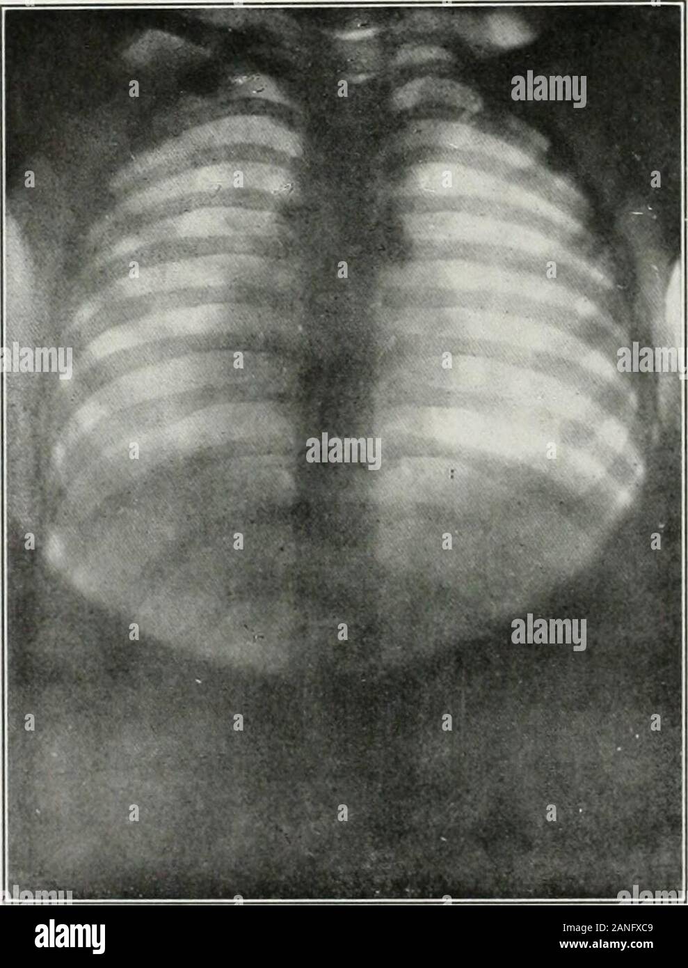
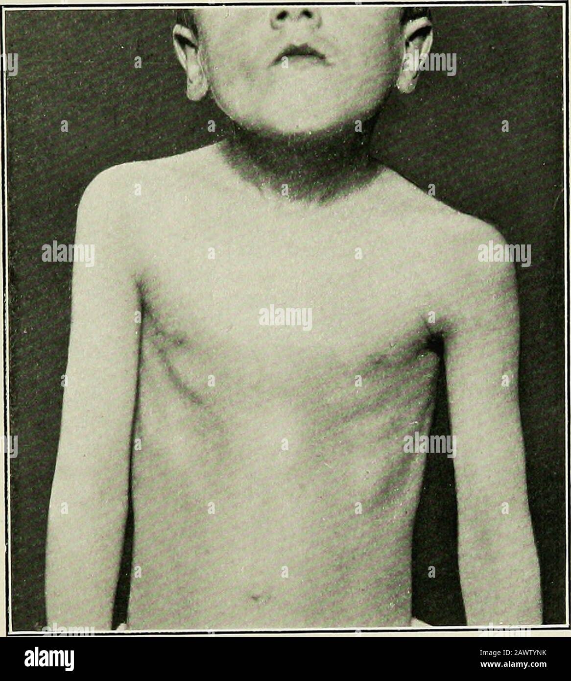
 The medical diseases of children . ortunity for a pathologicalexamination arise—that in place of such a condition we have a collec-tion of small dilated bronchioles. EMPHYSEMA. Acute emphysema develops very rapidly in young children. Itmay be dependent upon the occurrence of bronchitis, pneumonia(particularly consecutive broncho-pneumonia), or pulmonary collapse.In such cases it is compensatory in origin. It may arise chiefly fromobstruction to expiration, as in whooping-cough, asthma, and laryngealstenosis. Chronic emphysema, producing the barrel-shaped chest and hyper-trophic lungs, as are s Stock Photohttps://www.alamy.com/image-license-details/?v=1https://www.alamy.com/the-medical-diseases-of-children-ortunity-for-a-pathologicalexamination-arisethat-in-place-of-such-a-condition-we-have-a-collec-tion-of-small-dilated-bronchioles-emphysema-acute-emphysema-develops-very-rapidly-in-young-children-itmay-be-dependent-upon-the-occurrence-of-bronchitis-pneumoniaparticularly-consecutive-broncho-pneumonia-or-pulmonary-collapsein-such-cases-it-is-compensatory-in-origin-it-may-arise-chiefly-fromobstruction-to-expiration-as-in-whooping-cough-asthma-and-laryngealstenosis-chronic-emphysema-producing-the-barrel-shaped-chest-and-hyper-trophic-lungs-as-are-s-image342912015.html
RM2AWTYNK–The medical diseases of children . ortunity for a pathologicalexamination arise—that in place of such a condition we have a collec-tion of small dilated bronchioles. EMPHYSEMA. Acute emphysema develops very rapidly in young children. Itmay be dependent upon the occurrence of bronchitis, pneumonia(particularly consecutive broncho-pneumonia), or pulmonary collapse.In such cases it is compensatory in origin. It may arise chiefly fromobstruction to expiration, as in whooping-cough, asthma, and laryngealstenosis. Chronic emphysema, producing the barrel-shaped chest and hyper-trophic lungs, as are s