Quick filters:
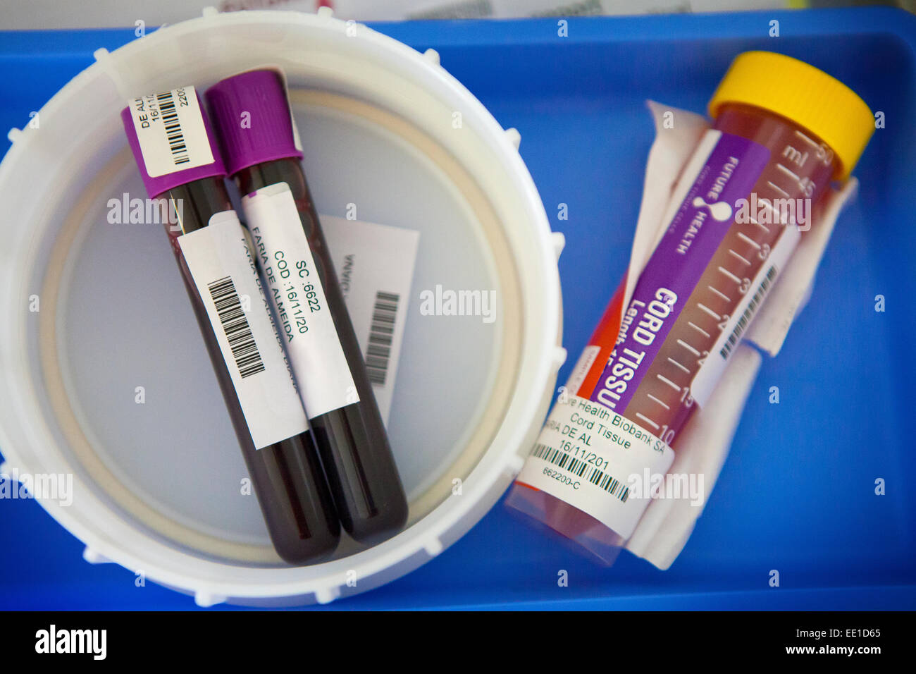
Cord blood specimen Stock Photos and Images
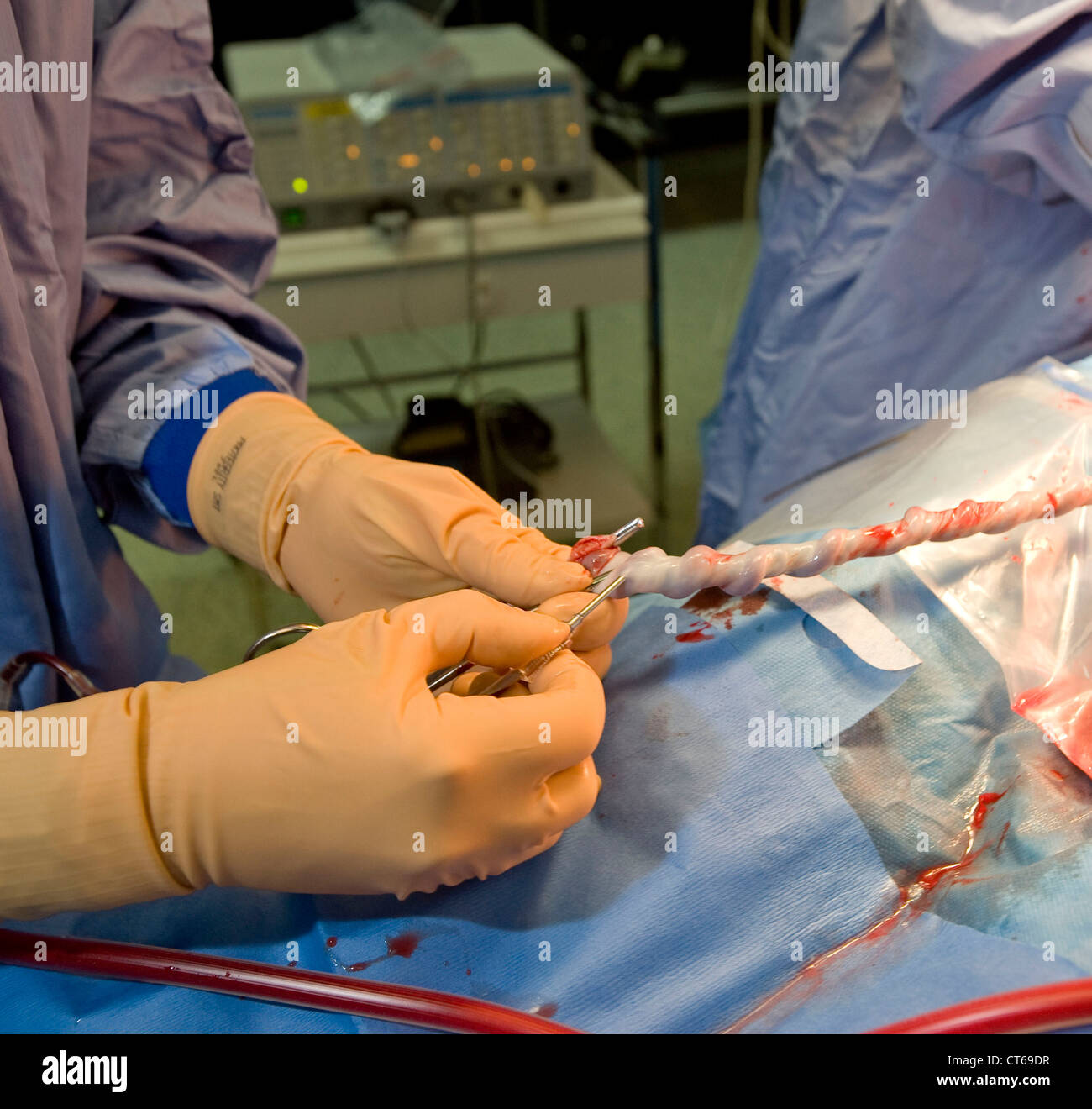 CORD BLOOD SPECIMEN Stock Photohttps://www.alamy.com/image-license-details/?v=1https://www.alamy.com/stock-photo-cord-blood-specimen-49311635.html
RMCT69DR–CORD BLOOD SPECIMEN
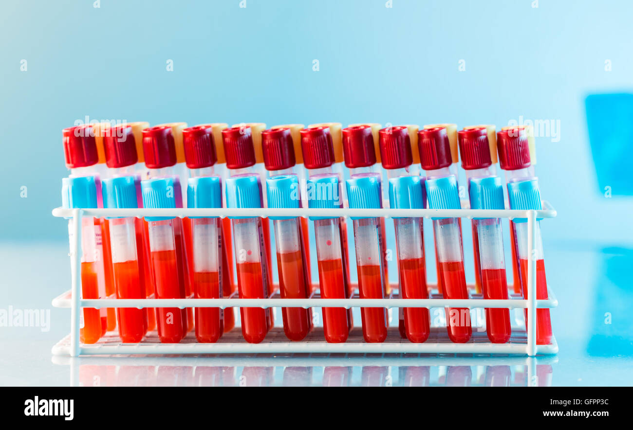 test tubes with blood on a tray Stock Photohttps://www.alamy.com/image-license-details/?v=1https://www.alamy.com/stock-photo-test-tubes-with-blood-on-a-tray-112982336.html
RFGFPP3C–test tubes with blood on a tray
 . Manual of antenatal pathology and hygiene : the foetus. foetus ; normally, however, vitelline nutrition is of short duration,being limited by the close of the neofoetal period, or very soon there-after. With some forms of monstrosity it may be greatly prolonged,and, even when no malformation exists in the infant, persistent andpervious vitelline vessels may be traced in the cord and full-timeplacenta, and these may contain blood. An example of these per- NUTRITION OF THE FOETUS 155 inanent vitelline vessels I met with some years ago; the specimen ishere figured (Fig. 27). More recently Bover Stock Photohttps://www.alamy.com/image-license-details/?v=1https://www.alamy.com/manual-of-antenatal-pathology-and-hygiene-the-foetus-foetus-normally-however-vitelline-nutrition-is-of-short-durationbeing-limited-by-the-close-of-the-neofoetal-period-or-very-soon-there-after-with-some-forms-of-monstrosity-it-may-be-greatly-prolongedand-even-when-no-malformation-exists-in-the-infant-persistent-andpervious-vitelline-vessels-may-be-traced-in-the-cord-and-full-timeplacenta-and-these-may-contain-blood-an-example-of-these-per-nutrition-of-the-foetus-155-inanent-vitelline-vessels-i-met-with-some-years-ago-the-specimen-ishere-figured-fig-27-more-recently-bover-image336857265.html
RM2AG14TH–. Manual of antenatal pathology and hygiene : the foetus. foetus ; normally, however, vitelline nutrition is of short duration,being limited by the close of the neofoetal period, or very soon there-after. With some forms of monstrosity it may be greatly prolonged,and, even when no malformation exists in the infant, persistent andpervious vitelline vessels may be traced in the cord and full-timeplacenta, and these may contain blood. An example of these per- NUTRITION OF THE FOETUS 155 inanent vitelline vessels I met with some years ago; the specimen ishere figured (Fig. 27). More recently Bover
 test tubes with blood on a tray Stock Photohttps://www.alamy.com/image-license-details/?v=1https://www.alamy.com/stock-photo-test-tubes-with-blood-on-a-tray-112982296.html
RFGFPP20–test tubes with blood on a tray
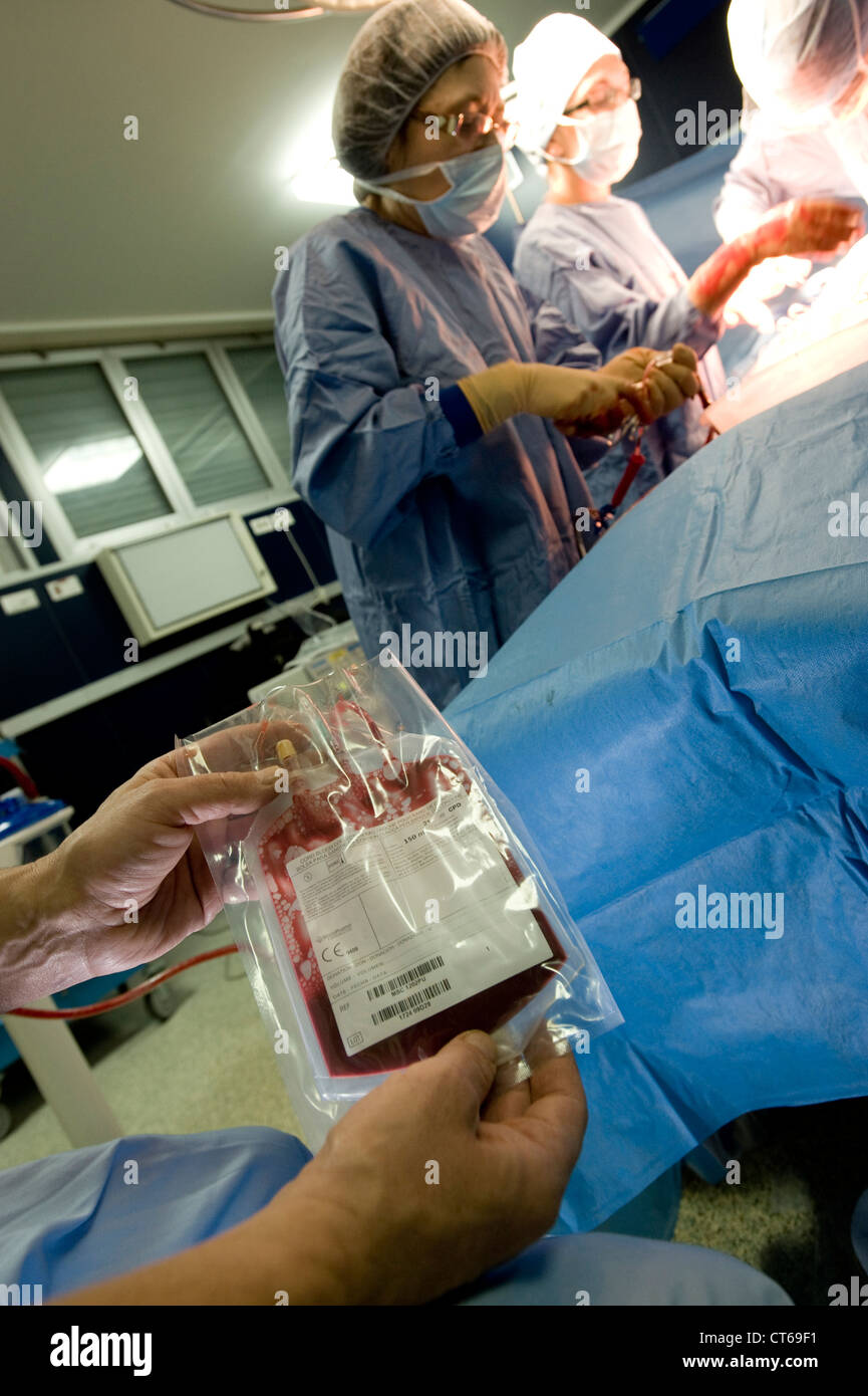 CORD BLOOD SPECIMEN Stock Photohttps://www.alamy.com/image-license-details/?v=1https://www.alamy.com/stock-photo-cord-blood-specimen-49311669.html
RMCT69F1–CORD BLOOD SPECIMEN
 . A study of the causes underlying the origin of human monsters : third contribution to the study of the pathology of human embryos . Fig. 279.—Photograph of a section of the specimen showing the cavityand cord within. Slightly reduced. 274 MALL. [Vol. XIX. and fully one millimeter in diameter; there are also numerousvessels in the villi of the chorion. The tissue of the chorionis hyaline, with a diminished number of nuclei in it. Undoubtedly the foetus escaped in some way shortly beforethe abortion, the membranes and cord remaining some time,long enough to undergo these changes. The blood-ves Stock Photohttps://www.alamy.com/image-license-details/?v=1https://www.alamy.com/a-study-of-the-causes-underlying-the-origin-of-human-monsters-third-contribution-to-the-study-of-the-pathology-of-human-embryos-fig-279photograph-of-a-section-of-the-specimen-showing-the-cavityand-cord-within-slightly-reduced-274-mall-vol-xix-and-fully-one-millimeter-in-diameter-there-are-also-numerousvessels-in-the-villi-of-the-chorion-the-tissue-of-the-chorionis-hyaline-with-a-diminished-number-of-nuclei-in-it-undoubtedly-the-foetus-escaped-in-some-way-shortly-beforethe-abortion-the-membranes-and-cord-remaining-some-timelong-enough-to-undergo-these-changes-the-blood-ves-image369729894.html
RM2CDEJ72–. A study of the causes underlying the origin of human monsters : third contribution to the study of the pathology of human embryos . Fig. 279.—Photograph of a section of the specimen showing the cavityand cord within. Slightly reduced. 274 MALL. [Vol. XIX. and fully one millimeter in diameter; there are also numerousvessels in the villi of the chorion. The tissue of the chorionis hyaline, with a diminished number of nuclei in it. Undoubtedly the foetus escaped in some way shortly beforethe abortion, the membranes and cord remaining some time,long enough to undergo these changes. The blood-ves
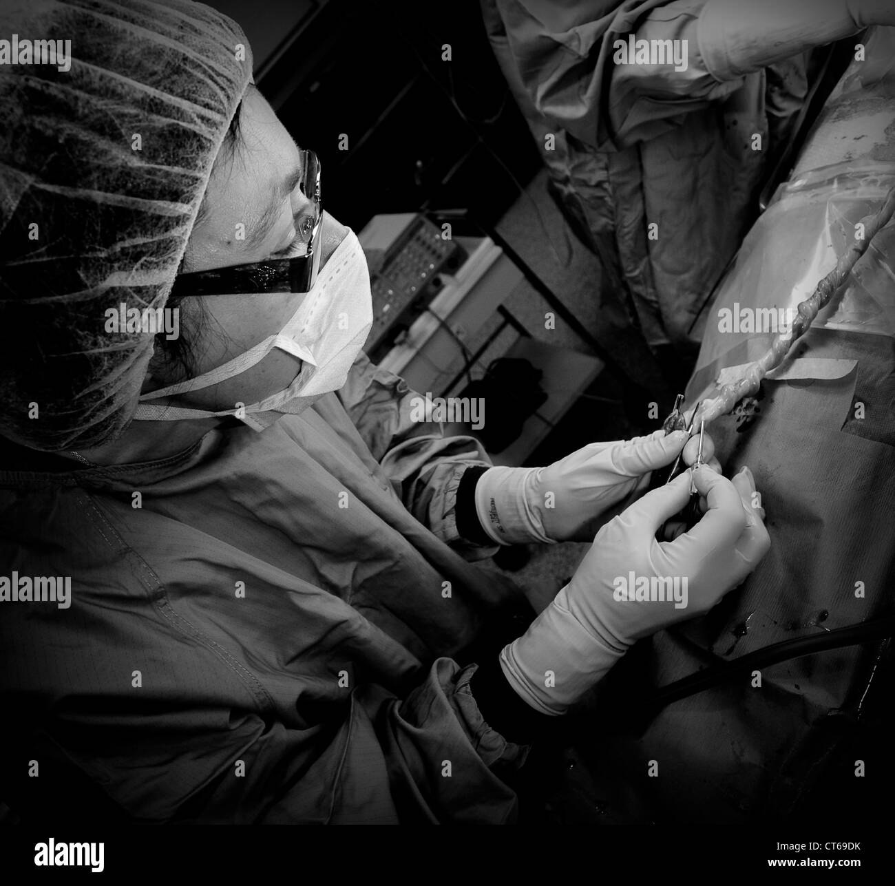 CORD BLOOD SPECIMEN Stock Photohttps://www.alamy.com/image-license-details/?v=1https://www.alamy.com/stock-photo-cord-blood-specimen-49311631.html
RMCT69DK–CORD BLOOD SPECIMEN
 . A study of the causes underlying the origin of human monsters : third contribution to the study of the pathology of human embryos . of the embryo show that the specimen ispathological, its head being rounded and the epidermis havingfallen off. The spinal cord is distended and the brain is solid.Veins and arteries are greatly distended with blood. Ryevesicles are atrophic, and the lenses are dissociated, but encir-cled by a sharply defined capsule. No. 292a. Ovum, 50 x 30 x 30 mm.; embryo, 3^ mm. Dr. West, Bellaire, Ohio. The ovum is from a woman thirty-one years old, who hasbeen married for Stock Photohttps://www.alamy.com/image-license-details/?v=1https://www.alamy.com/a-study-of-the-causes-underlying-the-origin-of-human-monsters-third-contribution-to-the-study-of-the-pathology-of-human-embryos-of-the-embryo-show-that-the-specimen-ispathological-its-head-being-rounded-and-the-epidermis-havingfallen-off-the-spinal-cord-is-distended-and-the-brain-is-solidveins-and-arteries-are-greatly-distended-with-blood-ryevesicles-are-atrophic-and-the-lenses-are-dissociated-but-encir-cled-by-a-sharply-defined-capsule-no-292a-ovum-50-x-30-x-30-mm-embryo-3-mm-dr-west-bellaire-ohio-the-ovum-is-from-a-woman-thirty-one-years-old-who-hasbeen-married-for-image369728046.html
RM2CDEFW2–. A study of the causes underlying the origin of human monsters : third contribution to the study of the pathology of human embryos . of the embryo show that the specimen ispathological, its head being rounded and the epidermis havingfallen off. The spinal cord is distended and the brain is solid.Veins and arteries are greatly distended with blood. Ryevesicles are atrophic, and the lenses are dissociated, but encir-cled by a sharply defined capsule. No. 292a. Ovum, 50 x 30 x 30 mm.; embryo, 3^ mm. Dr. West, Bellaire, Ohio. The ovum is from a woman thirty-one years old, who hasbeen married for
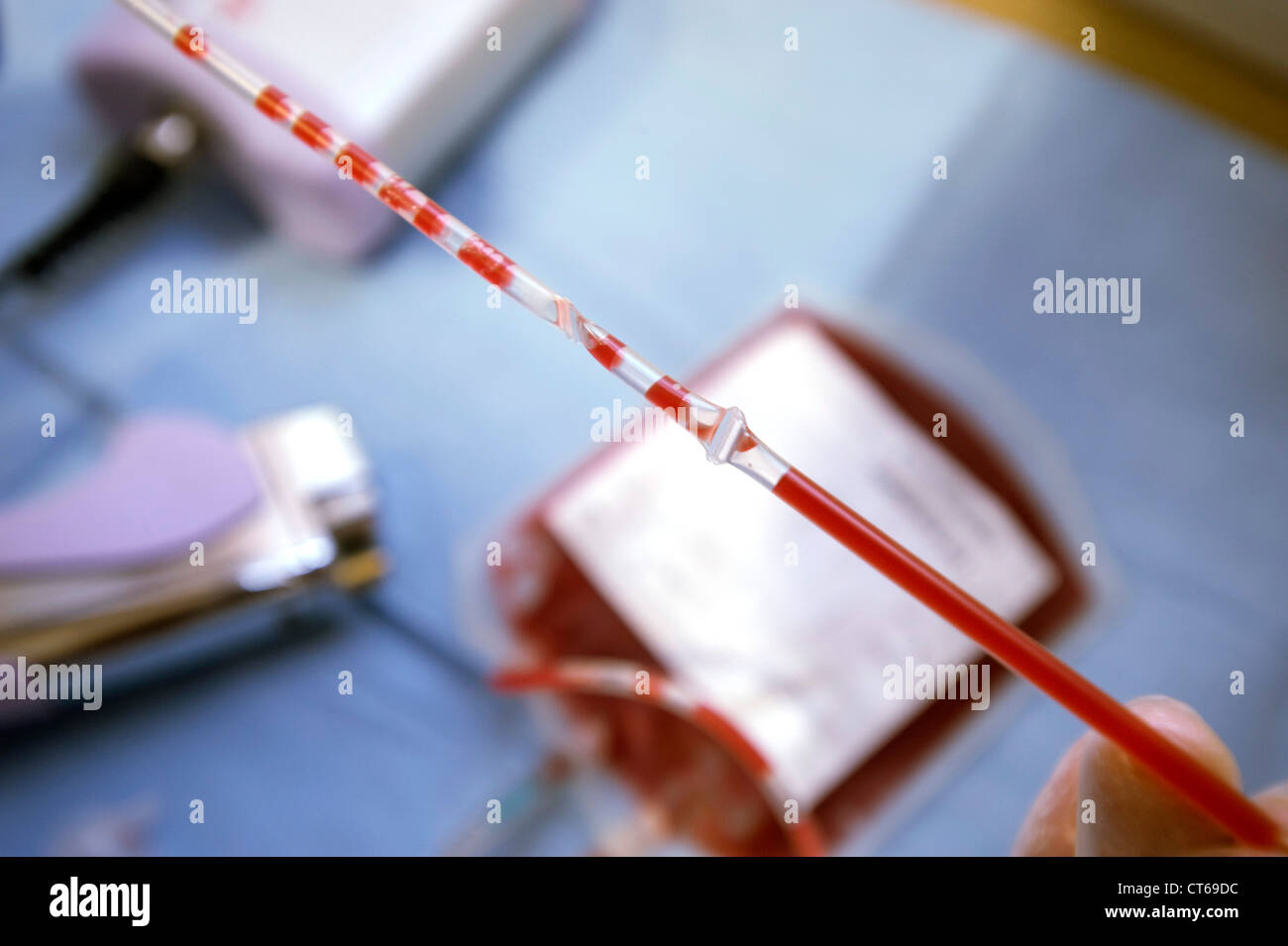 CORD BLOOD Stock Photohttps://www.alamy.com/image-license-details/?v=1https://www.alamy.com/stock-photo-cord-blood-49311624.html
RMCT69DC–CORD BLOOD
![. Micro-organisms and disease. An introduction to the study of specific micro-organisms. Microorganisms. xv] ANAEROBIC BACILLI 381 of human beings is probably only due to the presence of the foreign bodies themselves (earth, spUnters, &c.) which were the vehicles of the tetanus bacilli; in the internal organs there is no definite change. In the organs there are no bacilli present, nor was it possible to produce tetanus in other animals by inoculating them with the cord, nerves, blood or spleen of the animals dead of tetanus. In rabbits. Fig. 155.—Simil.r Specimen as in rKECEDLG figure. X Stock Photo . Micro-organisms and disease. An introduction to the study of specific micro-organisms. Microorganisms. xv] ANAEROBIC BACILLI 381 of human beings is probably only due to the presence of the foreign bodies themselves (earth, spUnters, &c.) which were the vehicles of the tetanus bacilli; in the internal organs there is no definite change. In the organs there are no bacilli present, nor was it possible to produce tetanus in other animals by inoculating them with the cord, nerves, blood or spleen of the animals dead of tetanus. In rabbits. Fig. 155.—Simil.r Specimen as in rKECEDLG figure. X Stock Photo](https://c8.alamy.com/comp/RE20BX/micro-organisms-and-disease-an-introduction-to-the-study-of-specific-micro-organisms-microorganisms-xv-anaerobic-bacilli-381-of-human-beings-is-probably-only-due-to-the-presence-of-the-foreign-bodies-themselves-earth-spunters-ampc-which-were-the-vehicles-of-the-tetanus-bacilli-in-the-internal-organs-there-is-no-definite-change-in-the-organs-there-are-no-bacilli-present-nor-was-it-possible-to-produce-tetanus-in-other-animals-by-inoculating-them-with-the-cord-nerves-blood-or-spleen-of-the-animals-dead-of-tetanus-in-rabbits-fig-155similr-specimen-as-in-rkecedlg-figure-x-RE20BX.jpg) . Micro-organisms and disease. An introduction to the study of specific micro-organisms. Microorganisms. xv] ANAEROBIC BACILLI 381 of human beings is probably only due to the presence of the foreign bodies themselves (earth, spUnters, &c.) which were the vehicles of the tetanus bacilli; in the internal organs there is no definite change. In the organs there are no bacilli present, nor was it possible to produce tetanus in other animals by inoculating them with the cord, nerves, blood or spleen of the animals dead of tetanus. In rabbits. Fig. 155.—Simil.r Specimen as in rKECEDLG figure. X Stock Photohttps://www.alamy.com/image-license-details/?v=1https://www.alamy.com/micro-organisms-and-disease-an-introduction-to-the-study-of-specific-micro-organisms-microorganisms-xv-anaerobic-bacilli-381-of-human-beings-is-probably-only-due-to-the-presence-of-the-foreign-bodies-themselves-earth-spunters-ampc-which-were-the-vehicles-of-the-tetanus-bacilli-in-the-internal-organs-there-is-no-definite-change-in-the-organs-there-are-no-bacilli-present-nor-was-it-possible-to-produce-tetanus-in-other-animals-by-inoculating-them-with-the-cord-nerves-blood-or-spleen-of-the-animals-dead-of-tetanus-in-rabbits-fig-155similr-specimen-as-in-rkecedlg-figure-x-image232384206.html
. Micro-organisms and disease. An introduction to the study of specific micro-organisms. Microorganisms. xv] ANAEROBIC BACILLI 381 of human beings is probably only due to the presence of the foreign bodies themselves (earth, spUnters, &c.) which were the vehicles of the tetanus bacilli; in the internal organs there is no definite change. In the organs there are no bacilli present, nor was it possible to produce tetanus in other animals by inoculating them with the cord, nerves, blood or spleen of the animals dead of tetanus. In rabbits. Fig. 155.—Simil.r Specimen as in rKECEDLG figure. X Stock Photohttps://www.alamy.com/image-license-details/?v=1https://www.alamy.com/micro-organisms-and-disease-an-introduction-to-the-study-of-specific-micro-organisms-microorganisms-xv-anaerobic-bacilli-381-of-human-beings-is-probably-only-due-to-the-presence-of-the-foreign-bodies-themselves-earth-spunters-ampc-which-were-the-vehicles-of-the-tetanus-bacilli-in-the-internal-organs-there-is-no-definite-change-in-the-organs-there-are-no-bacilli-present-nor-was-it-possible-to-produce-tetanus-in-other-animals-by-inoculating-them-with-the-cord-nerves-blood-or-spleen-of-the-animals-dead-of-tetanus-in-rabbits-fig-155similr-specimen-as-in-rkecedlg-figure-x-image232384206.htmlRMRE20BX–. Micro-organisms and disease. An introduction to the study of specific micro-organisms. Microorganisms. xv] ANAEROBIC BACILLI 381 of human beings is probably only due to the presence of the foreign bodies themselves (earth, spUnters, &c.) which were the vehicles of the tetanus bacilli; in the internal organs there is no definite change. In the organs there are no bacilli present, nor was it possible to produce tetanus in other animals by inoculating them with the cord, nerves, blood or spleen of the animals dead of tetanus. In rabbits. Fig. 155.—Simil.r Specimen as in rKECEDLG figure. X
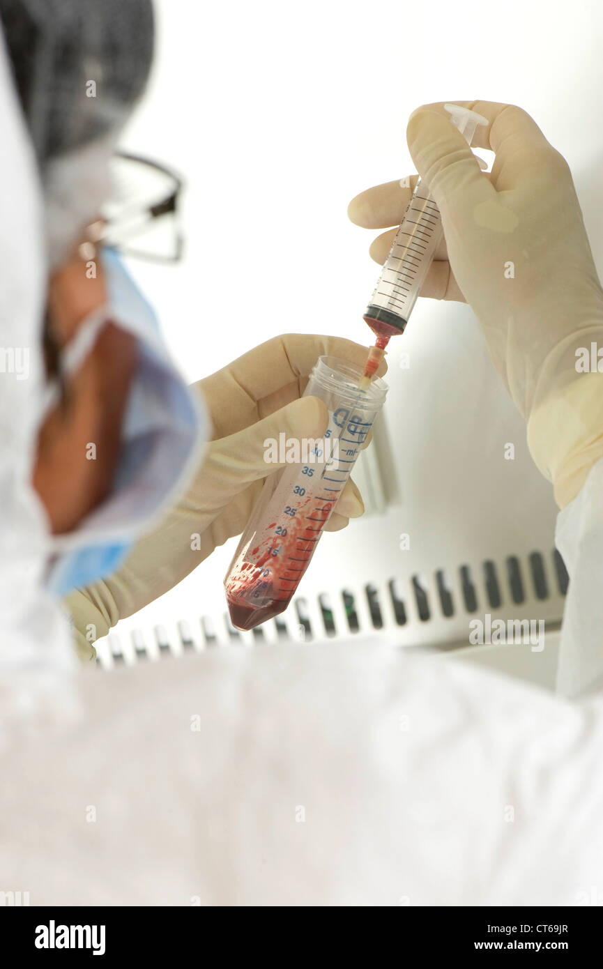 CORD BLOOD ANALYSIS Stock Photohttps://www.alamy.com/image-license-details/?v=1https://www.alamy.com/stock-photo-cord-blood-analysis-49311775.html
RMCT69JR–CORD BLOOD ANALYSIS
 . Carnegie Institution of Washington publication. ON THE FATE OF THE HUMAN EMBRYO IN TUBAL PREGNANCY. 71 No. 338c. (Prof. C. S. Minot, Boston, Mass.) (Plate 6, fig. 4.) Distorted embryo of fifth week. Normal. No. 342. (Professor Minot, Boston, Mass.) (Plate 5, fig. 2.) Pathological ovum, 30X20X20 mm.; pedicle within, 5X1 mm. The specimen has a very thin fibrous chorion, with traces of blood-vessels, practically without villi. Within is a thickened fibrous amnion, to which the process, the umbilical cord, is attached. The cord is also fibrous, contains remnants of its blood-vessels, and has att Stock Photohttps://www.alamy.com/image-license-details/?v=1https://www.alamy.com/carnegie-institution-of-washington-publication-on-the-fate-of-the-human-embryo-in-tubal-pregnancy-71-no-338c-prof-c-s-minot-boston-mass-plate-6-fig-4-distorted-embryo-of-fifth-week-normal-no-342-professor-minot-boston-mass-plate-5-fig-2-pathological-ovum-30x20x20-mm-pedicle-within-5x1-mm-the-specimen-has-a-very-thin-fibrous-chorion-with-traces-of-blood-vessels-practically-without-villi-within-is-a-thickened-fibrous-amnion-to-which-the-process-the-umbilical-cord-is-attached-the-cord-is-also-fibrous-contains-remnants-of-its-blood-vessels-and-has-att-image233475550.html
RMRFRMCE–. Carnegie Institution of Washington publication. ON THE FATE OF THE HUMAN EMBRYO IN TUBAL PREGNANCY. 71 No. 338c. (Prof. C. S. Minot, Boston, Mass.) (Plate 6, fig. 4.) Distorted embryo of fifth week. Normal. No. 342. (Professor Minot, Boston, Mass.) (Plate 5, fig. 2.) Pathological ovum, 30X20X20 mm.; pedicle within, 5X1 mm. The specimen has a very thin fibrous chorion, with traces of blood-vessels, practically without villi. Within is a thickened fibrous amnion, to which the process, the umbilical cord, is attached. The cord is also fibrous, contains remnants of its blood-vessels, and has att
 CORD BLOOD Stock Photohttps://www.alamy.com/image-license-details/?v=1https://www.alamy.com/stock-photo-cord-blood-49311611.html
RMCT69CY–CORD BLOOD
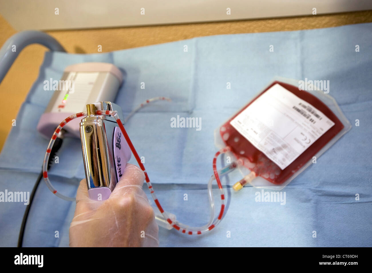 CORD BLOOD Stock Photohttps://www.alamy.com/image-license-details/?v=1https://www.alamy.com/stock-photo-cord-blood-49311629.html
RMCT69DH–CORD BLOOD
 CORD BLOOD Stock Photohttps://www.alamy.com/image-license-details/?v=1https://www.alamy.com/stock-photo-cord-blood-49311620.html
RMCT69D8–CORD BLOOD
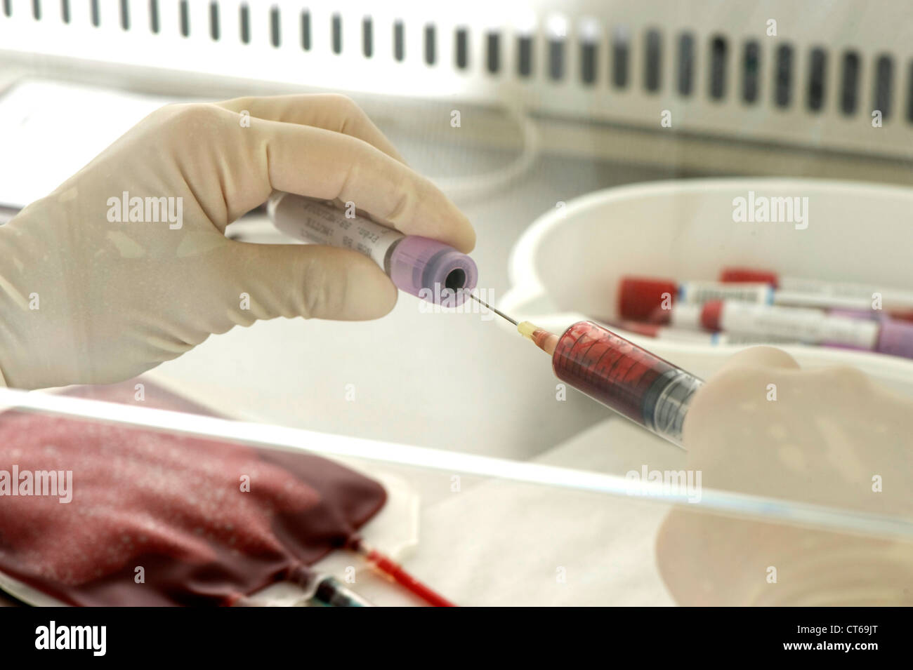 CORD BLOOD ANALYSIS Stock Photohttps://www.alamy.com/image-license-details/?v=1https://www.alamy.com/stock-photo-cord-blood-analysis-49311776.html
RMCT69JT–CORD BLOOD ANALYSIS
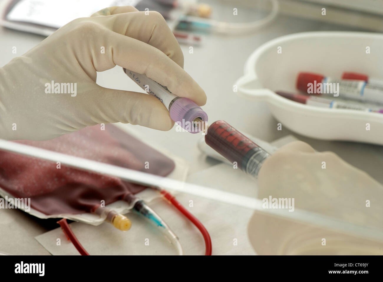 CORD BLOOD ANALYSIS Stock Photohttps://www.alamy.com/image-license-details/?v=1https://www.alamy.com/stock-photo-cord-blood-analysis-49311779.html
RMCT69JY–CORD BLOOD ANALYSIS
 CORD BLOOD ANALYSIS Stock Photohttps://www.alamy.com/image-license-details/?v=1https://www.alamy.com/stock-photo-cord-blood-analysis-49311781.html
RMCT69K1–CORD BLOOD ANALYSIS
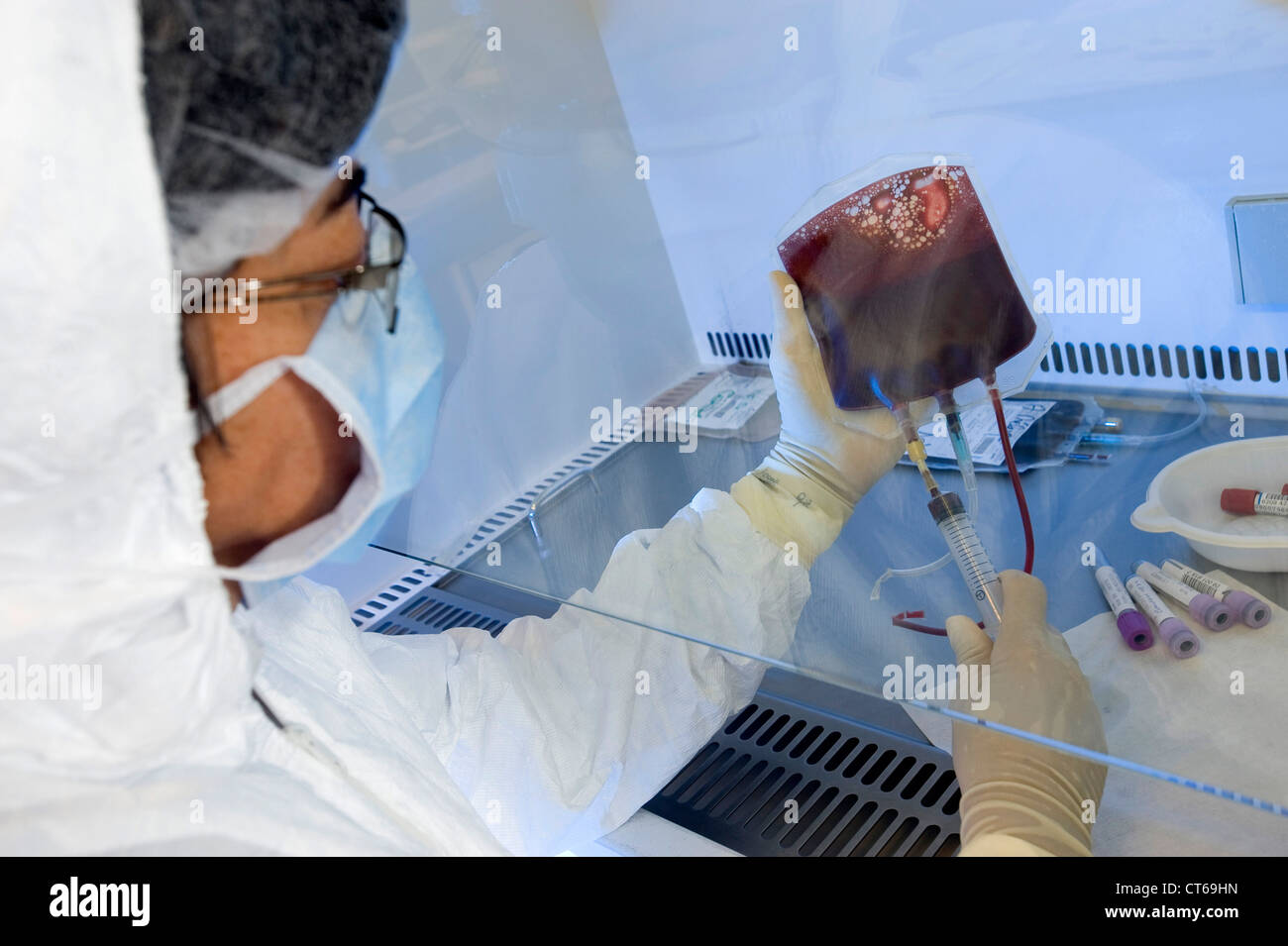 CORD BLOOD ANALYSIS Stock Photohttps://www.alamy.com/image-license-details/?v=1https://www.alamy.com/stock-photo-cord-blood-analysis-49311745.html
RMCT69HN–CORD BLOOD ANALYSIS
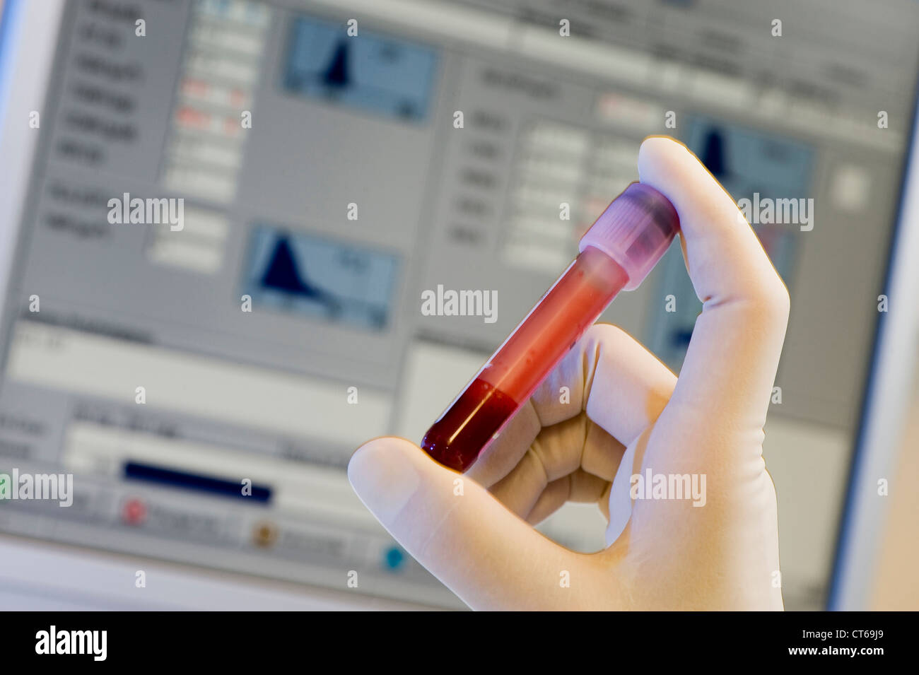 CORD BLOOD ANALYSIS Stock Photohttps://www.alamy.com/image-license-details/?v=1https://www.alamy.com/stock-photo-cord-blood-analysis-49311761.html
RMCT69J9–CORD BLOOD ANALYSIS
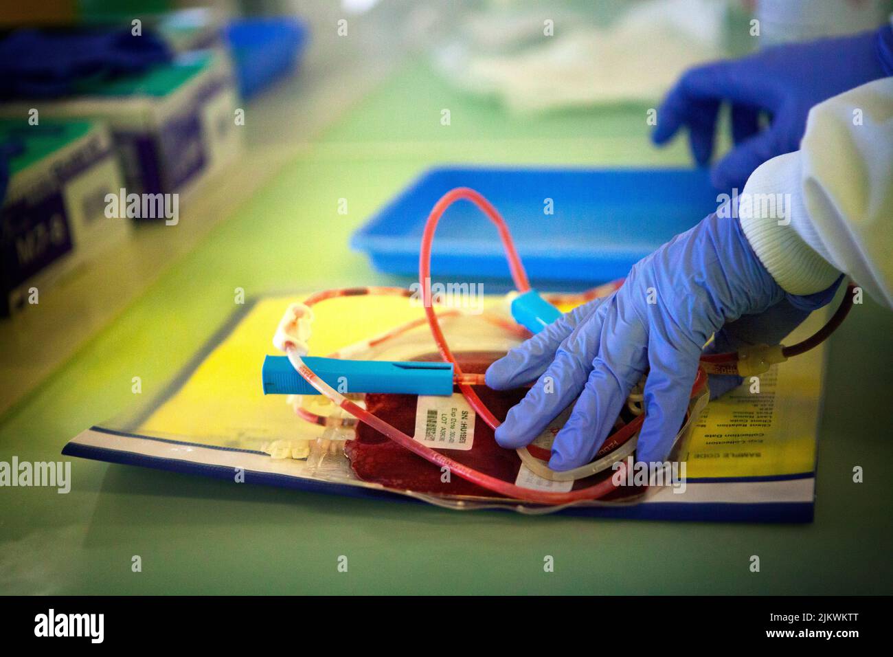 Biobank storing stem cells from blood and cord tissue. Stock Photohttps://www.alamy.com/image-license-details/?v=1https://www.alamy.com/biobank-storing-stem-cells-from-blood-and-cord-tissue-image476922792.html
RF2JKWKTT–Biobank storing stem cells from blood and cord tissue.
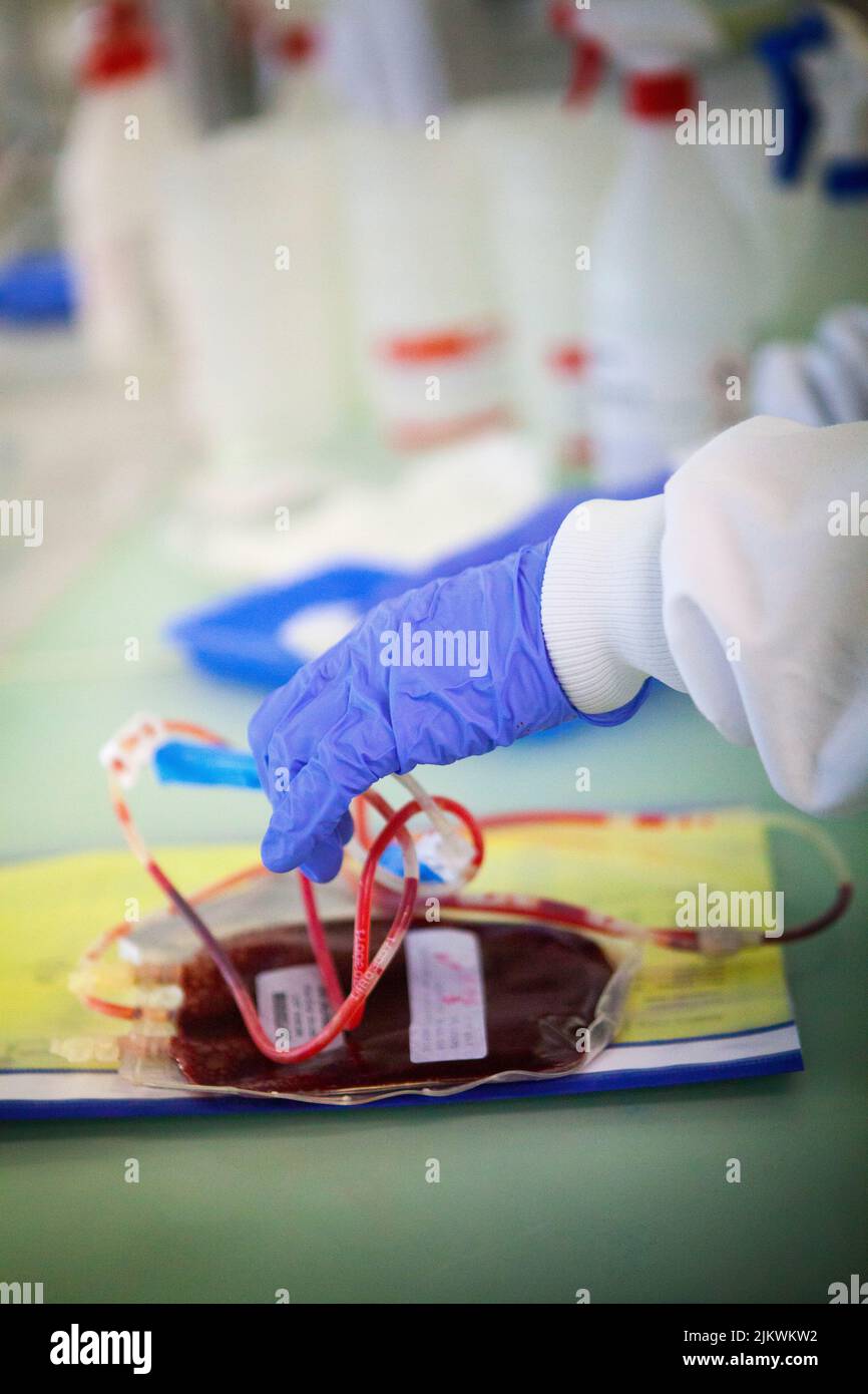 Biobank storing stem cells from blood and cord tissue. Stock Photohttps://www.alamy.com/image-license-details/?v=1https://www.alamy.com/biobank-storing-stem-cells-from-blood-and-cord-tissue-image476922798.html
RF2JKWKW2–Biobank storing stem cells from blood and cord tissue.
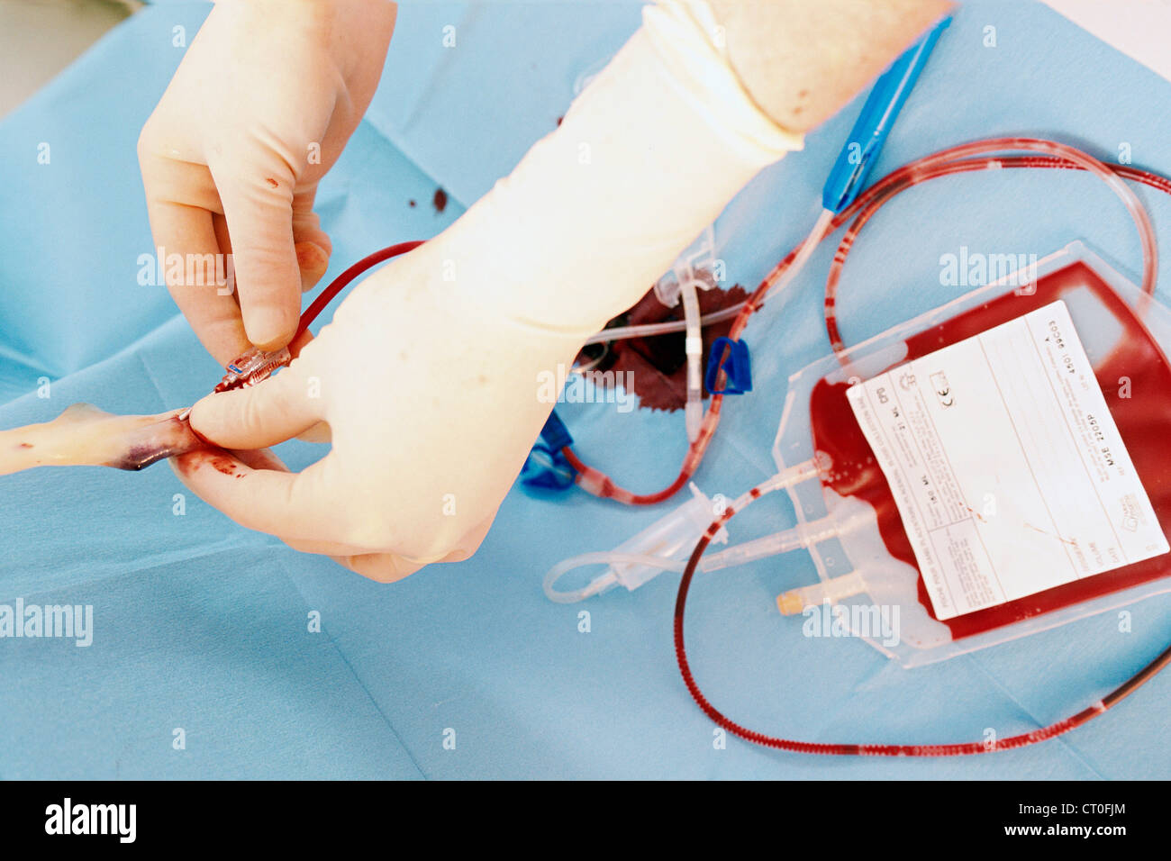 CORD BLOOD SPECIMEN Stock Photohttps://www.alamy.com/image-license-details/?v=1https://www.alamy.com/stock-photo-cord-blood-specimen-49184764.html
RMCT0FJM–CORD BLOOD SPECIMEN
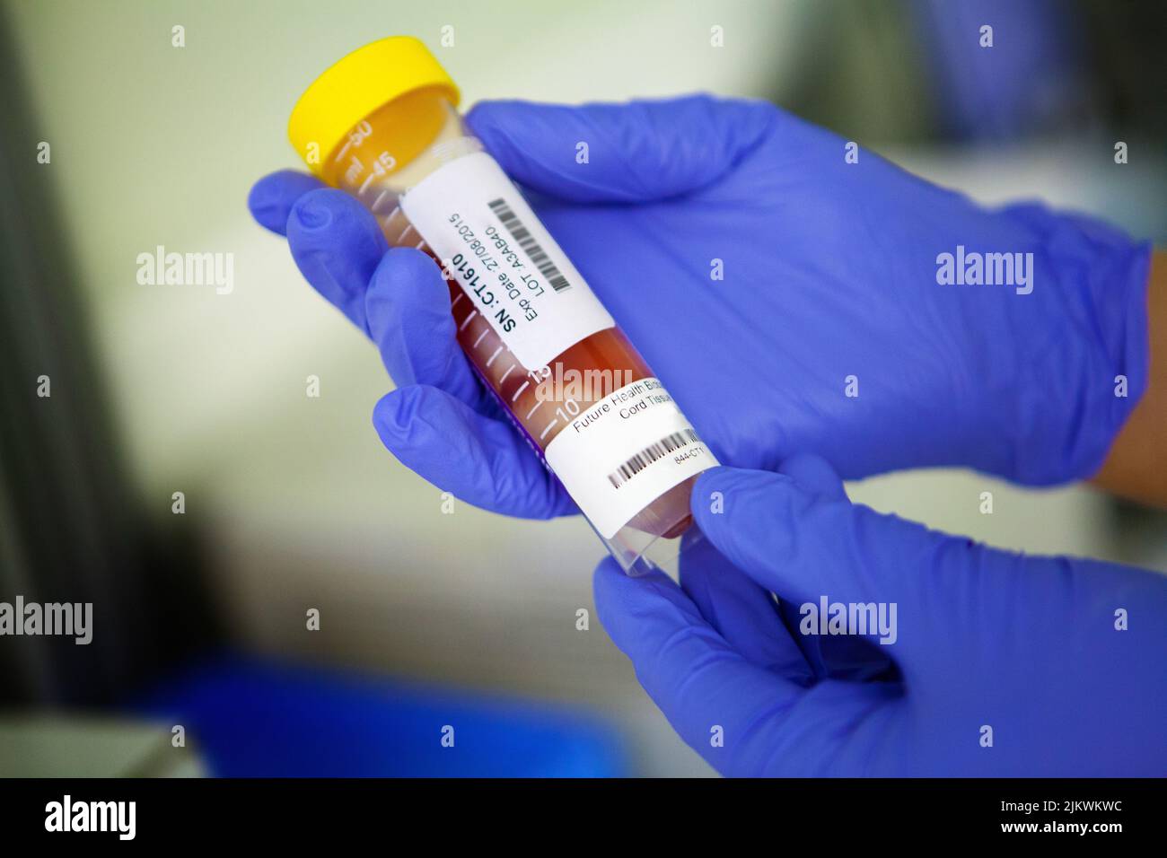 Biobank storing stem cells from blood and cord tissue. Stock Photohttps://www.alamy.com/image-license-details/?v=1https://www.alamy.com/biobank-storing-stem-cells-from-blood-and-cord-tissue-image476922808.html
RF2JKWKWC–Biobank storing stem cells from blood and cord tissue.
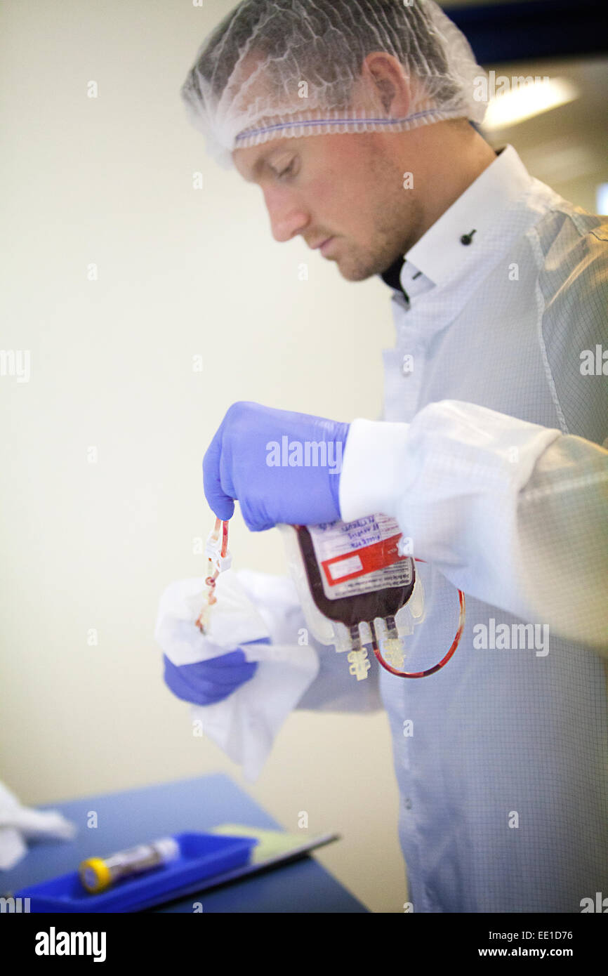 STEM CELL BIOBANK Stock Photohttps://www.alamy.com/image-license-details/?v=1https://www.alamy.com/stock-photo-stem-cell-biobank-77479002.html
RMEE1D76–STEM CELL BIOBANK
 STEM CELL BIOBANK Stock Photohttps://www.alamy.com/image-license-details/?v=1https://www.alamy.com/stock-photo-stem-cell-biobank-77478961.html
RMEE1D5N–STEM CELL BIOBANK
 STEM CELL BIOBANK Stock Photohttps://www.alamy.com/image-license-details/?v=1https://www.alamy.com/stock-photo-stem-cell-biobank-77478969.html
RMEE1D61–STEM CELL BIOBANK

