Cork tissue cells Black & White Stock Photos
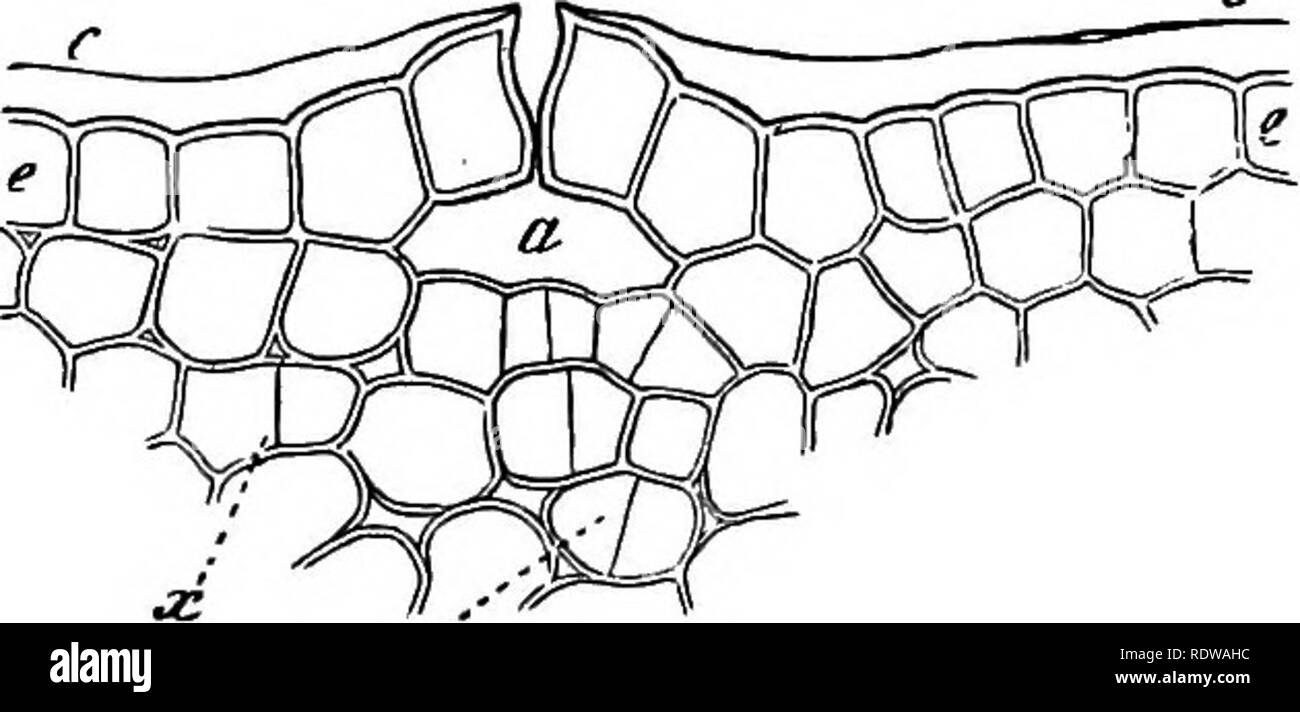
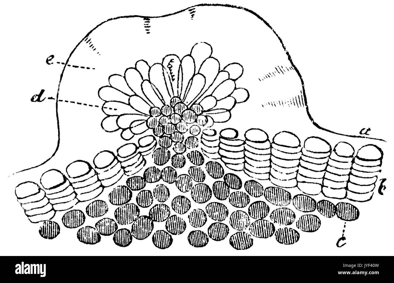 resin gland of a young birch branch in cross section. a) epidermis cells; b) situated just under the cell layer of cork; c) bark tissue; d) the papillae of the gland, which excreted the resin Stock Photohttps://www.alamy.com/image-license-details/?v=1https://www.alamy.com/resin-gland-of-a-young-birch-branch-in-cross-section-a-epidermis-cells-image154611097.html
RMJYF40W–resin gland of a young birch branch in cross section. a) epidermis cells; b) situated just under the cell layer of cork; c) bark tissue; d) the papillae of the gland, which excreted the resin
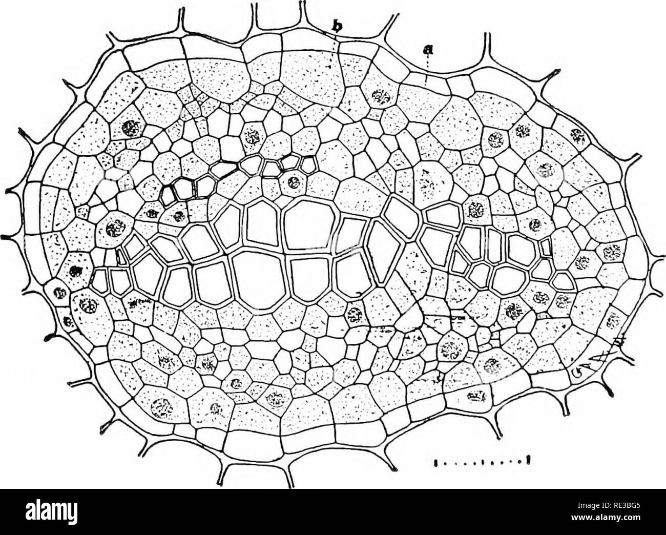 . Elementary botany. Botany. 36o RELA TION TO ENVIKONAfENT. (fig. 417). Surrounding the plerome and tilling the space between it and the dermatogen is the third formative tissue called the periblem, which later forms the cortex (bark or rind), and consists of parenchyma, collenchyma, sclerenchyma, or cork, etc., as the case may be. It should be understood that all these different forms and kinds of cells have been derived from meristem by gradual change. In the mature stems, therefore, there are three distinct regions, the central cylinder or stele, the corte.x, and the epidermis. 710. Central Stock Photohttps://www.alamy.com/image-license-details/?v=1https://www.alamy.com/elementary-botany-botany-36o-rela-tion-to-envikonafent-fig-417-surrounding-the-plerome-and-tilling-the-space-between-it-and-the-dermatogen-is-the-third-formative-tissue-called-the-periblem-which-later-forms-the-cortex-bark-or-rind-and-consists-of-parenchyma-collenchyma-sclerenchyma-or-cork-etc-as-the-case-may-be-it-should-be-understood-that-all-these-different-forms-and-kinds-of-cells-have-been-derived-from-meristem-by-gradual-change-in-the-mature-stems-therefore-there-are-three-distinct-regions-the-central-cylinder-or-stele-the-cortex-and-the-epidermis-710-central-image232414901.html
RMRE3BG5–. Elementary botany. Botany. 36o RELA TION TO ENVIKONAfENT. (fig. 417). Surrounding the plerome and tilling the space between it and the dermatogen is the third formative tissue called the periblem, which later forms the cortex (bark or rind), and consists of parenchyma, collenchyma, sclerenchyma, or cork, etc., as the case may be. It should be understood that all these different forms and kinds of cells have been derived from meristem by gradual change. In the mature stems, therefore, there are three distinct regions, the central cylinder or stele, the corte.x, and the epidermis. 710. Central
 resin gland of a young birch branch in cross section. a) epidermis cells; b) situated just under the cell layer of cork; c) bark tissue; d) the papillae of the gland, which excreted the resin, Betula pendula (syn. Betula verrucosa, Betula alba), anonym (botany book, 1875), Harzdrüse eines jungen Birkenzweigs im Querschnitt. a) Oberhautzellen; b) eine unter der Oberhautzelle gelegene Korkschicht; c) kollenchymartiges Rindegewebe; d) die Papillen der Drüse, welche das feste Harz e) ausgeschieden haben, bouleau verruqueux ou bouleau blanc, glande résine Stock Photohttps://www.alamy.com/image-license-details/?v=1https://www.alamy.com/resin-gland-of-a-young-birch-branch-in-cross-section-a-epidermis-cells-b-situated-just-under-the-cell-layer-of-cork-c-bark-tissue-d-the-papillae-of-the-gland-which-excreted-the-resin-betula-pendula-syn-betula-verrucosa-betula-alba-anonym-botany-book-1875-harzdrse-eines-jungen-birkenzweigs-im-querschnitt-a-oberhautzellen-b-eine-unter-der-oberhautzelle-gelegene-korkschicht-c-kollenchymartiges-rindegewebe-d-die-papillen-der-drse-welche-das-feste-harz-e-ausgeschieden-haben-bouleau-verruqueux-ou-bouleau-blanc-glande-rsine-image629011616.html
RM2YF9XMG–resin gland of a young birch branch in cross section. a) epidermis cells; b) situated just under the cell layer of cork; c) bark tissue; d) the papillae of the gland, which excreted the resin, Betula pendula (syn. Betula verrucosa, Betula alba), anonym (botany book, 1875), Harzdrüse eines jungen Birkenzweigs im Querschnitt. a) Oberhautzellen; b) eine unter der Oberhautzelle gelegene Korkschicht; c) kollenchymartiges Rindegewebe; d) die Papillen der Drüse, welche das feste Harz e) ausgeschieden haben, bouleau verruqueux ou bouleau blanc, glande résine
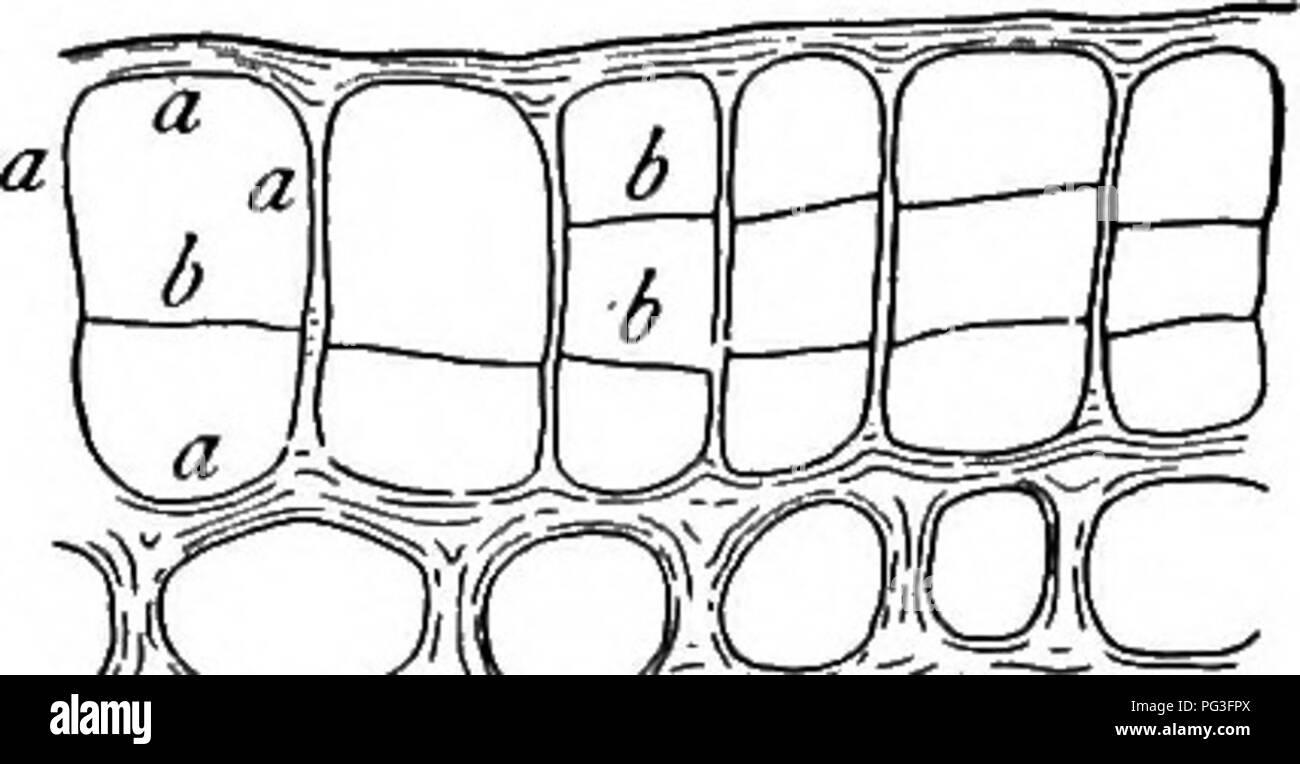 . A textbook of botany for colleges and universities ... Botany. STEMS 705. mechanical elements (largely bast fibers). The development of the cork cylinder usually occasions the death of all cells external to it, since it checks the move- ment of material from within. Cork. — Structural features. — The most important protective tissue of the bark is the cork, which is developed from a meristematic layer known as the phellogen or cork cambium. Occa- sionally this layer arises from the epi- dermis, as in some Rosaceae and in many herbs (fig. 1031), but much more commonly the phellogen layer aris Stock Photohttps://www.alamy.com/image-license-details/?v=1https://www.alamy.com/a-textbook-of-botany-for-colleges-and-universities-botany-stems-705-mechanical-elements-largely-bast-fibers-the-development-of-the-cork-cylinder-usually-occasions-the-death-of-all-cells-external-to-it-since-it-checks-the-move-ment-of-material-from-within-cork-structural-features-the-most-important-protective-tissue-of-the-bark-is-the-cork-which-is-developed-from-a-meristematic-layer-known-as-the-phellogen-or-cork-cambium-occa-sionally-this-layer-arises-from-the-epi-dermis-as-in-some-rosaceae-and-in-many-herbs-fig-1031-but-much-more-commonly-the-phellogen-layer-aris-image216437170.html
RMPG3FPX–. A textbook of botany for colleges and universities ... Botany. STEMS 705. mechanical elements (largely bast fibers). The development of the cork cylinder usually occasions the death of all cells external to it, since it checks the move- ment of material from within. Cork. — Structural features. — The most important protective tissue of the bark is the cork, which is developed from a meristematic layer known as the phellogen or cork cambium. Occa- sionally this layer arises from the epi- dermis, as in some Rosaceae and in many herbs (fig. 1031), but much more commonly the phellogen layer aris
 . The elements of vegetable histology. Plant anatomy. D 0. Plate 27.—Common Errors in Sketching. 1. The cell walls of these parenchyma cells should be continuous and should be brought back to the starting point. 2. The cells in this fragment of tissue should be surrounded by a cell wall of definite thickness and the space between the cells is much exaggerated. 3. The parts of this aggregate crystal (rosette type) are sharp pointed and not rounded as illustrated. 4. The sides of this crystal should be straight lines as acicular crystals are never curved. 5. These cork cells are too symmetrical Stock Photohttps://www.alamy.com/image-license-details/?v=1https://www.alamy.com/the-elements-of-vegetable-histology-plant-anatomy-d-0-plate-27common-errors-in-sketching-1-the-cell-walls-of-these-parenchyma-cells-should-be-continuous-and-should-be-brought-back-to-the-starting-point-2-the-cells-in-this-fragment-of-tissue-should-be-surrounded-by-a-cell-wall-of-definite-thickness-and-the-space-between-the-cells-is-much-exaggerated-3-the-parts-of-this-aggregate-crystal-rosette-type-are-sharp-pointed-and-not-rounded-as-illustrated-4-the-sides-of-this-crystal-should-be-straight-lines-as-acicular-crystals-are-never-curved-5-these-cork-cells-are-too-symmetrical-image232288893.html
RMRDWJRW–. The elements of vegetable histology. Plant anatomy. D 0. Plate 27.—Common Errors in Sketching. 1. The cell walls of these parenchyma cells should be continuous and should be brought back to the starting point. 2. The cells in this fragment of tissue should be surrounded by a cell wall of definite thickness and the space between the cells is much exaggerated. 3. The parts of this aggregate crystal (rosette type) are sharp pointed and not rounded as illustrated. 4. The sides of this crystal should be straight lines as acicular crystals are never curved. 5. These cork cells are too symmetrical
 . A textbook of botany for colleges and universities ... Botany. mechanical elements (largely bast fibers). The development of the cork cylinder usually occasions the death of all cells external to it, since it checks the move- ment of material from within. Cork. — Structural features. — The most important protective tissue of the bark is the cork, which is developed from a meristematic layer known as the phellogen or cork cambium. Occa- sionally this layer arises from the epi- dermis, as in some Rosaceae and in many herbs (fig. 1031), but much more commonly the phellogen layer arises in the p Stock Photohttps://www.alamy.com/image-license-details/?v=1https://www.alamy.com/a-textbook-of-botany-for-colleges-and-universities-botany-mechanical-elements-largely-bast-fibers-the-development-of-the-cork-cylinder-usually-occasions-the-death-of-all-cells-external-to-it-since-it-checks-the-move-ment-of-material-from-within-cork-structural-features-the-most-important-protective-tissue-of-the-bark-is-the-cork-which-is-developed-from-a-meristematic-layer-known-as-the-phellogen-or-cork-cambium-occa-sionally-this-layer-arises-from-the-epi-dermis-as-in-some-rosaceae-and-in-many-herbs-fig-1031-but-much-more-commonly-the-phellogen-layer-arises-in-the-p-image216437165.html
RMPG3FPN–. A textbook of botany for colleges and universities ... Botany. mechanical elements (largely bast fibers). The development of the cork cylinder usually occasions the death of all cells external to it, since it checks the move- ment of material from within. Cork. — Structural features. — The most important protective tissue of the bark is the cork, which is developed from a meristematic layer known as the phellogen or cork cambium. Occa- sionally this layer arises from the epi- dermis, as in some Rosaceae and in many herbs (fig. 1031), but much more commonly the phellogen layer arises in the p
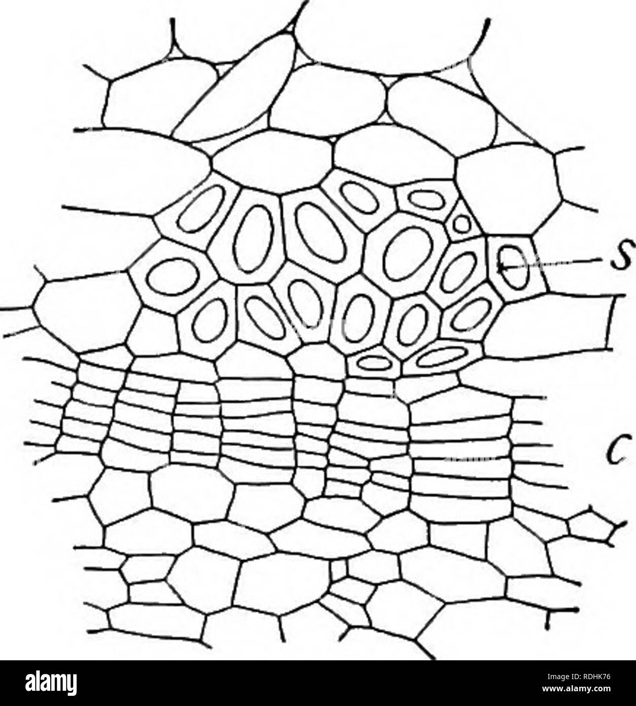 . A textbook of botany for colleges and universities ... Botany. mechanical elements (largely bast fibers). The development of the cork cylinder usually occasions the death of all cells external to it, since it checks the move- ment of material from within. Cork. — Structural features. — The most important protective tissue of the bark is the cork, which is developed from a meristematic layer known as the phellogen or cork cambium. Occa- sionally this layer arises from the epi- dermis, as in some Rosaceae and in many herbs (fig. 1031), but much more commonly the phellogen layer arises in the p Stock Photohttps://www.alamy.com/image-license-details/?v=1https://www.alamy.com/a-textbook-of-botany-for-colleges-and-universities-botany-mechanical-elements-largely-bast-fibers-the-development-of-the-cork-cylinder-usually-occasions-the-death-of-all-cells-external-to-it-since-it-checks-the-move-ment-of-material-from-within-cork-structural-features-the-most-important-protective-tissue-of-the-bark-is-the-cork-which-is-developed-from-a-meristematic-layer-known-as-the-phellogen-or-cork-cambium-occa-sionally-this-layer-arises-from-the-epi-dermis-as-in-some-rosaceae-and-in-many-herbs-fig-1031-but-much-more-commonly-the-phellogen-layer-arises-in-the-p-image232113594.html
RMRDHK76–. A textbook of botany for colleges and universities ... Botany. mechanical elements (largely bast fibers). The development of the cork cylinder usually occasions the death of all cells external to it, since it checks the move- ment of material from within. Cork. — Structural features. — The most important protective tissue of the bark is the cork, which is developed from a meristematic layer known as the phellogen or cork cambium. Occa- sionally this layer arises from the epi- dermis, as in some Rosaceae and in many herbs (fig. 1031), but much more commonly the phellogen layer arises in the p
 . Botany for agricultural students . Botany. much thicker and more protective than an epidermis. (Fig. 113.) The cork covering may be more or less flexible, as the rind of an Irish Potato or Sweet Potato, or harder and more brittle, as in the bark of trees, where it reaches its extreme thickness. Cork tissue con- sists of dead cells in the walls of which there is deposited a waxy substance much like cutin but called suberin to which much of the pro- tective character of cork is due. Cork coverings afford more protec- tion than an epidermis, but on ac- count of their opaqueness, they are not su Stock Photohttps://www.alamy.com/image-license-details/?v=1https://www.alamy.com/botany-for-agricultural-students-botany-much-thicker-and-more-protective-than-an-epidermis-fig-113-the-cork-covering-may-be-more-or-less-flexible-as-the-rind-of-an-irish-potato-or-sweet-potato-or-harder-and-more-brittle-as-in-the-bark-of-trees-where-it-reaches-its-extreme-thickness-cork-tissue-con-sists-of-dead-cells-in-the-walls-of-which-there-is-deposited-a-waxy-substance-much-like-cutin-but-called-suberin-to-which-much-of-the-pro-tective-character-of-cork-is-due-cork-coverings-afford-more-protec-tion-than-an-epidermis-but-on-ac-count-of-their-opaqueness-they-are-not-su-image216449117.html
RMPG431H–. Botany for agricultural students . Botany. much thicker and more protective than an epidermis. (Fig. 113.) The cork covering may be more or less flexible, as the rind of an Irish Potato or Sweet Potato, or harder and more brittle, as in the bark of trees, where it reaches its extreme thickness. Cork tissue con- sists of dead cells in the walls of which there is deposited a waxy substance much like cutin but called suberin to which much of the pro- tective character of cork is due. Cork coverings afford more protec- tion than an epidermis, but on ac- count of their opaqueness, they are not su
 . Botany, with agricultural applications. Botany. much thicker and more protective than an epidermis. (Fig. 113.) The cork covering may be more or less flexible, as the rind of an Irish Potato or Sweet Potato, or harder and more brittle, as in the bark of trees, where it reaches its extreme thickness. Cork tissue con- sists of dead cells in the walls of which there is deposited a waxy- substance much-Hke cutin but called suherin to which much of the pro- tective character of cork is due. Cork coverings afford more protec- tion than an epidermis, but on ac- count of their opaqueness, they are n Stock Photohttps://www.alamy.com/image-license-details/?v=1https://www.alamy.com/botany-with-agricultural-applications-botany-much-thicker-and-more-protective-than-an-epidermis-fig-113-the-cork-covering-may-be-more-or-less-flexible-as-the-rind-of-an-irish-potato-or-sweet-potato-or-harder-and-more-brittle-as-in-the-bark-of-trees-where-it-reaches-its-extreme-thickness-cork-tissue-con-sists-of-dead-cells-in-the-walls-of-which-there-is-deposited-a-waxy-substance-much-hke-cutin-but-called-suherin-to-which-much-of-the-pro-tective-character-of-cork-is-due-cork-coverings-afford-more-protec-tion-than-an-epidermis-but-on-ac-count-of-their-opaqueness-they-are-n-image232285642.html
RMRDWEKP–. Botany, with agricultural applications. Botany. much thicker and more protective than an epidermis. (Fig. 113.) The cork covering may be more or less flexible, as the rind of an Irish Potato or Sweet Potato, or harder and more brittle, as in the bark of trees, where it reaches its extreme thickness. Cork tissue con- sists of dead cells in the walls of which there is deposited a waxy- substance much-Hke cutin but called suherin to which much of the pro- tective character of cork is due. Cork coverings afford more protec- tion than an epidermis, but on ac- count of their opaqueness, they are n
 . Physiological botany; I. Outlines of the histology of phænogamous plants. II. Vegetable physiology. Plant physiology; Plant anatomy. EPIDERMIS. 65 sooner or later thrown off, and replaced by a subjacent protective tissue, — cork. 218. Except at peculiar openings (stomata, etc.), the epider- mal cells are in close apposition. Upon their exposed surface the}' are cutinized, and thus a continuous hyaline film is formed, known as the Cuticle?- 219. Sometimes the epidermis may be torn off without much disturbing the underlying tissues. 220. Besides the cells which compose the proper tissue of the Stock Photohttps://www.alamy.com/image-license-details/?v=1https://www.alamy.com/physiological-botany-i-outlines-of-the-histology-of-phnogamous-plants-ii-vegetable-physiology-plant-physiology-plant-anatomy-epidermis-65-sooner-or-later-thrown-off-and-replaced-by-a-subjacent-protective-tissue-cork-218-except-at-peculiar-openings-stomata-etc-the-epider-mal-cells-are-in-close-apposition-upon-their-exposed-surface-the-are-cutinized-and-thus-a-continuous-hyaline-film-is-formed-known-as-the-cuticle-219-sometimes-the-epidermis-may-be-torn-off-without-much-disturbing-the-underlying-tissues-220-besides-the-cells-which-compose-the-proper-tissue-of-the-image216445729.html
RMPG3XMH–. Physiological botany; I. Outlines of the histology of phænogamous plants. II. Vegetable physiology. Plant physiology; Plant anatomy. EPIDERMIS. 65 sooner or later thrown off, and replaced by a subjacent protective tissue, — cork. 218. Except at peculiar openings (stomata, etc.), the epider- mal cells are in close apposition. Upon their exposed surface the}' are cutinized, and thus a continuous hyaline film is formed, known as the Cuticle?- 219. Sometimes the epidermis may be torn off without much disturbing the underlying tissues. 220. Besides the cells which compose the proper tissue of the
 . A textbook of botany for colleges and universities ... Botany. STEMS 705. mechanical elements (largely bast fibers). The development of the cork cylinder usually occasions the death of all cells external to it, since it checks the move- ment of material from within. Cork. — Structural features. — The most important protective tissue of the bark is the cork, which is developed from a meristematic layer known as the phellogen or cork cambium. Occa- sionally this layer arises from the epi- dermis, as in some Rosaceae and in many herbs (fig. 1031), but much more commonly the phellogen layer aris Stock Photohttps://www.alamy.com/image-license-details/?v=1https://www.alamy.com/a-textbook-of-botany-for-colleges-and-universities-botany-stems-705-mechanical-elements-largely-bast-fibers-the-development-of-the-cork-cylinder-usually-occasions-the-death-of-all-cells-external-to-it-since-it-checks-the-move-ment-of-material-from-within-cork-structural-features-the-most-important-protective-tissue-of-the-bark-is-the-cork-which-is-developed-from-a-meristematic-layer-known-as-the-phellogen-or-cork-cambium-occa-sionally-this-layer-arises-from-the-epi-dermis-as-in-some-rosaceae-and-in-many-herbs-fig-1031-but-much-more-commonly-the-phellogen-layer-aris-image232113599.html
RMRDHK7B–. A textbook of botany for colleges and universities ... Botany. STEMS 705. mechanical elements (largely bast fibers). The development of the cork cylinder usually occasions the death of all cells external to it, since it checks the move- ment of material from within. Cork. — Structural features. — The most important protective tissue of the bark is the cork, which is developed from a meristematic layer known as the phellogen or cork cambium. Occa- sionally this layer arises from the epi- dermis, as in some Rosaceae and in many herbs (fig. 1031), but much more commonly the phellogen layer aris
 . Botany for agricultural students . Botany. 128 CELLS AND TISSUES. much thicker and more protective than an epidermis. (Fig. 113.) The cork covering may be more or less flexible, as the rind of an Irish Potato or Sweet Potato, or harder and more brittle, as in the bark of trees, where it reaches its extreme thickness. Cork tissue con- sists of dead cells in the walls of which there is deposited a waxy substance much like cutin but called suberin to which much of the pro- tective character of cork is due. Cork coverings afford more protec- tion than an epidermis, but on ac- count of their opaq Stock Photohttps://www.alamy.com/image-license-details/?v=1https://www.alamy.com/botany-for-agricultural-students-botany-128-cells-and-tissues-much-thicker-and-more-protective-than-an-epidermis-fig-113-the-cork-covering-may-be-more-or-less-flexible-as-the-rind-of-an-irish-potato-or-sweet-potato-or-harder-and-more-brittle-as-in-the-bark-of-trees-where-it-reaches-its-extreme-thickness-cork-tissue-con-sists-of-dead-cells-in-the-walls-of-which-there-is-deposited-a-waxy-substance-much-like-cutin-but-called-suberin-to-which-much-of-the-pro-tective-character-of-cork-is-due-cork-coverings-afford-more-protec-tion-than-an-epidermis-but-on-ac-count-of-their-opaq-image216449121.html
RMPG431N–. Botany for agricultural students . Botany. 128 CELLS AND TISSUES. much thicker and more protective than an epidermis. (Fig. 113.) The cork covering may be more or less flexible, as the rind of an Irish Potato or Sweet Potato, or harder and more brittle, as in the bark of trees, where it reaches its extreme thickness. Cork tissue con- sists of dead cells in the walls of which there is deposited a waxy substance much like cutin but called suberin to which much of the pro- tective character of cork is due. Cork coverings afford more protec- tion than an epidermis, but on ac- count of their opaq
 . Botany for agricultural students . Botany. 128 CELLS AND TISSUES. much thicker and more protective than an epidermis. (Fig. 113.) The cork covering may be more or less flexible, as the rind of an Irish Potato or Sweet Potato, or harder and more brittle, as in the bark of trees, where it reaches its extreme thickness. Cork tissue con- sists of dead cells in the walls of which there is deposited a waxy substance much like cutin but called suberin to which much of the pro- tective character of cork is due. Cork coverings afford more protec- tion than an epidermis, but on ac- count of their opaq Stock Photohttps://www.alamy.com/image-license-details/?v=1https://www.alamy.com/botany-for-agricultural-students-botany-128-cells-and-tissues-much-thicker-and-more-protective-than-an-epidermis-fig-113-the-cork-covering-may-be-more-or-less-flexible-as-the-rind-of-an-irish-potato-or-sweet-potato-or-harder-and-more-brittle-as-in-the-bark-of-trees-where-it-reaches-its-extreme-thickness-cork-tissue-con-sists-of-dead-cells-in-the-walls-of-which-there-is-deposited-a-waxy-substance-much-like-cutin-but-called-suberin-to-which-much-of-the-pro-tective-character-of-cork-is-due-cork-coverings-afford-more-protec-tion-than-an-epidermis-but-on-ac-count-of-their-opaq-image232031103.html
RMRDDX13–. Botany for agricultural students . Botany. 128 CELLS AND TISSUES. much thicker and more protective than an epidermis. (Fig. 113.) The cork covering may be more or less flexible, as the rind of an Irish Potato or Sweet Potato, or harder and more brittle, as in the bark of trees, where it reaches its extreme thickness. Cork tissue con- sists of dead cells in the walls of which there is deposited a waxy substance much like cutin but called suberin to which much of the pro- tective character of cork is due. Cork coverings afford more protec- tion than an epidermis, but on ac- count of their opaq
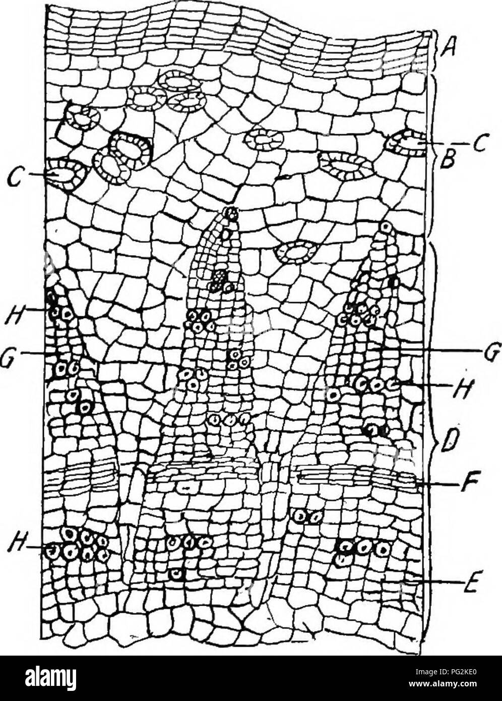 . Pharmaceutical botany. Botany; Botany, Medical. PLANT HAIES 29 Periderm.—-Periderm is a name applied to all the tissue, produced externally by the cork cambium (phellogen). This term appears often in pharmacognic and materia medica texts. Histology of Typical Monocotyl Stems (Endogenous).—Passing from exterior toward centre the following structures are seen: 1. Epidermis whose cells are cutinized in their outer walls. 2. Hypodermis, generally collenchymatic. 3. Cortex. 4. Endodermis or innermost layer of cortex generally with greatly suberized cell walls. 5. A large central zone of parenchym Stock Photohttps://www.alamy.com/image-license-details/?v=1https://www.alamy.com/pharmaceutical-botany-botany-botany-medical-plant-haies-29-periderm-periderm-is-a-name-applied-to-all-the-tissue-produced-externally-by-the-cork-cambium-phellogen-this-term-appears-often-in-pharmacognic-and-materia-medica-texts-histology-of-typical-monocotyl-stems-endogenouspassing-from-exterior-toward-centre-the-following-structures-are-seen-1-epidermis-whose-cells-are-cutinized-in-their-outer-walls-2-hypodermis-generally-collenchymatic-3-cortex-4-endodermis-or-innermost-layer-of-cortex-generally-with-greatly-suberized-cell-walls-5-a-large-central-zone-of-parenchym-image216418104.html
RMPG2KE0–. Pharmaceutical botany. Botany; Botany, Medical. PLANT HAIES 29 Periderm.—-Periderm is a name applied to all the tissue, produced externally by the cork cambium (phellogen). This term appears often in pharmacognic and materia medica texts. Histology of Typical Monocotyl Stems (Endogenous).—Passing from exterior toward centre the following structures are seen: 1. Epidermis whose cells are cutinized in their outer walls. 2. Hypodermis, generally collenchymatic. 3. Cortex. 4. Endodermis or innermost layer of cortex generally with greatly suberized cell walls. 5. A large central zone of parenchym
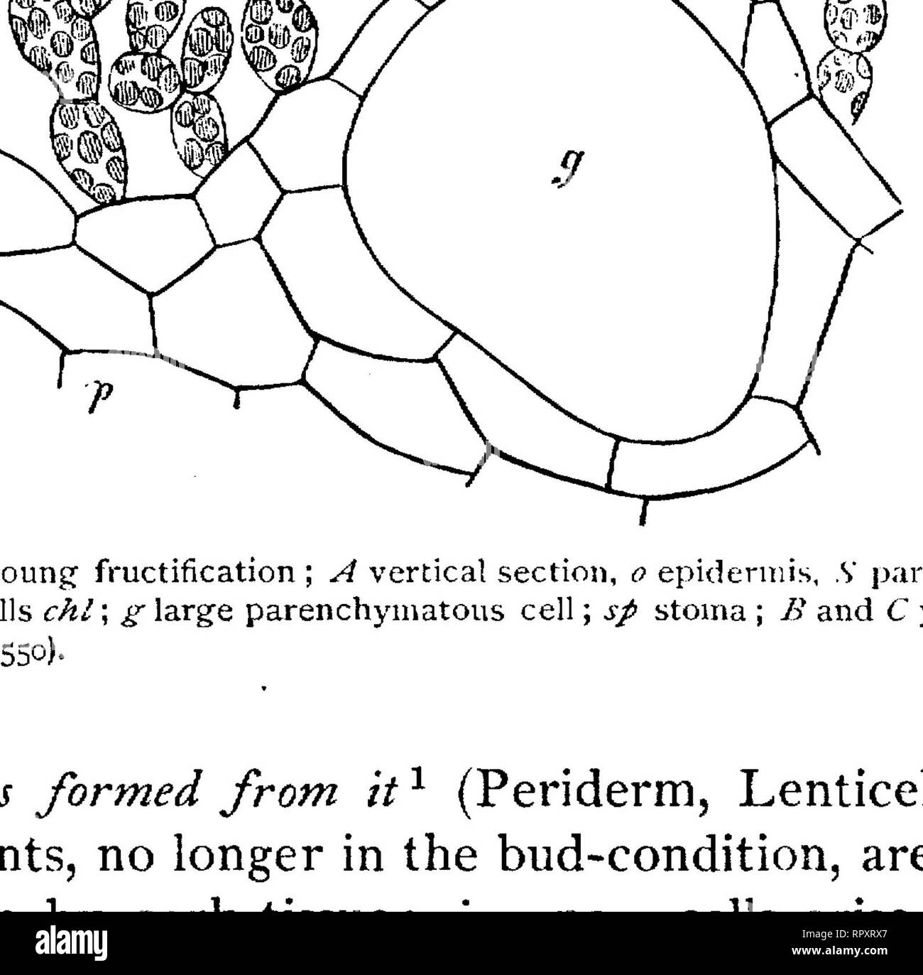 . Text-book of botany, morphological and physiological. Botany. THE EPIDERMAL TISSUE. I07 take place, by which the number of the rows of cells is increased. Of the two newly formed cells of each radial row (/. e. vertical to the surface of the organ) one remains thin-walled, rich in protoplasm, and capable of division; the other becomes suberised and permanent. Thus arises, usually parallel to the surface of the organ, a layer of cells capable of division, which continues to form new cork-cells, the Cork-cambium or layer of Phellogen. In general this is the innermost layer of the whole cork-ti Stock Photohttps://www.alamy.com/image-license-details/?v=1https://www.alamy.com/text-book-of-botany-morphological-and-physiological-botany-the-epidermal-tissue-i07-take-place-by-which-the-number-of-the-rows-of-cells-is-increased-of-the-two-newly-formed-cells-of-each-radial-row-e-vertical-to-the-surface-of-the-organ-one-remains-thin-walled-rich-in-protoplasm-and-capable-of-division-the-other-becomes-suberised-and-permanent-thus-arises-usually-parallel-to-the-surface-of-the-organ-a-layer-of-cells-capable-of-division-which-continues-to-form-new-cork-cells-the-cork-cambium-or-layer-of-phellogen-in-general-this-is-the-innermost-layer-of-the-whole-cork-ti-image237846735.html
RMRPXRX7–. Text-book of botany, morphological and physiological. Botany. THE EPIDERMAL TISSUE. I07 take place, by which the number of the rows of cells is increased. Of the two newly formed cells of each radial row (/. e. vertical to the surface of the organ) one remains thin-walled, rich in protoplasm, and capable of division; the other becomes suberised and permanent. Thus arises, usually parallel to the surface of the organ, a layer of cells capable of division, which continues to form new cork-cells, the Cork-cambium or layer of Phellogen. In general this is the innermost layer of the whole cork-ti
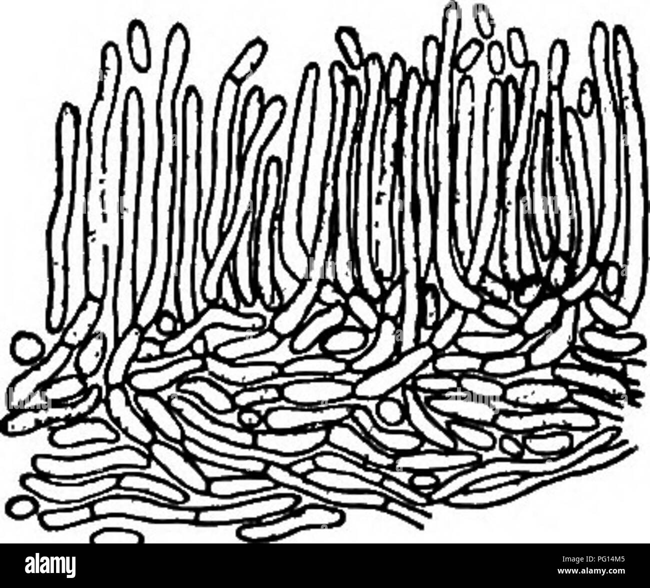 . Chestnut blight. Chestnut blight; Chestnut. Endothia Canker of Chestnut SS9 They are closely crowded together, so that in cross section they appear to make up a pseudoparenchymatous tissue. These cells are more densely filled with protoplasm, and contain more pigment, than the interior cells. Pycnidia On smooth-bark, young cankers, especially in the summer, the outer cork layer is raised in numerous little blisters, with slender, yellow, waxy tendrils curling from their ruptured apices (Plate XXXVIII, Fig. i). Under each blister is a single somewhat globose pycnidiimi, surrounded by a scanty Stock Photohttps://www.alamy.com/image-license-details/?v=1https://www.alamy.com/chestnut-blight-chestnut-blight-chestnut-endothia-canker-of-chestnut-ss9-they-are-closely-crowded-together-so-that-in-cross-section-they-appear-to-make-up-a-pseudoparenchymatous-tissue-these-cells-are-more-densely-filled-with-protoplasm-and-contain-more-pigment-than-the-interior-cells-pycnidia-on-smooth-bark-young-cankers-especially-in-the-summer-the-outer-cork-layer-is-raised-in-numerous-little-blisters-with-slender-yellow-waxy-tendrils-curling-from-their-ruptured-apices-plate-xxxviii-fig-i-under-each-blister-is-a-single-somewhat-globose-pycnidiimi-surrounded-by-a-scanty-image216384565.html
RMPG14M5–. Chestnut blight. Chestnut blight; Chestnut. Endothia Canker of Chestnut SS9 They are closely crowded together, so that in cross section they appear to make up a pseudoparenchymatous tissue. These cells are more densely filled with protoplasm, and contain more pigment, than the interior cells. Pycnidia On smooth-bark, young cankers, especially in the summer, the outer cork layer is raised in numerous little blisters, with slender, yellow, waxy tendrils curling from their ruptured apices (Plate XXXVIII, Fig. i). Under each blister is a single somewhat globose pycnidiimi, surrounded by a scanty
 . A manual of botany. Botany. TISSUE SYSTEMS 341 water or gases. At certain places in both stems and roots special structures are developed to allow of the admission of air to the tissues underlying it. These are lenticeh. In stems they are generally developed under places in the epidermis where stomata are present. Each consists of a little rounded spherical mass of corky cells arranged loosely together. They become exposed to the air by rupture of the epidermis above them. In the autumn a formation of cork takes place under them, by which the communication with the exterior is out off till t Stock Photohttps://www.alamy.com/image-license-details/?v=1https://www.alamy.com/a-manual-of-botany-botany-tissue-systems-341-water-or-gases-at-certain-places-in-both-stems-and-roots-special-structures-are-developed-to-allow-of-the-admission-of-air-to-the-tissues-underlying-it-these-are-lenticeh-in-stems-they-are-generally-developed-under-places-in-the-epidermis-where-stomata-are-present-each-consists-of-a-little-rounded-spherical-mass-of-corky-cells-arranged-loosely-together-they-become-exposed-to-the-air-by-rupture-of-the-epidermis-above-them-in-the-autumn-a-formation-of-cork-takes-place-under-them-by-which-the-communication-with-the-exterior-is-out-off-till-t-image232376212.html
RMRE1J6C–. A manual of botany. Botany. TISSUE SYSTEMS 341 water or gases. At certain places in both stems and roots special structures are developed to allow of the admission of air to the tissues underlying it. These are lenticeh. In stems they are generally developed under places in the epidermis where stomata are present. Each consists of a little rounded spherical mass of corky cells arranged loosely together. They become exposed to the air by rupture of the epidermis above them. In the autumn a formation of cork takes place under them, by which the communication with the exterior is out off till t
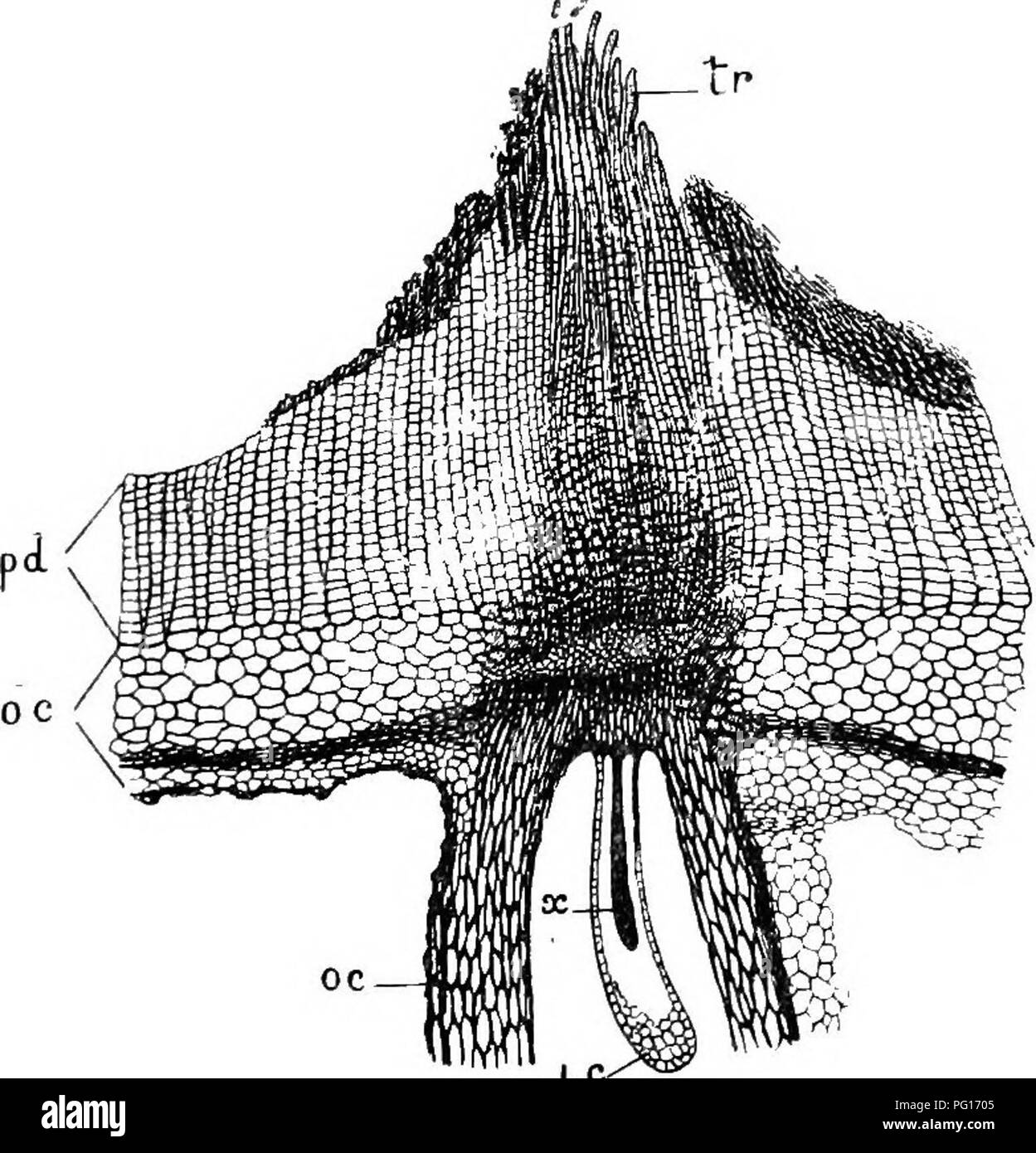 . Studies in fossil botany . Paleobotany. STIGMARIA 247 direction, for it is on the inner edge of the secondary cortical zone that remains of the delicate phellogen can be traced. The tissue thus formed cannot have been of the nature of cork, for the outer tissue shows no sign of withering, and some of its cells had some- times undergone tangential division, as if starting a. Fig. 100.—Stigmariaficoidcs. Part of transverbe section, to show base of a rootlet, /a, periderm of main axis ; o.c, outer cortex (including hypoderma) of main axis and of rootlet; i.c, inner cortex, x, xylem of rootlet ; Stock Photohttps://www.alamy.com/image-license-details/?v=1https://www.alamy.com/studies-in-fossil-botany-paleobotany-stigmaria-247-direction-for-it-is-on-the-inner-edge-of-the-secondary-cortical-zone-that-remains-of-the-delicate-phellogen-can-be-traced-the-tissue-thus-formed-cannot-have-been-of-the-nature-of-cork-for-the-outer-tissue-shows-no-sign-of-withering-and-some-of-its-cells-had-some-times-undergone-tangential-division-as-if-starting-a-fig-100stigmariaficoidcs-part-of-transverbe-section-to-show-base-of-a-rootlet-a-periderm-of-main-axis-oc-outer-cortex-including-hypoderma-of-main-axis-and-of-rootlet-ic-inner-cortex-x-xylem-of-rootlet-image216386357.html
RMPG1705–. Studies in fossil botany . Paleobotany. STIGMARIA 247 direction, for it is on the inner edge of the secondary cortical zone that remains of the delicate phellogen can be traced. The tissue thus formed cannot have been of the nature of cork, for the outer tissue shows no sign of withering, and some of its cells had some- times undergone tangential division, as if starting a. Fig. 100.—Stigmariaficoidcs. Part of transverbe section, to show base of a rootlet, /a, periderm of main axis ; o.c, outer cortex (including hypoderma) of main axis and of rootlet; i.c, inner cortex, x, xylem of rootlet ;
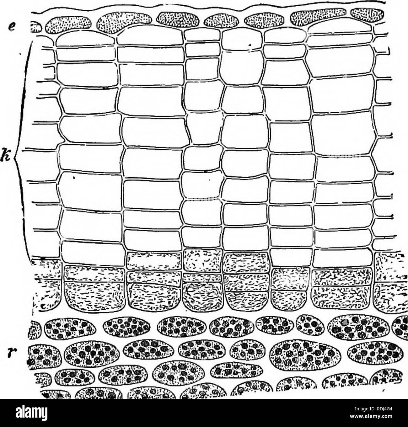
 . The essentials of botany. Botany. THE GROUPS OF TISSUES, OR TISSUE-SYSTEMS. 59 fundamental system are so disposed that the periphery is harder and firmer than the usually soft interior, although there are many exceptions. This general structure has given rise to the term Hypoderma for those portions of the fundamental system which lie immediately beneath or near to the epidermis. Hypoderma is not a distinctly limited. Fto. 38.—Transverse section of one-year-old stem of Ailanthus. c, epider mis; k, cork-cells; r, inner green cells; between k and r a layer of cells filled with protoplasm, call Stock Photohttps://www.alamy.com/image-license-details/?v=1https://www.alamy.com/the-essentials-of-botany-botany-the-groups-of-tissues-or-tissue-systems-59-fundamental-system-are-so-disposed-that-the-periphery-is-harder-and-firmer-than-the-usually-soft-interior-although-there-are-many-exceptions-this-general-structure-has-given-rise-to-the-term-hypoderma-for-those-portions-of-the-fundamental-system-which-lie-immediately-beneath-or-near-to-the-epidermis-hypoderma-is-not-a-distinctly-limited-fto-38transverse-section-of-one-year-old-stem-of-ailanthus-c-epider-mis-k-cork-cells-r-inner-green-cells-between-k-and-r-a-layer-of-cells-filled-with-protoplasm-call-image232327916.html
RMRDYCHG–. The essentials of botany. Botany. THE GROUPS OF TISSUES, OR TISSUE-SYSTEMS. 59 fundamental system are so disposed that the periphery is harder and firmer than the usually soft interior, although there are many exceptions. This general structure has given rise to the term Hypoderma for those portions of the fundamental system which lie immediately beneath or near to the epidermis. Hypoderma is not a distinctly limited. Fto. 38.—Transverse section of one-year-old stem of Ailanthus. c, epider mis; k, cork-cells; r, inner green cells; between k and r a layer of cells filled with protoplasm, call
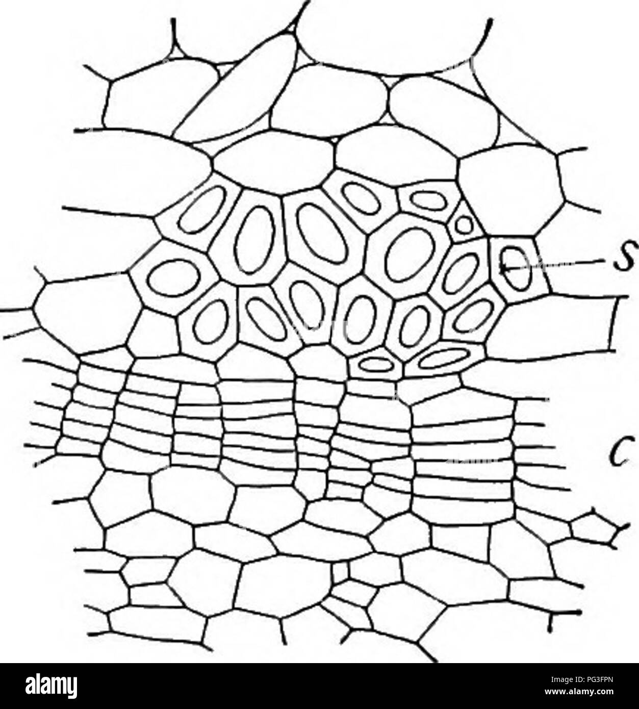
 . A textbook of botany for colleges and universities ... Botany. 1032 1033 Figs. 1032, 1033. — 1032, a partial cross section of a stem of Jussiaea peruviana from a dry habitat, showing the development of cork tissue (c) underneath a stereome bundle of thick-walled cells (s); from Schenck; 1033, a cross section of the outer part of a bur oak twig (Quercus macrocarpa), showing the layers of the periderm; p, the phello- gen, from which cork (c) develops externally and phelloderm (d) internally; note that the phelloderm contains chloroplasts, that the cork layer is without air spaces, and that the Stock Photohttps://www.alamy.com/image-license-details/?v=1https://www.alamy.com/a-textbook-of-botany-for-colleges-and-universities-botany-1032-1033-figs-1032-1033-1032-a-partial-cross-section-of-a-stem-of-jussiaea-peruviana-from-a-dry-habitat-showing-the-development-of-cork-tissue-c-underneath-a-stereome-bundle-of-thick-walled-cells-s-from-schenck-1033-a-cross-section-of-the-outer-part-of-a-bur-oak-twig-quercus-macrocarpa-showing-the-layers-of-the-periderm-p-the-phello-gen-from-which-cork-c-develops-externally-and-phelloderm-d-internally-note-that-the-phelloderm-contains-chloroplasts-that-the-cork-layer-is-without-air-spaces-and-that-the-image216437164.html
RMPG3FPM–. A textbook of botany for colleges and universities ... Botany. 1032 1033 Figs. 1032, 1033. — 1032, a partial cross section of a stem of Jussiaea peruviana from a dry habitat, showing the development of cork tissue (c) underneath a stereome bundle of thick-walled cells (s); from Schenck; 1033, a cross section of the outer part of a bur oak twig (Quercus macrocarpa), showing the layers of the periderm; p, the phello- gen, from which cork (c) develops externally and phelloderm (d) internally; note that the phelloderm contains chloroplasts, that the cork layer is without air spaces, and that the
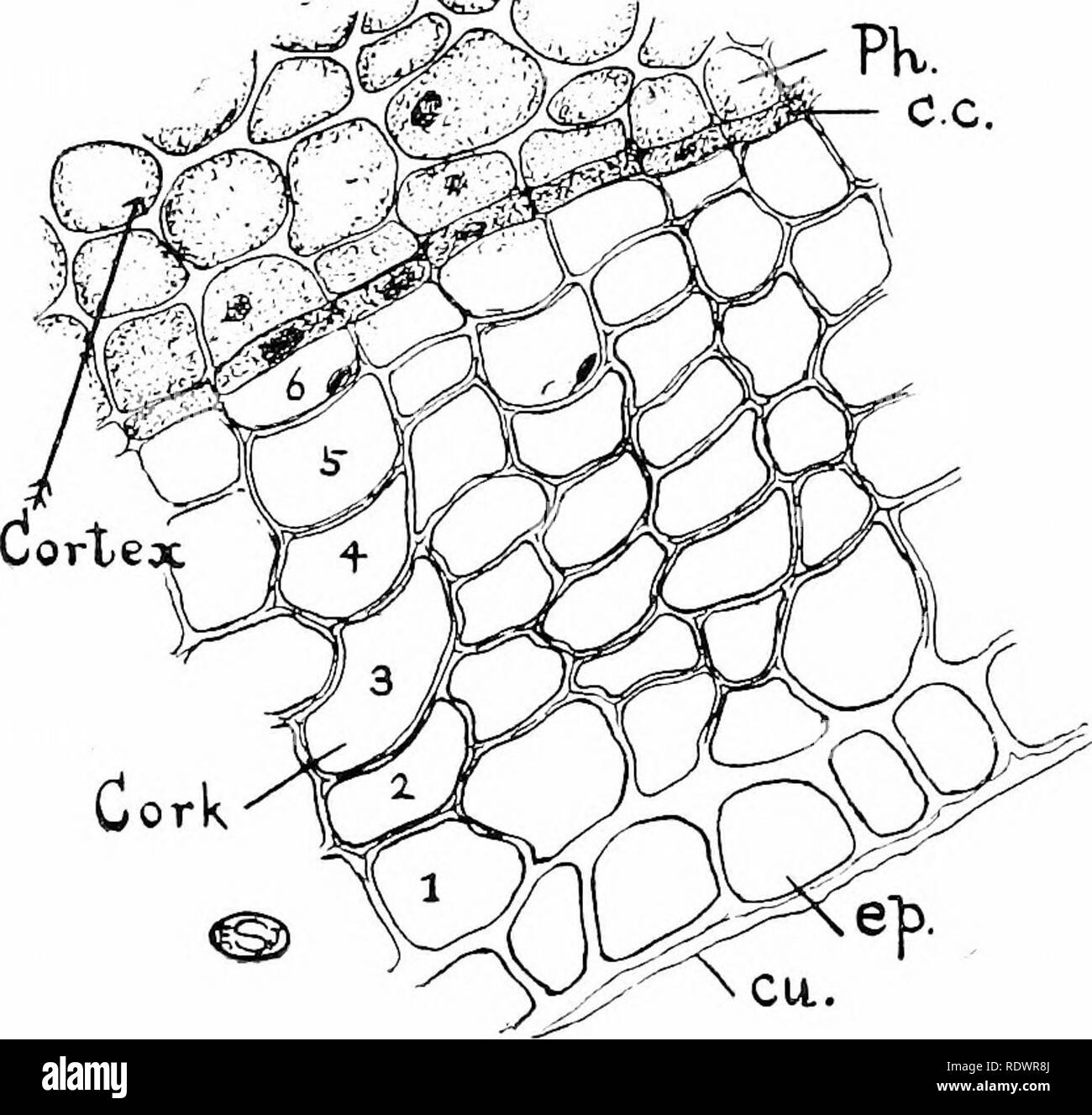 . An introduction to the structure and reproduction of plants. Plant anatomy; Plants. 136 CORK a continuous tissue consisting of numerous radial files of cells, each file (Fig, 65, 1-6) representing the product of one cork- cambium cell. This tissue is the cork and, apart from the absence of intercellular spaces between its cells, it is especially characterised by a chemical modification of the cell-walls spoken of as suherisation. This latter renders them practically imper- vious alike to gases and to liquids, features to which cork owes its utilisation in closing bottles.. Fig. 65.—Transvers Stock Photohttps://www.alamy.com/image-license-details/?v=1https://www.alamy.com/an-introduction-to-the-structure-and-reproduction-of-plants-plant-anatomy-plants-136-cork-a-continuous-tissue-consisting-of-numerous-radial-files-of-cells-each-file-fig-65-1-6-representing-the-product-of-one-cork-cambium-cell-this-tissue-is-the-cork-and-apart-from-the-absence-of-intercellular-spaces-between-its-cells-it-is-especially-characterised-by-a-chemical-modification-of-the-cell-walls-spoken-of-as-suherisation-this-latter-renders-them-practically-imper-vious-alike-to-gases-and-to-liquids-features-to-which-cork-owes-its-utilisation-in-closing-bottles-fig-65transvers-image232292386.html
RMRDWR8J–. An introduction to the structure and reproduction of plants. Plant anatomy; Plants. 136 CORK a continuous tissue consisting of numerous radial files of cells, each file (Fig, 65, 1-6) representing the product of one cork- cambium cell. This tissue is the cork and, apart from the absence of intercellular spaces between its cells, it is especially characterised by a chemical modification of the cell-walls spoken of as suherisation. This latter renders them practically imper- vious alike to gases and to liquids, features to which cork owes its utilisation in closing bottles.. Fig. 65.—Transvers
 . Pharmaceutical botany. Botany; Botany, Medical. WOODY FIBERS. ill f M 1 t -i i ) c 1 oxygen. In addition to stomata some leaves possess groups of water stomata which differ from transpiration stomata in that they always remain open, are circular in outline, give off water in droplets directly, and lie over a quantity of small-celled glandular material which is in connection with one or more fibrovascular bundles. Endodermis is the starch sheath layer of cells, constituting the innermost layer of cortex whose radial walls are more or less suberized. Cork or suberous tissue is com- posed of ce Stock Photohttps://www.alamy.com/image-license-details/?v=1https://www.alamy.com/pharmaceutical-botany-botany-botany-medical-woody-fibers-ill-f-m-1-t-i-i-c-1-oxygen-in-addition-to-stomata-some-leaves-possess-groups-of-water-stomata-which-differ-from-transpiration-stomata-in-that-they-always-remain-open-are-circular-in-outline-give-off-water-in-droplets-directly-and-lie-over-a-quantity-of-small-celled-glandular-material-which-is-in-connection-with-one-or-more-fibrovascular-bundles-endodermis-is-the-starch-sheath-layer-of-cells-constituting-the-innermost-layer-of-cortex-whose-radial-walls-are-more-or-less-suberized-cork-or-suberous-tissue-is-com-posed-of-ce-image216418192.html
RMPG2KH4–. Pharmaceutical botany. Botany; Botany, Medical. WOODY FIBERS. ill f M 1 t -i i ) c 1 oxygen. In addition to stomata some leaves possess groups of water stomata which differ from transpiration stomata in that they always remain open, are circular in outline, give off water in droplets directly, and lie over a quantity of small-celled glandular material which is in connection with one or more fibrovascular bundles. Endodermis is the starch sheath layer of cells, constituting the innermost layer of cortex whose radial walls are more or less suberized. Cork or suberous tissue is com- posed of ce
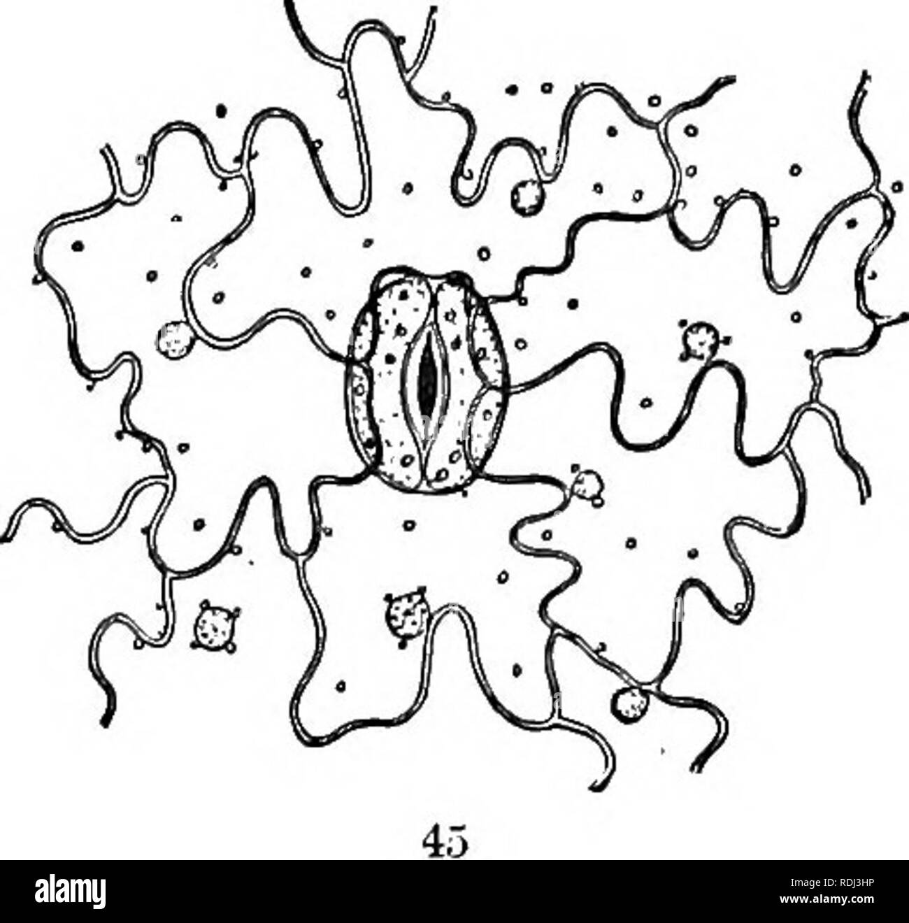 . Physiological botany; I. Outlines of the histology of phænogamous plants. II. Vegetable physiology. Plant physiology; Plant anatomy. EPIDERMIS. 65 sooner or later thrown off, and replaced by a subjacent protective tissue, — cork. 218. Except at peculiar openings (stomata, etc.), the epider- mal cells are in close apposition. Upon their exposed surface the}' are cutinized, and thus a continuous hyaline film is formed, known as the Cuticle?- 219. Sometimes the epidermis may be torn off without much disturbing the underlying tissues. 220. Besides the cells which compose the proper tissue of the Stock Photohttps://www.alamy.com/image-license-details/?v=1https://www.alamy.com/physiological-botany-i-outlines-of-the-histology-of-phnogamous-plants-ii-vegetable-physiology-plant-physiology-plant-anatomy-epidermis-65-sooner-or-later-thrown-off-and-replaced-by-a-subjacent-protective-tissue-cork-218-except-at-peculiar-openings-stomata-etc-the-epider-mal-cells-are-in-close-apposition-upon-their-exposed-surface-the-are-cutinized-and-thus-a-continuous-hyaline-film-is-formed-known-as-the-cuticle-219-sometimes-the-epidermis-may-be-torn-off-without-much-disturbing-the-underlying-tissues-220-besides-the-cells-which-compose-the-proper-tissue-of-the-image232123298.html
RMRDJ3HP–. Physiological botany; I. Outlines of the histology of phænogamous plants. II. Vegetable physiology. Plant physiology; Plant anatomy. EPIDERMIS. 65 sooner or later thrown off, and replaced by a subjacent protective tissue, — cork. 218. Except at peculiar openings (stomata, etc.), the epider- mal cells are in close apposition. Upon their exposed surface the}' are cutinized, and thus a continuous hyaline film is formed, known as the Cuticle?- 219. Sometimes the epidermis may be torn off without much disturbing the underlying tissues. 220. Besides the cells which compose the proper tissue of the
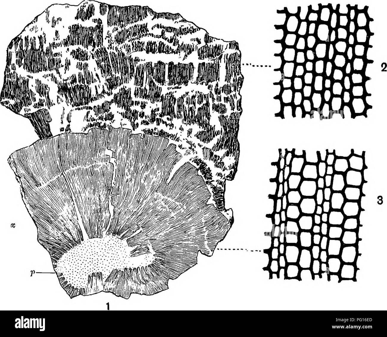 . Fossil plants : for students of botany and geology . Paleobotany. 318 CALAMITES. [CH. regularly arranged (fig. 78, 2) and rather thick-walled cells; this consists of periderm, a secondary tissue, which has been. Fig. 78. 1. Transverse section of a, thick Calamite stem. p, pith; X, secondary wood; c, bark. (| nat. size.) 2. Periderm cells of bark. 3. Xylem and medullary rays. (2 and 3, x 80.) From a specimen in the Williamson Collection (no. 79). developed by a cork-cambium during the increase in girth of the plant. The more delicate cortical tissues have not been preserved, and the more resi Stock Photohttps://www.alamy.com/image-license-details/?v=1https://www.alamy.com/fossil-plants-for-students-of-botany-and-geology-paleobotany-318-calamites-ch-regularly-arranged-fig-78-2-and-rather-thick-walled-cells-this-consists-of-periderm-a-secondary-tissue-which-has-been-fig-78-1-transverse-section-of-a-thick-calamite-stem-p-pith-x-secondary-wood-c-bark-nat-size-2-periderm-cells-of-bark-3-xylem-and-medullary-rays-2-and-3-x-80-from-a-specimen-in-the-williamson-collection-no-79-developed-by-a-cork-cambium-during-the-increase-in-girth-of-the-plant-the-more-delicate-cortical-tissues-have-not-been-preserved-and-the-more-resi-image216385973.html
RMPG16ED–. Fossil plants : for students of botany and geology . Paleobotany. 318 CALAMITES. [CH. regularly arranged (fig. 78, 2) and rather thick-walled cells; this consists of periderm, a secondary tissue, which has been. Fig. 78. 1. Transverse section of a, thick Calamite stem. p, pith; X, secondary wood; c, bark. (| nat. size.) 2. Periderm cells of bark. 3. Xylem and medullary rays. (2 and 3, x 80.) From a specimen in the Williamson Collection (no. 79). developed by a cork-cambium during the increase in girth of the plant. The more delicate cortical tissues have not been preserved, and the more resi
 . Botany, with agricultural applications. Botany. 128 CELLS AND TISSUES. much thicker and more protective than an epidermis. (Fig. 113.) The cork covering may be more or less flexible, as the rind of an Irish Potato or Sweet Potato, or harder and more brittle, as in the bark of trees, where it reaches its extreme thickness. Cork tissue con- sists of dead cells in the walls of which there is deposited a waxy- substance much-Hke cutin but called suherin to which much of the pro- tective character of cork is due. Cork coverings afford more protec- tion than an epidermis, but on ac- count of their Stock Photohttps://www.alamy.com/image-license-details/?v=1https://www.alamy.com/botany-with-agricultural-applications-botany-128-cells-and-tissues-much-thicker-and-more-protective-than-an-epidermis-fig-113-the-cork-covering-may-be-more-or-less-flexible-as-the-rind-of-an-irish-potato-or-sweet-potato-or-harder-and-more-brittle-as-in-the-bark-of-trees-where-it-reaches-its-extreme-thickness-cork-tissue-con-sists-of-dead-cells-in-the-walls-of-which-there-is-deposited-a-waxy-substance-much-hke-cutin-but-called-suherin-to-which-much-of-the-pro-tective-character-of-cork-is-due-cork-coverings-afford-more-protec-tion-than-an-epidermis-but-on-ac-count-of-their-image232285647.html
RMRDWEKY–. Botany, with agricultural applications. Botany. 128 CELLS AND TISSUES. much thicker and more protective than an epidermis. (Fig. 113.) The cork covering may be more or less flexible, as the rind of an Irish Potato or Sweet Potato, or harder and more brittle, as in the bark of trees, where it reaches its extreme thickness. Cork tissue con- sists of dead cells in the walls of which there is deposited a waxy- substance much-Hke cutin but called suherin to which much of the pro- tective character of cork is due. Cork coverings afford more protec- tion than an epidermis, but on ac- count of their
 . Pharmaceutical botany. Botany; Botany, Medical. ill f M 1 t -i i ) c 1 oxygen. In addition to stomata some leaves possess groups of water stomata which differ from transpiration stomata in that they always remain open, are circular in outline, give off water in droplets directly, and lie over a quantity of small-celled glandular material which is in connection with one or more fibrovascular bundles. Endodermis is the starch sheath layer of cells, constituting the innermost layer of cortex whose radial walls are more or less suberized. Cork or suberous tissue is com- posed of cells of tabular Stock Photohttps://www.alamy.com/image-license-details/?v=1https://www.alamy.com/pharmaceutical-botany-botany-botany-medical-ill-f-m-1-t-i-i-c-1-oxygen-in-addition-to-stomata-some-leaves-possess-groups-of-water-stomata-which-differ-from-transpiration-stomata-in-that-they-always-remain-open-are-circular-in-outline-give-off-water-in-droplets-directly-and-lie-over-a-quantity-of-small-celled-glandular-material-which-is-in-connection-with-one-or-more-fibrovascular-bundles-endodermis-is-the-starch-sheath-layer-of-cells-constituting-the-innermost-layer-of-cortex-whose-radial-walls-are-more-or-less-suberized-cork-or-suberous-tissue-is-com-posed-of-cells-of-tabular-image216418191.html
RMPG2KH3–. Pharmaceutical botany. Botany; Botany, Medical. ill f M 1 t -i i ) c 1 oxygen. In addition to stomata some leaves possess groups of water stomata which differ from transpiration stomata in that they always remain open, are circular in outline, give off water in droplets directly, and lie over a quantity of small-celled glandular material which is in connection with one or more fibrovascular bundles. Endodermis is the starch sheath layer of cells, constituting the innermost layer of cortex whose radial walls are more or less suberized. Cork or suberous tissue is com- posed of cells of tabular
 . Botany for agricultural students . Botany. much thicker and more protective than an epidermis. (Fig. 113.) The cork covering may be more or less flexible, as the rind of an Irish Potato or Sweet Potato, or harder and more brittle, as in the bark of trees, where it reaches its extreme thickness. Cork tissue con- sists of dead cells in the walls of which there is deposited a waxy substance much like cutin but called suberin to which much of the pro- tective character of cork is due. Cork coverings afford more protec- tion than an epidermis, but on ac- count of their opaqueness, they are not su Stock Photohttps://www.alamy.com/image-license-details/?v=1https://www.alamy.com/botany-for-agricultural-students-botany-much-thicker-and-more-protective-than-an-epidermis-fig-113-the-cork-covering-may-be-more-or-less-flexible-as-the-rind-of-an-irish-potato-or-sweet-potato-or-harder-and-more-brittle-as-in-the-bark-of-trees-where-it-reaches-its-extreme-thickness-cork-tissue-con-sists-of-dead-cells-in-the-walls-of-which-there-is-deposited-a-waxy-substance-much-like-cutin-but-called-suberin-to-which-much-of-the-pro-tective-character-of-cork-is-due-cork-coverings-afford-more-protec-tion-than-an-epidermis-but-on-ac-count-of-their-opaqueness-they-are-not-su-image232031096.html
RMRDDX0T–. Botany for agricultural students . Botany. much thicker and more protective than an epidermis. (Fig. 113.) The cork covering may be more or less flexible, as the rind of an Irish Potato or Sweet Potato, or harder and more brittle, as in the bark of trees, where it reaches its extreme thickness. Cork tissue con- sists of dead cells in the walls of which there is deposited a waxy substance much like cutin but called suberin to which much of the pro- tective character of cork is due. Cork coverings afford more protec- tion than an epidermis, but on ac- count of their opaqueness, they are not su
 . How crops grow. A treatise on the chemical composition, structure, and life of the plant, for all students of agriculture ... Agricultural chemistry; Growth (Plants). 276 HOW CEOPS GEOW. and deep longitudinal rifts, and it gradually decays or drops away exteriorly as the newer bark forms within. Corh is one form which the epidermal cells assume on the stem of the cork oak, on the potato tuber, and many other plants. Pith Mays. — Those portions of the first-formed cell- tissue which were intei-posed between the young and orig- inally ununited wood-fibers remain, and connect the pith with the Stock Photohttps://www.alamy.com/image-license-details/?v=1https://www.alamy.com/how-crops-grow-a-treatise-on-the-chemical-composition-structure-and-life-of-the-plant-for-all-students-of-agriculture-agricultural-chemistry-growth-plants-276-how-ceops-geow-and-deep-longitudinal-rifts-and-it-gradually-decays-or-drops-away-exteriorly-as-the-newer-bark-forms-within-corh-is-one-form-which-the-epidermal-cells-assume-on-the-stem-of-the-cork-oak-on-the-potato-tuber-and-many-other-plants-pith-mays-those-portions-of-the-first-formed-cell-tissue-which-were-intei-posed-between-the-young-and-orig-inally-ununited-wood-fibers-remain-and-connect-the-pith-with-the-image216440130.html
RMPG3KGJ–. How crops grow. A treatise on the chemical composition, structure, and life of the plant, for all students of agriculture ... Agricultural chemistry; Growth (Plants). 276 HOW CEOPS GEOW. and deep longitudinal rifts, and it gradually decays or drops away exteriorly as the newer bark forms within. Corh is one form which the epidermal cells assume on the stem of the cork oak, on the potato tuber, and many other plants. Pith Mays. — Those portions of the first-formed cell- tissue which were intei-posed between the young and orig- inally ununited wood-fibers remain, and connect the pith with the
 . An introduction to vegetable physiology. Plant physiology. THE SKELETON-OF THE PLANT 55 exterior and the metabolic tissue of the cortex of stems, thus cutting off the intercellular space system of the latter from access to the air, they are usually penetrated by special structures known as lenticels. These are made up of corky cells very loosely arranged, and consequently set up the communication needed (fig. 47). During the winter a layer of cork is formed below the lenticel. In the corky cell-wall the cutin is frequently associated with a certain amount of Ugnin. The thin corky walls posse Stock Photohttps://www.alamy.com/image-license-details/?v=1https://www.alamy.com/an-introduction-to-vegetable-physiology-plant-physiology-the-skeleton-of-the-plant-55-exterior-and-the-metabolic-tissue-of-the-cortex-of-stems-thus-cutting-off-the-intercellular-space-system-of-the-latter-from-access-to-the-air-they-are-usually-penetrated-by-special-structures-known-as-lenticels-these-are-made-up-of-corky-cells-very-loosely-arranged-and-consequently-set-up-the-communication-needed-fig-47-during-the-winter-a-layer-of-cork-is-formed-below-the-lenticel-in-the-corky-cell-wall-the-cutin-is-frequently-associated-with-a-certain-amount-of-ugnin-the-thin-corky-walls-posse-image232391025.html
RMRE293D–. An introduction to vegetable physiology. Plant physiology. THE SKELETON-OF THE PLANT 55 exterior and the metabolic tissue of the cortex of stems, thus cutting off the intercellular space system of the latter from access to the air, they are usually penetrated by special structures known as lenticels. These are made up of corky cells very loosely arranged, and consequently set up the communication needed (fig. 47). During the winter a layer of cork is formed below the lenticel. In the corky cell-wall the cutin is frequently associated with a certain amount of Ugnin. The thin corky walls posse
 . Elementary botany : theoretical and practical. A text-book designed primarily for students of science classes connected with the science and art department of the committee of council on education . Botany. Dicotyledonous Stem 43 cells immediately beneath it, more often from the more deeply lying cells. On the inner side there is developed a ring of meristem, known as phellogen or cork cambium, whilst within this there is a layer of cells containing chlorophyll, the phelloderma or green layer. Sooner or later all the cells outside the cork tissue dry up and shrivel, forming the outermost lay Stock Photohttps://www.alamy.com/image-license-details/?v=1https://www.alamy.com/elementary-botany-theoretical-and-practical-a-text-book-designed-primarily-for-students-of-science-classes-connected-with-the-science-and-art-department-of-the-committee-of-council-on-education-botany-dicotyledonous-stem-43-cells-immediately-beneath-it-more-often-from-the-more-deeply-lying-cells-on-the-inner-side-there-is-developed-a-ring-of-meristem-known-as-phellogen-or-cork-cambium-whilst-within-this-there-is-a-layer-of-cells-containing-chlorophyll-the-phelloderma-or-green-layer-sooner-or-later-all-the-cells-outside-the-cork-tissue-dry-up-and-shrivel-forming-the-outermost-lay-image232124036.html
RMRDJ4G4–. Elementary botany : theoretical and practical. A text-book designed primarily for students of science classes connected with the science and art department of the committee of council on education . Botany. Dicotyledonous Stem 43 cells immediately beneath it, more often from the more deeply lying cells. On the inner side there is developed a ring of meristem, known as phellogen or cork cambium, whilst within this there is a layer of cells containing chlorophyll, the phelloderma or green layer. Sooner or later all the cells outside the cork tissue dry up and shrivel, forming the outermost lay
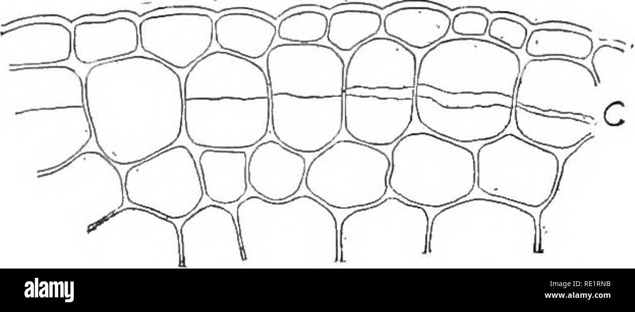 . Nature and development of plants. Botany. 86 NATURE OF CORK TISSUE increase materially in size, such as the majority of our annual plants; but in long lived stems, as shrubs and trees, where growth goes on from year to year the epidermis is not able to keep pace with the growth. 38. Cork Tissue.—To meet this condition a new tissue, the cork, is developed from certain cells called the cork cambium, usually situated near the epidermis (Fig. 47). The cells of the cork cambium divide much after the manner noted in the cam- bium of the vascular bundle, but there is this difference, the outer cell Stock Photohttps://www.alamy.com/image-license-details/?v=1https://www.alamy.com/nature-and-development-of-plants-botany-86-nature-of-cork-tissue-increase-materially-in-size-such-as-the-majority-of-our-annual-plants-but-in-long-lived-stems-as-shrubs-and-trees-where-growth-goes-on-from-year-to-year-the-epidermis-is-not-able-to-keep-pace-with-the-growth-38-cork-tissueto-meet-this-condition-a-new-tissue-the-cork-is-developed-from-certain-cells-called-the-cork-cambium-usually-situated-near-the-epidermis-fig-47-the-cells-of-the-cork-cambium-divide-much-after-the-manner-noted-in-the-cam-bium-of-the-vascular-bundle-but-there-is-this-difference-the-outer-cell-image232380551.html
RMRE1RNB–. Nature and development of plants. Botany. 86 NATURE OF CORK TISSUE increase materially in size, such as the majority of our annual plants; but in long lived stems, as shrubs and trees, where growth goes on from year to year the epidermis is not able to keep pace with the growth. 38. Cork Tissue.—To meet this condition a new tissue, the cork, is developed from certain cells called the cork cambium, usually situated near the epidermis (Fig. 47). The cells of the cork cambium divide much after the manner noted in the cam- bium of the vascular bundle, but there is this difference, the outer cell
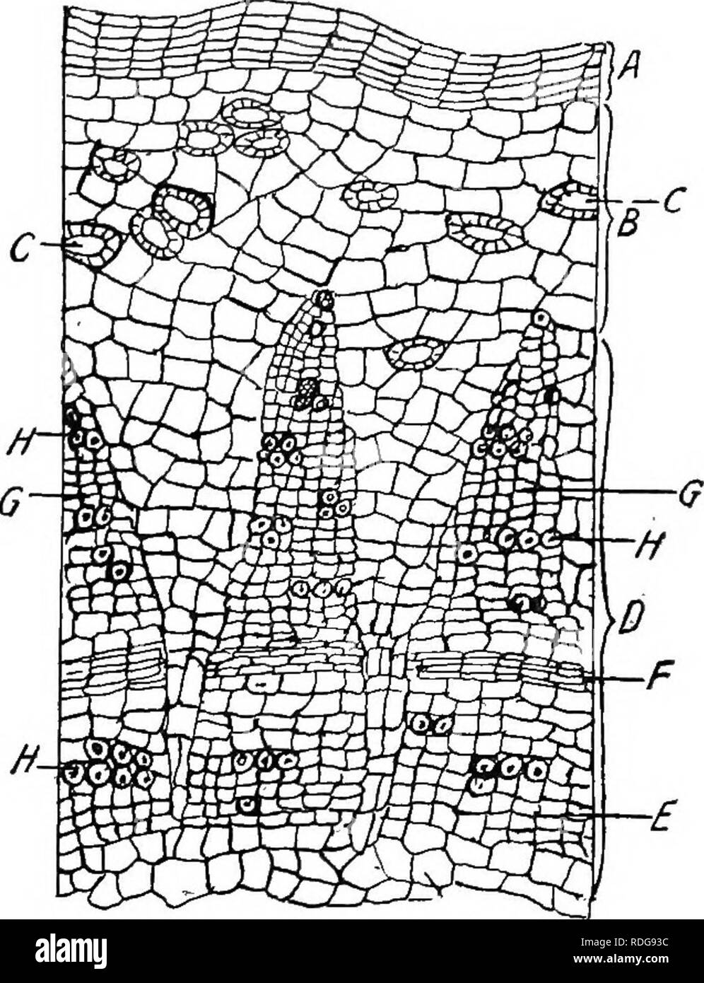 . Pharmaceutical botany. Botany; Botany, Medical. PLANT HAIES 29 Periderm.—-Periderm is a name applied to all the tissue, produced externally by the cork cambium (phellogen). This term appears often in pharmacognic and materia medica texts. Histology of Typical Monocotyl Stems (Endogenous).—Passing from exterior toward centre the following structures are seen: 1. Epidermis whose cells are cutinized in their outer walls. 2. Hypodermis, generally collenchymatic. 3. Cortex. 4. Endodermis or innermost layer of cortex generally with greatly suberized cell walls. 5. A large central zone of parenchym Stock Photohttps://www.alamy.com/image-license-details/?v=1https://www.alamy.com/pharmaceutical-botany-botany-botany-medical-plant-haies-29-periderm-periderm-is-a-name-applied-to-all-the-tissue-produced-externally-by-the-cork-cambium-phellogen-this-term-appears-often-in-pharmacognic-and-materia-medica-texts-histology-of-typical-monocotyl-stems-endogenouspassing-from-exterior-toward-centre-the-following-structures-are-seen-1-epidermis-whose-cells-are-cutinized-in-their-outer-walls-2-hypodermis-generally-collenchymatic-3-cortex-4-endodermis-or-innermost-layer-of-cortex-generally-with-greatly-suberized-cell-walls-5-a-large-central-zone-of-parenchym-image232083696.html
RMRDG93C–. Pharmaceutical botany. Botany; Botany, Medical. PLANT HAIES 29 Periderm.—-Periderm is a name applied to all the tissue, produced externally by the cork cambium (phellogen). This term appears often in pharmacognic and materia medica texts. Histology of Typical Monocotyl Stems (Endogenous).—Passing from exterior toward centre the following structures are seen: 1. Epidermis whose cells are cutinized in their outer walls. 2. Hypodermis, generally collenchymatic. 3. Cortex. 4. Endodermis or innermost layer of cortex generally with greatly suberized cell walls. 5. A large central zone of parenchym
 . Outlines of plant life : with special reference to form and function . Botany. 66 OUTLINES OF PLANT LIFE. In a large number of roots, the secondary changes result in increasing the diameter, sometimes very greatly, by the formation of concentric layers of new tissue in two or more regions, called the cambium regions. The outer growing layer, or cork cambium, usually formed in the cortex, produces tissues which are of such a nature as to protect the parts within. They constitute ^& periderm, and are ordinarily cork-like, i.e., thin-walled and impervious to water. Those cells which lie out Stock Photohttps://www.alamy.com/image-license-details/?v=1https://www.alamy.com/outlines-of-plant-life-with-special-reference-to-form-and-function-botany-66-outlines-of-plant-life-in-a-large-number-of-roots-the-secondary-changes-result-in-increasing-the-diameter-sometimes-very-greatly-by-the-formation-of-concentric-layers-of-new-tissue-in-two-or-more-regions-called-the-cambium-regions-the-outer-growing-layer-or-cork-cambium-usually-formed-in-the-cortex-produces-tissues-which-are-of-such-a-nature-as-to-protect-the-parts-within-they-constitute-amp-periderm-and-are-ordinarily-cork-like-ie-thin-walled-and-impervious-to-water-those-cells-which-lie-out-image232107636.html
RMRDHBJC–. Outlines of plant life : with special reference to form and function . Botany. 66 OUTLINES OF PLANT LIFE. In a large number of roots, the secondary changes result in increasing the diameter, sometimes very greatly, by the formation of concentric layers of new tissue in two or more regions, called the cambium regions. The outer growing layer, or cork cambium, usually formed in the cortex, produces tissues which are of such a nature as to protect the parts within. They constitute ^& periderm, and are ordinarily cork-like, i.e., thin-walled and impervious to water. Those cells which lie out
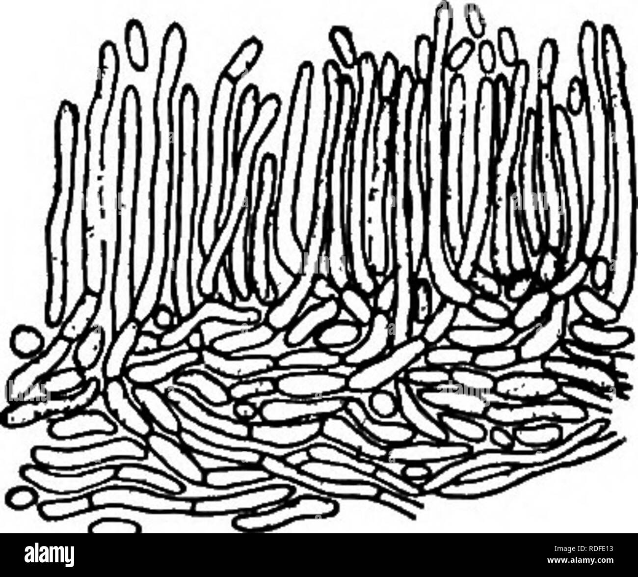 . Chestnut blight. Chestnut blight; Chestnut. Endothia Canker of Chestnut SS9 They are closely crowded together, so that in cross section they appear to make up a pseudoparenchymatous tissue. These cells are more densely filled with protoplasm, and contain more pigment, than the interior cells. Pycnidia On smooth-bark, young cankers, especially in the summer, the outer cork layer is raised in numerous little blisters, with slender, yellow, waxy tendrils curling from their ruptured apices (Plate XXXVIII, Fig. i). Under each blister is a single somewhat globose pycnidiimi, surrounded by a scanty Stock Photohttps://www.alamy.com/image-license-details/?v=1https://www.alamy.com/chestnut-blight-chestnut-blight-chestnut-endothia-canker-of-chestnut-ss9-they-are-closely-crowded-together-so-that-in-cross-section-they-appear-to-make-up-a-pseudoparenchymatous-tissue-these-cells-are-more-densely-filled-with-protoplasm-and-contain-more-pigment-than-the-interior-cells-pycnidia-on-smooth-bark-young-cankers-especially-in-the-summer-the-outer-cork-layer-is-raised-in-numerous-little-blisters-with-slender-yellow-waxy-tendrils-curling-from-their-ruptured-apices-plate-xxxviii-fig-i-under-each-blister-is-a-single-somewhat-globose-pycnidiimi-surrounded-by-a-scanty-image232065599.html
RMRDFE13–. Chestnut blight. Chestnut blight; Chestnut. Endothia Canker of Chestnut SS9 They are closely crowded together, so that in cross section they appear to make up a pseudoparenchymatous tissue. These cells are more densely filled with protoplasm, and contain more pigment, than the interior cells. Pycnidia On smooth-bark, young cankers, especially in the summer, the outer cork layer is raised in numerous little blisters, with slender, yellow, waxy tendrils curling from their ruptured apices (Plate XXXVIII, Fig. i). Under each blister is a single somewhat globose pycnidiimi, surrounded by a scanty
 . Nature and development of plants. Botany. 86 NATURE OF CORK TISSUE increase materially in size, such as the majority of our annual plants; but in long lived stems, as shrubs and trees, where growth goes on from year to year the epidermis is not able to keep pace with the growth. 38. Cork Tissue.—^To meet this condition a new tissue, the cork, is developed from certain cells called the cork cambium, usually situated near the epidermis (Fig. 47). The cells of the cork cambium divide much after the manner noted in the cam- bium of the vascular bundle, but there is this difference, the outer cel Stock Photohttps://www.alamy.com/image-license-details/?v=1https://www.alamy.com/nature-and-development-of-plants-botany-86-nature-of-cork-tissue-increase-materially-in-size-such-as-the-majority-of-our-annual-plants-but-in-long-lived-stems-as-shrubs-and-trees-where-growth-goes-on-from-year-to-year-the-epidermis-is-not-able-to-keep-pace-with-the-growth-38-cork-tissueto-meet-this-condition-a-new-tissue-the-cork-is-developed-from-certain-cells-called-the-cork-cambium-usually-situated-near-the-epidermis-fig-47-the-cells-of-the-cork-cambium-divide-much-after-the-manner-noted-in-the-cam-bium-of-the-vascular-bundle-but-there-is-this-difference-the-outer-cel-image232265988.html
RMRDTHHT–. Nature and development of plants. Botany. 86 NATURE OF CORK TISSUE increase materially in size, such as the majority of our annual plants; but in long lived stems, as shrubs and trees, where growth goes on from year to year the epidermis is not able to keep pace with the growth. 38. Cork Tissue.—^To meet this condition a new tissue, the cork, is developed from certain cells called the cork cambium, usually situated near the epidermis (Fig. 47). The cells of the cork cambium divide much after the manner noted in the cam- bium of the vascular bundle, but there is this difference, the outer cel
 . Botany for high schools and colleges. Botany. 126 BOTANY. their formation tlie cork-cells lose their protoplasmic con- tents, while beneath them new cells are constantly being cut off from the cells of the generating layer; in this way the mass of dead cork tissue is formed and pushed out from its. living base. 158.—The generating tissue is called the Phellogen,* or Cork-cambium; it occurs not only in the hypoderma, but in any other part of the fundamental system, and, as will be shown hereafter, in the secondary fibro-vascular bundles. When a living portion of a plant is injured, as by cutt Stock Photohttps://www.alamy.com/image-license-details/?v=1https://www.alamy.com/botany-for-high-schools-and-colleges-botany-126-botany-their-formation-tlie-cork-cells-lose-their-protoplasmic-con-tents-while-beneath-them-new-cells-are-constantly-being-cut-off-from-the-cells-of-the-generating-layer-in-this-way-the-mass-of-dead-cork-tissue-is-formed-and-pushed-out-from-its-living-base-158the-generating-tissue-is-called-the-phellogen-or-cork-cambium-it-occurs-not-only-in-the-hypoderma-but-in-any-other-part-of-the-fundamental-system-and-as-will-be-shown-hereafter-in-the-secondary-fibro-vascular-bundles-when-a-living-portion-of-a-plant-is-injured-as-by-cutt-image232272056.html
RMRDTWAG–. Botany for high schools and colleges. Botany. 126 BOTANY. their formation tlie cork-cells lose their protoplasmic con- tents, while beneath them new cells are constantly being cut off from the cells of the generating layer; in this way the mass of dead cork tissue is formed and pushed out from its. living base. 158.—The generating tissue is called the Phellogen,* or Cork-cambium; it occurs not only in the hypoderma, but in any other part of the fundamental system, and, as will be shown hereafter, in the secondary fibro-vascular bundles. When a living portion of a plant is injured, as by cutt
 . Studies in fossil botany . Paleobotany. STIGMARIA 247 direction, for it is on the inner edge of the secondary cortical zone that remains of the delicate phellogen can be traced. The tissue thus formed cannot have been of the nature of cork, for the outer tissue shows no sign of withering, and some of its cells had some- times undergone tangential division, as if starting a. Fig. 100.—Stigmariaficoidcs. Part of transverbe section, to show base of a rootlet, /a, periderm of main axis ; o.c, outer cortex (including hypoderma) of main axis and of rootlet; i.c, inner cortex, x, xylem of rootlet ; Stock Photohttps://www.alamy.com/image-license-details/?v=1https://www.alamy.com/studies-in-fossil-botany-paleobotany-stigmaria-247-direction-for-it-is-on-the-inner-edge-of-the-secondary-cortical-zone-that-remains-of-the-delicate-phellogen-can-be-traced-the-tissue-thus-formed-cannot-have-been-of-the-nature-of-cork-for-the-outer-tissue-shows-no-sign-of-withering-and-some-of-its-cells-had-some-times-undergone-tangential-division-as-if-starting-a-fig-100stigmariaficoidcs-part-of-transverbe-section-to-show-base-of-a-rootlet-a-periderm-of-main-axis-oc-outer-cortex-including-hypoderma-of-main-axis-and-of-rootlet-ic-inner-cortex-x-xylem-of-rootlet-image231976787.html
RMRDBCN7–. Studies in fossil botany . Paleobotany. STIGMARIA 247 direction, for it is on the inner edge of the secondary cortical zone that remains of the delicate phellogen can be traced. The tissue thus formed cannot have been of the nature of cork, for the outer tissue shows no sign of withering, and some of its cells had some- times undergone tangential division, as if starting a. Fig. 100.—Stigmariaficoidcs. Part of transverbe section, to show base of a rootlet, /a, periderm of main axis ; o.c, outer cortex (including hypoderma) of main axis and of rootlet; i.c, inner cortex, x, xylem of rootlet ;
 . Plant life, considered with special references to form and function. Plant physiology. no PLANT LIFE. The outside tissues of the jjeridevm rarely remain living^ No intercellular spaces arise between the flat cells, which early lose their contents, while the walls become waterproof. Such a tissue is known as cork (fig, 128). Other cells may be altered into mecliaiiical tissues bv the thickening of their walls and the death of the protoplasm. Zones of cork often alternate in the periderm with zones of mechanical tissues Since no water solution can pass through a cork zone, it is evident that a Stock Photohttps://www.alamy.com/image-license-details/?v=1https://www.alamy.com/plant-life-considered-with-special-references-to-form-and-function-plant-physiology-no-plant-life-the-outside-tissues-of-the-jjeridevm-rarely-remain-living-no-intercellular-spaces-arise-between-the-flat-cells-which-early-lose-their-contents-while-the-walls-become-waterproof-such-a-tissue-is-known-as-cork-fig-128-other-cells-may-be-altered-into-mecliaiiical-tissues-bv-the-thickening-of-their-walls-and-the-death-of-the-protoplasm-zones-of-cork-often-alternate-in-the-periderm-with-zones-of-mechanical-tissues-since-no-water-solution-can-pass-through-a-cork-zone-it-is-evident-that-a-image232326174.html
RMRDYABA–. Plant life, considered with special references to form and function. Plant physiology. no PLANT LIFE. The outside tissues of the jjeridevm rarely remain living^ No intercellular spaces arise between the flat cells, which early lose their contents, while the walls become waterproof. Such a tissue is known as cork (fig, 128). Other cells may be altered into mecliaiiical tissues bv the thickening of their walls and the death of the protoplasm. Zones of cork often alternate in the periderm with zones of mechanical tissues Since no water solution can pass through a cork zone, it is evident that a
 . Botany for high schools and colleges. Botany. 126 BOTANY.. their formation the cork-cells lose their protoplasmic con- tents, while beneath them new cells are constantly being cnt off from the cells of the generating layer ; in this way the mass of dead cork tissue is formed and pushed out from its living base. 158.—The generating tissue is called the Phellogen,* or Cork-cambium ; it occurs not only in the hypoderma, but in any other part of the fundamental system, and, as will be shown hereafter, in the secondary fibro-vascular bundles. When a living portion of a plant is injured, as by cut Stock Photohttps://www.alamy.com/image-license-details/?v=1https://www.alamy.com/botany-for-high-schools-and-colleges-botany-126-botany-their-formation-the-cork-cells-lose-their-protoplasmic-con-tents-while-beneath-them-new-cells-are-constantly-being-cnt-off-from-the-cells-of-the-generating-layer-in-this-way-the-mass-of-dead-cork-tissue-is-formed-and-pushed-out-from-its-living-base-158the-generating-tissue-is-called-the-phellogen-or-cork-cambium-it-occurs-not-only-in-the-hypoderma-but-in-any-other-part-of-the-fundamental-system-and-as-will-be-shown-hereafter-in-the-secondary-fibro-vascular-bundles-when-a-living-portion-of-a-plant-is-injured-as-by-cut-image232282440.html
RMRDWAHC–. Botany for high schools and colleges. Botany. 126 BOTANY.. their formation the cork-cells lose their protoplasmic con- tents, while beneath them new cells are constantly being cnt off from the cells of the generating layer ; in this way the mass of dead cork tissue is formed and pushed out from its living base. 158.—The generating tissue is called the Phellogen,* or Cork-cambium ; it occurs not only in the hypoderma, but in any other part of the fundamental system, and, as will be shown hereafter, in the secondary fibro-vascular bundles. When a living portion of a plant is injured, as by cut
 . A manual of botany. Botany. TISSUE SYSTEMS. Fig. 723. Outer portion of cortex of yoimg stem of Lime. ph. Phellogeu. per. Cork. cells thick, of brick-shaped cells without interspaces [fig. 723, per^. The cell-walls remain thin, but generally become com- pletely suberised. The merismatic layer of cells is known as the â phellogen (fig. 723, ph.); it is classed as a secondary nieristem, the cells regaining the power of dividing after ha^'ing assumed the condition of permanent tissue. Sometimes it is not the outer layer of the hypoderma Avhich becomes phellogen, but one deeper in the cortex. The Stock Photohttps://www.alamy.com/image-license-details/?v=1https://www.alamy.com/a-manual-of-botany-botany-tissue-systems-fig-723-outer-portion-of-cortex-of-yoimg-stem-of-lime-ph-phellogeu-per-cork-cells-thick-of-brick-shaped-cells-without-interspaces-fig-723-per-the-cell-walls-remain-thin-but-generally-become-com-pletely-suberised-the-merismatic-layer-of-cells-is-known-as-the-phellogen-fig-723-ph-it-is-classed-as-a-secondary-nieristem-the-cells-regaining-the-power-of-dividing-after-haing-assumed-the-condition-of-permanent-tissue-sometimes-it-is-not-the-outer-layer-of-the-hypoderma-avhich-becomes-phellogen-but-one-deeper-in-the-cortex-the-image232376230.html
RMRE1J72–. A manual of botany. Botany. TISSUE SYSTEMS. Fig. 723. Outer portion of cortex of yoimg stem of Lime. ph. Phellogeu. per. Cork. cells thick, of brick-shaped cells without interspaces [fig. 723, per^. The cell-walls remain thin, but generally become com- pletely suberised. The merismatic layer of cells is known as the â phellogen (fig. 723, ph.); it is classed as a secondary nieristem, the cells regaining the power of dividing after ha^'ing assumed the condition of permanent tissue. Sometimes it is not the outer layer of the hypoderma Avhich becomes phellogen, but one deeper in the cortex. The
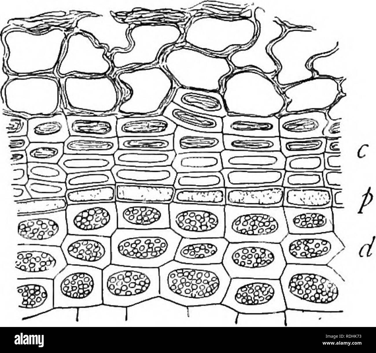 . A textbook of botany for colleges and universities ... Botany. 1032 1033 Figs. 1032, 1033. — 1032, a partial cross section of a stem of Jussiaea peruviana from a dry habitat, showing the development of cork tissue (c) underneath a stereome bundle of thick-walled cells (s); from Schenck; 1033, a cross section of the outer part of a bur oak twig (Quercus macrocarpa), showing the layers of the periderm; p, the phello- gen, from which cork (c) develops externally and phelloderm (d) internally; note that the phelloderm contains chloroplasts, that the cork layer is without air spaces, and that the Stock Photohttps://www.alamy.com/image-license-details/?v=1https://www.alamy.com/a-textbook-of-botany-for-colleges-and-universities-botany-1032-1033-figs-1032-1033-1032-a-partial-cross-section-of-a-stem-of-jussiaea-peruviana-from-a-dry-habitat-showing-the-development-of-cork-tissue-c-underneath-a-stereome-bundle-of-thick-walled-cells-s-from-schenck-1033-a-cross-section-of-the-outer-part-of-a-bur-oak-twig-quercus-macrocarpa-showing-the-layers-of-the-periderm-p-the-phello-gen-from-which-cork-c-develops-externally-and-phelloderm-d-internally-note-that-the-phelloderm-contains-chloroplasts-that-the-cork-layer-is-without-air-spaces-and-that-the-image232113591.html
RMRDHK73–. A textbook of botany for colleges and universities ... Botany. 1032 1033 Figs. 1032, 1033. — 1032, a partial cross section of a stem of Jussiaea peruviana from a dry habitat, showing the development of cork tissue (c) underneath a stereome bundle of thick-walled cells (s); from Schenck; 1033, a cross section of the outer part of a bur oak twig (Quercus macrocarpa), showing the layers of the periderm; p, the phello- gen, from which cork (c) develops externally and phelloderm (d) internally; note that the phelloderm contains chloroplasts, that the cork layer is without air spaces, and that the
 . Gray's school and field book of botany. Consisting of "Lessons in botany," and "Field, forest, and garden botany," bound in one volume. Botany; Botany. SECTION 16. J ANATOMY OF STEMS. 141 2. The Geben Babk or Middle Bark. This consists of cellukr tissue only, and contains the same green matter {chlorophyll, 417) as the leaves. In woody stems, before the season's growth is coinpleted, it becomes cov- ered by 3. The Corky Later or Outer Bark, the cells of which contain no chlorophyll, and are of the nature of cork. Common cork is the thick corky layer of the bark of the Cor Stock Photohttps://www.alamy.com/image-license-details/?v=1https://www.alamy.com/grays-school-and-field-book-of-botany-consisting-of-quotlessons-in-botanyquot-and-quotfield-forest-and-garden-botanyquot-bound-in-one-volume-botany-botany-section-16-j-anatomy-of-stems-141-2-the-geben-babk-or-middle-bark-this-consists-of-cellukr-tissue-only-and-contains-the-same-green-matter-chlorophyll-417-as-the-leaves-in-woody-stems-before-the-seasons-growth-is-coinpleted-it-becomes-cov-ered-by-3-the-corky-later-or-outer-bark-the-cells-of-which-contain-no-chlorophyll-and-are-of-the-nature-of-cork-common-cork-is-the-thick-corky-layer-of-the-bark-of-the-cor-image232272712.html
RMRDTX60–. Gray's school and field book of botany. Consisting of "Lessons in botany," and "Field, forest, and garden botany," bound in one volume. Botany; Botany. SECTION 16. J ANATOMY OF STEMS. 141 2. The Geben Babk or Middle Bark. This consists of cellukr tissue only, and contains the same green matter {chlorophyll, 417) as the leaves. In woody stems, before the season's growth is coinpleted, it becomes cov- ered by 3. The Corky Later or Outer Bark, the cells of which contain no chlorophyll, and are of the nature of cork. Common cork is the thick corky layer of the bark of the Cor
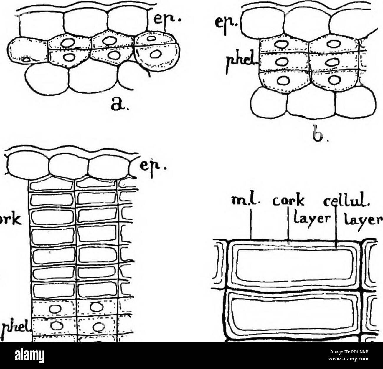 . Elements of plant biology. Plant physiology. 338 THE WOODY STEM cells (Fig. 56, a), i.e. immediately below the epidermis, sometimes from the epidermis itself, occasionally in a deeper layer of the cortex, and fairly often in the pericycle. In roots the phellogen is nearly always formed in the pericycle. Each phellogen cell arises as the result of two rapidly following divisions of the living tissue cell in which it is formed, the two new parallel walls being tangentially. cork CTT) Fig. 56.—Cork formation: a, b. Beginning of phellogen in the outer- most layer of cortex ; ep., epidermis ; phe Stock Photohttps://www.alamy.com/image-license-details/?v=1https://www.alamy.com/elements-of-plant-biology-plant-physiology-338-the-woody-stem-cells-fig-56-a-ie-immediately-below-the-epidermis-sometimes-from-the-epidermis-itself-occasionally-in-a-deeper-layer-of-the-cortex-and-fairly-often-in-the-pericycle-in-roots-the-phellogen-is-nearly-always-formed-in-the-pericycle-each-phellogen-cell-arises-as-the-result-of-two-rapidly-following-divisions-of-the-living-tissue-cell-in-which-it-is-formed-the-two-new-parallel-walls-being-tangentially-cork-ctt-fig-56cork-formation-a-b-beginning-of-phellogen-in-the-outer-most-layer-of-cortex-ep-epidermis-phe-image232115503.html
RMRDHNKB–. Elements of plant biology. Plant physiology. 338 THE WOODY STEM cells (Fig. 56, a), i.e. immediately below the epidermis, sometimes from the epidermis itself, occasionally in a deeper layer of the cortex, and fairly often in the pericycle. In roots the phellogen is nearly always formed in the pericycle. Each phellogen cell arises as the result of two rapidly following divisions of the living tissue cell in which it is formed, the two new parallel walls being tangentially. cork CTT) Fig. 56.—Cork formation: a, b. Beginning of phellogen in the outer- most layer of cortex ; ep., epidermis ; phe
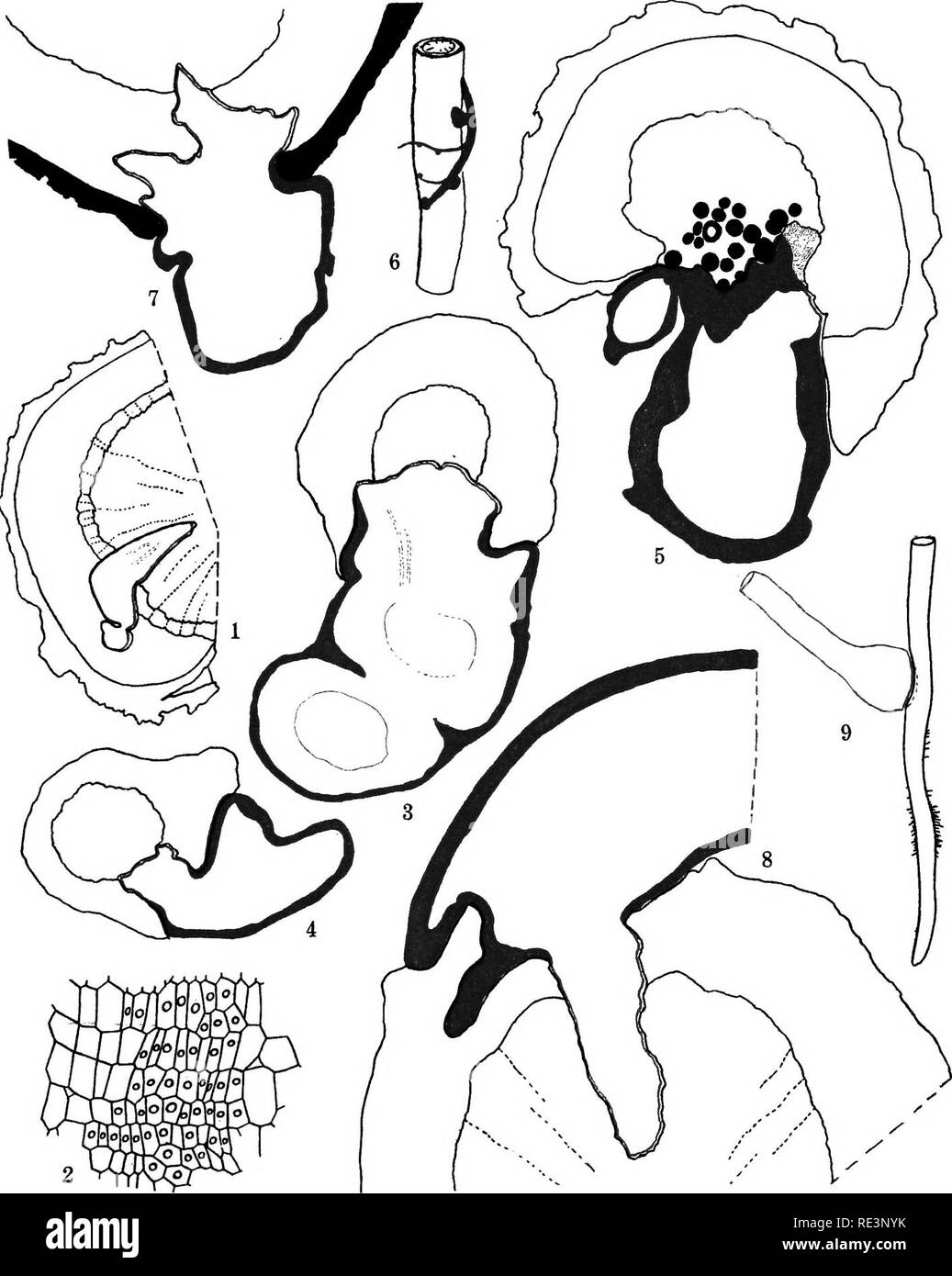 . The conditions of parasitism in plants. Parasitic plants. PLATE 4. Parasitism of Kraheria canescens on different hosts. 1, Aulo-parasitlsm of Kramerta; only aubmergnd part of batistorium is shown. 2, detail from yovmg haustorial cushion showing cells which form curving tissue of the mature organ. 3, Attachment of Krarneria on Eneelia /arinoaa. 4, Penetration of haustorium to wood in root of Xenodora scabra. 5, Krameria on root of ParMmonia mierophylla, showing plugging of ducts of host. The heavy red margin indicates extent of formation of cork in hauslionum. 6, Attachment of Krameria roots Stock Photohttps://www.alamy.com/image-license-details/?v=1https://www.alamy.com/the-conditions-of-parasitism-in-plants-parasitic-plants-plate-4-parasitism-of-kraheria-canescens-on-different-hosts-1-aulo-parasitlsm-of-kramerta-only-aubmergnd-part-of-batistorium-is-shown-2-detail-from-yovmg-haustorial-cushion-showing-cells-which-form-curving-tissue-of-the-mature-organ-3-attachment-of-krarneria-on-eneelia-arinoaa-4-penetration-of-haustorium-to-wood-in-root-of-xenodora-scabra-5-krameria-on-root-of-parmmonia-mierophylla-showing-plugging-of-ducts-of-host-the-heavy-red-margin-indicates-extent-of-formation-of-cork-in-hauslionum-6-attachment-of-krameria-roots-image232423063.html
RMRE3NYK–. The conditions of parasitism in plants. Parasitic plants. PLATE 4. Parasitism of Kraheria canescens on different hosts. 1, Aulo-parasitlsm of Kramerta; only aubmergnd part of batistorium is shown. 2, detail from yovmg haustorial cushion showing cells which form curving tissue of the mature organ. 3, Attachment of Krarneria on Eneelia /arinoaa. 4, Penetration of haustorium to wood in root of Xenodora scabra. 5, Krameria on root of ParMmonia mierophylla, showing plugging of ducts of host. The heavy red margin indicates extent of formation of cork in hauslionum. 6, Attachment of Krameria roots
 . The elements of vegetable histology. Plant anatomy. 174 THE ELEMENTS OF VEGETABLE HISTOLOGY less, thin-walled, rectangular cells, immediately beneath the corky.tissue. The walls of these cells are of cellu- lose and may be readily distinguished from the su- berized cork cells by micro-chemical tests. The phel-. Platb 59.—Bark Structure (Powdered material). A. Chionanthus, root bark. 1. Bark parenchyma (longitudinal view), con- taining starch, crystals and resin masses. 2. Bark parenchyma crossed by medul- lary ray cells. 3. Cork tissue. 4. Bark parenchyma (transverse view). 5. Fibers, 6. Cry Stock Photohttps://www.alamy.com/image-license-details/?v=1https://www.alamy.com/the-elements-of-vegetable-histology-plant-anatomy-174-the-elements-of-vegetable-histology-less-thin-walled-rectangular-cells-immediately-beneath-the-corkytissue-the-walls-of-these-cells-are-of-cellu-lose-and-may-be-readily-distinguished-from-the-su-berized-cork-cells-by-micro-chemical-tests-the-phel-platb-59bark-structure-powdered-material-a-chionanthus-root-bark-1-bark-parenchyma-longitudinal-view-con-taining-starch-crystals-and-resin-masses-2-bark-parenchyma-crossed-by-medul-lary-ray-cells-3-cork-tissue-4-bark-parenchyma-transverse-view-5-fibers-6-cry-image232288162.html
RMRDWHWP–. The elements of vegetable histology. Plant anatomy. 174 THE ELEMENTS OF VEGETABLE HISTOLOGY less, thin-walled, rectangular cells, immediately beneath the corky.tissue. The walls of these cells are of cellu- lose and may be readily distinguished from the su- berized cork cells by micro-chemical tests. The phel-. Platb 59.—Bark Structure (Powdered material). A. Chionanthus, root bark. 1. Bark parenchyma (longitudinal view), con- taining starch, crystals and resin masses. 2. Bark parenchyma crossed by medul- lary ray cells. 3. Cork tissue. 4. Bark parenchyma (transverse view). 5. Fibers, 6. Cry
 . A manual of botany. Botany. Fig. 723. Outer portion of cortex of yoimg stem of Lime. ph. Phellogeu. per. Cork. cells thick, of brick-shaped cells without interspaces [fig. 723, per^. The cell-walls remain thin, but generally become com- pletely suberised. The merismatic layer of cells is known as the â phellogen (fig. 723, ph.); it is classed as a secondary nieristem, the cells regaining the power of dividing after ha^'ing assumed the condition of permanent tissue. Sometimes it is not the outer layer of the hypoderma Avhich becomes phellogen, but one deeper in the cortex. The depth varies in Stock Photohttps://www.alamy.com/image-license-details/?v=1https://www.alamy.com/a-manual-of-botany-botany-fig-723-outer-portion-of-cortex-of-yoimg-stem-of-lime-ph-phellogeu-per-cork-cells-thick-of-brick-shaped-cells-without-interspaces-fig-723-per-the-cell-walls-remain-thin-but-generally-become-com-pletely-suberised-the-merismatic-layer-of-cells-is-known-as-the-phellogen-fig-723-ph-it-is-classed-as-a-secondary-nieristem-the-cells-regaining-the-power-of-dividing-after-haing-assumed-the-condition-of-permanent-tissue-sometimes-it-is-not-the-outer-layer-of-the-hypoderma-avhich-becomes-phellogen-but-one-deeper-in-the-cortex-the-depth-varies-in-image232376223.html
RMRE1J6R–. A manual of botany. Botany. Fig. 723. Outer portion of cortex of yoimg stem of Lime. ph. Phellogeu. per. Cork. cells thick, of brick-shaped cells without interspaces [fig. 723, per^. The cell-walls remain thin, but generally become com- pletely suberised. The merismatic layer of cells is known as the â phellogen (fig. 723, ph.); it is classed as a secondary nieristem, the cells regaining the power of dividing after ha^'ing assumed the condition of permanent tissue. Sometimes it is not the outer layer of the hypoderma Avhich becomes phellogen, but one deeper in the cortex. The depth varies in
 . Pharmaceutical botany. Botany; Botany, Medical. WOODY FIBERS. ill f M 1 t -i i ) c 1 oxygen. In addition to stomata some leaves possess groups of water stomata which differ from transpiration stomata in that they always remain open, are circular in outline, give off water in droplets directly, and lie over a quantity of small-celled glandular material which is in connection with one or more fibrovascular bundles. Endodermis is the starch sheath layer of cells, constituting the innermost layer of cortex whose radial walls are more or less suberized. Cork or suberous tissue is com- posed of ce Stock Photohttps://www.alamy.com/image-license-details/?v=1https://www.alamy.com/pharmaceutical-botany-botany-botany-medical-woody-fibers-ill-f-m-1-t-i-i-c-1-oxygen-in-addition-to-stomata-some-leaves-possess-groups-of-water-stomata-which-differ-from-transpiration-stomata-in-that-they-always-remain-open-are-circular-in-outline-give-off-water-in-droplets-directly-and-lie-over-a-quantity-of-small-celled-glandular-material-which-is-in-connection-with-one-or-more-fibrovascular-bundles-endodermis-is-the-starch-sheath-layer-of-cells-constituting-the-innermost-layer-of-cortex-whose-radial-walls-are-more-or-less-suberized-cork-or-suberous-tissue-is-com-posed-of-ce-image232083776.html
RMRDG968–. Pharmaceutical botany. Botany; Botany, Medical. WOODY FIBERS. ill f M 1 t -i i ) c 1 oxygen. In addition to stomata some leaves possess groups of water stomata which differ from transpiration stomata in that they always remain open, are circular in outline, give off water in droplets directly, and lie over a quantity of small-celled glandular material which is in connection with one or more fibrovascular bundles. Endodermis is the starch sheath layer of cells, constituting the innermost layer of cortex whose radial walls are more or less suberized. Cork or suberous tissue is com- posed of ce
 . Pharmaceutical botany. Botany; Botany, Medical. ill f M 1 t -i i ) c 1 oxygen. In addition to stomata some leaves possess groups of water stomata which differ from transpiration stomata in that they always remain open, are circular in outline, give off water in droplets directly, and lie over a quantity of small-celled glandular material which is in connection with one or more fibrovascular bundles. Endodermis is the starch sheath layer of cells, constituting the innermost layer of cortex whose radial walls are more or less suberized. Cork or suberous tissue is com- posed of cells of tabular Stock Photohttps://www.alamy.com/image-license-details/?v=1https://www.alamy.com/pharmaceutical-botany-botany-botany-medical-ill-f-m-1-t-i-i-c-1-oxygen-in-addition-to-stomata-some-leaves-possess-groups-of-water-stomata-which-differ-from-transpiration-stomata-in-that-they-always-remain-open-are-circular-in-outline-give-off-water-in-droplets-directly-and-lie-over-a-quantity-of-small-celled-glandular-material-which-is-in-connection-with-one-or-more-fibrovascular-bundles-endodermis-is-the-starch-sheath-layer-of-cells-constituting-the-innermost-layer-of-cortex-whose-radial-walls-are-more-or-less-suberized-cork-or-suberous-tissue-is-com-posed-of-cells-of-tabular-image232083771.html
RMRDG963–. Pharmaceutical botany. Botany; Botany, Medical. ill f M 1 t -i i ) c 1 oxygen. In addition to stomata some leaves possess groups of water stomata which differ from transpiration stomata in that they always remain open, are circular in outline, give off water in droplets directly, and lie over a quantity of small-celled glandular material which is in connection with one or more fibrovascular bundles. Endodermis is the starch sheath layer of cells, constituting the innermost layer of cortex whose radial walls are more or less suberized. Cork or suberous tissue is com- posed of cells of tabular
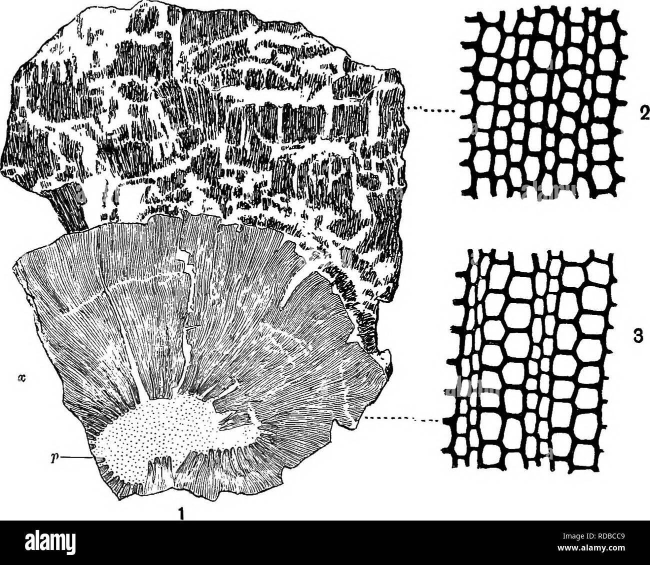 . Fossil plants : for students of botany and geology . Paleobotany. 318 CALAMITES. [CH. regularly arranged (fig. 78, 2) and rather thick-walled cells; this consists of periderm, a secondary tissue, which has been. Fig. 78. 1. Transverse section of a, thick Calamite stem. p, pith; X, secondary wood; c, bark. (| nat. size.) 2. Periderm cells of bark. 3. Xylem and medullary rays. (2 and 3, x 80.) From a specimen in the Williamson Collection (no. 79). developed by a cork-cambium during the increase in girth of the plant. The more delicate cortical tissues have not been preserved, and the more resi Stock Photohttps://www.alamy.com/image-license-details/?v=1https://www.alamy.com/fossil-plants-for-students-of-botany-and-geology-paleobotany-318-calamites-ch-regularly-arranged-fig-78-2-and-rather-thick-walled-cells-this-consists-of-periderm-a-secondary-tissue-which-has-been-fig-78-1-transverse-section-of-a-thick-calamite-stem-p-pith-x-secondary-wood-c-bark-nat-size-2-periderm-cells-of-bark-3-xylem-and-medullary-rays-2-and-3-x-80-from-a-specimen-in-the-williamson-collection-no-79-developed-by-a-cork-cambium-during-the-increase-in-girth-of-the-plant-the-more-delicate-cortical-tissues-have-not-been-preserved-and-the-more-resi-image231976537.html
RMRDBCC9–. Fossil plants : for students of botany and geology . Paleobotany. 318 CALAMITES. [CH. regularly arranged (fig. 78, 2) and rather thick-walled cells; this consists of periderm, a secondary tissue, which has been. Fig. 78. 1. Transverse section of a, thick Calamite stem. p, pith; X, secondary wood; c, bark. (| nat. size.) 2. Periderm cells of bark. 3. Xylem and medullary rays. (2 and 3, x 80.) From a specimen in the Williamson Collection (no. 79). developed by a cork-cambium during the increase in girth of the plant. The more delicate cortical tissues have not been preserved, and the more resi
 . Elementary botany. Botany. Fig. 414. Fig. 415. Transverse section of portion of -Margin of leaf of Pinus pinaster, transverse tomato stem. cp^ epidermis: ch. section, t:, cuticularized layer of outer wall chloroph>-ll-bearing cells; co, collen- chyma; cp, parenchyma. of epidennis; i, inner non-cuticularized layer; c', thickened outer wall of marginal cell; ^', 1', hypoderma â -f elongated scle- renchyma, p, chloroph"ll-bearing paren- chyma; pr, contracted protoplasmic con- tents. X800. (After Sachs.) 699. Cork.âIn many cases there is a development of "cork" tissue undernea Stock Photohttps://www.alamy.com/image-license-details/?v=1https://www.alamy.com/elementary-botany-botany-fig-414-fig-415-transverse-section-of-portion-of-margin-of-leaf-of-pinus-pinaster-transverse-tomato-stem-cp-epidermis-ch-section-t-cuticularized-layer-of-outer-wall-chlorophgt-ll-bearing-cells-co-collen-chyma-cp-parenchyma-of-epidennis-i-inner-non-cuticularized-layer-c-thickened-outer-wall-of-marginal-cell-1-hypoderma-f-elongated-scle-renchyma-p-chlorophquotll-bearing-paren-chyma-pr-contracted-protoplasmic-con-tents-x800-after-sachs-699-corkin-many-cases-there-is-a-development-of-quotcorkquot-tissue-undernea-image232414911.html
RMRE3BGF–. Elementary botany. Botany. Fig. 414. Fig. 415. Transverse section of portion of -Margin of leaf of Pinus pinaster, transverse tomato stem. cp^ epidermis: ch. section, t:, cuticularized layer of outer wall chloroph>-ll-bearing cells; co, collen- chyma; cp, parenchyma. of epidennis; i, inner non-cuticularized layer; c', thickened outer wall of marginal cell; ^', 1', hypoderma â -f elongated scle- renchyma, p, chloroph"ll-bearing paren- chyma; pr, contracted protoplasmic con- tents. X800. (After Sachs.) 699. Cork.âIn many cases there is a development of "cork" tissue undernea
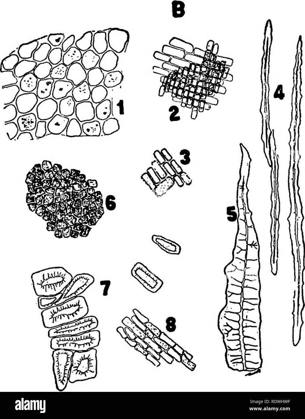 . The elements of vegetable histology. Plant anatomy. STEM STRUCTURE 175 duces more cork than phelloderm, and the latter tissue may occasionally be reduced to a single layer of cells. In powdered materials cork cells are usually apparent on surface view, and occur in thick masses. <y* v. Plate 59—Continued. B. Chionanthus, stem bark. 1. Bark parenchyma (transverse view), contain- ing starch, crystals and resin masses. 2. Bark parenchyma crossed by medullary ray cells. 3. Medullary ray cells. 4. Fibers. 5. Fibers showing branching pores. 6. Cork tissue. 7. Stone cells. 8. Bark parenchyma (lo Stock Photohttps://www.alamy.com/image-license-details/?v=1https://www.alamy.com/the-elements-of-vegetable-histology-plant-anatomy-stem-structure-175-duces-more-cork-than-phelloderm-and-the-latter-tissue-may-occasionally-be-reduced-to-a-single-layer-of-cells-in-powdered-materials-cork-cells-are-usually-apparent-on-surface-view-and-occur-in-thick-masses-lty-v-plate-59continued-b-chionanthus-stem-bark-1-bark-parenchyma-transverse-view-contain-ing-starch-crystals-and-resin-masses-2-bark-parenchyma-crossed-by-medullary-ray-cells-3-medullary-ray-cells-4-fibers-5-fibers-showing-branching-pores-6-cork-tissue-7-stone-cells-8-bark-parenchyma-lo-image232288155.html
RMRDWHWF–. The elements of vegetable histology. Plant anatomy. STEM STRUCTURE 175 duces more cork than phelloderm, and the latter tissue may occasionally be reduced to a single layer of cells. In powdered materials cork cells are usually apparent on surface view, and occur in thick masses. <y* v. Plate 59—Continued. B. Chionanthus, stem bark. 1. Bark parenchyma (transverse view), contain- ing starch, crystals and resin masses. 2. Bark parenchyma crossed by medullary ray cells. 3. Medullary ray cells. 4. Fibers. 5. Fibers showing branching pores. 6. Cork tissue. 7. Stone cells. 8. Bark parenchyma (lo
 . The essentials of botany. Botany. 60 BOTANY. some cases (e.g., the cork-oak) are thin and weak, while in others (e.g., the beech) they are much thickened, and in all cases they are nearly impermeable to water. True cork is destitute of intercellular spaces, its cells being of regu- lar shape (generally cuboidal) and fitted closely to each other (Fig. 38). 105. Cork-substance is formed by the repeated subdivi- sion of the cells of a meristem layer of the fundamental. Fig. 39.—Cross-section through a lentieel of Birch, e, epiaermis: s, a breathing-pore. Magnified 280 times. tissue (Pig. 38); t Stock Photohttps://www.alamy.com/image-license-details/?v=1https://www.alamy.com/the-essentials-of-botany-botany-60-botany-some-cases-eg-the-cork-oak-are-thin-and-weak-while-in-others-eg-the-beech-they-are-much-thickened-and-in-all-cases-they-are-nearly-impermeable-to-water-true-cork-is-destitute-of-intercellular-spaces-its-cells-being-of-regu-lar-shape-generally-cuboidal-and-fitted-closely-to-each-other-fig-38-105-cork-substance-is-formed-by-the-repeated-subdivi-sion-of-the-cells-of-a-meristem-layer-of-the-fundamental-fig-39cross-section-through-a-lentieel-of-birch-e-epiaermis-s-a-breathing-pore-magnified-280-times-tissue-pig-38-t-image232327911.html
RMRDYCHB–. The essentials of botany. Botany. 60 BOTANY. some cases (e.g., the cork-oak) are thin and weak, while in others (e.g., the beech) they are much thickened, and in all cases they are nearly impermeable to water. True cork is destitute of intercellular spaces, its cells being of regu- lar shape (generally cuboidal) and fitted closely to each other (Fig. 38). 105. Cork-substance is formed by the repeated subdivi- sion of the cells of a meristem layer of the fundamental. Fig. 39.—Cross-section through a lentieel of Birch, e, epiaermis: s, a breathing-pore. Magnified 280 times. tissue (Pig. 38); t
 . How crops grow. A treatise on the chemical composition, structure, and life of the plant, for all students of agriculture ... Agricultural chemistry; Growth (Plants). 276 HOW CEOPS GEOW. and deep longitudinal rifts, and it gradually decays or drops away exteriorly as the newer bark forms within. Corh is one form which the epidermal cells assume on the stem of the cork oak, on the potato tuber, and many other plants. Pith Mays. — Those portions of the first-formed cell- tissue which were intei-posed between the young and orig- inally ununited wood-fibers remain, and connect the pith with the Stock Photohttps://www.alamy.com/image-license-details/?v=1https://www.alamy.com/how-crops-grow-a-treatise-on-the-chemical-composition-structure-and-life-of-the-plant-for-all-students-of-agriculture-agricultural-chemistry-growth-plants-276-how-ceops-geow-and-deep-longitudinal-rifts-and-it-gradually-decays-or-drops-away-exteriorly-as-the-newer-bark-forms-within-corh-is-one-form-which-the-epidermal-cells-assume-on-the-stem-of-the-cork-oak-on-the-potato-tuber-and-many-other-plants-pith-mays-those-portions-of-the-first-formed-cell-tissue-which-were-intei-posed-between-the-young-and-orig-inally-ununited-wood-fibers-remain-and-connect-the-pith-with-the-image232122564.html
RMRDJ2KG–. How crops grow. A treatise on the chemical composition, structure, and life of the plant, for all students of agriculture ... Agricultural chemistry; Growth (Plants). 276 HOW CEOPS GEOW. and deep longitudinal rifts, and it gradually decays or drops away exteriorly as the newer bark forms within. Corh is one form which the epidermal cells assume on the stem of the cork oak, on the potato tuber, and many other plants. Pith Mays. — Those portions of the first-formed cell- tissue which were intei-posed between the young and orig- inally ununited wood-fibers remain, and connect the pith with the