Cross section of brain Black & White Stock Photos
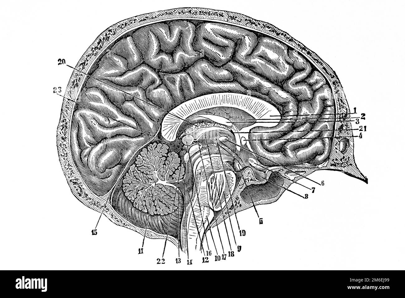 Cross section of brain anatomy. Antique illustration from a medical book. 1889. Stock Photohttps://www.alamy.com/image-license-details/?v=1https://www.alamy.com/cross-section-of-brain-anatomy-antique-illustration-from-a-medical-book-1889-image503110309.html
RF2M6EJ99–Cross section of brain anatomy. Antique illustration from a medical book. 1889.
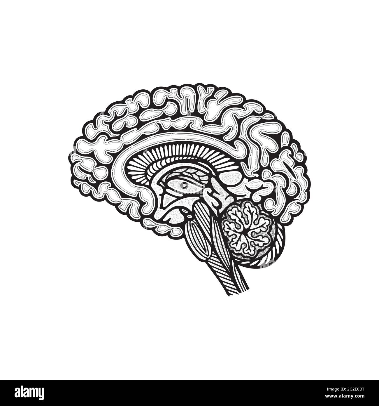 Brain side view cross section. Human brain cross cut hand drawn vector illustration. Brain outline drawing. Part of set. Stock Vectorhttps://www.alamy.com/image-license-details/?v=1https://www.alamy.com/brain-side-view-cross-section-human-brain-cross-cut-hand-drawn-vector-illustration-brain-outline-drawing-part-of-set-image431796172.html
RF2G2E0BT–Brain side view cross section. Human brain cross cut hand drawn vector illustration. Brain outline drawing. Part of set.
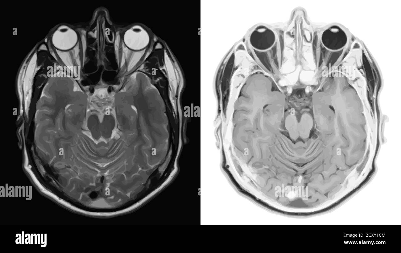 Realistic cross section of brain with CT scan, MRI Magnetic resonance imaging of head layer. Vector illustration. Stock Photohttps://www.alamy.com/image-license-details/?v=1https://www.alamy.com/realistic-cross-section-of-brain-with-ct-scan-mri-magnetic-resonance-imaging-of-head-layer-vector-illustration-image446834100.html
RF2GXY1CM–Realistic cross section of brain with CT scan, MRI Magnetic resonance imaging of head layer. Vector illustration.
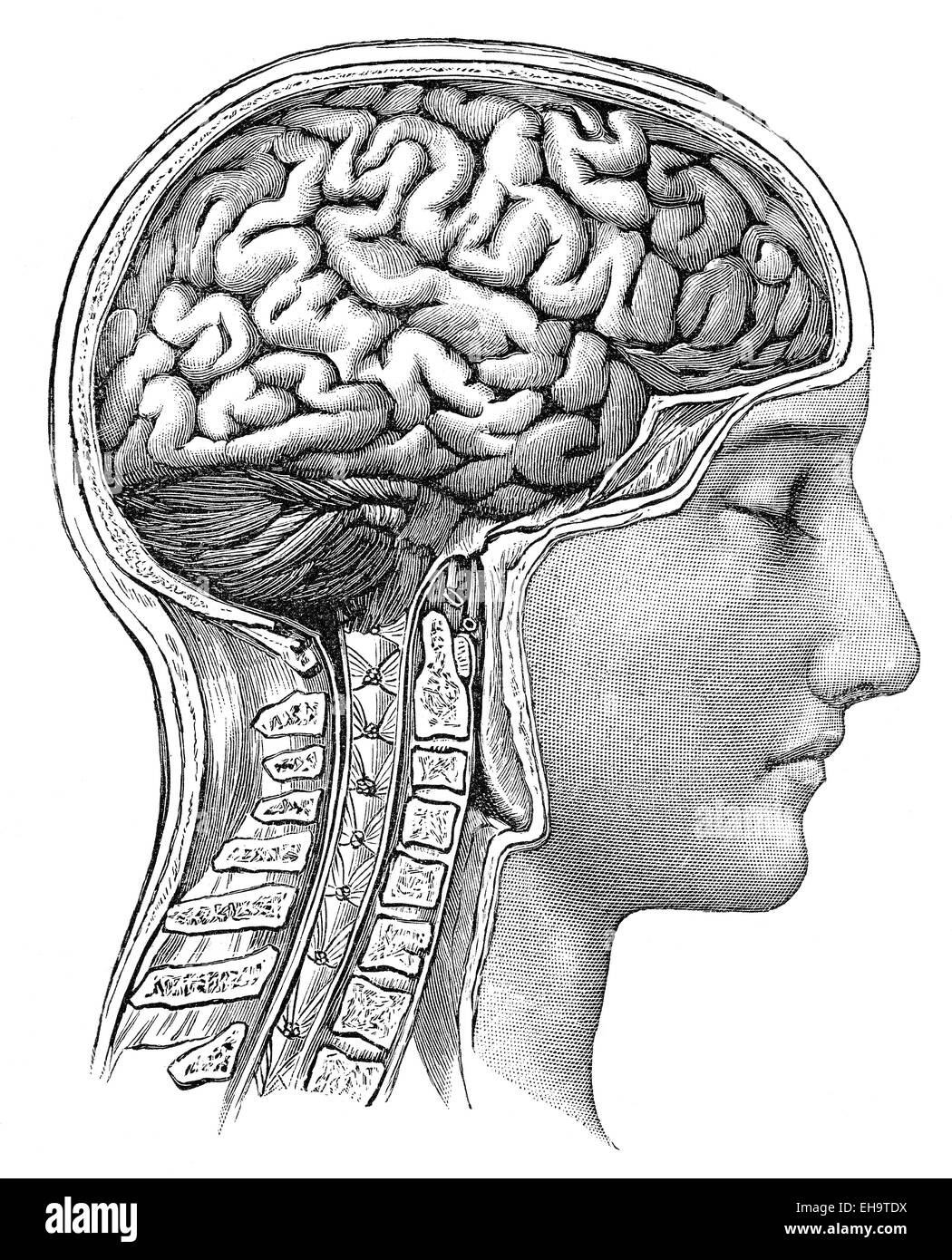 The human brain, health counselor, 19th Century, Stock Photohttps://www.alamy.com/image-license-details/?v=1https://www.alamy.com/stock-photo-the-human-brain-health-counselor-19th-century-79507398.html
RMEH9TDX–The human brain, health counselor, 19th Century,
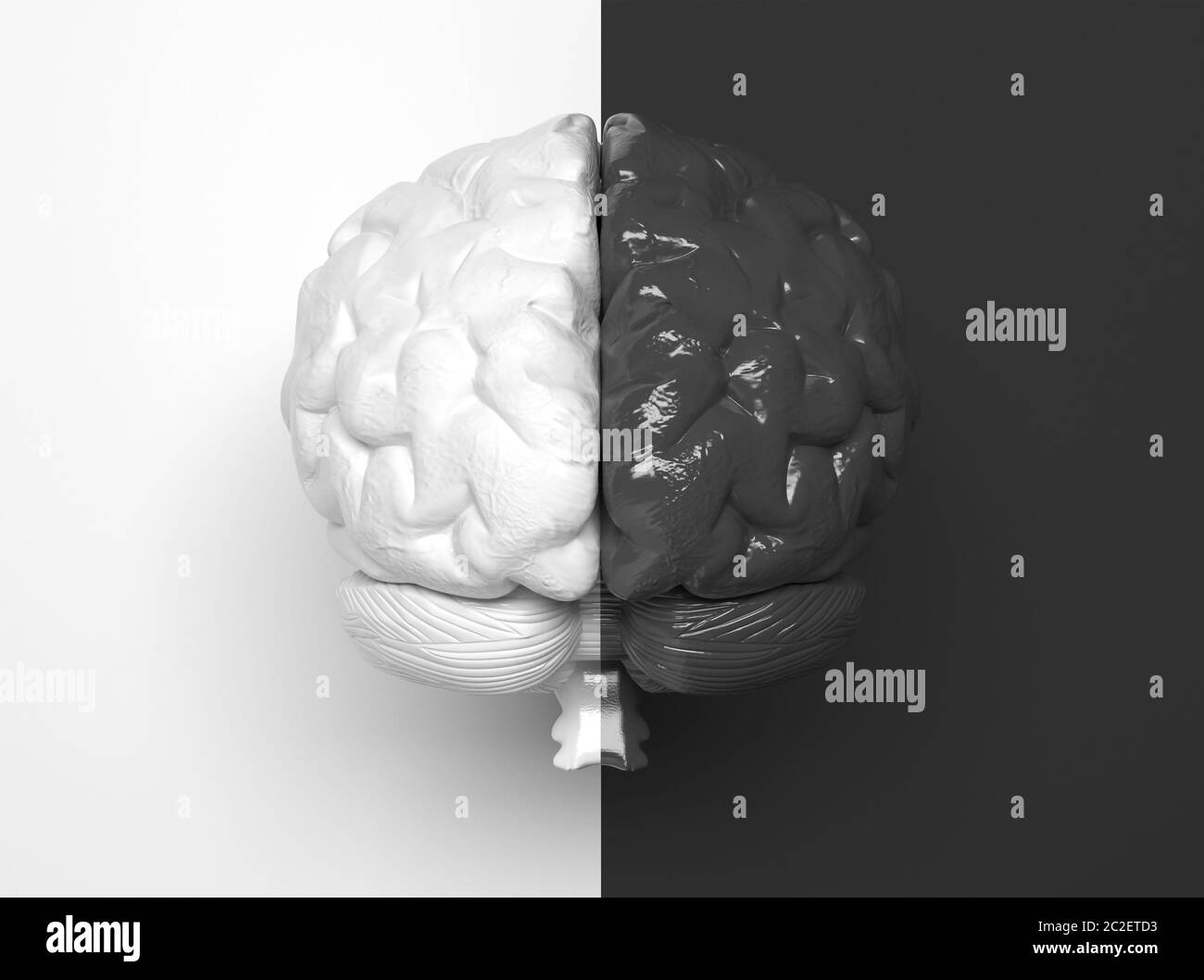 Black and white human brain divided in half into two parts in the middle. One half is white, the other half is black. Creative conceptual illustratio Stock Photohttps://www.alamy.com/image-license-details/?v=1https://www.alamy.com/black-and-white-human-brain-divided-in-half-into-two-parts-in-the-middle-one-half-is-white-the-other-half-is-black-creative-conceptual-illustratio-image362973551.html
RF2C2ETD3–Black and white human brain divided in half into two parts in the middle. One half is white, the other half is black. Creative conceptual illustratio
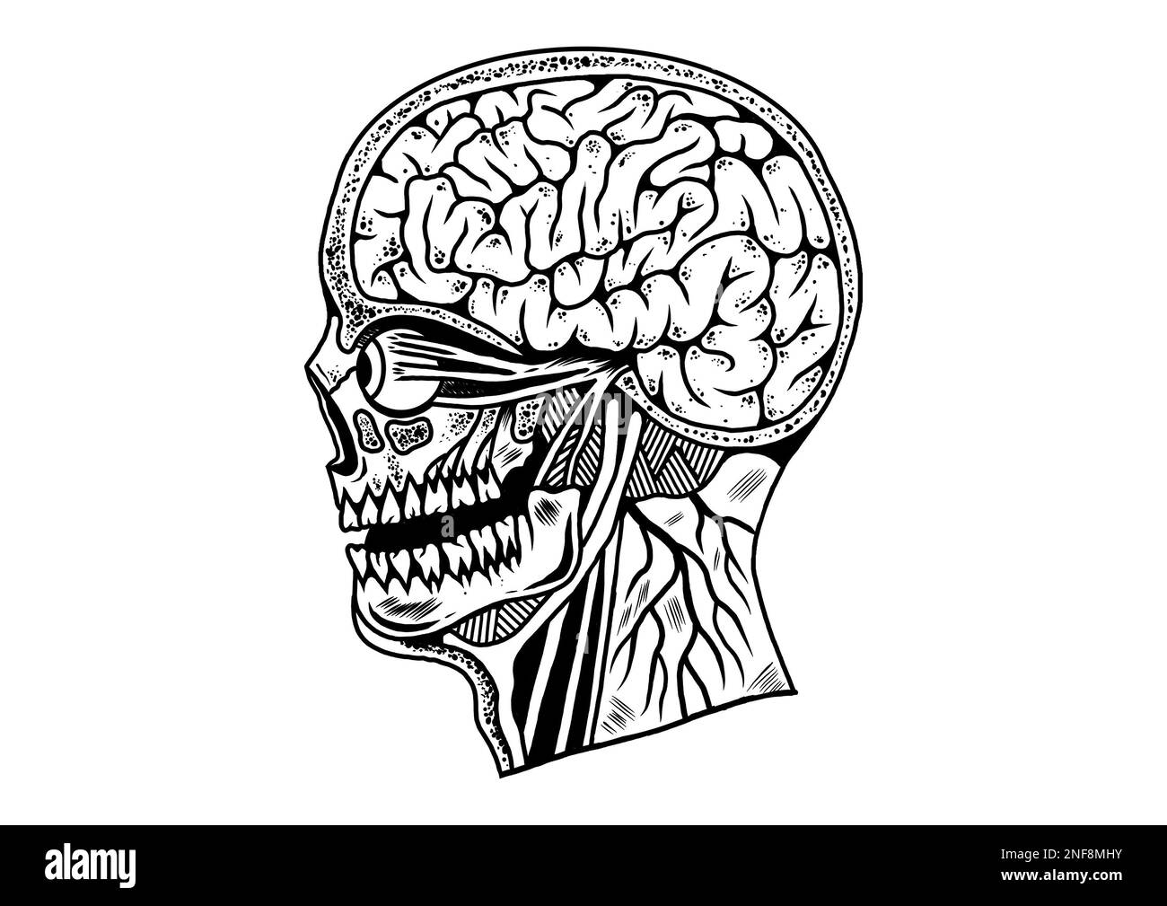 Old school traditional tattoo inspired cool graphic design illustration human head cross section anatomy in black and white Stock Photohttps://www.alamy.com/image-license-details/?v=1https://www.alamy.com/old-school-traditional-tattoo-inspired-cool-graphic-design-illustration-human-head-cross-section-anatomy-in-black-and-white-image525722679.html
RF2NF8MHY–Old school traditional tattoo inspired cool graphic design illustration human head cross section anatomy in black and white
 Head CT scan of an 85 year old female patient with signs of dementia Stock Photohttps://www.alamy.com/image-license-details/?v=1https://www.alamy.com/head-ct-scan-of-an-85-year-old-female-patient-with-signs-of-dementia-image442412924.html
RM2GKNJ5G–Head CT scan of an 85 year old female patient with signs of dementia
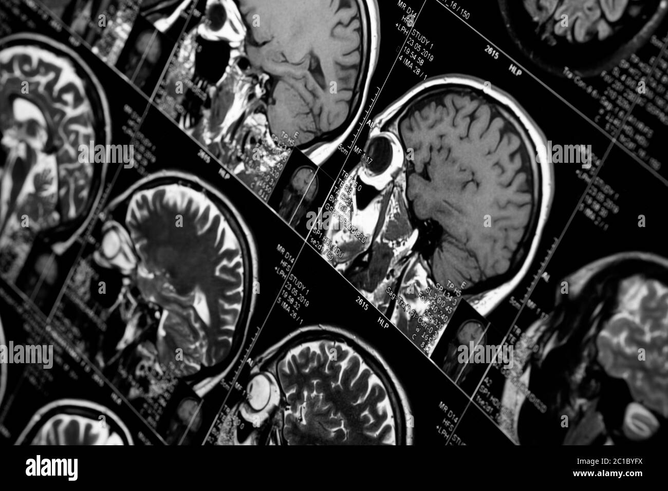 MRI scan of human brain, black and white Stock Photohttps://www.alamy.com/image-license-details/?v=1https://www.alamy.com/mri-scan-of-human-brain-black-and-white-image362295470.html
RF2C1BYFX–MRI scan of human brain, black and white
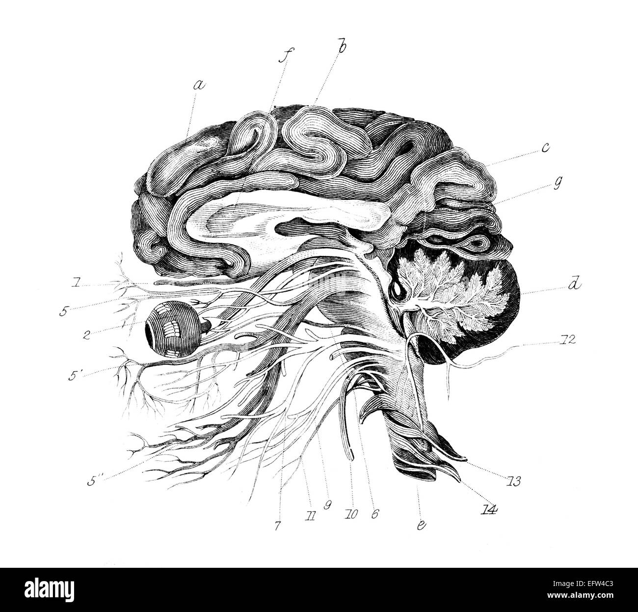 photographed from a book titled the 'National Encyclopedia', published in London in 1881. Copyright has expired on this artwork. Stock Photohttps://www.alamy.com/image-license-details/?v=1https://www.alamy.com/stock-photo-photographed-from-a-book-titled-the-national-encyclopedia-published-78613587.html
RFEFW4C3–photographed from a book titled the 'National Encyclopedia', published in London in 1881. Copyright has expired on this artwork.
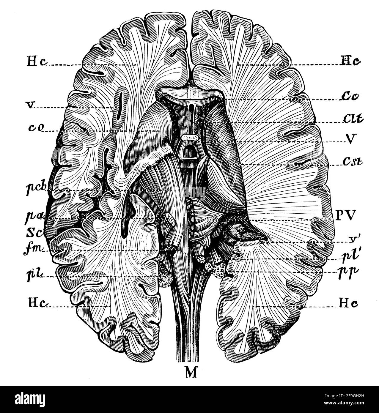 Cross section of the brain of human. Illustration of the 19th century. Germany. White background. Stock Photohttps://www.alamy.com/image-license-details/?v=1https://www.alamy.com/cross-section-of-the-brain-of-human-illustration-of-the-19th-century-germany-white-background-image418945369.html
RF2F9GH2H–Cross section of the brain of human. Illustration of the 19th century. Germany. White background.
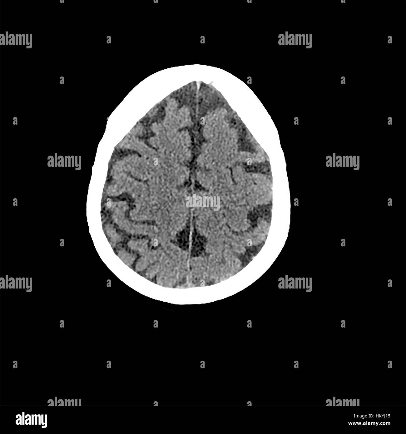 Head CT scan of an 85 year old female patient with signs of dementia Stock Photohttps://www.alamy.com/image-license-details/?v=1https://www.alamy.com/stock-photo-head-ct-scan-of-an-85-year-old-female-patient-with-signs-of-dementia-132757889.html
RMHKYJ15–Head CT scan of an 85 year old female patient with signs of dementia
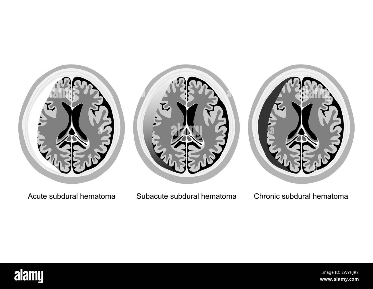 Three medical illustrations showing acute, subacute, and chronic subdural hematoma stages in brain injury. Stock Vectorhttps://www.alamy.com/image-license-details/?v=1https://www.alamy.com/three-medical-illustrations-showing-acute-subacute-and-chronic-subdural-hematoma-stages-in-brain-injury-image602136171.html
RF2WYHJR7–Three medical illustrations showing acute, subacute, and chronic subdural hematoma stages in brain injury.
 Skull Skeleton anatomy human. Skeletal system cross section. vector illustration Stock Vectorhttps://www.alamy.com/image-license-details/?v=1https://www.alamy.com/skull-skeleton-anatomy-human-skeletal-system-cross-section-vector-illustration-image222892384.html
RFPXHHE8–Skull Skeleton anatomy human. Skeletal system cross section. vector illustration
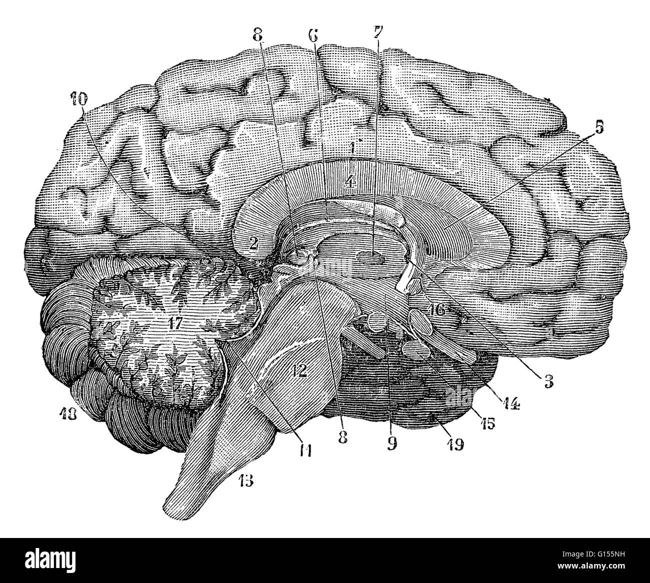 Illustration of a cross-section of the brain showing parts such as the cerebrum, cerebellum, corpus callosum, medulla oblongata, temporal lobe, hypothalamus, frontal lobe, limbic system, corpus callosum, parietal lobe, thalamus, occipital lobe, midbrain, Stock Photohttps://www.alamy.com/image-license-details/?v=1https://www.alamy.com/stock-photo-illustration-of-a-cross-section-of-the-brain-showing-parts-such-as-103991149.html
RMG155NH–Illustration of a cross-section of the brain showing parts such as the cerebrum, cerebellum, corpus callosum, medulla oblongata, temporal lobe, hypothalamus, frontal lobe, limbic system, corpus callosum, parietal lobe, thalamus, occipital lobe, midbrain,
 set of serial MRI scans of sixty years old caucasian female head in sagittal and horizontal planes Stock Photohttps://www.alamy.com/image-license-details/?v=1https://www.alamy.com/set-of-serial-mri-scans-of-sixty-years-old-caucasian-female-head-in-sagittal-and-horizontal-planes-image355947280.html
RF2BK2PAT–set of serial MRI scans of sixty years old caucasian female head in sagittal and horizontal planes
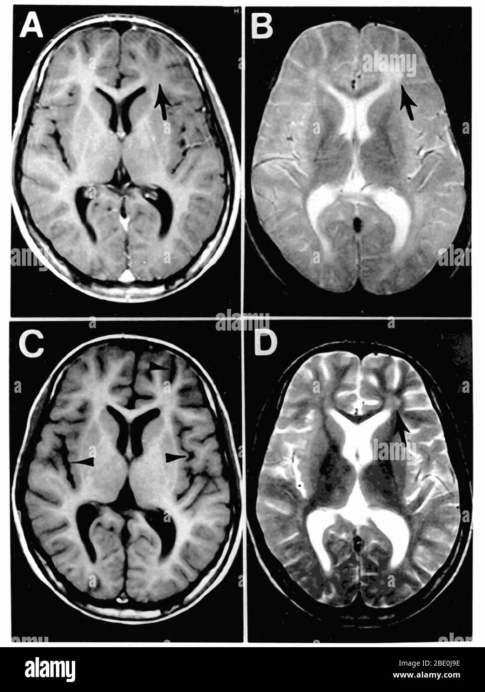 Subacute sclerosing panencephalitis (A complication of measles infection). Figure 1. MRI scans of the brain at the time of presentation in the neurology clinic (A and B) and 3 months later (C and D). Panels A and C are T1-weighted images; B and D are T2-weighted images. The initial MRI scan (A and B) reveals a focal abnormality in the subcortical white matter of the left frontal lobe, consisting of a hypointense signal on the T1-weighted image (arrow in A) and a hyperintense signal on the T2-weighted image (arrow in B). In the follow-up scan, the focal abnormality in the left frontal lobe is l Stock Photohttps://www.alamy.com/image-license-details/?v=1https://www.alamy.com/subacute-sclerosing-panencephalitis-a-complication-of-measles-infection-figure-1-mri-scans-of-the-brain-at-the-time-of-presentation-in-the-neurology-clinic-a-and-b-and-3-months-later-c-and-d-panels-a-and-c-are-t1-weighted-images-b-and-d-are-t2-weighted-images-the-initial-mri-scan-a-and-b-reveals-a-focal-abnormality-in-the-subcortical-white-matter-of-the-left-frontal-lobe-consisting-of-a-hypointense-signal-on-the-t1-weighted-image-arrow-in-a-and-a-hyperintense-signal-on-the-t2-weighted-image-arrow-in-b-in-the-follow-up-scan-the-focal-abnormality-in-the-left-frontal-lobe-is-l-image352826922.html
RM2BE0J9E–Subacute sclerosing panencephalitis (A complication of measles infection). Figure 1. MRI scans of the brain at the time of presentation in the neurology clinic (A and B) and 3 months later (C and D). Panels A and C are T1-weighted images; B and D are T2-weighted images. The initial MRI scan (A and B) reveals a focal abnormality in the subcortical white matter of the left frontal lobe, consisting of a hypointense signal on the T1-weighted image (arrow in A) and a hyperintense signal on the T2-weighted image (arrow in B). In the follow-up scan, the focal abnormality in the left frontal lobe is l
 Festival of Britain human brain exhibit, South Bank, London Stock Photohttps://www.alamy.com/image-license-details/?v=1https://www.alamy.com/festival-of-britain-human-brain-exhibit-south-bank-london-image501422086.html
RM2M3NMYJ–Festival of Britain human brain exhibit, South Bank, London
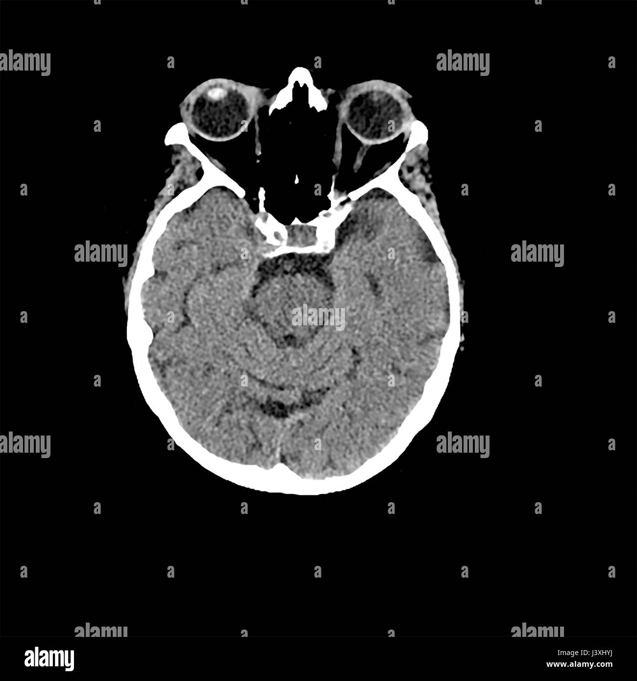 Head CT scan of an 85 year old female patient with signs of dementia Stock Photohttps://www.alamy.com/image-license-details/?v=1https://www.alamy.com/stock-photo-head-ct-scan-of-an-85-year-old-female-patient-with-signs-of-dementia-140111766.html
RFJ3XHYJ–Head CT scan of an 85 year old female patient with signs of dementia
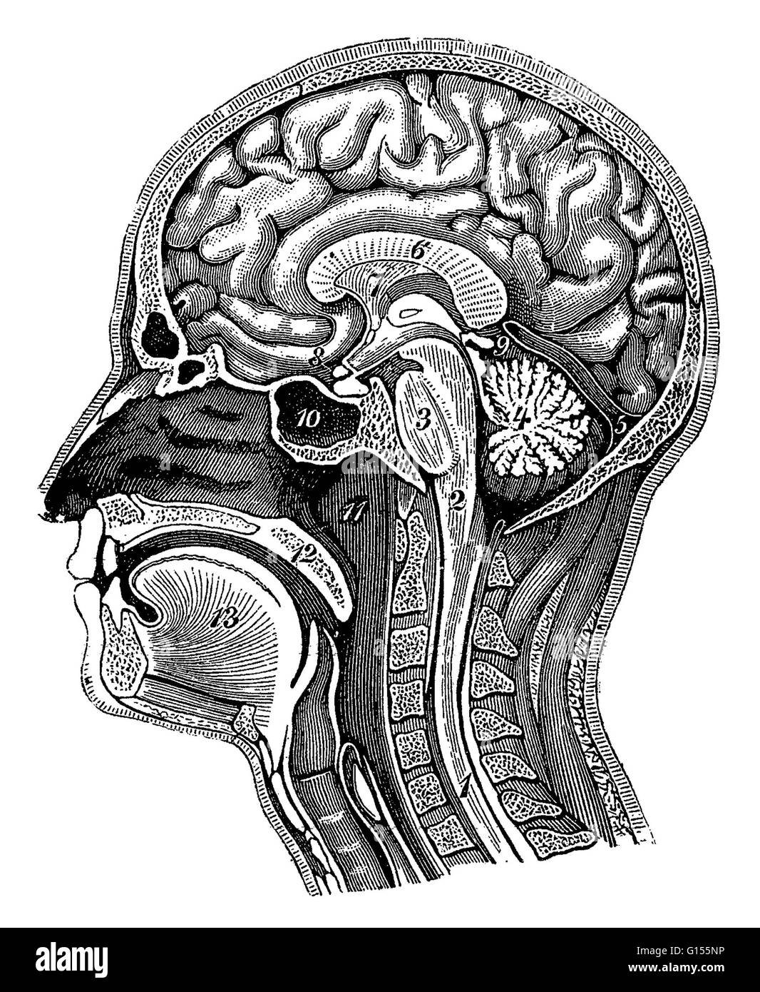 Illustration of a cross-section of the head showing the brain showing the spinal cord, medulla oblongata, pons, cerebellum, transverse fissure, corpus callosum, septum pellucidum, optic chiasm, pineal gland, sphenoid sinus, nasopharynx, soft palate and to Stock Photohttps://www.alamy.com/image-license-details/?v=1https://www.alamy.com/stock-photo-illustration-of-a-cross-section-of-the-head-showing-the-brain-showing-103991154.html
RMG155NP–Illustration of a cross-section of the head showing the brain showing the spinal cord, medulla oblongata, pons, cerebellum, transverse fissure, corpus callosum, septum pellucidum, optic chiasm, pineal gland, sphenoid sinus, nasopharynx, soft palate and to
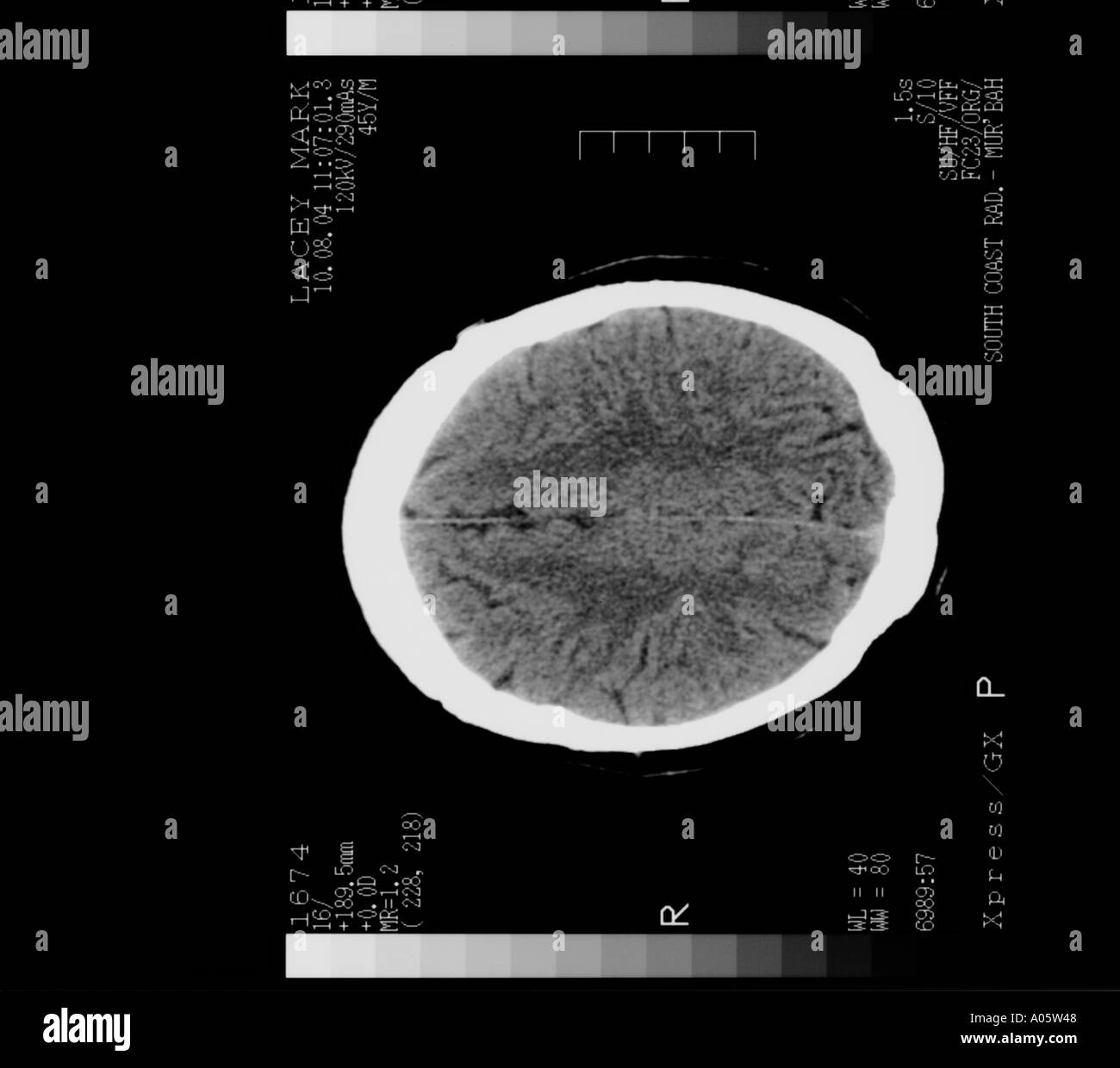 brain scan cross section Stock Photohttps://www.alamy.com/image-license-details/?v=1https://www.alamy.com/brain-scan-cross-section-image3228999.html
RMA05W48–brain scan cross section
RF2X1Y0H7–Pancreas icon vector illustration simple design
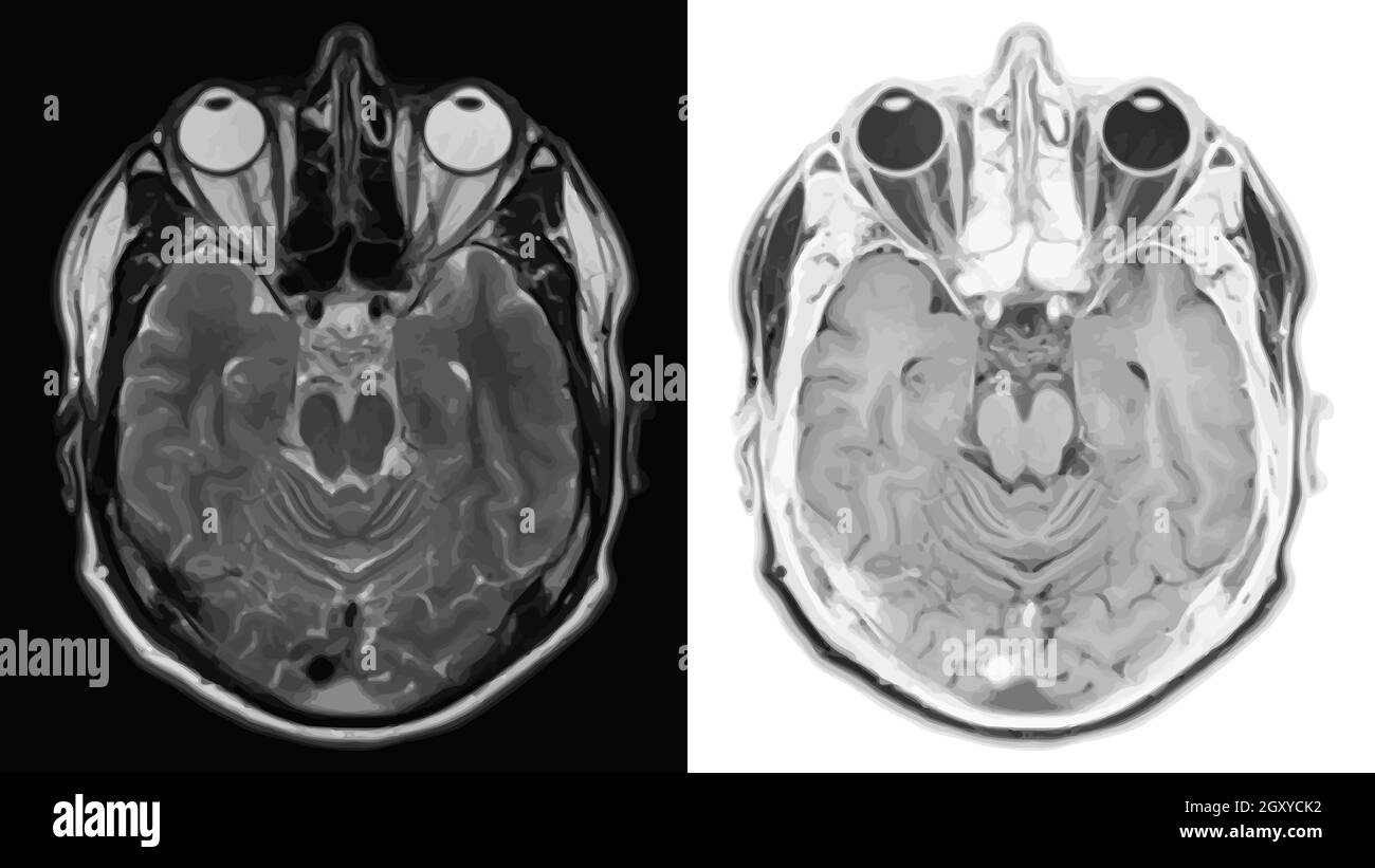 Realistic cross section of brain with CT scan, MRI Magnetic resonance imaging of head layer. Vector illustration. Stock Vectorhttps://www.alamy.com/image-license-details/?v=1https://www.alamy.com/realistic-cross-section-of-brain-with-ct-scan-mri-magnetic-resonance-imaging-of-head-layer-vector-illustration-image446842902.html
RF2GXYCK2–Realistic cross section of brain with CT scan, MRI Magnetic resonance imaging of head layer. Vector illustration.
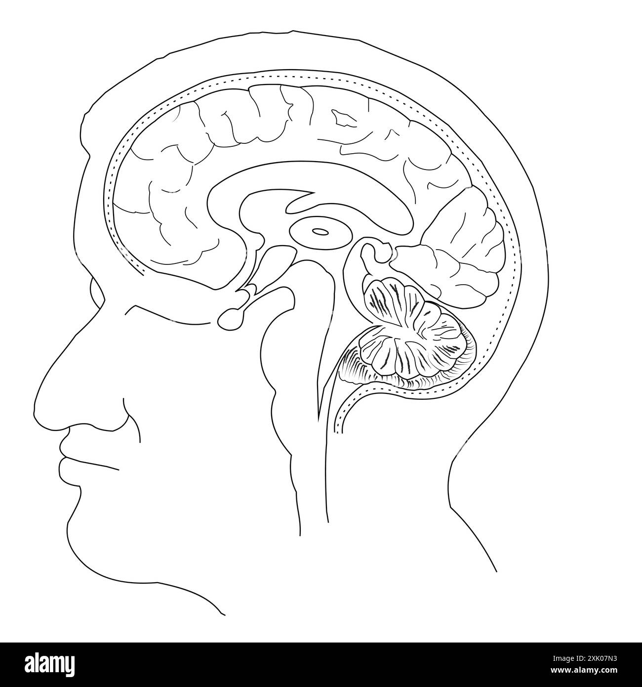 Human Brain Cross-Section Illustration without label Stock Vectorhttps://www.alamy.com/image-license-details/?v=1https://www.alamy.com/human-brain-cross-section-illustration-without-label-image614047423.html
RF2XK07N3–Human Brain Cross-Section Illustration without label
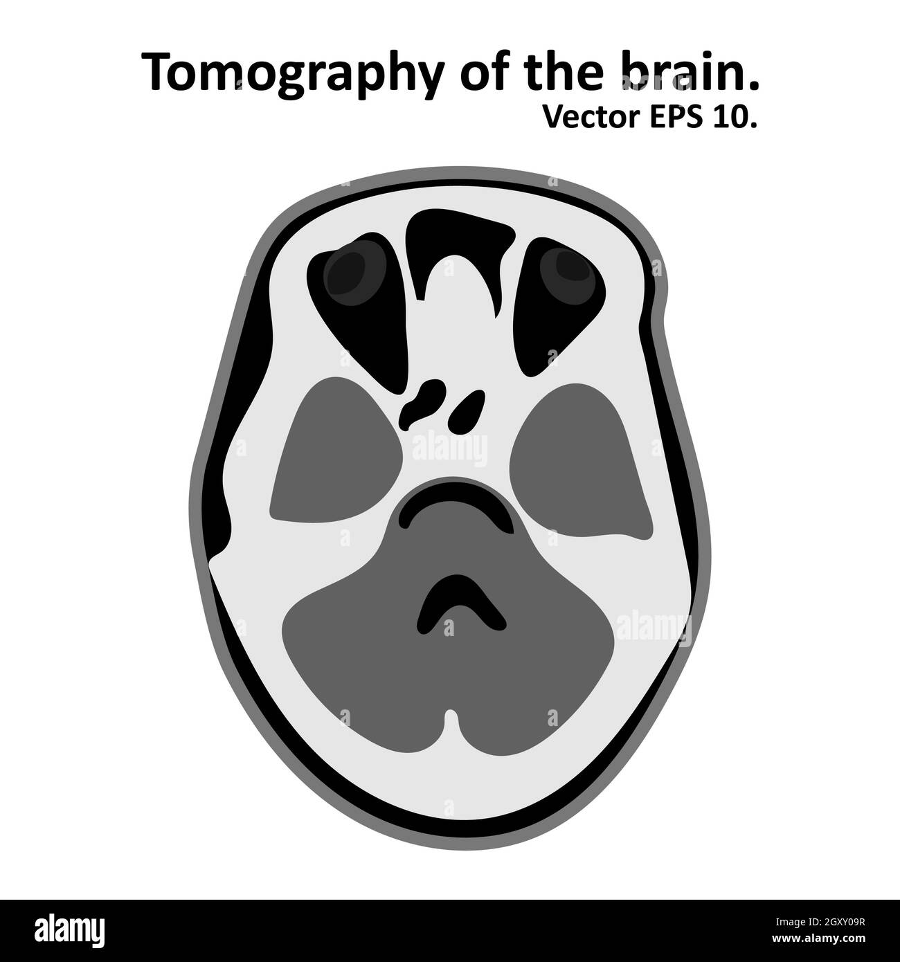 Cross section of the brain with magnetic resonance imaging. MRI / CT scan. Vector EPS10. Stock Photohttps://www.alamy.com/image-license-details/?v=1https://www.alamy.com/cross-section-of-the-brain-with-magnetic-resonance-imaging-mri-ct-scan-vector-eps10-image446833235.html
RF2GXY09R–Cross section of the brain with magnetic resonance imaging. MRI / CT scan. Vector EPS10.
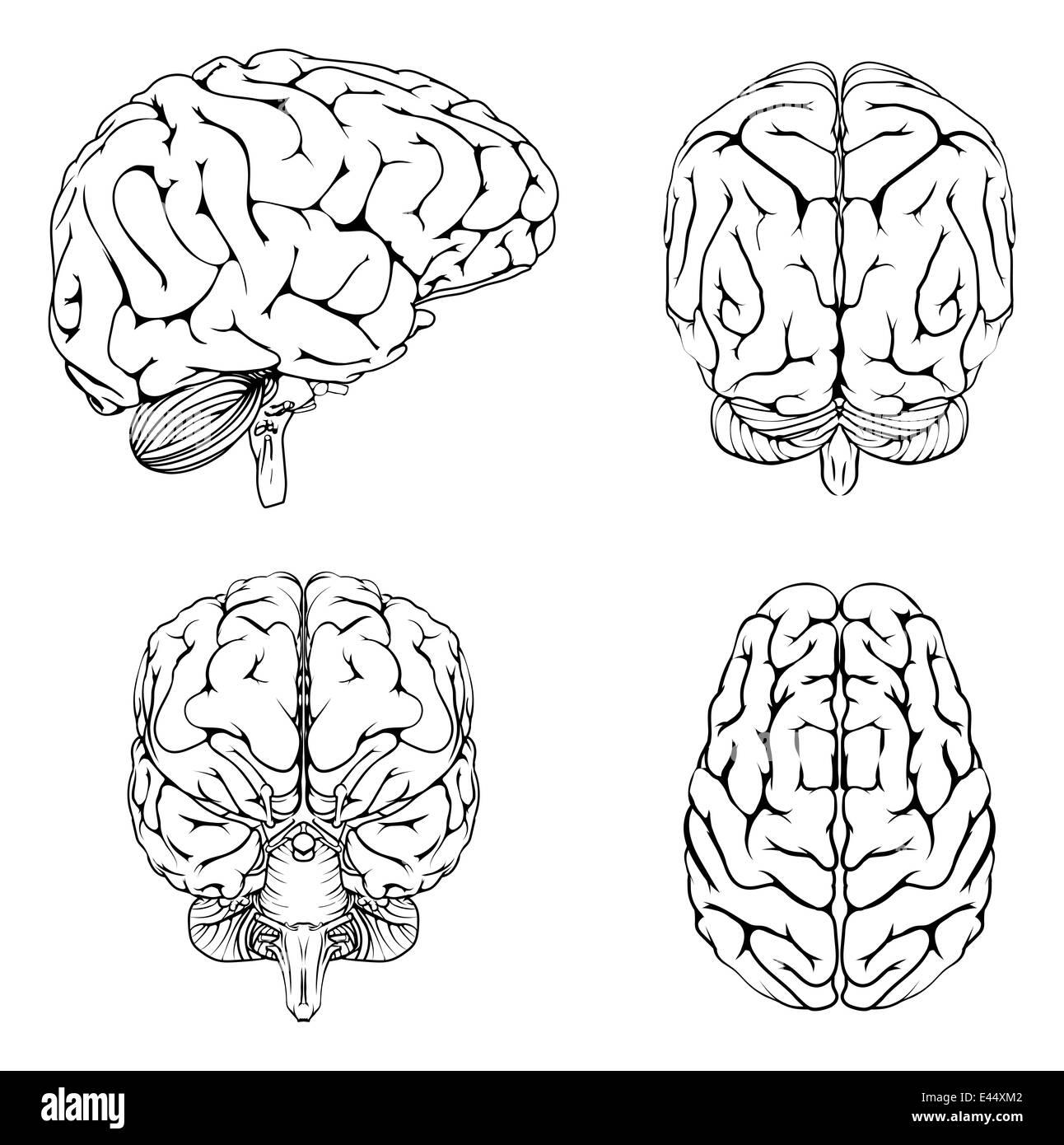 A diagram of a brain from the top side front and back in outline Stock Photohttps://www.alamy.com/image-license-details/?v=1https://www.alamy.com/stock-photo-a-diagram-of-a-brain-from-the-top-side-front-and-back-in-outline-71408850.html
RFE44XM2–A diagram of a brain from the top side front and back in outline
 3d rendering. 3d illustration . Human parts with black background .Hand Stock Photohttps://www.alamy.com/image-license-details/?v=1https://www.alamy.com/3d-rendering-3d-illustration-human-parts-with-black-background-hand-image360340181.html
RF2BX6WG5–3d rendering. 3d illustration . Human parts with black background .Hand
 Shown are the regions of the human brain affected by boxing, side view (top) and cross-section (bottom). Shown are the skull, cerebellum, and the base Stock Photohttps://www.alamy.com/image-license-details/?v=1https://www.alamy.com/stock-photo-shown-are-the-regions-of-the-human-brain-affected-by-boxing-side-view-130775909.html
RFHGNA05–Shown are the regions of the human brain affected by boxing, side view (top) and cross-section (bottom). Shown are the skull, cerebellum, and the base
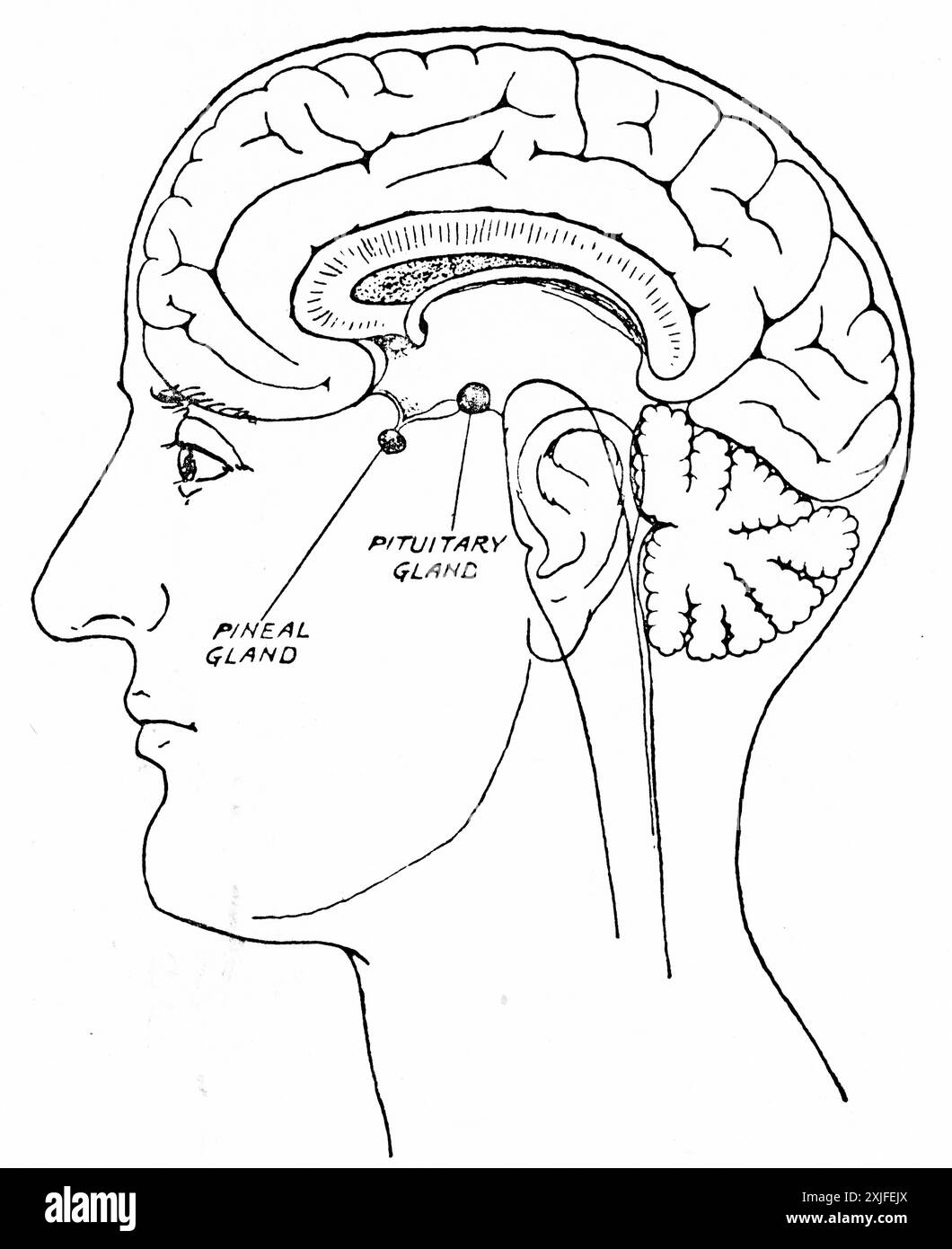 A cross section of the human head showing the brain, pituitary and pineal glands. Stock Photohttps://www.alamy.com/image-license-details/?v=1https://www.alamy.com/a-cross-section-of-the-human-head-showing-the-brain-pituitary-and-pineal-glands-image613767474.html
RM2XJFEJX–A cross section of the human head showing the brain, pituitary and pineal glands.
 Vertical section of the brain through the two hemispheres. Illustration of the 19th century. Germany. White background. Stock Photohttps://www.alamy.com/image-license-details/?v=1https://www.alamy.com/vertical-section-of-the-brain-through-the-two-hemispheres-illustration-of-the-19th-century-germany-white-background-image418945372.html
RF2F9GH2M–Vertical section of the brain through the two hemispheres. Illustration of the 19th century. Germany. White background.
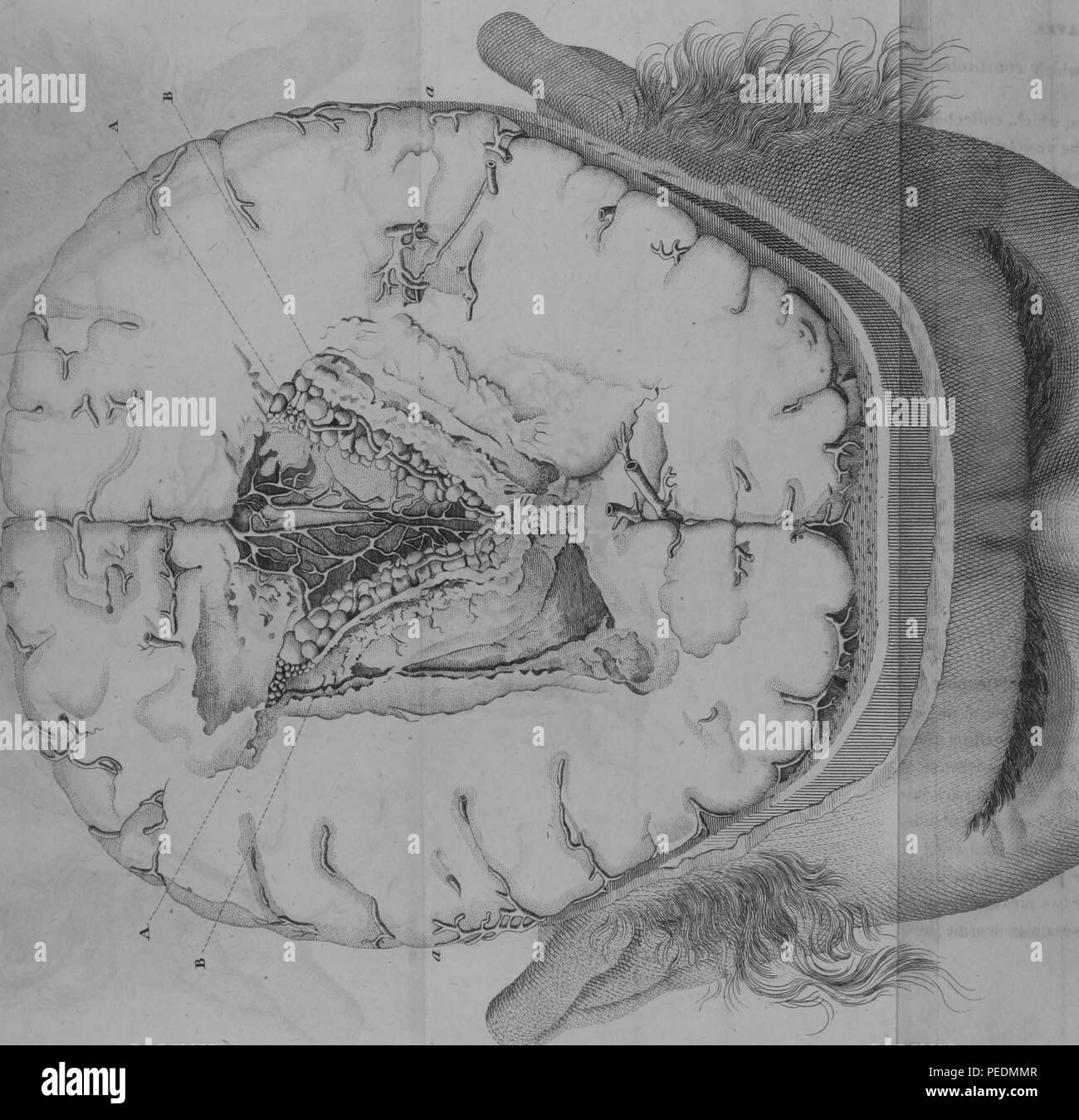 Black and white print illustrating a cross-section of the lateral ventricles of a human brain, with alphabetized figures indicating the sites of ventricular worm infestations, 1825. Courtesy Internet Archive. () Stock Photohttps://www.alamy.com/image-license-details/?v=1https://www.alamy.com/black-and-white-print-illustrating-a-cross-section-of-the-lateral-ventricles-of-a-human-brain-with-alphabetized-figures-indicating-the-sites-of-ventricular-worm-infestations-1825-courtesy-internet-archive-image215431239.html
RMPEDMMR–Black and white print illustrating a cross-section of the lateral ventricles of a human brain, with alphabetized figures indicating the sites of ventricular worm infestations, 1825. Courtesy Internet Archive. ()
RFWR5YN5–Brain dementia icon. Outline brain dementia vector icon for web design isolated on white background
 . Elements of physiological psychology; a treatise of the activities and nature of the mind, from the physical and experimental points of view . dorsal horns of gray matter. The most strikingfeature of this section is the appearance of large numbers of fibres,crossing from the ventral column of each side to the lateral columnof the other. This is the crossing or decussation of the pyramids;here motor fibres from the left hemisphere cross to the right side ofthe cord, and vice versa, so that the left hemisphere of the brain con-trols the right half of the body. This section shows another pe- IN Stock Photohttps://www.alamy.com/image-license-details/?v=1https://www.alamy.com/elements-of-physiological-psychology-a-treatise-of-the-activities-and-nature-of-the-mind-from-the-physical-and-experimental-points-of-view-dorsal-horns-of-gray-matter-the-most-strikingfeature-of-this-section-is-the-appearance-of-large-numbers-of-fibrescrossing-from-the-ventral-column-of-each-side-to-the-lateral-columnof-the-other-this-is-the-crossing-or-decussation-of-the-pyramidshere-motor-fibres-from-the-left-hemisphere-cross-to-the-right-side-ofthe-cord-and-vice-versa-so-that-the-left-hemisphere-of-the-brain-con-trols-the-right-half-of-the-body-this-section-shows-another-pe-in-image369747365.html
RM2CDFCF1–. Elements of physiological psychology; a treatise of the activities and nature of the mind, from the physical and experimental points of view . dorsal horns of gray matter. The most strikingfeature of this section is the appearance of large numbers of fibres,crossing from the ventral column of each side to the lateral columnof the other. This is the crossing or decussation of the pyramids;here motor fibres from the left hemisphere cross to the right side ofthe cord, and vice versa, so that the left hemisphere of the brain con-trols the right half of the body. This section shows another pe- IN
 Open mri machine scanning a patient lying inside for medical diagnosis Stock Vectorhttps://www.alamy.com/image-license-details/?v=1https://www.alamy.com/open-mri-machine-scanning-a-patient-lying-inside-for-medical-diagnosis-image612303999.html
RF2XG4RYY–Open mri machine scanning a patient lying inside for medical diagnosis
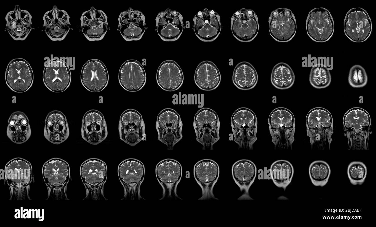 set of serial MRI scans of sixty years old caucasian female head in frontal and horizontal planes Stock Photohttps://www.alamy.com/image-license-details/?v=1https://www.alamy.com/set-of-serial-mri-scans-of-sixty-years-old-caucasian-female-head-in-frontal-and-horizontal-planes-image355564707.html
RF2BJDABF–set of serial MRI scans of sixty years old caucasian female head in frontal and horizontal planes
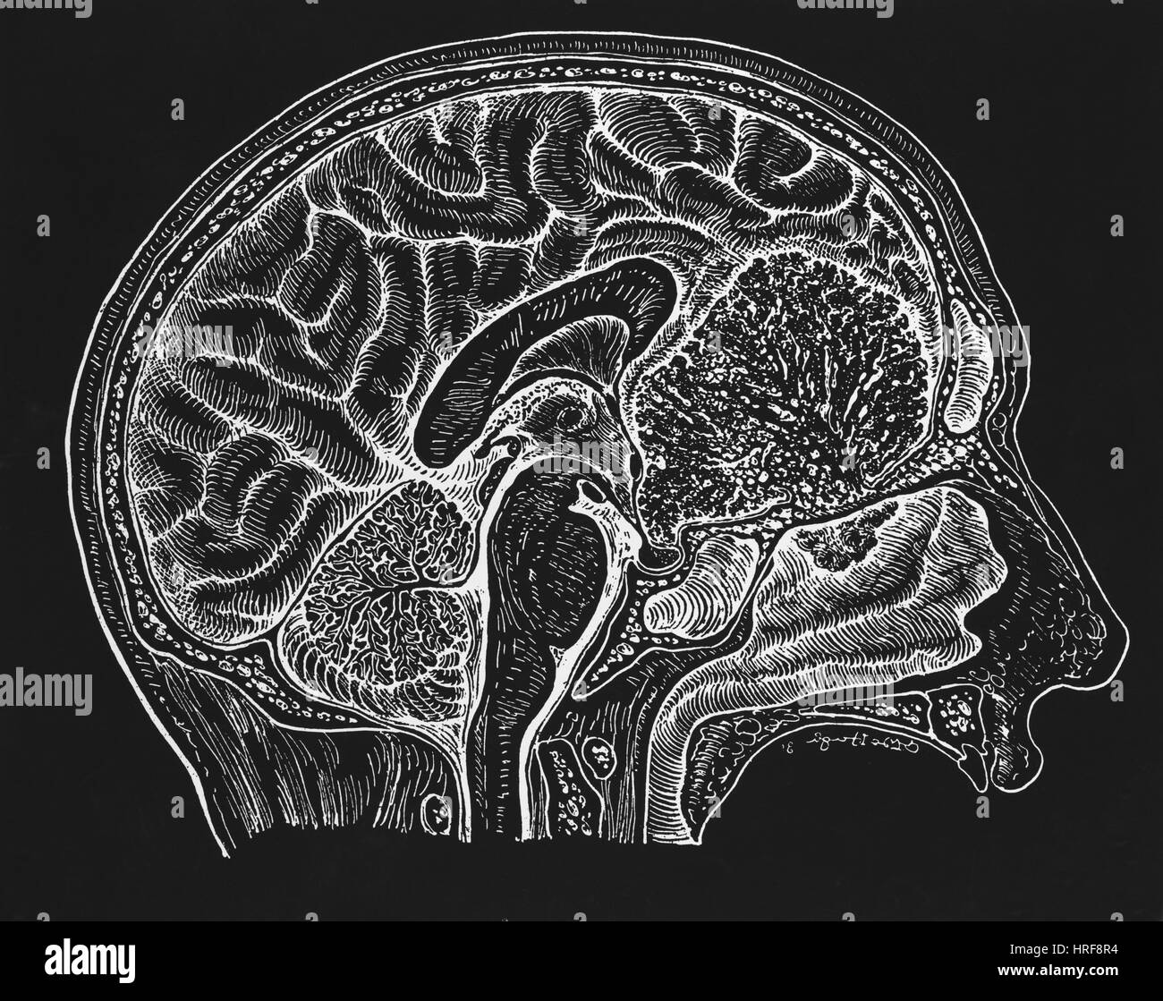 Brain, cross-section Stock Photohttps://www.alamy.com/image-license-details/?v=1https://www.alamy.com/stock-photo-brain-cross-section-134945864.html
RMHRF8R4–Brain, cross-section
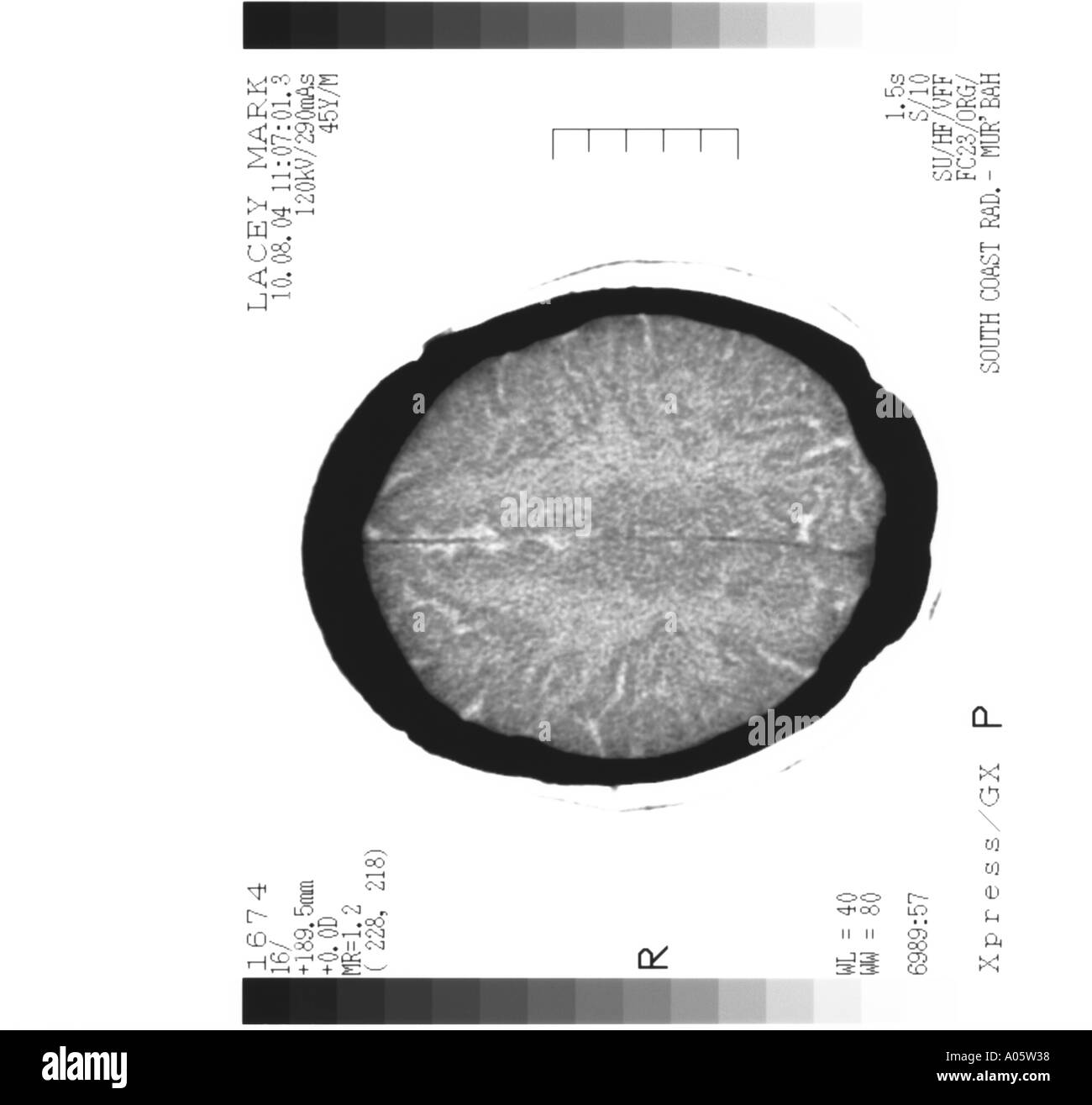 brain scan cross section Stock Photohttps://www.alamy.com/image-license-details/?v=1https://www.alamy.com/brain-scan-cross-section-image3228983.html
RMA05W38–brain scan cross section
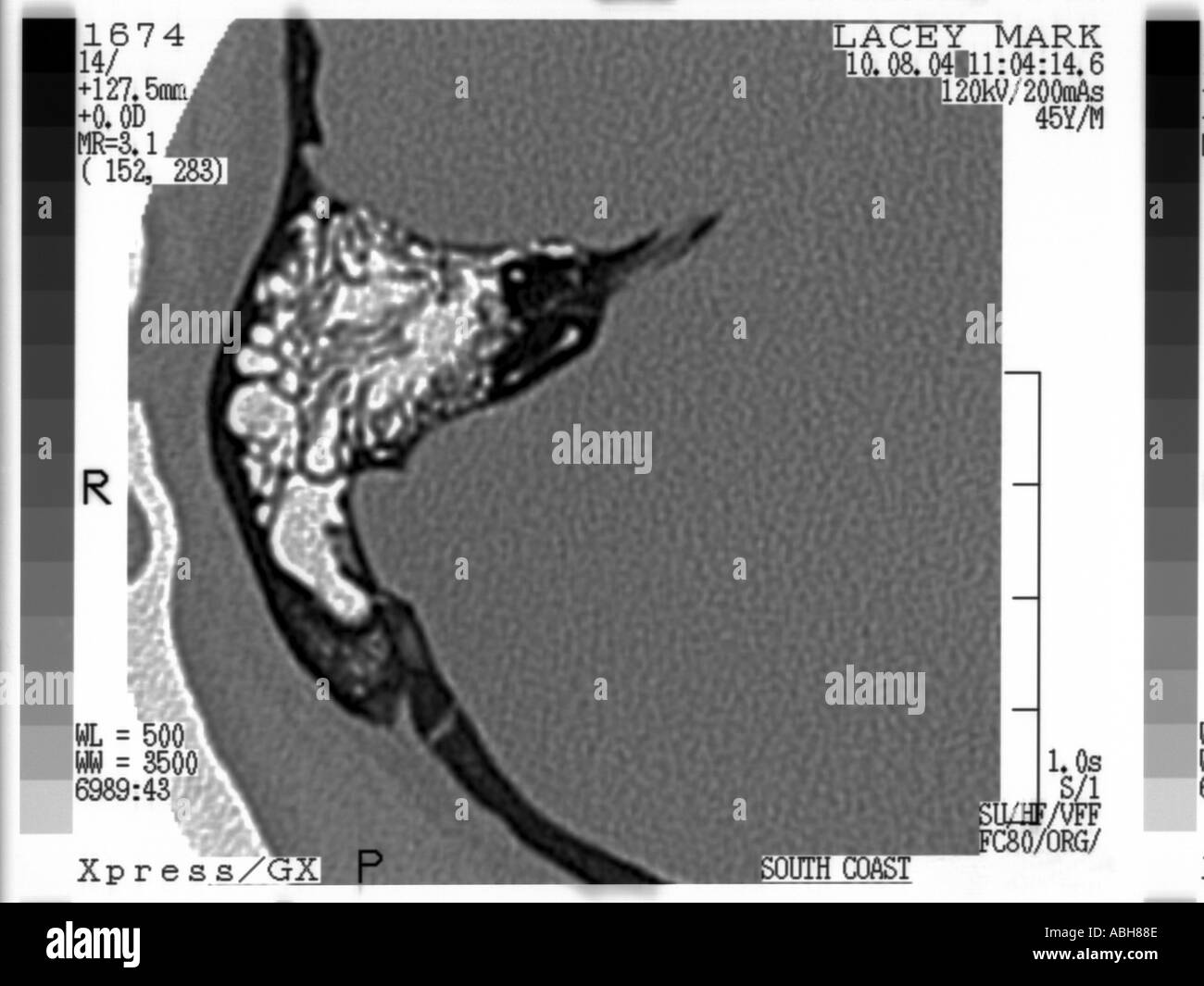 brain scan of skull with section through inner ear Stock Photohttps://www.alamy.com/image-license-details/?v=1https://www.alamy.com/brain-scan-of-skull-with-section-through-inner-ear-image2381965.html
RMABH88E–brain scan of skull with section through inner ear
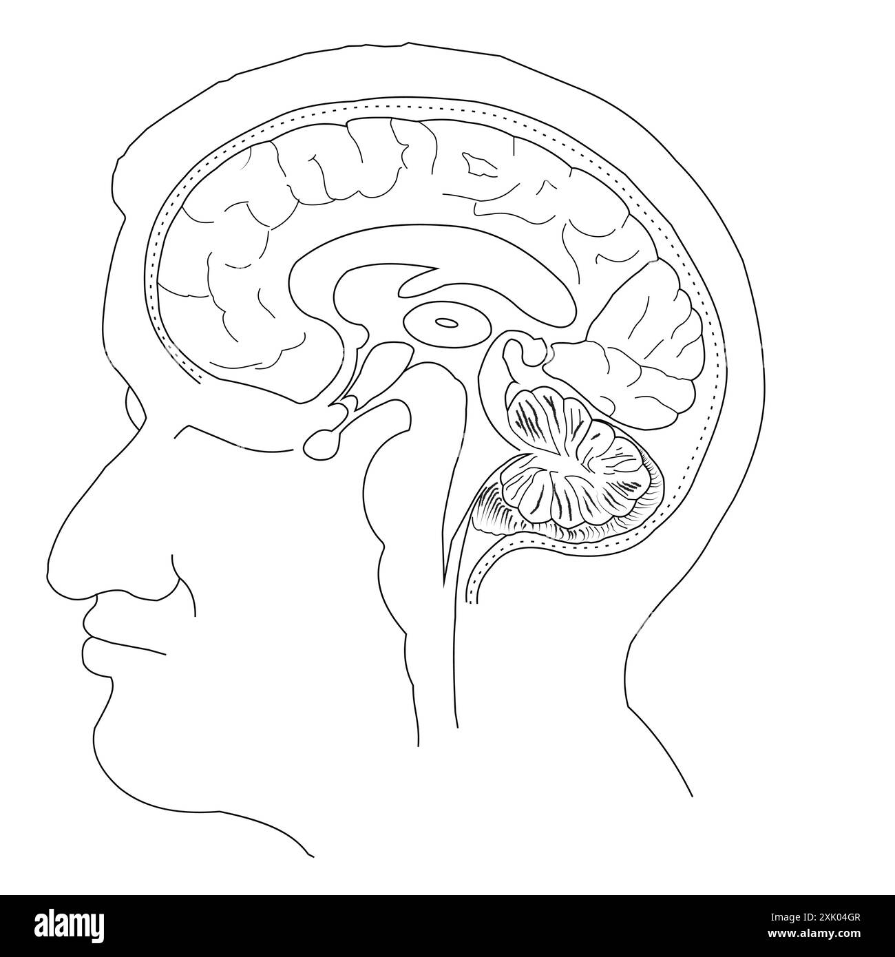 Human Brain Cross-Section Vector Illustration without label Stock Vectorhttps://www.alamy.com/image-license-details/?v=1https://www.alamy.com/human-brain-cross-section-vector-illustration-without-label-image614044951.html
RF2XK04GR–Human Brain Cross-Section Vector Illustration without label
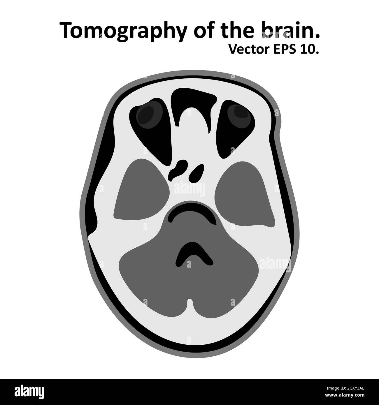 Cross section of the brain with magnetic resonance imaging. MRI / CT scan. Vector EPS10. Stock Vectorhttps://www.alamy.com/image-license-details/?v=1https://www.alamy.com/cross-section-of-the-brain-with-magnetic-resonance-imaging-mri-ct-scan-vector-eps10-image446835606.html
RF2GXY3AE–Cross section of the brain with magnetic resonance imaging. MRI / CT scan. Vector EPS10.
 3d rendering. 3d illustration . Human parts with black background .Hand Stock Photohttps://www.alamy.com/image-license-details/?v=1https://www.alamy.com/3d-rendering-3d-illustration-human-parts-with-black-background-hand-image360340179.html
RF2BX6WG3–3d rendering. 3d illustration . Human parts with black background .Hand
RFWHE8F0–Brain dementia icon. Simple illustration of brain dementia vector icon for web design isolated on white background
 . Elements of physiological psychology; a treatise of the activities and nature of the mind, from the physical and experimental points of view . Fig. 38.—Cross-section of the Mid-Brain at tlie Level of the Posterior Quadrigemina. (Mar-burg.) In place of the fourth ventricle we have here the narrow Aqueduct, Aq. Abovethis, on each side, lie the posterior quadrigemina, Qp. The aqueduct is immediatelysurrounded by gray matter, below which is Flm, the median longitudinal bundle. Belowthis is a large mass of decussating fibres, D, from the superior cerebellar peduncles; belowand to the side of this Stock Photohttps://www.alamy.com/image-license-details/?v=1https://www.alamy.com/elements-of-physiological-psychology-a-treatise-of-the-activities-and-nature-of-the-mind-from-the-physical-and-experimental-points-of-view-fig-38cross-section-of-the-mid-brain-at-tlie-level-of-the-posterior-quadrigemina-mar-burg-in-place-of-the-fourth-ventricle-we-have-here-the-narrow-aqueduct-aq-abovethis-on-each-side-lie-the-posterior-quadrigemina-qp-the-aqueduct-is-immediatelysurrounded-by-gray-matter-below-which-is-flm-the-median-longitudinal-bundle-belowthis-is-a-large-mass-of-decussating-fibres-d-from-the-superior-cerebellar-peduncles-belowand-to-the-side-of-this-image369747130.html
RM2CDFC6J–. Elements of physiological psychology; a treatise of the activities and nature of the mind, from the physical and experimental points of view . Fig. 38.—Cross-section of the Mid-Brain at tlie Level of the Posterior Quadrigemina. (Mar-burg.) In place of the fourth ventricle we have here the narrow Aqueduct, Aq. Abovethis, on each side, lie the posterior quadrigemina, Qp. The aqueduct is immediatelysurrounded by gray matter, below which is Flm, the median longitudinal bundle. Belowthis is a large mass of decussating fibres, D, from the superior cerebellar peduncles; belowand to the side of this
RF2AYJ4D2–Gland thyroid icon. Outline gland thyroid vector icon for web design isolated on white background
 Brain cross-section Stock Photohttps://www.alamy.com/image-license-details/?v=1https://www.alamy.com/stock-photo-brain-cross-section-134995070.html
RMHRHFGE–Brain cross-section
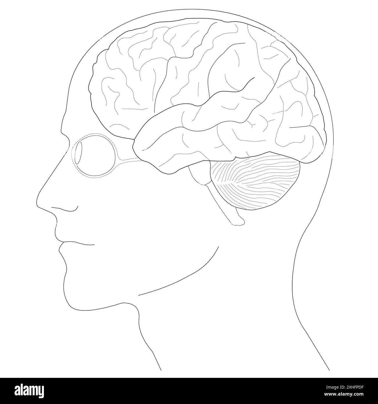 Human brain anatomy structure illustration Stock Vectorhttps://www.alamy.com/image-license-details/?v=1https://www.alamy.com/human-brain-anatomy-structure-illustration-image613158939.html
RF2XHFPDF–Human brain anatomy structure illustration
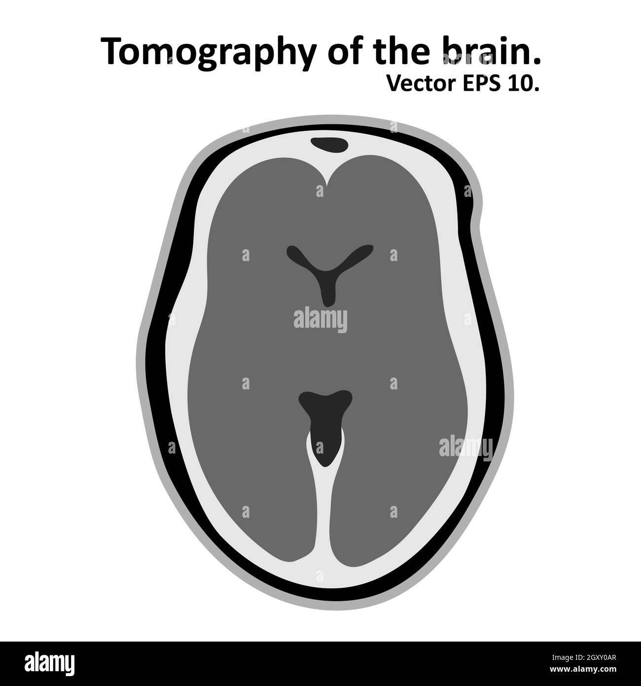 Magnetic resonance imaging of the brain. Cross section of the brain with MRI / CT scan. Vector EPS10. Stock Photohttps://www.alamy.com/image-license-details/?v=1https://www.alamy.com/magnetic-resonance-imaging-of-the-brain-cross-section-of-the-brain-with-mri-ct-scan-vector-eps10-image446833263.html
RF2GXY0AR–Magnetic resonance imaging of the brain. Cross section of the brain with MRI / CT scan. Vector EPS10.
 3d rendering. 3d illustration . Human parts with black background .Legs Stock Photohttps://www.alamy.com/image-license-details/?v=1https://www.alamy.com/3d-rendering-3d-illustration-human-parts-with-black-background-legs-image360340184.html
RF2BX6WG8–3d rendering. 3d illustration . Human parts with black background .Legs
 . Mammalian anatomy : with special reference to the cat . Mammals; Anatomy, Comparative; Cats. THE NERVOUS SYSTEM. 1S7 inferior colliculi. The anterior pair lie nearer to the midline than the posterior pair, which are slightly separated by a de- pression occupied by the middle portion of the central lobe of the cerebellum. The posterior pair are united by a white commissure. The posterior commissure of the brain unites the cranial portions of the anterior pair (Fig. 95). Its cut end may be seen ventrad to the base of the pineal gland.. cm oc v Fig. 97. Cross-section of the Brain in the Plane x Stock Photohttps://www.alamy.com/image-license-details/?v=1https://www.alamy.com/mammalian-anatomy-with-special-reference-to-the-cat-mammals-anatomy-comparative-cats-the-nervous-system-1s7-inferior-colliculi-the-anterior-pair-lie-nearer-to-the-midline-than-the-posterior-pair-which-are-slightly-separated-by-a-de-pression-occupied-by-the-middle-portion-of-the-central-lobe-of-the-cerebellum-the-posterior-pair-are-united-by-a-white-commissure-the-posterior-commissure-of-the-brain-unites-the-cranial-portions-of-the-anterior-pair-fig-95-its-cut-end-may-be-seen-ventrad-to-the-base-of-the-pineal-gland-cm-oc-v-fig-97-cross-section-of-the-brain-in-the-plane-x-image232141982.html
RMRDJYD2–. Mammalian anatomy : with special reference to the cat . Mammals; Anatomy, Comparative; Cats. THE NERVOUS SYSTEM. 1S7 inferior colliculi. The anterior pair lie nearer to the midline than the posterior pair, which are slightly separated by a de- pression occupied by the middle portion of the central lobe of the cerebellum. The posterior pair are united by a white commissure. The posterior commissure of the brain unites the cranial portions of the anterior pair (Fig. 95). Its cut end may be seen ventrad to the base of the pineal gland.. cm oc v Fig. 97. Cross-section of the Brain in the Plane x
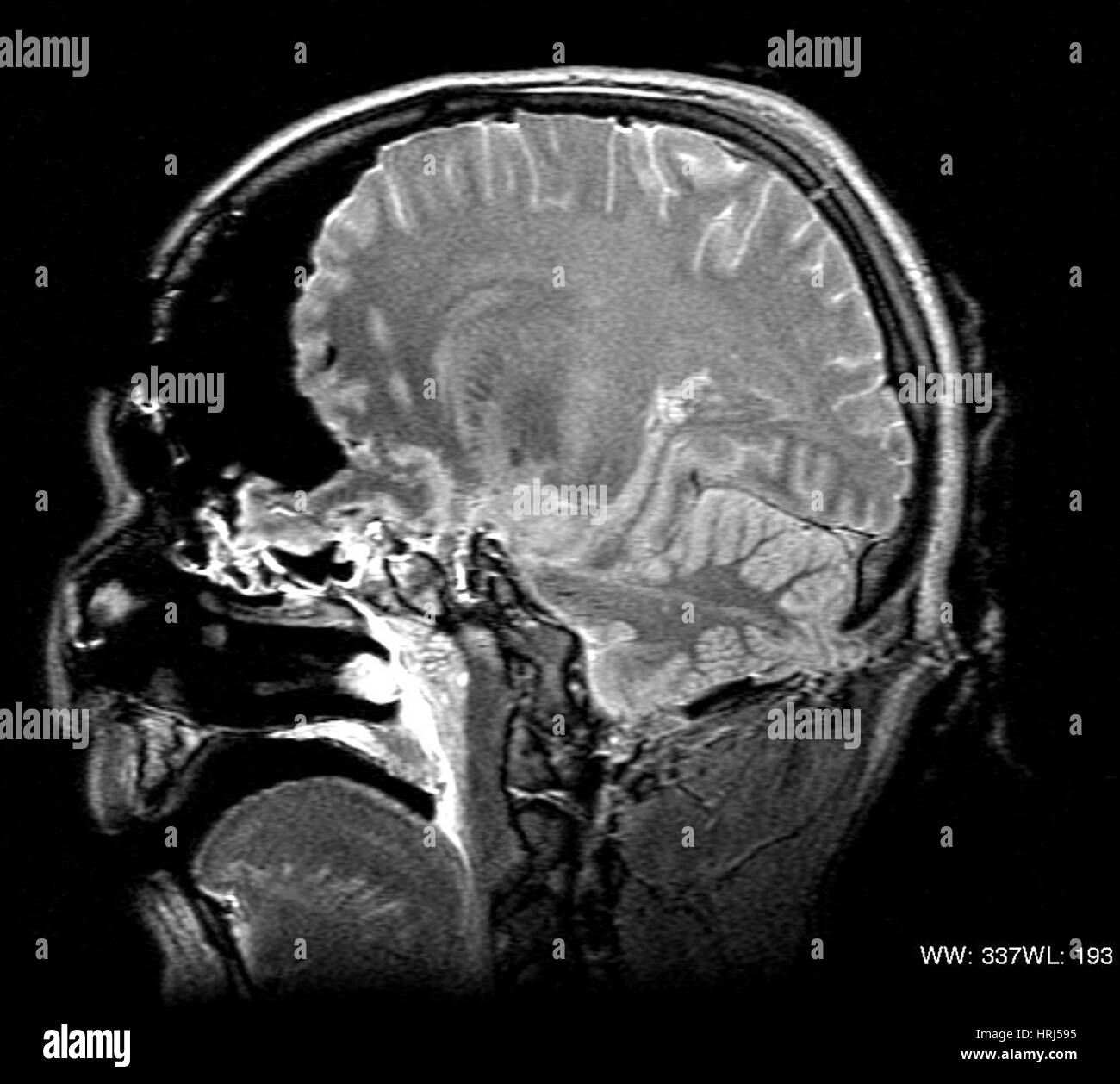 MRI Cross Section, Wound Track Stock Photohttps://www.alamy.com/image-license-details/?v=1https://www.alamy.com/stock-photo-mri-cross-section-wound-track-135008977.html
RMHRJ595–MRI Cross Section, Wound Track
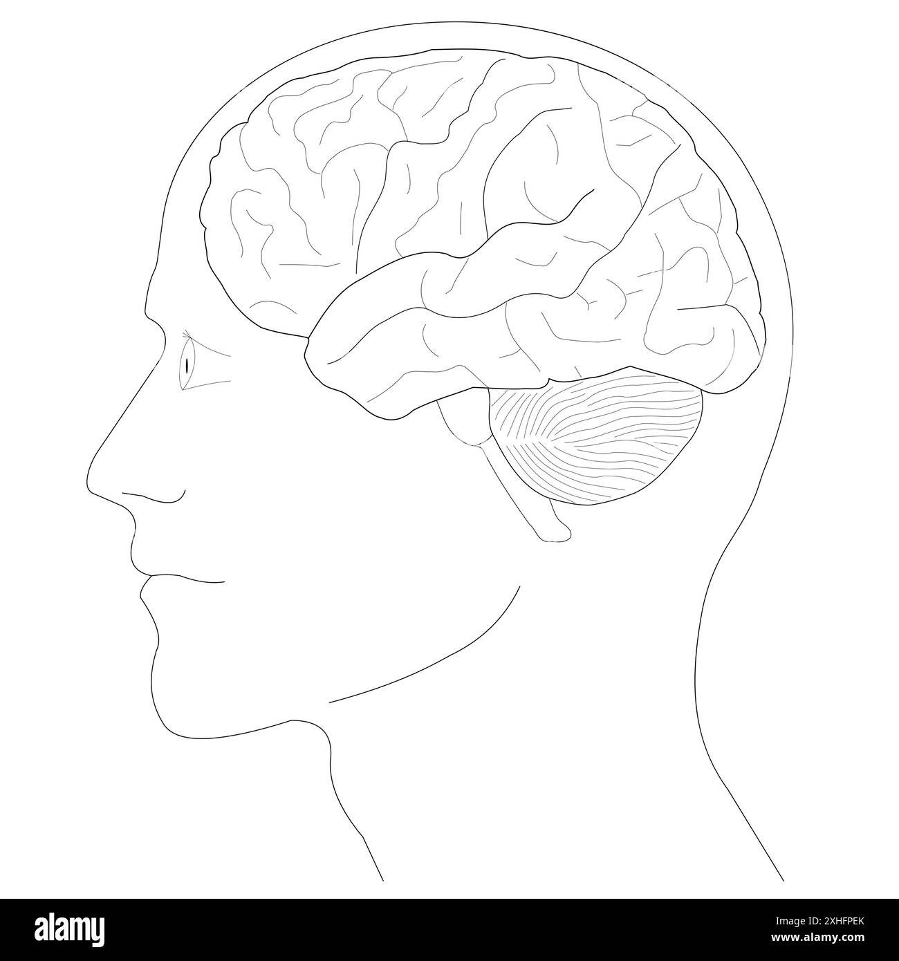 Human Brain anatomy Diagram, line art Stock Vectorhttps://www.alamy.com/image-license-details/?v=1https://www.alamy.com/human-brain-anatomy-diagram-line-art-image613158971.html
RF2XHFPEK–Human Brain anatomy Diagram, line art
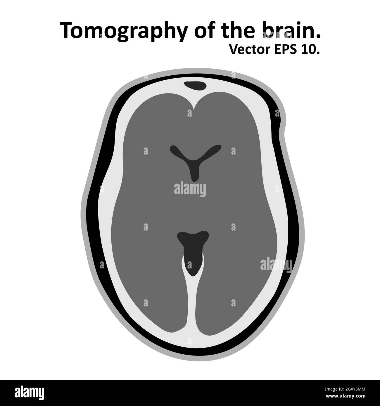 Magnetic resonance imaging of the brain. Cross section of the brain with MRI / CT scan. Vector EPS10. Stock Vectorhttps://www.alamy.com/image-license-details/?v=1https://www.alamy.com/magnetic-resonance-imaging-of-the-brain-cross-section-of-the-brain-with-mri-ct-scan-vector-eps10-image446835892.html
RF2GXY3MM–Magnetic resonance imaging of the brain. Cross section of the brain with MRI / CT scan. Vector EPS10.
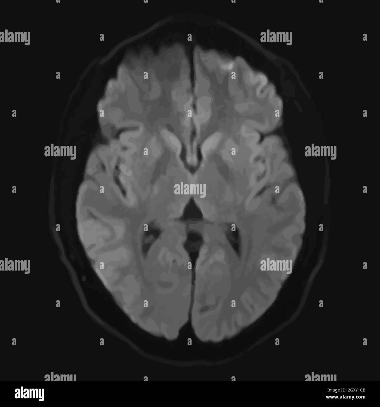 Realistic axial image of male cerebrum with CT scan, MRI Magnetic resonance imaging layer of brain. Isolated on dark background. Vector illustration. Stock Photohttps://www.alamy.com/image-license-details/?v=1https://www.alamy.com/realistic-axial-image-of-male-cerebrum-with-ct-scan-mri-magnetic-resonance-imaging-layer-of-brain-isolated-on-dark-background-vector-illustration-image446834091.html
RF2GXY1CB–Realistic axial image of male cerebrum with CT scan, MRI Magnetic resonance imaging layer of brain. Isolated on dark background. Vector illustration.
 3d rendering. 3d illustration . Human parts with black background Stock Photohttps://www.alamy.com/image-license-details/?v=1https://www.alamy.com/3d-rendering-3d-illustration-human-parts-with-black-background-image360340182.html
RF2BX6WG6–3d rendering. 3d illustration . Human parts with black background
 . Principles of veterinary science; a text-book for use in agricultural schools. Veterinary medicine. 56 PRINCIPLES OP VETERINARY SCIENCE ing in the brain. When they are cut or interfered with the muscle becomes paralyzed. Blood Supply.—A plentiful supply of blood is furnished through the arteries of each muscle and is removed through the corre- sponding veins (Fig. 12). Arrangement.—Muscles are arranged in groups of two, one group acts in opposition or is antagonistic to the other. The effectiveness of this arrangement is seen in the precision of locomotion.. Fig. 12.—Cross-section of left le Stock Photohttps://www.alamy.com/image-license-details/?v=1https://www.alamy.com/principles-of-veterinary-science-a-text-book-for-use-in-agricultural-schools-veterinary-medicine-56-principles-op-veterinary-science-ing-in-the-brain-when-they-are-cut-or-interfered-with-the-muscle-becomes-paralyzed-blood-supplya-plentiful-supply-of-blood-is-furnished-through-the-arteries-of-each-muscle-and-is-removed-through-the-corre-sponding-veins-fig-12-arrangementmuscles-are-arranged-in-groups-of-two-one-group-acts-in-opposition-or-is-antagonistic-to-the-other-the-effectiveness-of-this-arrangement-is-seen-in-the-precision-of-locomotion-fig-12cross-section-of-left-le-image232319695.html
RMRDY23Y–. Principles of veterinary science; a text-book for use in agricultural schools. Veterinary medicine. 56 PRINCIPLES OP VETERINARY SCIENCE ing in the brain. When they are cut or interfered with the muscle becomes paralyzed. Blood Supply.—A plentiful supply of blood is furnished through the arteries of each muscle and is removed through the corre- sponding veins (Fig. 12). Arrangement.—Muscles are arranged in groups of two, one group acts in opposition or is antagonistic to the other. The effectiveness of this arrangement is seen in the precision of locomotion.. Fig. 12.—Cross-section of left le
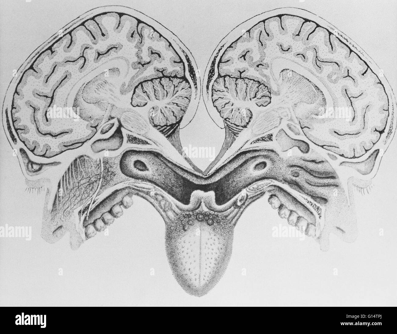 This is an illustration of a cross-sectional view of the human head, done by Paolo Masscagni, (1755-1815). Stock Photohttps://www.alamy.com/image-license-details/?v=1https://www.alamy.com/stock-photo-this-is-an-illustration-of-a-cross-sectional-view-of-the-human-head-103984122.html
RMG14TPJ–This is an illustration of a cross-sectional view of the human head, done by Paolo Masscagni, (1755-1815).
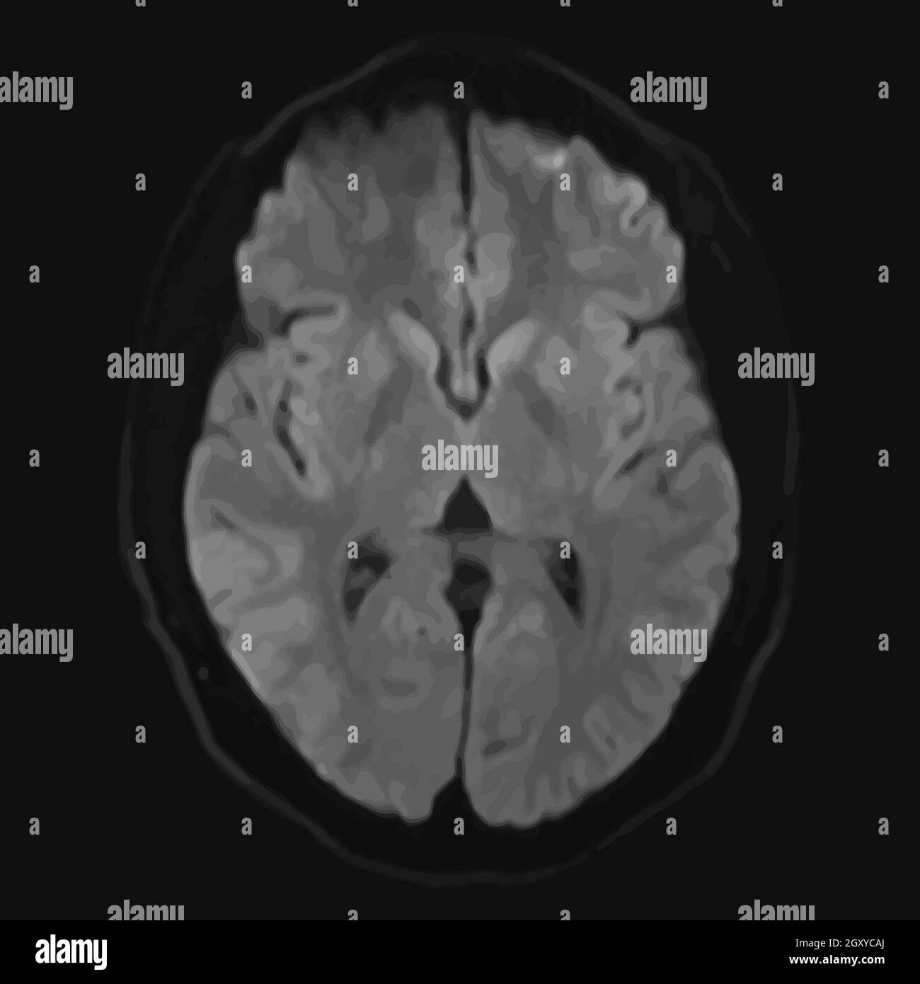 Realistic axial image of male cerebrum with CT scan, MRI Magnetic resonance imaging layer of brain. Isolated on dark background. Vector illustration. Stock Vectorhttps://www.alamy.com/image-license-details/?v=1https://www.alamy.com/realistic-axial-image-of-male-cerebrum-with-ct-scan-mri-magnetic-resonance-imaging-layer-of-brain-isolated-on-dark-background-vector-illustration-image446842666.html
RF2GXYCAJ–Realistic axial image of male cerebrum with CT scan, MRI Magnetic resonance imaging layer of brain. Isolated on dark background. Vector illustration.
 3d rendering. 3d illustration . Human parts with black background .Legs Stock Photohttps://www.alamy.com/image-license-details/?v=1https://www.alamy.com/3d-rendering-3d-illustration-human-parts-with-black-background-legs-image360340188.html
RF2BX6WGC–3d rendering. 3d illustration . Human parts with black background .Legs
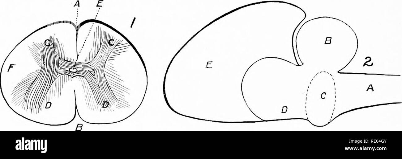 . Veterinary studies for agricultural students. Veterinary medicine. 34 ANATOMY. each side and at each articulation of the vertebrae. They are named cervical, dorsal, etc., according to location in the spinal column. The spinal nerves supply, by their superior branches, the skin and muscles of the neck and spinal column. By their in- ferior branches they supply the lower portion of the body and limbs and furnish other branches which in part make up the two great sympathetic nerve trunks.. FIG. 17. SPINAL CORD AND BRAIN IN DIAGRAM. (M. H. It.) 1. Cross Section of the Spinal Cord. A, Superior me Stock Photohttps://www.alamy.com/image-license-details/?v=1https://www.alamy.com/veterinary-studies-for-agricultural-students-veterinary-medicine-34-anatomy-each-side-and-at-each-articulation-of-the-vertebrae-they-are-named-cervical-dorsal-etc-according-to-location-in-the-spinal-column-the-spinal-nerves-supply-by-their-superior-branches-the-skin-and-muscles-of-the-neck-and-spinal-column-by-their-in-ferior-branches-they-supply-the-lower-portion-of-the-body-and-limbs-and-furnish-other-branches-which-in-part-make-up-the-two-great-sympathetic-nerve-trunks-fig-17-spinal-cord-and-brain-in-diagram-m-h-it-1-cross-section-of-the-spinal-cord-a-superior-me-image232343579.html
RMRE04GY–. Veterinary studies for agricultural students. Veterinary medicine. 34 ANATOMY. each side and at each articulation of the vertebrae. They are named cervical, dorsal, etc., according to location in the spinal column. The spinal nerves supply, by their superior branches, the skin and muscles of the neck and spinal column. By their in- ferior branches they supply the lower portion of the body and limbs and furnish other branches which in part make up the two great sympathetic nerve trunks.. FIG. 17. SPINAL CORD AND BRAIN IN DIAGRAM. (M. H. It.) 1. Cross Section of the Spinal Cord. A, Superior me
 A composite image showing a normal coronal (frontal) cross-sectional MRI image of the brain (left) with a coronal MRI image of a brain with advanced Alzheimer's disease (at right). The diseased brain shows severe generalized atrophy (shrinkage) of brain tissue with an accentuated loss of tissue involving the temporal lobes. Stock Photohttps://www.alamy.com/image-license-details/?v=1https://www.alamy.com/a-composite-image-showing-a-normal-coronal-frontal-cross-sectional-mri-image-of-the-brain-left-with-a-coronal-mri-image-of-a-brain-with-advanced-alzheimers-disease-at-right-the-diseased-brain-shows-severe-generalized-atrophy-shrinkage-of-brain-tissue-with-an-accentuated-loss-of-tissue-involving-the-temporal-lobes-image458814654.html
RM2HJCPNJ–A composite image showing a normal coronal (frontal) cross-sectional MRI image of the brain (left) with a coronal MRI image of a brain with advanced Alzheimer's disease (at right). The diseased brain shows severe generalized atrophy (shrinkage) of brain tissue with an accentuated loss of tissue involving the temporal lobes.
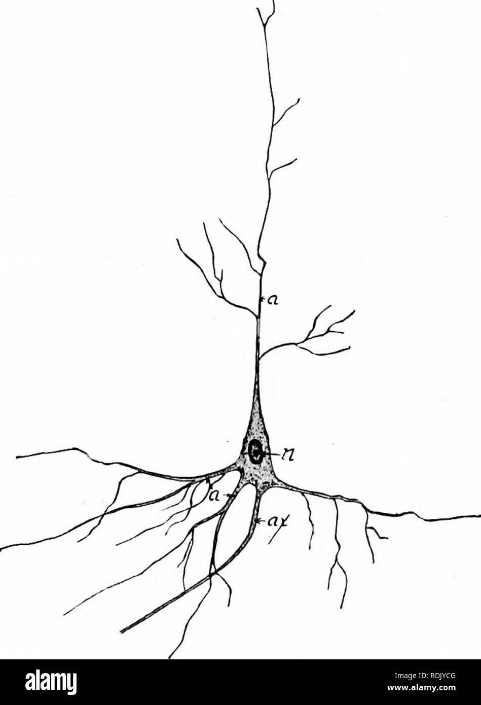 . Mammalian anatomy : with special reference to the cat . Mammals; Anatomy, Comparative; Cats. 196 ELEMENTS OF MAMMALIAN ANATOMY. the canalis centralis, about one-half a millimeter in diameter, extends throughout the cord, opening into the fourth ven- tricle of the brain. A cross-section of the cord shows the gray matter arranged in the shape of a letter H. The ventral columns of gray matter are the anterior horns, and the posterior columns, the posterior horns (Fig. 100). Many of the fibers extend in a longitudinal direction throughout the cord, but the roots of the spinal nerves upon enterin Stock Photohttps://www.alamy.com/image-license-details/?v=1https://www.alamy.com/mammalian-anatomy-with-special-reference-to-the-cat-mammals-anatomy-comparative-cats-196-elements-of-mammalian-anatomy-the-canalis-centralis-about-one-half-a-millimeter-in-diameter-extends-throughout-the-cord-opening-into-the-fourth-ven-tricle-of-the-brain-a-cross-section-of-the-cord-shows-the-gray-matter-arranged-in-the-shape-of-a-letter-h-the-ventral-columns-of-gray-matter-are-the-anterior-horns-and-the-posterior-columns-the-posterior-horns-fig-100-many-of-the-fibers-extend-in-a-longitudinal-direction-throughout-the-cord-but-the-roots-of-the-spinal-nerves-upon-enterin-image232141968.html
RMRDJYCG–. Mammalian anatomy : with special reference to the cat . Mammals; Anatomy, Comparative; Cats. 196 ELEMENTS OF MAMMALIAN ANATOMY. the canalis centralis, about one-half a millimeter in diameter, extends throughout the cord, opening into the fourth ven- tricle of the brain. A cross-section of the cord shows the gray matter arranged in the shape of a letter H. The ventral columns of gray matter are the anterior horns, and the posterior columns, the posterior horns (Fig. 100). Many of the fibers extend in a longitudinal direction throughout the cord, but the roots of the spinal nerves upon enterin
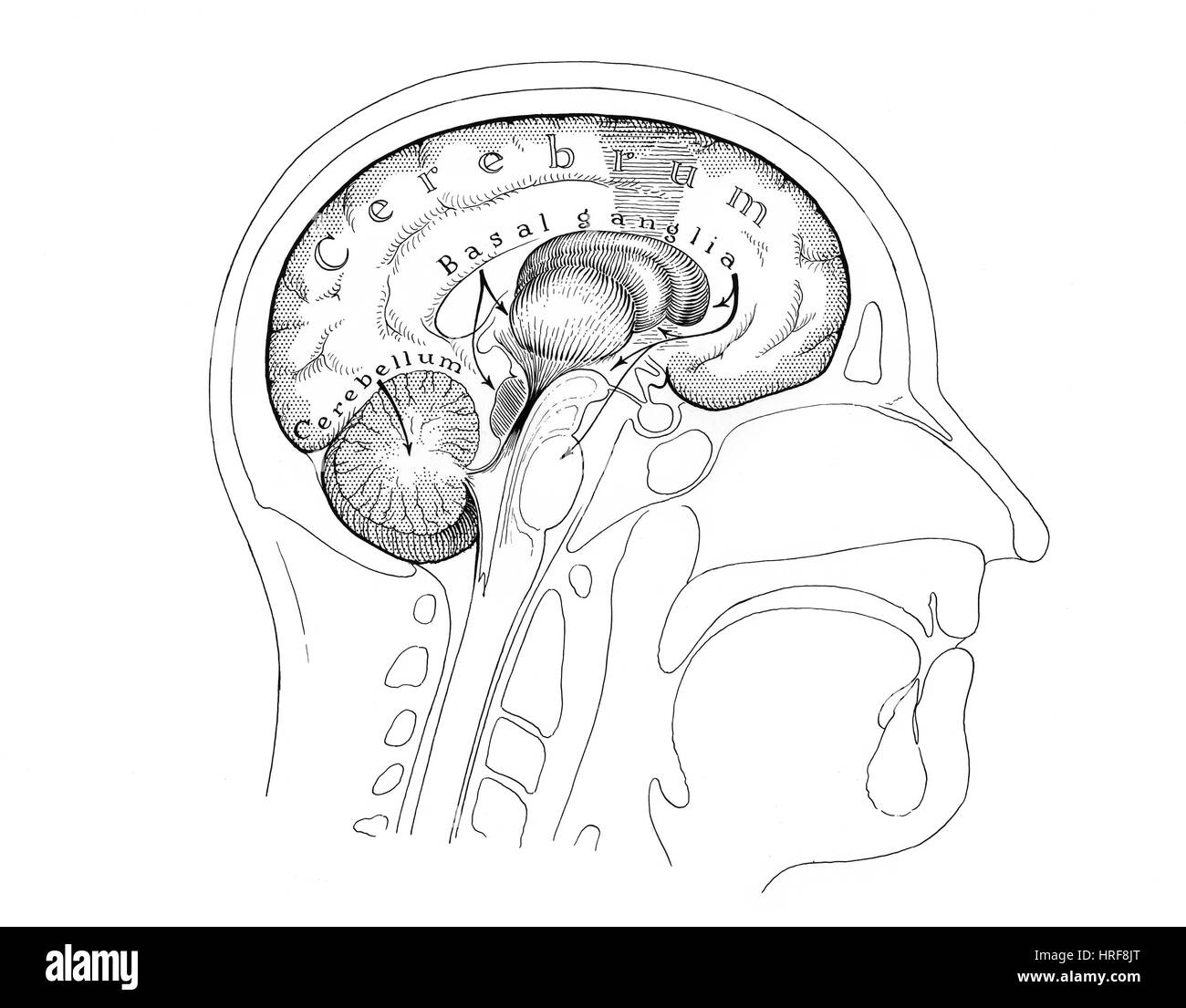 Human Brain Stock Photohttps://www.alamy.com/image-license-details/?v=1https://www.alamy.com/stock-photo-human-brain-134945744.html
RMHRF8JT–Human Brain
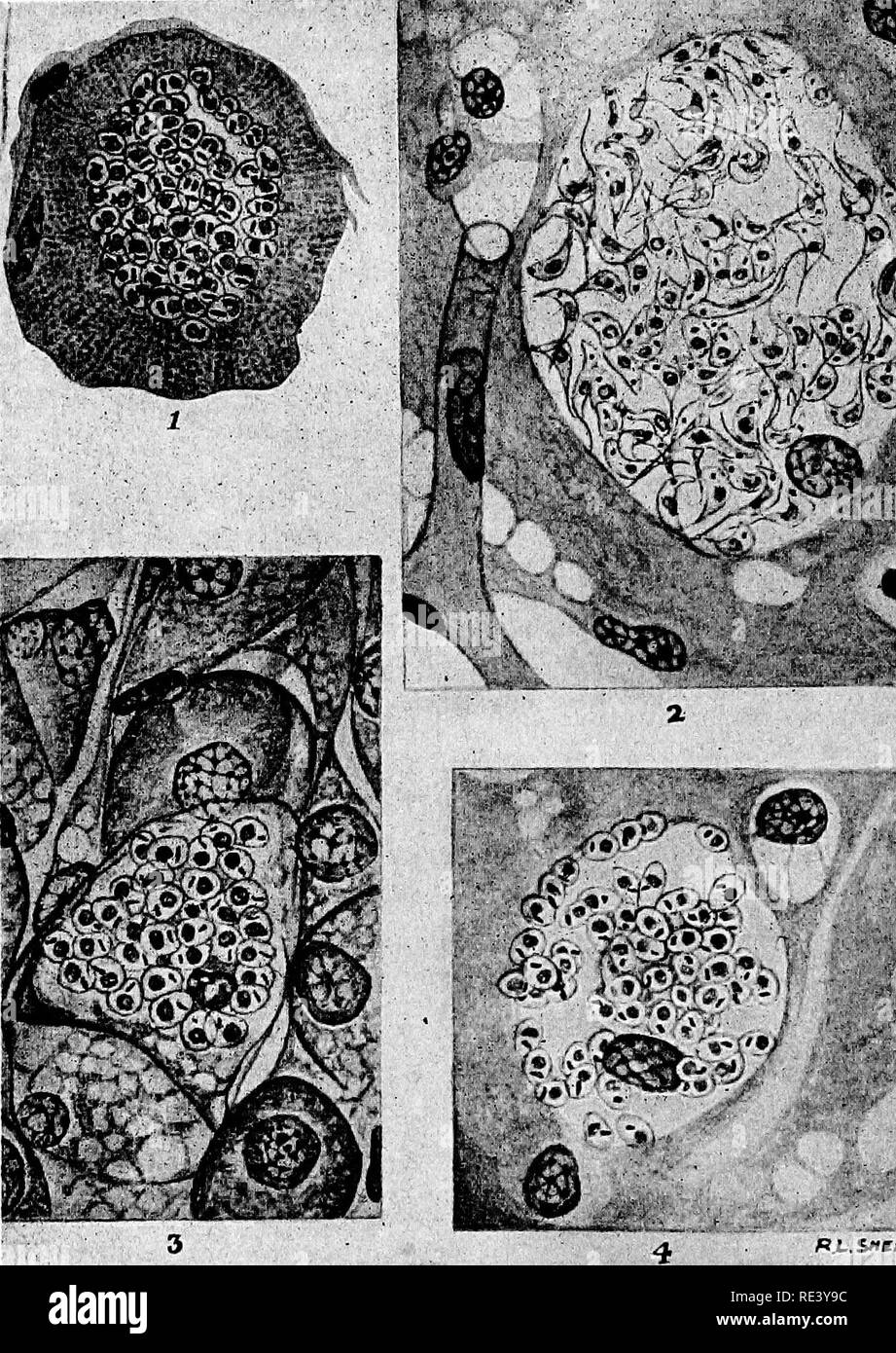 . Pathogenic micro-organisms. A text-book of microbiology for physicians and students of medicine. (Based upon Williams' Bacteriology). Bacteriology; Pathogenic bacteria. 412 SPECIFIC MICRO-ORGANISMS. *lntav'* PL s«rw^«o- PlG. 168.—Schizotrypanum cruzi developing in the tissues of the guinea-pig. I. Cross-section of a striated muscle fiber containing Schiozirypanum cruzi: Note dividing forms. 2. Section of brain showing a Schizotrypanum. cyst within a neuroglia cell, containing chiefly flagellated forms. 3. Section through the supra- renal capsule, fascicular zone. 4. Section of brain showing Stock Photohttps://www.alamy.com/image-license-details/?v=1https://www.alamy.com/pathogenic-micro-organisms-a-text-book-of-microbiology-for-physicians-and-students-of-medicine-based-upon-williams-bacteriology-bacteriology-pathogenic-bacteria-412-specific-micro-organisms-lntav-pl-srwo-plg-168schizotrypanum-cruzi-developing-in-the-tissues-of-the-guinea-pig-i-cross-section-of-a-striated-muscle-fiber-containing-schiozirypanum-cruzi-note-dividing-forms-2-section-of-brain-showing-a-schizotrypanum-cyst-within-a-neuroglia-cell-containing-chiefly-flagellated-forms-3-section-through-the-supra-renal-capsule-fascicular-zone-4-section-of-brain-showing-image232427256.html
RMRE3Y9C–. Pathogenic micro-organisms. A text-book of microbiology for physicians and students of medicine. (Based upon Williams' Bacteriology). Bacteriology; Pathogenic bacteria. 412 SPECIFIC MICRO-ORGANISMS. *lntav'* PL s«rw^«o- PlG. 168.—Schizotrypanum cruzi developing in the tissues of the guinea-pig. I. Cross-section of a striated muscle fiber containing Schiozirypanum cruzi: Note dividing forms. 2. Section of brain showing a Schizotrypanum. cyst within a neuroglia cell, containing chiefly flagellated forms. 3. Section through the supra- renal capsule, fascicular zone. 4. Section of brain showing
 . The physiology of domestic animals ... Physiology, Comparative; Veterinary physiology. FUNCTIONS OF THE BRAIN. II. 831. Fig. 361. medulla; P,pons varolii, with itg ganglia on either side (in purple). In III, the nuclei of the cranial nerve-roots are numbered to correspond with the nerves. Red is used for the motor nuclei, and blue for the sensory nuclei. The tracts in the cord are designated by the area similarly colored in the cross-section of the cord beneath, c', column of TUrck ; c. crossed pyramidal column ; a, anterior horn ; a', anterior root-zone; e, direct cerebellar column; b, post Stock Photohttps://www.alamy.com/image-license-details/?v=1https://www.alamy.com/the-physiology-of-domestic-animals-physiology-comparative-veterinary-physiology-functions-of-the-brain-ii-831-fig-361-medulla-ppons-varolii-with-itg-ganglia-on-either-side-in-purple-in-iii-the-nuclei-of-the-cranial-nerve-roots-are-numbered-to-correspond-with-the-nerves-red-is-used-for-the-motor-nuclei-and-blue-for-the-sensory-nuclei-the-tracts-in-the-cord-are-designated-by-the-area-similarly-colored-in-the-cross-section-of-the-cord-beneath-c-column-of-turck-c-crossed-pyramidal-column-a-anterior-horn-a-anterior-root-zone-e-direct-cerebellar-column-b-post-image232425206.html
RMRE3TM6–. The physiology of domestic animals ... Physiology, Comparative; Veterinary physiology. FUNCTIONS OF THE BRAIN. II. 831. Fig. 361. medulla; P,pons varolii, with itg ganglia on either side (in purple). In III, the nuclei of the cranial nerve-roots are numbered to correspond with the nerves. Red is used for the motor nuclei, and blue for the sensory nuclei. The tracts in the cord are designated by the area similarly colored in the cross-section of the cord beneath, c', column of TUrck ; c. crossed pyramidal column ; a, anterior horn ; a', anterior root-zone; e, direct cerebellar column; b, post
 . Principles of modern biology. Biology. B SOMITES SPINAL NERVE SOMITES DIGESTIVE TRACT COELOM GENITAL RIDGE. GILL CLEFTS HEART Fig. 15-11. Some generalized stages in the development of a primitive verte- brate. Stages A to E are cross sections cut from the mid-body region; whereas F is a longitudinal section. nerves of the bod) represent outgrowths from the neural tube. Each nerve consists of a bundle of fibers that originate from nerve cells as soon as these begin to differen- tiate in the developing brain and spinal cord. The flexible unsegmented notochord is a skeletal rod that is found in Stock Photohttps://www.alamy.com/image-license-details/?v=1https://www.alamy.com/principles-of-modern-biology-biology-b-somites-spinal-nerve-somites-digestive-tract-coelom-genital-ridge-gill-clefts-heart-fig-15-11-some-generalized-stages-in-the-development-of-a-primitive-verte-brate-stages-a-to-e-are-cross-sections-cut-from-the-mid-body-region-whereas-f-is-a-longitudinal-section-nerves-of-the-bod-represent-outgrowths-from-the-neural-tube-each-nerve-consists-of-a-bundle-of-fibers-that-originate-from-nerve-cells-as-soon-as-these-begin-to-differen-tiate-in-the-developing-brain-and-spinal-cord-the-flexible-unsegmented-notochord-is-a-skeletal-rod-that-is-found-in-image232337584.html
RMRDYTXT–. Principles of modern biology. Biology. B SOMITES SPINAL NERVE SOMITES DIGESTIVE TRACT COELOM GENITAL RIDGE. GILL CLEFTS HEART Fig. 15-11. Some generalized stages in the development of a primitive verte- brate. Stages A to E are cross sections cut from the mid-body region; whereas F is a longitudinal section. nerves of the bod) represent outgrowths from the neural tube. Each nerve consists of a bundle of fibers that originate from nerve cells as soon as these begin to differen- tiate in the developing brain and spinal cord. The flexible unsegmented notochord is a skeletal rod that is found in
 . A text-book upon the pathogenic Bacteria and Protozoa for students of medicine and physicians. Bacteriology; Pathogenic bacteria; Protozoa. 1 i ^if J *5 (4^ V I J X l,.X. "o'w 'Xiv' /r ujA' Xf li^w >T3l'..'U" .'^')'".. "> â iWKf^Sb^ V"SIS"^. R,L.<,J1tpPAKD. Fig. 2i8.âSchizotrypanum criizi developing in the tissues of the guinea-pig. i. Cross-section of a striated muscle fiber containing Schizotrypanum cruzi: Note dividing forms. 2. Section of brain showing a Schizotrypanum cyst within a neuroglia cell, containing chiefly flagellated forms. 3. Section Stock Photohttps://www.alamy.com/image-license-details/?v=1https://www.alamy.com/a-text-book-upon-the-pathogenic-bacteria-and-protozoa-for-students-of-medicine-and-physicians-bacteriology-pathogenic-bacteria-protozoa-1-i-if-j-5-4-v-i-j-x-lx-quotow-xiv-r-uja-xf-liw-gtt3luquot-quot-quotgt-iwkfsb-vquotsisquot-rlltj1tppakd-fig-2i8schizotrypanum-criizi-developing-in-the-tissues-of-the-guinea-pig-i-cross-section-of-a-striated-muscle-fiber-containing-schizotrypanum-cruzi-note-dividing-forms-2-section-of-brain-showing-a-schizotrypanum-cyst-within-a-neuroglia-cell-containing-chiefly-flagellated-forms-3-section-image232371495.html
RMRE1C5Y–. A text-book upon the pathogenic Bacteria and Protozoa for students of medicine and physicians. Bacteriology; Pathogenic bacteria; Protozoa. 1 i ^if J *5 (4^ V I J X l,.X. "o'w 'Xiv' /r ujA' Xf li^w >T3l'..'U" .'^')'".. "> â iWKf^Sb^ V"SIS"^. R,L.<,J1tpPAKD. Fig. 2i8.âSchizotrypanum criizi developing in the tissues of the guinea-pig. i. Cross-section of a striated muscle fiber containing Schizotrypanum cruzi: Note dividing forms. 2. Section of brain showing a Schizotrypanum cyst within a neuroglia cell, containing chiefly flagellated forms. 3. Section