Quick filters:
Ct lung Stock Photos and Images
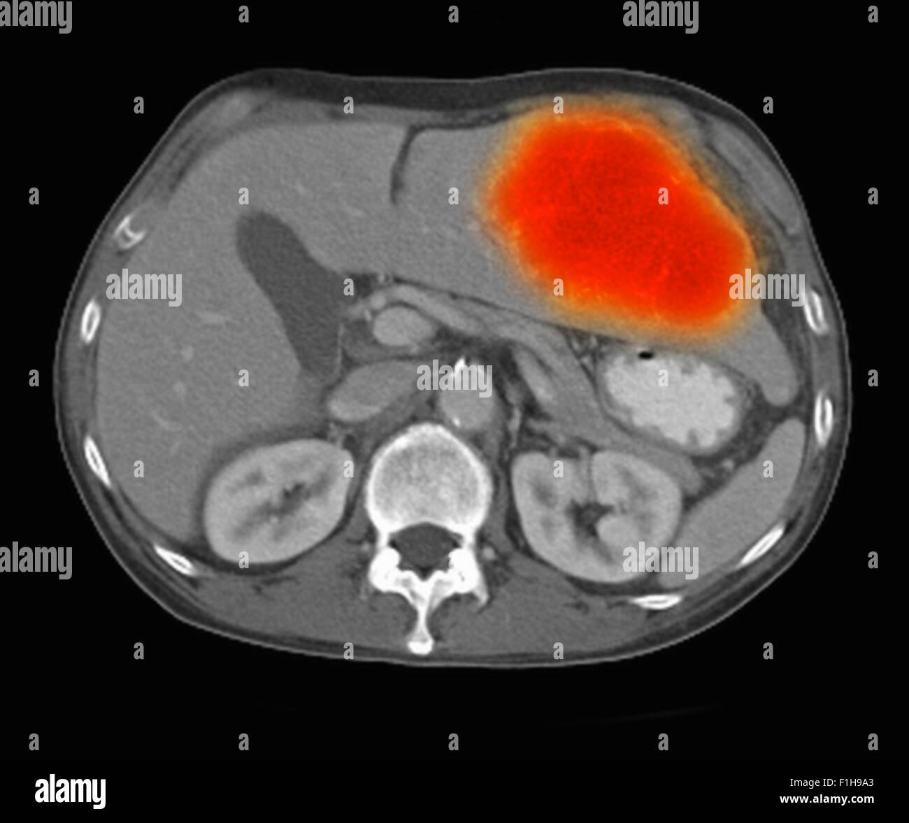 Image co-registered PET-CT study dual modality scanner. Patient multiple metastatic lesions liver & lung Stock Photohttps://www.alamy.com/image-license-details/?v=1https://www.alamy.com/stock-photo-image-co-registered-pet-ct-study-dual-modality-scanner-patient-multiple-87047019.html
RFF1H9A3–Image co-registered PET-CT study dual modality scanner. Patient multiple metastatic lesions liver & lung
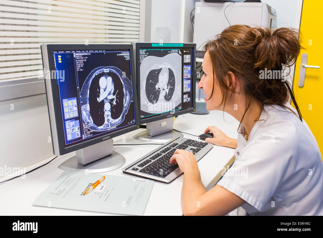 Thorax CT scan, Angoulême hospital, France . Stock Photohttps://www.alamy.com/image-license-details/?v=1https://www.alamy.com/stock-photo-thorax-ct-scan-angoulme-hospital-france-72441472.html
RME5RYRC–Thorax CT scan, Angoulême hospital, France .
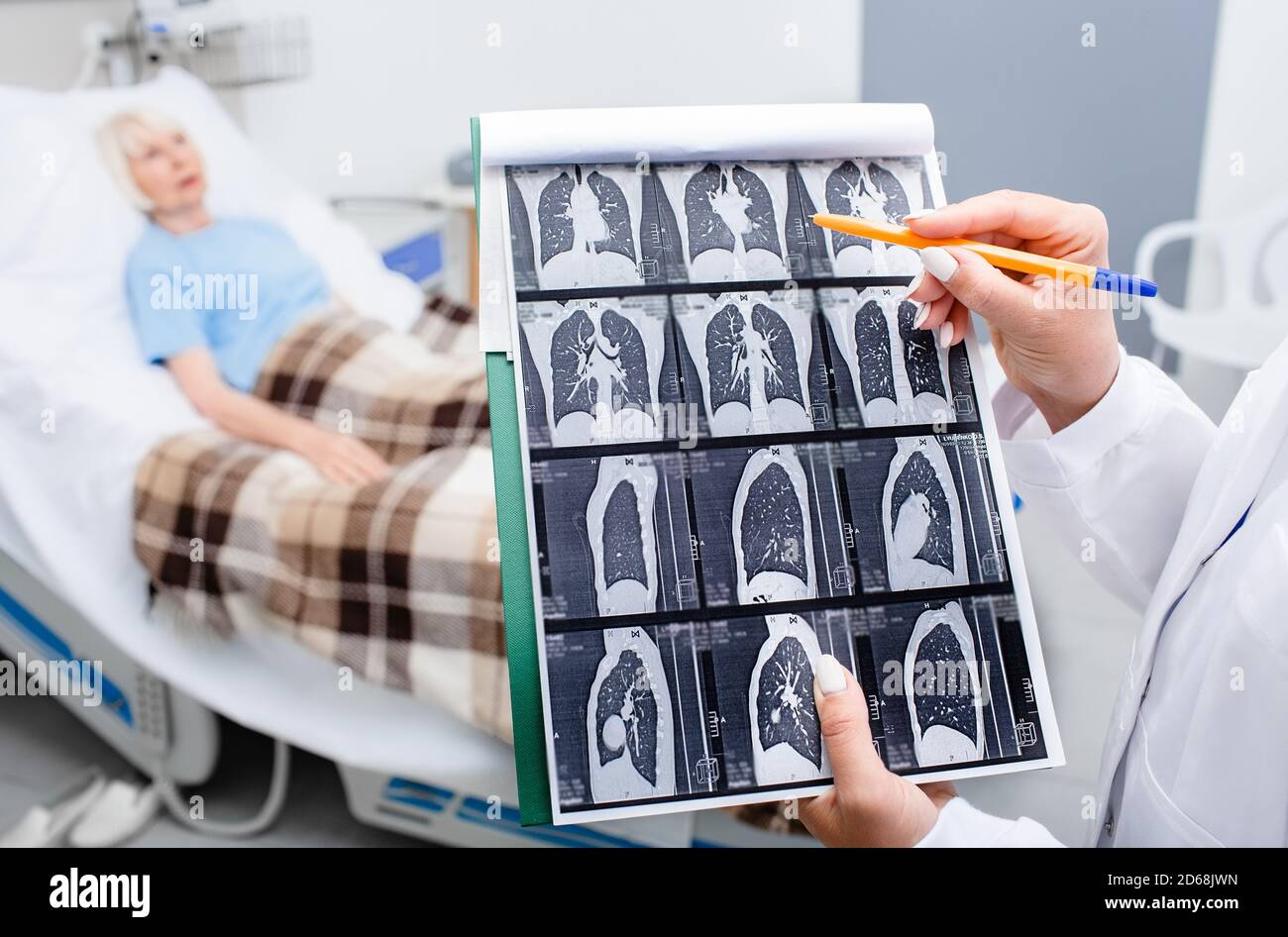 Close-up CT lung screening. Diagnostics and treatment of patients with pneumonia and lung diseases at hospital. patient in a hospital ward Stock Photohttps://www.alamy.com/image-license-details/?v=1https://www.alamy.com/close-up-ct-lung-screening-diagnostics-and-treatment-of-patients-with-pneumonia-and-lung-diseases-at-hospital-patient-in-a-hospital-ward-image382506481.html
RF2D68JWN–Close-up CT lung screening. Diagnostics and treatment of patients with pneumonia and lung diseases at hospital. patient in a hospital ward
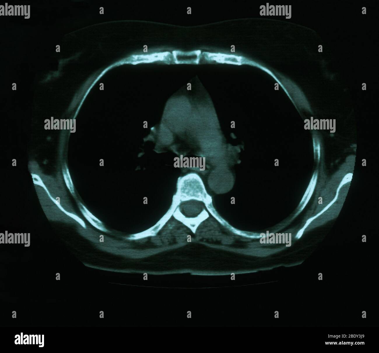 Granuloma & Calcification in Lung Stock Photohttps://www.alamy.com/image-license-details/?v=1https://www.alamy.com/granuloma-calcification-in-lung-image352793457.html
RM2BDY3J9–Granuloma & Calcification in Lung
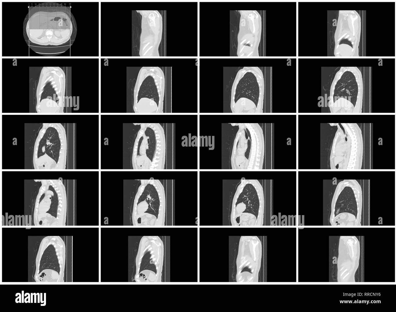 ct scan step set of body lung sagittal view Stock Photohttps://www.alamy.com/image-license-details/?v=1https://www.alamy.com/ct-scan-step-set-of-body-lung-sagittal-view-image238152522.html
RFRRCNY6–ct scan step set of body lung sagittal view
 CT Chest or CT lung 3d rendering image with blue color showing Trachea and lung in respiratory system. Stock Photohttps://www.alamy.com/image-license-details/?v=1https://www.alamy.com/ct-chest-or-ct-lung-3d-rendering-image-with-blue-color-showing-trachea-and-lung-in-respiratory-system-image567017167.html
RF2RXDT53–CT Chest or CT lung 3d rendering image with blue color showing Trachea and lung in respiratory system.
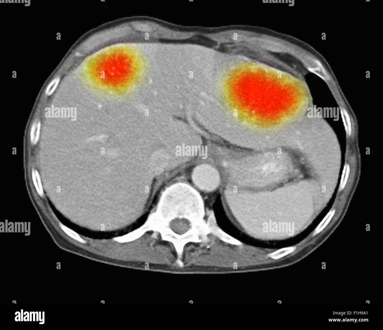 Image co-registered PET-CT study dual modality scanner. Patient multiple metastatic lesions liver & lung Stock Photohttps://www.alamy.com/image-license-details/?v=1https://www.alamy.com/stock-photo-image-co-registered-pet-ct-study-dual-modality-scanner-patient-multiple-87047017.html
RFF1H9A1–Image co-registered PET-CT study dual modality scanner. Patient multiple metastatic lesions liver & lung
 Focused worried multiethnic healthcare workers analyzing CT lung screening while working in medical clinic or hospital, medical team looking at x-ray Stock Photohttps://www.alamy.com/image-license-details/?v=1https://www.alamy.com/focused-worried-multiethnic-healthcare-workers-analyzing-ct-lung-screening-while-working-in-medical-clinic-or-hospital-medical-team-looking-at-x-ray-image461136104.html
RF2HP6FPG–Focused worried multiethnic healthcare workers analyzing CT lung screening while working in medical clinic or hospital, medical team looking at x-ray
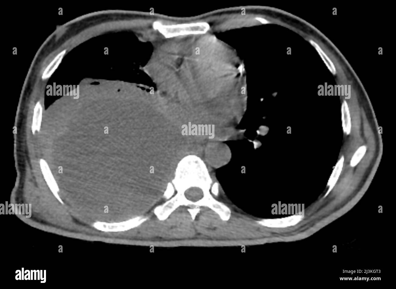 Pleural empyema, CT scan Stock Photohttps://www.alamy.com/image-license-details/?v=1https://www.alamy.com/pleural-empyema-ct-scan-image466954211.html
RF2J3KGT3–Pleural empyema, CT scan
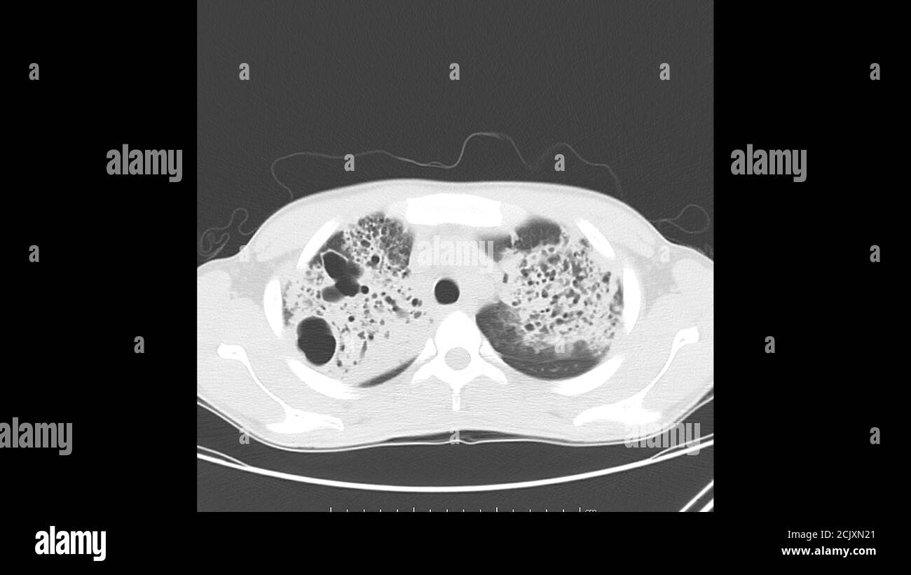 Axial computed Tomography of the chest (lung window) in a known patient of Active Tuberculosis (TB) showing pneumonia and cavitation in both lungs Stock Photohttps://www.alamy.com/image-license-details/?v=1https://www.alamy.com/axial-computed-tomography-of-the-chest-lung-window-in-a-known-patient-of-active-tuberculosis-tb-showing-pneumonia-and-cavitation-in-both-lungs-image373068809.html
RF2CJXN21–Axial computed Tomography of the chest (lung window) in a known patient of Active Tuberculosis (TB) showing pneumonia and cavitation in both lungs
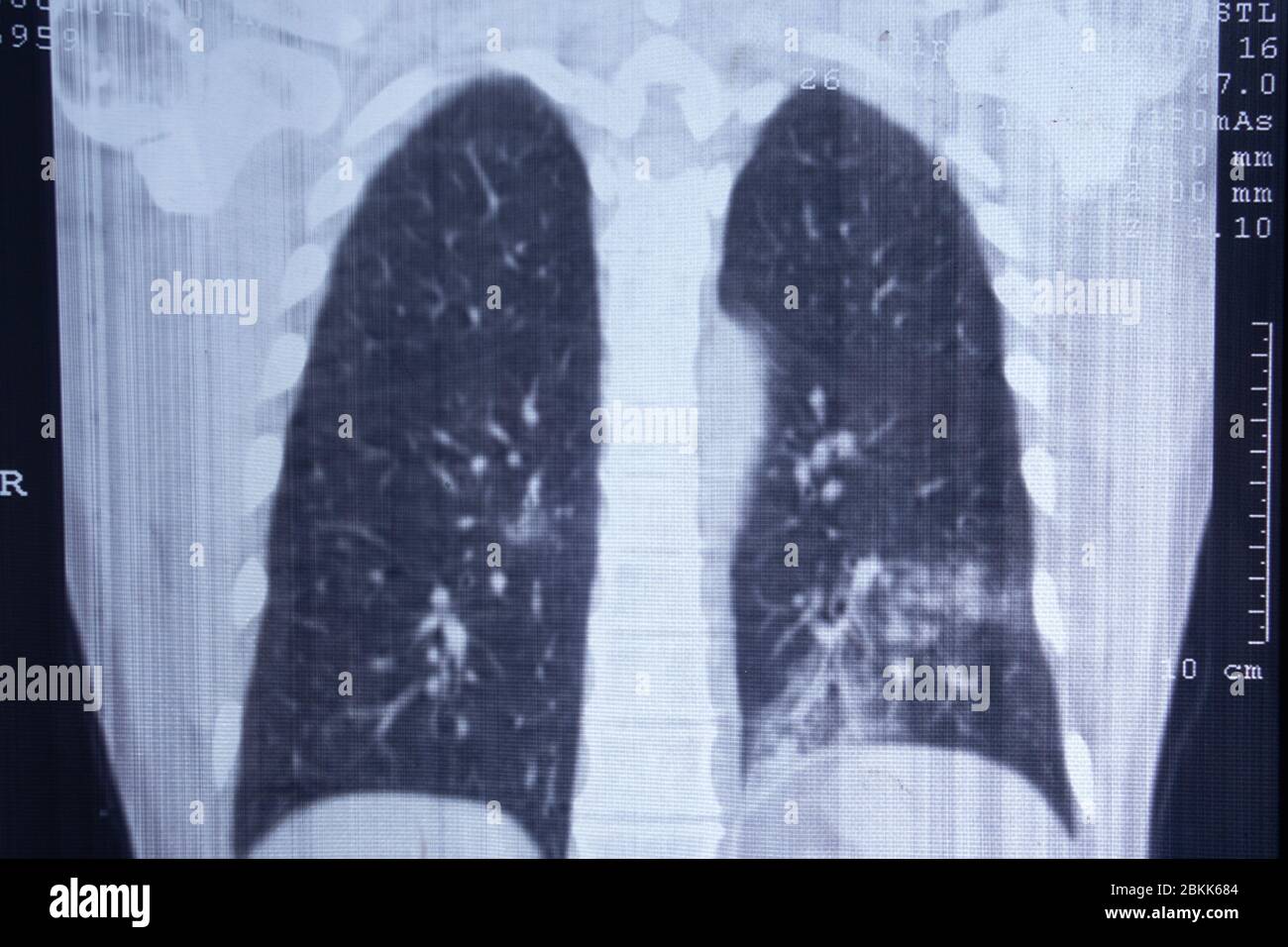 X-ray of pneumonia-affected lungs. CT scan. Stock Photohttps://www.alamy.com/image-license-details/?v=1https://www.alamy.com/x-ray-of-pneumonia-affected-lungs-ct-scan-image356307844.html
RF2BKK684–X-ray of pneumonia-affected lungs. CT scan.
 Rearview of ethnic female doctor analyzing X-ray or MRI scan while working in hospital, selective focus. Physician looking at CT screening, using Stock Photohttps://www.alamy.com/image-license-details/?v=1https://www.alamy.com/rearview-of-ethnic-female-doctor-analyzing-x-ray-or-mri-scan-while-working-in-hospital-selective-focus-physician-looking-at-ct-screening-using-image548988016.html
RF2PW4FPT–Rearview of ethnic female doctor analyzing X-ray or MRI scan while working in hospital, selective focus. Physician looking at CT screening, using
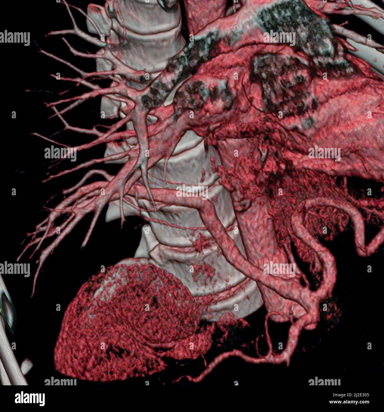 Congenital anomaly, lung Stock Photohttps://www.alamy.com/image-license-details/?v=1https://www.alamy.com/congenital-anomaly-lung-image466218933.html
RF2J2E305–Congenital anomaly, lung
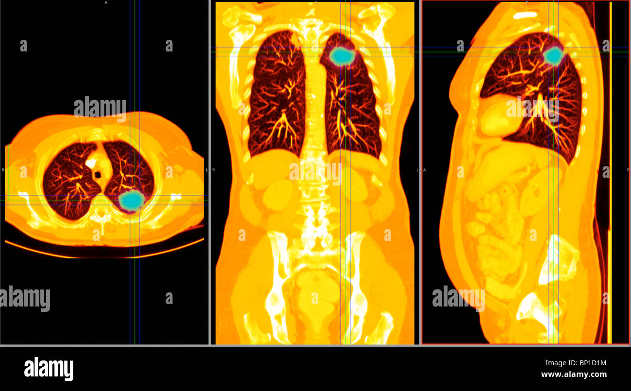 CT scan axial section showing a cancer in the right lung of a 77 year old woman Stock Photohttps://www.alamy.com/image-license-details/?v=1https://www.alamy.com/stock-photo-ct-scan-axial-section-showing-a-cancer-in-the-right-lung-of-a-77-year-30764992.html
RMBP1D1M–CT scan axial section showing a cancer in the right lung of a 77 year old woman
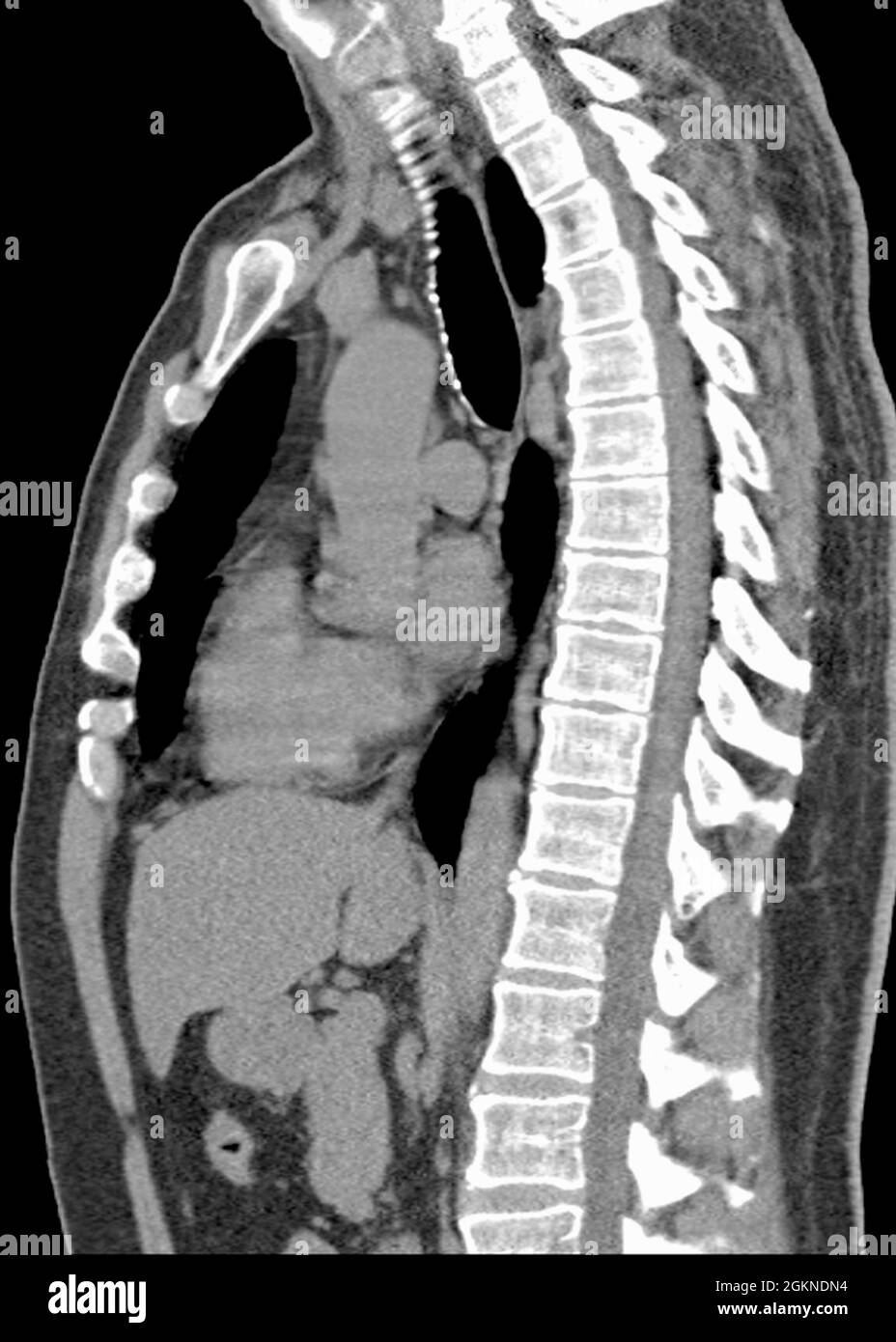 X-ray computed tomography (CT) of an adult male's chest side view Stock Photohttps://www.alamy.com/image-license-details/?v=1https://www.alamy.com/x-ray-computed-tomography-ct-of-an-adult-males-chest-side-view-image442409440.html
RM2GKNDN4–X-ray computed tomography (CT) of an adult male's chest side view
 Man analyzing digital CT scan of his lungs at home. Pneumonia and disease diagnosis. Pills and medical bottles Stock Photohttps://www.alamy.com/image-license-details/?v=1https://www.alamy.com/man-analyzing-digital-ct-scan-of-his-lungs-at-home-pneumonia-and-disease-diagnosis-pills-and-medical-bottles-image452301553.html
RF2H7T36W–Man analyzing digital CT scan of his lungs at home. Pneumonia and disease diagnosis. Pills and medical bottles
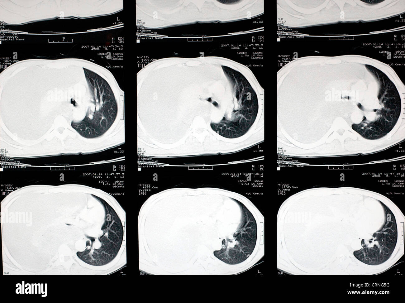 A CT scan showing a severe haemothroax on the right side of the patient's chest. Stock Photohttps://www.alamy.com/image-license-details/?v=1https://www.alamy.com/stock-photo-a-ct-scan-showing-a-severe-haemothroax-on-the-right-side-of-the-patients-49031516.html
RMCRNG5G–A CT scan showing a severe haemothroax on the right side of the patient's chest.
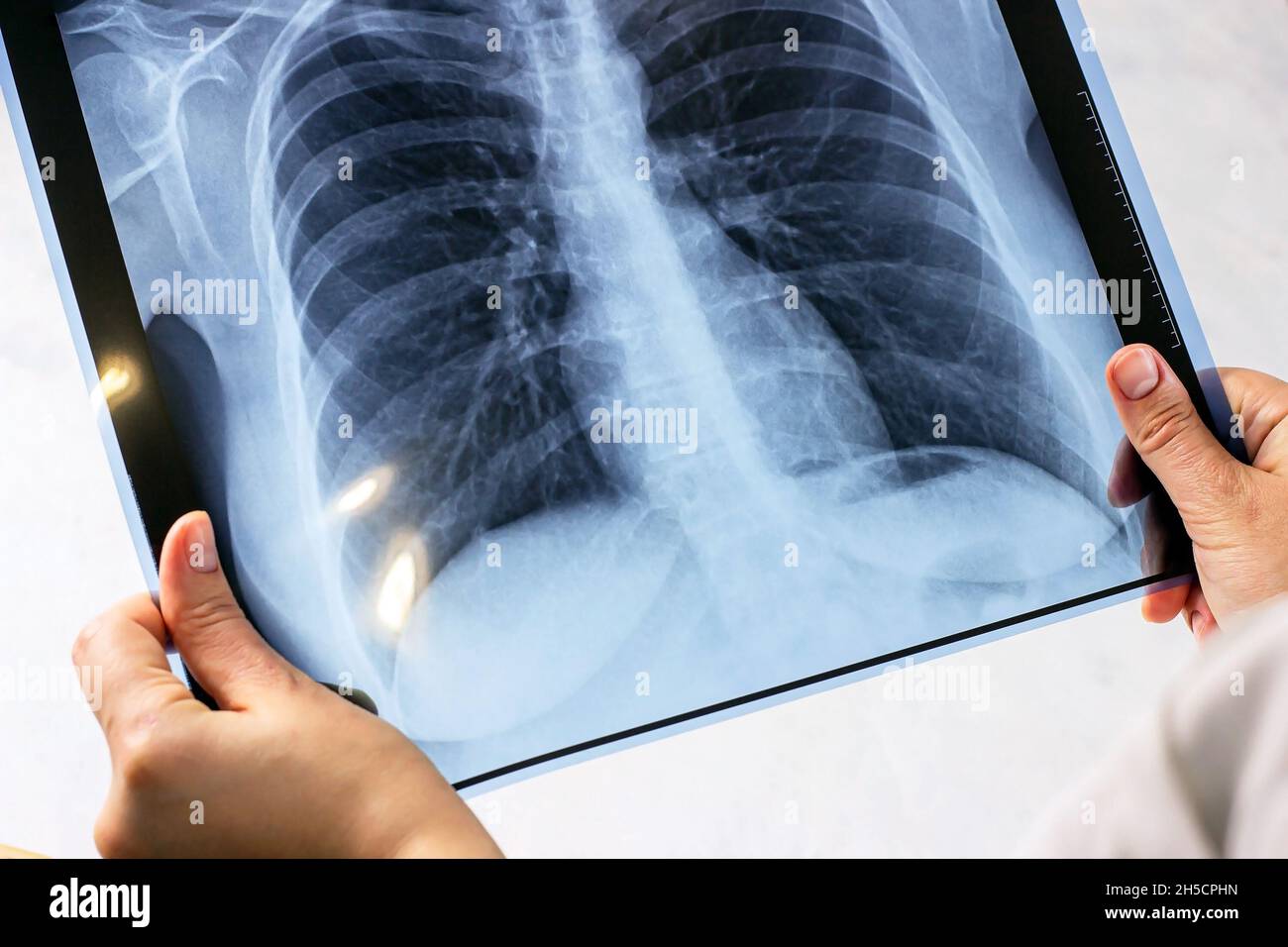 Woman hands with blue medical x-ray of chest scan on light background. Covid desease, cancer examination, pneumonia, asthma and tuberculosis diagnosti Stock Photohttps://www.alamy.com/image-license-details/?v=1https://www.alamy.com/woman-hands-with-blue-medical-x-ray-of-chest-scan-on-light-background-covid-desease-cancer-examination-pneumonia-asthma-and-tuberculosis-diagnosti-image450824017.html
RF2H5CPHN–Woman hands with blue medical x-ray of chest scan on light background. Covid desease, cancer examination, pneumonia, asthma and tuberculosis diagnosti
 a doctor in a protective suit and mask looks at the results of a CT scan .coronavirus. Stock Photohttps://www.alamy.com/image-license-details/?v=1https://www.alamy.com/a-doctor-in-a-protective-suit-and-mask-looks-at-the-results-of-a-ct-scan-coronavirus-image386249038.html
RF2DCB4GE–a doctor in a protective suit and mask looks at the results of a CT scan .coronavirus.
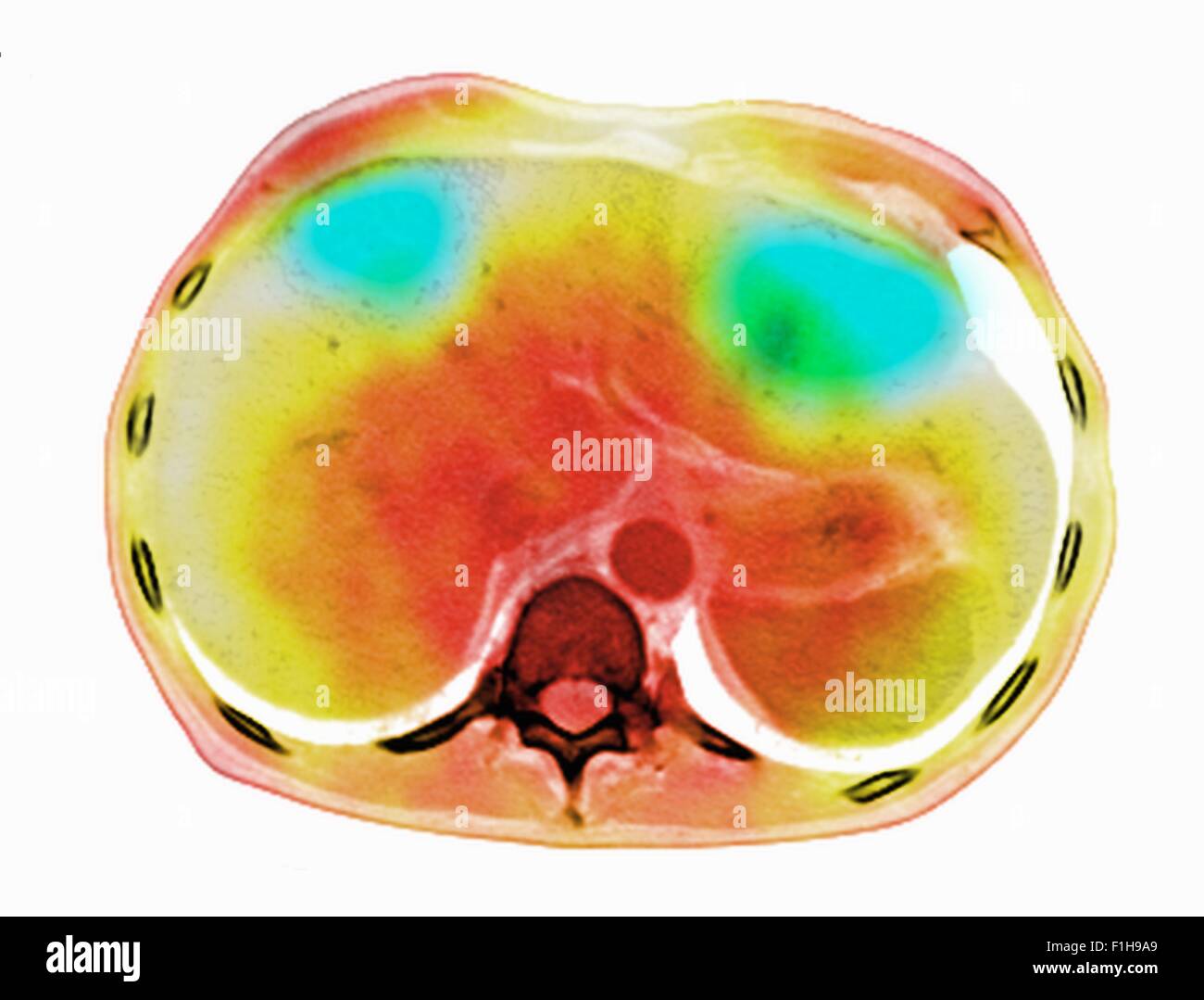 Image co-registered PET-CT study dual modality scanner. Patient multiple metastatic lesions liver & lung Stock Photohttps://www.alamy.com/image-license-details/?v=1https://www.alamy.com/stock-photo-image-co-registered-pet-ct-study-dual-modality-scanner-patient-multiple-87047025.html
RFF1H9A9–Image co-registered PET-CT study dual modality scanner. Patient multiple metastatic lesions liver & lung
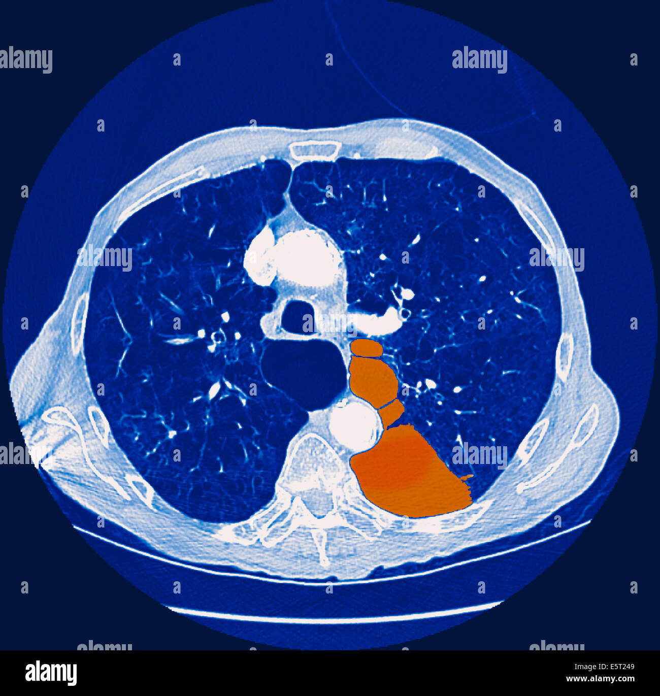 Computed tomography (CT) scan of a section through the chest of a patient with a pneumothorax, or collapsed lung (orange), The Stock Photohttps://www.alamy.com/image-license-details/?v=1https://www.alamy.com/stock-photo-computed-tomography-ct-scan-of-a-section-through-the-chest-of-a-patient-72443289.html
RME5T249–Computed tomography (CT) scan of a section through the chest of a patient with a pneumothorax, or collapsed lung (orange), The
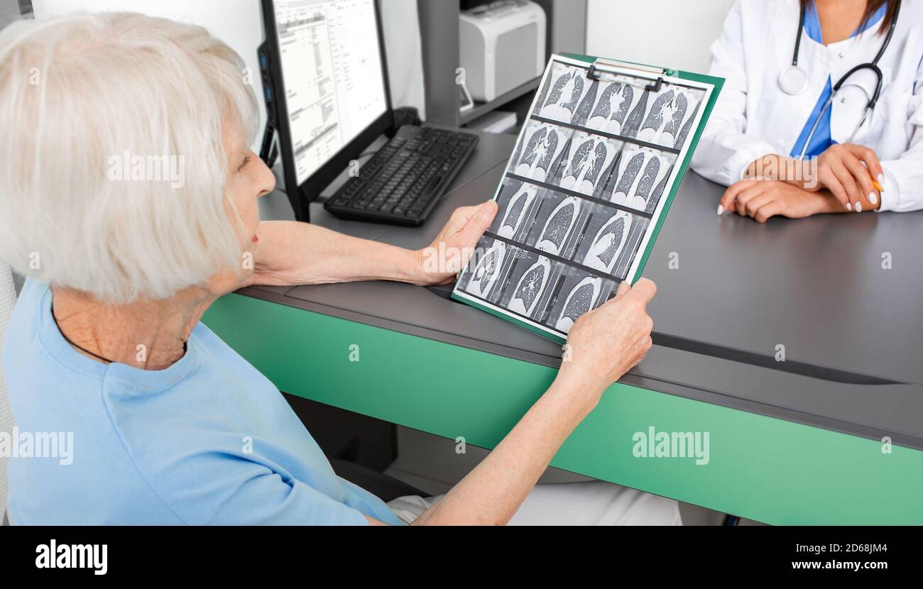 elderly patient holds the results of a CT scan of the lungs. Lung inflammation, bronchitis and coronavirus Stock Photohttps://www.alamy.com/image-license-details/?v=1https://www.alamy.com/elderly-patient-holds-the-results-of-a-ct-scan-of-the-lungs-lung-inflammation-bronchitis-and-coronavirus-image382506324.html
RF2D68JM4–elderly patient holds the results of a CT scan of the lungs. Lung inflammation, bronchitis and coronavirus
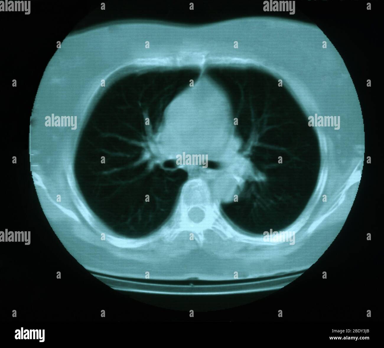 Granuloma & Calcification in Lung Stock Photohttps://www.alamy.com/image-license-details/?v=1https://www.alamy.com/granuloma-calcification-in-lung-image352793459.html
RM2BDY3JB–Granuloma & Calcification in Lung
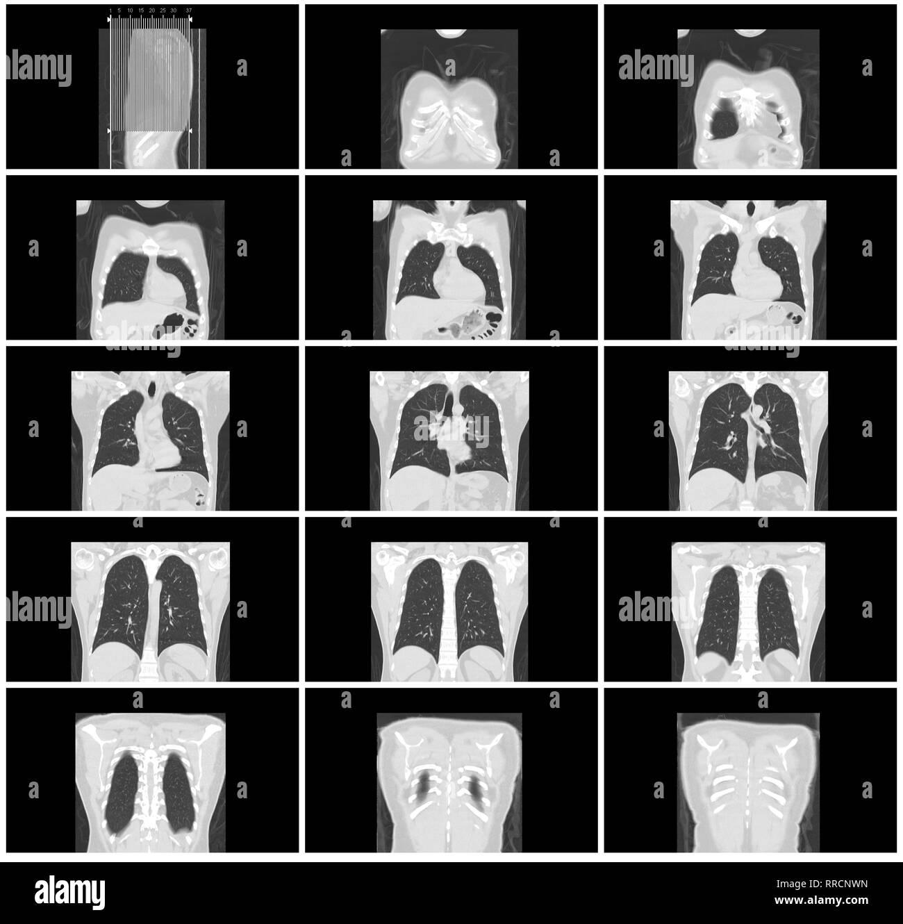 ct scan step set of body lung coronal view Stock Photohttps://www.alamy.com/image-license-details/?v=1https://www.alamy.com/ct-scan-step-set-of-body-lung-coronal-view-image238152481.html
RFRRCNWN–ct scan step set of body lung coronal view
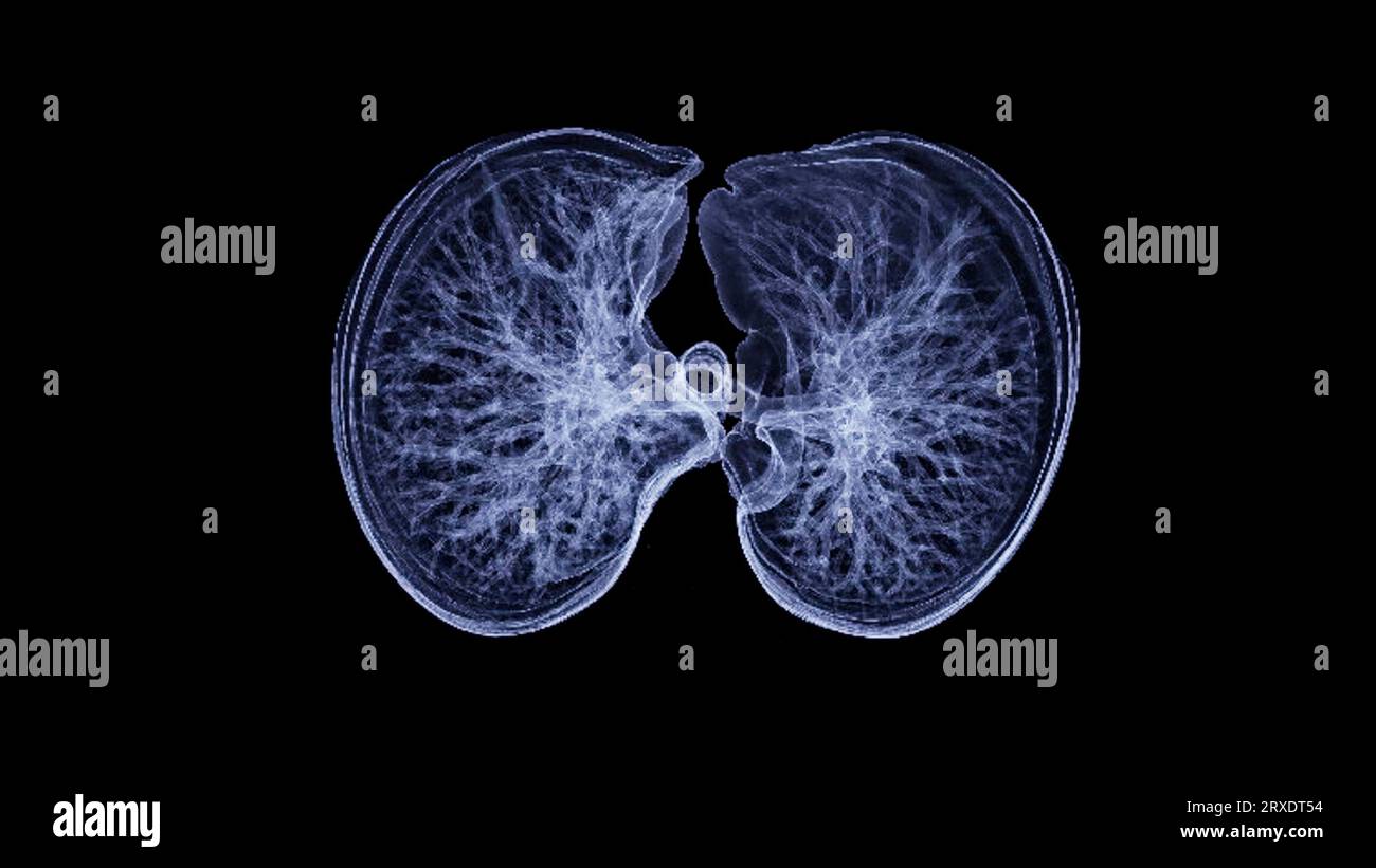 CT Chest or CT lung 3d rendering image with blue color showing Trachea and lung in respiratory system. Stock Photohttps://www.alamy.com/image-license-details/?v=1https://www.alamy.com/ct-chest-or-ct-lung-3d-rendering-image-with-blue-color-showing-trachea-and-lung-in-respiratory-system-image567017168.html
RF2RXDT54–CT Chest or CT lung 3d rendering image with blue color showing Trachea and lung in respiratory system.
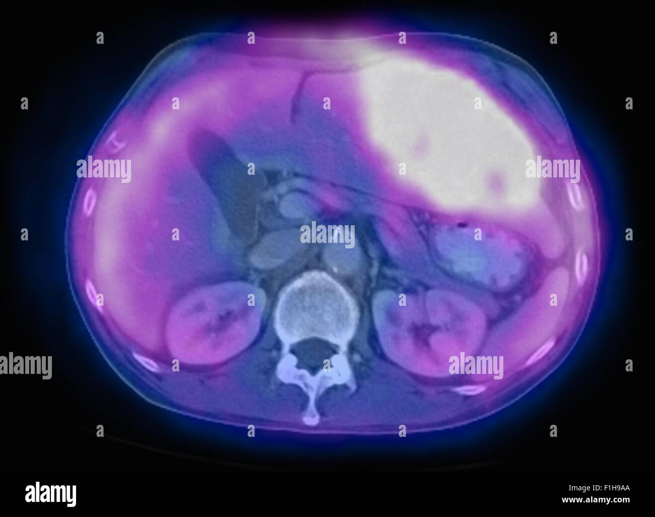 Image co-registered PET-CT study dual modality scanner. Patient multiple metastatic lesions liver & lung Stock Photohttps://www.alamy.com/image-license-details/?v=1https://www.alamy.com/stock-photo-image-co-registered-pet-ct-study-dual-modality-scanner-patient-multiple-87047026.html
RFF1H9AA–Image co-registered PET-CT study dual modality scanner. Patient multiple metastatic lesions liver & lung
 Focused worried multiethnic healthcare workers analyzing CT lung screening while working in medical clinic or hospital, medical team looking at x-ray Stock Photohttps://www.alamy.com/image-license-details/?v=1https://www.alamy.com/focused-worried-multiethnic-healthcare-workers-analyzing-ct-lung-screening-while-working-in-medical-clinic-or-hospital-medical-team-looking-at-x-ray-image461136189.html
RF2HP6FWH–Focused worried multiethnic healthcare workers analyzing CT lung screening while working in medical clinic or hospital, medical team looking at x-ray
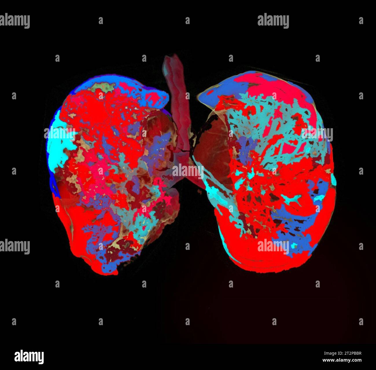 Healthy lungs, CT scan Stock Photohttps://www.alamy.com/image-license-details/?v=1https://www.alamy.com/healthy-lungs-ct-scan-image569663355.html
RF2T2PBBR–Healthy lungs, CT scan
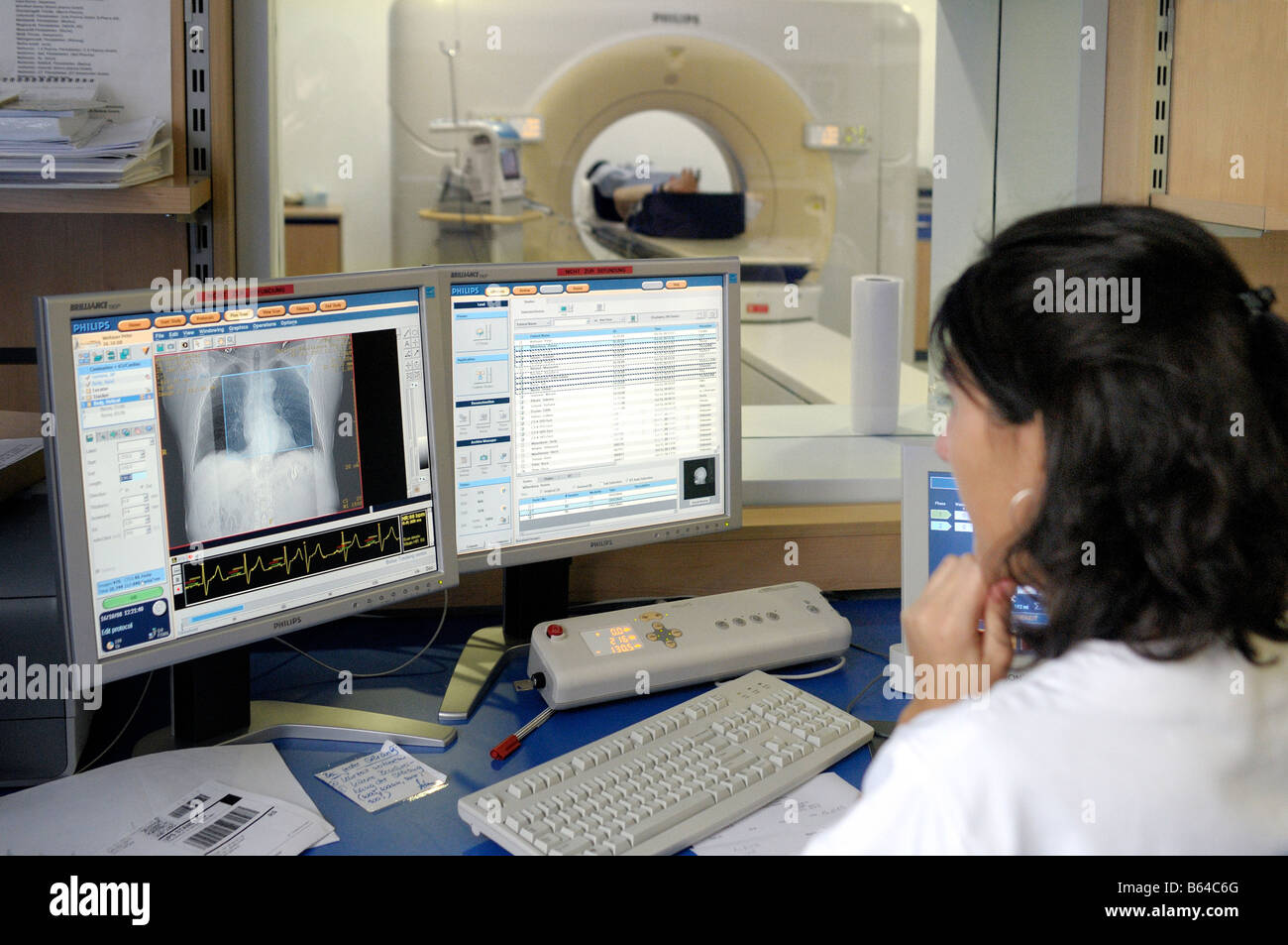 Female radiologist looking at patients lung and heart during a MRI scan Stock Photohttps://www.alamy.com/image-license-details/?v=1https://www.alamy.com/stock-photo-female-radiologist-looking-at-patients-lung-and-heart-during-a-mri-20995704.html
RMB64C6G–Female radiologist looking at patients lung and heart during a MRI scan
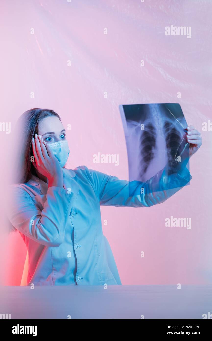 lung diagnostic scared radiologist chest x-ray Stock Photohttps://www.alamy.com/image-license-details/?v=1https://www.alamy.com/lung-diagnostic-scared-radiologist-chest-x-ray-image485350083.html
RF2K5HGYF–lung diagnostic scared radiologist chest x-ray
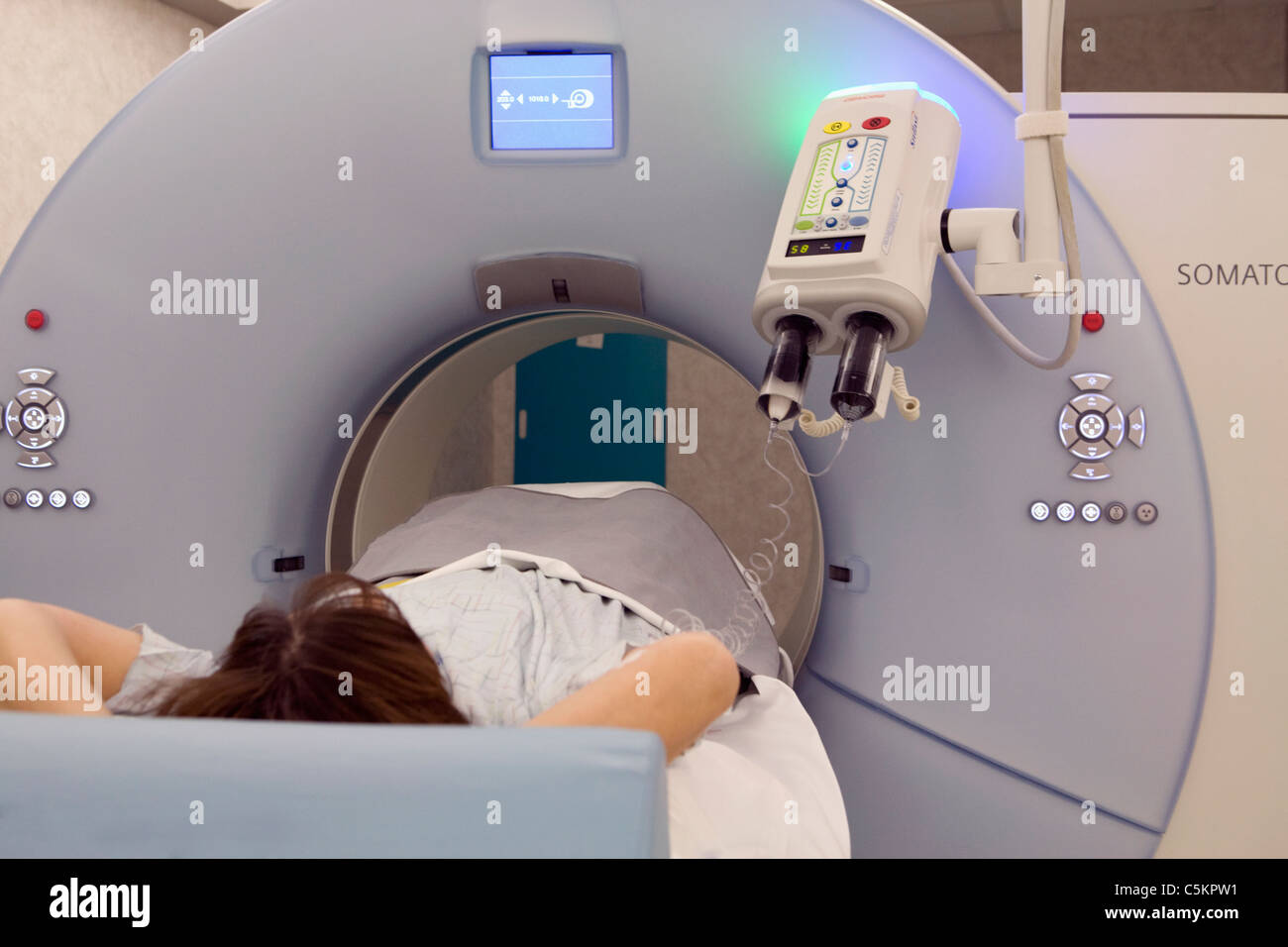 Dual energy SOMOTOM Definition CT scanner Stock Photohttps://www.alamy.com/image-license-details/?v=1https://www.alamy.com/stock-photo-dual-energy-somotom-definition-ct-scanner-37929053.html
RMC5KPW1–Dual energy SOMOTOM Definition CT scanner
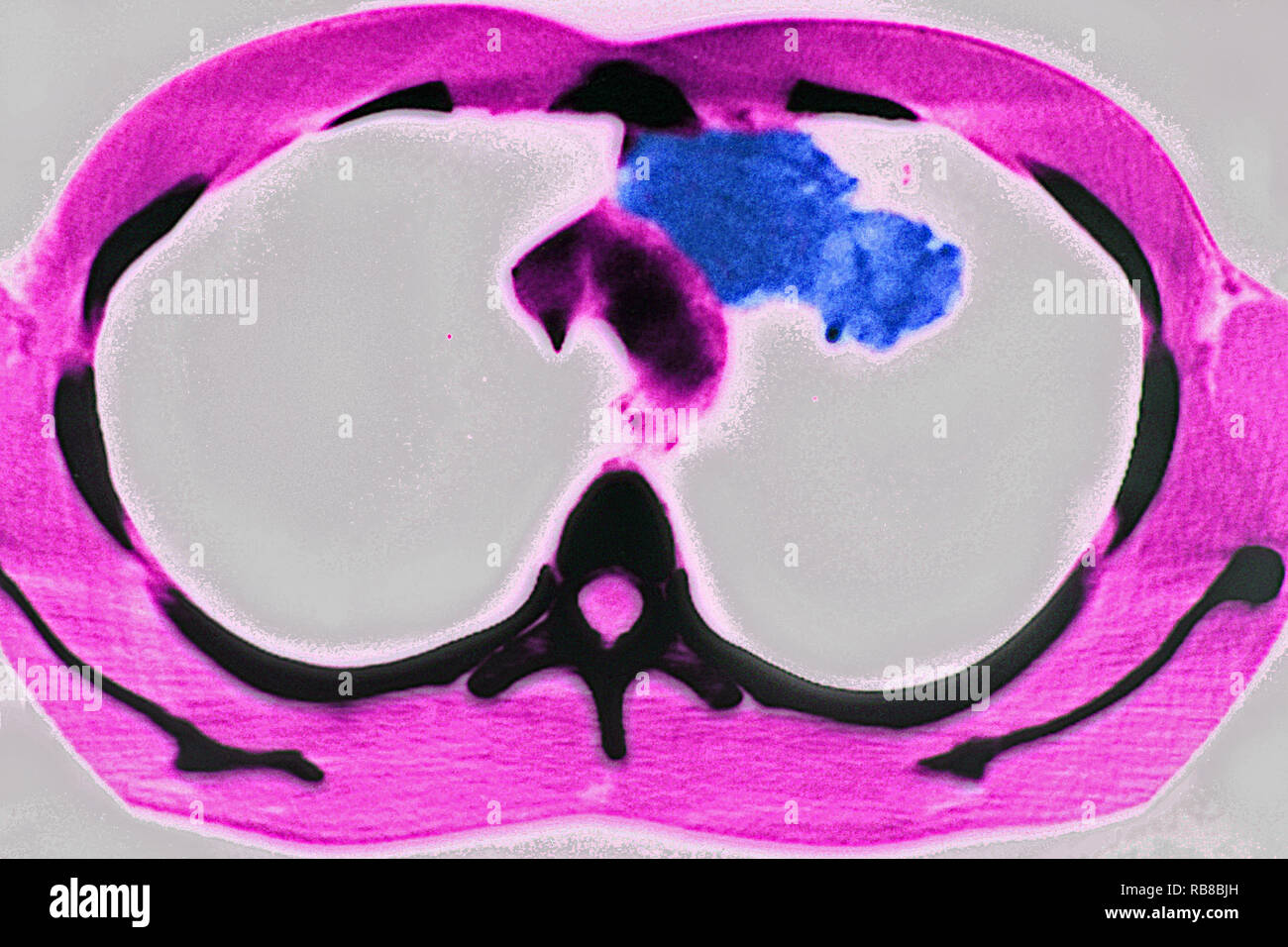 PULMONARY TUBERCULOSIS, CT-SCAN Stock Photohttps://www.alamy.com/image-license-details/?v=1https://www.alamy.com/pulmonary-tuberculosis-ct-scan-image230680761.html
RMRB8BJH–PULMONARY TUBERCULOSIS, CT-SCAN
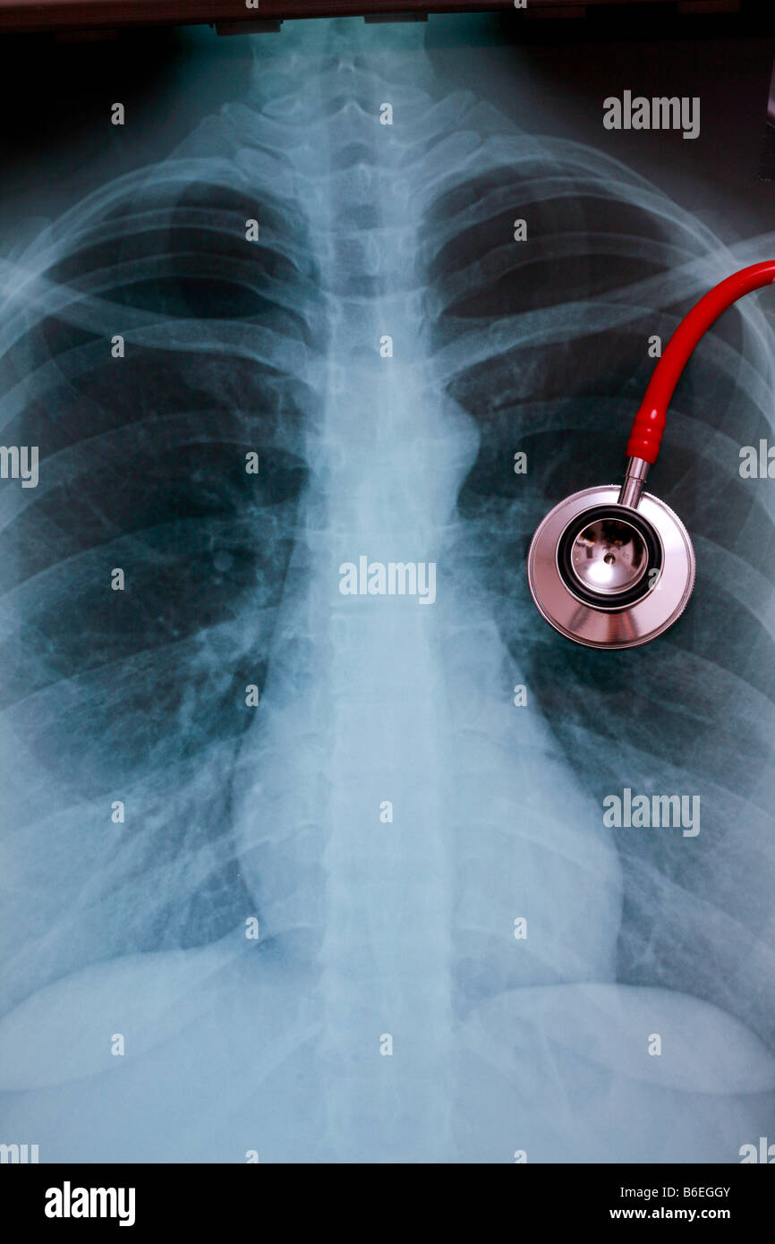 chest x-ray CXR with stethoscope overlying left lung Stock Photohttps://www.alamy.com/image-license-details/?v=1https://www.alamy.com/stock-photo-chest-x-ray-cxr-with-stethoscope-overlying-left-lung-21218651.html
RFB6EGGY–chest x-ray CXR with stethoscope overlying left lung
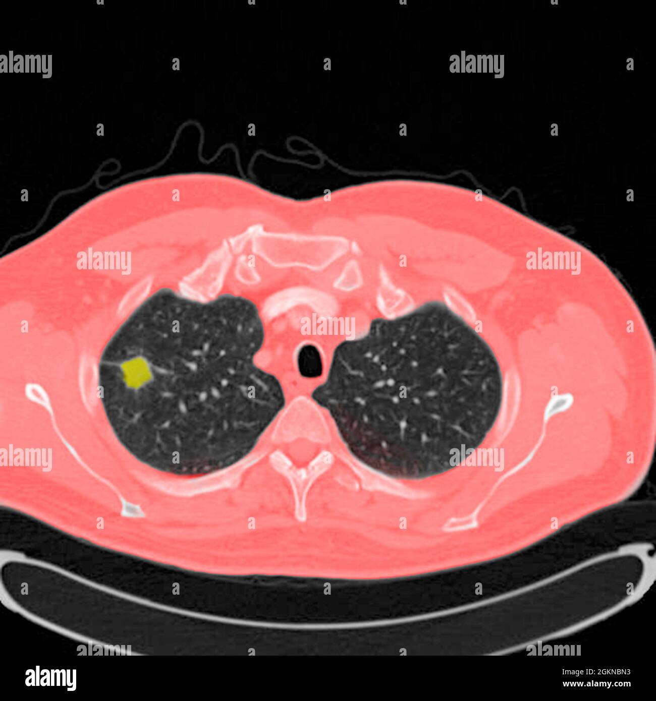 Chest CT scan (X-ray computed tomography) of a male 54 year old patient. A tumour can be seen in the left upper lobe of his lungs Stock Photohttps://www.alamy.com/image-license-details/?v=1https://www.alamy.com/chest-ct-scan-x-ray-computed-tomography-of-a-male-54-year-old-patient-a-tumour-can-be-seen-in-the-left-upper-lobe-of-his-lungs-image442407871.html
RM2GKNBN3–Chest CT scan (X-ray computed tomography) of a male 54 year old patient. A tumour can be seen in the left upper lobe of his lungs
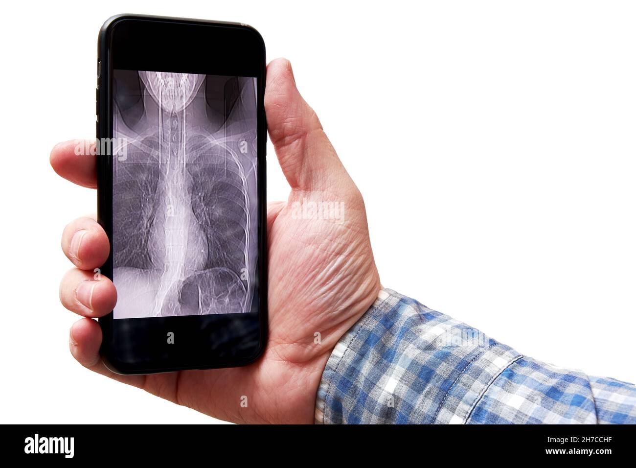 Man's hand holding a mobile phone with a CT scan image of lungs. Pneumonia and disease diagnosis Stock Photohttps://www.alamy.com/image-license-details/?v=1https://www.alamy.com/mans-hand-holding-a-mobile-phone-with-a-ct-scan-image-of-lungs-pneumonia-and-disease-diagnosis-image452045483.html
RF2H7CCHF–Man's hand holding a mobile phone with a CT scan image of lungs. Pneumonia and disease diagnosis
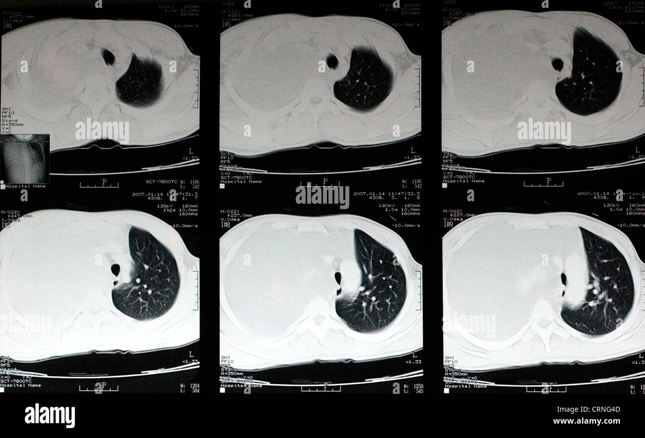 A CT scan showing a severe haemothroax on the right side of the patient's chest. Stock Photohttps://www.alamy.com/image-license-details/?v=1https://www.alamy.com/stock-photo-a-ct-scan-showing-a-severe-haemothroax-on-the-right-side-of-the-patients-49031485.html
RMCRNG4D–A CT scan showing a severe haemothroax on the right side of the patient's chest.
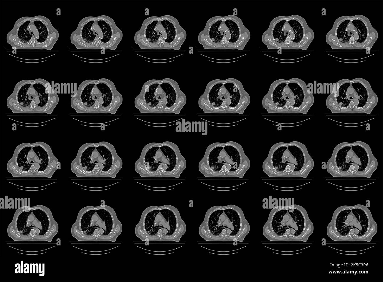 Overview of a computed tomography scan of a patient with a lung tumor Stock Photohttps://www.alamy.com/image-license-details/?v=1https://www.alamy.com/overview-of-a-computed-tomography-scan-of-a-patient-with-a-lung-tumor-image485230010.html
RF2K5C3R6–Overview of a computed tomography scan of a patient with a lung tumor
 a doctor in a protective suit and mask looks at the results of a CT scan of the lungs Stock Photohttps://www.alamy.com/image-license-details/?v=1https://www.alamy.com/a-doctor-in-a-protective-suit-and-mask-looks-at-the-results-of-a-ct-scan-of-the-lungs-image386248540.html
RF2DCB3XM–a doctor in a protective suit and mask looks at the results of a CT scan of the lungs
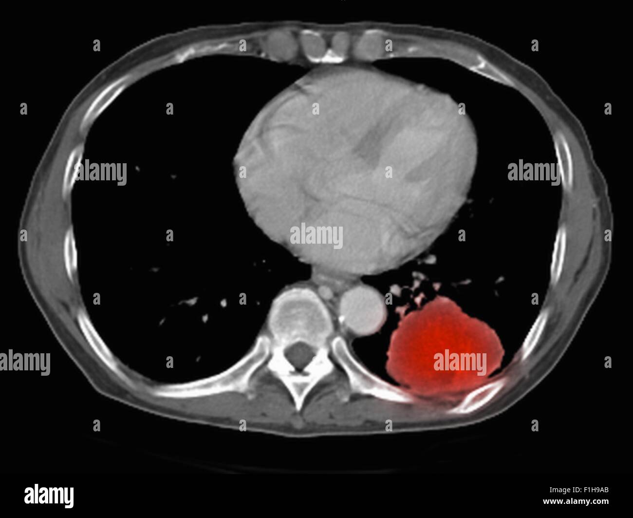 Image co-registered PET-CT study dual modality scanner. Patient multiple metastatic lesions liver & lung Stock Photohttps://www.alamy.com/image-license-details/?v=1https://www.alamy.com/stock-photo-image-co-registered-pet-ct-study-dual-modality-scanner-patient-multiple-87047027.html
RFF1H9AB–Image co-registered PET-CT study dual modality scanner. Patient multiple metastatic lesions liver & lung
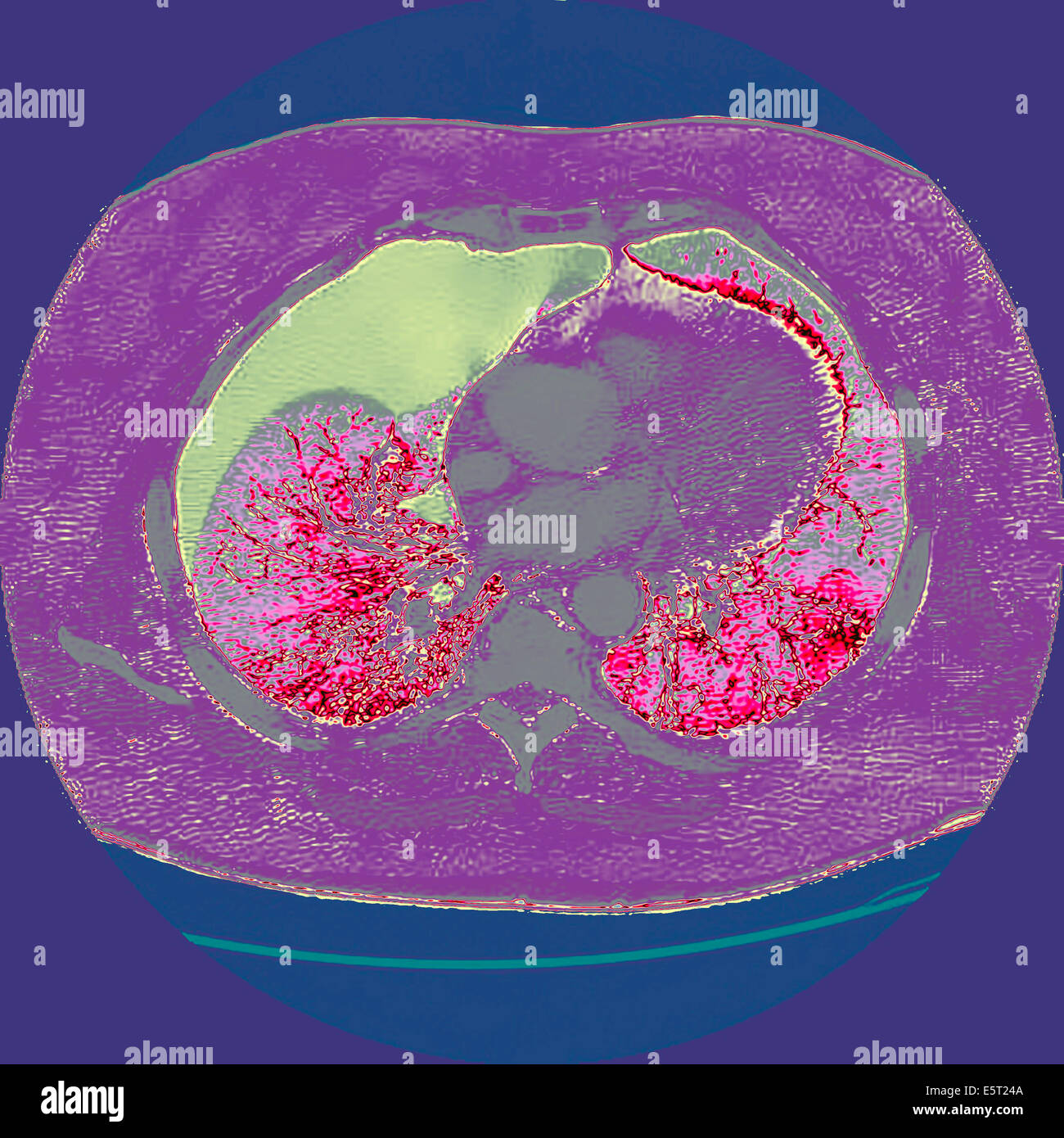 Computed tomography (CT) scan of a section through the chest of a patient with a pneumothorax, or collapsed lung (grey), The Stock Photohttps://www.alamy.com/image-license-details/?v=1https://www.alamy.com/stock-photo-computed-tomography-ct-scan-of-a-section-through-the-chest-of-a-patient-72443290.html
RME5T24A–Computed tomography (CT) scan of a section through the chest of a patient with a pneumothorax, or collapsed lung (grey), The
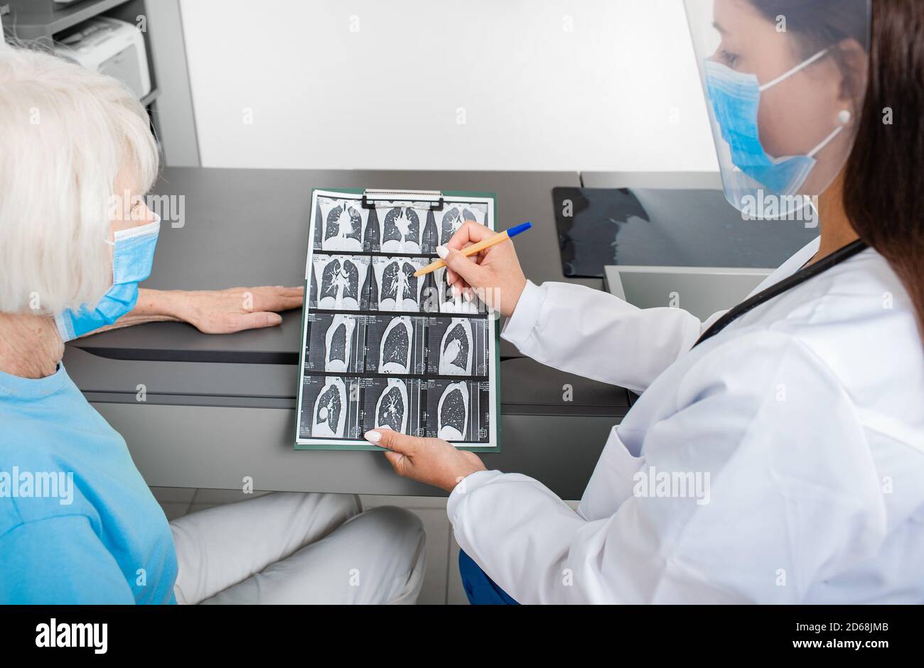 Pulmonologist showing a senior patient a CT scan of her lungs. Pneumonia, coronavirus, lung disease Stock Photohttps://www.alamy.com/image-license-details/?v=1https://www.alamy.com/pulmonologist-showing-a-senior-patient-a-ct-scan-of-her-lungs-pneumonia-coronavirus-lung-disease-image382506331.html
RF2D68JMB–Pulmonologist showing a senior patient a CT scan of her lungs. Pneumonia, coronavirus, lung disease
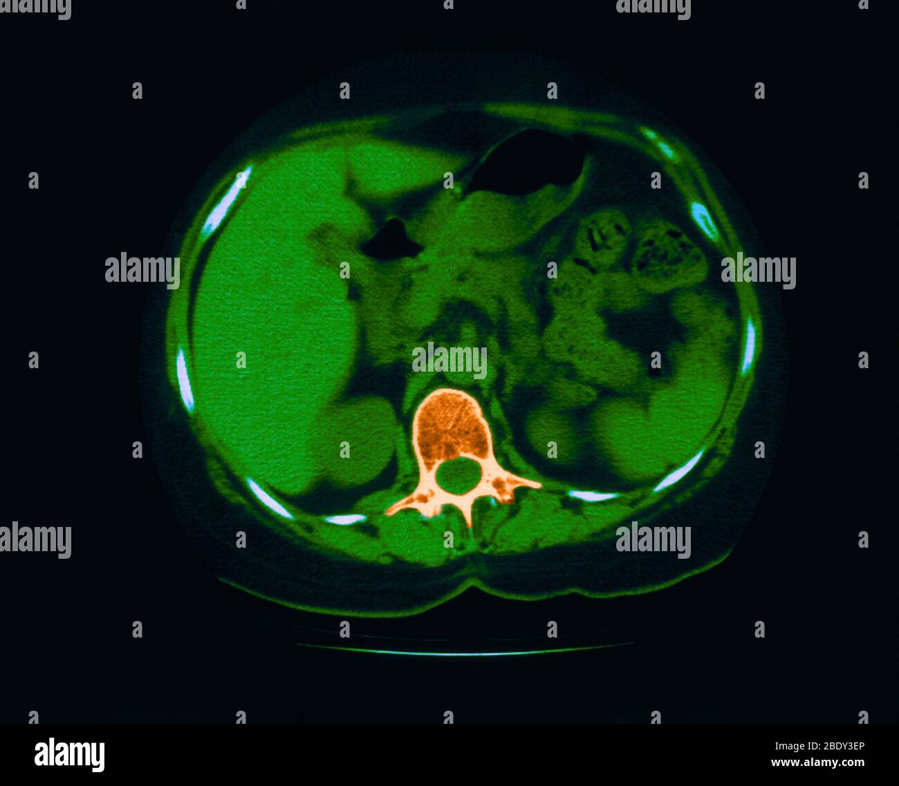 Scoliosis, Granuloma & Calcification in Lung Stock Photohttps://www.alamy.com/image-license-details/?v=1https://www.alamy.com/scoliosis-granuloma-calcification-in-lung-image352793358.html
RM2BDY3EP–Scoliosis, Granuloma & Calcification in Lung
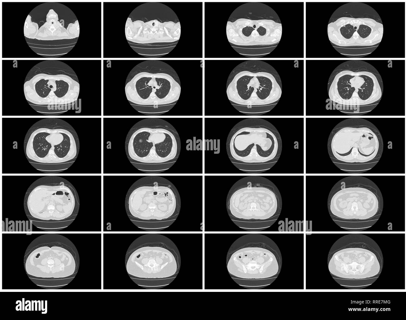 ct scan step set of upper body lung axial top view Stock Photohttps://www.alamy.com/image-license-details/?v=1https://www.alamy.com/ct-scan-step-set-of-upper-body-lung-axial-top-view-image238185264.html
RFRRE7MG–ct scan step set of upper body lung axial top view
 CT Chest or CT lung 3d rendering image with blue color showing Trachea and lung in respiratory system. Stock Photohttps://www.alamy.com/image-license-details/?v=1https://www.alamy.com/ct-chest-or-ct-lung-3d-rendering-image-with-blue-color-showing-trachea-and-lung-in-respiratory-system-image567017164.html
RF2RXDT50–CT Chest or CT lung 3d rendering image with blue color showing Trachea and lung in respiratory system.
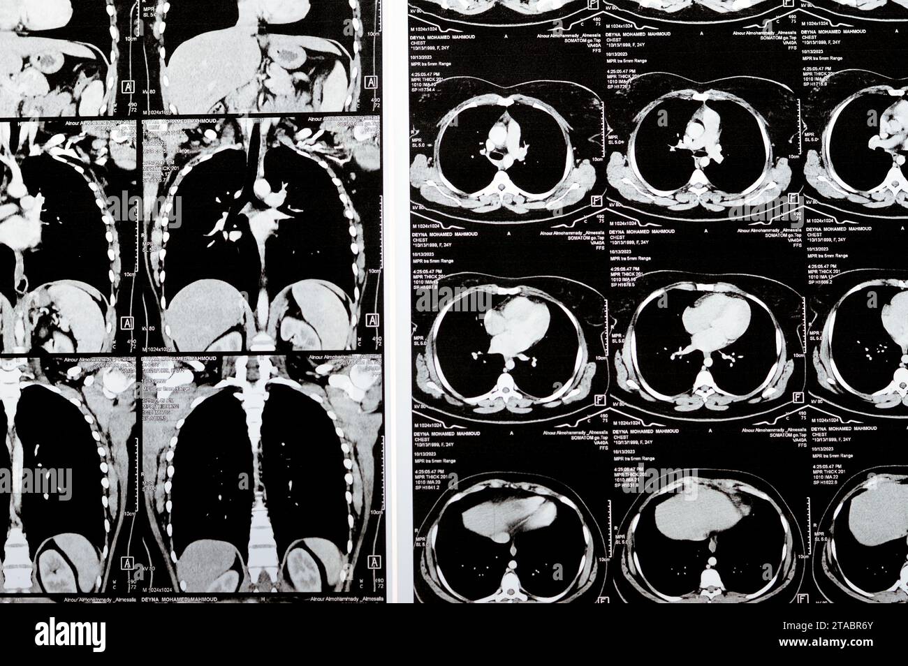 Cairo, Egypt, October 15 2023: CT scan axial slices through chest with contrast injection showing low grade of inflammatory reaction, parenchymal vess Stock Photohttps://www.alamy.com/image-license-details/?v=1https://www.alamy.com/cairo-egypt-october-15-2023-ct-scan-axial-slices-through-chest-with-contrast-injection-showing-low-grade-of-inflammatory-reaction-parenchymal-vess-image574348403.html
RF2TABR6Y–Cairo, Egypt, October 15 2023: CT scan axial slices through chest with contrast injection showing low grade of inflammatory reaction, parenchymal vess
 Focused worried multiethnic healthcare workers analyzing CT lung screening while working in medical clinic or hospital, medical team looking at x-ray Stock Photohttps://www.alamy.com/image-license-details/?v=1https://www.alamy.com/focused-worried-multiethnic-healthcare-workers-analyzing-ct-lung-screening-while-working-in-medical-clinic-or-hospital-medical-team-looking-at-x-ray-image461136158.html
RF2HP6FTE–Focused worried multiethnic healthcare workers analyzing CT lung screening while working in medical clinic or hospital, medical team looking at x-ray
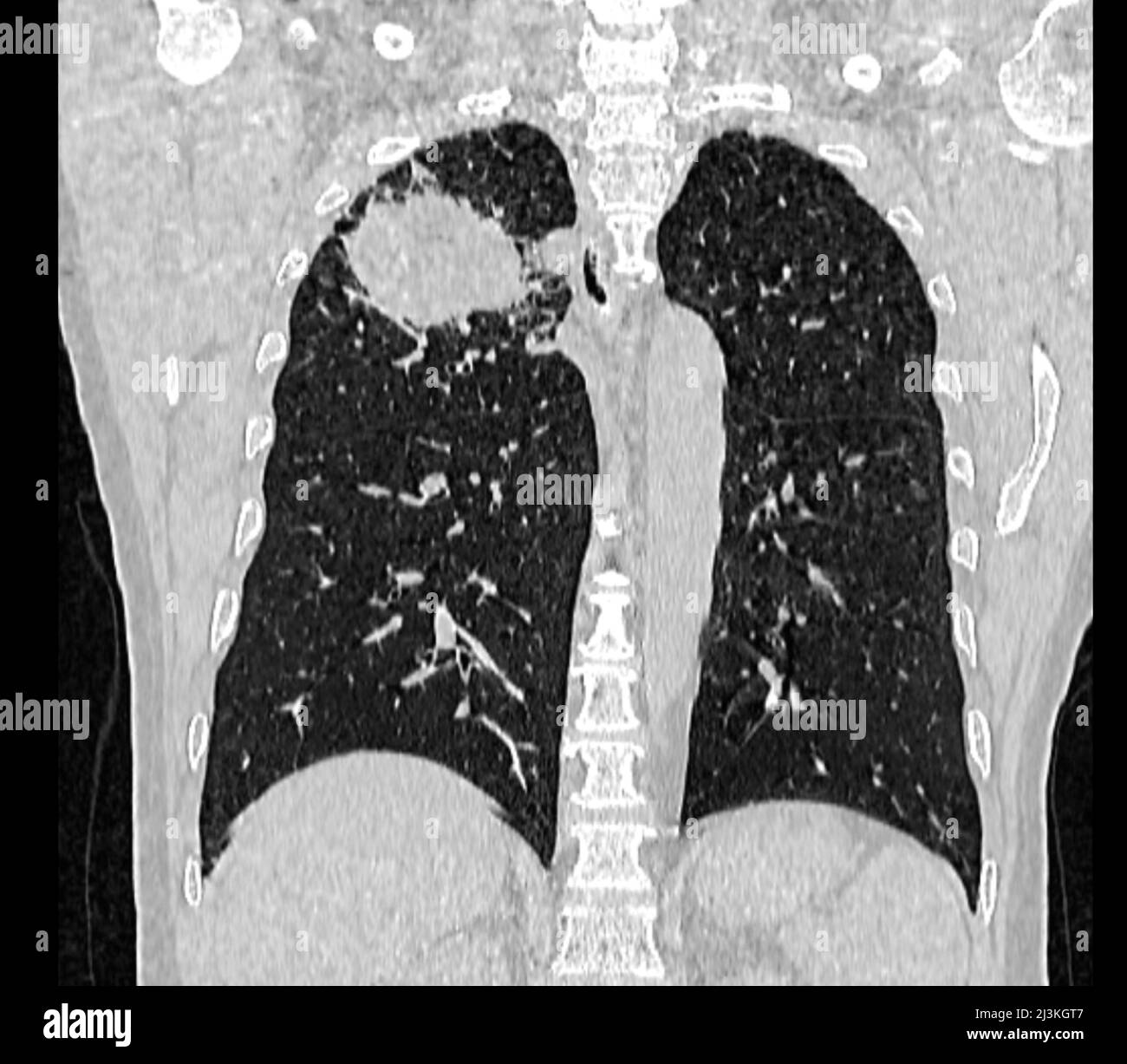 Lung carcinoma, CT scan Stock Photohttps://www.alamy.com/image-license-details/?v=1https://www.alamy.com/lung-carcinoma-ct-scan-image466954215.html
RF2J3KGT7–Lung carcinoma, CT scan
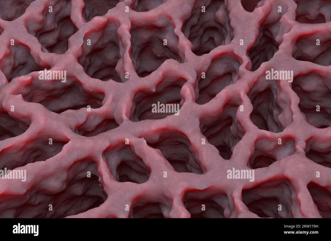 Lung tissue - isometric view 3d illustration Stock Photohttps://www.alamy.com/image-license-details/?v=1https://www.alamy.com/lung-tissue-isometric-view-3d-illustration-image566125885.html
RF2RW179H–Lung tissue - isometric view 3d illustration
 radiology checkup lung pneumonia medic chest x-ray Stock Photohttps://www.alamy.com/image-license-details/?v=1https://www.alamy.com/radiology-checkup-lung-pneumonia-medic-chest-x-ray-image485350082.html
RF2K5HGYE–radiology checkup lung pneumonia medic chest x-ray
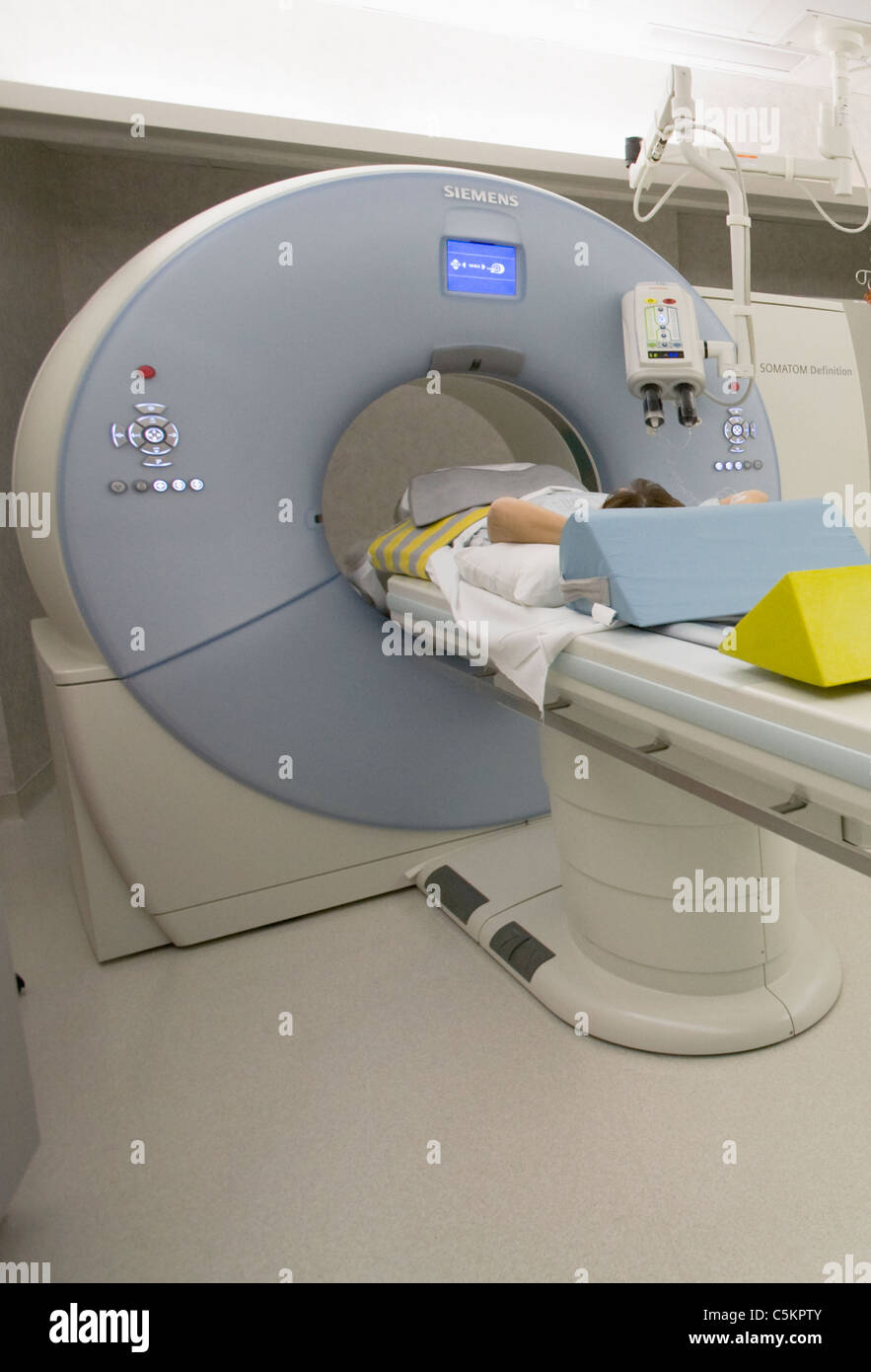 Dual energy SOMOTOM Definition CT scanner Stock Photohttps://www.alamy.com/image-license-details/?v=1https://www.alamy.com/stock-photo-dual-energy-somotom-definition-ct-scanner-37929051.html
RMC5KPTY–Dual energy SOMOTOM Definition CT scanner
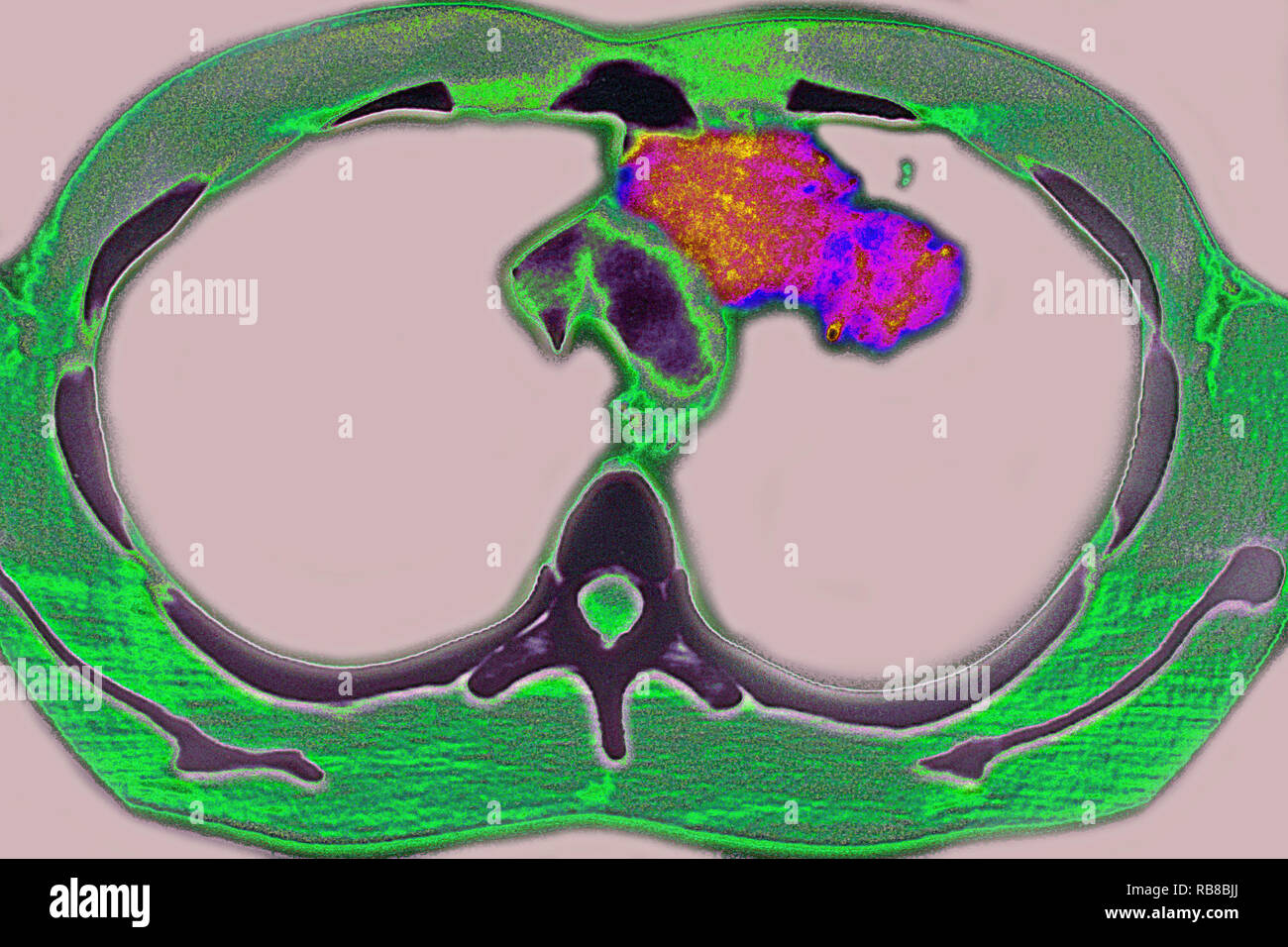 PULMONARY TUBERCULOSIS, CT-SCAN Stock Photohttps://www.alamy.com/image-license-details/?v=1https://www.alamy.com/pulmonary-tuberculosis-ct-scan-image230680762.html
RMRB8BJJ–PULMONARY TUBERCULOSIS, CT-SCAN
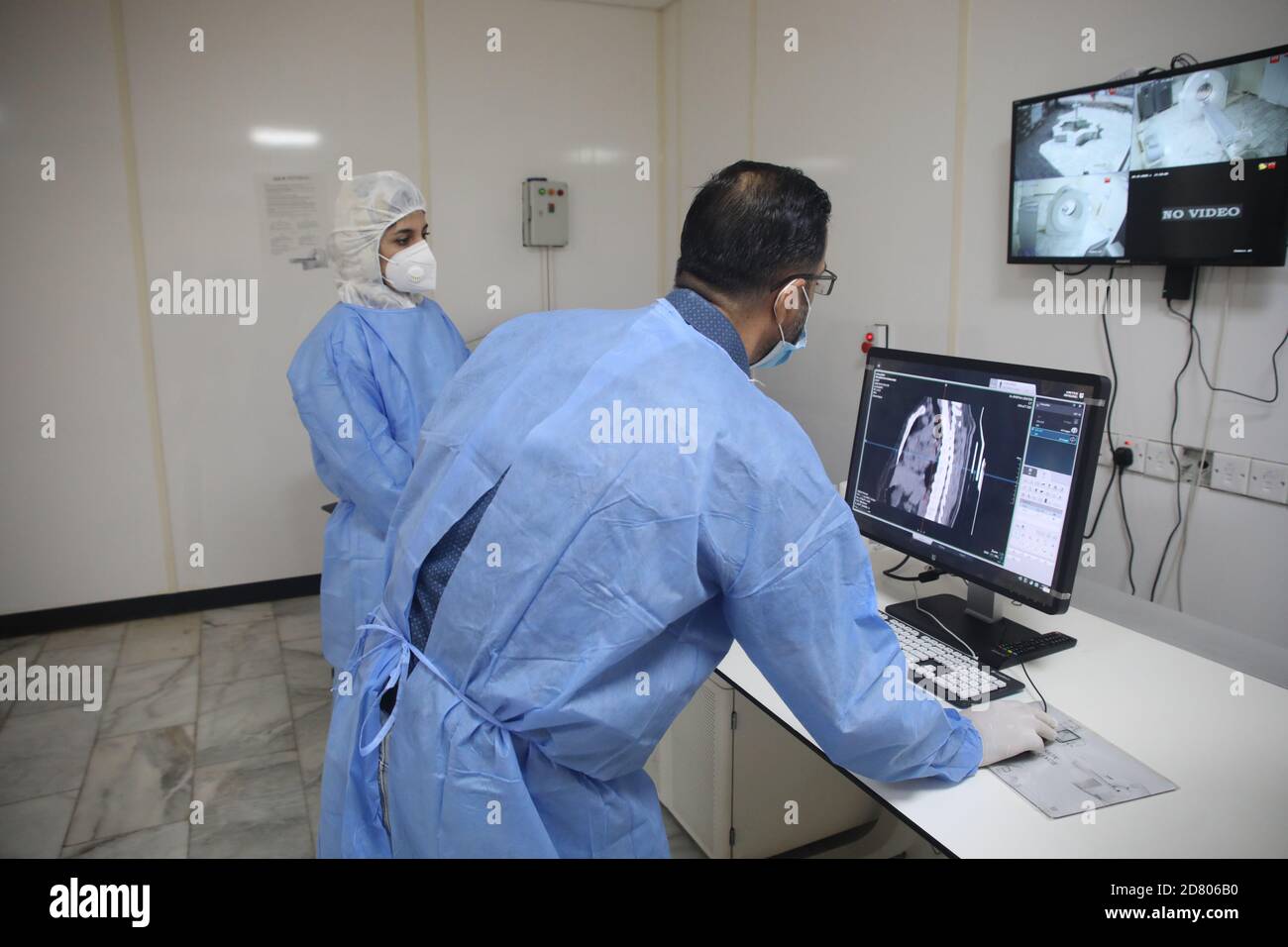 Berlin, Germany. 26th Oct, 2020. Dr. Mohammed Abdul-Hussein (R) and CT technician Hanan Jamal examine lung images in a Chinese-built CT room at al-Shifaa Center in Baghdad, Iraq, Oct. 12, 2020. TO GO WITH HEADLINES OF Oct. 26, 2020. Credit: Xinhua/Alamy Live News Stock Photohttps://www.alamy.com/image-license-details/?v=1https://www.alamy.com/berlin-germany-26th-oct-2020-dr-mohammed-abdul-hussein-r-and-ct-technician-hanan-jamal-examine-lung-images-in-a-chinese-built-ct-room-at-al-shifaa-center-in-baghdad-iraq-oct-12-2020-to-go-with-headlines-of-oct-26-2020-credit-xinhuaalamy-live-news-image383550356.html
RM2D806B0–Berlin, Germany. 26th Oct, 2020. Dr. Mohammed Abdul-Hussein (R) and CT technician Hanan Jamal examine lung images in a Chinese-built CT room at al-Shifaa Center in Baghdad, Iraq, Oct. 12, 2020. TO GO WITH HEADLINES OF Oct. 26, 2020. Credit: Xinhua/Alamy Live News
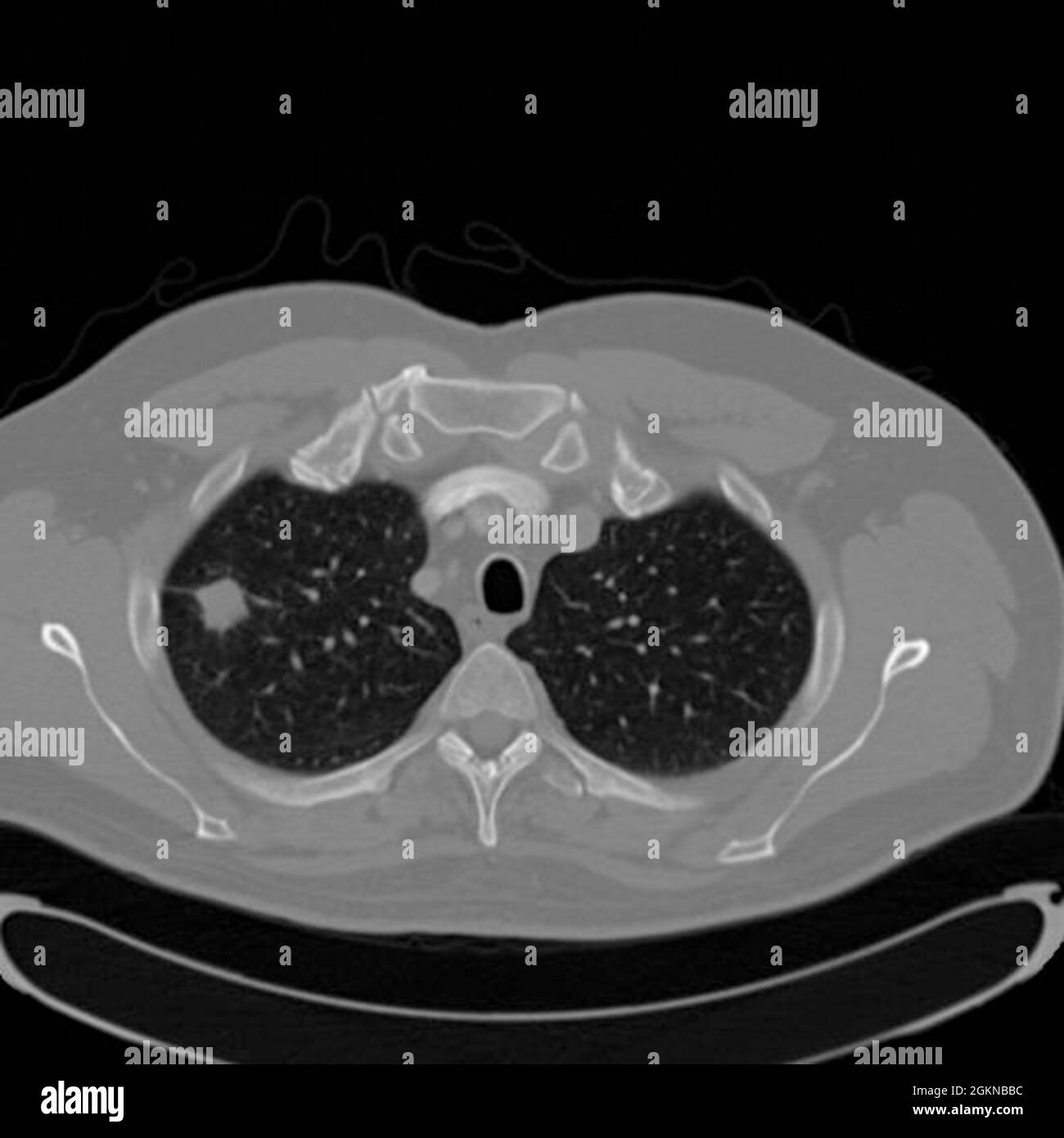 Chest CT scan (X-ray computed tomography) of a male 54 year old patient. A tumour can be seen in the left upper lobe of his lungs Stock Photohttps://www.alamy.com/image-license-details/?v=1https://www.alamy.com/chest-ct-scan-x-ray-computed-tomography-of-a-male-54-year-old-patient-a-tumour-can-be-seen-in-the-left-upper-lobe-of-his-lungs-image442407600.html
RM2GKNBBC–Chest CT scan (X-ray computed tomography) of a male 54 year old patient. A tumour can be seen in the left upper lobe of his lungs
 Old man analyzing his lungs CT scan on a phone. Pneumonia and disease diagnosis. Pills and medical bottles Stock Photohttps://www.alamy.com/image-license-details/?v=1https://www.alamy.com/old-man-analyzing-his-lungs-ct-scan-on-a-phone-pneumonia-and-disease-diagnosis-pills-and-medical-bottles-image456430220.html
RF2HEG5B8–Old man analyzing his lungs CT scan on a phone. Pneumonia and disease diagnosis. Pills and medical bottles
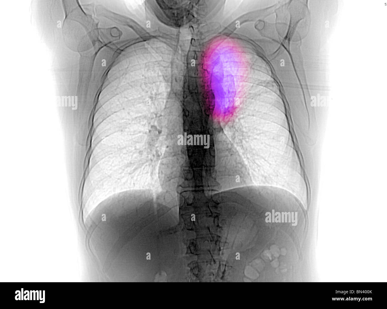 CT scan scout image from a 54 year old man with lung cancer Stock Photohttps://www.alamy.com/image-license-details/?v=1https://www.alamy.com/stock-photo-ct-scan-scout-image-from-a-54-year-old-man-with-lung-cancer-30205971.html
RMBN400K–CT scan scout image from a 54 year old man with lung cancer
 Conceptual complex of medical examination results in one image. Stock Photohttps://www.alamy.com/image-license-details/?v=1https://www.alamy.com/conceptual-complex-of-medical-examination-results-in-one-image-image398169922.html
RF2E3P5PA–Conceptual complex of medical examination results in one image.
 a doctor in a protective suit and mask looks at the results of a CT scan of the lungs Stock Photohttps://www.alamy.com/image-license-details/?v=1https://www.alamy.com/a-doctor-in-a-protective-suit-and-mask-looks-at-the-results-of-a-ct-scan-of-the-lungs-image386248639.html
RF2DCB427–a doctor in a protective suit and mask looks at the results of a CT scan of the lungs
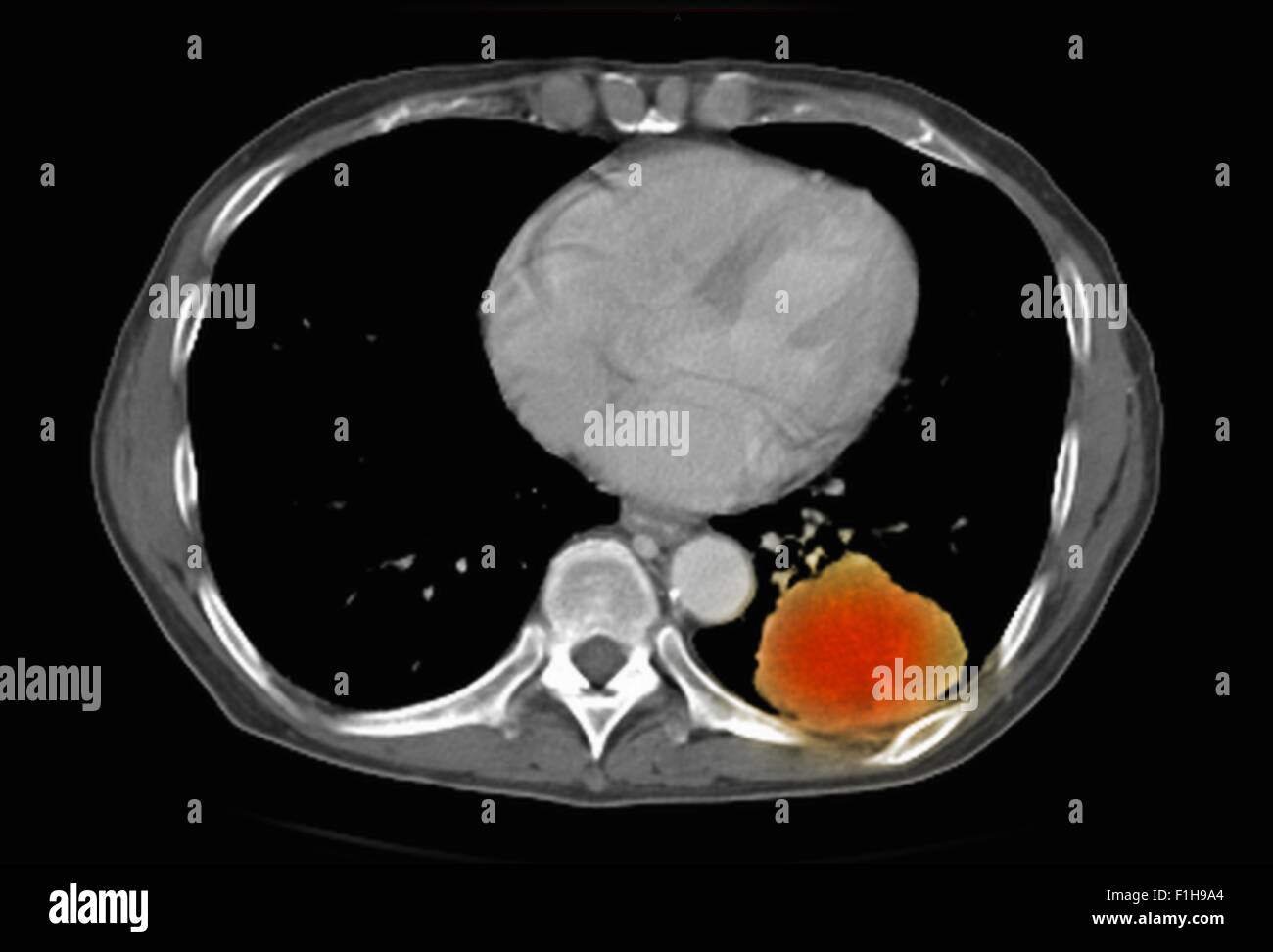 Image co-registered PET-CT study dual modality scanner. Patient multiple metastatic lesions liver & lung Stock Photohttps://www.alamy.com/image-license-details/?v=1https://www.alamy.com/stock-photo-image-co-registered-pet-ct-study-dual-modality-scanner-patient-multiple-87047020.html
RFF1H9A4–Image co-registered PET-CT study dual modality scanner. Patient multiple metastatic lesions liver & lung
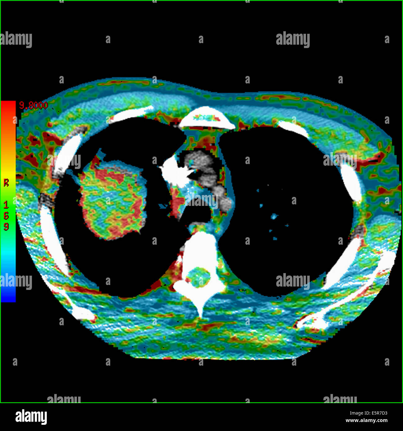 Colored Computed Tomography (CT) scan of the abdomen showing a lung cancer, The red parts of the tumor are the vascularized Stock Photohttps://www.alamy.com/image-license-details/?v=1https://www.alamy.com/stock-photo-colored-computed-tomography-ct-scan-of-the-abdomen-showing-a-lung-72425503.html
RME5R7D3–Colored Computed Tomography (CT) scan of the abdomen showing a lung cancer, The red parts of the tumor are the vascularized
 Pulmonologist wearing in protective mask showing man patient a CT scan of his lungs. Pneumonia, coronavirus, lung disease Stock Photohttps://www.alamy.com/image-license-details/?v=1https://www.alamy.com/pulmonologist-wearing-in-protective-mask-showing-man-patient-a-ct-scan-of-his-lungs-pneumonia-coronavirus-lung-disease-image389258509.html
RF2DH875H–Pulmonologist wearing in protective mask showing man patient a CT scan of his lungs. Pneumonia, coronavirus, lung disease
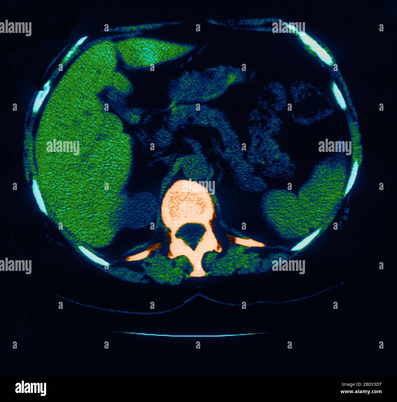 Scoliosis, Granuloma & Calcification in Lung Stock Photohttps://www.alamy.com/image-license-details/?v=1https://www.alamy.com/scoliosis-granuloma-calcification-in-lung-image352793332.html
RM2BDY3DT–Scoliosis, Granuloma & Calcification in Lung
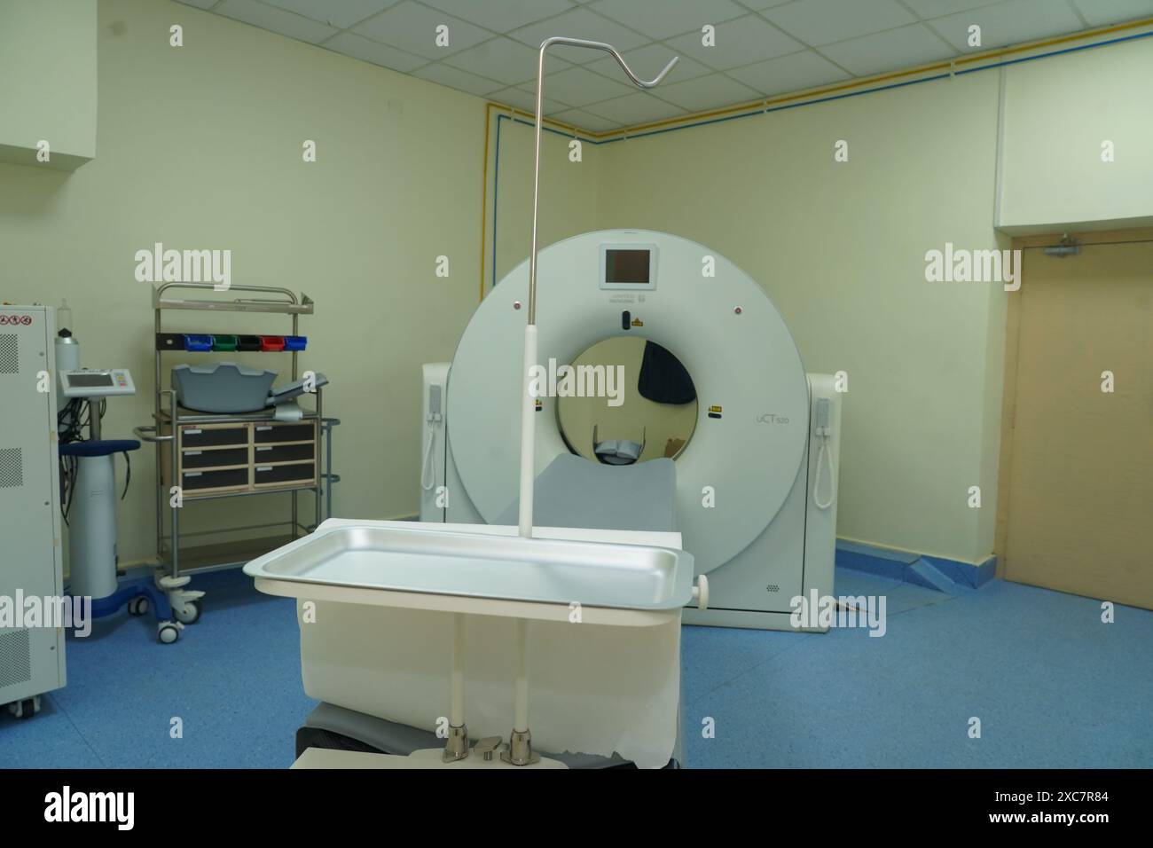 Advanced imaging device, detailed body scans, diagnostic tool, medical imaging, radiology equipment. Stock Photohttps://www.alamy.com/image-license-details/?v=1https://www.alamy.com/advanced-imaging-device-detailed-body-scans-diagnostic-tool-medical-imaging-radiology-equipment-image609910676.html
RF2XC7R84–Advanced imaging device, detailed body scans, diagnostic tool, medical imaging, radiology equipment.
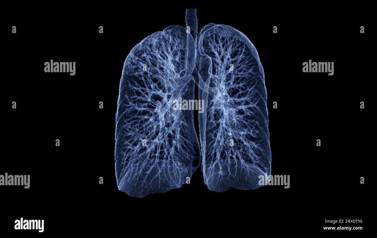 CT Chest or CT lung 3d rendering image with blue color showing Trachea and lung in respiratory system. Stock Photohttps://www.alamy.com/image-license-details/?v=1https://www.alamy.com/ct-chest-or-ct-lung-3d-rendering-image-with-blue-color-showing-trachea-and-lung-in-respiratory-system-image567017170.html
RF2RXDT56–CT Chest or CT lung 3d rendering image with blue color showing Trachea and lung in respiratory system.
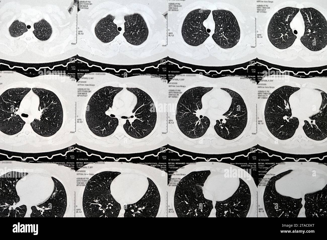 Cairo, Egypt, October 15 2023: CT scan axial slices through chest with contrast injection showing low grade of inflammatory reaction, parenchymal vess Stock Photohttps://www.alamy.com/image-license-details/?v=1https://www.alamy.com/cairo-egypt-october-15-2023-ct-scan-axial-slices-through-chest-with-contrast-injection-showing-low-grade-of-inflammatory-reaction-parenchymal-vess-image574363660.html
RF2TACEKT–Cairo, Egypt, October 15 2023: CT scan axial slices through chest with contrast injection showing low grade of inflammatory reaction, parenchymal vess
 Focused worried multiethnic healthcare workers analyzing CT lung screening while working in medical clinic or hospital, medical team looking at x-ray Stock Photohttps://www.alamy.com/image-license-details/?v=1https://www.alamy.com/focused-worried-multiethnic-healthcare-workers-analyzing-ct-lung-screening-while-working-in-medical-clinic-or-hospital-medical-team-looking-at-x-ray-image460820558.html
RF2HNM592–Focused worried multiethnic healthcare workers analyzing CT lung screening while working in medical clinic or hospital, medical team looking at x-ray
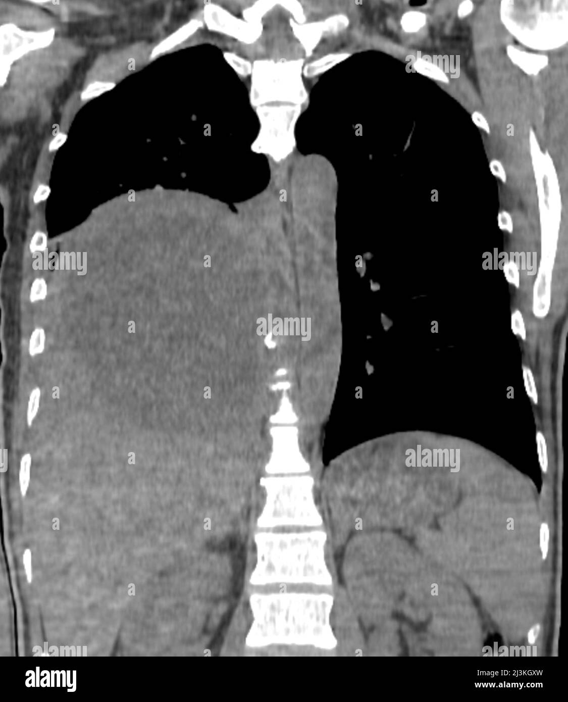 Pleural empyema, CT scan Stock Photohttps://www.alamy.com/image-license-details/?v=1https://www.alamy.com/pleural-empyema-ct-scan-image466954289.html
RF2J3KGXW–Pleural empyema, CT scan
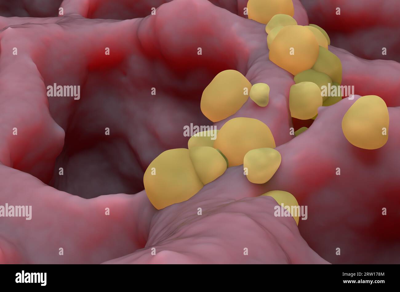 Small cancer tumors on the lung tissue: lung cancer (LC) - closeup view 3d illustration Stock Photohttps://www.alamy.com/image-license-details/?v=1https://www.alamy.com/small-cancer-tumors-on-the-lung-tissue-lung-cancer-lc-closeup-view-3d-illustration-image566125860.html
RF2RW178M–Small cancer tumors on the lung tissue: lung cancer (LC) - closeup view 3d illustration
 diagnostic x-ray lung checkup doctor chest film Stock Photohttps://www.alamy.com/image-license-details/?v=1https://www.alamy.com/diagnostic-x-ray-lung-checkup-doctor-chest-film-image485350149.html
RF2K5HH1W–diagnostic x-ray lung checkup doctor chest film
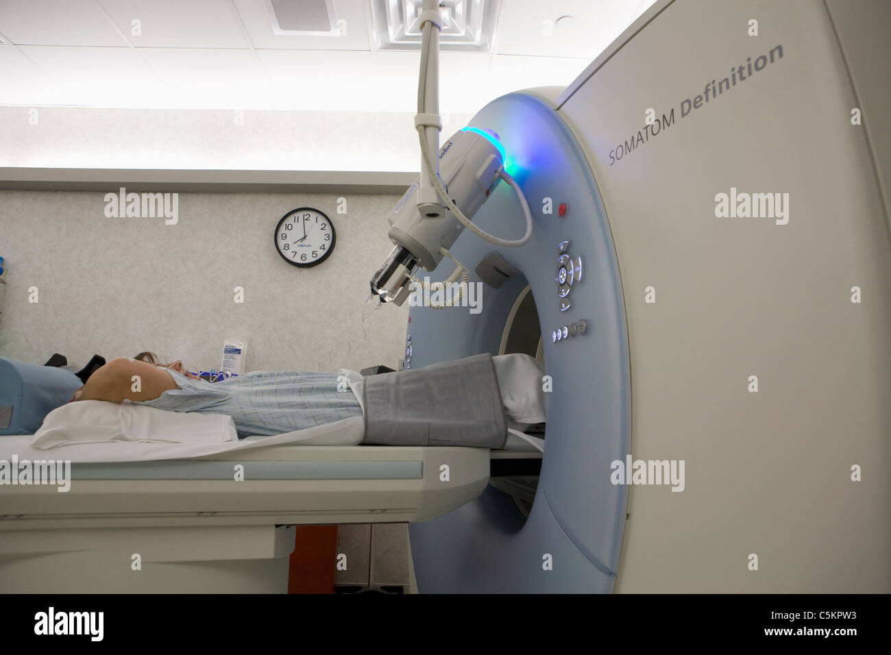 Dual energy SOMOTOM Definition CT scanner Stock Photohttps://www.alamy.com/image-license-details/?v=1https://www.alamy.com/stock-photo-dual-energy-somotom-definition-ct-scanner-37929055.html
RMC5KPW3–Dual energy SOMOTOM Definition CT scanner
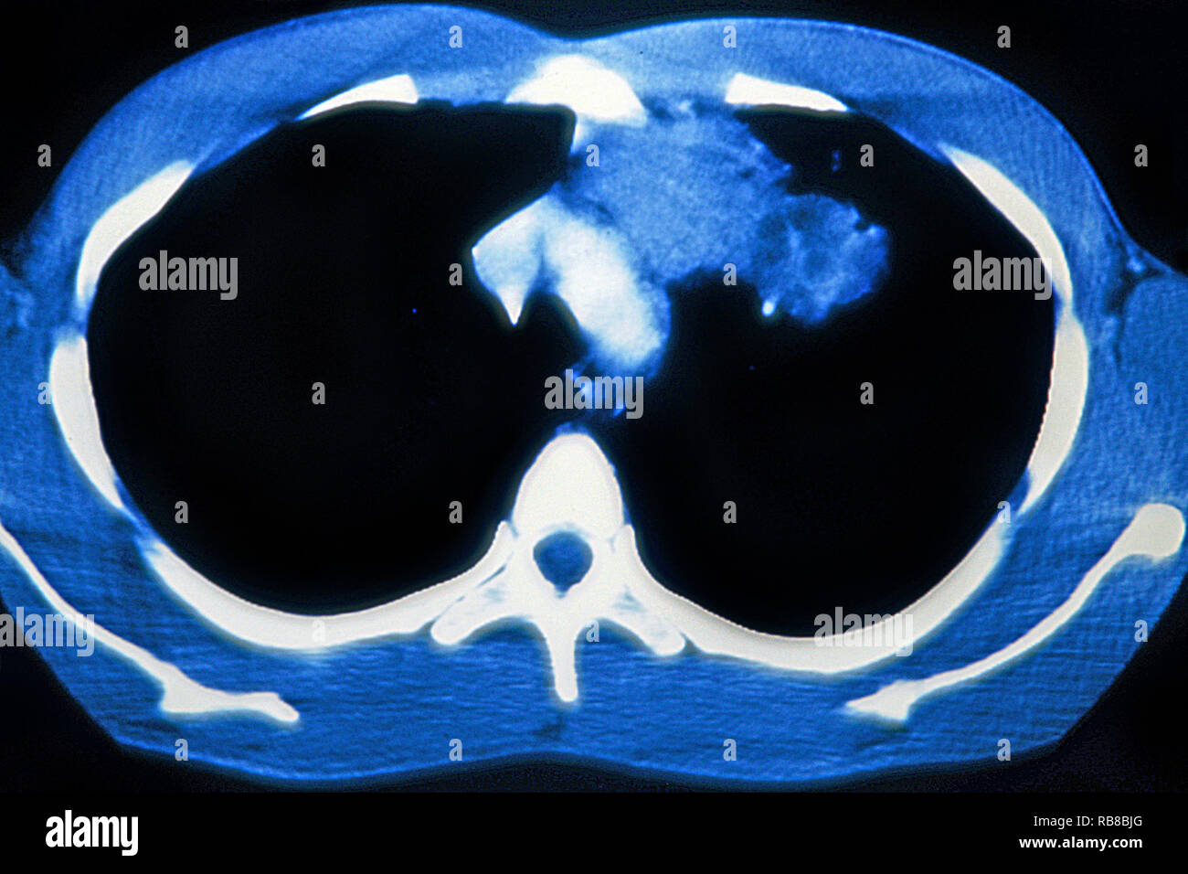 PULMONARY TUBERCULOSIS, CT-SCAN Stock Photohttps://www.alamy.com/image-license-details/?v=1https://www.alamy.com/pulmonary-tuberculosis-ct-scan-image230680760.html
RMRB8BJG–PULMONARY TUBERCULOSIS, CT-SCAN
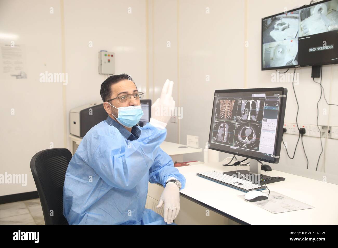 Baghdad. 17th Oct, 2020. Dr. Mohammed Abdul-Hussein examines lung images at al-Shifaa Center in Baghdad, Iraq, Oct. 12, 2020. In a specialized COVID-19 hospital in Iraqi capital of Baghdad, a Chinese-donated CT scan, mobile X-ray equipment, and other medical supplies are saving lives in the front-line battle against the COVID-19 pandemic. Already 400 patients have benefited from the important medical donation. TO GO WITH 'Spotlight: Chinese-donated CT scan, mobile X-ray save lives in specialized COVID-19 hospital in Iraq' Credit: Xinhua/Alamy Live News Stock Photohttps://www.alamy.com/image-license-details/?v=1https://www.alamy.com/baghdad-17th-oct-2020-dr-mohammed-abdul-hussein-examines-lung-images-at-al-shifaa-center-in-baghdad-iraq-oct-12-2020-in-a-specialized-covid-19-hospital-in-iraqi-capital-of-baghdad-a-chinese-donated-ct-scan-mobile-x-ray-equipment-and-other-medical-supplies-are-saving-lives-in-the-front-line-battle-against-the-covid-19-pandemic-already-400-patients-have-benefited-from-the-important-medical-donation-to-go-with-spotlight-chinese-donated-ct-scan-mobile-x-ray-save-lives-in-specialized-covid-19-hospital-in-iraq-credit-xinhuaalamy-live-news-image382685321.html
RM2D6GR0W–Baghdad. 17th Oct, 2020. Dr. Mohammed Abdul-Hussein examines lung images at al-Shifaa Center in Baghdad, Iraq, Oct. 12, 2020. In a specialized COVID-19 hospital in Iraqi capital of Baghdad, a Chinese-donated CT scan, mobile X-ray equipment, and other medical supplies are saving lives in the front-line battle against the COVID-19 pandemic. Already 400 patients have benefited from the important medical donation. TO GO WITH 'Spotlight: Chinese-donated CT scan, mobile X-ray save lives in specialized COVID-19 hospital in Iraq' Credit: Xinhua/Alamy Live News
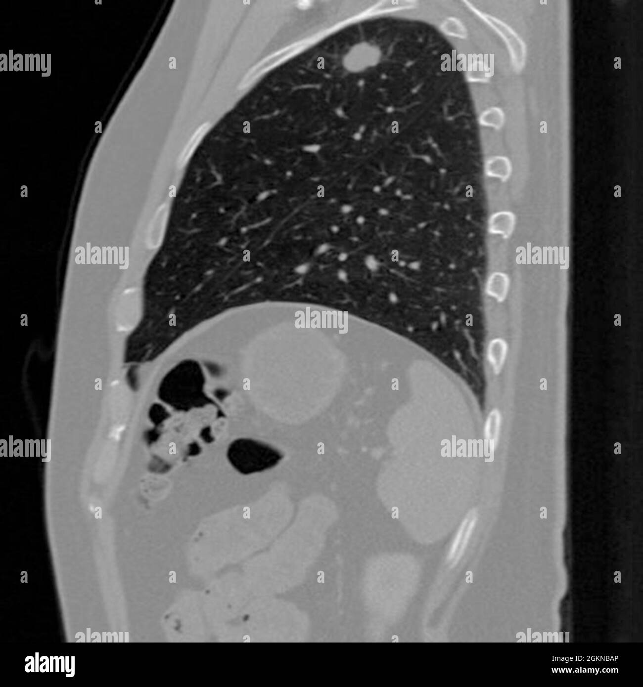 Side view Chest CT scan (X-ray computed tomography) of a male 54 year old patient. A tumour can be seen in the left upper lobe of his lungs Stock Photohttps://www.alamy.com/image-license-details/?v=1https://www.alamy.com/side-view-chest-ct-scan-x-ray-computed-tomography-of-a-male-54-year-old-patient-a-tumour-can-be-seen-in-the-left-upper-lobe-of-his-lungs-image442407582.html
RM2GKNBAP–Side view Chest CT scan (X-ray computed tomography) of a male 54 year old patient. A tumour can be seen in the left upper lobe of his lungs
 Man sitting with a phone and looking at a CT scan of his lungs. Pneumonia and disease diagnosis. Pills and medical bottles Stock Photohttps://www.alamy.com/image-license-details/?v=1https://www.alamy.com/man-sitting-with-a-phone-and-looking-at-a-ct-scan-of-his-lungs-pneumonia-and-disease-diagnosis-pills-and-medical-bottles-image452045777.html
RF2H7CD01–Man sitting with a phone and looking at a CT scan of his lungs. Pneumonia and disease diagnosis. Pills and medical bottles
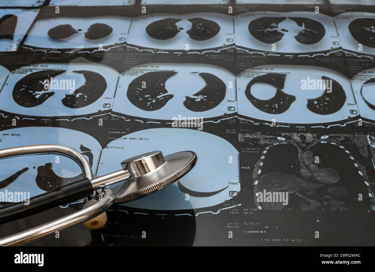 CT image of a human lung and a phonendoscope, checking for a virus.Close-up Stock Photohttps://www.alamy.com/image-license-details/?v=1https://www.alamy.com/ct-image-of-a-human-lung-and-a-phonendoscope-checking-for-a-virusclose-up-image598124084.html
RF2WN2WAC–CT image of a human lung and a phonendoscope, checking for a virus.Close-up
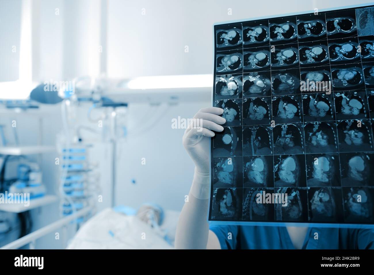 Female doctor looks at the scan image of patient thoracic organs in the hospital room. Stock Photohttps://www.alamy.com/image-license-details/?v=1https://www.alamy.com/female-doctor-looks-at-the-scan-image-of-patient-thoracic-organs-in-the-hospital-room-image459201213.html
RF2HK2BR9–Female doctor looks at the scan image of patient thoracic organs in the hospital room.
 Respiratory Syncytial Virus with lung ct scan aside on light blue background. RSV disease concept. Stock Photohttps://www.alamy.com/image-license-details/?v=1https://www.alamy.com/respiratory-syncytial-virus-with-lung-ct-scan-aside-on-light-blue-background-rsv-disease-concept-image504317956.html
RF2M8DJKG–Respiratory Syncytial Virus with lung ct scan aside on light blue background. RSV disease concept.
 Image co-registered PET-CT study dual modality scanner. Patient multiple metastatic lesions liver & lung Stock Photohttps://www.alamy.com/image-license-details/?v=1https://www.alamy.com/stock-photo-image-co-registered-pet-ct-study-dual-modality-scanner-patient-multiple-87047024.html
RFF1H9A8–Image co-registered PET-CT study dual modality scanner. Patient multiple metastatic lesions liver & lung
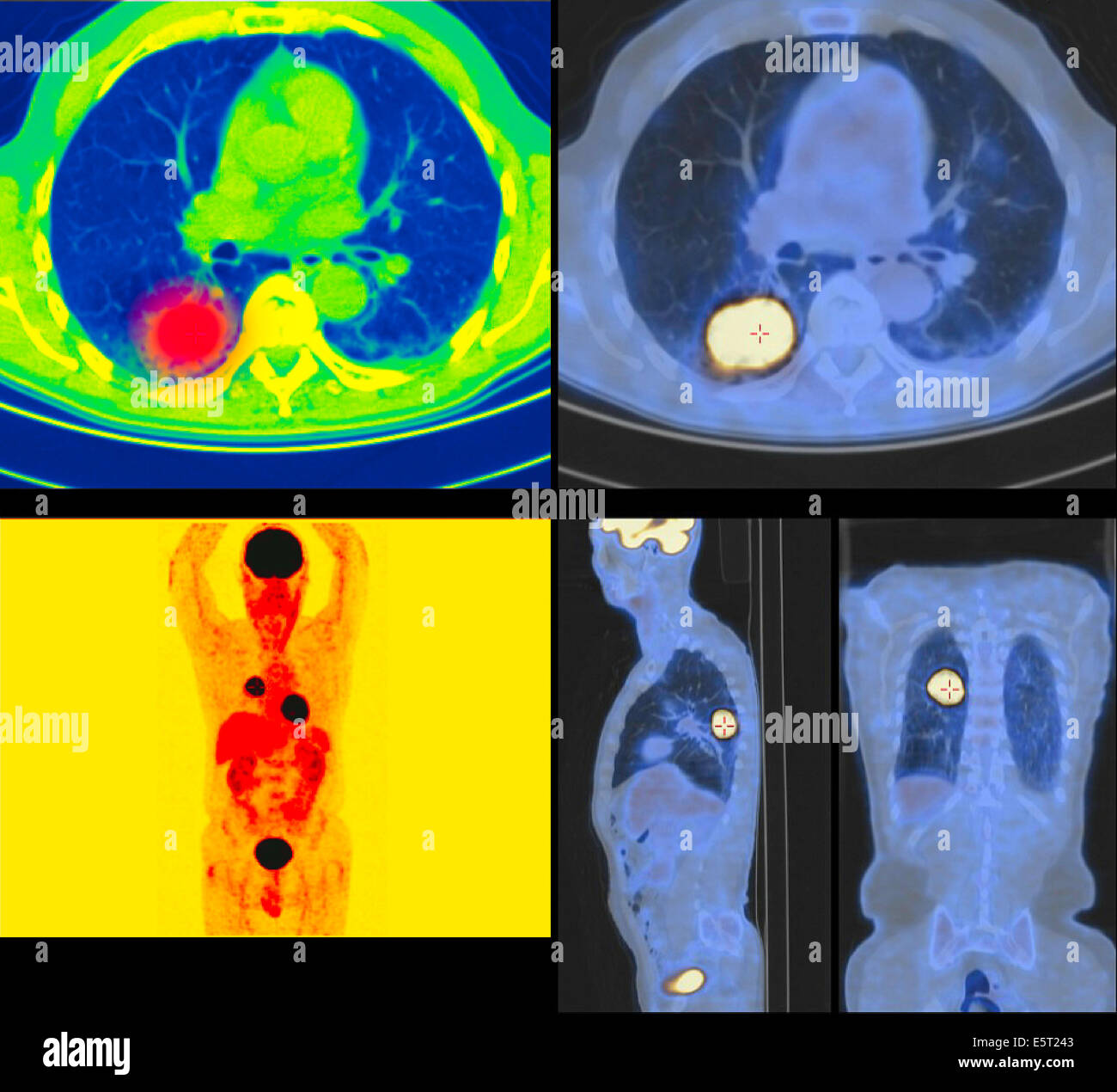 Positron emission tomography (PET) scans of a patient with a tumour the upper lobe of the left lung. Stock Photohttps://www.alamy.com/image-license-details/?v=1https://www.alamy.com/stock-photo-positron-emission-tomography-pet-scans-of-a-patient-with-a-tumour-72443283.html
RME5T243–Positron emission tomography (PET) scans of a patient with a tumour the upper lobe of the left lung.
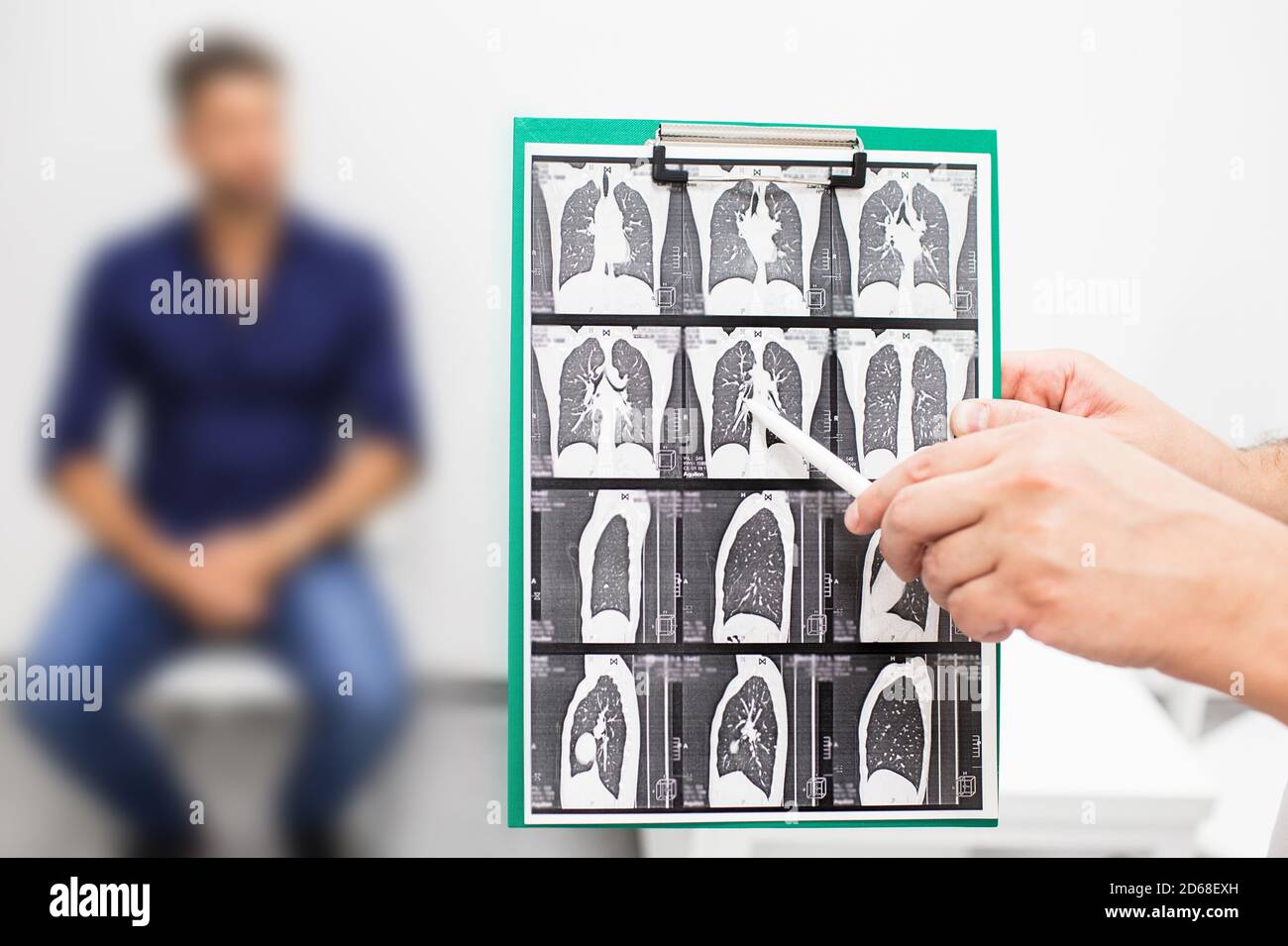 Pulmonologist showing CT scan of lungs patient with pulmonary fibrosis, after recovery, lung disease Stock Photohttps://www.alamy.com/image-license-details/?v=1https://www.alamy.com/pulmonologist-showing-ct-scan-of-lungs-patient-with-pulmonary-fibrosis-after-recovery-lung-disease-image382503369.html
RF2D68EXH–Pulmonologist showing CT scan of lungs patient with pulmonary fibrosis, after recovery, lung disease
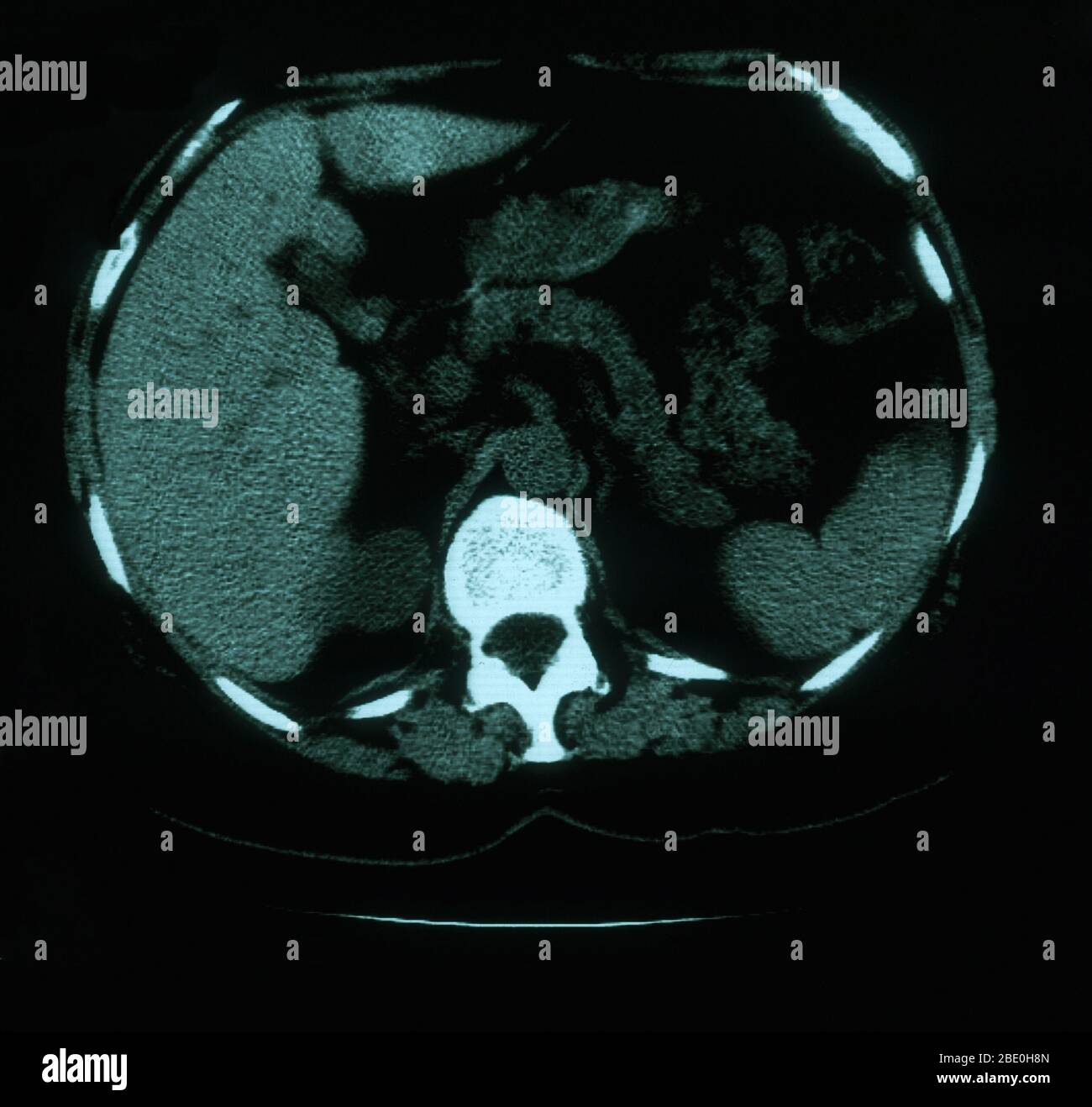 An axial (cross sectional) CT image through the lungs of a 53 year old female. The scan shows tortuosity and prominence of vasculature in the right superior mediastinum. Also present are calcified left hilar nodules and multiple punctate calcifications throughout the spleen which are consistent with granuloma. Stock Photohttps://www.alamy.com/image-license-details/?v=1https://www.alamy.com/an-axial-cross-sectional-ct-image-through-the-lungs-of-a-53-year-old-female-the-scan-shows-tortuosity-and-prominence-of-vasculature-in-the-right-superior-mediastinum-also-present-are-calcified-left-hilar-nodules-and-multiple-punctate-calcifications-throughout-the-spleen-which-are-consistent-with-granuloma-image352826117.html
RM2BE0H8N–An axial (cross sectional) CT image through the lungs of a 53 year old female. The scan shows tortuosity and prominence of vasculature in the right superior mediastinum. Also present are calcified left hilar nodules and multiple punctate calcifications throughout the spleen which are consistent with granuloma.
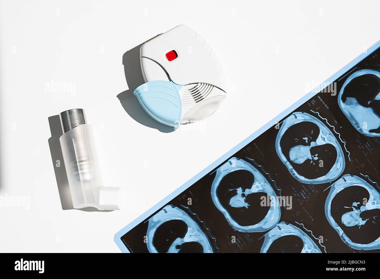 Some asthma inhalers and CT scan of the lungs on white table. Aerosol for inhalation for treat lung inflammation and prevent asthma attack. Stock Photohttps://www.alamy.com/image-license-details/?v=1https://www.alamy.com/some-asthma-inhalers-and-ct-scan-of-the-lungs-on-white-table-aerosol-for-inhalation-for-treat-lung-inflammation-and-prevent-asthma-attack-image471802383.html
RF2JBGCN3–Some asthma inhalers and CT scan of the lungs on white table. Aerosol for inhalation for treat lung inflammation and prevent asthma attack.
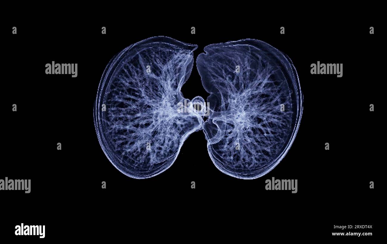 CT Chest or CT lung 3d rendering image with blue color showing Trachea and lung in respiratory system. Stock Photohttps://www.alamy.com/image-license-details/?v=1https://www.alamy.com/ct-chest-or-ct-lung-3d-rendering-image-with-blue-color-showing-trachea-and-lung-in-respiratory-system-image567017162.html
RF2RXDT4X–CT Chest or CT lung 3d rendering image with blue color showing Trachea and lung in respiratory system.
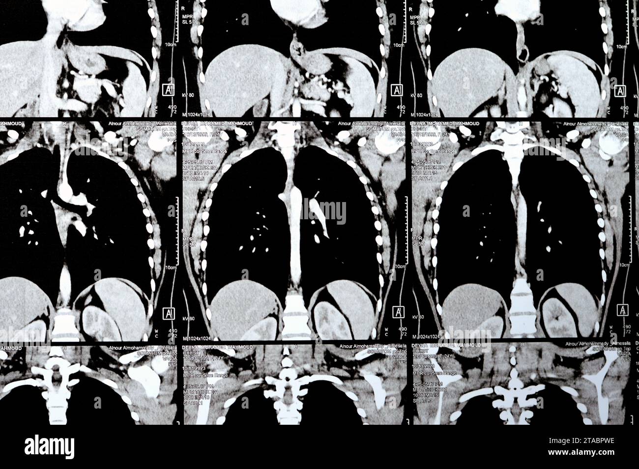 Cairo, Egypt, October 15 2023: CT scan axial slices through chest with contrast injection showing low grade of inflammatory reaction, parenchymal vess Stock Photohttps://www.alamy.com/image-license-details/?v=1https://www.alamy.com/cairo-egypt-october-15-2023-ct-scan-axial-slices-through-chest-with-contrast-injection-showing-low-grade-of-inflammatory-reaction-parenchymal-vess-image574348138.html
RF2TABPWE–Cairo, Egypt, October 15 2023: CT scan axial slices through chest with contrast injection showing low grade of inflammatory reaction, parenchymal vess
 Doctor and patient in protective face masks discuss X-ray result during appointment. African American woman physician with CT lung screening explainin Stock Photohttps://www.alamy.com/image-license-details/?v=1https://www.alamy.com/doctor-and-patient-in-protective-face-masks-discuss-x-ray-result-during-appointment-african-american-woman-physician-with-ct-lung-screening-explainin-image460820552.html
RF2HNM58T–Doctor and patient in protective face masks discuss X-ray result during appointment. African American woman physician with CT lung screening explainin
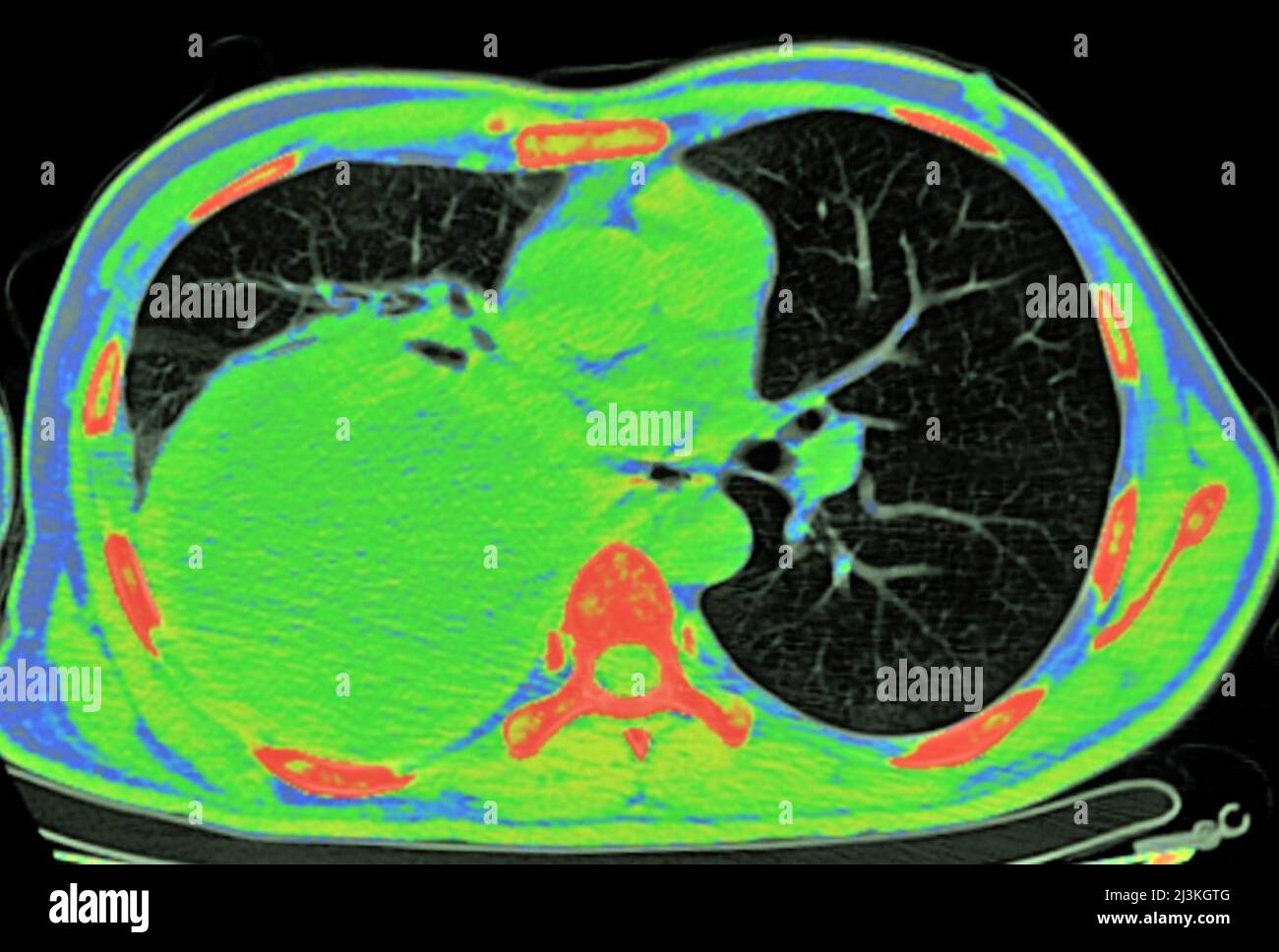 Pleural empyema, CT scan Stock Photohttps://www.alamy.com/image-license-details/?v=1https://www.alamy.com/pleural-empyema-ct-scan-image466954224.html
RF2J3KGTG–Pleural empyema, CT scan
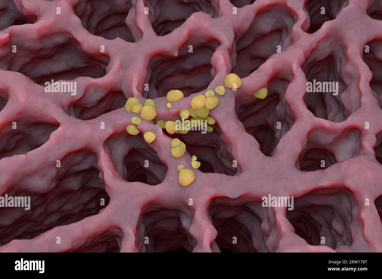 Small cancer tumors on the lung tissue: lung cancer (LC) - isometric view 3d illustration Stock Photohttps://www.alamy.com/image-license-details/?v=1https://www.alamy.com/small-cancer-tumors-on-the-lung-tissue-lung-cancer-lc-isometric-view-3d-illustration-image566125864.html
RF2RW178T–Small cancer tumors on the lung tissue: lung cancer (LC) - isometric view 3d illustration
 An X-ray of a human spine Stock Photohttps://www.alamy.com/image-license-details/?v=1https://www.alamy.com/an-x-ray-of-a-human-spine-image611053109.html
RF2XE3TD9–An X-ray of a human spine
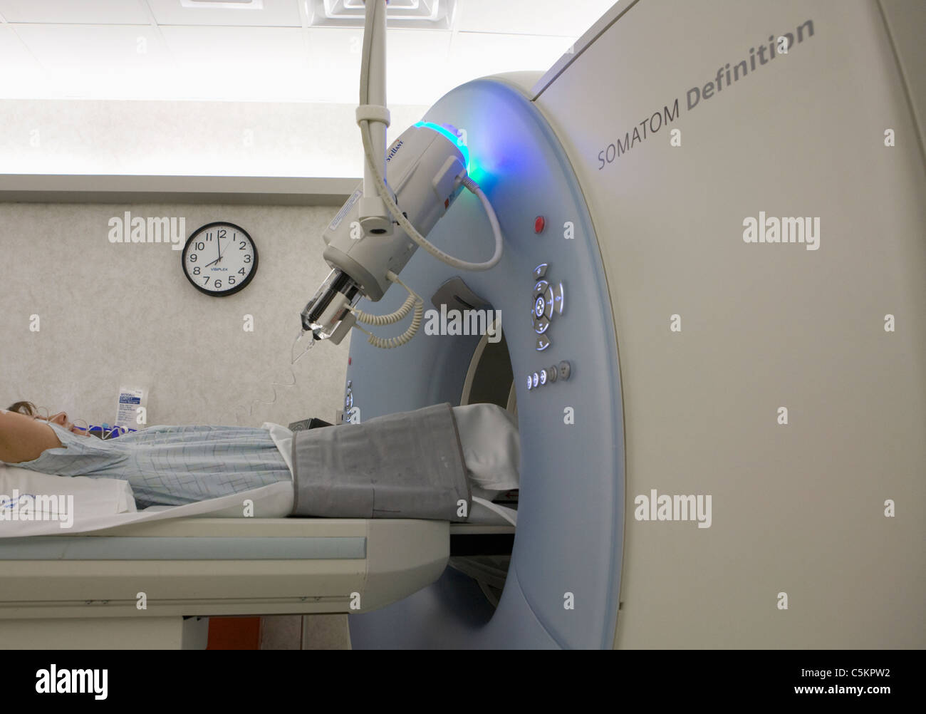 Dual energy SOMOTOM Definition CT scanner Stock Photohttps://www.alamy.com/image-license-details/?v=1https://www.alamy.com/stock-photo-dual-energy-somotom-definition-ct-scanner-37929054.html
RMC5KPW2–Dual energy SOMOTOM Definition CT scanner
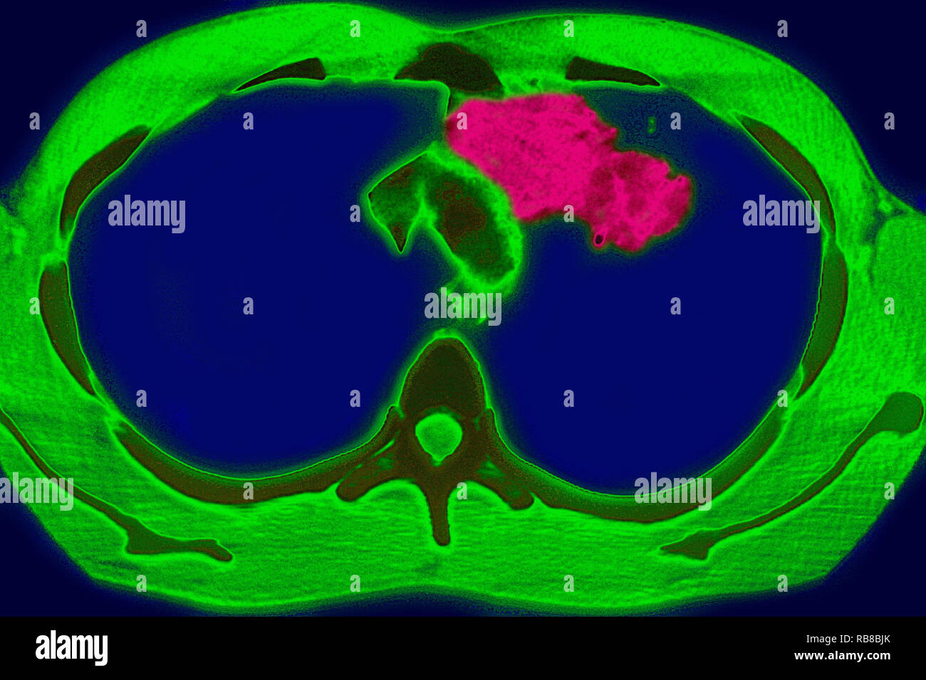 PULMONARY TUBERCULOSIS, CT-SCAN Stock Photohttps://www.alamy.com/image-license-details/?v=1https://www.alamy.com/pulmonary-tuberculosis-ct-scan-image230680763.html
RMRB8BJK–PULMONARY TUBERCULOSIS, CT-SCAN
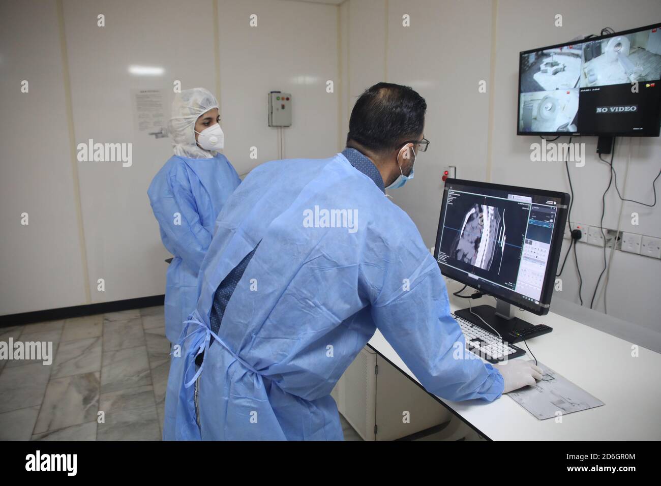 Baghdad. 17th Oct, 2020. Dr. Mohammed Abdul-Hussein (R) and CT technician Hanan Jamal examine lung images at al-Shifaa Center in Baghdad, Iraq, Oct. 12, 2020. In a specialized COVID-19 hospital in Iraqi capital of Baghdad, a Chinese-donated CT scan, mobile X-ray equipment, and other medical supplies are saving lives in the front-line battle against the COVID-19 pandemic. Already 400 patients have benefited from the important medical donation. TO GO WITH 'Spotlight: Chinese-donated CT scan, mobile X-ray save lives in specialized COVID-19 hospital in Iraq' Credit: Xinhua/Alamy Live News Stock Photohttps://www.alamy.com/image-license-details/?v=1https://www.alamy.com/baghdad-17th-oct-2020-dr-mohammed-abdul-hussein-r-and-ct-technician-hanan-jamal-examine-lung-images-at-al-shifaa-center-in-baghdad-iraq-oct-12-2020-in-a-specialized-covid-19-hospital-in-iraqi-capital-of-baghdad-a-chinese-donated-ct-scan-mobile-x-ray-equipment-and-other-medical-supplies-are-saving-lives-in-the-front-line-battle-against-the-covid-19-pandemic-already-400-patients-have-benefited-from-the-important-medical-donation-to-go-with-spotlight-chinese-donated-ct-scan-mobile-x-ray-save-lives-in-specialized-covid-19-hospital-in-iraq-credit-xinhuaalamy-live-news-image382685316.html
RM2D6GR0M–Baghdad. 17th Oct, 2020. Dr. Mohammed Abdul-Hussein (R) and CT technician Hanan Jamal examine lung images at al-Shifaa Center in Baghdad, Iraq, Oct. 12, 2020. In a specialized COVID-19 hospital in Iraqi capital of Baghdad, a Chinese-donated CT scan, mobile X-ray equipment, and other medical supplies are saving lives in the front-line battle against the COVID-19 pandemic. Already 400 patients have benefited from the important medical donation. TO GO WITH 'Spotlight: Chinese-donated CT scan, mobile X-ray save lives in specialized COVID-19 hospital in Iraq' Credit: Xinhua/Alamy Live News
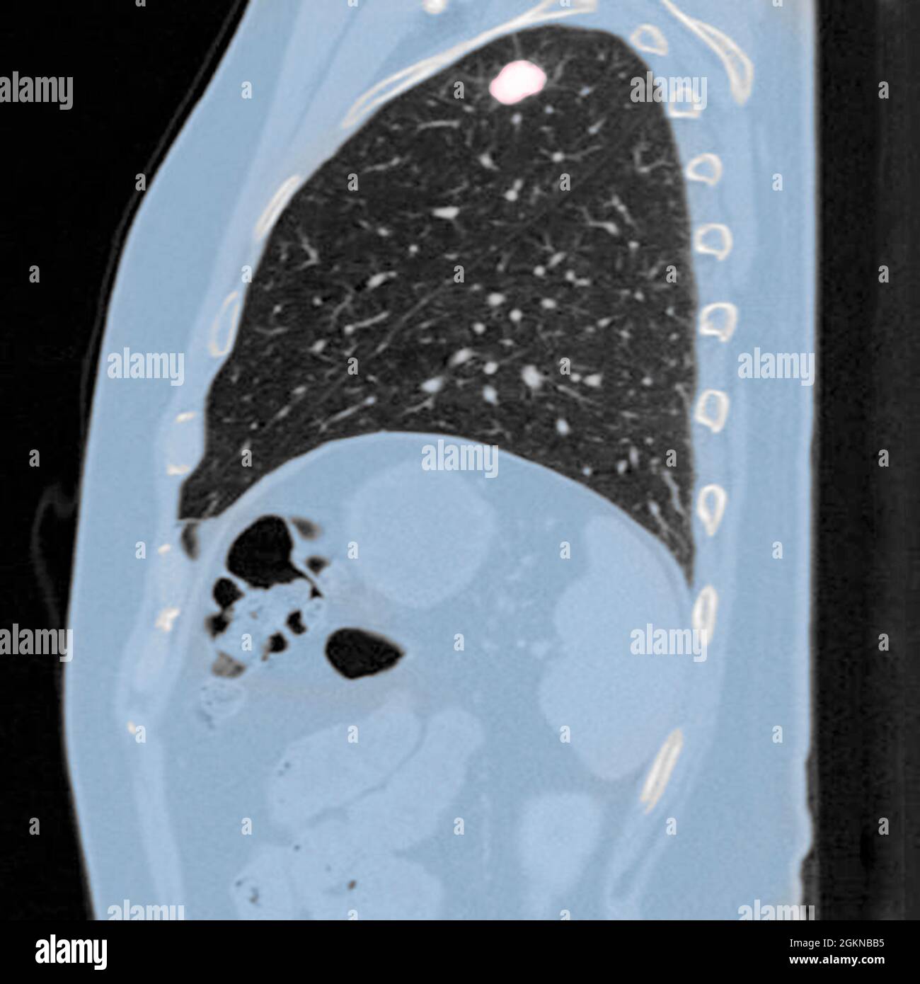 Side view Chest CT scan (X-ray computed tomography) of a male 54 year old patient. A tumour can be seen in the left upper lobe of his lungs Stock Photohttps://www.alamy.com/image-license-details/?v=1https://www.alamy.com/side-view-chest-ct-scan-x-ray-computed-tomography-of-a-male-54-year-old-patient-a-tumour-can-be-seen-in-the-left-upper-lobe-of-his-lungs-image442407593.html
RM2GKNBB5–Side view Chest CT scan (X-ray computed tomography) of a male 54 year old patient. A tumour can be seen in the left upper lobe of his lungs
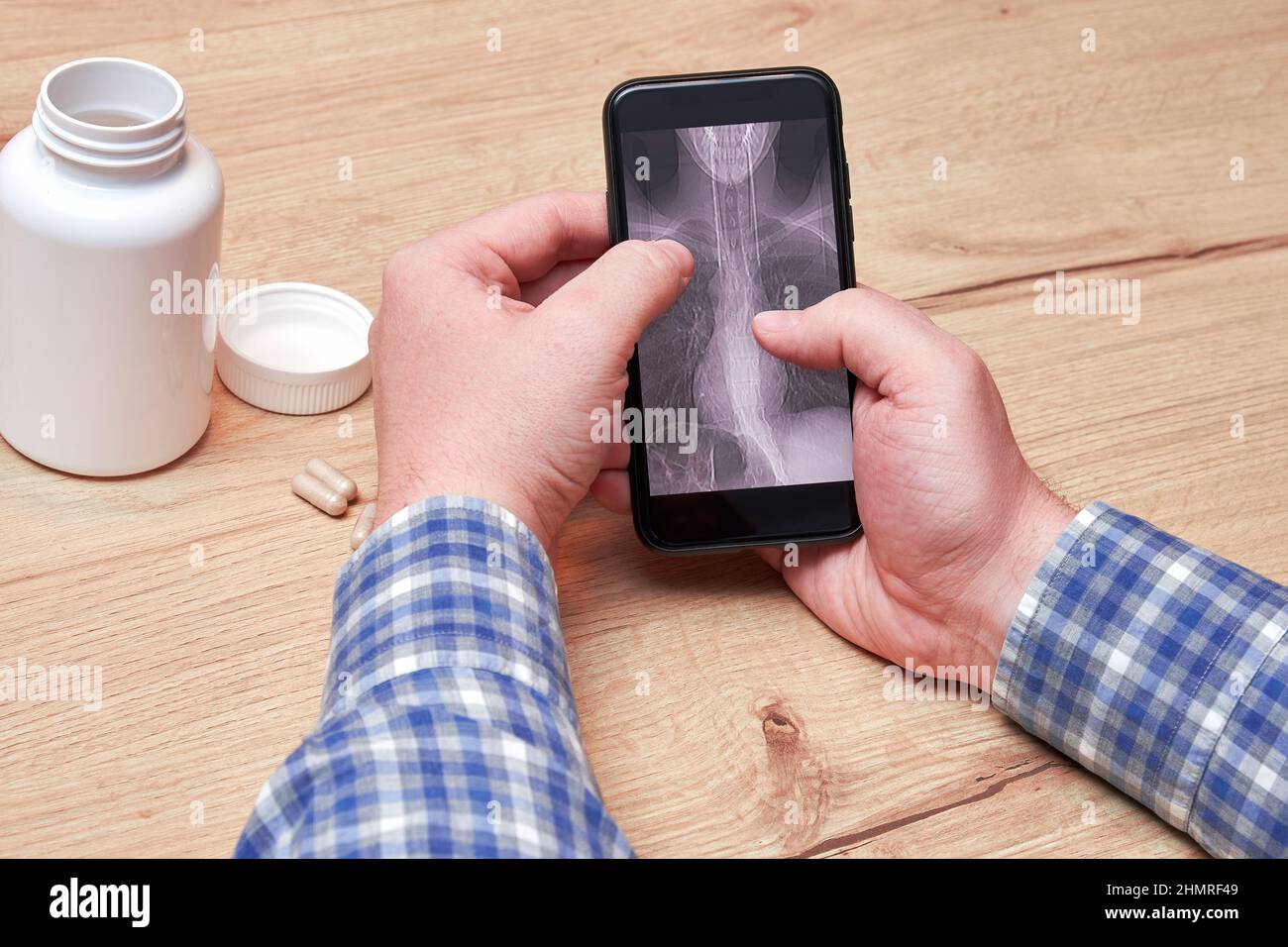 An old sick man checking a CT scan of his lungs on the cell phone screen. Pneumonia and disease diagnosis. Pills and medical bottles Stock Photohttps://www.alamy.com/image-license-details/?v=1https://www.alamy.com/an-old-sick-man-checking-a-ct-scan-of-his-lungs-on-the-cell-phone-screen-pneumonia-and-disease-diagnosis-pills-and-medical-bottles-image460279465.html
RF2HMRF49–An old sick man checking a CT scan of his lungs on the cell phone screen. Pneumonia and disease diagnosis. Pills and medical bottles
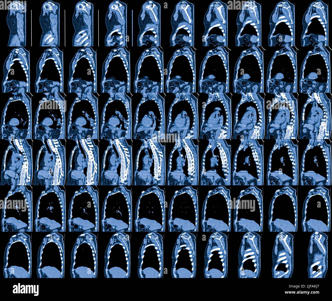 Magnetic Resonance Image (MRI) of chest organs Stock Photohttps://www.alamy.com/image-license-details/?v=1https://www.alamy.com/magnetic-resonance-image-mri-of-chest-organs-image474134720.html
RF2JFAKJT–Magnetic Resonance Image (MRI) of chest organs
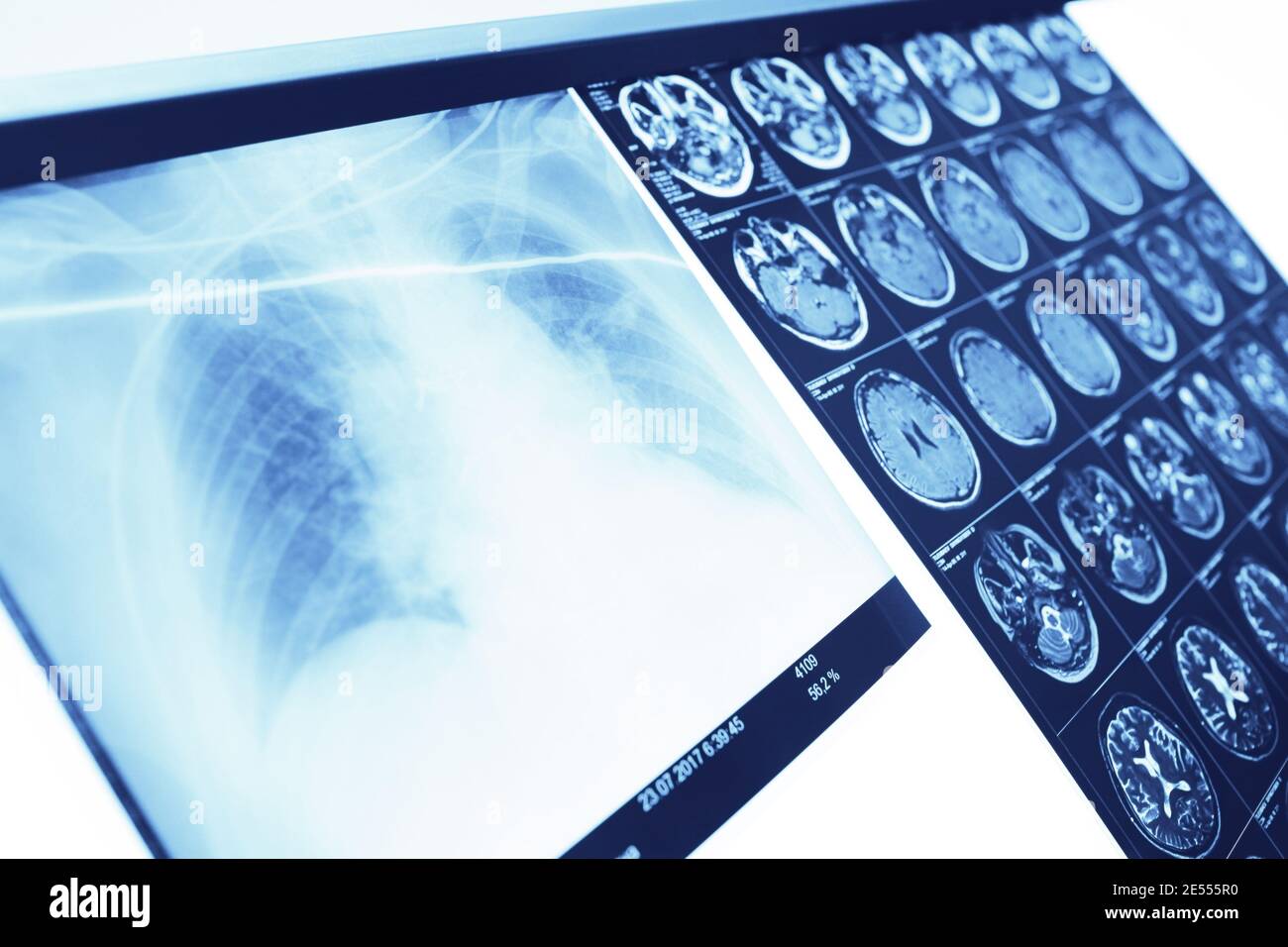 Survey results on the negatoscope in doctor's office. Stock Photohttps://www.alamy.com/image-license-details/?v=1https://www.alamy.com/survey-results-on-the-negatoscope-in-doctors-office-image399026068.html
RF2E555R0–Survey results on the negatoscope in doctor's office.
RF2G4DF44–Vector Covid-19 Prevention Filled Outline Icons. Coronavirus, Social Distancing, Quarantine, Hand Washing, Stay Home. 48x48 Pixel Perfect. Editable St
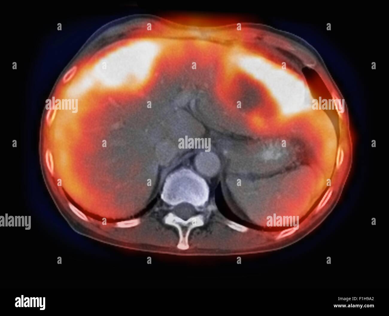 Image co-registered PET-CT study dual modality scanner. Patient multiple metastatic lesions liver & lung Stock Photohttps://www.alamy.com/image-license-details/?v=1https://www.alamy.com/stock-photo-image-co-registered-pet-ct-study-dual-modality-scanner-patient-multiple-87047018.html
RFF1H9A2–Image co-registered PET-CT study dual modality scanner. Patient multiple metastatic lesions liver & lung
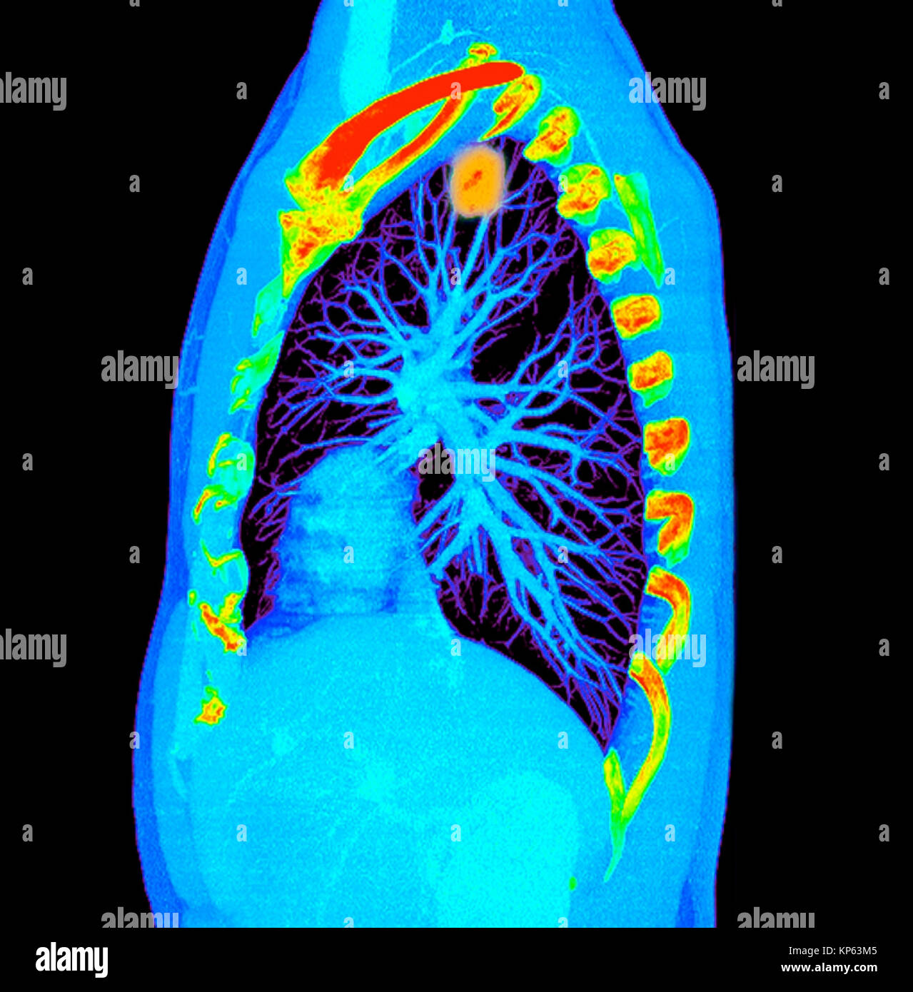 Coloured CTscan of the upper chest showing a tumor in the lung. Stock Photohttps://www.alamy.com/image-license-details/?v=1https://www.alamy.com/stock-image-coloured-ctscan-of-the-upper-chest-showing-a-tumor-in-the-lung-168550373.html
RMKP63M5–Coloured CTscan of the upper chest showing a tumor in the lung.
 doctor shows a senior patient a CT scan of her lungs. Pneumonia, coronavirus, lung disease Stock Photohttps://www.alamy.com/image-license-details/?v=1https://www.alamy.com/doctor-shows-a-senior-patient-a-ct-scan-of-her-lungs-pneumonia-coronavirus-lung-disease-image407160655.html
RF2EJBNFY–doctor shows a senior patient a CT scan of her lungs. Pneumonia, coronavirus, lung disease
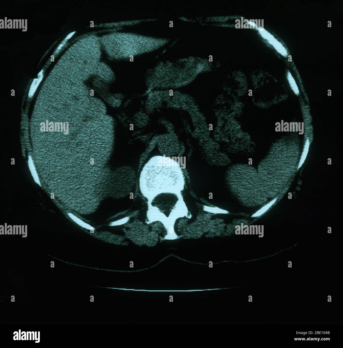 An axial (cross sectional) CT image through the lungs of a 53 year old female. The scan shows tortuosity and prominence of vasculature in the right superior mediastinum. Also present are calcified left hilar nodules and multiple punctate calcifications throughout the spleen which are consistent with granuloma. Stock Photohttps://www.alamy.com/image-license-details/?v=1https://www.alamy.com/an-axial-cross-sectional-ct-image-through-the-lungs-of-a-53-year-old-female-the-scan-shows-tortuosity-and-prominence-of-vasculature-in-the-right-superior-mediastinum-also-present-are-calcified-left-hilar-nodules-and-multiple-punctate-calcifications-throughout-the-spleen-which-are-consistent-with-granuloma-image352834619.html
RM2BE104B–An axial (cross sectional) CT image through the lungs of a 53 year old female. The scan shows tortuosity and prominence of vasculature in the right superior mediastinum. Also present are calcified left hilar nodules and multiple punctate calcifications throughout the spleen which are consistent with granuloma.
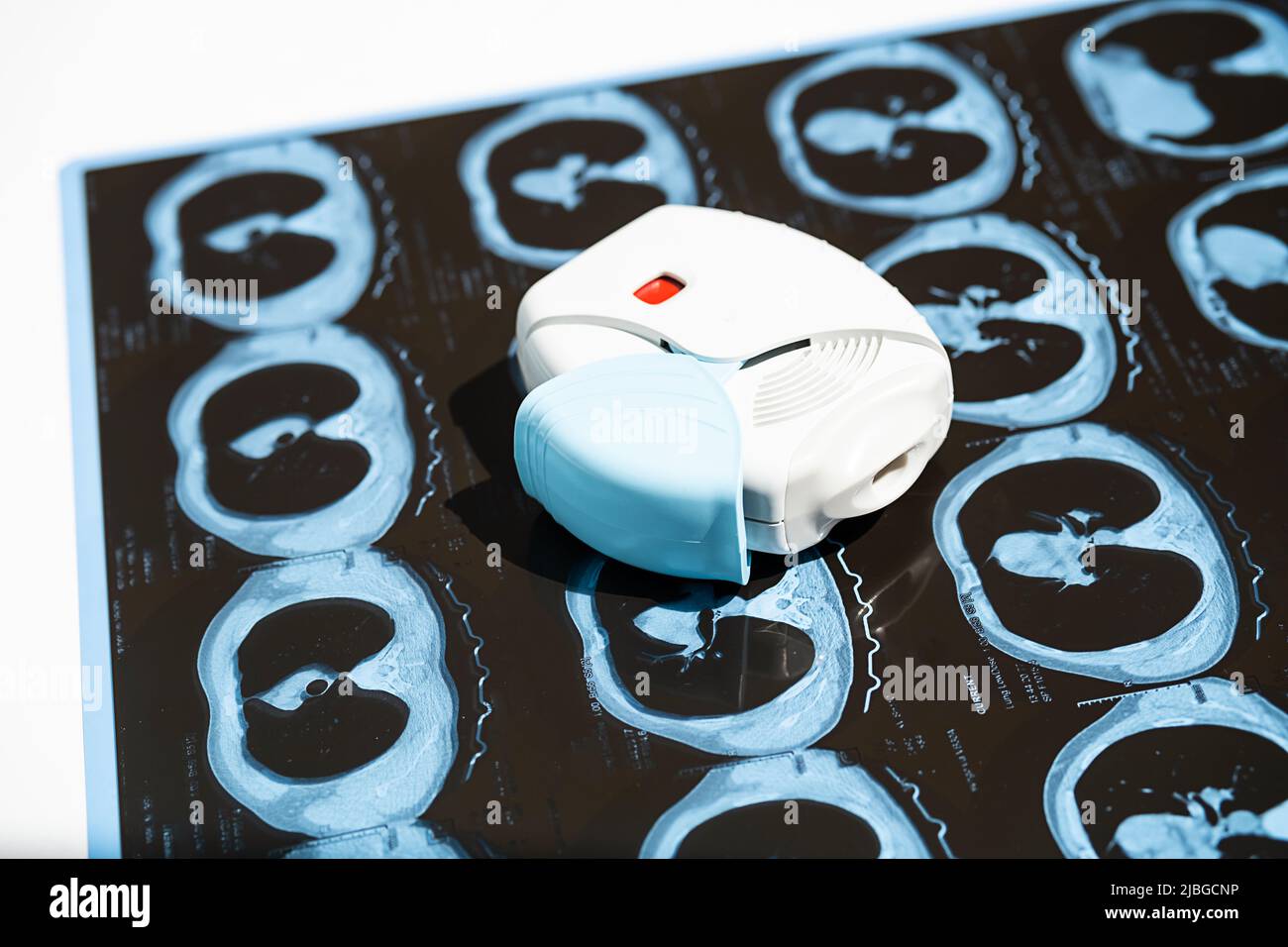 Asthma inhaler and CT scan of the lungs on white table. Aerosol for inhalation for treat lung inflammation and prevent asthma attack. Stock Photohttps://www.alamy.com/image-license-details/?v=1https://www.alamy.com/asthma-inhaler-and-ct-scan-of-the-lungs-on-white-table-aerosol-for-inhalation-for-treat-lung-inflammation-and-prevent-asthma-attack-image471802402.html
RF2JBGCNP–Asthma inhaler and CT scan of the lungs on white table. Aerosol for inhalation for treat lung inflammation and prevent asthma attack.