Depressor labii inferioris Stock Photos and Images
(128)See depressor labii inferioris stock video clipsQuick filters:
Depressor labii inferioris Stock Photos and Images
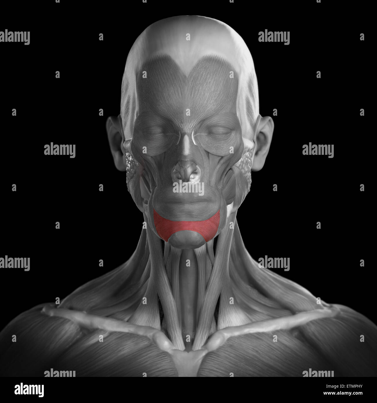 Conceptual image of the muscles of the face with the depressor labii inferioris muscles highlighted. Stock Photohttps://www.alamy.com/image-license-details/?v=1https://www.alamy.com/stock-photo-conceptual-image-of-the-muscles-of-the-face-with-the-depressor-labii-84050007.html
RMETMPHY–Conceptual image of the muscles of the face with the depressor labii inferioris muscles highlighted.
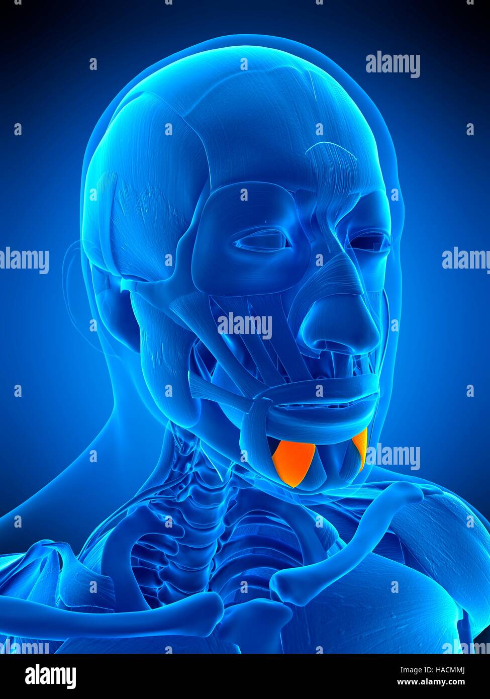 Illustration of the depressor labii inferioris muscle. Stock Photohttps://www.alamy.com/image-license-details/?v=1https://www.alamy.com/stock-photo-illustration-of-the-depressor-labii-inferioris-muscle-126898818.html
RFHACMMJ–Illustration of the depressor labii inferioris muscle.
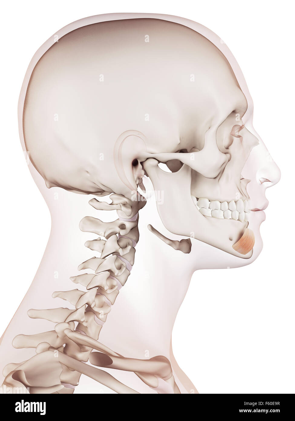 medically accurate muscle illustration of the depressor labii inferioris Stock Photohttps://www.alamy.com/image-license-details/?v=1https://www.alamy.com/stock-photo-medically-accurate-muscle-illustration-of-the-depressor-labii-inferioris-89751027.html
RFF60E9R–medically accurate muscle illustration of the depressor labii inferioris
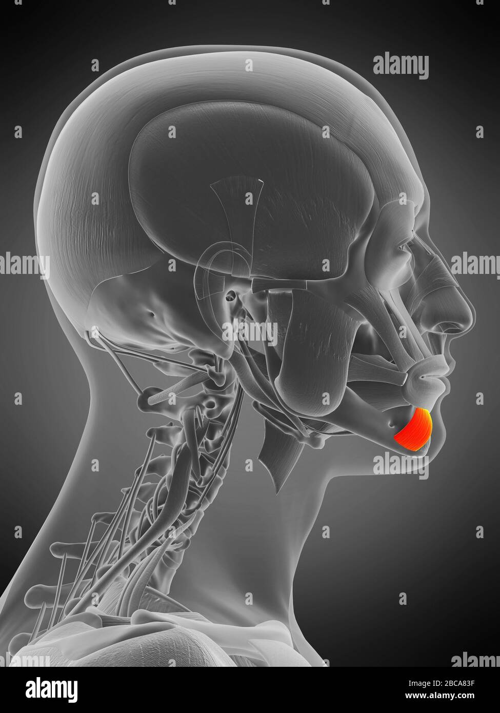 Depressor labii inferioris muscle, illustration. Stock Photohttps://www.alamy.com/image-license-details/?v=1https://www.alamy.com/depressor-labii-inferioris-muscle-illustration-image351809123.html
RF2BCA83F–Depressor labii inferioris muscle, illustration.
 3d rendered medically accurate illustration of the depressor labii inferioris Stock Photohttps://www.alamy.com/image-license-details/?v=1https://www.alamy.com/3d-rendered-medically-accurate-illustration-of-the-depressor-labii-inferioris-image257793148.html
RFTYBDP4–3d rendered medically accurate illustration of the depressor labii inferioris
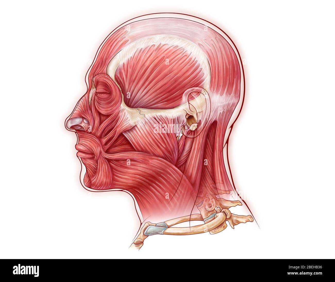 Facial Muscles, illustration Stock Photohttps://www.alamy.com/image-license-details/?v=1https://www.alamy.com/facial-muscles-illustration-image353194442.html
RM2BEHB36–Facial Muscles, illustration
 medical accurate illustration of the depressor labii inferioris Stock Photohttps://www.alamy.com/image-license-details/?v=1https://www.alamy.com/stock-photo-medical-accurate-illustration-of-the-depressor-labii-inferioris-89737605.html
RFF5YW6D–medical accurate illustration of the depressor labii inferioris
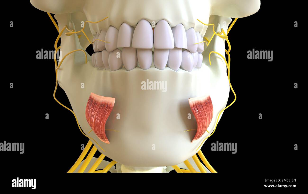 Depressor Labii Inferioris Muscle anatomy for medical concept 3D illustration Stock Photohttps://www.alamy.com/image-license-details/?v=1https://www.alamy.com/depressor-labii-inferioris-muscle-anatomy-for-medical-concept-3d-illustration-image502254249.html
RF2M53JBN–Depressor Labii Inferioris Muscle anatomy for medical concept 3D illustration
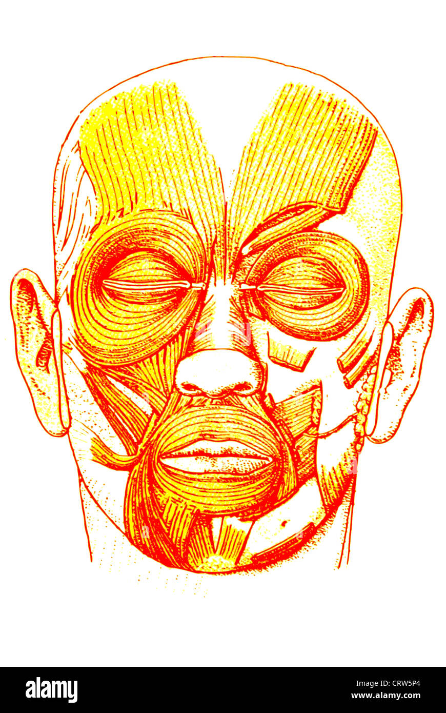 Facial muscles Stock Photohttps://www.alamy.com/image-license-details/?v=1https://www.alamy.com/stock-photo-facial-muscles-49111164.html
RFCRW5P4–Facial muscles
 The anatomist's vade mecum : a system of human anatomy . nferior labial group. (Orbicularis oris),*Depressor labii inferioris,Depressor anguli oris.Levator labii inferioris. 7. Maxillary group. Masseter,Temporalis,Buccinator,Pterygoideus externus,Pterygoideus internus. 8. Auricular group. Attollens aurem,Attrahens aurem,Retrahens aurem. * The orbicularis oris, from encircling the mouth, belongs necessarily toboth the superior and inferior labial regions ; it is therefore enclosed withinparentheses in both. 166 CRANIAL GROUP. 1. Cranial group.—Occipito-frontalis. Dissection.—The occipito-fronta Stock Photohttps://www.alamy.com/image-license-details/?v=1https://www.alamy.com/the-anatomists-vade-mecum-a-system-of-human-anatomy-nferior-labial-group-orbicularis-orisdepressor-labii-inferiorisdepressor-anguli-orislevator-labii-inferioris-7-maxillary-group-massetertemporalisbuccinatorpterygoideus-externuspterygoideus-internus-8-auricular-group-attollens-auremattrahens-auremretrahens-aurem-the-orbicularis-oris-from-encircling-the-mouth-belongs-necessarily-toboth-the-superior-and-inferior-labial-regions-it-is-therefore-enclosed-withinparentheses-in-both-166-cranial-group-1-cranial-groupoccipito-frontalis-dissectionthe-occipito-fronta-image342758952.html
RM2AWJ0F4–The anatomist's vade mecum : a system of human anatomy . nferior labial group. (Orbicularis oris),*Depressor labii inferioris,Depressor anguli oris.Levator labii inferioris. 7. Maxillary group. Masseter,Temporalis,Buccinator,Pterygoideus externus,Pterygoideus internus. 8. Auricular group. Attollens aurem,Attrahens aurem,Retrahens aurem. * The orbicularis oris, from encircling the mouth, belongs necessarily toboth the superior and inferior labial regions ; it is therefore enclosed withinparentheses in both. 166 CRANIAL GROUP. 1. Cranial group.—Occipito-frontalis. Dissection.—The occipito-fronta
 Anatomical model showing the facial muscles. Stock Photohttps://www.alamy.com/image-license-details/?v=1https://www.alamy.com/stock-photo-anatomical-model-showing-the-facial-muscles-52090686.html
RMD0MX5J–Anatomical model showing the facial muscles.
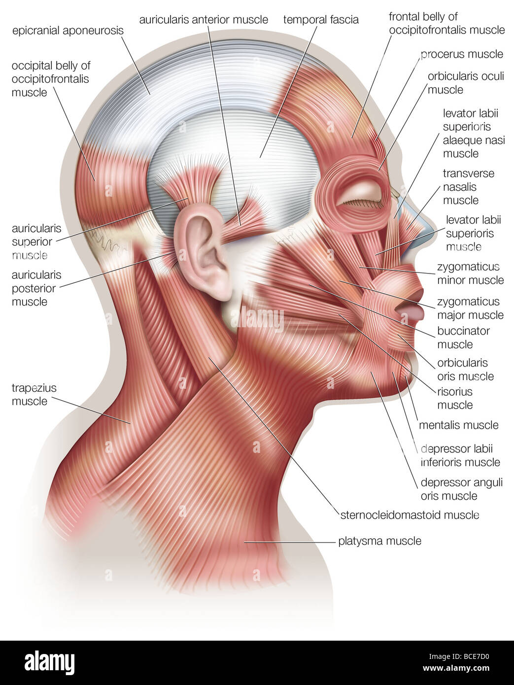 The muscles of the human head, used in facial expression. Stock Photohttps://www.alamy.com/image-license-details/?v=1https://www.alamy.com/stock-photo-the-muscles-of-the-human-head-used-in-facial-expression-24899420.html
RMBCE7D0–The muscles of the human head, used in facial expression.
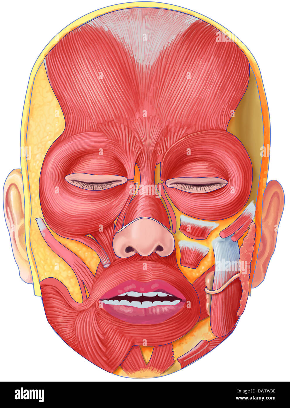 Muscle face drawing Stock Photohttps://www.alamy.com/image-license-details/?v=1https://www.alamy.com/muscle-face-drawing-image67544050.html
RMDWTW3E–Muscle face drawing
 . Elementary anatomy and physiology : for colleges, academies, and other schools . A Lateral View of the Deep-seated Layer of Muscles on the Face and Neck. 1, Tem- poral Muscle deprived of its Fascia. 2, Corrugator Supereilii. 3, Nasal Slip of the Oc- cipito Frontalis. 4. Superior or Nasal Extremity of the Levator Labii Superioris Alaequo Nasi. 5, Compressor Naris. G, Levator Anguli Oris. 7, Depressor Labii Superioris Aheque Nasi. S, Buccinator. 9, Orbicularis Oris. 10, Depressor Labii Inferioris. 11, Levator Labii Inferioris. 12, Anterior Belly of the Digastricus. 13, Mylo-Hyoid. 14, Stylo-II Stock Photohttps://www.alamy.com/image-license-details/?v=1https://www.alamy.com/elementary-anatomy-and-physiology-for-colleges-academies-and-other-schools-a-lateral-view-of-the-deep-seated-layer-of-muscles-on-the-face-and-neck-1-tem-poral-muscle-deprived-of-its-fascia-2-corrugator-supereilii-3-nasal-slip-of-the-oc-cipito-frontalis-4-superior-or-nasal-extremity-of-the-levator-labii-superioris-alaequo-nasi-5-compressor-naris-g-levator-anguli-oris-7-depressor-labii-superioris-aheque-nasi-s-buccinator-9-orbicularis-oris-10-depressor-labii-inferioris-11-levator-labii-inferioris-12-anterior-belly-of-the-digastricus-13-mylo-hyoid-14-stylo-ii-image178409723.html
RMMA77BR–. Elementary anatomy and physiology : for colleges, academies, and other schools . A Lateral View of the Deep-seated Layer of Muscles on the Face and Neck. 1, Tem- poral Muscle deprived of its Fascia. 2, Corrugator Supereilii. 3, Nasal Slip of the Oc- cipito Frontalis. 4. Superior or Nasal Extremity of the Levator Labii Superioris Alaequo Nasi. 5, Compressor Naris. G, Levator Anguli Oris. 7, Depressor Labii Superioris Aheque Nasi. S, Buccinator. 9, Orbicularis Oris. 10, Depressor Labii Inferioris. 11, Levator Labii Inferioris. 12, Anterior Belly of the Digastricus. 13, Mylo-Hyoid. 14, Stylo-II
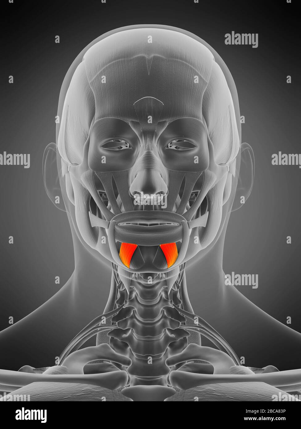 Depressor labii inferioris muscle, illustration. Stock Photohttps://www.alamy.com/image-license-details/?v=1https://www.alamy.com/depressor-labii-inferioris-muscle-illustration-image351809130.html
RF2BCA83P–Depressor labii inferioris muscle, illustration.
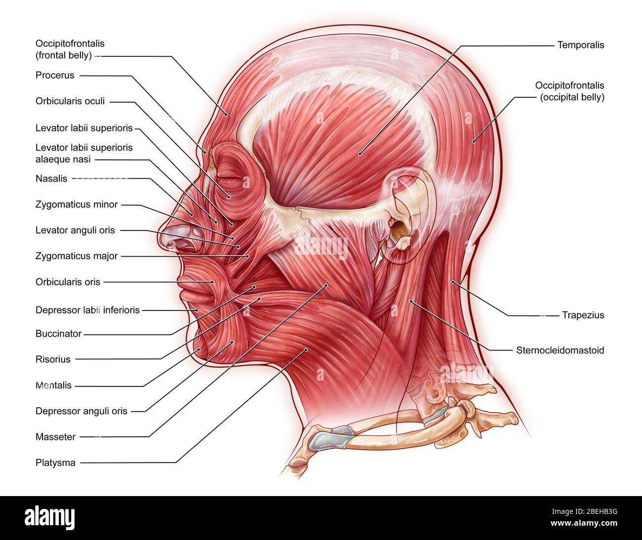 An illustration of the muscles of the face from a lateral view. Stock Photohttps://www.alamy.com/image-license-details/?v=1https://www.alamy.com/an-illustration-of-the-muscles-of-the-face-from-a-lateral-view-image353194452.html
RM2BEHB3G–An illustration of the muscles of the face from a lateral view.
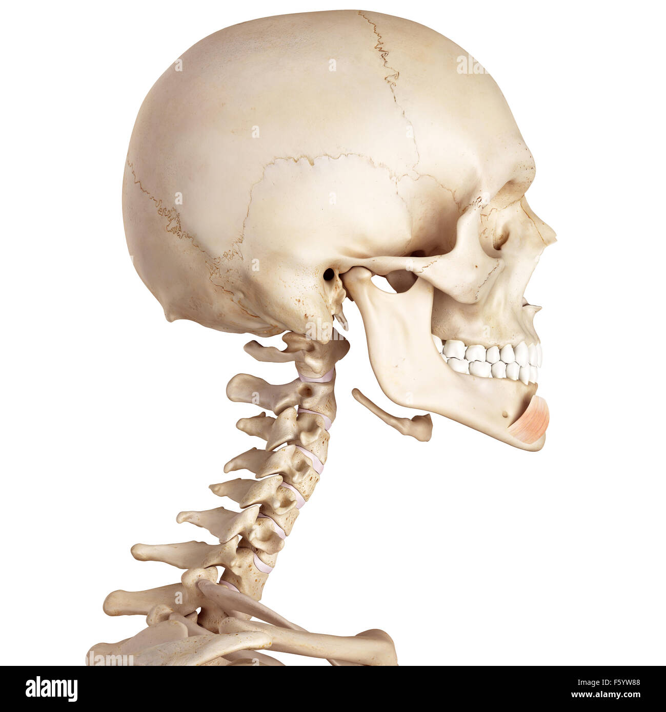 medical accurate illustration of the depressor labii inferioris Stock Photohttps://www.alamy.com/image-license-details/?v=1https://www.alamy.com/stock-photo-medical-accurate-illustration-of-the-depressor-labii-inferioris-89737656.html
RFF5YW88–medical accurate illustration of the depressor labii inferioris
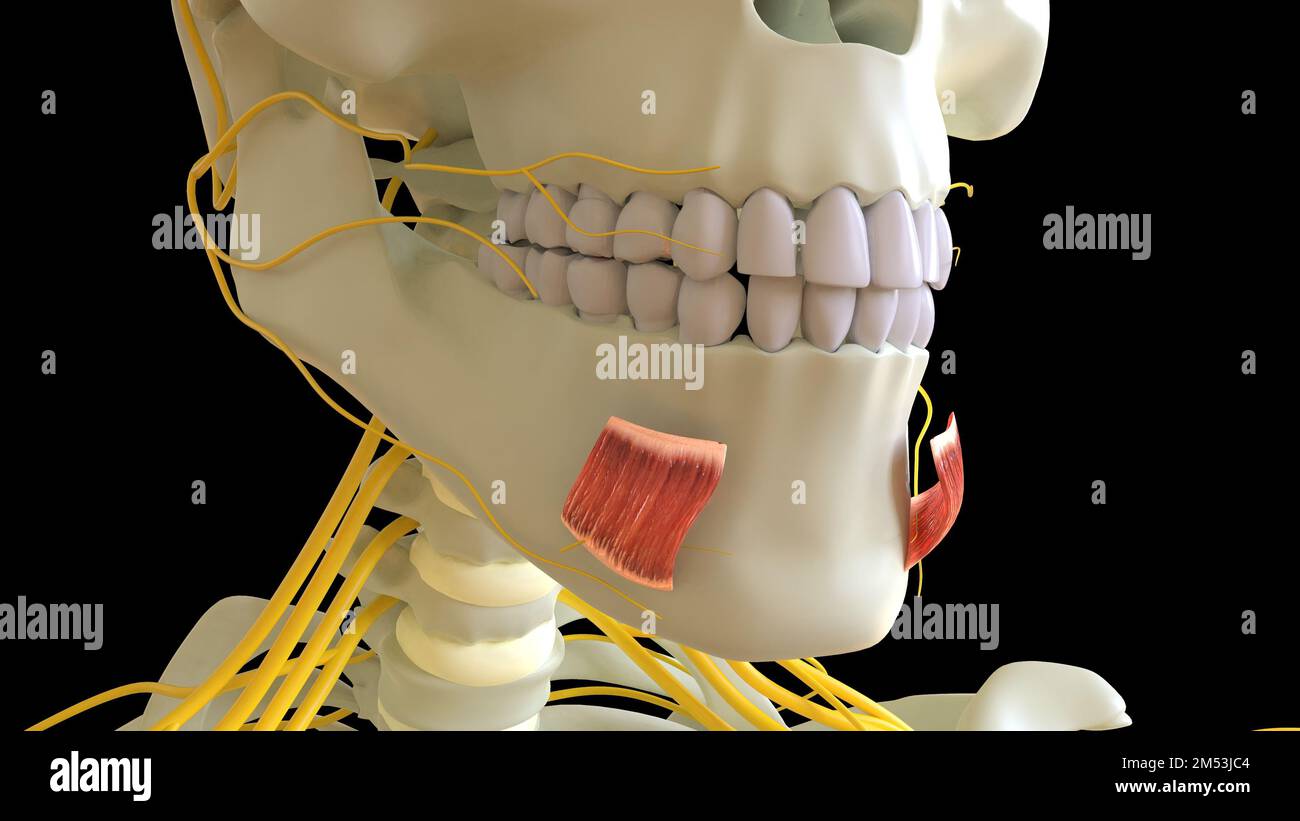 Depressor Labii Inferioris Muscle anatomy for medical concept 3D illustration Stock Photohttps://www.alamy.com/image-license-details/?v=1https://www.alamy.com/depressor-labii-inferioris-muscle-anatomy-for-medical-concept-3d-illustration-image502254260.html
RF2M53JC4–Depressor Labii Inferioris Muscle anatomy for medical concept 3D illustration
 medically accurate muscle illustration of the depressor labii inferioris Stock Photohttps://www.alamy.com/image-license-details/?v=1https://www.alamy.com/stock-photo-medically-accurate-muscle-illustration-of-the-depressor-labii-inferioris-89751026.html
RFF60E9P–medically accurate muscle illustration of the depressor labii inferioris
 A system of human anatomy, general and special . d the external pterygoid muscle. The sig-moid notch is crossed by the masseteric artery and nerve. The internal surface of the ramus is marked near its centre by alarge oblique foramen, the inferior dental, for the entrance of the in-ferior dental artery and nerve into the dental canal. Bounding thisopening is a sharp margin, to which is attached the internal lateral * The lower jaw. 1. The body. 2. The ramus. 3. The symphysis. 4. The fossafor the depressor labii inferioris muscle. 5. The mental foramen. 6. The external ob-lique ridge. 7. The gr Stock Photohttps://www.alamy.com/image-license-details/?v=1https://www.alamy.com/a-system-of-human-anatomy-general-and-special-d-the-external-pterygoid-muscle-the-sig-moid-notch-is-crossed-by-the-masseteric-artery-and-nerve-the-internal-surface-of-the-ramus-is-marked-near-its-centre-by-alarge-oblique-foramen-the-inferior-dental-for-the-entrance-of-the-in-ferior-dental-artery-and-nerve-into-the-dental-canal-bounding-thisopening-is-a-sharp-margin-to-which-is-attached-the-internal-lateral-the-lower-jaw-1-the-body-2-the-ramus-3-the-symphysis-4-the-fossafor-the-depressor-labii-inferioris-muscle-5-the-mental-foramen-6-the-external-ob-lique-ridge-7-the-gr-image342754622.html
RM2AWHR0E–A system of human anatomy, general and special . d the external pterygoid muscle. The sig-moid notch is crossed by the masseteric artery and nerve. The internal surface of the ramus is marked near its centre by alarge oblique foramen, the inferior dental, for the entrance of the in-ferior dental artery and nerve into the dental canal. Bounding thisopening is a sharp margin, to which is attached the internal lateral * The lower jaw. 1. The body. 2. The ramus. 3. The symphysis. 4. The fossafor the depressor labii inferioris muscle. 5. The mental foramen. 6. The external ob-lique ridge. 7. The gr
 3d rendered medically accurate illustration of the depressor labii inferioris Stock Photohttps://www.alamy.com/image-license-details/?v=1https://www.alamy.com/3d-rendered-medically-accurate-illustration-of-the-depressor-labii-inferioris-image257793133.html
RFTYBDNH–3d rendered medically accurate illustration of the depressor labii inferioris
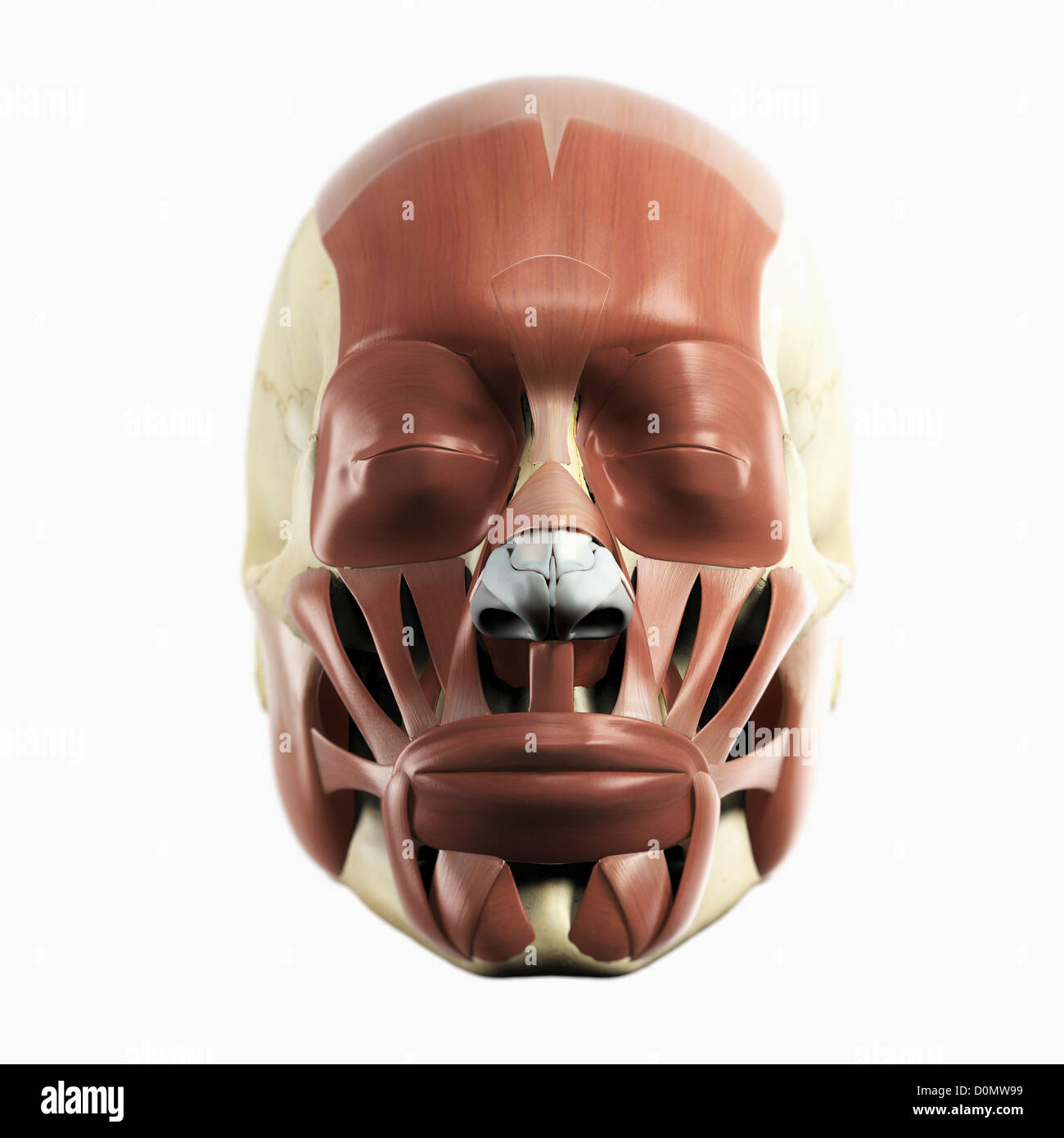 Anatomical model showing the facial muscles. Stock Photohttps://www.alamy.com/image-license-details/?v=1https://www.alamy.com/stock-photo-anatomical-model-showing-the-facial-muscles-52090005.html
RMD0MW99–Anatomical model showing the facial muscles.
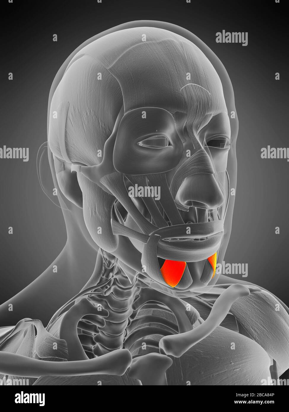 Depressor labii inferioris muscle, illustration. Stock Photohttps://www.alamy.com/image-license-details/?v=1https://www.alamy.com/depressor-labii-inferioris-muscle-illustration-image351809158.html
RF2BCA84P–Depressor labii inferioris muscle, illustration.
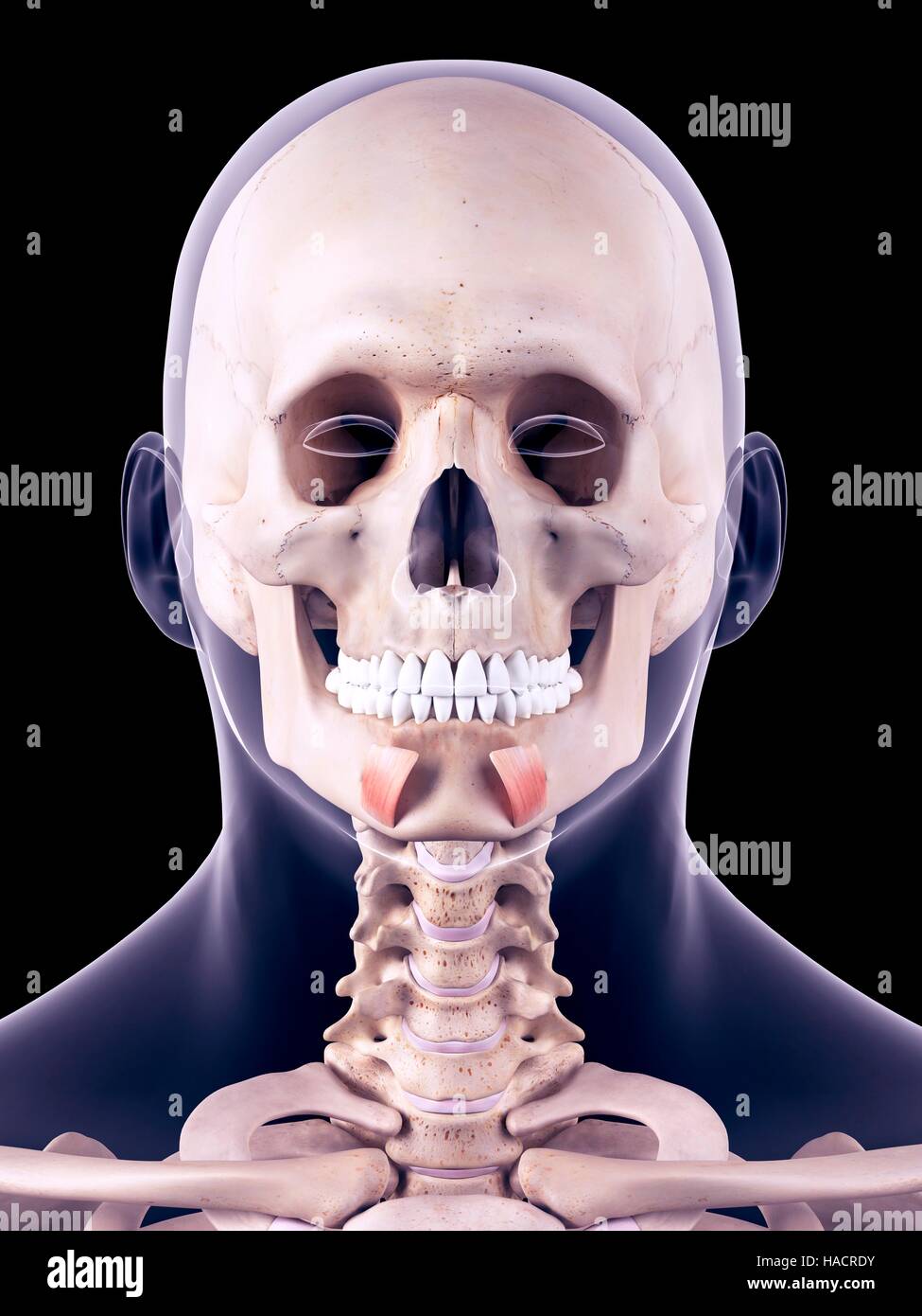 Illustration of the depressor labii inferioris muscles. Stock Photohttps://www.alamy.com/image-license-details/?v=1https://www.alamy.com/stock-photo-illustration-of-the-depressor-labii-inferioris-muscles-126900983.html
RFHACRDY–Illustration of the depressor labii inferioris muscles.
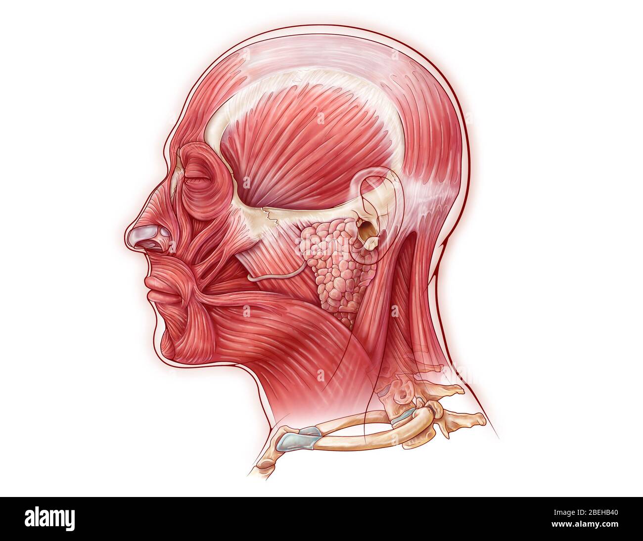 Facial Muscles, illustration Stock Photohttps://www.alamy.com/image-license-details/?v=1https://www.alamy.com/facial-muscles-illustration-image353194464.html
RM2BEHB40–Facial Muscles, illustration
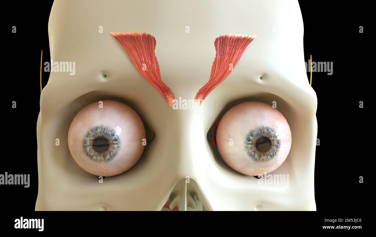 Depressor Supercilii Muscle anatomy for medical concept 3D illustration Stock Photohttps://www.alamy.com/image-license-details/?v=1https://www.alamy.com/depressor-supercilii-muscle-anatomy-for-medical-concept-3d-illustration-image502254256.html
RF2M53JC0–Depressor Supercilii Muscle anatomy for medical concept 3D illustration
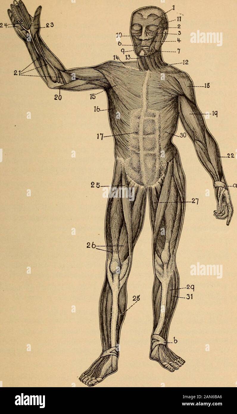 Anatomy, physiology and hygiene for high schools . ssimus dorsi 15 Pectoralis major (chest muscle) moves the trunk backward,move the trunk forward. compresses the abdominal viscera. raise and depress the ribs. raises the thorax. move head backward; move shoul-ders backward. draws arms downward and back-ward. draws arms across front of chest. Muscles of the head. 1 Occipito-frontalis moves the scalp and eyebrows. 2 Orbicularis palpebrae closes the eye. Levator palpebrae opens the eye. 11 Temporal raises the lower jaw. 9 Masseter Face muscles. raises the lower jaw. 7 Depressor labii inferioris d Stock Photohttps://www.alamy.com/image-license-details/?v=1https://www.alamy.com/anatomy-physiology-and-hygiene-for-high-schools-ssimus-dorsi-15-pectoralis-major-chest-muscle-moves-the-trunk-backwardmove-the-trunk-forward-compresses-the-abdominal-viscera-raise-and-depress-the-ribs-raises-the-thorax-move-head-backward-move-shoul-ders-backward-draws-arms-downward-and-back-ward-draws-arms-across-front-of-chest-muscles-of-the-head-1-occipito-frontalis-moves-the-scalp-and-eyebrows-2-orbicularis-palpebrae-closes-the-eye-levator-palpebrae-opens-the-eye-11-temporal-raises-the-lower-jaw-9-masseter-face-muscles-raises-the-lower-jaw-7-depressor-labii-inferioris-d-image340045390.html
RM2AN6BA6–Anatomy, physiology and hygiene for high schools . ssimus dorsi 15 Pectoralis major (chest muscle) moves the trunk backward,move the trunk forward. compresses the abdominal viscera. raise and depress the ribs. raises the thorax. move head backward; move shoul-ders backward. draws arms downward and back-ward. draws arms across front of chest. Muscles of the head. 1 Occipito-frontalis moves the scalp and eyebrows. 2 Orbicularis palpebrae closes the eye. Levator palpebrae opens the eye. 11 Temporal raises the lower jaw. 9 Masseter Face muscles. raises the lower jaw. 7 Depressor labii inferioris d
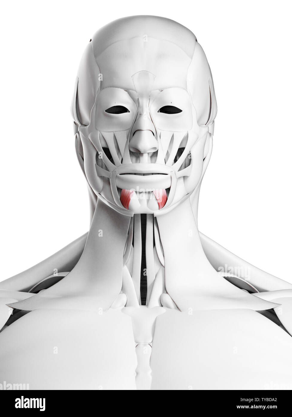 3d rendered medically accurate illustration of the depressor labii inferioris Stock Photohttps://www.alamy.com/image-license-details/?v=1https://www.alamy.com/3d-rendered-medically-accurate-illustration-of-the-depressor-labii-inferioris-image257792810.html
RFTYBDA2–3d rendered medically accurate illustration of the depressor labii inferioris
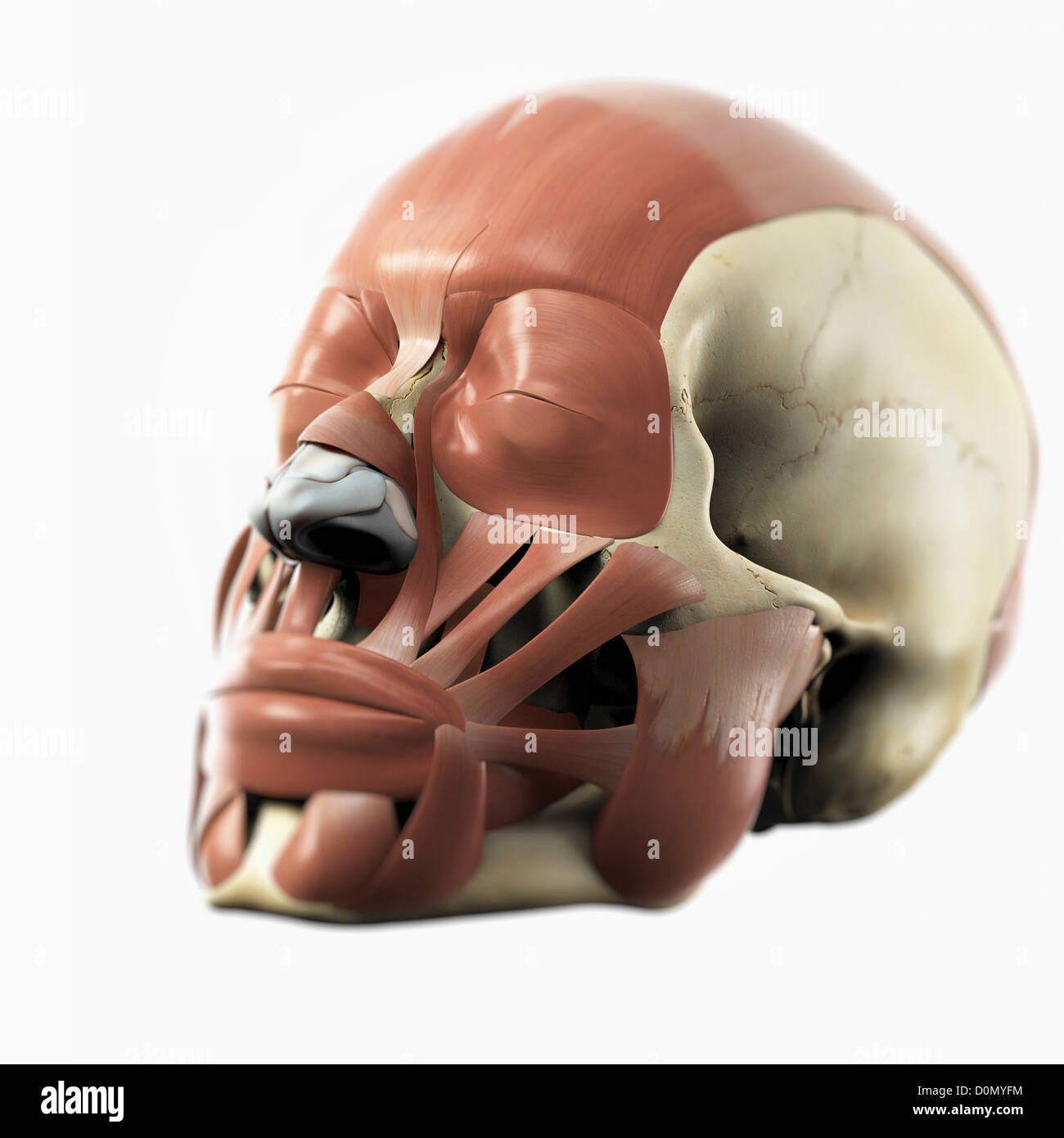 Anatomical model showing the facial muscles. Stock Photohttps://www.alamy.com/image-license-details/?v=1https://www.alamy.com/stock-photo-anatomical-model-showing-the-facial-muscles-52091752.html
RMD0MYFM–Anatomical model showing the facial muscles.
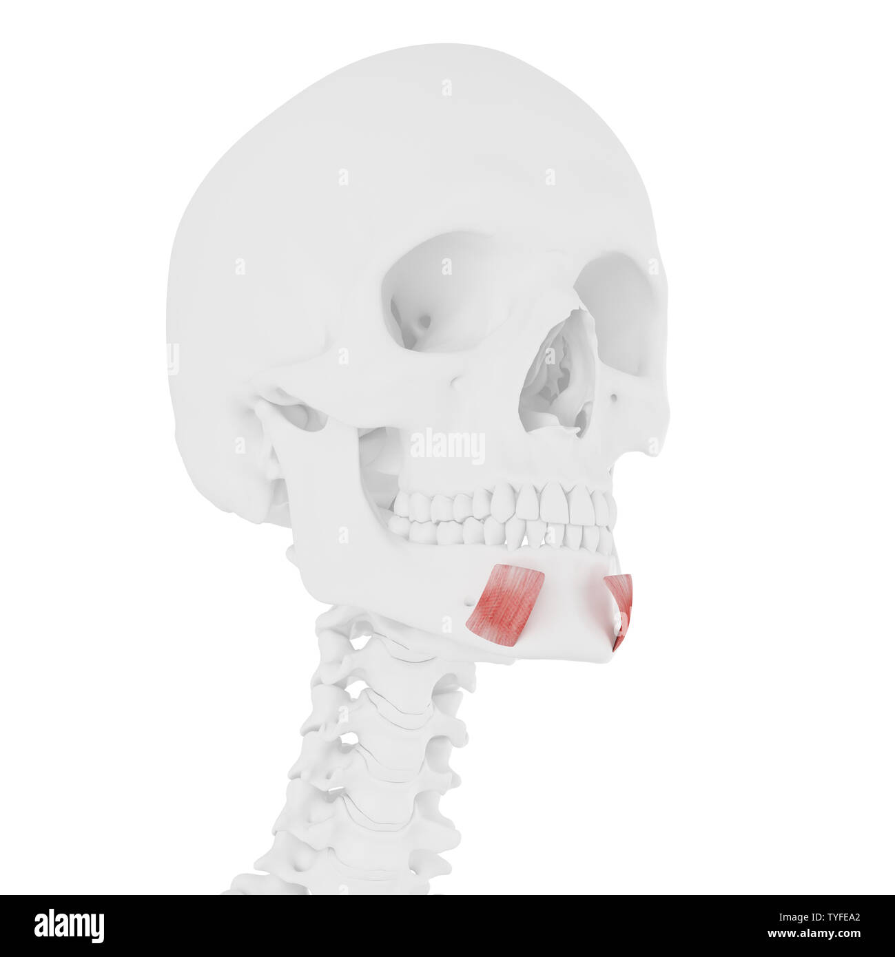 3d rendered medically accurate illustration of the Depressor Labii Inferioris Stock Photohttps://www.alamy.com/image-license-details/?v=1https://www.alamy.com/3d-rendered-medically-accurate-illustration-of-the-depressor-labii-inferioris-image257881402.html
RFTYFEA2–3d rendered medically accurate illustration of the Depressor Labii Inferioris
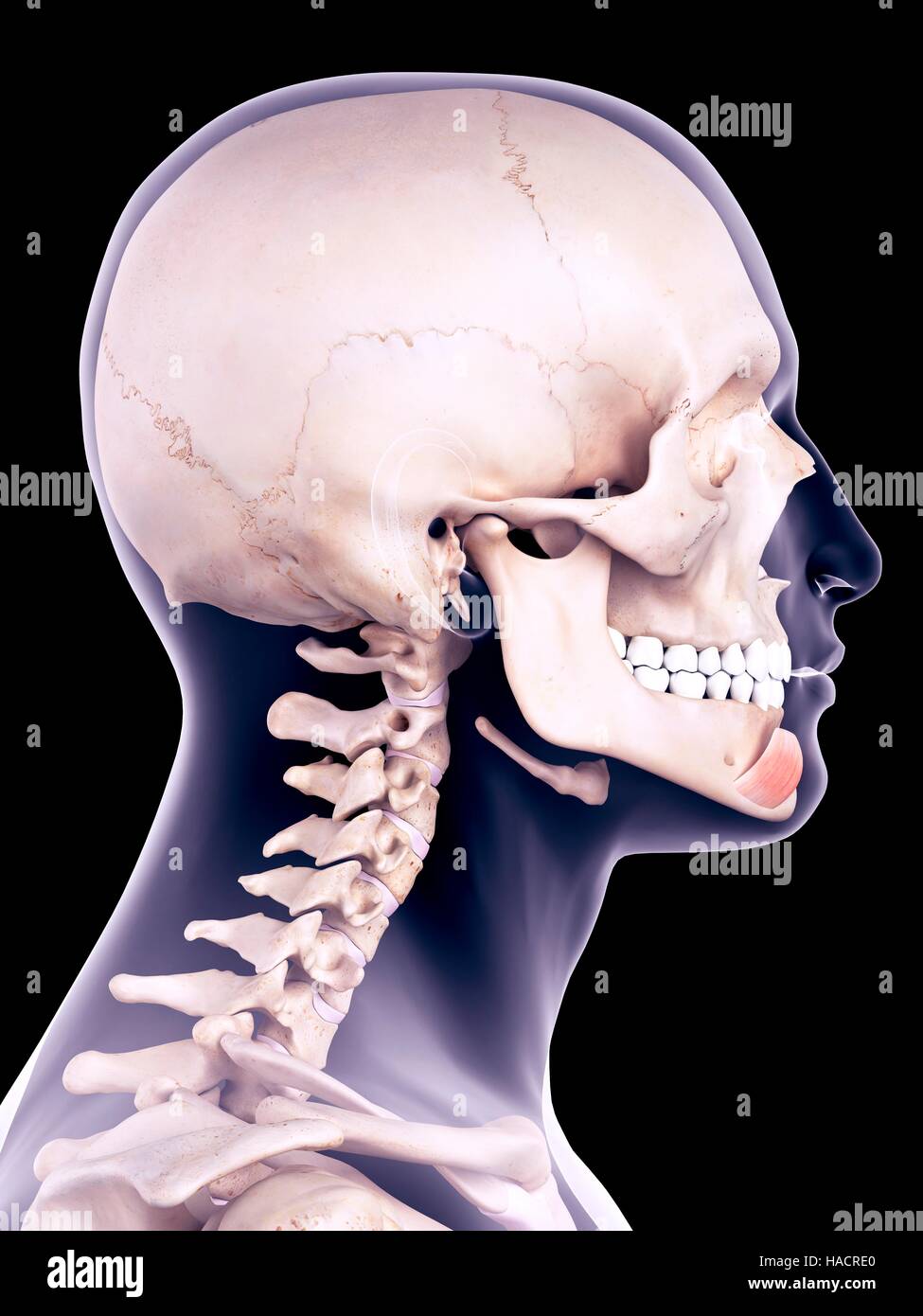 Illustration of the depressor labii inferioris muscle. Stock Photohttps://www.alamy.com/image-license-details/?v=1https://www.alamy.com/stock-photo-illustration-of-the-depressor-labii-inferioris-muscle-126900984.html
RFHACRE0–Illustration of the depressor labii inferioris muscle.
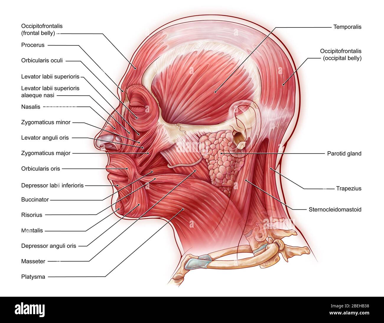 Facial Muscles, illustration Stock Photohttps://www.alamy.com/image-license-details/?v=1https://www.alamy.com/facial-muscles-illustration-image353194444.html
RM2BEHB38–Facial Muscles, illustration
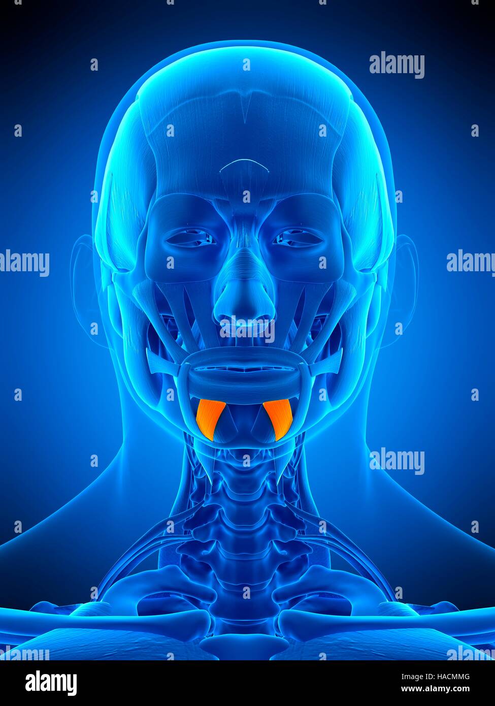 Illustration of the depressor labii inferioris muscle. Stock Photohttps://www.alamy.com/image-license-details/?v=1https://www.alamy.com/stock-photo-illustration-of-the-depressor-labii-inferioris-muscle-126898816.html
RFHACMMG–Illustration of the depressor labii inferioris muscle.
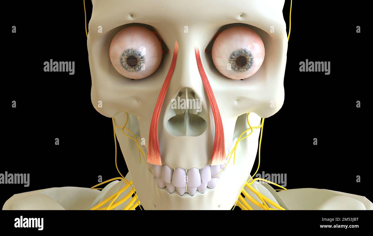 Levator Labii Superioris Muscle anatomy for medical concept 3D illustration Stock Photohttps://www.alamy.com/image-license-details/?v=1https://www.alamy.com/levator-labii-superioris-muscle-anatomy-for-medical-concept-3d-illustration-image502254252.html
RF2M53JBT–Levator Labii Superioris Muscle anatomy for medical concept 3D illustration
 Directions for the dissection and study of the cranial nerves and blood vessels of the horse .. . labialis. 15. M. levator labii superioris proprius. 16. M. dilator naris lateralis. 17. M. zygomaticus. 18. M. buccinator. 19. M. cutaneous faciei (facial panniculus). 20. M. depressor labii inferioris. 21. M. orbicularis oris. 22. Gl. parotis. 23. Ductus parotideus. 24. V. jugularis. 25. V. maxillaris externa. 26. V. labialis communis. 27. V. lateralis nasi. 28. V. dorsalis nasi. 29. V. angularis oculi. 30. V. masseterica. (This vein and artery are incorrectly represented as lying on the surface Stock Photohttps://www.alamy.com/image-license-details/?v=1https://www.alamy.com/directions-for-the-dissection-and-study-of-the-cranial-nerves-and-blood-vessels-of-the-horse-labialis-15-m-levator-labii-superioris-proprius-16-m-dilator-naris-lateralis-17-m-zygomaticus-18-m-buccinator-19-m-cutaneous-faciei-facial-panniculus-20-m-depressor-labii-inferioris-21-m-orbicularis-oris-22-gl-parotis-23-ductus-parotideus-24-v-jugularis-25-v-maxillaris-externa-26-v-labialis-communis-27-v-lateralis-nasi-28-v-dorsalis-nasi-29-v-angularis-oculi-30-v-masseterica-this-vein-and-artery-are-incorrectly-represented-as-lying-on-the-surface-image340228859.html
RM2ANENAK–Directions for the dissection and study of the cranial nerves and blood vessels of the horse .. . labialis. 15. M. levator labii superioris proprius. 16. M. dilator naris lateralis. 17. M. zygomaticus. 18. M. buccinator. 19. M. cutaneous faciei (facial panniculus). 20. M. depressor labii inferioris. 21. M. orbicularis oris. 22. Gl. parotis. 23. Ductus parotideus. 24. V. jugularis. 25. V. maxillaris externa. 26. V. labialis communis. 27. V. lateralis nasi. 28. V. dorsalis nasi. 29. V. angularis oculi. 30. V. masseterica. (This vein and artery are incorrectly represented as lying on the surface
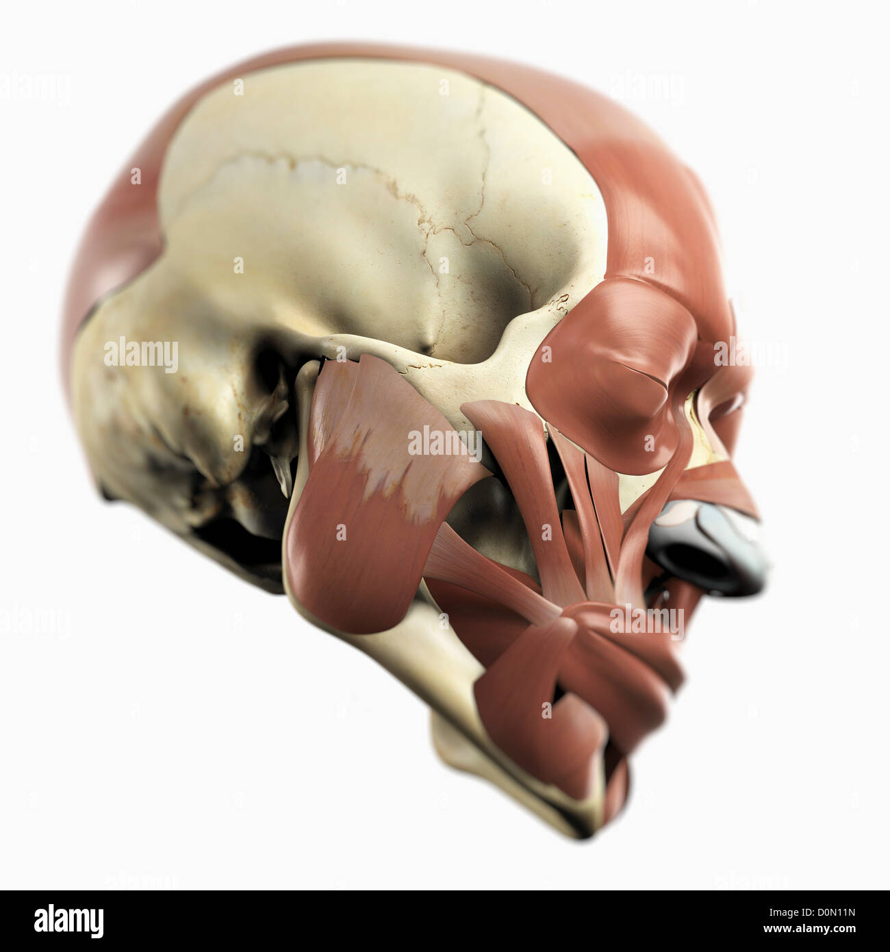 Anatomical model showing the facial muscles. Stock Photohttps://www.alamy.com/image-license-details/?v=1https://www.alamy.com/stock-photo-anatomical-model-showing-the-facial-muscles-52092929.html
RMD0N11N–Anatomical model showing the facial muscles.
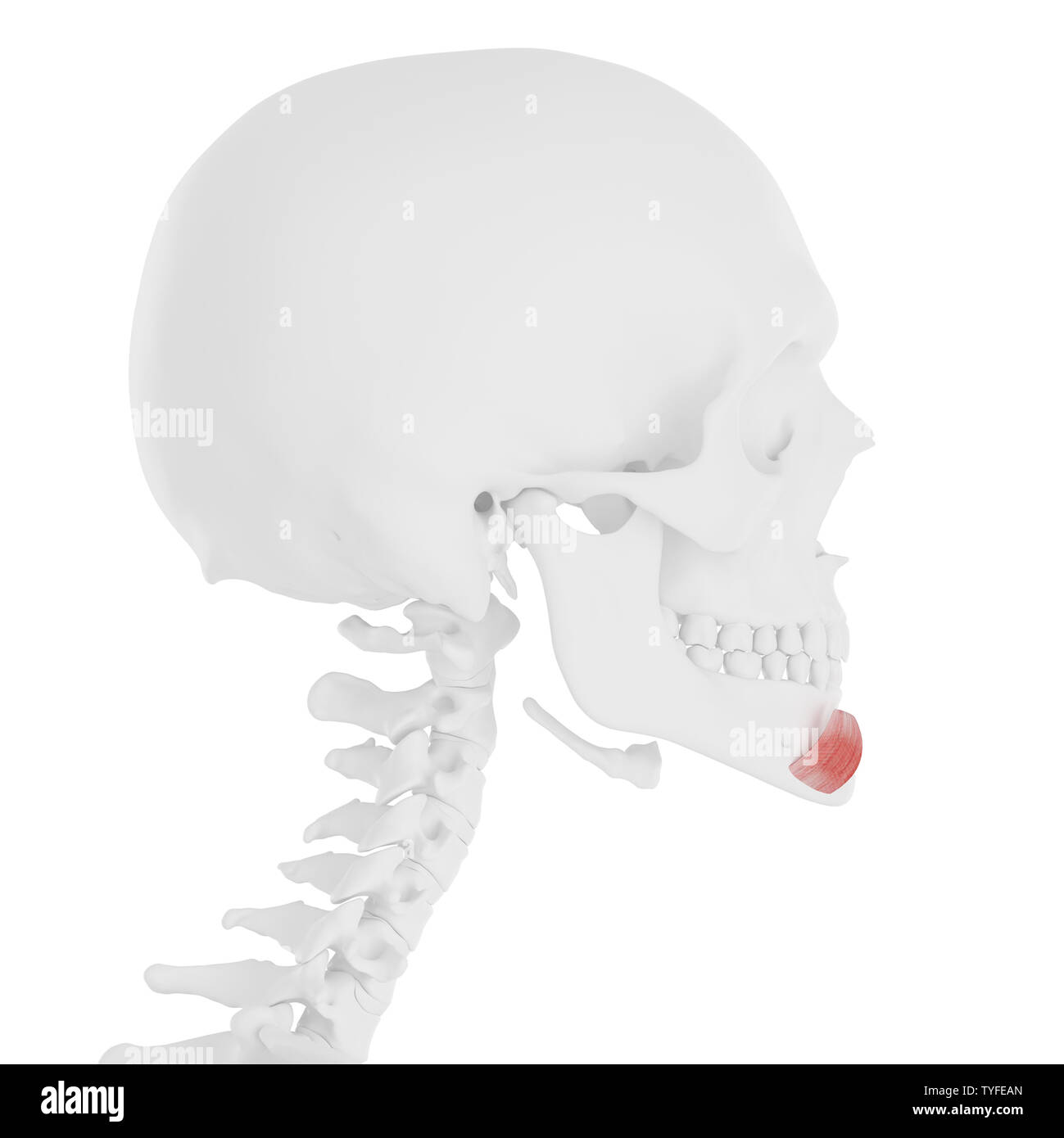 3d rendered medically accurate illustration of the Depressor Labii Inferioris Stock Photohttps://www.alamy.com/image-license-details/?v=1https://www.alamy.com/3d-rendered-medically-accurate-illustration-of-the-depressor-labii-inferioris-image257881421.html
RFTYFEAN–3d rendered medically accurate illustration of the Depressor Labii Inferioris
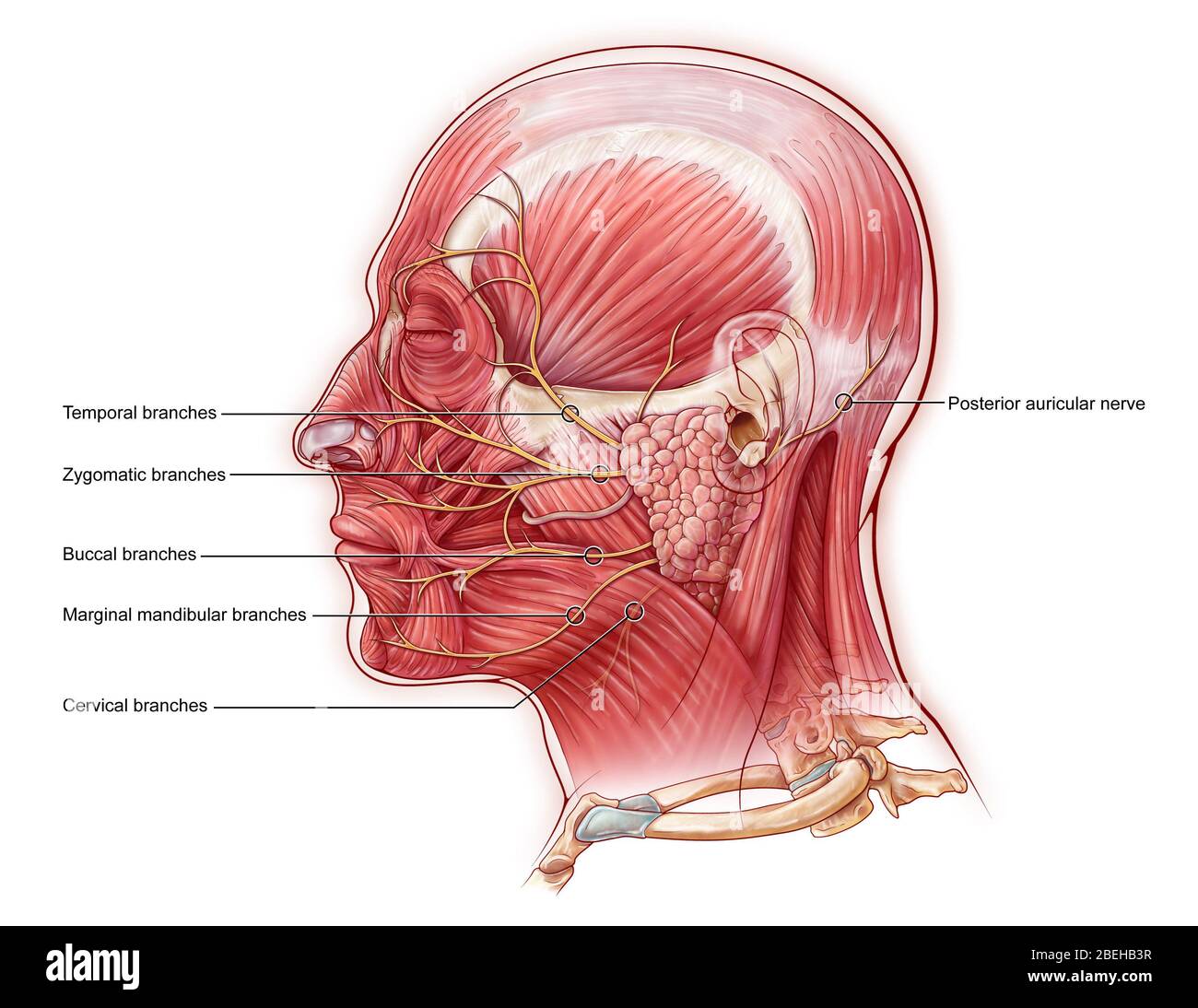 Facial Nerve, illustration Stock Photohttps://www.alamy.com/image-license-details/?v=1https://www.alamy.com/facial-nerve-illustration-image353194459.html
RM2BEHB3R–Facial Nerve, illustration
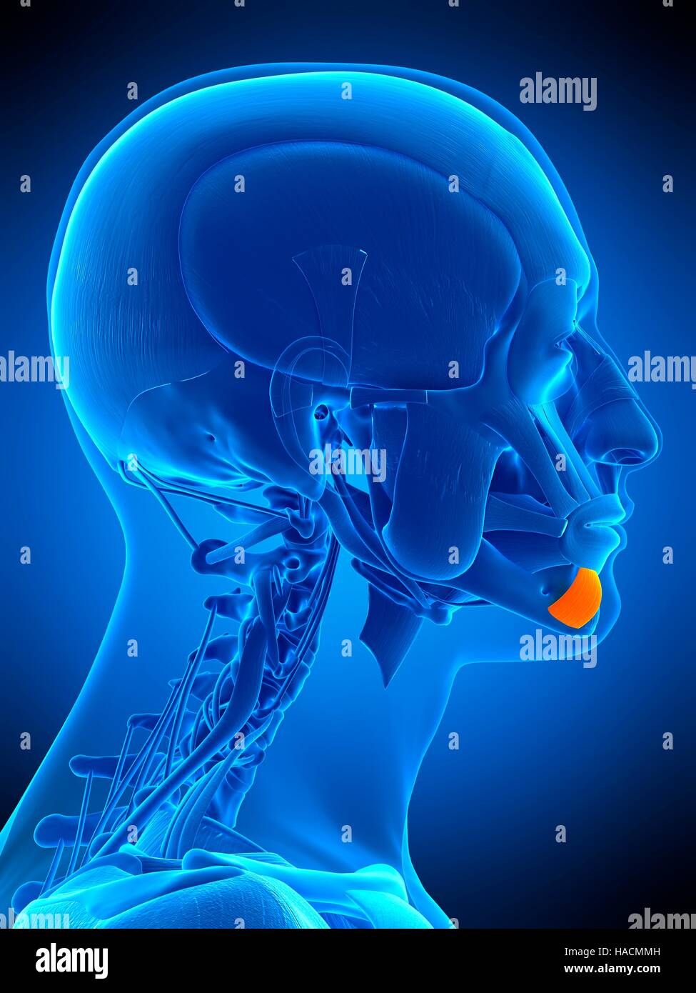 Illustration of the depressor labii inferioris muscle. Stock Photohttps://www.alamy.com/image-license-details/?v=1https://www.alamy.com/stock-photo-illustration-of-the-depressor-labii-inferioris-muscle-126898817.html
RFHACMMH–Illustration of the depressor labii inferioris muscle.
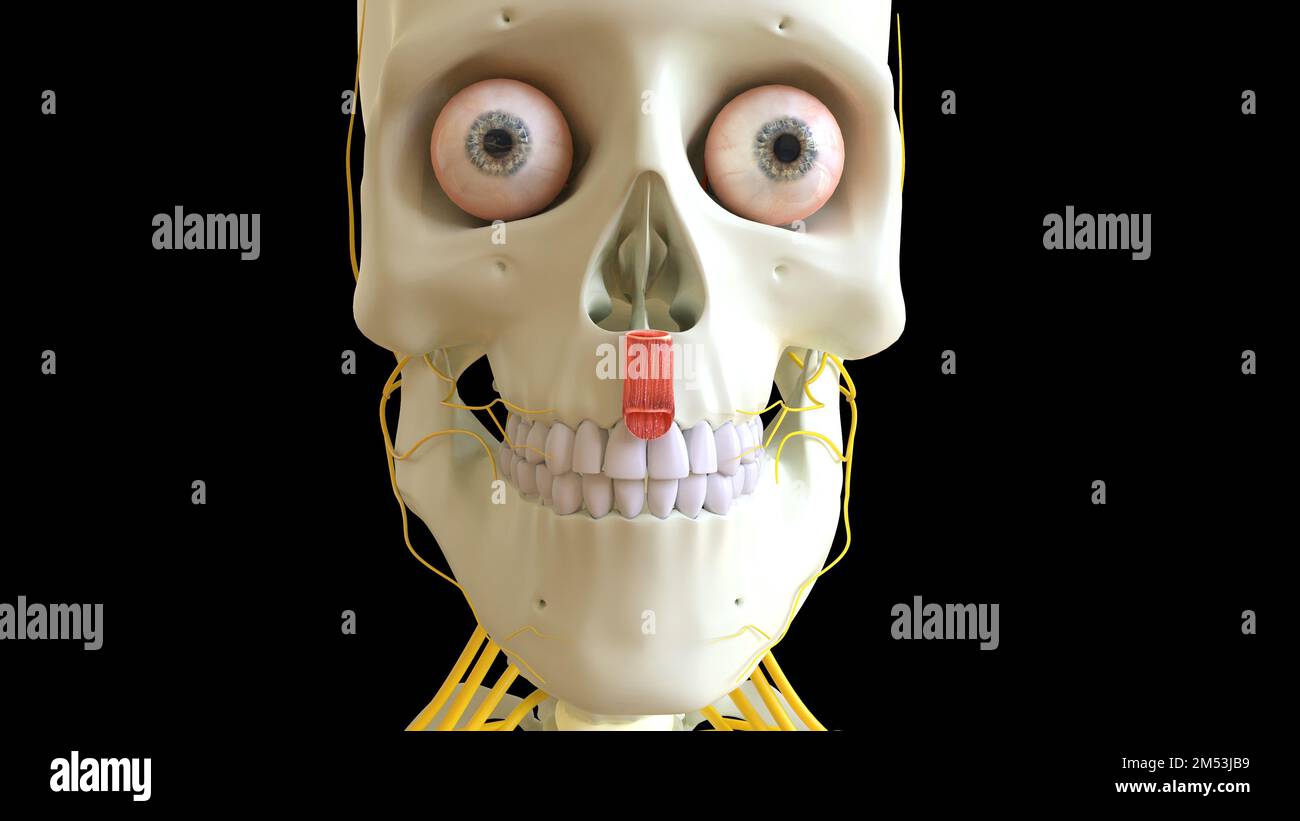 Depressor Septi Nasi muscle anatomy for medical concept 3D illustration Stock Photohttps://www.alamy.com/image-license-details/?v=1https://www.alamy.com/depressor-septi-nasi-muscle-anatomy-for-medical-concept-3d-illustration-image502254237.html
RF2M53JB9–Depressor Septi Nasi muscle anatomy for medical concept 3D illustration
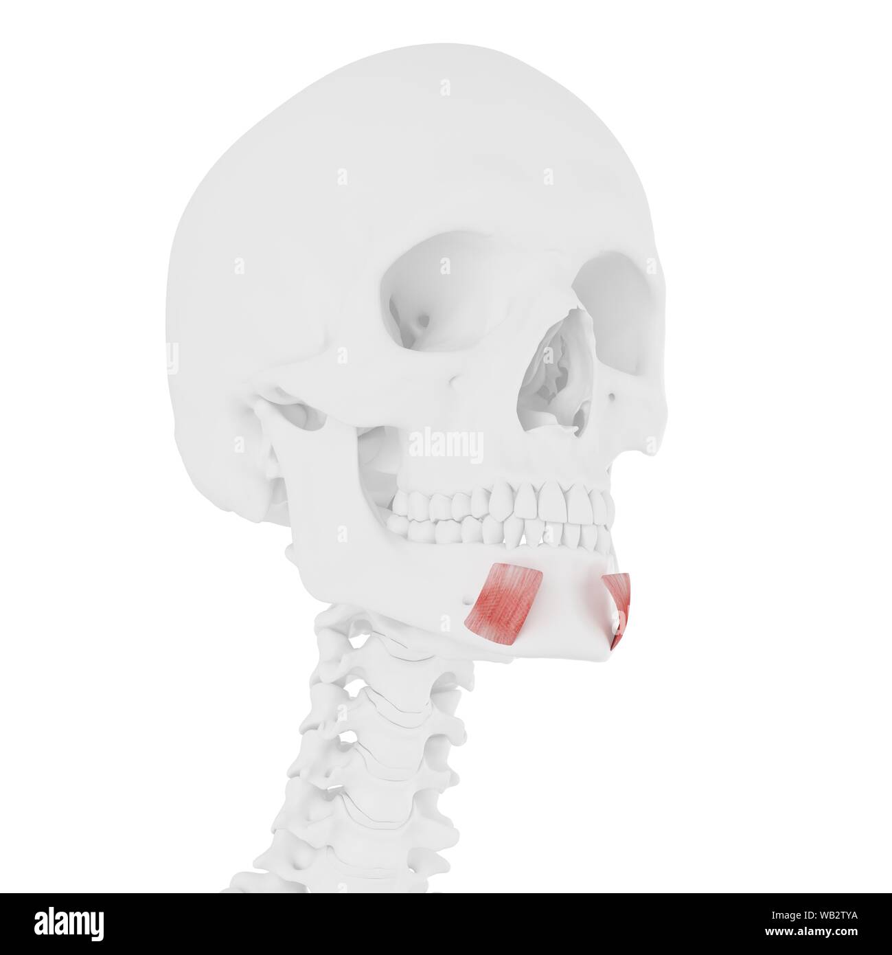 Depressor labii inferioris muscle, computer illustration. Stock Photohttps://www.alamy.com/image-license-details/?v=1https://www.alamy.com/depressor-labii-inferioris-muscle-computer-illustration-image264980222.html
RFWB2TYA–Depressor labii inferioris muscle, computer illustration.
 . Annals of surgery. Paralysis of depressor labii inferioris (right) following extensive sub-maxillary dissection. Fig. 2. Outline of inferior branches of the facial nerve. Scar of extensivedissection for tubercular nodes. Stock Photohttps://www.alamy.com/image-license-details/?v=1https://www.alamy.com/annals-of-surgery-paralysis-of-depressor-labii-inferioris-right-following-extensive-sub-maxillary-dissection-fig-2-outline-of-inferior-branches-of-the-facial-nerve-scar-of-extensivedissection-for-tubercular-nodes-image370649716.html
RM2CF0FDT–. Annals of surgery. Paralysis of depressor labii inferioris (right) following extensive sub-maxillary dissection. Fig. 2. Outline of inferior branches of the facial nerve. Scar of extensivedissection for tubercular nodes.
 Anatomical model showing the facial muscles. Stock Photohttps://www.alamy.com/image-license-details/?v=1https://www.alamy.com/stock-photo-anatomical-model-showing-the-facial-muscles-52089754.html
RMD0MW0A–Anatomical model showing the facial muscles.
 3d rendered medically accurate illustration of the Depressor Labii Inferioris Stock Photohttps://www.alamy.com/image-license-details/?v=1https://www.alamy.com/3d-rendered-medically-accurate-illustration-of-the-depressor-labii-inferioris-image257881459.html
RFTYFEC3–3d rendered medically accurate illustration of the Depressor Labii Inferioris
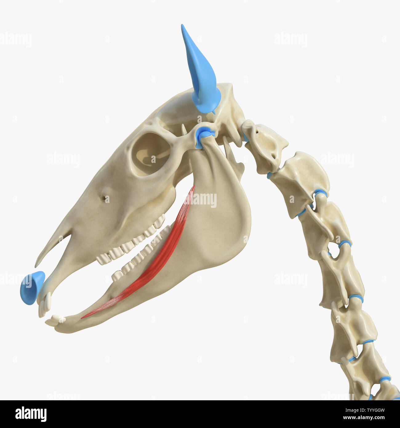 3d rendered medically accurate illustration of the equine muscle anatomy - Depressor Labii Inferioris Stock Photohttps://www.alamy.com/image-license-details/?v=1https://www.alamy.com/3d-rendered-medically-accurate-illustration-of-the-equine-muscle-anatomy-depressor-labii-inferioris-image258146585.html
RFTYYGGW–3d rendered medically accurate illustration of the equine muscle anatomy - Depressor Labii Inferioris
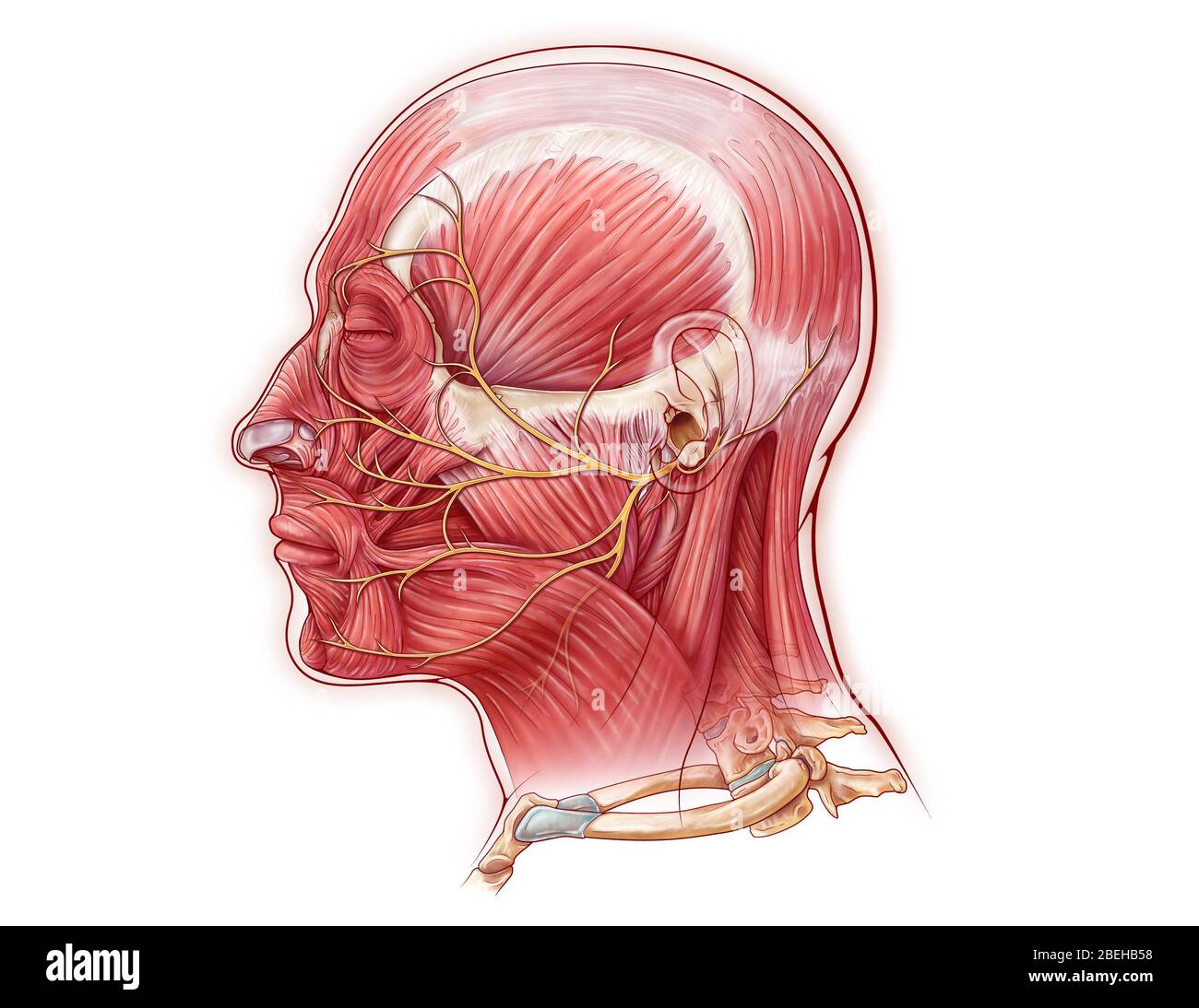 Facial Nerve, illustration Stock Photohttps://www.alamy.com/image-license-details/?v=1https://www.alamy.com/facial-nerve-illustration-image353194500.html
RM2BEHB58–Facial Nerve, illustration
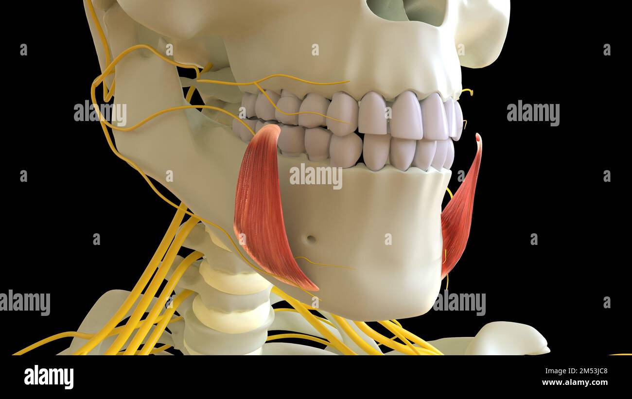 Depressor Anguli Oris Muscle anatomy for medical concept 3D illustration Stock Photohttps://www.alamy.com/image-license-details/?v=1https://www.alamy.com/depressor-anguli-oris-muscle-anatomy-for-medical-concept-3d-illustration-image502254264.html
RF2M53JC8–Depressor Anguli Oris Muscle anatomy for medical concept 3D illustration
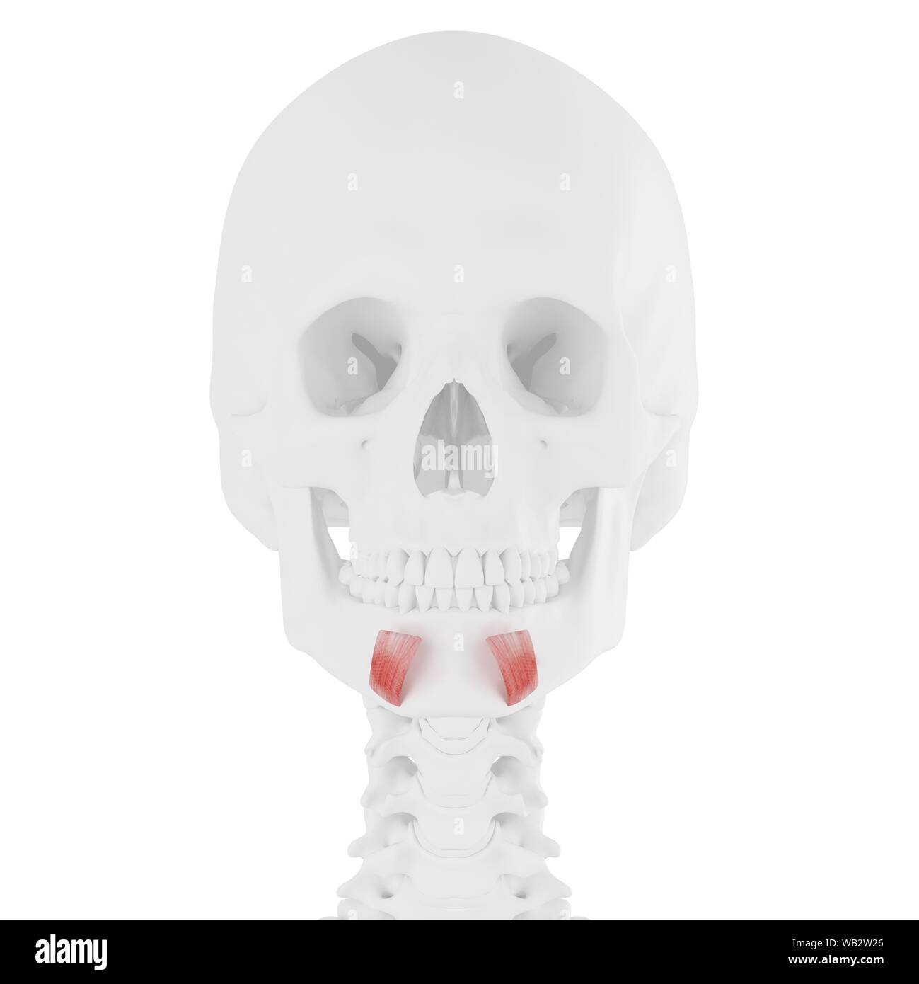 Depressor labii inferioris muscle, computer illustration. Stock Photohttps://www.alamy.com/image-license-details/?v=1https://www.alamy.com/depressor-labii-inferioris-muscle-computer-illustration-image264980302.html
RFWB2W26–Depressor labii inferioris muscle, computer illustration.
![. Handbook of anatomy; being a complete compend of anatomy, including the anatomy of the viscera a chapter on dental anatomy, numerous tables, and incorporating the newer nomenclature adopted by the German anatomical Society, generally designated the Basle nomenclature or BNA . ernal pterygoid muscle, andon its outer side the masseter. It articulates with the two temporal bones. The muscular attachments are fifteen pairs; to the externalsurface, sis—depressor anguli oris, depressor labii inferioris,levator labii inferioris, orbicularis oris, platysma myoides andbuccinator; from the interna] Bu Stock Photo . Handbook of anatomy; being a complete compend of anatomy, including the anatomy of the viscera a chapter on dental anatomy, numerous tables, and incorporating the newer nomenclature adopted by the German anatomical Society, generally designated the Basle nomenclature or BNA . ernal pterygoid muscle, andon its outer side the masseter. It articulates with the two temporal bones. The muscular attachments are fifteen pairs; to the externalsurface, sis—depressor anguli oris, depressor labii inferioris,levator labii inferioris, orbicularis oris, platysma myoides andbuccinator; from the interna] Bu Stock Photo](https://c8.alamy.com/comp/2CD8BXN/handbook-of-anatomy-being-a-complete-compend-of-anatomy-including-the-anatomy-of-the-viscera-a-chapter-on-dental-anatomy-numerous-tables-and-incorporating-the-newer-nomenclature-adopted-by-the-german-anatomical-society-generally-designated-the-basle-nomenclature-or-bna-ernal-pterygoid-muscle-andon-its-outer-side-the-masseter-it-articulates-with-the-two-temporal-bones-the-muscular-attachments-are-fifteen-pairs-to-the-externalsurface-sisdepressor-anguli-oris-depressor-labii-inferiorislevator-labii-inferioris-orbicularis-oris-platysma-myoides-andbuccinator-from-the-interna-bu-2CD8BXN.jpg) . Handbook of anatomy; being a complete compend of anatomy, including the anatomy of the viscera a chapter on dental anatomy, numerous tables, and incorporating the newer nomenclature adopted by the German anatomical Society, generally designated the Basle nomenclature or BNA . ernal pterygoid muscle, andon its outer side the masseter. It articulates with the two temporal bones. The muscular attachments are fifteen pairs; to the externalsurface, sis—depressor anguli oris, depressor labii inferioris,levator labii inferioris, orbicularis oris, platysma myoides andbuccinator; from the interna] Bu Stock Photohttps://www.alamy.com/image-license-details/?v=1https://www.alamy.com/handbook-of-anatomy-being-a-complete-compend-of-anatomy-including-the-anatomy-of-the-viscera-a-chapter-on-dental-anatomy-numerous-tables-and-incorporating-the-newer-nomenclature-adopted-by-the-german-anatomical-society-generally-designated-the-basle-nomenclature-or-bna-ernal-pterygoid-muscle-andon-its-outer-side-the-masseter-it-articulates-with-the-two-temporal-bones-the-muscular-attachments-are-fifteen-pairs-to-the-externalsurface-sisdepressor-anguli-oris-depressor-labii-inferiorislevator-labii-inferioris-orbicularis-oris-platysma-myoides-andbuccinator-from-the-interna-bu-image369593245.html
. Handbook of anatomy; being a complete compend of anatomy, including the anatomy of the viscera a chapter on dental anatomy, numerous tables, and incorporating the newer nomenclature adopted by the German anatomical Society, generally designated the Basle nomenclature or BNA . ernal pterygoid muscle, andon its outer side the masseter. It articulates with the two temporal bones. The muscular attachments are fifteen pairs; to the externalsurface, sis—depressor anguli oris, depressor labii inferioris,levator labii inferioris, orbicularis oris, platysma myoides andbuccinator; from the interna] Bu Stock Photohttps://www.alamy.com/image-license-details/?v=1https://www.alamy.com/handbook-of-anatomy-being-a-complete-compend-of-anatomy-including-the-anatomy-of-the-viscera-a-chapter-on-dental-anatomy-numerous-tables-and-incorporating-the-newer-nomenclature-adopted-by-the-german-anatomical-society-generally-designated-the-basle-nomenclature-or-bna-ernal-pterygoid-muscle-andon-its-outer-side-the-masseter-it-articulates-with-the-two-temporal-bones-the-muscular-attachments-are-fifteen-pairs-to-the-externalsurface-sisdepressor-anguli-oris-depressor-labii-inferiorislevator-labii-inferioris-orbicularis-oris-platysma-myoides-andbuccinator-from-the-interna-bu-image369593245.htmlRM2CD8BXN–. Handbook of anatomy; being a complete compend of anatomy, including the anatomy of the viscera a chapter on dental anatomy, numerous tables, and incorporating the newer nomenclature adopted by the German anatomical Society, generally designated the Basle nomenclature or BNA . ernal pterygoid muscle, andon its outer side the masseter. It articulates with the two temporal bones. The muscular attachments are fifteen pairs; to the externalsurface, sis—depressor anguli oris, depressor labii inferioris,levator labii inferioris, orbicularis oris, platysma myoides andbuccinator; from the interna] Bu
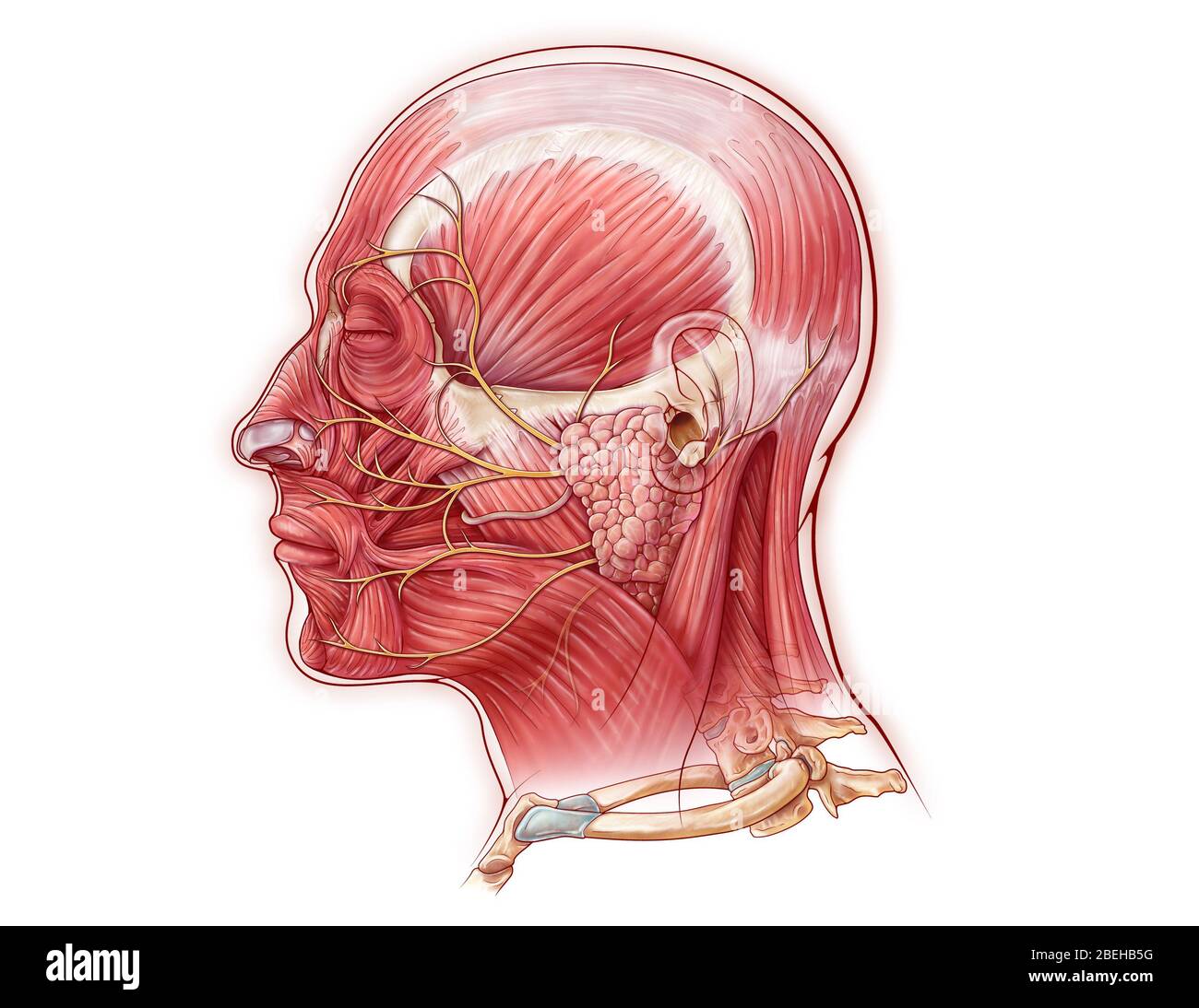 Facial Nerve, illustration Stock Photohttps://www.alamy.com/image-license-details/?v=1https://www.alamy.com/facial-nerve-illustration-image353194508.html
RM2BEHB5G–Facial Nerve, illustration
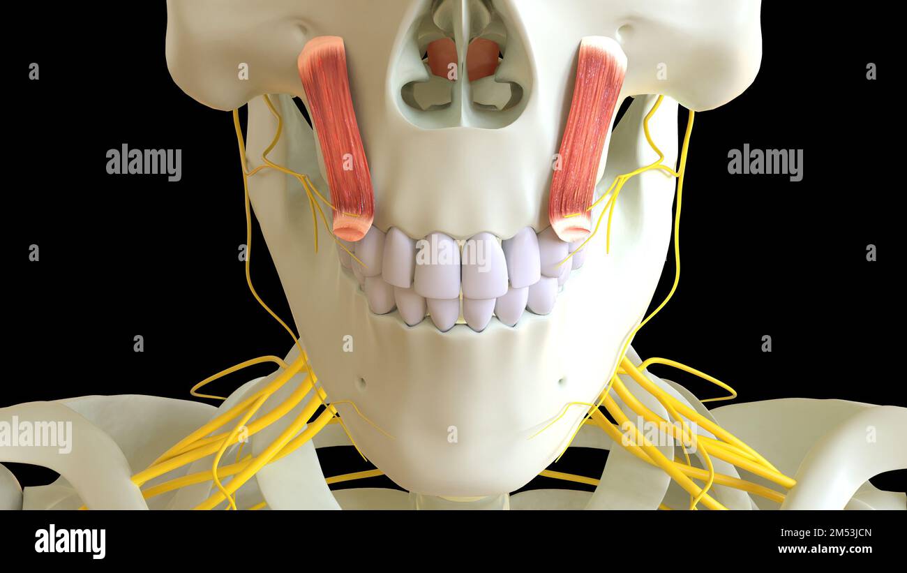 Levator Labii Superioris Muscle anatomy for medical concept 3D illustration Stock Photohttps://www.alamy.com/image-license-details/?v=1https://www.alamy.com/levator-labii-superioris-muscle-anatomy-for-medical-concept-3d-illustration-image502254277.html
RF2M53JCN–Levator Labii Superioris Muscle anatomy for medical concept 3D illustration
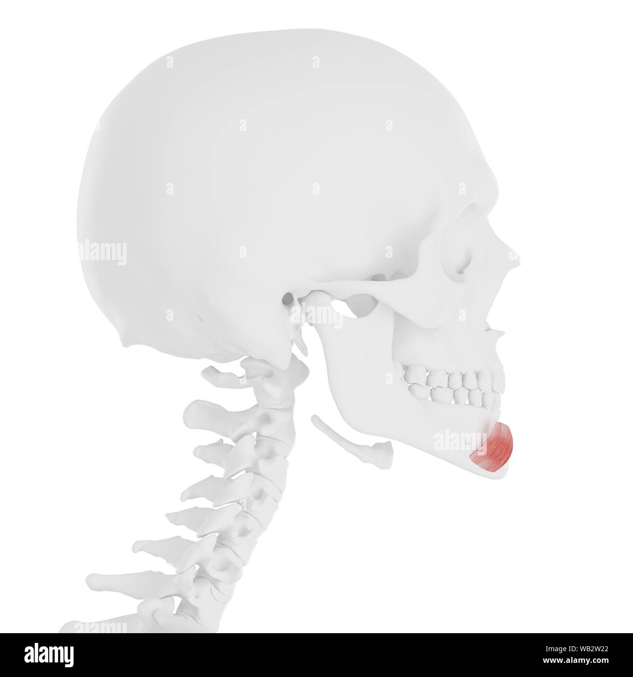 Depressor labii inferioris muscle, computer illustration. Stock Photohttps://www.alamy.com/image-license-details/?v=1https://www.alamy.com/depressor-labii-inferioris-muscle-computer-illustration-image264980298.html
RFWB2W22–Depressor labii inferioris muscle, computer illustration.
 . A system of anatomy for the use of students of medicine (Volume 1). e maxilla tothe angle of the mouth, to the depressor labii inferioris within,and to the skin and fat without, gradually turning narrower :and is Inserted into the angle of the mouth, joining with the zygo-matics major and levator anguli oris. Use. To pull down the corner of the mouth. 2. Depressor Labii Inferioris, Arises, broad and fleshy, intermixed with fat, from the inferiorpart of the lower jaw next to the chin; runs obliquely upwards,and is Inserted into the edge of the under lip, extends along one halfof the lid, and Stock Photohttps://www.alamy.com/image-license-details/?v=1https://www.alamy.com/a-system-of-anatomy-for-the-use-of-students-of-medicine-volume-1-e-maxilla-tothe-angle-of-the-mouth-to-the-depressor-labii-inferioris-withinand-to-the-skin-and-fat-without-gradually-turning-narrower-and-is-inserted-into-the-angle-of-the-mouth-joining-with-the-zygo-matics-major-and-levator-anguli-oris-use-to-pull-down-the-corner-of-the-mouth-2-depressor-labii-inferioris-arises-broad-and-fleshy-intermixed-with-fat-from-the-inferiorpart-of-the-lower-jaw-next-to-the-chin-runs-obliquely-upwardsand-is-inserted-into-the-edge-of-the-under-lip-extends-along-one-halfof-the-lid-and-image370490788.html
RM2CEN8NT–. A system of anatomy for the use of students of medicine (Volume 1). e maxilla tothe angle of the mouth, to the depressor labii inferioris within,and to the skin and fat without, gradually turning narrower :and is Inserted into the angle of the mouth, joining with the zygo-matics major and levator anguli oris. Use. To pull down the corner of the mouth. 2. Depressor Labii Inferioris, Arises, broad and fleshy, intermixed with fat, from the inferiorpart of the lower jaw next to the chin; runs obliquely upwards,and is Inserted into the edge of the under lip, extends along one halfof the lid, and
 Horse depressor labii inferioris muscle, illustration Stock Photohttps://www.alamy.com/image-license-details/?v=1https://www.alamy.com/horse-depressor-labii-inferioris-muscle-illustration-image273702224.html
RFWW85YC–Horse depressor labii inferioris muscle, illustration
 Human head musculature, artwork Stock Photohttps://www.alamy.com/image-license-details/?v=1https://www.alamy.com/human-head-musculature-artwork-image65260421.html
RFDP4T99–Human head musculature, artwork
 Human head musculature, artwork Stock Photohttps://www.alamy.com/image-license-details/?v=1https://www.alamy.com/human-head-musculature-artwork-image65257935.html
RFDP4N4F–Human head musculature, artwork
 Muscular system computer artwork. Stock Photohttps://www.alamy.com/image-license-details/?v=1https://www.alamy.com/muscular-system-computer-artwork-image69886262.html
RFE1KGHX–Muscular system computer artwork.
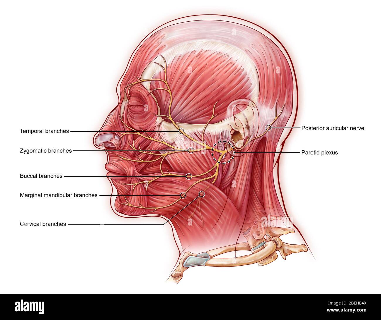 Facial Nerve, illustration Stock Photohttps://www.alamy.com/image-license-details/?v=1https://www.alamy.com/facial-nerve-illustration-image353194490.html
RM2BEHB4X–Facial Nerve, illustration
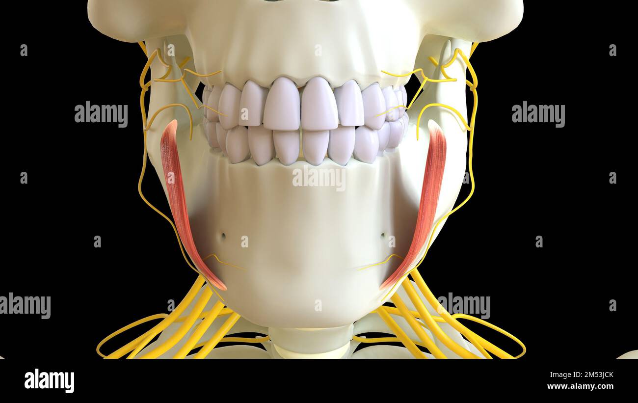 Depressor Anguli Oris Muscle anatomy for medical concept 3D illustration Stock Photohttps://www.alamy.com/image-license-details/?v=1https://www.alamy.com/depressor-anguli-oris-muscle-anatomy-for-medical-concept-3d-illustration-image502254275.html
RF2M53JCK–Depressor Anguli Oris Muscle anatomy for medical concept 3D illustration
 . Handbook of anatomy; being a complete compend of anatomy, including the anatomy of the viscera a chapter on dental anatomy, numerous tables, and incorporating the newer nomenclature adopted by the German anatomical Society, generally designated the Basle nomenclature or BNA . tence. The sphincter action ofthe lips is accomplished by a complicated interlacing of themuscle fibers from buccinator, depressor labii inferioris, depres-sor anguli oris, zygomaticus and risorius. The buccinator formsthe muscular body of the cheek. Its attachment to the maxillahas to be considered in outlining upper a Stock Photohttps://www.alamy.com/image-license-details/?v=1https://www.alamy.com/handbook-of-anatomy-being-a-complete-compend-of-anatomy-including-the-anatomy-of-the-viscera-a-chapter-on-dental-anatomy-numerous-tables-and-incorporating-the-newer-nomenclature-adopted-by-the-german-anatomical-society-generally-designated-the-basle-nomenclature-or-bna-tence-the-sphincter-action-ofthe-lips-is-accomplished-by-a-complicated-interlacing-of-themuscle-fibers-from-buccinator-depressor-labii-inferioris-depres-sor-anguli-oris-zygomaticus-and-risorius-the-buccinator-formsthe-muscular-body-of-the-cheek-its-attachment-to-the-maxillahas-to-be-considered-in-outlining-upper-a-image369838560.html
RM2CDKGT0–. Handbook of anatomy; being a complete compend of anatomy, including the anatomy of the viscera a chapter on dental anatomy, numerous tables, and incorporating the newer nomenclature adopted by the German anatomical Society, generally designated the Basle nomenclature or BNA . tence. The sphincter action ofthe lips is accomplished by a complicated interlacing of themuscle fibers from buccinator, depressor labii inferioris, depres-sor anguli oris, zygomaticus and risorius. The buccinator formsthe muscular body of the cheek. Its attachment to the maxillahas to be considered in outlining upper a
 Muscular system computer artwork. Stock Photohttps://www.alamy.com/image-license-details/?v=1https://www.alamy.com/muscular-system-computer-artwork-image69886249.html
RFE1KGHD–Muscular system computer artwork.
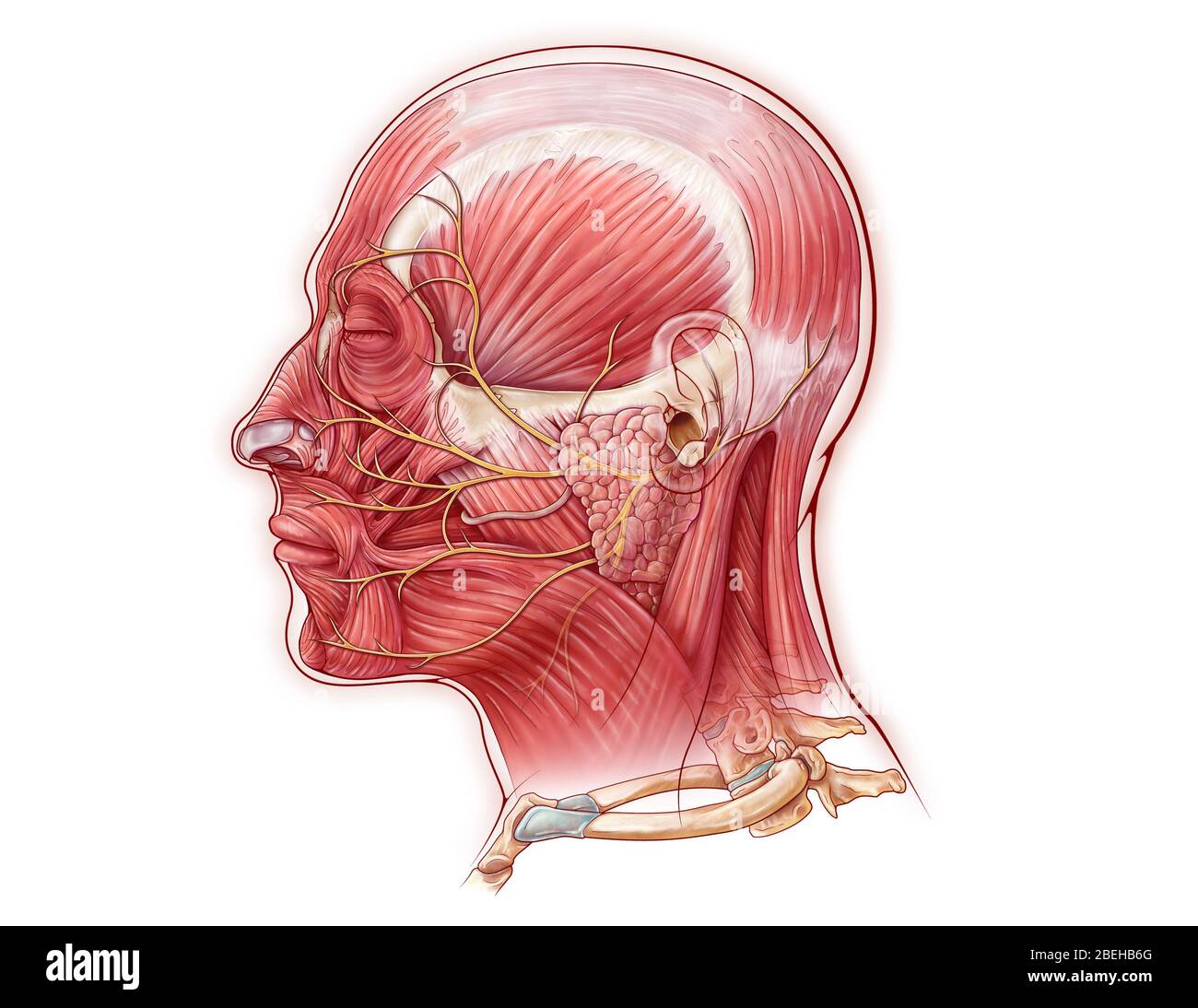 Facial Nerve, illustration Stock Photohttps://www.alamy.com/image-license-details/?v=1https://www.alamy.com/facial-nerve-illustration-image353194536.html
RM2BEHB6G–Facial Nerve, illustration
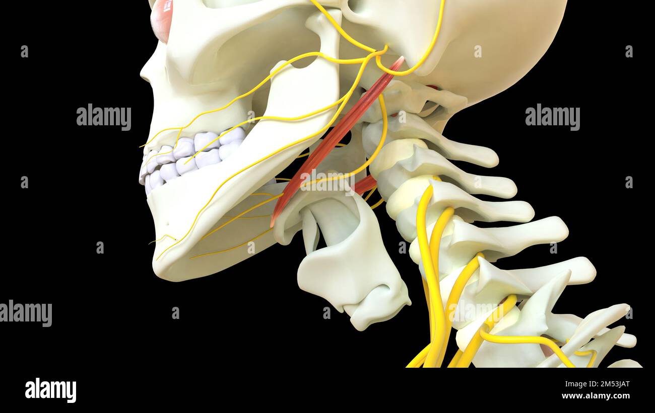 Stylohyoid Muscle anatomy for medical concept 3D illustration Stock Photohttps://www.alamy.com/image-license-details/?v=1https://www.alamy.com/stylohyoid-muscle-anatomy-for-medical-concept-3d-illustration-image502254224.html
RF2M53JAT–Stylohyoid Muscle anatomy for medical concept 3D illustration
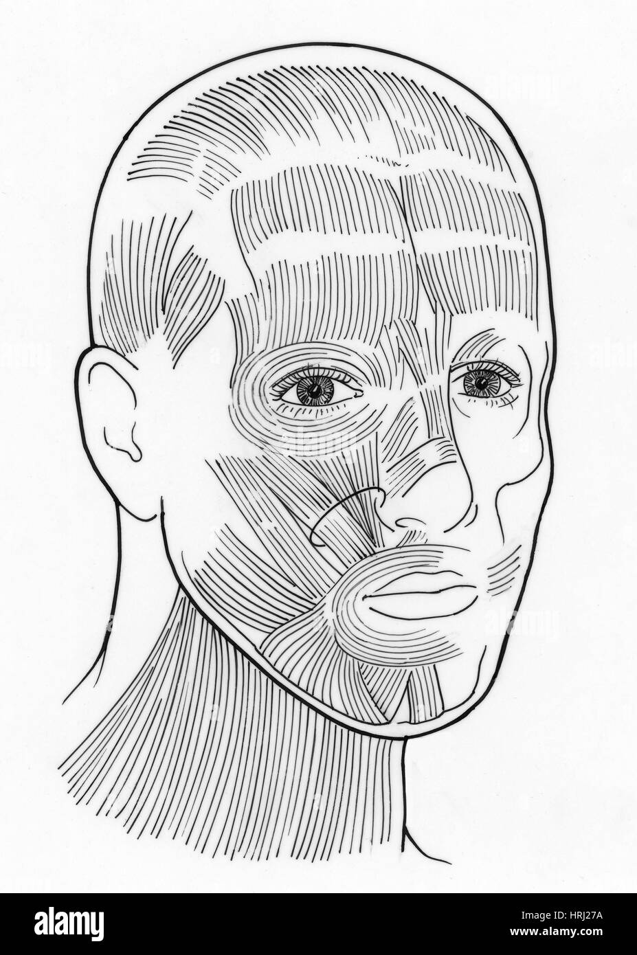 Illustration of Facial Muscles Stock Photohttps://www.alamy.com/image-license-details/?v=1https://www.alamy.com/stock-photo-illustration-of-facial-muscles-135006574.html
RMHRJ27A–Illustration of Facial Muscles
 . The hydropathic family physician : a ready prescriber and hygienic adviser with reference to the nature, causes, prevention, and treatment of diseases, accidents, and casualties of every kind . L. Depressor anguli oris. M. Sterno-cleido mastnideus. 0 Depressor labii inferioris.P. Orbicularis oris. Q. Temporalis. B. Splenius. S. Trapezius, sen Cncullaris. T. Sterno*hyoideus. a. Helix, b. Anti-helix. & Concha. Through gymnastics alone, that is, muscular exercise in a greatvariety of ways, suited according to the nature of the case, some ofthe most inveterate diseases may be cured. There are ma Stock Photohttps://www.alamy.com/image-license-details/?v=1https://www.alamy.com/the-hydropathic-family-physician-a-ready-prescriber-and-hygienic-adviser-with-reference-to-the-nature-causes-prevention-and-treatment-of-diseases-accidents-and-casualties-of-every-kind-l-depressor-anguli-oris-m-sterno-cleido-mastnideus-0-depressor-labii-inferiorisp-orbicularis-oris-q-temporalis-b-splenius-s-trapezius-sen-cncullaris-t-sternohyoideus-a-helix-b-anti-helix-concha-through-gymnastics-alone-that-is-muscular-exercise-in-a-greatvariety-of-ways-suited-according-to-the-nature-of-the-case-some-ofthe-most-inveterate-diseases-may-be-cured-there-are-ma-image370187137.html
RM2CE7DD5–. The hydropathic family physician : a ready prescriber and hygienic adviser with reference to the nature, causes, prevention, and treatment of diseases, accidents, and casualties of every kind . L. Depressor anguli oris. M. Sterno-cleido mastnideus. 0 Depressor labii inferioris.P. Orbicularis oris. Q. Temporalis. B. Splenius. S. Trapezius, sen Cncullaris. T. Sterno*hyoideus. a. Helix, b. Anti-helix. & Concha. Through gymnastics alone, that is, muscular exercise in a greatvariety of ways, suited according to the nature of the case, some ofthe most inveterate diseases may be cured. There are ma
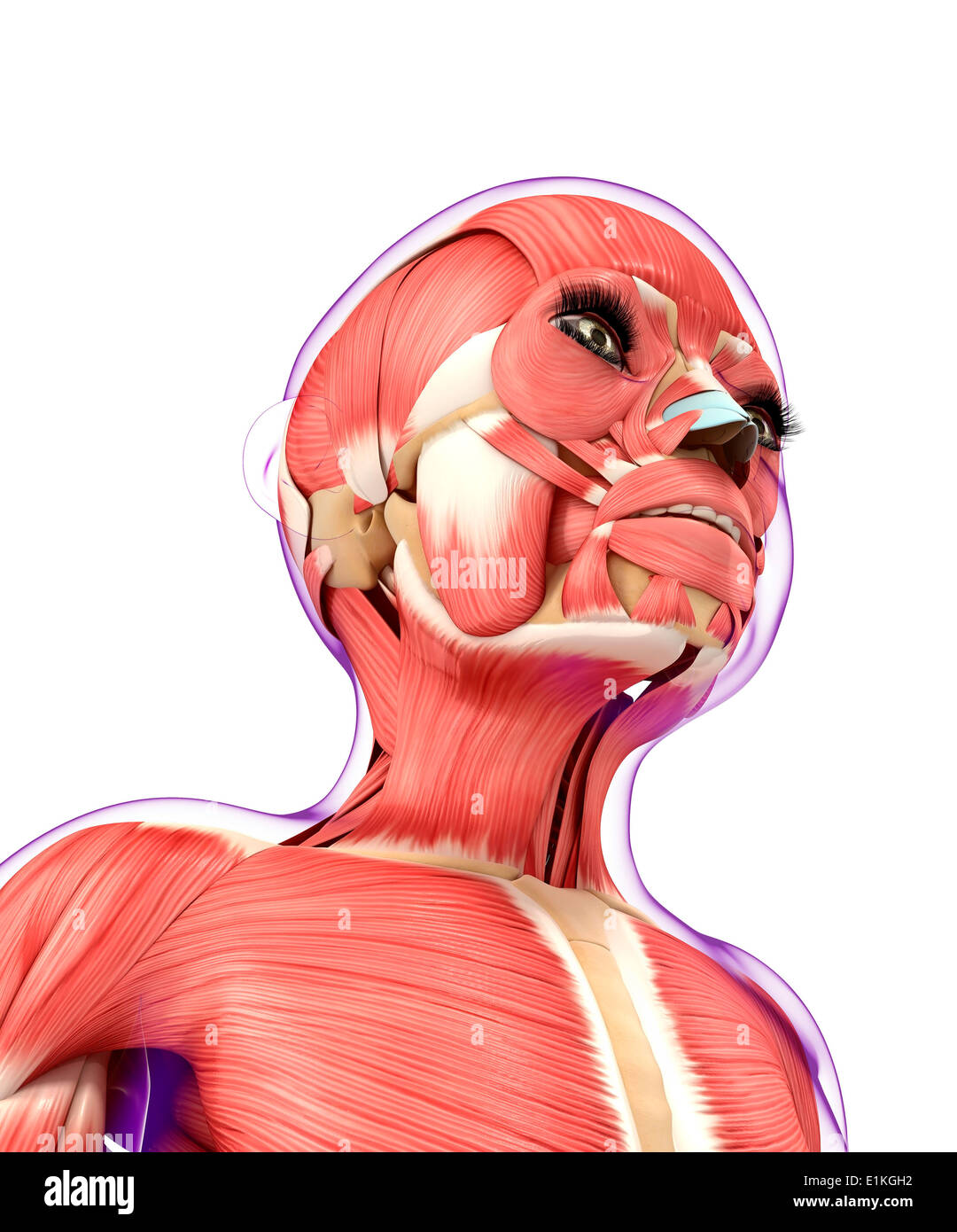 Muscular system computer artwork. Stock Photohttps://www.alamy.com/image-license-details/?v=1https://www.alamy.com/muscular-system-computer-artwork-image69886238.html
RFE1KGH2–Muscular system computer artwork.
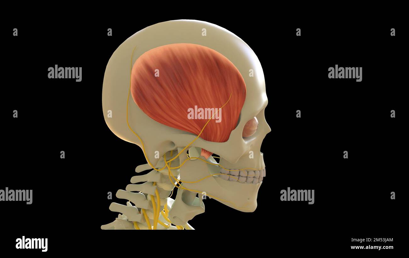 Temporal Muscle anatomy for medical concept 3D illustration Stock Photohttps://www.alamy.com/image-license-details/?v=1https://www.alamy.com/temporal-muscle-anatomy-for-medical-concept-3d-illustration-image502254220.html
RF2M53JAM–Temporal Muscle anatomy for medical concept 3D illustration
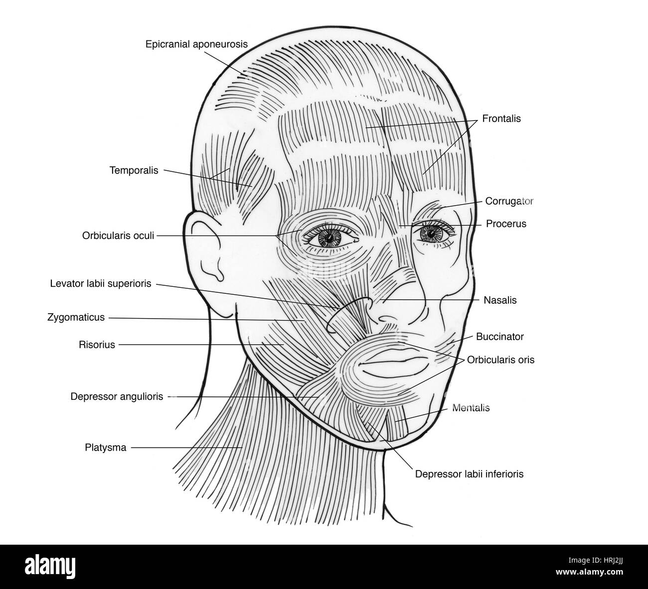 Illustration of Facial Muscles Stock Photohttps://www.alamy.com/image-license-details/?v=1https://www.alamy.com/stock-photo-illustration-of-facial-muscles-135006890.html
RMHRJ2JJ–Illustration of Facial Muscles
 . Buffalo Medical Journal. al nerve: inferior branch. 8, Hypoglossalnerve. 9. Accessorious nerve. 10. Erbs point (supraclavicular point). 11. Phrenic nerve.12. Brachial plexus. 13. Axillary nerve. MUSCLES. a. Frontalis, b. Corrugator supercilii. c. Orbicularis palpebrarum, d. Nasal mus-cles, e. Zygomatic muscles, f. Orbicularis oris. g. Massetor. h. Levater menti. i. Quad-ratus mcnti (depressor labii inferioris). k. Platysina myoidcs. 1. Hyoid muscles.m Sterno-clcido-mastoid. n. Omo-hyoid. o. Splenicus. p. Trapezius, r. Levator anguliscapull. 6. Triangularis menti (depressor anguli oris), t. S Stock Photohttps://www.alamy.com/image-license-details/?v=1https://www.alamy.com/buffalo-medical-journal-al-nerve-inferior-branch-8-hypoglossalnerve-9-accessorious-nerve-10-erbs-point-supraclavicular-point-11-phrenic-nerve12-brachial-plexus-13-axillary-nerve-muscles-a-frontalis-b-corrugator-supercilii-c-orbicularis-palpebrarum-d-nasal-mus-cles-e-zygomatic-muscles-f-orbicularis-oris-g-massetor-h-levater-menti-i-quad-ratus-mcnti-depressor-labii-inferioris-k-platysina-myoidcs-1-hyoid-musclesm-sterno-clcido-mastoid-n-omo-hyoid-o-splenicus-p-trapezius-r-levator-anguliscapull-6-triangularis-menti-depressor-anguli-oris-t-s-image370442070.html
RM2CEK2HX–. Buffalo Medical Journal. al nerve: inferior branch. 8, Hypoglossalnerve. 9. Accessorious nerve. 10. Erbs point (supraclavicular point). 11. Phrenic nerve.12. Brachial plexus. 13. Axillary nerve. MUSCLES. a. Frontalis, b. Corrugator supercilii. c. Orbicularis palpebrarum, d. Nasal mus-cles, e. Zygomatic muscles, f. Orbicularis oris. g. Massetor. h. Levater menti. i. Quad-ratus mcnti (depressor labii inferioris). k. Platysina myoidcs. 1. Hyoid muscles.m Sterno-clcido-mastoid. n. Omo-hyoid. o. Splenicus. p. Trapezius, r. Levator anguliscapull. 6. Triangularis menti (depressor anguli oris), t. S
 Muscular system computer artwork. Stock Photohttps://www.alamy.com/image-license-details/?v=1https://www.alamy.com/muscular-system-computer-artwork-image69885965.html
RFE1KG79–Muscular system computer artwork.
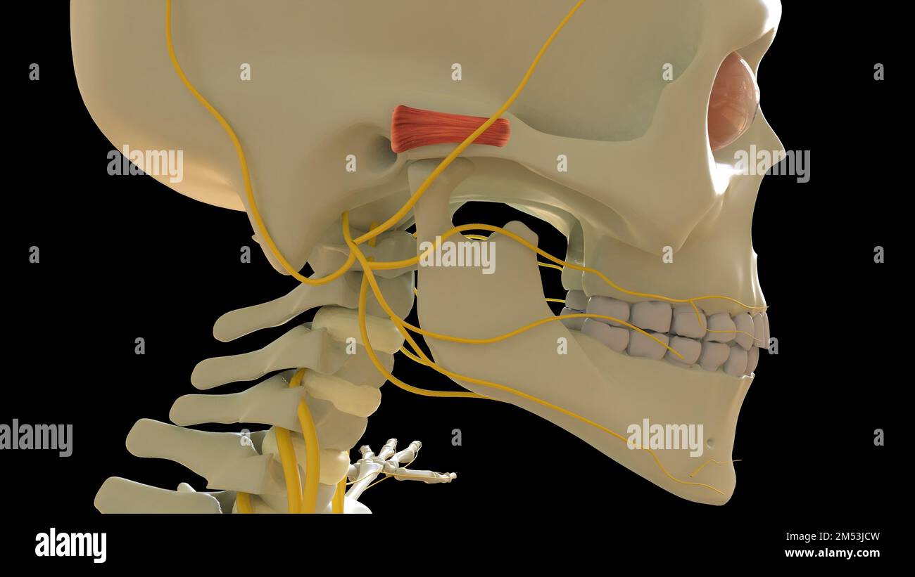 Auricular Anterior Muscle anatomy for medical concept 3D illustration Stock Photohttps://www.alamy.com/image-license-details/?v=1https://www.alamy.com/auricular-anterior-muscle-anatomy-for-medical-concept-3d-illustration-image502254281.html
RF2M53JCW–Auricular Anterior Muscle anatomy for medical concept 3D illustration
 Illustration of Facial Muscles Stock Photohttps://www.alamy.com/image-license-details/?v=1https://www.alamy.com/stock-photo-illustration-of-facial-muscles-135008264.html
RMHRJ4BM–Illustration of Facial Muscles
 . The anatomy of the domestic animals. Veterinary anatomy. THE NERVOUS SYSTEM OF THE OX 839 The facial nerve diviflcs into its two terminal branches before reaching the border of the jaw. The superior buccal nerve is the larger of the two; it crosses the masseter much lower than in the horse. The relatively small inferior buccal nerve runs beneath the parotid or in the gland substance parallel with the border of the lower jaw, crosses under the insertion of the sterno-cephalicus, and runs for- ward along the depressor labii inferioris. At the point where it crosses the facial. Fig. 666.—Superf Stock Photohttps://www.alamy.com/image-license-details/?v=1https://www.alamy.com/the-anatomy-of-the-domestic-animals-veterinary-anatomy-the-nervous-system-of-the-ox-839-the-facial-nerve-diviflcs-into-its-two-terminal-branches-before-reaching-the-border-of-the-jaw-the-superior-buccal-nerve-is-the-larger-of-the-two-it-crosses-the-masseter-much-lower-than-in-the-horse-the-relatively-small-inferior-buccal-nerve-runs-beneath-the-parotid-or-in-the-gland-substance-parallel-with-the-border-of-the-lower-jaw-crosses-under-the-insertion-of-the-sterno-cephalicus-and-runs-for-ward-along-the-depressor-labii-inferioris-at-the-point-where-it-crosses-the-facial-fig-666superf-image236793435.html
RMRN6TCB–. The anatomy of the domestic animals. Veterinary anatomy. THE NERVOUS SYSTEM OF THE OX 839 The facial nerve diviflcs into its two terminal branches before reaching the border of the jaw. The superior buccal nerve is the larger of the two; it crosses the masseter much lower than in the horse. The relatively small inferior buccal nerve runs beneath the parotid or in the gland substance parallel with the border of the lower jaw, crosses under the insertion of the sterno-cephalicus, and runs for- ward along the depressor labii inferioris. At the point where it crosses the facial. Fig. 666.—Superf
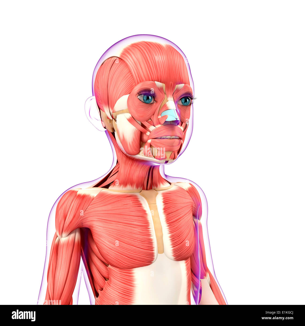 Child's muscular system computer artwork. Stock Photohttps://www.alamy.com/image-license-details/?v=1https://www.alamy.com/childs-muscular-system-computer-artwork-image69886114.html
RFE1KGCJ–Child's muscular system computer artwork.
 Platysma Muscle anatomy for medical concept 3D illustration Stock Photohttps://www.alamy.com/image-license-details/?v=1https://www.alamy.com/platysma-muscle-anatomy-for-medical-concept-3d-illustration-image502254244.html
RF2M53JBG–Platysma Muscle anatomy for medical concept 3D illustration
 Illustration of Facial Muscles Stock Photohttps://www.alamy.com/image-license-details/?v=1https://www.alamy.com/stock-photo-illustration-of-facial-muscles-135008265.html
RMHRJ4BN–Illustration of Facial Muscles
 . The anatomy of the domestic animals . Veterinary anatomy. THE NERVOUS SYSTEM OF THE OX 839 The facial nerve divides into its two terminal branches before reaching the border of the jaw. The superior buccal nerve is the larger of the two; it crosses the masseter much lower than in the horse. The relatively small inferior buccal nerve runs beneath the parotid or in the gland substance parallel with the border of the lower jaw, crosses under the insertion of the sterno-cephalicus, and runs for- ward along the depressor labii inferioris. At the point where it crosses the facial. Fig. 666.—Superf Stock Photohttps://www.alamy.com/image-license-details/?v=1https://www.alamy.com/the-anatomy-of-the-domestic-animals-veterinary-anatomy-the-nervous-system-of-the-ox-839-the-facial-nerve-divides-into-its-two-terminal-branches-before-reaching-the-border-of-the-jaw-the-superior-buccal-nerve-is-the-larger-of-the-two-it-crosses-the-masseter-much-lower-than-in-the-horse-the-relatively-small-inferior-buccal-nerve-runs-beneath-the-parotid-or-in-the-gland-substance-parallel-with-the-border-of-the-lower-jaw-crosses-under-the-insertion-of-the-sterno-cephalicus-and-runs-for-ward-along-the-depressor-labii-inferioris-at-the-point-where-it-crosses-the-facial-fig-666superf-image232322881.html
RMRDY65N–. The anatomy of the domestic animals . Veterinary anatomy. THE NERVOUS SYSTEM OF THE OX 839 The facial nerve divides into its two terminal branches before reaching the border of the jaw. The superior buccal nerve is the larger of the two; it crosses the masseter much lower than in the horse. The relatively small inferior buccal nerve runs beneath the parotid or in the gland substance parallel with the border of the lower jaw, crosses under the insertion of the sterno-cephalicus, and runs for- ward along the depressor labii inferioris. At the point where it crosses the facial. Fig. 666.—Superf
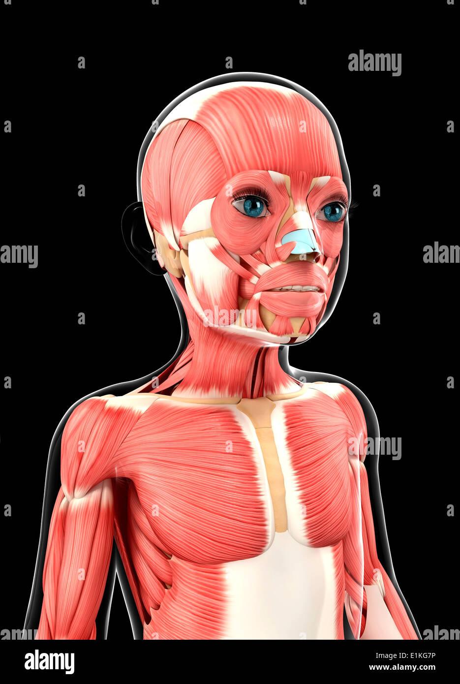 Child's muscular system computer artwork. Stock Photohttps://www.alamy.com/image-license-details/?v=1https://www.alamy.com/childs-muscular-system-computer-artwork-image69885978.html
RFE1KG7P–Child's muscular system computer artwork.
 Corrugator Supercilii Muscle anatomy for medical concept 3D illustration Stock Photohttps://www.alamy.com/image-license-details/?v=1https://www.alamy.com/corrugator-supercilii-muscle-anatomy-for-medical-concept-3d-illustration-image502254278.html
RF2M53JCP–Corrugator Supercilii Muscle anatomy for medical concept 3D illustration
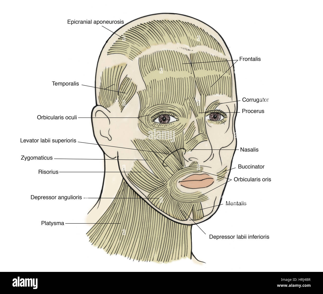 Illustration of Facial Muscles Stock Photohttps://www.alamy.com/image-license-details/?v=1https://www.alamy.com/stock-photo-illustration-of-facial-muscles-135008267.html
RMHRJ4BR–Illustration of Facial Muscles
 . The anatomy of the domestic animals. Veterinary anatomy. THE NERVOUS SYSTEM OF THE OX 839 The facial nerve divides into its two terminal branches before reaching the border of the jav. The superior buccal nerve is the larger of the two: it crosses the niasseter much lower than in the horse. The relatively small inferior buccal nerve runs beneath the parotid or in the gland substance parallel with the border of the lower jaw, crosses under the insertion of the sterno-cephalicus, and runs for- ward along the depressor labii inferioris. At the point where it crosses the facial. Fig. 666.—Sttpe Stock Photohttps://www.alamy.com/image-license-details/?v=1https://www.alamy.com/the-anatomy-of-the-domestic-animals-veterinary-anatomy-the-nervous-system-of-the-ox-839-the-facial-nerve-divides-into-its-two-terminal-branches-before-reaching-the-border-of-the-jav-the-superior-buccal-nerve-is-the-larger-of-the-two-it-crosses-the-niasseter-much-lower-than-in-the-horse-the-relatively-small-inferior-buccal-nerve-runs-beneath-the-parotid-or-in-the-gland-substance-parallel-with-the-border-of-the-lower-jaw-crosses-under-the-insertion-of-the-sterno-cephalicus-and-runs-for-ward-along-the-depressor-labii-inferioris-at-the-point-where-it-crosses-the-facial-fig-666sttpe-image236773305.html
RMRN5XND–. The anatomy of the domestic animals. Veterinary anatomy. THE NERVOUS SYSTEM OF THE OX 839 The facial nerve divides into its two terminal branches before reaching the border of the jav. The superior buccal nerve is the larger of the two: it crosses the niasseter much lower than in the horse. The relatively small inferior buccal nerve runs beneath the parotid or in the gland substance parallel with the border of the lower jaw, crosses under the insertion of the sterno-cephalicus, and runs for- ward along the depressor labii inferioris. At the point where it crosses the facial. Fig. 666.—Sttpe
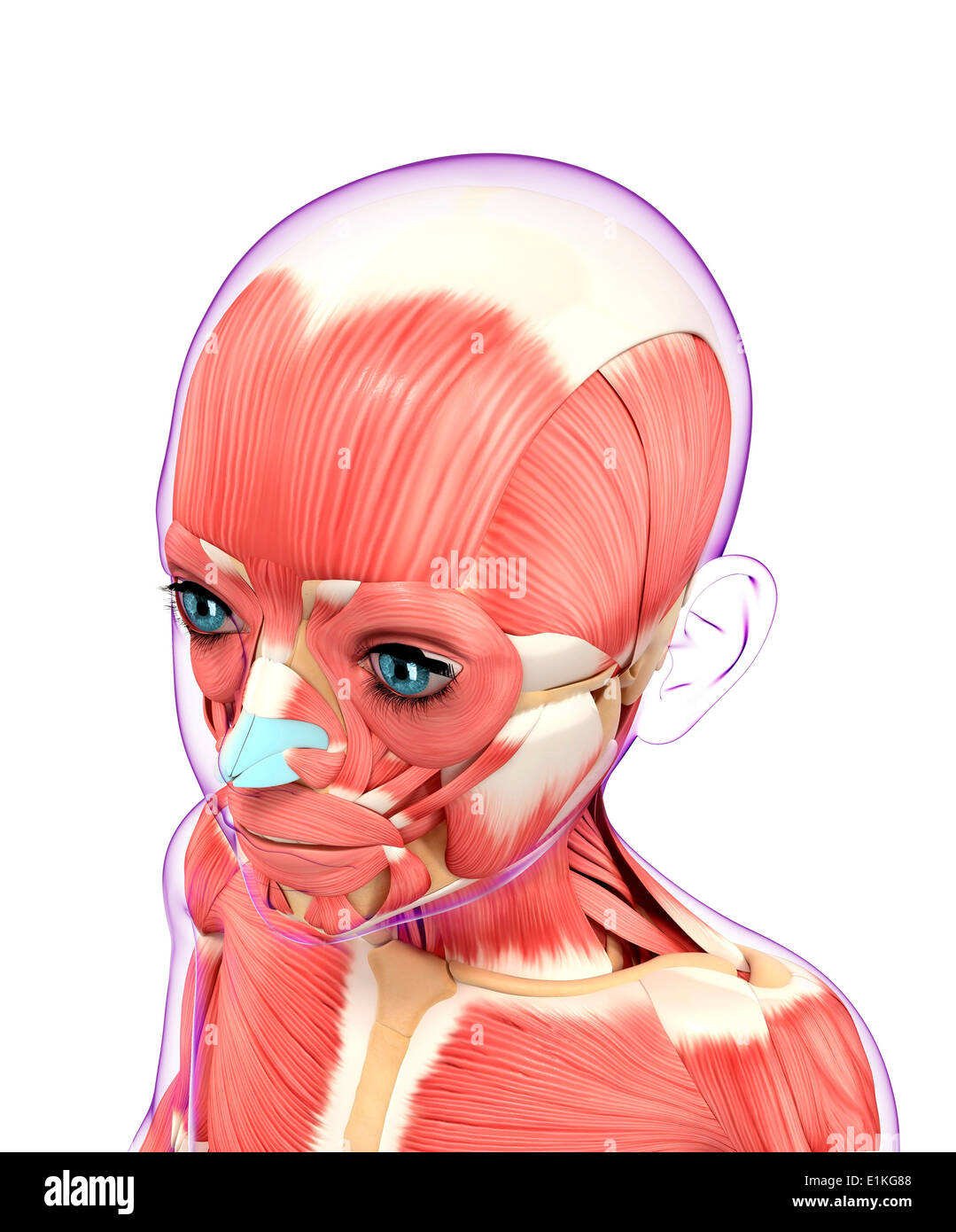 Muscular system computer artwork. Stock Photohttps://www.alamy.com/image-license-details/?v=1https://www.alamy.com/muscular-system-computer-artwork-image69885992.html
RFE1KG88–Muscular system computer artwork.
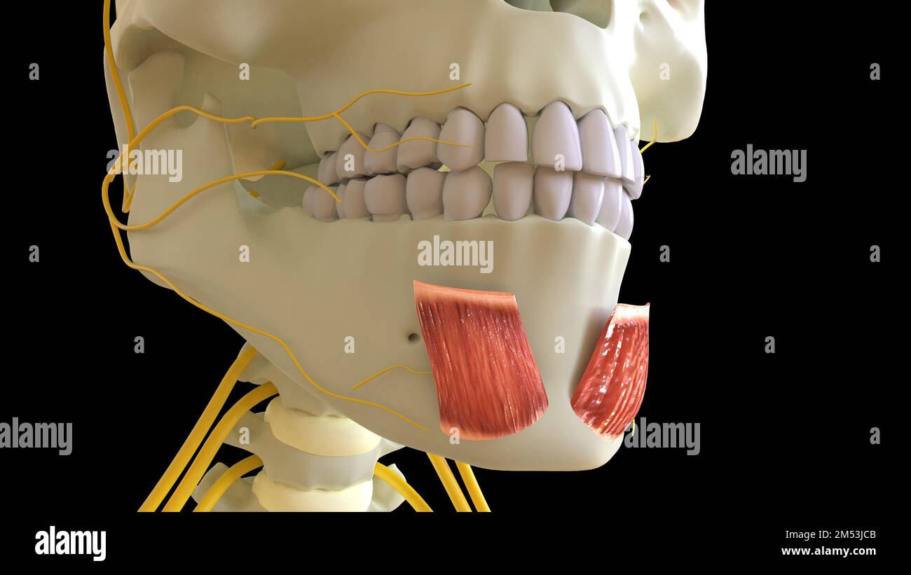 Mentalis Muscle anatomy for medical concept 3D illustration Stock Photohttps://www.alamy.com/image-license-details/?v=1https://www.alamy.com/mentalis-muscle-anatomy-for-medical-concept-3d-illustration-image502254267.html
RF2M53JCB–Mentalis Muscle anatomy for medical concept 3D illustration
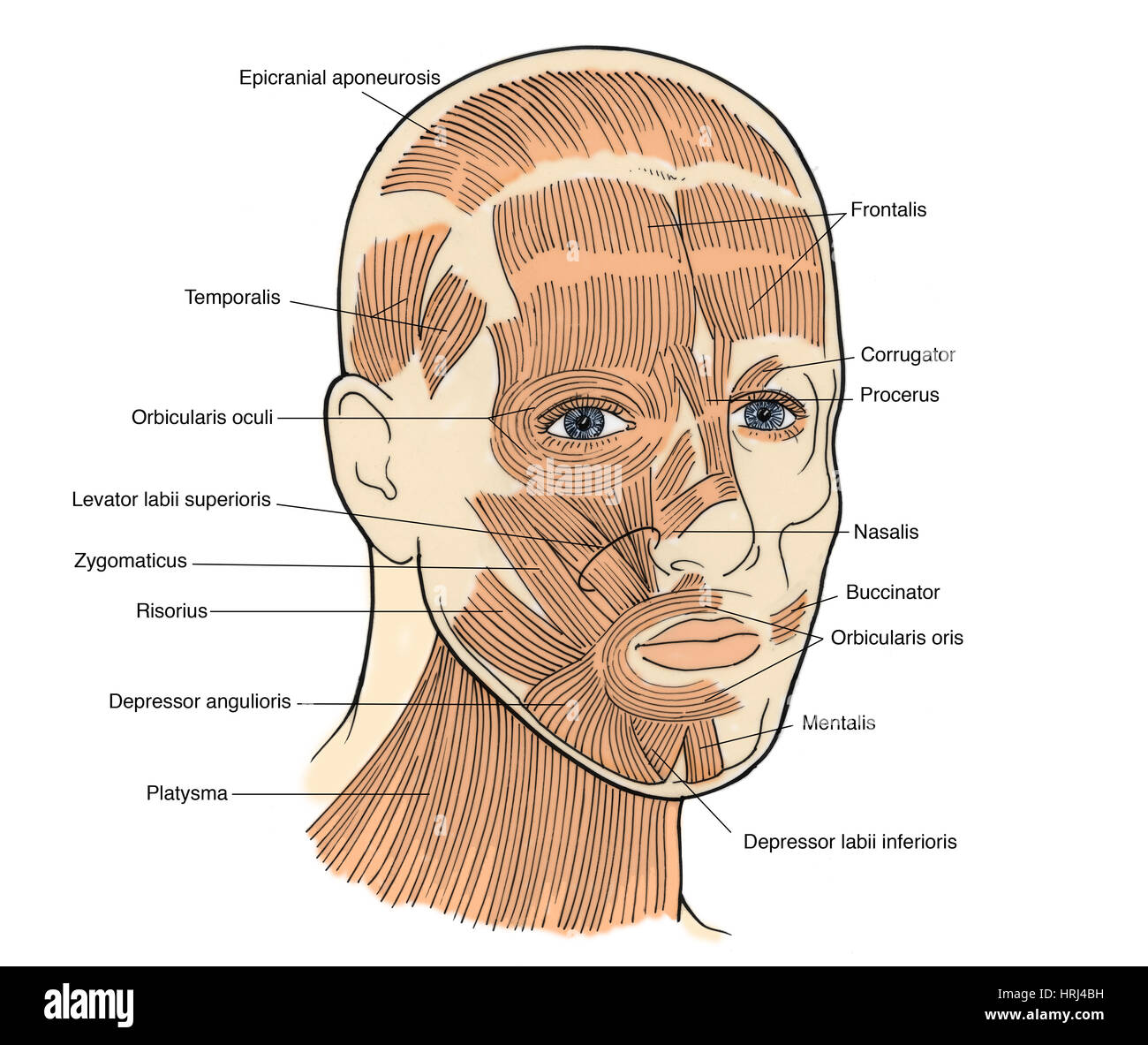 Illustration of Facial Muscles Stock Photohttps://www.alamy.com/image-license-details/?v=1https://www.alamy.com/stock-photo-illustration-of-facial-muscles-135008261.html
RMHRJ4BH–Illustration of Facial Muscles
 . The anatomy of the domestic animals . Veterinary anatomy. THE COMMON CAROTID ARTERY 709 carotid; it gives off a branch to the mandibular gland, and the sublingual artery. After turning around the j aw the facial gives off the two labial arteries. The inferior labial artery is small; it runs forward along the ventral margin of the depressor labii inferioris. The superior labial is large; it passes forward ventral to the depressor labii superioris, and usually gives off a muscular branch which runs for- ward almost parallel with the lateral nasal. The angular artery is very small or absent, an Stock Photohttps://www.alamy.com/image-license-details/?v=1https://www.alamy.com/the-anatomy-of-the-domestic-animals-veterinary-anatomy-the-common-carotid-artery-709-carotid-it-gives-off-a-branch-to-the-mandibular-gland-and-the-sublingual-artery-after-turning-around-the-j-aw-the-facial-gives-off-the-two-labial-arteries-the-inferior-labial-artery-is-small-it-runs-forward-along-the-ventral-margin-of-the-depressor-labii-inferioris-the-superior-labial-is-large-it-passes-forward-ventral-to-the-depressor-labii-superioris-and-usually-gives-off-a-muscular-branch-which-runs-for-ward-almost-parallel-with-the-lateral-nasal-the-angular-artery-is-very-small-or-absent-an-image232323368.html
RMRDY6R4–. The anatomy of the domestic animals . Veterinary anatomy. THE COMMON CAROTID ARTERY 709 carotid; it gives off a branch to the mandibular gland, and the sublingual artery. After turning around the j aw the facial gives off the two labial arteries. The inferior labial artery is small; it runs forward along the ventral margin of the depressor labii inferioris. The superior labial is large; it passes forward ventral to the depressor labii superioris, and usually gives off a muscular branch which runs for- ward almost parallel with the lateral nasal. The angular artery is very small or absent, an
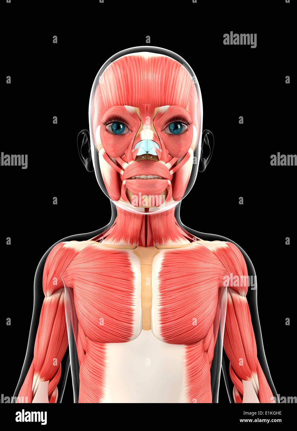 Muscular system computer artwork. Stock Photohttps://www.alamy.com/image-license-details/?v=1https://www.alamy.com/muscular-system-computer-artwork-image69886250.html
RFE1KGHE–Muscular system computer artwork.
 Sternocleidomastoid Muscle anatomy for medical concept 3D illustration Stock Photohttps://www.alamy.com/image-license-details/?v=1https://www.alamy.com/sternocleidomastoid-muscle-anatomy-for-medical-concept-3d-illustration-image502254215.html
RF2M53JAF–Sternocleidomastoid Muscle anatomy for medical concept 3D illustration
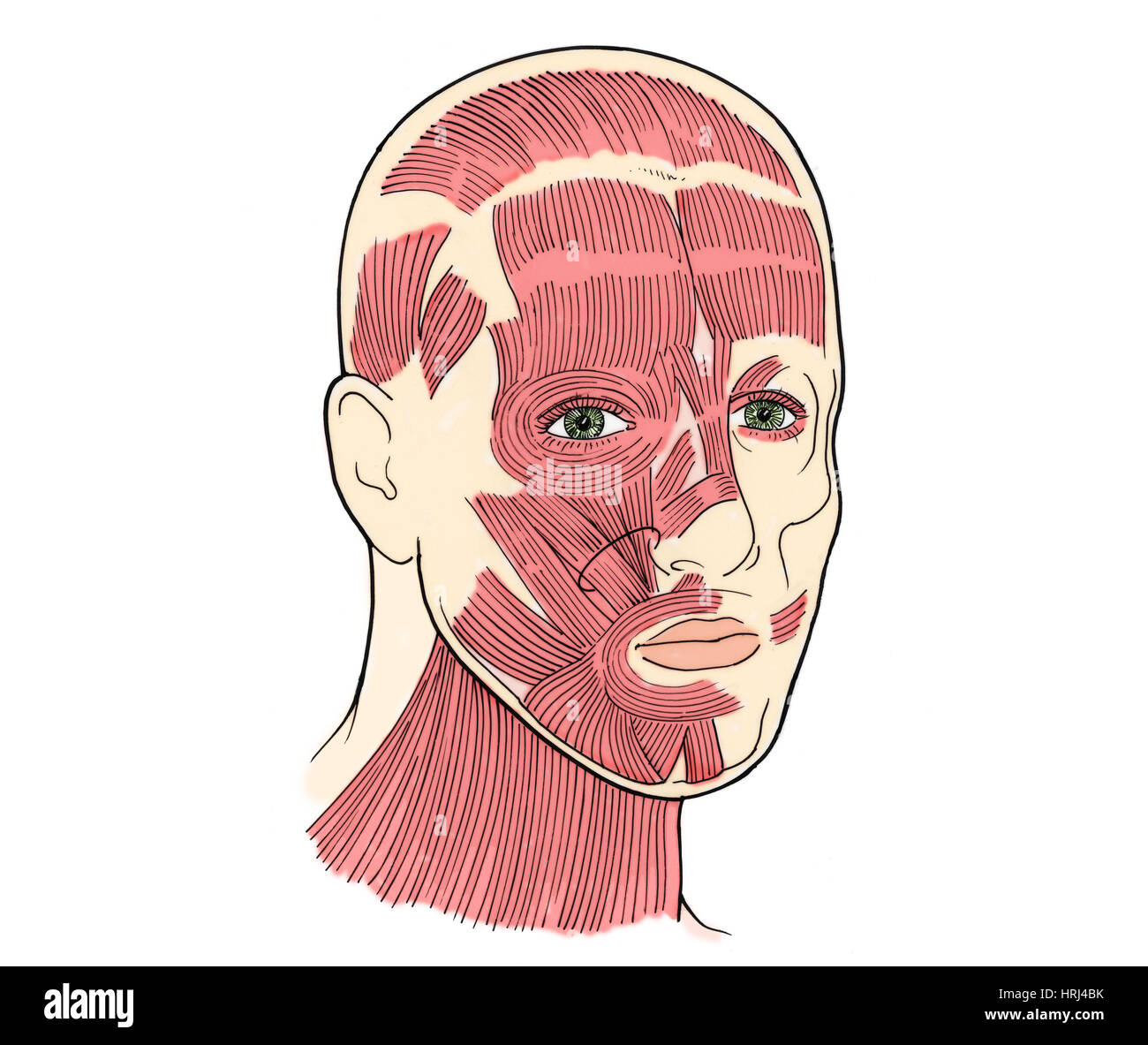 Illustration of Facial Muscles Stock Photohttps://www.alamy.com/image-license-details/?v=1https://www.alamy.com/stock-photo-illustration-of-facial-muscles-135008263.html
RMHRJ4BK–Illustration of Facial Muscles
 . The anatomy of the domestic animals. Veterinary anatomy. THE EXTERNAL CAROTID ARTERY 643 A considerable branch may be given off in the mandibular space, which turns around the lower border of the jaw and enters the middle of the lower part of the masseter muscle. In some cases this artery is of large size and its pulsation can be felt. It is accompanieil by a vein. (4) The inferior labial artery (A. labialis inferior) arises from the external maxillary a little before it reaches the depressor labii inferioris (Fig. 560). It passes forvs'ard, dips under the depressor muscle, and continues to Stock Photohttps://www.alamy.com/image-license-details/?v=1https://www.alamy.com/the-anatomy-of-the-domestic-animals-veterinary-anatomy-the-external-carotid-artery-643-a-considerable-branch-may-be-given-off-in-the-mandibular-space-which-turns-around-the-lower-border-of-the-jaw-and-enters-the-middle-of-the-lower-part-of-the-masseter-muscle-in-some-cases-this-artery-is-of-large-size-and-its-pulsation-can-be-felt-it-is-accompanieil-by-a-vein-4-the-inferior-labial-artery-a-labialis-inferior-arises-from-the-external-maxillary-a-little-before-it-reaches-the-depressor-labii-inferioris-fig-560-it-passes-forvsard-dips-under-the-depressor-muscle-and-continues-to-image236759707.html
RMRN59BR–. The anatomy of the domestic animals. Veterinary anatomy. THE EXTERNAL CAROTID ARTERY 643 A considerable branch may be given off in the mandibular space, which turns around the lower border of the jaw and enters the middle of the lower part of the masseter muscle. In some cases this artery is of large size and its pulsation can be felt. It is accompanieil by a vein. (4) The inferior labial artery (A. labialis inferior) arises from the external maxillary a little before it reaches the depressor labii inferioris (Fig. 560). It passes forvs'ard, dips under the depressor muscle, and continues to
 Muscular system computer artwork. Stock Photohttps://www.alamy.com/image-license-details/?v=1https://www.alamy.com/muscular-system-computer-artwork-image69886227.html
RFE1KGGK–Muscular system computer artwork.
 Depressor labii inferioris muscle, illustration Stock Photohttps://www.alamy.com/image-license-details/?v=1https://www.alamy.com/depressor-labii-inferioris-muscle-illustration-image328920999.html
RF2A33J2F–Depressor labii inferioris muscle, illustration
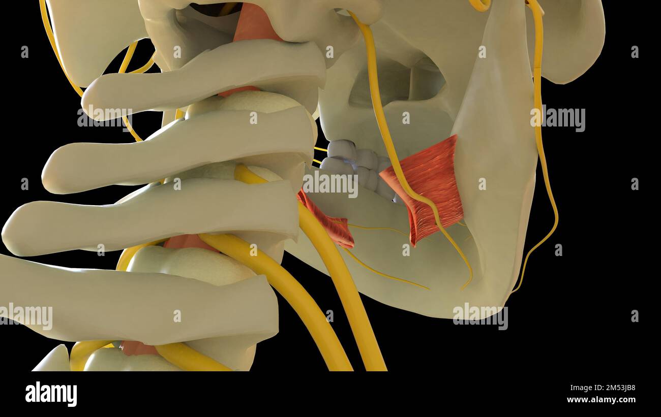 Hyoglossus Muscle anatomy for medical concept 3D illustration Stock Photohttps://www.alamy.com/image-license-details/?v=1https://www.alamy.com/hyoglossus-muscle-anatomy-for-medical-concept-3d-illustration-image502254236.html
RF2M53JB8–Hyoglossus Muscle anatomy for medical concept 3D illustration
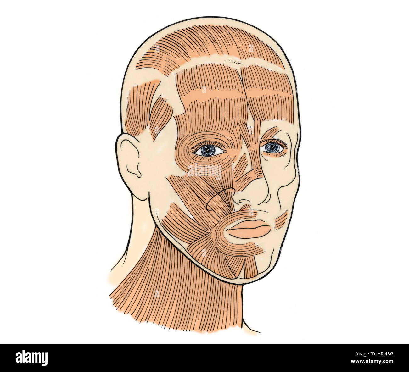 Illustration of Facial Muscles Stock Photohttps://www.alamy.com/image-license-details/?v=1https://www.alamy.com/stock-photo-illustration-of-facial-muscles-135008260.html
RMHRJ4BG–Illustration of Facial Muscles
 . The anatomy of the domestic animals . Veterinary anatomy. THE EXTERNAL CAROTID ARTERY 643 A considerable branch may be given off m the mandibular space, which turns around the lower border of the jaw and enters the middle of the lower part of the masseter muscle. In some cases this artery is of large size and its pulsation can be felt. It is accompanied by a vein. (4) The inferior labial artery (A. labialis inferior) arises from the external maxillary a little before it reaches the depressor labii inferioris (Fig. 560). It passes forward, dips under the depressor muscle, and continues to the Stock Photohttps://www.alamy.com/image-license-details/?v=1https://www.alamy.com/the-anatomy-of-the-domestic-animals-veterinary-anatomy-the-external-carotid-artery-643-a-considerable-branch-may-be-given-off-m-the-mandibular-space-which-turns-around-the-lower-border-of-the-jaw-and-enters-the-middle-of-the-lower-part-of-the-masseter-muscle-in-some-cases-this-artery-is-of-large-size-and-its-pulsation-can-be-felt-it-is-accompanied-by-a-vein-4-the-inferior-labial-artery-a-labialis-inferior-arises-from-the-external-maxillary-a-little-before-it-reaches-the-depressor-labii-inferioris-fig-560-it-passes-forward-dips-under-the-depressor-muscle-and-continues-to-the-image232323702.html
RMRDY772–. The anatomy of the domestic animals . Veterinary anatomy. THE EXTERNAL CAROTID ARTERY 643 A considerable branch may be given off m the mandibular space, which turns around the lower border of the jaw and enters the middle of the lower part of the masseter muscle. In some cases this artery is of large size and its pulsation can be felt. It is accompanied by a vein. (4) The inferior labial artery (A. labialis inferior) arises from the external maxillary a little before it reaches the depressor labii inferioris (Fig. 560). It passes forward, dips under the depressor muscle, and continues to the
 Depressor labii inferioris muscle, illustration Stock Photohttps://www.alamy.com/image-license-details/?v=1https://www.alamy.com/depressor-labii-inferioris-muscle-illustration-image328920997.html
RF2A33J2D–Depressor labii inferioris muscle, illustration
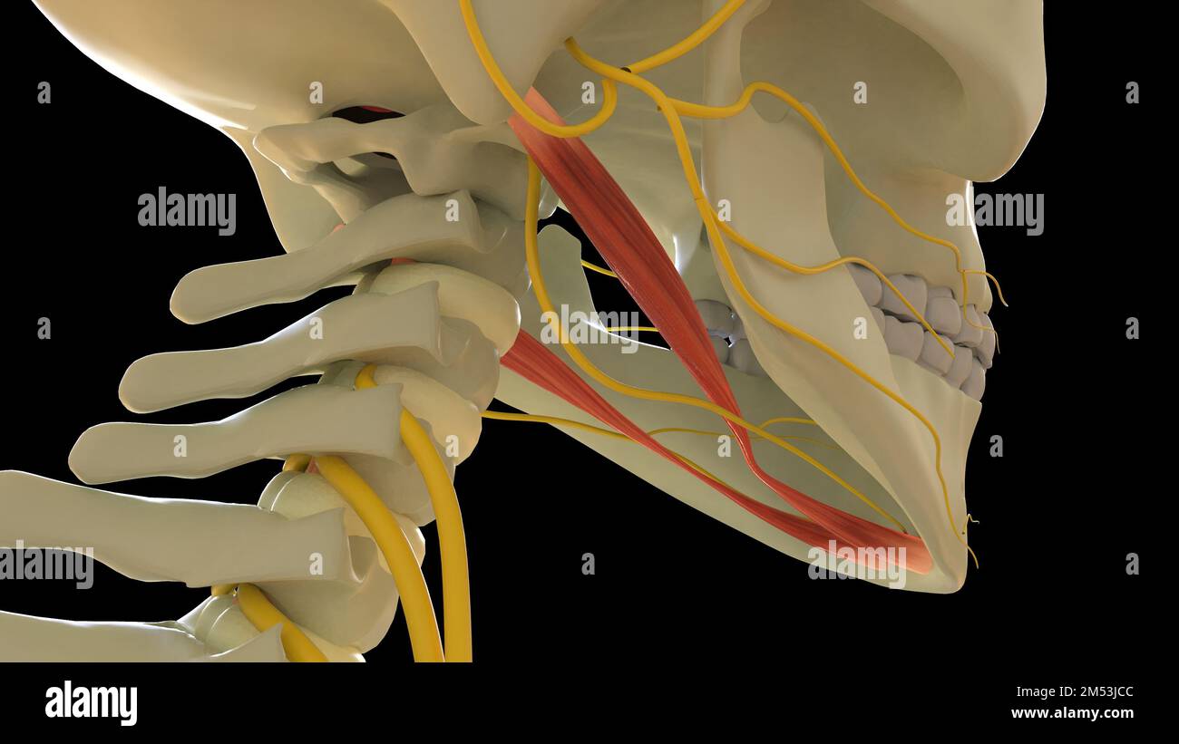 Digastric Muscle anatomy for medical concept 3D illustration Stock Photohttps://www.alamy.com/image-license-details/?v=1https://www.alamy.com/digastric-muscle-anatomy-for-medical-concept-3d-illustration-image502254268.html
RF2M53JCC–Digastric Muscle anatomy for medical concept 3D illustration
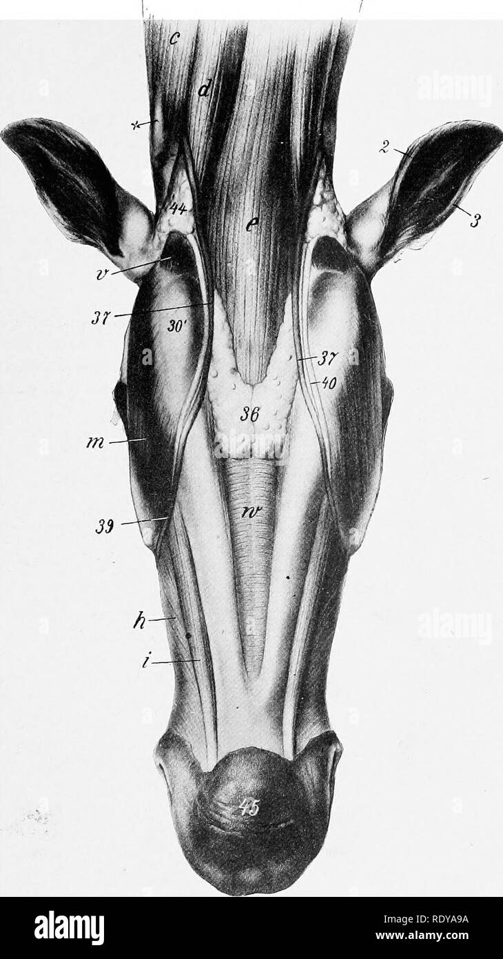 . The anatomy of the domestic animals . Veterinary anatomy. MANDIBULAR MUSCLES 263 muscle extending from the paramastoid process of the occipital bone to the posterior border of the lower jaw; it is covered by the parotid gland.. Fig. 265.—Mandibular anb Laryngeal Regions of Horse, after Kemoval of Skin and Cutaneus. c, Brachiocephalicus: d, sterno-cephalicus; e, omo-hyoideus and sterno-hyoideus; h, buccinator; i, depressor labii inferioris: m, masseter; v, occipito-mandibularis; w, mylo-hyoideus; 3, posterior, 3, anterior, border of external ear; SO', angle of jaw; 36, mandibular lymph-glands Stock Photohttps://www.alamy.com/image-license-details/?v=1https://www.alamy.com/the-anatomy-of-the-domestic-animals-veterinary-anatomy-mandibular-muscles-263-muscle-extending-from-the-paramastoid-process-of-the-occipital-bone-to-the-posterior-border-of-the-lower-jaw-it-is-covered-by-the-parotid-gland-fig-265mandibular-anb-laryngeal-regions-of-horse-after-kemoval-of-skin-and-cutaneus-c-brachiocephalicus-d-sterno-cephalicus-e-omo-hyoideus-and-sterno-hyoideus-h-buccinator-i-depressor-labii-inferioris-m-masseter-v-occipito-mandibularis-w-mylo-hyoideus-3-posterior-3-anterior-border-of-external-ear-so-angle-of-jaw-36-mandibular-lymph-glands-image232326118.html
RMRDYA9A–. The anatomy of the domestic animals . Veterinary anatomy. MANDIBULAR MUSCLES 263 muscle extending from the paramastoid process of the occipital bone to the posterior border of the lower jaw; it is covered by the parotid gland.. Fig. 265.—Mandibular anb Laryngeal Regions of Horse, after Kemoval of Skin and Cutaneus. c, Brachiocephalicus: d, sterno-cephalicus; e, omo-hyoideus and sterno-hyoideus; h, buccinator; i, depressor labii inferioris: m, masseter; v, occipito-mandibularis; w, mylo-hyoideus; 3, posterior, 3, anterior, border of external ear; SO', angle of jaw; 36, mandibular lymph-glands
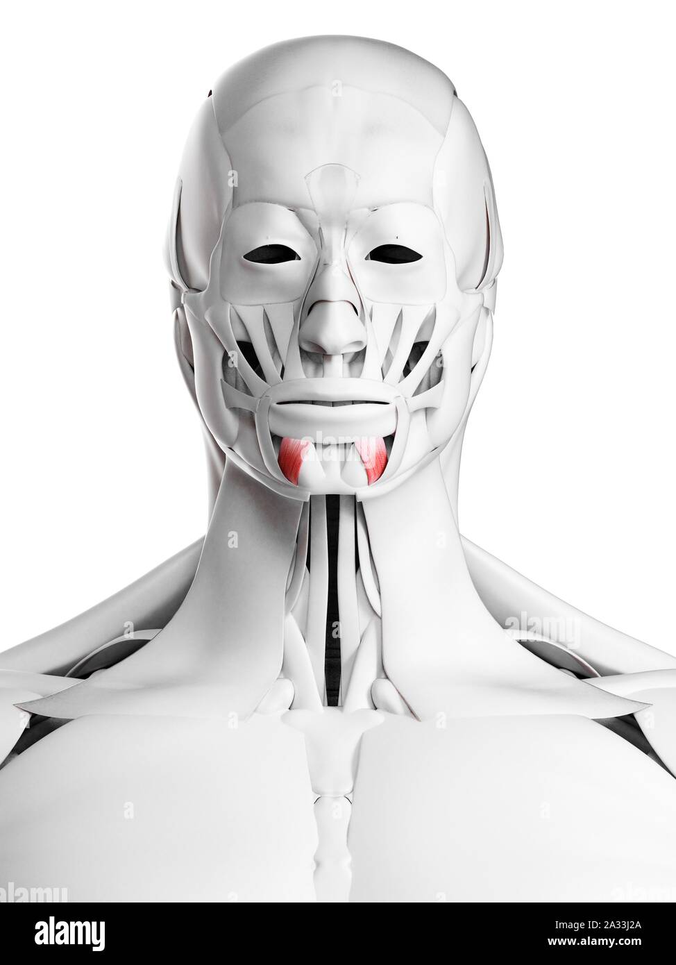 Depressor labii inferioris muscle, illustration Stock Photohttps://www.alamy.com/image-license-details/?v=1https://www.alamy.com/depressor-labii-inferioris-muscle-illustration-image328920994.html
RF2A33J2A–Depressor labii inferioris muscle, illustration
 Human facial muscles, illustration. Stock Photohttps://www.alamy.com/image-license-details/?v=1https://www.alamy.com/stock-photo-human-facial-muscles-illustration-111973562.html
RFGE4RBP–Human facial muscles, illustration.
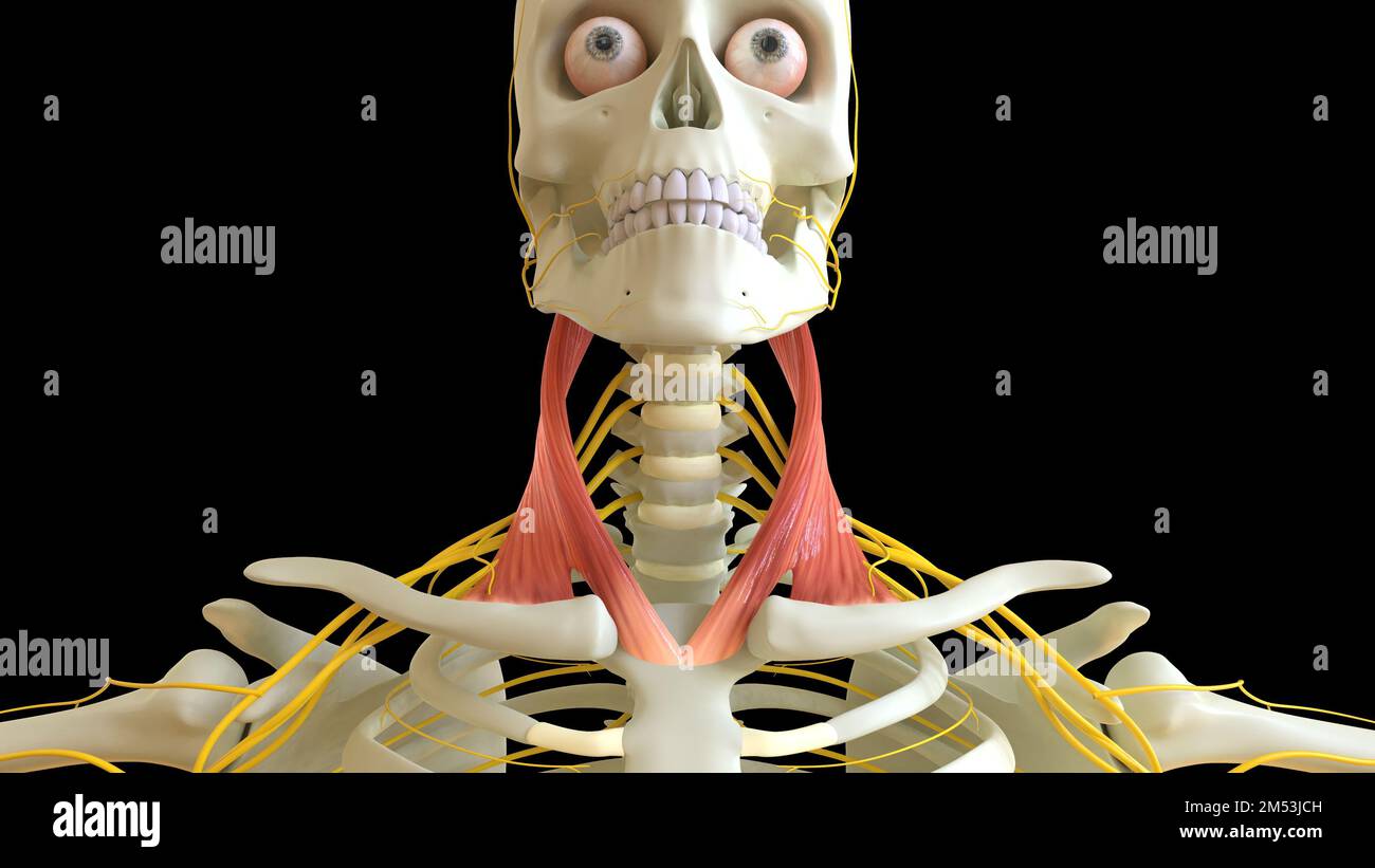 Sternocleidomastoid Muscle anatomy for medical concept 3D illustration Stock Photohttps://www.alamy.com/image-license-details/?v=1https://www.alamy.com/sternocleidomastoid-muscle-anatomy-for-medical-concept-3d-illustration-image502254273.html
RF2M53JCH–Sternocleidomastoid Muscle anatomy for medical concept 3D illustration