Dermis micrograph Stock Photos and Images
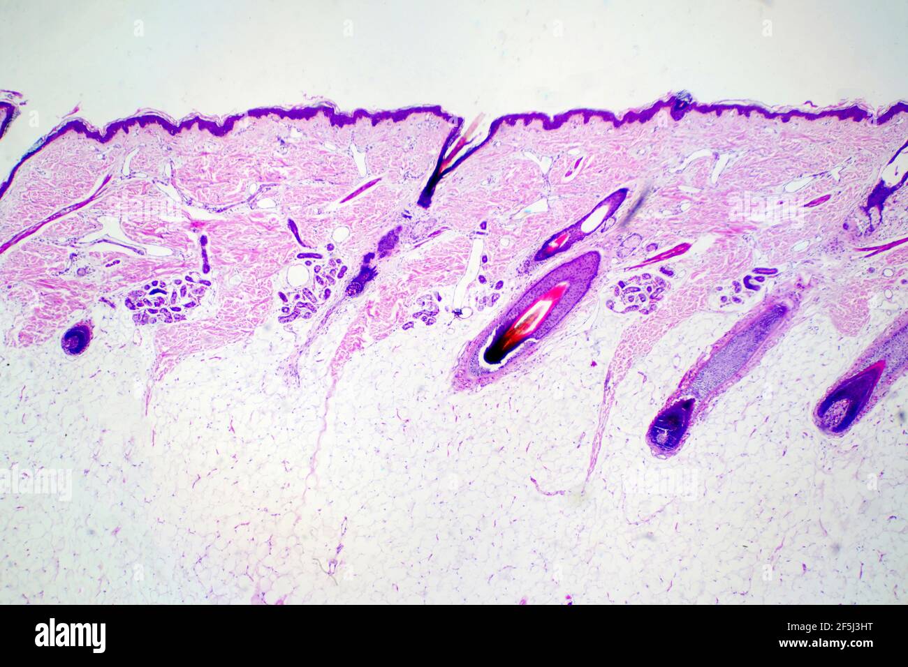 Human scalp, light micrograph Stock Photohttps://www.alamy.com/image-license-details/?v=1https://www.alamy.com/human-scalp-light-micrograph-image416520100.html
RF2F5J3HT–Human scalp, light micrograph
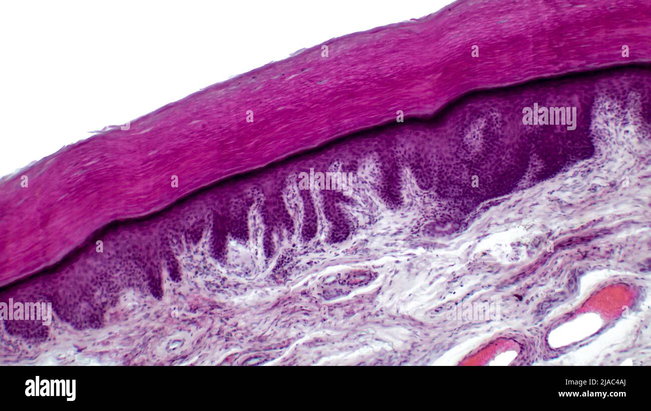 Light micrograph of epithelial tissue from the skin. Human finger section showing epidermis, dermis and connective tissues. Stock Photohttps://www.alamy.com/image-license-details/?v=1https://www.alamy.com/light-micrograph-of-epithelial-tissue-from-the-skin-human-finger-section-showing-epidermis-dermis-and-connective-tissues-image471093354.html
RF2JAC4AJ–Light micrograph of epithelial tissue from the skin. Human finger section showing epidermis, dermis and connective tissues.
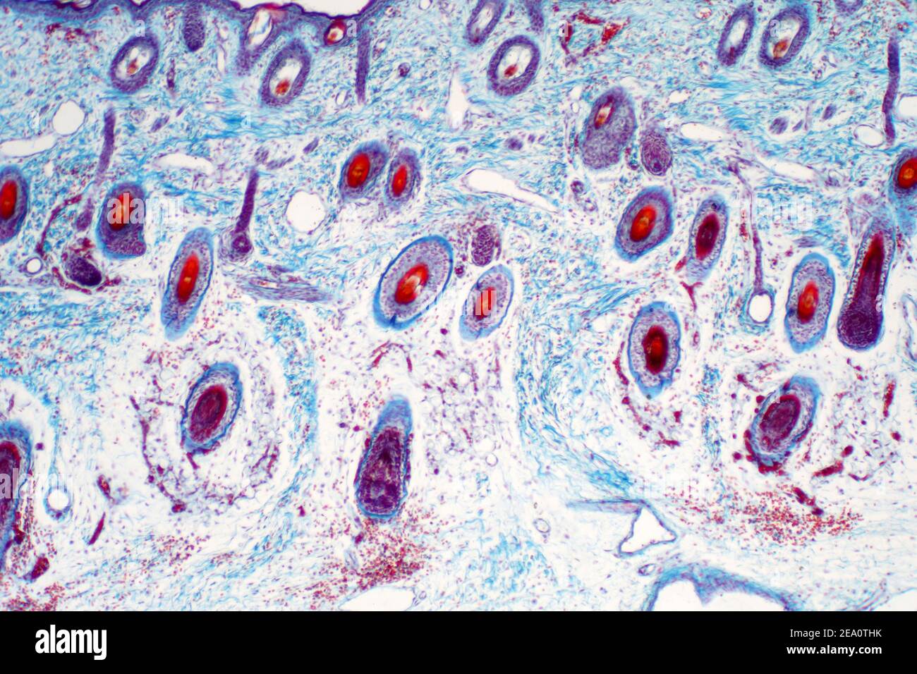 Human skin, light micrograph Stock Photohttps://www.alamy.com/image-license-details/?v=1https://www.alamy.com/human-skin-light-micrograph-image402004335.html
RF2EA0THK–Human skin, light micrograph
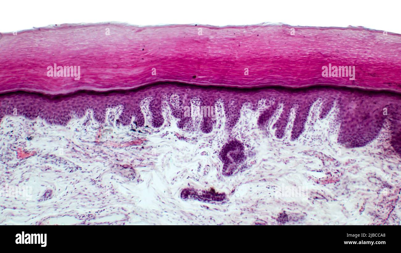 Skin. Light micrograph of epithelial tissue from the skin. Human finger section showing epidermis, dermis and connective tissues.Hematoxylin and eosin Stock Photohttps://www.alamy.com/image-license-details/?v=1https://www.alamy.com/skin-light-micrograph-of-epithelial-tissue-from-the-skin-human-finger-section-showing-epidermis-dermis-and-connective-tissueshematoxylin-and-eosin-image471714272.html
RF2JBCCA8–Skin. Light micrograph of epithelial tissue from the skin. Human finger section showing epidermis, dermis and connective tissues.Hematoxylin and eosin
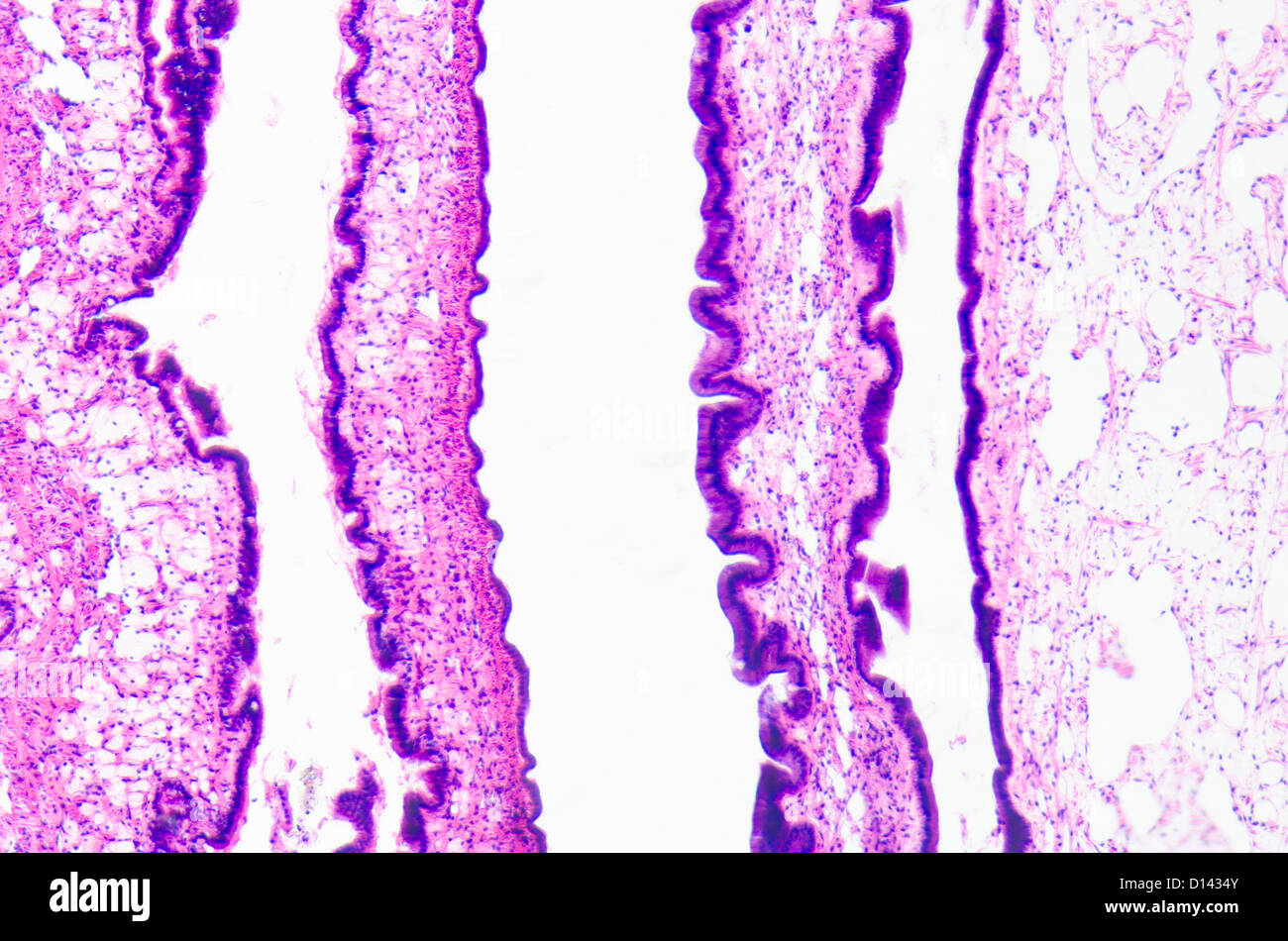 micrograph of medical science cilliated epithelium tissue cell Stock Photohttps://www.alamy.com/image-license-details/?v=1https://www.alamy.com/stock-photo-micrograph-of-medical-science-cilliated-epithelium-tissue-cell-52336059.html
RFD1434Y–micrograph of medical science cilliated epithelium tissue cell
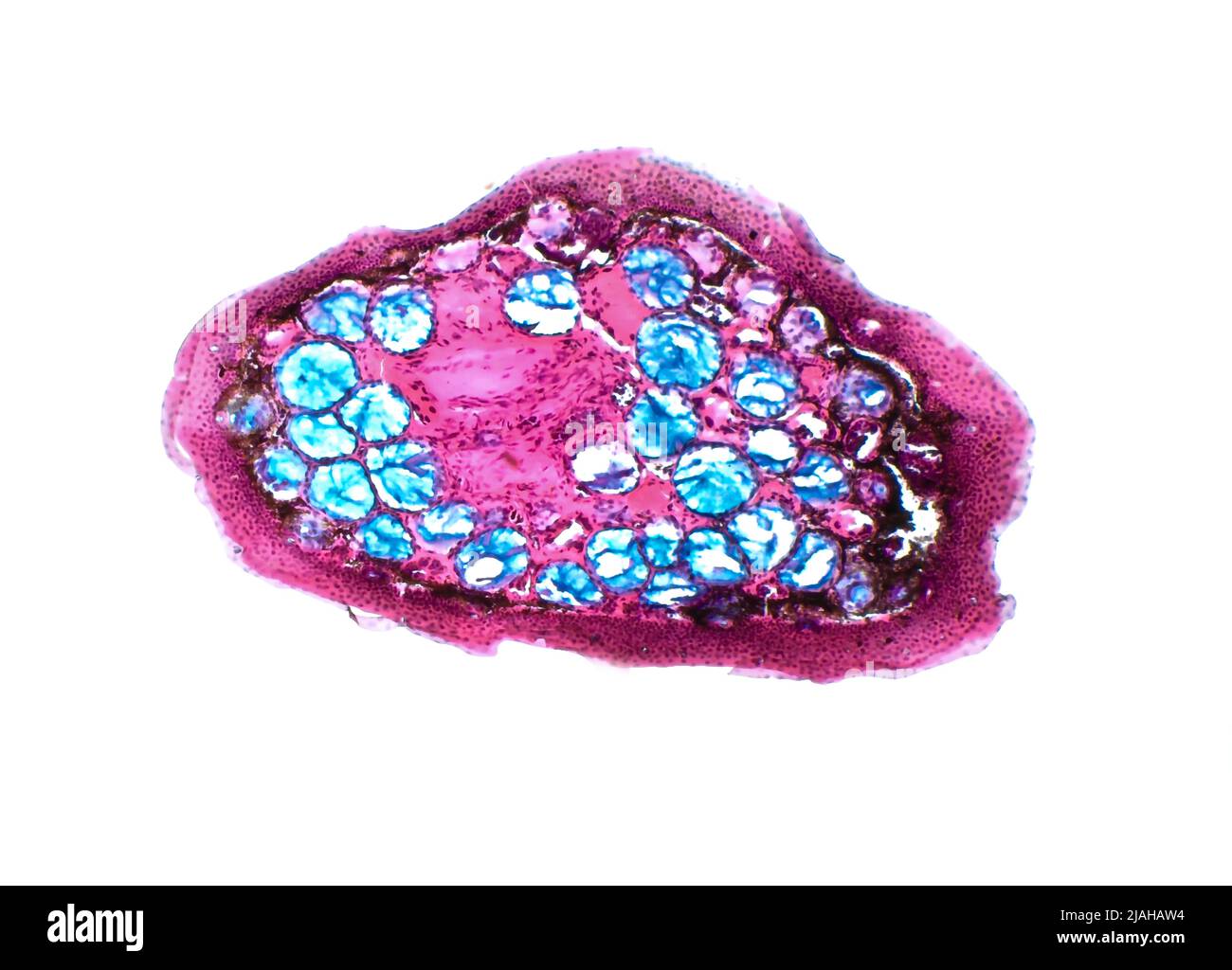 Frog skin glands. Light micrograph of a section through the skin of an marsh frog. The large round (blue) structures are poison glands. Stock Photohttps://www.alamy.com/image-license-details/?v=1https://www.alamy.com/frog-skin-glands-light-micrograph-of-a-section-through-the-skin-of-an-marsh-frog-the-large-round-blue-structures-are-poison-glands-image471208224.html
RF2JAHAW4–Frog skin glands. Light micrograph of a section through the skin of an marsh frog. The large round (blue) structures are poison glands.
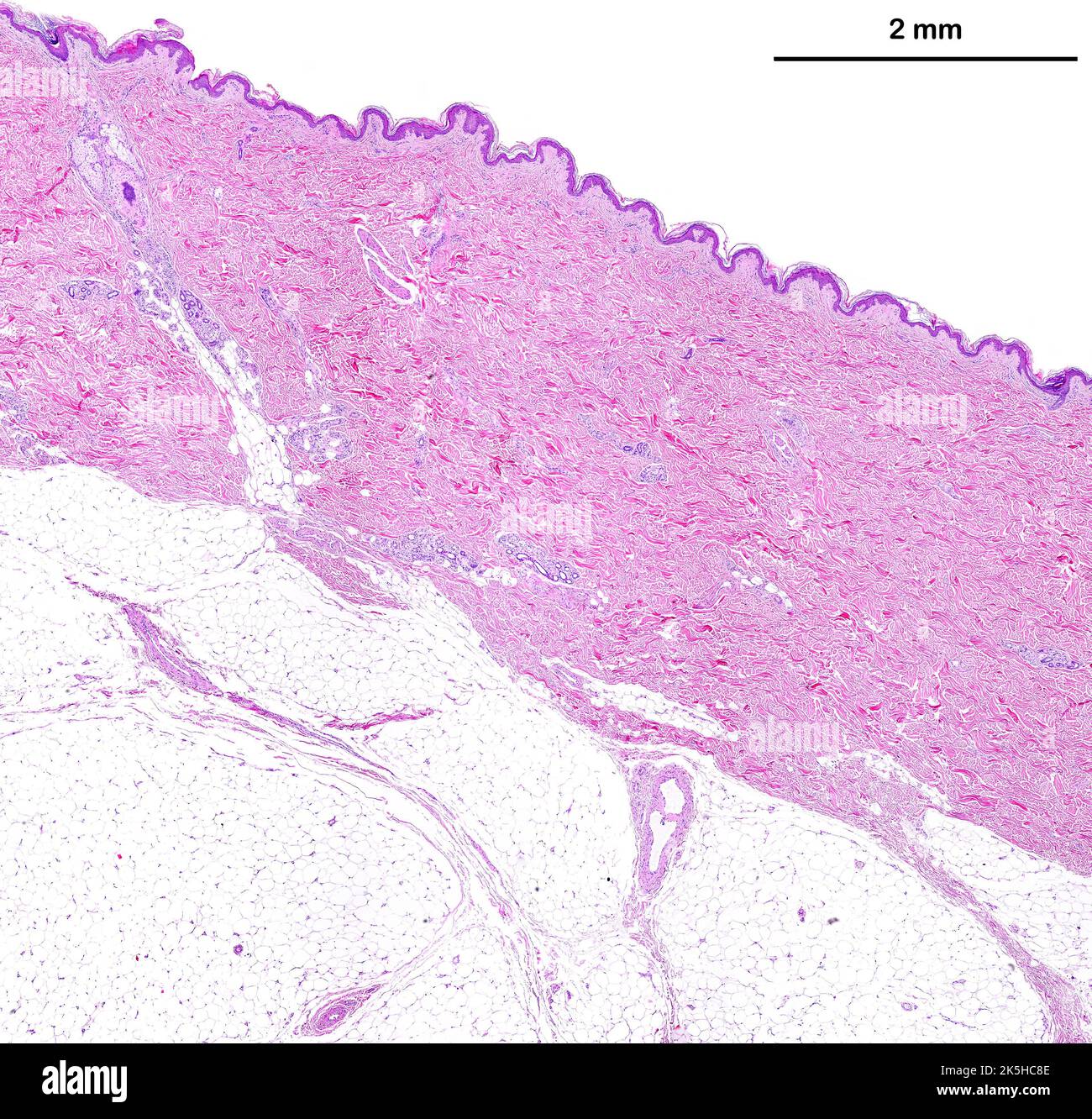 Low power light microscope micrograph of thin skin showing from top to bottom, the epidermis, a very thick dermis showing sweat glands, and the adipos Stock Photohttps://www.alamy.com/image-license-details/?v=1https://www.alamy.com/low-power-light-microscope-micrograph-of-thin-skin-showing-from-top-to-bottom-the-epidermis-a-very-thick-dermis-showing-sweat-glands-and-the-adipos-image485346414.html
RF2K5HC8E–Low power light microscope micrograph of thin skin showing from top to bottom, the epidermis, a very thick dermis showing sweat glands, and the adipos
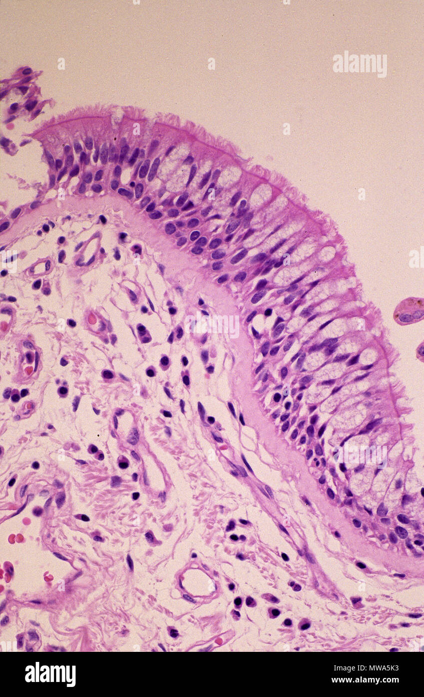 Epithelium tissue of trachea.Tracheal epithelium Stock Photohttps://www.alamy.com/image-license-details/?v=1https://www.alamy.com/epithelium-tissue-of-tracheatracheal-epithelium-image187694055.html
RFMWA5K3–Epithelium tissue of trachea.Tracheal epithelium
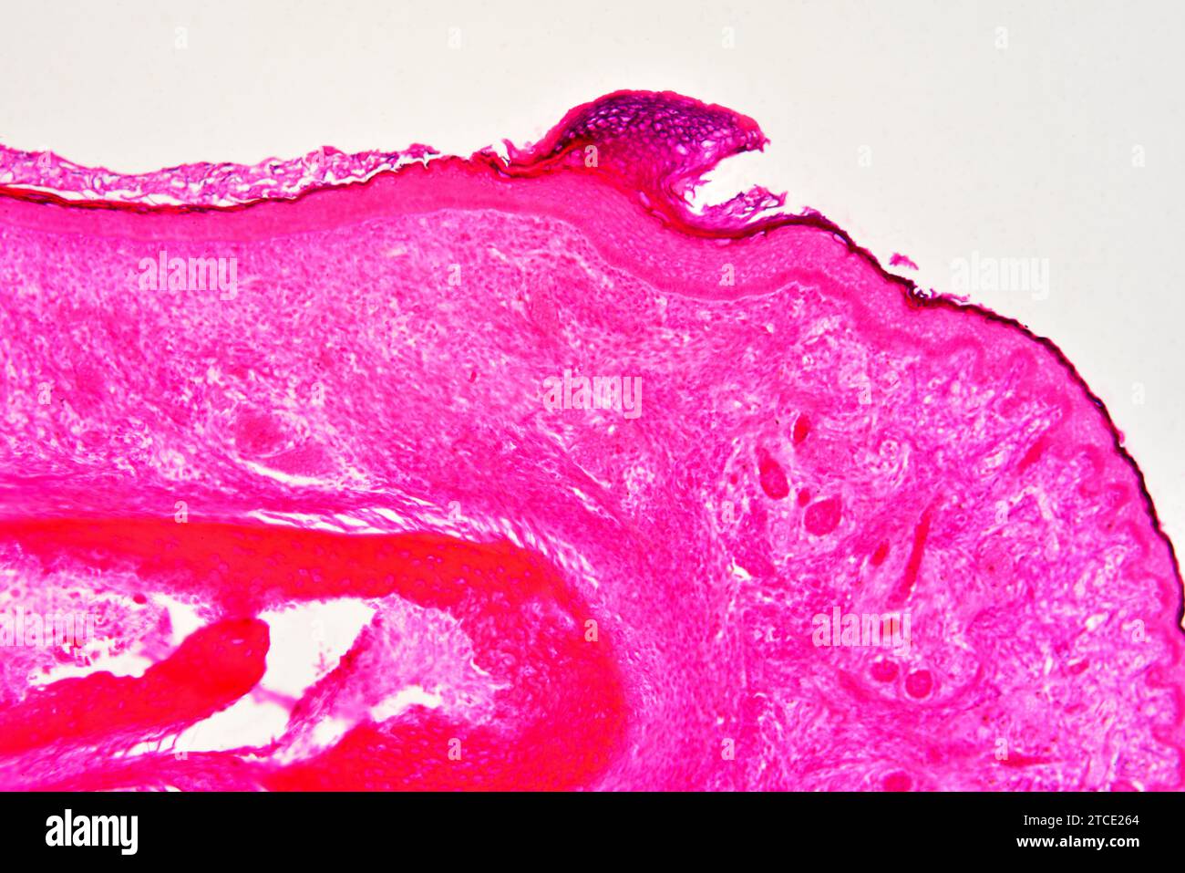 Human fetus finger showing nail, epidermis, dermis and bone. Optical microscope X100. Stock Photohttps://www.alamy.com/image-license-details/?v=1https://www.alamy.com/human-fetus-finger-showing-nail-epidermis-dermis-and-bone-optical-microscope-x100-image575627084.html
RF2TCE264–Human fetus finger showing nail, epidermis, dermis and bone. Optical microscope X100.
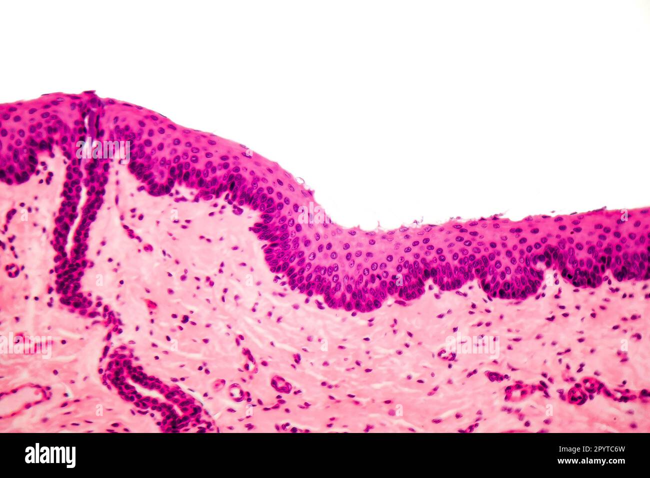 Human stratified squamous epithelium under microscope, light micrograph Stock Photohttps://www.alamy.com/image-license-details/?v=1https://www.alamy.com/human-stratified-squamous-epithelium-under-microscope-light-micrograph-image550653569.html
RF2PYTC6W–Human stratified squamous epithelium under microscope, light micrograph
 Human finger section showing epidermis (stratified squamous epithelium), dermis and connective tissues. Photomicrograph. Stock Photohttps://www.alamy.com/image-license-details/?v=1https://www.alamy.com/human-finger-section-showing-epidermis-stratified-squamous-epithelium-dermis-and-connective-tissues-photomicrograph-image578271985.html
RF2TGPFPW–Human finger section showing epidermis (stratified squamous epithelium), dermis and connective tissues. Photomicrograph.
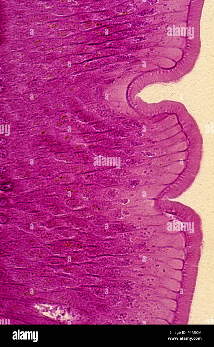 Epithelial cells of intestine of Ascaris. 140x Stock Photohttps://www.alamy.com/image-license-details/?v=1https://www.alamy.com/epithelial-cells-of-intestine-of-ascaris-140x-image209506324.html
RFP4RRCM–Epithelial cells of intestine of Ascaris. 140x
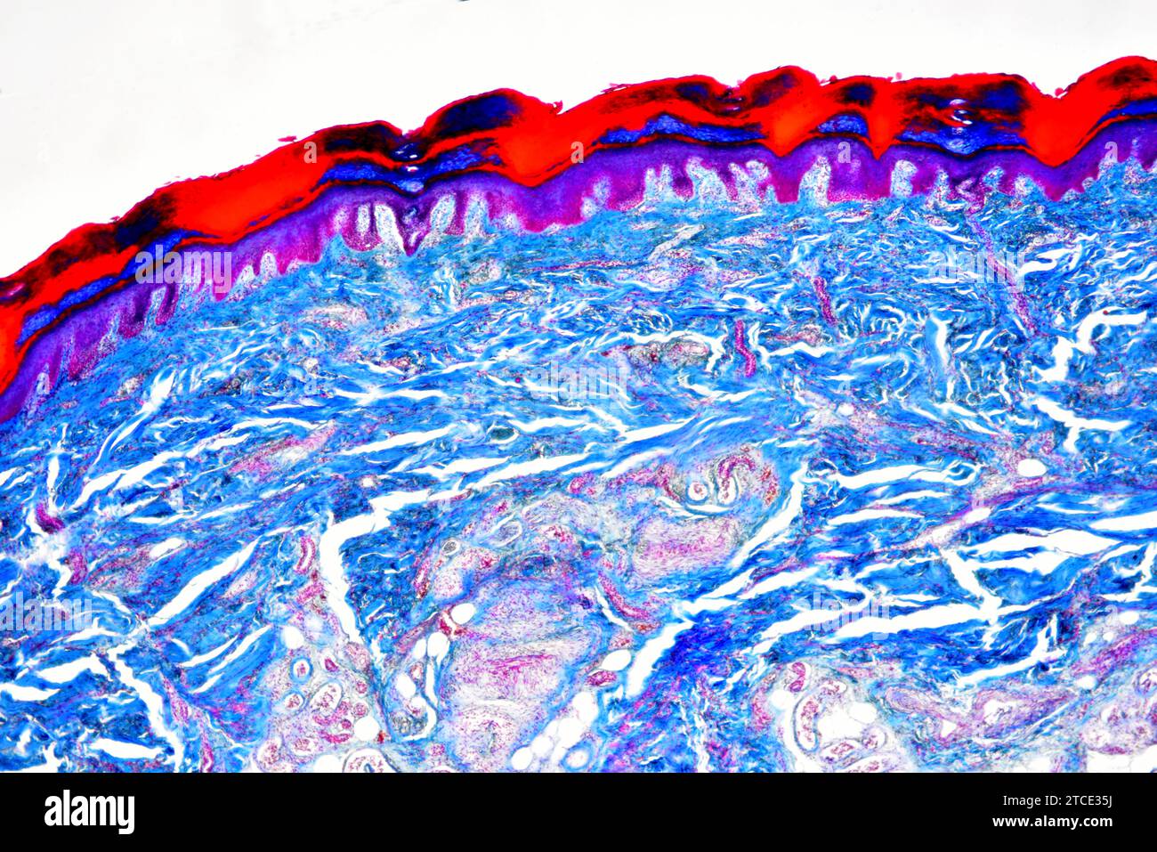 Human skin showing epidermis, dermis, blood vessels and collagen fibers. Optical microscope X40. Stock Photohttps://www.alamy.com/image-license-details/?v=1https://www.alamy.com/human-skin-showing-epidermis-dermis-blood-vessels-and-collagen-fibers-optical-microscope-x40-image575627854.html
RF2TCE35J–Human skin showing epidermis, dermis, blood vessels and collagen fibers. Optical microscope X40.
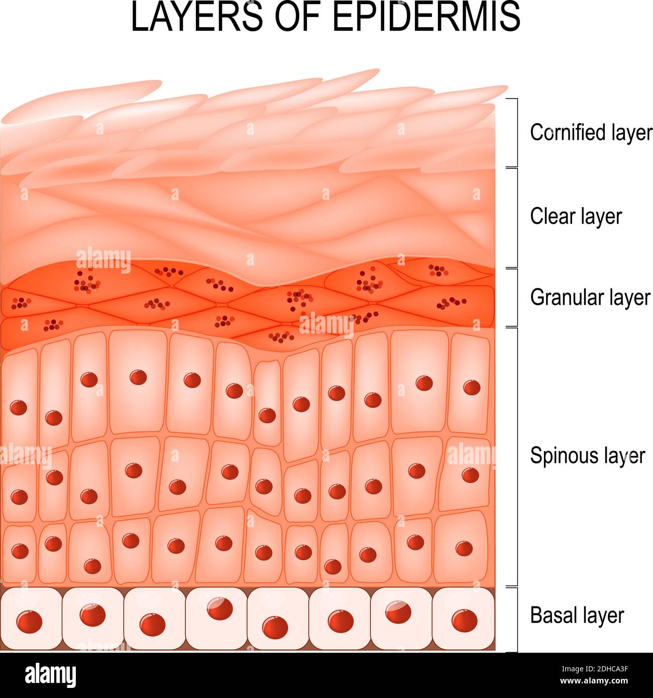 Structure of epidermis : cornified (stratum corneum), clear or translucent layer (lucidum), granular (stratum granulosum), spinous (spinosum) Stock Vectorhttps://www.alamy.com/image-license-details/?v=1https://www.alamy.com/structure-of-epidermis-cornified-stratum-corneum-clear-or-translucent-layer-lucidum-granular-stratum-granulosum-spinous-spinosum-image389348611.html
RF2DHCA3F–Structure of epidermis : cornified (stratum corneum), clear or translucent layer (lucidum), granular (stratum granulosum), spinous (spinosum)
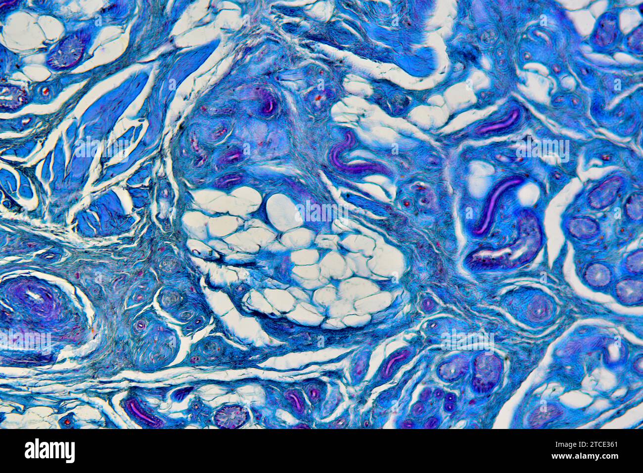 Human skin showing dermis with sweat gland, blood vessels and collagen fibers. Optical microscope X100. Stock Photohttps://www.alamy.com/image-license-details/?v=1https://www.alamy.com/human-skin-showing-dermis-with-sweat-gland-blood-vessels-and-collagen-fibers-optical-microscope-x100-image575627865.html
RF2TCE361–Human skin showing dermis with sweat gland, blood vessels and collagen fibers. Optical microscope X100.
 Each eyelid (or tarsus) consists of a thin envelope of tissue that protects the eye by covering the cornea. Haematoxylin end eosin stain. Stock Photohttps://www.alamy.com/image-license-details/?v=1https://www.alamy.com/each-eyelid-or-tarsus-consists-of-a-thin-envelope-of-tissue-that-protects-the-eye-by-covering-the-cornea-haematoxylin-end-eosin-stain-image572182042.html
RF2T6W40X–Each eyelid (or tarsus) consists of a thin envelope of tissue that protects the eye by covering the cornea. Haematoxylin end eosin stain.
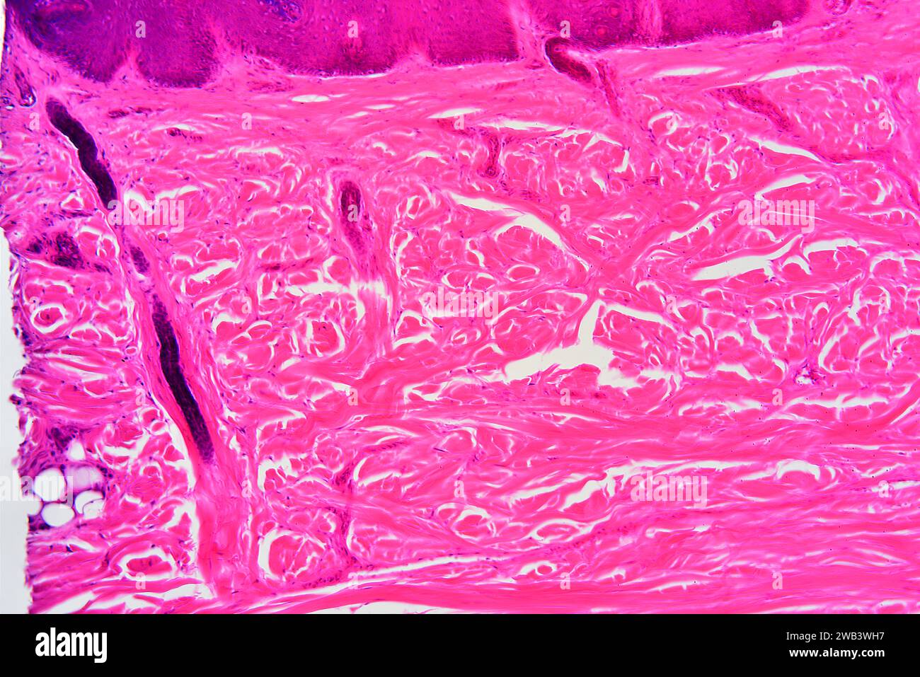 Human skin showing epidermis, dermis with sweat glands and connective tissue. X75 at 10 cm wide. Stock Photohttps://www.alamy.com/image-license-details/?v=1https://www.alamy.com/human-skin-showing-epidermis-dermis-with-sweat-glands-and-connective-tissue-x75-at-10-cm-wide-image591999667.html
RF2WB3WH7–Human skin showing epidermis, dermis with sweat glands and connective tissue. X75 at 10 cm wide.
 Dermis and epidermis Stock Photohttps://www.alamy.com/image-license-details/?v=1https://www.alamy.com/dermis-and-epidermis-image6069928.html
RFA2YMX9–Dermis and epidermis
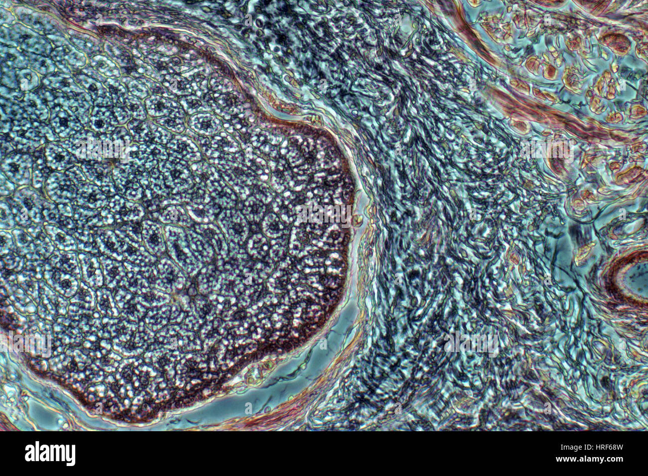 Sebaceous gland Stock Photohttps://www.alamy.com/image-license-details/?v=1https://www.alamy.com/stock-photo-sebaceous-gland-134943897.html
RMHRF68W–Sebaceous gland
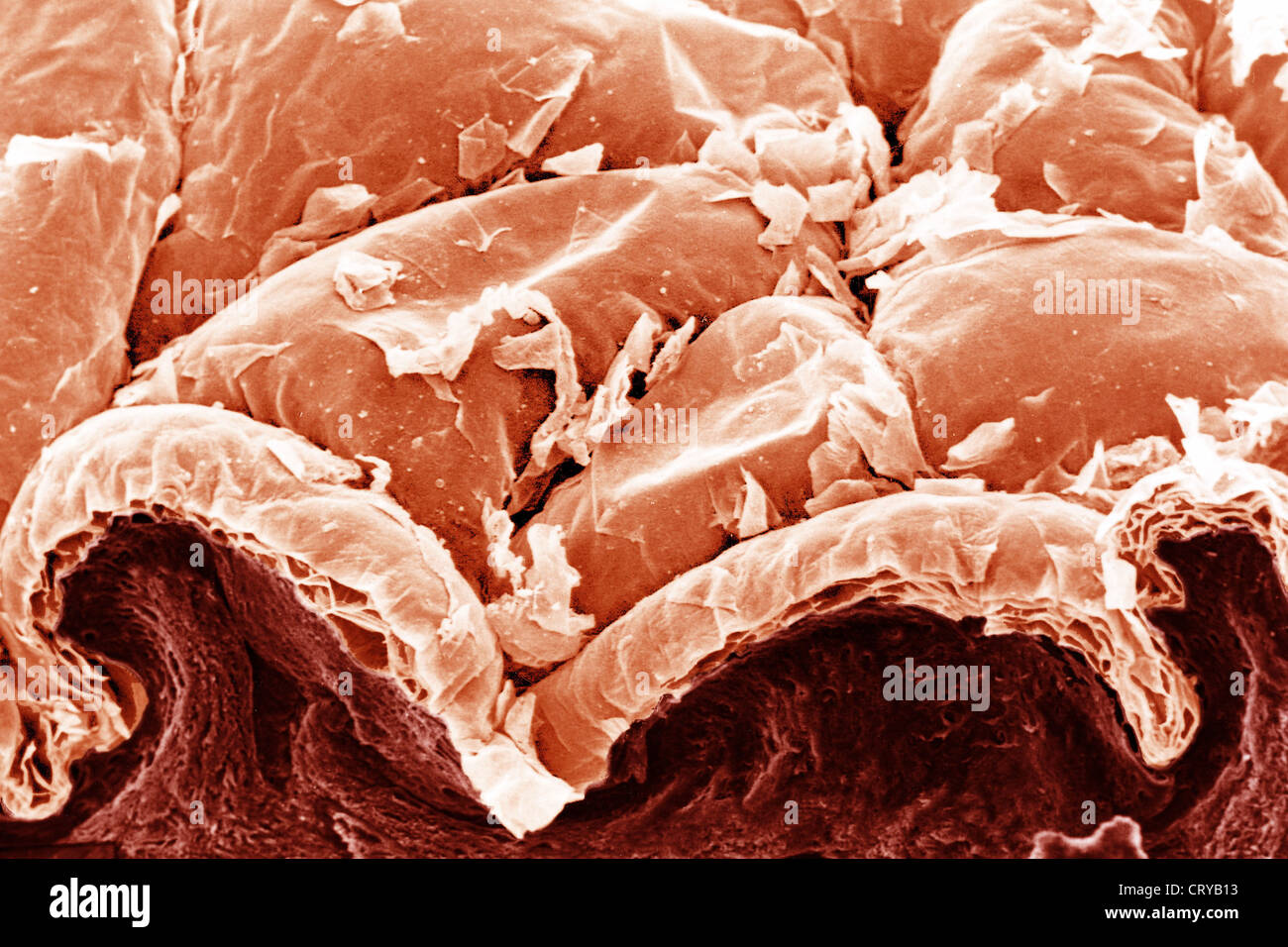 SKIN, SEM Stock Photohttps://www.alamy.com/image-license-details/?v=1https://www.alamy.com/stock-photo-skin-sem-49159183.html
RMCRYB13–SKIN, SEM
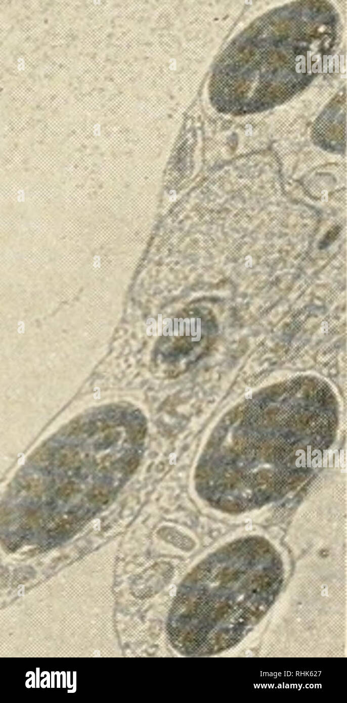 . The biology of hydra and of some other coelenterates, 1961. Hydra; Cnidaria; Ctenophora; Cnidaria; Hydra. y9i. Fig. 2. Low magnification electron micrograph of Chlorohydra. The epi- dermis is at the top and the gastrodermis at the bottom. Zoochlorella appear in the gastrodermal cells. Cross sections of muscle fibers lie adjacent to the mesolamella in the epidermis. Desmosomes are apparent in both the epi- dermis and the gastrodermis (arrows). Specialized muscle-to-muscle attach- ment is indicated by increased densities such as at the small circle. Note the large intracellular vacuoles in b Stock Photohttps://www.alamy.com/image-license-details/?v=1https://www.alamy.com/the-biology-of-hydra-and-of-some-other-coelenterates-1961-hydra-cnidaria-ctenophora-cnidaria-hydra-y9i-fig-2-low-magnification-electron-micrograph-of-chlorohydra-the-epi-dermis-is-at-the-top-and-the-gastrodermis-at-the-bottom-zoochlorella-appear-in-the-gastrodermal-cells-cross-sections-of-muscle-fibers-lie-adjacent-to-the-mesolamella-in-the-epidermis-desmosomes-are-apparent-in-both-the-epi-dermis-and-the-gastrodermis-arrows-specialized-muscle-to-muscle-attach-ment-is-indicated-by-increased-densities-such-as-at-the-small-circle-note-the-large-intracellular-vacuoles-in-b-image234605791.html
RMRHK627–. The biology of hydra and of some other coelenterates, 1961. Hydra; Cnidaria; Ctenophora; Cnidaria; Hydra. y9i. Fig. 2. Low magnification electron micrograph of Chlorohydra. The epi- dermis is at the top and the gastrodermis at the bottom. Zoochlorella appear in the gastrodermal cells. Cross sections of muscle fibers lie adjacent to the mesolamella in the epidermis. Desmosomes are apparent in both the epi- dermis and the gastrodermis (arrows). Specialized muscle-to-muscle attach- ment is indicated by increased densities such as at the small circle. Note the large intracellular vacuoles in b
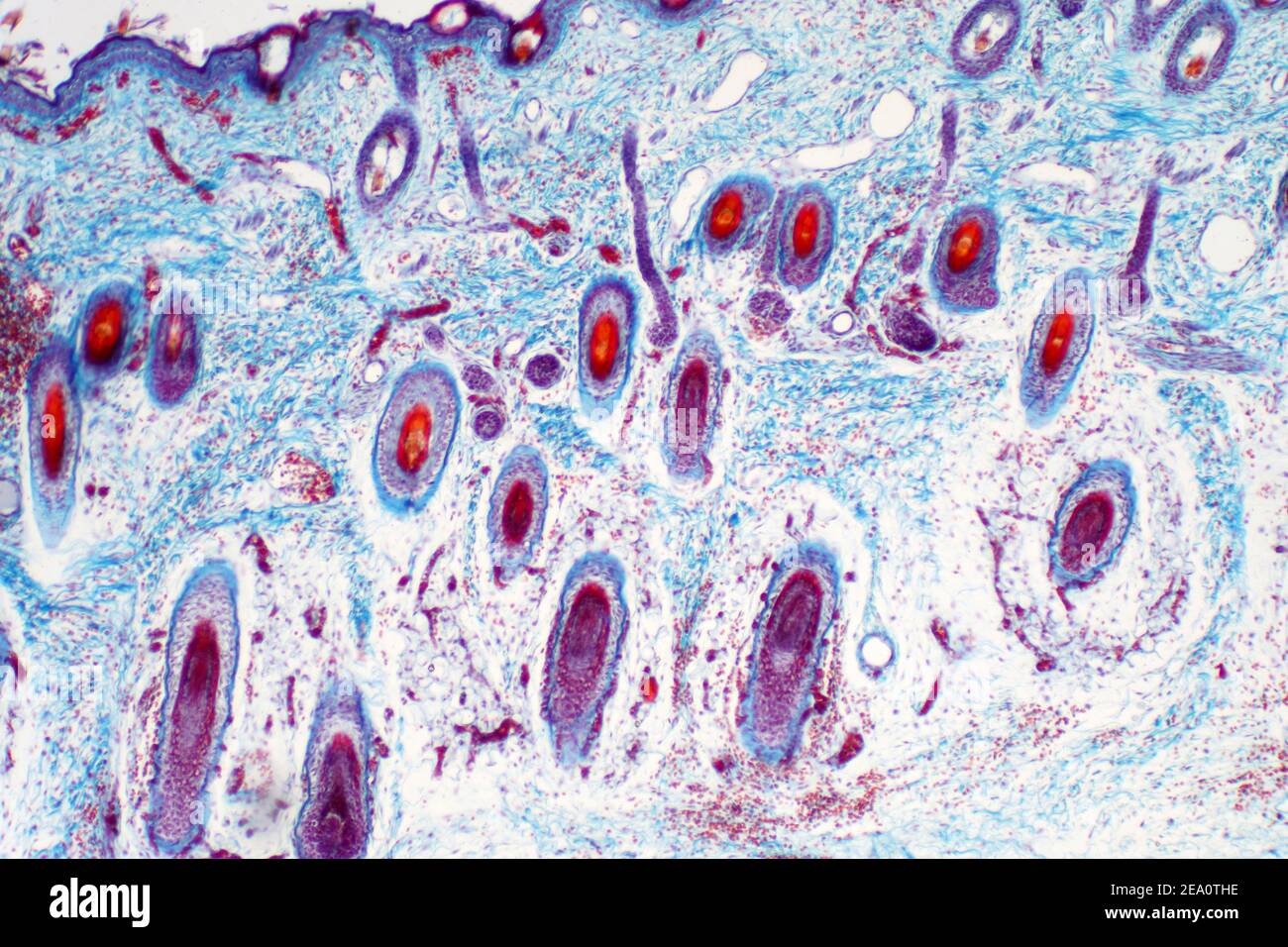 Human skin, light micrograph Stock Photohttps://www.alamy.com/image-license-details/?v=1https://www.alamy.com/human-skin-light-micrograph-image402004330.html
RF2EA0THE–Human skin, light micrograph
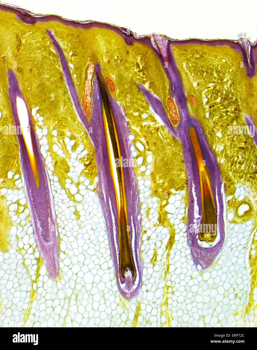 Hairy skin. Light micrograph of a thick section of human skin, showing three central hair follicles. The outer layer of the skin, the epidermis, is the thin, purple band, supported by the deeper, yellow, dermis. Beneath is the subcutaneous layer of connec Stock Photohttps://www.alamy.com/image-license-details/?v=1https://www.alamy.com/stock-photo-hairy-skin-light-micrograph-of-a-thick-section-of-human-skin-showing-83480388.html
RFERPT2C–Hairy skin. Light micrograph of a thick section of human skin, showing three central hair follicles. The outer layer of the skin, the epidermis, is the thin, purple band, supported by the deeper, yellow, dermis. Beneath is the subcutaneous layer of connec
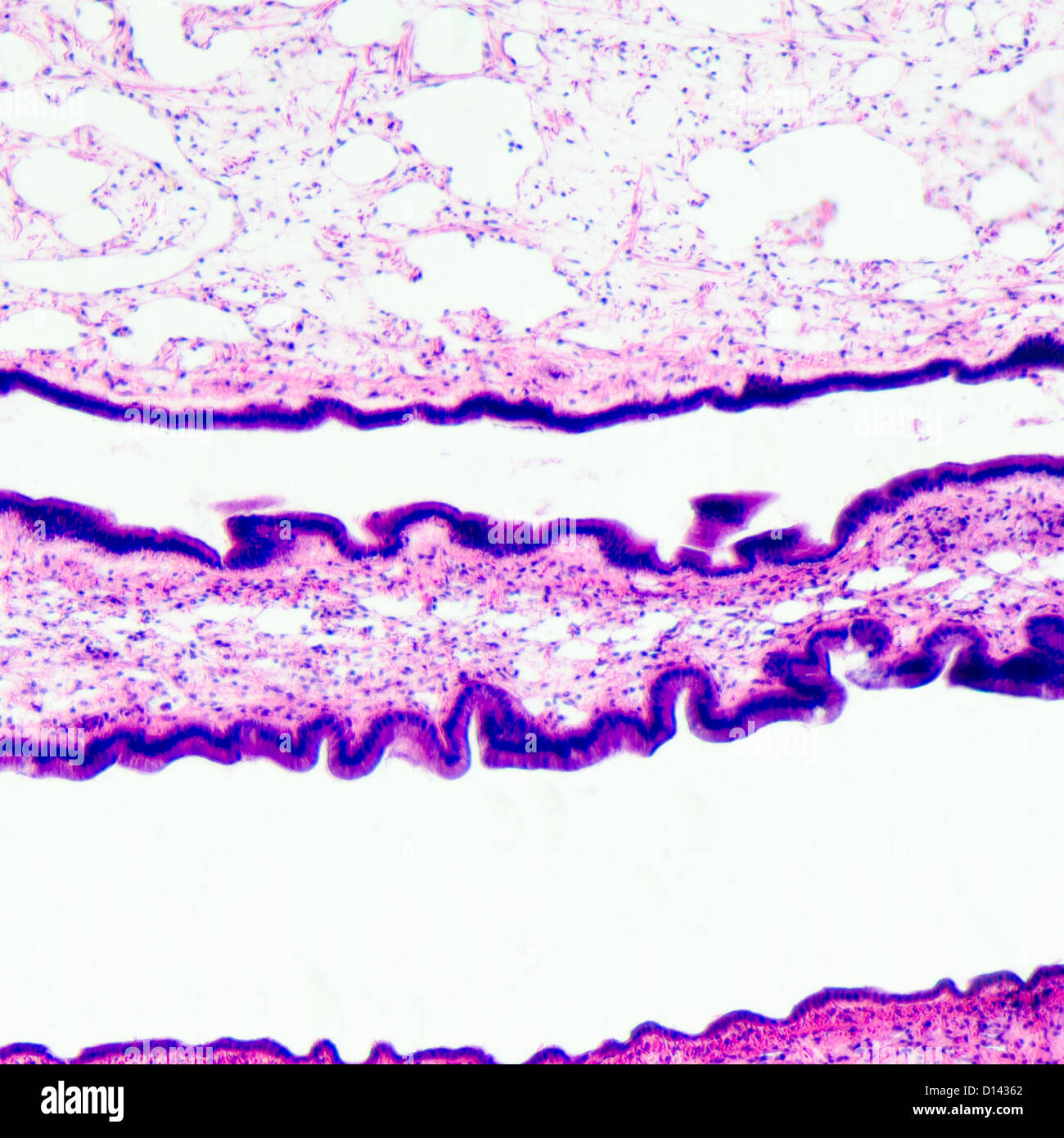 micrograph of medical science cilliated epithelium tissue cell Stock Photohttps://www.alamy.com/image-license-details/?v=1https://www.alamy.com/stock-photo-micrograph-of-medical-science-cilliated-epithelium-tissue-cell-52336090.html
RFD14362–micrograph of medical science cilliated epithelium tissue cell
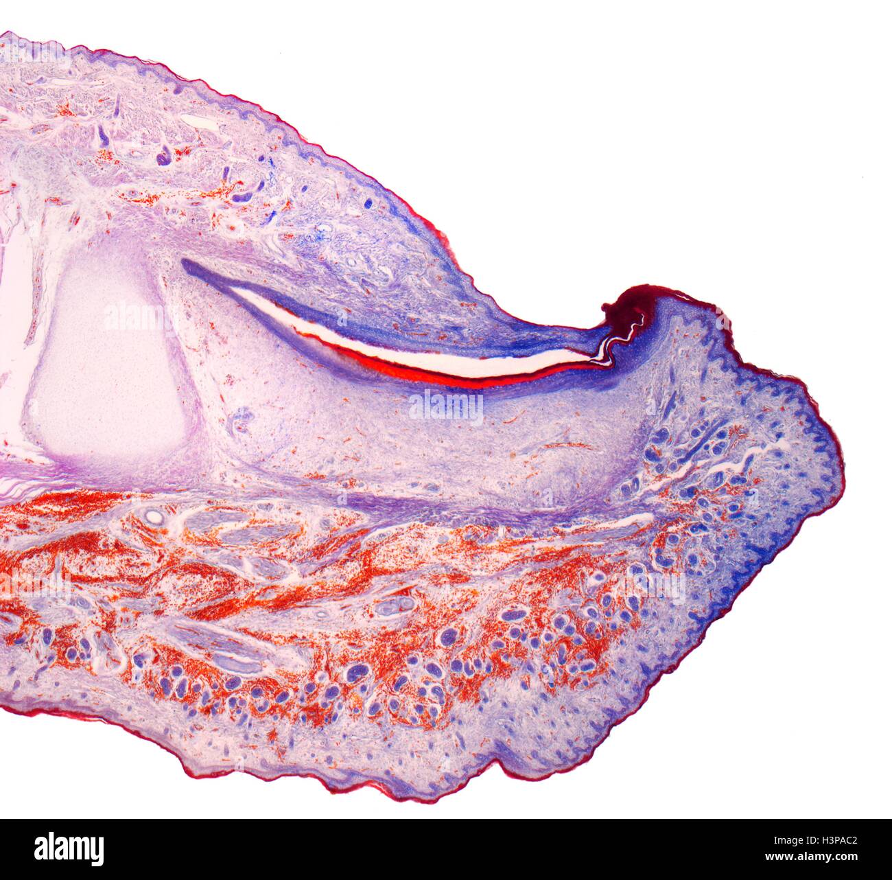 Fingertip. Light micrograph (LM) of a section through the fingertip. The nail (orange) is at top center, with the nail root below. The nail bed is dark purple and is continous with the epithelium. The tip of the finger bone is pale pink(left center). Beneath the stratified squamous epithelium is the dermis, which contains adipose (fat) cells connective tissue and blood vessels. Magnification: x5 when printed at 10 centimetres wide. Stock Photohttps://www.alamy.com/image-license-details/?v=1https://www.alamy.com/stock-photo-fingertip-light-micrograph-lm-of-a-section-through-the-fingertip-the-122807666.html
RFH3PAC2–Fingertip. Light micrograph (LM) of a section through the fingertip. The nail (orange) is at top center, with the nail root below. The nail bed is dark purple and is continous with the epithelium. The tip of the finger bone is pale pink(left center). Beneath the stratified squamous epithelium is the dermis, which contains adipose (fat) cells connective tissue and blood vessels. Magnification: x5 when printed at 10 centimetres wide.
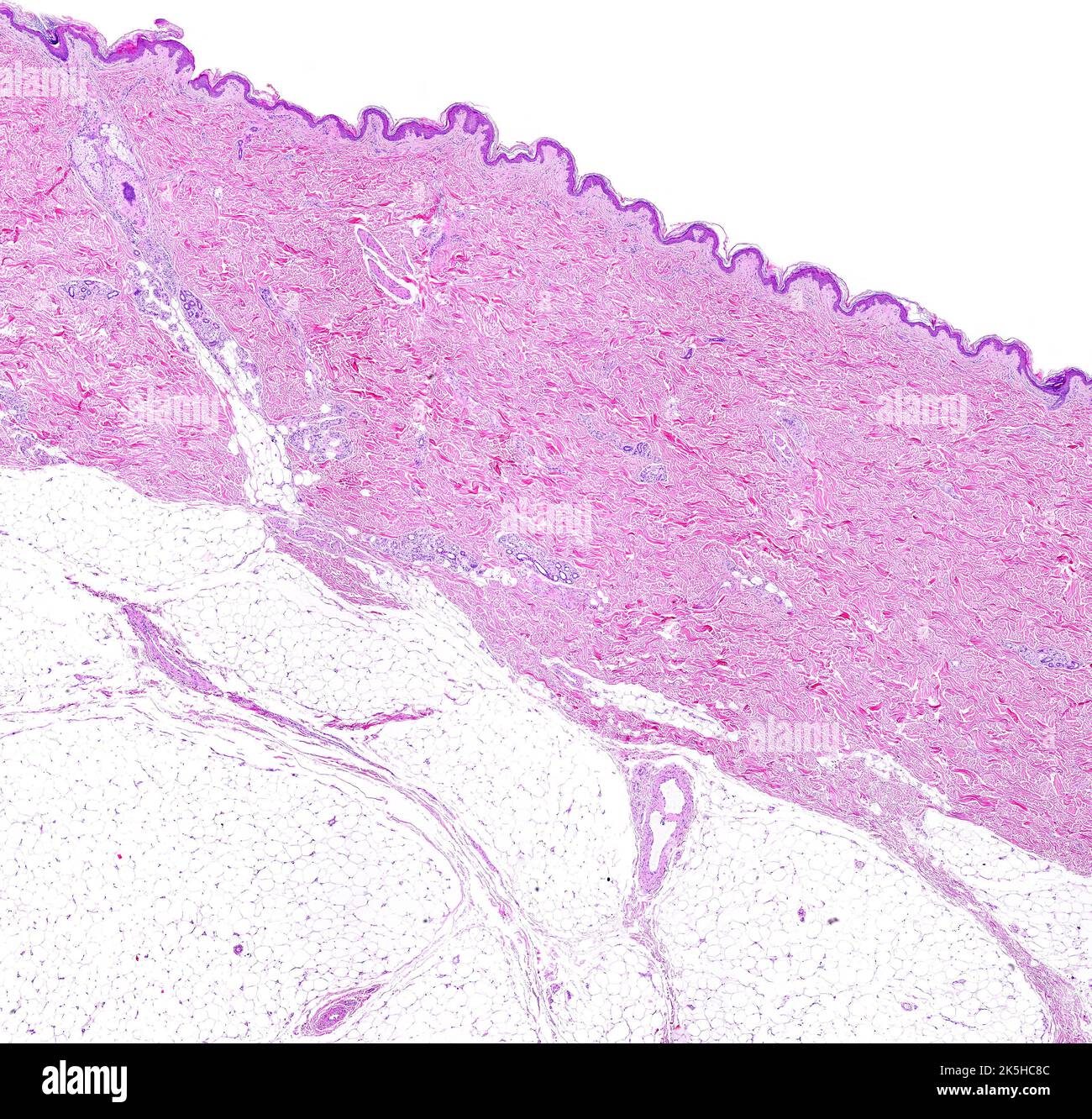 Low power light microscope micrograph of thin skin showing from top to bottom, the epidermis, a very thick dermis showing sweat glands, and the adipos Stock Photohttps://www.alamy.com/image-license-details/?v=1https://www.alamy.com/low-power-light-microscope-micrograph-of-thin-skin-showing-from-top-to-bottom-the-epidermis-a-very-thick-dermis-showing-sweat-glands-and-the-adipos-image485346412.html
RF2K5HC8C–Low power light microscope micrograph of thin skin showing from top to bottom, the epidermis, a very thick dermis showing sweat glands, and the adipos
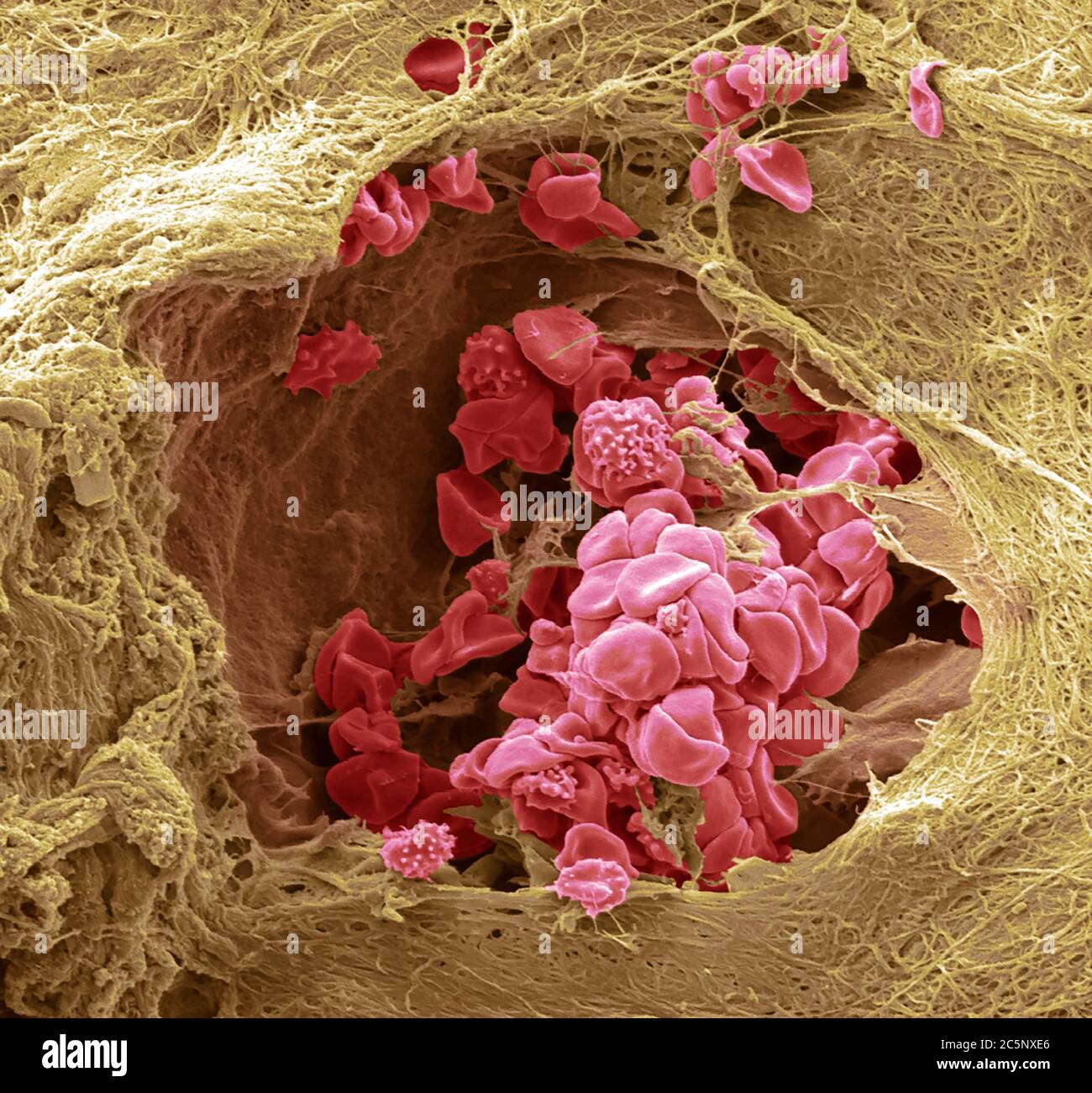 Skin blood vessel. Coloured scanning electron micrograph (SEM) of a blood vessel (arteriole) in the dermis of the skin. In the blood vessel are red blood cells (erythrocytes, red) which carry oxygen around the body. Some of these red blood cells are crenated. The blood vessel is surrounded by connective tissue which gives the skin its tone and elasticity. Magnification: 1000 when printed at 10 centimetres wide. Stock Photohttps://www.alamy.com/image-license-details/?v=1https://www.alamy.com/skin-blood-vessel-coloured-scanning-electron-micrograph-sem-of-a-blood-vessel-arteriole-in-the-dermis-of-the-skin-in-the-blood-vessel-are-red-blood-cells-erythrocytes-red-which-carry-oxygen-around-the-body-some-of-these-red-blood-cells-are-crenated-the-blood-vessel-is-surrounded-by-connective-tissue-which-gives-the-skin-its-tone-and-elasticity-magnification-1000-when-printed-at-10-centimetres-wide-image364972782.html
RF2C5NXE6–Skin blood vessel. Coloured scanning electron micrograph (SEM) of a blood vessel (arteriole) in the dermis of the skin. In the blood vessel are red blood cells (erythrocytes, red) which carry oxygen around the body. Some of these red blood cells are crenated. The blood vessel is surrounded by connective tissue which gives the skin its tone and elasticity. Magnification: 1000 when printed at 10 centimetres wide.
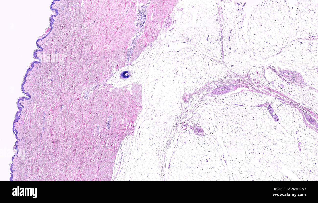 Low power light microscope micrograph of thin skin showing form left to right, the epidermis, a very thick dermis showing sweat glands and a hair foll Stock Photohttps://www.alamy.com/image-license-details/?v=1https://www.alamy.com/low-power-light-microscope-micrograph-of-thin-skin-showing-form-left-to-right-the-epidermis-a-very-thick-dermis-showing-sweat-glands-and-a-hair-foll-image485346409.html
RF2K5HC89–Low power light microscope micrograph of thin skin showing form left to right, the epidermis, a very thick dermis showing sweat glands and a hair foll
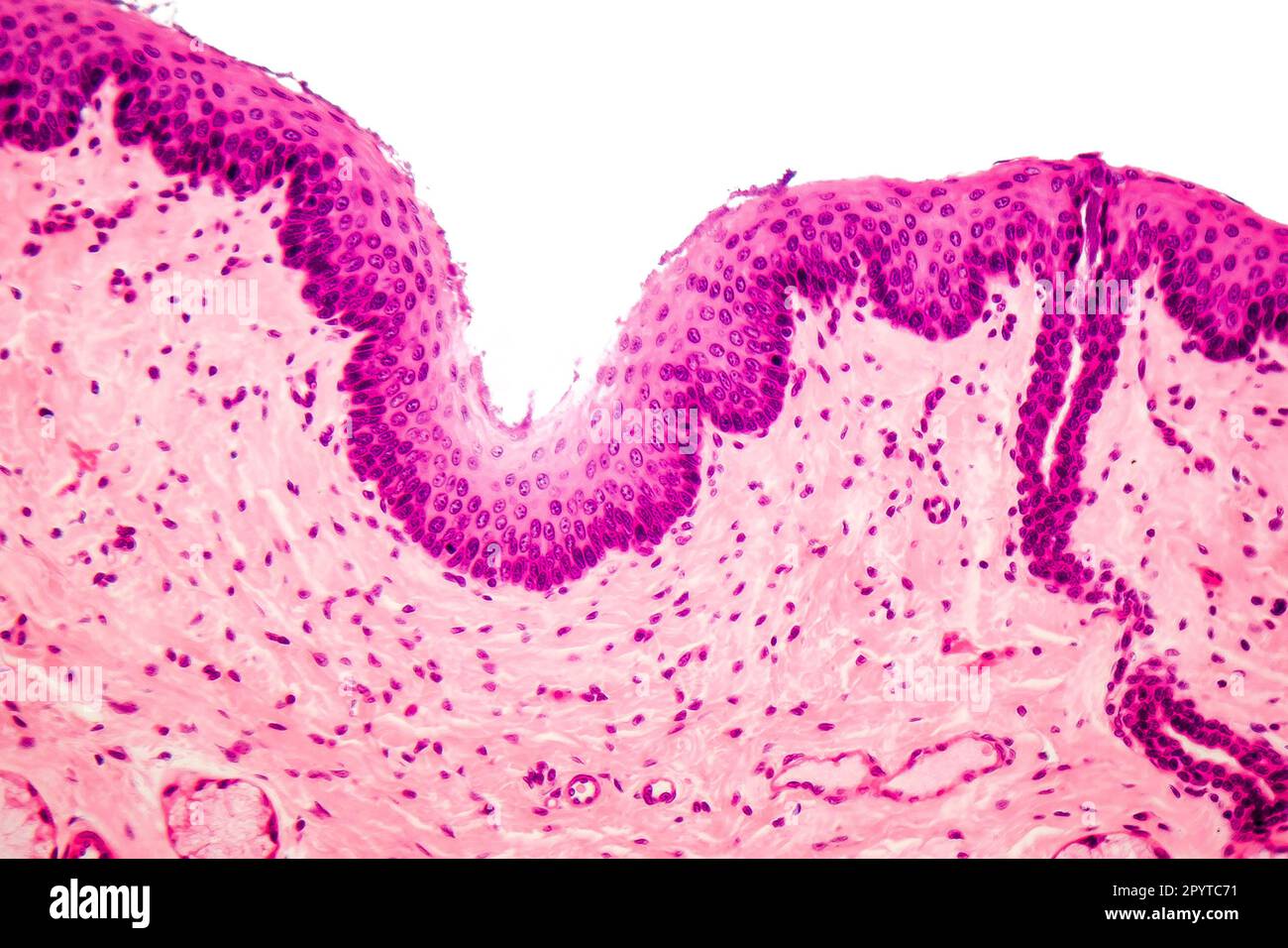 Human stratified squamous epithelium under microscope, light micrograph Stock Photohttps://www.alamy.com/image-license-details/?v=1https://www.alamy.com/human-stratified-squamous-epithelium-under-microscope-light-micrograph-image550653573.html
RF2PYTC71–Human stratified squamous epithelium under microscope, light micrograph
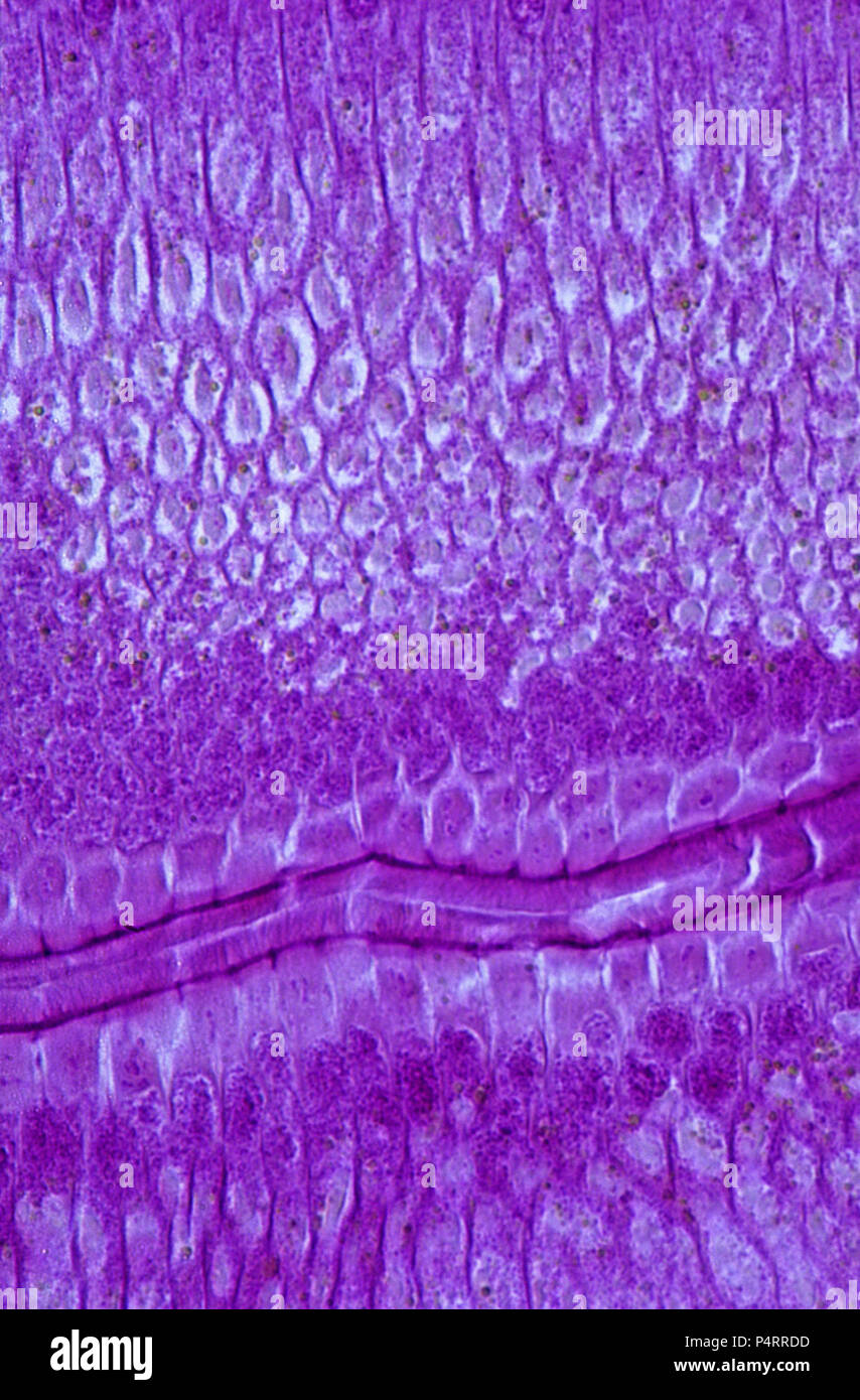 Epithelial cells of intestine of Ascaris.140x Stock Photohttps://www.alamy.com/image-license-details/?v=1https://www.alamy.com/epithelial-cells-of-intestine-of-ascaris140x-image209506345.html
RFP4RRDD–Epithelial cells of intestine of Ascaris.140x
 Silver impregnated Meissner corpuscles located in the dermal papillae of the glabrous skin of the finger pads. The spiral path of the nerve fiber Stock Photohttps://www.alamy.com/image-license-details/?v=1https://www.alamy.com/silver-impregnated-meissner-corpuscles-located-in-the-dermal-papillae-of-the-glabrous-skin-of-the-finger-pads-the-spiral-path-of-the-nerve-fiber-image401743613.html
RF2E9H025–Silver impregnated Meissner corpuscles located in the dermal papillae of the glabrous skin of the finger pads. The spiral path of the nerve fiber
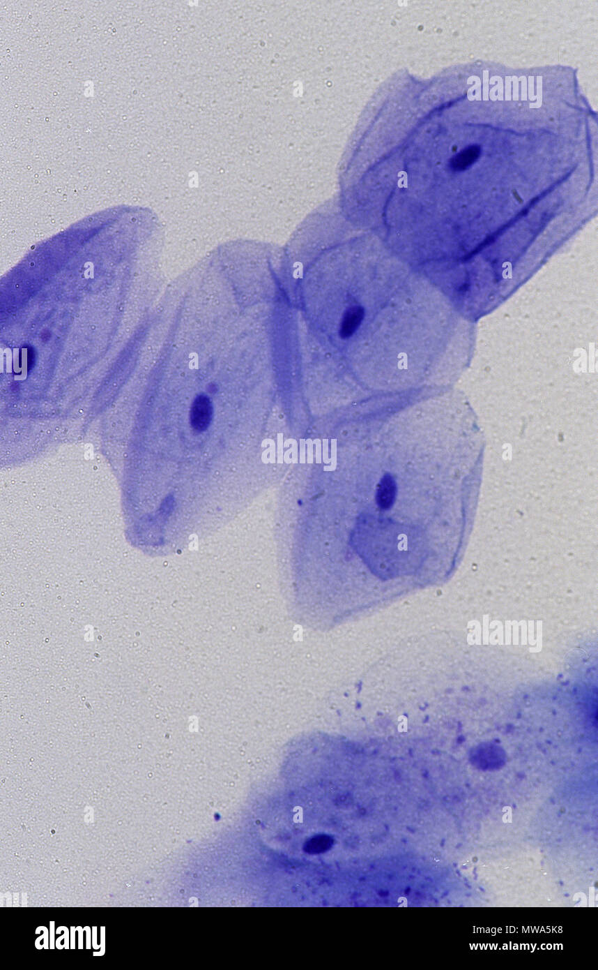 Epithelial cell of oral mucosa.140x Stock Photohttps://www.alamy.com/image-license-details/?v=1https://www.alamy.com/epithelial-cell-of-oral-mucosa140x-image187694060.html
RFMWA5K8–Epithelial cell of oral mucosa.140x
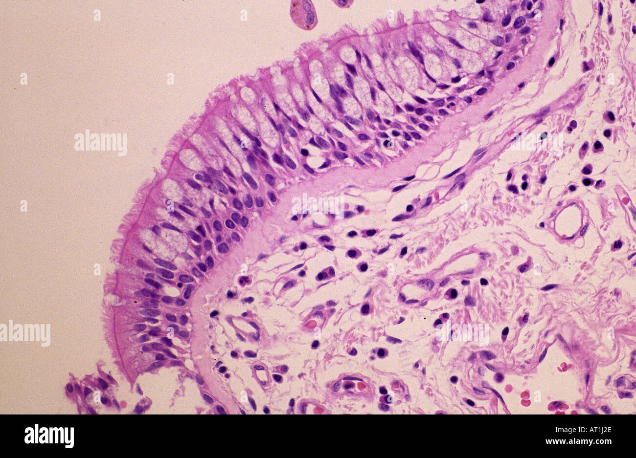 Epithelial tissue of trachea Tracheal epithelium Stock Photohttps://www.alamy.com/image-license-details/?v=1https://www.alamy.com/epithelial-tissue-of-trachea-tracheal-epithelium-image5279277.html
RFAT1J2E–Epithelial tissue of trachea Tracheal epithelium
 Human skin showing epidermis, dermis with sweat glands and connective tissue. X75 at 10 cm wide. Stock Photohttps://www.alamy.com/image-license-details/?v=1https://www.alamy.com/human-skin-showing-epidermis-dermis-with-sweat-glands-and-connective-tissue-x75-at-10-cm-wide-image591999669.html
RF2WB3WH9–Human skin showing epidermis, dermis with sweat glands and connective tissue. X75 at 10 cm wide.
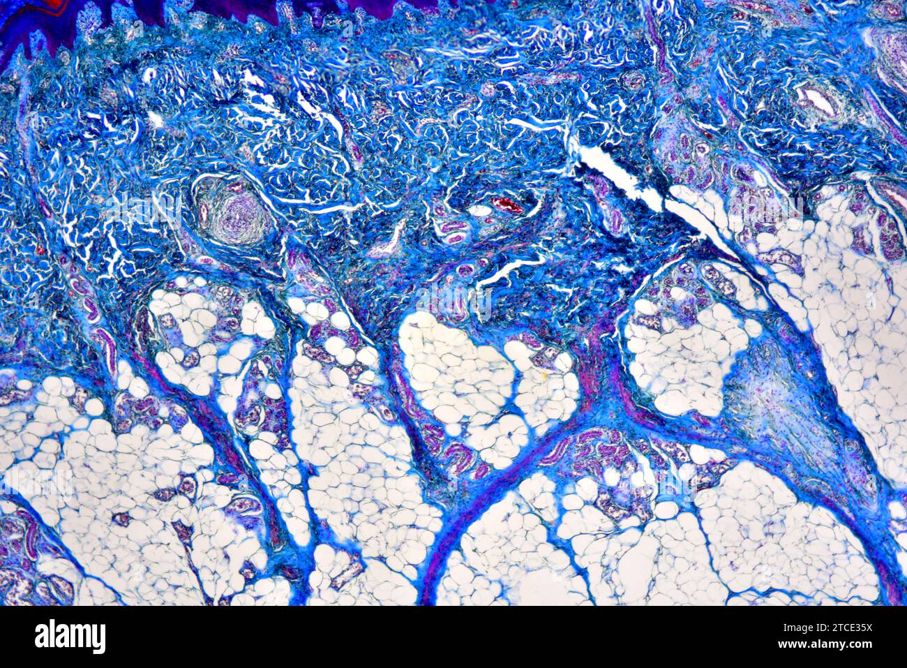 Human skin showing epidermis and dermis with sweat gland, blood vessels and collagen fibers. Optical microscope X40. Stock Photohttps://www.alamy.com/image-license-details/?v=1https://www.alamy.com/human-skin-showing-epidermis-and-dermis-with-sweat-gland-blood-vessels-and-collagen-fibers-optical-microscope-x40-image575627862.html
RF2TCE35X–Human skin showing epidermis and dermis with sweat gland, blood vessels and collagen fibers. Optical microscope X40.
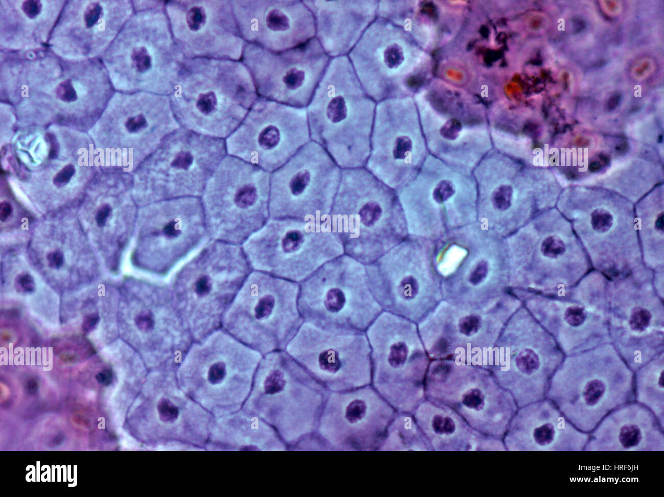 Frog Skin Stock Photohttps://www.alamy.com/image-license-details/?v=1https://www.alamy.com/stock-photo-frog-skin-134944169.html
RMHRF6JH–Frog Skin
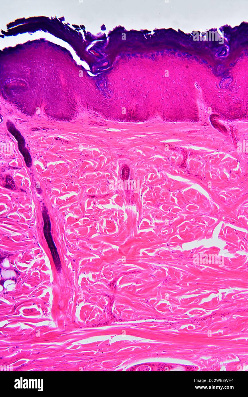 Human skin showing epidermis, dermis with sweat glands and connective tissue. X75 at 10 cm high. Stock Photohttps://www.alamy.com/image-license-details/?v=1https://www.alamy.com/human-skin-showing-epidermis-dermis-with-sweat-glands-and-connective-tissue-x75-at-10-cm-high-image591999664.html
RF2WB3WH4–Human skin showing epidermis, dermis with sweat glands and connective tissue. X75 at 10 cm high.
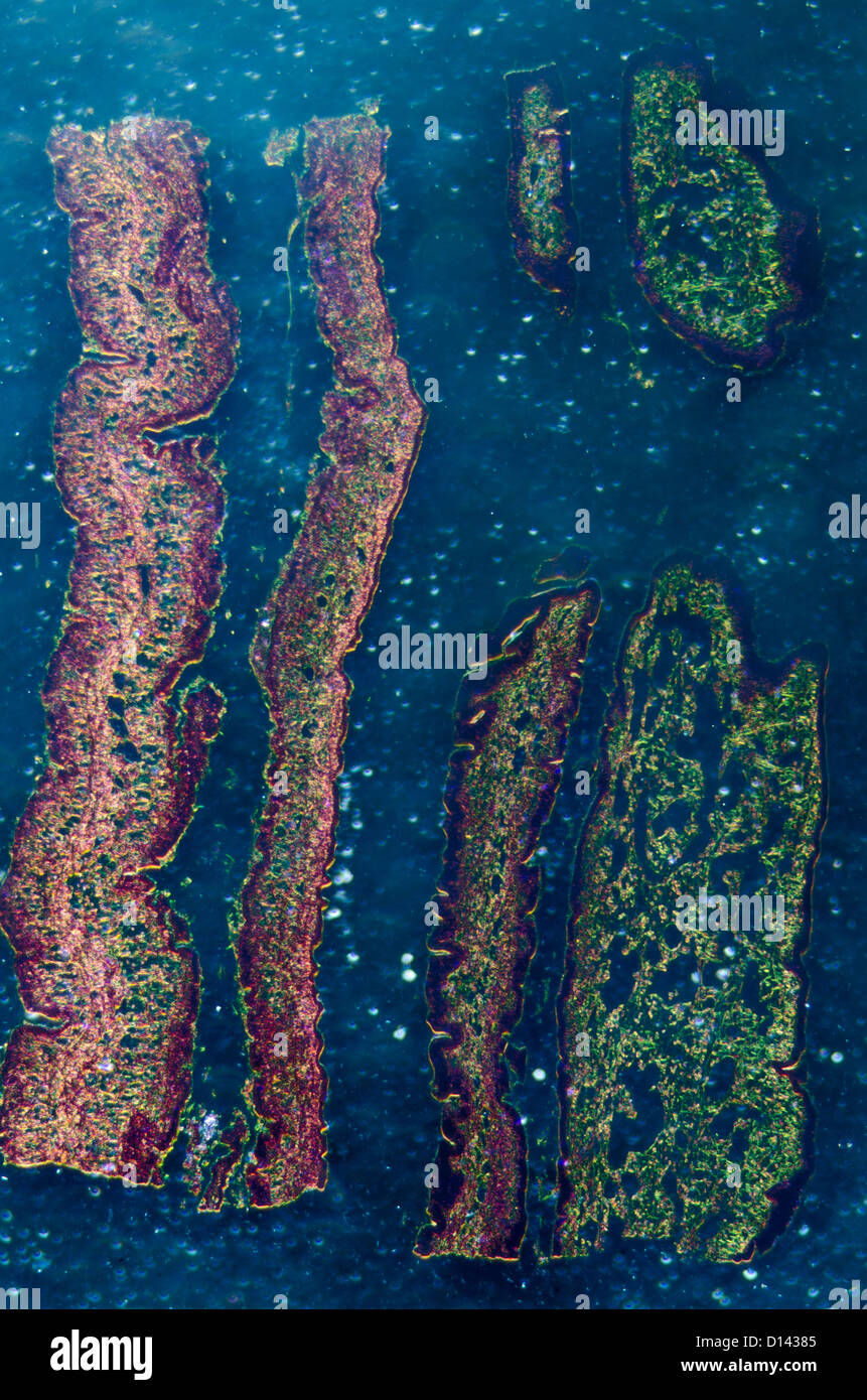 micrograph of medical science cilliated epithelium tissue cell Stock Photohttps://www.alamy.com/image-license-details/?v=1https://www.alamy.com/stock-photo-micrograph-of-medical-science-cilliated-epithelium-tissue-cell-52336149.html
RFD14385–micrograph of medical science cilliated epithelium tissue cell
 Skin blood vessel. Coloured scanning electron micrograph (SEM) of a blood vessel (arteriole) in the dermis of the skin. In the blood vessel are red blood cells (erythrocytes, red) which carry oxygen around the body. Some of these red blood cells are crenated. The blood vessel is surrounded by connective tissue which gives the skin its tone and elasticity. Magnification: 1000 when printed at 10 centimetres wide. Stock Photohttps://www.alamy.com/image-license-details/?v=1https://www.alamy.com/skin-blood-vessel-coloured-scanning-electron-micrograph-sem-of-a-blood-vessel-arteriole-in-the-dermis-of-the-skin-in-the-blood-vessel-are-red-blood-cells-erythrocytes-red-which-carry-oxygen-around-the-body-some-of-these-red-blood-cells-are-crenated-the-blood-vessel-is-surrounded-by-connective-tissue-which-gives-the-skin-its-tone-and-elasticity-magnification-1000-when-printed-at-10-centimetres-wide-image364972719.html
RF2C5NXBY–Skin blood vessel. Coloured scanning electron micrograph (SEM) of a blood vessel (arteriole) in the dermis of the skin. In the blood vessel are red blood cells (erythrocytes, red) which carry oxygen around the body. Some of these red blood cells are crenated. The blood vessel is surrounded by connective tissue which gives the skin its tone and elasticity. Magnification: 1000 when printed at 10 centimetres wide.
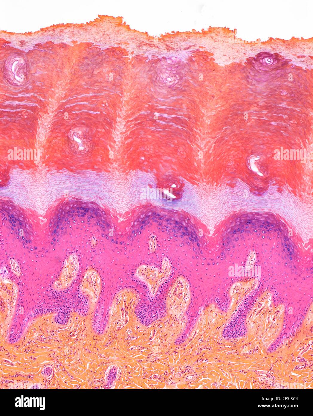 Plantar skin, LM Stock Photohttps://www.alamy.com/image-license-details/?v=1https://www.alamy.com/plantar-skin-lm-image416519940.html
RF2F5J3C4–Plantar skin, LM
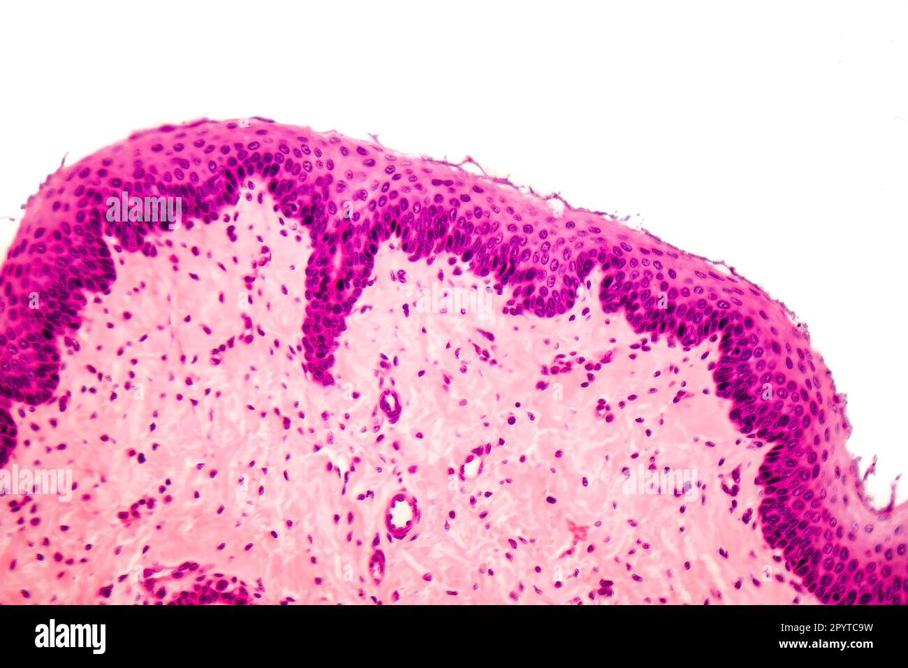 Human stratified squamous epithelium under microscope, light micrograph Stock Photohttps://www.alamy.com/image-license-details/?v=1https://www.alamy.com/human-stratified-squamous-epithelium-under-microscope-light-micrograph-image550653653.html
RF2PYTC9W–Human stratified squamous epithelium under microscope, light micrograph
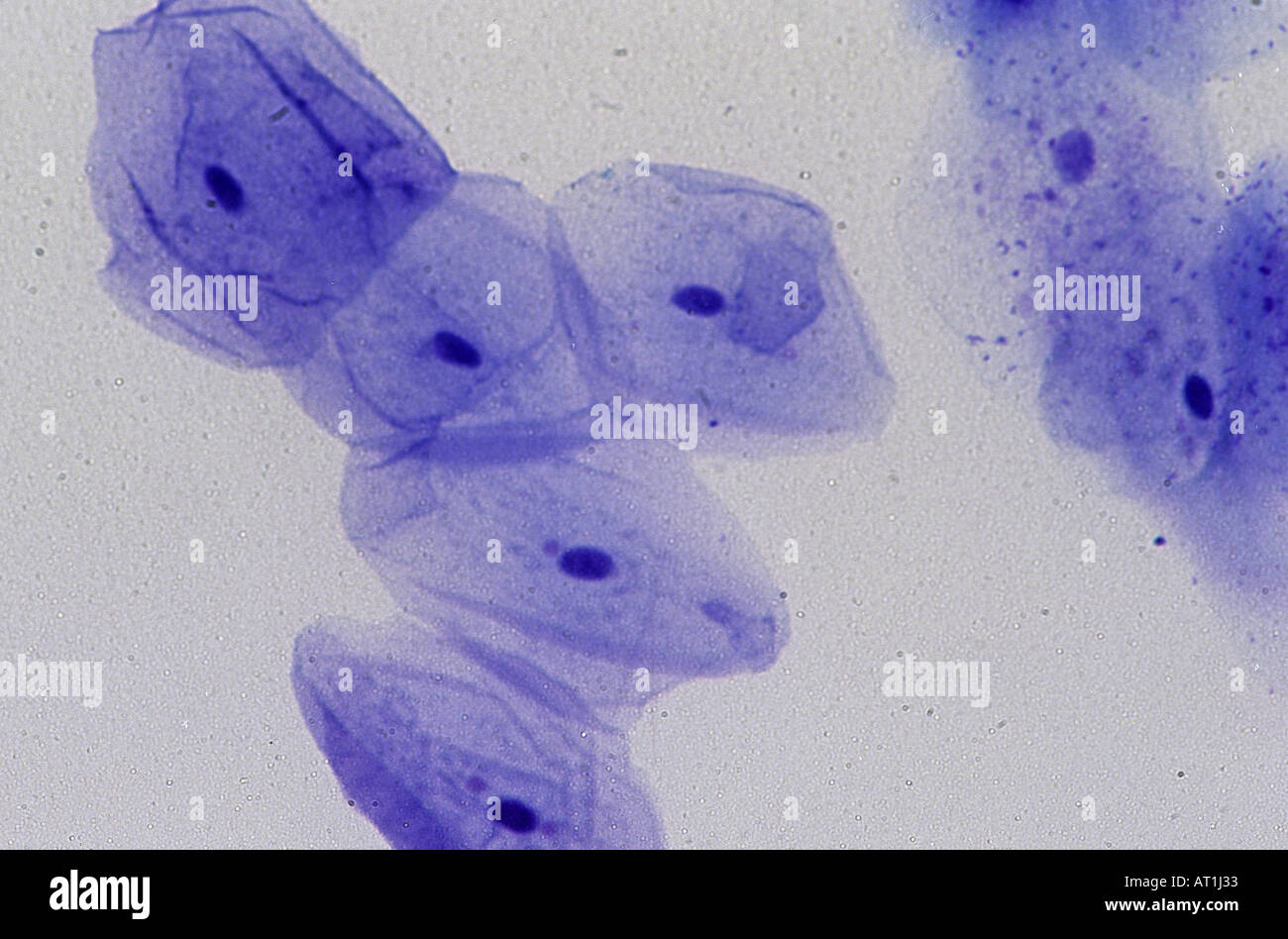 Epithelial cell of oral mucosa 140x Stock Photohttps://www.alamy.com/image-license-details/?v=1https://www.alamy.com/epithelial-cell-of-oral-mucosa-140x-image5279282.html
RFAT1J33–Epithelial cell of oral mucosa 140x
 Hair Follicle Stock Photohttps://www.alamy.com/image-license-details/?v=1https://www.alamy.com/stock-photo-hair-follicle-134943546.html
RMHRF5TA–Hair Follicle
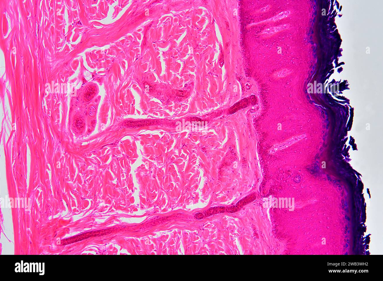 Human skin showing epidermis and dermis with sweat glands and connective tissue. X75 at 10 cm wide. Stock Photohttps://www.alamy.com/image-license-details/?v=1https://www.alamy.com/human-skin-showing-epidermis-and-dermis-with-sweat-glands-and-connective-tissue-x75-at-10-cm-wide-image591999662.html
RF2WB3WH2–Human skin showing epidermis and dermis with sweat glands and connective tissue. X75 at 10 cm wide.
 Human skin showing epidermis, dermis, adipose tissue, blood vessels and collagen fibers. Optical microscope X40. Stock Photohttps://www.alamy.com/image-license-details/?v=1https://www.alamy.com/human-skin-showing-epidermis-dermis-adipose-tissue-blood-vessels-and-collagen-fibers-optical-microscope-x40-image575627858.html
RF2TCE35P–Human skin showing epidermis, dermis, adipose tissue, blood vessels and collagen fibers. Optical microscope X40.
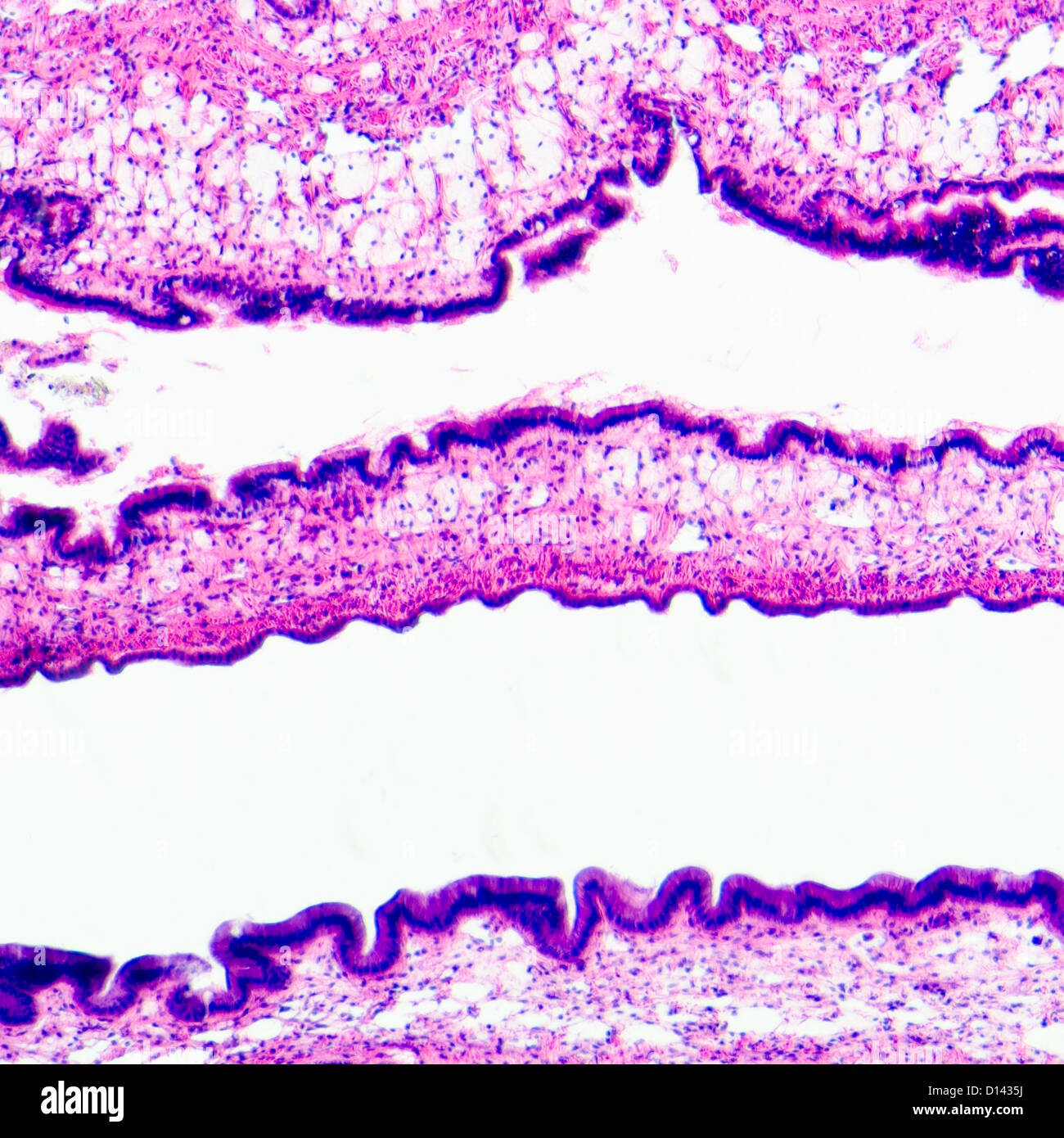 micrograph of medical science cilliated epithelium tissue cell Stock Photohttps://www.alamy.com/image-license-details/?v=1https://www.alamy.com/stock-photo-micrograph-of-medical-science-cilliated-epithelium-tissue-cell-52336078.html
RFD1435J–micrograph of medical science cilliated epithelium tissue cell
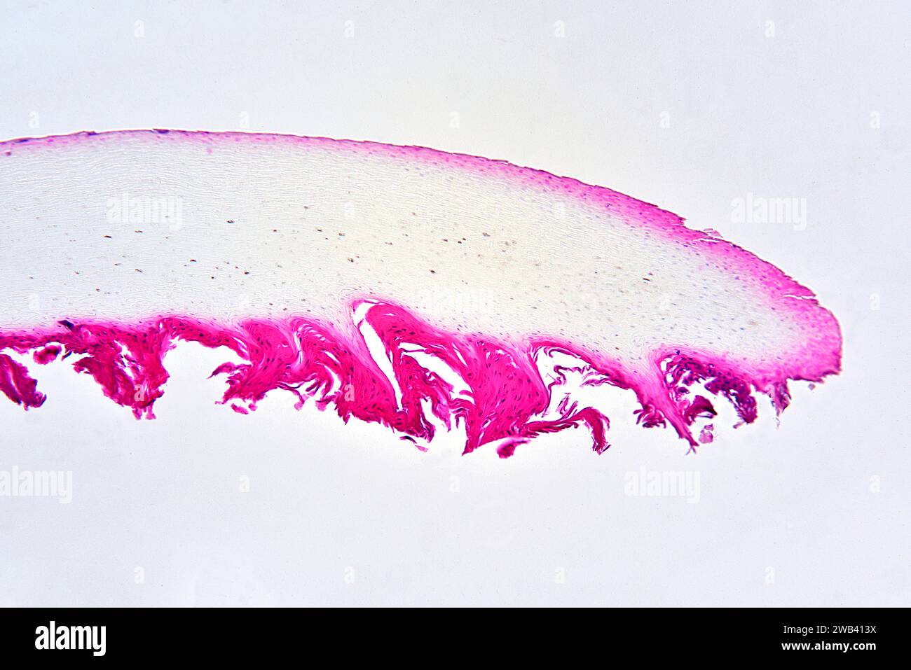 Human nail section showing dermis, nail matrix and nail plate of hard keratin. X25 at 10 cm wide. Stock Photohttps://www.alamy.com/image-license-details/?v=1https://www.alamy.com/human-nail-section-showing-dermis-nail-matrix-and-nail-plate-of-hard-keratin-x25-at-10-cm-wide-image592002430.html
RF2WB413X–Human nail section showing dermis, nail matrix and nail plate of hard keratin. X25 at 10 cm wide.
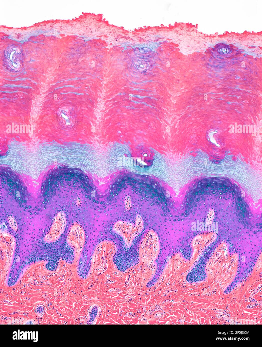 Plantar skin, LM Stock Photohttps://www.alamy.com/image-license-details/?v=1https://www.alamy.com/plantar-skin-lm-image416519956.html
RF2F5J3CM–Plantar skin, LM
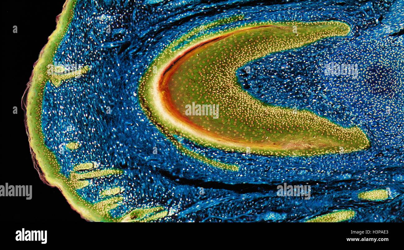 Developing nail. Light micrograph (LM) of longitudinal section through a fetal finger tip to show the developing nail. The large area of green-yellow nail bed epithelium is tipped by the developing nail. Blue connective tissue (dermis) forms the bulk of the image. Yellow stratified squamous epithelium of the skin is far left. Magnification: x15 when printed at 10 centimetres wide. Stock Photohttps://www.alamy.com/image-license-details/?v=1https://www.alamy.com/stock-photo-developing-nail-light-micrograph-lm-of-longitudinal-section-through-122807723.html
RFH3PAE3–Developing nail. Light micrograph (LM) of longitudinal section through a fetal finger tip to show the developing nail. The large area of green-yellow nail bed epithelium is tipped by the developing nail. Blue connective tissue (dermis) forms the bulk of the image. Yellow stratified squamous epithelium of the skin is far left. Magnification: x15 when printed at 10 centimetres wide.
 Connective tissue. Coloured scanning electron micrograph (SEM) of collagen from a nasal poly biopsy. Collagen is a protein with a high tensile strength, providing structure and elasticity to epithelium, tendons, ligaments and bones. It is the most abundant protein in the body. In the dermis, collagen forms rope-like fibres that are arranged irregularly. Magnification: x1300 when printed 10 centimetres wide. Stock Photohttps://www.alamy.com/image-license-details/?v=1https://www.alamy.com/connective-tissue-coloured-scanning-electron-micrograph-sem-of-collagen-from-a-nasal-poly-biopsy-collagen-is-a-protein-with-a-high-tensile-strength-providing-structure-and-elasticity-to-epithelium-tendons-ligaments-and-bones-it-is-the-most-abundant-protein-in-the-body-in-the-dermis-collagen-forms-rope-like-fibres-that-are-arranged-irregularly-magnification-x1300-when-printed-10-centimetres-wide-image631222587.html
RF2YJXJRR–Connective tissue. Coloured scanning electron micrograph (SEM) of collagen from a nasal poly biopsy. Collagen is a protein with a high tensile strength, providing structure and elasticity to epithelium, tendons, ligaments and bones. It is the most abundant protein in the body. In the dermis, collagen forms rope-like fibres that are arranged irregularly. Magnification: x1300 when printed 10 centimetres wide.
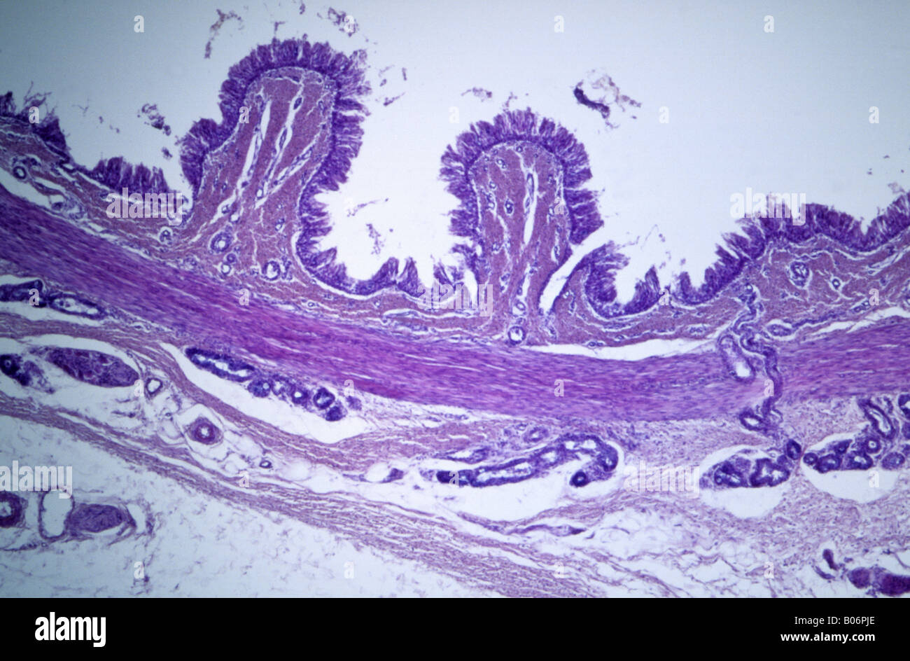 Cilia ephitelial tissue Stock Photohttps://www.alamy.com/image-license-details/?v=1https://www.alamy.com/stock-photo-cilia-ephitelial-tissue-17359846.html
RFB06PJE–Cilia ephitelial tissue
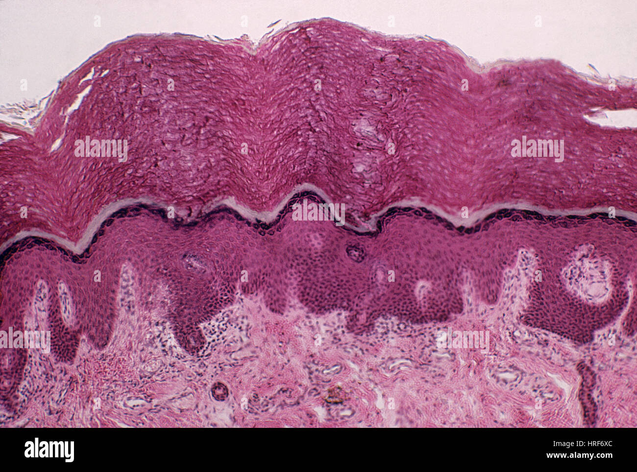 Fingertip, Skin Stock Photohttps://www.alamy.com/image-license-details/?v=1https://www.alamy.com/stock-photo-fingertip-skin-134944388.html
RMHRF6XC–Fingertip, Skin
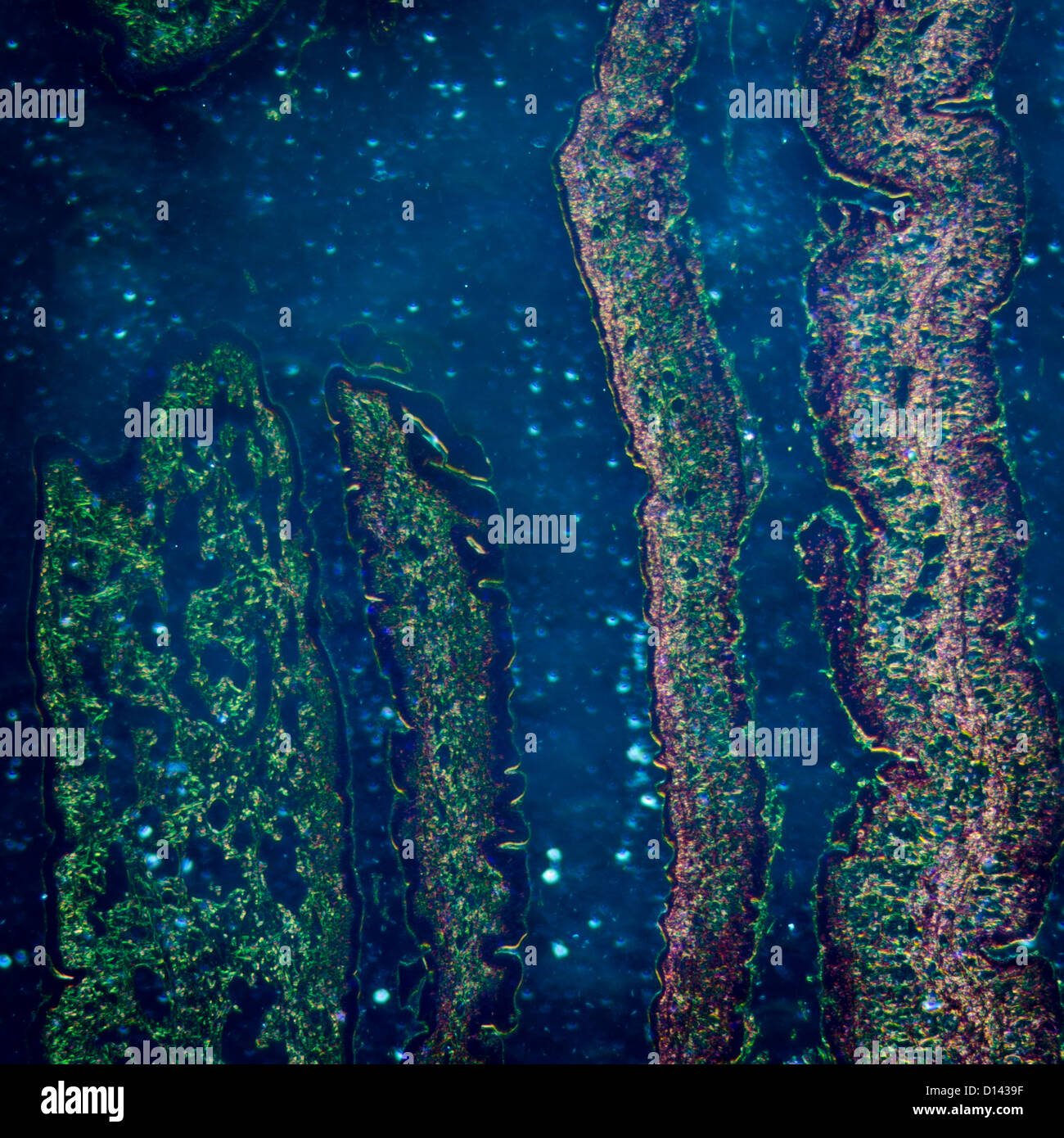 micrograph of medical science cilliated epithelium tissue cell Stock Photohttps://www.alamy.com/image-license-details/?v=1https://www.alamy.com/stock-photo-micrograph-of-medical-science-cilliated-epithelium-tissue-cell-52336187.html
RFD1439F–micrograph of medical science cilliated epithelium tissue cell
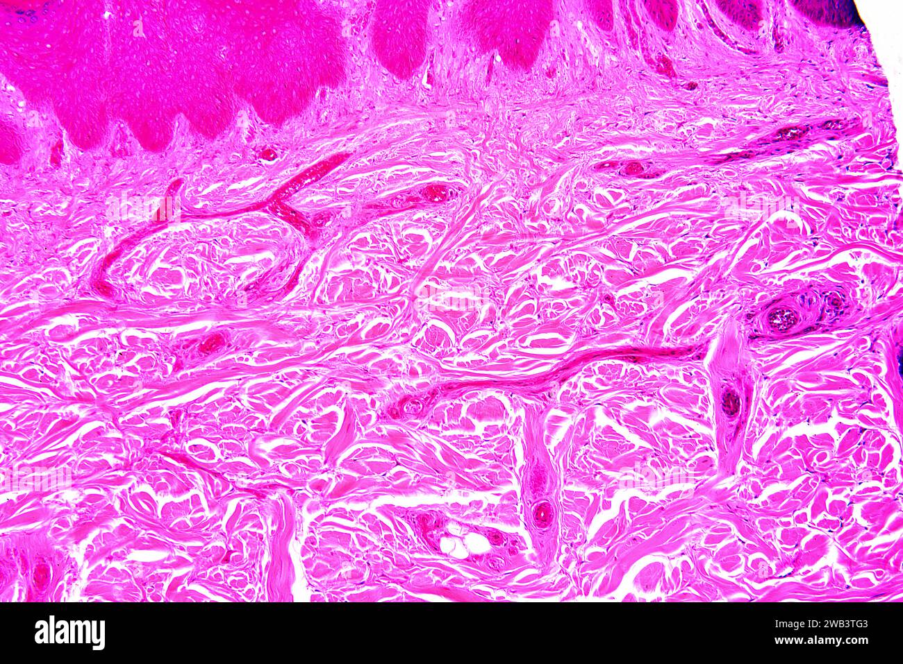 Human hand skin showing basal epidermis and dermis with connective tissue and blood vessels. X75 at 10 cm wide. Stock Photohttps://www.alamy.com/image-license-details/?v=1https://www.alamy.com/human-hand-skin-showing-basal-epidermis-and-dermis-with-connective-tissue-and-blood-vessels-x75-at-10-cm-wide-image591998851.html
RF2WB3TG3–Human hand skin showing basal epidermis and dermis with connective tissue and blood vessels. X75 at 10 cm wide.
 Connective tissue. Coloured scanning electron micrograph (SEM) of collagen from a nasal poly biopsy. Collagen is a protein with a high tensile strength, providing structure and elasticity to epithelium, tendons, ligaments and bones. It is the most abundant protein in the body. In the dermis, collagen forms rope-like fibres that are arranged irregularly. Magnification: x1300 when printed 10 centimetres wide. Stock Photohttps://www.alamy.com/image-license-details/?v=1https://www.alamy.com/connective-tissue-coloured-scanning-electron-micrograph-sem-of-collagen-from-a-nasal-poly-biopsy-collagen-is-a-protein-with-a-high-tensile-strength-providing-structure-and-elasticity-to-epithelium-tendons-ligaments-and-bones-it-is-the-most-abundant-protein-in-the-body-in-the-dermis-collagen-forms-rope-like-fibres-that-are-arranged-irregularly-magnification-x1300-when-printed-10-centimetres-wide-image631222635.html
RF2YJXJWF–Connective tissue. Coloured scanning electron micrograph (SEM) of collagen from a nasal poly biopsy. Collagen is a protein with a high tensile strength, providing structure and elasticity to epithelium, tendons, ligaments and bones. It is the most abundant protein in the body. In the dermis, collagen forms rope-like fibres that are arranged irregularly. Magnification: x1300 when printed 10 centimetres wide.
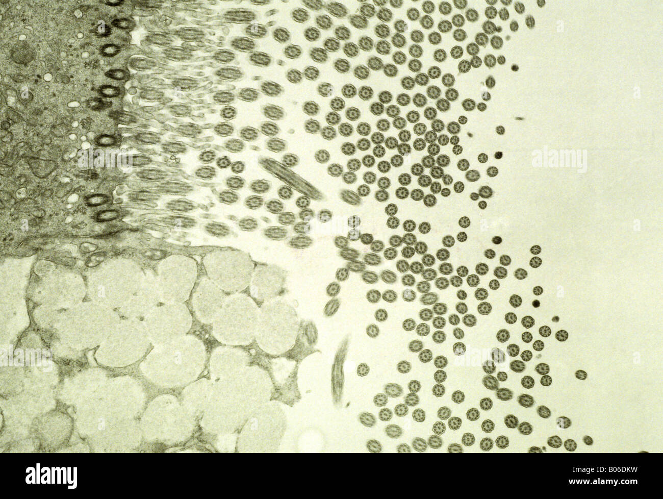 Epithelial trachea Stock Photohttps://www.alamy.com/image-license-details/?v=1https://www.alamy.com/stock-photo-epithelial-trachea-17352829.html
RFB06DKW–Epithelial trachea
 Scalp Stock Photohttps://www.alamy.com/image-license-details/?v=1https://www.alamy.com/stock-photo-scalp-134944389.html
RMHRF6XD–Scalp
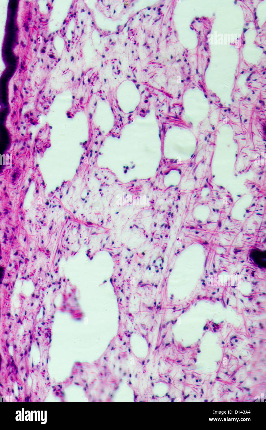 micrograph of medical science cilliated epithelium tissue cell Stock Photohttps://www.alamy.com/image-license-details/?v=1https://www.alamy.com/stock-photo-micrograph-of-medical-science-cilliated-epithelium-tissue-cell-52336204.html
RFD143A4–micrograph of medical science cilliated epithelium tissue cell
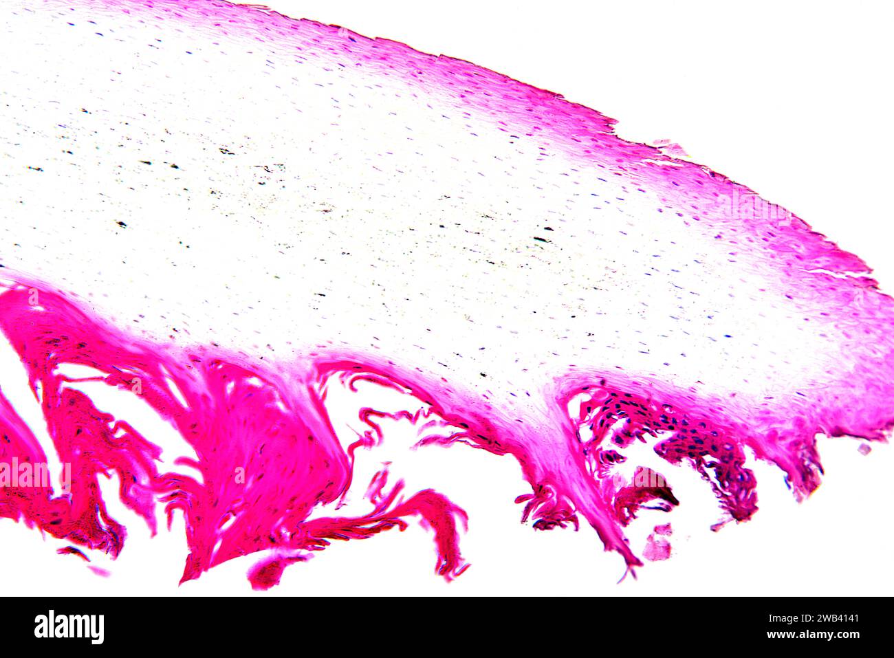 Human nail section showing dermis, nail matrix and nail plate of hard keratin. X75 at 10 cm wide. Stock Photohttps://www.alamy.com/image-license-details/?v=1https://www.alamy.com/human-nail-section-showing-dermis-nail-matrix-and-nail-plate-of-hard-keratin-x75-at-10-cm-wide-image592002433.html
RF2WB4141–Human nail section showing dermis, nail matrix and nail plate of hard keratin. X75 at 10 cm wide.
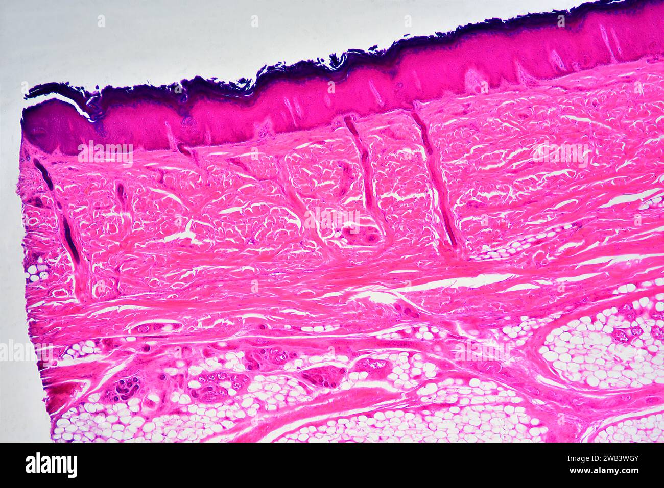 Human skin showing epidermis, dermis with sweat glands, adipose tissue and connective tissue. X25 at 10 cm wide. Stock Photohttps://www.alamy.com/image-license-details/?v=1https://www.alamy.com/human-skin-showing-epidermis-dermis-with-sweat-glands-adipose-tissue-and-connective-tissue-x25-at-10-cm-wide-image591999659.html
RF2WB3WGY–Human skin showing epidermis, dermis with sweat glands, adipose tissue and connective tissue. X25 at 10 cm wide.
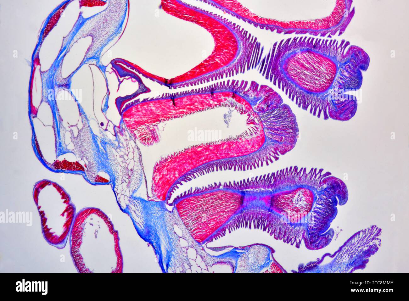 Asterias sp. cross section showing dermis, mucus glands, suckers and tube feets. Light microscope X50 at 10 cm wide. Stock Photohttps://www.alamy.com/image-license-details/?v=1https://www.alamy.com/asterias-sp-cross-section-showing-dermis-mucus-glands-suckers-and-tube-feets-light-microscope-x50-at-10-cm-wide-image575509899.html
RF2TC8MMY–Asterias sp. cross section showing dermis, mucus glands, suckers and tube feets. Light microscope X50 at 10 cm wide.
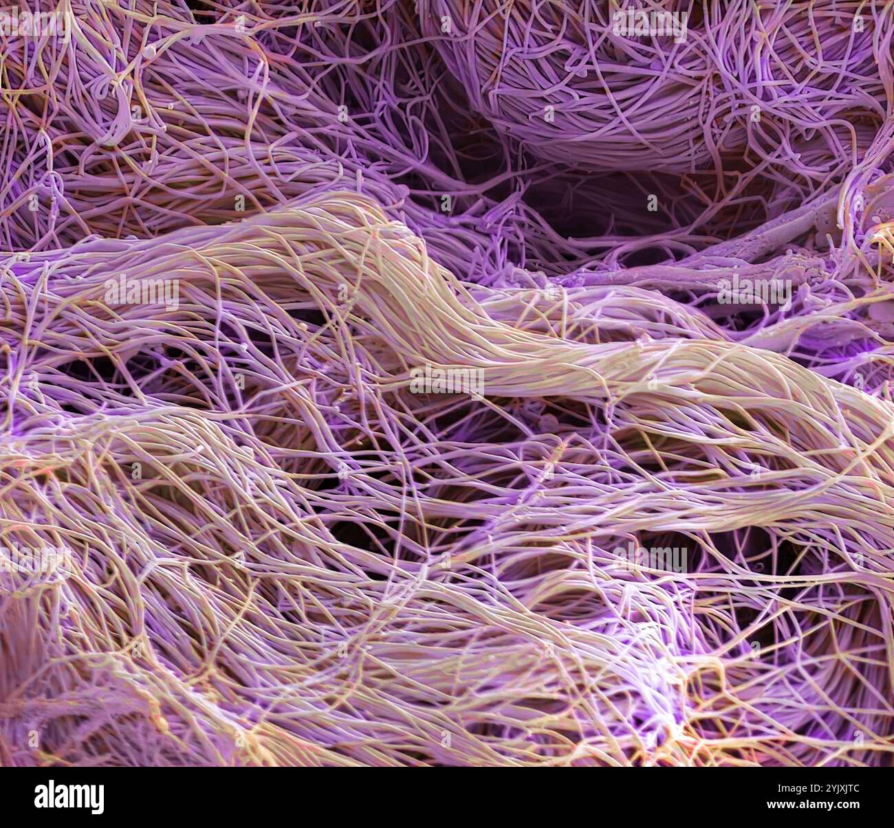 Connective tissue. Coloured scanning electron micrograph (SEM) of collagen from a nasal poly biopsy. Collagen is a protein with a high tensile strength, providing structure and elasticity to epithelium, tendons, ligaments and bones. It is the most abundant protein in the body. In the dermis, collagen forms rope-like fibres that are arranged irregularly. Magnification: x1300 when printed 10 centimetres wide. Stock Photohttps://www.alamy.com/image-license-details/?v=1https://www.alamy.com/connective-tissue-coloured-scanning-electron-micrograph-sem-of-collagen-from-a-nasal-poly-biopsy-collagen-is-a-protein-with-a-high-tensile-strength-providing-structure-and-elasticity-to-epithelium-tendons-ligaments-and-bones-it-is-the-most-abundant-protein-in-the-body-in-the-dermis-collagen-forms-rope-like-fibres-that-are-arranged-irregularly-magnification-x1300-when-printed-10-centimetres-wide-image631222604.html
RF2YJXJTC–Connective tissue. Coloured scanning electron micrograph (SEM) of collagen from a nasal poly biopsy. Collagen is a protein with a high tensile strength, providing structure and elasticity to epithelium, tendons, ligaments and bones. It is the most abundant protein in the body. In the dermis, collagen forms rope-like fibres that are arranged irregularly. Magnification: x1300 when printed 10 centimetres wide.
 Eyelash. Coloured scanning electron micrograph (SEM) of a fracture through an eyelash. A hair is growing from a follicle in the underlying layer of skin (the dermis) surrounding the eye. The portion of the hair protruding from the skin is made up of dead tissue and the protein keratin. Eyelashes protect the eye from debris. They are sensitive to being touched, thus providing a warning that an object is near the eye which is then closed reflexively. Magnification: x1600 when printed at 10 centimetres wide. Stock Photohttps://www.alamy.com/image-license-details/?v=1https://www.alamy.com/eyelash-coloured-scanning-electron-micrograph-sem-of-a-fracture-through-an-eyelash-a-hair-is-growing-from-a-follicle-in-the-underlying-layer-of-skin-the-dermis-surrounding-the-eye-the-portion-of-the-hair-protruding-from-the-skin-is-made-up-of-dead-tissue-and-the-protein-keratin-eyelashes-protect-the-eye-from-debris-they-are-sensitive-to-being-touched-thus-providing-a-warning-that-an-object-is-near-the-eye-which-is-then-closed-reflexively-magnification-x1600-when-printed-at-10-centimetres-wide-image622879732.html
RF2Y5AHCM–Eyelash. Coloured scanning electron micrograph (SEM) of a fracture through an eyelash. A hair is growing from a follicle in the underlying layer of skin (the dermis) surrounding the eye. The portion of the hair protruding from the skin is made up of dead tissue and the protein keratin. Eyelashes protect the eye from debris. They are sensitive to being touched, thus providing a warning that an object is near the eye which is then closed reflexively. Magnification: x1600 when printed at 10 centimetres wide.
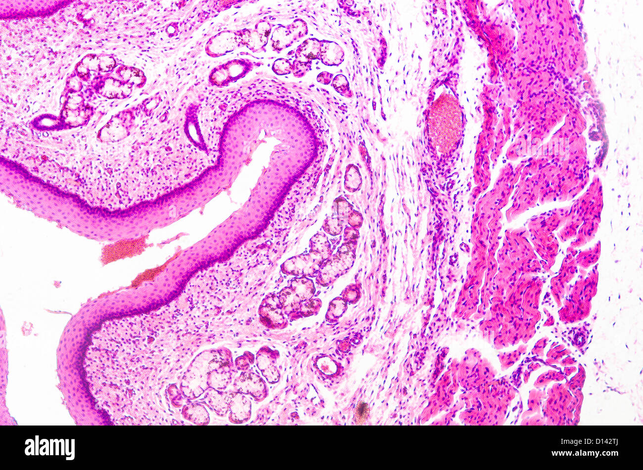 micrograph of medical science stratified squamous epithelium tissue cell Stock Photohttps://www.alamy.com/image-license-details/?v=1https://www.alamy.com/stock-photo-micrograph-of-medical-science-stratified-squamous-epithelium-tissue-52335826.html
RFD142TJ–micrograph of medical science stratified squamous epithelium tissue cell
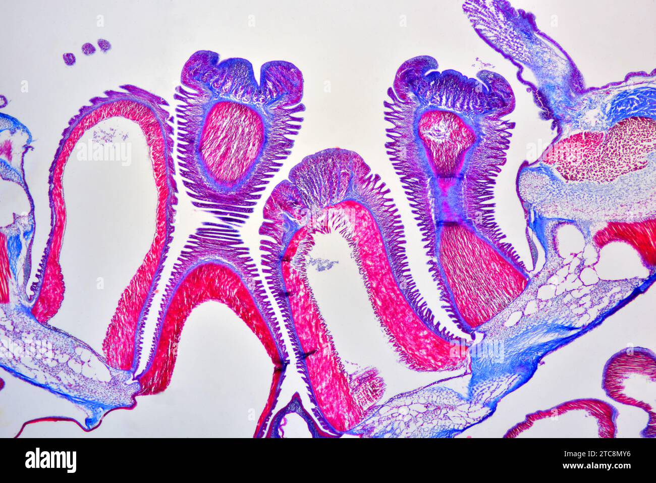 Asterias sp. cross section showing dermis, mucus glands, suckers and tube feets. Light microscope X50 at 10 cm wide. Stock Photohttps://www.alamy.com/image-license-details/?v=1https://www.alamy.com/asterias-sp-cross-section-showing-dermis-mucus-glands-suckers-and-tube-feets-light-microscope-x50-at-10-cm-wide-image575510074.html
RF2TC8MY6–Asterias sp. cross section showing dermis, mucus glands, suckers and tube feets. Light microscope X50 at 10 cm wide.
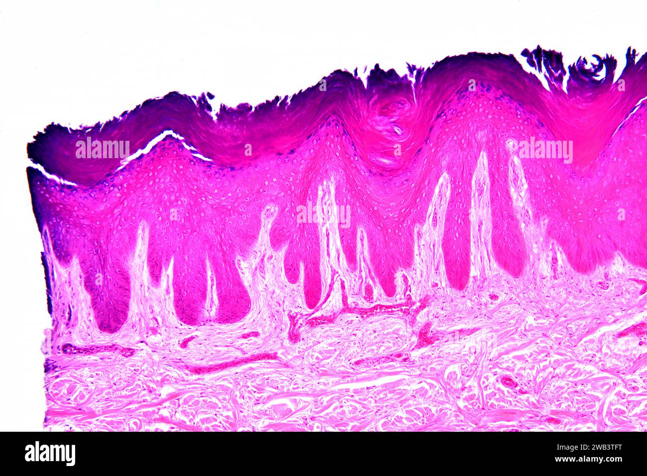 Stratified squamous epithelium from human hand skin showing keratinized epidermis and dermis with connective tissues. X75 at 10 cm wide. Stock Photohttps://www.alamy.com/image-license-details/?v=1https://www.alamy.com/stratified-squamous-epithelium-from-human-hand-skin-showing-keratinized-epidermis-and-dermis-with-connective-tissues-x75-at-10-cm-wide-image591998844.html
RF2WB3TFT–Stratified squamous epithelium from human hand skin showing keratinized epidermis and dermis with connective tissues. X75 at 10 cm wide.
 Eyelash. Coloured scanning electron micrograph (SEM) of a fracture through an eyelash. A hair is growing from a follicle in the underlying layer of skin (the dermis) surrounding the eye. The portion of the hair protruding from the skin is made up of dead tissue and the protein keratin. Eyelashes protect the eye from debris. They are sensitive to being touched, thus providing a warning that an object is near the eye which is then closed reflexively. Magnification: x1600 when printed at 10 centimetres wide. Stock Photohttps://www.alamy.com/image-license-details/?v=1https://www.alamy.com/eyelash-coloured-scanning-electron-micrograph-sem-of-a-fracture-through-an-eyelash-a-hair-is-growing-from-a-follicle-in-the-underlying-layer-of-skin-the-dermis-surrounding-the-eye-the-portion-of-the-hair-protruding-from-the-skin-is-made-up-of-dead-tissue-and-the-protein-keratin-eyelashes-protect-the-eye-from-debris-they-are-sensitive-to-being-touched-thus-providing-a-warning-that-an-object-is-near-the-eye-which-is-then-closed-reflexively-magnification-x1600-when-printed-at-10-centimetres-wide-image622879710.html
RF2Y5AHBX–Eyelash. Coloured scanning electron micrograph (SEM) of a fracture through an eyelash. A hair is growing from a follicle in the underlying layer of skin (the dermis) surrounding the eye. The portion of the hair protruding from the skin is made up of dead tissue and the protein keratin. Eyelashes protect the eye from debris. They are sensitive to being touched, thus providing a warning that an object is near the eye which is then closed reflexively. Magnification: x1600 when printed at 10 centimetres wide.
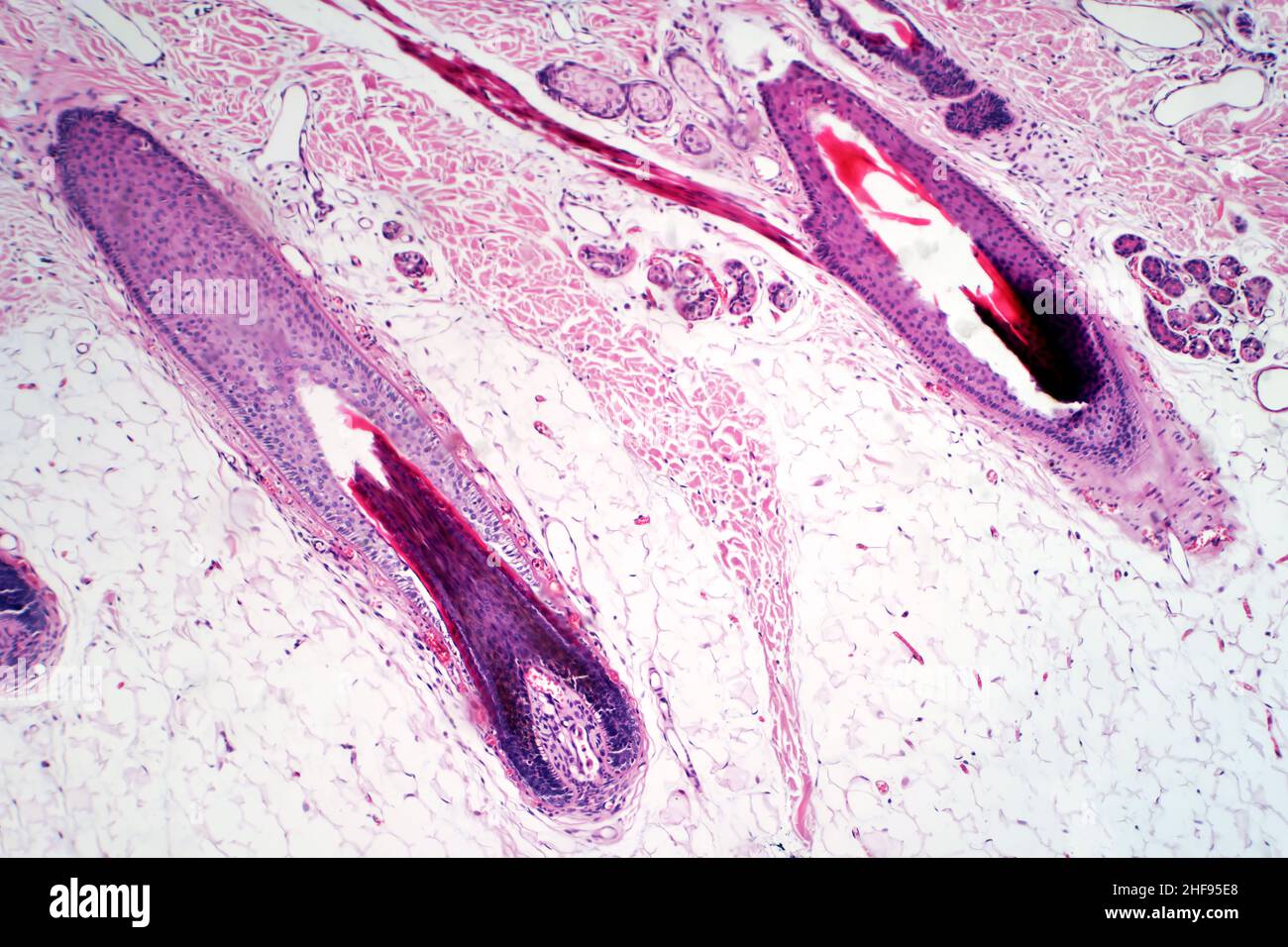 Human scalp and hair follicle, light micrograph Stock Photohttps://www.alamy.com/image-license-details/?v=1https://www.alamy.com/human-scalp-and-hair-follicle-light-micrograph-image456891296.html
RF2HF95E8–Human scalp and hair follicle, light micrograph
 Hair follicles, light micrograph Stock Photohttps://www.alamy.com/image-license-details/?v=1https://www.alamy.com/stock-photo-hair-follicles-light-micrograph-30585720.html
RFBNN8B4–Hair follicles, light micrograph
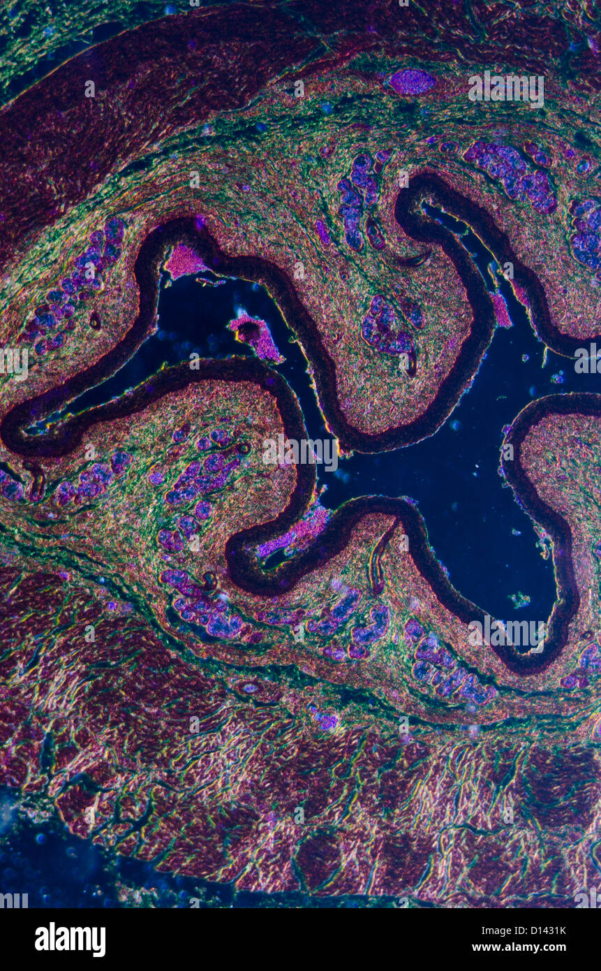 micrograph of medical science stratified squamous epithelium tissue cell Stock Photohttps://www.alamy.com/image-license-details/?v=1https://www.alamy.com/stock-photo-micrograph-of-medical-science-stratified-squamous-epithelium-tissue-52335967.html
RFD1431K–micrograph of medical science stratified squamous epithelium tissue cell
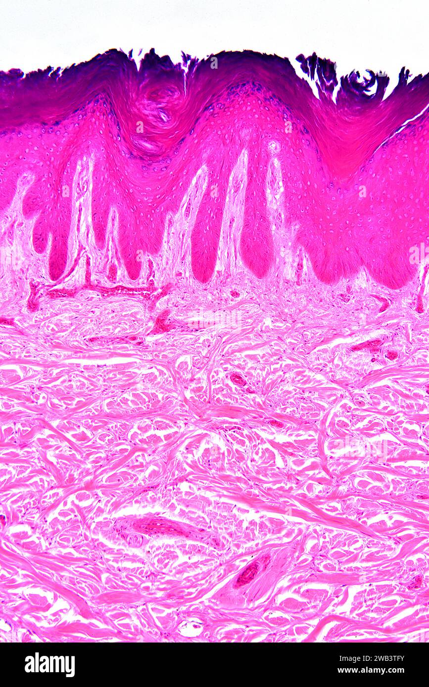 Stratified squamous epithelium from human hand skin showing keratinized epidermis and dermis with connective tissues. X75 at 10 cm wide. Stock Photohttps://www.alamy.com/image-license-details/?v=1https://www.alamy.com/stratified-squamous-epithelium-from-human-hand-skin-showing-keratinized-epidermis-and-dermis-with-connective-tissues-x75-at-10-cm-wide-image591998847.html
RF2WB3TFY–Stratified squamous epithelium from human hand skin showing keratinized epidermis and dermis with connective tissues. X75 at 10 cm wide.
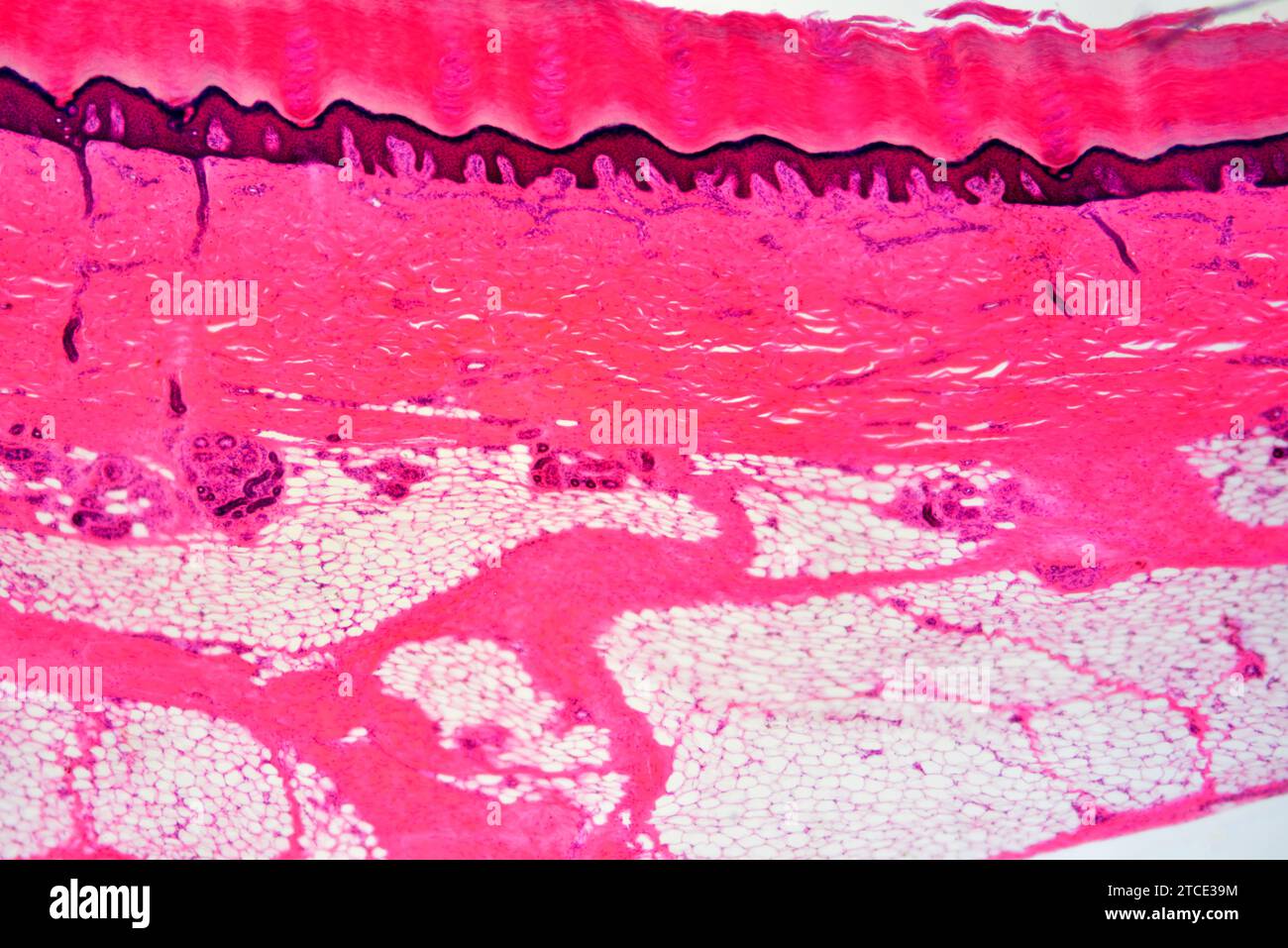 Skin showing transversal section epidermis and dermis with sweat gland, blood vessels, adipose tissue and collagen fibers. Optical microscope X40. Stock Photohttps://www.alamy.com/image-license-details/?v=1https://www.alamy.com/skin-showing-transversal-section-epidermis-and-dermis-with-sweat-gland-blood-vessels-adipose-tissue-and-collagen-fibers-optical-microscope-x40-image575627968.html
RF2TCE39M–Skin showing transversal section epidermis and dermis with sweat gland, blood vessels, adipose tissue and collagen fibers. Optical microscope X40.
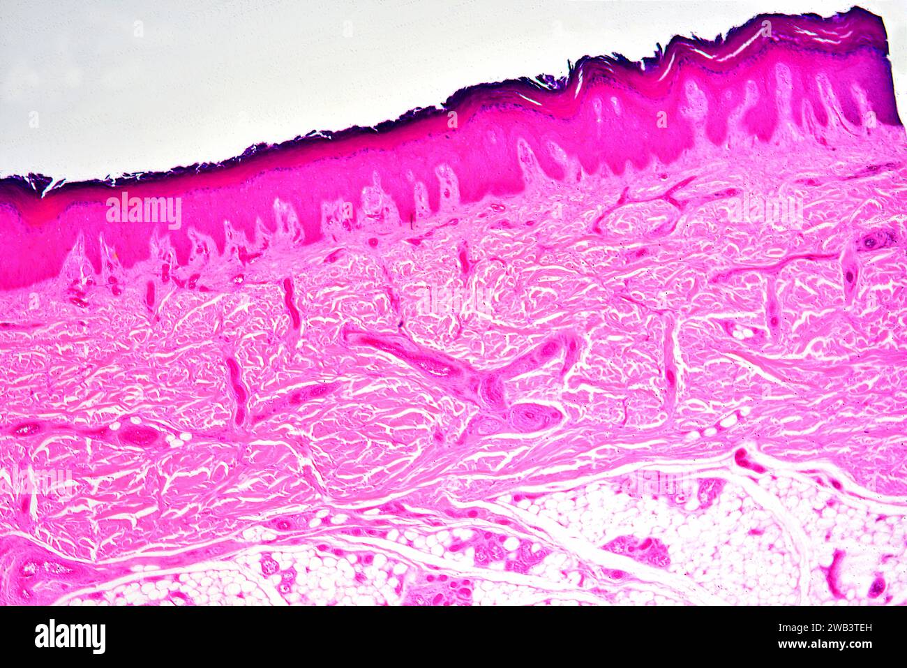 Stratified squamous epithelium from human hand skin showing keratinized epidermis and dermis with connective tissues. X25 at 10 cm wide. Stock Photohttps://www.alamy.com/image-license-details/?v=1https://www.alamy.com/stratified-squamous-epithelium-from-human-hand-skin-showing-keratinized-epidermis-and-dermis-with-connective-tissues-x25-at-10-cm-wide-image591998809.html
RF2WB3TEH–Stratified squamous epithelium from human hand skin showing keratinized epidermis and dermis with connective tissues. X25 at 10 cm wide.
 Hair follicles, light micrograph Stock Photohttps://www.alamy.com/image-license-details/?v=1https://www.alamy.com/stock-photo-hair-follicles-light-micrograph-30585722.html
RFBNN8B6–Hair follicles, light micrograph
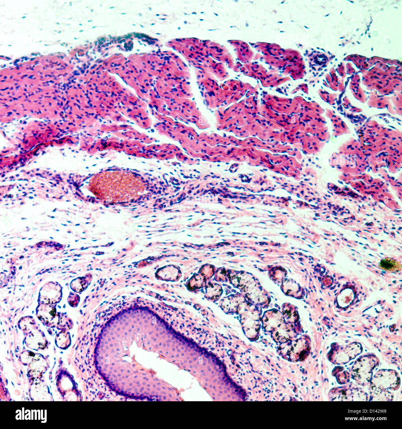 micrograph of medical science stratified squamous epithelium tissue cell Stock Photohttps://www.alamy.com/image-license-details/?v=1https://www.alamy.com/stock-photo-micrograph-of-medical-science-stratified-squamous-epithelium-tissue-52335844.html
RFD142W8–micrograph of medical science stratified squamous epithelium tissue cell
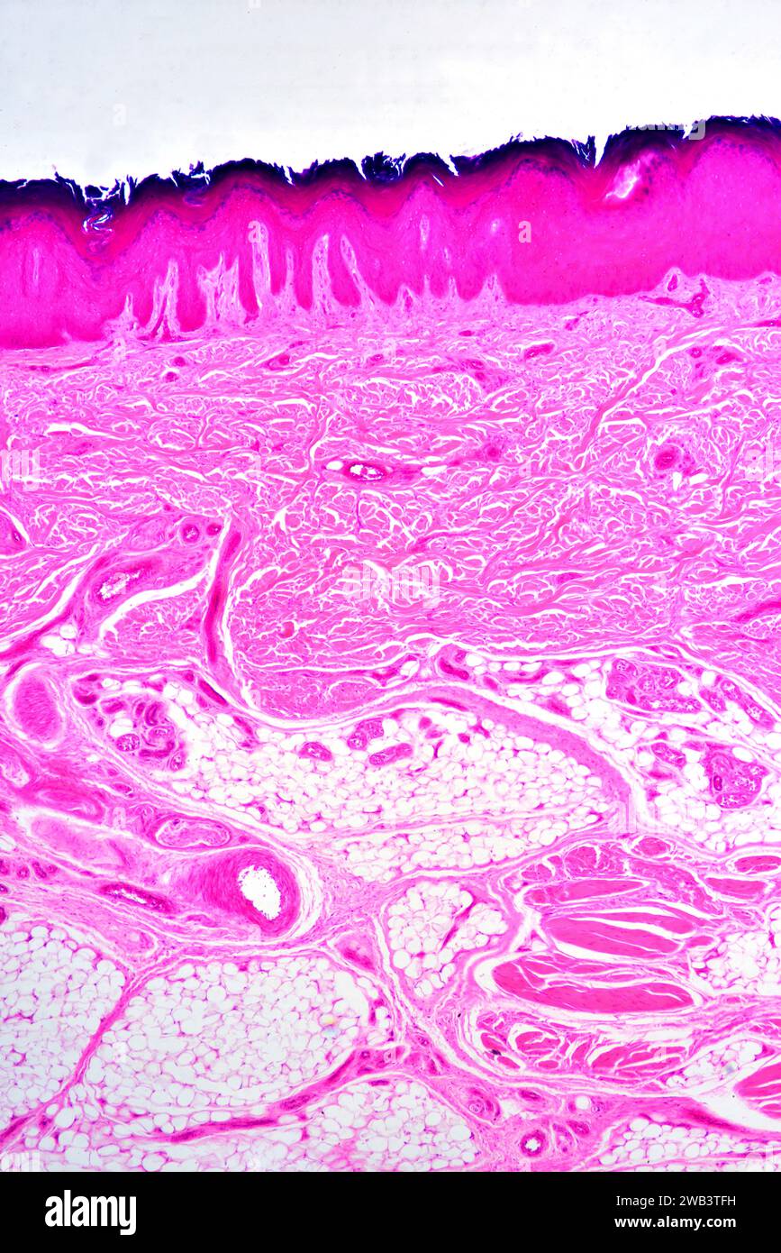 Stratified squamous epithelium from human hand skin showing keratinized epidermis and dermis with connective tissues. X25 at 10 cm wide. Stock Photohttps://www.alamy.com/image-license-details/?v=1https://www.alamy.com/stratified-squamous-epithelium-from-human-hand-skin-showing-keratinized-epidermis-and-dermis-with-connective-tissues-x25-at-10-cm-wide-image591998837.html
RF2WB3TFH–Stratified squamous epithelium from human hand skin showing keratinized epidermis and dermis with connective tissues. X25 at 10 cm wide.
 Human skin. Coloured scanning electronmicrograph (SEM) of healthy human skin. Stock Photohttps://www.alamy.com/image-license-details/?v=1https://www.alamy.com/stock-photo-human-skin-coloured-scanning-electronmicrograph-sem-of-healthy-human-21206250.html
RFB6E0P2–Human skin. Coloured scanning electronmicrograph (SEM) of healthy human skin.
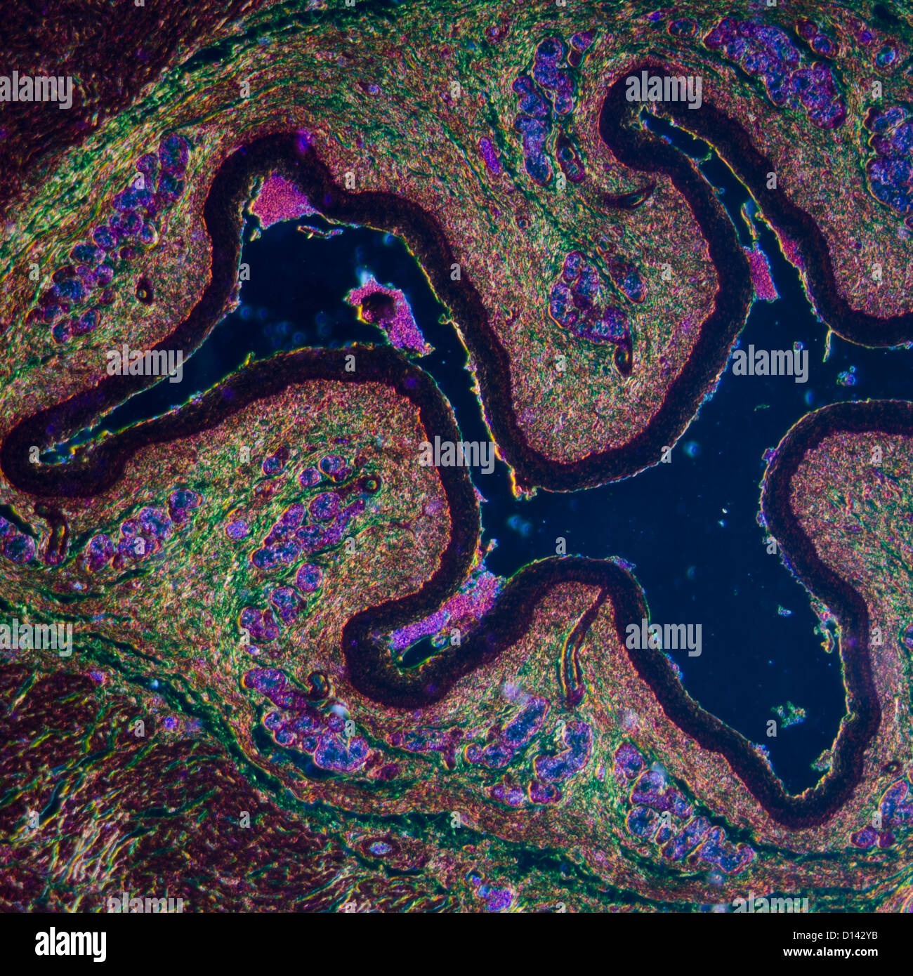 micrograph of medical science stratified squamous epithelium tissue cell Stock Photohttps://www.alamy.com/image-license-details/?v=1https://www.alamy.com/stock-photo-micrograph-of-medical-science-stratified-squamous-epithelium-tissue-52335903.html
RFD142YB–micrograph of medical science stratified squamous epithelium tissue cell
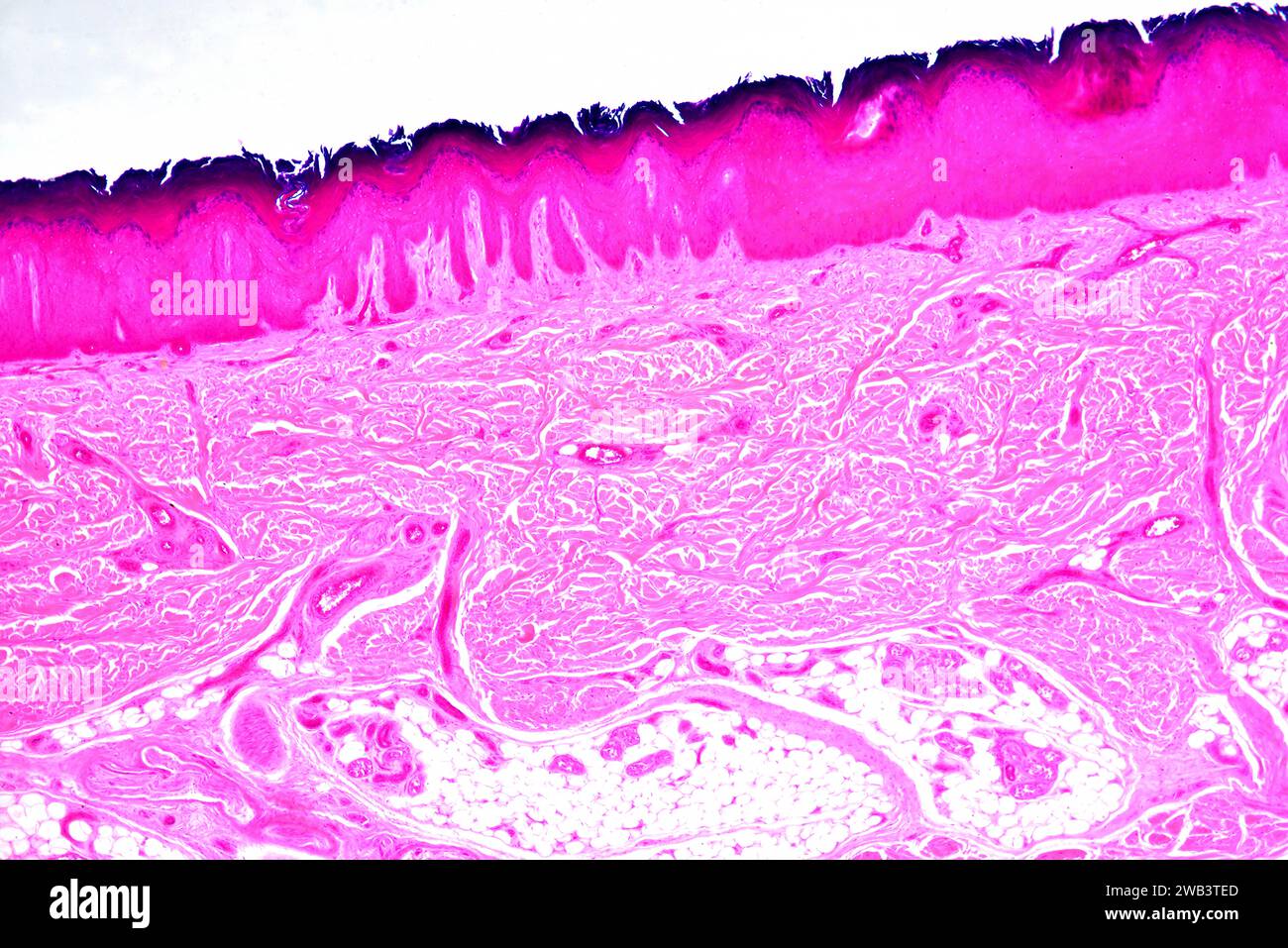 Stratified squamous epithelium from human hand skin showing keratinized epidermis and dermis with connective tissues. X25 at 10 cm wide. Stock Photohttps://www.alamy.com/image-license-details/?v=1https://www.alamy.com/stratified-squamous-epithelium-from-human-hand-skin-showing-keratinized-epidermis-and-dermis-with-connective-tissues-x25-at-10-cm-wide-image591998805.html
RF2WB3TED–Stratified squamous epithelium from human hand skin showing keratinized epidermis and dermis with connective tissues. X25 at 10 cm wide.
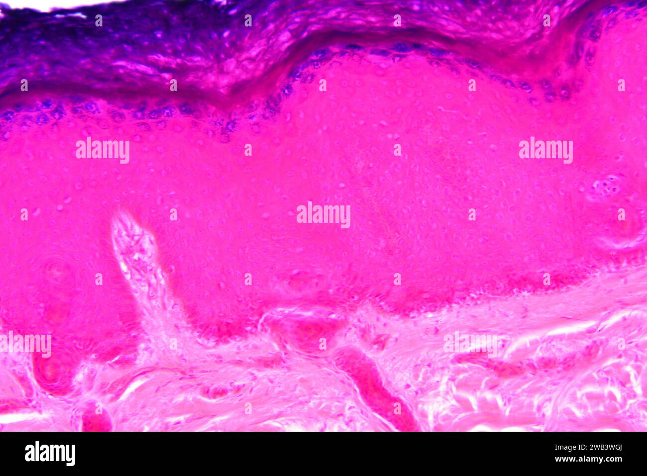 Human skin showing from top to bottom: epidermis with stratum corneum, stratum granulosum, stratum spinosum, stratum basale and dermis. X125 at 10 cm Stock Photohttps://www.alamy.com/image-license-details/?v=1https://www.alamy.com/human-skin-showing-from-top-to-bottom-epidermis-with-stratum-corneum-stratum-granulosum-stratum-spinosum-stratum-basale-and-dermis-x125-at-10-cm-image591999650.html
RF2WB3WGJ–Human skin showing from top to bottom: epidermis with stratum corneum, stratum granulosum, stratum spinosum, stratum basale and dermis. X125 at 10 cm
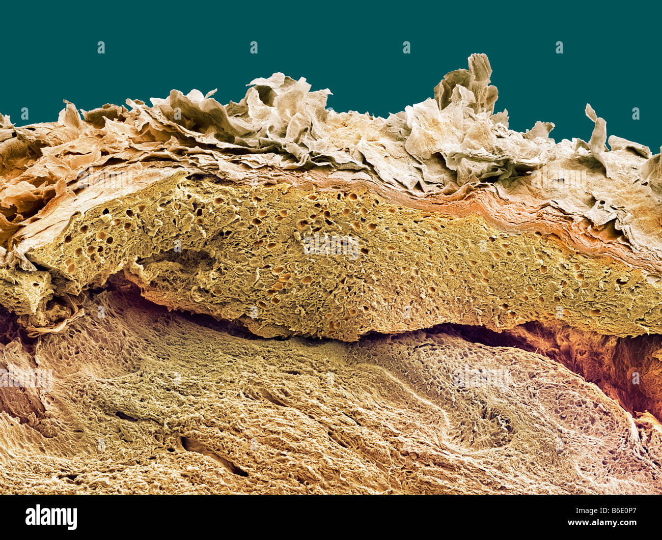 Skin. Coloured scanning electron micrograph(SEM) of a section through healthy skin. Stock Photohttps://www.alamy.com/image-license-details/?v=1https://www.alamy.com/stock-photo-skin-coloured-scanning-electron-micrographsem-of-a-section-through-21206255.html
RFB6E0P7–Skin. Coloured scanning electron micrograph(SEM) of a section through healthy skin.
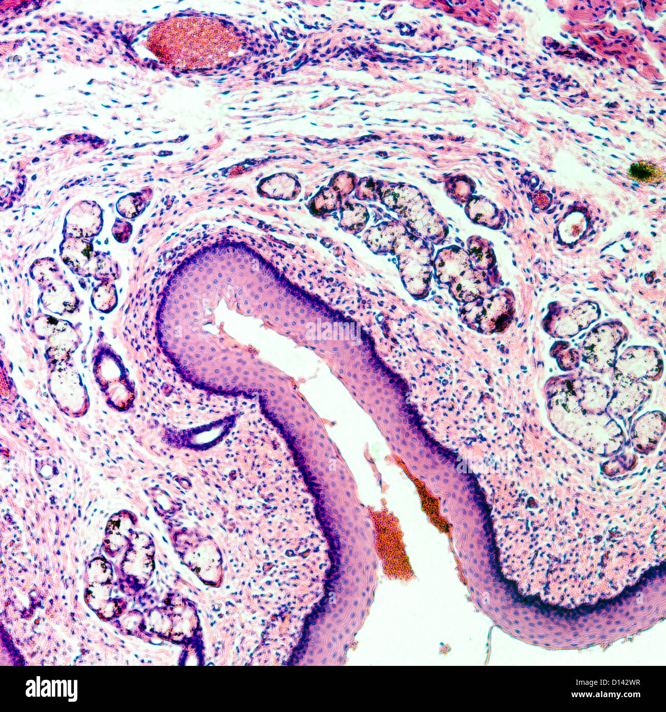 micrograph of medical science stratified squamous epithelium tissue cell Stock Photohttps://www.alamy.com/image-license-details/?v=1https://www.alamy.com/stock-photo-micrograph-of-medical-science-stratified-squamous-epithelium-tissue-52335859.html
RFD142WR–micrograph of medical science stratified squamous epithelium tissue cell
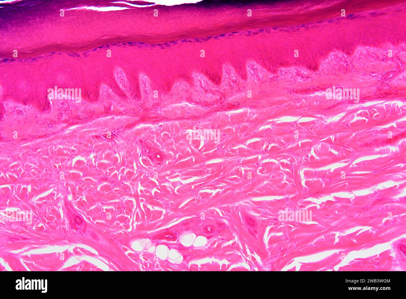 Human skin showing from top to bottom: epidermis with stratum corneum, stratum granulosum, stratum spinosum, stratum basale and dermis. X75 at 10 cm w Stock Photohttps://www.alamy.com/image-license-details/?v=1https://www.alamy.com/human-skin-showing-from-top-to-bottom-epidermis-with-stratum-corneum-stratum-granulosum-stratum-spinosum-stratum-basale-and-dermis-x75-at-10-cm-w-image591999652.html
RF2WB3WGM–Human skin showing from top to bottom: epidermis with stratum corneum, stratum granulosum, stratum spinosum, stratum basale and dermis. X75 at 10 cm w
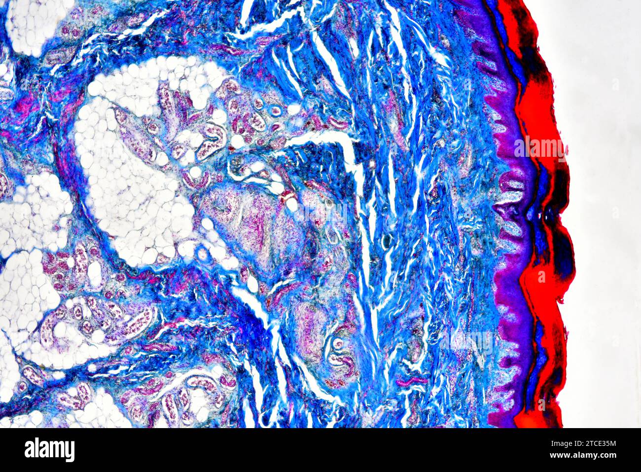 Human skin showing epidermis, dermis, adipose tissue, blood vessels and collagen fibers. Optical microscope X40. Stock Photohttps://www.alamy.com/image-license-details/?v=1https://www.alamy.com/human-skin-showing-epidermis-dermis-adipose-tissue-blood-vessels-and-collagen-fibers-optical-microscope-x40-image575627856.html
RF2TCE35M–Human skin showing epidermis, dermis, adipose tissue, blood vessels and collagen fibers. Optical microscope X40.
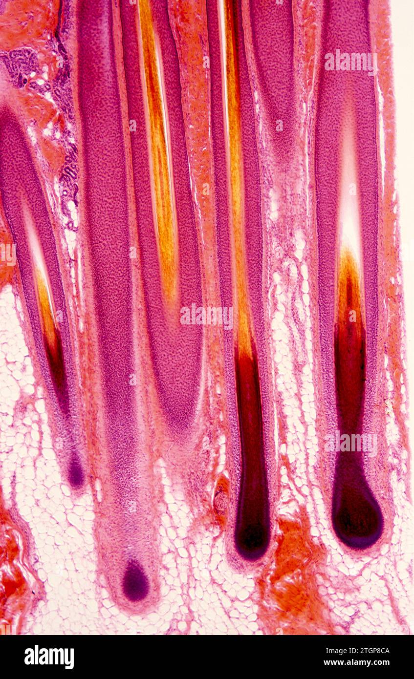 Hair follicles on a human skin. Photomicrograph. Stock Photohttps://www.alamy.com/image-license-details/?v=1https://www.alamy.com/hair-follicles-on-a-human-skin-photomicrograph-image578266202.html
RF2TGP8CA–Hair follicles on a human skin. Photomicrograph.
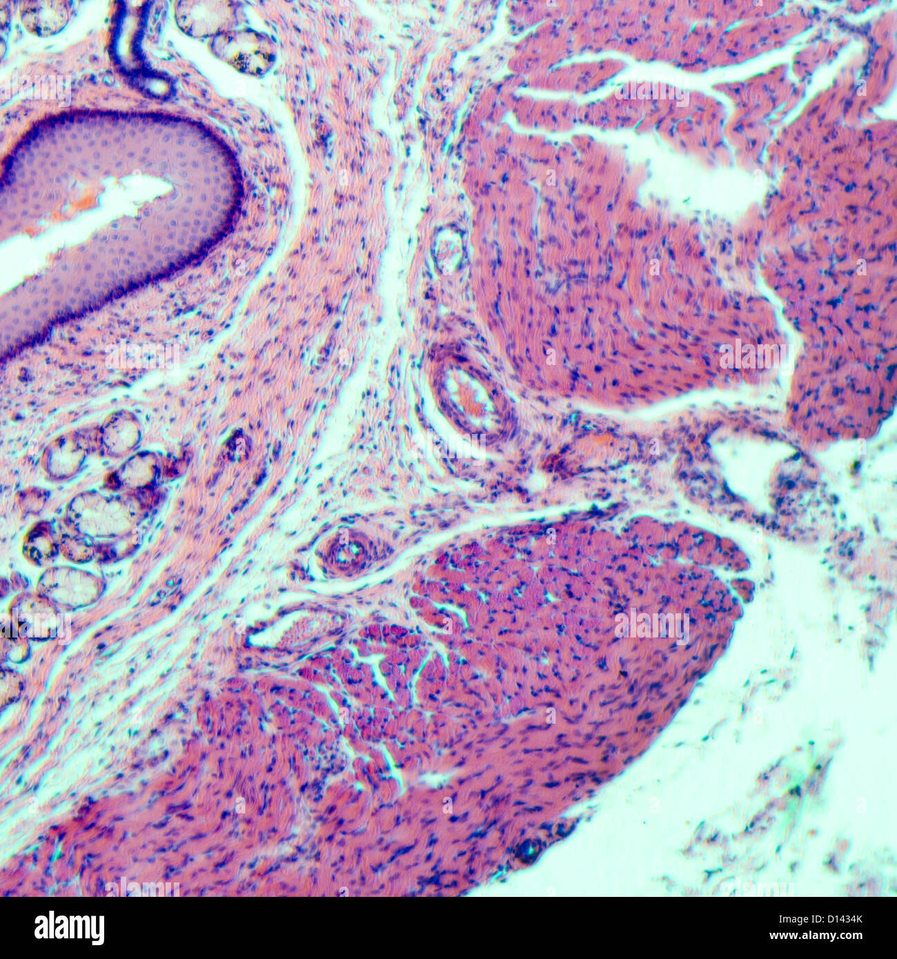 micrograph of medical science stratified squamous epithelium tissue cell Stock Photohttps://www.alamy.com/image-license-details/?v=1https://www.alamy.com/stock-photo-micrograph-of-medical-science-stratified-squamous-epithelium-tissue-52336051.html
RFD1434K–micrograph of medical science stratified squamous epithelium tissue cell
 Hair follicles on a human skin. Photomicrograph. Stock Photohttps://www.alamy.com/image-license-details/?v=1https://www.alamy.com/hair-follicles-on-a-human-skin-photomicrograph-image578266397.html
RF2TGP8K9–Hair follicles on a human skin. Photomicrograph.
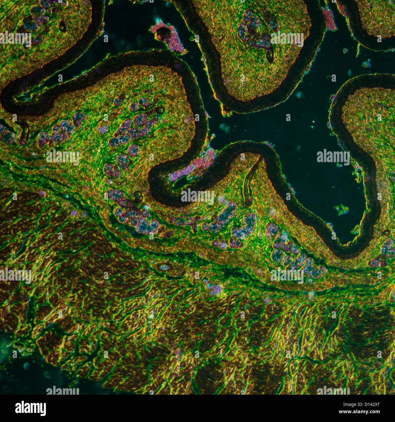 micrograph of medical science stratified squamous epithelium tissue cell Stock Photohttps://www.alamy.com/image-license-details/?v=1https://www.alamy.com/stock-photo-micrograph-of-medical-science-stratified-squamous-epithelium-tissue-52335888.html
RFD142XT–micrograph of medical science stratified squamous epithelium tissue cell
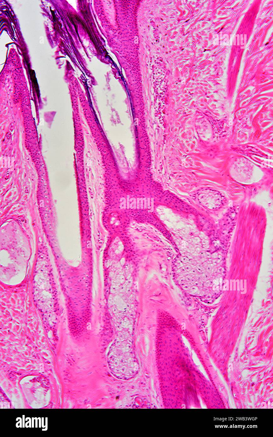 Human skin showing sebaceous gland and hair follicle. X75 at 10 cm high. Stock Photohttps://www.alamy.com/image-license-details/?v=1https://www.alamy.com/human-skin-showing-sebaceous-gland-and-hair-follicle-x75-at-10-cm-high-image591999654.html
RF2WB3WGP–Human skin showing sebaceous gland and hair follicle. X75 at 10 cm high.
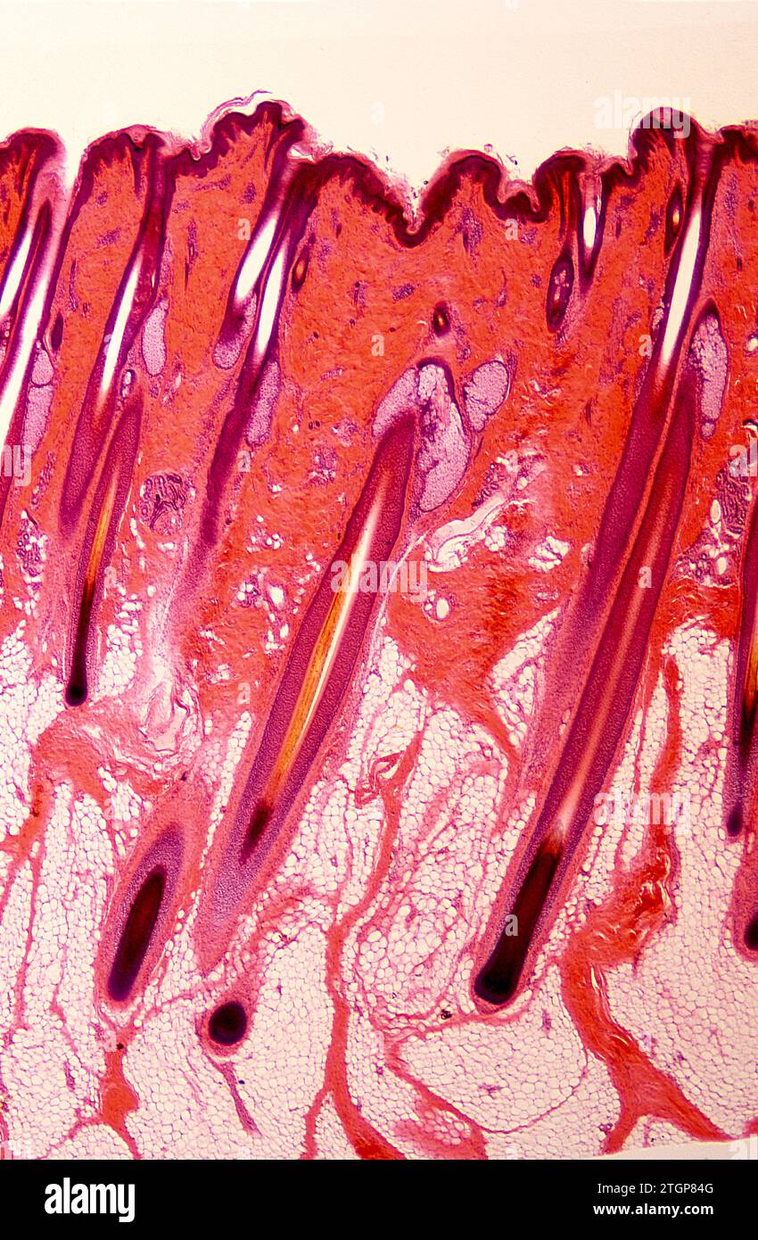 Hair follicles on a human skin. Photomicrograph. Stock Photohttps://www.alamy.com/image-license-details/?v=1https://www.alamy.com/hair-follicles-on-a-human-skin-photomicrograph-image578265984.html
RF2TGP84G–Hair follicles on a human skin. Photomicrograph.
 Human skin showing sebaceous gland, blood vessels and hair follicle. X75 at 10 cm wide. Stock Photohttps://www.alamy.com/image-license-details/?v=1https://www.alamy.com/human-skin-showing-sebaceous-gland-blood-vessels-and-hair-follicle-x75-at-10-cm-wide-image591999657.html
RF2WB3WGW–Human skin showing sebaceous gland, blood vessels and hair follicle. X75 at 10 cm wide.
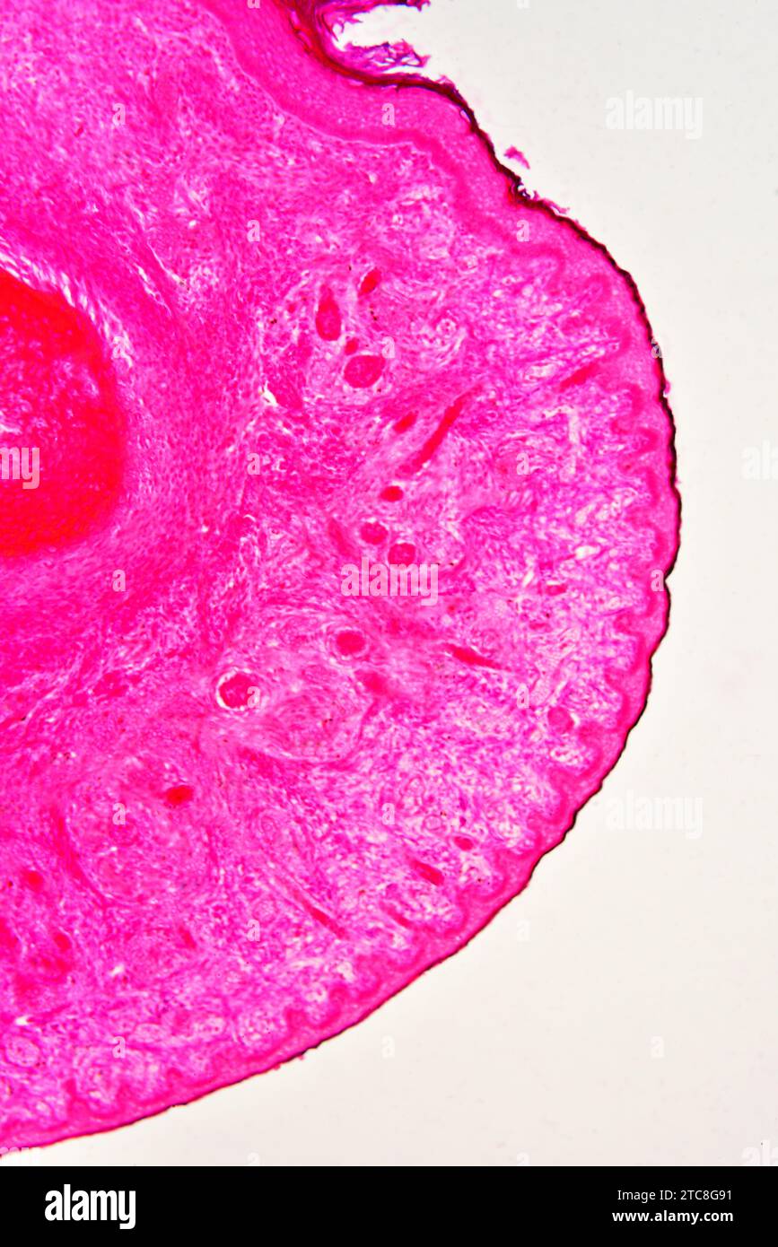 Epithelium of human fetus finger. Light microscope X100. Stock Photohttps://www.alamy.com/image-license-details/?v=1https://www.alamy.com/epithelium-of-human-fetus-finger-light-microscope-x100-image575506429.html
RF2TC8G91–Epithelium of human fetus finger. Light microscope X100.
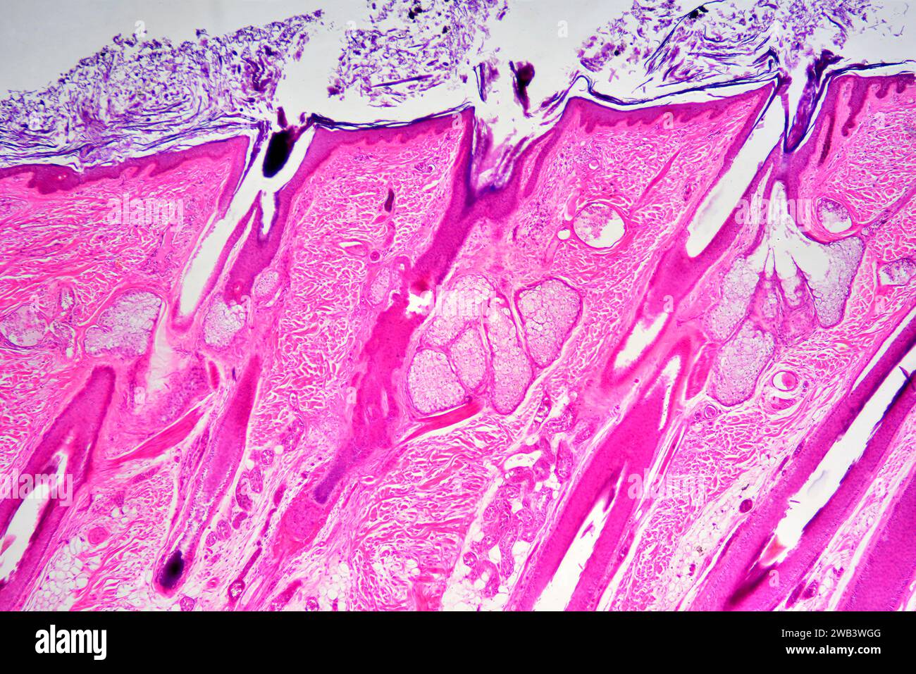 Sebaceous glands in human skin with hair follicles. X25 at 10 cm wide. Stock Photohttps://www.alamy.com/image-license-details/?v=1https://www.alamy.com/sebaceous-glands-in-human-skin-with-hair-follicles-x25-at-10-cm-wide-image591999648.html
RF2WB3WGG–Sebaceous glands in human skin with hair follicles. X25 at 10 cm wide.
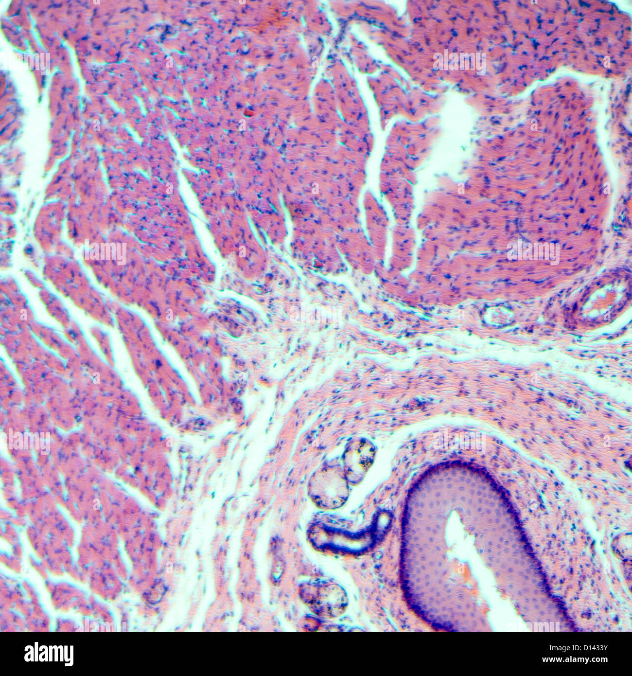 micrograph of medical science stratified squamous epithelium tissue cell Stock Photohttps://www.alamy.com/image-license-details/?v=1https://www.alamy.com/stock-photo-micrograph-of-medical-science-stratified-squamous-epithelium-tissue-52336031.html
RFD1433Y–micrograph of medical science stratified squamous epithelium tissue cell
 micrograph of medical science stratified squamous epithelium tissue cell Stock Photohttps://www.alamy.com/image-license-details/?v=1https://www.alamy.com/stock-photo-micrograph-of-medical-science-stratified-squamous-epithelium-tissue-52336013.html
RFD14339–micrograph of medical science stratified squamous epithelium tissue cell
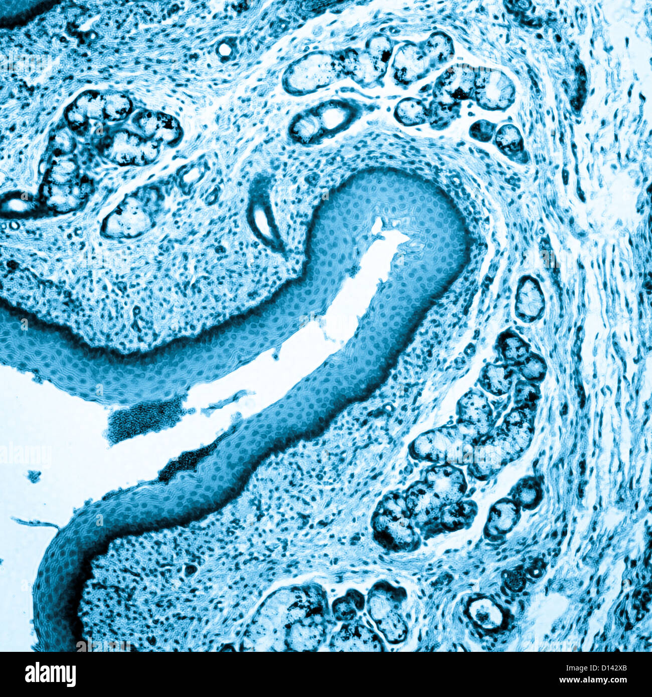 micrograph of medical science stratified squamous epithelium tissue cell Stock Photohttps://www.alamy.com/image-license-details/?v=1https://www.alamy.com/stock-photo-micrograph-of-medical-science-stratified-squamous-epithelium-tissue-52335875.html
RFD142XB–micrograph of medical science stratified squamous epithelium tissue cell