Quick filters:
Diagram of foot Stock Photos and Images
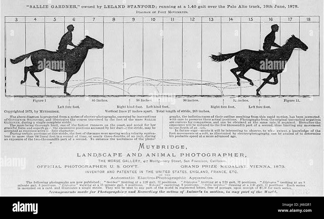 Muybridge Diagram of Foot Movements 1879 Stock Photohttps://www.alamy.com/image-license-details/?v=1https://www.alamy.com/stock-photo-muybridge-diagram-of-foot-movements-1879-140286469.html
RMJ46GR1–Muybridge Diagram of Foot Movements 1879
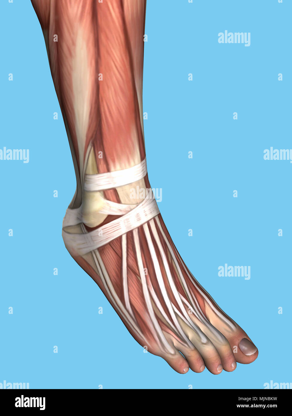 Anatomy of foot Stock Photohttps://www.alamy.com/image-license-details/?v=1https://www.alamy.com/anatomy-of-foot-image183637661.html
RMMJNBKW–Anatomy of foot
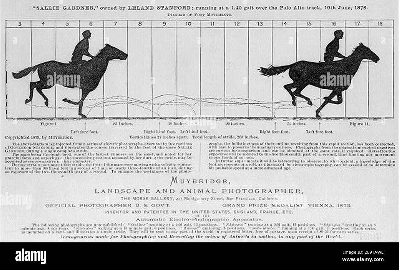 Muybridge Diagram of Foot Movements 1879. Stock Photohttps://www.alamy.com/image-license-details/?v=1https://www.alamy.com/muybridge-diagram-of-foot-movements-1879-image401905770.html
RM2E9TAWE–Muybridge Diagram of Foot Movements 1879.
 The horse in motion, illus. by Muybridge. Sallie Gardner, owned by Leland Stanford, running at a 1:40 gait over the Palo Alto track, 19 June 1878: 2 frames showing diagram of foot movements Stock Photohttps://www.alamy.com/image-license-details/?v=1https://www.alamy.com/the-horse-in-motion-illus-by-muybridge-sallie-gardner-owned-by-leland-stanford-running-at-a-140-gait-over-the-palo-alto-track-19-june-1878-2-frames-showing-diagram-of-foot-movements-image329741829.html
RM2A4D11W–The horse in motion, illus. by Muybridge. Sallie Gardner, owned by Leland Stanford, running at a 1:40 gait over the Palo Alto track, 19 June 1878: 2 frames showing diagram of foot movements
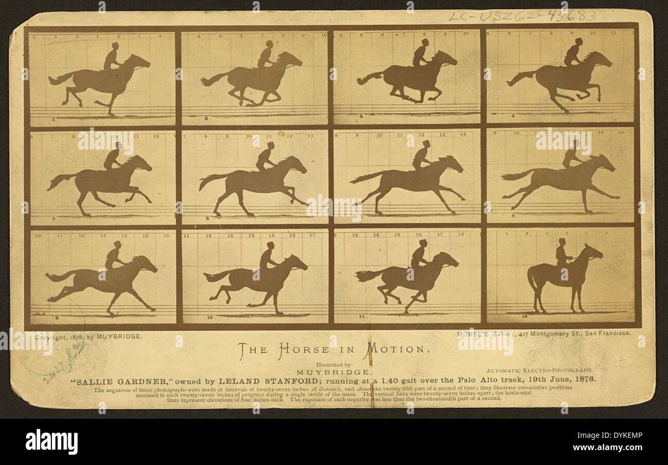 The horse in motion, illus. by Muybridge. 'Sallie Gardner,' owned by Leland Stanford, running at a 1:40 gait over the Palo Alto track, 19 June 1878: 12 frames showing diagram of foot movements (front) Stock Photohttps://www.alamy.com/image-license-details/?v=1https://www.alamy.com/the-horse-in-motion-illus-by-muybridge-sallie-gardner-owned-by-leland-image68655462.html
RMDYKEMP–The horse in motion, illus. by Muybridge. 'Sallie Gardner,' owned by Leland Stanford, running at a 1:40 gait over the Palo Alto track, 19 June 1878: 12 frames showing diagram of foot movements (front)
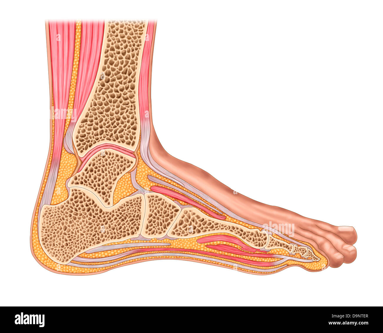 Cross section of anterior human foot with muscles and ligaments. Stock Photohttps://www.alamy.com/image-license-details/?v=1https://www.alamy.com/stock-photo-cross-section-of-anterior-human-foot-with-muscles-and-ligaments-57643231.html
RFD9NTER–Cross section of anterior human foot with muscles and ligaments.
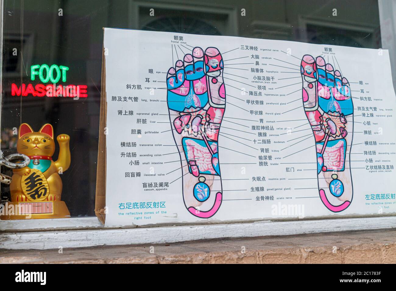 New Orleans Louisiana,French Quarter,store window,Oriental foot massage,feet,diagram,pressure points,reflective zones,Chinese language,bilingual,symbo Stock Photohttps://www.alamy.com/image-license-details/?v=1https://www.alamy.com/new-orleans-louisianafrench-quarterstore-windoworiental-foot-massagefeetdiagrampressure-pointsreflective-zoneschinese-languagebilingualsymbo-image362192419.html
RM2C1783F–New Orleans Louisiana,French Quarter,store window,Oriental foot massage,feet,diagram,pressure points,reflective zones,Chinese language,bilingual,symbo
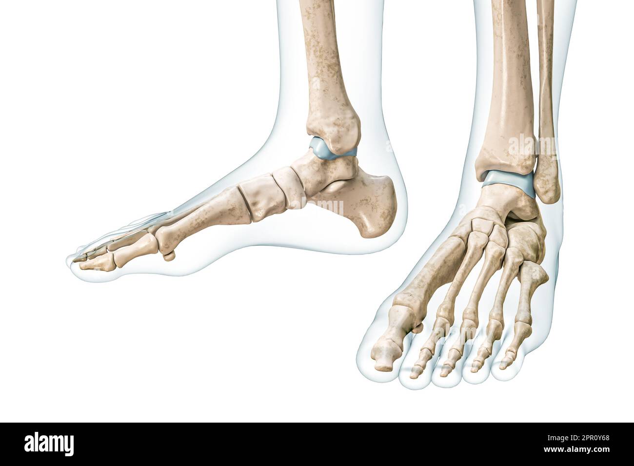 Feet and ankle bones with body contours 3D rendering illustration isolated on white with copy space. Human skeleton and foot anatomy, medical diagram, Stock Photohttps://www.alamy.com/image-license-details/?v=1https://www.alamy.com/feet-and-ankle-bones-with-body-contours-3d-rendering-illustration-isolated-on-white-with-copy-space-human-skeleton-and-foot-anatomy-medical-diagram-image547679840.html
RF2PR0Y68–Feet and ankle bones with body contours 3D rendering illustration isolated on white with copy space. Human skeleton and foot anatomy, medical diagram,
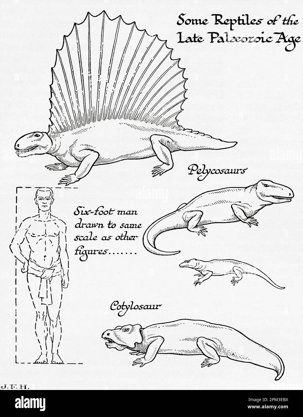 Some reptiles of the Late Paleozoic or Palaeozoic Era. Shown in the diagram a six foot man drawn to the same scale as other figures. From the book Outline of History by H.G. Wells, published 1920. Stock Photohttps://www.alamy.com/image-license-details/?v=1https://www.alamy.com/some-reptiles-of-the-late-paleozoic-or-palaeozoic-era-shown-in-the-diagram-a-six-foot-man-drawn-to-the-same-scale-as-other-figures-from-the-book-outline-of-history-by-hg-wells-published-1920-image545891694.html
RM2PM3EBX–Some reptiles of the Late Paleozoic or Palaeozoic Era. Shown in the diagram a six foot man drawn to the same scale as other figures. From the book Outline of History by H.G. Wells, published 1920.
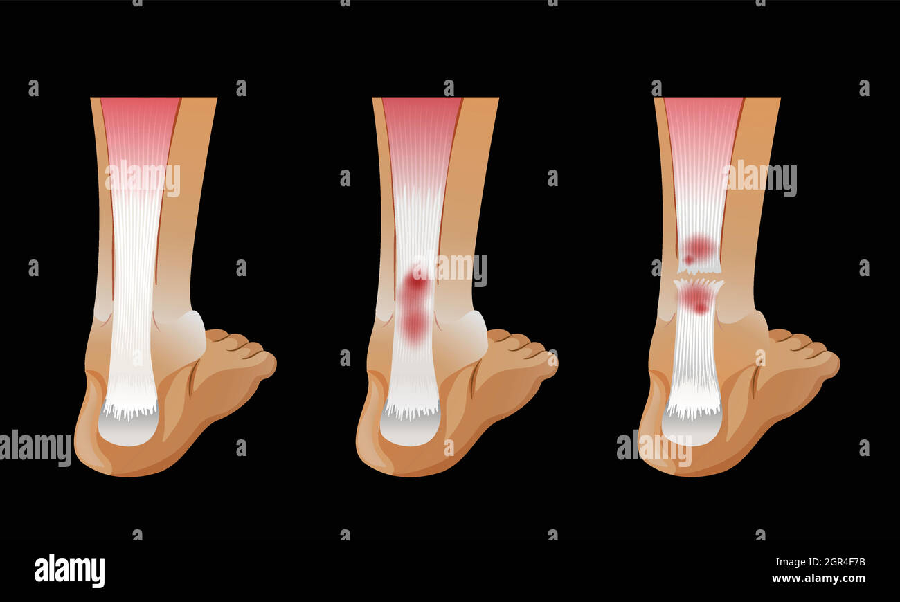 Diagram showing broken bone in human foot Stock Vectorhttps://www.alamy.com/image-license-details/?v=1https://www.alamy.com/diagram-showing-broken-bone-in-human-foot-image444496063.html
RF2GR4F7B–Diagram showing broken bone in human foot
 Major Ligaments of the Foot Stock Photohttps://www.alamy.com/image-license-details/?v=1https://www.alamy.com/stock-photo-major-ligaments-of-the-foot-135011678.html
RMHRJ8NJ–Major Ligaments of the Foot
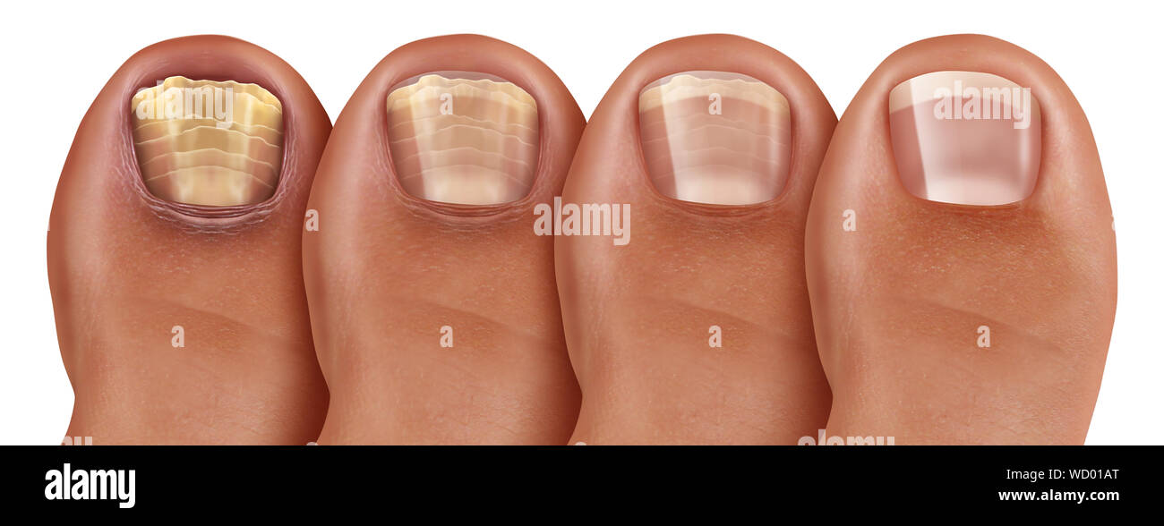 Fungal nail infection recovery diagram and onychomycosisor tinea unguium treatment as an infected foot toenail or toe nail with damaged unhealthy. Stock Photohttps://www.alamy.com/image-license-details/?v=1https://www.alamy.com/fungal-nail-infection-recovery-diagram-and-onychomycosisor-tinea-unguium-treatment-as-an-infected-foot-toenail-or-toe-nail-with-damaged-unhealthy-image266147136.html
RFWD01AT–Fungal nail infection recovery diagram and onychomycosisor tinea unguium treatment as an infected foot toenail or toe nail with damaged unhealthy.
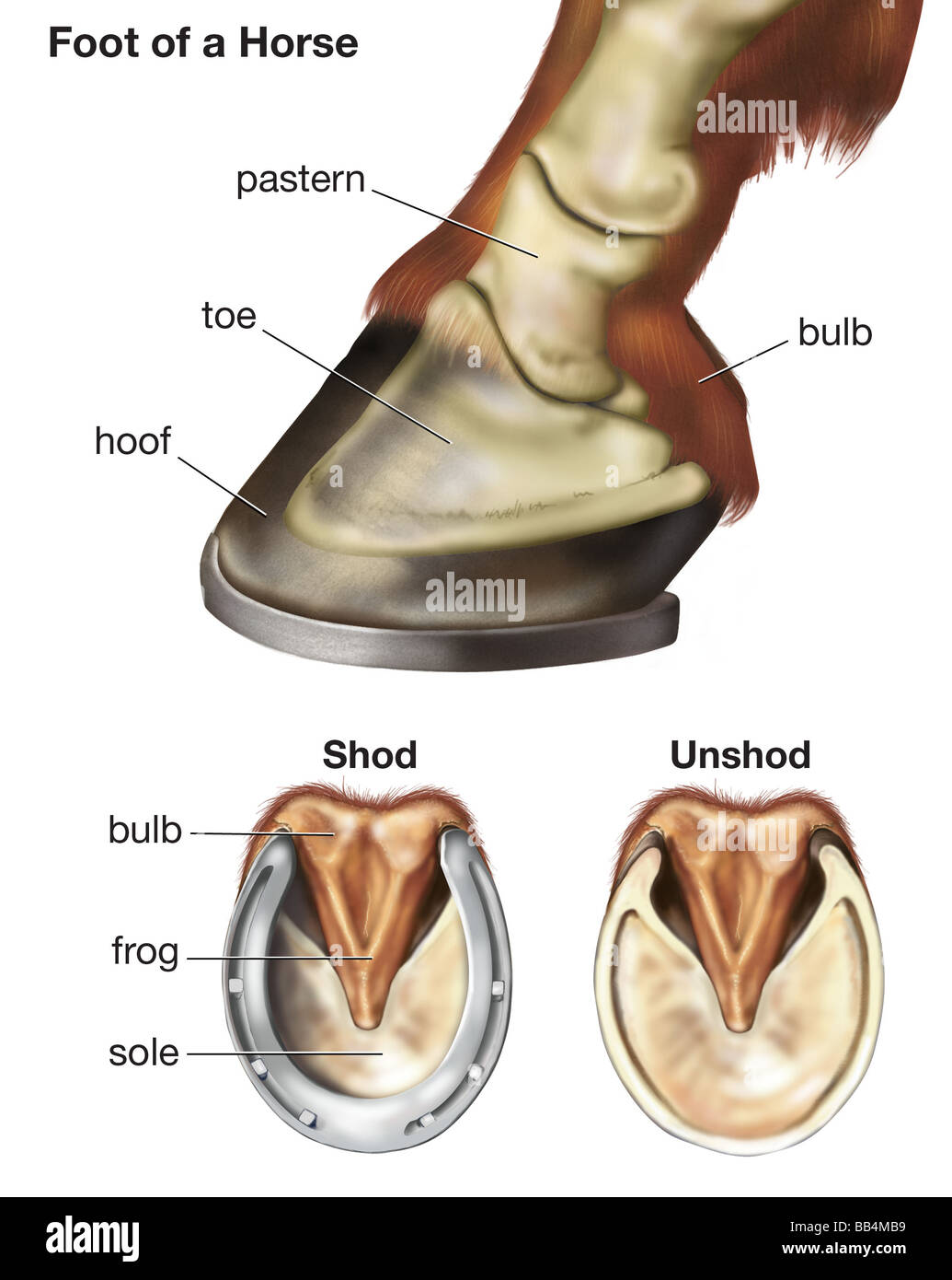 Foot of a horse showing the position of skeletal bones, as well as the difference between a shod and unshod hoof Stock Photohttps://www.alamy.com/image-license-details/?v=1https://www.alamy.com/stock-photo-foot-of-a-horse-showing-the-position-of-skeletal-bones-as-well-as-24075389.html
RMBB4MB9–Foot of a horse showing the position of skeletal bones, as well as the difference between a shod and unshod hoof
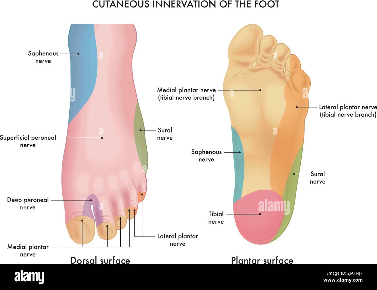 Medical illustration of the cutaneous innervation of the foot, with annotations. Stock Vectorhttps://www.alamy.com/image-license-details/?v=1https://www.alamy.com/medical-illustration-of-the-cutaneous-innervation-of-the-foot-with-annotations-image470865423.html
RF2JA1NJ7–Medical illustration of the cutaneous innervation of the foot, with annotations.
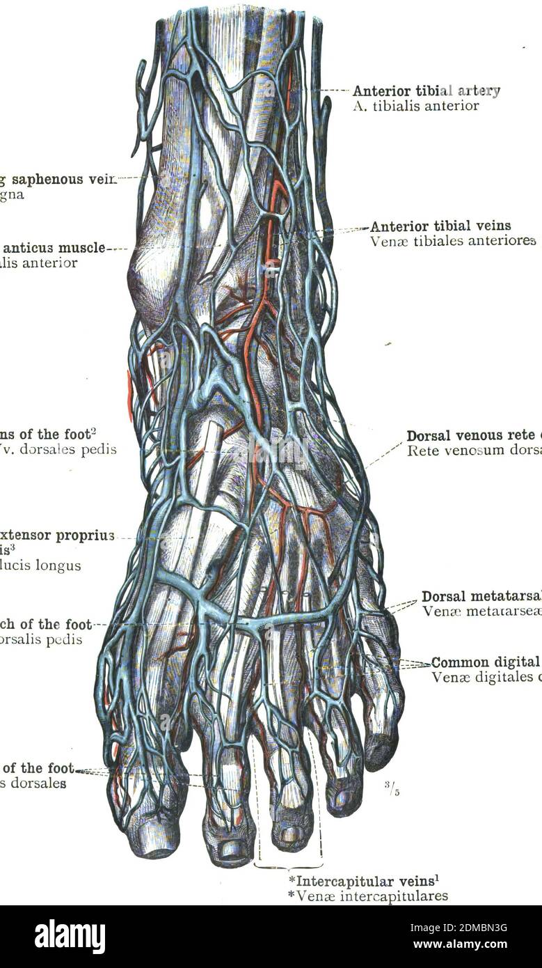 The foot arteries anatomy diagram - topography Stock Photohttps://www.alamy.com/image-license-details/?v=1https://www.alamy.com/the-foot-arteries-anatomy-diagram-topography-image391179252.html
RF2DMBN3G–The foot arteries anatomy diagram - topography
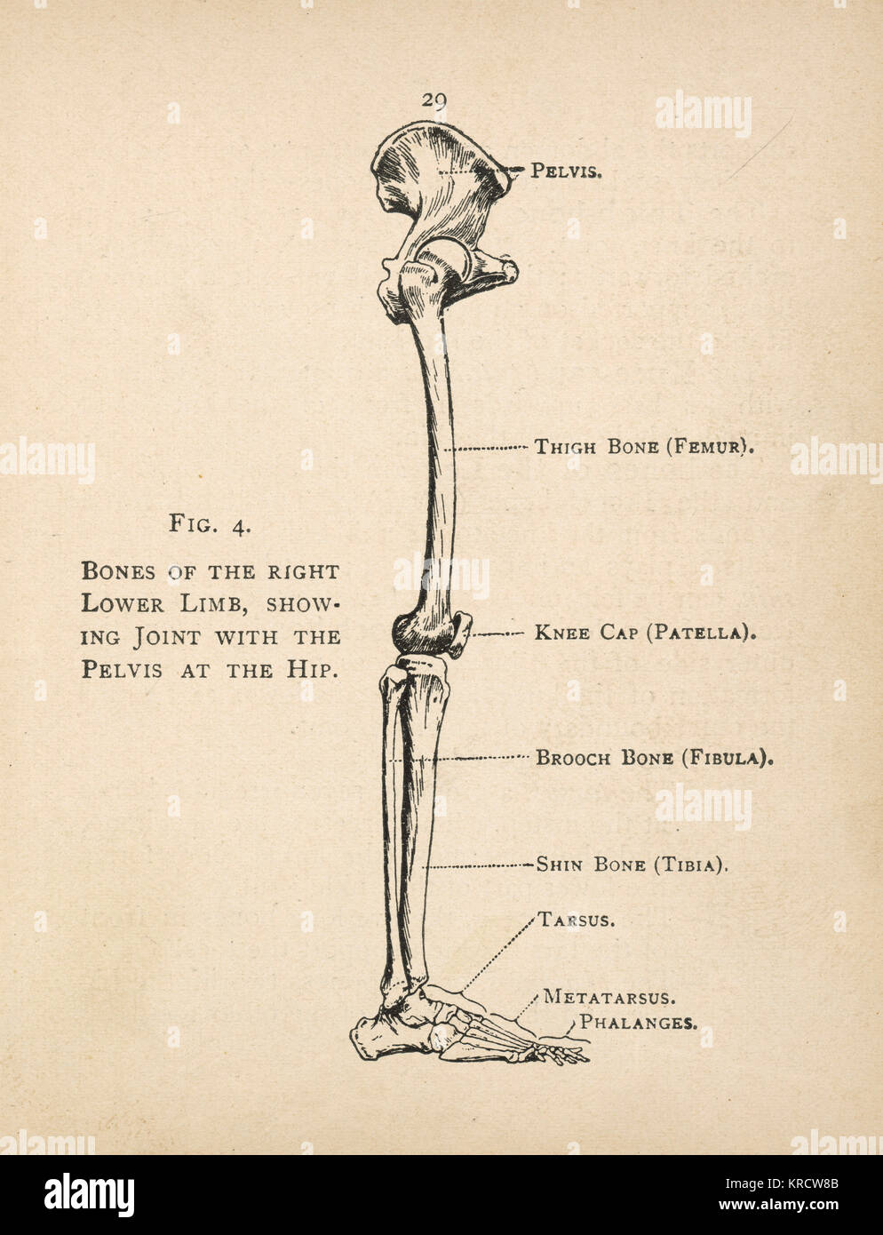 Diagram of the bones of the right leg, showing the joint with the pelvis at the hip. Date: 1908 Stock Photohttps://www.alamy.com/image-license-details/?v=1https://www.alamy.com/stock-image-diagram-of-the-bones-of-the-right-leg-showing-the-joint-with-the-pelvis-169313659.html
RMKRCW8B–Diagram of the bones of the right leg, showing the joint with the pelvis at the hip. Date: 1908
 Bones of the foot of 12 different mamals. Stock Photohttps://www.alamy.com/image-license-details/?v=1https://www.alamy.com/bones-of-the-foot-of-12-different-mamals-image481927462.html
RM2K01KB2–Bones of the foot of 12 different mamals.
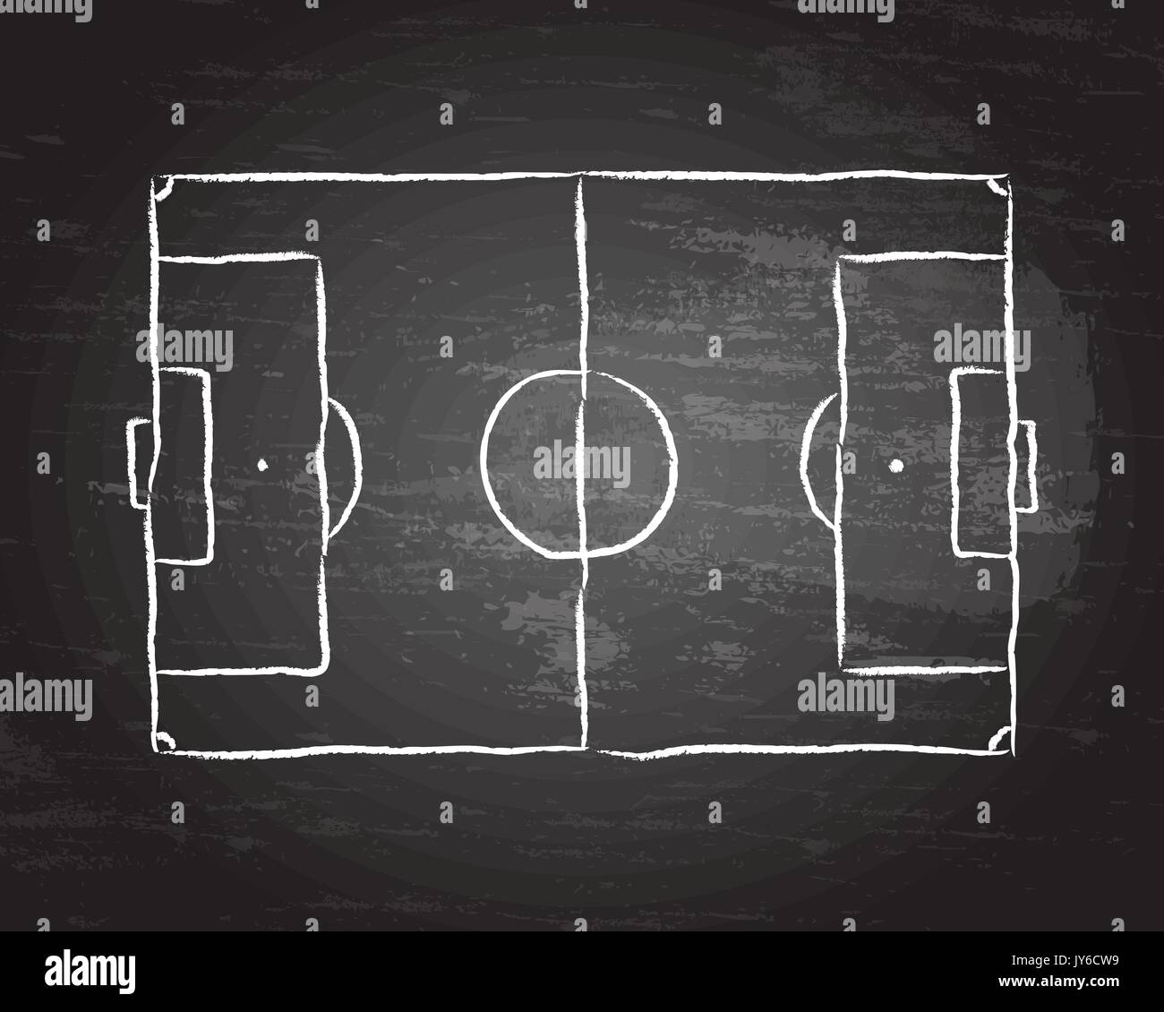 Soccer football pitch diagram on blackboard Stock Vectorhttps://www.alamy.com/image-license-details/?v=1https://www.alamy.com/soccer-football-pitch-diagram-on-blackboard-image154420485.html
RFJY6CW9–Soccer football pitch diagram on blackboard
 Football play drawn on Green Chalk Board with Hand. Stock Photohttps://www.alamy.com/image-license-details/?v=1https://www.alamy.com/football-play-drawn-on-green-chalk-board-with-hand-image68936852.html
RFE049JC–Football play drawn on Green Chalk Board with Hand.
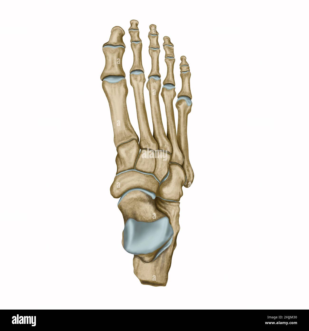 Foot dorsal view, foot anatomy, ankle bone Stock Photohttps://www.alamy.com/image-license-details/?v=1https://www.alamy.com/foot-dorsal-view-foot-anatomy-ankle-bone-image458944276.html
RF2HJJM30–Foot dorsal view, foot anatomy, ankle bone
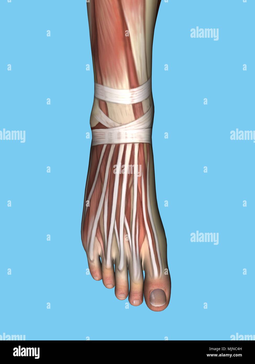 Anatomy of foot Stock Photohttps://www.alamy.com/image-license-details/?v=1https://www.alamy.com/anatomy-of-foot-image183638017.html
RMMJNC4H–Anatomy of foot
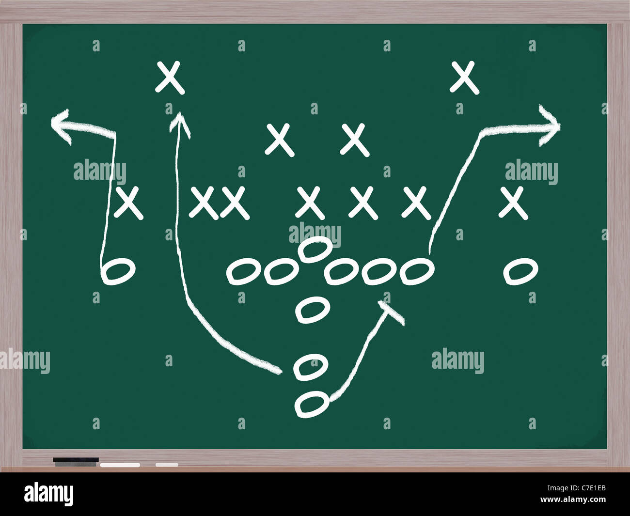 A football play diagram on a chalkboard in white chalk showing the formations and assignments. Stock Photohttps://www.alamy.com/image-license-details/?v=1https://www.alamy.com/stock-photo-a-football-play-diagram-on-a-chalkboard-in-white-chalk-showing-the-39031843.html
RFC7E1EB–A football play diagram on a chalkboard in white chalk showing the formations and assignments.
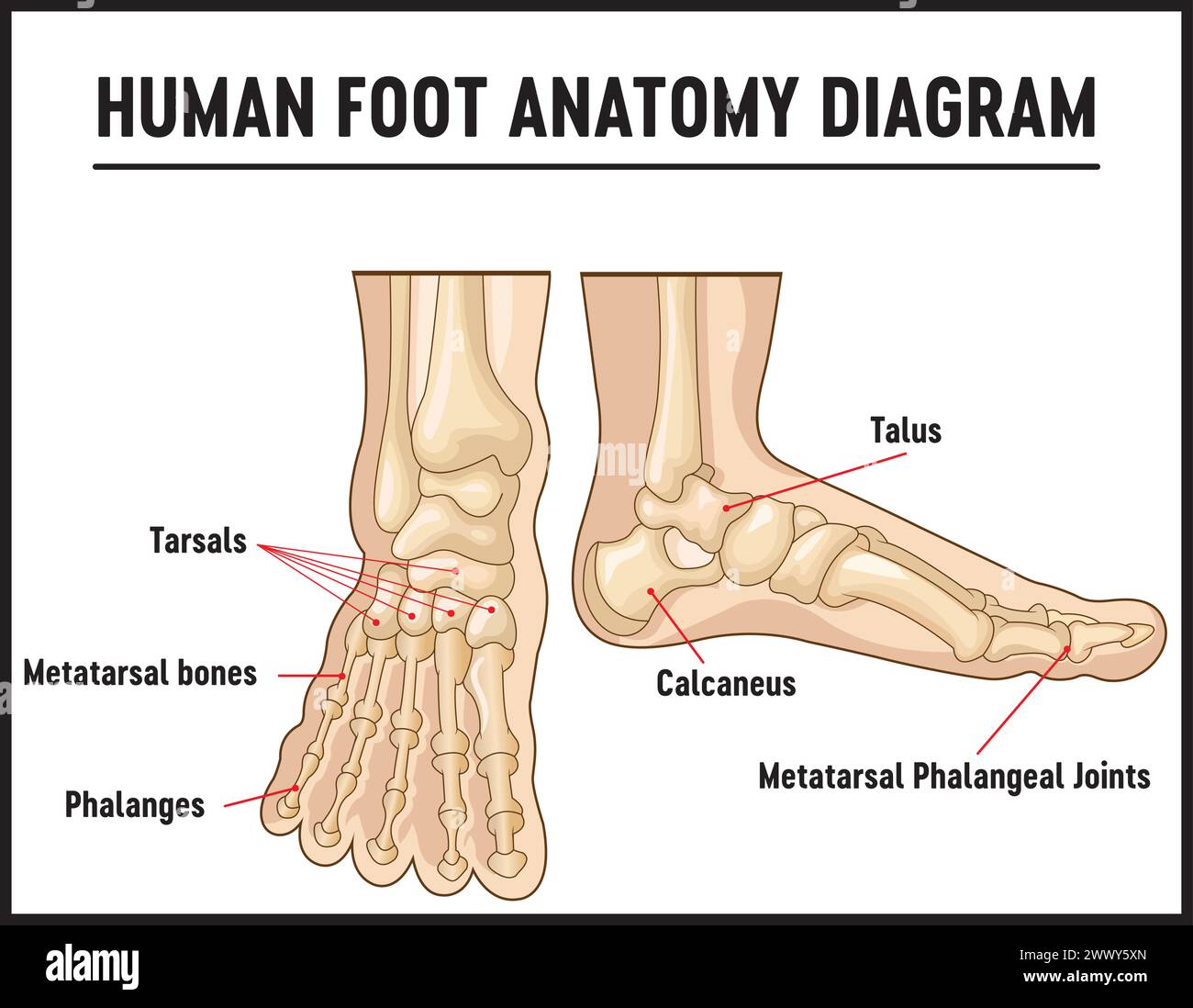 Bones of the human foot with the name and description of all sites. Superior view. Human anatomy. Vector illustration isolated on a white background. Stock Vectorhttps://www.alamy.com/image-license-details/?v=1https://www.alamy.com/bones-of-the-human-foot-with-the-name-and-description-of-all-sites-superior-view-human-anatomy-vector-illustration-isolated-on-a-white-background-image601116285.html
RF2WWY5XN–Bones of the human foot with the name and description of all sites. Superior view. Human anatomy. Vector illustration isolated on a white background.
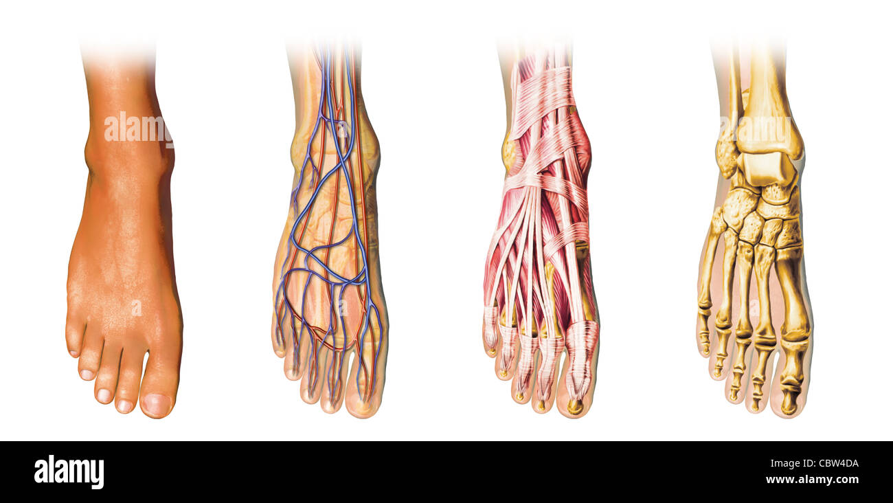 Human foot anatomy cutaway representation, showing skin, veins and arterias, muscles, bones. With clipping path included. Stock Photohttps://www.alamy.com/image-license-details/?v=1https://www.alamy.com/stock-photo-human-foot-anatomy-cutaway-representation-showing-skin-veins-and-arterias-41734262.html
RFCBW4DA–Human foot anatomy cutaway representation, showing skin, veins and arterias, muscles, bones. With clipping path included.
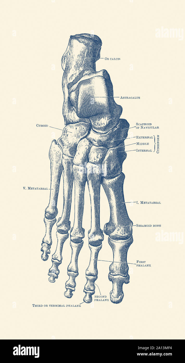 Vintage anatomy print of the human right foot with each bone labeled. Stock Photohttps://www.alamy.com/image-license-details/?v=1https://www.alamy.com/vintage-anatomy-print-of-the-human-right-foot-with-each-bone-labeled-image327693608.html
RF2A13MF4–Vintage anatomy print of the human right foot with each bone labeled.
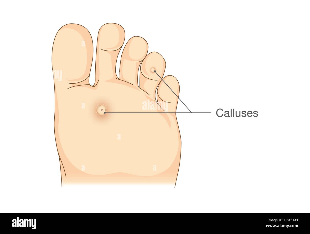 Small circles dead skin on the bottom of soles and toes. Stock Vectorhttps://www.alamy.com/image-license-details/?v=1https://www.alamy.com/stock-photo-small-circles-dead-skin-on-the-bottom-of-soles-and-toes-130571866.html
RFHGC1MX–Small circles dead skin on the bottom of soles and toes.
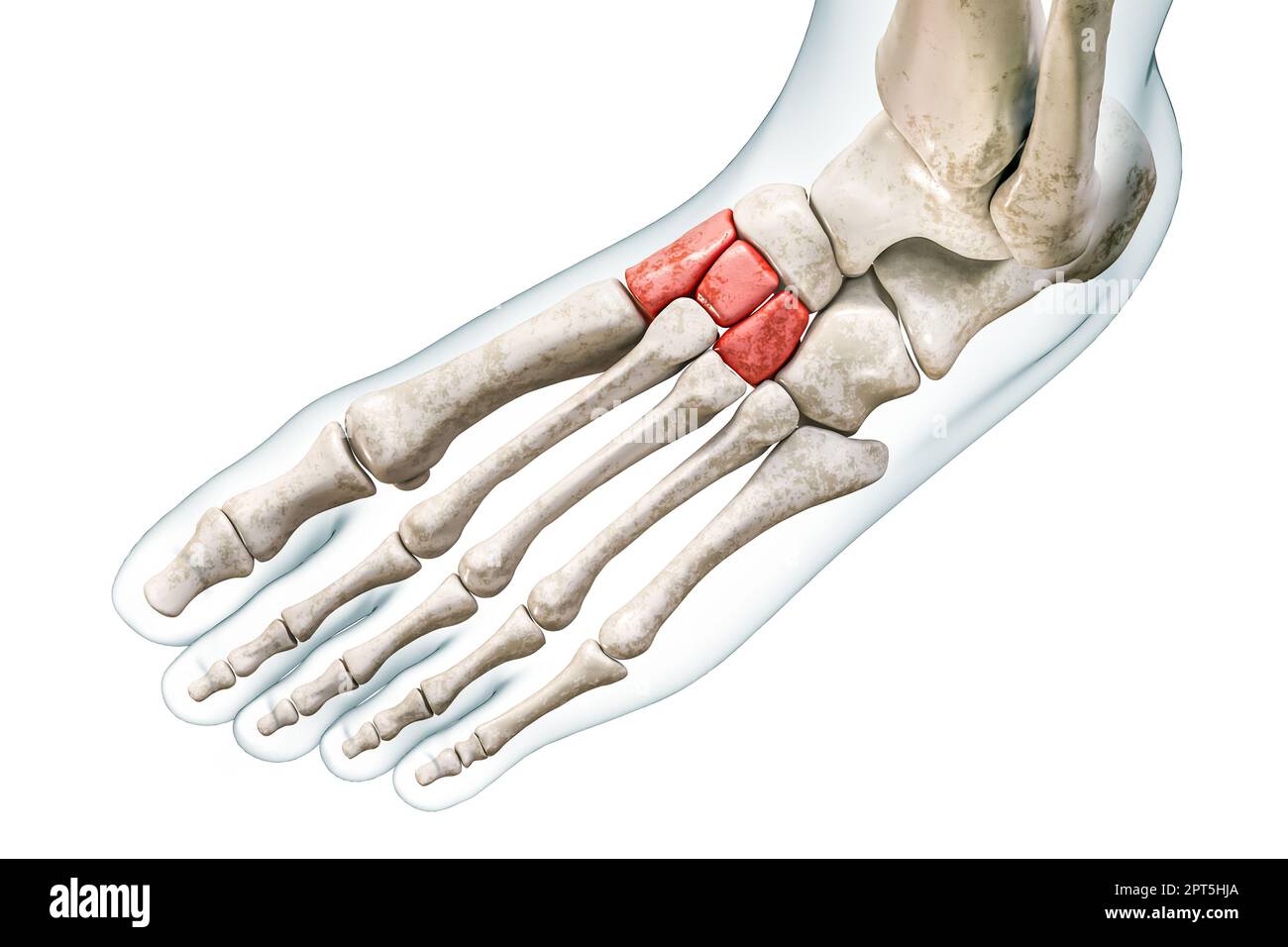 Cuneiform tarsal bones in red with body 3D rendering illustration isolated on white with copy space. Human skeleton and foot anatomy, medical diagram, Stock Photohttps://www.alamy.com/image-license-details/?v=1https://www.alamy.com/cuneiform-tarsal-bones-in-red-with-body-3d-rendering-illustration-isolated-on-white-with-copy-space-human-skeleton-and-foot-anatomy-medical-diagram-image548396754.html
RF2PT5HJA–Cuneiform tarsal bones in red with body 3D rendering illustration isolated on white with copy space. Human skeleton and foot anatomy, medical diagram,
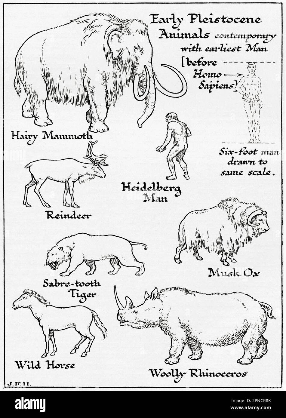 Diagram of Early Pleistocene animals contemporary with earliest man, before Homosapiens, including Hairy Mammoth, Reindeer, Sabre-Tooth Tiger, Wild Horse, Woolly Rhinoceros, Musk Ox and Heidelberg Man. Shown in the diagram a six foot man drawn to the same scale as other figures. From the book Outline of History by H.G. Wells, published 1920. Stock Photohttps://www.alamy.com/image-license-details/?v=1https://www.alamy.com/diagram-of-early-pleistocene-animals-contemporary-with-earliest-man-before-homosapiens-including-hairy-mammoth-reindeer-sabre-tooth-tiger-wild-horse-woolly-rhinoceros-musk-ox-and-heidelberg-man-shown-in-the-diagram-a-six-foot-man-drawn-to-the-same-scale-as-other-figures-from-the-book-outline-of-history-by-hg-wells-published-1920-image546710883.html
RM2PNCR8K–Diagram of Early Pleistocene animals contemporary with earliest man, before Homosapiens, including Hairy Mammoth, Reindeer, Sabre-Tooth Tiger, Wild Horse, Woolly Rhinoceros, Musk Ox and Heidelberg Man. Shown in the diagram a six foot man drawn to the same scale as other figures. From the book Outline of History by H.G. Wells, published 1920.
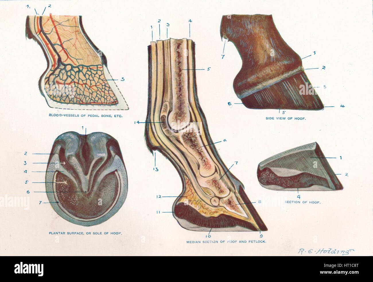 Structure of the foot of a horse, c1907 (c1910). Artist: RE Holding. Stock Photohttps://www.alamy.com/image-license-details/?v=1https://www.alamy.com/stock-photo-structure-of-the-foot-of-a-horse-c1907-c1910-artist-re-holding-135255928.html
RMHT1C8T–Structure of the foot of a horse, c1907 (c1910). Artist: RE Holding.
 Major Ligaments of the Foot Stock Photohttps://www.alamy.com/image-license-details/?v=1https://www.alamy.com/stock-photo-major-ligaments-of-the-foot-135012491.html
RMHRJ9PK–Major Ligaments of the Foot
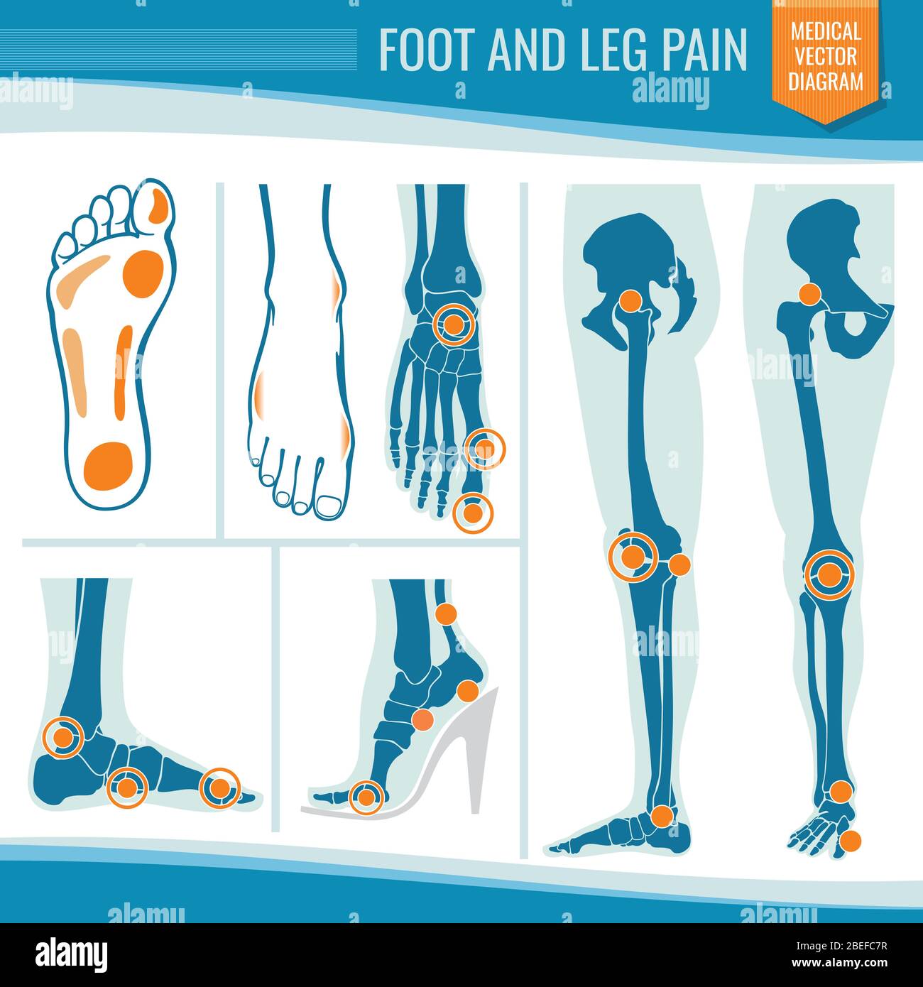 Foot and leg pain. Arthritis and rheumatism orthopedic medical vector diagram. Illustration of rheumatism leg joint Stock Vectorhttps://www.alamy.com/image-license-details/?v=1https://www.alamy.com/foot-and-leg-pain-arthritis-and-rheumatism-orthopedic-medical-vector-diagram-illustration-of-rheumatism-leg-joint-image353151451.html
RF2BEFC7R–Foot and leg pain. Arthritis and rheumatism orthopedic medical vector diagram. Illustration of rheumatism leg joint
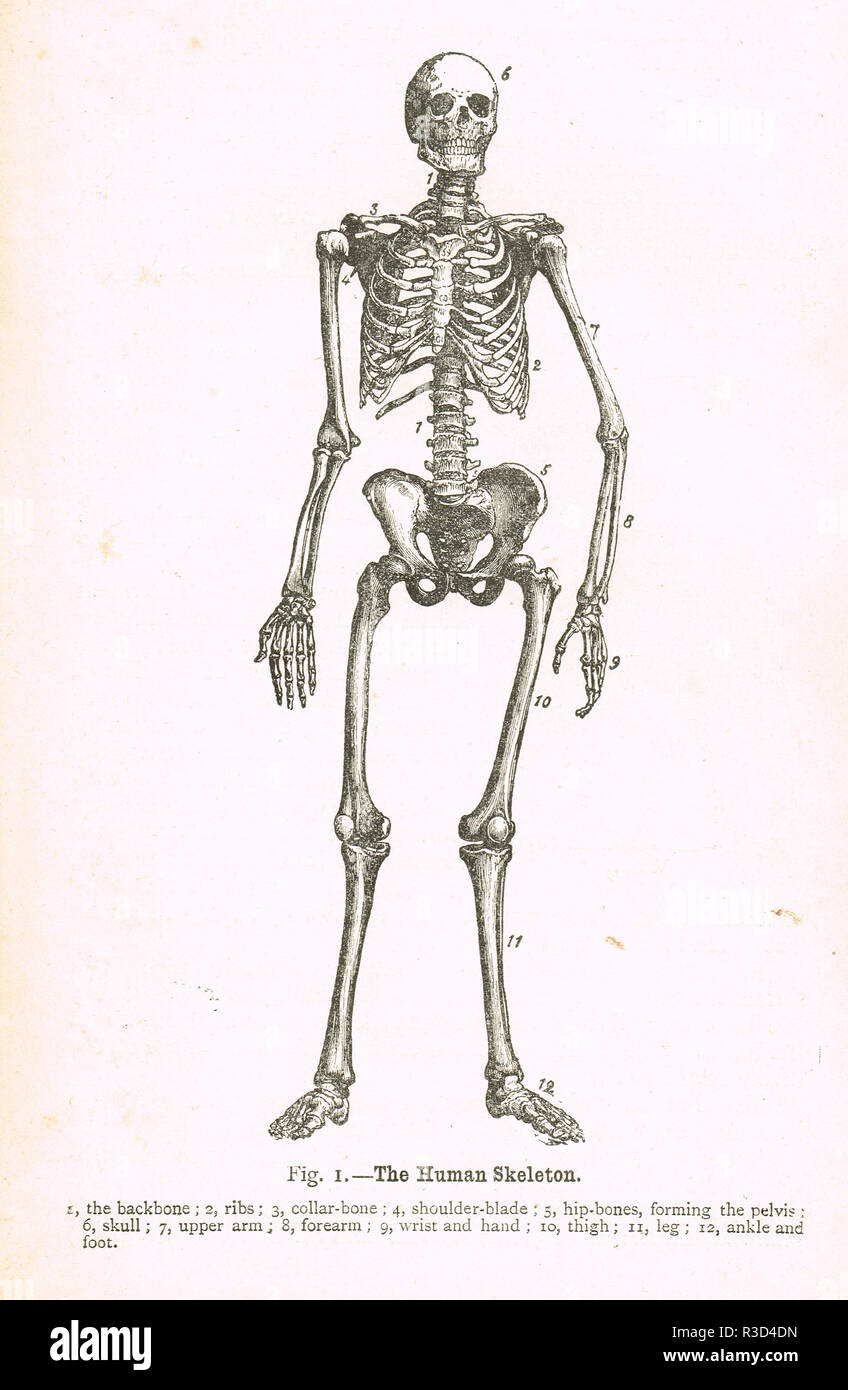 The Human Skeleton. A 19th Century diagram Stock Photohttps://www.alamy.com/image-license-details/?v=1https://www.alamy.com/the-human-skeleton-a-19th-century-diagram-image225867649.html
RMR3D4DN–The Human Skeleton. A 19th Century diagram
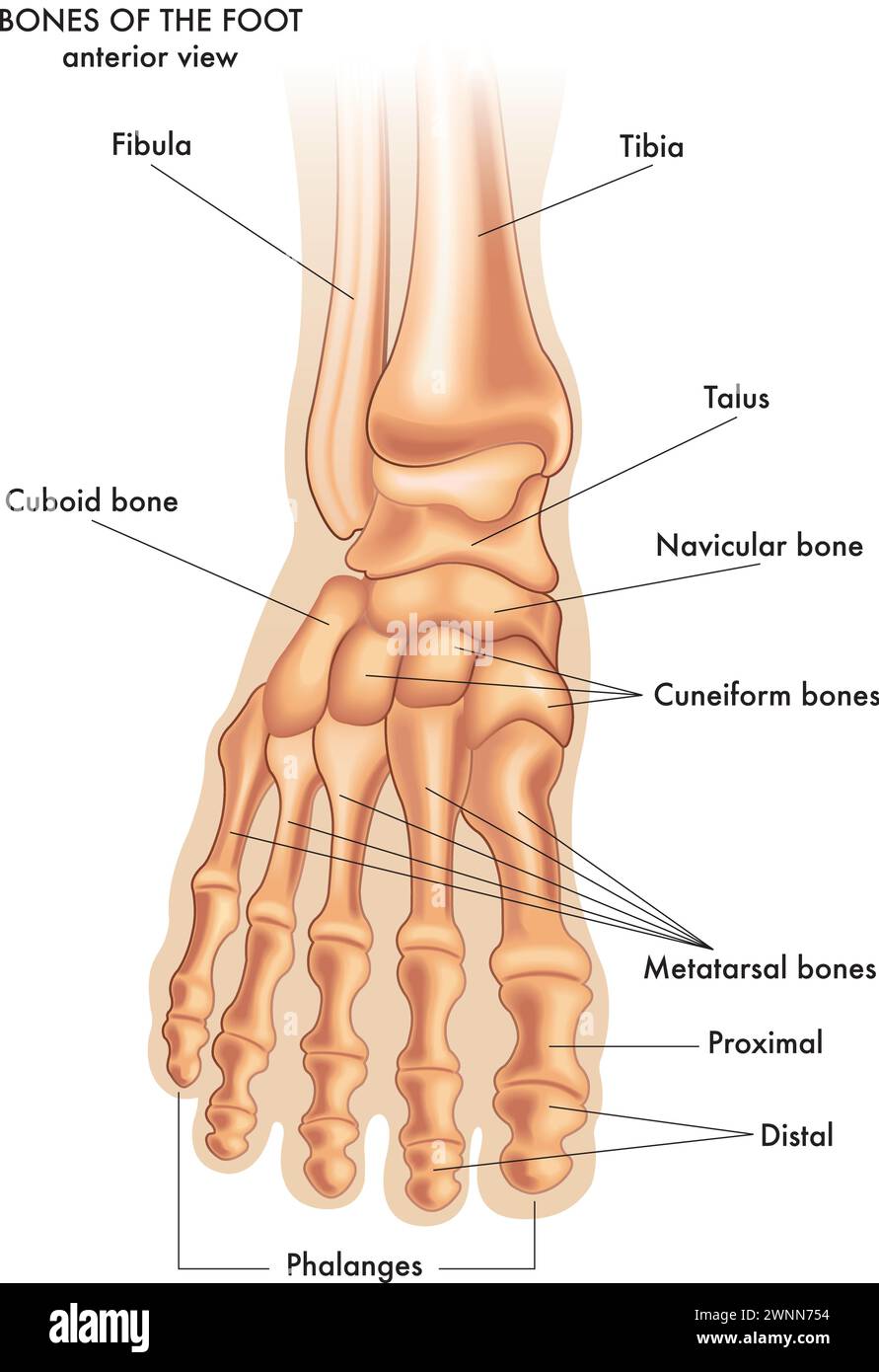 Medical illustration of the main parts of the bones of the foot in anterior view, with annotations. Stock Vectorhttps://www.alamy.com/image-license-details/?v=1https://www.alamy.com/medical-illustration-of-the-main-parts-of-the-bones-of-the-foot-in-anterior-view-with-annotations-image598526912.html
RF2WNN754–Medical illustration of the main parts of the bones of the foot in anterior view, with annotations.
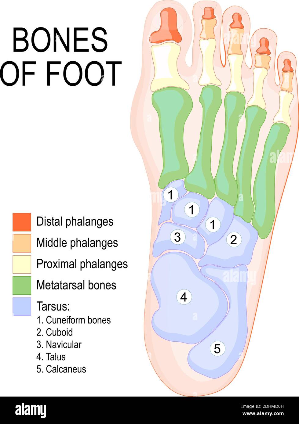 Bones of foot. Human Anatomy. The diagram shows the placement and names of all bones of foot. Stock Vectorhttps://www.alamy.com/image-license-details/?v=1https://www.alamy.com/bones-of-foot-human-anatomy-the-diagram-shows-the-placement-and-names-of-all-bones-of-foot-image389526497.html
RF2DHMD0H–Bones of foot. Human Anatomy. The diagram shows the placement and names of all bones of foot.
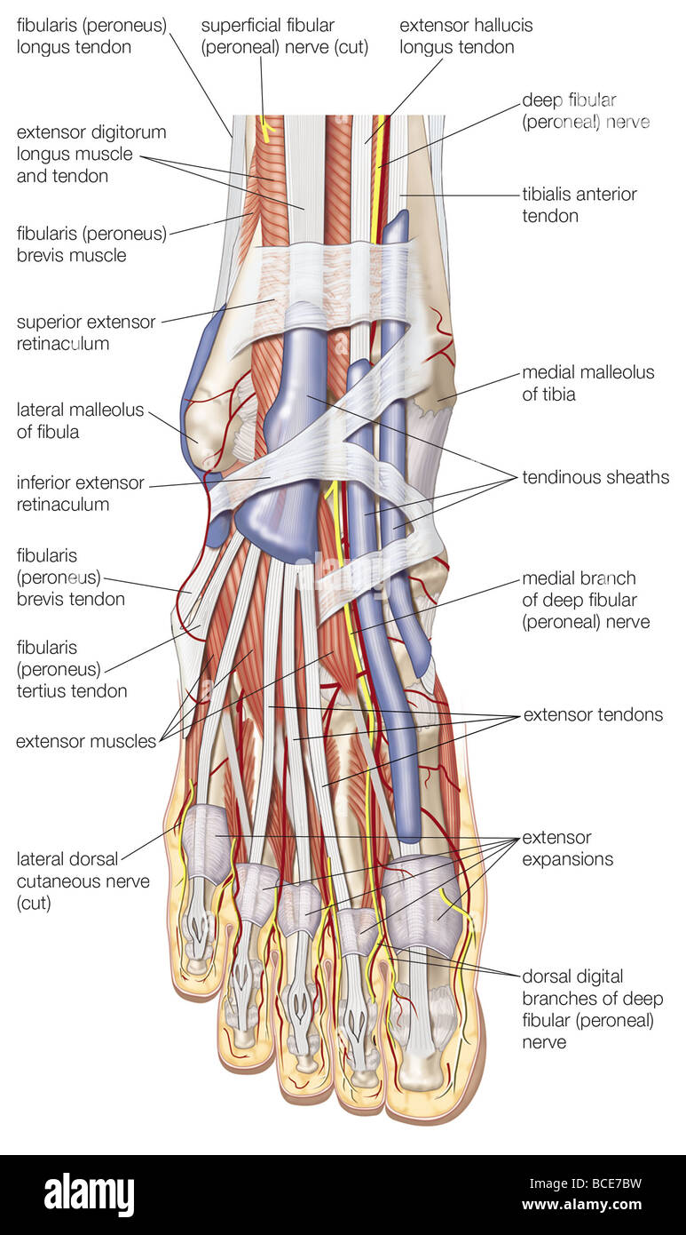 Dorsal view of the right foot, showing the major muscles, tendons, and nerves. Stock Photohttps://www.alamy.com/image-license-details/?v=1https://www.alamy.com/stock-photo-dorsal-view-of-the-right-foot-showing-the-major-muscles-tendons-and-24899389.html
RMBCE7BW–Dorsal view of the right foot, showing the major muscles, tendons, and nerves.
 Bones of the human foot. From a 19th century engraving. Stock Photohttps://www.alamy.com/image-license-details/?v=1https://www.alamy.com/bones-of-the-human-foot-from-a-19th-century-engraving-image481926990.html
RM2K01JP6–Bones of the human foot. From a 19th century engraving.
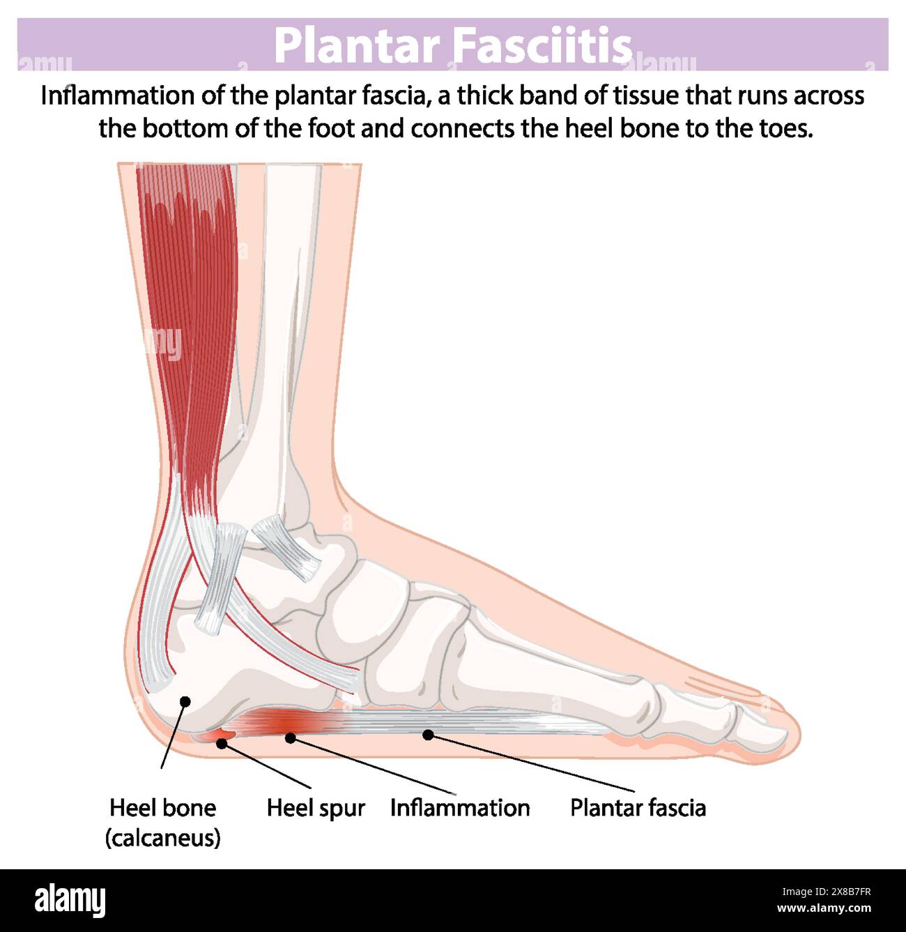 Detailed diagram of foot inflammation Stock Vectorhttps://www.alamy.com/image-license-details/?v=1https://www.alamy.com/detailed-diagram-of-foot-inflammation-image607527531.html
RF2X8B7FR–Detailed diagram of foot inflammation
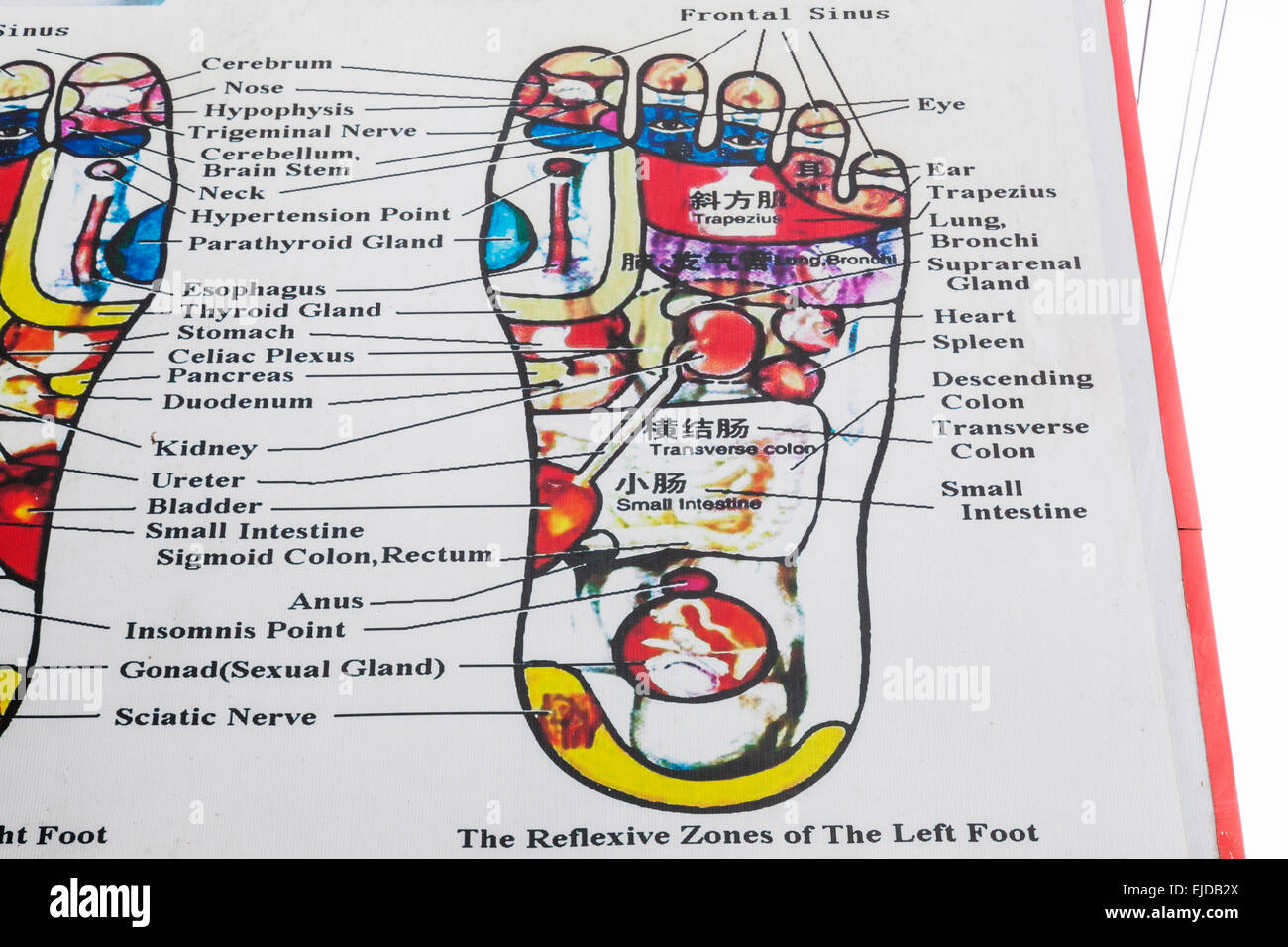 Cambodia, Siem Reap, Foot Reflexology Chart Stock Photohttps://www.alamy.com/image-license-details/?v=1https://www.alamy.com/stock-photo-cambodia-siem-reap-foot-reflexology-chart-80199362.html
RMEJDB2X–Cambodia, Siem Reap, Foot Reflexology Chart
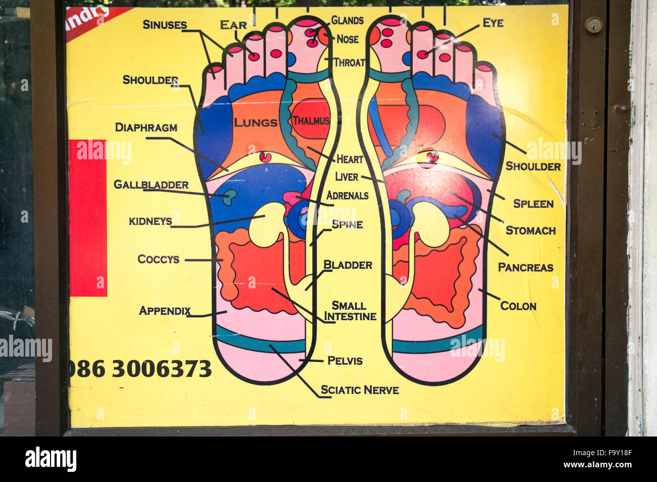 Massage parlour diagram of the human foot, Bangkok, Thailand Stock Photohttps://www.alamy.com/image-license-details/?v=1https://www.alamy.com/stock-photo-massage-parlour-diagram-of-the-human-foot-bangkok-thailand-92177471.html
RMF9Y18F–Massage parlour diagram of the human foot, Bangkok, Thailand
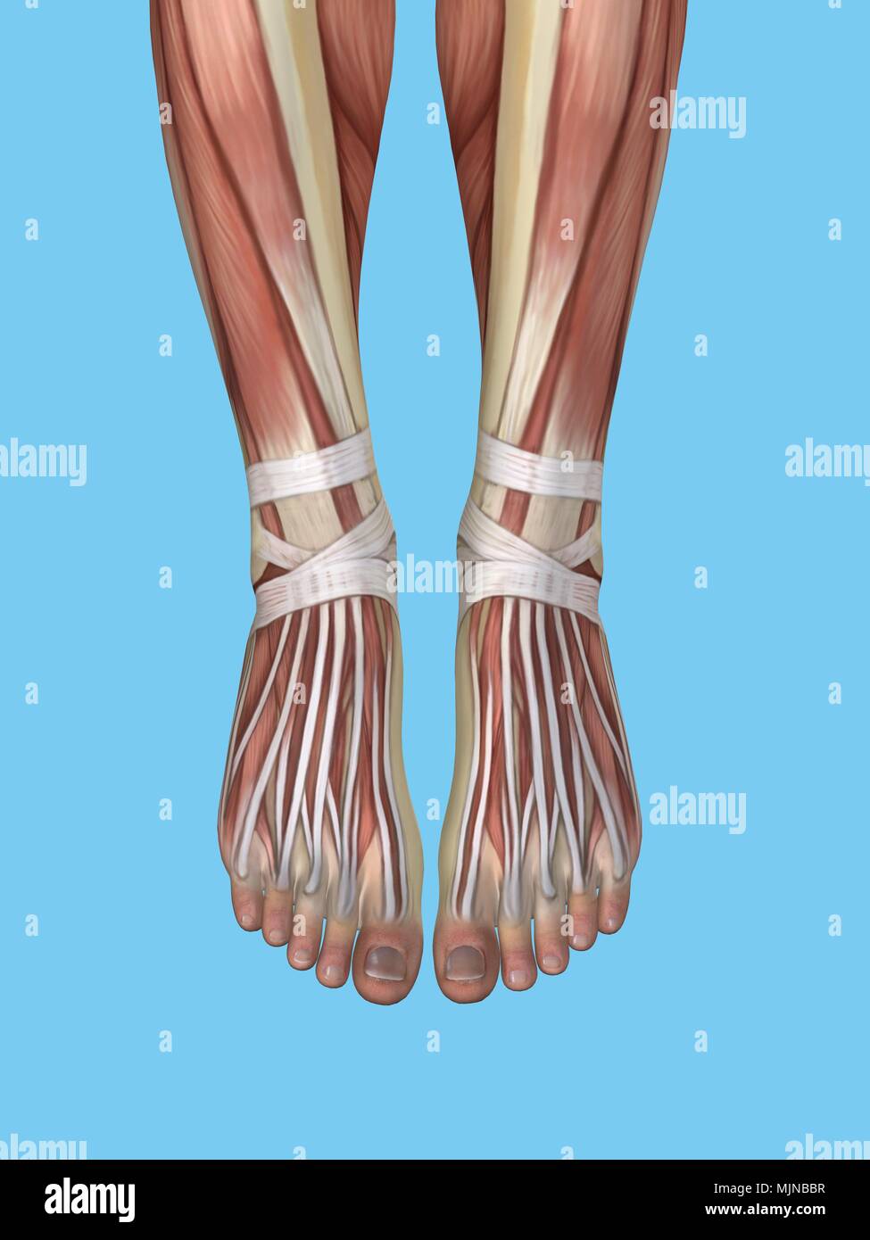 Anatomy of foot Stock Photohttps://www.alamy.com/image-license-details/?v=1https://www.alamy.com/anatomy-of-foot-image183637435.html
RMMJNBBR–Anatomy of foot
 Diagnosis of gout vector illustration. Cartoon tiny people study infographic foot medical diagram, doctors diagnose arthritis inflammation and pain in joint articulation due to uric acid crystals Stock Vectorhttps://www.alamy.com/image-license-details/?v=1https://www.alamy.com/diagnosis-of-gout-vector-illustration-cartoon-tiny-people-study-infographic-foot-medical-diagram-doctors-diagnose-arthritis-inflammation-and-pain-in-joint-articulation-due-to-uric-acid-crystals-image568458853.html
RF2T0RF1W–Diagnosis of gout vector illustration. Cartoon tiny people study infographic foot medical diagram, doctors diagnose arthritis inflammation and pain in joint articulation due to uric acid crystals
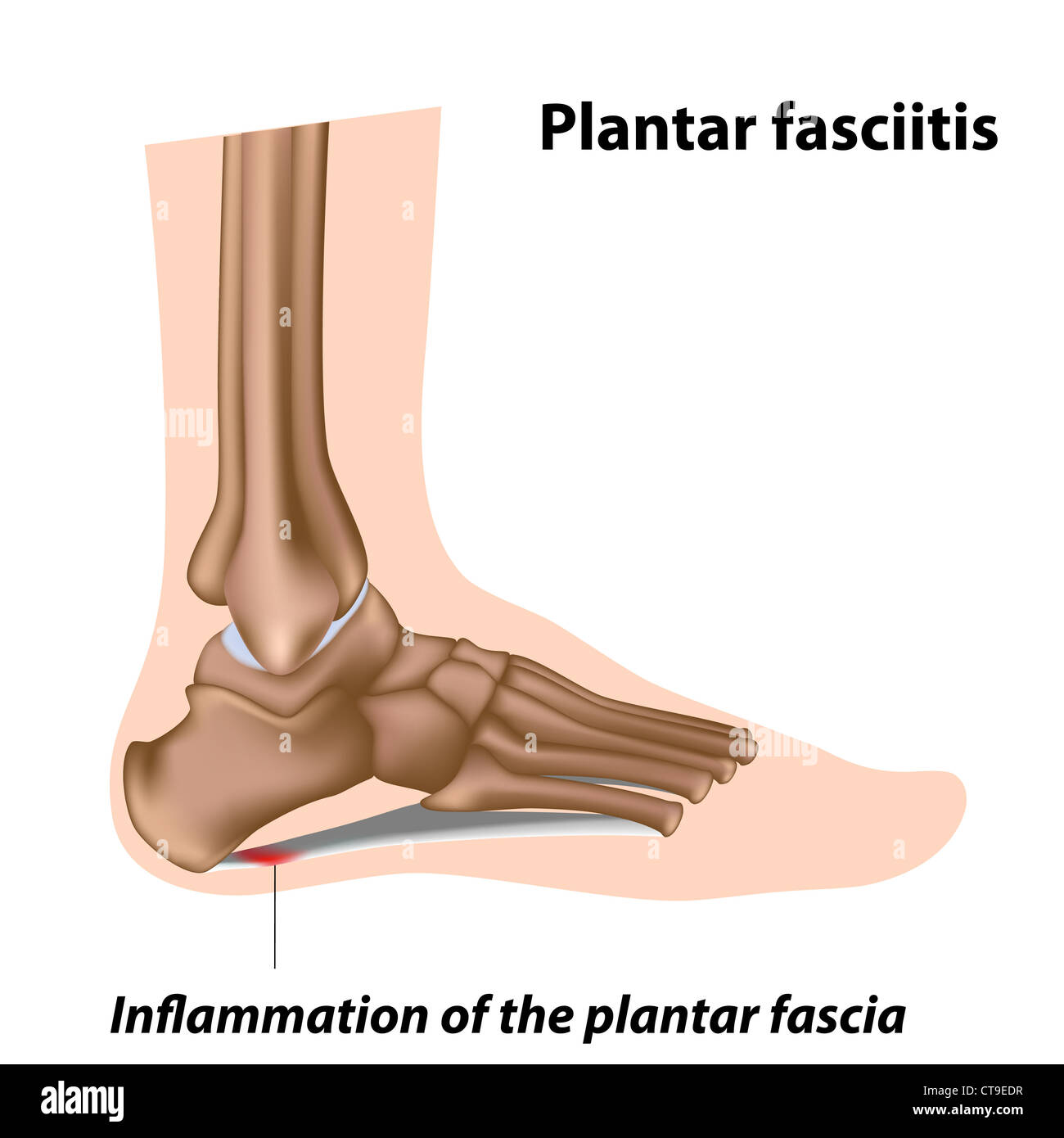 Plantar fasciitis, foot problem Stock Photohttps://www.alamy.com/image-license-details/?v=1https://www.alamy.com/stock-photo-plantar-fasciitis-foot-problem-49381411.html
RFCT9EDR–Plantar fasciitis, foot problem
 Varicose veins, illustration Stock Photohttps://www.alamy.com/image-license-details/?v=1https://www.alamy.com/varicose-veins-illustration-image478059186.html
RF2JNNDAA–Varicose veins, illustration
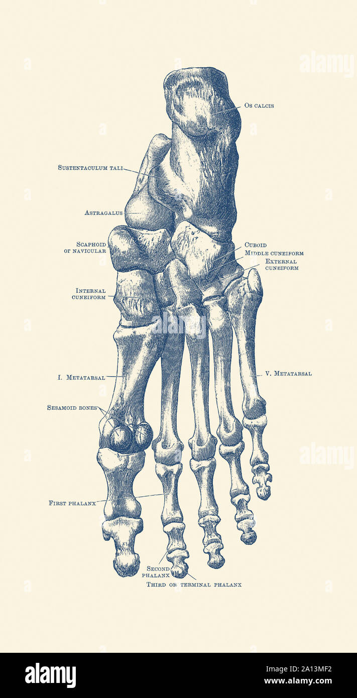 Vintage anatomy print of the human left foot with each bone labeled. Stock Photohttps://www.alamy.com/image-license-details/?v=1https://www.alamy.com/vintage-anatomy-print-of-the-human-left-foot-with-each-bone-labeled-image327693606.html
RF2A13MF2–Vintage anatomy print of the human left foot with each bone labeled.
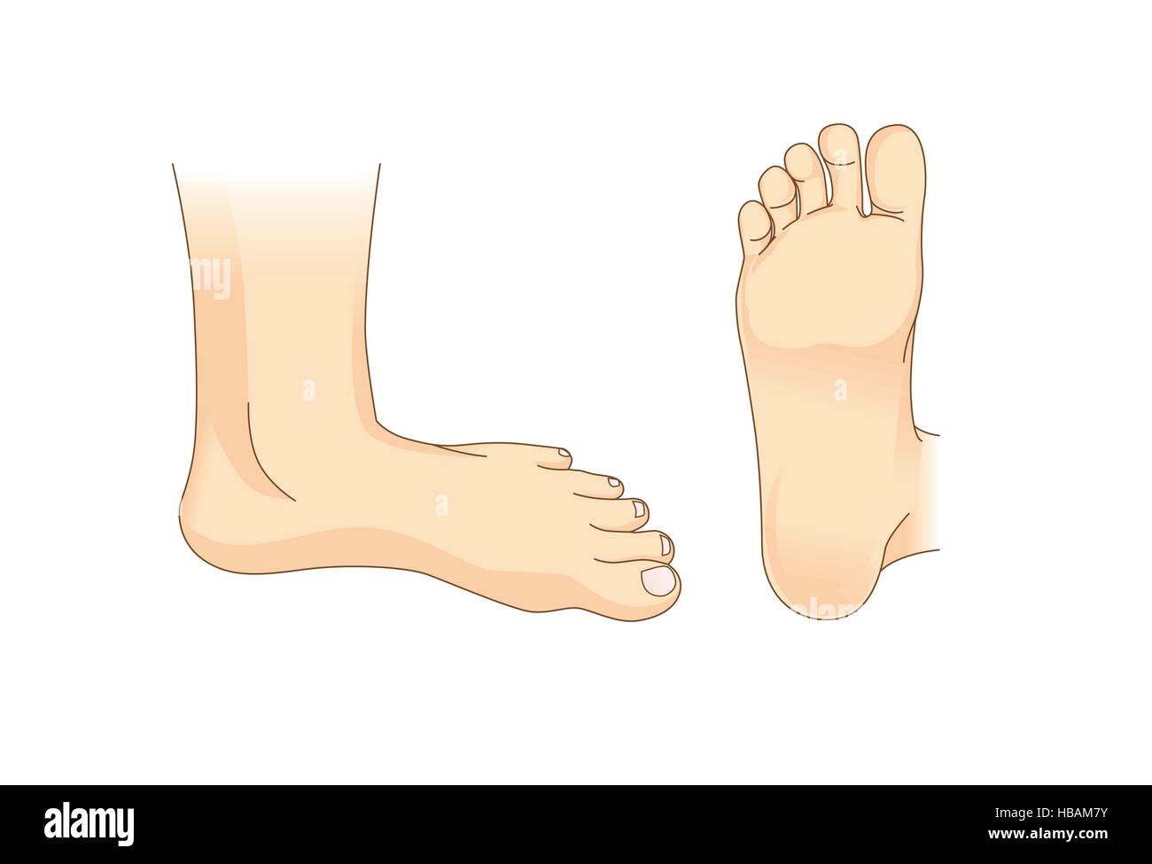 Foot vector in side view and bottom of foot Stock Vectorhttps://www.alamy.com/image-license-details/?v=1https://www.alamy.com/stock-photo-foot-vector-in-side-view-and-bottom-of-foot-127469215.html
RFHBAM7Y–Foot vector in side view and bottom of foot
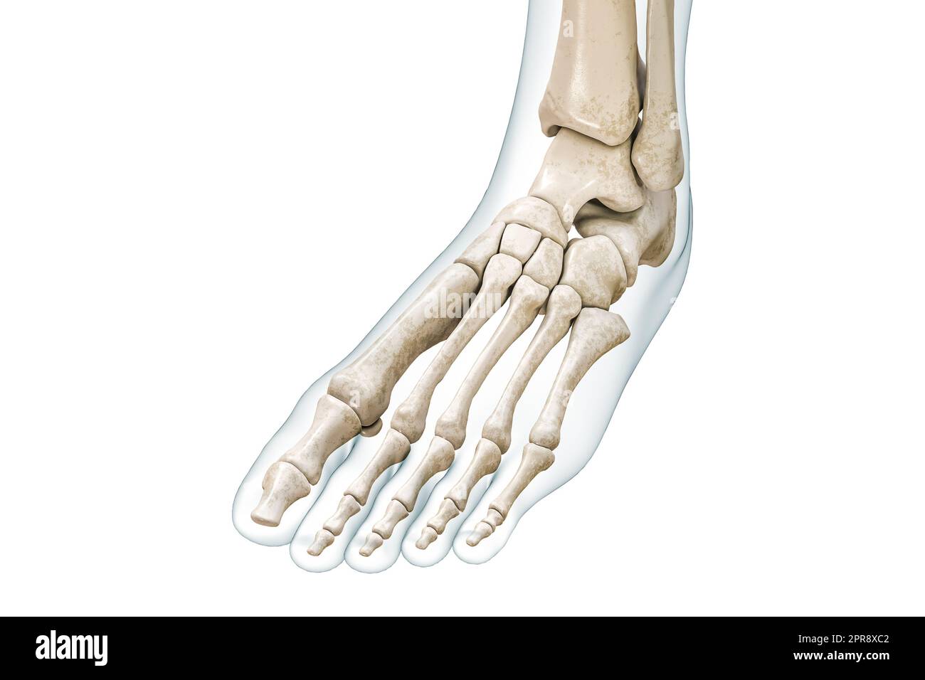 Foot and toe bones with body contours 3D rendering illustration isolated on white with copy space. Human skeleton and leg anatomy, medical diagram, os Stock Photohttps://www.alamy.com/image-license-details/?v=1https://www.alamy.com/foot-and-toe-bones-with-body-contours-3d-rendering-illustration-isolated-on-white-with-copy-space-human-skeleton-and-leg-anatomy-medical-diagram-os-image547854834.html
RF2PR8XC2–Foot and toe bones with body contours 3D rendering illustration isolated on white with copy space. Human skeleton and leg anatomy, medical diagram, os
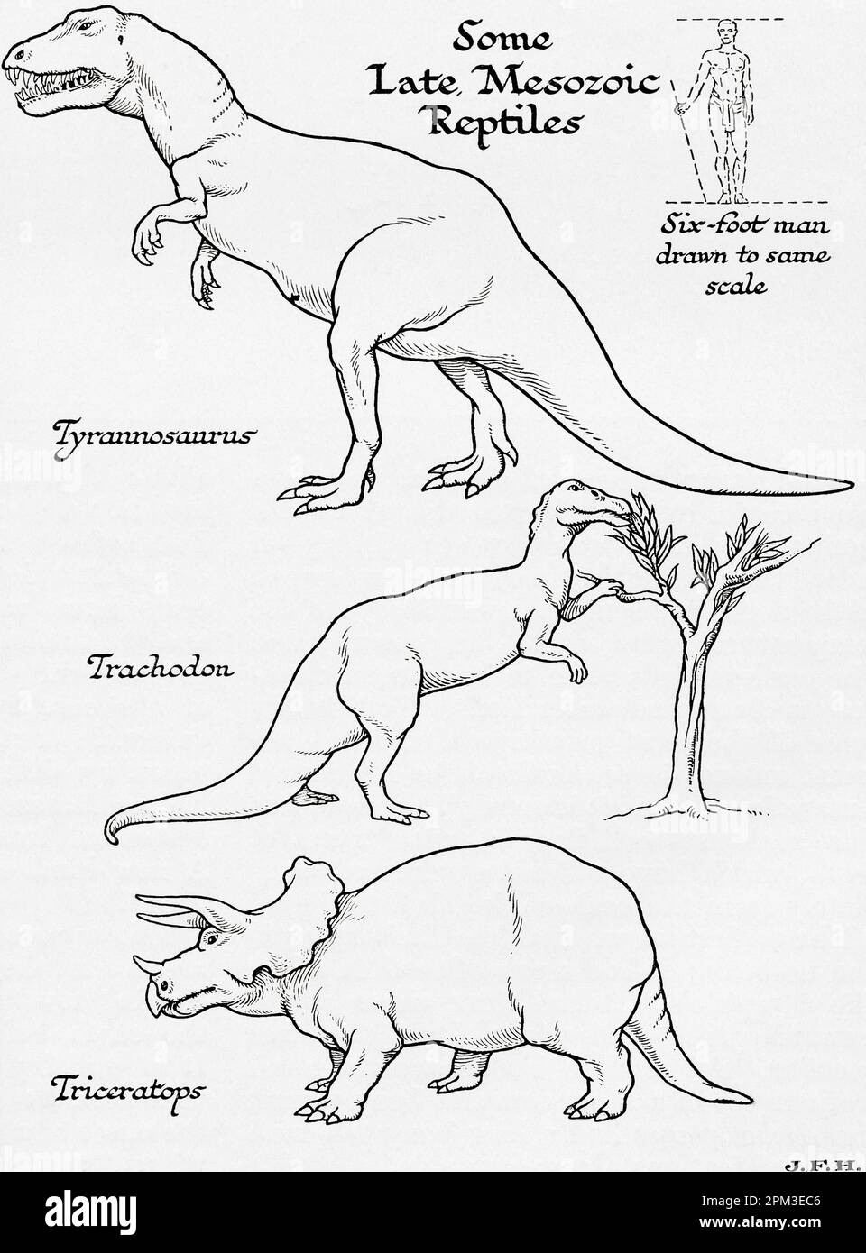 Late Mesozoic Era reptiles. Tyrannosaurus, Trachodon and Triceraptops. Shown in the diagram a six foot man drawn to the same scale as other figures. From the book Outline of History by H.G. Wells, published 1920. Stock Photohttps://www.alamy.com/image-license-details/?v=1https://www.alamy.com/late-mesozoic-era-reptiles-tyrannosaurus-trachodon-and-triceraptops-shown-in-the-diagram-a-six-foot-man-drawn-to-the-same-scale-as-other-figures-from-the-book-outline-of-history-by-hg-wells-published-1920-image545891702.html
RM2PM3EC6–Late Mesozoic Era reptiles. Tyrannosaurus, Trachodon and Triceraptops. Shown in the diagram a six foot man drawn to the same scale as other figures. From the book Outline of History by H.G. Wells, published 1920.
 Caucasian male wearing running shoes and shorts, running on treadmill Stock Photohttps://www.alamy.com/image-license-details/?v=1https://www.alamy.com/caucasian-male-wearing-running-shoes-and-shorts-running-on-treadmill-image607597964.html
RF2X8EDB8–Caucasian male wearing running shoes and shorts, running on treadmill
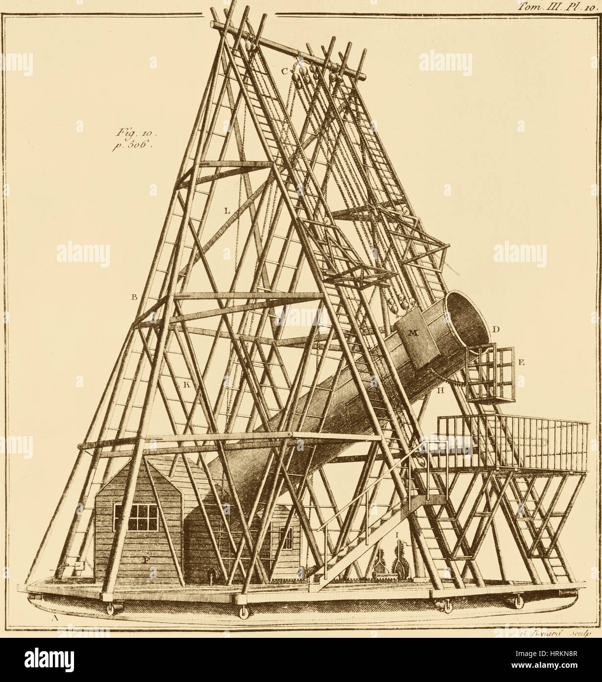 William Herschel 40 Foot Telescope, 1880s Stock Photohttps://www.alamy.com/image-license-details/?v=1https://www.alamy.com/stock-photo-william-herschel-40-foot-telescope-1880s-135043463.html
RMHRKN8R–William Herschel 40 Foot Telescope, 1880s
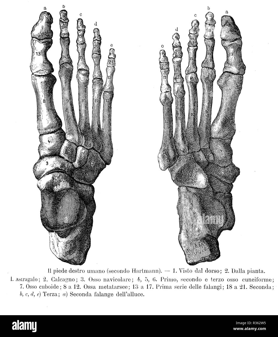 Vintage illustration of anatomy, right foot bones, dorsalis and sole view with Italian anatomical descriptions Stock Photohttps://www.alamy.com/image-license-details/?v=1https://www.alamy.com/vintage-illustration-of-anatomy-right-foot-bones-dorsalis-and-sole-view-with-italian-anatomical-descriptions-image225712737.html
RFR362W5–Vintage illustration of anatomy, right foot bones, dorsalis and sole view with Italian anatomical descriptions
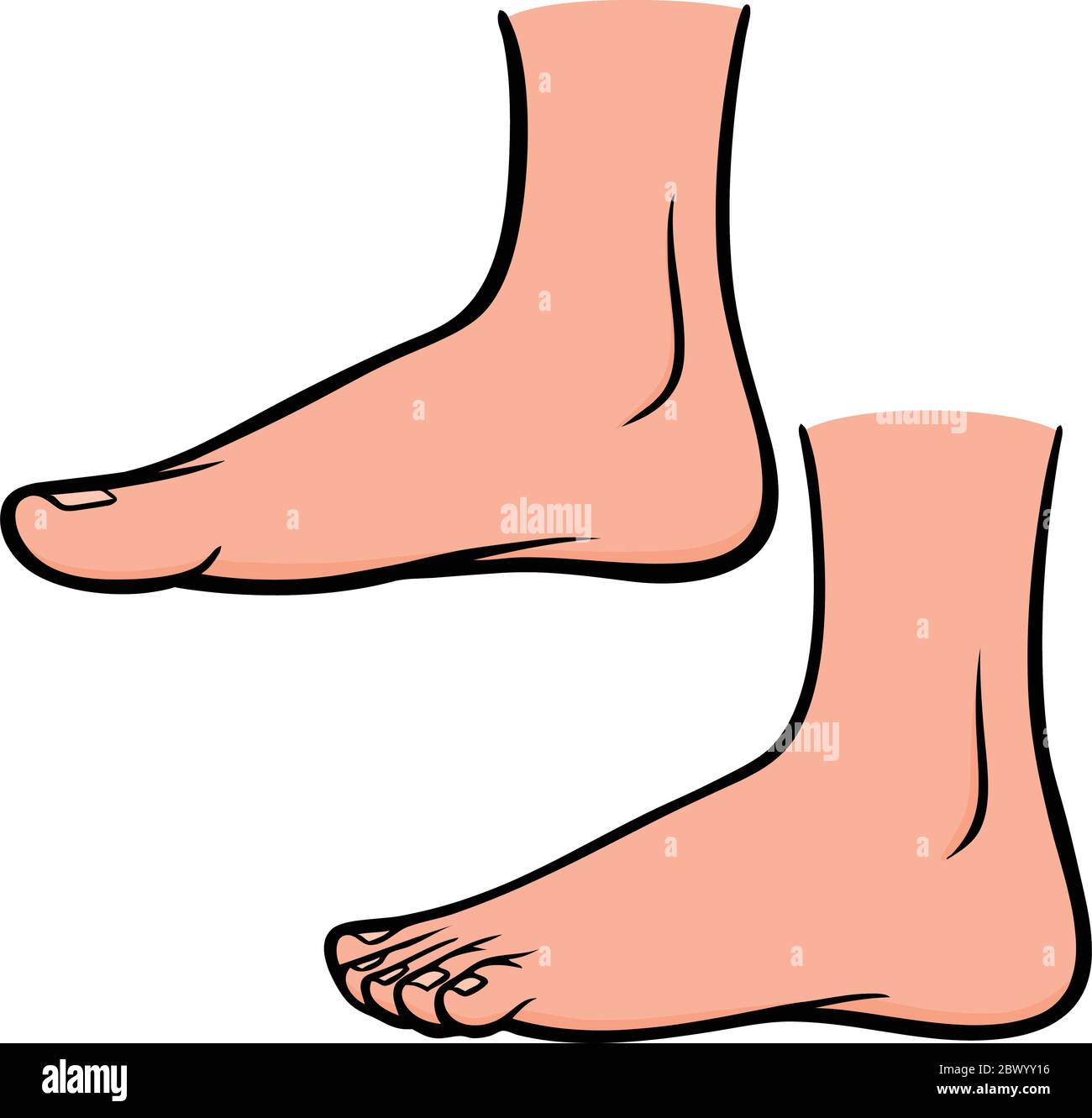 Foot Profiles- An Illustration of Foot Profiles. Stock Vectorhttps://www.alamy.com/image-license-details/?v=1https://www.alamy.com/foot-profiles-an-illustration-of-foot-profiles-image360187666.html
RF2BWYY16–Foot Profiles- An Illustration of Foot Profiles.
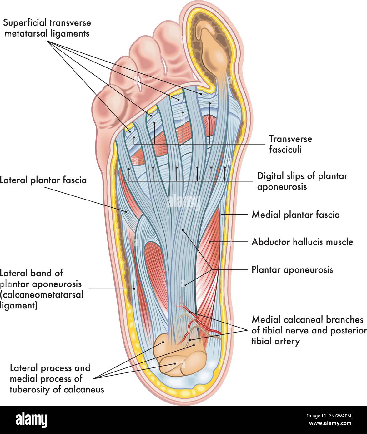 Foot anatomy illustration, with annotations. Stock Vectorhttps://www.alamy.com/image-license-details/?v=1https://www.alamy.com/foot-anatomy-illustration-with-annotations-image526702812.html
RF2NGWAPM–Foot anatomy illustration, with annotations.
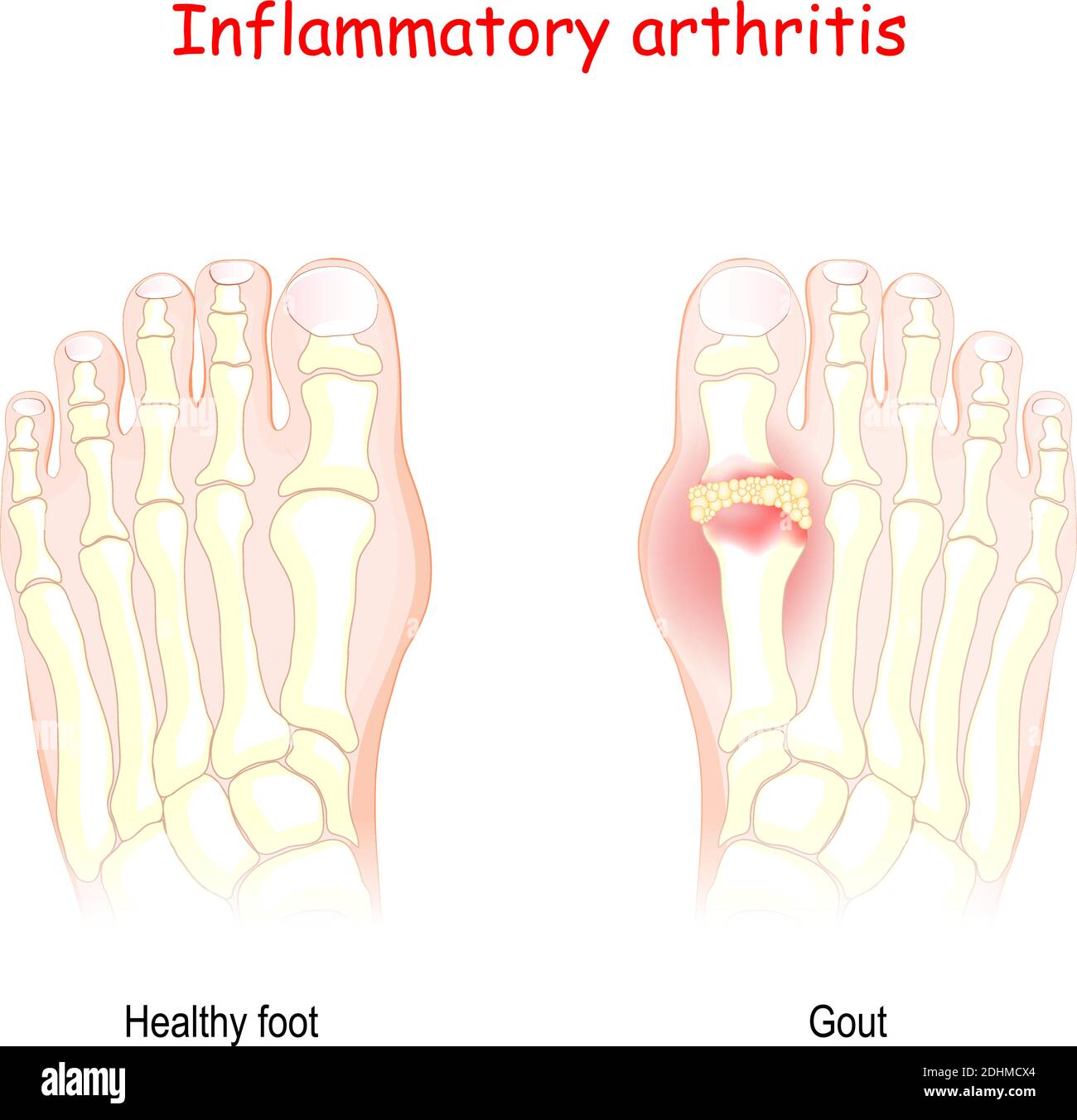 Gout. inflammatory arthritis. Vector Diagram of healthy foot and foot with gout. uric acid crystallizes and the crystals deposit in joints Stock Vectorhttps://www.alamy.com/image-license-details/?v=1https://www.alamy.com/gout-inflammatory-arthritis-vector-diagram-of-healthy-foot-and-foot-with-gout-uric-acid-crystallizes-and-the-crystals-deposit-in-joints-image389526428.html
RF2DHMCX4–Gout. inflammatory arthritis. Vector Diagram of healthy foot and foot with gout. uric acid crystallizes and the crystals deposit in joints
 percent symbol with one foot and a spring climbing onto a moutain chart white background Stock Photohttps://www.alamy.com/image-license-details/?v=1https://www.alamy.com/stock-photo-percent-symbol-with-one-foot-and-a-spring-climbing-onto-a-moutain-55871854.html
RFD6W53A–percent symbol with one foot and a spring climbing onto a moutain chart white background
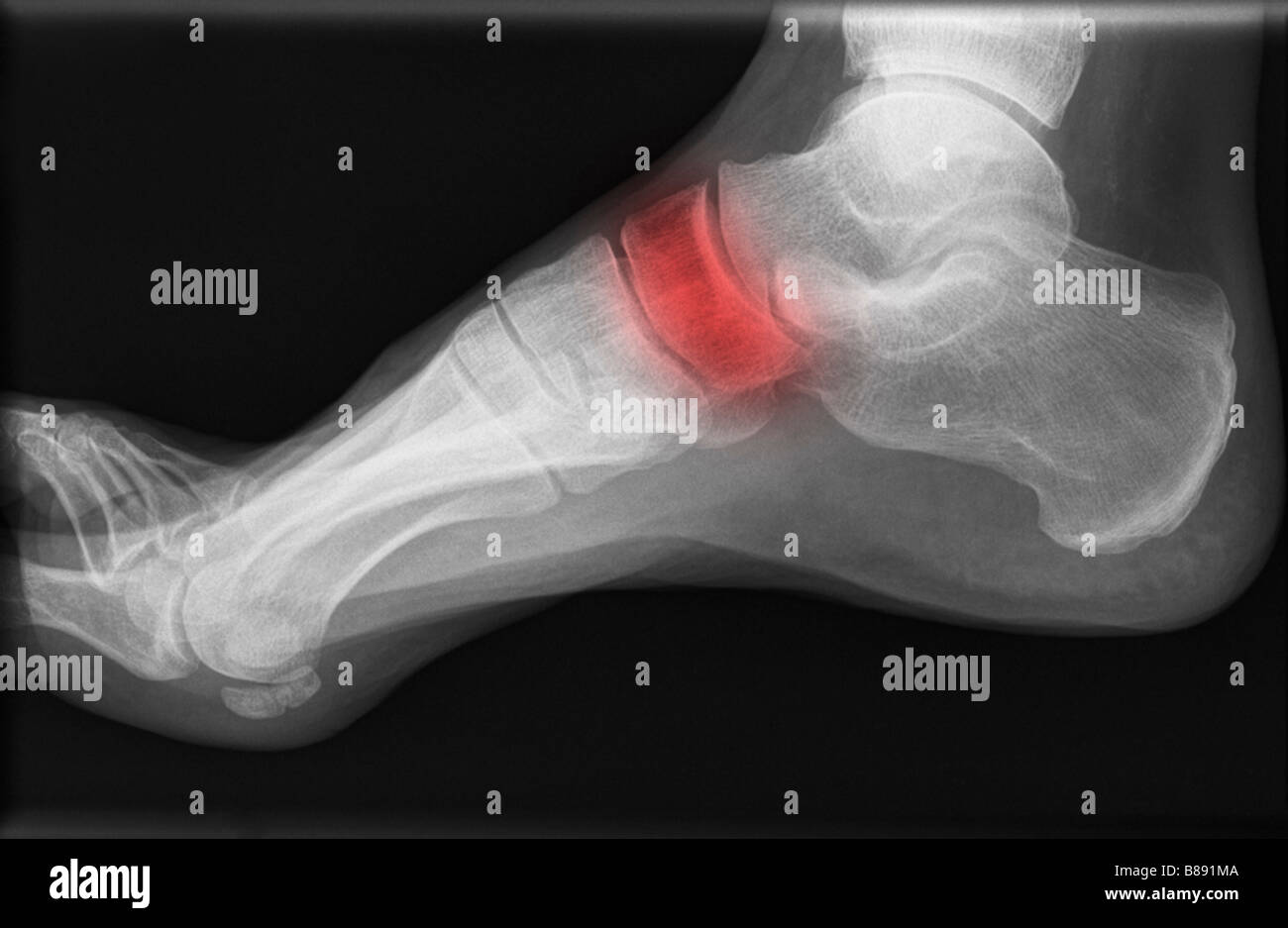 Xray of human foot illustrating problems with a bone Stock Photohttps://www.alamy.com/image-license-details/?v=1https://www.alamy.com/stock-photo-xray-of-human-foot-illustrating-problems-with-a-bone-22326538.html
RMB891MA–Xray of human foot illustrating problems with a bone
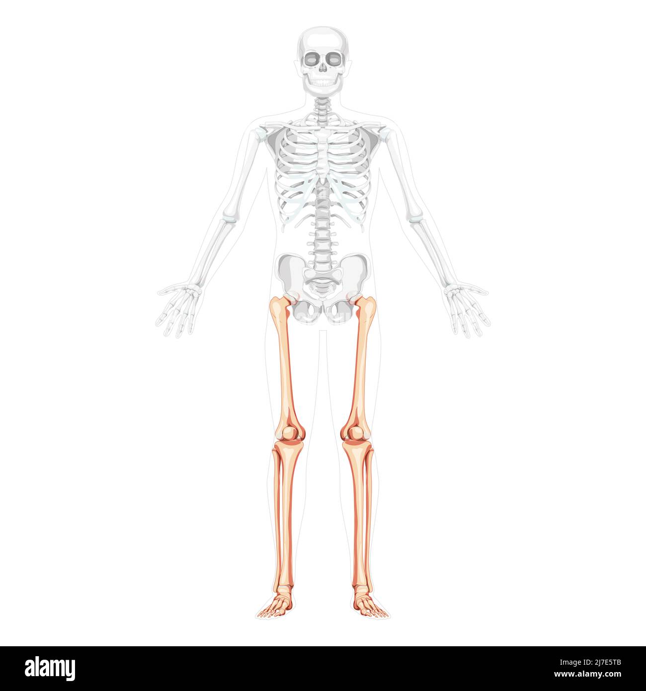 Skeleton Thighs and legs lower limb Human front view with partly transparent bones position. Anatomically correct fibula, tibia, foot realistic flat concept Vector illustration of anatomy isolated Stock Vectorhttps://www.alamy.com/image-license-details/?v=1https://www.alamy.com/skeleton-thighs-and-legs-lower-limb-human-front-view-with-partly-transparent-bones-position-anatomically-correct-fibula-tibia-foot-realistic-flat-concept-vector-illustration-of-anatomy-isolated-image469294459.html
RF2J7E5TB–Skeleton Thighs and legs lower limb Human front view with partly transparent bones position. Anatomically correct fibula, tibia, foot realistic flat concept Vector illustration of anatomy isolated
 Cambodia, Siem Reap, Foot Reflexology Chart Stock Photohttps://www.alamy.com/image-license-details/?v=1https://www.alamy.com/stock-photo-cambodia-siem-reap-foot-reflexology-chart-80199361.html
RMEJDB2W–Cambodia, Siem Reap, Foot Reflexology Chart
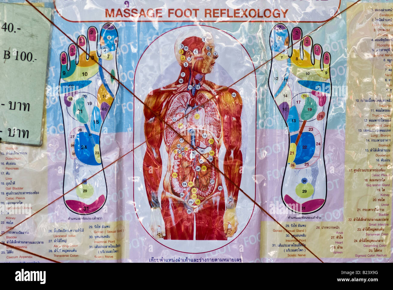 Foot Massage manual Bangkok Thailand Stock Photohttps://www.alamy.com/image-license-details/?v=1https://www.alamy.com/stock-photo-foot-massage-manual-bangkok-thailand-18526188.html
RMB23X9G–Foot Massage manual Bangkok Thailand
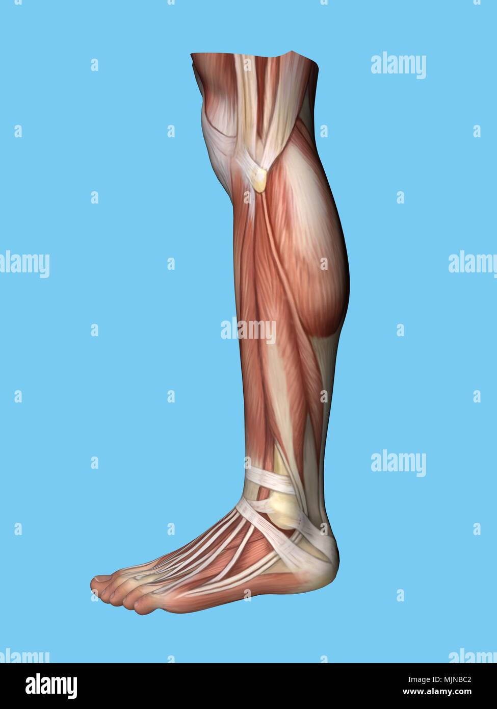 Anatomy of foot Stock Photohttps://www.alamy.com/image-license-details/?v=1https://www.alamy.com/anatomy-of-foot-image183637442.html
RMMJNBC2–Anatomy of foot
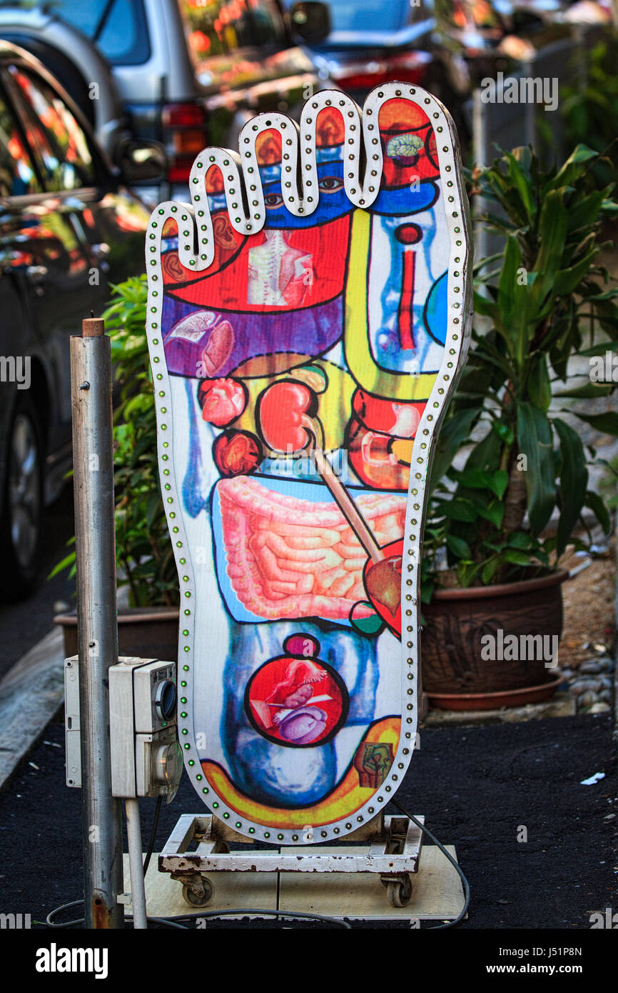 A giant foot advertises a reflexology and massage business in Kuala Lumpur, Malaysia, Southeast Asia. Stock Photohttps://www.alamy.com/image-license-details/?v=1https://www.alamy.com/stock-photo-a-giant-foot-advertises-a-reflexology-and-massage-business-in-kuala-140795669.html
RFJ51P8N–A giant foot advertises a reflexology and massage business in Kuala Lumpur, Malaysia, Southeast Asia.
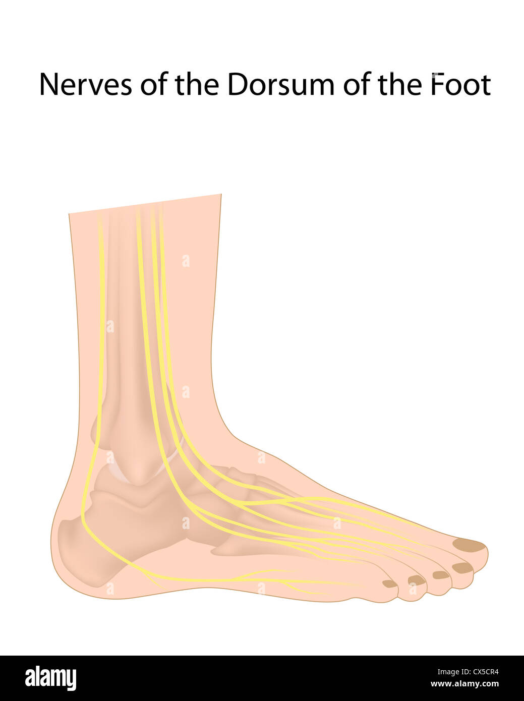 Dorsal digital nerves of foot, commonly affected in diabetic neuropathy Stock Photohttps://www.alamy.com/image-license-details/?v=1https://www.alamy.com/stock-photo-dorsal-digital-nerves-of-foot-commonly-affected-in-diabetic-neuropathy-50521608.html
RFCX5CR4–Dorsal digital nerves of foot, commonly affected in diabetic neuropathy
 career, chart, ascent, diagram, graphic, bar chart, upswing, bull market, boom, Stock Photohttps://www.alamy.com/image-license-details/?v=1https://www.alamy.com/stock-photo-career-chart-ascent-diagram-graphic-bar-chart-upswing-bull-market-131769311.html
RFHJAH2R–career, chart, ascent, diagram, graphic, bar chart, upswing, bull market, boom,
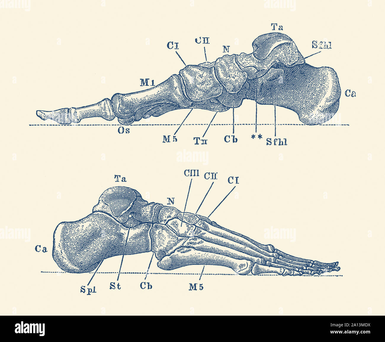 Vintage anatomy print showing a dual view of the human foot with bones labeled. Stock Photohttps://www.alamy.com/image-license-details/?v=1https://www.alamy.com/vintage-anatomy-print-showing-a-dual-view-of-the-human-foot-with-bones-labeled-image327693574.html
RF2A13MDX–Vintage anatomy print showing a dual view of the human foot with bones labeled.
 Arteries and nerves of the Foot. Stock Vectorhttps://www.alamy.com/image-license-details/?v=1https://www.alamy.com/stock-photo-arteries-and-nerves-of-the-foot-127469202.html
RFHBAM7E–Arteries and nerves of the Foot.
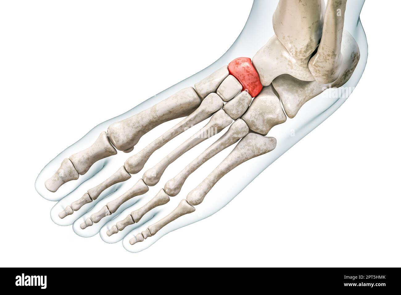 Navicular tarsal bone in red with body 3D rendering illustration isolated on white with copy space. Human skeleton and foot anatomy, medical diagram, Stock Photohttps://www.alamy.com/image-license-details/?v=1https://www.alamy.com/navicular-tarsal-bone-in-red-with-body-3d-rendering-illustration-isolated-on-white-with-copy-space-human-skeleton-and-foot-anatomy-medical-diagram-image548396819.html
RF2PT5HMK–Navicular tarsal bone in red with body 3D rendering illustration isolated on white with copy space. Human skeleton and foot anatomy, medical diagram,
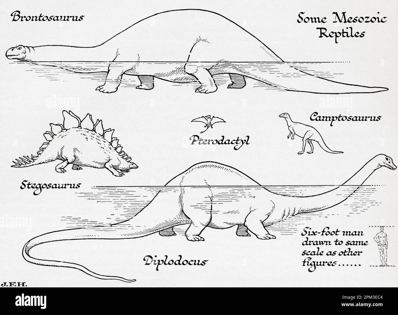 Mesozoic reptiles. Brontosaurus, Pterodactyl, Campotosaurus, Stegosaurus, Diplodocus. Shown in the diagram a six foot man drawn to the same scale as other figures. From the book Outline of History by H.G. Wells, published 1920. Stock Photohttps://www.alamy.com/image-license-details/?v=1https://www.alamy.com/mesozoic-reptiles-brontosaurus-pterodactyl-campotosaurus-stegosaurus-diplodocus-shown-in-the-diagram-a-six-foot-man-drawn-to-the-same-scale-as-other-figures-from-the-book-outline-of-history-by-hg-wells-published-1920-image545891700.html
RM2PM3EC4–Mesozoic reptiles. Brontosaurus, Pterodactyl, Campotosaurus, Stegosaurus, Diplodocus. Shown in the diagram a six foot man drawn to the same scale as other figures. From the book Outline of History by H.G. Wells, published 1920.
 Young person in jeans and black sneakers walking on street Stock Photohttps://www.alamy.com/image-license-details/?v=1https://www.alamy.com/young-person-in-jeans-and-black-sneakers-walking-on-street-image607597992.html
RF2X8EDC8–Young person in jeans and black sneakers walking on street
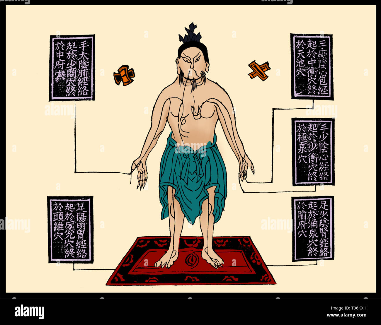 Woodblock illustration from an edition of 1909 (1st year of the Xuantong reign period of the Qing dynasty). Shows the figure of a middle-aged man, with the Heart Envelope channel of hand jueyin, the heart channel of hand shaoyin, the kidney channel of foot shaoyin, the lung channel of hand taiyin and the stomach channel of foot yangming drawn on his body. Stock Photohttps://www.alamy.com/image-license-details/?v=1https://www.alamy.com/woodblock-illustration-from-an-edition-of-1909-1st-year-of-the-xuantong-reign-period-of-the-qing-dynasty-shows-the-figure-of-a-middle-aged-man-with-the-heart-envelope-channel-of-hand-jueyin-the-heart-channel-of-hand-shaoyin-the-kidney-channel-of-foot-shaoyin-the-lung-channel-of-hand-taiyin-and-the-stomach-channel-of-foot-yangming-drawn-on-his-body-image246624409.html
RMT96KXH–Woodblock illustration from an edition of 1909 (1st year of the Xuantong reign period of the Qing dynasty). Shows the figure of a middle-aged man, with the Heart Envelope channel of hand jueyin, the heart channel of hand shaoyin, the kidney channel of foot shaoyin, the lung channel of hand taiyin and the stomach channel of foot yangming drawn on his body.
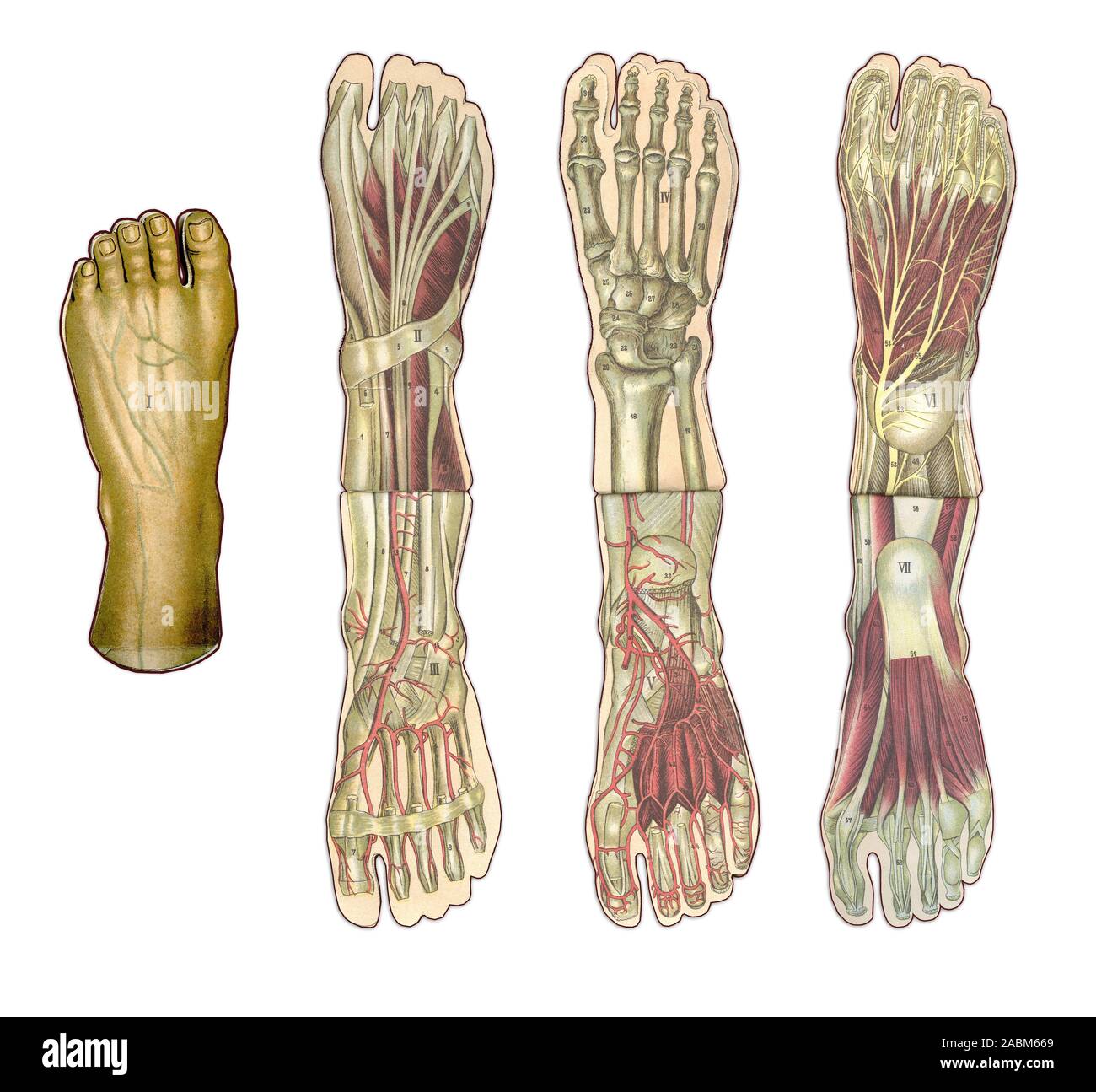 Medicine and healthcare illustrated table, human foot anatomy: skin and ectodermal tissues, bones, muscles nerves, blood vessels Stock Photohttps://www.alamy.com/image-license-details/?v=1https://www.alamy.com/medicine-and-healthcare-illustrated-table-human-foot-anatomy-skin-and-ectodermal-tissues-bones-muscles-nerves-blood-vessels-image334202129.html
RF2ABM669–Medicine and healthcare illustrated table, human foot anatomy: skin and ectodermal tissues, bones, muscles nerves, blood vessels
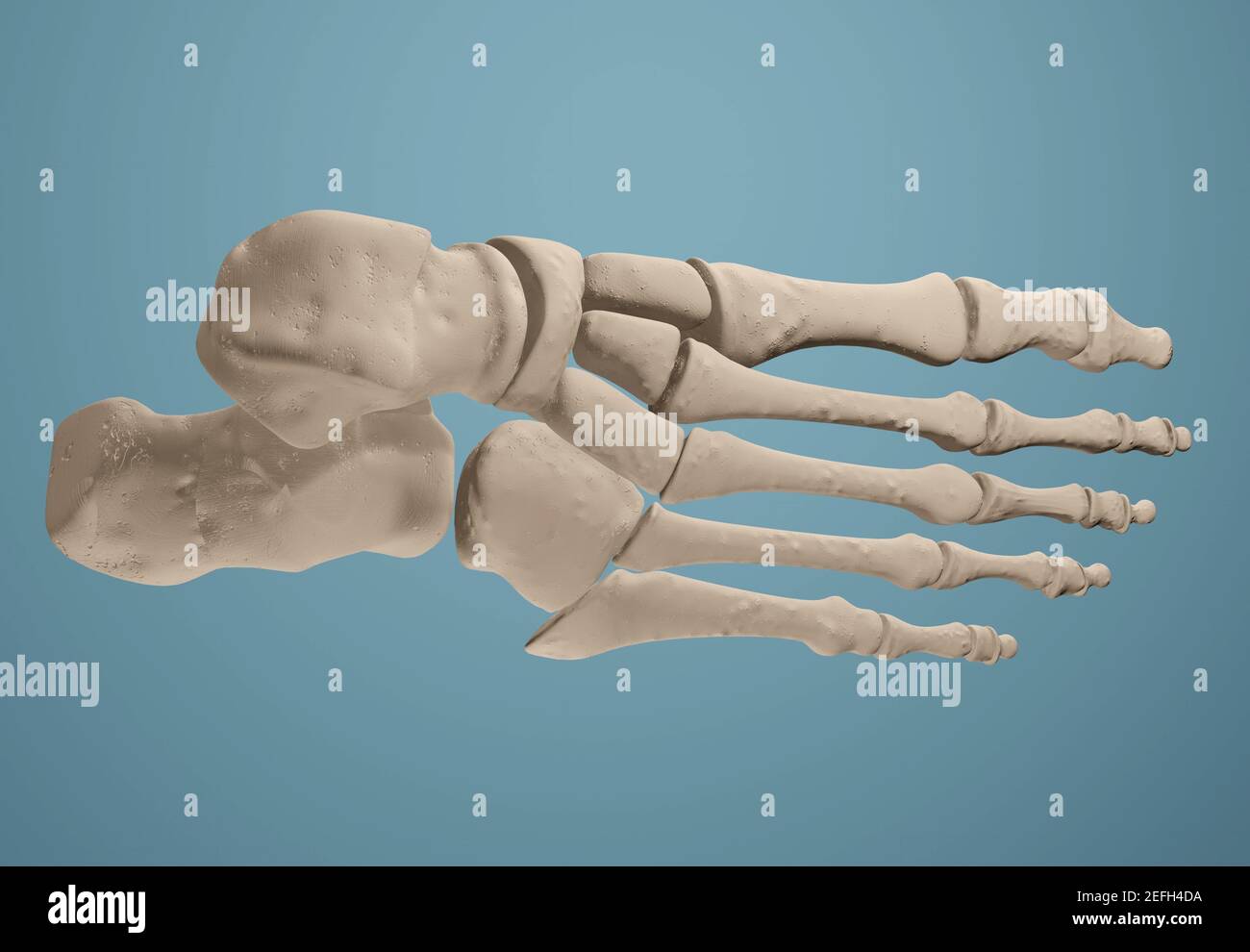 3D render showing bones of the foot. Stock Photohttps://www.alamy.com/image-license-details/?v=1https://www.alamy.com/3d-render-showing-bones-of-the-foot-image405434998.html
RF2EFH4DA–3D render showing bones of the foot.
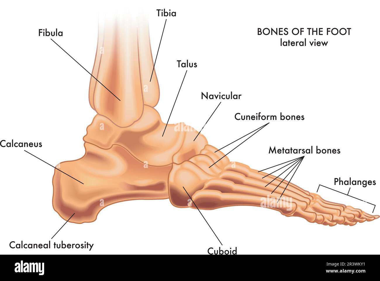 Medical illustration of the major parts of the foot bones in lateral view, with annotations. Stock Vectorhttps://www.alamy.com/image-license-details/?v=1https://www.alamy.com/medical-illustration-of-the-major-parts-of-the-foot-bones-in-lateral-view-with-annotations-image553140197.html
RF2R3WKY1–Medical illustration of the major parts of the foot bones in lateral view, with annotations.
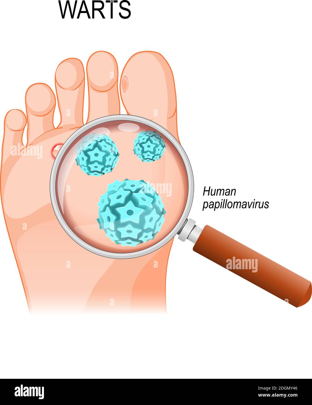 Foot Warts are caused by infection with a type of human papillomavirus. Close-up of HPV Stock Vectorhttps://www.alamy.com/image-license-details/?v=1https://www.alamy.com/foot-warts-are-caused-by-infection-with-a-type-of-human-papillomavirus-close-up-of-hpv-image388922918.html
RF2DGMY46–Foot Warts are caused by infection with a type of human papillomavirus. Close-up of HPV
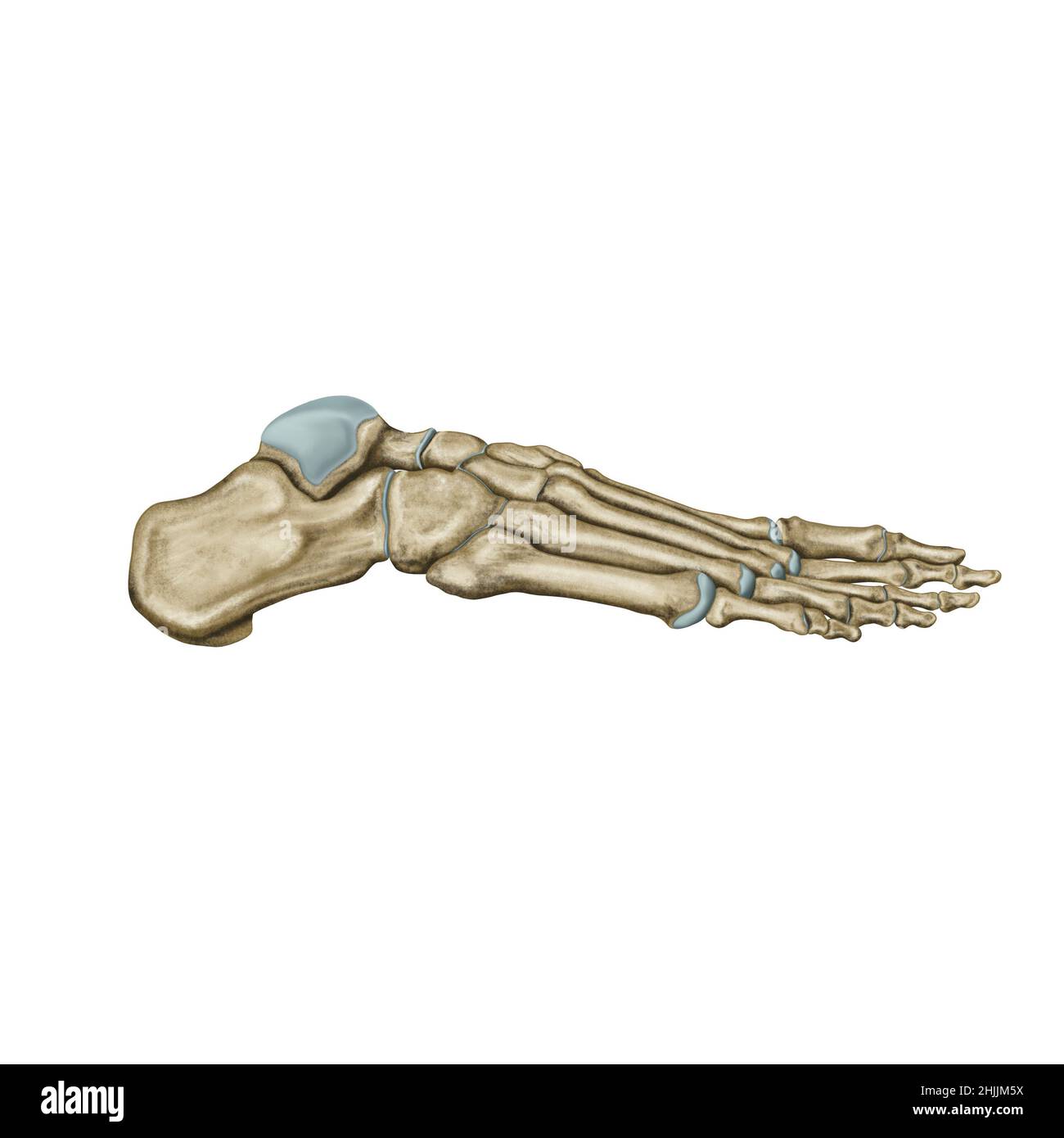 Foot side view, foot anatomy, hand drawn leg bone Stock Photohttps://www.alamy.com/image-license-details/?v=1https://www.alamy.com/foot-side-view-foot-anatomy-hand-drawn-leg-bone-image458944358.html
RF2HJJM5X–Foot side view, foot anatomy, hand drawn leg bone
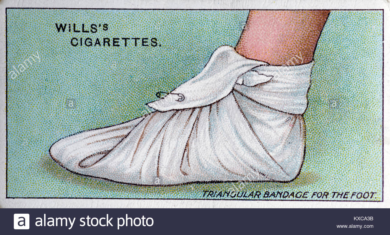 Vintage First Aid illustrations - Triangular bandage for the foot Stock Photohttps://www.alamy.com/image-license-details/?v=1https://www.alamy.com/stock-photo-vintage-first-aid-illustrations-triangular-bandage-for-the-foot-171145727.html
RMKXCA3B–Vintage First Aid illustrations - Triangular bandage for the foot
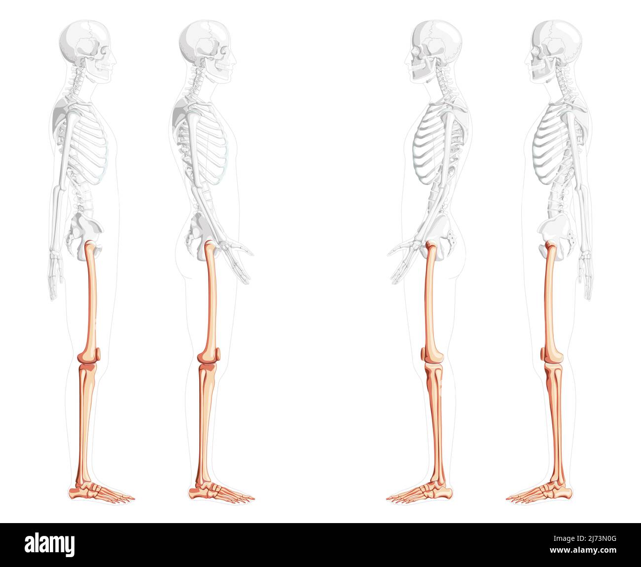 Skeleton Thighs and legs lower limb Human side view with partly transparent bones position. Set of patella, fibula, tibia, foot realistic flat natural color Vector illustration of anatomy isolated Stock Vectorhttps://www.alamy.com/image-license-details/?v=1https://www.alamy.com/skeleton-thighs-and-legs-lower-limb-human-side-view-with-partly-transparent-bones-position-set-of-patella-fibula-tibia-foot-realistic-flat-natural-color-vector-illustration-of-anatomy-isolated-image469064864.html
RF2J73N0G–Skeleton Thighs and legs lower limb Human side view with partly transparent bones position. Set of patella, fibula, tibia, foot realistic flat natural color Vector illustration of anatomy isolated
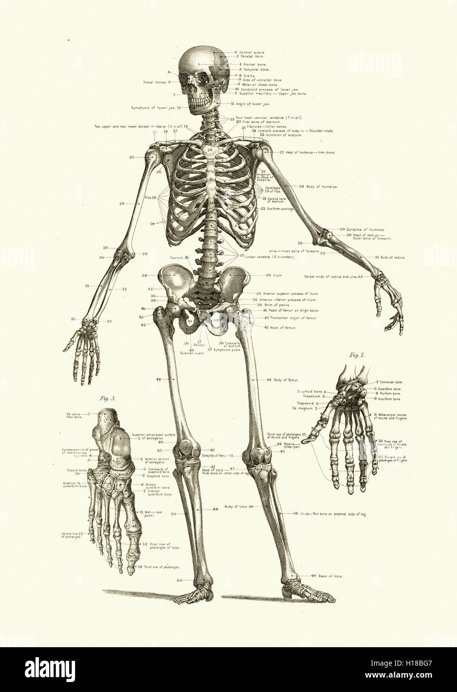 Human skeleton, showing the bones of the body Stock Photohttps://www.alamy.com/image-license-details/?v=1https://www.alamy.com/stock-photo-human-skeleton-showing-the-bones-of-the-body-121271927.html
RMH18BG7–Human skeleton, showing the bones of the body
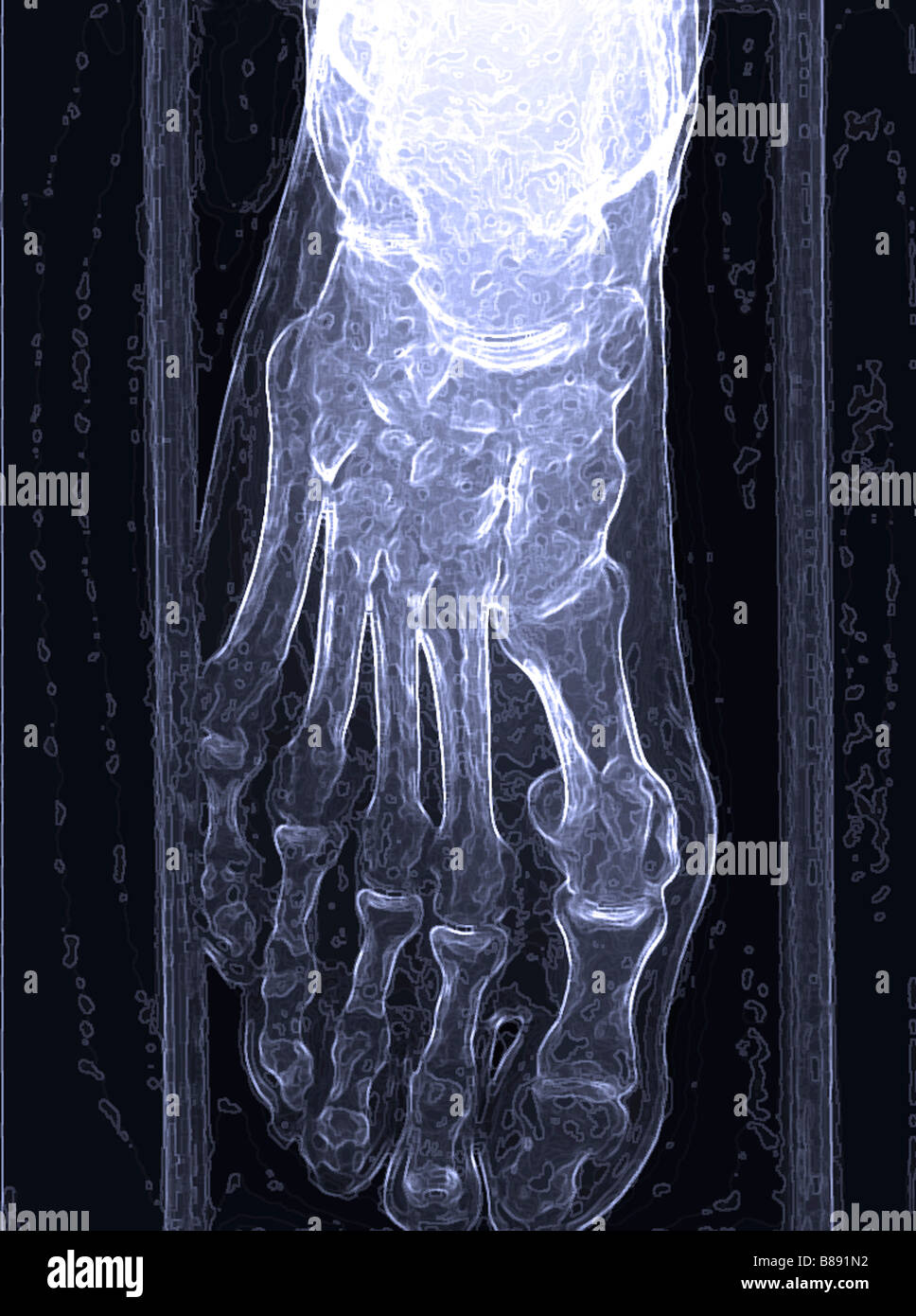 Illustration of an Xray of a human foot Stock Photohttps://www.alamy.com/image-license-details/?v=1https://www.alamy.com/stock-photo-illustration-of-an-xray-of-a-human-foot-22326558.html
RMB891N2–Illustration of an Xray of a human foot
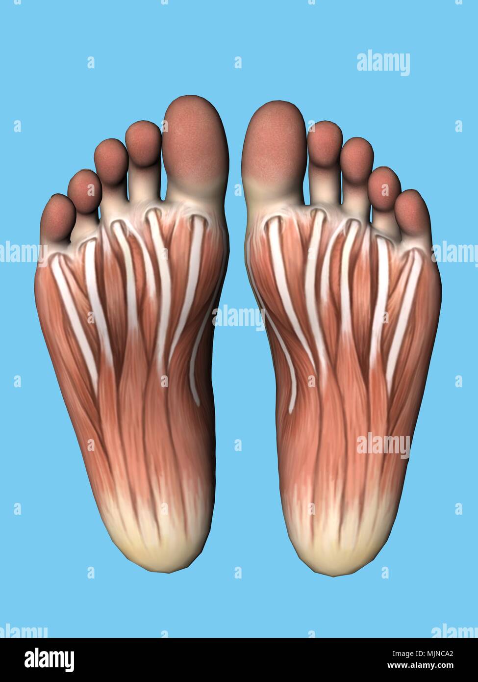 Anatomy bottom view of foot. Stock Photohttps://www.alamy.com/image-license-details/?v=1https://www.alamy.com/anatomy-bottom-view-of-foot-image183638170.html
RMMJNCA2–Anatomy bottom view of foot.
 A 19th century dictionary description and picture of snow shoes as they were at that time Stock Photohttps://www.alamy.com/image-license-details/?v=1https://www.alamy.com/a-19th-century-dictionary-description-and-picture-of-snow-shoes-as-they-were-at-that-time-image488028746.html
RM2K9YHJ2–A 19th century dictionary description and picture of snow shoes as they were at that time
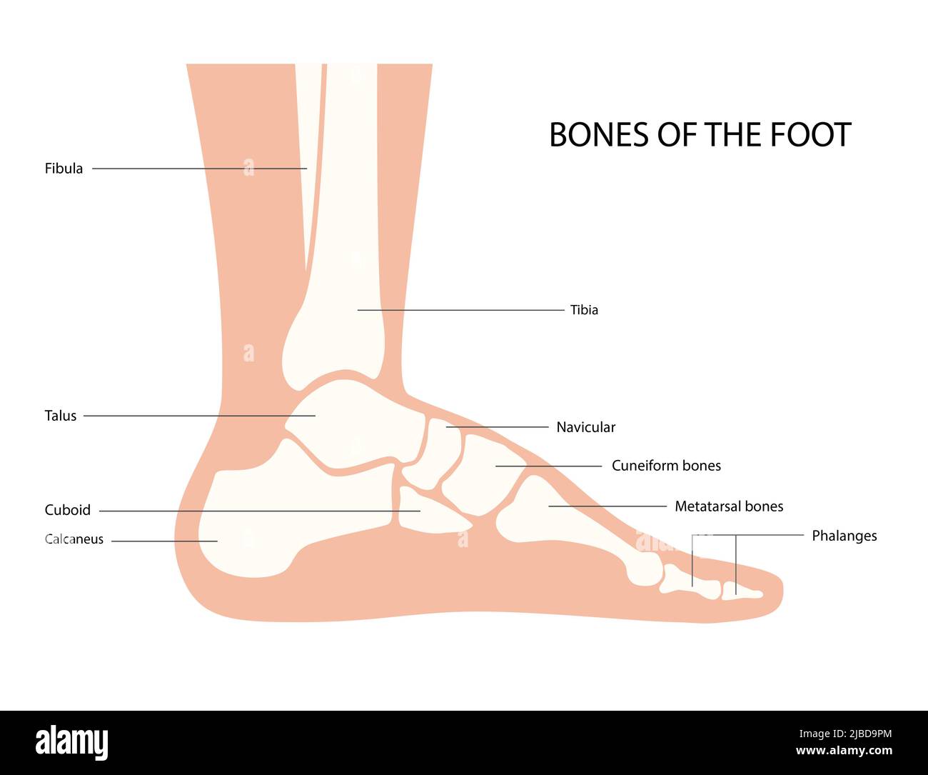 Foot anatomy, illustration Stock Photohttps://www.alamy.com/image-license-details/?v=1https://www.alamy.com/foot-anatomy-illustration-image471734220.html
RF2JBD9PM–Foot anatomy, illustration
 career, chart, ascent, diagram, graphic, bar chart, upswing, bull market, boom, Stock Photohttps://www.alamy.com/image-license-details/?v=1https://www.alamy.com/stock-photo-career-chart-ascent-diagram-graphic-bar-chart-upswing-bull-market-131745891.html
RFHJ9F6B–career, chart, ascent, diagram, graphic, bar chart, upswing, bull market, boom,
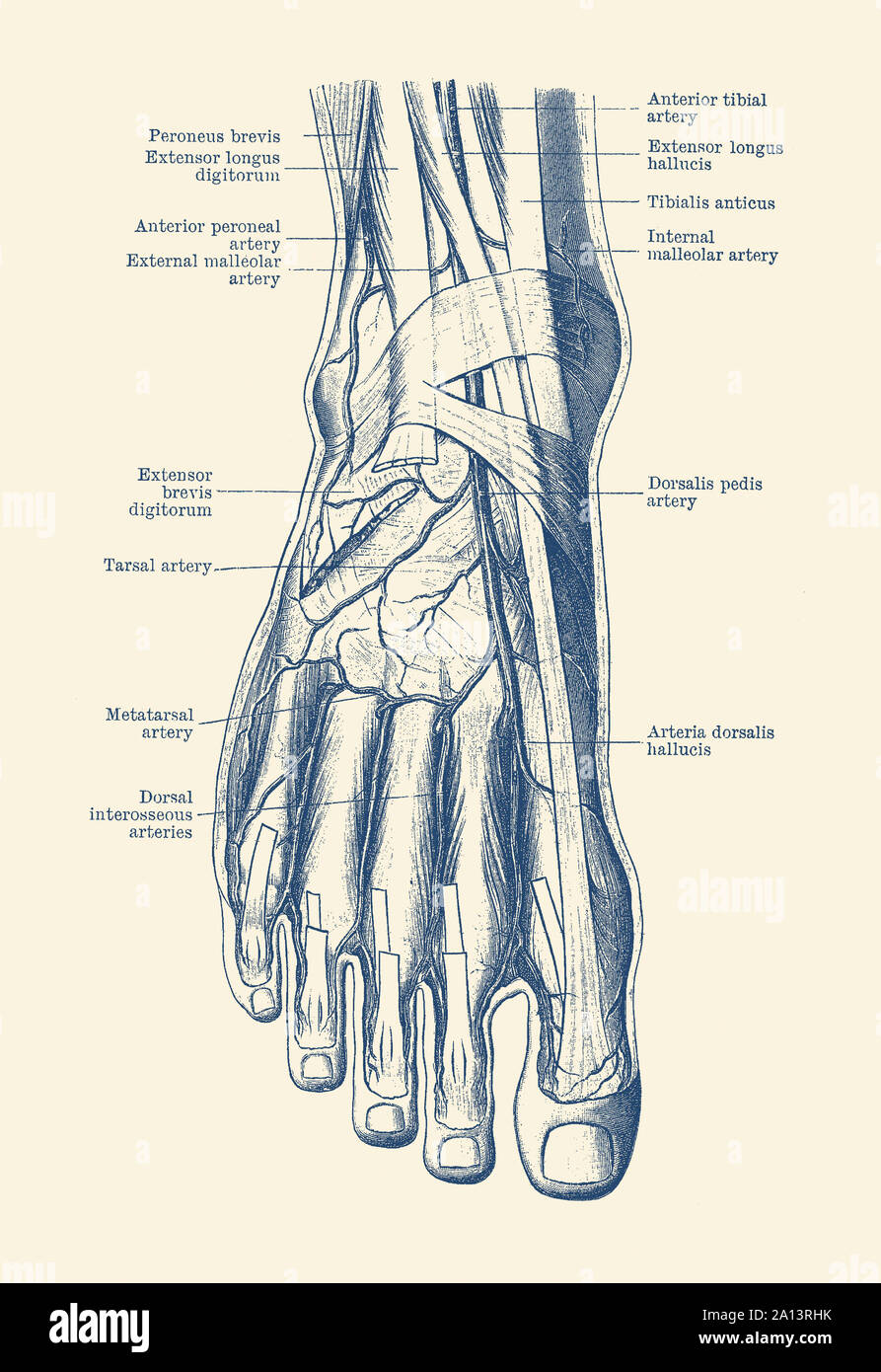 Vintage anatomy print of the human foot, showcasing the veins and arteries. Stock Photohttps://www.alamy.com/image-license-details/?v=1https://www.alamy.com/vintage-anatomy-print-of-the-human-foot-showcasing-the-veins-and-arteries-image327696031.html
RF2A13RHK–Vintage anatomy print of the human foot, showcasing the veins and arteries.
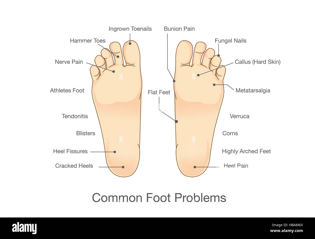 Common foot problems. Stock Vectorhttps://www.alamy.com/image-license-details/?v=1https://www.alamy.com/stock-photo-common-foot-problems-127469186.html
RFHBAM6X–Common foot problems.
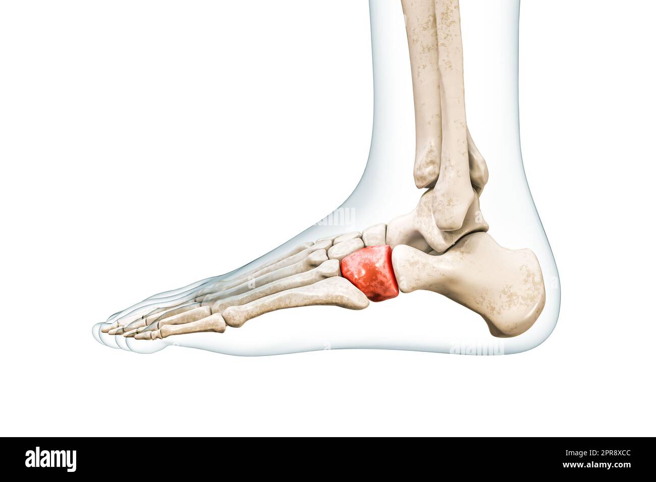 Cuboid tarsal bone in red with body 3D rendering illustration isolated on white with copy space. Human skeleton and foot anatomy, medical diagram, ost Stock Photohttps://www.alamy.com/image-license-details/?v=1https://www.alamy.com/cuboid-tarsal-bone-in-red-with-body-3d-rendering-illustration-isolated-on-white-with-copy-space-human-skeleton-and-foot-anatomy-medical-diagram-ost-image547854844.html
RF2PR8XCC–Cuboid tarsal bone in red with body 3D rendering illustration isolated on white with copy space. Human skeleton and foot anatomy, medical diagram, ost
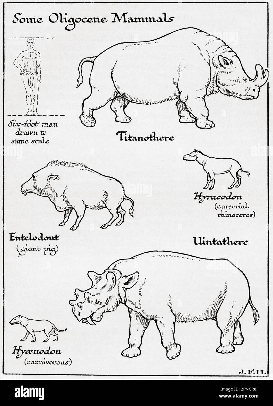 Diagram of Oligocene Mammals including, Titanothere, Entelodont, Hyracodon, Uintathere, Hyaenodon. Shown in the diagram a six foot man drawn to the same scale as other figures. From the book Outline of History by H.G. Wells, published 1920. Stock Photohttps://www.alamy.com/image-license-details/?v=1https://www.alamy.com/diagram-of-oligocene-mammals-including-titanothere-entelodont-hyracodon-uintathere-hyaenodon-shown-in-the-diagram-a-six-foot-man-drawn-to-the-same-scale-as-other-figures-from-the-book-outline-of-history-by-hg-wells-published-1920-image546710879.html
RM2PNCR8F–Diagram of Oligocene Mammals including, Titanothere, Entelodont, Hyracodon, Uintathere, Hyaenodon. Shown in the diagram a six foot man drawn to the same scale as other figures. From the book Outline of History by H.G. Wells, published 1920.
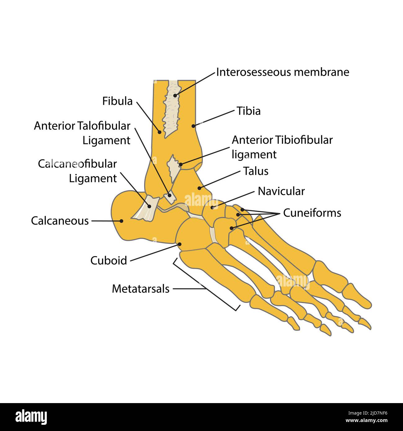 illustration of foot bone. vector. on white background Stock Vectorhttps://www.alamy.com/image-license-details/?v=1https://www.alamy.com/illustration-of-foot-bone-vector-on-white-background-image472841018.html
RF2JD7NF6–illustration of foot bone. vector. on white background
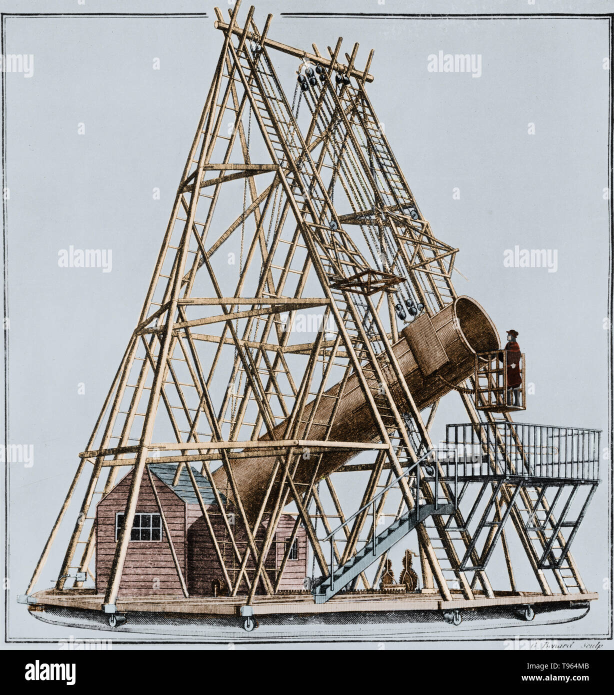 William Herschel's 40 foot telescope, also known as the Great Forty Foot telescope, was a reflecting telescope constructed between 1785 and 1789 at Observatory House in Slough, England. It used a 47 inch diameter primary mirror with a 1,200 cm focal length. It was the largest telescope in the world for 50 years. It may have been used to discover Enceladus and Mimas, the 6th and 7th moons of Saturn. It was dismantled in 1840; today the original mirror and a 10 foot section of the tube remain. Stock Photohttps://www.alamy.com/image-license-details/?v=1https://www.alamy.com/william-herschels-40-foot-telescope-also-known-as-the-great-forty-foot-telescope-was-a-reflecting-telescope-constructed-between-1785-and-1789-at-observatory-house-in-slough-england-it-used-a-47-inch-diameter-primary-mirror-with-a-1200-cm-focal-length-it-was-the-largest-telescope-in-the-world-for-50-years-it-may-have-been-used-to-discover-enceladus-and-mimas-the-6th-and-7th-moons-of-saturn-it-was-dismantled-in-1840-today-the-original-mirror-and-a-10-foot-section-of-the-tube-remain-image246612475.html
RMT964MB–William Herschel's 40 foot telescope, also known as the Great Forty Foot telescope, was a reflecting telescope constructed between 1785 and 1789 at Observatory House in Slough, England. It used a 47 inch diameter primary mirror with a 1,200 cm focal length. It was the largest telescope in the world for 50 years. It may have been used to discover Enceladus and Mimas, the 6th and 7th moons of Saturn. It was dismantled in 1840; today the original mirror and a 10 foot section of the tube remain.
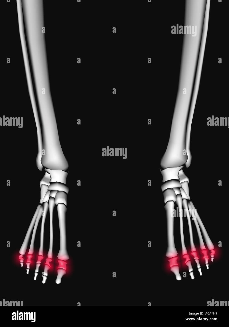 Illustrative diagram showing skeleton showing phalanges highlighted in a red glow Stock Photohttps://www.alamy.com/image-license-details/?v=1https://www.alamy.com/illustrative-diagram-showing-skeleton-showing-phalanges-highlighted-image5676376.html
RMA0AFH9–Illustrative diagram showing skeleton showing phalanges highlighted in a red glow
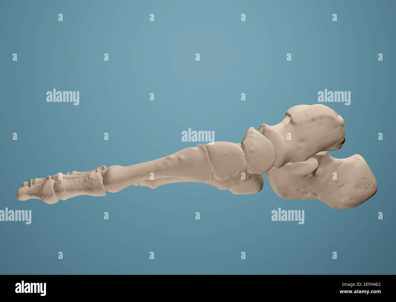 3D render showing bones of the foot. Stock Photohttps://www.alamy.com/image-license-details/?v=1https://www.alamy.com/3d-render-showing-bones-of-the-foot-image405435018.html
RF2EFH4E2–3D render showing bones of the foot.
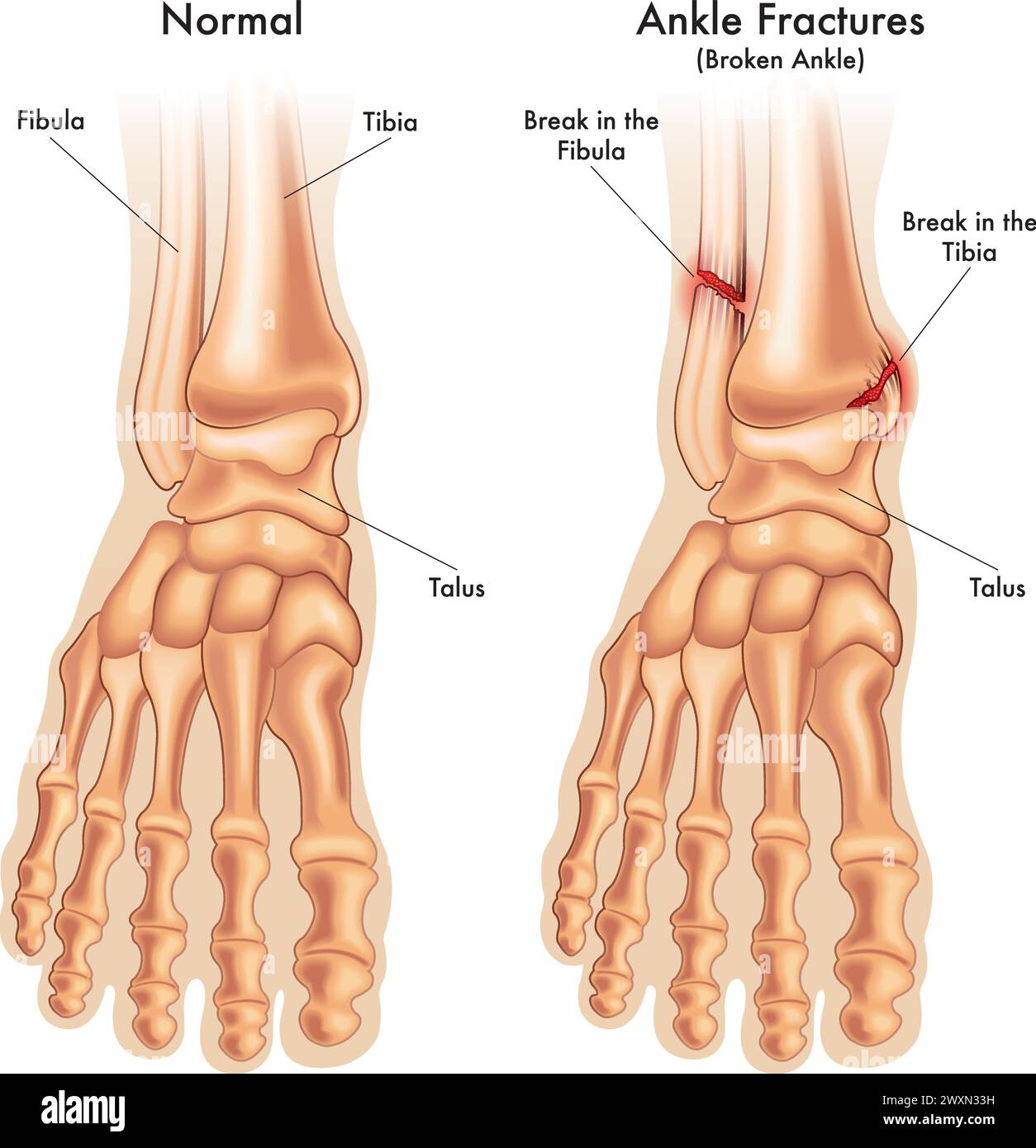 medical illustration compares a normal foot ankle, with a foot ankle fractured in two places, with annotations. Stock Vectorhttps://www.alamy.com/image-license-details/?v=1https://www.alamy.com/medical-illustration-compares-a-normal-foot-ankle-with-a-foot-ankle-fractured-in-two-places-with-annotations-image601597013.html
RF2WXN33H–medical illustration compares a normal foot ankle, with a foot ankle fractured in two places, with annotations.
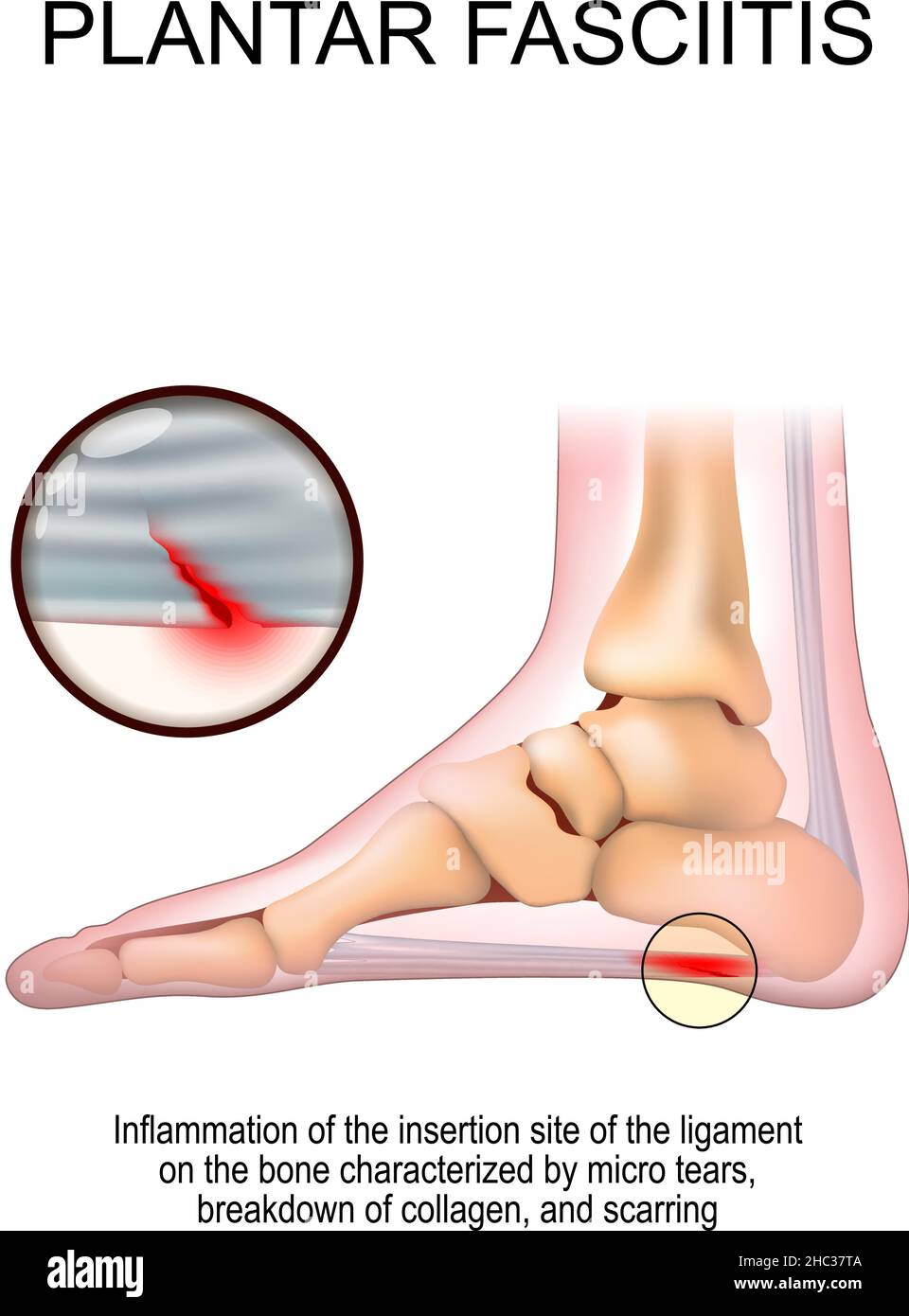 Plantar fasciitis. foot with the symptoms of plantar fasciitis. disorder of the connective tissue which supports the arch of the foot. Stock Vectorhttps://www.alamy.com/image-license-details/?v=1https://www.alamy.com/plantar-fasciitis-foot-with-the-symptoms-of-plantar-fasciitis-disorder-of-the-connective-tissue-which-supports-the-arch-of-the-foot-image454917466.html
RF2HC37TA–Plantar fasciitis. foot with the symptoms of plantar fasciitis. disorder of the connective tissue which supports the arch of the foot.
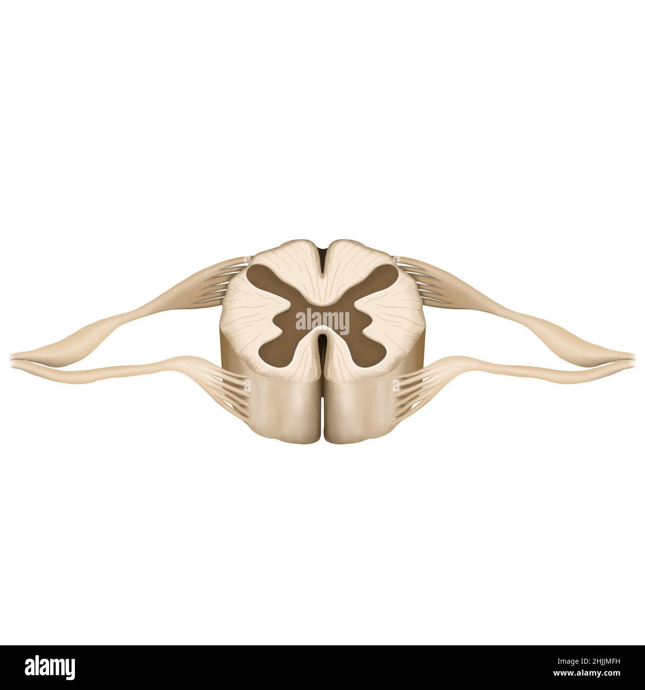 Spinal cord, Spinal cord anatomy, spinal cord drawing Stock Photohttps://www.alamy.com/image-license-details/?v=1https://www.alamy.com/spinal-cord-spinal-cord-anatomy-spinal-cord-drawing-image458944629.html
RF2HJJMFH–Spinal cord, Spinal cord anatomy, spinal cord drawing
 Man standing in his comfort zone, diagram with various zones, logo design. Stepping outside comfort zone, choice and choosing between leaving comfort Stock Vectorhttps://www.alamy.com/image-license-details/?v=1https://www.alamy.com/man-standing-in-his-comfort-zone-diagram-with-various-zones-logo-design-stepping-outside-comfort-zone-choice-and-choosing-between-leaving-comfort-image491379699.html
RF2KFC7PY–Man standing in his comfort zone, diagram with various zones, logo design. Stepping outside comfort zone, choice and choosing between leaving comfort
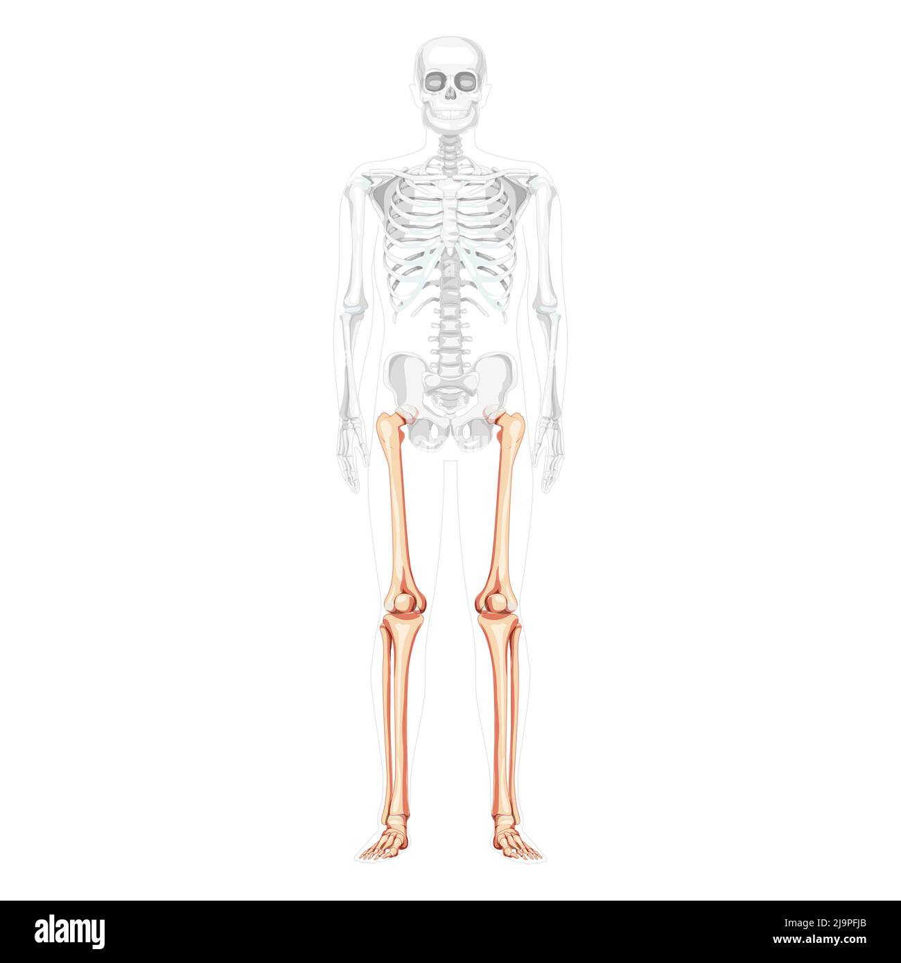 Skeleton Thighs and legs lower limb Human front view with partly transparent bones position. Femur, patella, fibula, foot realistic flat Vector illustration of anatomy isolated on white background Stock Vectorhttps://www.alamy.com/image-license-details/?v=1https://www.alamy.com/skeleton-thighs-and-legs-lower-limb-human-front-view-with-partly-transparent-bones-position-femur-patella-fibula-foot-realistic-flat-vector-illustration-of-anatomy-isolated-on-white-background-image470707059.html
RF2J9PFJB–Skeleton Thighs and legs lower limb Human front view with partly transparent bones position. Femur, patella, fibula, foot realistic flat Vector illustration of anatomy isolated on white background
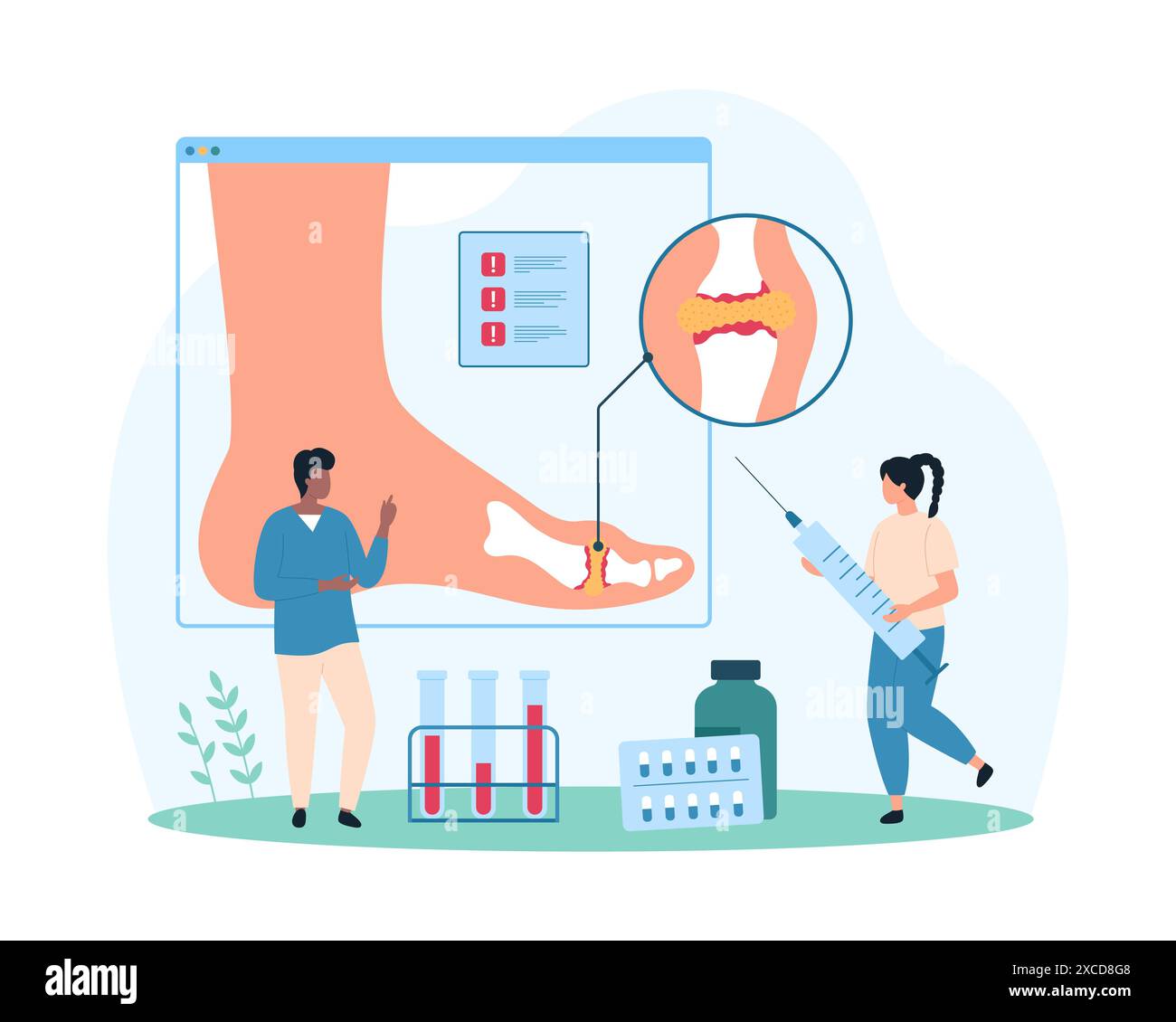 Diagnosis and treatment of gout, inflammatory arthritis of foot joints, rheumatology. Tiny people study infographic medical chart of ankle, watch out for uric acid crystals cartoon vector illustration Stock Vectorhttps://www.alamy.com/image-license-details/?v=1https://www.alamy.com/diagnosis-and-treatment-of-gout-inflammatory-arthritis-of-foot-joints-rheumatology-tiny-people-study-infographic-medical-chart-of-ankle-watch-out-for-uric-acid-crystals-cartoon-vector-illustration-image610030856.html
RF2XCD8G8–Diagnosis and treatment of gout, inflammatory arthritis of foot joints, rheumatology. Tiny people study infographic medical chart of ankle, watch out for uric acid crystals cartoon vector illustration
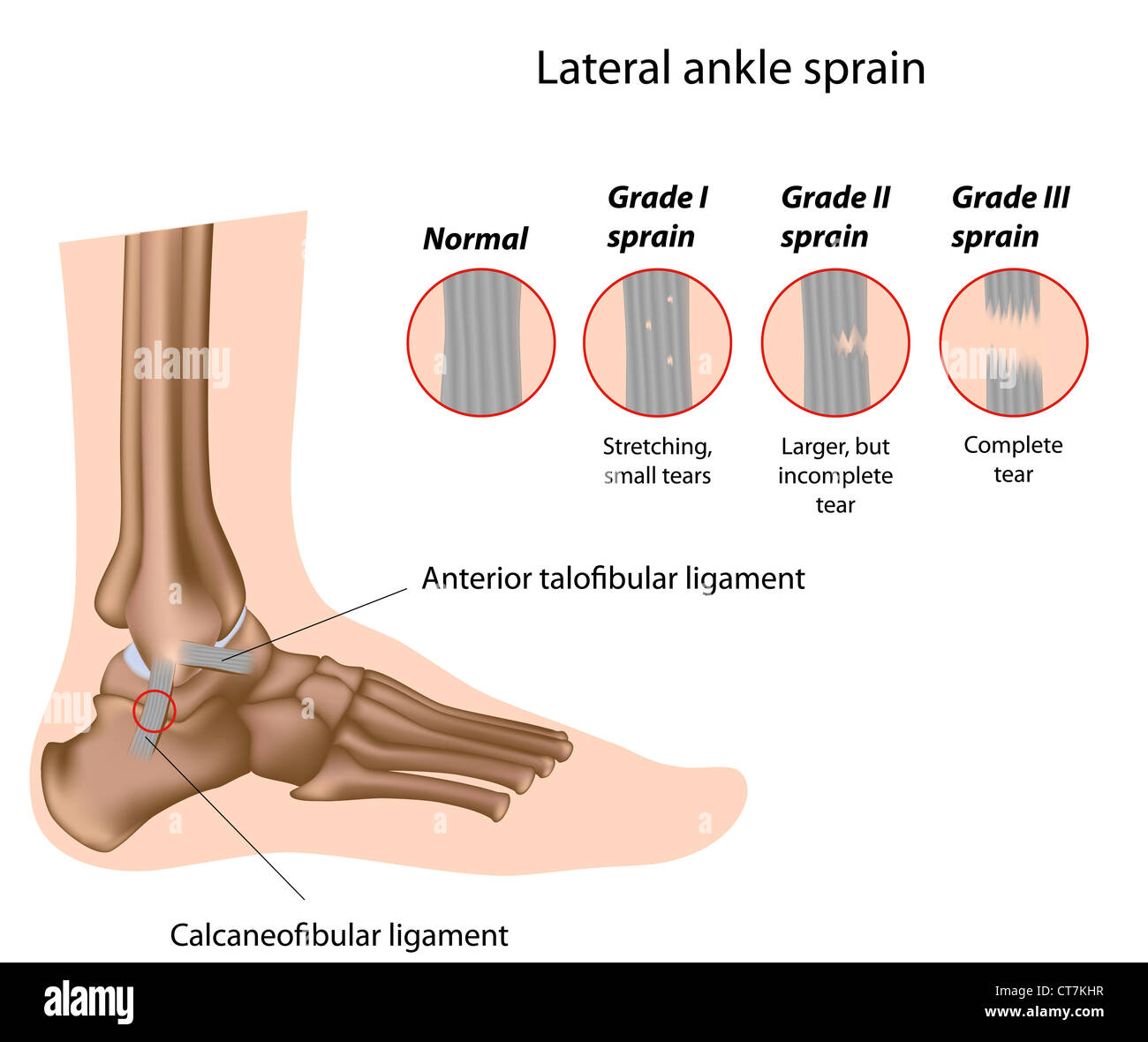 Ankle sprain grading Stock Photohttps://www.alamy.com/image-license-details/?v=1https://www.alamy.com/stock-photo-ankle-sprain-grading-49341539.html
RFCT7KHR–Ankle sprain grading
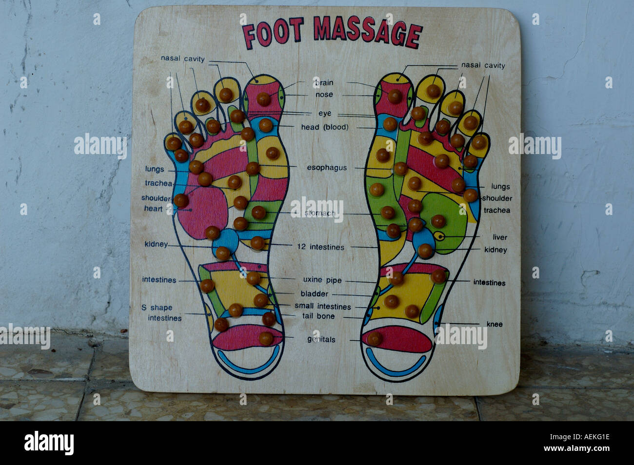 Reflexology chart, correlating areas of the feet with portions of the body on wooden board Stock Photohttps://www.alamy.com/image-license-details/?v=1https://www.alamy.com/reflexology-chart-correlating-areas-of-the-feet-with-portions-of-the-image4473885.html
RMAEKG1E–Reflexology chart, correlating areas of the feet with portions of the body on wooden board
 Achilles tendon Stock Photohttps://www.alamy.com/image-license-details/?v=1https://www.alamy.com/stock-photo-achilles-tendon-54596190.html
RFD4R1YX–Achilles tendon
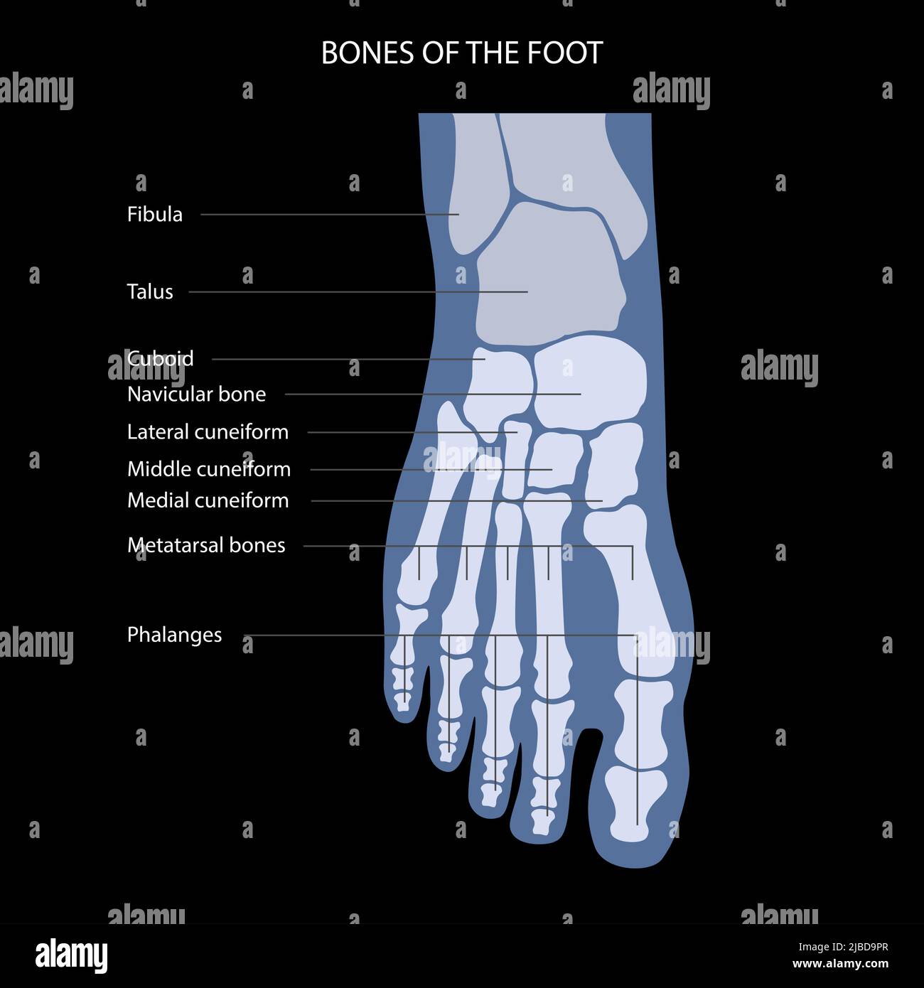 Foot anatomy, illustration Stock Photohttps://www.alamy.com/image-license-details/?v=1https://www.alamy.com/foot-anatomy-illustration-image471734223.html
RF2JBD9PR–Foot anatomy, illustration
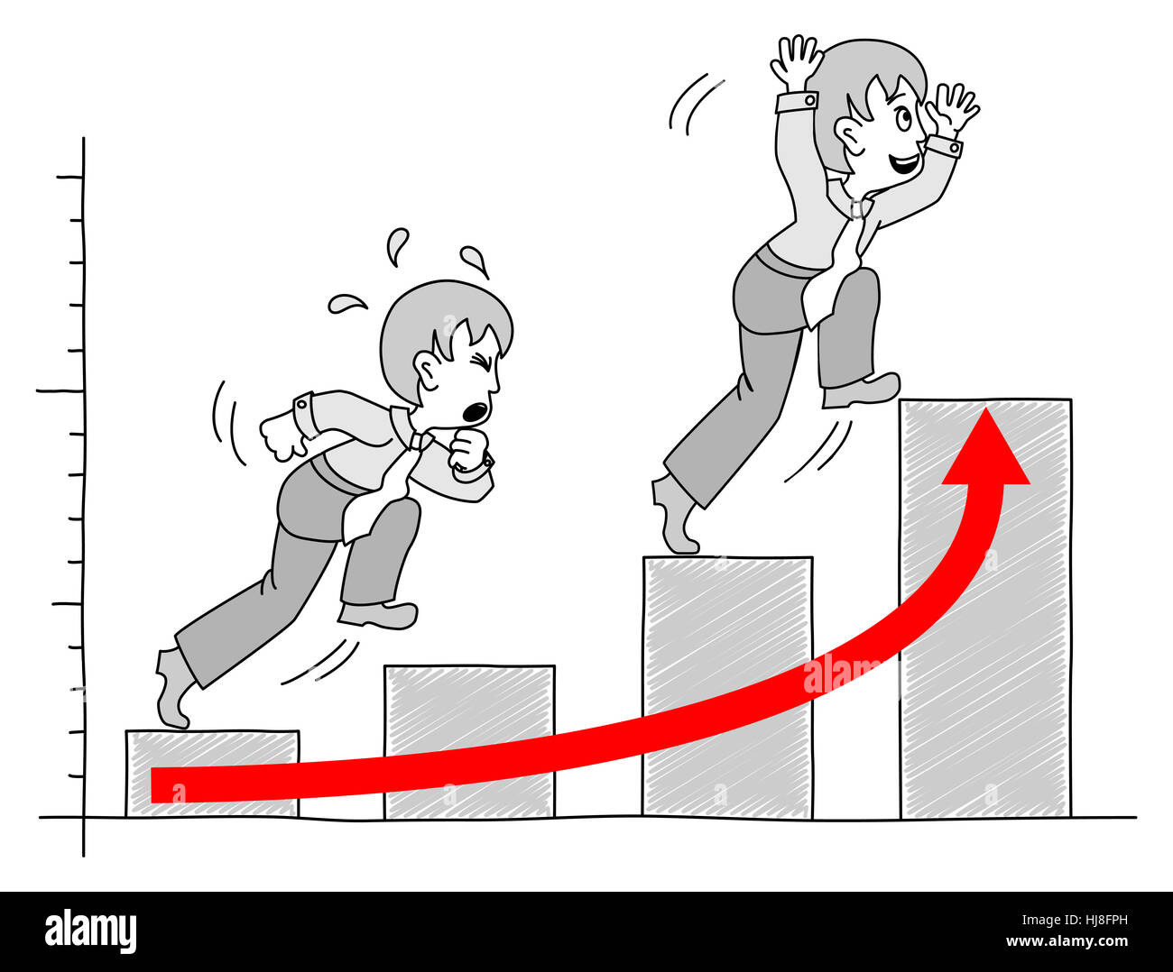 career, chart, ascent, diagram, graphic, bar chart, upswing, bull market, boom, Stock Photohttps://www.alamy.com/image-license-details/?v=1https://www.alamy.com/stock-photo-career-chart-ascent-diagram-graphic-bar-chart-upswing-bull-market-131724393.html
RFHJ8FPH–career, chart, ascent, diagram, graphic, bar chart, upswing, bull market, boom,