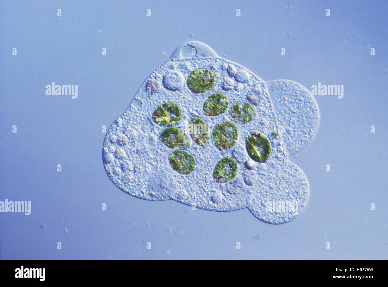
Differential interference contrast micrograph Stock Photos and Images
(22)See differential interference contrast micrograph stock video clipsQuick filters:
Differential interference contrast micrograph Stock Photos and Images
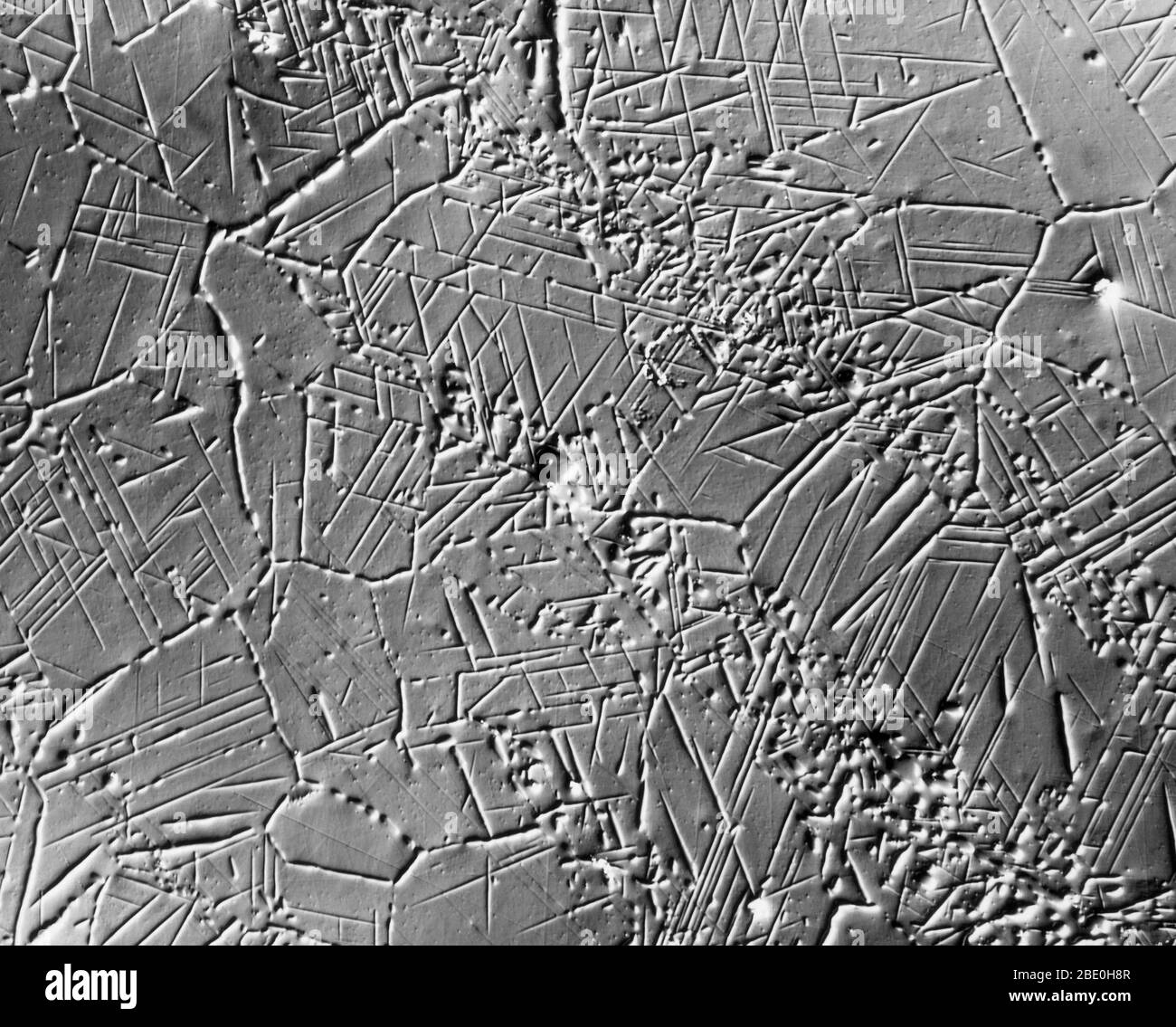 Scanning Electron Micrograph (SEM) showing sigma-phase crystallization in steel. Magnification: 500X at 8x10'. Nomarski interference contrast method. Stock Photohttps://www.alamy.com/image-license-details/?v=1https://www.alamy.com/scanning-electron-micrograph-sem-showing-sigma-phase-crystallization-in-steel-magnification-500x-at-8x10-nomarski-interference-contrast-method-image352826119.html
RM2BE0H8R–Scanning Electron Micrograph (SEM) showing sigma-phase crystallization in steel. Magnification: 500X at 8x10'. Nomarski interference contrast method.
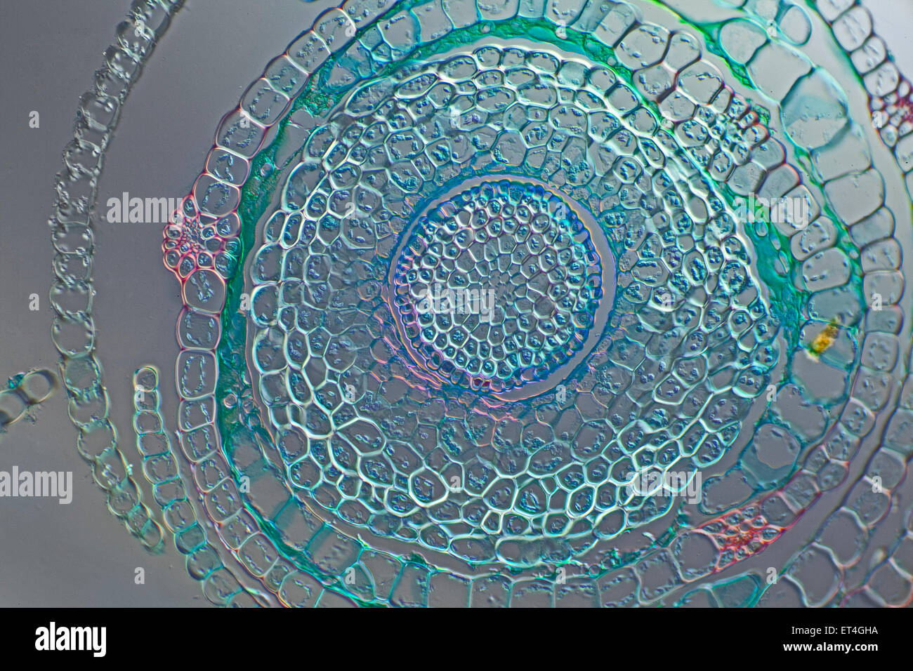 Moss stem TS Funaria sp. Differential interference contrast (DIC) Nomarski Stock Photohttps://www.alamy.com/image-license-details/?v=1https://www.alamy.com/stock-photo-moss-stem-ts-funaria-sp-differential-interference-contrast-dic-nomarski-83694054.html
RMET4GHA–Moss stem TS Funaria sp. Differential interference contrast (DIC) Nomarski
 Differential interference contrast light micrograph of different species of fossil unicellular radiolarians (protozoa), from Barbados. Here, finely structured skeletons of different fossil types of radiolarian are seen. Made of silicates, the skeleton divides the inner cell nucleus and protoplasm from external protoplasm which extends out of the holes in the skeleton. In this way, radiolarians feed by trapping micro- organisms in the water. These tiny skeletons are among nature's most beautiful structures, and each species has a unique design. Plankton provides marine food. Stock Photohttps://www.alamy.com/image-license-details/?v=1https://www.alamy.com/stock-image-differential-interference-contrast-light-micrograph-of-different-species-166440663.html
RFKJP0NB–Differential interference contrast light micrograph of different species of fossil unicellular radiolarians (protozoa), from Barbados. Here, finely structured skeletons of different fossil types of radiolarian are seen. Made of silicates, the skeleton divides the inner cell nucleus and protoplasm from external protoplasm which extends out of the holes in the skeleton. In this way, radiolarians feed by trapping micro- organisms in the water. These tiny skeletons are among nature's most beautiful structures, and each species has a unique design. Plankton provides marine food.
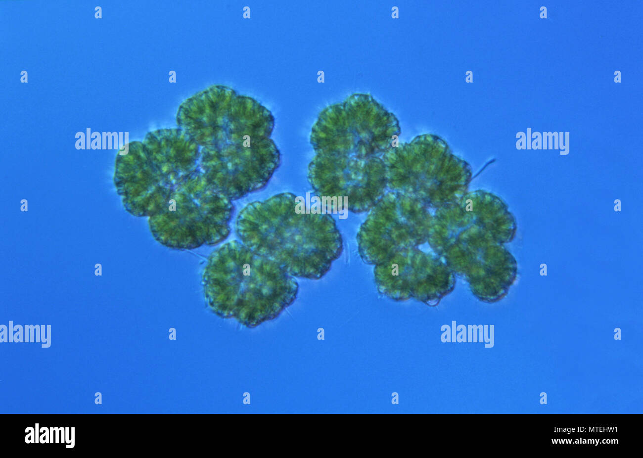 Algae.Dictyosphaerium sp.Differential interference contrast microscopy or Normarsky microscopy. Stock Photohttps://www.alamy.com/image-license-details/?v=1https://www.alamy.com/algaedictyosphaerium-spdifferential-interference-contrast-microscopy-or-normarsky-microscopy-image187176781.html
RFMTEHW1–Algae.Dictyosphaerium sp.Differential interference contrast microscopy or Normarsky microscopy.
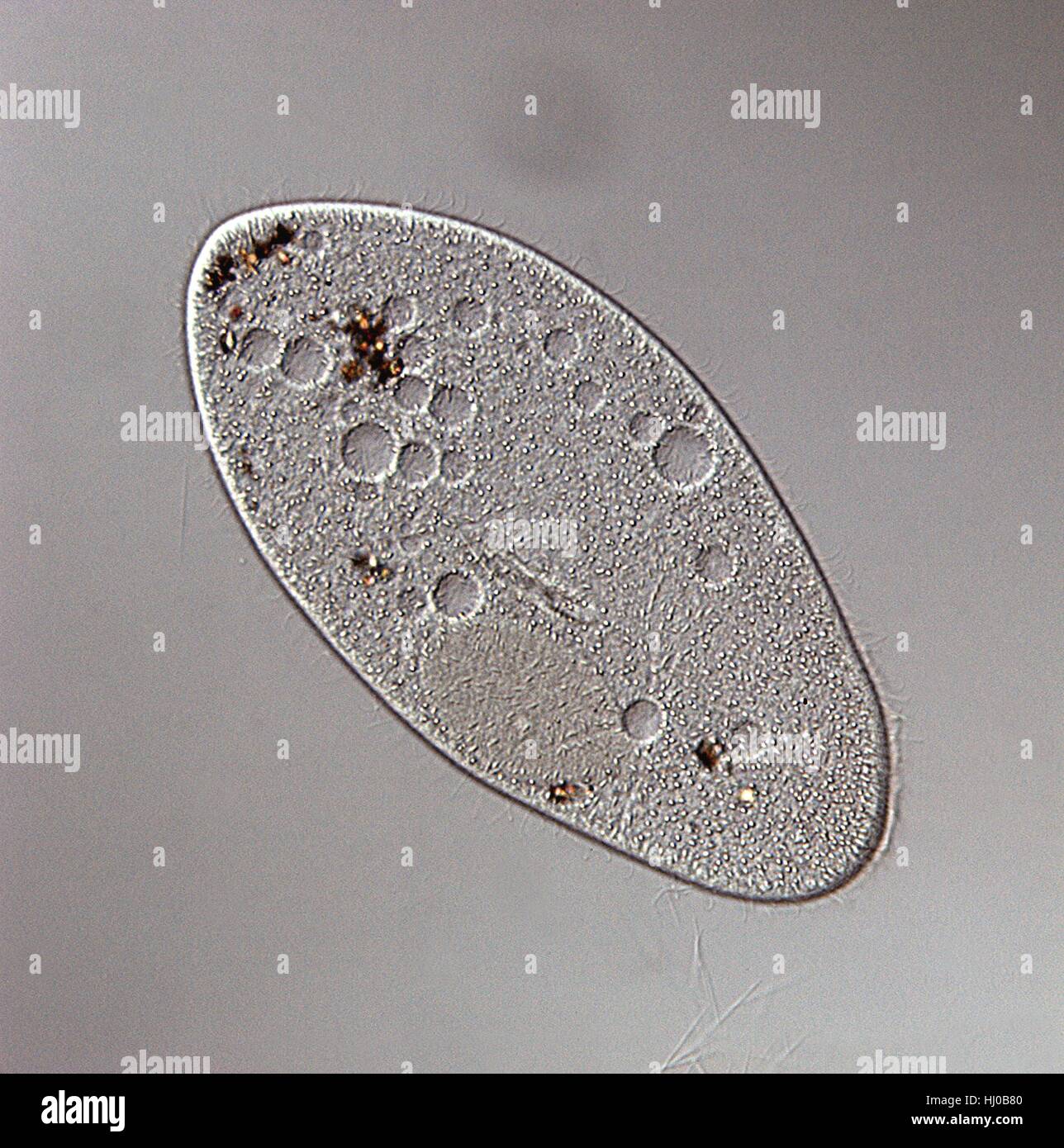 DIC light micrograph of Paramecium.(Paramecium multimicronuleatum),a ciliate protozoan,with oral groove,food vacuoles,nucleus (left edge) cilia.Paramecium are found mainly in stagnant ponds,feeding on bacteria plant particles.They have permanent mouth called oral grove.Food taken in through oral Stock Photohttps://www.alamy.com/image-license-details/?v=1https://www.alamy.com/stock-photo-dic-light-micrograph-of-parameciumparamecium-multimicronuleatuma-ciliate-131545232.html
RFHJ0B80–DIC light micrograph of Paramecium.(Paramecium multimicronuleatum),a ciliate protozoan,with oral groove,food vacuoles,nucleus (left edge) cilia.Paramecium are found mainly in stagnant ponds,feeding on bacteria plant particles.They have permanent mouth called oral grove.Food taken in through oral
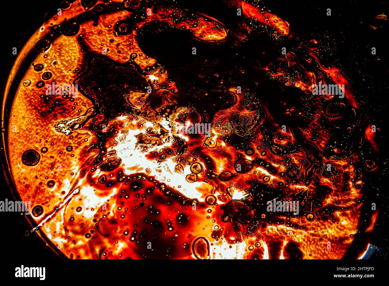 landscape, lava, night, volcano, Copper Mine Museum North Snowdonia Sygun, ferrous, oxide, pool,,ferrous sulfate wall art ,ulphate crystals. Different Stock Photohttps://www.alamy.com/image-license-details/?v=1https://www.alamy.com/landscape-lava-night-volcano-copper-mine-museum-north-snowdonia-sygun-ferrous-oxide-poolferrous-sulfate-wall-art-ulphate-crystals-different-image462718801.html
RM2HTPJFD–landscape, lava, night, volcano, Copper Mine Museum North Snowdonia Sygun, ferrous, oxide, pool,,ferrous sulfate wall art ,ulphate crystals. Different
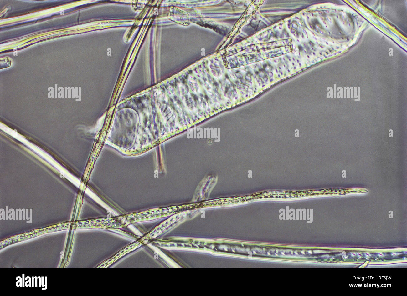 Macerated Oak Wood Stock Photohttps://www.alamy.com/image-license-details/?v=1https://www.alamy.com/stock-photo-macerated-oak-wood-134944177.html
RMHRF6JW–Macerated Oak Wood
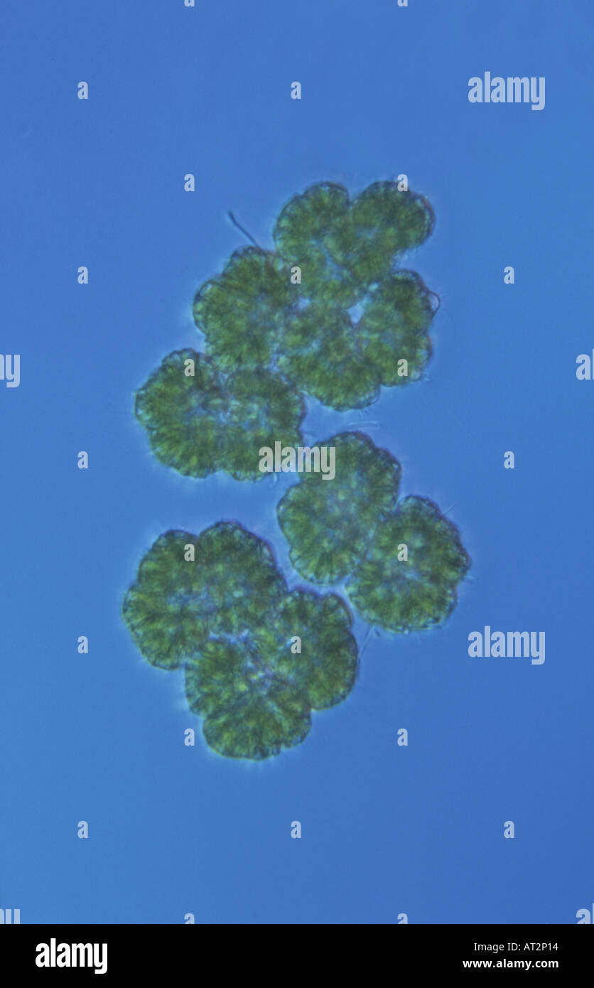 Algae Dictyosphaerium sp Differential interference contrast microscopy or Normarsky microscopy Stock Photohttps://www.alamy.com/image-license-details/?v=1https://www.alamy.com/algae-dictyosphaerium-sp-differential-interference-contrast-microscopy-image5283347.html
RFAT2P14–Algae Dictyosphaerium sp Differential interference contrast microscopy or Normarsky microscopy
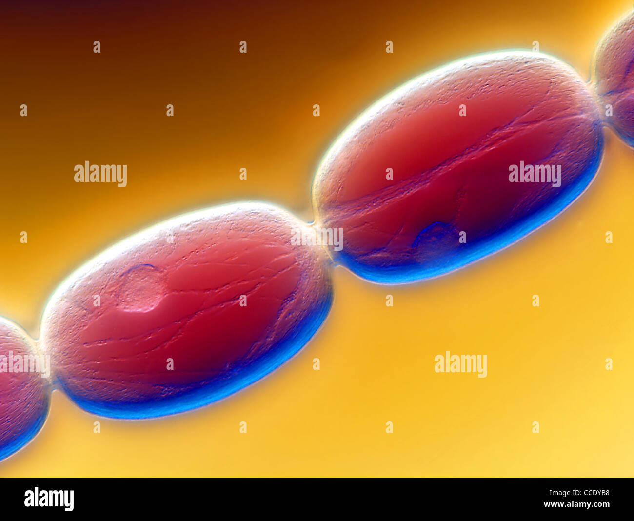 Stamen hair cells of a Tradescantia flower. Stock Photohttps://www.alamy.com/image-license-details/?v=1https://www.alamy.com/stock-photo-stamen-hair-cells-of-a-tradescantia-flower-42103468.html
RMCCDYB8–Stamen hair cells of a Tradescantia flower.
 . The Biological bulletin. Biology; Zoology; Biology; Marine Biology. 620 S. SMILEY. FIGURE 10, Doubly exposed polarizing and differential interference contrast micrograph of the lar- 3el and madreporic crystal. E = esophagus, EV = evaginations of the hydrocoel, HC = hydro- coel, N< = madreporic crystal. Mag. 1230X. FIGURE 11. Doubly exposed polarizing and DIG micrograph of the madreporic crystal surrounding the hydroporic canal. Note that the madreporic crystal has the shape of a fine filigree. E = esophagus, MC = madreporic crystal, S = stomach. Mag. 620X.. Please note that these images a Stock Photohttps://www.alamy.com/image-license-details/?v=1https://www.alamy.com/the-biological-bulletin-biology-zoology-biology-marine-biology-620-s-smiley-figure-10-doubly-exposed-polarizing-and-differential-interference-contrast-micrograph-of-the-lar-3el-and-madreporic-crystal-e-=-esophagus-ev-=-evaginations-of-the-hydrocoel-hc-=-hydro-coel-nlt-=-madreporic-crystal-mag-1230x-figure-11-doubly-exposed-polarizing-and-dig-micrograph-of-the-madreporic-crystal-surrounding-the-hydroporic-canal-note-that-the-madreporic-crystal-has-the-shape-of-a-fine-filigree-e-=-esophagus-mc-=-madreporic-crystal-s-=-stomach-mag-620x-please-note-that-these-images-a-image234643947.html
RMRHMXMY–. The Biological bulletin. Biology; Zoology; Biology; Marine Biology. 620 S. SMILEY. FIGURE 10, Doubly exposed polarizing and differential interference contrast micrograph of the lar- 3el and madreporic crystal. E = esophagus, EV = evaginations of the hydrocoel, HC = hydro- coel, N< = madreporic crystal. Mag. 1230X. FIGURE 11. Doubly exposed polarizing and DIG micrograph of the madreporic crystal surrounding the hydroporic canal. Note that the madreporic crystal has the shape of a fine filigree. E = esophagus, MC = madreporic crystal, S = stomach. Mag. 620X.. Please note that these images a
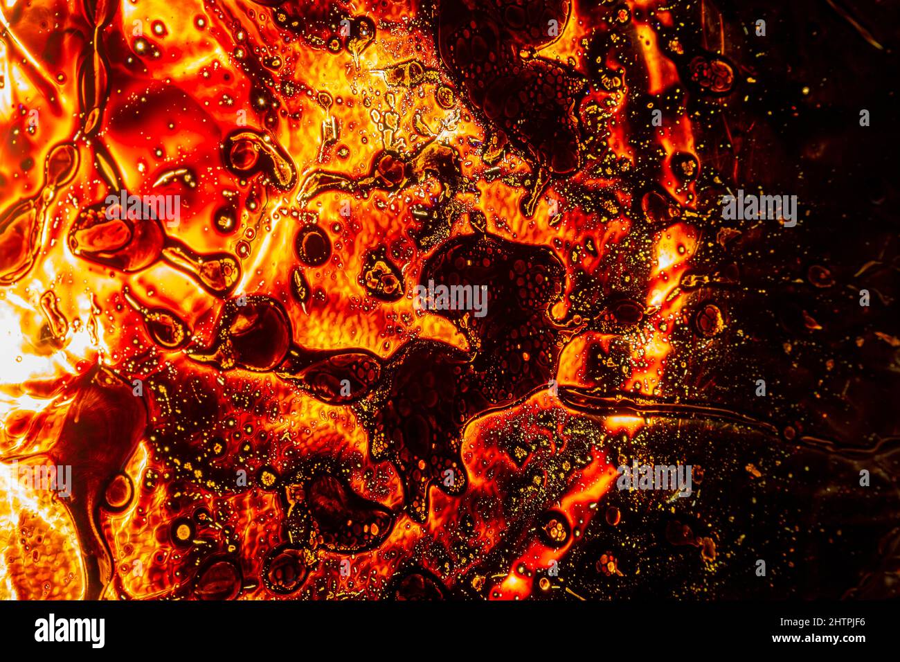 landscape, lava, night, volcano, Copper Mine Museum North Snowdonia Sygun, ferrous, oxide, pool,,ferrous sulfate wall art ,ulphate crystals. Different Stock Photohttps://www.alamy.com/image-license-details/?v=1https://www.alamy.com/landscape-lava-night-volcano-copper-mine-museum-north-snowdonia-sygun-ferrous-oxide-poolferrous-sulfate-wall-art-ulphate-crystals-different-image462718794.html
RM2HTPJF6–landscape, lava, night, volcano, Copper Mine Museum North Snowdonia Sygun, ferrous, oxide, pool,,ferrous sulfate wall art ,ulphate crystals. Different
 Rotifer Stock Photohttps://www.alamy.com/image-license-details/?v=1https://www.alamy.com/stock-photo-rotifer-134944626.html
RMHRF76X–Rotifer
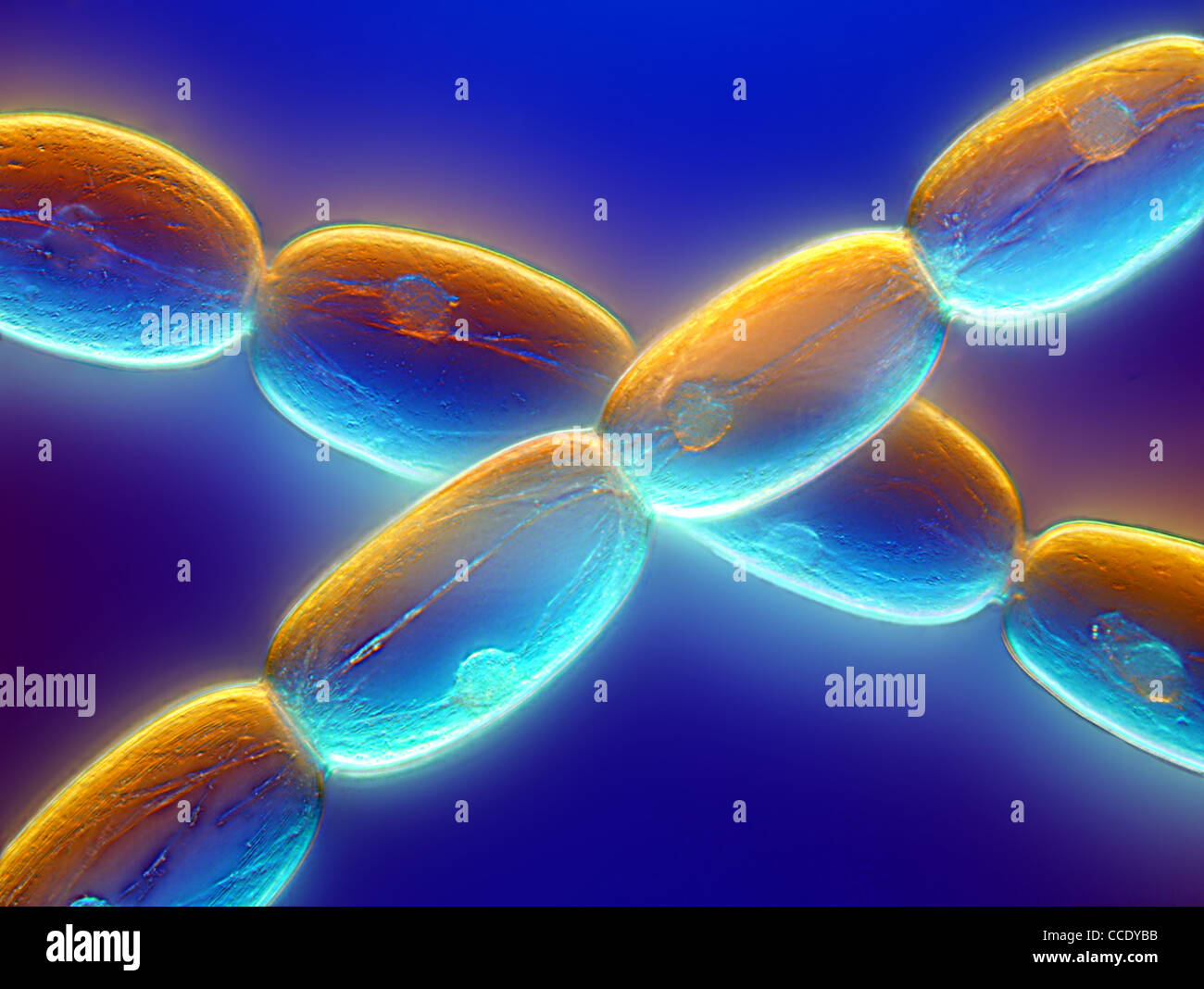 Stamen hair cells of a Tradescantia flower. Stock Photohttps://www.alamy.com/image-license-details/?v=1https://www.alamy.com/stock-photo-stamen-hair-cells-of-a-tradescantia-flower-42103471.html
RMCCDYBB–Stamen hair cells of a Tradescantia flower.
 . The Biological bulletin. Biology; Zoology; Biology; Marine Biology. 64 J. BUCKLAND-NICKS AND A. N. HODGSON 14 Figures 11-17. Micrographs of sperm and fertilized eggs of Ciillix'liiiuii ftiMnni'its: SEM = scanning electron micrograph; TEM = transmission electron micrograph; DIG LM = differential interference contrast light micrograph. Figure 11. SEM of polyspermic egg that has been rolled on sticky (ape. stripping the vitelline layer (VL) next to a penetrating sperm (Sp) and revealing the fertilization cone (FC) beneath it. Scale bar = 2 jj.ni. Figure 12. DIG LM of polyspermic egg showing one Stock Photohttps://www.alamy.com/image-license-details/?v=1https://www.alamy.com/the-biological-bulletin-biology-zoology-biology-marine-biology-64-j-buckland-nicks-and-a-n-hodgson-14-figures-11-17-micrographs-of-sperm-and-fertilized-eggs-of-ciillixliiiuii-ftimnniits-sem-=-scanning-electron-micrograph-tem-=-transmission-electron-micrograph-dig-lm-=-differential-interference-contrast-light-micrograph-figure-11-sem-of-polyspermic-egg-that-has-been-rolled-on-sticky-ape-stripping-the-vitelline-layer-vl-next-to-a-penetrating-sperm-sp-and-revealing-the-fertilization-cone-fc-beneath-it-scale-bar-=-2-jjni-figure-12-dig-lm-of-polyspermic-egg-showing-one-image234617929.html
RMRHKNFN–. The Biological bulletin. Biology; Zoology; Biology; Marine Biology. 64 J. BUCKLAND-NICKS AND A. N. HODGSON 14 Figures 11-17. Micrographs of sperm and fertilized eggs of Ciillix'liiiuii ftiMnni'its: SEM = scanning electron micrograph; TEM = transmission electron micrograph; DIG LM = differential interference contrast light micrograph. Figure 11. SEM of polyspermic egg that has been rolled on sticky (ape. stripping the vitelline layer (VL) next to a penetrating sperm (Sp) and revealing the fertilization cone (FC) beneath it. Scale bar = 2 jj.ni. Figure 12. DIG LM of polyspermic egg showing one
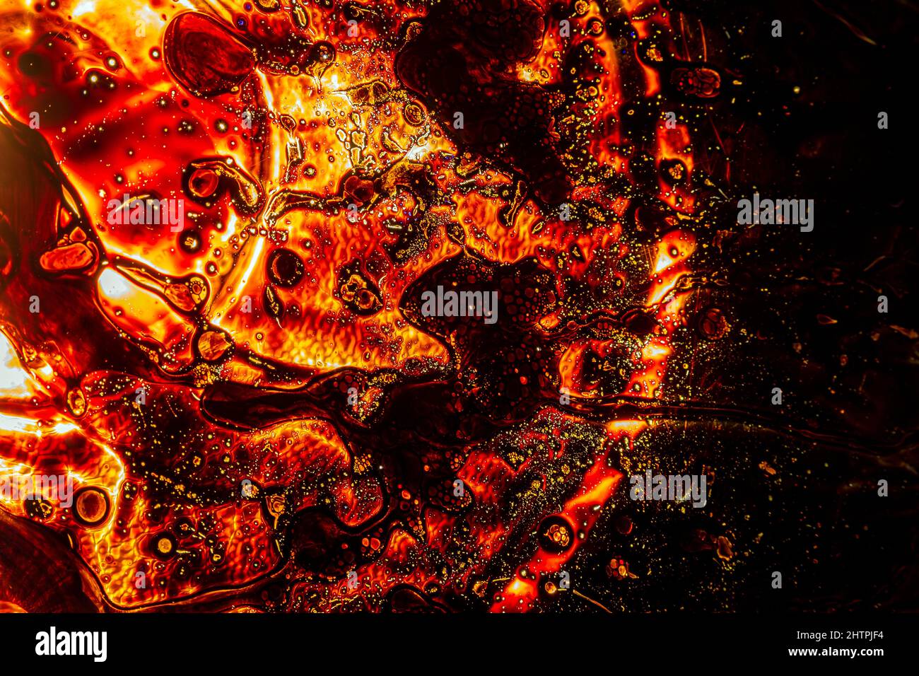 landscape, lava, night, volcano, Copper Mine Museum North Snowdonia Sygun, ferrous, oxide, pool,,ferrous sulfate wall art ,ulphate crystals. Different Stock Photohttps://www.alamy.com/image-license-details/?v=1https://www.alamy.com/landscape-lava-night-volcano-copper-mine-museum-north-snowdonia-sygun-ferrous-oxide-poolferrous-sulfate-wall-art-ulphate-crystals-different-image462718792.html
RM2HTPJF4–landscape, lava, night, volcano, Copper Mine Museum North Snowdonia Sygun, ferrous, oxide, pool,,ferrous sulfate wall art ,ulphate crystals. Different
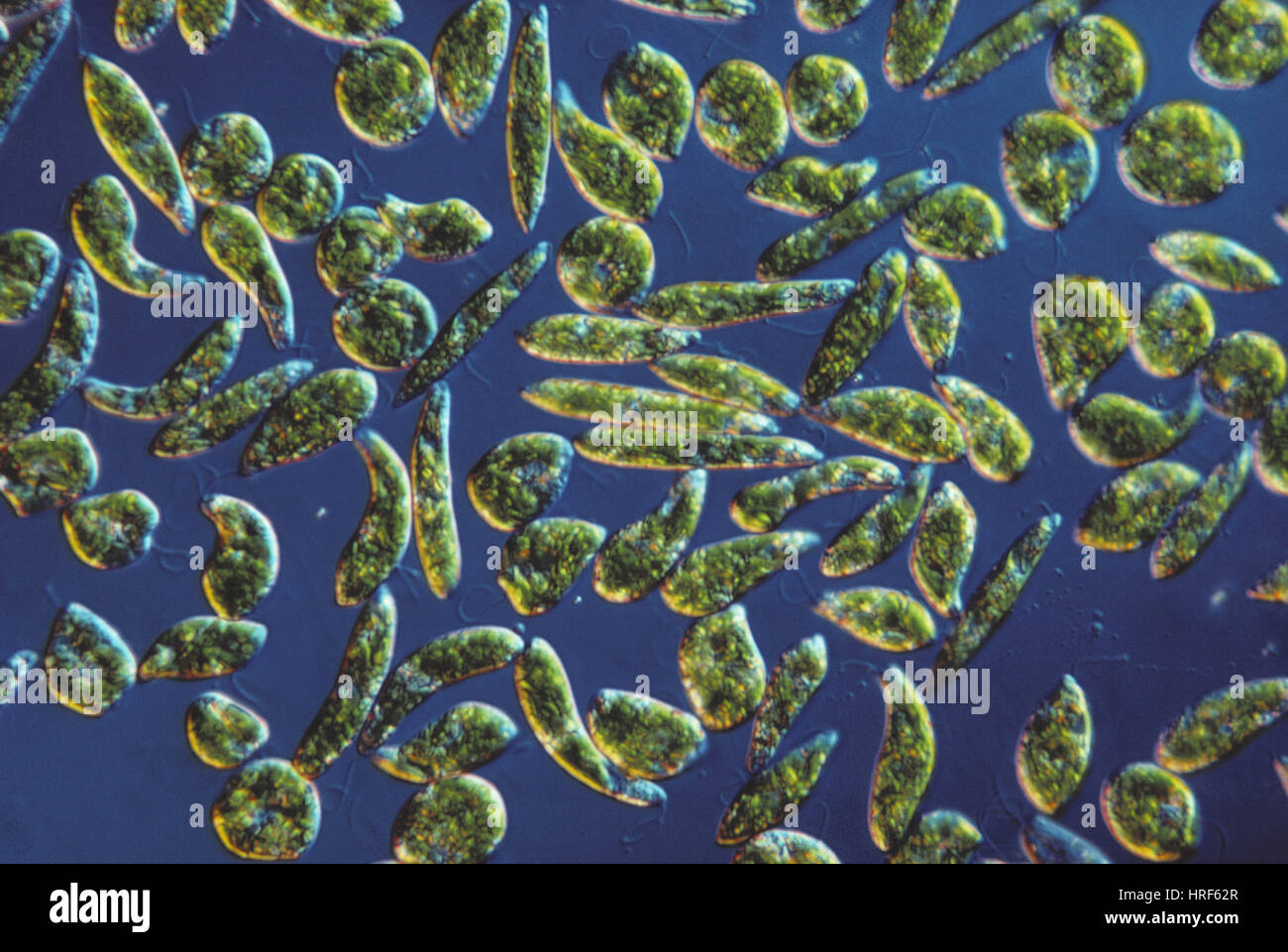 Euglena gracilis, LM Stock Photohttps://www.alamy.com/image-license-details/?v=1https://www.alamy.com/stock-photo-euglena-gracilis-lm-134943727.html
RMHRF62R–Euglena gracilis, LM
 . The Biological bulletin. Biology; Zoology; Biology; Marine Biology. FERTILIZATION IN CALLOCHITON 63. Figures 7-10. Micrographs of fertilized eggs of Callochiton castaneus: SEM = scanning electron micro- graph: DIC LM = differential interference contrast light micrograph. Figure 7. SEM of an unfertilized egg split in half with a Kesei microknife to show egg membrane cups (arrowheads). The vitelline layer has been removed. Scale bar = 10 (nm. Figure 8. SEM view of broken edge of the jelly hull, showing regular arrangement of pores above (arrowheads) and penetrating sperm (Sp) on vitelline laye Stock Photohttps://www.alamy.com/image-license-details/?v=1https://www.alamy.com/the-biological-bulletin-biology-zoology-biology-marine-biology-fertilization-in-callochiton-63-figures-7-10-micrographs-of-fertilized-eggs-of-callochiton-castaneus-sem-=-scanning-electron-micro-graph-dic-lm-=-differential-interference-contrast-light-micrograph-figure-7-sem-of-an-unfertilized-egg-split-in-half-with-a-kesei-microknife-to-show-egg-membrane-cups-arrowheads-the-vitelline-layer-has-been-removed-scale-bar-=-10-nm-figure-8-sem-view-of-broken-edge-of-the-jelly-hull-showing-regular-arrangement-of-pores-above-arrowheads-and-penetrating-sperm-sp-on-vitelline-laye-image234617953.html
RMRHKNGH–. The Biological bulletin. Biology; Zoology; Biology; Marine Biology. FERTILIZATION IN CALLOCHITON 63. Figures 7-10. Micrographs of fertilized eggs of Callochiton castaneus: SEM = scanning electron micro- graph: DIC LM = differential interference contrast light micrograph. Figure 7. SEM of an unfertilized egg split in half with a Kesei microknife to show egg membrane cups (arrowheads). The vitelline layer has been removed. Scale bar = 10 (nm. Figure 8. SEM view of broken edge of the jelly hull, showing regular arrangement of pores above (arrowheads) and penetrating sperm (Sp) on vitelline laye
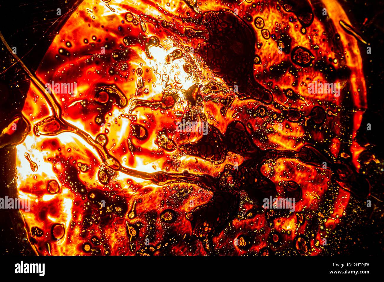 landscape, lava, night, volcano, Copper Mine Museum North Snowdonia Sygun, ferrous, oxide, pool,,ferrous sulfate wall art ,ulphate crystals. Different Stock Photohttps://www.alamy.com/image-license-details/?v=1https://www.alamy.com/landscape-lava-night-volcano-copper-mine-museum-north-snowdonia-sygun-ferrous-oxide-poolferrous-sulfate-wall-art-ulphate-crystals-different-image462718796.html
RM2HTPJF8–landscape, lava, night, volcano, Copper Mine Museum North Snowdonia Sygun, ferrous, oxide, pool,,ferrous sulfate wall art ,ulphate crystals. Different
 Epiphyseal Growth Plate Stock Photohttps://www.alamy.com/image-license-details/?v=1https://www.alamy.com/stock-photo-epiphyseal-growth-plate-134944575.html
RMHRF753–Epiphyseal Growth Plate
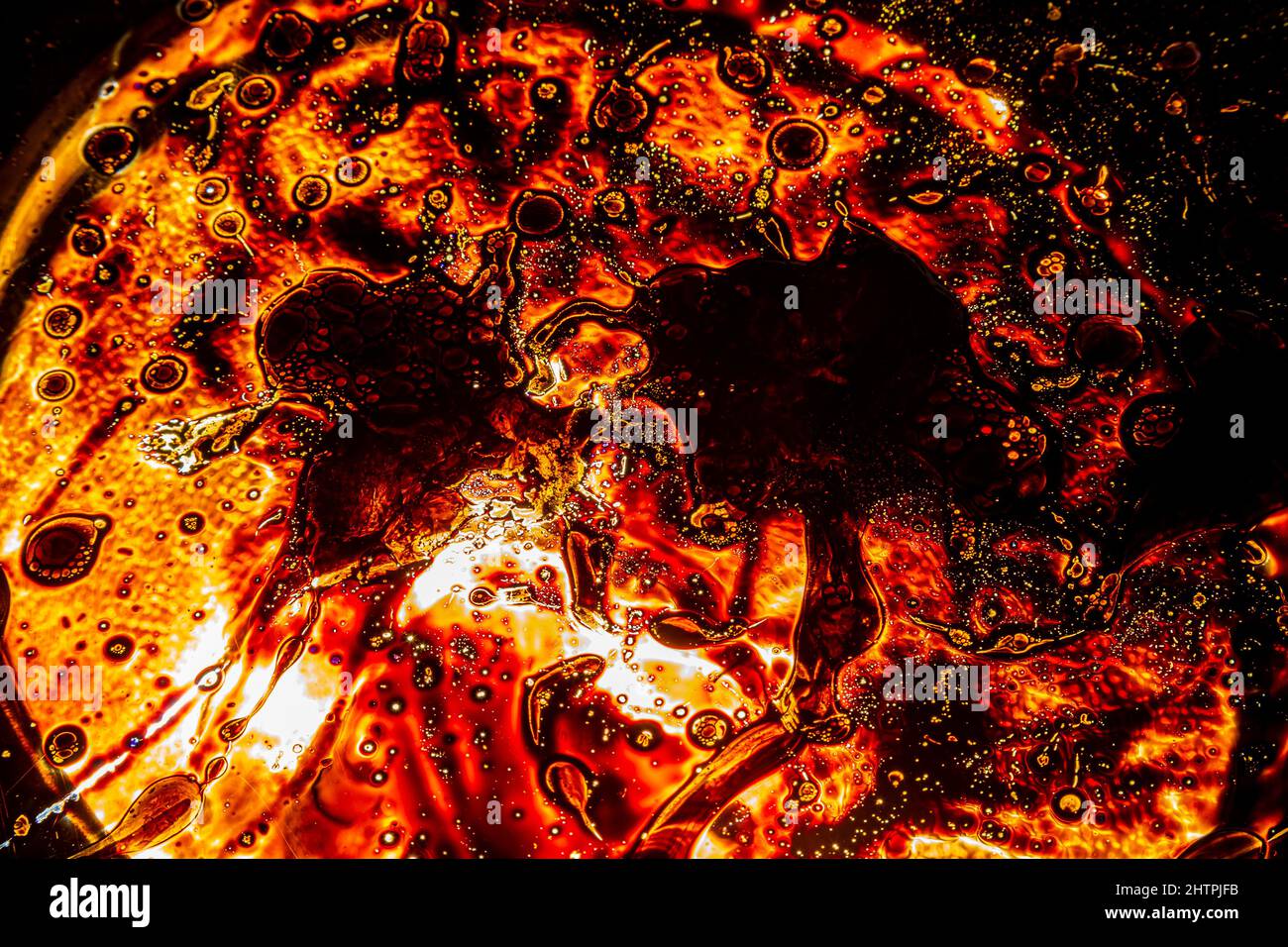 landscape, lava, night, volcano, Copper Mine Museum North Snowdonia Sygun, ferrous, oxide, pool,,ferrous sulfate wall art ,ulphate crystals. Different Stock Photohttps://www.alamy.com/image-license-details/?v=1https://www.alamy.com/landscape-lava-night-volcano-copper-mine-museum-north-snowdonia-sygun-ferrous-oxide-poolferrous-sulfate-wall-art-ulphate-crystals-different-image462718799.html
RM2HTPJFB–landscape, lava, night, volcano, Copper Mine Museum North Snowdonia Sygun, ferrous, oxide, pool,,ferrous sulfate wall art ,ulphate crystals. Different
