Quick filters:
Dissected heart Stock Photos and Images
 Dissected heart showing valves and atrium/ventricle chambers Stock Photohttps://www.alamy.com/image-license-details/?v=1https://www.alamy.com/stock-photo-dissected-heart-showing-valves-and-atriumventricle-chambers-90641700.html
RFF7D2BG–Dissected heart showing valves and atrium/ventricle chambers
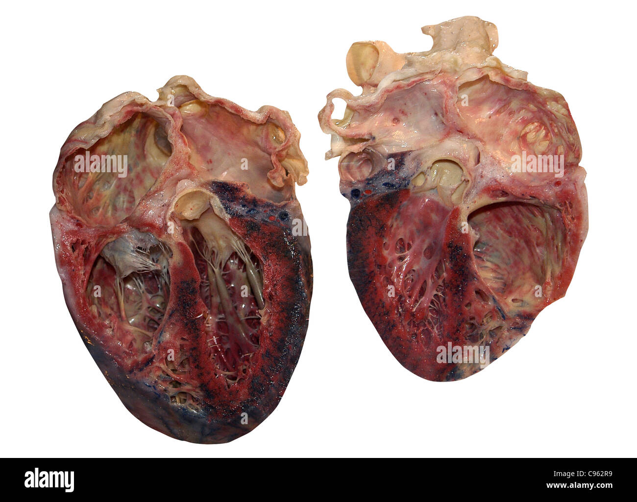 Dissected human heart. Stock Photohttps://www.alamy.com/image-license-details/?v=1https://www.alamy.com/stock-photo-dissected-human-heart-40086573.html
RFC962R9–Dissected human heart.
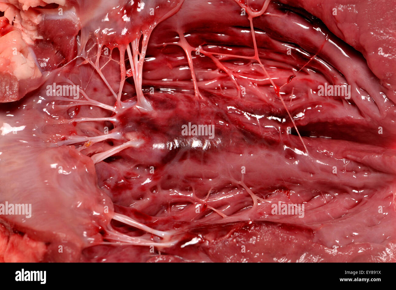 Lamb's heart bought from a supermarket. Interior showing 'heartstrings' (tendons) Stock Photohttps://www.alamy.com/image-license-details/?v=1https://www.alamy.com/stock-photo-lambs-heart-bought-from-a-supermarket-interior-showing-heartstrings-85619910.html
RMEY891X–Lamb's heart bought from a supermarket. Interior showing 'heartstrings' (tendons)
 Heart, Anatomical Illustration, 1822 Stock Photohttps://www.alamy.com/image-license-details/?v=1https://www.alamy.com/heart-anatomical-illustration-1822-image352802416.html
RM2BDYF28–Heart, Anatomical Illustration, 1822
 Dissected heart: back view. Watercolour, 18--? Stock Photohttps://www.alamy.com/image-license-details/?v=1https://www.alamy.com/dissected-heart-back-view-watercolour-18-image450000162.html
RM2H437PA–Dissected heart: back view. Watercolour, 18--?
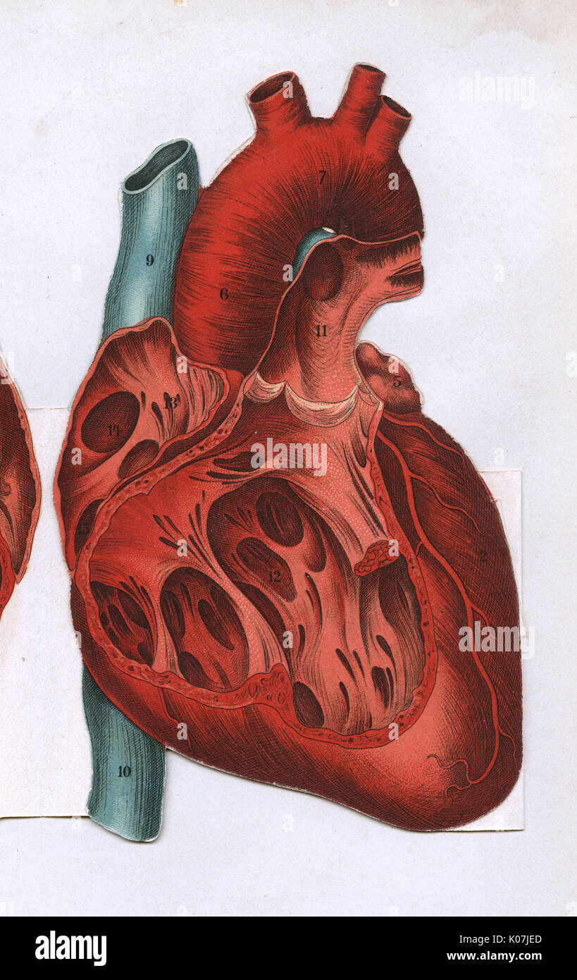 Cross section of a heart Stock Photohttps://www.alamy.com/image-license-details/?v=1https://www.alamy.com/cross-section-of-a-heart-image155061493.html
RMK07JED–Cross section of a heart
 Insides of a butternut squash looking like a dissected, broken heart Stock Photohttps://www.alamy.com/image-license-details/?v=1https://www.alamy.com/stock-photo-insides-of-a-butternut-squash-looking-like-a-dissected-broken-heart-309955147.html
RFS07JXK–Insides of a butternut squash looking like a dissected, broken heart
 Great White Shark s Heart Carcharodon carcharias being dissected at the Natal Sharks Board South Africa Stock Photohttps://www.alamy.com/image-license-details/?v=1https://www.alamy.com/great-white-shark-s-heart-carcharodon-carcharias-being-dissected-at-image3220437.html
RFA02YD6–Great White Shark s Heart Carcharodon carcharias being dissected at the Natal Sharks Board South Africa
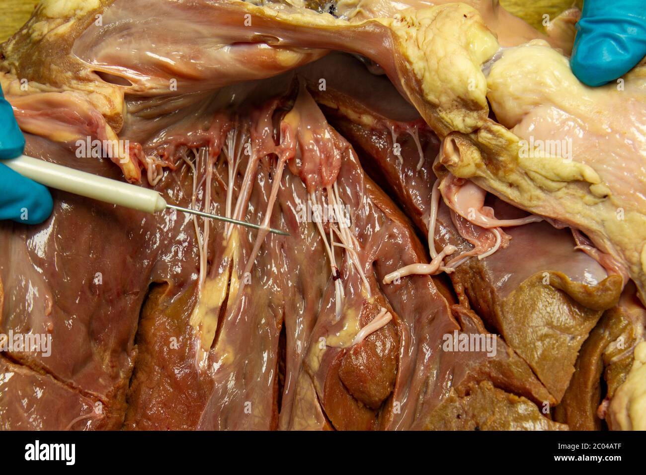 Close up view of a partially dissected cow heart as used in a UK high school demonstration. Stock Photohttps://www.alamy.com/image-license-details/?v=1https://www.alamy.com/close-up-view-of-a-partially-dissected-cow-heart-as-used-in-a-uk-high-school-demonstration-image361514063.html
RM2C04ATF–Close up view of a partially dissected cow heart as used in a UK high school demonstration.
 1849 Medical Illustration of Human Anatomy Showing the Heart, Intestine, Lungs, Stomach, Liver, Kidneys of the Abdomen and Thorax Stock Photohttps://www.alamy.com/image-license-details/?v=1https://www.alamy.com/1849-medical-illustration-of-human-anatomy-showing-the-heart-intestine-lungs-stomach-liver-kidneys-of-the-abdomen-and-thorax-image242172755.html
RFT1YWPY–1849 Medical Illustration of Human Anatomy Showing the Heart, Intestine, Lungs, Stomach, Liver, Kidneys of the Abdomen and Thorax
 Closeup of the gloved hands of an anatomy student holding a dissected sheep heart with an opening into the ventricle Stock Photohttps://www.alamy.com/image-license-details/?v=1https://www.alamy.com/closeup-of-the-gloved-hands-of-an-anatomy-student-holding-a-dissected-image69709604.html
RFE1BF8M–Closeup of the gloved hands of an anatomy student holding a dissected sheep heart with an opening into the ventricle
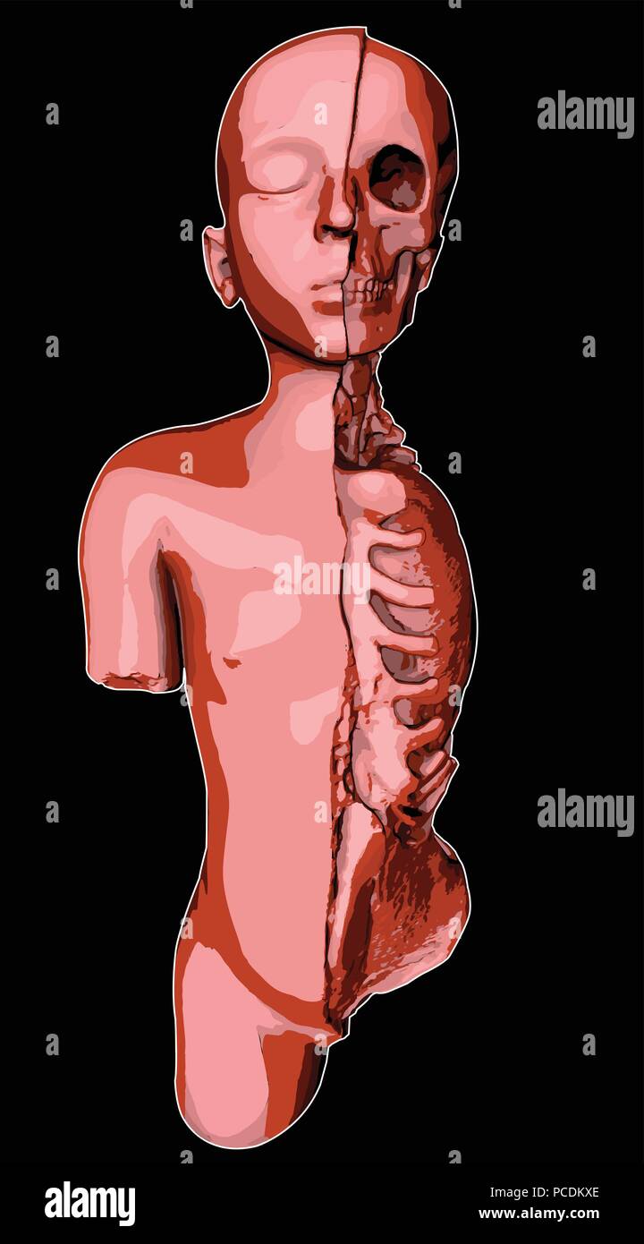 Drawing of a dissected child, anatomical study of a child, internal organs and 3d section Stock Vectorhttps://www.alamy.com/image-license-details/?v=1https://www.alamy.com/drawing-of-a-dissected-child-anatomical-study-of-a-child-internal-organs-and-3d-section-image214201302.html
RFPCDKXE–Drawing of a dissected child, anatomical study of a child, internal organs and 3d section
 The Heart Institute Stock Photohttps://www.alamy.com/image-license-details/?v=1https://www.alamy.com/the-heart-institute-image257042354.html
RMTX5842–The Heart Institute
 Watermelon Vector set. Whole and cut slice, dissected, in shape of heart. Flat collection illustration. Stock Vectorhttps://www.alamy.com/image-license-details/?v=1https://www.alamy.com/watermelon-vector-set-whole-and-cut-slice-dissected-in-shape-of-heart-flat-collection-illustration-image607699079.html
RF2X8K2AF–Watermelon Vector set. Whole and cut slice, dissected, in shape of heart. Flat collection illustration.
 William Hogarth. The Reward of Cruelty. 1750. England. Woodcut in black on ivory laid paper In the conclusion to the print series Four Stages of Cruelty, the corpse of the murderer Tom Nero is dissected in an anatomy theater. At this time the bodies of criminals were the main source of cadavers; here Hogarth pointedly left the hangman’s noose around Nero’s neck. The dog gnawing on an discarded organ, possibly the heart, refers to the character’s unseemly torture of a dog in the first print of the series.Hogarth commissioned this work and Cruelty in Perfection in a rare foray into the woodcut m Stock Photohttps://www.alamy.com/image-license-details/?v=1https://www.alamy.com/william-hogarth-the-reward-of-cruelty-1750-england-woodcut-in-black-on-ivory-laid-paper-in-the-conclusion-to-the-print-series-four-stages-of-cruelty-the-corpse-of-the-murderer-tom-nero-is-dissected-in-an-anatomy-theater-at-this-time-the-bodies-of-criminals-were-the-main-source-of-cadavers-here-hogarth-pointedly-left-the-hangmans-noose-around-neros-neck-the-dog-gnawing-on-an-discarded-organ-possibly-the-heart-refers-to-the-characters-unseemly-torture-of-a-dog-in-the-first-print-of-the-serieshogarth-commissioned-this-work-and-cruelty-in-perfection-in-a-rare-foray-into-the-woodcut-m-image337957549.html
RM2AHR88D–William Hogarth. The Reward of Cruelty. 1750. England. Woodcut in black on ivory laid paper In the conclusion to the print series Four Stages of Cruelty, the corpse of the murderer Tom Nero is dissected in an anatomy theater. At this time the bodies of criminals were the main source of cadavers; here Hogarth pointedly left the hangman’s noose around Nero’s neck. The dog gnawing on an discarded organ, possibly the heart, refers to the character’s unseemly torture of a dog in the first print of the series.Hogarth commissioned this work and Cruelty in Perfection in a rare foray into the woodcut m
 Anatomical illustration of a male figure with his abdomen and chest opened to reveal the internal organs. His right hand holds a second set of genitalia, and there is a sketch of the liver and gallbladder in the upper left corner. From a Persian translation of an Arabic medical book, ca. 18th century. The image draws upon Indian and early-modern sources. The image employs the European device of depicting animated partially-dissected bodies holding up body parts. Stock Photohttps://www.alamy.com/image-license-details/?v=1https://www.alamy.com/stock-photo-anatomical-illustration-of-a-male-figure-with-his-abdomen-and-chest-32392727.html
RMBTKH73–Anatomical illustration of a male figure with his abdomen and chest opened to reveal the internal organs. His right hand holds a second set of genitalia, and there is a sketch of the liver and gallbladder in the upper left corner. From a Persian translation of an Arabic medical book, ca. 18th century. The image draws upon Indian and early-modern sources. The image employs the European device of depicting animated partially-dissected bodies holding up body parts.
 wave beige sandy beach signed painted engraved carved gashed dissected mapped Stock Photohttps://www.alamy.com/image-license-details/?v=1https://www.alamy.com/stock-photo-wave-beige-sandy-beach-signed-painted-engraved-carved-gashed-dissected-141300950.html
RFJ5TPPE–wave beige sandy beach signed painted engraved carved gashed dissected mapped
 Volkmar Schneider Stock Photohttps://www.alamy.com/image-license-details/?v=1https://www.alamy.com/stock-photo-volkmar-schneider-111819037.html
RMGDWP91–Volkmar Schneider
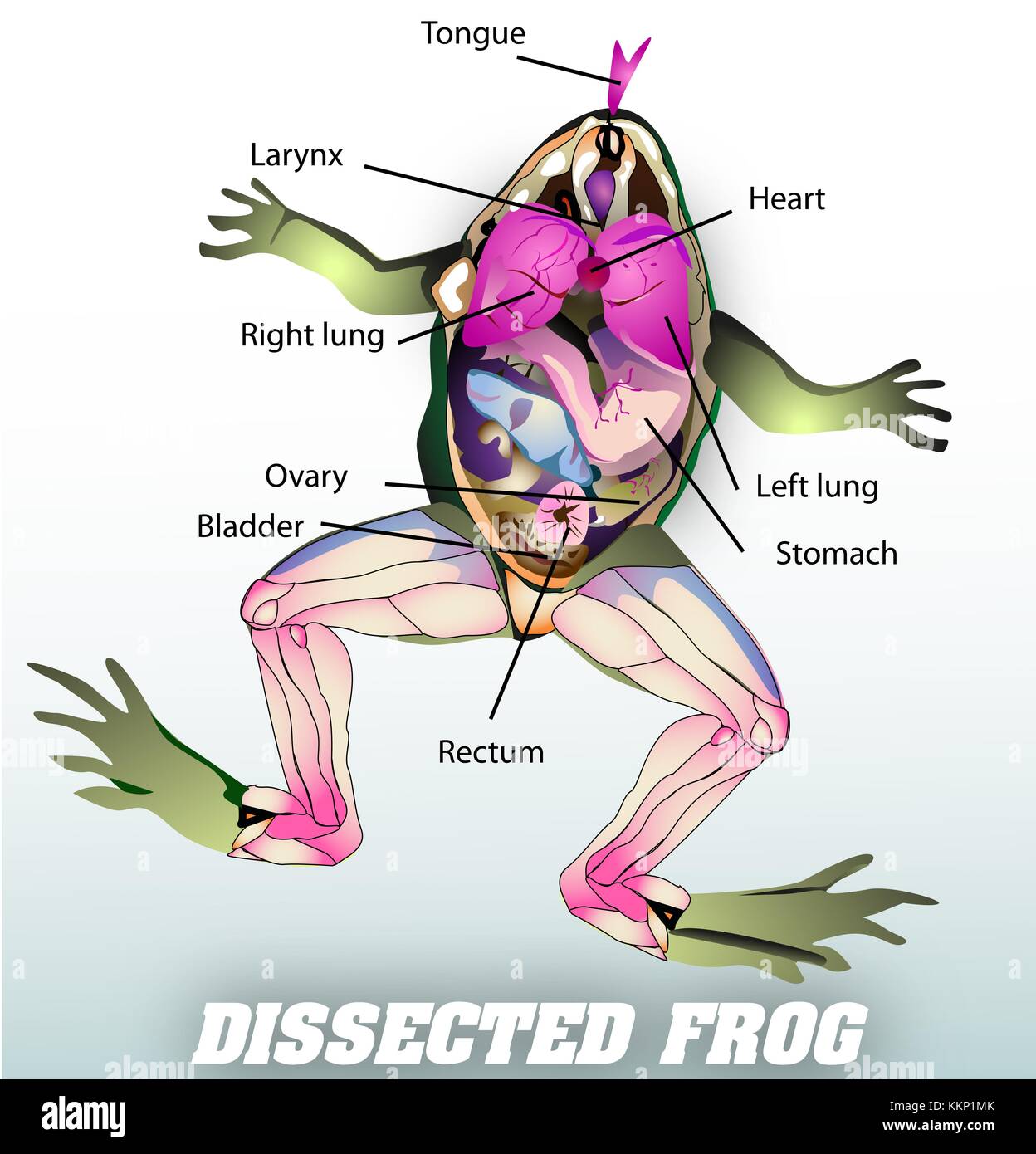 dissected frog Stock Photohttps://www.alamy.com/image-license-details/?v=1https://www.alamy.com/stock-image-dissected-frog-167056083.html
RFKKP1MK–dissected frog
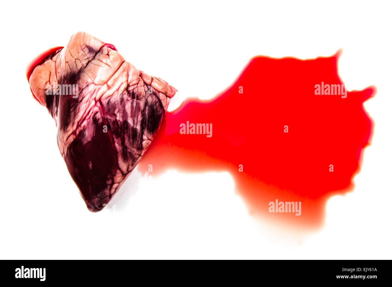 Dissected heart and blood Stock Photohttps://www.alamy.com/image-license-details/?v=1https://www.alamy.com/stock-photo-dissected-heart-and-blood-80502726.html
RFEJY61A–Dissected heart and blood
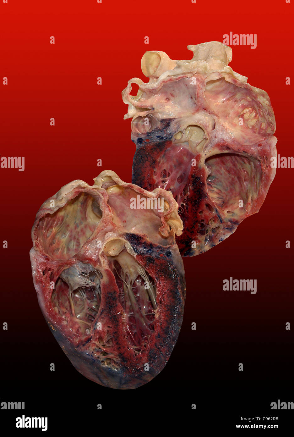 Dissected human heart. Stock Photohttps://www.alamy.com/image-license-details/?v=1https://www.alamy.com/stock-photo-dissected-human-heart-40086572.html
RFC962R8–Dissected human heart.
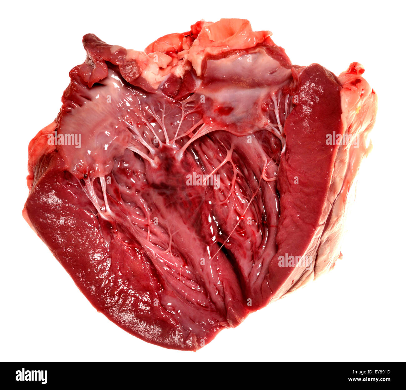 Lamb's heart bought from a supermarket. Interior showing 'heartstrings' (tendons) Stock Photohttps://www.alamy.com/image-license-details/?v=1https://www.alamy.com/stock-photo-lambs-heart-bought-from-a-supermarket-interior-showing-heartstrings-85619897.html
RMEY891D–Lamb's heart bought from a supermarket. Interior showing 'heartstrings' (tendons)
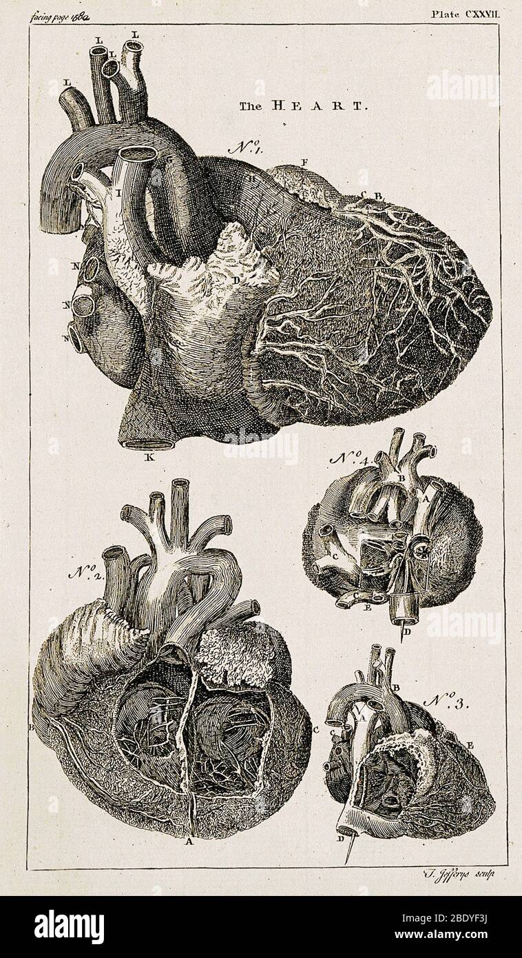 Heart, Anatomical Illustration, 1763 Stock Photohttps://www.alamy.com/image-license-details/?v=1https://www.alamy.com/heart-anatomical-illustration-1763-image352802454.html
RM2BDYF3J–Heart, Anatomical Illustration, 1763
 Sheep's heart - cut open showing ventricles, valves and 'heartstrings' (tendons) Stock Photohttps://www.alamy.com/image-license-details/?v=1https://www.alamy.com/stock-photo-sheeps-heart-cut-open-showing-ventricles-valves-and-heartstrings-tendons-85271431.html
RMEXMCG7–Sheep's heart - cut open showing ventricles, valves and 'heartstrings' (tendons)
 Heart, Anatomical Illustration, 1814 Stock Photohttps://www.alamy.com/image-license-details/?v=1https://www.alamy.com/stock-photo-heart-anatomical-illustration-1814-135096591.html
RMHRP527–Heart, Anatomical Illustration, 1814
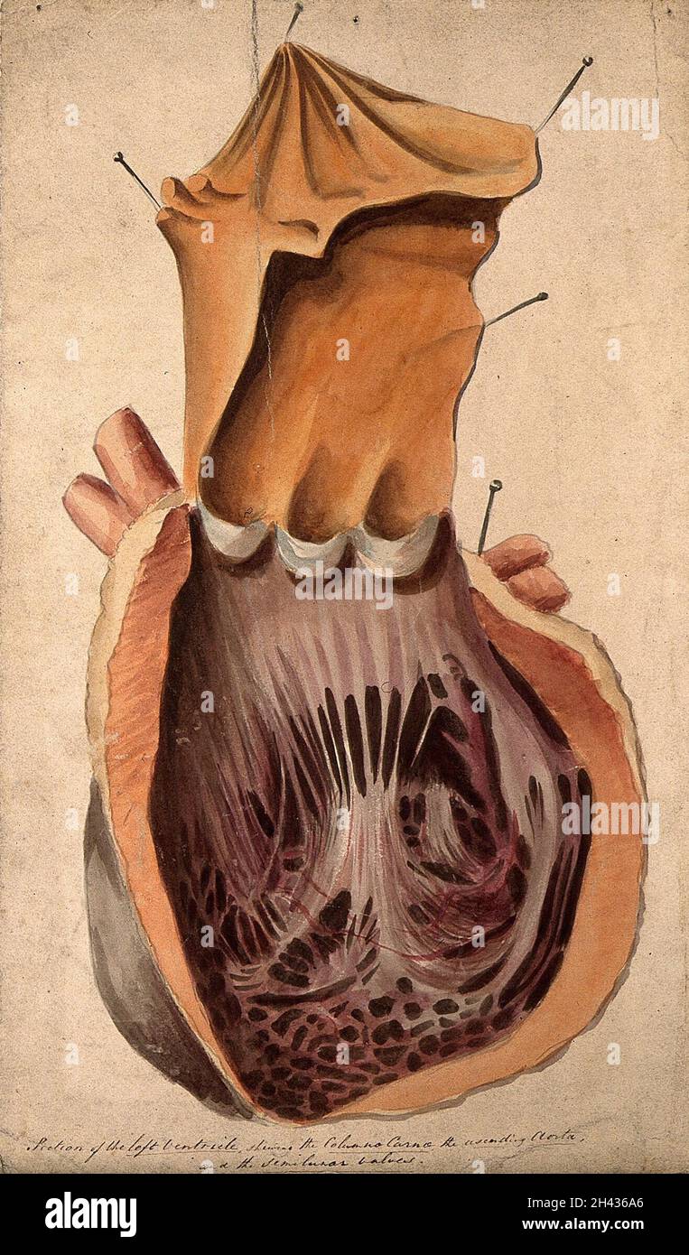 Dissected heart: section of the left ventricle. Watercolour, 18--? Stock Photohttps://www.alamy.com/image-license-details/?v=1https://www.alamy.com/dissected-heart-section-of-the-left-ventricle-watercolour-18-image449999038.html
RM2H436A6–Dissected heart: section of the left ventricle. Watercolour, 18--?
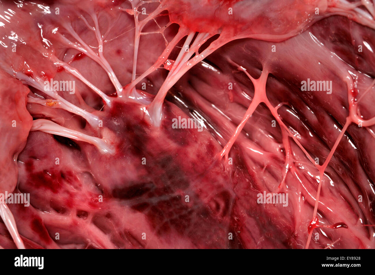 Lamb's heart bought from a supermarket. Interior showing 'heartstrings' (tendons) Stock Photohttps://www.alamy.com/image-license-details/?v=1https://www.alamy.com/stock-photo-lambs-heart-bought-from-a-supermarket-interior-showing-heartstrings-85619920.html
RMEY8928–Lamb's heart bought from a supermarket. Interior showing 'heartstrings' (tendons)
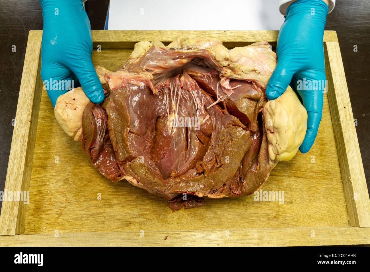 Close up view of a partially dissected cow heart as used in a UK high school demonstration. Stock Photohttps://www.alamy.com/image-license-details/?v=1https://www.alamy.com/close-up-view-of-a-partially-dissected-cow-heart-as-used-in-a-uk-high-school-demonstration-image361513863.html
RM2C04AHB–Close up view of a partially dissected cow heart as used in a UK high school demonstration.
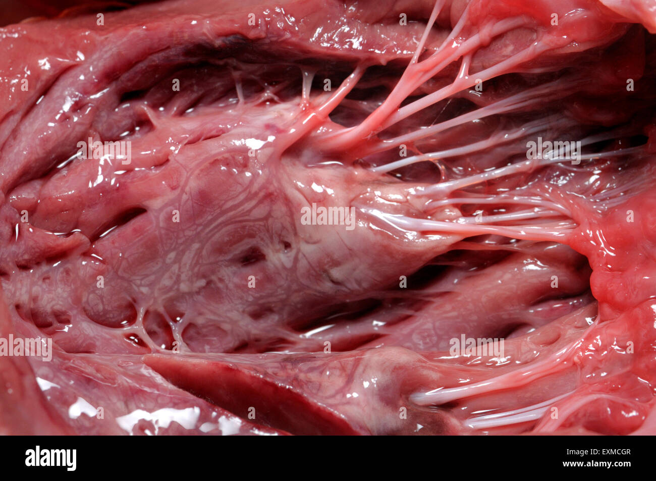 Sheep's heart - cut open showing ventricles, valves and 'heartstrings' (tendons) Stock Photohttps://www.alamy.com/image-license-details/?v=1https://www.alamy.com/stock-photo-sheeps-heart-cut-open-showing-ventricles-valves-and-heartstrings-tendons-85271447.html
RMEXMCGR–Sheep's heart - cut open showing ventricles, valves and 'heartstrings' (tendons)
 1849 Medical Illustration of Human Anatomy Showing the Heart, Intestine, Lungs, Stomach, Liver, Kidneys of the Abdomen and Thorax Stock Photohttps://www.alamy.com/image-license-details/?v=1https://www.alamy.com/1849-medical-illustration-of-human-anatomy-showing-the-heart-intestine-lungs-stomach-liver-kidneys-of-the-abdomen-and-thorax-image242172782.html
RFT1YWRX–1849 Medical Illustration of Human Anatomy Showing the Heart, Intestine, Lungs, Stomach, Liver, Kidneys of the Abdomen and Thorax
 Great White Shark Carcharodon carcharias heart being dissected at the Natal Sharks Board South Africa Stock Photohttps://www.alamy.com/image-license-details/?v=1https://www.alamy.com/great-white-shark-carcharodon-carcharias-heart-being-dissected-at-image3220479.html
RFA03G00–Great White Shark Carcharodon carcharias heart being dissected at the Natal Sharks Board South Africa
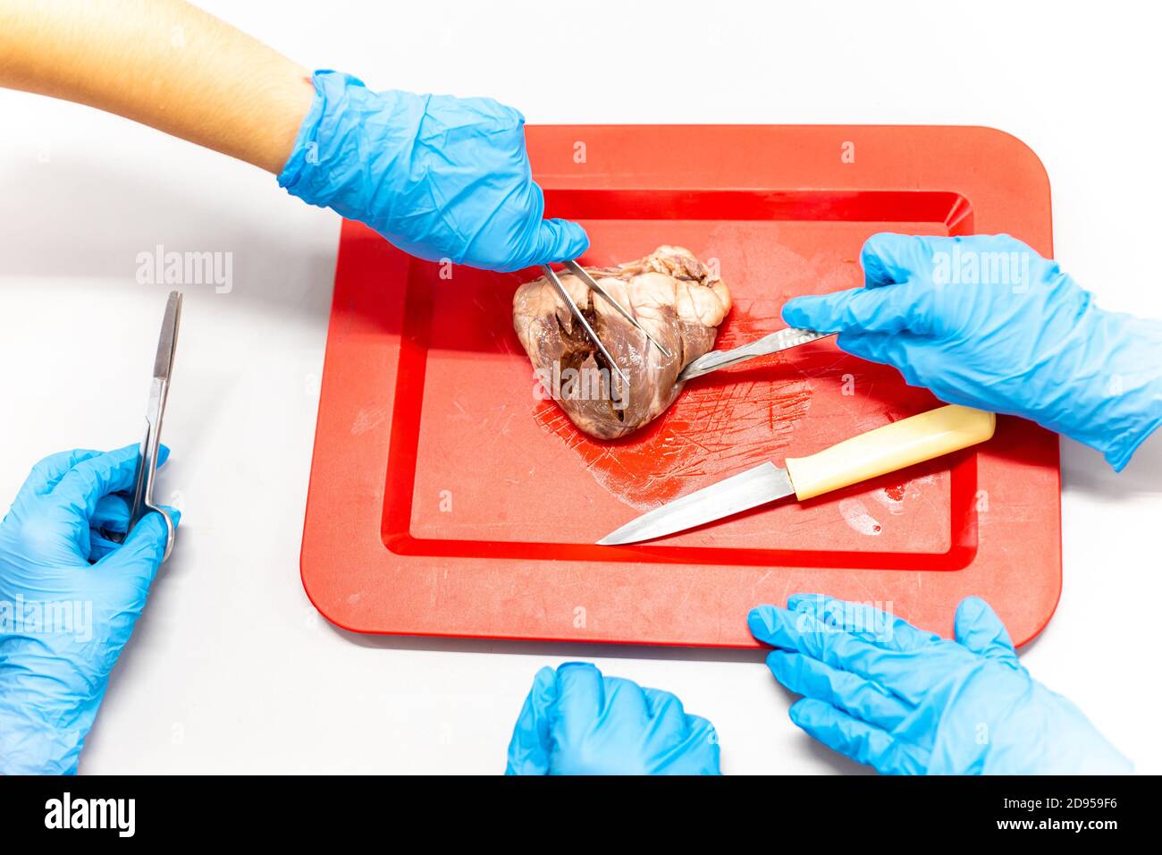 Medical students doing sheep heart dissection in the lab class Stock Photohttps://www.alamy.com/image-license-details/?v=1https://www.alamy.com/medical-students-doing-sheep-heart-dissection-in-the-lab-class-image384277242.html
RF2D959F6–Medical students doing sheep heart dissection in the lab class
 Watermelon Vector set. Whole and cut slice, dissected, bitten, in shape of heart. Flat collection illustration. Stock Vectorhttps://www.alamy.com/image-license-details/?v=1https://www.alamy.com/watermelon-vector-set-whole-and-cut-slice-dissected-bitten-in-shape-of-heart-flat-collection-illustration-image607699072.html
RF2X8K2A8–Watermelon Vector set. Whole and cut slice, dissected, bitten, in shape of heart. Flat collection illustration.
 An anatomic section of a human thorax with the heart is on display in the Museum of Anatomy which reopens to public after 20 years renovation work, in Montpellier, southern France on February 4, 2010. The museum is located in the oldest medical facility in France, and the conservatory was created in 1794. The collection houses a vast array of specimens from throughout the ages. It contains dissected anatomical organs, rare specimens, and a variety of items that can only be described as monstrosities. A surgical theatre was built in 1806, the first purpose built example of its type. Photo by Pa Stock Photohttps://www.alamy.com/image-license-details/?v=1https://www.alamy.com/an-anatomic-section-of-a-human-thorax-with-the-heart-is-on-display-in-the-museum-of-anatomy-which-reopens-to-public-after-20-years-renovation-work-in-montpellier-southern-france-on-february-4-2010-the-museum-is-located-in-the-oldest-medical-facility-in-france-and-the-conservatory-was-created-in-1794-the-collection-houses-a-vast-array-of-specimens-from-throughout-the-ages-it-contains-dissected-anatomical-organs-rare-specimens-and-a-variety-of-items-that-can-only-be-described-as-monstrosities-a-surgical-theatre-was-built-in-1806-the-first-purpose-built-example-of-its-type-photo-by-pa-image397524310.html
RF2E2MP8P–An anatomic section of a human thorax with the heart is on display in the Museum of Anatomy which reopens to public after 20 years renovation work, in Montpellier, southern France on February 4, 2010. The museum is located in the oldest medical facility in France, and the conservatory was created in 1794. The collection houses a vast array of specimens from throughout the ages. It contains dissected anatomical organs, rare specimens, and a variety of items that can only be described as monstrosities. A surgical theatre was built in 1806, the first purpose built example of its type. Photo by Pa
 School boy pupil touching internal organs of a dissected pig during school science lesson Stock Photohttps://www.alamy.com/image-license-details/?v=1https://www.alamy.com/school-boy-pupil-touching-internal-organs-of-a-dissected-pig-during-image9198333.html
RMARNJRE–School boy pupil touching internal organs of a dissected pig during school science lesson
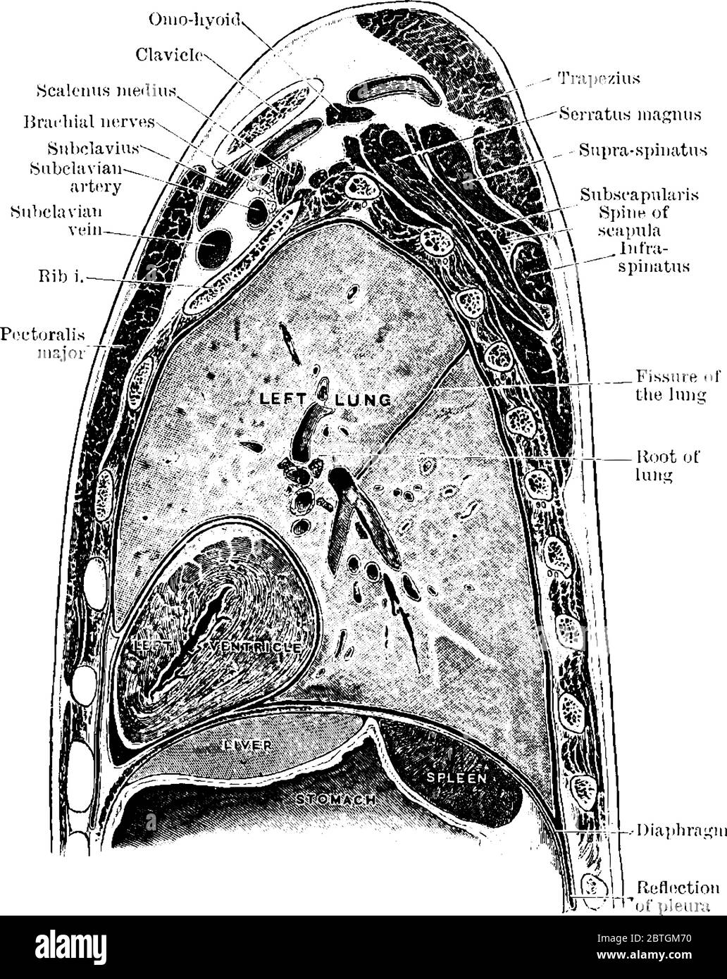 The sagittal section through the left shoulder, lung, and apex of the heart, vintage line drawing or engraving illustration. Stock Vectorhttps://www.alamy.com/image-license-details/?v=1https://www.alamy.com/the-sagittal-section-through-the-left-shoulder-lung-and-apex-of-the-heart-vintage-line-drawing-or-engraving-illustration-image359326212.html
RF2BTGM70–The sagittal section through the left shoulder, lung, and apex of the heart, vintage line drawing or engraving illustration.
 Frogs were dissected into pieces. Stock Photohttps://www.alamy.com/image-license-details/?v=1https://www.alamy.com/frogs-were-dissected-into-pieces-image211464308.html
RFP810TM–Frogs were dissected into pieces.
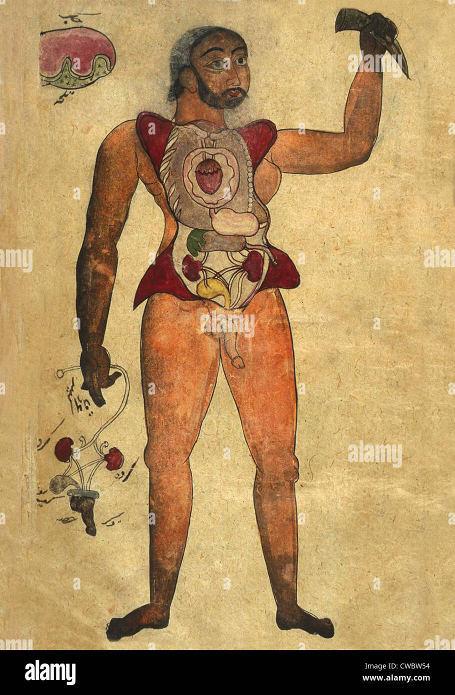 Anatomical illustration of a male figure with his abdomen and chest opened to reveal the internal organs. His right hand holds Stock Photohttps://www.alamy.com/image-license-details/?v=1https://www.alamy.com/stock-photo-anatomical-illustration-of-a-male-figure-with-his-abdomen-and-chest-50048352.html
RMCWBW54–Anatomical illustration of a male figure with his abdomen and chest opened to reveal the internal organs. His right hand holds
 Heart coated with a layer of fat caused by high cholesterol lipid lipids damage Stock Photohttps://www.alamy.com/image-license-details/?v=1https://www.alamy.com/stock-photo-heart-coated-with-a-layer-of-fat-caused-by-high-cholesterol-lipid-90641690.html
RFF7D2B6–Heart coated with a layer of fat caused by high cholesterol lipid lipids damage
 . Post-mortem pathology; a manual of post-mortem examinations and the interpretations to be drawn therefrom; a practical treatise for students and practitioners. Fig. so.—The left auricle and ventricle are fully opened, exposing thepapillary muscles, endocardium, etc. litral valve, chordae tendineae.. FlG. 8i. -Completed incisions of the heart, the organ having been reconstructed after the examina-tion of all its cavities and parts. The coronary artery has not been dissected out. This is done for severalinches with the scissors, and then transverse incisions may be made with the knife about th Stock Photohttps://www.alamy.com/image-license-details/?v=1https://www.alamy.com/post-mortem-pathology-a-manual-of-post-mortem-examinations-and-the-interpretations-to-be-drawn-therefrom-a-practical-treatise-for-students-and-practitioners-fig-sothe-left-auricle-and-ventricle-are-fully-opened-exposing-thepapillary-muscles-endocardium-etc-litral-valve-chordae-tendineae-flg-8i-completed-incisions-of-the-heart-the-organ-having-been-reconstructed-after-the-examina-tion-of-all-its-cavities-and-parts-the-coronary-artery-has-not-been-dissected-out-this-is-done-for-severalinches-with-the-scissors-and-then-transverse-incisions-may-be-made-with-the-knife-about-th-image336840852.html
RM2AG0BXC–. Post-mortem pathology; a manual of post-mortem examinations and the interpretations to be drawn therefrom; a practical treatise for students and practitioners. Fig. so.—The left auricle and ventricle are fully opened, exposing thepapillary muscles, endocardium, etc. litral valve, chordae tendineae.. FlG. 8i. -Completed incisions of the heart, the organ having been reconstructed after the examina-tion of all its cavities and parts. The coronary artery has not been dissected out. This is done for severalinches with the scissors, and then transverse incisions may be made with the knife about th
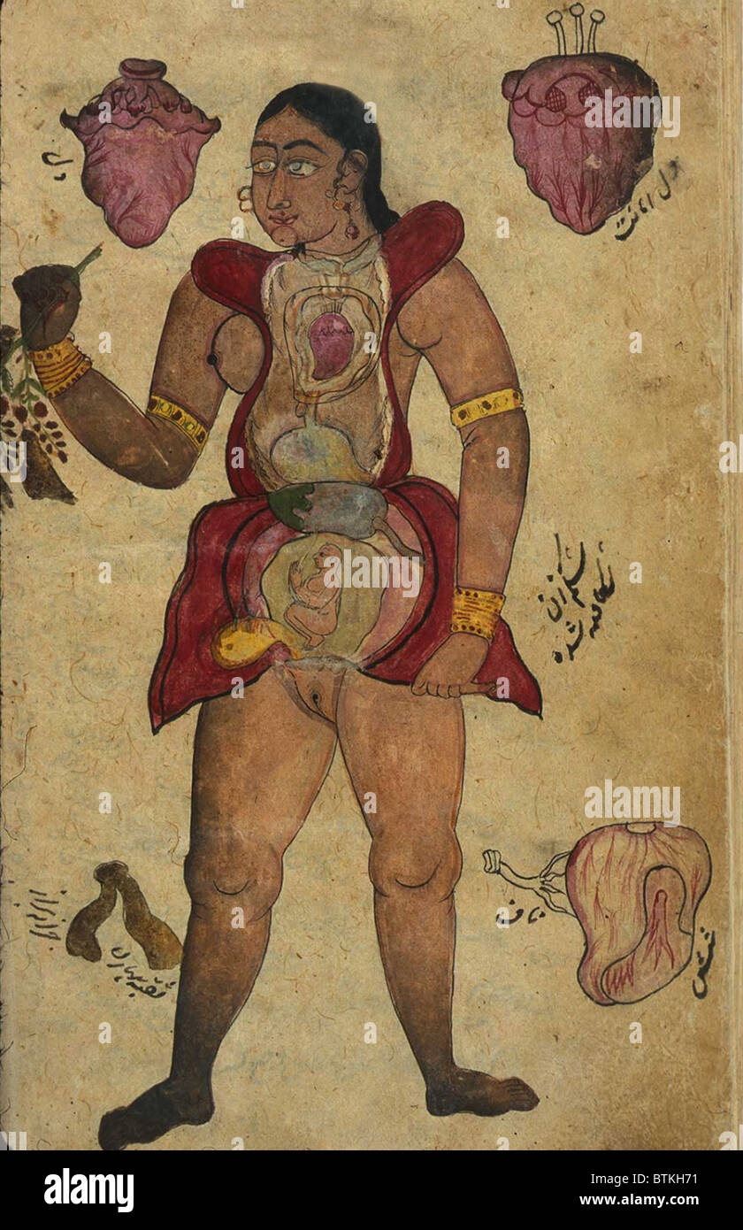 Anatomical illustration of a pregnant female figure with her abdomen and chest opened to reveal the internal organs and a fetus Stock Photohttps://www.alamy.com/image-license-details/?v=1https://www.alamy.com/stock-photo-anatomical-illustration-of-a-pregnant-female-figure-with-her-abdomen-32392725.html
RMBTKH71–Anatomical illustration of a pregnant female figure with her abdomen and chest opened to reveal the internal organs and a fetus
 School science laboratory parents open evening mother and children watching dissection of a pig Stock Photohttps://www.alamy.com/image-license-details/?v=1https://www.alamy.com/school-science-laboratory-parents-open-evening-mother-and-children-image9198335.html
RMARNJT0–School science laboratory parents open evening mother and children watching dissection of a pig
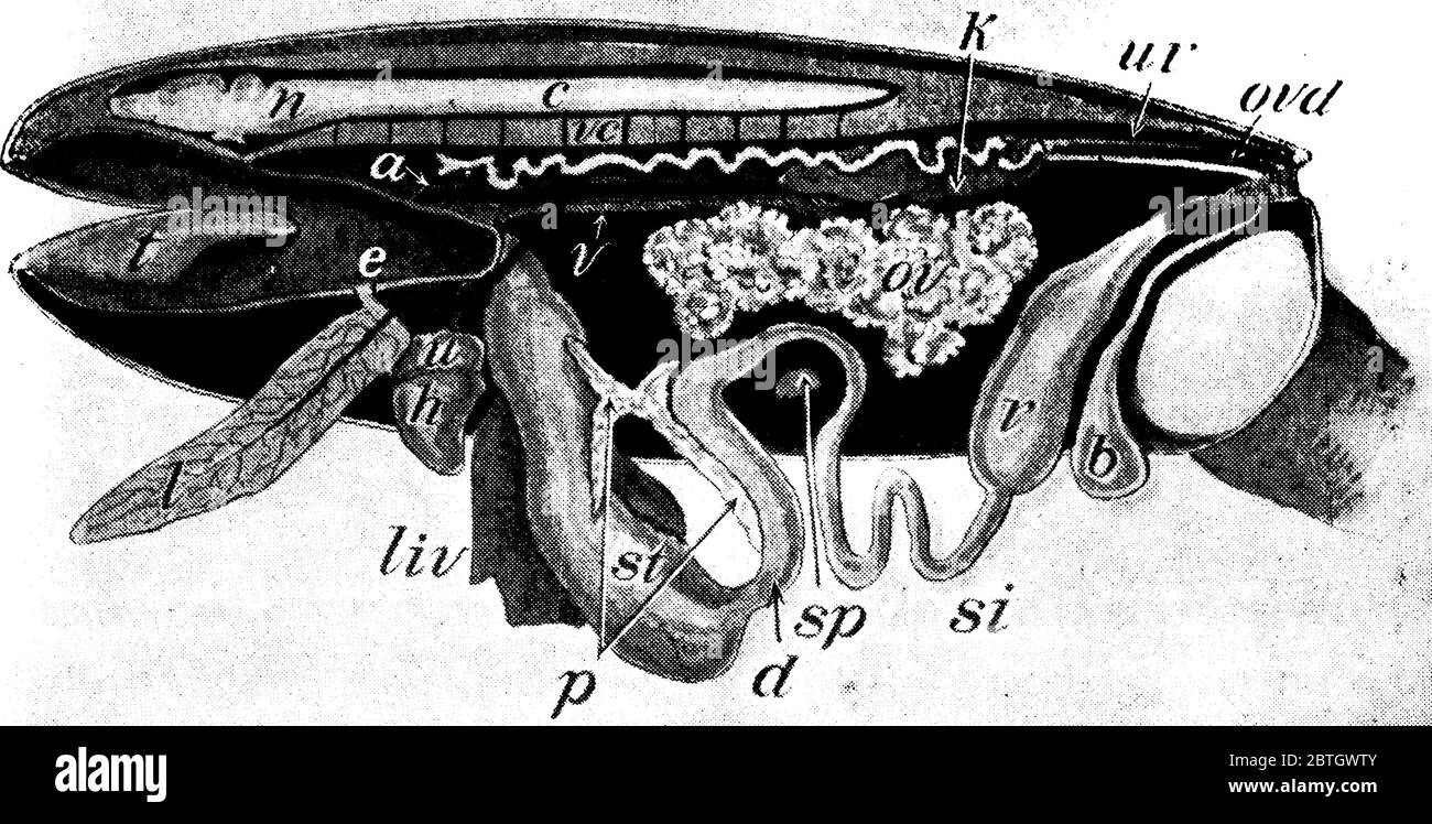 A frog with the left side cut away and some of the organs pulled downward, with the parts like, aorta, bladder, spinal cord, small intestine, kidney, Stock Vectorhttps://www.alamy.com/image-license-details/?v=1https://www.alamy.com/a-frog-with-the-left-side-cut-away-and-some-of-the-organs-pulled-downward-with-the-parts-like-aorta-bladder-spinal-cord-small-intestine-kidney-image359330635.html
RF2BTGWTY–A frog with the left side cut away and some of the organs pulled downward, with the parts like, aorta, bladder, spinal cord, small intestine, kidney,
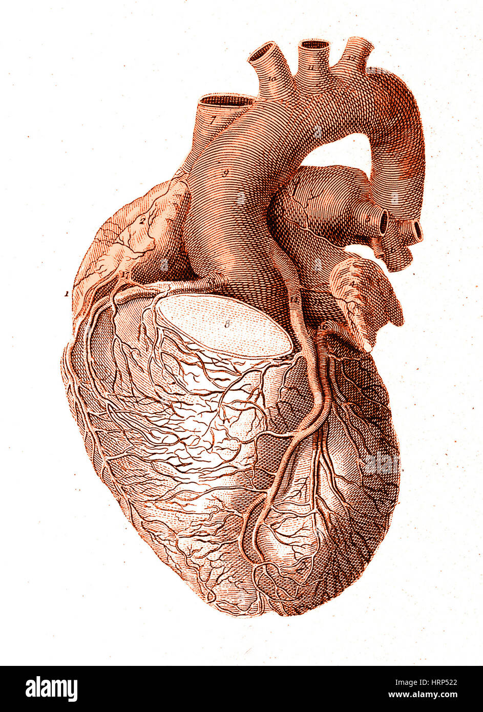 Heart, Anatomical Illustration, 1814 Stock Photohttps://www.alamy.com/image-license-details/?v=1https://www.alamy.com/stock-photo-heart-anatomical-illustration-1814-135096586.html
RMHRP522–Heart, Anatomical Illustration, 1814
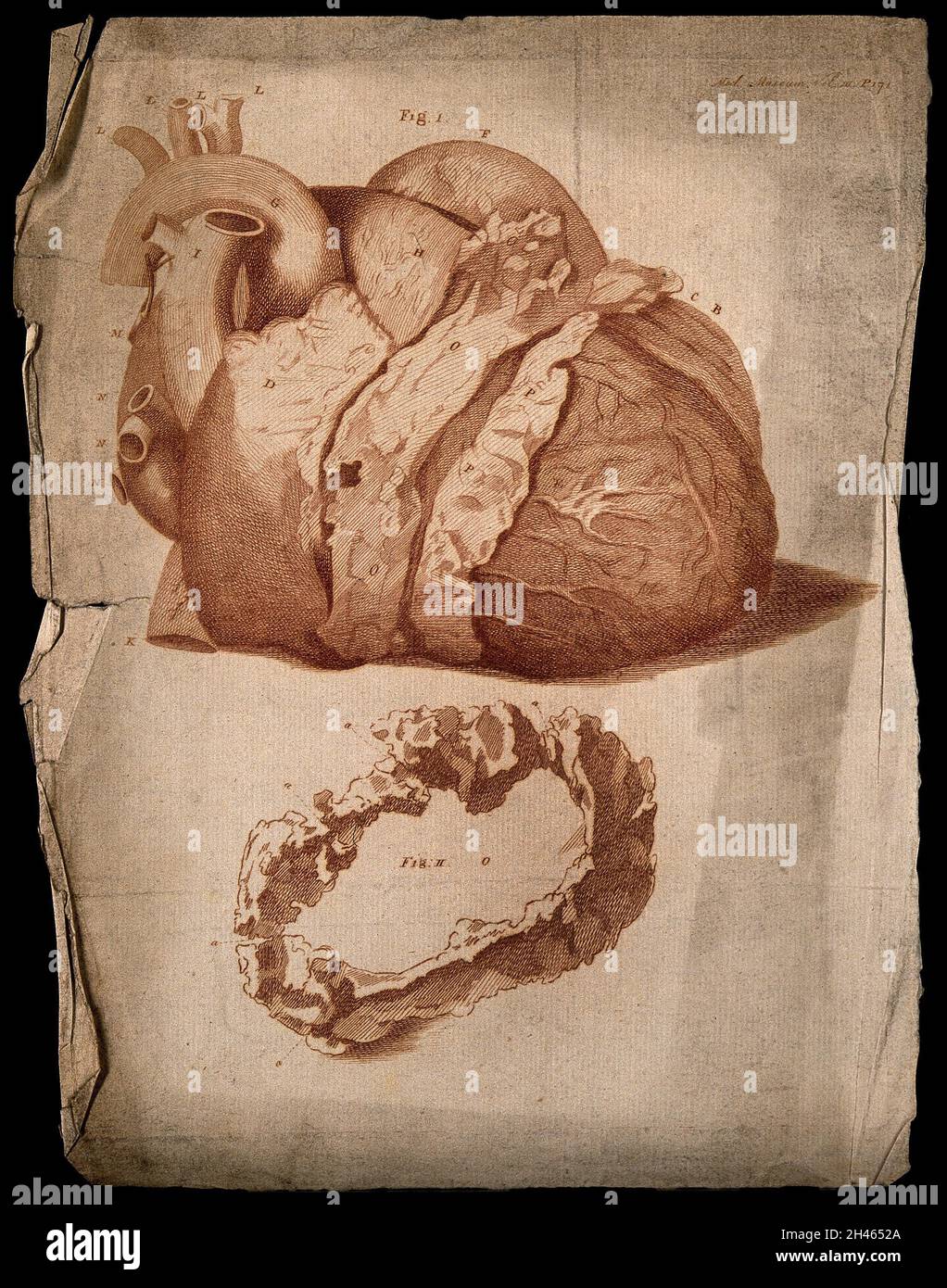 Semi-dissected heart, lettered for key. Etching, c.1760. Stock Photohttps://www.alamy.com/image-license-details/?v=1https://www.alamy.com/semi-dissected-heart-lettered-for-key-etching-c1760-image450063890.html
RM2H4652A–Semi-dissected heart, lettered for key. Etching, c.1760.
 willow, osier (Salix spec.), tree slice with core rot, Germany Stock Photohttps://www.alamy.com/image-license-details/?v=1https://www.alamy.com/willow-osier-salix-spec-tree-slice-with-core-rot-germany-image469087548.html
RM2J74NXM–willow, osier (Salix spec.), tree slice with core rot, Germany
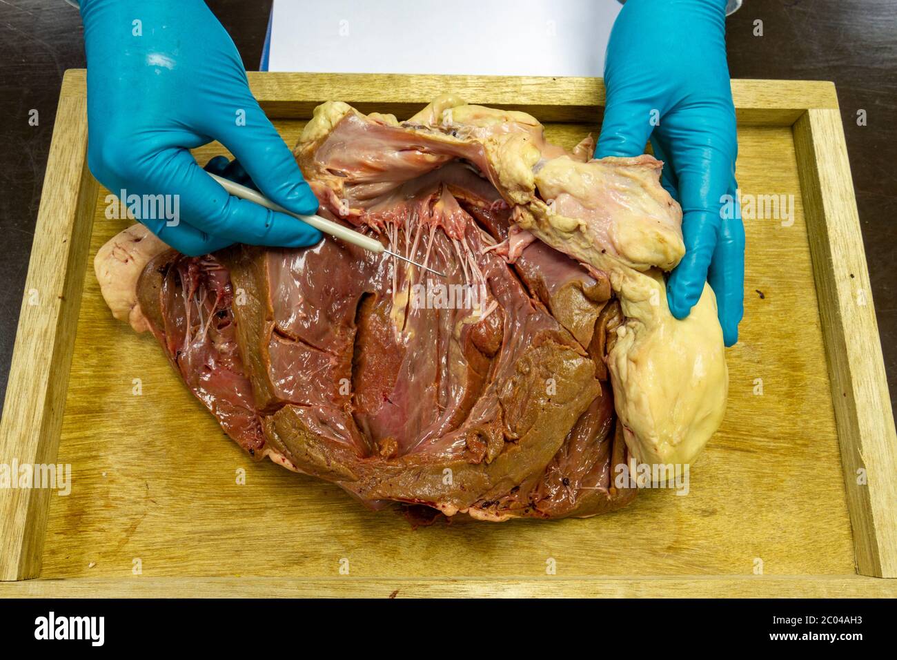 Close up view of a partially dissected cow heart as used in a UK high school demonstration. Stock Photohttps://www.alamy.com/image-license-details/?v=1https://www.alamy.com/close-up-view-of-a-partially-dissected-cow-heart-as-used-in-a-uk-high-school-demonstration-image361513855.html
RM2C04AH3–Close up view of a partially dissected cow heart as used in a UK high school demonstration.
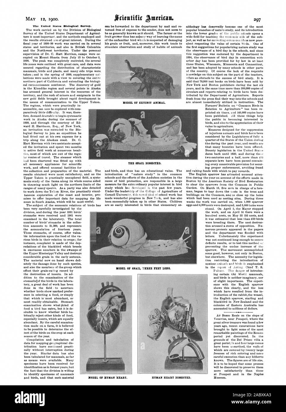 The United States Biological Survey. MODEL OF HUMAN HEART. HUMAN HEART DISSECTED., scientific american, 1900-05-12 Stock Photohttps://www.alamy.com/image-license-details/?v=1https://www.alamy.com/the-united-states-biological-survey-model-of-human-heart-human-heart-dissected-scientific-american-1900-05-12-image334344139.html
RM2ABXKA3–The United States Biological Survey. MODEL OF HUMAN HEART. HUMAN HEART DISSECTED., scientific american, 1900-05-12
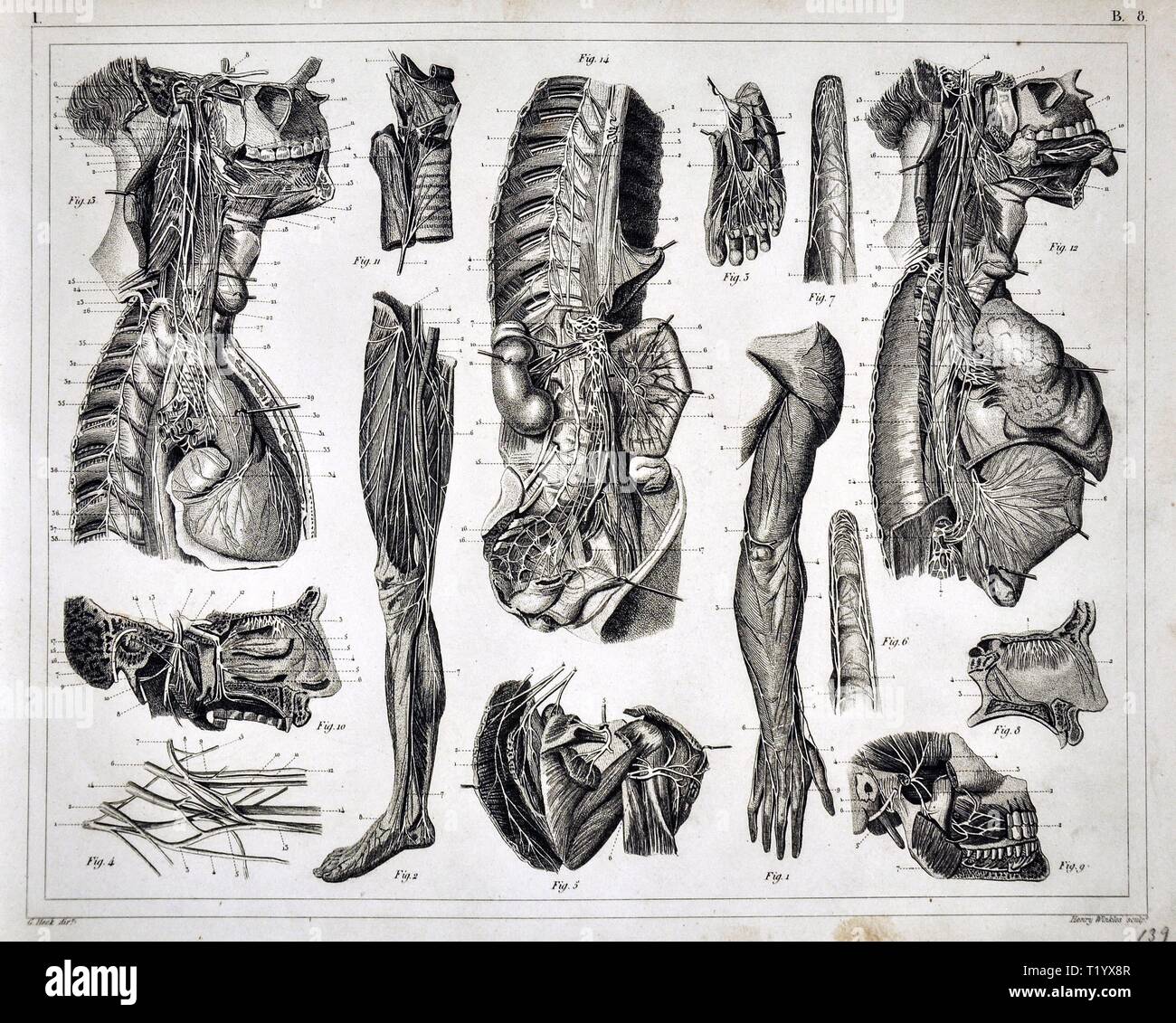 1849 Anatomy Print of the Human Muscular Skeletal System Dissection Stock Photohttps://www.alamy.com/image-license-details/?v=1https://www.alamy.com/1849-anatomy-print-of-the-human-muscular-skeletal-system-dissection-image242173143.html
RFT1YX8R–1849 Anatomy Print of the Human Muscular Skeletal System Dissection
 Great White Shark Carcharodon carcharias heart being dissected at the Natal Sharks Board South Africa Stock Photohttps://www.alamy.com/image-license-details/?v=1https://www.alamy.com/great-white-shark-carcharodon-carcharias-heart-being-dissected-at-image3220485.html
RFA03G06–Great White Shark Carcharodon carcharias heart being dissected at the Natal Sharks Board South Africa
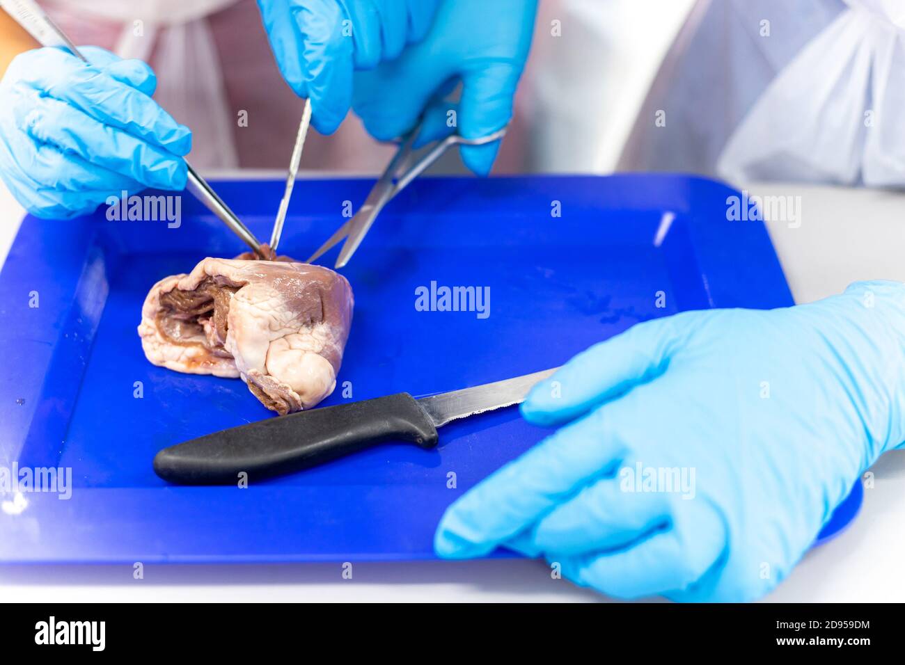 Medical students doing sheep heart dissection in the lab class Stock Photohttps://www.alamy.com/image-license-details/?v=1https://www.alamy.com/medical-students-doing-sheep-heart-dissection-in-the-lab-class-image384277200.html
RF2D959DM–Medical students doing sheep heart dissection in the lab class
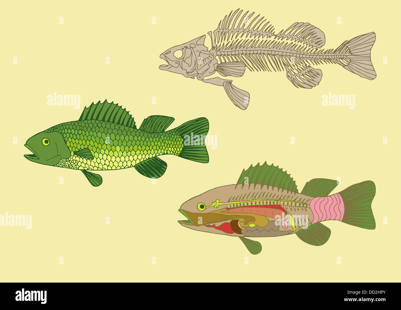 zoology, anatomy of fish , cross-section and skeleton Stock Photohttps://www.alamy.com/image-license-details/?v=1https://www.alamy.com/stock-photo-zoology-anatomy-of-fish-cross-section-and-skeleton-59679507.html
RFDD2HPY–zoology, anatomy of fish , cross-section and skeleton
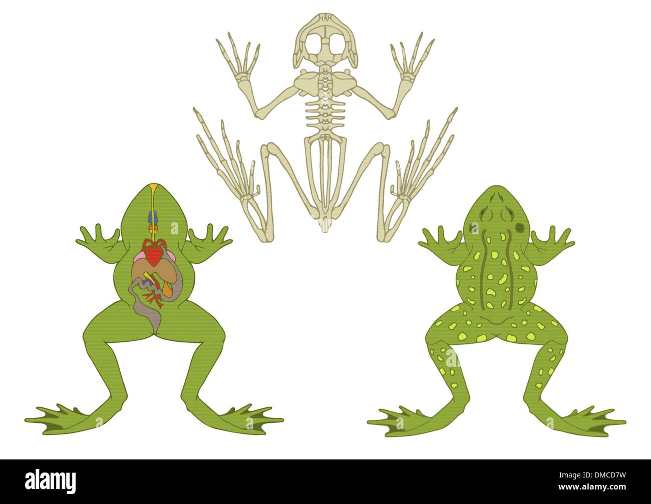 amphibian anatomy Stock Vectorhttps://www.alamy.com/image-license-details/?v=1https://www.alamy.com/amphibian-anatomy-image64198061.html
RFDMCD7W–amphibian anatomy
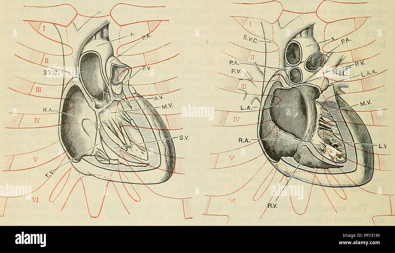 . Cunningham's Text-book of anatomy. Anatomy. Fig. 1099. Fig. 1100. From photographs of a formalin-hardened subject, with the heart dissected in situ, to show the relations of its cavities and valves to the anterior wall of the thorax. In Fig. 1097 the anterior wall of the right ventricle has been removed and the pulmonary artery opened. In Fig. 1098 the anterior walls of the ascending aorta and of the right atrium have been removed ; also the anterior cusp of the tricuspid valve. 111 FlS'mitraltv^TCeater Part °f the interventricular sePtum nas beeu removed, exposing the anterior cusp of Fig' Stock Photohttps://www.alamy.com/image-license-details/?v=1https://www.alamy.com/cunninghams-text-book-of-anatomy-anatomy-fig-1099-fig-1100-from-photographs-of-a-formalin-hardened-subject-with-the-heart-dissected-in-situ-to-show-the-relations-of-its-cavities-and-valves-to-the-anterior-wall-of-the-thorax-in-fig-1097-the-anterior-wall-of-the-right-ventricle-has-been-removed-and-the-pulmonary-artery-opened-in-fig-1098-the-anterior-walls-of-the-ascending-aorta-and-of-the-right-atrium-have-been-removed-also-the-anterior-cusp-of-the-tricuspid-valve-111-flsmitraltvtceater-part-f-the-interventricular-septum-nas-beeu-removed-exposing-the-anterior-cusp-of-fig-image216339360.html
RMPFY31M–. Cunningham's Text-book of anatomy. Anatomy. Fig. 1099. Fig. 1100. From photographs of a formalin-hardened subject, with the heart dissected in situ, to show the relations of its cavities and valves to the anterior wall of the thorax. In Fig. 1097 the anterior wall of the right ventricle has been removed and the pulmonary artery opened. In Fig. 1098 the anterior walls of the ascending aorta and of the right atrium have been removed ; also the anterior cusp of the tricuspid valve. 111 FlS'mitraltv^TCeater Part °f the interventricular sePtum nas beeu removed, exposing the anterior cusp of Fig'
 April 2019 - Seville Spain Typical Tapas restaurant interior with bottle of wine and stuffed bull head embalm Stock Photohttps://www.alamy.com/image-license-details/?v=1https://www.alamy.com/april-2019-seville-spain-typical-tapas-restaurant-interior-with-bottle-of-wine-and-stuffed-bull-head-embalm-image243660919.html
RFT4BKYK–April 2019 - Seville Spain Typical Tapas restaurant interior with bottle of wine and stuffed bull head embalm
 Frogs were dissected into pieces. Stock Photohttps://www.alamy.com/image-license-details/?v=1https://www.alamy.com/frogs-were-dissected-into-pieces-image61983648.html
RFDGRGNM–Frogs were dissected into pieces.
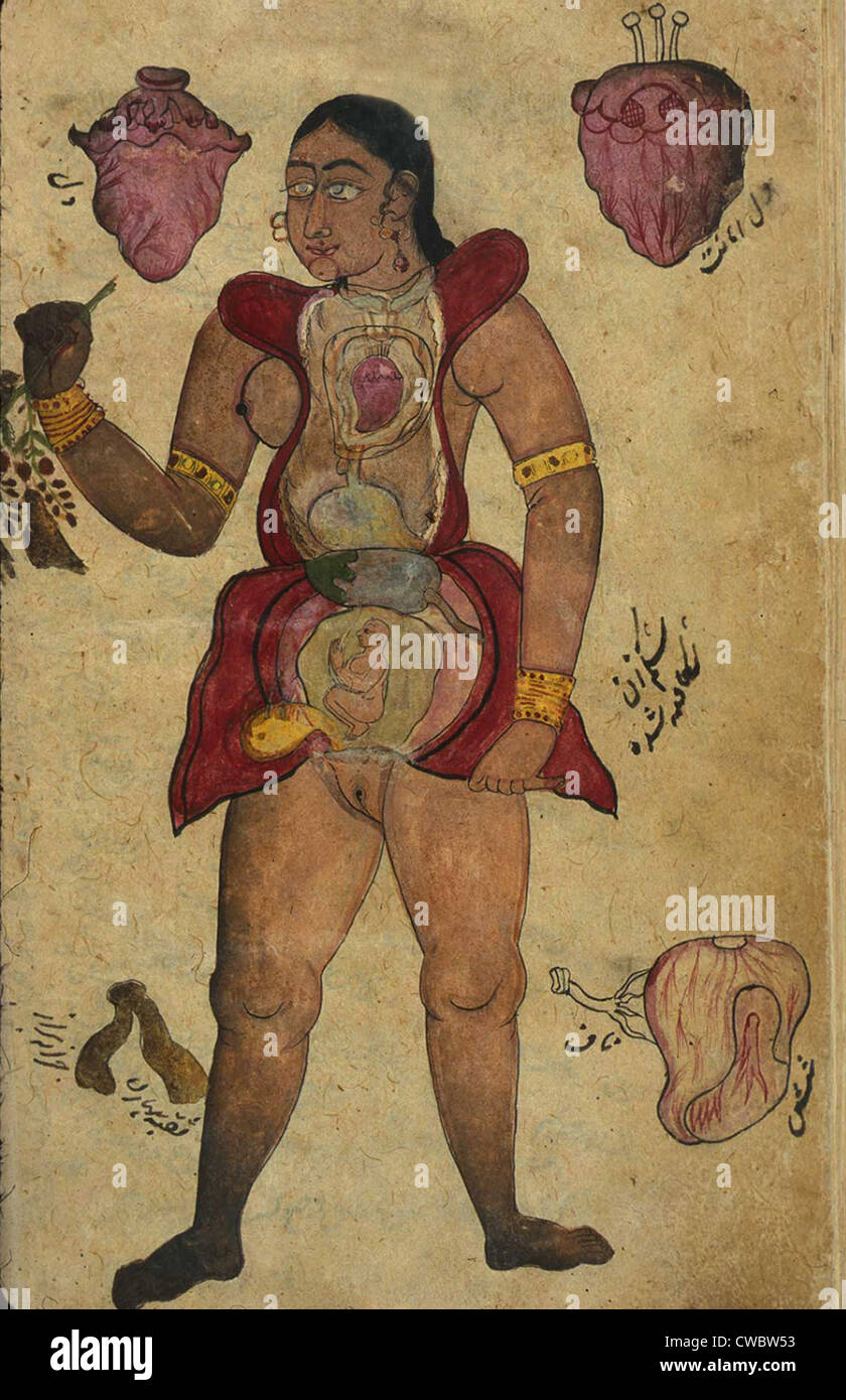 Anatomical illustration of a pregnant female figure with her abdomen and chest opened to reveal the internal organs and a fetus Stock Photohttps://www.alamy.com/image-license-details/?v=1https://www.alamy.com/stock-photo-anatomical-illustration-of-a-pregnant-female-figure-with-her-abdomen-50048351.html
RMCWBW53–Anatomical illustration of a pregnant female figure with her abdomen and chest opened to reveal the internal organs and a fetus
 Hearts coated with a layer of fat caused by high cholestrol lipid lipids damage, one dissected to show ventricle and atrium chamber Stock Photohttps://www.alamy.com/image-license-details/?v=1https://www.alamy.com/stock-photo-hearts-coated-with-a-layer-of-fat-caused-by-high-cholestrol-lipid-90641692.html
RFF7D2B8–Hearts coated with a layer of fat caused by high cholestrol lipid lipids damage, one dissected to show ventricle and atrium chamber
 Human physiology : designed for colleges and the higher classes in schools and for general reading . FIG. 35.. PLAN OF THE PERICARDIUM. CIKCULATION. 85 Action of the heart involuntary. Number of its beats. upper part of the heart on to the inside of the pericardium.The arrangement of this membrane, as it fits on to the heart,is much like the common double nightcap, as it fits on tothe head; and if it were dissected off whole from the outsideof the heart and the inside of the pericardium, it would belike such a nightcap when taken off from the head—that is, asac without an outlet. Now, this sac Stock Photohttps://www.alamy.com/image-license-details/?v=1https://www.alamy.com/human-physiology-designed-for-colleges-and-the-higher-classes-in-schools-and-for-general-reading-fig-35-plan-of-the-pericardium-cikculation-85-action-of-the-heart-involuntary-number-of-its-beats-upper-part-of-the-heart-on-to-the-inside-of-the-pericardiumthe-arrangement-of-this-membrane-as-it-fits-on-to-the-heartis-much-like-the-common-double-nightcap-as-it-fits-on-tothe-head-and-if-it-were-dissected-off-whole-from-the-outsideof-the-heart-and-the-inside-of-the-pericardium-it-would-belike-such-a-nightcap-when-taken-off-from-the-headthat-is-asac-without-an-outlet-now-this-sac-image343399574.html
RM2AXK5JE–Human physiology : designed for colleges and the higher classes in schools and for general reading . FIG. 35.. PLAN OF THE PERICARDIUM. CIKCULATION. 85 Action of the heart involuntary. Number of its beats. upper part of the heart on to the inside of the pericardium.The arrangement of this membrane, as it fits on to the heart,is much like the common double nightcap, as it fits on tothe head; and if it were dissected off whole from the outsideof the heart and the inside of the pericardium, it would belike such a nightcap when taken off from the head—that is, asac without an outlet. Now, this sac
 The spectacular objects on display include the world's most famous watch , Brequet's No,160 Marie Antoinette ,which is on loan from the LA Mayer Museum for Islamic Art and on public display for the first time in theUK.In 1783 , one of the greatest watchmakers of all time , Abraham - Louis Brequet was given an unlimited budget to craft an exceptional timepiece for Queen Marie Antoinette . Crafted over four decades from the finest materials including sapphires ,platinum and gold ,it exceeded all other watches of it's time in beauty and complexity and became Brequet's masterpiece . Stock Photohttps://www.alamy.com/image-license-details/?v=1https://www.alamy.com/the-spectacular-objects-on-display-include-the-worlds-most-famous-watch-brequets-no160-marie-antoinette-which-is-on-loan-from-the-la-mayer-museum-for-islamic-art-and-on-public-display-for-the-first-time-in-theukin-1783-one-of-the-greatest-watchmakers-of-all-time-abraham-louis-brequet-was-given-an-unlimited-budget-to-craft-an-exceptional-timepiece-for-queen-marie-antoinette-crafted-over-four-decades-from-the-finest-materials-including-sapphires-platinum-and-gold-it-exceeded-all-other-watches-of-its-time-in-beauty-and-complexity-and-became-brequets-masterpiece-image635348907.html
RM2YWJJ0B–The spectacular objects on display include the world's most famous watch , Brequet's No,160 Marie Antoinette ,which is on loan from the LA Mayer Museum for Islamic Art and on public display for the first time in theUK.In 1783 , one of the greatest watchmakers of all time , Abraham - Louis Brequet was given an unlimited budget to craft an exceptional timepiece for Queen Marie Antoinette . Crafted over four decades from the finest materials including sapphires ,platinum and gold ,it exceeded all other watches of it's time in beauty and complexity and became Brequet's masterpiece .
 Heart, Anatomical Illustration, 1814 Stock Photohttps://www.alamy.com/image-license-details/?v=1https://www.alamy.com/stock-photo-heart-anatomical-illustration-1814-135096581.html
RMHRP51W–Heart, Anatomical Illustration, 1814
 The body of a youth with his trunk dissected to reveal the ribs and viscera, especially the position of the heart. Coloured lithograph by J.B. Léveillé after William Fairland, 1869. Stock Photohttps://www.alamy.com/image-license-details/?v=1https://www.alamy.com/the-body-of-a-youth-with-his-trunk-dissected-to-reveal-the-ribs-and-viscera-especially-the-position-of-the-heart-coloured-lithograph-by-jb-lveill-after-william-fairland-1869-image450117097.html
RM2H48GXH–The body of a youth with his trunk dissected to reveal the ribs and viscera, especially the position of the heart. Coloured lithograph by J.B. Léveillé after William Fairland, 1869.
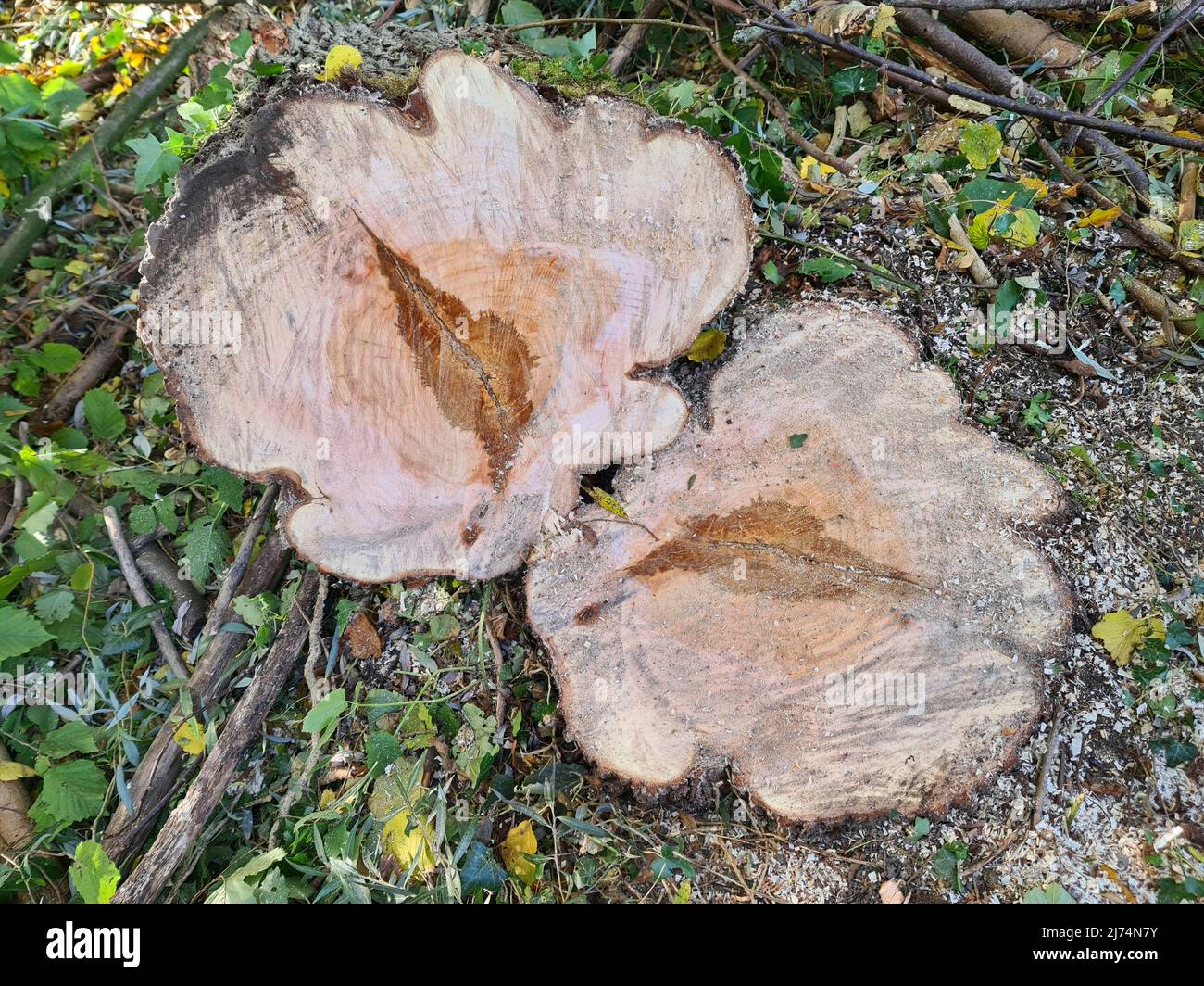 willow, osier (Salix spec.), tree slice with core rot, Germany Stock Photohttps://www.alamy.com/image-license-details/?v=1https://www.alamy.com/willow-osier-salix-spec-tree-slice-with-core-rot-germany-image469087023.html
RM2J74N7Y–willow, osier (Salix spec.), tree slice with core rot, Germany
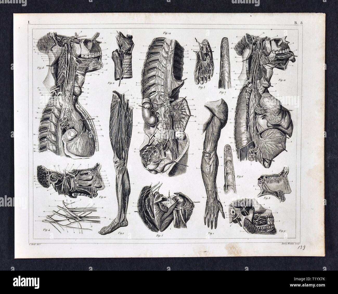 1849 Anatomy Print of the Human Muscular Skeletal System Dissection Stock Photohttps://www.alamy.com/image-license-details/?v=1https://www.alamy.com/1849-anatomy-print-of-the-human-muscular-skeletal-system-dissection-image242173111.html
RFT1YX7K–1849 Anatomy Print of the Human Muscular Skeletal System Dissection
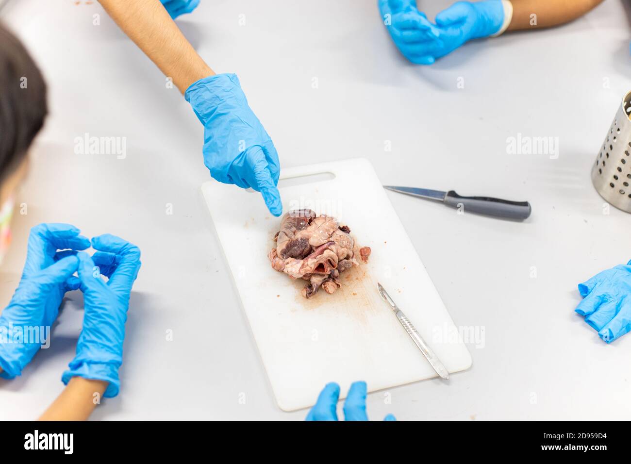 Medical students doing sheep heart dissection in the lab class Stock Photohttps://www.alamy.com/image-license-details/?v=1https://www.alamy.com/medical-students-doing-sheep-heart-dissection-in-the-lab-class-image384277184.html
RF2D959D4–Medical students doing sheep heart dissection in the lab class
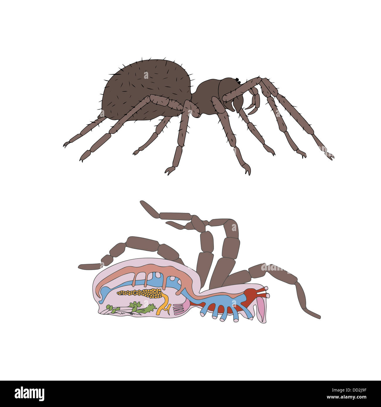 zoology, anatomy, morphology, cross-section of spider Stock Photohttps://www.alamy.com/image-license-details/?v=1https://www.alamy.com/stock-photo-zoology-anatomy-morphology-cross-section-of-spider-59679915.html
RFDD2J9F–zoology, anatomy, morphology, cross-section of spider
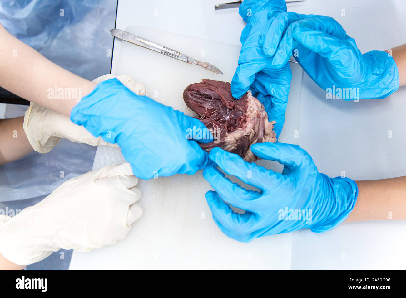 Medical students doing sheep heart dissection in the lab class Stock Photohttps://www.alamy.com/image-license-details/?v=1https://www.alamy.com/medical-students-doing-sheep-heart-dissection-in-the-lab-class-image330895298.html
RF2A69G96–Medical students doing sheep heart dissection in the lab class
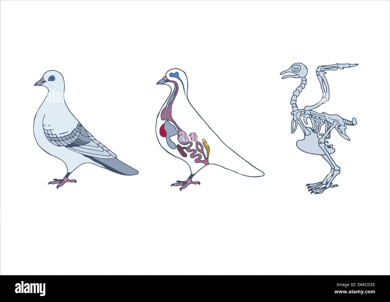 zoology, anatomy of bird Stock Vectorhttps://www.alamy.com/image-license-details/?v=1https://www.alamy.com/zoology-anatomy-of-bird-image64197929.html
RFDMCD35–zoology, anatomy of bird
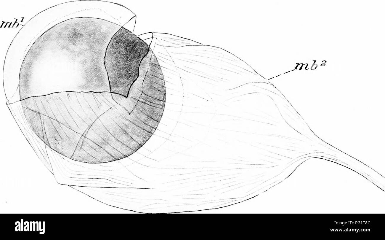 . Natural history of the American lobster... Decapoda (Crustacea); Lobster fisheries. Fifi. J. Fig. I.—Immature ovary of lobster with abnormal ring on left anterior lobe for transmission cf left antennal artery {ani. art) H, heart. Fig. 2.—Reproductive organs from right side of male, dissected to show sperm duct, and spcrmatophore (Spli) pressed from slit made in its side. p. s, gl. s, sp. mu^ duct, ejac, proximal segment, glandular segment, spnincter muscle, and ductus ejaculator- ius of vas deferens: pap, papilla for opening of duct on coxa of fifth pereiopod. Fig. 3.—Transverse section (in Stock Photohttps://www.alamy.com/image-license-details/?v=1https://www.alamy.com/natural-history-of-the-american-lobster-decapoda-crustacea-lobster-fisheries-fifi-j-fig-iimmature-ovary-of-lobster-with-abnormal-ring-on-left-anterior-lobe-for-transmission-cf-left-antennal-artery-ani-art-h-heart-fig-2reproductive-organs-from-right-side-of-male-dissected-to-show-sperm-duct-and-spcrmatophore-spli-pressed-from-slit-made-in-its-side-p-s-gl-s-sp-mu-duct-ejac-proximal-segment-glandular-segment-spnincter-muscle-and-ductus-ejaculator-ius-of-vas-deferens-pap-papilla-for-opening-of-duct-on-coxa-of-fifth-pereiopod-fig-3transverse-section-in-image216399916.html
RMPG1T8C–. Natural history of the American lobster... Decapoda (Crustacea); Lobster fisheries. Fifi. J. Fig. I.—Immature ovary of lobster with abnormal ring on left anterior lobe for transmission cf left antennal artery {ani. art) H, heart. Fig. 2.—Reproductive organs from right side of male, dissected to show sperm duct, and spcrmatophore (Spli) pressed from slit made in its side. p. s, gl. s, sp. mu^ duct, ejac, proximal segment, glandular segment, spnincter muscle, and ductus ejaculator- ius of vas deferens: pap, papilla for opening of duct on coxa of fifth pereiopod. Fig. 3.—Transverse section (in
 Hearts coated with a layer of fat caused by high cholestrol lipid lipids damage Stock Photohttps://www.alamy.com/image-license-details/?v=1https://www.alamy.com/stock-photo-hearts-coated-with-a-layer-of-fat-caused-by-high-cholestrol-lipid-90641691.html
RFF7D2B7–Hearts coated with a layer of fat caused by high cholestrol lipid lipids damage
 . Heart disease, with special reference to prognosis and treatment. FIG. 2.—THE HEART WITH LUNGS INSITU TO ILLUSTRATE AREA OF SUPER-FICIAL CARDIAC DULNESS (Sibsori).. FIG. 3.—THE HEART WITH OVERLYINGLUNG DISSECTED OFF TO ILLUSTRATEAREA OF DEEP DULNESS (Sibson). fifth cartilage, and curving, so as to form a hollow spacefor the lodgment of the apex, ends behind the sixthcartilage. The inner margin of the right lung continues its coursenearly straight downwards behind and a little to the leftof the centre of the sternum, to the level of the sixth RELATION OF HEART TO CHEST WALLS. 15 chondrosterna Stock Photohttps://www.alamy.com/image-license-details/?v=1https://www.alamy.com/heart-disease-with-special-reference-to-prognosis-and-treatment-fig-2the-heart-with-lungs-insitu-to-illustrate-area-of-super-ficial-cardiac-dulness-sibsori-fig-3the-heart-with-overlyinglung-dissected-off-to-illustratearea-of-deep-dulness-sibson-fifth-cartilage-and-curving-so-as-to-form-a-hollow-spacefor-the-lodgment-of-the-apex-ends-behind-the-sixthcartilage-the-inner-margin-of-the-right-lung-continues-its-coursenearly-straight-downwards-behind-and-a-little-to-the-leftof-the-centre-of-the-sternum-to-the-level-of-the-sixth-relation-of-heart-to-chest-walls-15-chondrosterna-image336853743.html
RM2AG10AR–. Heart disease, with special reference to prognosis and treatment. FIG. 2.—THE HEART WITH LUNGS INSITU TO ILLUSTRATE AREA OF SUPER-FICIAL CARDIAC DULNESS (Sibsori).. FIG. 3.—THE HEART WITH OVERLYINGLUNG DISSECTED OFF TO ILLUSTRATEAREA OF DEEP DULNESS (Sibson). fifth cartilage, and curving, so as to form a hollow spacefor the lodgment of the apex, ends behind the sixthcartilage. The inner margin of the right lung continues its coursenearly straight downwards behind and a little to the leftof the centre of the sternum, to the level of the sixth RELATION OF HEART TO CHEST WALLS. 15 chondrosterna
 The spectacular objects on display include the world's most famous watch , Brequet's No,160 Marie Antoinette ,which is on loan from the LA Mayer Museum for Islamic Art and on public display for the first time in theUK.In 1783 , one of the greatest watchmakers of all time , Abraham - Louis Brequet was given an unlimited budget to craft an exceptional timepiece for Queen Marie Antoinette . Crafted over four decades from the finest materials including sapphires ,platinum and gold ,it exceeded all other watches of it's time in beauty and complexity and became Brequet's masterpiece . Stock Photohttps://www.alamy.com/image-license-details/?v=1https://www.alamy.com/the-spectacular-objects-on-display-include-the-worlds-most-famous-watch-brequets-no160-marie-antoinette-which-is-on-loan-from-the-la-mayer-museum-for-islamic-art-and-on-public-display-for-the-first-time-in-theukin-1783-one-of-the-greatest-watchmakers-of-all-time-abraham-louis-brequet-was-given-an-unlimited-budget-to-craft-an-exceptional-timepiece-for-queen-marie-antoinette-crafted-over-four-decades-from-the-finest-materials-including-sapphires-platinum-and-gold-it-exceeded-all-other-watches-of-its-time-in-beauty-and-complexity-and-became-brequets-masterpiece-image635347650.html
RM2YWJGBE–The spectacular objects on display include the world's most famous watch , Brequet's No,160 Marie Antoinette ,which is on loan from the LA Mayer Museum for Islamic Art and on public display for the first time in theUK.In 1783 , one of the greatest watchmakers of all time , Abraham - Louis Brequet was given an unlimited budget to craft an exceptional timepiece for Queen Marie Antoinette . Crafted over four decades from the finest materials including sapphires ,platinum and gold ,it exceeded all other watches of it's time in beauty and complexity and became Brequet's masterpiece .
 Heart, Anatomical Illustration, 1814 Stock Photohttps://www.alamy.com/image-license-details/?v=1https://www.alamy.com/stock-photo-heart-anatomical-illustration-1814-135096593.html
RMHRP529–Heart, Anatomical Illustration, 1814
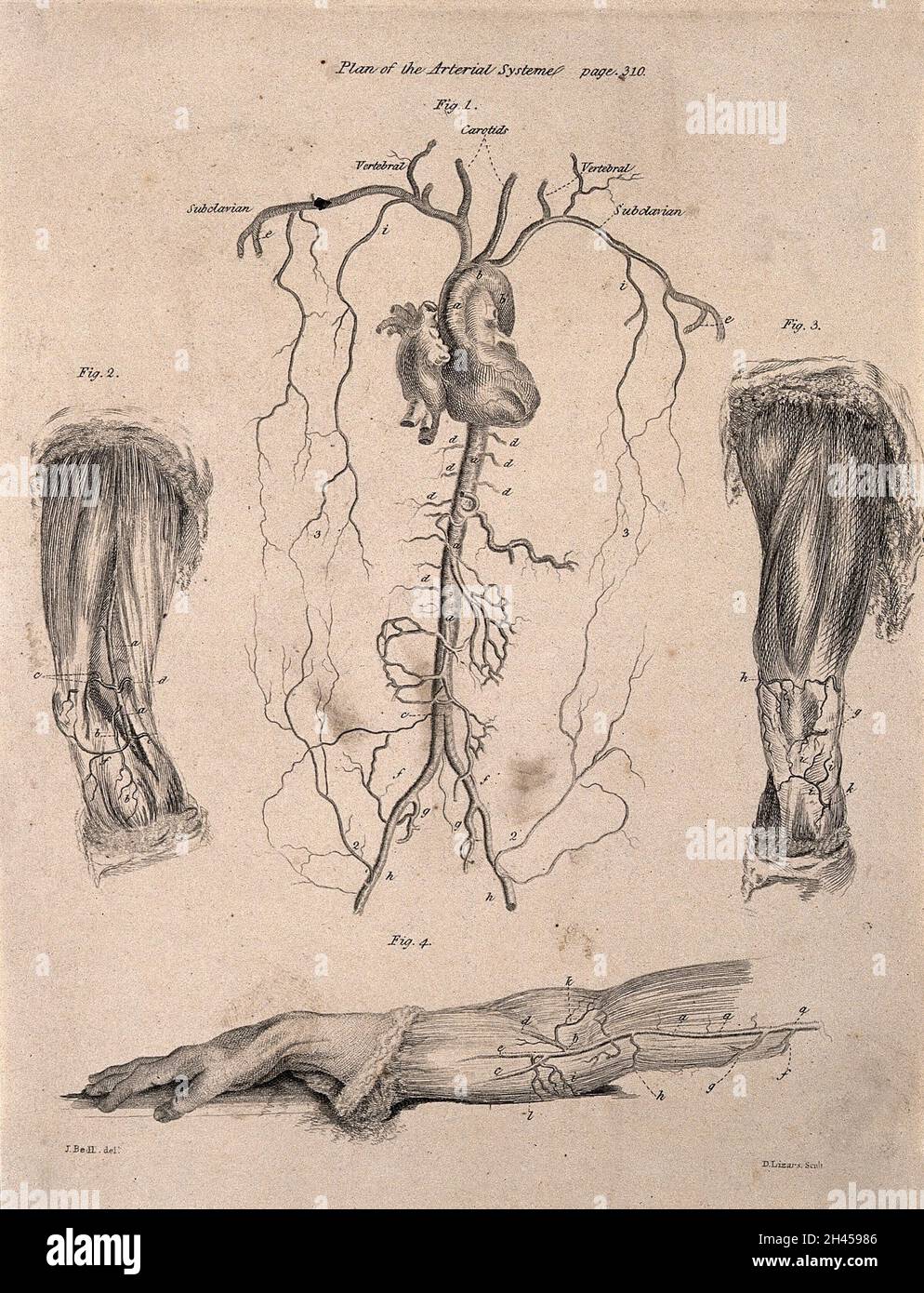 A human heart with surrounding arteries; two dissected legs and a dissected arm, lettered for key. Etching by D. Lizars after J. Bell, c. 1810 (?). Stock Photohttps://www.alamy.com/image-license-details/?v=1https://www.alamy.com/a-human-heart-with-surrounding-arteries-two-dissected-legs-and-a-dissected-arm-lettered-for-key-etching-by-d-lizars-after-j-bell-c-1810-image450045238.html
RM2H45986–A human heart with surrounding arteries; two dissected legs and a dissected arm, lettered for key. Etching by D. Lizars after J. Bell, c. 1810 (?).
 willow, osier (Salix spec.), tree slice with core rot, Germany Stock Photohttps://www.alamy.com/image-license-details/?v=1https://www.alamy.com/willow-osier-salix-spec-tree-slice-with-core-rot-germany-image469087142.html
RM2J74NC6–willow, osier (Salix spec.), tree slice with core rot, Germany
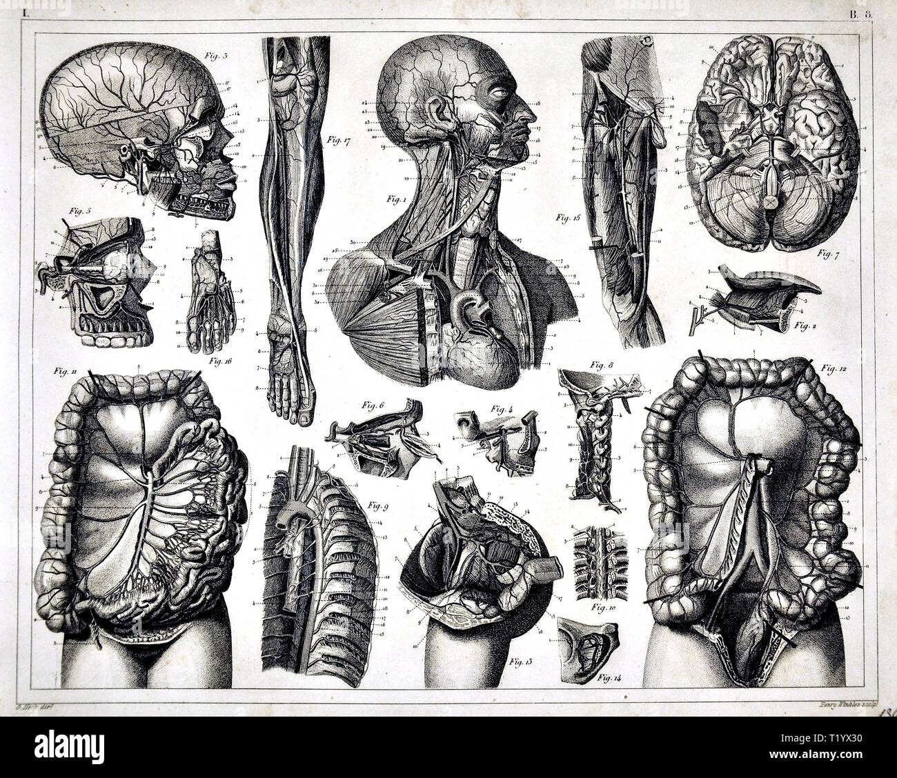 1849 Medical Illustration of Human Anatomy Circulatory System Stock Photohttps://www.alamy.com/image-license-details/?v=1https://www.alamy.com/1849-medical-illustration-of-human-anatomy-circulatory-system-image242172980.html
RFT1YX30–1849 Medical Illustration of Human Anatomy Circulatory System
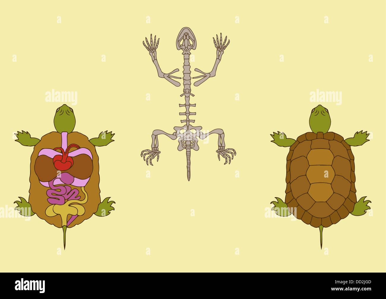 turtle anatomy Stock Photohttps://www.alamy.com/image-license-details/?v=1https://www.alamy.com/stock-photo-turtle-anatomy-59680109.html
RFDD2JGD–turtle anatomy
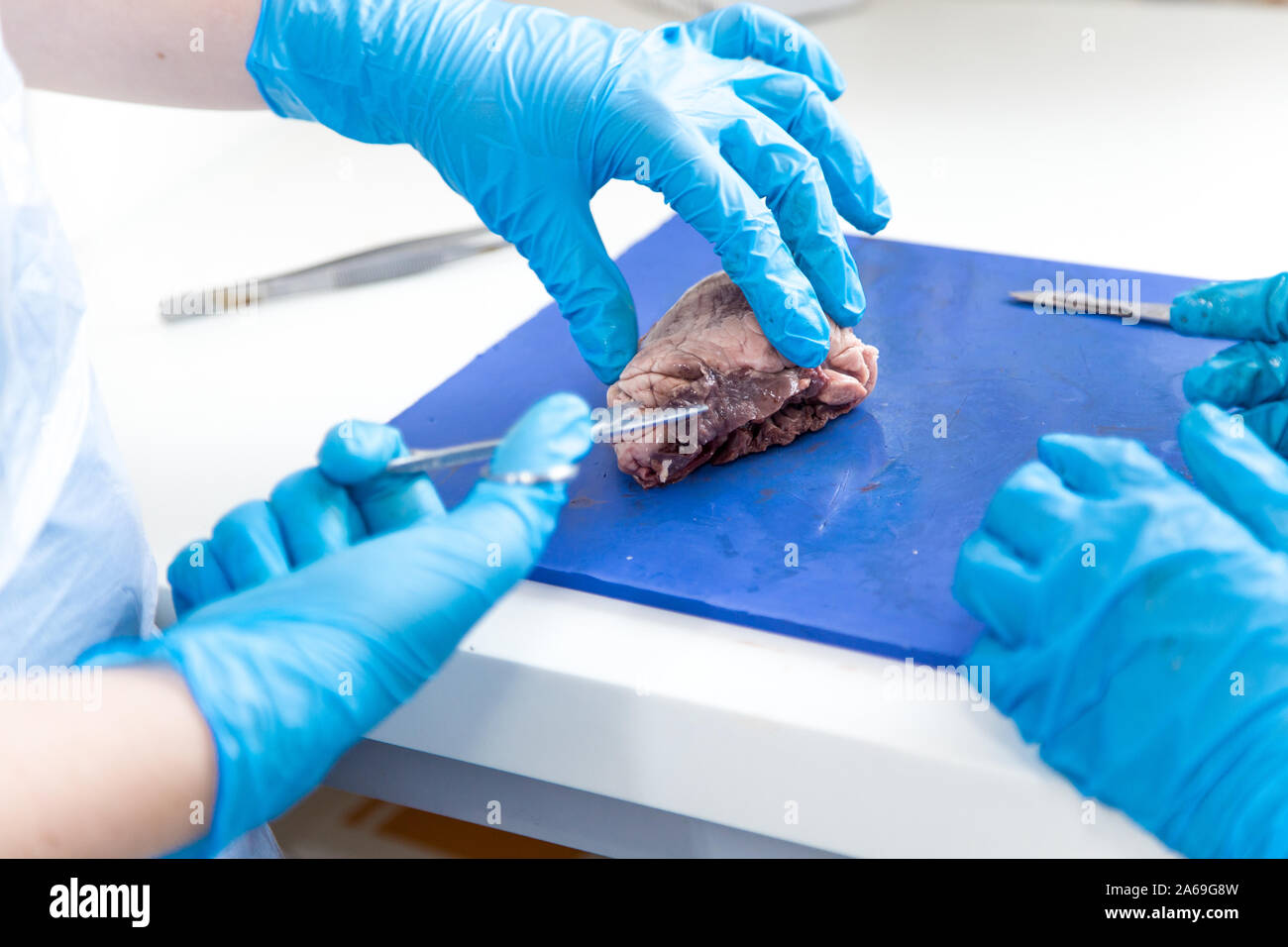 Medical students doing sheep heart dissection in the lab class Stock Photohttps://www.alamy.com/image-license-details/?v=1https://www.alamy.com/medical-students-doing-sheep-heart-dissection-in-the-lab-class-image330895289.html
RF2A69G8W–Medical students doing sheep heart dissection in the lab class
 anatomy of fish Stock Vectorhttps://www.alamy.com/image-license-details/?v=1https://www.alamy.com/anatomy-of-fish-image64198062.html
RFDMCD7X–anatomy of fish
 Regional anatomy in its relation to medicine and surgery . Fip2. Dissected, Photogrdph/d and Colored from Nature ny GeoeoE Mc. Cleu.au, M D. Copyright, 183/. by Geopge Mc Clellan,M D. ArmitrongsCn Lith Soalo THE REaiON OF THE THORAX. 287 in relation to the roots of the lungs, and the coronary plexuses. Thebranches of the latter respectively accompany the ramifications of thecoronary arteries both without and within the substance of the heart. Itis said that there are minute intra-cardiac ganglia, which possibly presideover the functional contractions of the heart. The lymphatic vessels of the Stock Photohttps://www.alamy.com/image-license-details/?v=1https://www.alamy.com/regional-anatomy-in-its-relation-to-medicine-and-surgery-fip2-dissected-photogrdphd-and-colored-from-nature-ny-geoeoe-mc-cleuau-m-d-copyright-183-by-geopge-mc-clellanm-d-armitrongscn-lith-soalo-the-reaion-of-the-thorax-287-in-relation-to-the-roots-of-the-lungs-and-the-coronary-plexuses-thebranches-of-the-latter-respectively-accompany-the-ramifications-of-thecoronary-arteries-both-without-and-within-the-substance-of-the-heart-itis-said-that-there-are-minute-intra-cardiac-ganglia-which-possibly-presideover-the-functional-contractions-of-the-heart-the-lymphatic-vessels-of-the-image338271446.html
RM2AJ9GK2–Regional anatomy in its relation to medicine and surgery . Fip2. Dissected, Photogrdph/d and Colored from Nature ny GeoeoE Mc. Cleu.au, M D. Copyright, 183/. by Geopge Mc Clellan,M D. ArmitrongsCn Lith Soalo THE REaiON OF THE THORAX. 287 in relation to the roots of the lungs, and the coronary plexuses. Thebranches of the latter respectively accompany the ramifications of thecoronary arteries both without and within the substance of the heart. Itis said that there are minute intra-cardiac ganglia, which possibly presideover the functional contractions of the heart. The lymphatic vessels of the
 The spectacular objects on display include the world's most famous watch , Brequet's No,160 Marie Antoinette ,which is on loan from the LA Mayer Museum for Islamic Art and on public display for the first time in theUK.In 1783 , one of the greatest watchmakers of all time , Abraham - Louis Brequet was given an unlimited budget to craft an exceptional timepiece for Queen Marie Antoinette . Crafted over four decades from the finest materials including sapphires ,platinum and gold ,it exceeded all other watches of it's time in beauty and complexity and became Brequet's masterpiece . Stock Photohttps://www.alamy.com/image-license-details/?v=1https://www.alamy.com/the-spectacular-objects-on-display-include-the-worlds-most-famous-watch-brequets-no160-marie-antoinette-which-is-on-loan-from-the-la-mayer-museum-for-islamic-art-and-on-public-display-for-the-first-time-in-theukin-1783-one-of-the-greatest-watchmakers-of-all-time-abraham-louis-brequet-was-given-an-unlimited-budget-to-craft-an-exceptional-timepiece-for-queen-marie-antoinette-crafted-over-four-decades-from-the-finest-materials-including-sapphires-platinum-and-gold-it-exceeded-all-other-watches-of-its-time-in-beauty-and-complexity-and-became-brequets-masterpiece-image635347351.html
RM2YWJG0R–The spectacular objects on display include the world's most famous watch , Brequet's No,160 Marie Antoinette ,which is on loan from the LA Mayer Museum for Islamic Art and on public display for the first time in theUK.In 1783 , one of the greatest watchmakers of all time , Abraham - Louis Brequet was given an unlimited budget to craft an exceptional timepiece for Queen Marie Antoinette . Crafted over four decades from the finest materials including sapphires ,platinum and gold ,it exceeded all other watches of it's time in beauty and complexity and became Brequet's masterpiece .
 Heart, Anatomical Illustration, 1814 Stock Photohttps://www.alamy.com/image-license-details/?v=1https://www.alamy.com/stock-photo-heart-anatomical-illustration-1814-135096588.html
RMHRP524–Heart, Anatomical Illustration, 1814
 The body of a youth with his trunk dissected: two figures showing the ribs and viscera, especially the position of the heart in systole and diastole. Coloured lithograph by J.B. Léveillé after William Fairland, 1869. Stock Photohttps://www.alamy.com/image-license-details/?v=1https://www.alamy.com/the-body-of-a-youth-with-his-trunk-dissected-two-figures-showing-the-ribs-and-viscera-especially-the-position-of-the-heart-in-systole-and-diastole-coloured-lithograph-by-jb-lveill-after-william-fairland-1869-image450067065.html
RM2H4693N–The body of a youth with his trunk dissected: two figures showing the ribs and viscera, especially the position of the heart in systole and diastole. Coloured lithograph by J.B. Léveillé after William Fairland, 1869.
 willow, osier (Salix spec.), tree slice with core rot, Germany Stock Photohttps://www.alamy.com/image-license-details/?v=1https://www.alamy.com/willow-osier-salix-spec-tree-slice-with-core-rot-germany-image469087161.html
RM2J74NCW–willow, osier (Salix spec.), tree slice with core rot, Germany
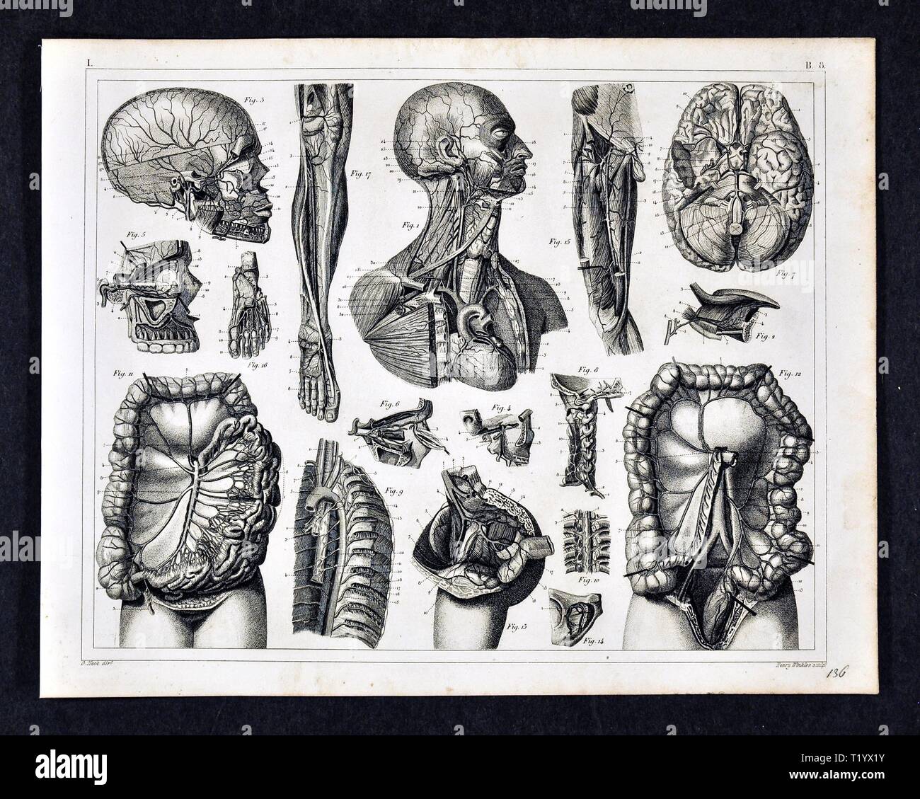 1849 Medical Illustration of Human Anatomy Circulatory System Stock Photohttps://www.alamy.com/image-license-details/?v=1https://www.alamy.com/1849-medical-illustration-of-human-anatomy-circulatory-system-image242172951.html
RFT1YX1Y–1849 Medical Illustration of Human Anatomy Circulatory System
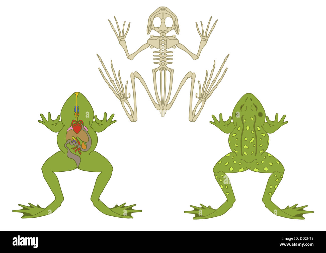 zoology, anatomy of amphibian, cross-section and skeleton Stock Photohttps://www.alamy.com/image-license-details/?v=1https://www.alamy.com/stock-photo-zoology-anatomy-of-amphibian-cross-section-and-skeleton-59679544.html
RFDD2HT8–zoology, anatomy of amphibian, cross-section and skeleton
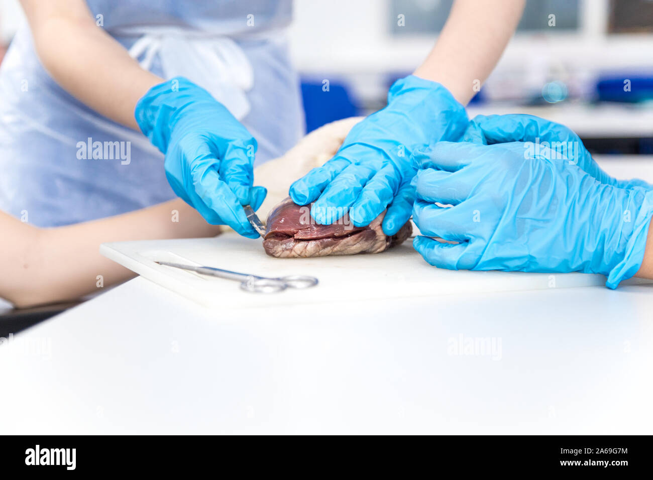 Medical students doing sheep heart dissection in the lab class Stock Photohttps://www.alamy.com/image-license-details/?v=1https://www.alamy.com/medical-students-doing-sheep-heart-dissection-in-the-lab-class-image330895256.html
RF2A69G7M–Medical students doing sheep heart dissection in the lab class
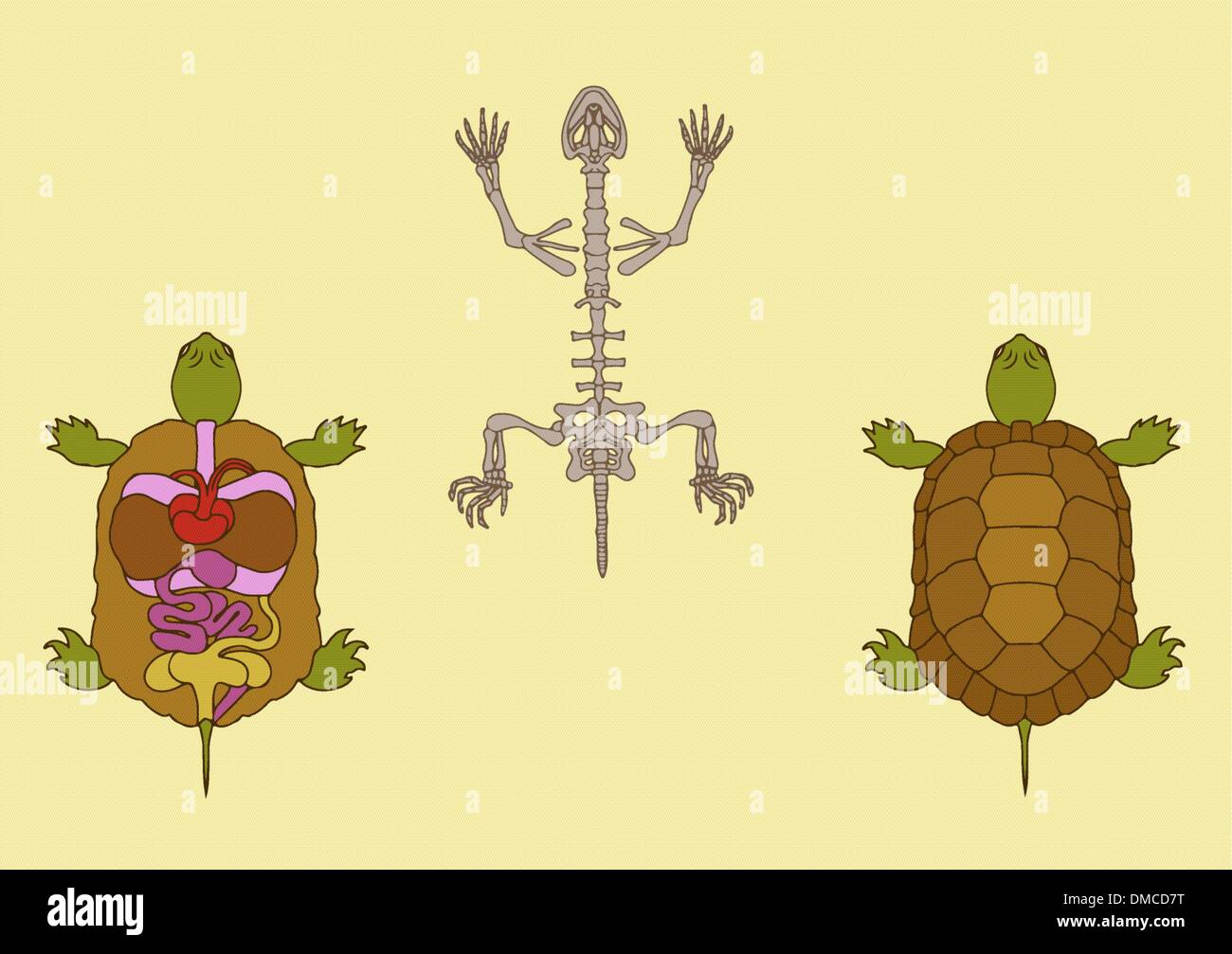 anatomy of reptile Stock Vectorhttps://www.alamy.com/image-license-details/?v=1https://www.alamy.com/anatomy-of-reptile-image64198060.html
RFDMCD7T–anatomy of reptile
 Medical students doing sheep heart dissection in the lab class Stock Photohttps://www.alamy.com/image-license-details/?v=1https://www.alamy.com/medical-students-doing-sheep-heart-dissection-in-the-lab-class-image384277182.html
RF2D959D2–Medical students doing sheep heart dissection in the lab class
 A treatise on zoology . ^ the junctionof two efferent vesselsin each arch in Ceratodus(Fig. 220). They join thedorsal aorta. From theposterior epibranchial,the sixth aortic arch,counting the mandibularas the first, is given offa pulmonary artery tothe air-bladder. The Vcratodiis torsterl, Kretlt. Ventral iew of the heart . cc t. dissected so as to expose the inside of the ventricle and prCSenCe ot tWO etterent vessels in each branchialbar in both the Dipnoiand the Selachii is prob-ably of no phylogeneticsignificance; in the rela-tion of the epibranchialarches to the bars the Dipnoi are the mo Stock Photohttps://www.alamy.com/image-license-details/?v=1https://www.alamy.com/a-treatise-on-zoology-the-junctionof-two-efferent-vesselsin-each-arch-in-ceratodusfig-220-they-join-thedorsal-aorta-from-theposterior-epibranchialthe-sixth-aortic-archcounting-the-mandibularas-the-first-is-given-offa-pulmonary-artery-tothe-air-bladder-the-vcratodiis-torsterl-kretlt-ventral-iew-of-the-heart-cc-t-dissected-so-as-to-expose-the-inside-of-the-ventricle-and-prcsence-ot-two-etterent-vessels-in-each-branchialbar-in-both-the-dipnoiand-the-selachii-is-prob-ably-of-no-phylogeneticsignificance-in-the-rela-tion-of-the-epibranchialarches-to-the-bars-the-dipnoi-are-the-mo-image338305783.html
RM2AJB4DB–A treatise on zoology . ^ the junctionof two efferent vesselsin each arch in Ceratodus(Fig. 220). They join thedorsal aorta. From theposterior epibranchial,the sixth aortic arch,counting the mandibularas the first, is given offa pulmonary artery tothe air-bladder. The Vcratodiis torsterl, Kretlt. Ventral iew of the heart . cc t. dissected so as to expose the inside of the ventricle and prCSenCe ot tWO etterent vessels in each branchialbar in both the Dipnoiand the Selachii is prob-ably of no phylogeneticsignificance; in the rela-tion of the epibranchialarches to the bars the Dipnoi are the mo
 The spectacular objects on display include the world's most famous watch , Brequet's No,160 Marie Antoinette ,which is on loan from the LA Mayer Museum for Islamic Art and on public display for the first time in theUK.In 1783 , one of the greatest watchmakers of all time , Abraham - Louis Brequet was given an unlimited budget to craft an exceptional timepiece for Queen Marie Antoinette . Crafted over four decades from the finest materials including sapphires ,platinum and gold ,it exceeded all other watches of it's time in beauty and complexity and became Brequet's masterpiece . Stock Photohttps://www.alamy.com/image-license-details/?v=1https://www.alamy.com/the-spectacular-objects-on-display-include-the-worlds-most-famous-watch-brequets-no160-marie-antoinette-which-is-on-loan-from-the-la-mayer-museum-for-islamic-art-and-on-public-display-for-the-first-time-in-theukin-1783-one-of-the-greatest-watchmakers-of-all-time-abraham-louis-brequet-was-given-an-unlimited-budget-to-craft-an-exceptional-timepiece-for-queen-marie-antoinette-crafted-over-four-decades-from-the-finest-materials-including-sapphires-platinum-and-gold-it-exceeded-all-other-watches-of-its-time-in-beauty-and-complexity-and-became-brequets-masterpiece-image635348923.html
RM2YWJJ0Y–The spectacular objects on display include the world's most famous watch , Brequet's No,160 Marie Antoinette ,which is on loan from the LA Mayer Museum for Islamic Art and on public display for the first time in theUK.In 1783 , one of the greatest watchmakers of all time , Abraham - Louis Brequet was given an unlimited budget to craft an exceptional timepiece for Queen Marie Antoinette . Crafted over four decades from the finest materials including sapphires ,platinum and gold ,it exceeded all other watches of it's time in beauty and complexity and became Brequet's masterpiece .
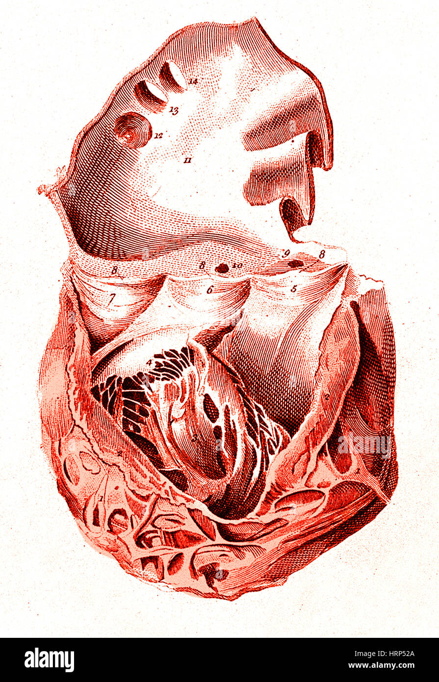 Heart, Anatomical Illustration, 1814 Stock Photohttps://www.alamy.com/image-license-details/?v=1https://www.alamy.com/stock-photo-heart-anatomical-illustration-1814-135096594.html
RMHRP52A–Heart, Anatomical Illustration, 1814
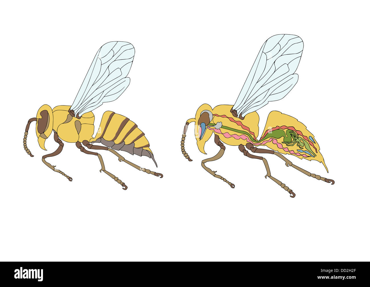 zoology, anatomy, morphology, cross-section of bee Stock Photohttps://www.alamy.com/image-license-details/?v=1https://www.alamy.com/stock-photo-zoology-anatomy-morphology-cross-section-of-bee-59678935.html
RFDD2H2F–zoology, anatomy, morphology, cross-section of bee
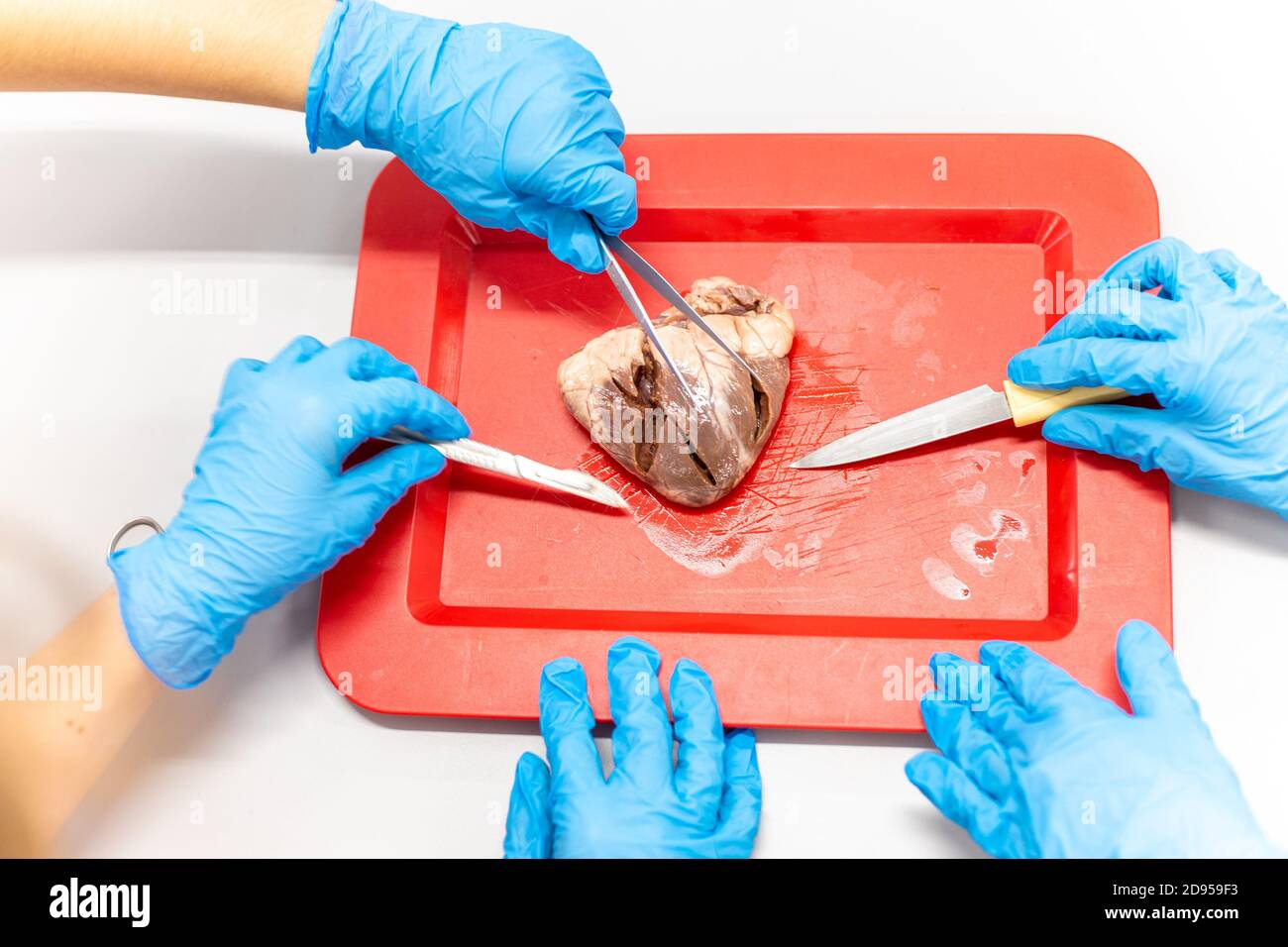 Medical students doing sheep heart dissection in the lab class Stock Photohttps://www.alamy.com/image-license-details/?v=1https://www.alamy.com/medical-students-doing-sheep-heart-dissection-in-the-lab-class-image384277239.html
RF2D959F3–Medical students doing sheep heart dissection in the lab class
 The Pulse / Rush Medical College yearbook . Heres a toast to the stiff with the ,tatooed arm, One who to his love was true.For he carried her name as a lovers charm, Pricked deep in his skin,with blue. Her name was Mary—how little she knows Of the fate her lover met—He was a sailor—the anchor shows— I suppose she waits for him yet. Heres hoping shell think he was lost at sea, For twould spoil it all if she knewThat her sailor-boy was dissected by me, And the arm was included too.I suppose that he often told Mary, he knew That her name was cut deep in his heart;;But I proved that the statement Stock Photohttps://www.alamy.com/image-license-details/?v=1https://www.alamy.com/the-pulse-rush-medical-college-yearbook-heres-a-toast-to-the-stiff-with-the-tatooed-arm-one-who-to-his-love-was-truefor-he-carried-her-name-as-a-lovers-charm-pricked-deep-in-his-skinwith-blue-her-name-was-maryhow-little-she-knows-of-the-fate-her-lover-methe-was-a-sailorthe-anchor-shows-i-suppose-she-waits-for-him-yet-heres-hoping-shell-think-he-was-lost-at-sea-for-twould-spoil-it-all-if-she-knewthat-her-sailor-boy-was-dissected-by-me-and-the-arm-was-included-tooi-suppose-that-he-often-told-mary-he-knew-that-her-name-was-cut-deep-in-his-heartbut-i-proved-that-the-statement-image343033954.html
RM2AX2F8J–The Pulse / Rush Medical College yearbook . Heres a toast to the stiff with the ,tatooed arm, One who to his love was true.For he carried her name as a lovers charm, Pricked deep in his skin,with blue. Her name was Mary—how little she knows Of the fate her lover met—He was a sailor—the anchor shows— I suppose she waits for him yet. Heres hoping shell think he was lost at sea, For twould spoil it all if she knewThat her sailor-boy was dissected by me, And the arm was included too.I suppose that he often told Mary, he knew That her name was cut deep in his heart;;But I proved that the statement
 The spectacular objects on display include the world's most famous watch , Brequet's No,160 Marie Antoinette ,which is on loan from the LA Mayer Museum for Islamic Art and on public display for the first time in theUK.In 1783 , one of the greatest watchmakers of all time , Abraham - Louis Brequet was given an unlimited budget to craft an exceptional timepiece for Queen Marie Antoinette . Crafted over four decades from the finest materials including sapphires ,platinum and gold ,it exceeded all other watches of it's time in beauty and complexity and became Brequet's masterpiece . Stock Photohttps://www.alamy.com/image-license-details/?v=1https://www.alamy.com/the-spectacular-objects-on-display-include-the-worlds-most-famous-watch-brequets-no160-marie-antoinette-which-is-on-loan-from-the-la-mayer-museum-for-islamic-art-and-on-public-display-for-the-first-time-in-theukin-1783-one-of-the-greatest-watchmakers-of-all-time-abraham-louis-brequet-was-given-an-unlimited-budget-to-craft-an-exceptional-timepiece-for-queen-marie-antoinette-crafted-over-four-decades-from-the-finest-materials-including-sapphires-platinum-and-gold-it-exceeded-all-other-watches-of-its-time-in-beauty-and-complexity-and-became-brequets-masterpiece-image635350671.html
RM2YWJM7B–The spectacular objects on display include the world's most famous watch , Brequet's No,160 Marie Antoinette ,which is on loan from the LA Mayer Museum for Islamic Art and on public display for the first time in theUK.In 1783 , one of the greatest watchmakers of all time , Abraham - Louis Brequet was given an unlimited budget to craft an exceptional timepiece for Queen Marie Antoinette . Crafted over four decades from the finest materials including sapphires ,platinum and gold ,it exceeded all other watches of it's time in beauty and complexity and became Brequet's masterpiece .
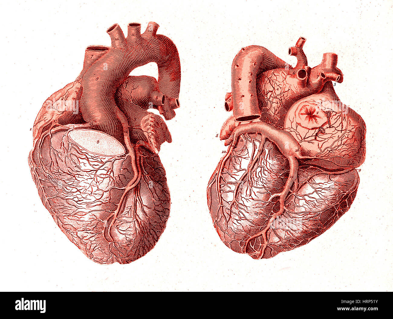 Heart, Anatomical Illustration, 1814 Stock Photohttps://www.alamy.com/image-license-details/?v=1https://www.alamy.com/stock-photo-heart-anatomical-illustration-1814-135096583.html
RMHRP51Y–Heart, Anatomical Illustration, 1814
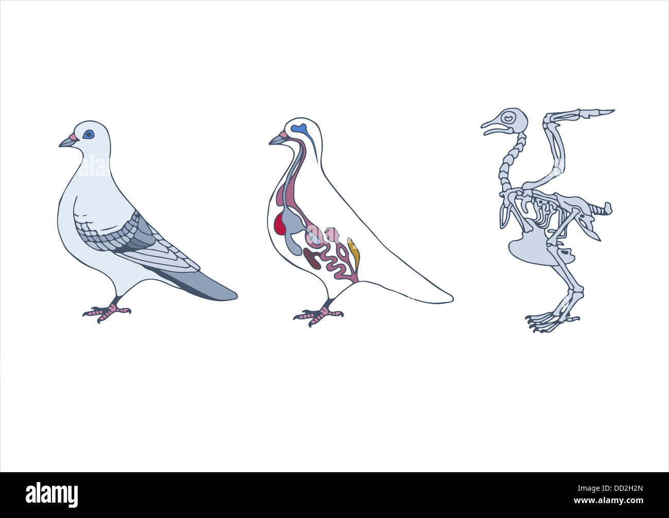 zoology, anatomy of bird, cross-section and skeleton Stock Photohttps://www.alamy.com/image-license-details/?v=1https://www.alamy.com/stock-photo-zoology-anatomy-of-bird-cross-section-and-skeleton-59678941.html
RFDD2H2N–zoology, anatomy of bird, cross-section and skeleton
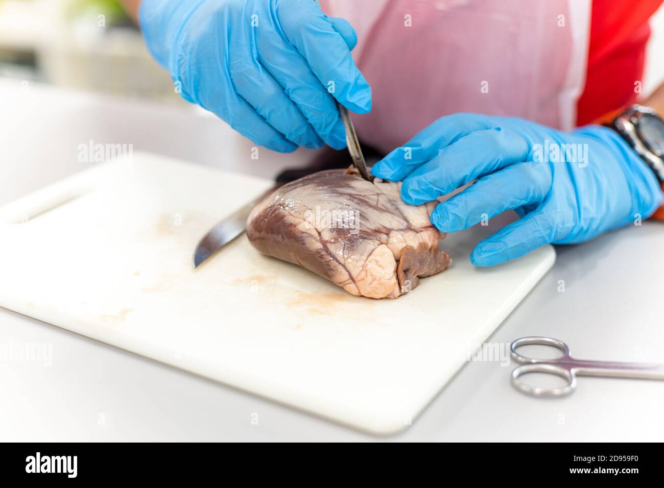 Medical students doing sheep heart dissection in the lab class Stock Photohttps://www.alamy.com/image-license-details/?v=1https://www.alamy.com/medical-students-doing-sheep-heart-dissection-in-the-lab-class-image384277236.html
RF2D959F0–Medical students doing sheep heart dissection in the lab class
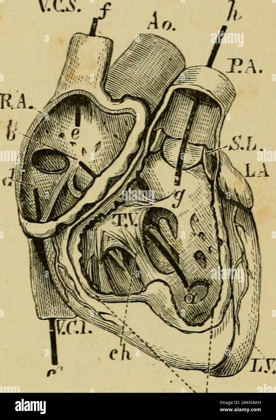 The common frog . II. RX Fig. 76.—I. The left side ; an 1 II. the right side of the Heart of Man dissected..—LA, the left auricle; PV, the four pulnmnary veins; cd, a style passedthrough the aiiriculo-ventri ular aperture; MW, the mitral valve; nb. a stylepassed through the left ventricle into the aorta ; J?A, RV, parts of the right sideof the heart: Pa, pulmonary artery.II.—RA, the right auricle; VCS, superior vena cava; VCI, inferior vena cava,the styles, y^, cd, being passed through them into the auricle ; ab, style parsedthrough the aurlculo-ventricular aperture ; Tl^, tricuspid valve ; R Stock Photohttps://www.alamy.com/image-license-details/?v=1https://www.alamy.com/the-common-frog-ii-rx-fig-76i-the-left-side-an-1-ii-the-right-side-of-the-heart-of-man-dissectedla-the-left-auricle-pv-the-four-pulnmnary-veins-cd-a-style-passedthrough-the-aiiriculo-ventri-ular-aperture-mw-the-mitral-valve-nb-a-stylepassed-through-the-left-ventricle-into-the-aorta-ja-rv-parts-of-the-right-sideof-the-heart-pa-pulmonary-arteryiira-the-right-auricle-vcs-superior-vena-cava-vci-inferior-vena-cavathe-styles-y-cd-being-passed-through-them-into-the-auricle-ab-style-parsedthrough-the-aurlculo-ventricular-aperture-tl-tricuspid-valve-r-image339144837.html
RM2AKNAKH–The common frog . II. RX Fig. 76.—I. The left side ; an 1 II. the right side of the Heart of Man dissected..—LA, the left auricle; PV, the four pulnmnary veins; cd, a style passedthrough the aiiriculo-ventri ular aperture; MW, the mitral valve; nb. a stylepassed through the left ventricle into the aorta ; J?A, RV, parts of the right sideof the heart: Pa, pulmonary artery.II.—RA, the right auricle; VCS, superior vena cava; VCI, inferior vena cava,the styles, y^, cd, being passed through them into the auricle ; ab, style parsedthrough the aurlculo-ventricular aperture ; Tl^, tricuspid valve ; R