Quick filters:
Distal Stock Photos and Images
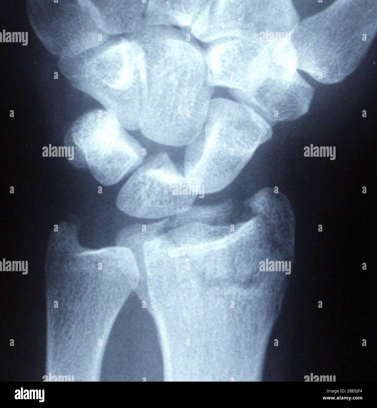 X-ray of a displaced intra-articular distal radius fracture (broken wrist) in an external fixator. Stock Photohttps://www.alamy.com/image-license-details/?v=1https://www.alamy.com/x-ray-of-a-displaced-intra-articular-distal-radius-fracture-broken-wrist-in-an-external-fixator-image352827080.html
RM2BE0JF4–X-ray of a displaced intra-articular distal radius fracture (broken wrist) in an external fixator.
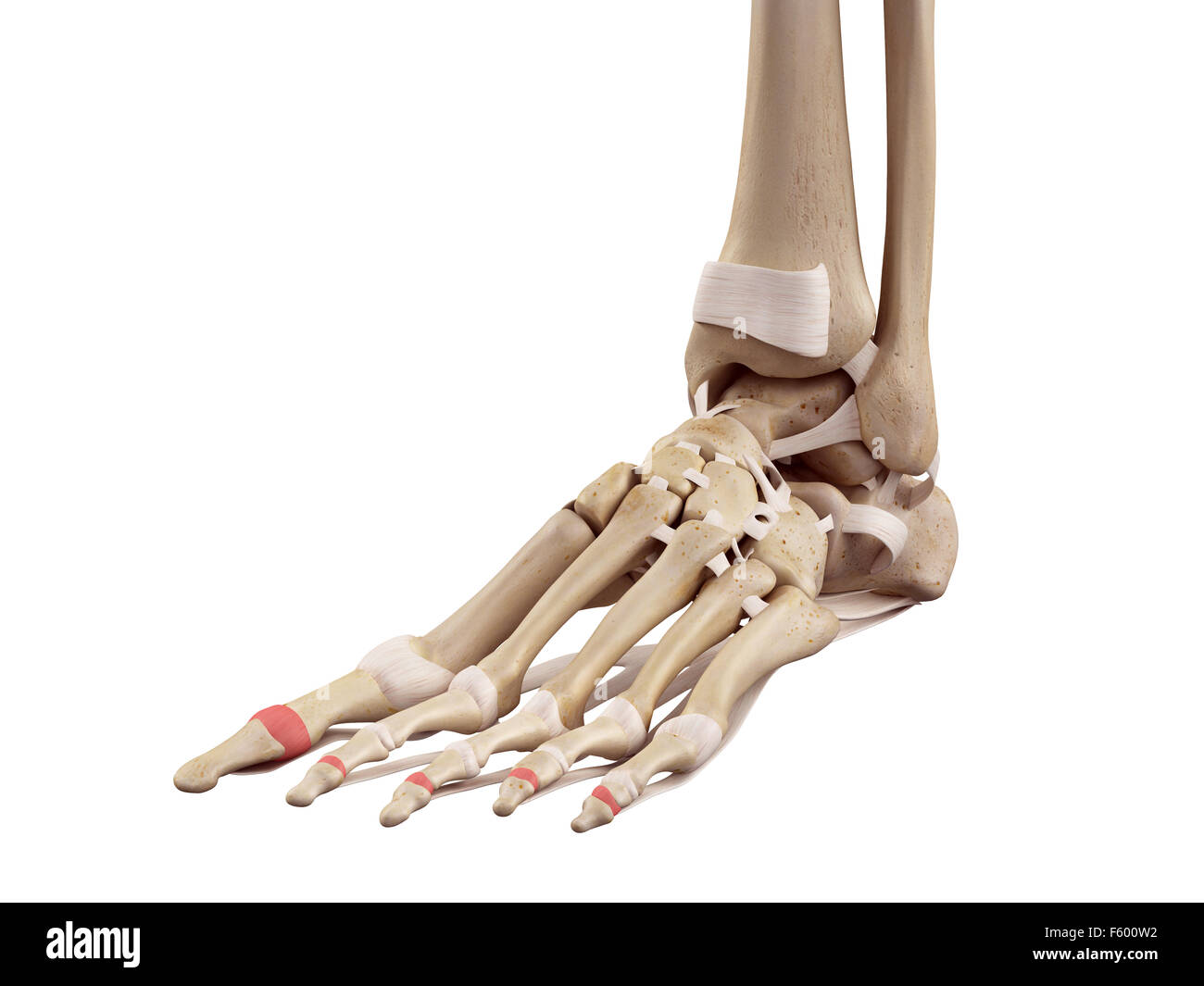 medical accurate illustration of the distal joint capsules Stock Photohttps://www.alamy.com/image-license-details/?v=1https://www.alamy.com/stock-photo-medical-accurate-illustration-of-the-distal-joint-capsules-89740478.html
RFF600W2–medical accurate illustration of the distal joint capsules
 With the hand manipulated into the desired position the pin is pushed into the distal ulna. Stock Photohttps://www.alamy.com/image-license-details/?v=1https://www.alamy.com/stock-photo-with-the-hand-manipulated-into-the-desired-position-the-pin-is-pushed-49044395.html
RMCRP4HF–With the hand manipulated into the desired position the pin is pushed into the distal ulna.
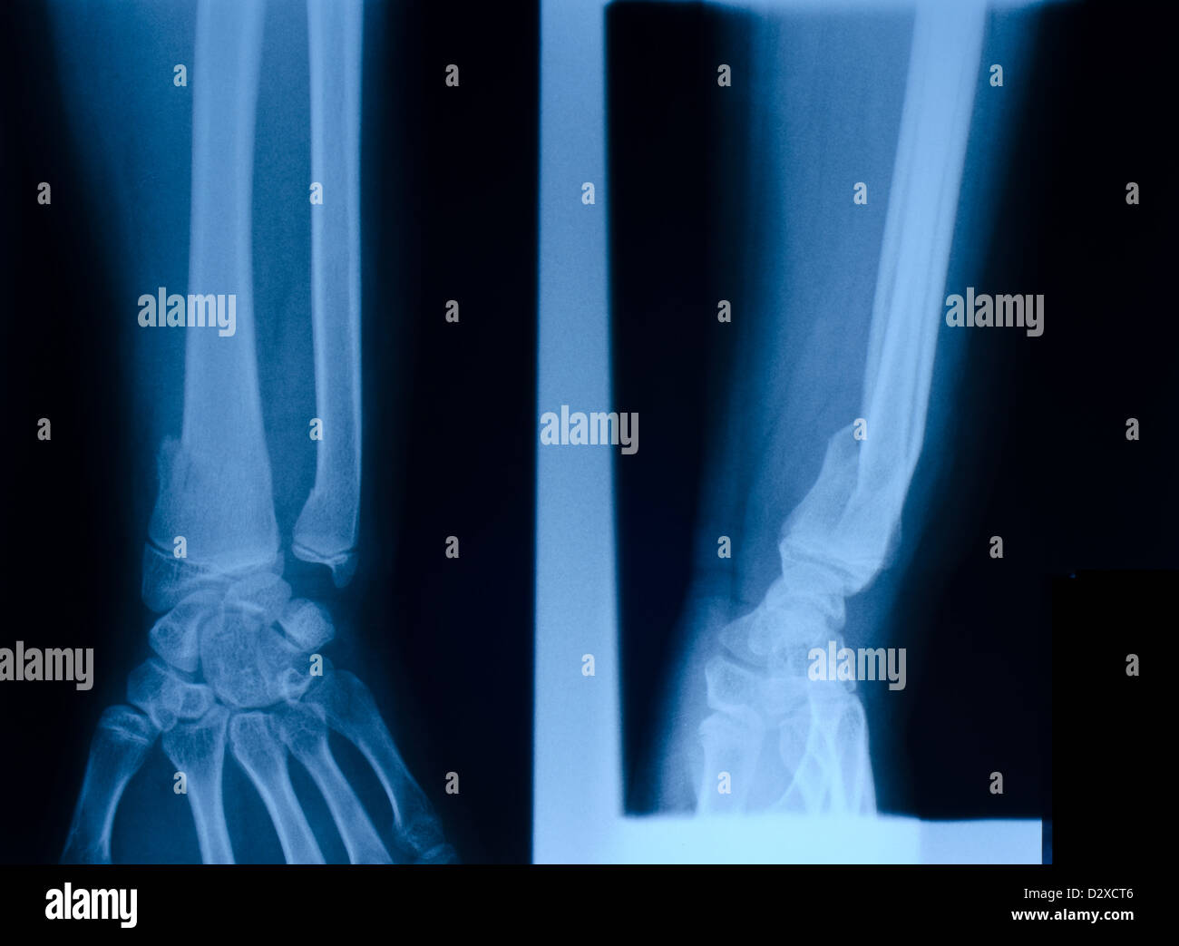 X ray film of distal radias fracture Stock Photohttps://www.alamy.com/image-license-details/?v=1https://www.alamy.com/stock-photo-x-ray-film-of-distal-radias-fracture-53441254.html
RFD2XCT6–X ray film of distal radias fracture
 x-ray of a comminuted distal radius wrist fracture Stock Photohttps://www.alamy.com/image-license-details/?v=1https://www.alamy.com/stock-photo-x-ray-of-a-comminuted-distal-radius-wrist-fracture-26900553.html
RMBFNBX1–x-ray of a comminuted distal radius wrist fracture
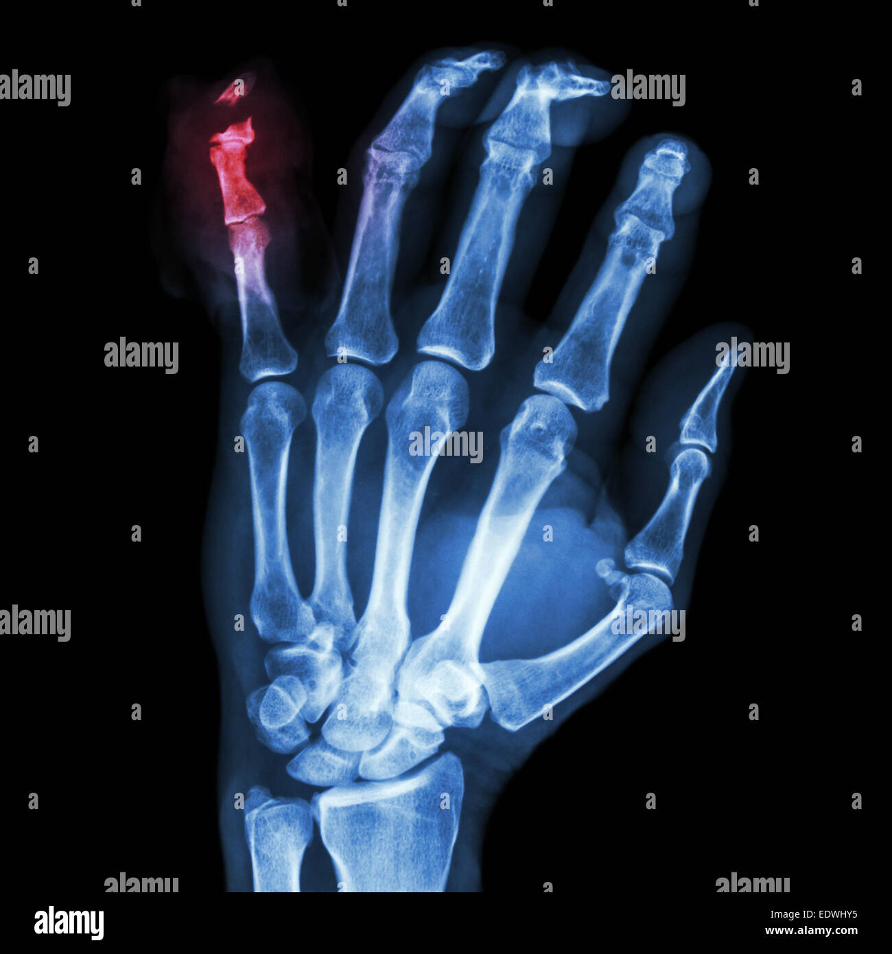 film x-ray hand AP : show fracture distal pharange little finger Stock Photohttps://www.alamy.com/image-license-details/?v=1https://www.alamy.com/stock-photo-film-x-ray-hand-ap-show-fracture-distal-pharange-little-finger-77394889.html
RFEDWHY5–film x-ray hand AP : show fracture distal pharange little finger
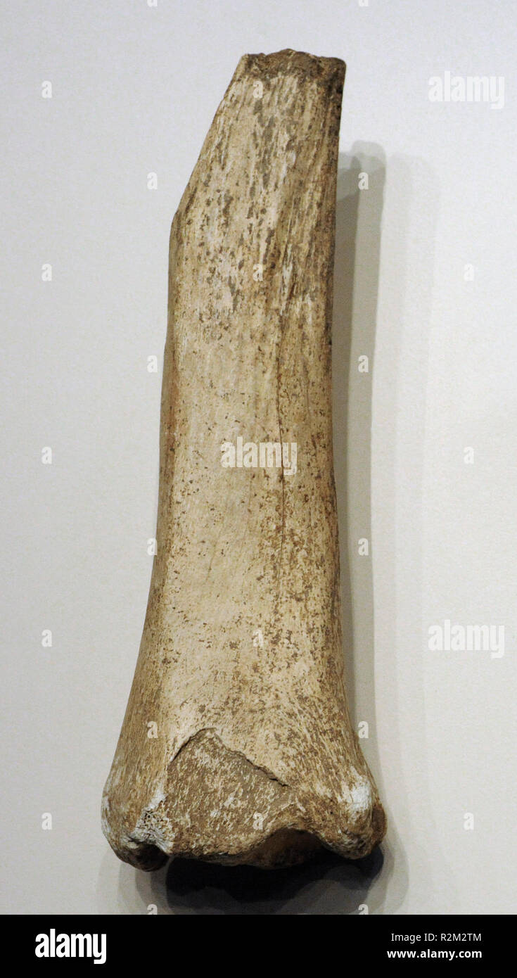 Diaphysis and distal epiphysis of horse tibia (Equus caballus). Mousterian period. Found in Cueva de Hornos de la Peña (San Felices de Buelna, Cantabria). National Archaeological Museum. Madrid. Spain. Stock Photohttps://www.alamy.com/image-license-details/?v=1https://www.alamy.com/diaphysis-and-distal-epiphysis-of-horse-tibia-equus-caballus-mousterian-period-found-in-cueva-de-hornos-de-la-pea-san-felices-de-buelna-cantabria-national-archaeological-museum-madrid-spain-image225405396.html
RMR2M2TM–Diaphysis and distal epiphysis of horse tibia (Equus caballus). Mousterian period. Found in Cueva de Hornos de la Peña (San Felices de Buelna, Cantabria). National Archaeological Museum. Madrid. Spain.
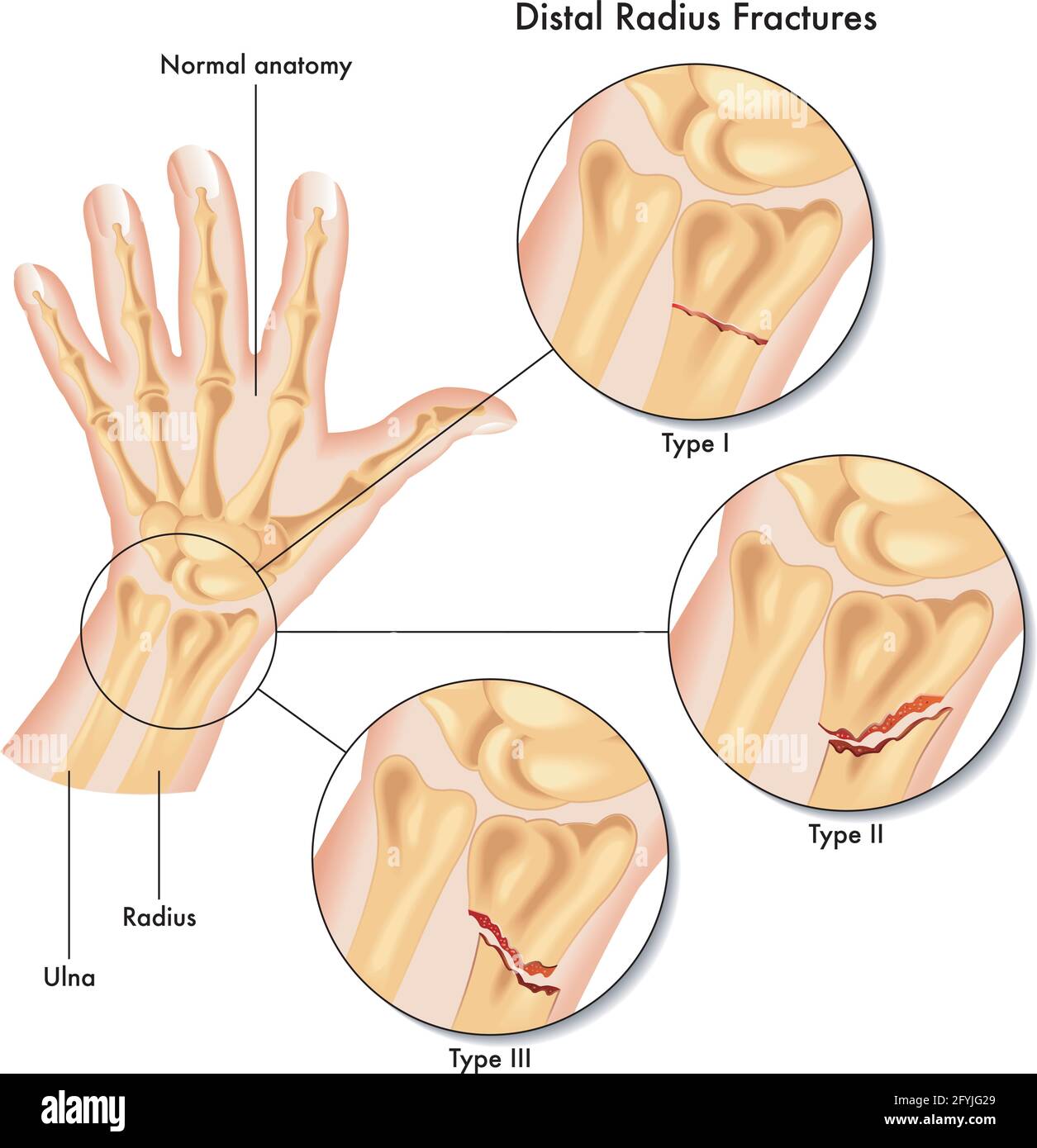 Medical illustration of the various kinds of fracture of the distal radius. Stock Vectorhttps://www.alamy.com/image-license-details/?v=1https://www.alamy.com/medical-illustration-of-the-various-kinds-of-fracture-of-the-distal-radius-image430052289.html
RF2FYJG29–Medical illustration of the various kinds of fracture of the distal radius.
 x-ray of a distal radius fracture Stock Photohttps://www.alamy.com/image-license-details/?v=1https://www.alamy.com/x-ray-of-a-distal-radius-fracture-image65501452.html
RFDPFRNG–x-ray of a distal radius fracture
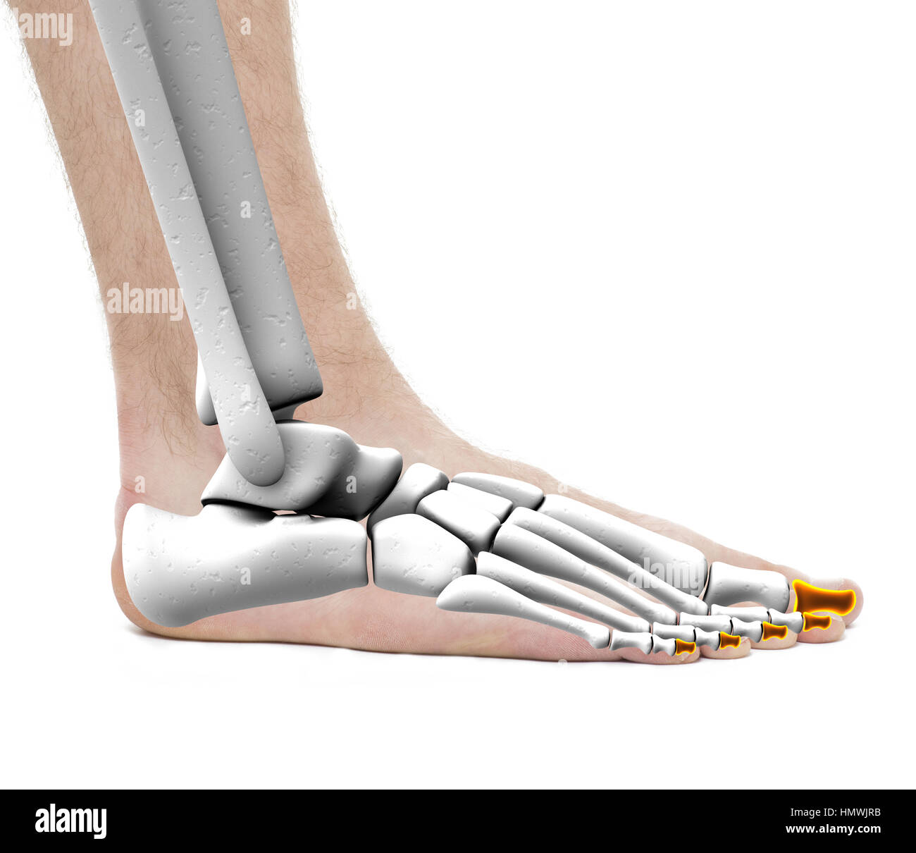 Distal Phalanges - Anatomy Male - Studio photo isolated on white Stock Photohttps://www.alamy.com/image-license-details/?v=1https://www.alamy.com/stock-photo-distal-phalanges-anatomy-male-studio-photo-isolated-on-white-133329263.html
RFHMWJRB–Distal Phalanges - Anatomy Male - Studio photo isolated on white
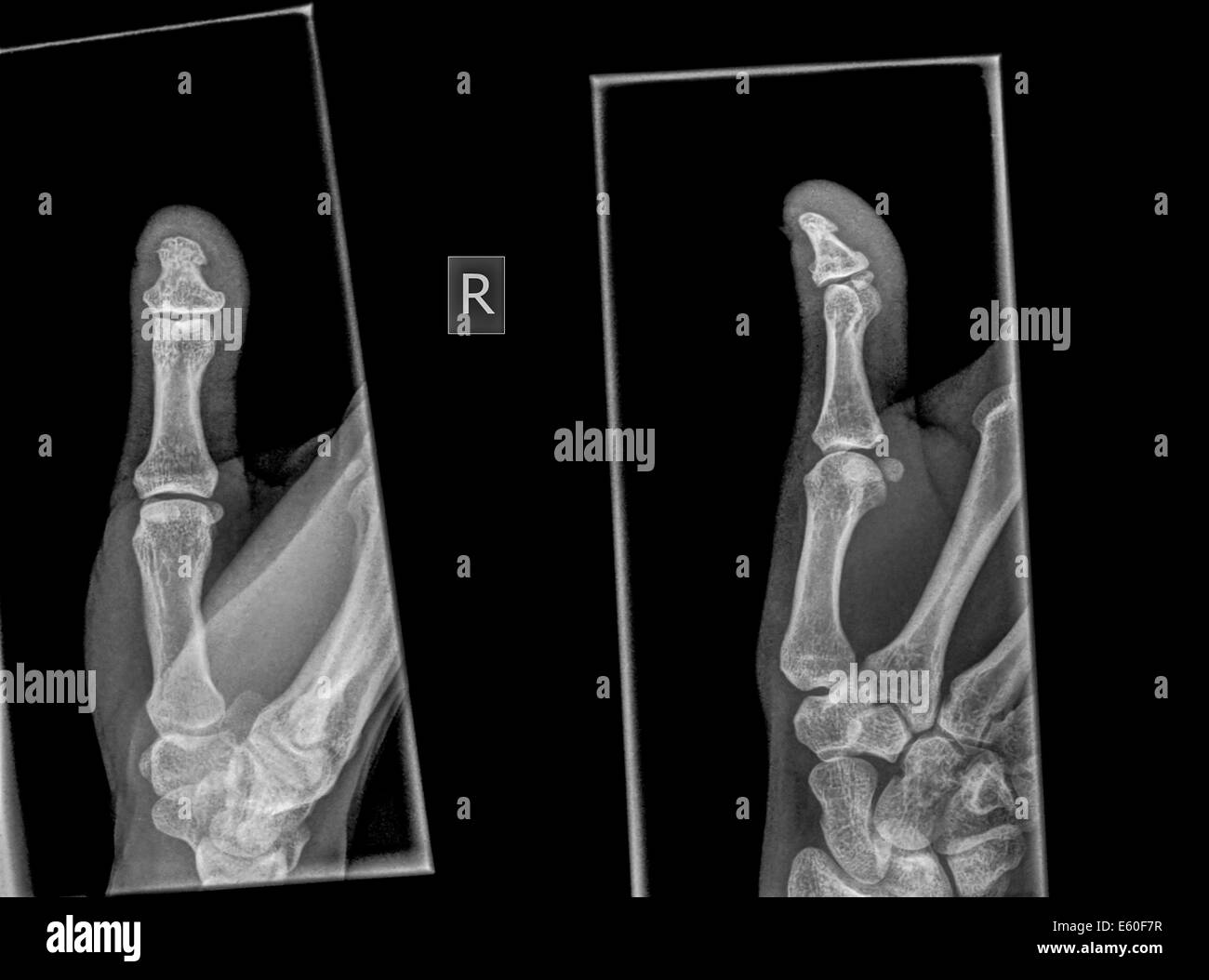 X-ray of a thumb with a broken distal phalanx Stock Photohttps://www.alamy.com/image-license-details/?v=1https://www.alamy.com/stock-photo-x-ray-of-a-thumb-with-a-broken-distal-phalanx-72541387.html
RME60F7R–X-ray of a thumb with a broken distal phalanx
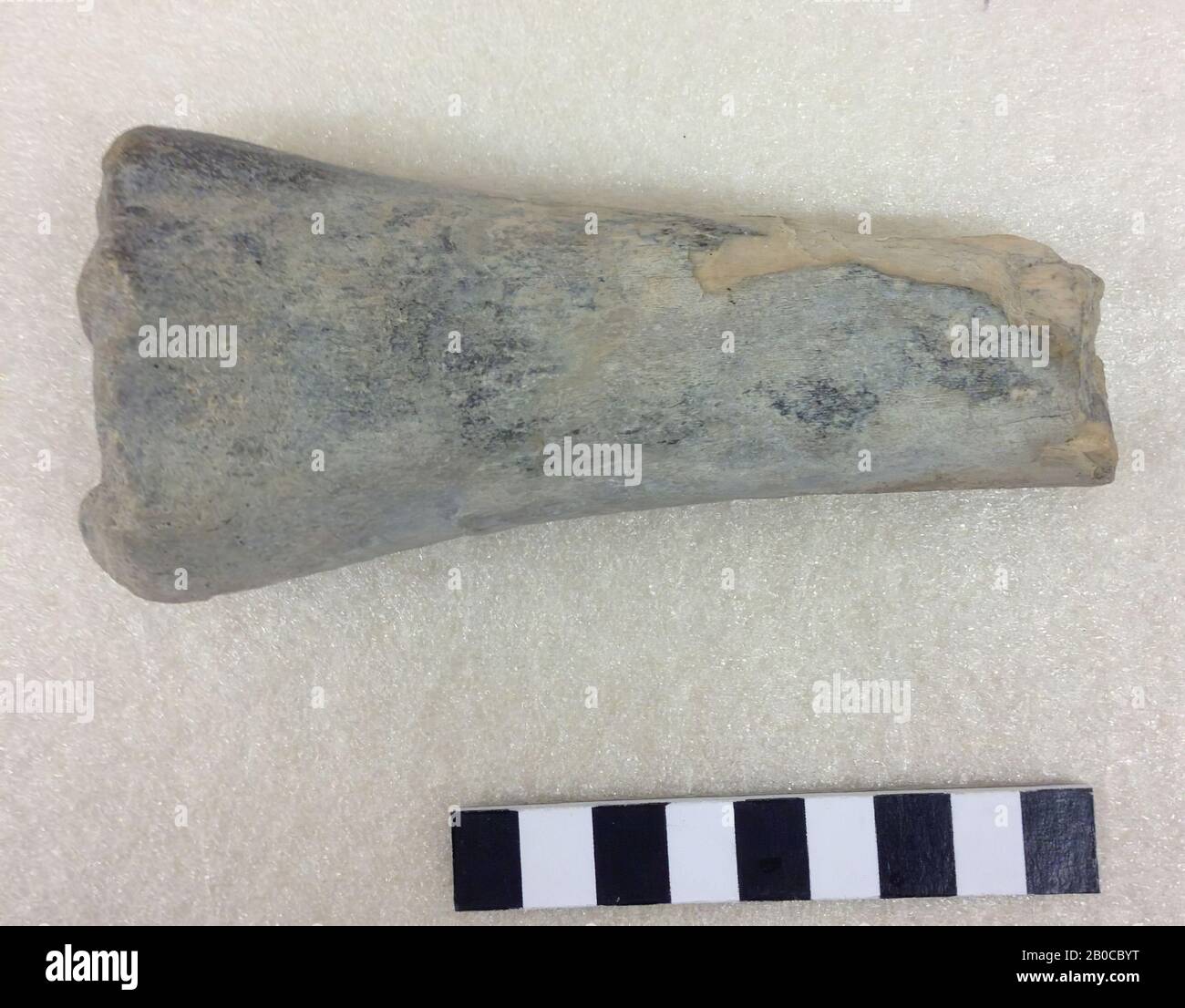 Distal and medial part. Possible metatarsus or metacarpus., Bone, organic, petrified bone, L: 13.2 cm, W: 6.1 cm, H: 3.9 cm, Early Stock Photohttps://www.alamy.com/image-license-details/?v=1https://www.alamy.com/distal-and-medial-part-possible-metatarsus-or-metacarpus-bone-organic-petrified-bone-l-132-cm-w-61-cm-h-39-cm-early-image344480188.html
RM2B0CBYT–Distal and medial part. Possible metatarsus or metacarpus., Bone, organic, petrified bone, L: 13.2 cm, W: 6.1 cm, H: 3.9 cm, Early
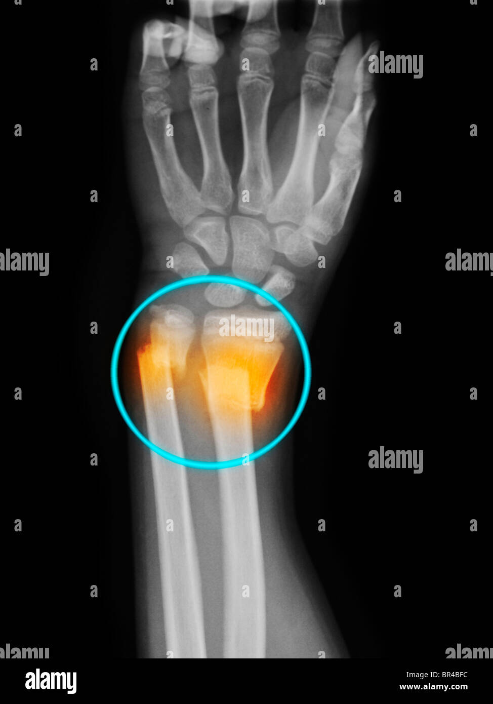
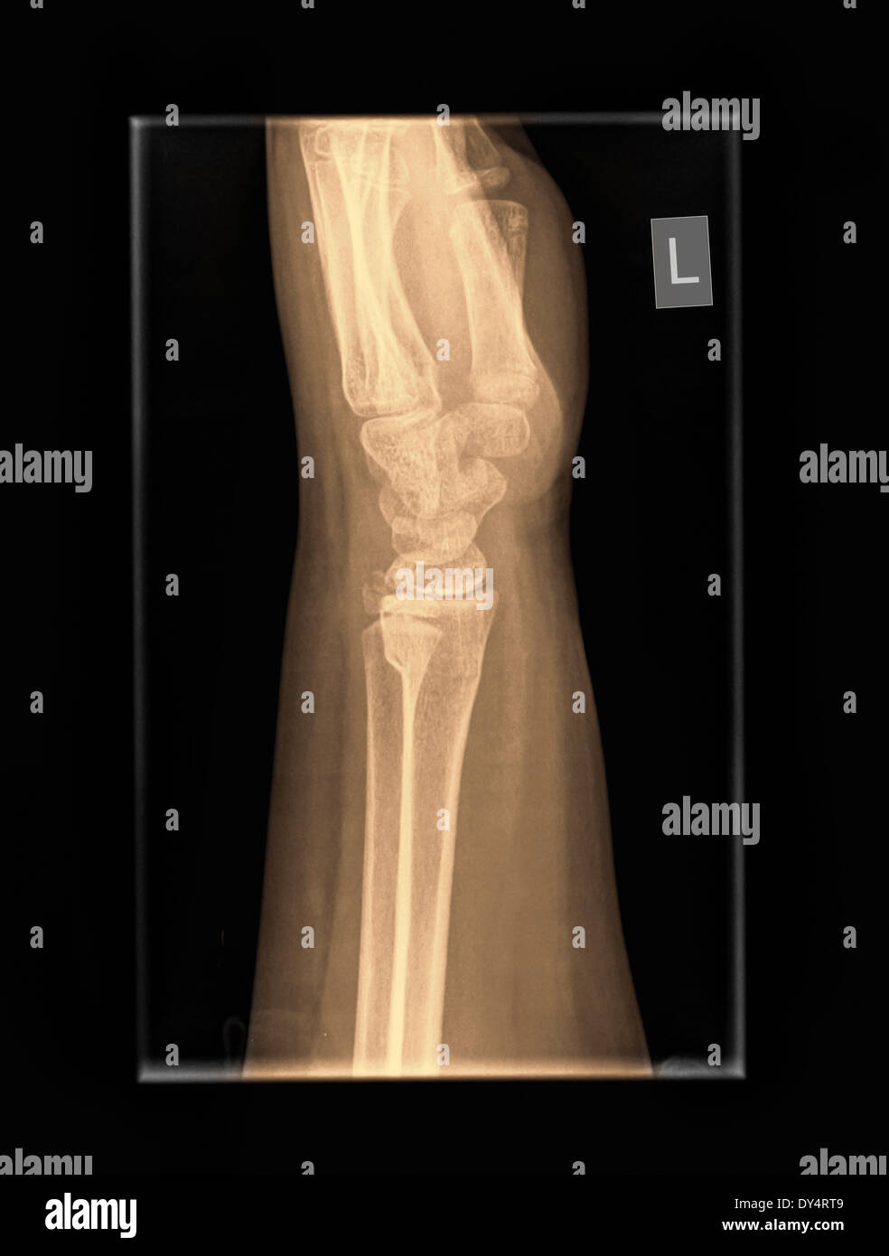 X-ray of wrist of 9 year old male patient with distal radius and ulna fractures Stock Photohttps://www.alamy.com/image-license-details/?v=1https://www.alamy.com/x-ray-of-wrist-of-9-year-old-male-patient-with-distal-radius-and-ulna-image68333337.html
RFDY4RT9–X-ray of wrist of 9 year old male patient with distal radius and ulna fractures
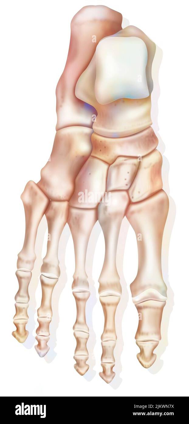 Superior view of the foot and the different bones: calcaneus, talus. Stock Photohttps://www.alamy.com/image-license-details/?v=1https://www.alamy.com/superior-view-of-the-foot-and-the-different-bones-calcaneus-talus-image476923886.html
RF2JKWN7X–Superior view of the foot and the different bones: calcaneus, talus.
 Mouth profile with a distal bite before the orthotropics or orthotropics treatment. Vector illustration Stock Vectorhttps://www.alamy.com/image-license-details/?v=1https://www.alamy.com/mouth-profile-with-a-distal-bite-before-the-orthotropics-or-orthotropics-treatment-vector-illustration-image246145541.html
RFT8CW45–Mouth profile with a distal bite before the orthotropics or orthotropics treatment. Vector illustration
 Conceptual image of centrioles. Stock Photohttps://www.alamy.com/image-license-details/?v=1https://www.alamy.com/conceptual-image-of-centrioles-image68934321.html
RFE046C1–Conceptual image of centrioles.
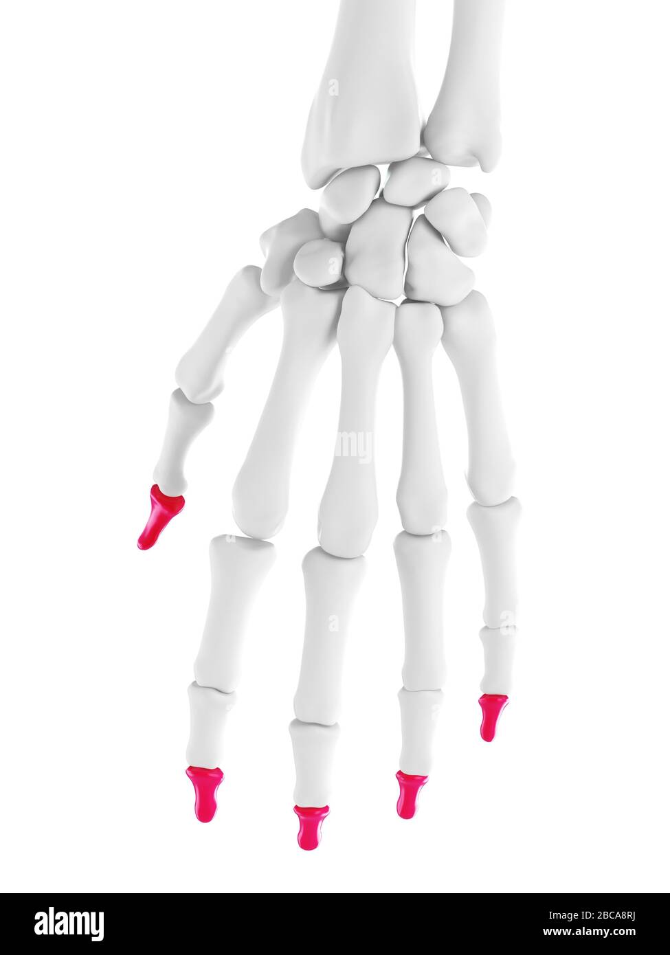 Distal phalanges, illustration. Stock Photohttps://www.alamy.com/image-license-details/?v=1https://www.alamy.com/distal-phalanges-illustration-image351809686.html
RF2BCA8RJ–Distal phalanges, illustration.
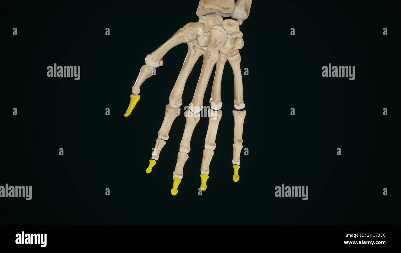 Distal Phalanges of Hand Stock Photohttps://www.alamy.com/image-license-details/?v=1https://www.alamy.com/distal-phalanges-of-hand-image491881220.html
RF2KG73EC–Distal Phalanges of Hand
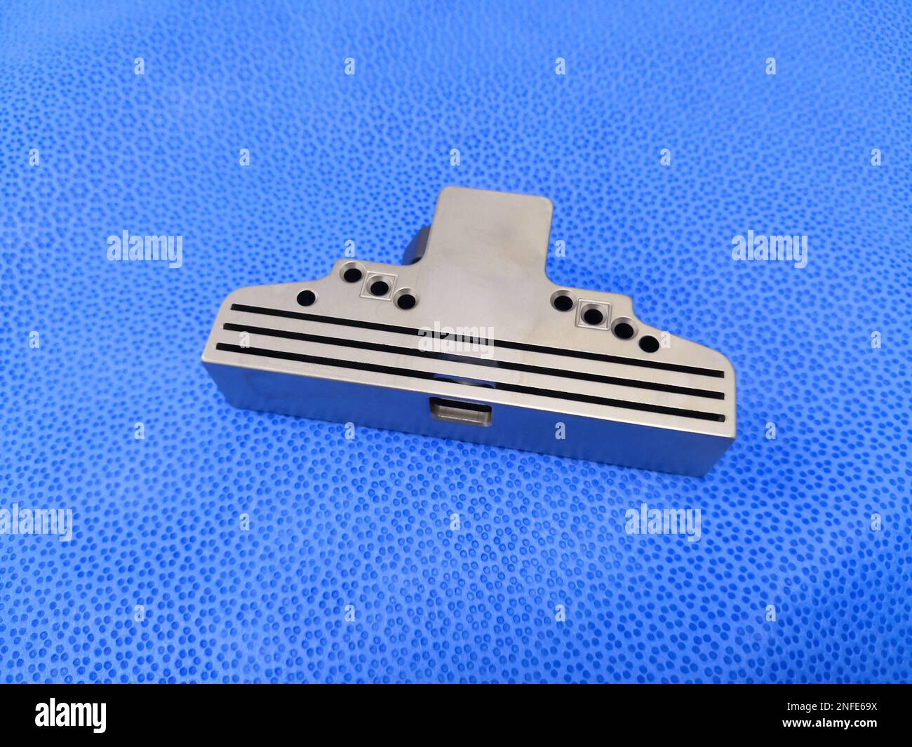 Total Knee Replacement Revision Instrument Distal Femoral Cutting Block Stock Photohttps://www.alamy.com/image-license-details/?v=1https://www.alamy.com/total-knee-replacement-revision-instrument-distal-femoral-cutting-block-image525843190.html
RF2NFE69X–Total Knee Replacement Revision Instrument Distal Femoral Cutting Block
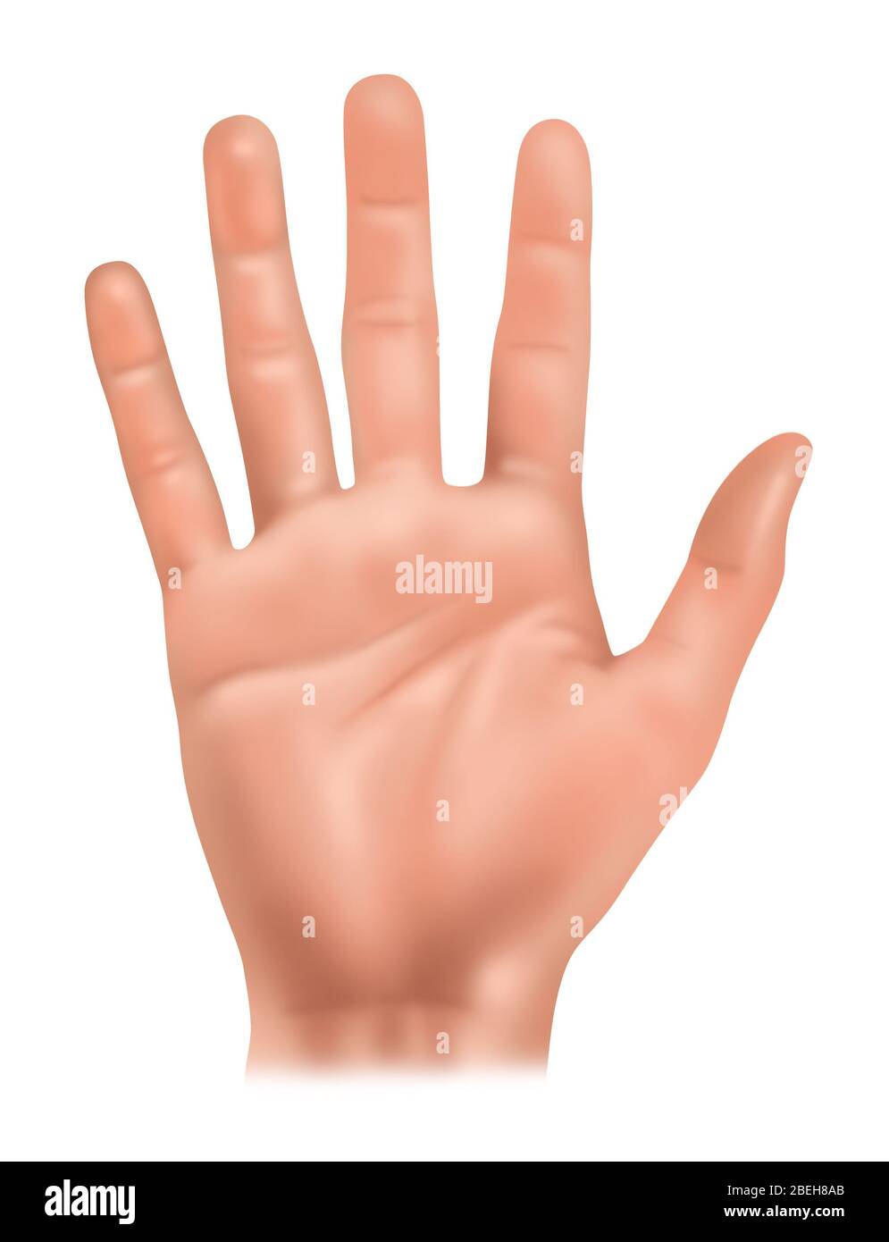 Hand Anatomy, Illustration Stock Photohttps://www.alamy.com/image-license-details/?v=1https://www.alamy.com/hand-anatomy-illustration-image353192291.html
RF2BEH8AB–Hand Anatomy, Illustration
 medical accurate illustration of the distal joint capsules Stock Photohttps://www.alamy.com/image-license-details/?v=1https://www.alamy.com/stock-photo-medical-accurate-illustration-of-the-distal-joint-capsules-89740564.html
RFF60104–medical accurate illustration of the distal joint capsules
 Supracondylar fracture or broken elbow in Asian, 11-year-old child. Fracture of distal humerus bone close to elbow. Caused by a fall on outstretched e Stock Photohttps://www.alamy.com/image-license-details/?v=1https://www.alamy.com/supracondylar-fracture-or-broken-elbow-in-asian-11-year-old-child-fracture-of-distal-humerus-bone-close-to-elbow-caused-by-a-fall-on-outstretched-e-image470064358.html
RF2J8N7TP–Supracondylar fracture or broken elbow in Asian, 11-year-old child. Fracture of distal humerus bone close to elbow. Caused by a fall on outstretched e
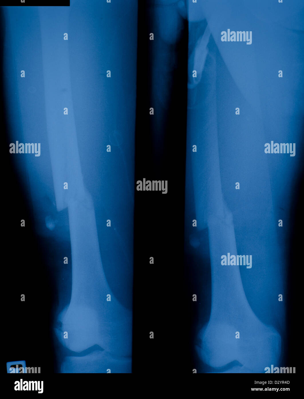 X ray film of distal femur fracture. Stock Photohttps://www.alamy.com/image-license-details/?v=1https://www.alamy.com/stock-photo-x-ray-film-of-distal-femur-fracture-53471277.html
RFD2YR4D–X ray film of distal femur fracture.
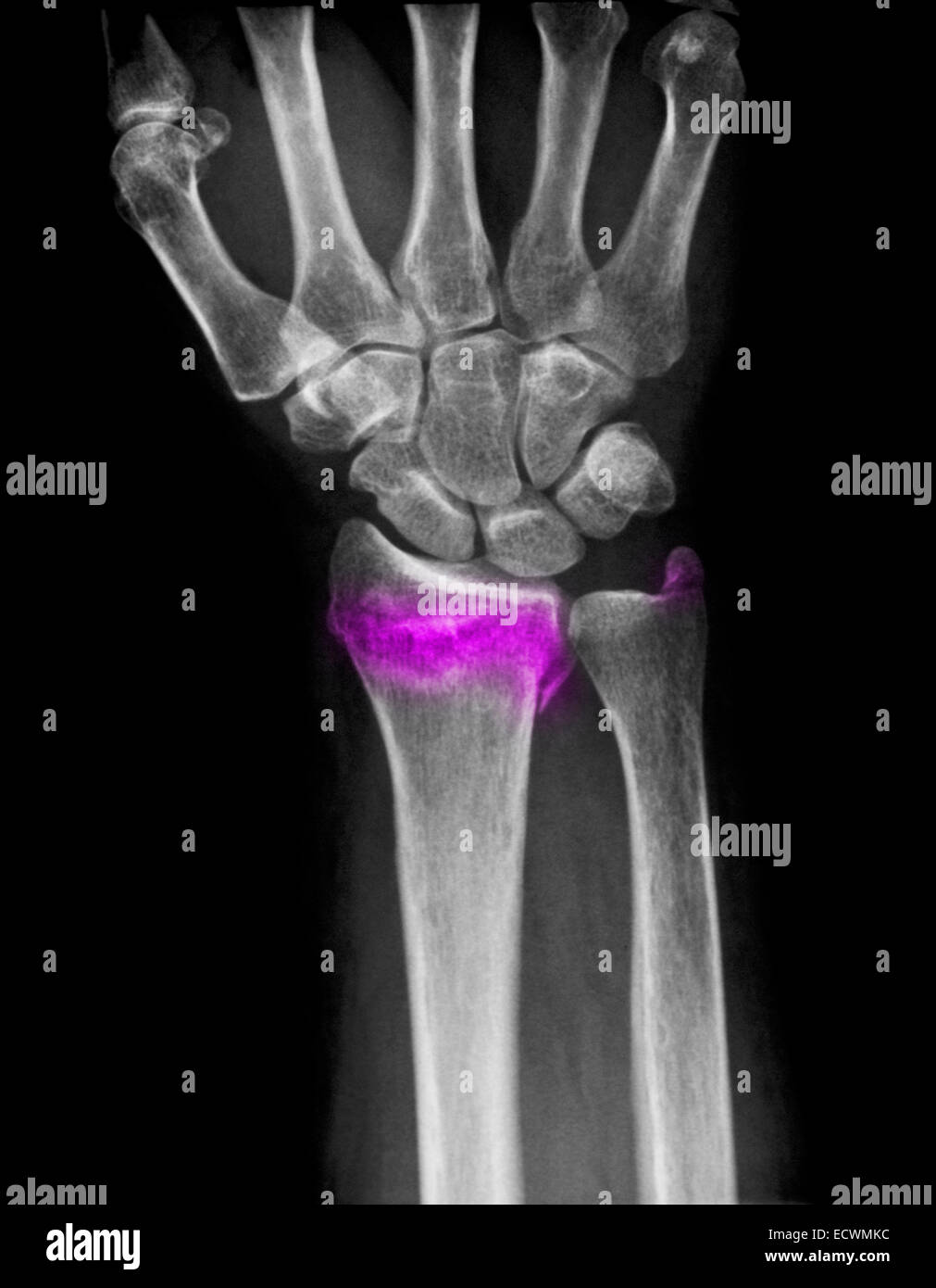 Wrist x-ray showing a fractured distal radius and ulna. Stock Photohttps://www.alamy.com/image-license-details/?v=1https://www.alamy.com/stock-photo-wrist-x-ray-showing-a-fractured-distal-radius-and-ulna-76782368.html
RMECWMKC–Wrist x-ray showing a fractured distal radius and ulna.
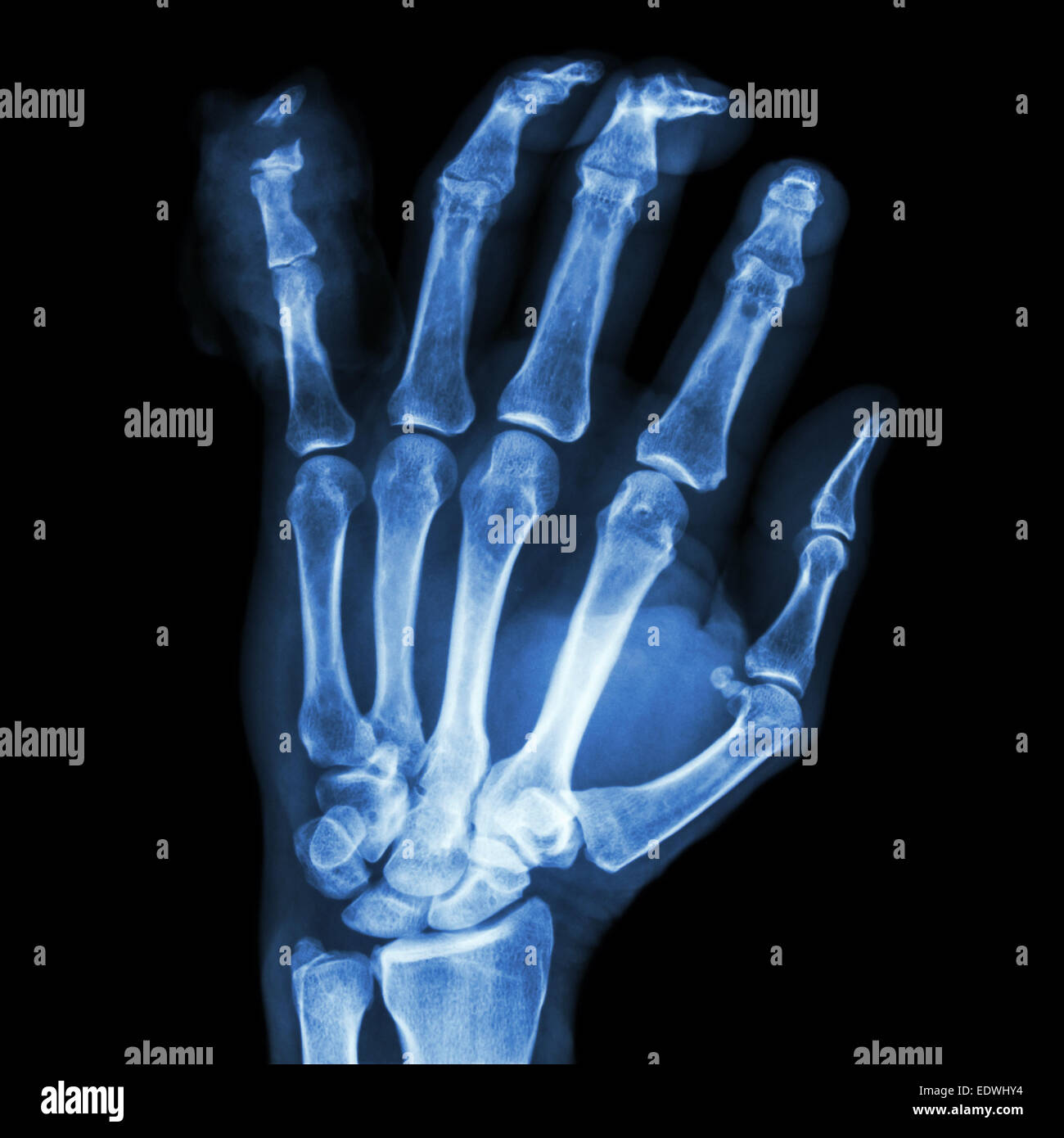 film x-ray hand AP : show fracture distal pharange little finger Stock Photohttps://www.alamy.com/image-license-details/?v=1https://www.alamy.com/stock-photo-film-x-ray-hand-ap-show-fracture-distal-pharange-little-finger-77394888.html
RFEDWHY4–film x-ray hand AP : show fracture distal pharange little finger
 Highway stretches into distal snowcapped mountains Stock Photohttps://www.alamy.com/image-license-details/?v=1https://www.alamy.com/stock-photo-highway-stretches-into-distal-snowcapped-mountains-87218840.html
RMF1W4EG–Highway stretches into distal snowcapped mountains
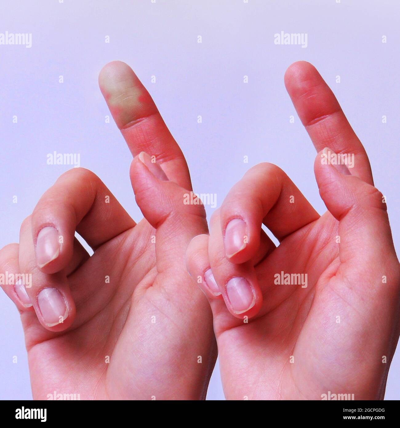 Capillary Refill Stock Photohttps://www.alamy.com/image-license-details/?v=1https://www.alamy.com/capillary-refill-image438130940.html
RF2GCPGDG–Capillary Refill
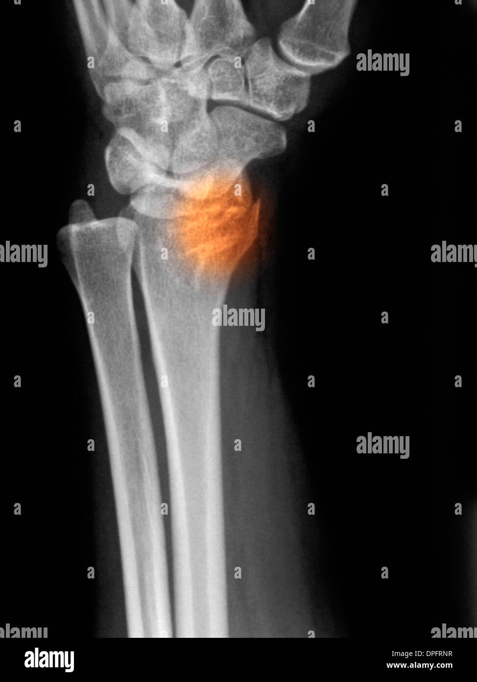 x-ray showing a distal radius fracture Stock Photohttps://www.alamy.com/image-license-details/?v=1https://www.alamy.com/x-ray-showing-a-distal-radius-fracture-image65501459.html
RFDPFRNR–x-ray showing a distal radius fracture
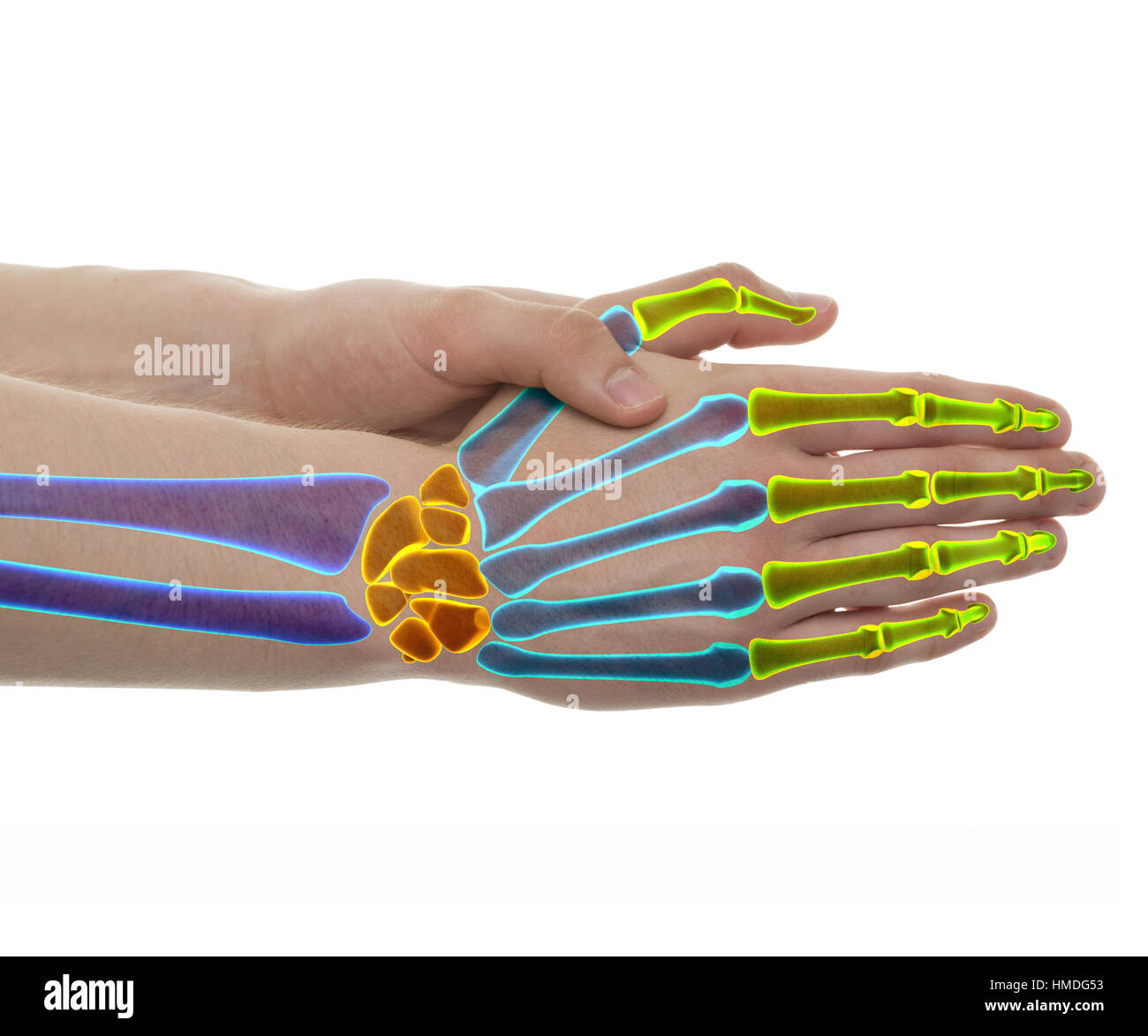 Hand Bones Color Separated - Studio shot with 3D illustration isolated on white Stock Photohttps://www.alamy.com/image-license-details/?v=1https://www.alamy.com/stock-photo-hand-bones-color-separated-studio-shot-with-3d-illustration-isolated-133063759.html
RFHMDG53–Hand Bones Color Separated - Studio shot with 3D illustration isolated on white
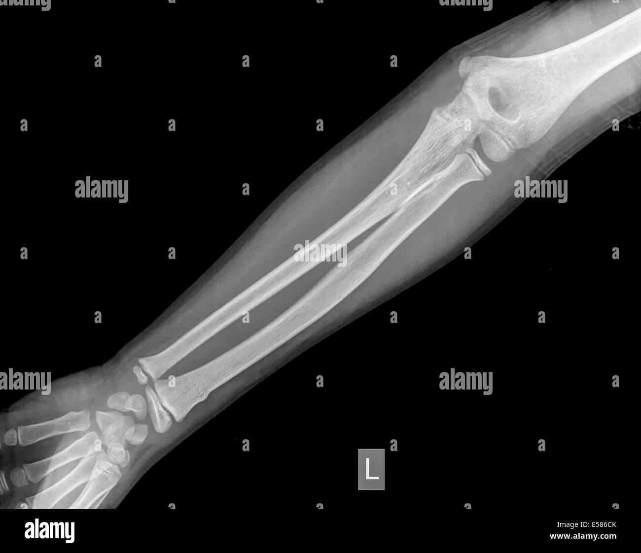 forearm of a 9 year old male patient with a Distal Radius Fracture Stock Photohttps://www.alamy.com/image-license-details/?v=1https://www.alamy.com/stock-photo-forearm-of-a-9-year-old-male-patient-with-a-distal-radius-fracture-72095427.html
RME586CK–forearm of a 9 year old male patient with a Distal Radius Fracture
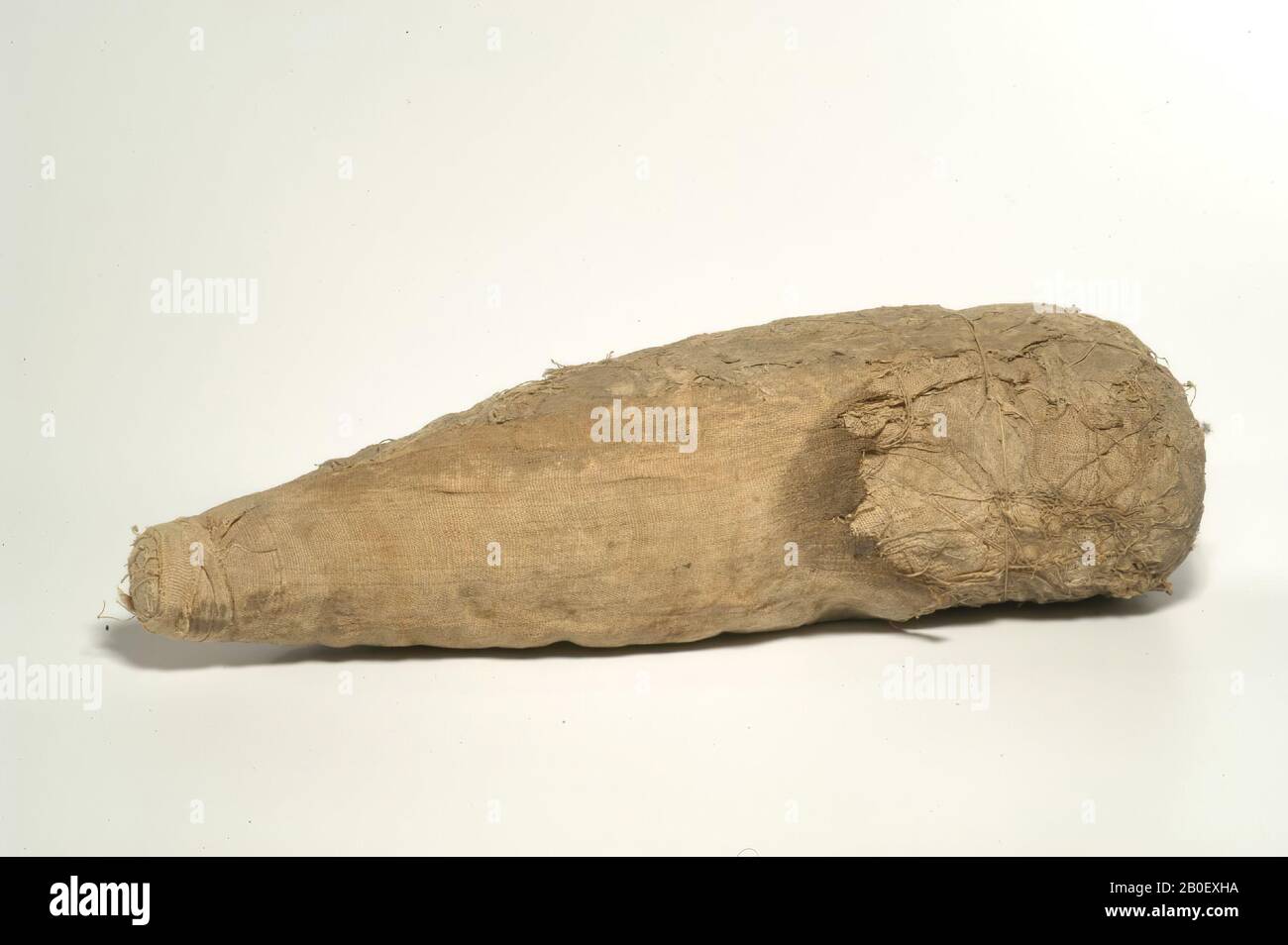 ibis, The mummy has the shape of a triangular body with a straight shoulder area, tapering to the caudal end. The distal part of the body is wrapped in a decayed sheet of two self-bands (warp-faced tabby weave, about 10 x 22 threads Stock Photohttps://www.alamy.com/image-license-details/?v=1https://www.alamy.com/ibis-the-mummy-has-the-shape-of-a-triangular-body-with-a-straight-shoulder-area-tapering-to-the-caudal-end-the-distal-part-of-the-body-is-wrapped-in-a-decayed-sheet-of-two-self-bands-warp-faced-tabby-weave-about-10-x-22-threads-image344535558.html
RM2B0EXHA–ibis, The mummy has the shape of a triangular body with a straight shoulder area, tapering to the caudal end. The distal part of the body is wrapped in a decayed sheet of two self-bands (warp-faced tabby weave, about 10 x 22 threads
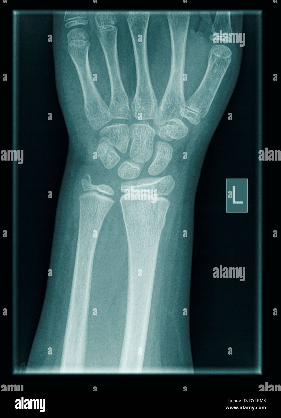 X-ray of wrist of 9 year old male patient with distal radius and ulna fractures Stock Photohttps://www.alamy.com/image-license-details/?v=1https://www.alamy.com/x-ray-of-wrist-of-9-year-old-male-patient-with-distal-radius-and-ulna-image68333219.html
RFDY4RM3–X-ray of wrist of 9 year old male patient with distal radius and ulna fractures
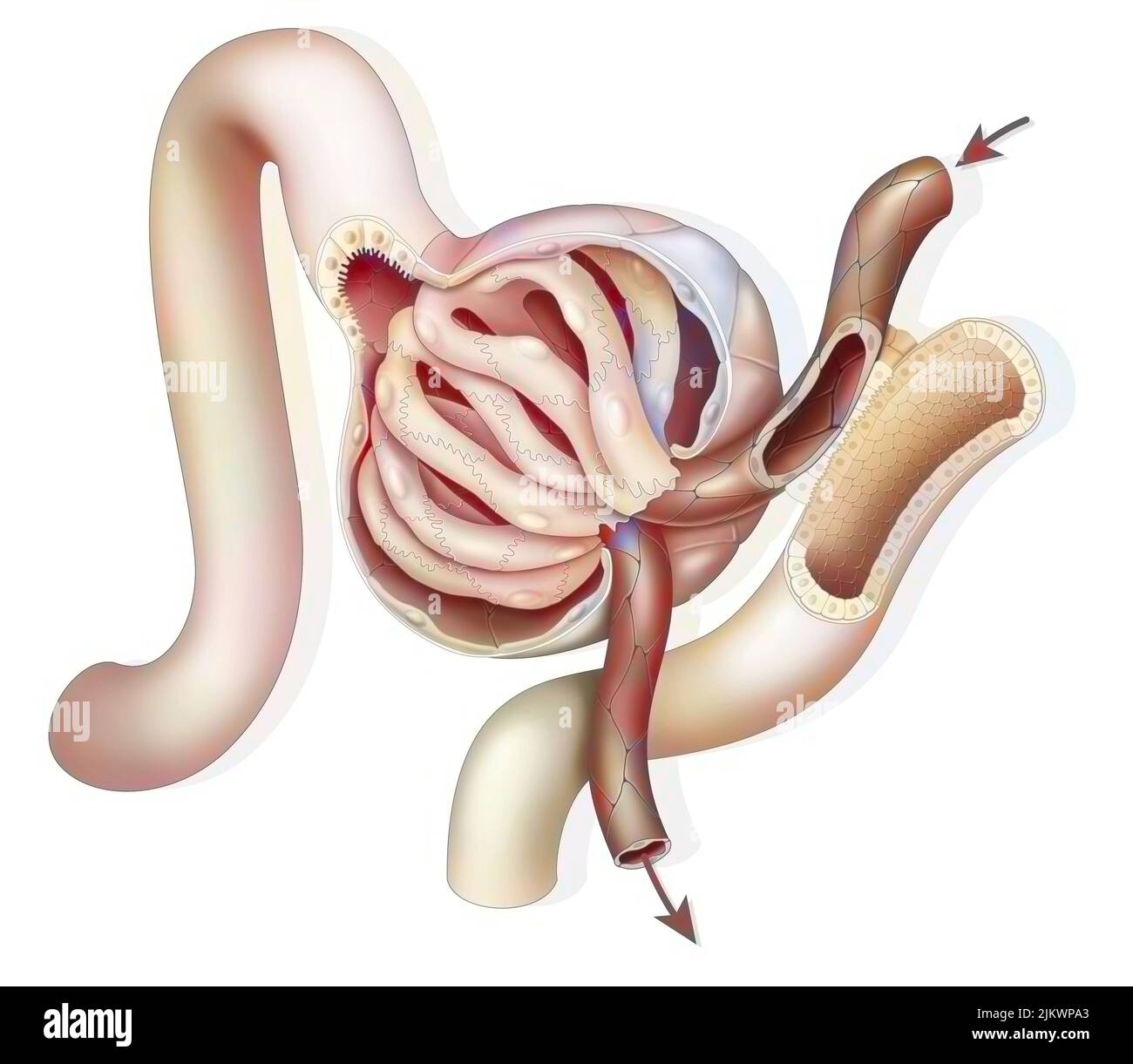 Anatomy of a renal glomerulus with afferent and efferent glomerular arteriole. Stock Photohttps://www.alamy.com/image-license-details/?v=1https://www.alamy.com/anatomy-of-a-renal-glomerulus-with-afferent-and-efferent-glomerular-arteriole-image476924731.html
RF2JKWPA3–Anatomy of a renal glomerulus with afferent and efferent glomerular arteriole.
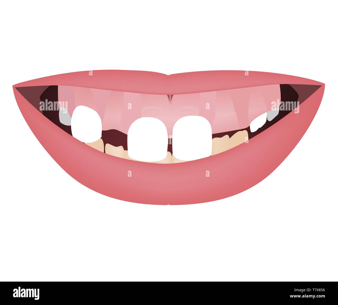 Kids jaws with gummy smile, crooked lower teeth and distal bite before the orthotropics or orthotropics treatment. Vector illustration Stock Vectorhttps://www.alamy.com/image-license-details/?v=1https://www.alamy.com/kids-jaws-with-gummy-smile-crooked-lower-teeth-and-distal-bite-before-the-orthotropics-or-orthotropics-treatment-vector-illustration-image245824914.html
RFT7X856–Kids jaws with gummy smile, crooked lower teeth and distal bite before the orthotropics or orthotropics treatment. Vector illustration
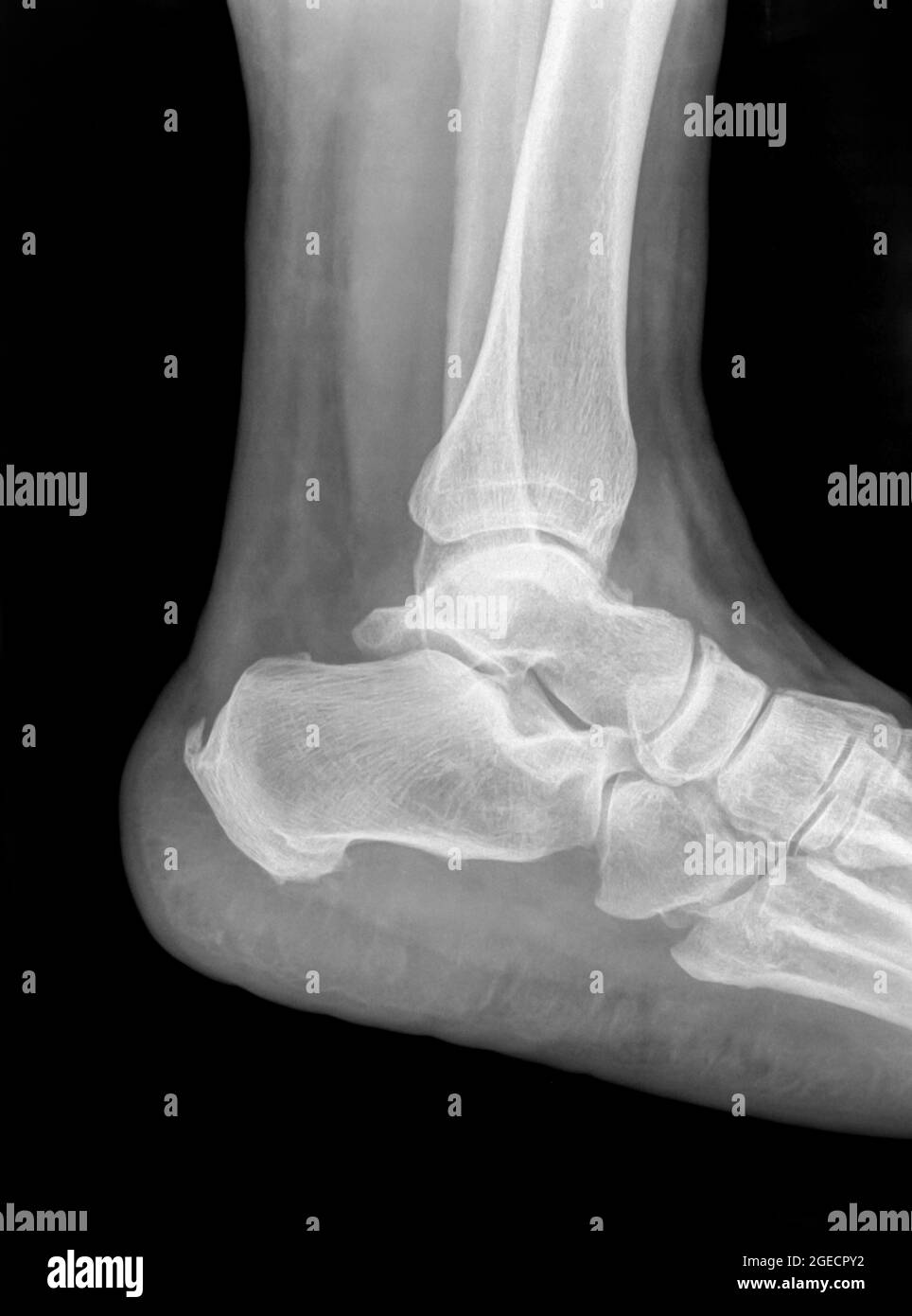 X-ray of an ankle 50 year old male with a fractured distal tibia. Side View Stock Photohttps://www.alamy.com/image-license-details/?v=1https://www.alamy.com/x-ray-of-an-ankle-50-year-old-male-with-a-fractured-distal-tibia-side-view-image439145814.html
RM2GECPY2–X-ray of an ankle 50 year old male with a fractured distal tibia. Side View
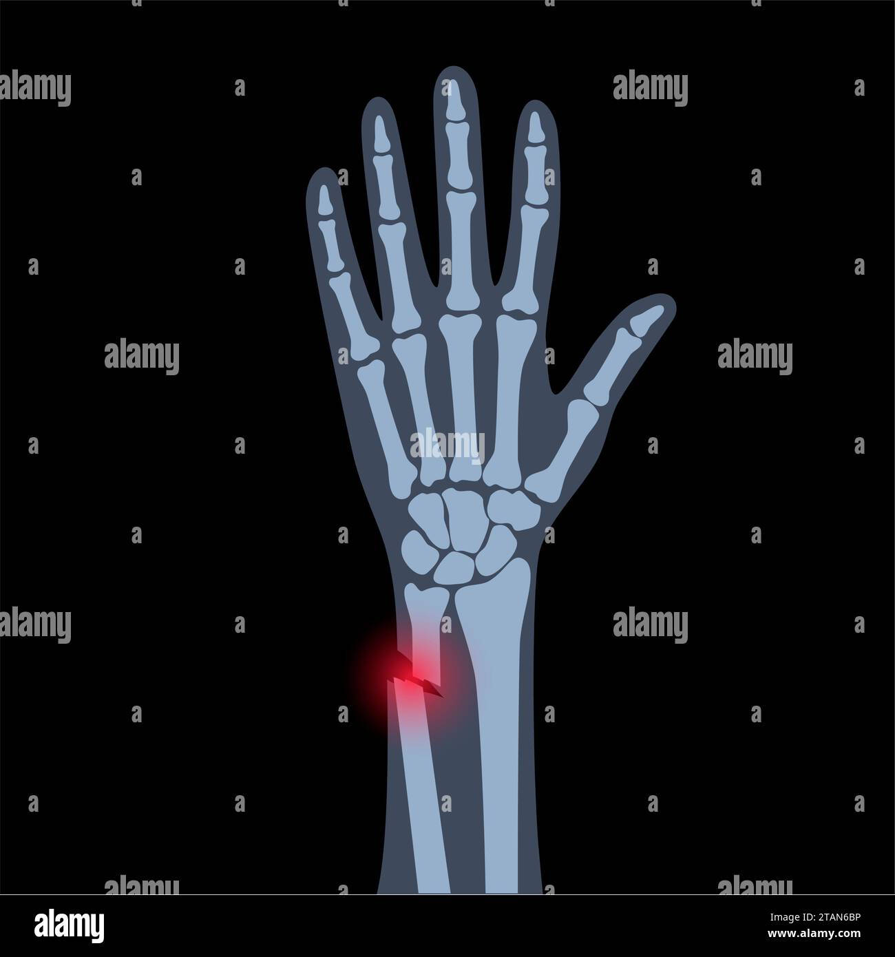 Fractured wrist, illustration Stock Photohttps://www.alamy.com/image-license-details/?v=1https://www.alamy.com/fractured-wrist-illustration-image574554730.html
RF2TAN6BP–Fractured wrist, illustration
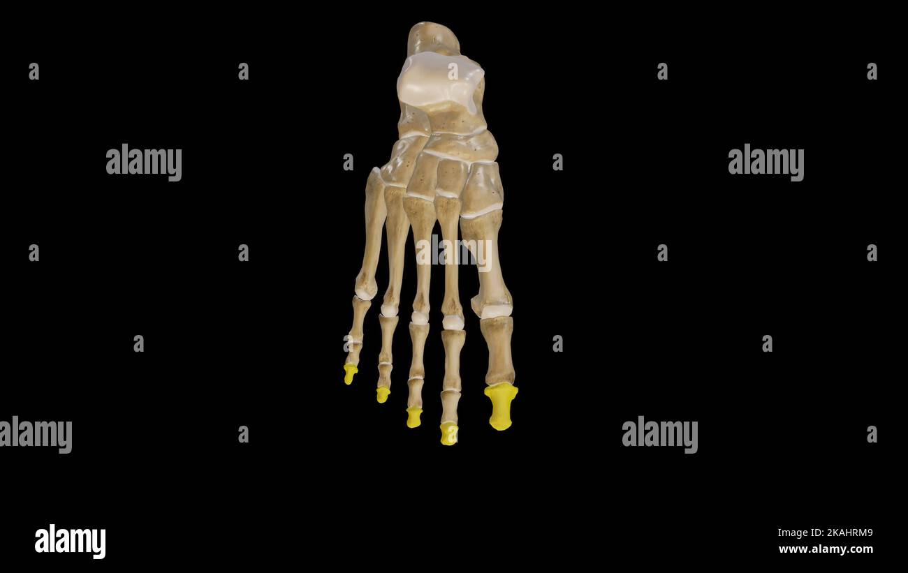 Distal Phalanges of Foot Stock Photohttps://www.alamy.com/image-license-details/?v=1https://www.alamy.com/distal-phalanges-of-foot-image488428649.html
RF2KAHRM9–Distal Phalanges of Foot
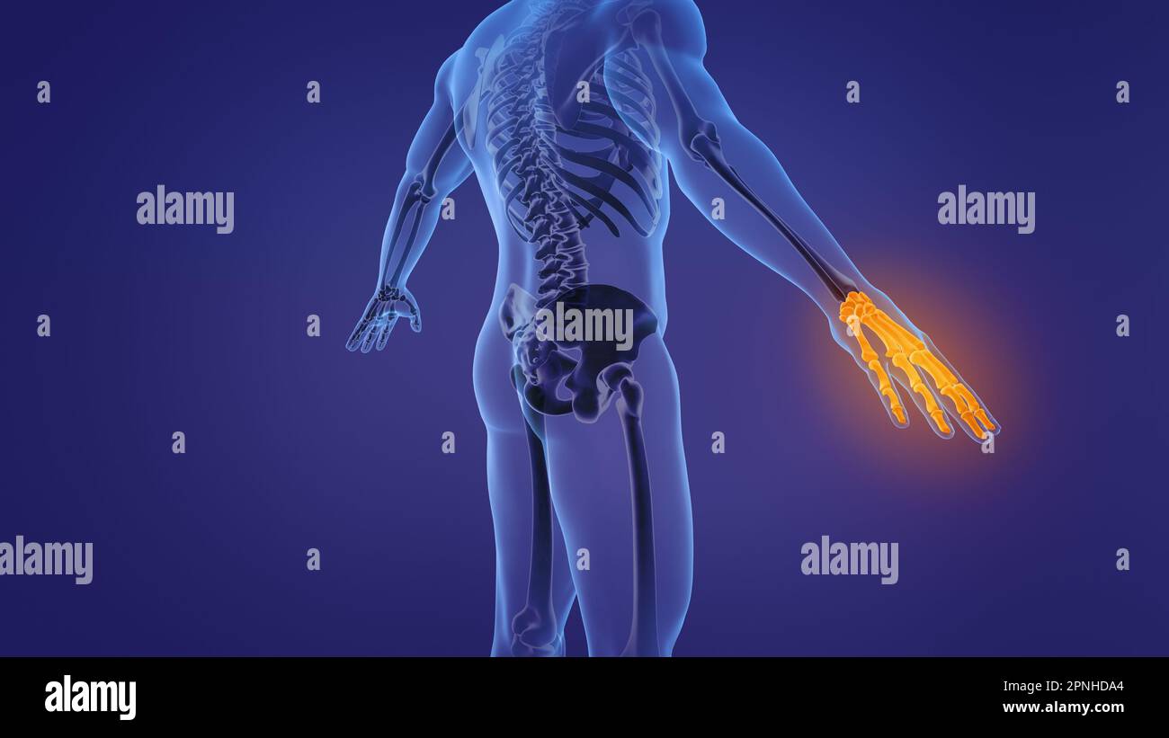 Anatomy of the human hand Stock Photohttps://www.alamy.com/image-license-details/?v=1https://www.alamy.com/anatomy-of-the-human-hand-image546812844.html
RF2PNHDA4–Anatomy of the human hand
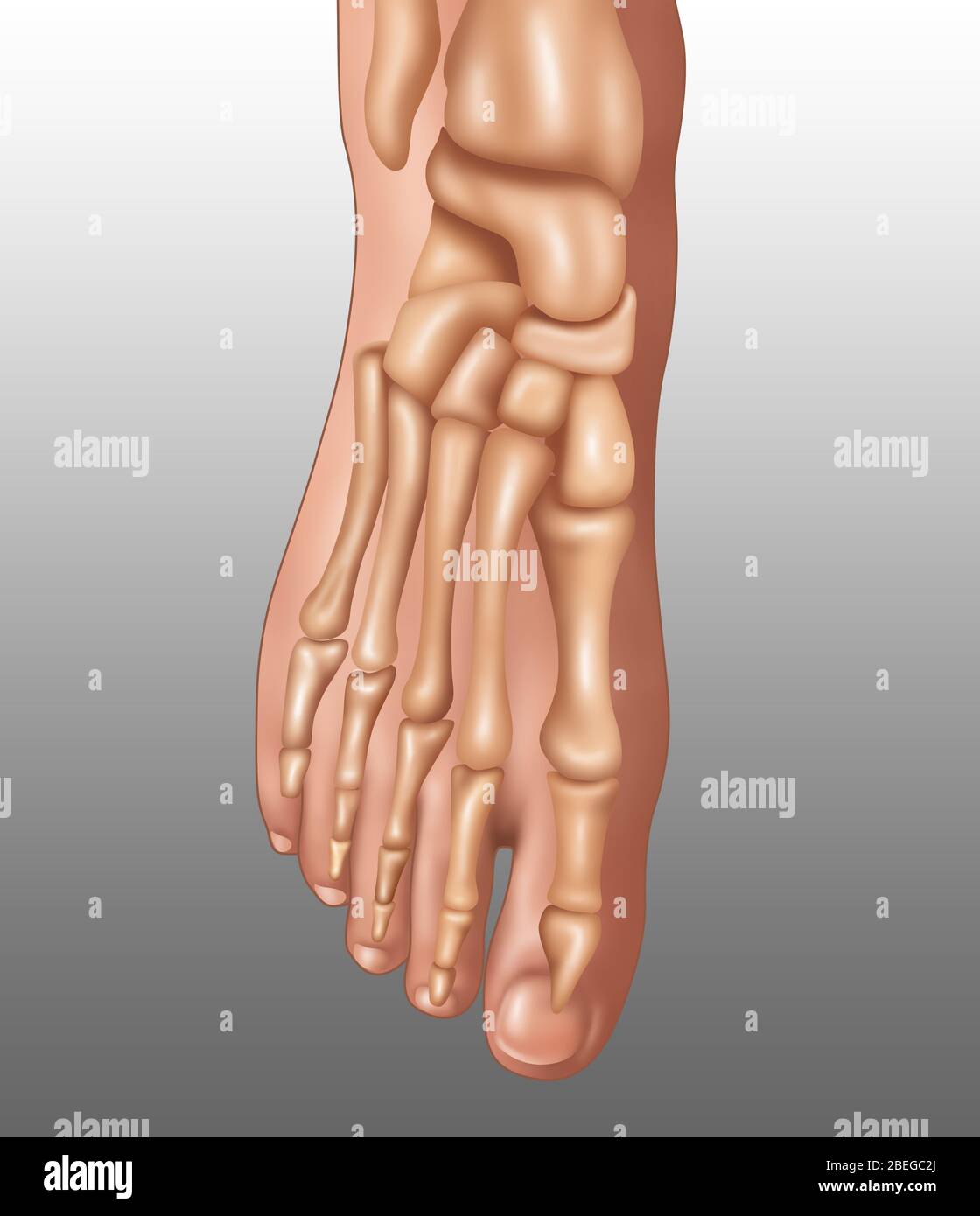 Foot Bones, Illustration Stock Photohttps://www.alamy.com/image-license-details/?v=1https://www.alamy.com/foot-bones-illustration-image353173258.html
RF2BEGC2J–Foot Bones, Illustration
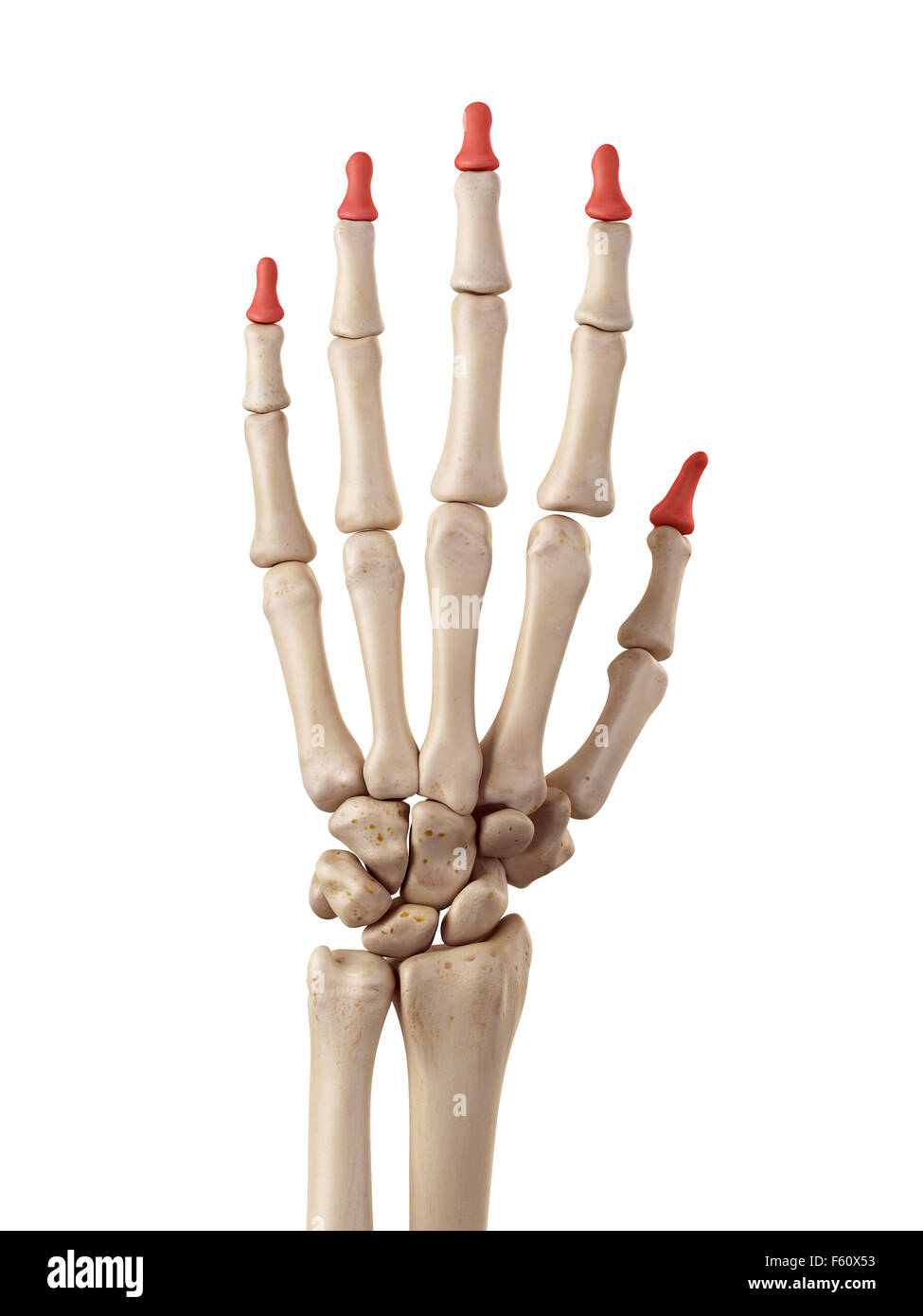 medical accurate illustration of the distal phalanx bones Stock Photohttps://www.alamy.com/image-license-details/?v=1https://www.alamy.com/stock-photo-medical-accurate-illustration-of-the-distal-phalanx-bones-89760303.html
RFF60X53–medical accurate illustration of the distal phalanx bones
 Skeleton hand with pointing finger. Vector illustration. Stock Vectorhttps://www.alamy.com/image-license-details/?v=1https://www.alamy.com/skeleton-hand-with-pointing-finger-vector-illustration-image525021901.html
RF2NE4PP5–Skeleton hand with pointing finger. Vector illustration.
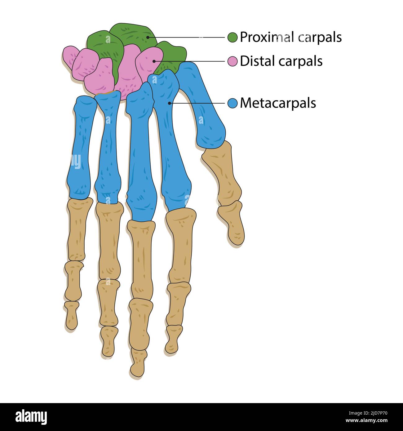 human hand bone. vector illustration. on white background Stock Vectorhttps://www.alamy.com/image-license-details/?v=1https://www.alamy.com/human-hand-bone-vector-illustration-on-white-background-image472841572.html
RF2JD7P70–human hand bone. vector illustration. on white background
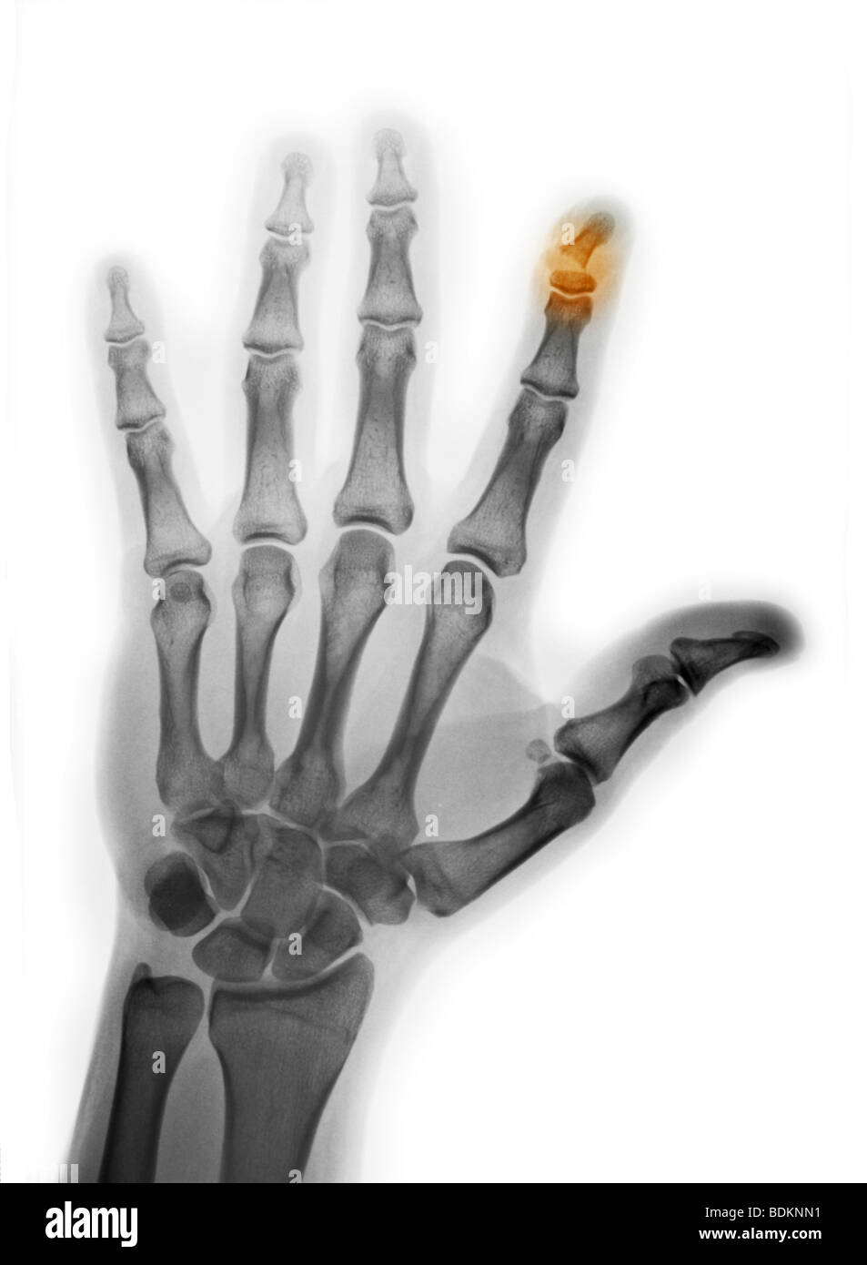 hand x-ray showing a fracture of the distal phalanx of the index finger Stock Photohttps://www.alamy.com/image-license-details/?v=1https://www.alamy.com/stock-photo-hand-x-ray-showing-a-fracture-of-the-distal-phalanx-of-the-index-finger-25635037.html
RMBDKNN1–hand x-ray showing a fracture of the distal phalanx of the index finger
 Senior woman with a broken fractured wrist with multiple breaks wearing an external metal frame drilled into the hand and forearm bones. Such contraptions keep the wrist steady so the bones can heal. Stock Photohttps://www.alamy.com/image-license-details/?v=1https://www.alamy.com/stock-photo-senior-woman-with-a-broken-fractured-wrist-with-multiple-breaks-wearing-18046976.html
RMB1A32T–Senior woman with a broken fractured wrist with multiple breaks wearing an external metal frame drilled into the hand and forearm bones. Such contraptions keep the wrist steady so the bones can heal.
 3D image of Trichlormethiazide skeletal formula - molecular chemical structure of diuretic medicament isolated on white background Stock Photohttps://www.alamy.com/image-license-details/?v=1https://www.alamy.com/3d-image-of-trichlormethiazide-skeletal-formula-molecular-chemical-structure-of-diuretic-medicament-isolated-on-white-background-image487590970.html
RF2K97K76–3D image of Trichlormethiazide skeletal formula - molecular chemical structure of diuretic medicament isolated on white background
 Bryophyllum delagoense, a succulent plant native to Madagascar. Noted for vegetatively growing small plantlets on the distal ends of its phylloclades, Stock Photohttps://www.alamy.com/image-license-details/?v=1https://www.alamy.com/bryophyllum-delagoense-a-succulent-plant-native-to-madagascar-noted-for-vegetatively-growing-small-plantlets-on-the-distal-ends-of-its-phylloclades-image441399338.html
RF2GJ3DA2–Bryophyllum delagoense, a succulent plant native to Madagascar. Noted for vegetatively growing small plantlets on the distal ends of its phylloclades,
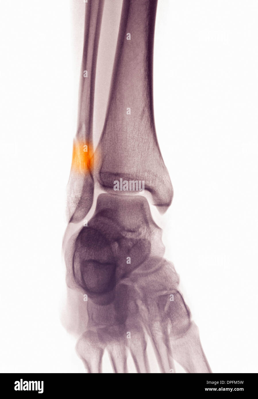 ankle x-ray showing fractured distal fibula Stock Photohttps://www.alamy.com/image-license-details/?v=1https://www.alamy.com/ankle-x-ray-showing-fractured-distal-fibula-image65498661.html
RFDPFM5W–ankle x-ray showing fractured distal fibula
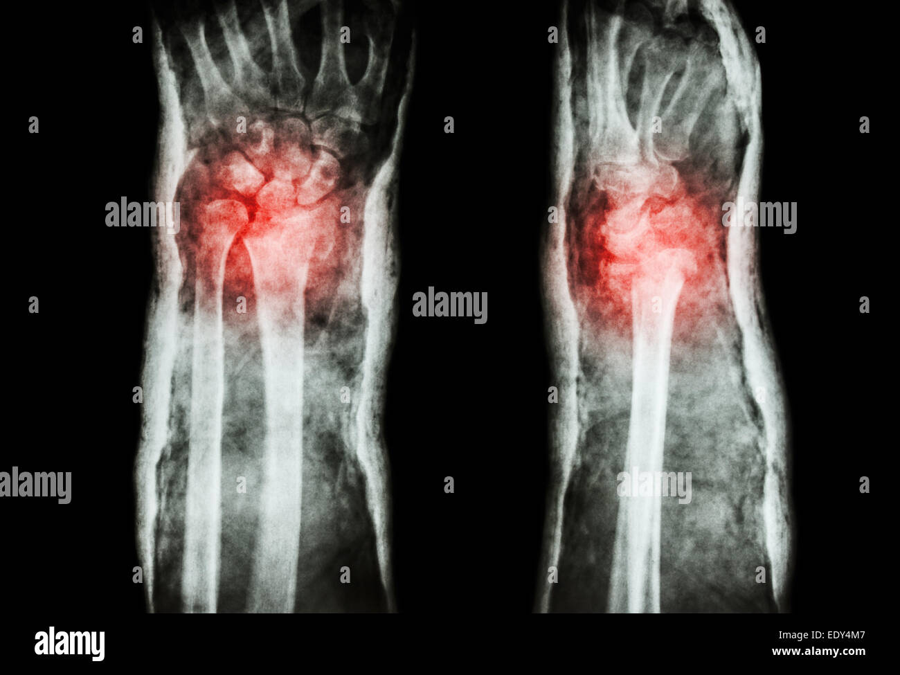 fracture distal radius (Colles' fracture) and cast (Front & Side) Stock Photohttps://www.alamy.com/image-license-details/?v=1https://www.alamy.com/stock-photo-fracture-distal-radius-colles-fracture-and-cast-front-side-77428407.html
RFEDY4M7–fracture distal radius (Colles' fracture) and cast (Front & Side)
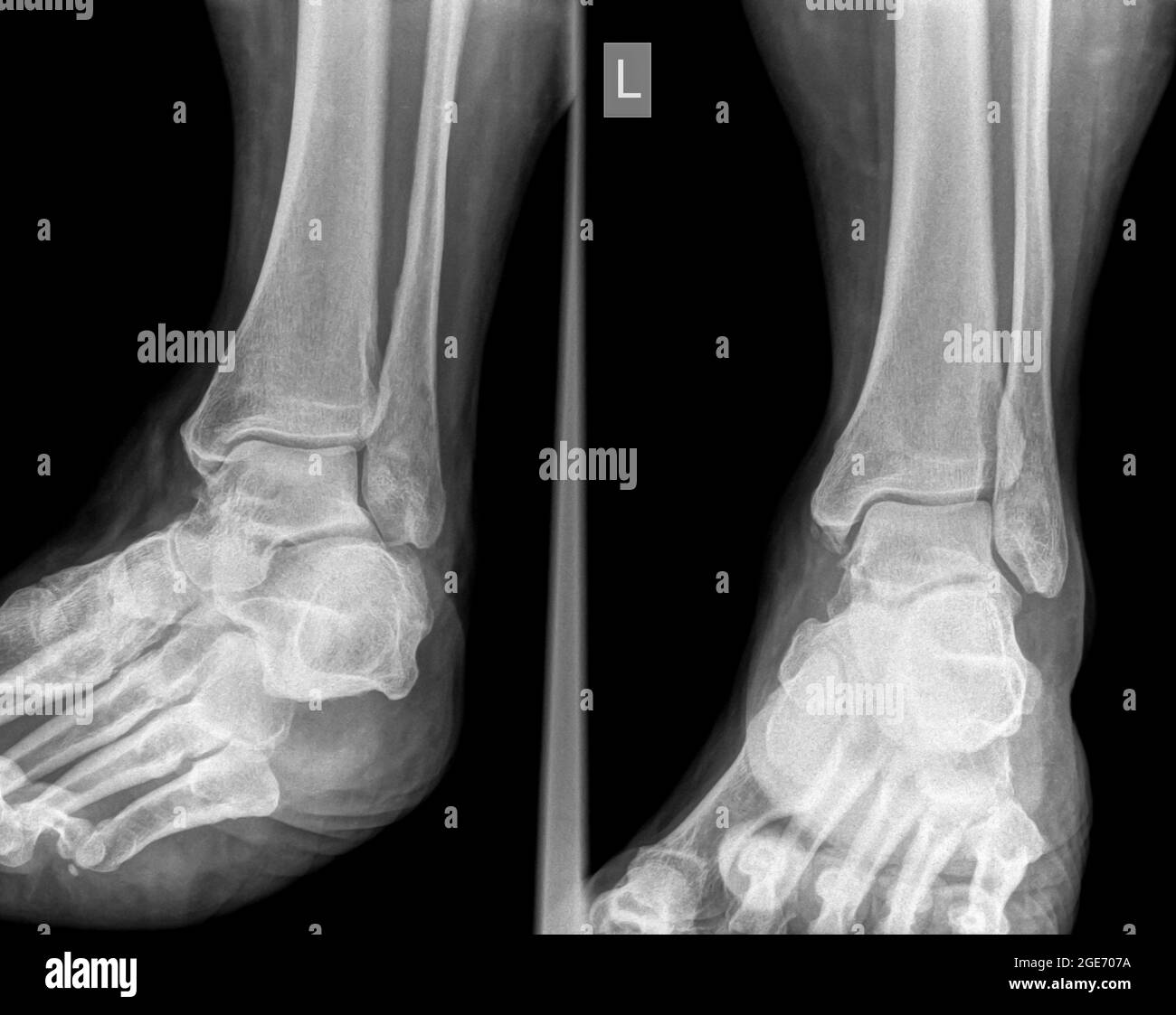 X-ray of an ankle 50 year old male with a fractured distal tibia. front View Stock Photohttps://www.alamy.com/image-license-details/?v=1https://www.alamy.com/x-ray-of-an-ankle-50-year-old-male-with-a-fractured-distal-tibia-front-view-image439018254.html
RM2GE707A–X-ray of an ankle 50 year old male with a fractured distal tibia. front View
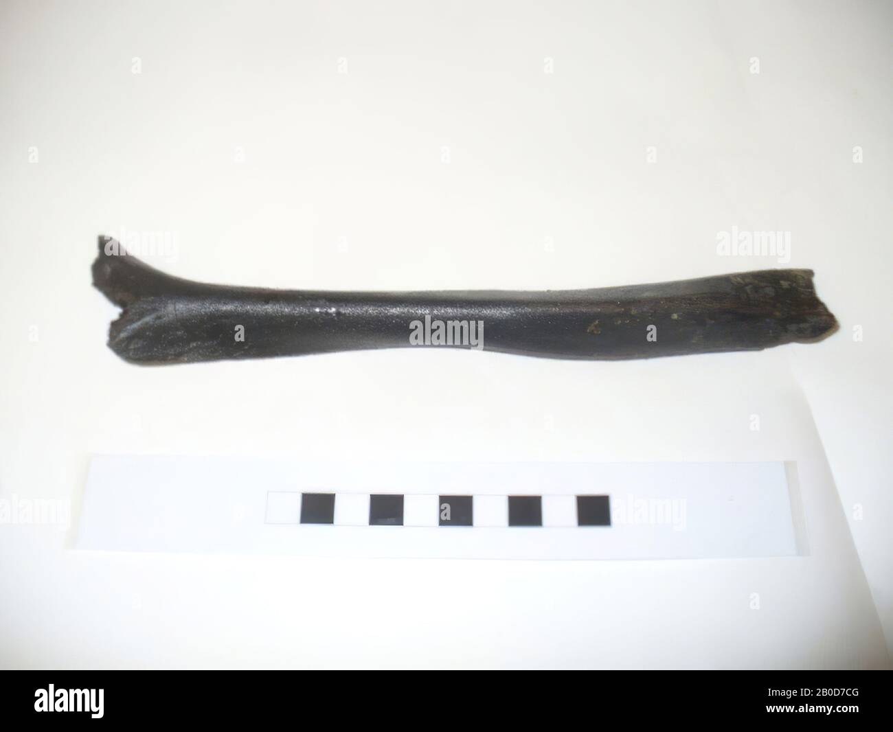 A human humerus (humerus) from the North Sea. Both the distal and proximal side are damaged. There is also a part of the back of the bone, where sampling took place for C14 research, humerus, organic, bone, l. 22.6 cm, the Netherlands, South Holland, Maasvlakte, North Sea Stock Photohttps://www.alamy.com/image-license-details/?v=1https://www.alamy.com/a-human-humerus-humerus-from-the-north-sea-both-the-distal-and-proximal-side-are-damaged-there-is-also-a-part-of-the-back-of-the-bone-where-sampling-took-place-for-c14-research-humerus-organic-bone-l-226-cm-the-netherlands-south-holland-maasvlakte-north-sea-image344498576.html
RM2B0D7CG–A human humerus (humerus) from the North Sea. Both the distal and proximal side are damaged. There is also a part of the back of the bone, where sampling took place for C14 research, humerus, organic, bone, l. 22.6 cm, the Netherlands, South Holland, Maasvlakte, North Sea
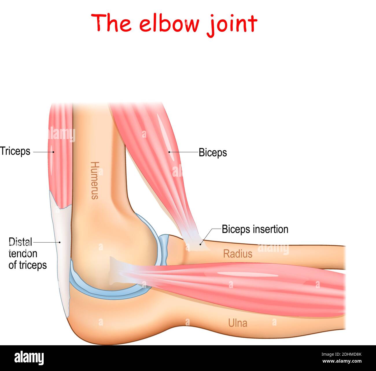 Anatomy of a elbow joint. parts of the arm. bones (humerus, radius, ulna) Muscle (Triceps, Biceps) and Distal tendon of triceps. Stock Vectorhttps://www.alamy.com/image-license-details/?v=1https://www.alamy.com/anatomy-of-a-elbow-joint-parts-of-the-arm-bones-humerus-radius-ulna-muscle-triceps-biceps-and-distal-tendon-of-triceps-image389526723.html
RF2DHMD8K–Anatomy of a elbow joint. parts of the arm. bones (humerus, radius, ulna) Muscle (Triceps, Biceps) and Distal tendon of triceps.
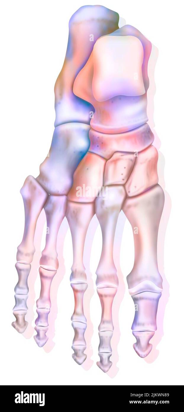 Superior view of the foot and the different bones: calcaneus, talus. Stock Photohttps://www.alamy.com/image-license-details/?v=1https://www.alamy.com/superior-view-of-the-foot-and-the-different-bones-calcaneus-talus-image476923897.html
RF2JKWN89–Superior view of the foot and the different bones: calcaneus, talus.
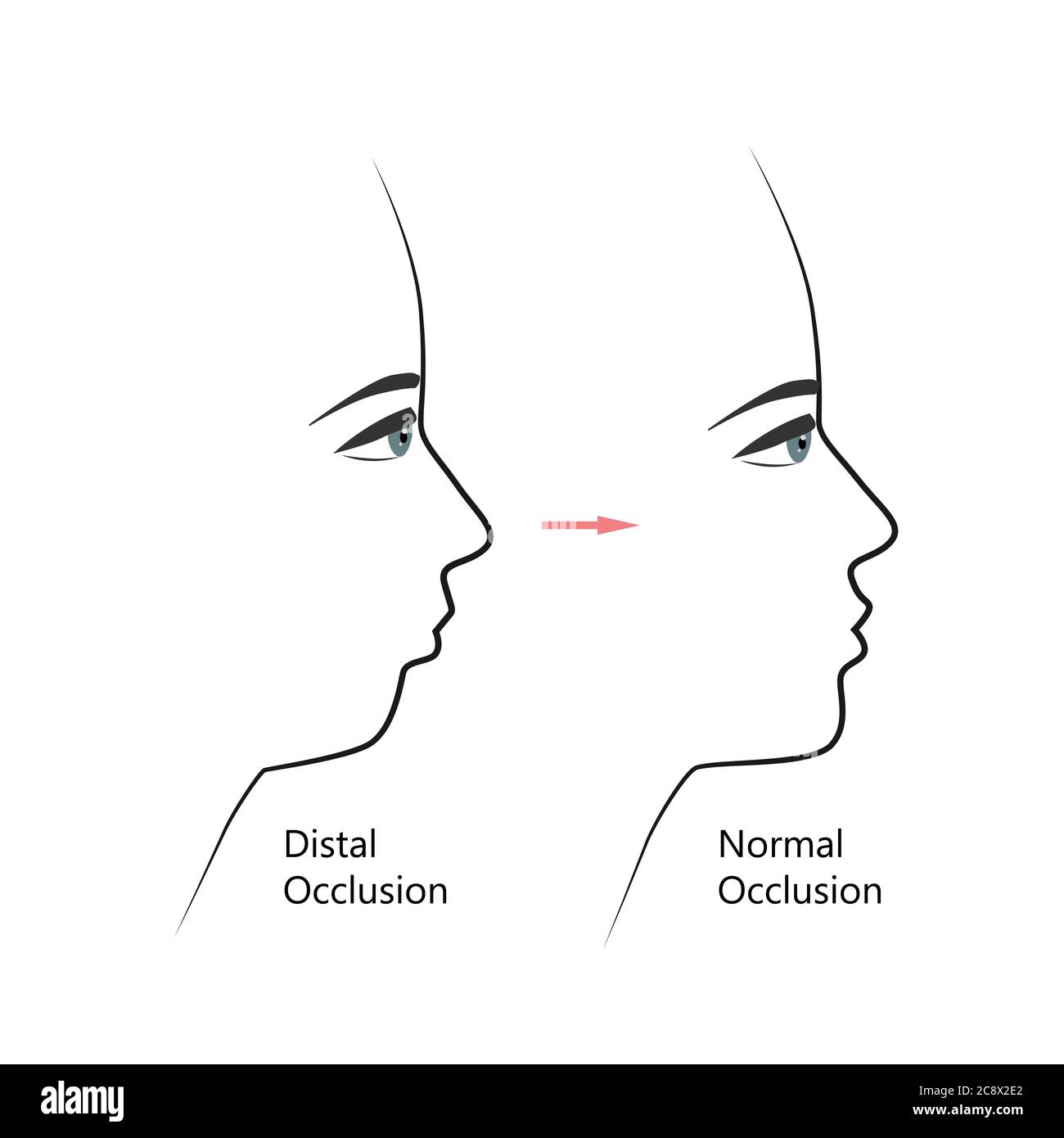 Distal bite profile before and after orthodontic treatment. Human with malocclusion, lower jaw pushing back, bite correction by braces. Vector Stock Vectorhttps://www.alamy.com/image-license-details/?v=1https://www.alamy.com/distal-bite-profile-before-and-after-orthodontic-treatment-human-with-malocclusion-lower-jaw-pushing-back-bite-correction-by-braces-vector-image366907690.html
RF2C8X2E2–Distal bite profile before and after orthodontic treatment. Human with malocclusion, lower jaw pushing back, bite correction by braces. Vector
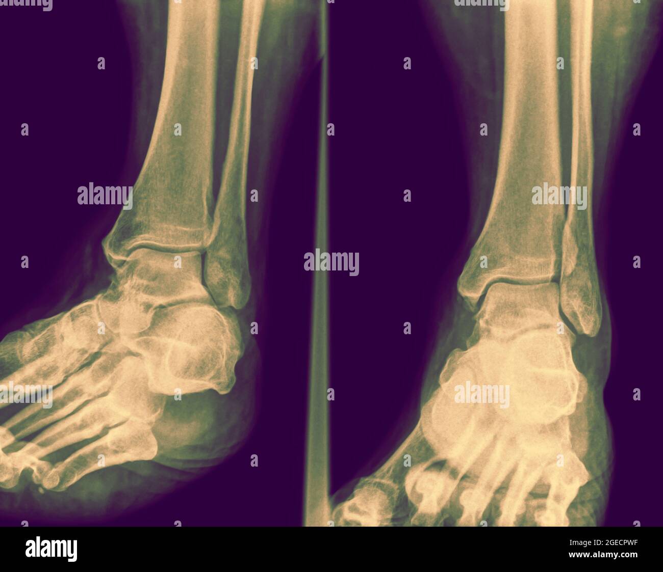 X-ray of an ankle 50 year old male with a fractured distal tibia. front View Stock Photohttps://www.alamy.com/image-license-details/?v=1https://www.alamy.com/x-ray-of-an-ankle-50-year-old-male-with-a-fractured-distal-tibia-front-view-image439145771.html
RM2GECPWF–X-ray of an ankle 50 year old male with a fractured distal tibia. front View
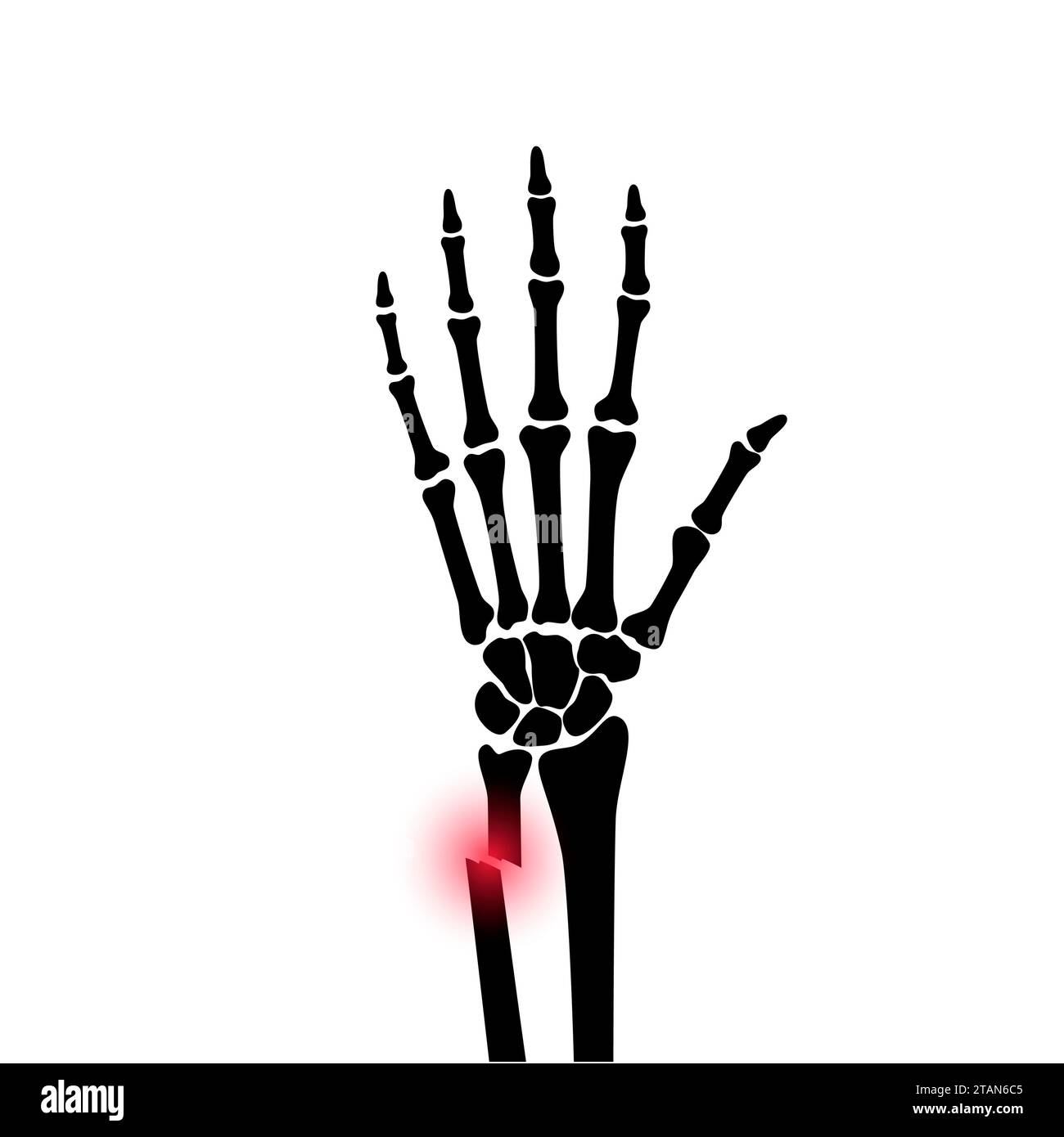 Fractured wrist, illustration Stock Photohttps://www.alamy.com/image-license-details/?v=1https://www.alamy.com/fractured-wrist-illustration-image574554741.html
RF2TAN6C5–Fractured wrist, illustration
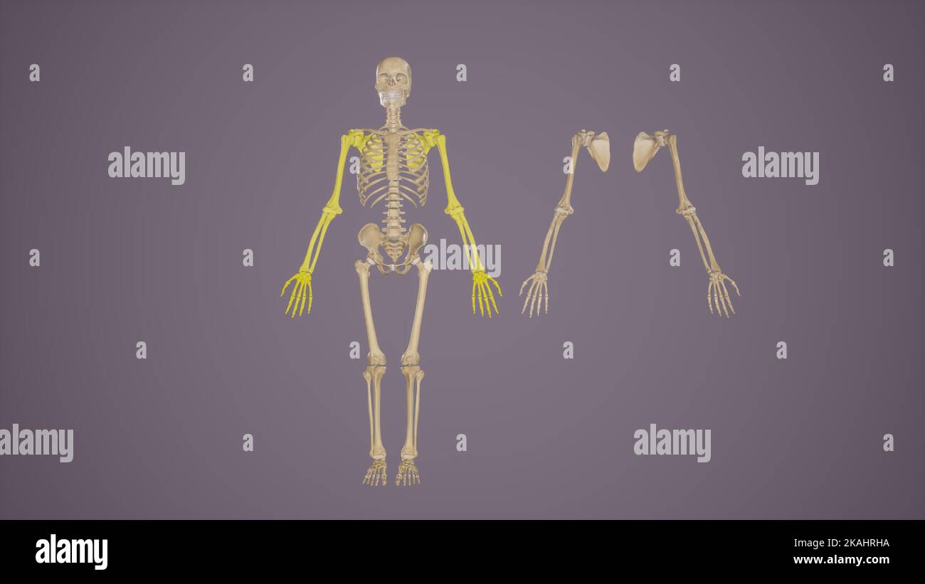 Bones of Upper Limbs- Anterior View Stock Photohttps://www.alamy.com/image-license-details/?v=1https://www.alamy.com/bones-of-upper-limbs-anterior-view-image488428566.html
RF2KAHRHA–Bones of Upper Limbs- Anterior View
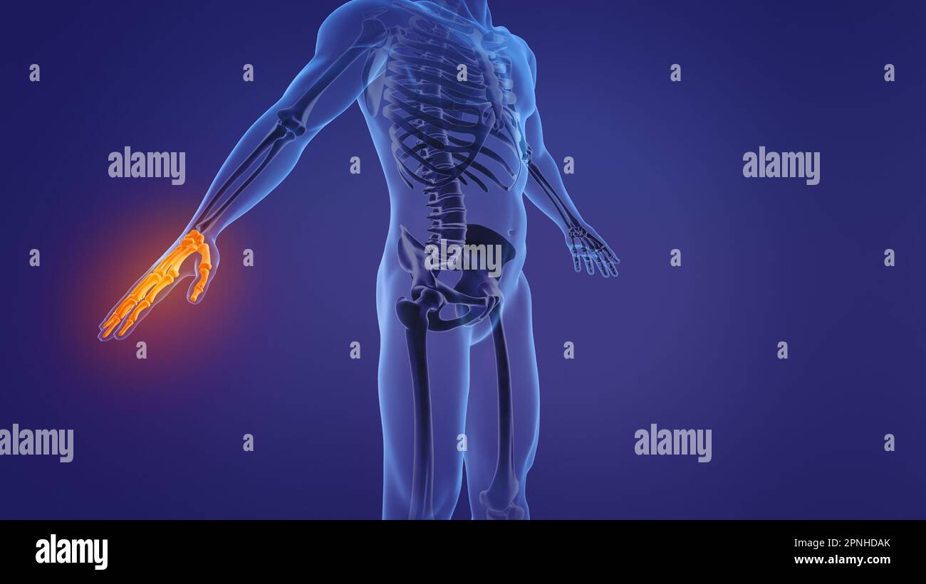 Anatomy of the human hand Stock Photohttps://www.alamy.com/image-license-details/?v=1https://www.alamy.com/anatomy-of-the-human-hand-image546812859.html
RF2PNHDAK–Anatomy of the human hand
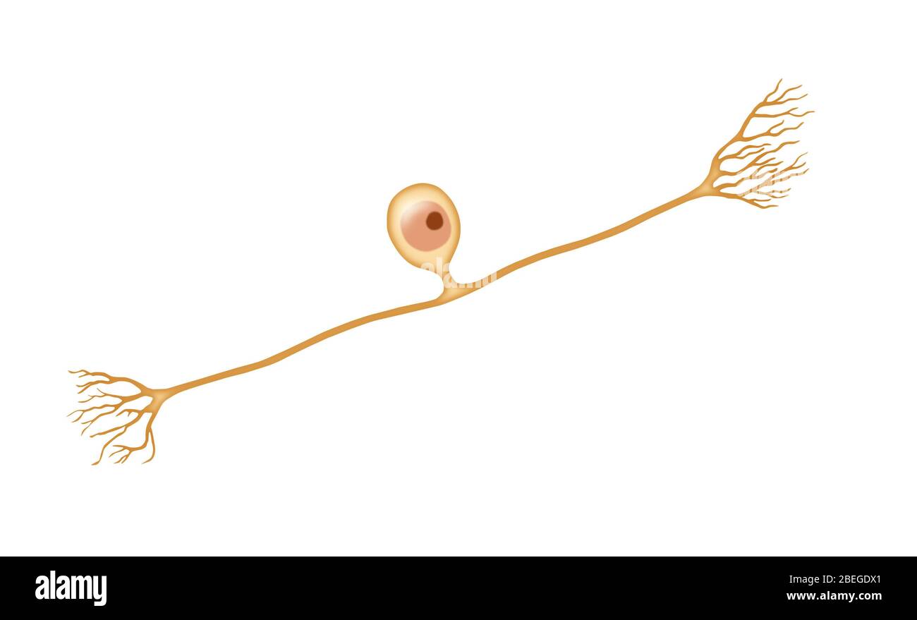 Pseudounipolar Neuron Stock Photohttps://www.alamy.com/image-license-details/?v=1https://www.alamy.com/pseudounipolar-neuron-image353174697.html
RM2BEGDX1–Pseudounipolar Neuron
 medical accurate illustration of the distal phalanx bones Stock Photohttps://www.alamy.com/image-license-details/?v=1https://www.alamy.com/stock-photo-medical-accurate-illustration-of-the-distal-phalanx-bones-89759810.html
RFF60WFE–medical accurate illustration of the distal phalanx bones
 mixed forest Stock Photohttps://www.alamy.com/image-license-details/?v=1https://www.alamy.com/stock-image-mixed-forest-164041174.html
RFKETM5A–mixed forest
 Nelaton device for fractures of the distal radius, vintage engraved illustration.Usual Medicine Dictionary by Dr Labarthe - 1885 Stock Vectorhttps://www.alamy.com/image-license-details/?v=1https://www.alamy.com/stock-photo-nelaton-device-for-fractures-of-the-distal-radius-vintage-engraved-84420015.html
RFEW9JGF–Nelaton device for fractures of the distal radius, vintage engraved illustration.Usual Medicine Dictionary by Dr Labarthe - 1885
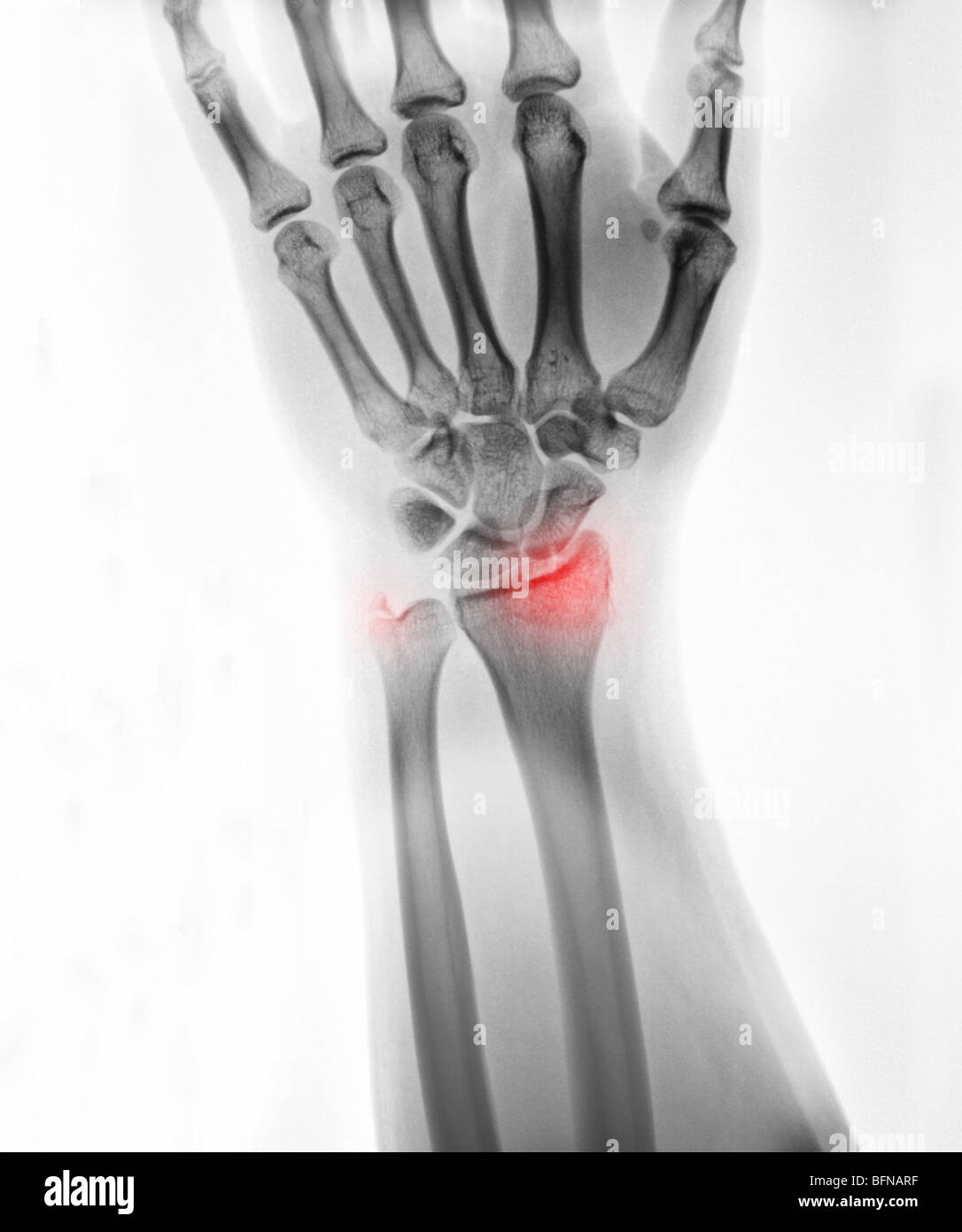 x-ray of the hand and wrist of an 18 year old male showing a fractured wrist at the distal radius and ulnar styloid Stock Photohttps://www.alamy.com/image-license-details/?v=1https://www.alamy.com/stock-photo-x-ray-of-the-hand-and-wrist-of-an-18-year-old-male-showing-a-fractured-26899699.html
RMBFNARF–x-ray of the hand and wrist of an 18 year old male showing a fractured wrist at the distal radius and ulnar styloid
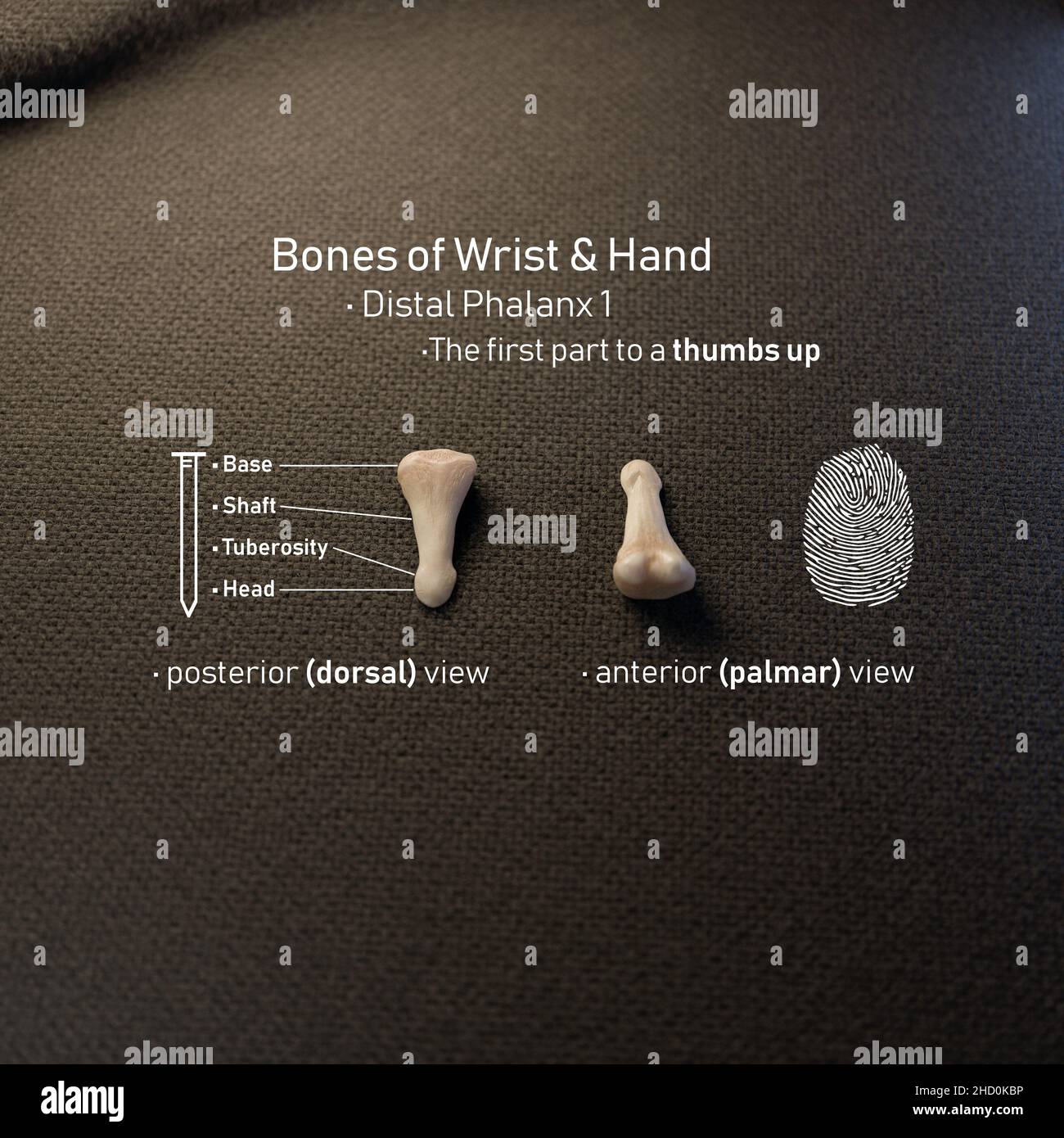 3D Rendering Distal Phalanx with Anatomical Terms Stock Photohttps://www.alamy.com/image-license-details/?v=1https://www.alamy.com/3d-rendering-distal-phalanx-with-anatomical-terms-image455475322.html
RF2HD0KBP–3D Rendering Distal Phalanx with Anatomical Terms
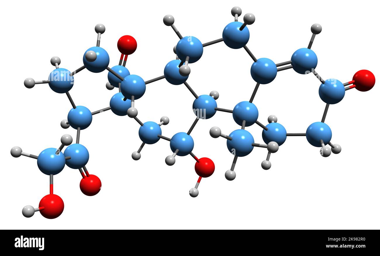 3D image of aldosterone skeletal formula - molecular chemical structure of mineralocorticoid steroid hormone ALD isolated on white background Stock Photohttps://www.alamy.com/image-license-details/?v=1https://www.alamy.com/3d-image-of-aldosterone-skeletal-formula-molecular-chemical-structure-of-mineralocorticoid-steroid-hormone-ald-isolated-on-white-background-image487600036.html
RF2K982R0–3D image of aldosterone skeletal formula - molecular chemical structure of mineralocorticoid steroid hormone ALD isolated on white background
 Butterfly antenna with scales, distal region. Light microscope X50 at 10 cm wide. Stock Photohttps://www.alamy.com/image-license-details/?v=1https://www.alamy.com/butterfly-antenna-with-scales-distal-region-light-microscope-x50-at-10-cm-wide-image575512866.html
RF2TC8TEX–Butterfly antenna with scales, distal region. Light microscope X50 at 10 cm wide.
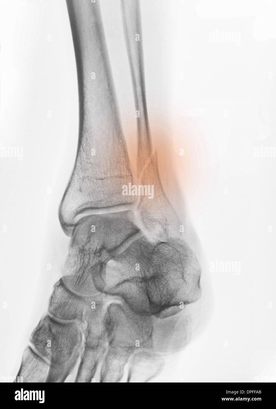 x-ray of a fracture of the distal fibula Stock Photohttps://www.alamy.com/image-license-details/?v=1https://www.alamy.com/x-ray-of-a-fracture-of-the-distal-fibula-image65494867.html
RFDPFFAB–x-ray of a fracture of the distal fibula
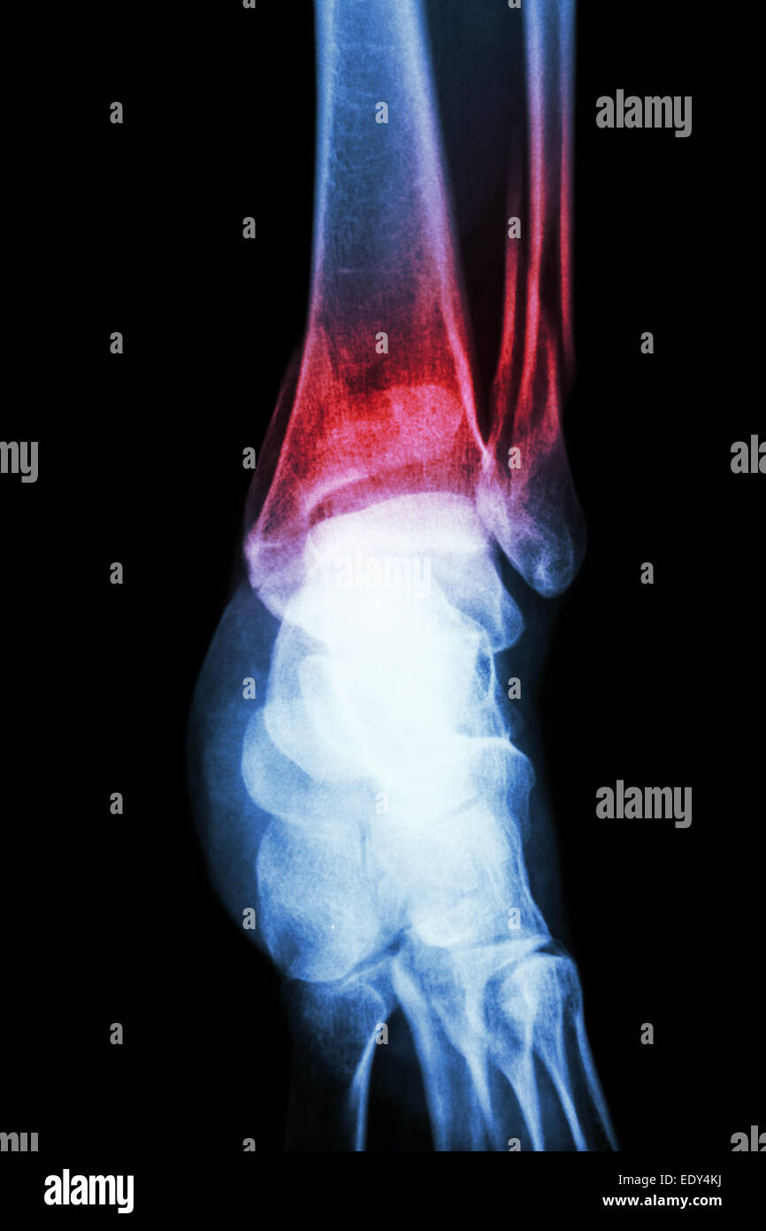 film x-ray ankle show fracture distal tibia and fibula (leg's bone) Stock Photohttps://www.alamy.com/image-license-details/?v=1https://www.alamy.com/stock-photo-film-x-ray-ankle-show-fracture-distal-tibia-and-fibula-legs-bone-77428390.html
RFEDY4KJ–film x-ray ankle show fracture distal tibia and fibula (leg's bone)
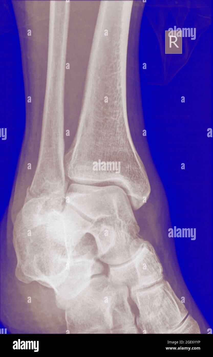 X-ray of a 43 year old male patient with a fracture in the distal radius Stock Photohttps://www.alamy.com/image-license-details/?v=1https://www.alamy.com/x-ray-of-a-43-year-old-male-patient-with-a-fracture-in-the-distal-radius-image439018042.html
RM2GE6YYP–X-ray of a 43 year old male patient with a fracture in the distal radius
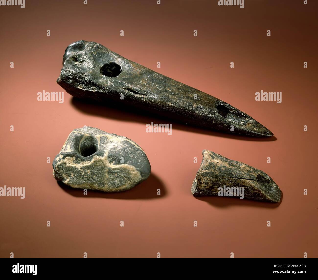 ax made from distal end of right radius of Bison priscus (steppewisent). Cool and well preserved. An old damage to the angle of the joint button and an old crack. Black, with slightly lighter spots. At about 7.5 cm from the gewr. end a virtually circular perforation 2.7-2.9cm. The bone is further ground into one fine, very well symmetrical pointed ax, with the tip formed in the hard leg of the front of the bone. Along the edge of the oval opening, which forms the cavity of the bone at the cut, the traces of bores are in part still very intact Stock Photohttps://www.alamy.com/image-license-details/?v=1https://www.alamy.com/ax-made-from-distal-end-of-right-radius-of-bison-priscus-steppewisent-cool-and-well-preserved-an-old-damage-to-the-angle-of-the-joint-button-and-an-old-crack-black-with-slightly-lighter-spots-at-about-75-cm-from-the-gewr-end-a-virtually-circular-perforation-27-29cm-the-bone-is-further-ground-into-one-fine-very-well-symmetrical-pointed-ax-with-the-tip-formed-in-the-hard-leg-of-the-front-of-the-bone-along-the-edge-of-the-oval-opening-which-forms-the-cavity-of-the-bone-at-the-cut-the-traces-of-bores-are-in-part-still-very-intact-image344562775.html
RM2B0G59B–ax made from distal end of right radius of Bison priscus (steppewisent). Cool and well preserved. An old damage to the angle of the joint button and an old crack. Black, with slightly lighter spots. At about 7.5 cm from the gewr. end a virtually circular perforation 2.7-2.9cm. The bone is further ground into one fine, very well symmetrical pointed ax, with the tip formed in the hard leg of the front of the bone. Along the edge of the oval opening, which forms the cavity of the bone at the cut, the traces of bores are in part still very intact
 gastroduodenoscopy distal end and manipulator in hands of doctors in treatment room Stock Photohttps://www.alamy.com/image-license-details/?v=1https://www.alamy.com/gastroduodenoscopy-distal-end-and-manipulator-in-hands-of-doctors-in-treatment-room-image604308154.html
RF2X34H62–gastroduodenoscopy distal end and manipulator in hands of doctors in treatment room
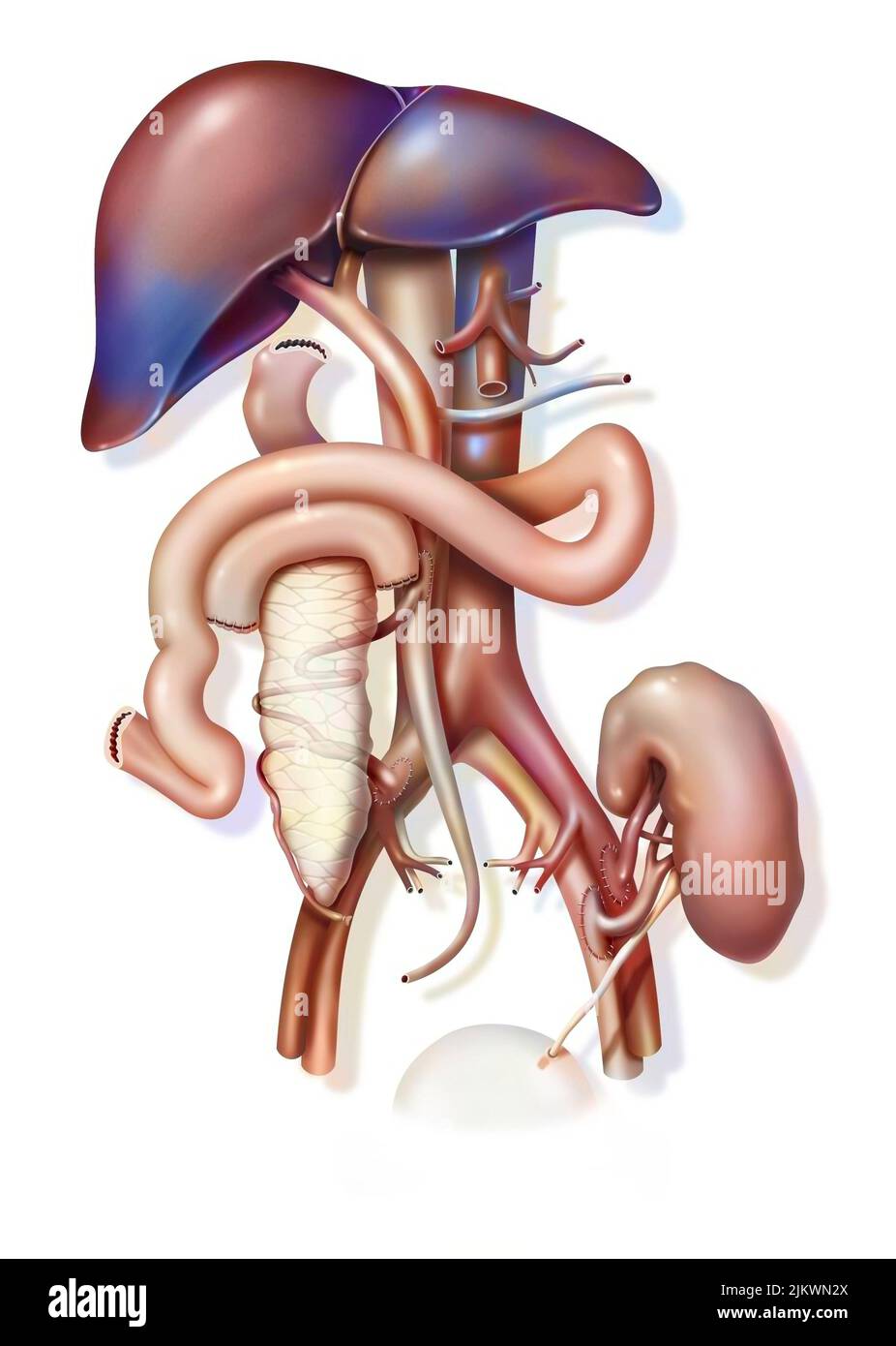 Combined pancreas and kidney transplant: implantation of grafts. Stock Photohttps://www.alamy.com/image-license-details/?v=1https://www.alamy.com/combined-pancreas-and-kidney-transplant-implantation-of-grafts-image476923746.html
RF2JKWN2X–Combined pancreas and kidney transplant: implantation of grafts.
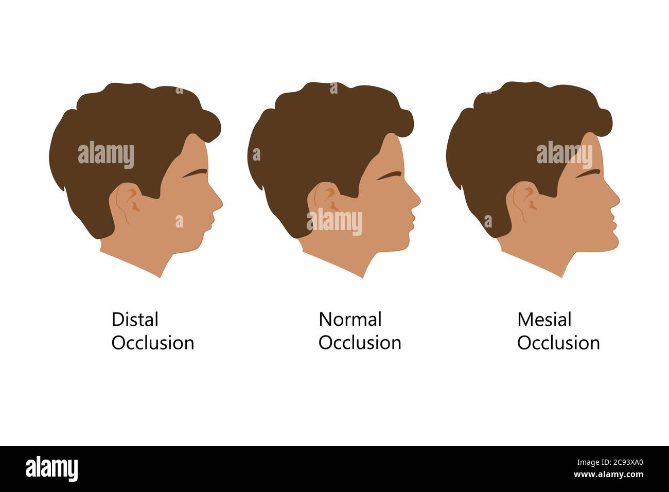 Guy with Distal, Normal, and Mesial bite profile, vector illustration. Overbite or underbite before and after orthodontic treatment. Human with Stock Vectorhttps://www.alamy.com/image-license-details/?v=1https://www.alamy.com/guy-with-distal-normal-and-mesial-bite-profile-vector-illustration-overbite-or-underbite-before-and-after-orthodontic-treatment-human-with-image367036152.html
RF2C93XA0–Guy with Distal, Normal, and Mesial bite profile, vector illustration. Overbite or underbite before and after orthodontic treatment. Human with
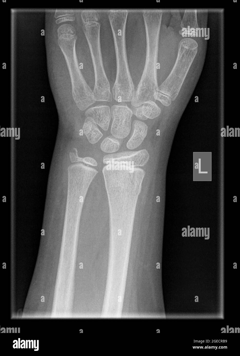 wrist of a 9 year old male patient with Distal Radius and Ulna Fractures Stock Photohttps://www.alamy.com/image-license-details/?v=1https://www.alamy.com/wrist-of-a-9-year-old-male-patient-with-distal-radius-and-ulna-fractures-image439146157.html
RM2GECRB9–wrist of a 9 year old male patient with Distal Radius and Ulna Fractures
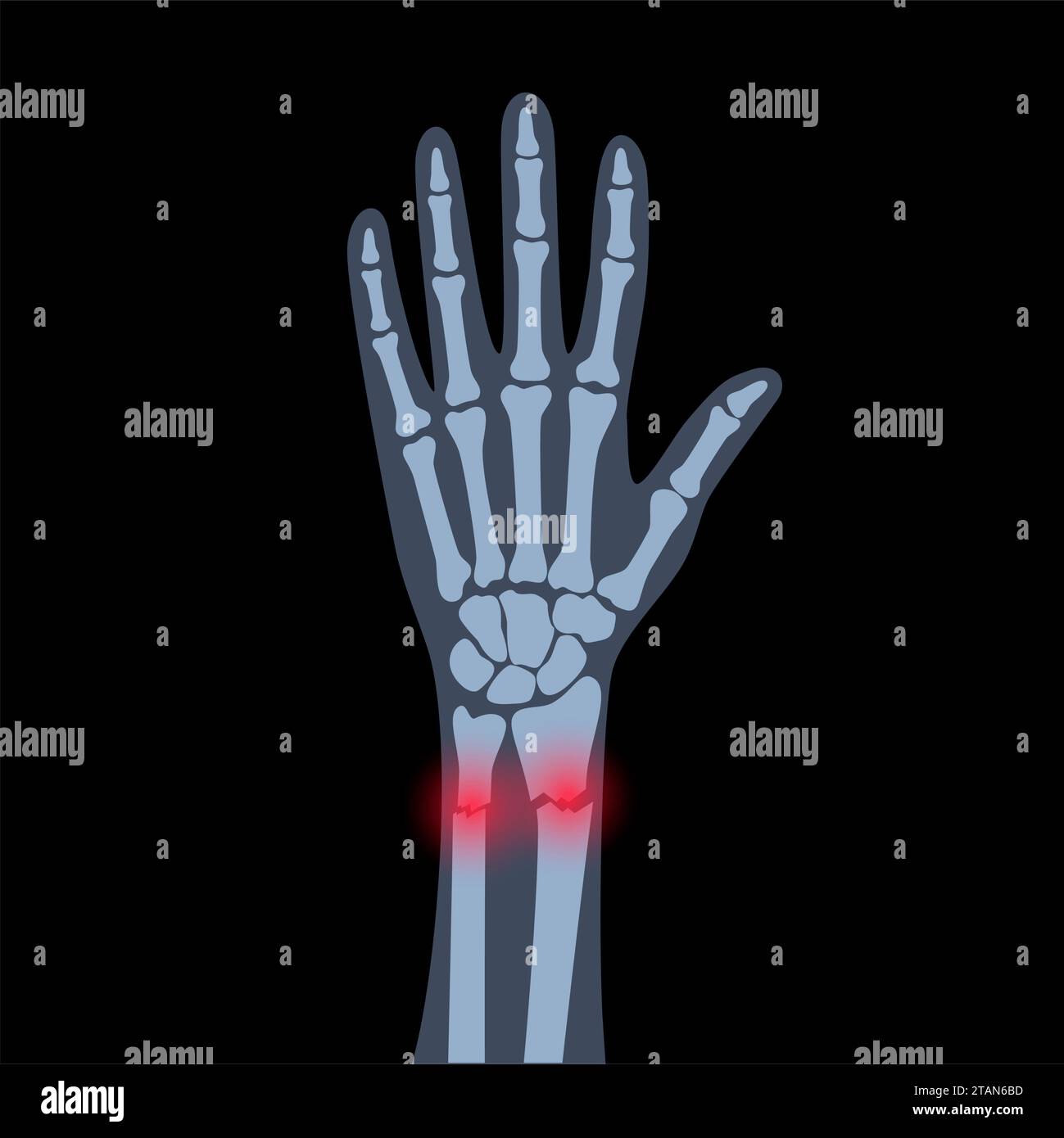 Fractured wrist, illustration Stock Photohttps://www.alamy.com/image-license-details/?v=1https://www.alamy.com/fractured-wrist-illustration-image574554721.html
RF2TAN6BD–Fractured wrist, illustration
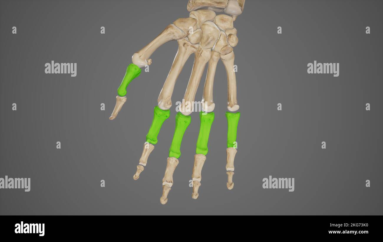 Proximal Phalanges of Hand Stock Photohttps://www.alamy.com/image-license-details/?v=1https://www.alamy.com/proximal-phalanges-of-hand-image491881348.html
RF2KG73K0–Proximal Phalanges of Hand
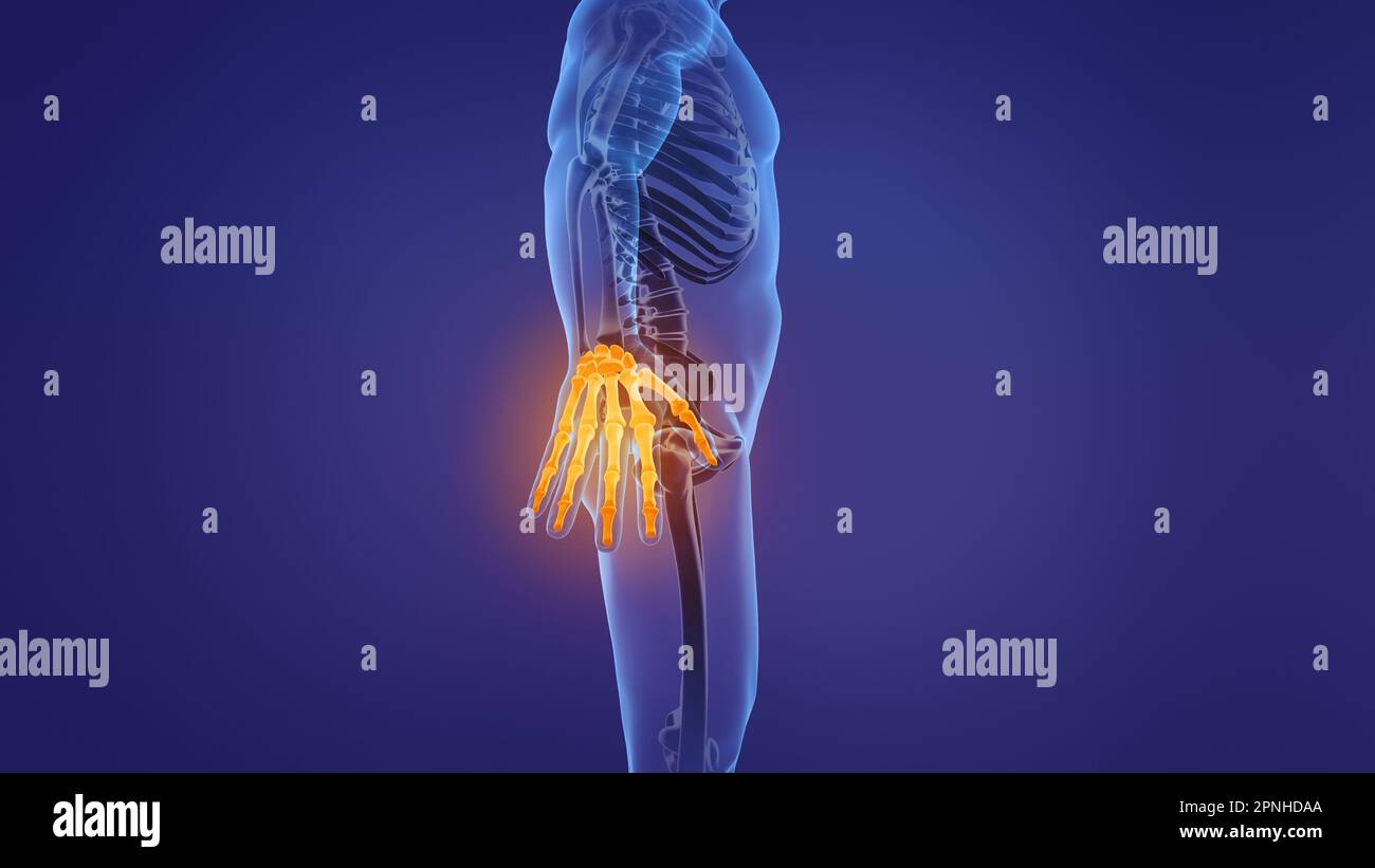 Anatomy of the human hand Stock Photohttps://www.alamy.com/image-license-details/?v=1https://www.alamy.com/anatomy-of-the-human-hand-image546812850.html
RF2PNHDAA–Anatomy of the human hand
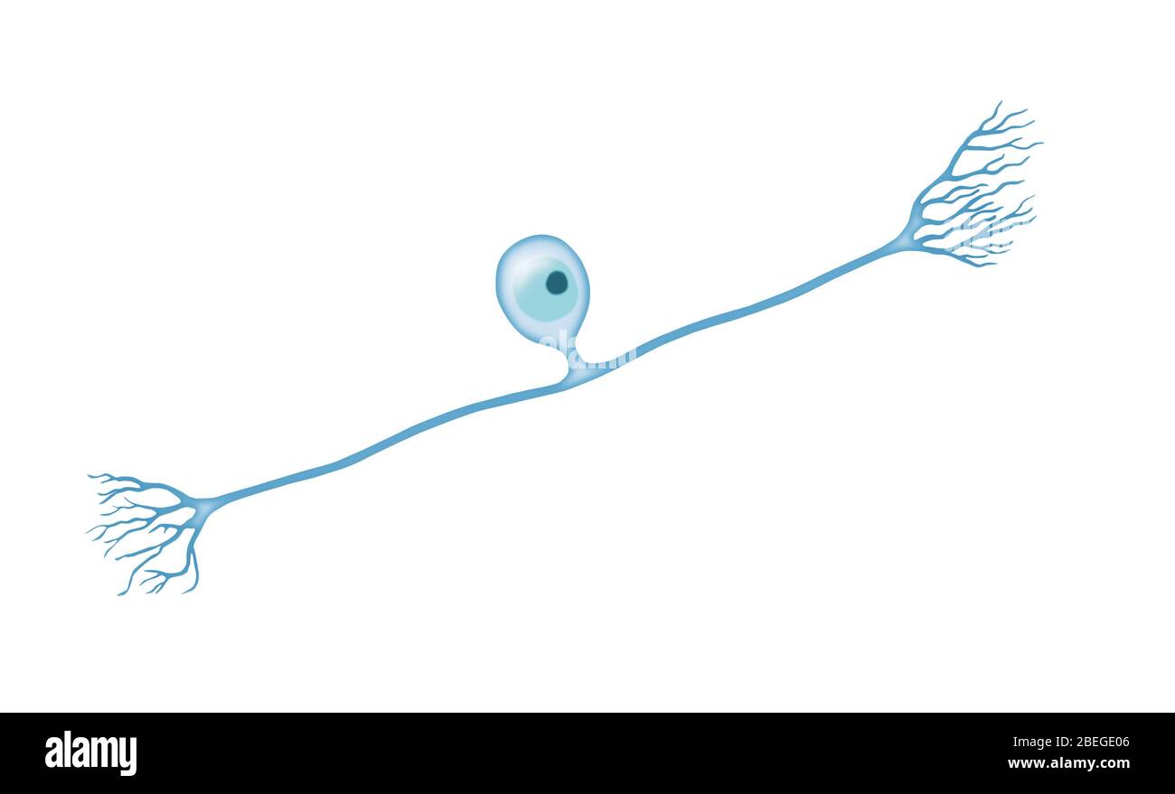 Pseudounipolar Neuron Stock Photohttps://www.alamy.com/image-license-details/?v=1https://www.alamy.com/pseudounipolar-neuron-image353174758.html
RM2BEGE06–Pseudounipolar Neuron
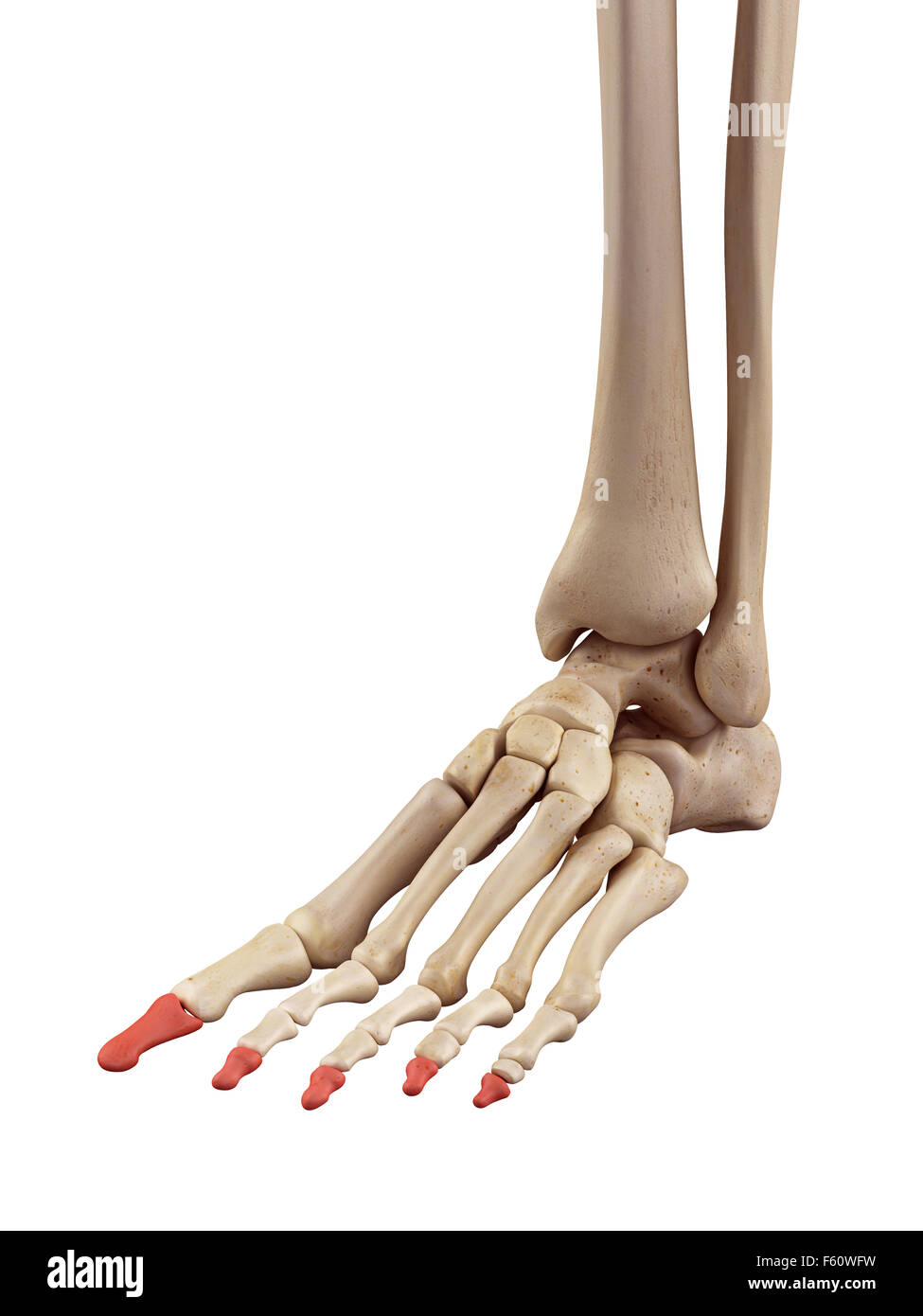 medical accurate illustration of the distal phalanx bones Stock Photohttps://www.alamy.com/image-license-details/?v=1https://www.alamy.com/stock-photo-medical-accurate-illustration-of-the-distal-phalanx-bones-89759821.html
RFF60WFW–medical accurate illustration of the distal phalanx bones
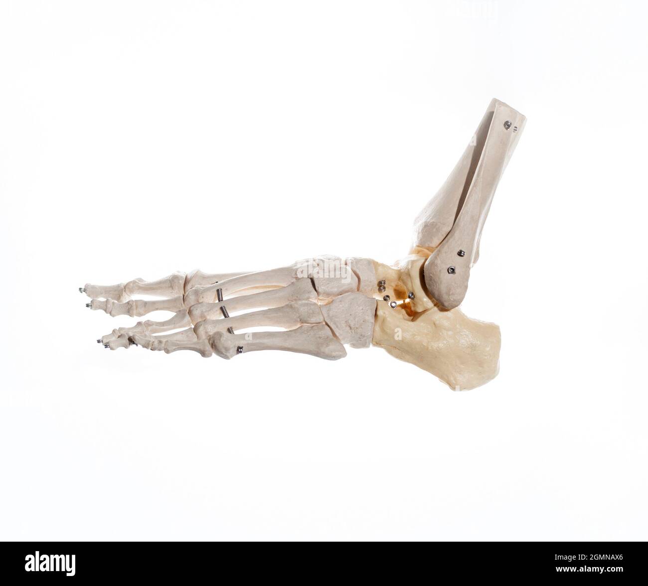 External lateral view of the skeleton of an articulated human foot, with the distal part of the tibia and fibula mounted on wire. Anatomy concept Stock Photohttps://www.alamy.com/image-license-details/?v=1https://www.alamy.com/external-lateral-view-of-the-skeleton-of-an-articulated-human-foot-with-the-distal-part-of-the-tibia-and-fibula-mounted-on-wire-anatomy-concept-image443021886.html
RF2GMNAX6–External lateral view of the skeleton of an articulated human foot, with the distal part of the tibia and fibula mounted on wire. Anatomy concept
 Nelaton device for fractures of the distal radius, vintage engraved illustration.Usual Medicine Dictionary by Dr Labarthe - 1885 Stock Vectorhttps://www.alamy.com/image-license-details/?v=1https://www.alamy.com/stock-photo-nelaton-device-for-fractures-of-the-distal-radius-vintage-engraved-84408207.html
RFEW93ER–Nelaton device for fractures of the distal radius, vintage engraved illustration.Usual Medicine Dictionary by Dr Labarthe - 1885
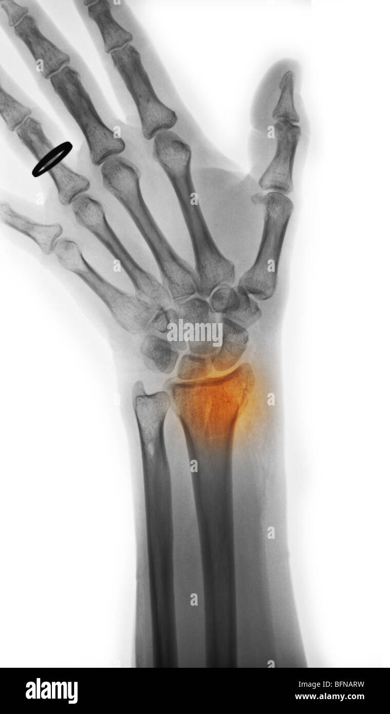 X-ray of the wrist of a 57 year old woman who fell and broke her distal radius Stock Photohttps://www.alamy.com/image-license-details/?v=1https://www.alamy.com/stock-photo-x-ray-of-the-wrist-of-a-57-year-old-woman-who-fell-and-broke-her-distal-26899709.html
RMBFNARW–X-ray of the wrist of a 57 year old woman who fell and broke her distal radius
 A man Distal Guillain-Barré syndrome (DGBS), which usually presents with predominantly motor involvement, and is indistinguishable from that found in Stock Photohttps://www.alamy.com/image-license-details/?v=1https://www.alamy.com/a-man-distal-guillain-barr-syndrome-dgbs-which-usually-presents-with-predominantly-motor-involvement-and-is-indistinguishable-from-that-found-in-image551631113.html
RF2R1CY35–A man Distal Guillain-Barré syndrome (DGBS), which usually presents with predominantly motor involvement, and is indistinguishable from that found in
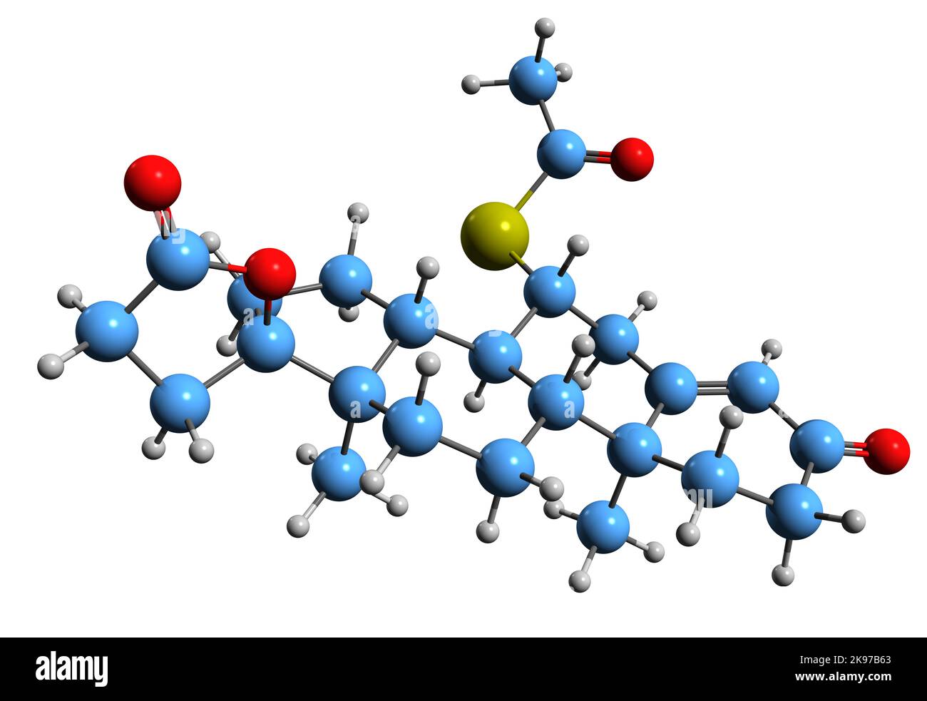 3D image of Spironolactone skeletal formula - molecular chemical structure of Antimineralocorticoid isolated on white background Stock Photohttps://www.alamy.com/image-license-details/?v=1https://www.alamy.com/3d-image-of-spironolactone-skeletal-formula-molecular-chemical-structure-of-antimineralocorticoid-isolated-on-white-background-image487584667.html
RF2K97B63–3D image of Spironolactone skeletal formula - molecular chemical structure of Antimineralocorticoid isolated on white background
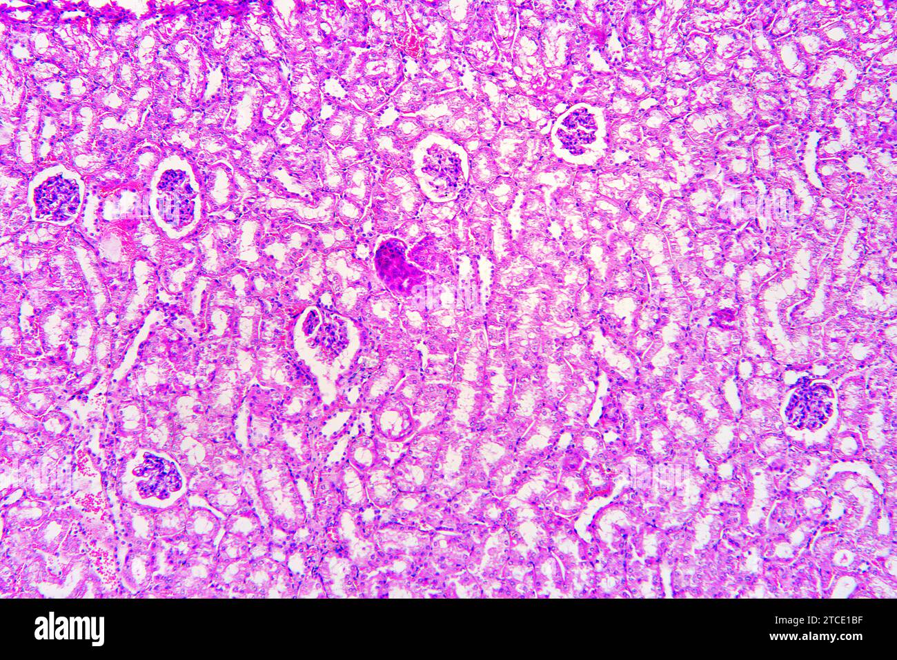 Kidney section showing nephrons, Bowman capsules, glomerulus and distal and proximal tubules. Optical microscope X100. Stock Photohttps://www.alamy.com/image-license-details/?v=1https://www.alamy.com/kidney-section-showing-nephrons-bowman-capsules-glomerulus-and-distal-and-proximal-tubules-optical-microscope-x100-image575626451.html
RF2TCE1BF–Kidney section showing nephrons, Bowman capsules, glomerulus and distal and proximal tubules. Optical microscope X100.
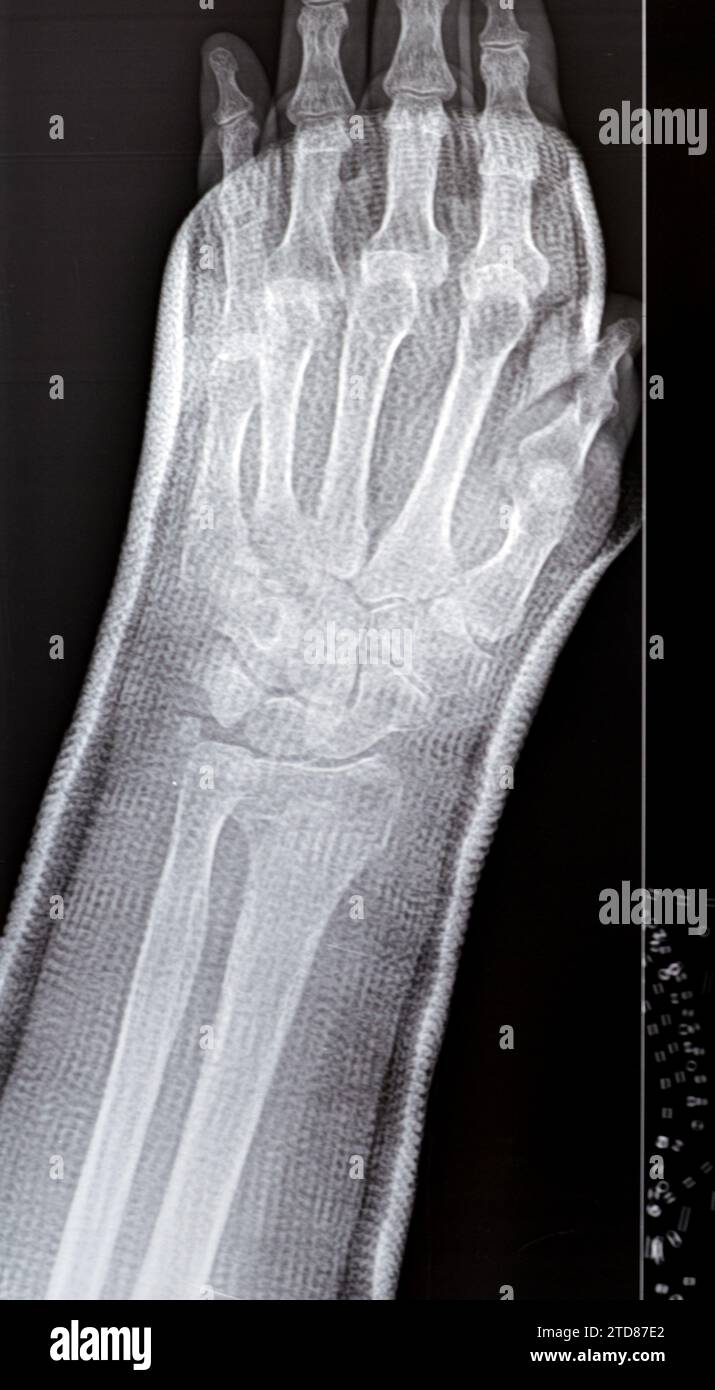 Colles fracture reduction of an old female, a type of fracture of the distal forearm, the broken end of the radius is bent backwards, as a result of a Stock Photohttps://www.alamy.com/image-license-details/?v=1https://www.alamy.com/colles-fracture-reduction-of-an-old-female-a-type-of-fracture-of-the-distal-forearm-the-broken-end-of-the-radius-is-bent-backwards-as-a-result-of-a-image576114170.html
RF2TD87E2–Colles fracture reduction of an old female, a type of fracture of the distal forearm, the broken end of the radius is bent backwards, as a result of a
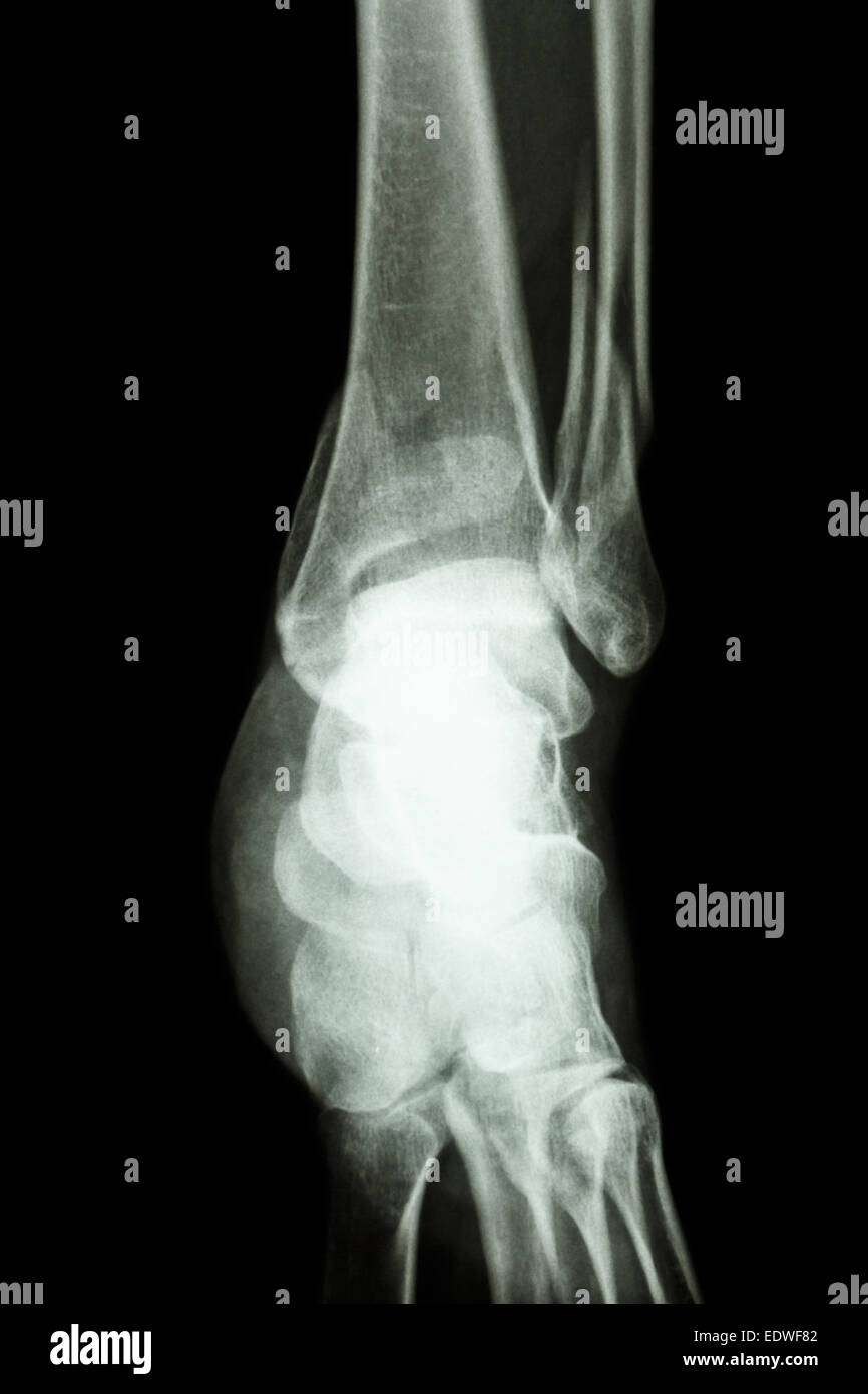 film x-ray ankle show fracture distal tibia and fibula (leg's bone) Stock Photohttps://www.alamy.com/image-license-details/?v=1https://www.alamy.com/stock-photo-film-x-ray-ankle-show-fracture-distal-tibia-and-fibula-legs-bone-77392786.html
RFEDWF82–film x-ray ankle show fracture distal tibia and fibula (leg's bone)
 7 month old baby with distal fracture in femur Stock Photohttps://www.alamy.com/image-license-details/?v=1https://www.alamy.com/7-month-old-baby-with-distal-fracture-in-femur-image438722952.html
RM2GDNFGT–7 month old baby with distal fracture in femur
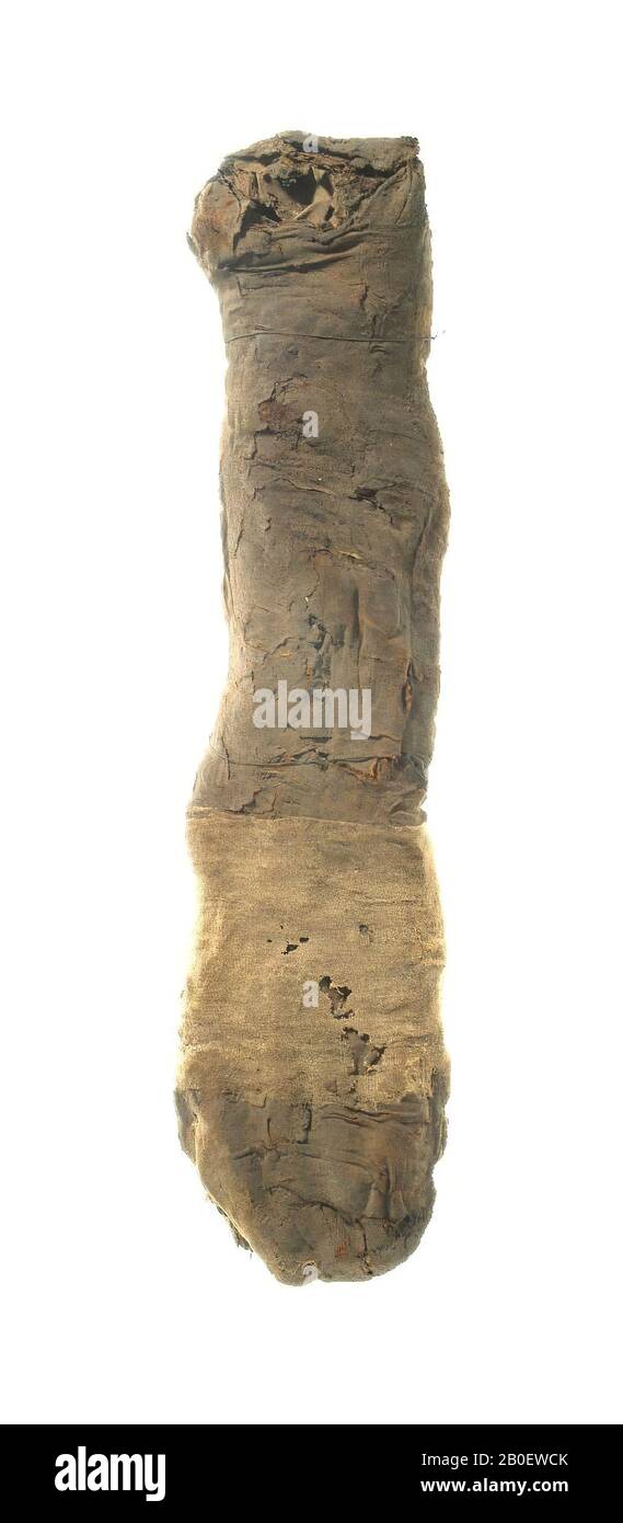 cat, Cat mummy with flattened body and protruding head. The animal is lying on the right side, which is slightly flattened and less dark than the other one. A small piece of straw adheres to the upper face. Concentric bandages can only be detected near the distal end of the mummy. Otherwise the body is wrapped in irregular sheets or medium-fine linen (warp-faced tabby weave, about 11 x 34 threads Stock Photohttps://www.alamy.com/image-license-details/?v=1https://www.alamy.com/cat-cat-mummy-with-flattened-body-and-protruding-head-the-animal-is-lying-on-the-right-side-which-is-slightly-flattened-and-less-dark-than-the-other-one-a-small-piece-of-straw-adheres-to-the-upper-face-concentric-bandages-can-only-be-detected-near-the-distal-end-of-the-mummy-otherwise-the-body-is-wrapped-in-irregular-sheets-or-medium-fine-linen-warp-faced-tabby-weave-about-11-x-34-threads-image344534643.html
RM2B0EWCK–cat, Cat mummy with flattened body and protruding head. The animal is lying on the right side, which is slightly flattened and less dark than the other one. A small piece of straw adheres to the upper face. Concentric bandages can only be detected near the distal end of the mummy. Otherwise the body is wrapped in irregular sheets or medium-fine linen (warp-faced tabby weave, about 11 x 34 threads
 Angioplasty Distal, Vascular Interventional Radiology, Operating Room, Surgery, Hospital Donostia, San Sebastian, Gipuzkoa, Basque Country, Spain Stock Photohttps://www.alamy.com/image-license-details/?v=1https://www.alamy.com/angioplasty-distal-vascular-interventional-radiology-operating-room-surgery-hospital-donostia-san-sebastian-gipuzkoa-basque-country-spain-image604681233.html
RM2X3NH29–Angioplasty Distal, Vascular Interventional Radiology, Operating Room, Surgery, Hospital Donostia, San Sebastian, Gipuzkoa, Basque Country, Spain
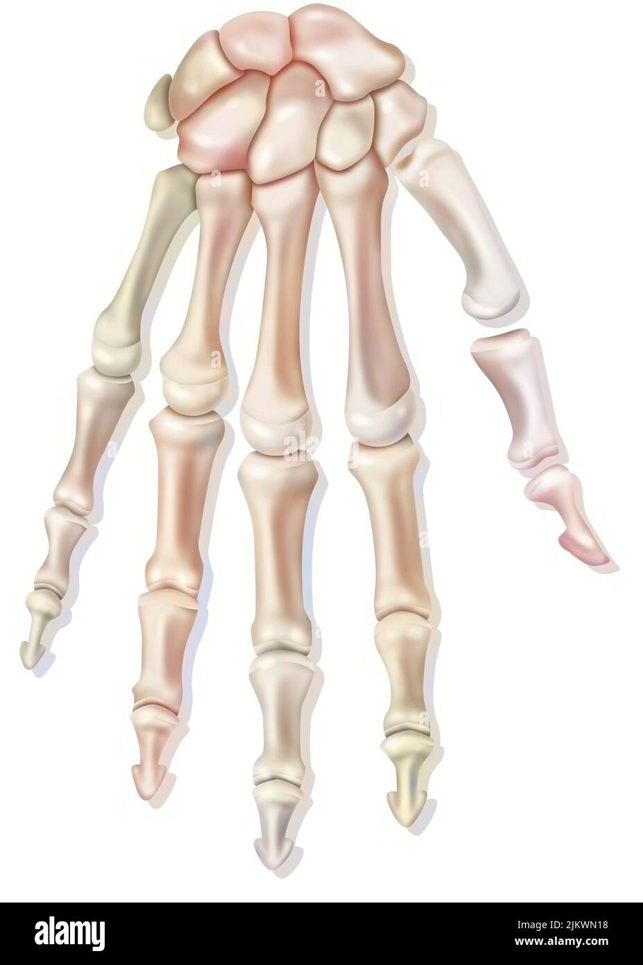 Bones of the right hand in dorsal view. Stock Photohttps://www.alamy.com/image-license-details/?v=1https://www.alamy.com/bones-of-the-right-hand-in-dorsal-view-image476923700.html
RF2JKWN18–Bones of the right hand in dorsal view.
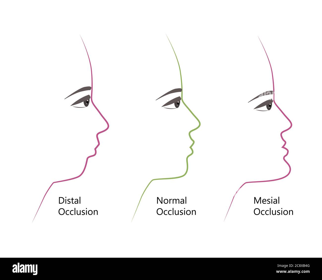 distal, Normal, and Mesial bite profile, vector illustration. Overbite or underbite before and after orthodontic treatment. Human with malocclusion Stock Vectorhttps://www.alamy.com/image-license-details/?v=1https://www.alamy.com/distal-normal-and-mesial-bite-profile-vector-illustration-overbite-or-underbite-before-and-after-orthodontic-treatment-human-with-malocclusion-image366914480.html
RF2C8XB4G–distal, Normal, and Mesial bite profile, vector illustration. Overbite or underbite before and after orthodontic treatment. Human with malocclusion
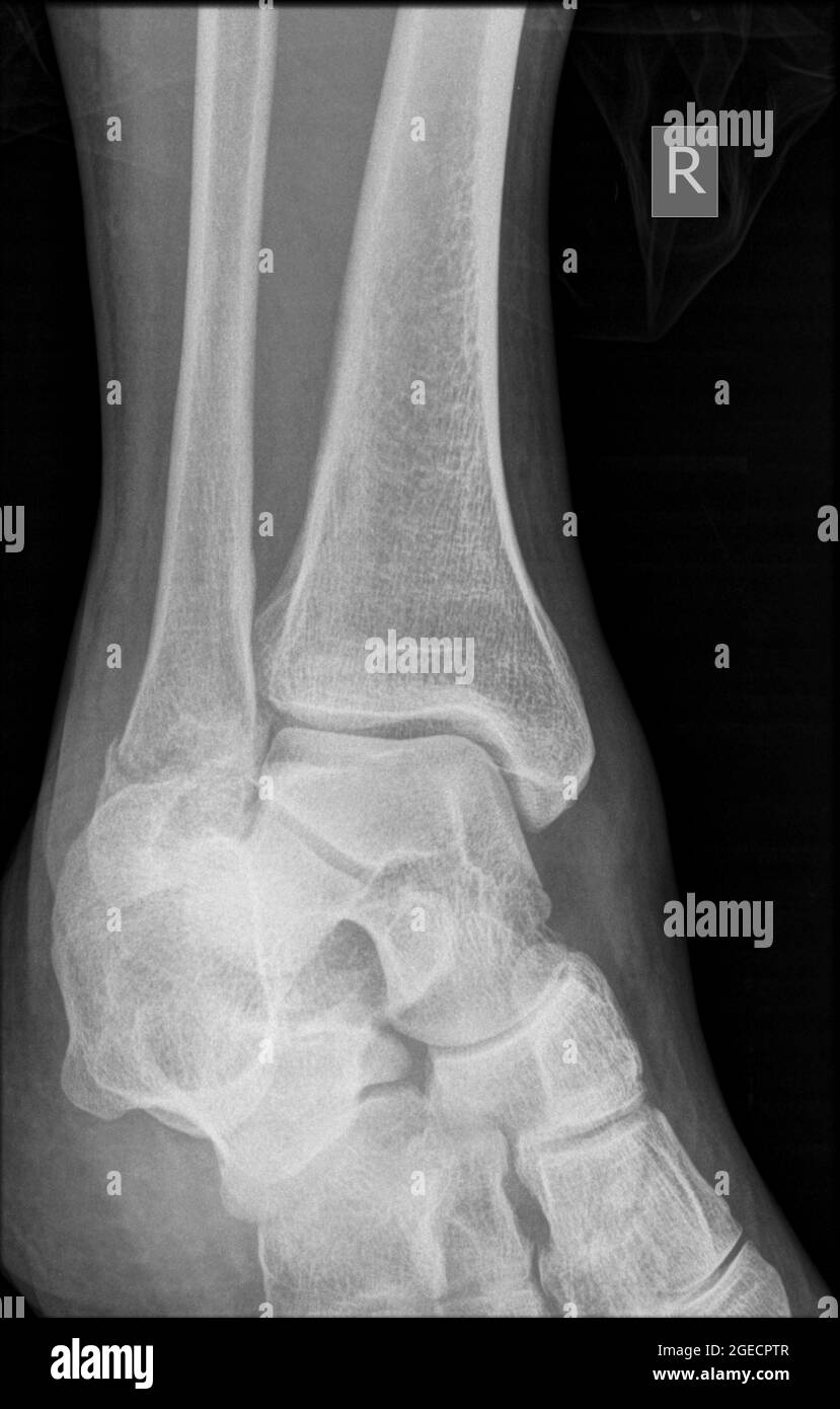 X-ray of a 43 year old male patient with a fracture in the distal radius Stock Photohttps://www.alamy.com/image-license-details/?v=1https://www.alamy.com/x-ray-of-a-43-year-old-male-patient-with-a-fracture-in-the-distal-radius-image439145751.html
RM2GECPTR–X-ray of a 43 year old male patient with a fracture in the distal radius
 Fractured wrist, illustration Stock Photohttps://www.alamy.com/image-license-details/?v=1https://www.alamy.com/fractured-wrist-illustration-image574554797.html
RF2TAN6E5–Fractured wrist, illustration
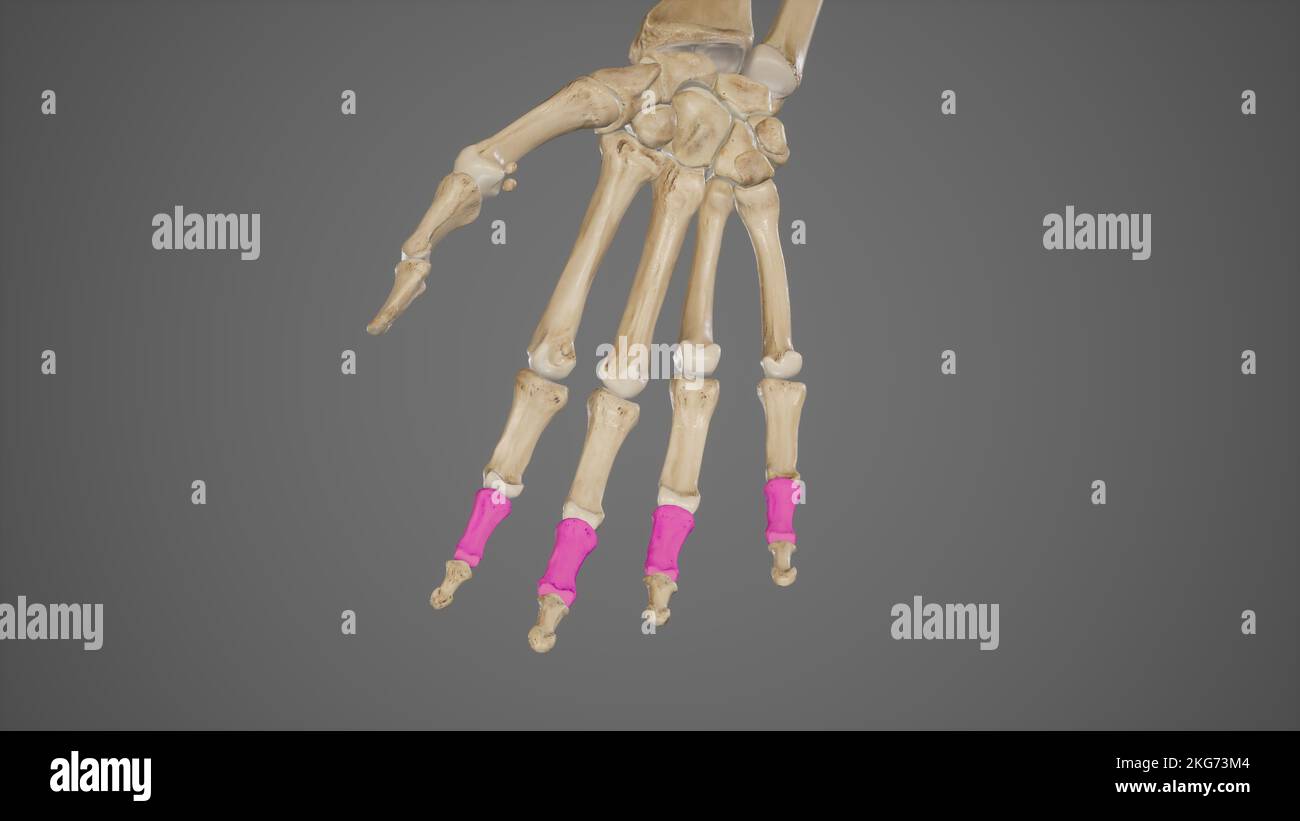 Middle Phalanges of Hand Stock Photohttps://www.alamy.com/image-license-details/?v=1https://www.alamy.com/middle-phalanges-of-hand-image491881380.html
RF2KG73M4–Middle Phalanges of Hand
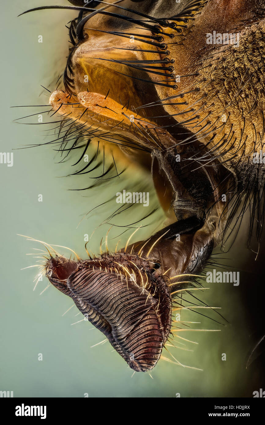 The sponging mouthparts consist of a fleshy, elbowed labium, at the distal end of which are large, sponge-like organs called the labella (singular, la Stock Photohttps://www.alamy.com/image-license-details/?v=1https://www.alamy.com/stock-photo-the-sponging-mouthparts-consist-of-a-fleshy-elbowed-labium-at-the-128873022.html
RMHDJJRX–The sponging mouthparts consist of a fleshy, elbowed labium, at the distal end of which are large, sponge-like organs called the labella (singular, la
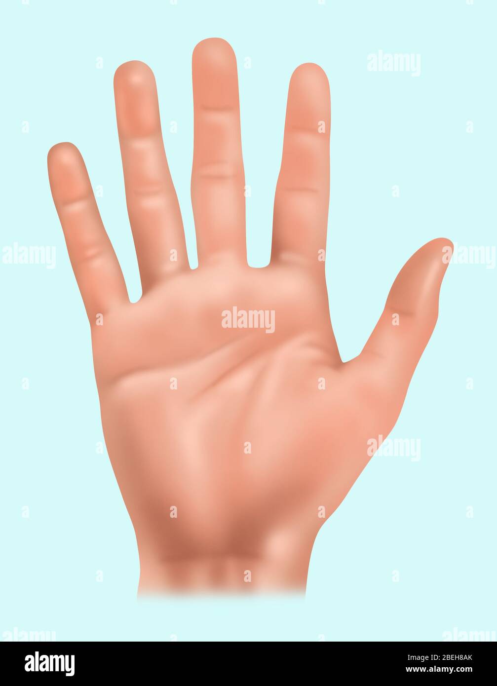 Hand Anatomy, Illustration Stock Photohttps://www.alamy.com/image-license-details/?v=1https://www.alamy.com/hand-anatomy-illustration-image353192299.html
RF2BEH8AK–Hand Anatomy, Illustration
 medical accurate illustration of the distal phalanx bones Stock Photohttps://www.alamy.com/image-license-details/?v=1https://www.alamy.com/stock-photo-medical-accurate-illustration-of-the-distal-phalanx-bones-89759790.html
RFF60WEP–medical accurate illustration of the distal phalanx bones
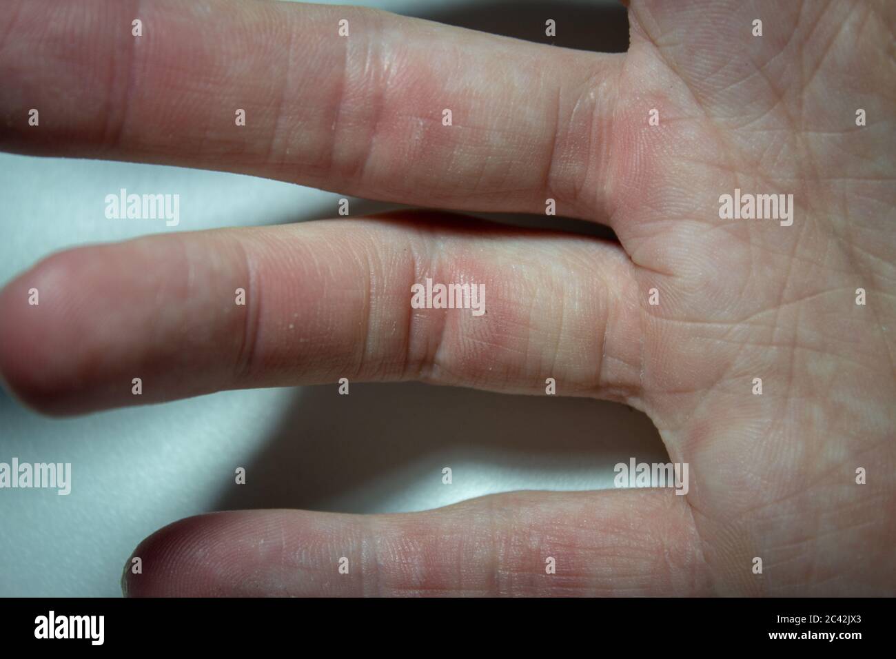 Hand with small calluses after a hard session on the climbing gym Stock Photohttps://www.alamy.com/image-license-details/?v=1https://www.alamy.com/hand-with-small-calluses-after-a-hard-session-on-the-climbing-gym-image363935099.html
RF2C42JX3–Hand with small calluses after a hard session on the climbing gym
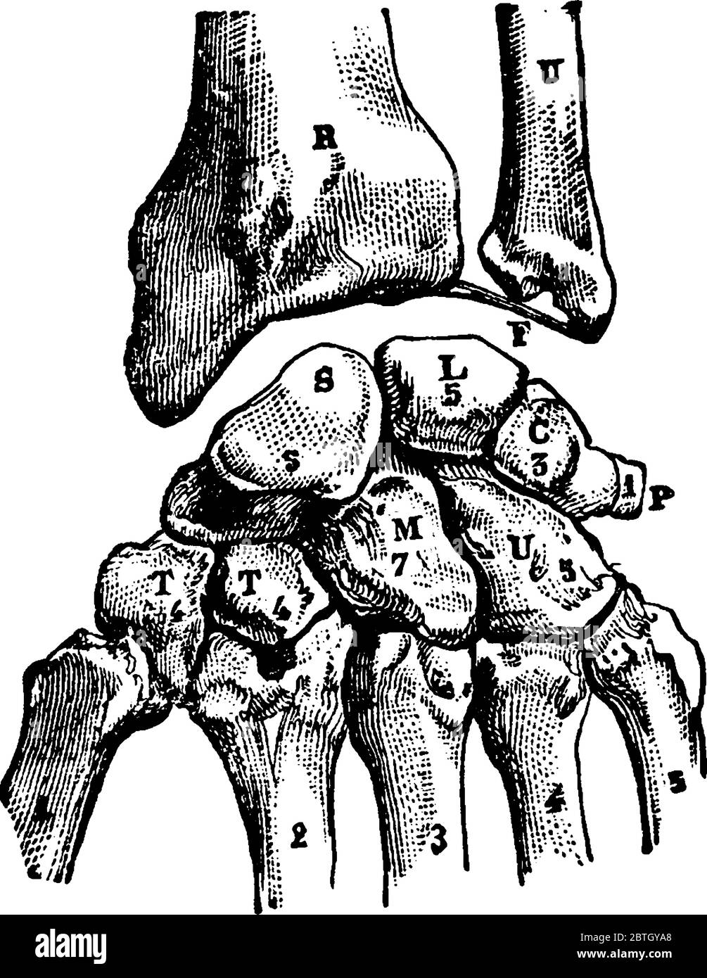 Carpal Bones of the hand they are arranged into two rows of four a proximal and a distal row, vintage line drawing or engraving illustration. Stock Vectorhttps://www.alamy.com/image-license-details/?v=1https://www.alamy.com/carpal-bones-of-the-hand-they-are-arranged-into-two-rows-of-four-a-proximal-and-a-distal-row-vintage-line-drawing-or-engraving-illustration-image359331792.html
RF2BTGYA8–Carpal Bones of the hand they are arranged into two rows of four a proximal and a distal row, vintage line drawing or engraving illustration.
