Quick filters:
Dorso lateral fold Stock Photos and Images
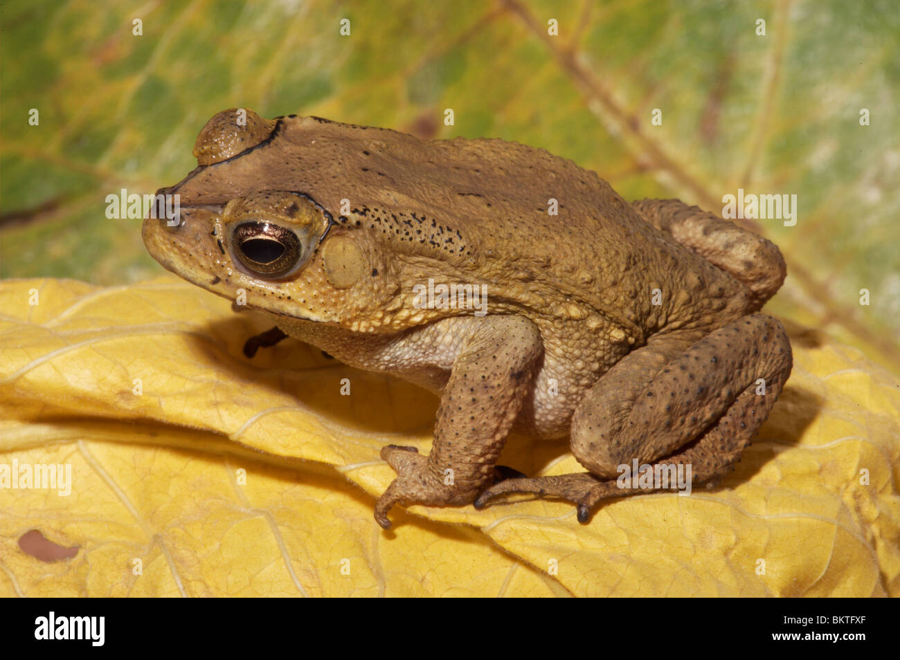
 Asian Common Toad (Bufo melanosticus). Singapore. Stock Photohttps://www.alamy.com/image-license-details/?v=1https://www.alamy.com/stock-photo-asian-common-toad-bufo-melanosticus-singapore-29428183.html
RMBKTFXF–Asian Common Toad (Bufo melanosticus). Singapore.
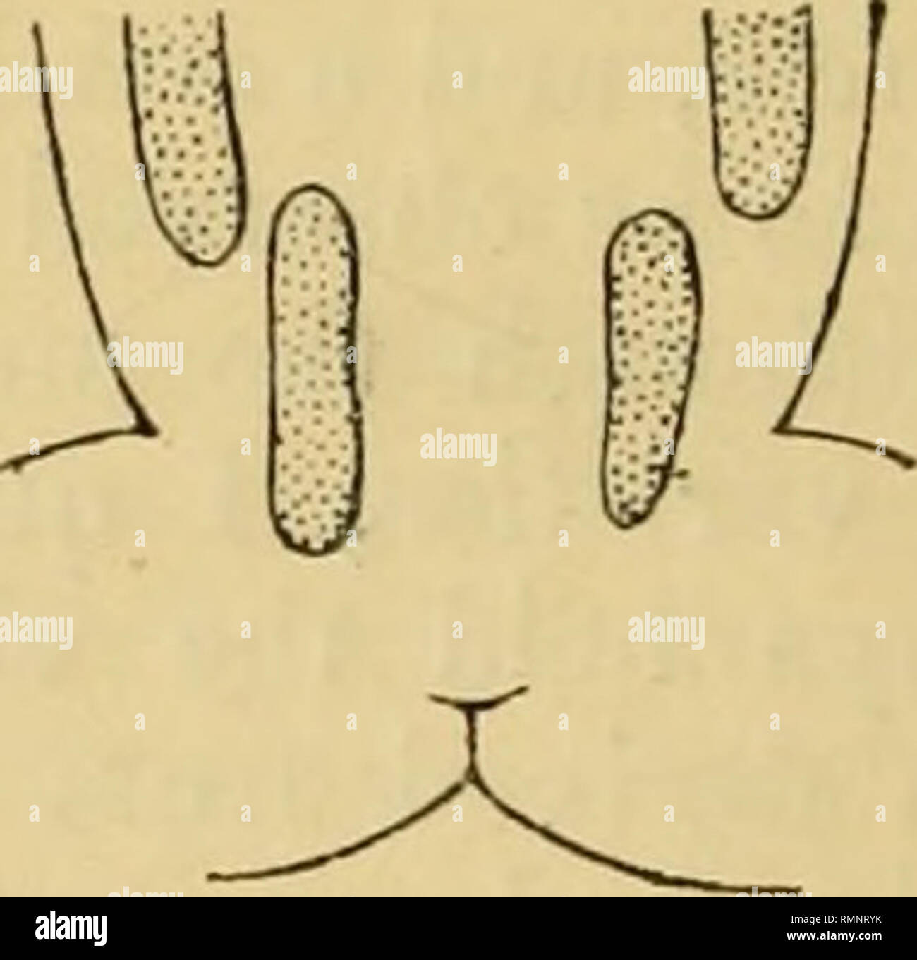
 . The Annals and magazine of natural history; zoology, botany, and geology. Natural history; Zoology; Botany; Geology. Posterior extremities of dorso-lateral folds in specimens from Berlin, var. ridibunda (a), Cadillac, var. ridibunda (b), and Vienna, f. li/pica (c). In the former, and also in the var. lessonce, the glandular dorso-lateral fold ends abruptly at some distance in front of the thigh, and it is often followed by a detached portion parallel with it but nearer to the mid-dorsal line and extend- ing on the base of the thigh. In the var. chinensis the fold extends uninterrupted to the Stock Photohttps://www.alamy.com/image-license-details/?v=1https://www.alamy.com/the-annals-and-magazine-of-natural-history-zoology-botany-and-geology-natural-history-zoology-botany-geology-posterior-extremities-of-dorso-lateral-folds-in-specimens-from-berlin-var-ridibunda-a-cadillac-var-ridibunda-b-and-vienna-f-lipica-c-in-the-former-and-also-in-the-var-lessonce-the-glandular-dorso-lateral-fold-ends-abruptly-at-some-distance-in-front-of-the-thigh-and-it-is-often-followed-by-a-detached-portion-parallel-with-it-but-nearer-to-the-mid-dorsal-line-and-extend-ing-on-the-base-of-the-thigh-in-the-var-chinensis-the-fold-extends-uninterrupted-to-the-image236507703.html
RMRMNRYK–. The Annals and magazine of natural history; zoology, botany, and geology. Natural history; Zoology; Botany; Geology. Posterior extremities of dorso-lateral folds in specimens from Berlin, var. ridibunda (a), Cadillac, var. ridibunda (b), and Vienna, f. li/pica (c). In the former, and also in the var. lessonce, the glandular dorso-lateral fold ends abruptly at some distance in front of the thigh, and it is often followed by a detached portion parallel with it but nearer to the mid-dorsal line and extend- ing on the base of the thigh. In the var. chinensis the fold extends uninterrupted to the
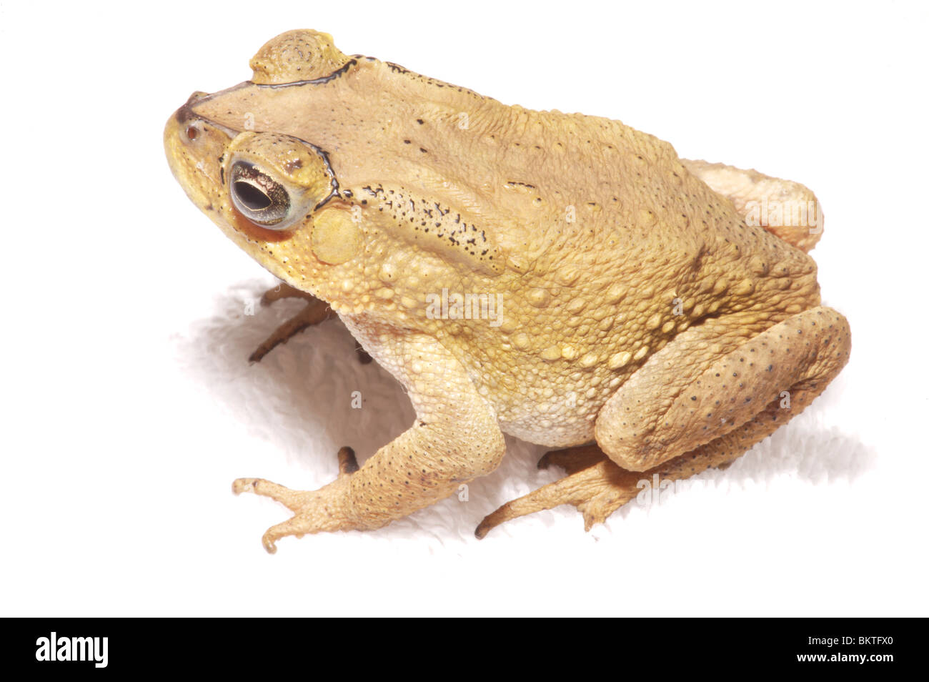
 Asian Common Toad (Bufo melanosticus) Singapore. Stock Photohttps://www.alamy.com/image-license-details/?v=1https://www.alamy.com/stock-photo-asian-common-toad-bufo-melanosticus-singapore-29428168.html
RMBKTFX0–Asian Common Toad (Bufo melanosticus) Singapore.
 . The structure and classification of birds . Fig. 25.—Diagkammatic Tbansveese Section of Emo, to show the Peo- JEOTION OP Oblique Septum. a, as a free fold ; &, falciform ligament. dissepimental nature of the oblique septa occurs in a fewbirds {e.g. emu and Cariama), in which the posterior endof the oblique septum, though firmly attached to the dorso-lateral parietes, is not so attached ventrally, but projectsinto the abdominal cavity as a free fold. In these cases thefree fold is double (see fig. 25), the inner half being conti-nuous with the horizontal septum. To the possible signi-ficance Stock Photohttps://www.alamy.com/image-license-details/?v=1https://www.alamy.com/the-structure-and-classification-of-birds-fig-25diagkammatic-tbansveese-section-of-emo-to-show-the-peo-jeotion-op-oblique-septum-a-as-a-free-fold-falciform-ligament-dissepimental-nature-of-the-oblique-septa-occurs-in-a-fewbirds-eg-emu-and-cariama-in-which-the-posterior-endof-the-oblique-septum-though-firmly-attached-to-the-dorso-lateral-parietes-is-not-so-attached-ventrally-but-projectsinto-the-abdominal-cavity-as-a-free-fold-in-these-cases-thefree-fold-is-double-see-fig-25-the-inner-half-being-conti-nuous-with-the-horizontal-septum-to-the-possible-signi-ficance-image375202462.html
RM2CPBXFX–. The structure and classification of birds . Fig. 25.—Diagkammatic Tbansveese Section of Emo, to show the Peo- JEOTION OP Oblique Septum. a, as a free fold ; &, falciform ligament. dissepimental nature of the oblique septa occurs in a fewbirds {e.g. emu and Cariama), in which the posterior endof the oblique septum, though firmly attached to the dorso-lateral parietes, is not so attached ventrally, but projectsinto the abdominal cavity as a free fold. In these cases thefree fold is double (see fig. 25), the inner half being conti-nuous with the horizontal septum. To the possible signi-ficance
 . Bulletin - United States National Museum. Science. Figs. 117-121.—Rana narina. Nat. size. 117, top of uead; 118, side of head; 119, open mouth; 120, underside of hand; 121, underside of foot. xo. loct, sci. coll.. tokyo. metatarsal tubercle but slightly prominent, narrow, less than one- half the length of the inner toe; no outer metatarsal tubercle; fore limbs longer than tibia; tibio-tarsal joint extends considerably beyond the snout; heels overlap considerably when thighs are bent at right angles to the axis of the body; no dorsal or dorso-lateral folds; no tar- sal fold; both surfaces smo Stock Photohttps://www.alamy.com/image-license-details/?v=1https://www.alamy.com/bulletin-united-states-national-museum-science-figs-117-121rana-narina-nat-size-117-top-of-uead-118-side-of-head-119-open-mouth-120-underside-of-hand-121-underside-of-foot-xo-loct-sci-coll-tokyo-metatarsal-tubercle-but-slightly-prominent-narrow-less-than-one-half-the-length-of-the-inner-toe-no-outer-metatarsal-tubercle-fore-limbs-longer-than-tibia-tibio-tarsal-joint-extends-considerably-beyond-the-snout-heels-overlap-considerably-when-thighs-are-bent-at-right-angles-to-the-axis-of-the-body-no-dorsal-or-dorso-lateral-folds-no-tar-sal-fold-both-surfaces-smo-image233750599.html
RMRG877K–. Bulletin - United States National Museum. Science. Figs. 117-121.—Rana narina. Nat. size. 117, top of uead; 118, side of head; 119, open mouth; 120, underside of hand; 121, underside of foot. xo. loct, sci. coll.. tokyo. metatarsal tubercle but slightly prominent, narrow, less than one- half the length of the inner toe; no outer metatarsal tubercle; fore limbs longer than tibia; tibio-tarsal joint extends considerably beyond the snout; heels overlap considerably when thighs are bent at right angles to the axis of the body; no dorsal or dorso-lateral folds; no tar- sal fold; both surfaces smo
 . Bulletin of the British Museum (Natural History), Geology. SHELL STRUCTURE. Fig. 3A. Ventro-lateral perspective view of the dorsal valve interior of Dyoros sp. from the Permian of Texas illustrating the surface morphology. a. - anderidia; a.ad. - anterior adductor scar; b.p. - brachial platform; b.r. - brachial ridge; c.p. - cardinal process; dm.f. - dorso-median fold; m.s. — median septum (here strongly tuberculate); p.ad. - posterior adductor scar; p.m. - posterior margin of valve; s. - socket.. Please note that these images are extracted from scanned page images that may have been digital Stock Photohttps://www.alamy.com/image-license-details/?v=1https://www.alamy.com/bulletin-of-the-british-museum-natural-history-geology-shell-structure-fig-3a-ventro-lateral-perspective-view-of-the-dorsal-valve-interior-of-dyoros-sp-from-the-permian-of-texas-illustrating-the-surface-morphology-a-anderidia-aad-anterior-adductor-scar-bp-brachial-platform-br-brachial-ridge-cp-cardinal-process-dmf-dorso-median-fold-ms-median-septum-here-strongly-tuberculate-pad-posterior-adductor-scar-pm-posterior-margin-of-valve-s-socket-please-note-that-these-images-are-extracted-from-scanned-page-images-that-may-have-been-digital-image233976065.html
RMRGJET1–. Bulletin of the British Museum (Natural History), Geology. SHELL STRUCTURE. Fig. 3A. Ventro-lateral perspective view of the dorsal valve interior of Dyoros sp. from the Permian of Texas illustrating the surface morphology. a. - anderidia; a.ad. - anterior adductor scar; b.p. - brachial platform; b.r. - brachial ridge; c.p. - cardinal process; dm.f. - dorso-median fold; m.s. — median septum (here strongly tuberculate); p.ad. - posterior adductor scar; p.m. - posterior margin of valve; s. - socket.. Please note that these images are extracted from scanned page images that may have been digital
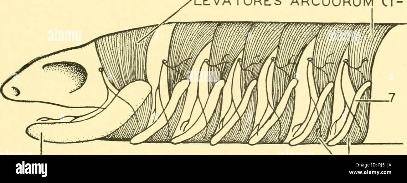
 . Chordate anatomy. Chordata; Anatomy, Comparative. i|i^ ^^^--^ FiM-rniX"^ MYOTOMIC buds' ^'^-"-^ h, FIN-FOLD "•""Sypogldssus muscle buds B. COELOM,/;- Fig. 188.—Diagram of bvidding of hypoglossal and pectoral fin muscles from trunk myotomes in an elasmobranch embryo. A. Lateral view after Braus. 2-6, visceral arches. B. Cross section in region of pectoral fin-fold. LEVATORES ARCUORUM C(-7). VISCERAL SKELETAL ARCHES Cl-7) DEPRESSORES ARCUORUM Cl-7^ LEVATORES [-4 DIGASTRICUS MASSETER TEMPORALIS DORSO-LARYNGIS AND DORSO- TRACHEALIS. Please note that these images ar Stock Photohttps://www.alamy.com/image-license-details/?v=1https://www.alamy.com/chordate-anatomy-chordata-anatomy-comparative-ii-fim-rnixquot-myotomic-buds-quot-h-fin-fold-quotquotquotsypogldssus-muscle-buds-b-coelom-fig-188diagram-of-bvidding-of-hypoglossal-and-pectoral-fin-muscles-from-trunk-myotomes-in-an-elasmobranch-embryo-a-lateral-view-after-braus-2-6-visceral-arches-b-cross-section-in-region-of-pectoral-fin-fold-levatores-arcuorum-c-7-visceral-skeletal-arches-cl-7-depressores-arcuorum-cl-7-levatores-4-digastricus-masseter-temporalis-dorso-laryngis-and-dorso-trachealis-please-note-that-these-images-ar-image234909650.html
RMRJ51JA–. Chordate anatomy. Chordata; Anatomy, Comparative. i|i^ ^^^--^ FiM-rniX"^ MYOTOMIC buds' ^'^-"-^ h, FIN-FOLD "•""Sypogldssus muscle buds B. COELOM,/;- Fig. 188.—Diagram of bvidding of hypoglossal and pectoral fin muscles from trunk myotomes in an elasmobranch embryo. A. Lateral view after Braus. 2-6, visceral arches. B. Cross section in region of pectoral fin-fold. LEVATORES ARCUORUM C(-7). VISCERAL SKELETAL ARCHES Cl-7) DEPRESSORES ARCUORUM Cl-7^ LEVATORES [-4 DIGASTRICUS MASSETER TEMPORALIS DORSO-LARYNGIS AND DORSO- TRACHEALIS. Please note that these images ar