Duck kidney Cut Out Stock Images
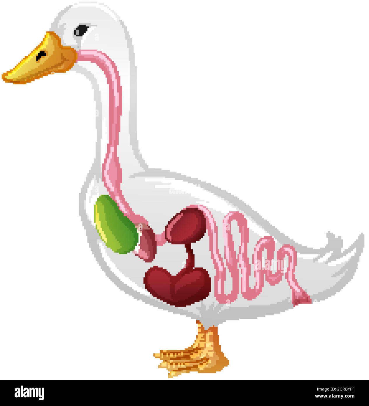
 Internal Anatomy of a Duck isolated on white background Stock Vectorhttps://www.alamy.com/image-license-details/?v=1https://www.alamy.com/internal-anatomy-of-a-duck-isolated-on-white-background-image444659559.html
RF2GRBYPF–Internal Anatomy of a Duck isolated on white background
 . The structure and classification of birds . ^ v.aX. m Fig. 23.—Eespieatoey Organs of Duck. /-I-,air sacs; TV, trachea ; d.Z, dorsal margin ; v.a.^, ventral marginof lungs; l-3,clissepimentsbetween air sacs; Ao, aorta; c.a, coeliac artery; m.a, mesenteric :wi, muscular fibres ono.j,oblique septum ; VJi, vertical median septum ; L.c, longus colli; K, kidney. (After Huxley.). .K. Fig. 24.—Bespiiiatoey Oegans of Aptektx. Lbtteeing as in Fig. 23(aftee Huxley 40 STRUCTUliE AND CLASSIFICATION OF BIRDS attachment of the falciform Hgament. Dorsally they areattached to the parietes. As a general rule Stock Photohttps://www.alamy.com/image-license-details/?v=1https://www.alamy.com/the-structure-and-classification-of-birds-vax-m-fig-23eespieatoey-organs-of-duck-i-air-sacs-tv-trachea-dz-dorsal-margin-va-ventral-marginof-lungs-l-3clissepimentsbetween-air-sacs-ao-aorta-ca-coeliac-artery-ma-mesenteric-wi-muscular-fibres-onojoblique-septum-vji-vertical-median-septum-lc-longus-colli-k-kidney-after-huxley-k-fig-24bespiiiatoey-oegans-of-aptektx-lbtteeing-as-in-fig-23aftee-huxley-40-structulie-and-classification-of-birds-attachment-of-the-falciform-hgament-dorsally-they-areattached-to-the-parietes-as-a-general-rule-image375202875.html
RM2CPBY2K–. The structure and classification of birds . ^ v.aX. m Fig. 23.—Eespieatoey Organs of Duck. /-I-,air sacs; TV, trachea ; d.Z, dorsal margin ; v.a.^, ventral marginof lungs; l-3,clissepimentsbetween air sacs; Ao, aorta; c.a, coeliac artery; m.a, mesenteric :wi, muscular fibres ono.j,oblique septum ; VJi, vertical median septum ; L.c, longus colli; K, kidney. (After Huxley.). .K. Fig. 24.—Bespiiiatoey Oegans of Aptektx. Lbtteeing as in Fig. 23(aftee Huxley 40 STRUCTUliE AND CLASSIFICATION OF BIRDS attachment of the falciform Hgament. Dorsally they areattached to the parietes. As a general rule
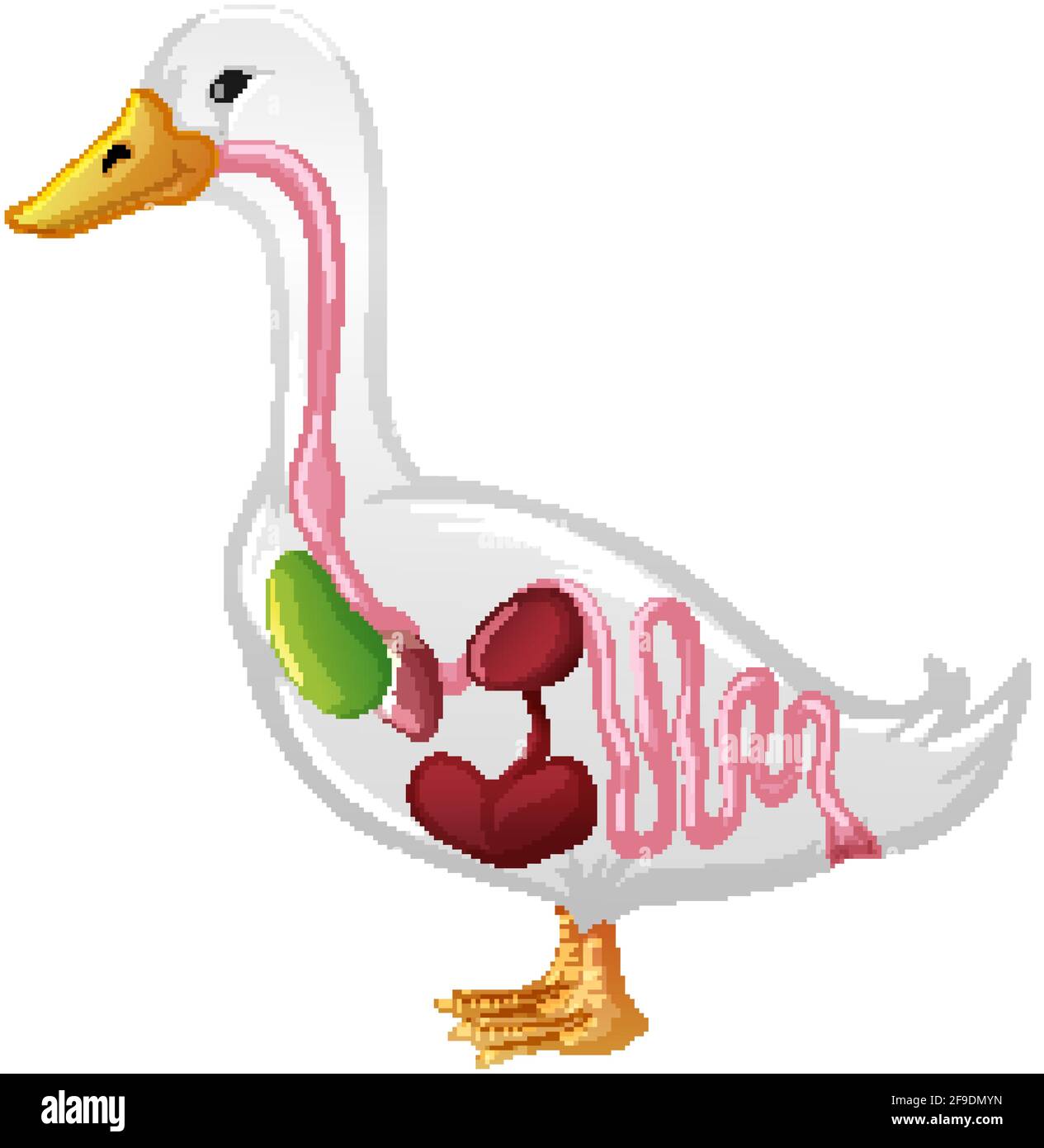
 Internal Anatomy of a Duck isolated on white background illustration Stock Vectorhttps://www.alamy.com/image-license-details/?v=1https://www.alamy.com/internal-anatomy-of-a-duck-isolated-on-white-background-illustration-image418882569.html
RF2F9DMYN–Internal Anatomy of a Duck isolated on white background illustration
 A duck in cartoon style isolated on white background Stock Vectorhttps://www.alamy.com/image-license-details/?v=1https://www.alamy.com/a-duck-in-cartoon-style-isolated-on-white-background-image444528680.html
RF2GR60T8–A duck in cartoon style isolated on white background
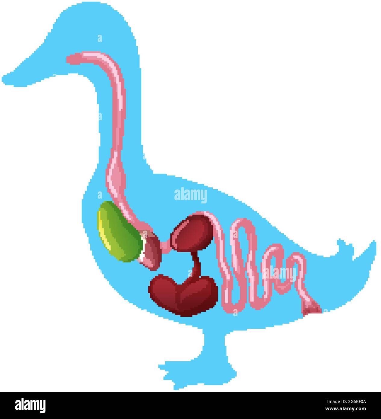
 Internal Anatomy of a Duck isolated on white background illustration Stock Vectorhttps://www.alamy.com/image-license-details/?v=1https://www.alamy.com/internal-anatomy-of-a-duck-isolated-on-white-background-illustration-image434375994.html
RF2G6KF0A–Internal Anatomy of a Duck isolated on white background illustration
 A duck in cartoon style isolated on white background illustration Stock Vectorhttps://www.alamy.com/image-license-details/?v=1https://www.alamy.com/a-duck-in-cartoon-style-isolated-on-white-background-illustration-image400964999.html
RF2E89EXF–A duck in cartoon style isolated on white background illustration