Quick filters:
Duodenal flexure Stock Photos and Images
 . A text-book of comparative physiology for students and practitioners of comparative (veterinary) medicine . in the crop secretes a Fig. 234.—General view of digestive apparatus of fowl (after Chauveau). 1, tongue;2 pharynx; 3, first portion of oesophagus; 4, crop; 5, second portion of oesopha-gus; 6, succentric ventricle (proventricuniB); 7, gizzard; 8, origin of duodenum; !),first branch of duodenal flexure; 10, second branch of same; 11, origin of floatingportion of small intestine; 12, small intestine; 12, terminal portion of this intes-tine, Hanked on each side by the two coeca (regarded Stock Photohttps://www.alamy.com/image-license-details/?v=1https://www.alamy.com/a-text-book-of-comparative-physiology-for-students-and-practitioners-of-comparative-veterinary-medicine-in-the-crop-secretes-a-fig-234general-view-of-digestive-apparatus-of-fowl-after-chauveau-1-tongue2-pharynx-3-first-portion-of-oesophagus-4-crop-5-second-portion-of-oesopha-gus-6-succentric-ventricle-proventricunib-7-gizzard-8-origin-of-duodenum-!first-branch-of-duodenal-flexure-10-second-branch-of-same-11-origin-of-floatingportion-of-small-intestine-12-small-intestine-12-terminal-portion-of-this-intes-tine-hanked-on-each-side-by-the-two-coeca-regarded-image372492176.html
RM2CJ0DG0–. A text-book of comparative physiology for students and practitioners of comparative (veterinary) medicine . in the crop secretes a Fig. 234.—General view of digestive apparatus of fowl (after Chauveau). 1, tongue;2 pharynx; 3, first portion of oesophagus; 4, crop; 5, second portion of oesopha-gus; 6, succentric ventricle (proventricuniB); 7, gizzard; 8, origin of duodenum; !),first branch of duodenal flexure; 10, second branch of same; 11, origin of floatingportion of small intestine; 12, small intestine; 12, terminal portion of this intes-tine, Hanked on each side by the two coeca (regarded
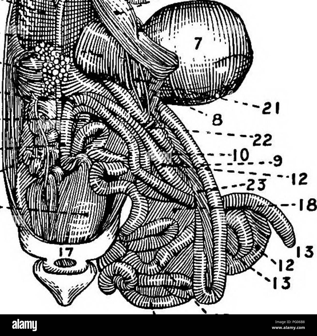 . Conkey's poultry book : a handy book of reference on poultry raising. Poultry; Poultry; Poultry. 1, tongue; 2, pharynx, show- ing opening to larynx; 3, up- per portion of oesophagus; 4, crop; 5, lower portion of oesophagus; 6, succentric ven- tricle; 7, gizzard; 8, origin of the duodenum; 9, first branch of duodenal flexure; 10, sec- ond branch of same; 11, origin of the floating portion of small intestine; 12, small intestine; 13, free extremities of, the cseca; 14, insertion of these two organs into the intestinal tube; '" rectum; 16, cloaca; 17, anus; 18, mesentery; 19, left lobe of Stock Photohttps://www.alamy.com/image-license-details/?v=1https://www.alamy.com/conkeys-poultry-book-a-handy-book-of-reference-on-poultry-raising-poultry-poultry-poultry-1-tongue-2-pharynx-show-ing-opening-to-larynx-3-up-per-portion-of-oesophagus-4-crop-5-lower-portion-of-oesophagus-6-succentric-ven-tricle-7-gizzard-8-origin-of-the-duodenum-9-first-branch-of-duodenal-flexure-10-sec-ond-branch-of-same-11-origin-of-the-floating-portion-of-small-intestine-12-small-intestine-13-free-extremities-of-the-cseca-14-insertion-of-these-two-organs-into-the-intestinal-tube-quot-rectum-16-cloaca-17-anus-18-mesentery-19-left-lobe-of-image216363932.html
RMPG06B8–. Conkey's poultry book : a handy book of reference on poultry raising. Poultry; Poultry; Poultry. 1, tongue; 2, pharynx, show- ing opening to larynx; 3, up- per portion of oesophagus; 4, crop; 5, lower portion of oesophagus; 6, succentric ven- tricle; 7, gizzard; 8, origin of the duodenum; 9, first branch of duodenal flexure; 10, sec- ond branch of same; 11, origin of the floating portion of small intestine; 12, small intestine; 13, free extremities of, the cseca; 14, insertion of these two organs into the intestinal tube; '" rectum; 16, cloaca; 17, anus; 18, mesentery; 19, left lobe of
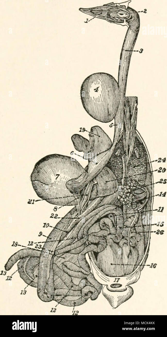 . Fig. 14.—Digestive App.4^r.tus of the Chicke.n. In this Hg-ure all of the head has been removed except the lower iaw which has been turned sidewise to show the ton;rue and the openings to the trachea and ••=« 1, tontrue; 2, pharynx, showing opening- to larvnx; 3, upper portion of OESophag-us; 4, crop; s, lower portion of oesophagus; 6, succentric ventricle- /,g-izzard; 8, ong-in of the duodenum: 9. first branch of duodenal flexure' ^fu^^l i: !;:^';"^,l-lSucr""'"'" ^"^ binkry-ducts; 23. pancVeas; Stock Photohttps://www.alamy.com/image-license-details/?v=1https://www.alamy.com/fig-14digestive-app4rtus-of-the-chicken-in-this-hg-ure-all-of-the-head-has-been-removed-except-the-lower-iaw-which-has-been-turned-sidewise-to-show-the-tonrue-and-the-openings-to-the-trachea-and-=-1-tontrue-2-pharynx-showing-opening-to-larvnx-3-upper-portion-of-oesophag-us-4-crop-s-lower-portion-of-oesophagus-6-succentric-ventricle-g-izzard-8-ong-in-of-the-duodenum-9-first-branch-of-duodenal-flexure-ful-i-!quotl-lsucrquotquotquotquot-quot-binkry-ducts-23-pancveas-image179900327.html
RMMCK4KK–. Fig. 14.—Digestive App.4^r.tus of the Chicke.n. In this Hg-ure all of the head has been removed except the lower iaw which has been turned sidewise to show the ton;rue and the openings to the trachea and ••=« 1, tontrue; 2, pharynx, showing opening- to larvnx; 3, upper portion of OESophag-us; 4, crop; s, lower portion of oesophagus; 6, succentric ventricle- /,g-izzard; 8, ong-in of the duodenum: 9. first branch of duodenal flexure' ^fu^^l i: !;:^';"^,l-lSucr""'"'" ^"^ binkry-ducts; 23. pancVeas;
 The diseases of poultry (1899) The diseases of poultry diseasesofpoultr00salmrich Year: 1899 DISEASES OF POULTRY, 67 Fig. 14.—Digestive App.4r.tus of the Chicke.n. In this Hg-ure all of the head has been removed except the lower iaw which has been turned sidewise to show the ton;rue and the openings to the trachea and ••=« 1, tontrue; 2, pharynx, showing opening- to larvnx; 3, upper portion of OESophag-us; 4, crop; s, lower portion of oesophagus; 6, succentric ventricle- /,g-izzard; 8, ong-in of the duodenum: 9. first branch of duodenal flexure' ful i: !;:';',l-lSucr'''''' ' binkry-ducts Stock Photohttps://www.alamy.com/image-license-details/?v=1https://www.alamy.com/the-diseases-of-poultry-1899-the-diseases-of-poultry-diseasesofpoultr00salmrich-year-1899-diseases-of-poultry-67-fig-14digestive-app4rtus-of-the-chicken-in-this-hg-ure-all-of-the-head-has-been-removed-except-the-lower-iaw-which-has-been-turned-sidewise-to-show-the-tonrue-and-the-openings-to-the-trachea-and-=-1-tontrue-2-pharynx-showing-opening-to-larvnx-3-upper-portion-of-oesophag-us-4-crop-s-lower-portion-of-oesophagus-6-succentric-ventricle-g-izzard-8-ong-in-of-the-duodenum-9-first-branch-of-duodenal-flexure-ful-i-!l-lsucr-binkry-ducts-image241940396.html
RMT1H9CC–The diseases of poultry (1899) The diseases of poultry diseasesofpoultr00salmrich Year: 1899 DISEASES OF POULTRY, 67 Fig. 14.—Digestive App.4r.tus of the Chicke.n. In this Hg-ure all of the head has been removed except the lower iaw which has been turned sidewise to show the ton;rue and the openings to the trachea and ••=« 1, tontrue; 2, pharynx, showing opening- to larvnx; 3, upper portion of OESophag-us; 4, crop; s, lower portion of oesophagus; 6, succentric ventricle- /,g-izzard; 8, ong-in of the duodenum: 9. first branch of duodenal flexure' ful i: !;:';',l-lSucr'''''' ' binkry-ducts
 . Poultry feeding and fattening, including preparation for market, special finishing methods, as practiced by American and foreign experts, handling broilers, capons, waterfowl, etc. ... Poultry. NDTKITION FOR LAYERS 47 canal and to expose the ovary and oviduct. 1, tongue; 2, pharynx; 3, first portion of the oesophagus; 4,. Fig. 6—^ANATOMY OF A FOWL (Howard) crop; 5, second portion of the oesophagus; 6, succentric ventricle; 7, gizzard; 8, origin of the duodenum; 9, first branch of the duodenal flexure; 10, second branch. Please note that these images are extracted from scanned page images tha Stock Photohttps://www.alamy.com/image-license-details/?v=1https://www.alamy.com/poultry-feeding-and-fattening-including-preparation-for-market-special-finishing-methods-as-practiced-by-american-and-foreign-experts-handling-broilers-capons-waterfowl-etc-poultry-ndtkition-for-layers-47-canal-and-to-expose-the-ovary-and-oviduct-1-tongue-2-pharynx-3-first-portion-of-the-oesophagus-4-fig-6anatomy-of-a-fowl-howard-crop-5-second-portion-of-the-oesophagus-6-succentric-ventricle-7-gizzard-8-origin-of-the-duodenum-9-first-branch-of-the-duodenal-flexure-10-second-branch-please-note-that-these-images-are-extracted-from-scanned-page-images-tha-image232215428.html
RMRDP944–. Poultry feeding and fattening, including preparation for market, special finishing methods, as practiced by American and foreign experts, handling broilers, capons, waterfowl, etc. ... Poultry. NDTKITION FOR LAYERS 47 canal and to expose the ovary and oviduct. 1, tongue; 2, pharynx; 3, first portion of the oesophagus; 4,. Fig. 6—^ANATOMY OF A FOWL (Howard) crop; 5, second portion of the oesophagus; 6, succentric ventricle; 7, gizzard; 8, origin of the duodenum; 9, first branch of the duodenal flexure; 10, second branch. Please note that these images are extracted from scanned page images tha
 . Cyclopedia of farm animals. Domestic animals; Animal products. Fig. 27. The digestive apparatus of a common fowl. 1, tongue; 2. esophagus, first part; 3, crop; 4, esophagus, second part; 5. succentrie ventricle; 6, gizzard: 7, origin of duodenum; 8, second blanch of duodenal flexure; 9, origin of floating part of small intestine; 10, small intestine: 11, caeca: 12, insertion of ca?ca; 13, rectum; 14, cloaca; 15, pancreas; 16, liver; 17, gall-bladder; 18, spleen. denum. Villi for absorption are numerous. Fowls have two club-shaped ceeca six to eighteen inches long ; they secrete a macerating Stock Photohttps://www.alamy.com/image-license-details/?v=1https://www.alamy.com/cyclopedia-of-farm-animals-domestic-animals-animal-products-fig-27-the-digestive-apparatus-of-a-common-fowl-1-tongue-2-esophagus-first-part-3-crop-4-esophagus-second-part-5-succentrie-ventricle-6-gizzard-7-origin-of-duodenum-8-second-blanch-of-duodenal-flexure-9-origin-of-floating-part-of-small-intestine-10-small-intestine-11-caeca-12-insertion-of-caca-13-rectum-14-cloaca-15-pancreas-16-liver-17-gall-bladder-18-spleen-denum-villi-for-absorption-are-numerous-fowls-have-two-club-shaped-ceeca-six-to-eighteen-inches-long-they-secrete-a-macerating-image216195830.html
RMPFMFYJ–. Cyclopedia of farm animals. Domestic animals; Animal products. Fig. 27. The digestive apparatus of a common fowl. 1, tongue; 2. esophagus, first part; 3, crop; 4, esophagus, second part; 5. succentrie ventricle; 6, gizzard: 7, origin of duodenum; 8, second blanch of duodenal flexure; 9, origin of floating part of small intestine; 10, small intestine: 11, caeca: 12, insertion of ca?ca; 13, rectum; 14, cloaca; 15, pancreas; 16, liver; 17, gall-bladder; 18, spleen. denum. Villi for absorption are numerous. Fowls have two club-shaped ceeca six to eighteen inches long ; they secrete a macerating
 . Commercial poultry raising; a thoroughly practical and complete reference work for the amateur, fancier or general farmer, especially adapted to the commercial poultryman. Poultry. I, Tong ue; 2, pharynx; 3, first portion of esopha- gus; 4, crop; 5, second portion of esophagus; 6, suc- centric ventricle; 7, gizzard; 8, ori- gin of the duoden- um; 9, first branch of the duodenal flexure; 10, second branch of same; II, origin of the floating portion of the small intes- tine; 12, small in- testine; 13, free extremities of the caecums; 14, in- sertion of these two culs-de- sac into the intestina Stock Photohttps://www.alamy.com/image-license-details/?v=1https://www.alamy.com/commercial-poultry-raising-a-thoroughly-practical-and-complete-reference-work-for-the-amateur-fancier-or-general-farmer-especially-adapted-to-the-commercial-poultryman-poultry-i-tong-ue-2-pharynx-3-first-portion-of-esopha-gus-4-crop-5-second-portion-of-esophagus-6-suc-centric-ventricle-7-gizzard-8-ori-gin-of-the-duoden-um-9-first-branch-of-the-duodenal-flexure-10-second-branch-of-same-ii-origin-of-the-floating-portion-of-the-small-intes-tine-12-small-in-testine-13-free-extremities-of-the-caecums-14-in-sertion-of-these-two-culs-de-sac-into-the-intestina-image232703107.html
RMREGF57–. Commercial poultry raising; a thoroughly practical and complete reference work for the amateur, fancier or general farmer, especially adapted to the commercial poultryman. Poultry. I, Tong ue; 2, pharynx; 3, first portion of esopha- gus; 4, crop; 5, second portion of esophagus; 6, suc- centric ventricle; 7, gizzard; 8, ori- gin of the duoden- um; 9, first branch of the duodenal flexure; 10, second branch of same; II, origin of the floating portion of the small intes- tine; 12, small in- testine; 13, free extremities of the caecums; 14, in- sertion of these two culs-de- sac into the intestina
 . Cyclopedia of farm animals. Domestic animals; Animal products. Fig. 27. The digestive apparatus of a common fowl. 1, tongue; 2. esophagus, first part; 3, crop; 4, esophagus, second part; 5. succentrie ventricle; 6, gizzard: 7, origin of duodenum; 8, second blanch of duodenal flexure; 9, origin of floating part of small intestine; 10, small intestine: 11, caeca: 12, insertion of ca?ca; 13, rectum; 14, cloaca; 15, pancreas; 16, liver; 17, gall-bladder; 18, spleen. denum. Villi for absorption are numerous. Fowls have two club-shaped ceeca six to eighteen inches long ; they secrete a macerating Stock Photohttps://www.alamy.com/image-license-details/?v=1https://www.alamy.com/cyclopedia-of-farm-animals-domestic-animals-animal-products-fig-27-the-digestive-apparatus-of-a-common-fowl-1-tongue-2-esophagus-first-part-3-crop-4-esophagus-second-part-5-succentrie-ventricle-6-gizzard-7-origin-of-duodenum-8-second-blanch-of-duodenal-flexure-9-origin-of-floating-part-of-small-intestine-10-small-intestine-11-caeca-12-insertion-of-caca-13-rectum-14-cloaca-15-pancreas-16-liver-17-gall-bladder-18-spleen-denum-villi-for-absorption-are-numerous-fowls-have-two-club-shaped-ceeca-six-to-eighteen-inches-long-they-secrete-a-macerating-image231832367.html
RMRD4TFB–. Cyclopedia of farm animals. Domestic animals; Animal products. Fig. 27. The digestive apparatus of a common fowl. 1, tongue; 2. esophagus, first part; 3, crop; 4, esophagus, second part; 5. succentrie ventricle; 6, gizzard: 7, origin of duodenum; 8, second blanch of duodenal flexure; 9, origin of floating part of small intestine; 10, small intestine: 11, caeca: 12, insertion of ca?ca; 13, rectum; 14, cloaca; 15, pancreas; 16, liver; 17, gall-bladder; 18, spleen. denum. Villi for absorption are numerous. Fowls have two club-shaped ceeca six to eighteen inches long ; they secrete a macerating
 . Conkey's poultry book : a handy book of reference on poultry raising. Poultry; Poultry; Poultry. 1, tongue; 2, pharynx, show- ing opening to larynx; 3, up- per portion of oesophagus; 4, crop; 5, lower portion of oesophagus; 6, succentric ven- tricle; 7, gizzard; 8, origin of the duodenum; 9, first branch of duodenal flexure; 10, sec- ond branch of same; 11, origin of the floating portion of small intestine; 12, small intestine; 13, free extremities of, the cseca; 14, insertion of these two organs into the intestinal tube; '" rectum; 16, cloaca; 17, anus; 18, mesentery; 19, left lobe of Stock Photohttps://www.alamy.com/image-license-details/?v=1https://www.alamy.com/conkeys-poultry-book-a-handy-book-of-reference-on-poultry-raising-poultry-poultry-poultry-1-tongue-2-pharynx-show-ing-opening-to-larynx-3-up-per-portion-of-oesophagus-4-crop-5-lower-portion-of-oesophagus-6-succentric-ven-tricle-7-gizzard-8-origin-of-the-duodenum-9-first-branch-of-duodenal-flexure-10-sec-ond-branch-of-same-11-origin-of-the-floating-portion-of-small-intestine-12-small-intestine-13-free-extremities-of-the-cseca-14-insertion-of-these-two-organs-into-the-intestinal-tube-quot-rectum-16-cloaca-17-anus-18-mesentery-19-left-lobe-of-image232064962.html
RMRDFD6A–. Conkey's poultry book : a handy book of reference on poultry raising. Poultry; Poultry; Poultry. 1, tongue; 2, pharynx, show- ing opening to larynx; 3, up- per portion of oesophagus; 4, crop; 5, lower portion of oesophagus; 6, succentric ven- tricle; 7, gizzard; 8, origin of the duodenum; 9, first branch of duodenal flexure; 10, sec- ond branch of same; 11, origin of the floating portion of small intestine; 12, small intestine; 13, free extremities of, the cseca; 14, insertion of these two organs into the intestinal tube; '" rectum; 16, cloaca; 17, anus; 18, mesentery; 19, left lobe of
 . Anatomy of the woodchuck (Marmota monax). Woodchuck; Mammals. 94 Anatomy of the Woodchuck, Marmota monax. Fig. 5-11. Abdomen, deep ventral view. 1 left lateral liver lobe, 2 pyloric part of the stomach, 3 body of the stomach, 4 spleen, 5 left lobe of the pancreas, 6 left adrenal gland, 7 left kidney, 8 caudal duodenal flexure, 9 descending duo- denum, 10 right lobe of the pancreas, 1 1 right kidney, 12 cranial flexure of the duodenum, 13 quadrate liver lobe, 14 left medial liver lobe. right lateral and medial lobes. The caudate lobe presents papillary and caudate processes. Interlobar fissur Stock Photohttps://www.alamy.com/image-license-details/?v=1https://www.alamy.com/anatomy-of-the-woodchuck-marmota-monax-woodchuck-mammals-94-anatomy-of-the-woodchuck-marmota-monax-fig-5-11-abdomen-deep-ventral-view-1-left-lateral-liver-lobe-2-pyloric-part-of-the-stomach-3-body-of-the-stomach-4-spleen-5-left-lobe-of-the-pancreas-6-left-adrenal-gland-7-left-kidney-8-caudal-duodenal-flexure-9-descending-duo-denum-10-right-lobe-of-the-pancreas-1-1-right-kidney-12-cranial-flexure-of-the-duodenum-13-quadrate-liver-lobe-14-left-medial-liver-lobe-right-lateral-and-medial-lobes-the-caudate-lobe-presents-papillary-and-caudate-processes-interlobar-fissur-image236799936.html
RMRN74MG–. Anatomy of the woodchuck (Marmota monax). Woodchuck; Mammals. 94 Anatomy of the Woodchuck, Marmota monax. Fig. 5-11. Abdomen, deep ventral view. 1 left lateral liver lobe, 2 pyloric part of the stomach, 3 body of the stomach, 4 spleen, 5 left lobe of the pancreas, 6 left adrenal gland, 7 left kidney, 8 caudal duodenal flexure, 9 descending duo- denum, 10 right lobe of the pancreas, 1 1 right kidney, 12 cranial flexure of the duodenum, 13 quadrate liver lobe, 14 left medial liver lobe. right lateral and medial lobes. The caudate lobe presents papillary and caudate processes. Interlobar fissur
 . Commercial poultry raising;. Poultry. I, Tongue; 2, pharynx; 3, first portion of esopha- gus; 4, crop; 5, second portion of esophagus; 6, suc- centric ventricle; 7, gizzard; 8, ori- gin of the duoden- um; 9, first branch of the duodenal flexure; 10, second branch of same; II, origin of the floating portion of the small intes- tine; 12, small in- testine; 13, free extremities of the csecums; 14, in- sertion of these two culs-de- sac into the intestinal tube; 15, rectum; 16, cloaca; 17, anus; 18, mesen- tery; 19, left lobe of liver; 20, right â 4- lobe of liver; 21, gall-bladder; 22, insertion Stock Photohttps://www.alamy.com/image-license-details/?v=1https://www.alamy.com/commercial-poultry-raising-poultry-i-tongue-2-pharynx-3-first-portion-of-esopha-gus-4-crop-5-second-portion-of-esophagus-6-suc-centric-ventricle-7-gizzard-8-ori-gin-of-the-duoden-um-9-first-branch-of-the-duodenal-flexure-10-second-branch-of-same-ii-origin-of-the-floating-portion-of-the-small-intes-tine-12-small-in-testine-13-free-extremities-of-the-csecums-14-in-sertion-of-these-two-culs-de-sac-into-the-intestinal-tube-15-rectum-16-cloaca-17-anus-18-mesen-tery-19-left-lobe-of-liver-20-right-4-lobe-of-liver-21-gall-bladder-22-insertion-image232702620.html
RMREGEFT–. Commercial poultry raising;. Poultry. I, Tongue; 2, pharynx; 3, first portion of esopha- gus; 4, crop; 5, second portion of esophagus; 6, suc- centric ventricle; 7, gizzard; 8, ori- gin of the duoden- um; 9, first branch of the duodenal flexure; 10, second branch of same; II, origin of the floating portion of the small intes- tine; 12, small in- testine; 13, free extremities of the csecums; 14, in- sertion of these two culs-de- sac into the intestinal tube; 15, rectum; 16, cloaca; 17, anus; 18, mesen- tery; 19, left lobe of liver; 20, right â 4- lobe of liver; 21, gall-bladder; 22, insertion
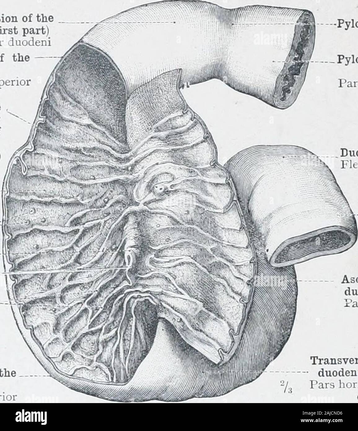 An atlas of human anatomy for students and physicians . ayerStratumcirculare Longitudinal layer Stratumlongitudinale of the pyloric portion of the stomach Serous coat /Tunica serosa Fasciculi of thelongitudinal layerwhich enter thepyloric sphincter Fig. 717.—M. Sphincter Pylori, the Pyloric Sphincter, in Longitudinal Section. Tubus digestorius—Alimentary canal. 440 ABDOMINAL AND PELVIC PORTIONS OF THE DIGESTIVE ORGANS Superior portion of theduodenxun (first part)Pars superior duodeni * Superior flexure of theduodenum »Flexura duodeni superi i Duodenal papilla, with the orifice of the accessory Stock Photohttps://www.alamy.com/image-license-details/?v=1https://www.alamy.com/an-atlas-of-human-anatomy-for-students-and-physicians-ayerstratumcirculare-longitudinal-layer-stratumlongitudinale-of-the-pyloric-portion-of-the-stomach-serous-coat-tunica-serosa-fasciculi-of-thelongitudinal-layerwhich-enter-thepyloric-sphincter-fig-717m-sphincter-pylori-the-pyloric-sphincter-in-longitudinal-section-tubus-digestoriusalimentary-canal-440-abdominal-and-pelvic-portions-of-the-digestive-organs-superior-portion-of-theduodenxun-first-partpars-superior-duodeni-superior-flexure-of-theduodenum-flexura-duodeni-superi-i-duodenal-papilla-with-the-orifice-of-the-accessory-image338341058.html
RM2AJCND6–An atlas of human anatomy for students and physicians . ayerStratumcirculare Longitudinal layer Stratumlongitudinale of the pyloric portion of the stomach Serous coat /Tunica serosa Fasciculi of thelongitudinal layerwhich enter thepyloric sphincter Fig. 717.—M. Sphincter Pylori, the Pyloric Sphincter, in Longitudinal Section. Tubus digestorius—Alimentary canal. 440 ABDOMINAL AND PELVIC PORTIONS OF THE DIGESTIVE ORGANS Superior portion of theduodenxun (first part)Pars superior duodeni * Superior flexure of theduodenum »Flexura duodeni superi i Duodenal papilla, with the orifice of the accessory
 Atlas and text-book of topographic and applied anatomy . Fig. 66. Fig. 67. Suprarenal gland Descending portion of the duode nferior duodenal flexi. Fig. 68. plenic flexure of col THE CONTENTS OF THE ABDOMEN. 141 the quadratus lumborum muscle. Such perinephritic abscesses and similar ones behind thekidney may extend upward, affecting the pleura (pleurisy), or the abscess may gravitate to theiliac fossa and pursue one of a number of subperitoneal courses similar to those taken by a retro-cecal (perityphlitic) abscess (see page 137). The method o) reaching the kidney from behind may be discerned Stock Photohttps://www.alamy.com/image-license-details/?v=1https://www.alamy.com/atlas-and-text-book-of-topographic-and-applied-anatomy-fig-66-fig-67-suprarenal-gland-descending-portion-of-the-duode-nferior-duodenal-flexi-fig-68-plenic-flexure-of-col-the-contents-of-the-abdomen-141-the-quadratus-lumborum-muscle-such-perinephritic-abscesses-and-similar-ones-behind-thekidney-may-extend-upward-affecting-the-pleura-pleurisy-or-the-abscess-may-gravitate-to-theiliac-fossa-and-pursue-one-of-a-number-of-subperitoneal-courses-similar-to-those-taken-by-a-retro-cecal-perityphlitic-abscess-see-page-137-the-method-o-reaching-the-kidney-from-behind-may-be-discerned-image338239430.html
RM2AJ83RJ–Atlas and text-book of topographic and applied anatomy . Fig. 66. Fig. 67. Suprarenal gland Descending portion of the duode nferior duodenal flexi. Fig. 68. plenic flexure of col THE CONTENTS OF THE ABDOMEN. 141 the quadratus lumborum muscle. Such perinephritic abscesses and similar ones behind thekidney may extend upward, affecting the pleura (pleurisy), or the abscess may gravitate to theiliac fossa and pursue one of a number of subperitoneal courses similar to those taken by a retro-cecal (perityphlitic) abscess (see page 137). The method o) reaching the kidney from behind may be discerned
 . Journal of anatomy. total length from fixedpoint to vestibule was only 13 cm., while in another with the left colonfreely mobile on a defined mesocolon the length was 28 cm. The Folds in Marsupials. (a) Wombat (Phascolomys).—There is a well-defined duodenal loop,and, as in the human, four portions may be recognised (fig. 3). It first The Peritoneum and Intestinal Tract in Monotremes and Marsupials 283 passes down and out for 2-3 cm., then descends for 9-12 cm., theninwards for 1-5 cm., and finally passes up and to the left of the midline to the duodeno-intestinal flexure for 3 cm. Owing to t Stock Photohttps://www.alamy.com/image-license-details/?v=1https://www.alamy.com/journal-of-anatomy-total-length-from-fixedpoint-to-vestibule-was-only-13-cm-while-in-another-with-the-left-colonfreely-mobile-on-a-defined-mesocolon-the-length-was-28-cm-the-folds-in-marsupials-a-wombat-phascolomysthere-is-a-well-defined-duodenal-loopand-as-in-the-human-four-portions-may-be-recognised-fig-3-it-first-the-peritoneum-and-intestinal-tract-in-monotremes-and-marsupials-283-passes-down-and-out-for-2-3-cm-then-descends-for-9-12-cm-theninwards-for-1-5-cm-and-finally-passes-up-and-to-the-left-of-the-midline-to-the-duodeno-intestinal-flexure-for-3-cm-owing-to-t-image336837751.html
RM2AG07YK–. Journal of anatomy. total length from fixedpoint to vestibule was only 13 cm., while in another with the left colonfreely mobile on a defined mesocolon the length was 28 cm. The Folds in Marsupials. (a) Wombat (Phascolomys).—There is a well-defined duodenal loop,and, as in the human, four portions may be recognised (fig. 3). It first The Peritoneum and Intestinal Tract in Monotremes and Marsupials 283 passes down and out for 2-3 cm., then descends for 9-12 cm., theninwards for 1-5 cm., and finally passes up and to the left of the midline to the duodeno-intestinal flexure for 3 cm. Owing to t
 . Archives of physical medicine and rehabilitation . Fig. 16—Inverted cecvun ascending colon and appendix.Simulated duodenal nicer. Surgical removal of appendixverified roentgenological diagnosis, with relief of aymp- /f. Fig. 17.—High situation of cecum and hepatic flexure.Short ascending colon. Boentgenological dlagnoBls of ap-pendicitis verified. Previous dia^rao*!* was gall-bladderdliaai*. 368 POSITIONAL ANOMALIES OF THE INTESTINAL TRACT—HUBEXY Stock Photohttps://www.alamy.com/image-license-details/?v=1https://www.alamy.com/archives-of-physical-medicine-and-rehabilitation-fig-16inverted-cecvun-ascending-colon-and-appendixsimulated-duodenal-nicer-surgical-removal-of-appendixverified-roentgenological-diagnosis-with-relief-of-aymp-f-fig-17high-situation-of-cecum-and-hepatic-flexureshort-ascending-colon-boentgenological-dlagnobls-of-ap-pendicitis-verified-previous-diarao!-was-gall-bladderdliaai-368-positional-anomalies-of-the-intestinal-tracthubexy-image375981117.html
RM2CRKBN1–. Archives of physical medicine and rehabilitation . Fig. 16—Inverted cecvun ascending colon and appendix.Simulated duodenal nicer. Surgical removal of appendixverified roentgenological diagnosis, with relief of aymp- /f. Fig. 17.—High situation of cecum and hepatic flexure.Short ascending colon. Boentgenological dlagnoBls of ap-pendicitis verified. Previous dia^rao*!* was gall-bladderdliaai*. 368 POSITIONAL ANOMALIES OF THE INTESTINAL TRACT—HUBEXY
 . On retro-peritoneal hernia . is is the fossawhich, as I believe, Huschkc was the first to describe. Inthe article to which I have previously referred, he expresslystates that the folds spring from the root of the transversemesocolon, a statement which is obviously untrue about allother folds. THE DUODENAL FOLDS AND FOSSM On dragging the transverse colon upwards, and the jejunumdownwards and to the right, there can be seen at the rootof the transverse mesocolon a fossa, which results from theplunging, as it were, of the duodeno-jejunal flexure backwardsinto the root of the transverse mesocolo Stock Photohttps://www.alamy.com/image-license-details/?v=1https://www.alamy.com/on-retro-peritoneal-hernia-is-is-the-fossawhich-as-i-believe-huschkc-was-the-first-to-describe-inthe-article-to-which-i-have-previously-referred-he-expresslystates-that-the-folds-spring-from-the-root-of-the-transversemesocolon-a-statement-which-is-obviously-untrue-about-allother-folds-the-duodenal-folds-and-fossm-on-dragging-the-transverse-colon-upwards-and-the-jejunumdownwards-and-to-the-right-there-can-be-seen-at-the-rootof-the-transverse-mesocolon-a-fossa-which-results-from-theplunging-as-it-were-of-the-duodeno-jejunal-flexure-backwardsinto-the-root-of-the-transverse-mesocolo-image369697481.html
RM2CDD4WD–. On retro-peritoneal hernia . is is the fossawhich, as I believe, Huschkc was the first to describe. Inthe article to which I have previously referred, he expresslystates that the folds spring from the root of the transversemesocolon, a statement which is obviously untrue about allother folds. THE DUODENAL FOLDS AND FOSSM On dragging the transverse colon upwards, and the jejunumdownwards and to the right, there can be seen at the rootof the transverse mesocolon a fossa, which results from theplunging, as it were, of the duodeno-jejunal flexure backwardsinto the root of the transverse mesocolo
 . Journal of radiology . Fig. 16.—Inverted cecum, ascending colon and appendix.Simulated duodenal ulcer. Surgical removal of appendixverified roentgenological diagnosis, with relief of symp-toms.. Fig. 17.—High situation of cecum and hepatic flexure.Short ascending colon. Roentgenological diagnosis of ap-pendicitis verified. Previous diagnosis was gall-bladderdisease. 368 POSITIONAL ANOMALIES OF THE INTESTINAL TRACT—HUBENY Fig. 18.—This and the two figures immediately following-are of the same patient. Clinical diagnosis was duodenalulcer. Roentgen examination of stomach and duodenumonly was r Stock Photohttps://www.alamy.com/image-license-details/?v=1https://www.alamy.com/journal-of-radiology-fig-16inverted-cecum-ascending-colon-and-appendixsimulated-duodenal-ulcer-surgical-removal-of-appendixverified-roentgenological-diagnosis-with-relief-of-symp-toms-fig-17high-situation-of-cecum-and-hepatic-flexureshort-ascending-colon-roentgenological-diagnosis-of-ap-pendicitis-verified-previous-diagnosis-was-gall-bladderdisease-368-positional-anomalies-of-the-intestinal-tracthubeny-fig-18this-and-the-two-figures-immediately-following-are-of-the-same-patient-clinical-diagnosis-was-duodenalulcer-roentgen-examination-of-stomach-and-duodenumonly-was-r-image375947754.html
RM2CRHW5E–. Journal of radiology . Fig. 16.—Inverted cecum, ascending colon and appendix.Simulated duodenal ulcer. Surgical removal of appendixverified roentgenological diagnosis, with relief of symp-toms.. Fig. 17.—High situation of cecum and hepatic flexure.Short ascending colon. Roentgenological diagnosis of ap-pendicitis verified. Previous diagnosis was gall-bladderdisease. 368 POSITIONAL ANOMALIES OF THE INTESTINAL TRACT—HUBENY Fig. 18.—This and the two figures immediately following-are of the same patient. Clinical diagnosis was duodenalulcer. Roentgen examination of stomach and duodenumonly was r
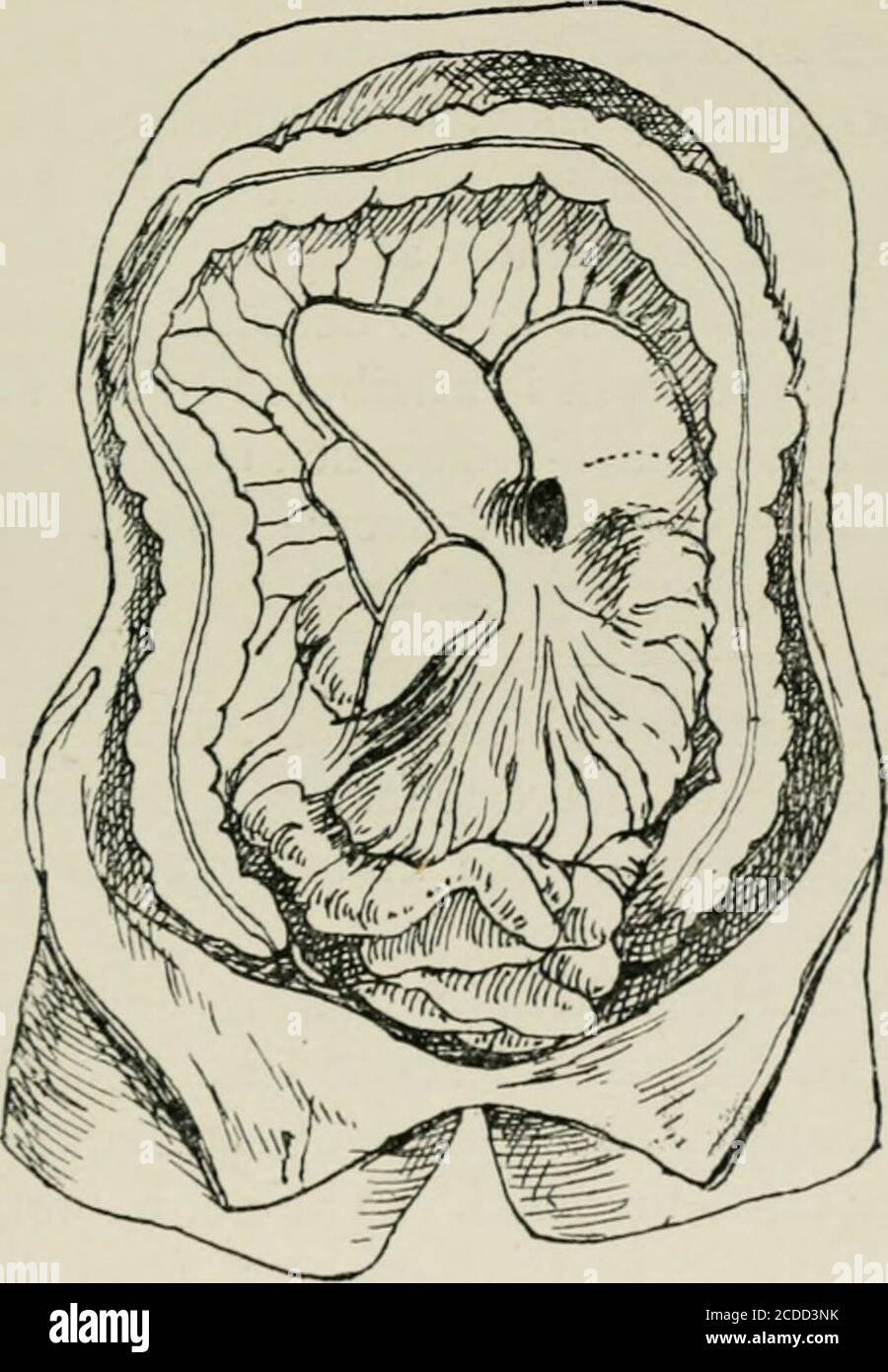 . On retro-peritoneal hernia . The duodeno-jejunal flexure is a hernial content of the cul-de-sac. Theinferior mesenteric vein running upwards and to the rightforms a concavity, which corresponds with a fair degree ofaccuracy to the upper limit of the fossa. The fossa is alwaysvascular. In the cases where this fossa is found there has never yetbeen seen any other form of duodenal fossa. In a few cases a division of the orifice of the sac by aperitoneal fold has been observed. This is probably pro- THE FOLDS AND FOSS^ 31 duced in a similar manner to the division of the superior andinferior duod Stock Photohttps://www.alamy.com/image-license-details/?v=1https://www.alamy.com/on-retro-peritoneal-hernia-the-duodeno-jejunal-flexure-is-a-hernial-content-of-the-cul-de-sac-theinferior-mesenteric-vein-running-upwards-and-to-the-rightforms-a-concavity-which-corresponds-with-a-fair-degree-ofaccuracy-to-the-upper-limit-of-the-fossa-the-fossa-is-alwaysvascular-in-the-cases-where-this-fossa-is-found-there-has-never-yetbeen-seen-any-other-form-of-duodenal-fossa-in-a-few-cases-a-division-of-the-orifice-of-the-sac-by-aperitoneal-fold-has-been-observed-this-is-probably-pro-the-folds-and-foss-31-duced-in-a-similar-manner-to-the-division-of-the-superior-andinferior-duod-image369696591.html
RM2CDD3NK–. On retro-peritoneal hernia . The duodeno-jejunal flexure is a hernial content of the cul-de-sac. Theinferior mesenteric vein running upwards and to the rightforms a concavity, which corresponds with a fair degree ofaccuracy to the upper limit of the fossa. The fossa is alwaysvascular. In the cases where this fossa is found there has never yetbeen seen any other form of duodenal fossa. In a few cases a division of the orifice of the sac by aperitoneal fold has been observed. This is probably pro- THE FOLDS AND FOSS^ 31 duced in a similar manner to the division of the superior andinferior duod
 . Archives of physical medicine and rehabilitation . wrong diag-nosis in seven cases. Five errors were due to prepyloriclesions, cancer in three cases, ulcer intwo. All of these lesions were nearthe pylorus. A cancer involving theliver, but whose origin was not de-termined, may have been the cause ofthe duodenal deformity in one case,and in another the lesion was a cancerof the hepatic flexure. Thus, in twenty-two of the twenty-three cases there was definite justifica-tion for surgical intervention, and intwo-thirds of them the lesion was inthe right upper quadrant of the abdo-men. In the rema Stock Photohttps://www.alamy.com/image-license-details/?v=1https://www.alamy.com/archives-of-physical-medicine-and-rehabilitation-wrong-diag-nosis-in-seven-cases-five-errors-were-due-to-prepyloriclesions-cancer-in-three-cases-ulcer-intwo-all-of-these-lesions-were-nearthe-pylorus-a-cancer-involving-theliver-but-whose-origin-was-not-de-termined-may-have-been-the-cause-ofthe-duodenal-deformity-in-one-caseand-in-another-the-lesion-was-a-cancerof-the-hepatic-flexure-thus-in-twenty-two-of-the-twenty-three-cases-there-was-definite-justifica-tion-for-surgical-intervention-and-intwo-thirds-of-them-the-lesion-was-inthe-right-upper-quadrant-of-the-abdo-men-in-the-rema-image376058702.html
RM2CRPXKX–. Archives of physical medicine and rehabilitation . wrong diag-nosis in seven cases. Five errors were due to prepyloriclesions, cancer in three cases, ulcer intwo. All of these lesions were nearthe pylorus. A cancer involving theliver, but whose origin was not de-termined, may have been the cause ofthe duodenal deformity in one case,and in another the lesion was a cancerof the hepatic flexure. Thus, in twenty-two of the twenty-three cases there was definite justifica-tion for surgical intervention, and intwo-thirds of them the lesion was inthe right upper quadrant of the abdo-men. In the rema
 . On retro-peritoneal hernia : being the 'Arris and Gale' lectures on the 'The anatomy and surgery of the peritoneal fossae' : delivered at the Royal College of Surgeons of England in 1897. II.—ROYAL COLLEGE OF SURGEONS. 168 DESCRIPTION OF PLATE III. III.—SPECIMEN 1083. GUYS HOSPITAL MUSEUM. Left Duodenal Hernia. The splenic flexure and descending part of a colon in theangle between which is situated a globular peritoneal sacmeasuring about 4 inches in diameter, and containing severalfeet of small intestine. The mouth of the sac is directed towardsthe vertebral column, and along its anterior m Stock Photohttps://www.alamy.com/image-license-details/?v=1https://www.alamy.com/on-retro-peritoneal-hernia-being-the-arris-and-gale-lectures-on-the-the-anatomy-and-surgery-of-the-peritoneal-fossae-delivered-at-the-royal-college-of-surgeons-of-england-in-1897-iiroyal-college-of-surgeons-168-description-of-plate-iii-iiispecimen-1083-guys-hospital-museum-left-duodenal-hernia-the-splenic-flexure-and-descending-part-of-a-colon-in-theangle-between-which-is-situated-a-globular-peritoneal-sacmeasuring-about-4-inches-in-diameter-and-containing-severalfeet-of-small-intestine-the-mouth-of-the-sac-is-directed-towardsthe-vertebral-column-and-along-its-anterior-m-image370484575.html
RM2CEN0RY–. On retro-peritoneal hernia : being the 'Arris and Gale' lectures on the 'The anatomy and surgery of the peritoneal fossae' : delivered at the Royal College of Surgeons of England in 1897. II.—ROYAL COLLEGE OF SURGEONS. 168 DESCRIPTION OF PLATE III. III.—SPECIMEN 1083. GUYS HOSPITAL MUSEUM. Left Duodenal Hernia. The splenic flexure and descending part of a colon in theangle between which is situated a globular peritoneal sacmeasuring about 4 inches in diameter, and containing severalfeet of small intestine. The mouth of the sac is directed towardsthe vertebral column, and along its anterior m
 . Journal of radiology . wrong diag-nosis in seven cases. Five errors were due to prepyloriclesions, cancer in three cases, ulcer intwo. All of these lesions were nearthe pylorus. A cancer involving theliver, but whose origin was not de-termined, may have been the cause ofthe duodenal deformity in one case,and in another the iesion was a cancerof the hepatic flexure. Thus, in twenty-two of the twenty-three cases there was definite justifica-tion for surgical intervention, and intwo-thirds of them the lesion was inthe right upper quadrant of the abdo-men. In the remaining instance noth-ing coul Stock Photohttps://www.alamy.com/image-license-details/?v=1https://www.alamy.com/journal-of-radiology-wrong-diag-nosis-in-seven-cases-five-errors-were-due-to-prepyloriclesions-cancer-in-three-cases-ulcer-intwo-all-of-these-lesions-were-nearthe-pylorus-a-cancer-involving-theliver-but-whose-origin-was-not-de-termined-may-have-been-the-cause-ofthe-duodenal-deformity-in-one-caseand-in-another-the-iesion-was-a-cancerof-the-hepatic-flexure-thus-in-twenty-two-of-the-twenty-three-cases-there-was-definite-justifica-tion-for-surgical-intervention-and-intwo-thirds-of-them-the-lesion-was-inthe-right-upper-quadrant-of-the-abdo-men-in-the-remaining-instance-noth-ing-coul-image375966972.html
RM2CRJNKT–. Journal of radiology . wrong diag-nosis in seven cases. Five errors were due to prepyloriclesions, cancer in three cases, ulcer intwo. All of these lesions were nearthe pylorus. A cancer involving theliver, but whose origin was not de-termined, may have been the cause ofthe duodenal deformity in one case,and in another the iesion was a cancerof the hepatic flexure. Thus, in twenty-two of the twenty-three cases there was definite justifica-tion for surgical intervention, and intwo-thirds of them the lesion was inthe right upper quadrant of the abdo-men. In the remaining instance noth-ing coul
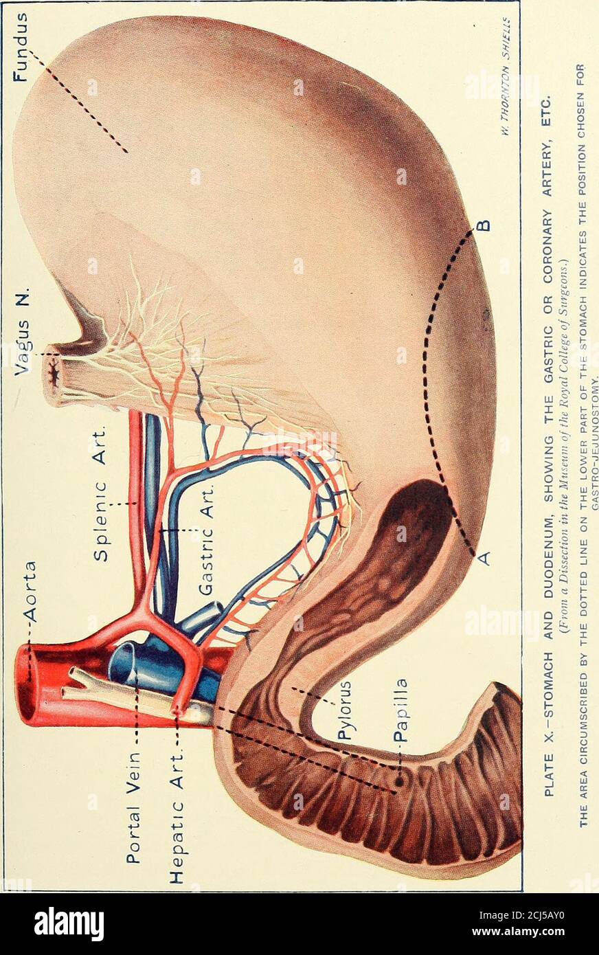 . A manual of operative surgery . terzi— S.M FIG. 49. -POSITION AND RELATIONS OF THE STOMACH AND DUODENUM, SHOWINGALSO THEIR ARTERIAL SUPPLY. {After Testut.) s, Stomach ; p, Pylorus ; D, Third part of duodenum; c, Hepatic flexure of colon (note thetransverse colon is cutaway) ; C. A., The coronary artery ; G. E. D., Right gastro-epiploicartery running in the great omentum ; s. M., Superior mesenteric artery, with its inferiorpancreatico-duodenal branch (p. d. i.). gastrojejunostomy we found the pylorus close to the rightanterior superior iliac spine ! The fundus of the stomach reaches on the l Stock Photohttps://www.alamy.com/image-license-details/?v=1https://www.alamy.com/a-manual-of-operative-surgery-terzi-sm-fig-49-position-and-relations-of-the-stomach-and-duodenum-showingalso-their-arterial-supply-after-testut-s-stomach-p-pylorus-d-third-part-of-duodenum-c-hepatic-flexure-of-colon-note-thetransverse-colon-is-cutaway-c-a-the-coronary-artery-g-e-d-right-gastro-epiploicartery-running-in-the-great-omentum-s-m-superior-mesenteric-artery-with-its-inferiorpancreatico-duodenal-branch-p-d-i-gastrojejunostomy-we-found-the-pylorus-close-to-the-rightanterior-superior-iliac-spine-!-the-fundus-of-the-stomach-reaches-on-the-l-image372599892.html
RM2CJ5AY0–. A manual of operative surgery . terzi— S.M FIG. 49. -POSITION AND RELATIONS OF THE STOMACH AND DUODENUM, SHOWINGALSO THEIR ARTERIAL SUPPLY. {After Testut.) s, Stomach ; p, Pylorus ; D, Third part of duodenum; c, Hepatic flexure of colon (note thetransverse colon is cutaway) ; C. A., The coronary artery ; G. E. D., Right gastro-epiploicartery running in the great omentum ; s. M., Superior mesenteric artery, with its inferiorpancreatico-duodenal branch (p. d. i.). gastrojejunostomy we found the pylorus close to the rightanterior superior iliac spine ! The fundus of the stomach reaches on the l
 . Archives of physical medicine and rehabilitation . int affection.) S^Y4 Table 3—Location of Lesions Which May Be Regarded AS Foci of Infection 1. Gastro-intestinal lesions in I 00cases (81 per cent of allcases) : Constricting bands ofcolon (including ter-minal ileum) oc-curred: Ileocecal 47 times Sigmoid 10 times Splenic Flexure. 8 times Total 65 times Duodenal bands or membranes ... .22 timesChronic appendicitis 22 timesIleocecal incompe-tency 12 times Chronic cholecystitis I 7 timesSpasticity of colon . I 0 timesAtonicity and re-dundancy of colons 5 timesDuodenal ulcer. ... 3 timesGastric Stock Photohttps://www.alamy.com/image-license-details/?v=1https://www.alamy.com/archives-of-physical-medicine-and-rehabilitation-int-affection-sy4-table-3location-of-lesions-which-may-be-regarded-as-foci-of-infection-1-gastro-intestinal-lesions-in-i-00cases-81-per-cent-of-allcases-constricting-bands-ofcolon-including-ter-minal-ileum-oc-curred-ileocecal-47-times-sigmoid-10-times-splenic-flexure-8-times-total-65-times-duodenal-bands-or-membranes-22-timeschronic-appendicitis-22-timesileocecal-incompe-tency-12-times-chronic-cholecystitis-i-7-timesspasticity-of-colon-i-0-timesatonicity-and-re-dundancy-of-colons-5-timesduodenal-ulcer-3-timesgastric-image376066417.html
RM2CRR8FD–. Archives of physical medicine and rehabilitation . int affection.) S^Y4 Table 3—Location of Lesions Which May Be Regarded AS Foci of Infection 1. Gastro-intestinal lesions in I 00cases (81 per cent of allcases) : Constricting bands ofcolon (including ter-minal ileum) oc-curred: Ileocecal 47 times Sigmoid 10 times Splenic Flexure. 8 times Total 65 times Duodenal bands or membranes ... .22 timesChronic appendicitis 22 timesIleocecal incompe-tency 12 times Chronic cholecystitis I 7 timesSpasticity of colon . I 0 timesAtonicity and re-dundancy of colons 5 timesDuodenal ulcer. ... 3 timesGastric
 . The American journal of roentgenology, radium therapy and nuclear medicine . Fig. 5 and 5. Illustrates a veil that involves the anterior surface of the cap and hepatic flexure of the colon.Fig. s et 5. Demontrant une voile qui comprend la anterieure du bulbe duodenale et la flexion hepatique du Fig. s et 5. Ilustrando un velo que envm Ive la cara anterior delbulbo duodenal, y el asa hepatica del colon. FIG 6 and 6. Illustrates a veil that involves the lesser curvature of the stomach and the superior surface of the cap.Fig 6 et 6. Demontrant une voile qui comprend la courbureIre de lestomac e Stock Photohttps://www.alamy.com/image-license-details/?v=1https://www.alamy.com/the-american-journal-of-roentgenology-radium-therapy-and-nuclear-medicine-fig-5-and-5-illustrates-a-veil-that-involves-the-anterior-surface-of-the-cap-and-hepatic-flexure-of-the-colonfig-s-et-5-demontrant-une-voile-qui-comprend-la-anterieure-du-bulbe-duodenale-et-la-flexion-hepatique-du-fig-s-et-5-ilustrando-un-velo-que-envm-ive-la-cara-anterior-delbulbo-duodenal-y-el-asa-hepatica-del-colon-fig-6-and-6-illustrates-a-veil-that-involves-the-lesser-curvature-of-the-stomach-and-the-superior-surface-of-the-capfig-6-et-6-demontrant-une-voile-qui-comprend-la-courbureire-de-lestomac-e-image376069041.html
RM2CRRBW5–. The American journal of roentgenology, radium therapy and nuclear medicine . Fig. 5 and 5. Illustrates a veil that involves the anterior surface of the cap and hepatic flexure of the colon.Fig. s et 5. Demontrant une voile qui comprend la anterieure du bulbe duodenale et la flexion hepatique du Fig. s et 5. Ilustrando un velo que envm Ive la cara anterior delbulbo duodenal, y el asa hepatica del colon. FIG 6 and 6. Illustrates a veil that involves the lesser curvature of the stomach and the superior surface of the cap.Fig 6 et 6. Demontrant une voile qui comprend la courbureIre de lestomac e
 . The American journal of roentgenology, radium therapy and nuclear medicine . Fig. 5 and 5. Illustrates a veil that involves the anterior surface of the cap and hepatic flexure of the colon.Fig. s et 5. Demontrant une voile qui comprend la anterieure du bulbe duodenale et la flexion hepatique du Fig. s et 5. Ilustrando un velo que envm Ive la cara anterior delbulbo duodenal, y el asa hepatica del colon. FIG 6 and 6. Illustrates a veil that involves the lesser curvature of the stomach and the superior surface of the cap.Fig 6 et 6. Demontrant une voile qui comprend la courbureIre de lestomac e Stock Photohttps://www.alamy.com/image-license-details/?v=1https://www.alamy.com/the-american-journal-of-roentgenology-radium-therapy-and-nuclear-medicine-fig-5-and-5-illustrates-a-veil-that-involves-the-anterior-surface-of-the-cap-and-hepatic-flexure-of-the-colonfig-s-et-5-demontrant-une-voile-qui-comprend-la-anterieure-du-bulbe-duodenale-et-la-flexion-hepatique-du-fig-s-et-5-ilustrando-un-velo-que-envm-ive-la-cara-anterior-delbulbo-duodenal-y-el-asa-hepatica-del-colon-fig-6-and-6-illustrates-a-veil-that-involves-the-lesser-curvature-of-the-stomach-and-the-superior-surface-of-the-capfig-6-et-6-demontrant-une-voile-qui-comprend-la-courbureire-de-lestomac-e-image376069005.html
RM2CRRBRW–. The American journal of roentgenology, radium therapy and nuclear medicine . Fig. 5 and 5. Illustrates a veil that involves the anterior surface of the cap and hepatic flexure of the colon.Fig. s et 5. Demontrant une voile qui comprend la anterieure du bulbe duodenale et la flexion hepatique du Fig. s et 5. Ilustrando un velo que envm Ive la cara anterior delbulbo duodenal, y el asa hepatica del colon. FIG 6 and 6. Illustrates a veil that involves the lesser curvature of the stomach and the superior surface of the cap.Fig 6 et 6. Demontrant une voile qui comprend la courbureIre de lestomac e
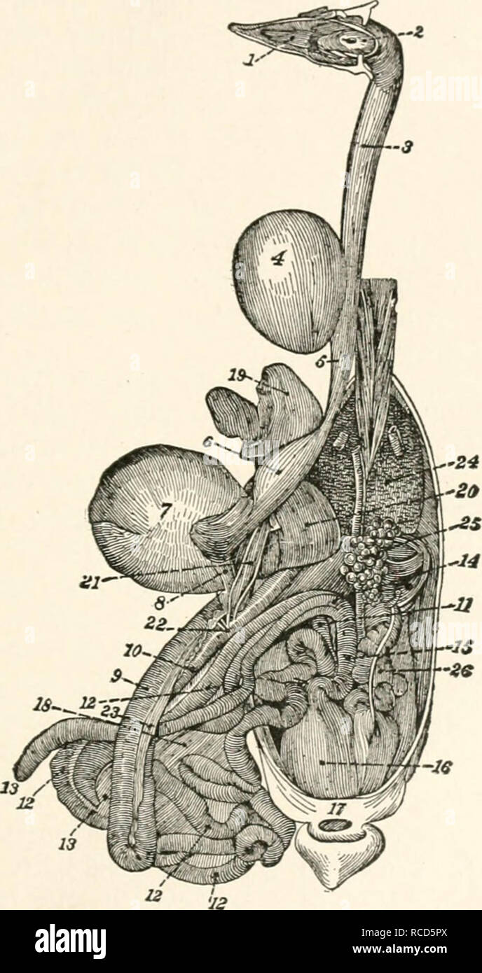 . The diseases of poultry. DISEASES OF POULTRY, 67. Fig. 14.—Digestive App.4^r.tus of the Chicke.n. In this Hg-ure all of the head has been removed except the lower iaw which has been turned sidewise to show the ton;rue and the openings to the trachea and ••=« 1, tontrue; 2, pharynx, showing opening- to larvnx; 3, upper portion of OESophag-us; 4, crop; s, lower portion of oesophagus; 6, succentric ventricle- /,g-izzard; 8, ong-in of the duodenum: 9. first branch of duodenal flexure' ^fu^^l i: !;:^';"^,l-lSucr""'"'" ^"^ binkry-ducts; 23. pancVeas;. Please note tha Stock Photohttps://www.alamy.com/image-license-details/?v=1https://www.alamy.com/the-diseases-of-poultry-diseases-of-poultry-67-fig-14digestive-app4rtus-of-the-chicken-in-this-hg-ure-all-of-the-head-has-been-removed-except-the-lower-iaw-which-has-been-turned-sidewise-to-show-the-tonrue-and-the-openings-to-the-trachea-and-=-1-tontrue-2-pharynx-showing-opening-to-larvnx-3-upper-portion-of-oesophag-us-4-crop-s-lower-portion-of-oesophagus-6-succentric-ventricle-g-izzard-8-ong-in-of-the-duodenum-9-first-branch-of-duodenal-flexure-ful-i-!quotl-lsucrquotquotquotquot-quot-binkry-ducts-23-pancveas-please-note-tha-image231400594.html
RMRCD5PX–. The diseases of poultry. DISEASES OF POULTRY, 67. Fig. 14.—Digestive App.4^r.tus of the Chicke.n. In this Hg-ure all of the head has been removed except the lower iaw which has been turned sidewise to show the ton;rue and the openings to the trachea and ••=« 1, tontrue; 2, pharynx, showing opening- to larvnx; 3, upper portion of OESophag-us; 4, crop; s, lower portion of oesophagus; 6, succentric ventricle- /,g-izzard; 8, ong-in of the duodenum: 9. first branch of duodenal flexure' ^fu^^l i: !;:^';"^,l-lSucr""'"'" ^"^ binkry-ducts; 23. pancVeas;. Please note tha
 . The anatomy of the domestic animals. Veterinary anatomy. THE LIVER 435 cave area which is the surface of contact with the stomach. (3) Leading from this to the right of the portal fissure and dorsally is the duodenal impression (Impressio duodenahs). (4) The colic impression (Impressio coHca) is situated ventrally and to the right of the gastric antl duodenal impressions, from which it is separated by a ridge; it corresponds to the extensive contact of the diaphragmatic flexure and right dorsal part of the colon. (5) A caecal impression (Impressio C£ecalis) may be found dorsal to the precedi Stock Photohttps://www.alamy.com/image-license-details/?v=1https://www.alamy.com/the-anatomy-of-the-domestic-animals-veterinary-anatomy-the-liver-435-cave-area-which-is-the-surface-of-contact-with-the-stomach-3-leading-from-this-to-the-right-of-the-portal-fissure-and-dorsally-is-the-duodenal-impression-impressio-duodenahs-4-the-colic-impression-impressio-cohca-is-situated-ventrally-and-to-the-right-of-the-gastric-antl-duodenal-impressions-from-which-it-is-separated-by-a-ridge-it-corresponds-to-the-extensive-contact-of-the-diaphragmatic-flexure-and-right-dorsal-part-of-the-colon-5-a-caecal-impression-impressio-cecalis-may-be-found-dorsal-to-the-precedi-image236799553.html
RMRN746W–. The anatomy of the domestic animals. Veterinary anatomy. THE LIVER 435 cave area which is the surface of contact with the stomach. (3) Leading from this to the right of the portal fissure and dorsally is the duodenal impression (Impressio duodenahs). (4) The colic impression (Impressio coHca) is situated ventrally and to the right of the gastric antl duodenal impressions, from which it is separated by a ridge; it corresponds to the extensive contact of the diaphragmatic flexure and right dorsal part of the colon. (5) A caecal impression (Impressio C£ecalis) may be found dorsal to the precedi
 . The anatomy of the domestic animals . Veterinary anatomy. THE LIVER 435 cave area which is the surface of contact with the stomach. (3) Leading from this to the right of the portal fissure and dorsally is the duodenal impression (Impressio duodenalis). (4) The colic impression (Impressio colica) is situated ventrally and to the right of the gastric and duodenal impressions, from which it is separated by a ridge; it corresponds to the extensive contact of the diaphragmatic flexure and right dorsal part of the colon. (5) A caecal impression (Impressio caecalis) may be found dorsal to the prece Stock Photohttps://www.alamy.com/image-license-details/?v=1https://www.alamy.com/the-anatomy-of-the-domestic-animals-veterinary-anatomy-the-liver-435-cave-area-which-is-the-surface-of-contact-with-the-stomach-3-leading-from-this-to-the-right-of-the-portal-fissure-and-dorsally-is-the-duodenal-impression-impressio-duodenalis-4-the-colic-impression-impressio-colica-is-situated-ventrally-and-to-the-right-of-the-gastric-and-duodenal-impressions-from-which-it-is-separated-by-a-ridge-it-corresponds-to-the-extensive-contact-of-the-diaphragmatic-flexure-and-right-dorsal-part-of-the-colon-5-a-caecal-impression-impressio-caecalis-may-be-found-dorsal-to-the-prece-image232314202.html
RMRDXR3P–. The anatomy of the domestic animals . Veterinary anatomy. THE LIVER 435 cave area which is the surface of contact with the stomach. (3) Leading from this to the right of the portal fissure and dorsally is the duodenal impression (Impressio duodenalis). (4) The colic impression (Impressio colica) is situated ventrally and to the right of the gastric and duodenal impressions, from which it is separated by a ridge; it corresponds to the extensive contact of the diaphragmatic flexure and right dorsal part of the colon. (5) A caecal impression (Impressio caecalis) may be found dorsal to the prece
![. The anatomy of the domestic animals. Veterinary anatomy. THE LIVER 435 cave area which is the surface of contact with the stomach. ("3) Leading from this to the right of the ]iortal fissure and dorsally is the duodenal impression (Impressio duodenahs). (4) The colic impression (Impressio colica) is situated ventrally and to the right of the gastric and (hiodcnal impressions, from which it is separated by a ridge; it corresponds to the extensive contact of the diaphragmatic flexure and right dorsal part of the colon. (5) A caecal impression (Impressio csecalis) may he found dorsal to the Stock Photo . The anatomy of the domestic animals. Veterinary anatomy. THE LIVER 435 cave area which is the surface of contact with the stomach. ("3) Leading from this to the right of the ]iortal fissure and dorsally is the duodenal impression (Impressio duodenahs). (4) The colic impression (Impressio colica) is situated ventrally and to the right of the gastric and (hiodcnal impressions, from which it is separated by a ridge; it corresponds to the extensive contact of the diaphragmatic flexure and right dorsal part of the colon. (5) A caecal impression (Impressio csecalis) may he found dorsal to the Stock Photo](https://c8.alamy.com/comp/RN747N/the-anatomy-of-the-domestic-animals-veterinary-anatomy-the-liver-435-cave-area-which-is-the-surface-of-contact-with-the-stomach-quot3-leading-from-this-to-the-right-of-the-iortal-fissure-and-dorsally-is-the-duodenal-impression-impressio-duodenahs-4-the-colic-impression-impressio-colica-is-situated-ventrally-and-to-the-right-of-the-gastric-and-hiodcnal-impressions-from-which-it-is-separated-by-a-ridge-it-corresponds-to-the-extensive-contact-of-the-diaphragmatic-flexure-and-right-dorsal-part-of-the-colon-5-a-caecal-impression-impressio-csecalis-may-he-found-dorsal-to-the-RN747N.jpg) . The anatomy of the domestic animals. Veterinary anatomy. THE LIVER 435 cave area which is the surface of contact with the stomach. ("3) Leading from this to the right of the ]iortal fissure and dorsally is the duodenal impression (Impressio duodenahs). (4) The colic impression (Impressio colica) is situated ventrally and to the right of the gastric and (hiodcnal impressions, from which it is separated by a ridge; it corresponds to the extensive contact of the diaphragmatic flexure and right dorsal part of the colon. (5) A caecal impression (Impressio csecalis) may he found dorsal to the Stock Photohttps://www.alamy.com/image-license-details/?v=1https://www.alamy.com/the-anatomy-of-the-domestic-animals-veterinary-anatomy-the-liver-435-cave-area-which-is-the-surface-of-contact-with-the-stomach-quot3-leading-from-this-to-the-right-of-the-iortal-fissure-and-dorsally-is-the-duodenal-impression-impressio-duodenahs-4-the-colic-impression-impressio-colica-is-situated-ventrally-and-to-the-right-of-the-gastric-and-hiodcnal-impressions-from-which-it-is-separated-by-a-ridge-it-corresponds-to-the-extensive-contact-of-the-diaphragmatic-flexure-and-right-dorsal-part-of-the-colon-5-a-caecal-impression-impressio-csecalis-may-he-found-dorsal-to-the-image236799577.html
. The anatomy of the domestic animals. Veterinary anatomy. THE LIVER 435 cave area which is the surface of contact with the stomach. ("3) Leading from this to the right of the ]iortal fissure and dorsally is the duodenal impression (Impressio duodenahs). (4) The colic impression (Impressio colica) is situated ventrally and to the right of the gastric and (hiodcnal impressions, from which it is separated by a ridge; it corresponds to the extensive contact of the diaphragmatic flexure and right dorsal part of the colon. (5) A caecal impression (Impressio csecalis) may he found dorsal to the Stock Photohttps://www.alamy.com/image-license-details/?v=1https://www.alamy.com/the-anatomy-of-the-domestic-animals-veterinary-anatomy-the-liver-435-cave-area-which-is-the-surface-of-contact-with-the-stomach-quot3-leading-from-this-to-the-right-of-the-iortal-fissure-and-dorsally-is-the-duodenal-impression-impressio-duodenahs-4-the-colic-impression-impressio-colica-is-situated-ventrally-and-to-the-right-of-the-gastric-and-hiodcnal-impressions-from-which-it-is-separated-by-a-ridge-it-corresponds-to-the-extensive-contact-of-the-diaphragmatic-flexure-and-right-dorsal-part-of-the-colon-5-a-caecal-impression-impressio-csecalis-may-he-found-dorsal-to-the-image236799577.htmlRMRN747N–. The anatomy of the domestic animals. Veterinary anatomy. THE LIVER 435 cave area which is the surface of contact with the stomach. ("3) Leading from this to the right of the ]iortal fissure and dorsally is the duodenal impression (Impressio duodenahs). (4) The colic impression (Impressio colica) is situated ventrally and to the right of the gastric and (hiodcnal impressions, from which it is separated by a ridge; it corresponds to the extensive contact of the diaphragmatic flexure and right dorsal part of the colon. (5) A caecal impression (Impressio csecalis) may he found dorsal to the
 . An atlas of human anatomy for students and physicians. Anatomy. 440 ABDOMINAL AND PELVIC PORTIONS OF THE DIGESTIVE ORGANS Superior portion of the duodenum (first part) Pars superior duodeni *Superior flexure of the duodenum' *Flexura duodeni superior Duodenal papilla, with the orifice of the accessory pancreatic duct (duct of Santorini) Papilladuodeni(Santonni) Descending portion of the duodenum (third parti Pars descendens duodeni Solitary glands (lymphoid follicles) Noduli lymphatic! solitarii Longitudinal fold, or caruncula major, of the duodenum= Plica lonsitudinalU duodeni Fraenulum car Stock Photohttps://www.alamy.com/image-license-details/?v=1https://www.alamy.com/an-atlas-of-human-anatomy-for-students-and-physicians-anatomy-440-abdominal-and-pelvic-portions-of-the-digestive-organs-superior-portion-of-the-duodenum-first-part-pars-superior-duodeni-superior-flexure-of-the-duodenum-flexura-duodeni-superior-duodenal-papilla-with-the-orifice-of-the-accessory-pancreatic-duct-duct-of-santorini-papilladuodenisantonni-descending-portion-of-the-duodenum-third-parti-pars-descendens-duodeni-solitary-glands-lymphoid-follicles-noduli-lymphatic!-solitarii-longitudinal-fold-or-caruncula-major-of-the-duodenum=-plica-lonsitudinalu-duodeni-fraenulum-car-image235397939.html
RMRJY8D7–. An atlas of human anatomy for students and physicians. Anatomy. 440 ABDOMINAL AND PELVIC PORTIONS OF THE DIGESTIVE ORGANS Superior portion of the duodenum (first part) Pars superior duodeni *Superior flexure of the duodenum' *Flexura duodeni superior Duodenal papilla, with the orifice of the accessory pancreatic duct (duct of Santorini) Papilladuodeni(Santonni) Descending portion of the duodenum (third parti Pars descendens duodeni Solitary glands (lymphoid follicles) Noduli lymphatic! solitarii Longitudinal fold, or caruncula major, of the duodenum= Plica lonsitudinalU duodeni Fraenulum car