Quick filters:
Early x ray Stock Photos and Images
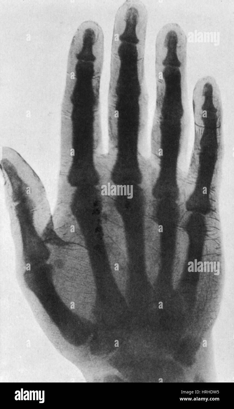 Early X-ray, 1897 Stock Photohttps://www.alamy.com/image-license-details/?v=1https://www.alamy.com/stock-photo-early-x-ray-1897-134993745.html
RMHRHDW5–Early X-ray, 1897
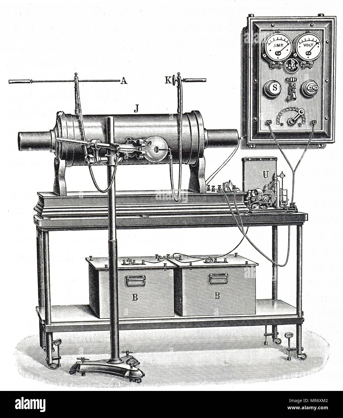 Engraving depicting an early x-ray apparatus powered by wet cells (B,B). Dated 20th century Stock Photohttps://www.alamy.com/image-license-details/?v=1https://www.alamy.com/engraving-depicting-an-early-x-ray-apparatus-powered-by-wet-cells-bb-dated-20th-century-image186393426.html
RMMR6XM2–Engraving depicting an early x-ray apparatus powered by wet cells (B,B). Dated 20th century
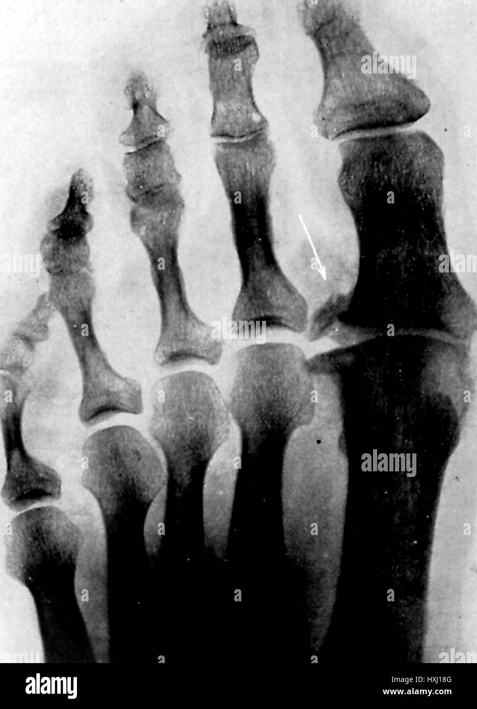 Early X Ray photograph showing the foot of a patient with osteoarthritis of the great toe joint, 1903. Stock Photohttps://www.alamy.com/image-license-details/?v=1https://www.alamy.com/stock-photo-early-x-ray-photograph-showing-the-foot-of-a-patient-with-osteoarthritis-136849792.html
RMHXJ18G–Early X Ray photograph showing the foot of a patient with osteoarthritis of the great toe joint, 1903.
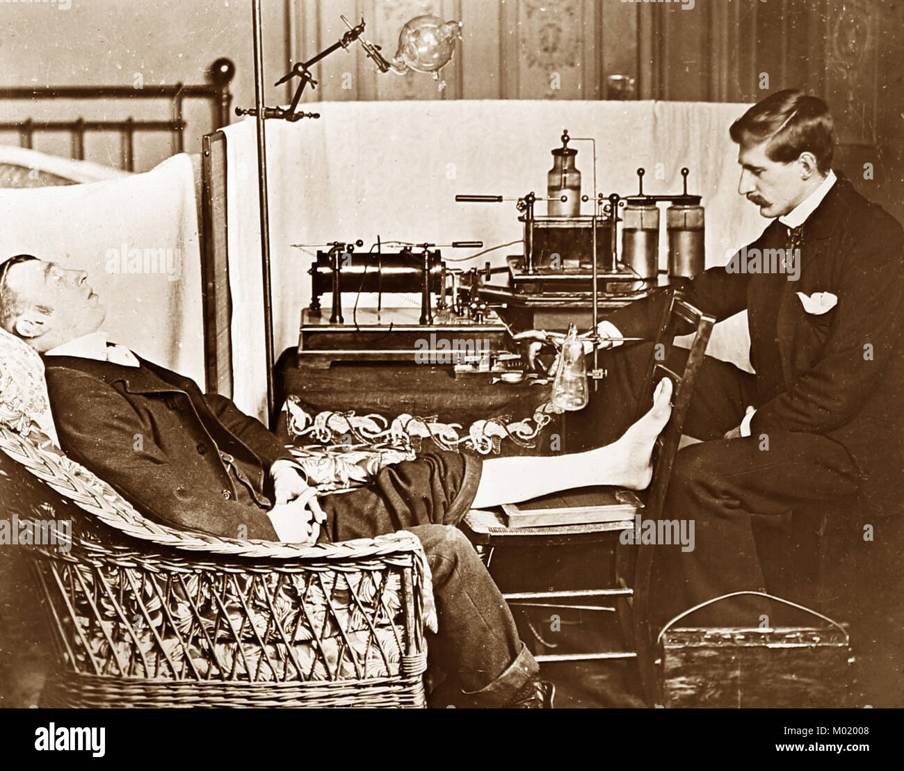 Early x-ray equipment in the laboratory of Henry Augustus Rowland, the first president of the American Physical Society, Victorian period Stock Photohttps://www.alamy.com/image-license-details/?v=1https://www.alamy.com/stock-photo-early-x-ray-equipment-in-the-laboratory-of-henry-augustus-rowland-172147592.html
RFM02008–Early x-ray equipment in the laboratory of Henry Augustus Rowland, the first president of the American Physical Society, Victorian period
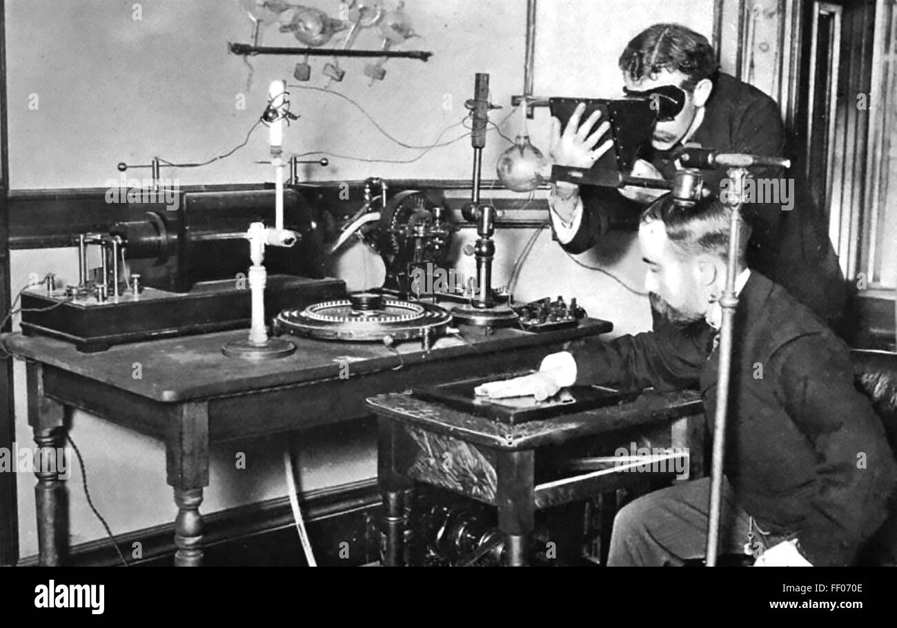 WILLIAM CROOKES (1832-1919) English chemist. Experimenters taking an x-ray photo with an early version of his Crookes Vacuum Tube about 1895. Stock Photohttps://www.alamy.com/image-license-details/?v=1https://www.alamy.com/stock-photo-william-crookes-1832-1919-english-chemist-experimenters-taking-an-95277182.html
RMFF070E–WILLIAM CROOKES (1832-1919) English chemist. Experimenters taking an x-ray photo with an early version of his Crookes Vacuum Tube about 1895.
 PSM V50 D682 An early x ray equipment Stock Photohttps://www.alamy.com/image-license-details/?v=1https://www.alamy.com/stock-photo-psm-v50-d682-an-early-x-ray-equipment-140367349.html
RMJ4A7YH–PSM V50 D682 An early x ray equipment
 Early X-ray bird Stock Photohttps://www.alamy.com/image-license-details/?v=1https://www.alamy.com/early-x-ray-bird-image265052008.html
RMWB64F4–Early X-ray bird
 921 Light yellow-white human figures over early X-ray paintings - Stock Photohttps://www.alamy.com/image-license-details/?v=1https://www.alamy.com/921-light-yellow-white-human-figures-over-early-x-ray-paintings-image213722930.html
RMPBKWNP–921 Light yellow-white human figures over early X-ray paintings -
 Early X-ray animals - Google Art Project Stock Photohttps://www.alamy.com/image-license-details/?v=1https://www.alamy.com/stock-photo-early-x-ray-animals-google-art-project-132682328.html
RMHKT5JG–Early X-ray animals - Google Art Project
 Early X-ray animals. Date/Period: -2000 - -1. Painting. Author: UNKNOWN. Stock Photohttps://www.alamy.com/image-license-details/?v=1https://www.alamy.com/early-x-ray-animals-dateperiod-2000-1-painting-author-unknown-image219071857.html
RMPMBGAW–Early X-ray animals. Date/Period: -2000 - -1. Painting. Author: UNKNOWN.
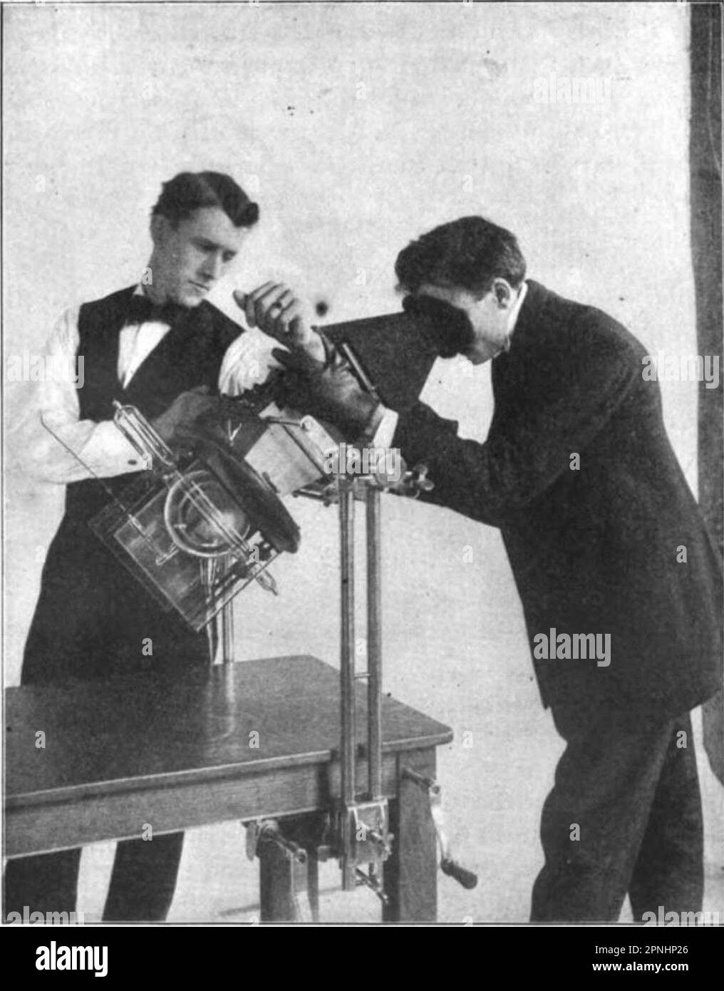 Early x-ray procedure Stock Photohttps://www.alamy.com/image-license-details/?v=1https://www.alamy.com/early-x-ray-procedure-image546819678.html
RM2PNHP26–Early x-ray procedure
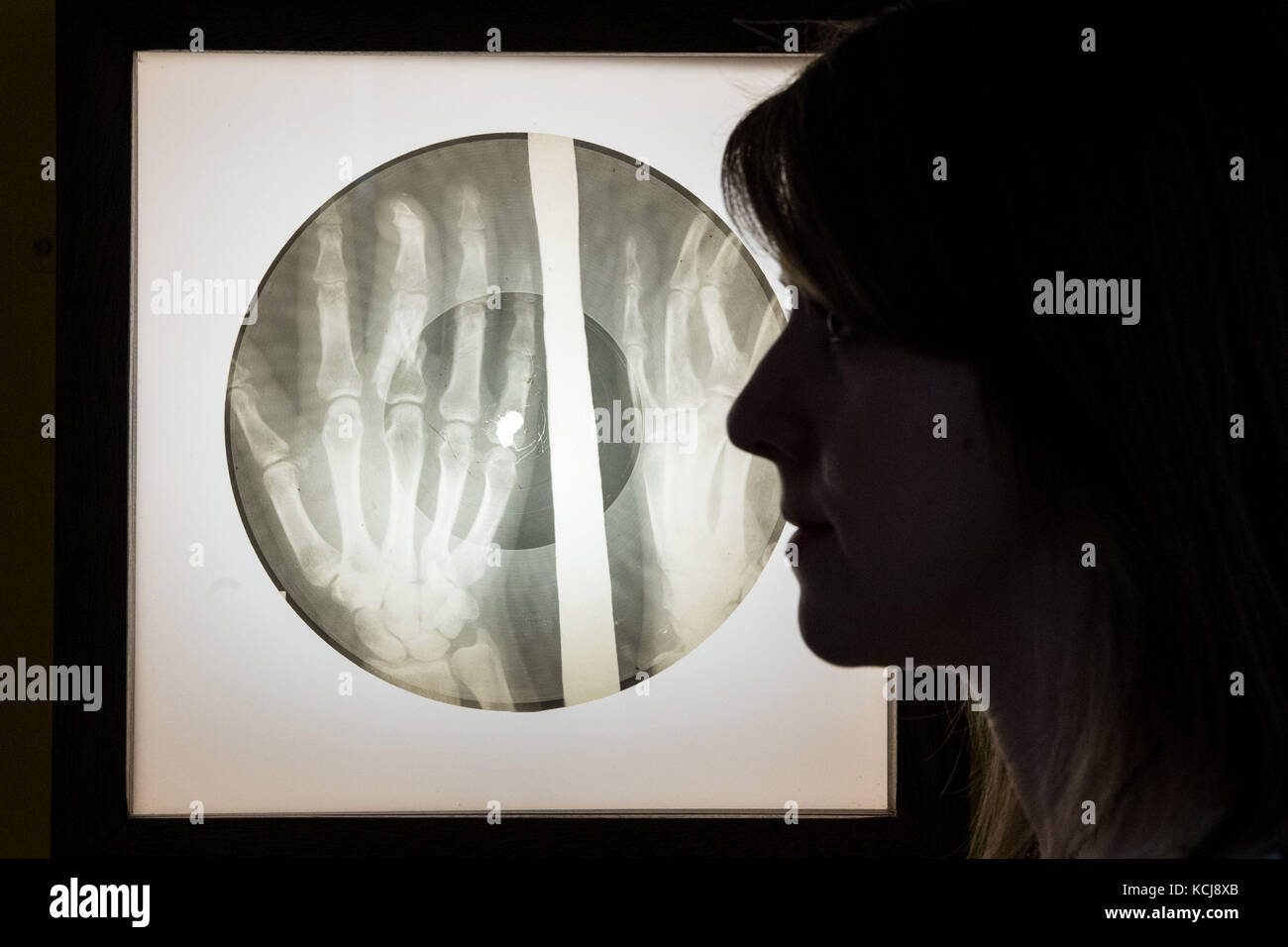 A visitor views a bootleg 78 rpm disc etched onto an old X-ray film, which were illicitly manufactured in the USSR from the late 1940's to early 1960s, part of Listen: 140 Years of Recorded Sound, at the British Library in London. Stock Photohttps://www.alamy.com/image-license-details/?v=1https://www.alamy.com/stock-image-a-visitor-views-a-bootleg-78-rpm-disc-etched-onto-an-old-x-ray-film-162671331.html
RMKCJ8XB–A visitor views a bootleg 78 rpm disc etched onto an old X-ray film, which were illicitly manufactured in the USSR from the late 1940's to early 1960s, part of Listen: 140 Years of Recorded Sound, at the British Library in London.
 Early Summer, William Trost Richards, American, 1833-1905, Oil on canvas, 1888, 24 1/4 x 20 1/16in., 61.6 x 51cm, 1888, american, early, Early summer, forest, green, lake, landscape, light, lightened forest, Luminist, nature, ndd7, oil, oil paint, oil painting, painting, pastoral, ray of light, Richards, Season, summer, sun-dappled, trees, Trost, water, William, William Trost Richards, wood, wooded Stock Photohttps://www.alamy.com/image-license-details/?v=1https://www.alamy.com/early-summer-william-trost-richards-american-1833-1905-oil-on-canvas-1888-24-14-x-20-116in-616-x-51cm-1888-american-early-early-summer-forest-green-lake-landscape-light-lightened-forest-luminist-nature-ndd7-oil-oil-paint-oil-painting-painting-pastoral-ray-of-light-richards-season-summer-sun-dappled-trees-trost-water-william-william-trost-richards-wood-wooded-image454273163.html
RM2HB1X1F–Early Summer, William Trost Richards, American, 1833-1905, Oil on canvas, 1888, 24 1/4 x 20 1/16in., 61.6 x 51cm, 1888, american, early, Early summer, forest, green, lake, landscape, light, lightened forest, Luminist, nature, ndd7, oil, oil paint, oil painting, painting, pastoral, ray of light, Richards, Season, summer, sun-dappled, trees, Trost, water, William, William Trost Richards, wood, wooded
 Inspired by Early Summer, William Trost Richards, American, 1833-1905, Oil on canvas, 1888, 24 1/4 x 20 1/16in., 61.6 x 51cm, 1888, american, early, Early summer, forest, green, lake, landscape, light, lightened forest, Luminist, nature, ndd7, oil, oil paint, oil painting, painting, pastoral, ray of, Reimagined by Artotop. Classic art reinvented with a modern twist. Design of warm cheerful glowing of brightness and light ray radiance. Photography inspired by surrealism and futurism, embracing dynamic energy of modern technology, movement, speed and revolutionize culture Stock Photohttps://www.alamy.com/image-license-details/?v=1https://www.alamy.com/inspired-by-early-summer-william-trost-richards-american-1833-1905-oil-on-canvas-1888-24-14-x-20-116in-616-x-51cm-1888-american-early-early-summer-forest-green-lake-landscape-light-lightened-forest-luminist-nature-ndd7-oil-oil-paint-oil-painting-painting-pastoral-ray-of-reimagined-by-artotop-classic-art-reinvented-with-a-modern-twist-design-of-warm-cheerful-glowing-of-brightness-and-light-ray-radiance-photography-inspired-by-surrealism-and-futurism-embracing-dynamic-energy-of-modern-technology-movement-speed-and-revolutionize-culture-image459252211.html
RF2HK4MTK–Inspired by Early Summer, William Trost Richards, American, 1833-1905, Oil on canvas, 1888, 24 1/4 x 20 1/16in., 61.6 x 51cm, 1888, american, early, Early summer, forest, green, lake, landscape, light, lightened forest, Luminist, nature, ndd7, oil, oil paint, oil painting, painting, pastoral, ray of, Reimagined by Artotop. Classic art reinvented with a modern twist. Design of warm cheerful glowing of brightness and light ray radiance. Photography inspired by surrealism and futurism, embracing dynamic energy of modern technology, movement, speed and revolutionize culture
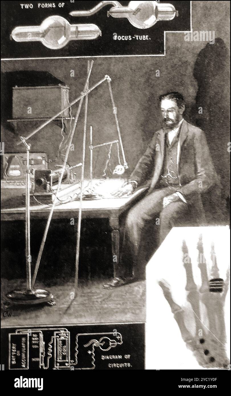 An early illustration of one of the first x-ray machines in use in Britain (first discovered 1895( Stock Photohttps://www.alamy.com/image-license-details/?v=1https://www.alamy.com/an-early-illustration-of-one-of-the-first-x-ray-machines-in-use-in-britain-first-discovered-1895-image626992255.html
RM2YC1Y0F–An early illustration of one of the first x-ray machines in use in Britain (first discovered 1895(
![[X-Ray of a Box Compasses and Drawing Tools] 1896 Dr. Henri van Heurck Belgian Shadows streak across an incandescent field, their silhouettes resisting recognition. Though nearly abstract, this image is no modernist exercise in form, but rather an early X-ray showing compasses and drawing tools inside of a closed box. Created within months of the X-ray’s discovery, it is one of the preliminary radiographic experiments recorded by the Belgian scientist Dr. Henri van Heurck, who had begun using fluorescent screens during this period to make X-rays with shortened exposure times. When he published Stock Photo [X-Ray of a Box Compasses and Drawing Tools] 1896 Dr. Henri van Heurck Belgian Shadows streak across an incandescent field, their silhouettes resisting recognition. Though nearly abstract, this image is no modernist exercise in form, but rather an early X-ray showing compasses and drawing tools inside of a closed box. Created within months of the X-ray’s discovery, it is one of the preliminary radiographic experiments recorded by the Belgian scientist Dr. Henri van Heurck, who had begun using fluorescent screens during this period to make X-rays with shortened exposure times. When he published Stock Photo](https://c8.alamy.com/comp/2HHWKK0/x-ray-of-a-box-compasses-and-drawing-tools-1896-dr-henri-van-heurck-belgian-shadows-streak-across-an-incandescent-field-their-silhouettes-resisting-recognition-though-nearly-abstract-this-image-is-no-modernist-exercise-in-form-but-rather-an-early-x-ray-showing-compasses-and-drawing-tools-inside-of-a-closed-box-created-within-months-of-the-x-rays-discovery-it-is-one-of-the-preliminary-radiographic-experiments-recorded-by-the-belgian-scientist-dr-henri-van-heurck-who-had-begun-using-fluorescent-screens-during-this-period-to-make-x-rays-with-shortened-exposure-times-when-he-published-2HHWKK0.jpg) [X-Ray of a Box Compasses and Drawing Tools] 1896 Dr. Henri van Heurck Belgian Shadows streak across an incandescent field, their silhouettes resisting recognition. Though nearly abstract, this image is no modernist exercise in form, but rather an early X-ray showing compasses and drawing tools inside of a closed box. Created within months of the X-ray’s discovery, it is one of the preliminary radiographic experiments recorded by the Belgian scientist Dr. Henri van Heurck, who had begun using fluorescent screens during this period to make X-rays with shortened exposure times. When he published Stock Photohttps://www.alamy.com/image-license-details/?v=1https://www.alamy.com/x-ray-of-a-box-compasses-and-drawing-tools-1896-dr-henri-van-heurck-belgian-shadows-streak-across-an-incandescent-field-their-silhouettes-resisting-recognition-though-nearly-abstract-this-image-is-no-modernist-exercise-in-form-but-rather-an-early-x-ray-showing-compasses-and-drawing-tools-inside-of-a-closed-box-created-within-months-of-the-x-rays-discovery-it-is-one-of-the-preliminary-radiographic-experiments-recorded-by-the-belgian-scientist-dr-henri-van-heurck-who-had-begun-using-fluorescent-screens-during-this-period-to-make-x-rays-with-shortened-exposure-times-when-he-published-image458482948.html
[X-Ray of a Box Compasses and Drawing Tools] 1896 Dr. Henri van Heurck Belgian Shadows streak across an incandescent field, their silhouettes resisting recognition. Though nearly abstract, this image is no modernist exercise in form, but rather an early X-ray showing compasses and drawing tools inside of a closed box. Created within months of the X-ray’s discovery, it is one of the preliminary radiographic experiments recorded by the Belgian scientist Dr. Henri van Heurck, who had begun using fluorescent screens during this period to make X-rays with shortened exposure times. When he published Stock Photohttps://www.alamy.com/image-license-details/?v=1https://www.alamy.com/x-ray-of-a-box-compasses-and-drawing-tools-1896-dr-henri-van-heurck-belgian-shadows-streak-across-an-incandescent-field-their-silhouettes-resisting-recognition-though-nearly-abstract-this-image-is-no-modernist-exercise-in-form-but-rather-an-early-x-ray-showing-compasses-and-drawing-tools-inside-of-a-closed-box-created-within-months-of-the-x-rays-discovery-it-is-one-of-the-preliminary-radiographic-experiments-recorded-by-the-belgian-scientist-dr-henri-van-heurck-who-had-begun-using-fluorescent-screens-during-this-period-to-make-x-rays-with-shortened-exposure-times-when-he-published-image458482948.htmlRM2HHWKK0–[X-Ray of a Box Compasses and Drawing Tools] 1896 Dr. Henri van Heurck Belgian Shadows streak across an incandescent field, their silhouettes resisting recognition. Though nearly abstract, this image is no modernist exercise in form, but rather an early X-ray showing compasses and drawing tools inside of a closed box. Created within months of the X-ray’s discovery, it is one of the preliminary radiographic experiments recorded by the Belgian scientist Dr. Henri van Heurck, who had begun using fluorescent screens during this period to make X-rays with shortened exposure times. When he published
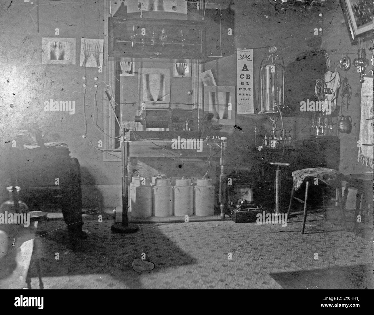 Picture of a doctor's office in 1905-1910 with one of the early X-ray machines. Stock Photohttps://www.alamy.com/image-license-details/?v=1https://www.alamy.com/picture-of-a-doctors-office-in-1905-1910-with-one-of-the-early-x-ray-machines-image602749438.html
RM2X0HH1J–Picture of a doctor's office in 1905-1910 with one of the early X-ray machines.
 Early morning sunlight filtering through trees giving a weird x files style image Stock Photohttps://www.alamy.com/image-license-details/?v=1https://www.alamy.com/early-morning-sunlight-filtering-through-trees-giving-a-weird-x-files-image945003.html
RMAE6B6B–Early morning sunlight filtering through trees giving a weird x files style image
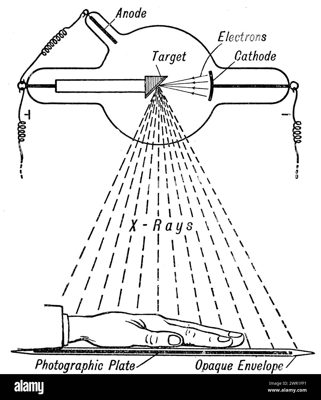 A diagram of a Crookes tube. A Crookes tube is an early experimental electrical discharge tube, with partial vacuum, invented by English physicist William Crookes (1832-1919) and others around 1869-1875, in which cathode rays, streams of electrons, were discovered. Wilhelm Röntgen (1845-1923), discovered X-rays using the Crookes tube in 1895. Stock Photohttps://www.alamy.com/image-license-details/?v=1https://www.alamy.com/a-diagram-of-acrookes-tube-acrookes-tubeis-an-early-experimental-electricaldischarge-tube-with-partial-vacuum-invented-by-english-physicistwilliam-crookes-1832-1919-and-others-around-1869-1875in-whichcathode-rays-streams-ofelectrons-were-discovered-wilhelm-rntgen-1845-1923-discovered-x-rays-using-the-crookes-tube-in-1895-image599323733.html
RM2WR1FF1–A diagram of a Crookes tube. A Crookes tube is an early experimental electrical discharge tube, with partial vacuum, invented by English physicist William Crookes (1832-1919) and others around 1869-1875, in which cathode rays, streams of electrons, were discovered. Wilhelm Röntgen (1845-1923), discovered X-rays using the Crookes tube in 1895.
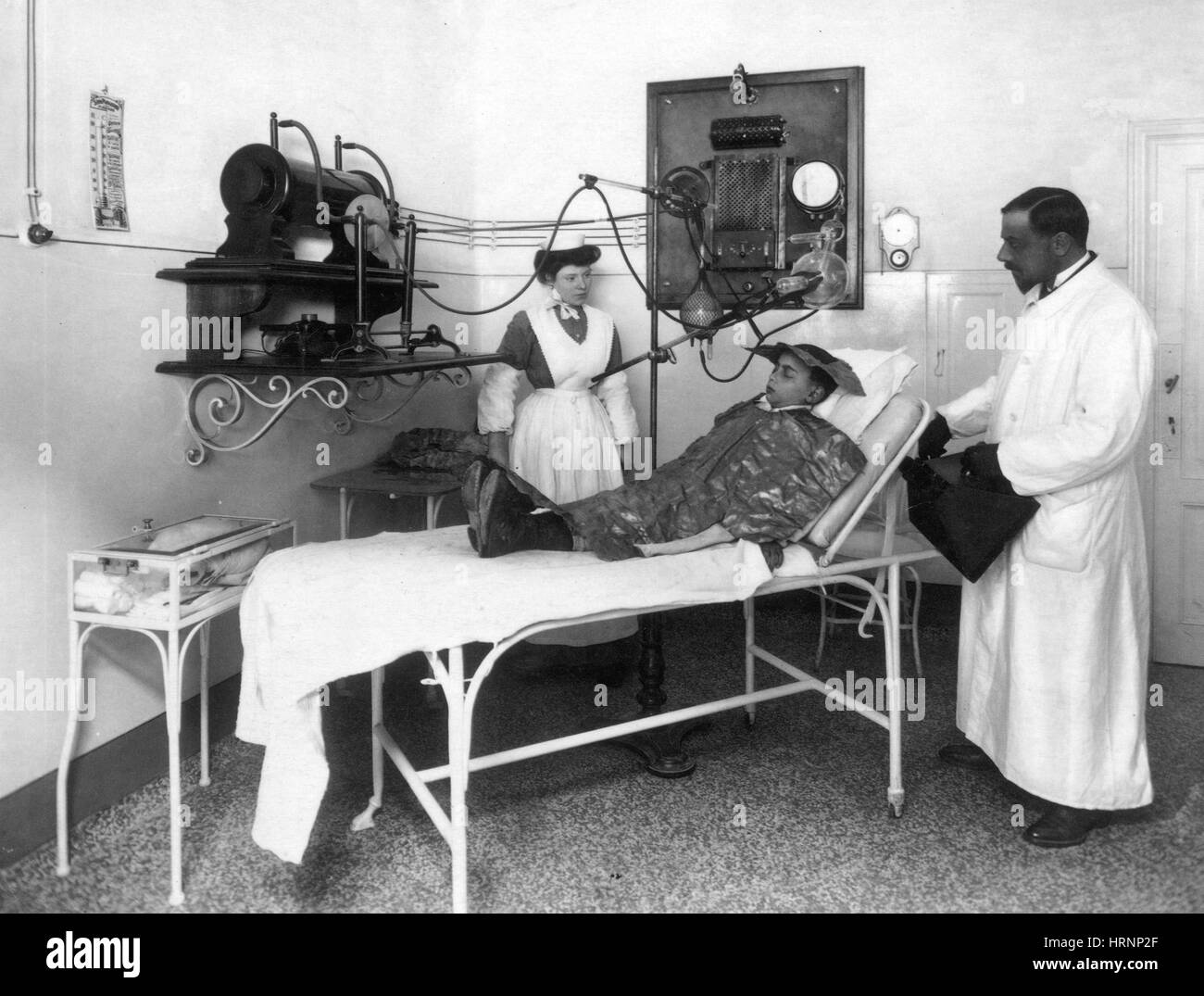 Roentgen Ray Therapy, Early 20th Century Stock Photohttps://www.alamy.com/image-license-details/?v=1https://www.alamy.com/stock-photo-roentgen-ray-therapy-early-20th-century-135087975.html
RMHRNP2F–Roentgen Ray Therapy, Early 20th Century
 Engraving depicting an early x-ray equipment being using used on a frog. Dated 20th century Stock Photohttps://www.alamy.com/image-license-details/?v=1https://www.alamy.com/engraving-depicting-an-early-x-ray-equipment-being-using-used-on-a-frog-dated-20th-century-image186393423.html
RMMR6XKY–Engraving depicting an early x-ray equipment being using used on a frog. Dated 20th century
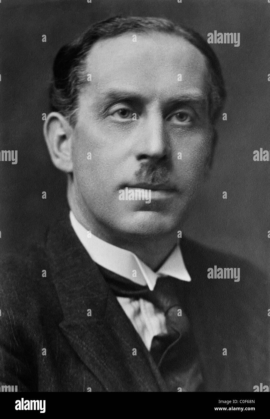 English physicist Charles Glover Barkla (1877 - 1944) - winner of the Nobel Prize in Physics in 1917 for his work on x-rays. Stock Photohttps://www.alamy.com/image-license-details/?v=1https://www.alamy.com/stock-photo-english-physicist-charles-glover-barkla-1877-1944-winner-of-the-nobel-34754965.html
RMC0F68N–English physicist Charles Glover Barkla (1877 - 1944) - winner of the Nobel Prize in Physics in 1917 for his work on x-rays.
 An old cigarette card (c. 1929) with a portrait of Wilhelm Conrad Röntgen (1845–1923) and an illustration of an X-ray of the bones of a human hand. Röntgen was a German mechanical engineer and physicist, who, in 1895, produced and detected electromagnetic radiation in a wavelength range known as X-rays or Röntgen rays, an achievement that earned him the first Nobel Prize in Physics in 1901. Röntgen saw the first radiographic image: his own skeleton on the barium platinocyanide screen. A few weeks after his discovery, he took a picture (a radiograph) using X-rays of his wife's hand. Stock Photohttps://www.alamy.com/image-license-details/?v=1https://www.alamy.com/an-old-cigarette-card-c-1929-with-a-portrait-of-wilhelm-conrad-rntgen-18451923-and-an-illustration-of-an-x-ray-of-the-bones-of-a-human-hand-rntgen-was-a-german-mechanical-engineer-and-physicist-who-in-1895-produced-and-detected-electromagnetic-radiation-in-a-wavelength-range-known-as-x-rays-or-rntgen-rays-an-achievement-that-earned-him-the-first-nobel-prize-in-physics-in-1901-rntgen-saw-the-first-radiographic-image-his-own-skeleton-on-the-barium-platinocyanide-screen-a-few-weeks-after-his-discovery-he-took-a-picture-a-radiograph-using-x-rays-of-his-wifes-hand-image366812052.html
RM2C8NMEC–An old cigarette card (c. 1929) with a portrait of Wilhelm Conrad Röntgen (1845–1923) and an illustration of an X-ray of the bones of a human hand. Röntgen was a German mechanical engineer and physicist, who, in 1895, produced and detected electromagnetic radiation in a wavelength range known as X-rays or Röntgen rays, an achievement that earned him the first Nobel Prize in Physics in 1901. Röntgen saw the first radiographic image: his own skeleton on the barium platinocyanide screen. A few weeks after his discovery, he took a picture (a radiograph) using X-rays of his wife's hand.
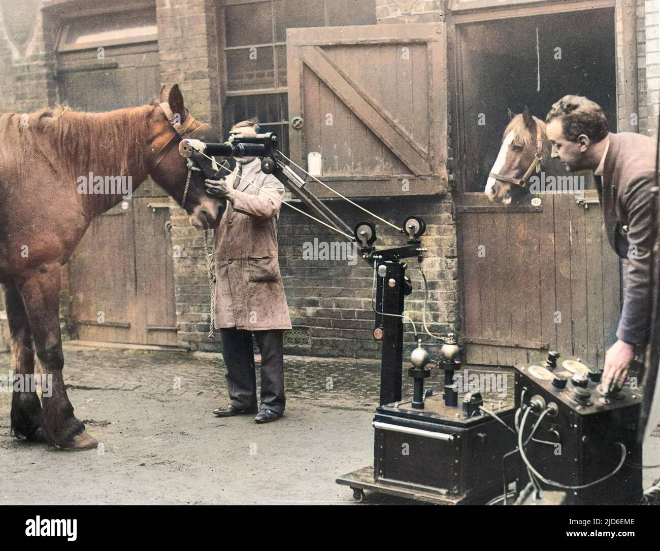 A horse being x-rayed at the Royal Veterinary College, Camden, London, using portable x-ray apparatus, which has proved invaluable in the treatment of animal ailments. Colourised version of : 10150403 Date: early 1930s Stock Photohttps://www.alamy.com/image-license-details/?v=1https://www.alamy.com/a-horse-being-x-rayed-at-the-royal-veterinary-college-camden-london-using-portable-x-ray-apparatus-which-has-proved-invaluable-in-the-treatment-of-animal-ailments-colourised-version-of-10150403-date-early-1930s-image472813726.html
RM2JD6EME–A horse being x-rayed at the Royal Veterinary College, Camden, London, using portable x-ray apparatus, which has proved invaluable in the treatment of animal ailments. Colourised version of : 10150403 Date: early 1930s
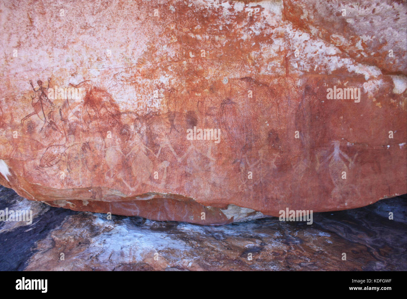 Light yellow white human figures over early X ray paintings Google Art Project Stock Photohttps://www.alamy.com/image-license-details/?v=1https://www.alamy.com/stock-image-light-yellow-white-human-figures-over-early-x-ray-paintings-google-163226379.html
RMKDFGWF–Light yellow white human figures over early X ray paintings Google Art Project
 Early X-ray animals Stock Photohttps://www.alamy.com/image-license-details/?v=1https://www.alamy.com/early-x-ray-animals-image265051990.html
RMWB64EE–Early X-ray animals
 Early X-ray wallaby. (-2000 - -1). Early X-ray wallaby - Google Art Project Stock Photohttps://www.alamy.com/image-license-details/?v=1https://www.alamy.com/early-x-ray-wallaby-2000-1-early-x-ray-wallaby-google-art-project-image184973101.html
RMMMX725–Early X-ray wallaby. (-2000 - -1). Early X-ray wallaby - Google Art Project
 Early X-ray wallaby - Google Art Project Stock Photohttps://www.alamy.com/image-license-details/?v=1https://www.alamy.com/stock-photo-early-x-ray-wallaby-google-art-project-132682332.html
RMHKT5JM–Early X-ray wallaby - Google Art Project
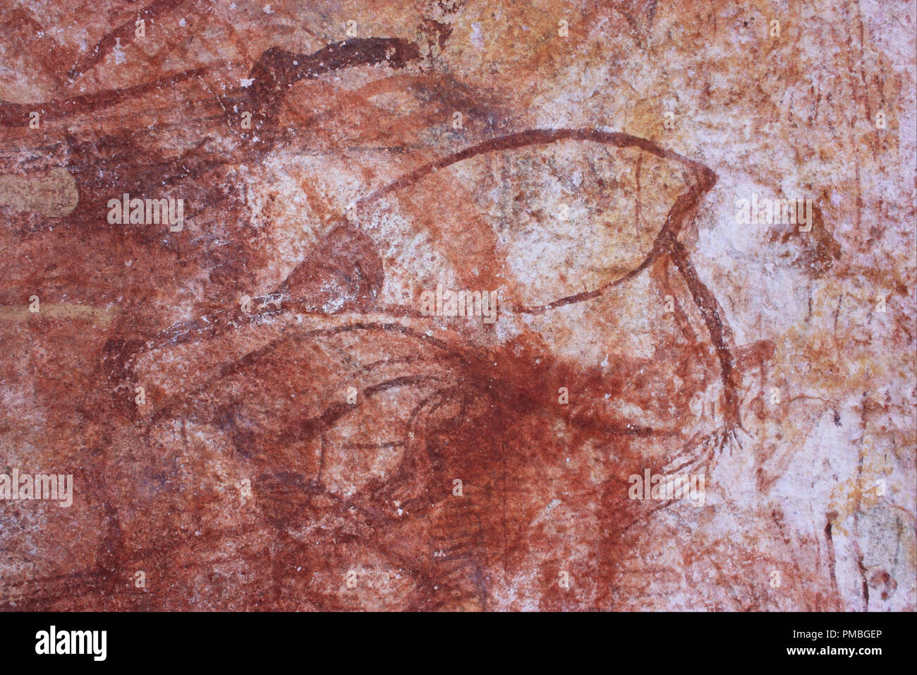 Early X-ray bird. Date/Period: -2000 - -1. Painting. Author: UNKNOWN. Stock Photohttps://www.alamy.com/image-license-details/?v=1https://www.alamy.com/early-x-ray-bird-dateperiod-2000-1-painting-author-unknown-image219071966.html
RMPMBGEP–Early X-ray bird. Date/Period: -2000 - -1. Painting. Author: UNKNOWN.
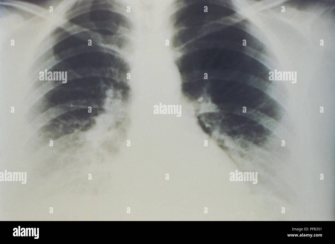 Early stages of bilateral pulmonary effusion due to Hantavirus pulmonary syndrome (HPS), revealed in the AP chest x-ray, 1994. Image courtesy Centers for Disease Control (CDC) / D. Loren Ketai, M.D. () Stock Photohttps://www.alamy.com/image-license-details/?v=1https://www.alamy.com/early-stages-of-bilateral-pulmonary-effusion-due-to-hantavirus-pulmonary-syndrome-hps-revealed-in-the-ap-chest-x-ray-1994-image-courtesy-centers-for-disease-control-cdc-d-loren-ketai-md-image215922365.html
RMPF8351–Early stages of bilateral pulmonary effusion due to Hantavirus pulmonary syndrome (HPS), revealed in the AP chest x-ray, 1994. Image courtesy Centers for Disease Control (CDC) / D. Loren Ketai, M.D. ()
 A visitor views a bootleg 78 rpm disc etched onto an old X-ray film, which were illicitly manufactured in the USSR from the late 1940's to early 1960s, part of Listen: 140 Years of Recorded Sound, at the British Library in London. Stock Photohttps://www.alamy.com/image-license-details/?v=1https://www.alamy.com/stock-image-a-visitor-views-a-bootleg-78-rpm-disc-etched-onto-an-old-x-ray-film-162671329.html
RMKCJ8X9–A visitor views a bootleg 78 rpm disc etched onto an old X-ray film, which were illicitly manufactured in the USSR from the late 1940's to early 1960s, part of Listen: 140 Years of Recorded Sound, at the British Library in London.
 The Shepherdess, Albert Pinkham Ryder, American, 1847-1917, Oil on panel, early 1880s, 10 1/8 x 6 13/16 in., 25.7 x 17.3 cm, american, early 1880s, oil, painting, panel, Ryder, shepherdess, x-ray Stock Photohttps://www.alamy.com/image-license-details/?v=1https://www.alamy.com/the-shepherdess-albert-pinkham-ryder-american-1847-1917-oil-on-panel-early-1880s-10-18-x-6-1316-in-257-x-173-cm-american-early-1880s-oil-painting-panel-ryder-shepherdess-x-ray-image454266777.html
RM2HB1HWD–The Shepherdess, Albert Pinkham Ryder, American, 1847-1917, Oil on panel, early 1880s, 10 1/8 x 6 13/16 in., 25.7 x 17.3 cm, american, early 1880s, oil, painting, panel, Ryder, shepherdess, x-ray
 Inspired by Crucifix on Altar, Mexican, Painting on fabric, late 18th-early 19th century, 29 x 17 in., 73.7 x 43.2 cm, Reimagined by Artotop. Classic art reinvented with a modern twist. Design of warm cheerful glowing of brightness and light ray radiance. Photography inspired by surrealism and futurism, embracing dynamic energy of modern technology, movement, speed and revolutionize culture Stock Photohttps://www.alamy.com/image-license-details/?v=1https://www.alamy.com/inspired-by-crucifix-on-altar-mexican-painting-on-fabric-late-18th-early-19th-century-29-x-17-in-737-x-432-cm-reimagined-by-artotop-classic-art-reinvented-with-a-modern-twist-design-of-warm-cheerful-glowing-of-brightness-and-light-ray-radiance-photography-inspired-by-surrealism-and-futurism-embracing-dynamic-energy-of-modern-technology-movement-speed-and-revolutionize-culture-image459254577.html
RF2HK4RW5–Inspired by Crucifix on Altar, Mexican, Painting on fabric, late 18th-early 19th century, 29 x 17 in., 73.7 x 43.2 cm, Reimagined by Artotop. Classic art reinvented with a modern twist. Design of warm cheerful glowing of brightness and light ray radiance. Photography inspired by surrealism and futurism, embracing dynamic energy of modern technology, movement, speed and revolutionize culture
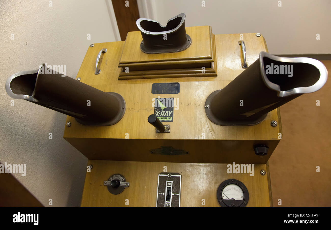 Foot X-Ray Machine at Great Basin Museum Stock Photohttps://www.alamy.com/image-license-details/?v=1https://www.alamy.com/stock-photo-foot-x-ray-machine-at-great-basin-museum-38032931.html
RMC5TFAY–Foot X-Ray Machine at Great Basin Museum
 Xray equipment, Henry Augustus Rowland's laboratory at John Hopkinds University Stock Photohttps://www.alamy.com/image-license-details/?v=1https://www.alamy.com/xray-equipment-henry-augustus-rowlands-laboratory-at-john-hopkinds-university-image260160486.html
RFW3799X–Xray equipment, Henry Augustus Rowland's laboratory at John Hopkinds University
 close up of a profusion crabapple tree blossoms in full bloom, with a sun ray hitting the edge of a petal. The scientific name for this violet-red flo Stock Photohttps://www.alamy.com/image-license-details/?v=1https://www.alamy.com/close-up-of-a-profusion-crabapple-tree-blossoms-in-full-bloom-with-a-sun-ray-hitting-the-edge-of-a-petal-the-scientific-name-for-this-violet-red-flo-image471263147.html
RF2JAKTXK–close up of a profusion crabapple tree blossoms in full bloom, with a sun ray hitting the edge of a petal. The scientific name for this violet-red flo
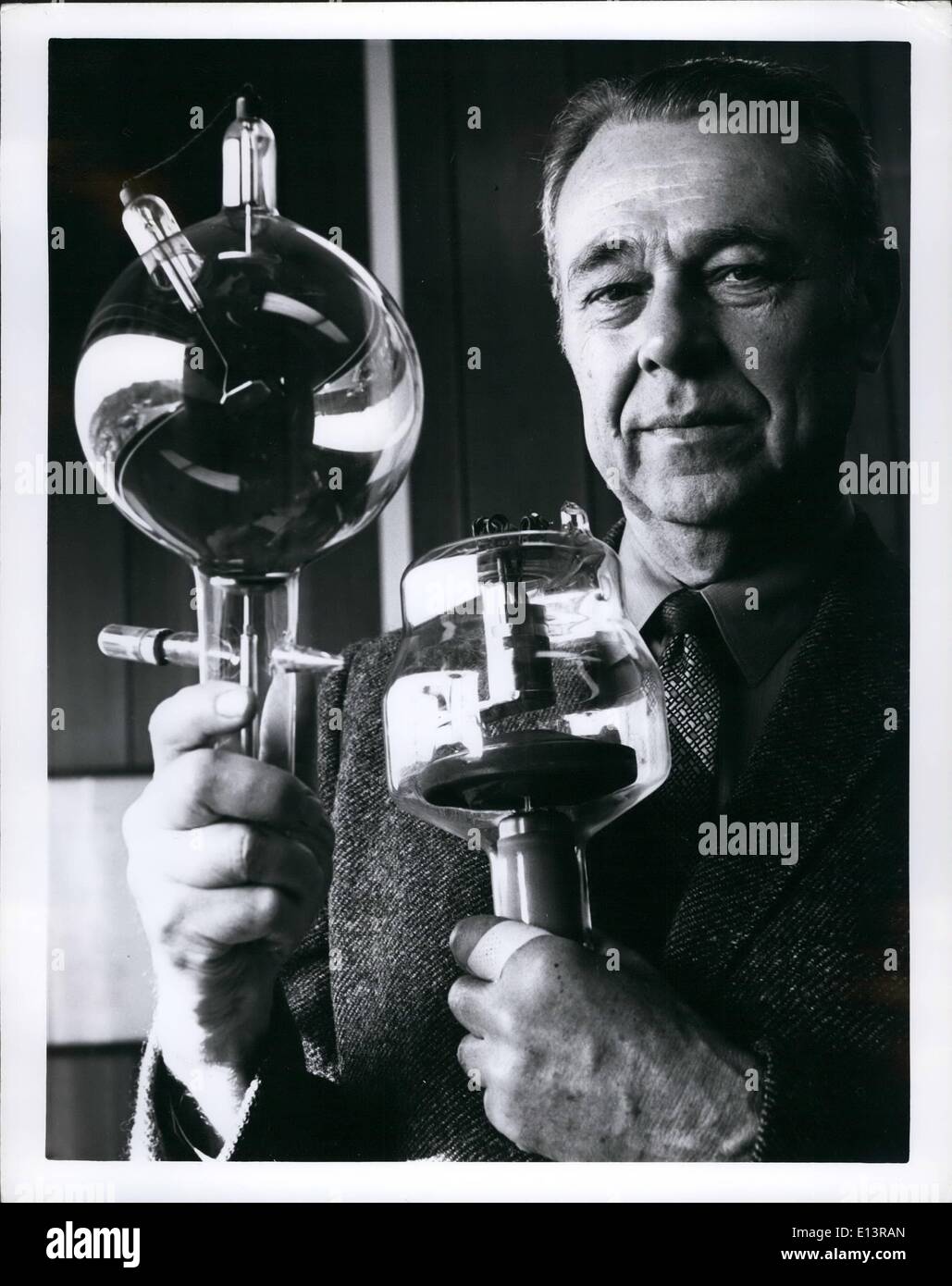 Mar. 22, 2012 - X-ray history under glass; Veteran x-ray tube engineer Thomas H. Rogers vice President of Raytheon's Machlett Laboratories, Stanford, Conn. Holds tow of his company's tubes that illustrate advances made in x-ray technology since 1895 when Roentgen discovered x-rays. Tube at left is an early model that permitted radiation in many directions. New tube at right has spiinning anode to produce sharper pictures with more consistency, fewer retakes, and greater safety for both patient and radiologist. Stock Photohttps://www.alamy.com/image-license-details/?v=1https://www.alamy.com/mar-22-2012-x-ray-history-under-glass-veteran-x-ray-tube-engineer-image69540317.html
RME13RAN–Mar. 22, 2012 - X-ray history under glass; Veteran x-ray tube engineer Thomas H. Rogers vice President of Raytheon's Machlett Laboratories, Stanford, Conn. Holds tow of his company's tubes that illustrate advances made in x-ray technology since 1895 when Roentgen discovered x-rays. Tube at left is an early model that permitted radiation in many directions. New tube at right has spiinning anode to produce sharper pictures with more consistency, fewer retakes, and greater safety for both patient and radiologist.
 Cape Canaveral, United States of America. 09 December, 2021. Flames shoot from the engines of the SpaceX Falcon 9 rocket carrying the NASA Imaging X-ray Polarimetry Explorer spacecraft blasts off from Launch Complex 39A early morning at the Kennedy Space Center December 9, 2021 in Cape Canaveral, Florida. The IXPE spacecraft is the first satellite dedicated to measuring the polarization of X-rays from a variety of cosmic sources. Credit: Joel Kowsky/NASA/Alamy Live News Stock Photohttps://www.alamy.com/image-license-details/?v=1https://www.alamy.com/cape-canaveral-united-states-of-america-09-december-2021-flames-shoot-from-the-engines-of-the-spacex-falcon-9-rocket-carrying-the-nasa-imaging-x-ray-polarimetry-explorer-spacecraft-blasts-off-from-launch-complex-39a-early-morning-at-the-kennedy-space-center-december-9-2021-in-cape-canaveral-florida-the-ixpe-spacecraft-is-the-first-satellite-dedicated-to-measuring-the-polarization-of-x-rays-from-a-variety-of-cosmic-sources-credit-joel-kowskynasaalamy-live-news-image453605337.html
RM2H9YE6H–Cape Canaveral, United States of America. 09 December, 2021. Flames shoot from the engines of the SpaceX Falcon 9 rocket carrying the NASA Imaging X-ray Polarimetry Explorer spacecraft blasts off from Launch Complex 39A early morning at the Kennedy Space Center December 9, 2021 in Cape Canaveral, Florida. The IXPE spacecraft is the first satellite dedicated to measuring the polarization of X-rays from a variety of cosmic sources. Credit: Joel Kowsky/NASA/Alamy Live News
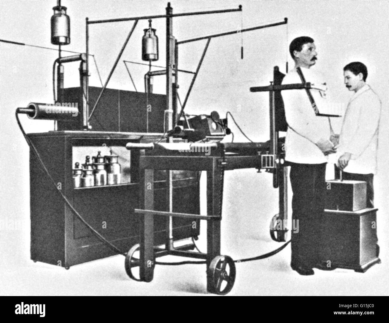 An early 20th century x-ray machine. Stock Photohttps://www.alamy.com/image-license-details/?v=1https://www.alamy.com/stock-photo-an-early-20th-century-x-ray-machine-104001072.html
RMG15JC0–An early 20th century x-ray machine.
 Engraving depicting a general view of early x-ray apparatus powered by wet cells. Dated 20th Century Stock Photohttps://www.alamy.com/image-license-details/?v=1https://www.alamy.com/stock-image-engraving-depicting-a-general-view-of-early-x-ray-apparatus-powered-165993584.html
RMKJ1JE8–Engraving depicting a general view of early x-ray apparatus powered by wet cells. Dated 20th Century
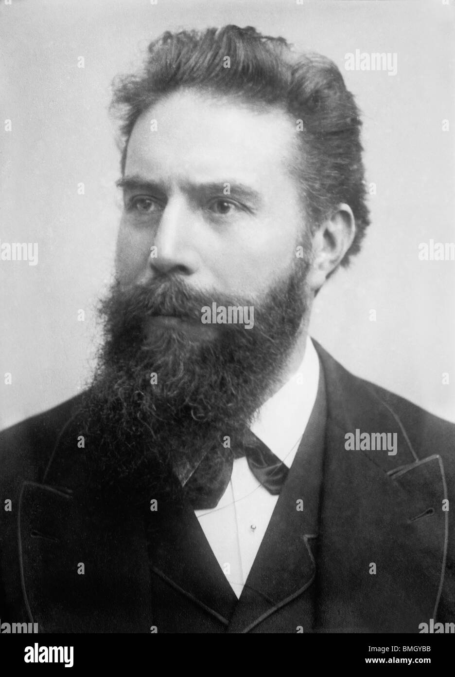 Undated portrait photo of German physicist Wilhelm Conrad Roentgen (1845 - 1923) - discoverer of x-rays and Nobel Prize winner. Stock Photohttps://www.alamy.com/image-license-details/?v=1https://www.alamy.com/stock-photo-undated-portrait-photo-of-german-physicist-wilhelm-conrad-roentgen-29876207.html
RMBMGYBB–Undated portrait photo of German physicist Wilhelm Conrad Roentgen (1845 - 1923) - discoverer of x-rays and Nobel Prize winner.
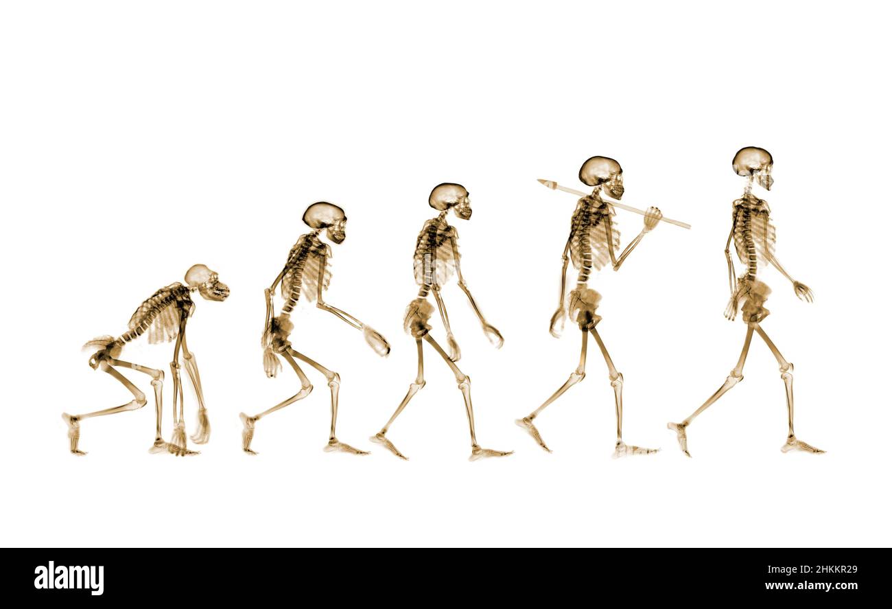 Human evolution, conceptual X-ray Stock Photohttps://www.alamy.com/image-license-details/?v=1https://www.alamy.com/human-evolution-conceptual-x-ray-image459583217.html
RF2HKKR29–Human evolution, conceptual X-ray
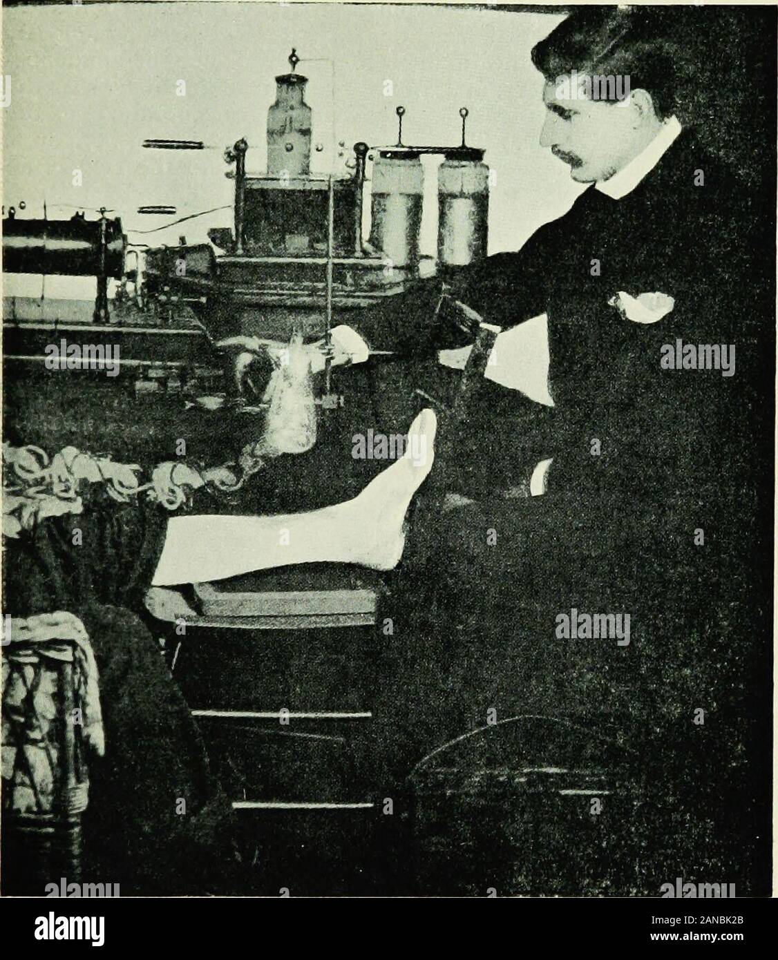 An essay on the history of electrotherapy and diagnosis; . Skiagrams of Howard Marshs case. names of different observers will be found throughoutRowlands masterly series of articles. So far we have read of the X rays as agents of deep scientific HISTORY OF ELECTROTHERAPY 149 interest, of such wonderful properties that they attract publicattention to an extent unparalleled in the history of science,and of great and increasing value in diagnosis. Now we haveto consider them in a far different aspect. For some time. Early X-ray Apparatus. (Rowland, 1896.) rumours had been afloat that exposure to Stock Photohttps://www.alamy.com/image-license-details/?v=1https://www.alamy.com/an-essay-on-the-history-of-electrotherapy-and-diagnosis-skiagrams-of-howard-marshs-case-names-of-different-observers-will-be-found-throughoutrowlands-masterly-series-of-articles-so-far-we-have-read-of-the-x-rays-as-agents-of-deep-scientific-history-of-electrotherapy-149-interest-of-such-wonderful-properties-that-they-attract-publicattention-to-an-extent-unparalleled-in-the-history-of-scienceand-of-great-and-increasing-value-in-diagnosis-now-we-haveto-consider-them-in-a-far-different-aspect-for-some-time-early-x-ray-apparatus-rowland-1896-rumours-had-been-afloat-that-exposure-to-image340161203.html
RM2ANBK2B–An essay on the history of electrotherapy and diagnosis; . Skiagrams of Howard Marshs case. names of different observers will be found throughoutRowlands masterly series of articles. So far we have read of the X rays as agents of deep scientific HISTORY OF ELECTROTHERAPY 149 interest, of such wonderful properties that they attract publicattention to an extent unparalleled in the history of science,and of great and increasing value in diagnosis. Now we haveto consider them in a far different aspect. For some time. Early X-ray Apparatus. (Rowland, 1896.) rumours had been afloat that exposure to
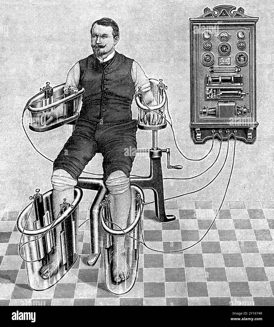 An illustration of a patient being treated with a four cell Schnee Bath, an early electric medical equipment for treating rheumatism Stock Photohttps://www.alamy.com/image-license-details/?v=1https://www.alamy.com/an-illustration-of-a-patient-being-treated-with-a-four-cell-schnee-bath-an-early-electric-medical-equipment-for-treating-rheumatism-image620325239.html
RM2Y1674R–An illustration of a patient being treated with a four cell Schnee Bath, an early electric medical equipment for treating rheumatism
 Early X-ray wallaby Stock Photohttps://www.alamy.com/image-license-details/?v=1https://www.alamy.com/early-x-ray-wallaby-image265051980.html
RMWB64E4–Early X-ray wallaby
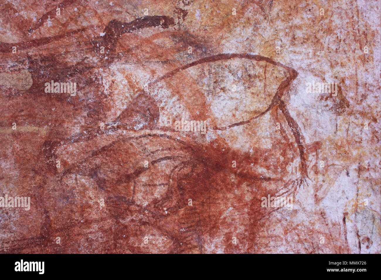 Early X-ray bird. (-2000 - -1). Early X-ray bird - Google Art Project Stock Photohttps://www.alamy.com/image-license-details/?v=1https://www.alamy.com/early-x-ray-bird-2000-1-early-x-ray-bird-google-art-project-image184973102.html
RMMMX726–Early X-ray bird. (-2000 - -1). Early X-ray bird - Google Art Project
 Early X-ray bird - Google Art Project Stock Photohttps://www.alamy.com/image-license-details/?v=1https://www.alamy.com/stock-photo-early-x-ray-bird-google-art-project-132682329.html
RMHKT5JH–Early X-ray bird - Google Art Project
 Early X-ray wallaby. Date/Period: -2000 - -1. Painting. Author: UNKNOWN. Stock Photohttps://www.alamy.com/image-license-details/?v=1https://www.alamy.com/early-x-ray-wallaby-dateperiod-2000-1-painting-author-unknown-image219071938.html
RMPMBGDP–Early X-ray wallaby. Date/Period: -2000 - -1. Painting. Author: UNKNOWN.
 Early medical x ray image of a hand, showing arthritis, 1909. Courtesy Internet Archive. () Stock Photohttps://www.alamy.com/image-license-details/?v=1https://www.alamy.com/stock-photo-early-medical-x-ray-image-of-a-hand-showing-arthritis-1909-courtesy-170790263.html
RMKWT4M7–Early medical x ray image of a hand, showing arthritis, 1909. Courtesy Internet Archive. ()
 Radiographer Mile Woodford at Salisbury District Hospital prepares to x-ray an early Bronze Age bag excavated from a Dartmoor burial site in Devon which could prove to be one of the most important archaeological finds of the last 100 years. Stock Photohttps://www.alamy.com/image-license-details/?v=1https://www.alamy.com/stock-photo-radiographer-mile-woodford-at-salisbury-district-hospital-prepares-106427429.html
RMG5457H–Radiographer Mile Woodford at Salisbury District Hospital prepares to x-ray an early Bronze Age bag excavated from a Dartmoor burial site in Devon which could prove to be one of the most important archaeological finds of the last 100 years.
 Evening Glow, The Old Red Cow, Albert Pinkham Ryder, American, 1847-1917, Oil on canvas, early-mid 1870s, 7 7/8 x 9 in., 20 x 22.8 cm, Albert, American, Buffalo, Bull, Canvas, Cow, Evening, Glow, Oil, Ox, painting, Pinkham, Red, Ryder, Tarus, x-ray Stock Photohttps://www.alamy.com/image-license-details/?v=1https://www.alamy.com/evening-glow-the-old-red-cow-albert-pinkham-ryder-american-1847-1917-oil-on-canvas-early-mid-1870s-7-78-x-9-in-20-x-228-cm-albert-american-buffalo-bull-canvas-cow-evening-glow-oil-ox-painting-pinkham-red-ryder-tarus-x-ray-image454266722.html
RM2HB1HRE–Evening Glow, The Old Red Cow, Albert Pinkham Ryder, American, 1847-1917, Oil on canvas, early-mid 1870s, 7 7/8 x 9 in., 20 x 22.8 cm, Albert, American, Buffalo, Bull, Canvas, Cow, Evening, Glow, Oil, Ox, painting, Pinkham, Red, Ryder, Tarus, x-ray
 Inspired by Martyrdom of St. Agatha, Oil on panel, Europe, early 16th century, 56 7/8 x 50 in., 144.5 x 127.0 cm, Reimagined by Artotop. Classic art reinvented with a modern twist. Design of warm cheerful glowing of brightness and light ray radiance. Photography inspired by surrealism and futurism, embracing dynamic energy of modern technology, movement, speed and revolutionize culture Stock Photohttps://www.alamy.com/image-license-details/?v=1https://www.alamy.com/inspired-by-martyrdom-of-st-agatha-oil-on-panel-europe-early-16th-century-56-78-x-50-in-1445-x-1270-cm-reimagined-by-artotop-classic-art-reinvented-with-a-modern-twist-design-of-warm-cheerful-glowing-of-brightness-and-light-ray-radiance-photography-inspired-by-surrealism-and-futurism-embracing-dynamic-energy-of-modern-technology-movement-speed-and-revolutionize-culture-image459255932.html
RF2HK4WHG–Inspired by Martyrdom of St. Agatha, Oil on panel, Europe, early 16th century, 56 7/8 x 50 in., 144.5 x 127.0 cm, Reimagined by Artotop. Classic art reinvented with a modern twist. Design of warm cheerful glowing of brightness and light ray radiance. Photography inspired by surrealism and futurism, embracing dynamic energy of modern technology, movement, speed and revolutionize culture
 A sick man in the bed is looking at a women with a mask over her mouth as she is bringing him a meal on a tray. A smaller image in the lower left corner of the poster shows a women getting a chest X-ray. Family members of people who have TB can easily become infected if not careful, and that they must have X-ray check ups in order to catch the disease early enough to be cured. Stock Photohttps://www.alamy.com/image-license-details/?v=1https://www.alamy.com/a-sick-man-in-the-bed-is-looking-at-a-women-with-a-mask-over-her-mouth-as-she-is-bringing-him-a-meal-on-a-tray-a-smaller-image-in-the-lower-left-corner-of-the-poster-shows-a-women-getting-a-chest-x-ray-family-members-of-people-who-have-tb-can-easily-become-infected-if-not-careful-and-that-they-must-have-x-ray-check-ups-in-order-to-catch-the-disease-early-enough-to-be-cured-image179715741.html
RMMCAN79–A sick man in the bed is looking at a women with a mask over her mouth as she is bringing him a meal on a tray. A smaller image in the lower left corner of the poster shows a women getting a chest X-ray. Family members of people who have TB can easily become infected if not careful, and that they must have X-ray check ups in order to catch the disease early enough to be cured.
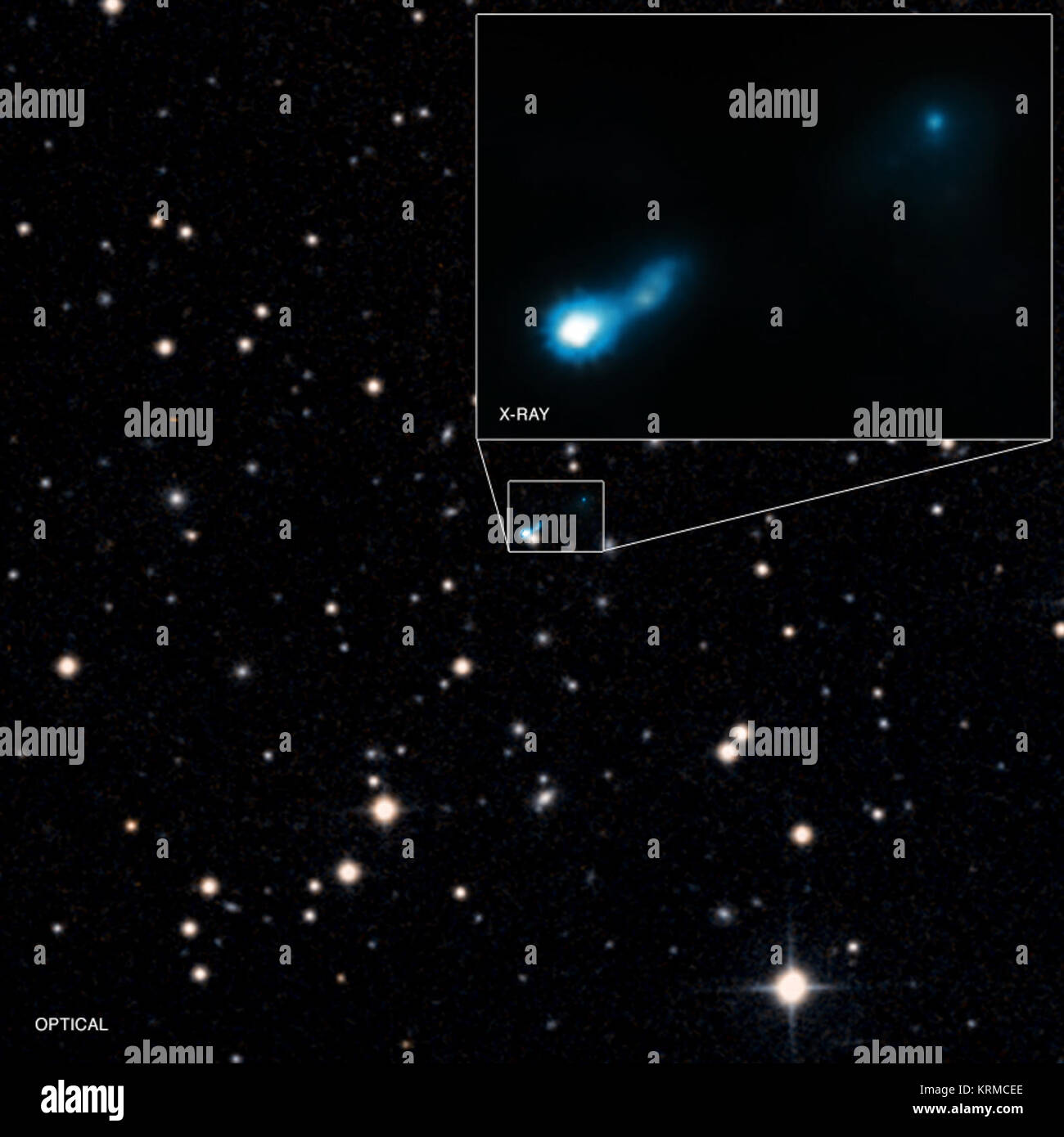 Jets in the early Universe give astronomers a way to probe the growth of black holes at a very early epoch in the cosmos. Using Chandra, astronomers recently discovered a jet in X-rays being illuminated by the cosmic microwave background. The light from this jet was emitted when the Universe was only one fifth of its present age. The main panel of this graphic shows Chandra's X-ray data combined with an optical image, while the inset focuses on the details of the X-ray emission. B3 0727 409 (Chandra) Stock Photohttps://www.alamy.com/image-license-details/?v=1https://www.alamy.com/stock-image-jets-in-the-early-universe-give-astronomers-a-way-to-probe-the-growth-169479254.html
RMKRMCEE–Jets in the early Universe give astronomers a way to probe the growth of black holes at a very early epoch in the cosmos. Using Chandra, astronomers recently discovered a jet in X-rays being illuminated by the cosmic microwave background. The light from this jet was emitted when the Universe was only one fifth of its present age. The main panel of this graphic shows Chandra's X-ray data combined with an optical image, while the inset focuses on the details of the X-ray emission. B3 0727 409 (Chandra)
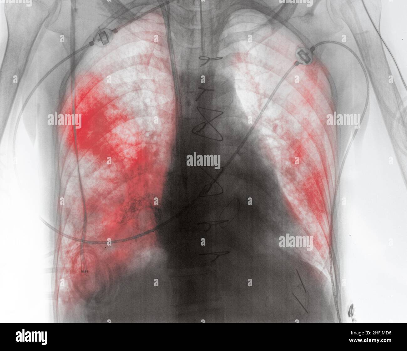 X-ray image of patient with lung inflammation in the early post-surgery period. Stock Photohttps://www.alamy.com/image-license-details/?v=1https://www.alamy.com/x-ray-image-of-patient-with-lung-inflammation-in-the-early-post-surgery-period-image457100594.html
RF2HFJMD6–X-ray image of patient with lung inflammation in the early post-surgery period.
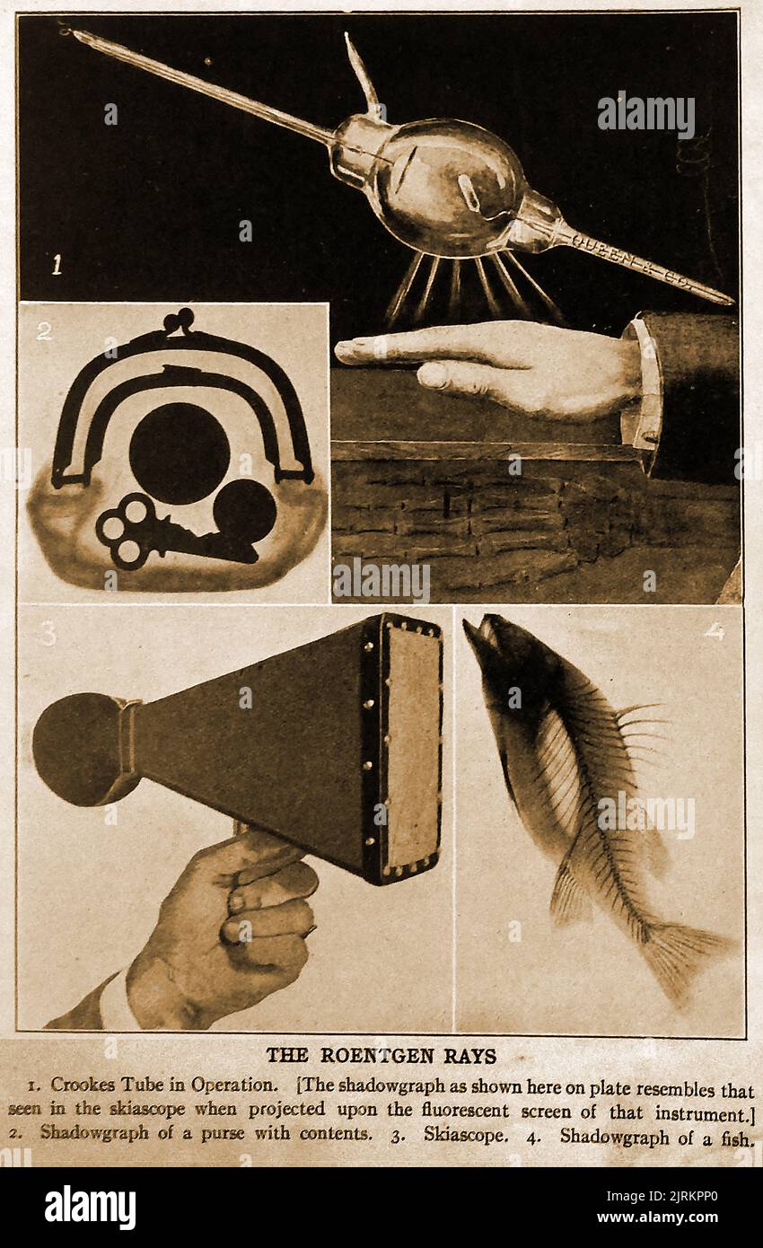 Vintage illustration explaining the uses of Roentgen (aka Röntgen or R Rays) in Crooke's Tube, shadowgraphs & Skioscope. The Roentgen is a measurement for the exposure of X-rays and gamma rays. The Crookes tube or Crookes–Hittorf tube led to the discovery of cathode rays. Eine Vintage-Illustration, die die Verwendung von Röntgen (auch bekannt als Röntgen oder R Rays) in Crooke's Tube, Schattendiagrammen und dem Skioskop erklärt. Der Röntgen ist ein Maß für die Exposition von Röntgen- und Gammastrahlen. Die Crookes-Röhre oder Crookes-Hittorf-Röhre führte zur Entdeckung von Kathodenstrahlen Stock Photohttps://www.alamy.com/image-license-details/?v=1https://www.alamy.com/vintage-illustration-explaining-the-uses-of-roentgen-aka-rntgen-or-r-rays-in-crookes-tube-shadowgraphs-skioscope-the-roentgen-is-a-measurement-for-the-exposure-of-x-rays-and-gamma-rays-the-crookes-tube-or-crookeshittorf-tube-led-to-the-discovery-of-cathode-rays-eine-vintage-illustration-die-die-verwendung-von-rntgen-auch-bekannt-als-rntgen-oder-r-rays-in-crookes-tube-schattendiagrammen-und-dem-skioskop-erklrt-der-rntgen-ist-ein-ma-fr-die-exposition-von-rntgen-und-gammastrahlen-die-crookes-rhre-oder-crookes-hittorf-rhre-fhrte-zur-entdeckung-von-kathodenstrahlen-image479251976.html
RM2JRKPP0–Vintage illustration explaining the uses of Roentgen (aka Röntgen or R Rays) in Crooke's Tube, shadowgraphs & Skioscope. The Roentgen is a measurement for the exposure of X-rays and gamma rays. The Crookes tube or Crookes–Hittorf tube led to the discovery of cathode rays. Eine Vintage-Illustration, die die Verwendung von Röntgen (auch bekannt als Röntgen oder R Rays) in Crooke's Tube, Schattendiagrammen und dem Skioskop erklärt. Der Röntgen ist ein Maß für die Exposition von Röntgen- und Gammastrahlen. Die Crookes-Röhre oder Crookes-Hittorf-Röhre führte zur Entdeckung von Kathodenstrahlen
 Cape Canaveral, United States of America. 09 December, 2021. A SpaceX Falcon 9 rocket carrying the NASA Imaging X-ray Polarimetry Explorer spacecraft blasts off from Launch Complex 39A early morning at the Kennedy Space Center December 9, 2021 in Cape Canaveral, Florida. The IXPE spacecraft is the first satellite dedicated to measuring the polarization of X-rays from a variety of cosmic sources. Credit: Kevin Davis and Chris Coleman/NASA/Alamy Live News Stock Photohttps://www.alamy.com/image-license-details/?v=1https://www.alamy.com/cape-canaveral-united-states-of-america-09-december-2021-a-spacex-falcon-9-rocket-carrying-the-nasa-imaging-x-ray-polarimetry-explorer-spacecraft-blasts-off-from-launch-complex-39a-early-morning-at-the-kennedy-space-center-december-9-2021-in-cape-canaveral-florida-the-ixpe-spacecraft-is-the-first-satellite-dedicated-to-measuring-the-polarization-of-x-rays-from-a-variety-of-cosmic-sources-credit-kevin-davis-and-chris-colemannasaalamy-live-news-image455045604.html
RM2HC938M–Cape Canaveral, United States of America. 09 December, 2021. A SpaceX Falcon 9 rocket carrying the NASA Imaging X-ray Polarimetry Explorer spacecraft blasts off from Launch Complex 39A early morning at the Kennedy Space Center December 9, 2021 in Cape Canaveral, Florida. The IXPE spacecraft is the first satellite dedicated to measuring the polarization of X-rays from a variety of cosmic sources. Credit: Kevin Davis and Chris Coleman/NASA/Alamy Live News
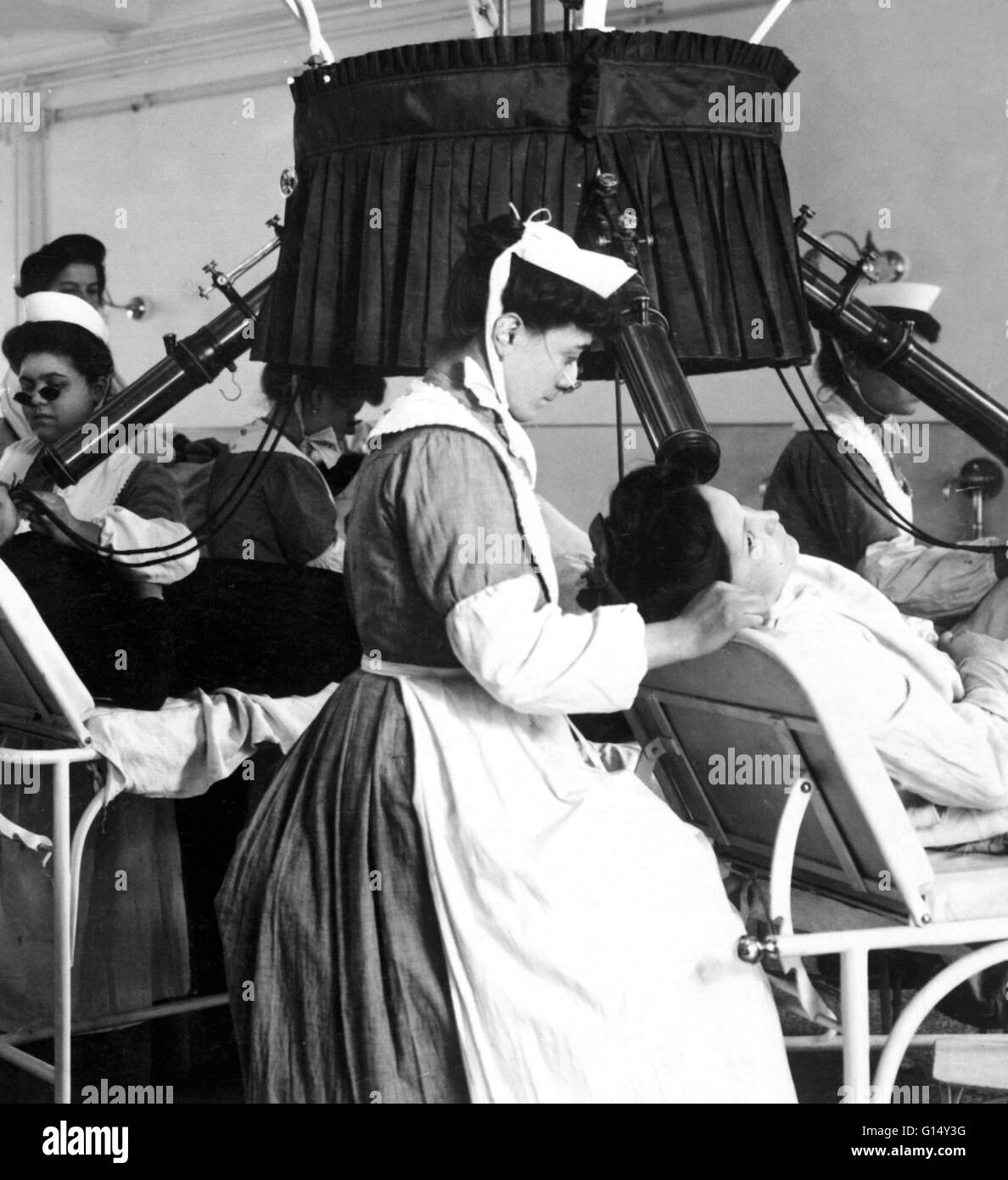 An early x-ray machine from 1900. X-rays were discovered by the German physicist Wilhelm Roentgen in 1895. They were soon applied to medicine because of their ability to pass through soft tissues but not so easily through bone, which casts a shadow on pho Stock Photohttps://www.alamy.com/image-license-details/?v=1https://www.alamy.com/stock-photo-an-early-x-ray-machine-from-1900-x-rays-were-discovered-by-the-german-103985940.html
RMG14Y3G–An early x-ray machine from 1900. X-rays were discovered by the German physicist Wilhelm Roentgen in 1895. They were soon applied to medicine because of their ability to pass through soft tissues but not so easily through bone, which casts a shadow on pho
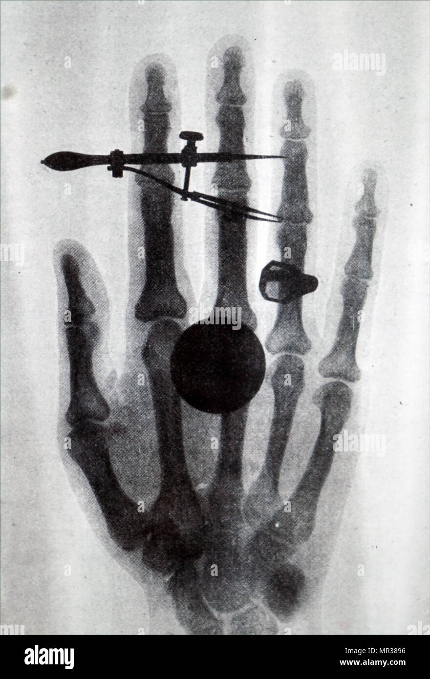 Early Röntgen X-Ray of Wilhelm Röntgen wife's hand. Wilhelm Röntgen (1845-1923) a German mechanical engineer and physicist, who produced and detected electromagnetic radiation in a wavelength range known as X-rays of Röntgen Rays. Dated 19th century Stock Photohttps://www.alamy.com/image-license-details/?v=1https://www.alamy.com/early-rntgen-x-ray-of-wilhelm-rntgen-wifes-hand-wilhelm-rntgen-1845-1923-a-german-mechanical-engineer-and-physicist-who-produced-and-detected-electromagnetic-radiation-in-a-wavelength-range-known-as-x-rays-of-rntgen-rays-dated-19th-century-image186313154.html
RMMR3896–Early Röntgen X-Ray of Wilhelm Röntgen wife's hand. Wilhelm Röntgen (1845-1923) a German mechanical engineer and physicist, who produced and detected electromagnetic radiation in a wavelength range known as X-rays of Röntgen Rays. Dated 19th century
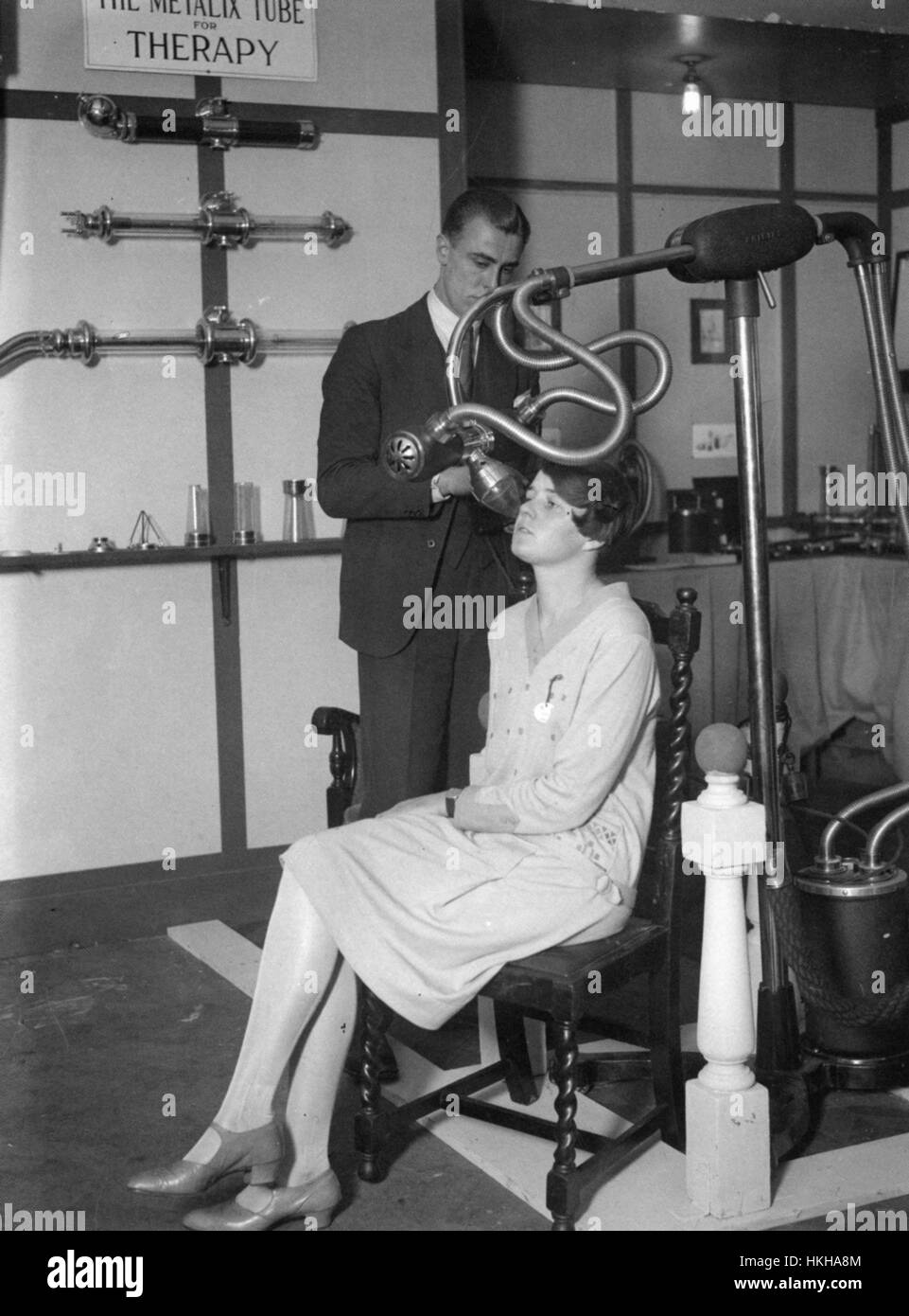 METALIX TUBE FOR THERAPY A type of x-ray machine made by the Philips/Muller company on show at a London exhibition in November 1928 Stock Photohttps://www.alamy.com/image-license-details/?v=1https://www.alamy.com/stock-photo-metalix-tube-for-therapy-a-type-of-x-ray-machine-made-by-the-philipsmuller-132532308.html
RMHKHA8M–METALIX TUBE FOR THERAPY A type of x-ray machine made by the Philips/Muller company on show at a London exhibition in November 1928
 West Dorset County Hospital, Dorchester, West Dorset, Dorset, 25/07/1986. An exterior view of part of West Dorset County Hospital. In the early 1980s plans were developed for a modern hospital to replace the old Dorchester Hospital, at a new site to the west of the town centre. A joint venture of Laing’s South West Region and Haden Young Limited were responsible for Phase I of the construction project, which was to provide 149 beds and included maternity and geriatric units, two operating theatres and a pathology and X-ray department. Phase I on the north part of the new site was comple Stock Photohttps://www.alamy.com/image-license-details/?v=1https://www.alamy.com/west-dorset-county-hospital-dorchester-west-dorset-dorset-25071986-an-exterior-view-of-part-of-west-dorset-county-hospital-in-the-early-1980s-plans-were-developed-for-a-modern-hospital-to-replace-the-old-dorchester-hospital-at-a-new-site-to-the-west-of-the-town-centre-a-joint-venture-of-laingx2019s-south-west-region-and-haden-young-limited-were-responsible-for-phase-i-of-the-construction-project-which-was-to-provide-149-beds-and-included-maternity-and-geriatric-units-two-operating-theatres-and-a-pathology-and-x-ray-department-phase-i-on-the-north-part-of-the-new-site-was-comple-image462503379.html
RM2HTCRNR–West Dorset County Hospital, Dorchester, West Dorset, Dorset, 25/07/1986. An exterior view of part of West Dorset County Hospital. In the early 1980s plans were developed for a modern hospital to replace the old Dorchester Hospital, at a new site to the west of the town centre. A joint venture of Laing’s South West Region and Haden Young Limited were responsible for Phase I of the construction project, which was to provide 149 beds and included maternity and geriatric units, two operating theatres and a pathology and X-ray department. Phase I on the north part of the new site was comple
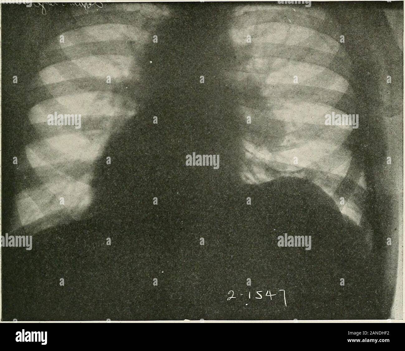 Diseases of the chest and the principles of physical diagnosis . aumbilical region while the lower portion of theabdomen remains supple (Dieulafoy). The use of an exploring needleshould be resorted to early. The X-ray findings in regard to thediaphragm are the same as in those cases in which the thoracic signspredominate. OlPABTMeMT OF CLUa^L P»- COLUMBIA UNIVERSITY , 437 W. 59th Street, DISEASES OF THE DIArtSfeWoldP^^ ^ 651 Signs in Gas-containing Abscesses.—(Pyopneumothorax subphren-icus).—Gas may be present from the onset or it may develop later. Forthis reason the physical signs may change Stock Photohttps://www.alamy.com/image-license-details/?v=1https://www.alamy.com/diseases-of-the-chest-and-the-principles-of-physical-diagnosis-aumbilical-region-while-the-lower-portion-of-theabdomen-remains-supple-dieulafoy-the-use-of-an-exploring-needleshould-be-resorted-to-early-the-x-ray-findings-in-regard-to-thediaphragm-are-the-same-as-in-those-cases-in-which-the-thoracic-signspredominate-olpabtmemt-of-clual-p-columbia-university-437-w-59th-street-diseases-of-the-diartsfewoldp-651-signs-in-gas-containing-abscessespyopneumothorax-subphren-icusgas-may-be-present-from-the-onset-or-it-may-develop-later-forthis-reason-the-physical-signs-may-change-image340203894.html
RM2ANDHF2–Diseases of the chest and the principles of physical diagnosis . aumbilical region while the lower portion of theabdomen remains supple (Dieulafoy). The use of an exploring needleshould be resorted to early. The X-ray findings in regard to thediaphragm are the same as in those cases in which the thoracic signspredominate. OlPABTMeMT OF CLUa^L P»- COLUMBIA UNIVERSITY , 437 W. 59th Street, DISEASES OF THE DIArtSfeWoldP^^ ^ 651 Signs in Gas-containing Abscesses.—(Pyopneumothorax subphren-icus).—Gas may be present from the onset or it may develop later. Forthis reason the physical signs may change
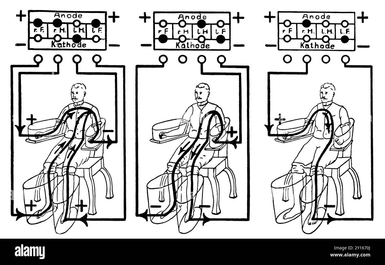 An illustration of a patient being treated with a four cell Schnee Bath, an early electric medical equipment for treating rheumatism Stock Photohttps://www.alamy.com/image-license-details/?v=1https://www.alamy.com/an-illustration-of-a-patient-being-treated-with-a-four-cell-schnee-bath-an-early-electric-medical-equipment-for-treating-rheumatism-image620325122.html
RM2Y1670J–An illustration of a patient being treated with a four cell Schnee Bath, an early electric medical equipment for treating rheumatism
 Early X-ray figure panel Stock Photohttps://www.alamy.com/image-license-details/?v=1https://www.alamy.com/early-x-ray-figure-panel-image265052016.html
RMWB64FC–Early X-ray figure panel
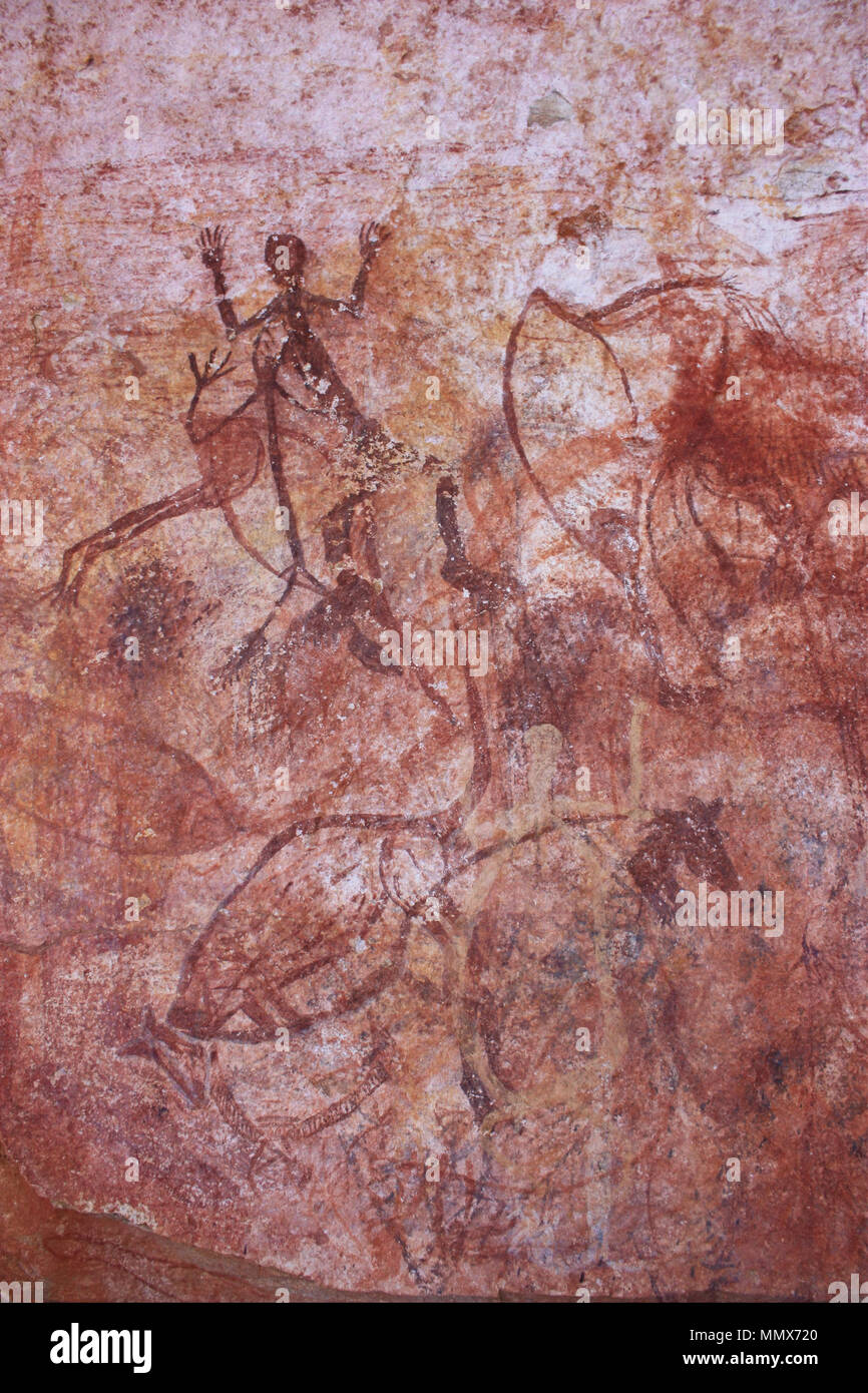 Early X-ray animals. (-2000 - -1). Early X-ray animals - Google Art Project Stock Photohttps://www.alamy.com/image-license-details/?v=1https://www.alamy.com/early-x-ray-animals-2000-1-early-x-ray-animals-google-art-project-image184973096.html
RMMMX720–Early X-ray animals. (-2000 - -1). Early X-ray animals - Google Art Project
 Early X-ray figure panel - Google Art Project Stock Photohttps://www.alamy.com/image-license-details/?v=1https://www.alamy.com/stock-photo-early-x-ray-figure-panel-google-art-project-132682324.html
RMHKT5JC–Early X-ray figure panel - Google Art Project
 Early X-ray figure panel. Date/Period: -2000 - -1. Painting. Author: UNKNOWN. Stock Photohttps://www.alamy.com/image-license-details/?v=1https://www.alamy.com/early-x-ray-figure-panel-dateperiod-2000-1-painting-author-unknown-image219071775.html
RMPMBG7Y–Early X-ray figure panel. Date/Period: -2000 - -1. Painting. Author: UNKNOWN.
 Early medical x ray image of a hand, showing arthritis, 1909. Courtesy Internet Archive. Note: Image has been digitally colorized using a modern process. Colors may not be period-accurate. () Stock Photohttps://www.alamy.com/image-license-details/?v=1https://www.alamy.com/early-medical-x-ray-image-of-a-hand-showing-arthritis-1909-courtesy-internet-archive-note-image-has-been-digitally-colorized-using-a-modern-process-colors-may-not-be-period-accurate-image181312735.html
RMMEYE6R–Early medical x ray image of a hand, showing arthritis, 1909. Courtesy Internet Archive. Note: Image has been digitally colorized using a modern process. Colors may not be period-accurate. ()
 A doctor in a blue uniform shows a picture of a fluorogram of fluorography, an X-ray of the lungs for the prevention and early diagnosis of Stock Photohttps://www.alamy.com/image-license-details/?v=1https://www.alamy.com/a-doctor-in-a-blue-uniform-shows-a-picture-of-a-fluorogram-of-fluorography-an-x-ray-of-the-lungs-for-the-prevention-and-early-diagnosis-of-image453136329.html
RF2H96409–A doctor in a blue uniform shows a picture of a fluorogram of fluorography, an X-ray of the lungs for the prevention and early diagnosis of
 Christ of Chalma, Oil on canvas, Mexico, late 18th-early 19th century, 24 3/8 x 16 1/2 in., 18th century, 19th century, christ, christian, Christianity, cross, crown, crucifixion, crucify, death, drapery, god, halo, jesus, Mary, Mexican Painting, nail, nailed, oil on canvas, punish, religious, saints, symbolic, thorns, x-ray Stock Photohttps://www.alamy.com/image-license-details/?v=1https://www.alamy.com/christ-of-chalma-oil-on-canvas-mexico-late-18th-early-19th-century-24-38-x-16-12-in-18th-century-19th-century-christ-christian-christianity-cross-crown-crucifixion-crucify-death-drapery-god-halo-jesus-mary-mexican-painting-nail-nailed-oil-on-canvas-punish-religious-saints-symbolic-thorns-x-ray-image454282378.html
RM2HB29PJ–Christ of Chalma, Oil on canvas, Mexico, late 18th-early 19th century, 24 3/8 x 16 1/2 in., 18th century, 19th century, christ, christian, Christianity, cross, crown, crucifixion, crucify, death, drapery, god, halo, jesus, Mary, Mexican Painting, nail, nailed, oil on canvas, punish, religious, saints, symbolic, thorns, x-ray
 Inspired by Forest Landscape, American, Oil on canvas, ca. late 19th or early 20th century, canvas: 7 x 14 in., 17.8 x 35.6 cm, Reimagined by Artotop. Classic art reinvented with a modern twist. Design of warm cheerful glowing of brightness and light ray radiance. Photography inspired by surrealism and futurism, embracing dynamic energy of modern technology, movement, speed and revolutionize culture Stock Photohttps://www.alamy.com/image-license-details/?v=1https://www.alamy.com/inspired-by-forest-landscape-american-oil-on-canvas-ca-late-19th-or-early-20th-century-canvas-7-x-14-in-178-x-356-cm-reimagined-by-artotop-classic-art-reinvented-with-a-modern-twist-design-of-warm-cheerful-glowing-of-brightness-and-light-ray-radiance-photography-inspired-by-surrealism-and-futurism-embracing-dynamic-energy-of-modern-technology-movement-speed-and-revolutionize-culture-image459267065.html
RF2HK5BR5–Inspired by Forest Landscape, American, Oil on canvas, ca. late 19th or early 20th century, canvas: 7 x 14 in., 17.8 x 35.6 cm, Reimagined by Artotop. Classic art reinvented with a modern twist. Design of warm cheerful glowing of brightness and light ray radiance. Photography inspired by surrealism and futurism, embracing dynamic energy of modern technology, movement, speed and revolutionize culture
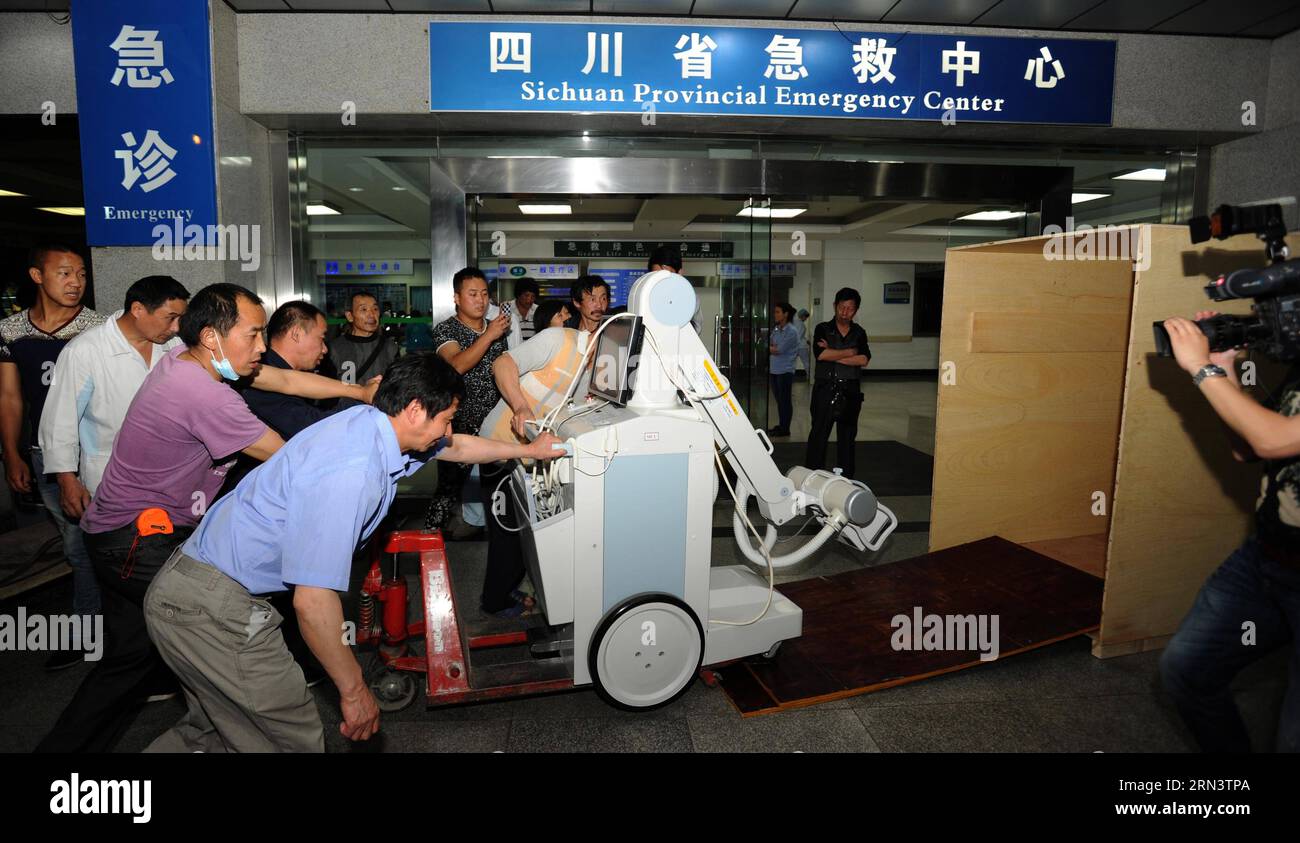 People transport a medical X-ray medicine from Sichuan Provincial Emergency Center in Chengdu, southwest China s Sichuan Province, April 26, 2015. A medical team departed from Chengdu to Nepal to help with relief work on early Monday morning. () (wf) CHINA-SICHUAN-NAPAL EARTHQUAKE-RESCUE TEAM(CN) Xinhua PUBLICATIONxNOTxINxCHN Celebrities Transportation a Medical X Ray Medicine from Sichuan Provincial EMERGENCY Center in Chengdu Southwest China S Sichuan Province April 26 2015 a Medical Team Departed from Chengdu to Nepal to Help With Relief Work ON Early Monday Morning WF China Sichuan NAPAL Stock Photohttps://www.alamy.com/image-license-details/?v=1https://www.alamy.com/people-transport-a-medical-x-ray-medicine-from-sichuan-provincial-emergency-center-in-chengdu-southwest-china-s-sichuan-province-april-26-2015-a-medical-team-departed-from-chengdu-to-nepal-to-help-with-relief-work-on-early-monday-morning-wf-china-sichuan-napal-earthquake-rescue-teamcn-xinhua-publicationxnotxinxchn-celebrities-transportation-a-medical-x-ray-medicine-from-sichuan-provincial-emergency-center-in-chengdu-southwest-china-s-sichuan-province-april-26-2015-a-medical-team-departed-from-chengdu-to-nepal-to-help-with-relief-work-on-early-monday-morning-wf-china-sichuan-napal-image563724850.html
RM2RN3TPA–People transport a medical X-ray medicine from Sichuan Provincial Emergency Center in Chengdu, southwest China s Sichuan Province, April 26, 2015. A medical team departed from Chengdu to Nepal to help with relief work on early Monday morning. () (wf) CHINA-SICHUAN-NAPAL EARTHQUAKE-RESCUE TEAM(CN) Xinhua PUBLICATIONxNOTxINxCHN Celebrities Transportation a Medical X Ray Medicine from Sichuan Provincial EMERGENCY Center in Chengdu Southwest China S Sichuan Province April 26 2015 a Medical Team Departed from Chengdu to Nepal to Help With Relief Work ON Early Monday Morning WF China Sichuan NAPAL
 Kevin Schawinski, astrophysicist, Yale University speaks at a press conference announcing black hole findings by NASA's Chandra X-ray Observatory, Wednesday, June 15, 2011 in Washington. Using the deepest X-ray image ever taken, Chandra discovered massive black holes were common in the early universe. Photo credit: (NASA/Carla Cioffi) Kevin Schawinski, swiss astrophysicist, 2011 Stock Photohttps://www.alamy.com/image-license-details/?v=1https://www.alamy.com/stock-image-kevin-schawinski-astrophysicist-yale-university-speaks-at-a-press-169436711.html
RMKRJE73–Kevin Schawinski, astrophysicist, Yale University speaks at a press conference announcing black hole findings by NASA's Chandra X-ray Observatory, Wednesday, June 15, 2011 in Washington. Using the deepest X-ray image ever taken, Chandra discovered massive black holes were common in the early universe. Photo credit: (NASA/Carla Cioffi) Kevin Schawinski, swiss astrophysicist, 2011
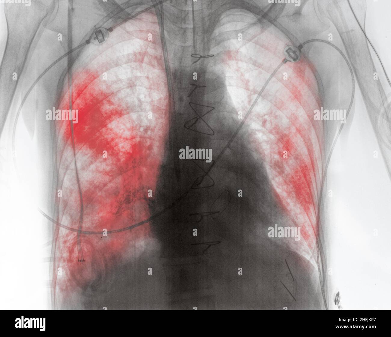 X-ray image of patient with lung inflammation in the early post-surgery period. Stock Photohttps://www.alamy.com/image-license-details/?v=1https://www.alamy.com/x-ray-image-of-patient-with-lung-inflammation-in-the-early-post-surgery-period-image457100063.html
RF2HFJKP7–X-ray image of patient with lung inflammation in the early post-surgery period.
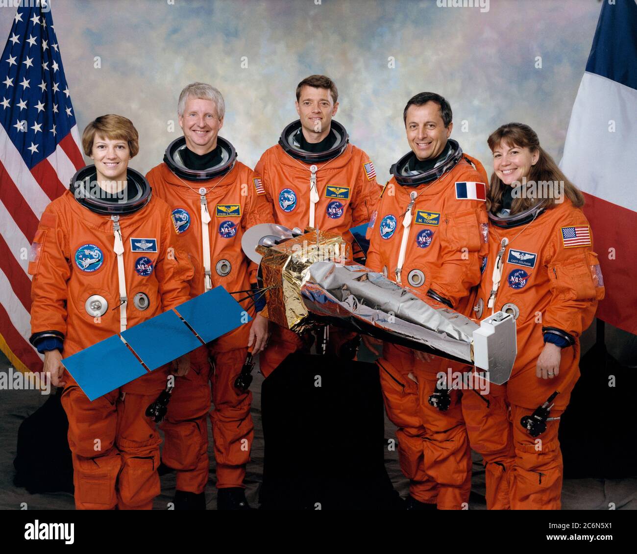 (September 1998) --- The five astronauts assigned to fly aboard the Space Shuttle Columbia early next year for the STS-93 mission pose with a small model of their primary payload-the Advanced X-ray Astrophysics Facility (AXAF). From the left are astronauts Eileen M. Collins, mission commander; Steven A. Hawley, mission specialist; Jeffrey S. Ashby, pilot; Michel Tognini and Catherine G. Coleman, both mission specialists. Tognini represents France's Centre National d'Etudes Spatiales (CNES). Stock Photohttps://www.alamy.com/image-license-details/?v=1https://www.alamy.com/september-1998-the-five-astronauts-assigned-to-fly-aboard-the-space-shuttle-columbia-early-next-year-for-the-sts-93-mission-pose-with-a-small-model-of-their-primary-payload-the-advanced-x-ray-astrophysics-facility-axaf-from-the-left-are-astronauts-eileen-m-collins-mission-commander-steven-a-hawley-mission-specialist-jeffrey-s-ashby-pilot-michel-tognini-and-catherine-g-coleman-both-mission-specialists-tognini-represents-frances-centre-national-detudes-spatiales-cnes-image365571305.html
RM2C6N5X1–(September 1998) --- The five astronauts assigned to fly aboard the Space Shuttle Columbia early next year for the STS-93 mission pose with a small model of their primary payload-the Advanced X-ray Astrophysics Facility (AXAF). From the left are astronauts Eileen M. Collins, mission commander; Steven A. Hawley, mission specialist; Jeffrey S. Ashby, pilot; Michel Tognini and Catherine G. Coleman, both mission specialists. Tognini represents France's Centre National d'Etudes Spatiales (CNES).
 Cape Canaveral, United States of America. 09 December, 2021. A SpaceX Falcon 9 rocket carrying the NASA Imaging X-ray Polarimetry Explorer spacecraft blasts off from Launch Complex 39A early morning at the Kennedy Space Center December 9, 2021 in Cape Canaveral, Florida. The IXPE spacecraft is the first satellite dedicated to measuring the polarization of X-rays from a variety of cosmic sources. Credit: Joel Kowsky/NASA/Alamy Live News Stock Photohttps://www.alamy.com/image-license-details/?v=1https://www.alamy.com/cape-canaveral-united-states-of-america-09-december-2021-a-spacex-falcon-9-rocket-carrying-the-nasa-imaging-x-ray-polarimetry-explorer-spacecraft-blasts-off-from-launch-complex-39a-early-morning-at-the-kennedy-space-center-december-9-2021-in-cape-canaveral-florida-the-ixpe-spacecraft-is-the-first-satellite-dedicated-to-measuring-the-polarization-of-x-rays-from-a-variety-of-cosmic-sources-credit-joel-kowskynasaalamy-live-news-image453605327.html
RM2H9YE67–Cape Canaveral, United States of America. 09 December, 2021. A SpaceX Falcon 9 rocket carrying the NASA Imaging X-ray Polarimetry Explorer spacecraft blasts off from Launch Complex 39A early morning at the Kennedy Space Center December 9, 2021 in Cape Canaveral, Florida. The IXPE spacecraft is the first satellite dedicated to measuring the polarization of X-rays from a variety of cosmic sources. Credit: Joel Kowsky/NASA/Alamy Live News
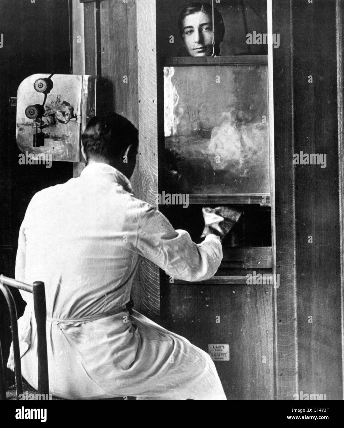 An early x-ray machine, Paris, 1914. X-rays were discovered by the German physicist Wilhelm Roentgen in 1895. They were soon applied to medicine because of their ability to pass through soft tissues but not so easily through bone, which casts a shadow on Stock Photohttps://www.alamy.com/image-license-details/?v=1https://www.alamy.com/stock-photo-an-early-x-ray-machine-paris-1914-x-rays-were-discovered-by-the-german-103985939.html
RMG14Y3F–An early x-ray machine, Paris, 1914. X-rays were discovered by the German physicist Wilhelm Roentgen in 1895. They were soon applied to medicine because of their ability to pass through soft tissues but not so easily through bone, which casts a shadow on
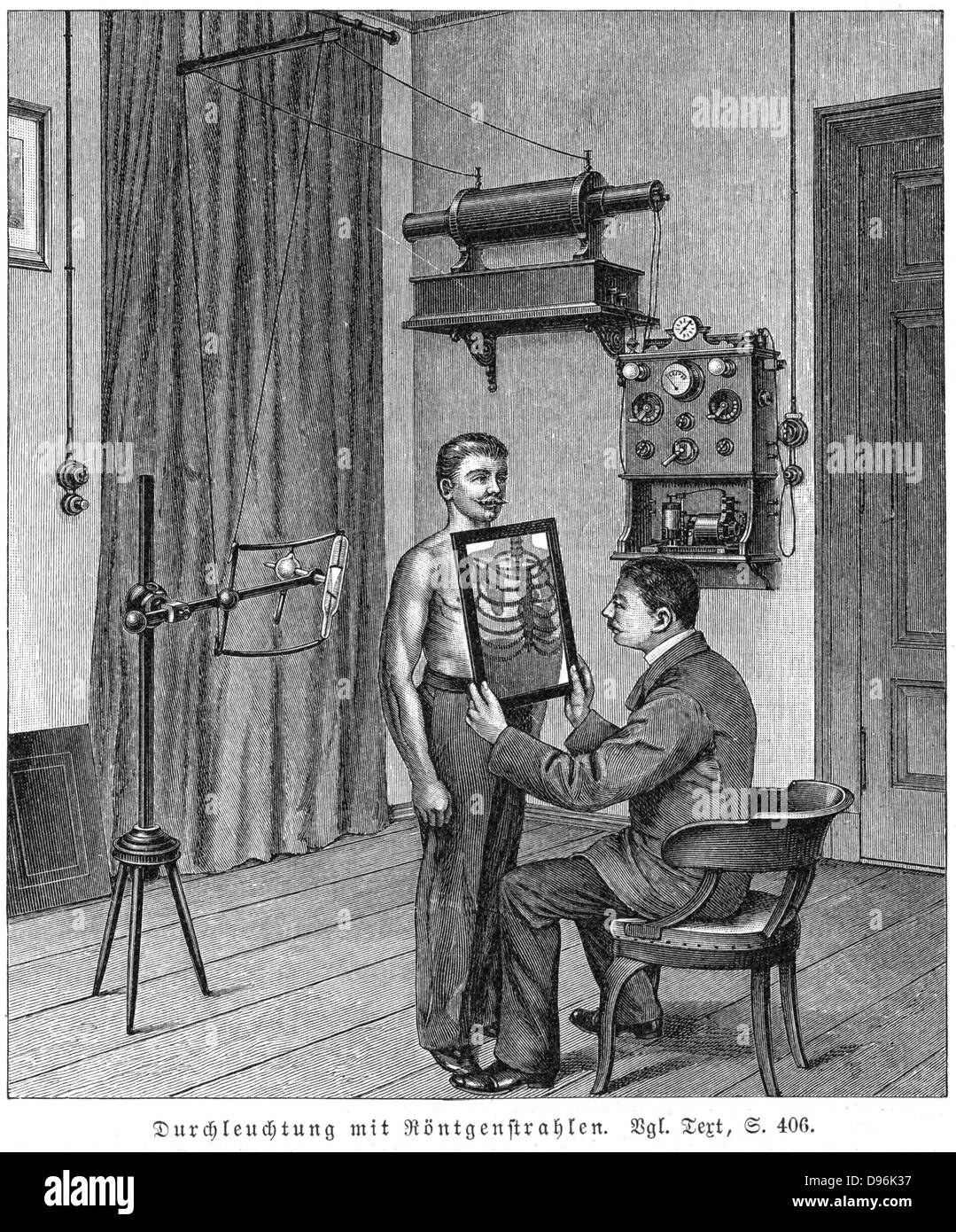 Examining patient's thorax using X-ray tube and fluorescent screen. X-ray tube (on tripod) is set at reqauired height and patient stands directly in front of it. In front of patient, operator holds specially coated paper screen. When X-rays fall on paper, coating fluoresces, leaving as shadows those dense portions of thorax, such as bones, which absorb X-rays. Engraving, Leipzig, 1903. Many who worked with early x-rays suffered the effects of over-exposure to radiation. Stock Photohttps://www.alamy.com/image-license-details/?v=1https://www.alamy.com/stock-photo-examining-patients-thorax-using-x-ray-tube-and-fluorescent-screen-57309707.html
RMD96K37–Examining patient's thorax using X-ray tube and fluorescent screen. X-ray tube (on tripod) is set at reqauired height and patient stands directly in front of it. In front of patient, operator holds specially coated paper screen. When X-rays fall on paper, coating fluoresces, leaving as shadows those dense portions of thorax, such as bones, which absorb X-rays. Engraving, Leipzig, 1903. Many who worked with early x-rays suffered the effects of over-exposure to radiation.
 HANDOUT - A handout photo dated 30 October 2013 shows an the piggy bank and an x-ray of probably Germany's oldest piggy bank in the Museum of Prehistory and Early History in Weimar, Germany. If it was shaken before, now it is only x-rayed. Photo: Hauke Arnold (ATTENTION: Editorial use only, Mandatory Credit: Photo: Hauke Arnold/Thueringisches Landesamt fuer Denkmalpflege und Archaeologie, Weimar/Museum fuer Ur- und Fruehgeschichte Thueringens) Stock Photohttps://www.alamy.com/image-license-details/?v=1https://www.alamy.com/handout-a-handout-photo-dated-30-october-2013-shows-an-the-piggy-bank-image62149359.html
RMDH343Y–HANDOUT - A handout photo dated 30 October 2013 shows an the piggy bank and an x-ray of probably Germany's oldest piggy bank in the Museum of Prehistory and Early History in Weimar, Germany. If it was shaken before, now it is only x-rayed. Photo: Hauke Arnold (ATTENTION: Editorial use only, Mandatory Credit: Photo: Hauke Arnold/Thueringisches Landesamt fuer Denkmalpflege und Archaeologie, Weimar/Museum fuer Ur- und Fruehgeschichte Thueringens)
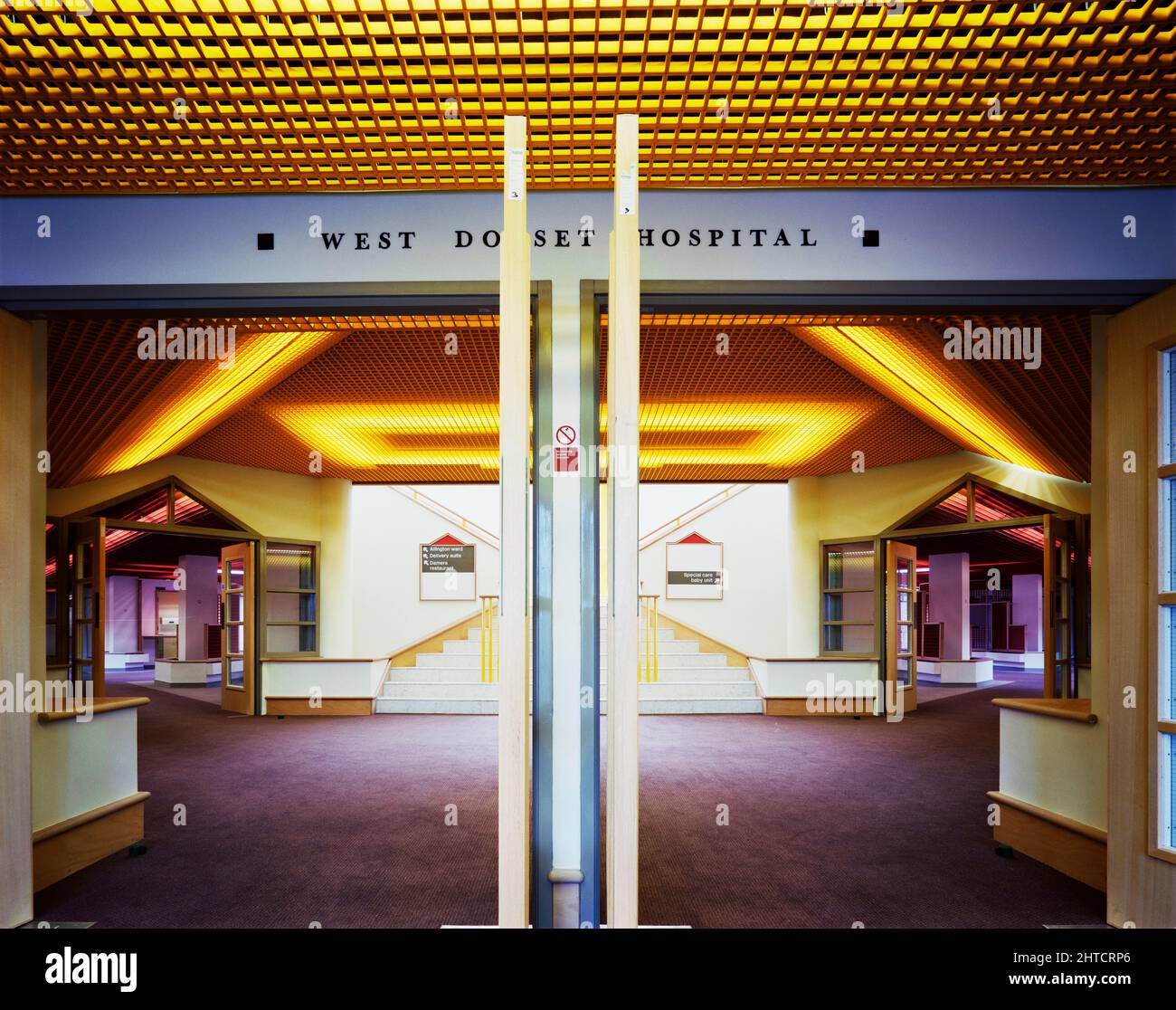 West Dorset County Hospital, Dorchester, West Dorset, Dorset, 08/04/1987. A view from the doorway into the main entrance lobby at West Dorset County Hospital. In the early 1980s plans were developed for a modern hospital to replace the old Dorchester Hospital, at a new site to the west of the town centre. A joint venture of Laing’s South West Region and Haden Young Limited were responsible for Phase I of the construction project, which was to provide 149 beds and included maternity and geriatric units, two operating theatres and a pathology and X-ray department. Phase I on the north par Stock Photohttps://www.alamy.com/image-license-details/?v=1https://www.alamy.com/west-dorset-county-hospital-dorchester-west-dorset-dorset-08041987-a-view-from-the-doorway-into-the-main-entrance-lobby-at-west-dorset-county-hospital-in-the-early-1980s-plans-were-developed-for-a-modern-hospital-to-replace-the-old-dorchester-hospital-at-a-new-site-to-the-west-of-the-town-centre-a-joint-venture-of-laingx2019s-south-west-region-and-haden-young-limited-were-responsible-for-phase-i-of-the-construction-project-which-was-to-provide-149-beds-and-included-maternity-and-geriatric-units-two-operating-theatres-and-a-pathology-and-x-ray-department-phase-i-on-the-north-par-image462503390.html
RM2HTCRP6–West Dorset County Hospital, Dorchester, West Dorset, Dorset, 08/04/1987. A view from the doorway into the main entrance lobby at West Dorset County Hospital. In the early 1980s plans were developed for a modern hospital to replace the old Dorchester Hospital, at a new site to the west of the town centre. A joint venture of Laing’s South West Region and Haden Young Limited were responsible for Phase I of the construction project, which was to provide 149 beds and included maternity and geriatric units, two operating theatres and a pathology and X-ray department. Phase I on the north par
 . The surgical treatment of X-ray carcinoma and other severe X-ray lesions, based upon an analysis of forty-seven cases . 22 ■^0^^^^. i!^^^, •^ 21 C. A. Porter. X ray lesions. THE PATHOLOGICAL HISTOLOGY OF CHRONIC X-RAY DER-MATITIS AND EARLY X-RAY CARCINOMA.* S. B. WOLBACH, M.D. {^Director of the Pathological Laboratory, Montreal General Hospital, Montreal.^ The development of multiple carcinomata in the skin ofpatients and operators who have suffered repeated injuriesfrom exposures to the X-rays has occurred so many timesthat the causal relationship has been generally accepted in allcountries Stock Photohttps://www.alamy.com/image-license-details/?v=1https://www.alamy.com/the-surgical-treatment-of-x-ray-carcinoma-and-other-severe-x-ray-lesions-based-upon-an-analysis-of-forty-seven-cases-22-0-i!-21-c-a-porter-x-ray-lesions-the-pathological-histology-of-chronic-x-ray-der-matitis-and-early-x-ray-carcinoma-s-b-wolbach-md-director-of-the-pathological-laboratory-montreal-general-hospital-montreal-the-development-of-multiple-carcinomata-in-the-skin-ofpatients-and-operators-who-have-suffered-repeated-injuriesfrom-exposures-to-the-x-rays-has-occurred-so-many-timesthat-the-causal-relationship-has-been-generally-accepted-in-allcountries-image376012197.html
RM2CRMRB1–. The surgical treatment of X-ray carcinoma and other severe X-ray lesions, based upon an analysis of forty-seven cases . 22 ■^0^^^^. i!^^^, •^ 21 C. A. Porter. X ray lesions. THE PATHOLOGICAL HISTOLOGY OF CHRONIC X-RAY DER-MATITIS AND EARLY X-RAY CARCINOMA.* S. B. WOLBACH, M.D. {^Director of the Pathological Laboratory, Montreal General Hospital, Montreal.^ The development of multiple carcinomata in the skin ofpatients and operators who have suffered repeated injuriesfrom exposures to the X-rays has occurred so many timesthat the causal relationship has been generally accepted in allcountries
 A patient being treated by Proffessor Wilhelm Conrad Röntgen with a friction spark treatment, an early electric medical Electro-Therapeutics Stock Photohttps://www.alamy.com/image-license-details/?v=1https://www.alamy.com/a-patient-being-treated-by-proffessor-wilhelm-conrad-rntgen-with-a-friction-spark-treatment-an-early-electric-medical-electro-therapeutics-image620325228.html
RM2Y1674C–A patient being treated by Proffessor Wilhelm Conrad Röntgen with a friction spark treatment, an early electric medical Electro-Therapeutics
 Early X-ray bird and human figure Stock Photohttps://www.alamy.com/image-license-details/?v=1https://www.alamy.com/early-x-ray-bird-and-human-figure-image265051984.html
RMWB64E8–Early X-ray bird and human figure
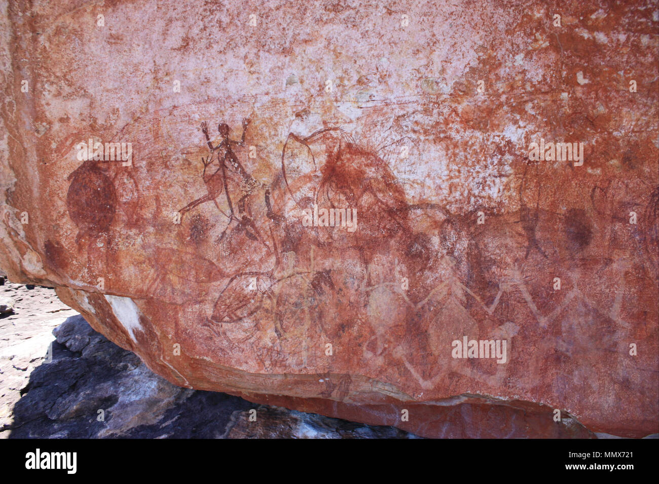 Early X-ray figure panel. (-2000 - -1). Early X-ray figure panel - Google Art Project Stock Photohttps://www.alamy.com/image-license-details/?v=1https://www.alamy.com/early-x-ray-figure-panel-2000-1-early-x-ray-figure-panel-google-art-project-image184973097.html
RMMMX721–Early X-ray figure panel. (-2000 - -1). Early X-ray figure panel - Google Art Project
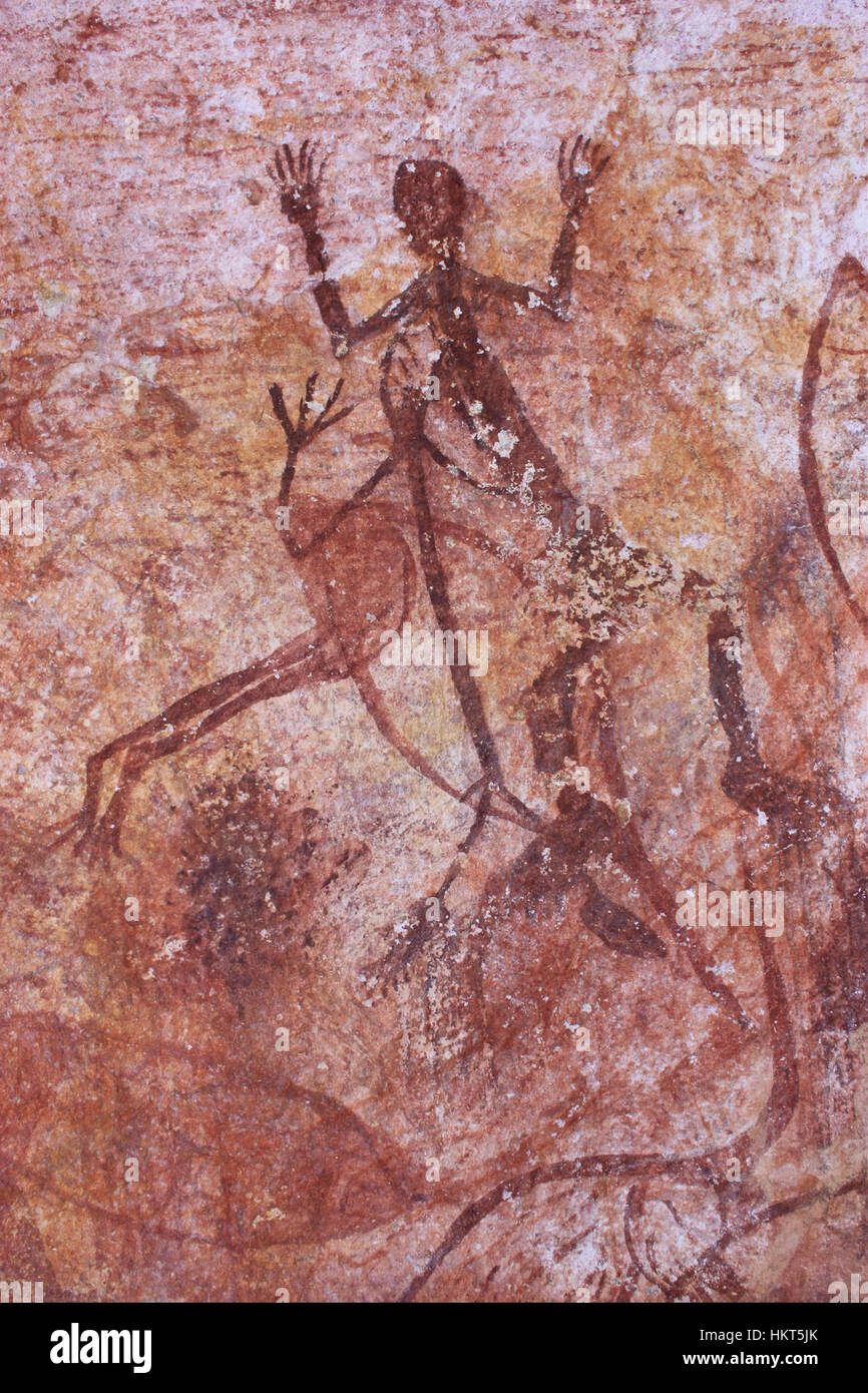 Early X-ray bird and human figure - Google Art Project Stock Photohttps://www.alamy.com/image-license-details/?v=1https://www.alamy.com/stock-photo-early-x-ray-bird-and-human-figure-google-art-project-132682331.html
RMHKT5JK–Early X-ray bird and human figure - Google Art Project
 Early X-ray bird and human figure. Date/Period: -2000 - -1. Painting. Author: UNKNOWN. Stock Photohttps://www.alamy.com/image-license-details/?v=1https://www.alamy.com/early-x-ray-bird-and-human-figure-dateperiod-2000-1-painting-author-unknown-image219071760.html
RMPMBG7C–Early X-ray bird and human figure. Date/Period: -2000 - -1. Painting. Author: UNKNOWN.
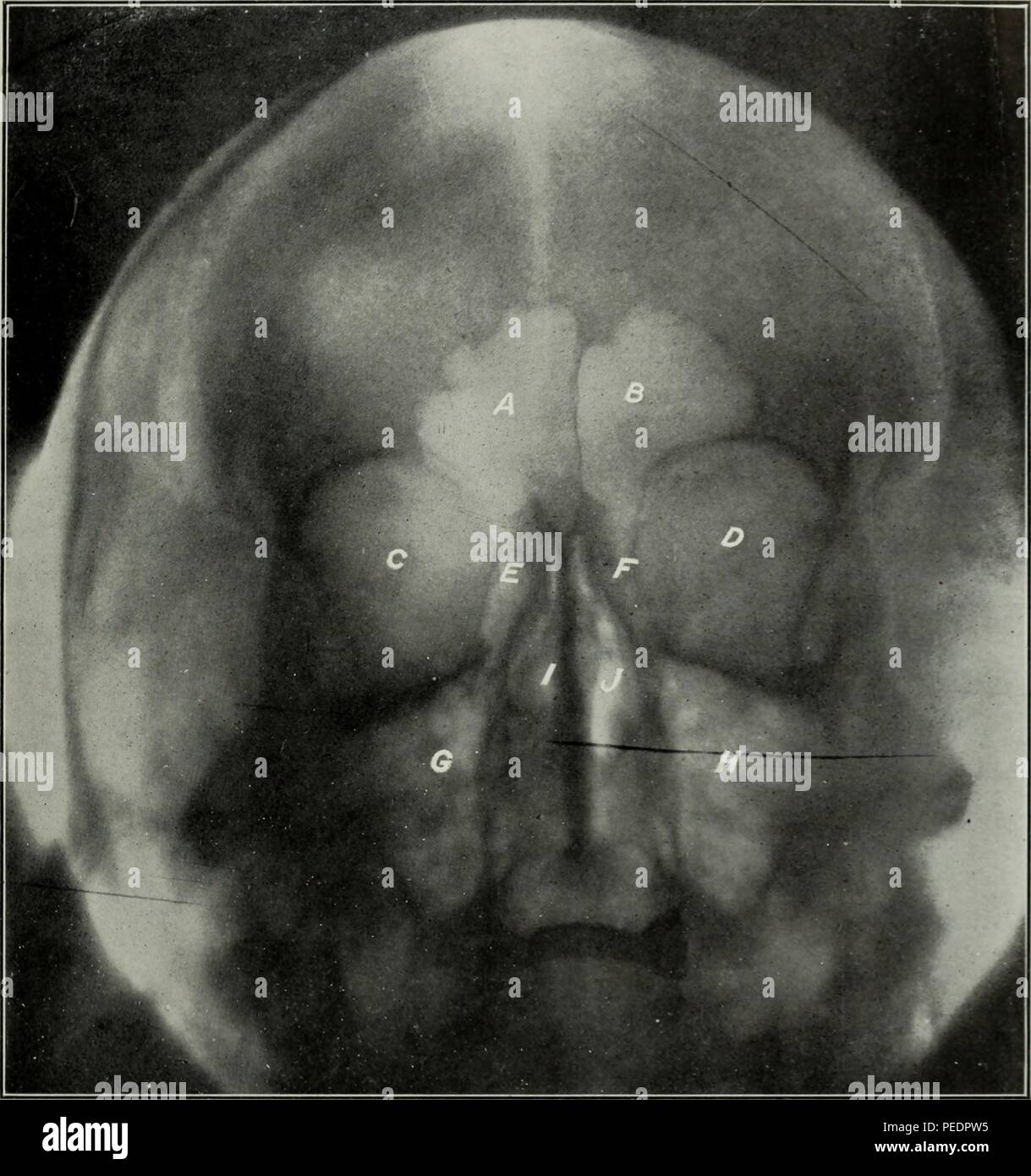 Black and white, early twentieth-century dental radiograph, with letters labeling the frontal and ethmoid sinuses, orbits, and nasal cavity, 1825. Courtesy Internet Archive. () Stock Photohttps://www.alamy.com/image-license-details/?v=1https://www.alamy.com/black-and-white-early-twentieth-century-dental-radiograph-with-letters-labeling-the-frontal-and-ethmoid-sinuses-orbits-and-nasal-cavity-1825-courtesy-internet-archive-image215432929.html
RMPEDPW5–Black and white, early twentieth-century dental radiograph, with letters labeling the frontal and ethmoid sinuses, orbits, and nasal cavity, 1825. Courtesy Internet Archive. ()
 A doctor in a blue uniform shows a picture of a fluorogram of fluorography, an X-ray of the lungs for the prevention and early diagnosis of Stock Photohttps://www.alamy.com/image-license-details/?v=1https://www.alamy.com/a-doctor-in-a-blue-uniform-shows-a-picture-of-a-fluorogram-of-fluorography-an-x-ray-of-the-lungs-for-the-prevention-and-early-diagnosis-of-image462732717.html
RF2HTR88D–A doctor in a blue uniform shows a picture of a fluorogram of fluorography, an X-ray of the lungs for the prevention and early diagnosis of
 Madonna and Child with Saints John the Baptist and Nicholas of Tolentino, Tempera and oil on poplar panel, early 1500s, 28 3/8 x 43 3/4 in., 72.1 x 111.1 cm, x-ray Stock Photohttps://www.alamy.com/image-license-details/?v=1https://www.alamy.com/madonna-and-child-with-saints-john-the-baptist-and-nicholas-of-tolentino-tempera-and-oil-on-poplar-panel-early-1500s-28-38-x-43-34-in-721-x-1111-cm-x-ray-image454273794.html
RM2HB1XT2–Madonna and Child with Saints John the Baptist and Nicholas of Tolentino, Tempera and oil on poplar panel, early 1500s, 28 3/8 x 43 3/4 in., 72.1 x 111.1 cm, x-ray
 Inspired by Tea Gardens, Albumen silver photograph, Suchou, China, late 19th-early 20th century, 6 5/16 x 8 in., 16 x 20.3 cm, Reimagined by Artotop. Classic art reinvented with a modern twist. Design of warm cheerful glowing of brightness and light ray radiance. Photography inspired by surrealism and futurism, embracing dynamic energy of modern technology, movement, speed and revolutionize culture Stock Photohttps://www.alamy.com/image-license-details/?v=1https://www.alamy.com/inspired-by-tea-gardens-albumen-silver-photograph-suchou-china-late-19th-early-20th-century-6-516-x-8-in-16-x-203-cm-reimagined-by-artotop-classic-art-reinvented-with-a-modern-twist-design-of-warm-cheerful-glowing-of-brightness-and-light-ray-radiance-photography-inspired-by-surrealism-and-futurism-embracing-dynamic-energy-of-modern-technology-movement-speed-and-revolutionize-culture-image459266758.html
RF2HK5BC6–Inspired by Tea Gardens, Albumen silver photograph, Suchou, China, late 19th-early 20th century, 6 5/16 x 8 in., 16 x 20.3 cm, Reimagined by Artotop. Classic art reinvented with a modern twist. Design of warm cheerful glowing of brightness and light ray radiance. Photography inspired by surrealism and futurism, embracing dynamic energy of modern technology, movement, speed and revolutionize culture
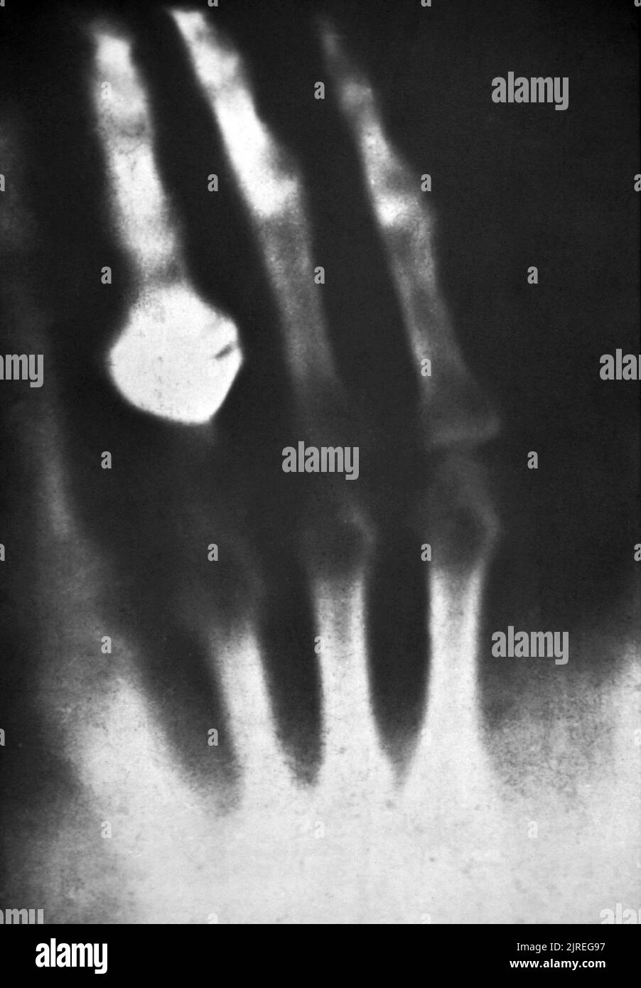 First X-ray photograph of a human being (1895). The picture was made by Wilhelm Roentgen (1845- 1923) shortly after his discovery of X-rays in 1895. It shows the hand of his wife, who is wearing a ring. Roentgen was professor of physics at Wurzburg, Germany. While using an electric discharge tube in a darkened room, he noticed that a card coated with barium platinocyanide glowed when the tube was switched on. The effect was not blocked by an intervening wall, or even a thin sheet of metal. Roentgen termed this X-ray radiation, and used the X-ray of his wife's hand as proof to show colleagues. Stock Photohttps://www.alamy.com/image-license-details/?v=1https://www.alamy.com/first-x-ray-photograph-of-a-human-being-1895-the-picture-was-made-by-wilhelm-roentgen-1845-1923-shortly-after-his-discovery-of-x-rays-in-1895-it-shows-the-hand-of-his-wife-who-is-wearing-a-ring-roentgen-was-professor-of-physics-at-wurzburg-germany-while-using-an-electric-discharge-tube-in-a-darkened-room-he-noticed-that-a-card-coated-with-barium-platinocyanide-glowed-when-the-tube-was-switched-on-the-effect-was-not-blocked-by-an-intervening-wall-or-even-a-thin-sheet-of-metal-roentgen-termed-this-x-ray-radiation-and-used-the-x-ray-of-his-wifes-hand-as-proof-to-show-colleagues-image479137155.html
RM2JREG97–First X-ray photograph of a human being (1895). The picture was made by Wilhelm Roentgen (1845- 1923) shortly after his discovery of X-rays in 1895. It shows the hand of his wife, who is wearing a ring. Roentgen was professor of physics at Wurzburg, Germany. While using an electric discharge tube in a darkened room, he noticed that a card coated with barium platinocyanide glowed when the tube was switched on. The effect was not blocked by an intervening wall, or even a thin sheet of metal. Roentgen termed this X-ray radiation, and used the X-ray of his wife's hand as proof to show colleagues.
 This composite image of VV 340 contains X-ray data from Chandra (purple) and optical data from Hubble (red, green, blue). The two galaxies shown here are in the early stage of an interaction that will eventually lead to them merging in millions of years. The Chandra data shows that the northern galaxy contains a rapidly growing supermassive black hole that is heavily obscured by dust and gas. Data from other wavelengths shows that the two interacting galaxies are evolving at different rates. UGC 9618, Chandra Hubble Stock Photohttps://www.alamy.com/image-license-details/?v=1https://www.alamy.com/stock-image-this-composite-image-of-vv-340-contains-x-ray-data-from-chandra-purple-169273428.html
RMKRB1YG–This composite image of VV 340 contains X-ray data from Chandra (purple) and optical data from Hubble (red, green, blue). The two galaxies shown here are in the early stage of an interaction that will eventually lead to them merging in millions of years. The Chandra data shows that the northern galaxy contains a rapidly growing supermassive black hole that is heavily obscured by dust and gas. Data from other wavelengths shows that the two interacting galaxies are evolving at different rates. UGC 9618, Chandra Hubble
 Chengdu. 26th Apr, 2015. People transport a medical X-ray medicine from Sichuan Provincial Emergency Center in Chengdu, southwest China's Sichuan Province, April 26, 2015. A medical team departed from Chengdu to Nepal to help with relief work on early Monday morning. Credit: Xinhua/Alamy Live News Stock Photohttps://www.alamy.com/image-license-details/?v=1https://www.alamy.com/stock-photo-chengdu-26th-apr-2015-people-transport-a-medical-x-ray-medicine-from-81811831.html
RMEN2RR3–Chengdu. 26th Apr, 2015. People transport a medical X-ray medicine from Sichuan Provincial Emergency Center in Chengdu, southwest China's Sichuan Province, April 26, 2015. A medical team departed from Chengdu to Nepal to help with relief work on early Monday morning. Credit: Xinhua/Alamy Live News
 The National Health Service (NHS)’s Mobile X-ray Unit (MXU) parked on a central London street, UK. The van is a public health initiative for Active Case Finding for TB in hard to reach groups. The rates of TB in London are higher than any other Western European capital and is a major public health problem. TB is an infectious disease, but treatable and curable if diagnosed in time. Early diagnosis is a key to TB control and Active Case Finding is an important part of this. Stock Photohttps://www.alamy.com/image-license-details/?v=1https://www.alamy.com/stock-photo-the-national-health-service-nhss-mobile-x-ray-unit-mxu-parked-on-a-95427209.html
RMFF72AH–The National Health Service (NHS)’s Mobile X-ray Unit (MXU) parked on a central London street, UK. The van is a public health initiative for Active Case Finding for TB in hard to reach groups. The rates of TB in London are higher than any other Western European capital and is a major public health problem. TB is an infectious disease, but treatable and curable if diagnosed in time. Early diagnosis is a key to TB control and Active Case Finding is an important part of this.
 Cape Canaveral, United States of America. 09 December, 2021. A SpaceX Falcon 9 rocket carrying the NASA Imaging X-ray Polarimetry Explorer spacecraft blasts off from Launch Complex 39A early morning at the Kennedy Space Center December 9, 2021 in Cape Canaveral, Florida. The IXPE spacecraft is the first satellite dedicated to measuring the polarization of X-rays from a variety of cosmic sources. Credit: Cory S. Huston/NASA/Alamy Live News Stock Photohttps://www.alamy.com/image-license-details/?v=1https://www.alamy.com/cape-canaveral-united-states-of-america-09-december-2021-a-spacex-falcon-9-rocket-carrying-the-nasa-imaging-x-ray-polarimetry-explorer-spacecraft-blasts-off-from-launch-complex-39a-early-morning-at-the-kennedy-space-center-december-9-2021-in-cape-canaveral-florida-the-ixpe-spacecraft-is-the-first-satellite-dedicated-to-measuring-the-polarization-of-x-rays-from-a-variety-of-cosmic-sources-credit-cory-s-hustonnasaalamy-live-news-image455045601.html
RM2HC938H–Cape Canaveral, United States of America. 09 December, 2021. A SpaceX Falcon 9 rocket carrying the NASA Imaging X-ray Polarimetry Explorer spacecraft blasts off from Launch Complex 39A early morning at the Kennedy Space Center December 9, 2021 in Cape Canaveral, Florida. The IXPE spacecraft is the first satellite dedicated to measuring the polarization of X-rays from a variety of cosmic sources. Credit: Cory S. Huston/NASA/Alamy Live News
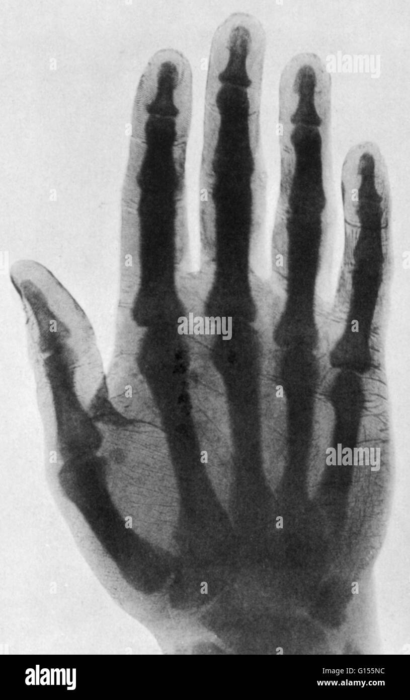 Enhanced early x-ray. One of the clearest of the early class of radiographs was published by David Walsh, M.D. of London in his book 'Roentgen Rays in Medical Work' of 1897. The fingerprints are clearly visible. Stock Photohttps://www.alamy.com/image-license-details/?v=1https://www.alamy.com/stock-photo-enhanced-early-x-ray-one-of-the-clearest-of-the-early-class-of-radiographs-103991144.html
RMG155NC–Enhanced early x-ray. One of the clearest of the early class of radiographs was published by David Walsh, M.D. of London in his book 'Roentgen Rays in Medical Work' of 1897. The fingerprints are clearly visible.
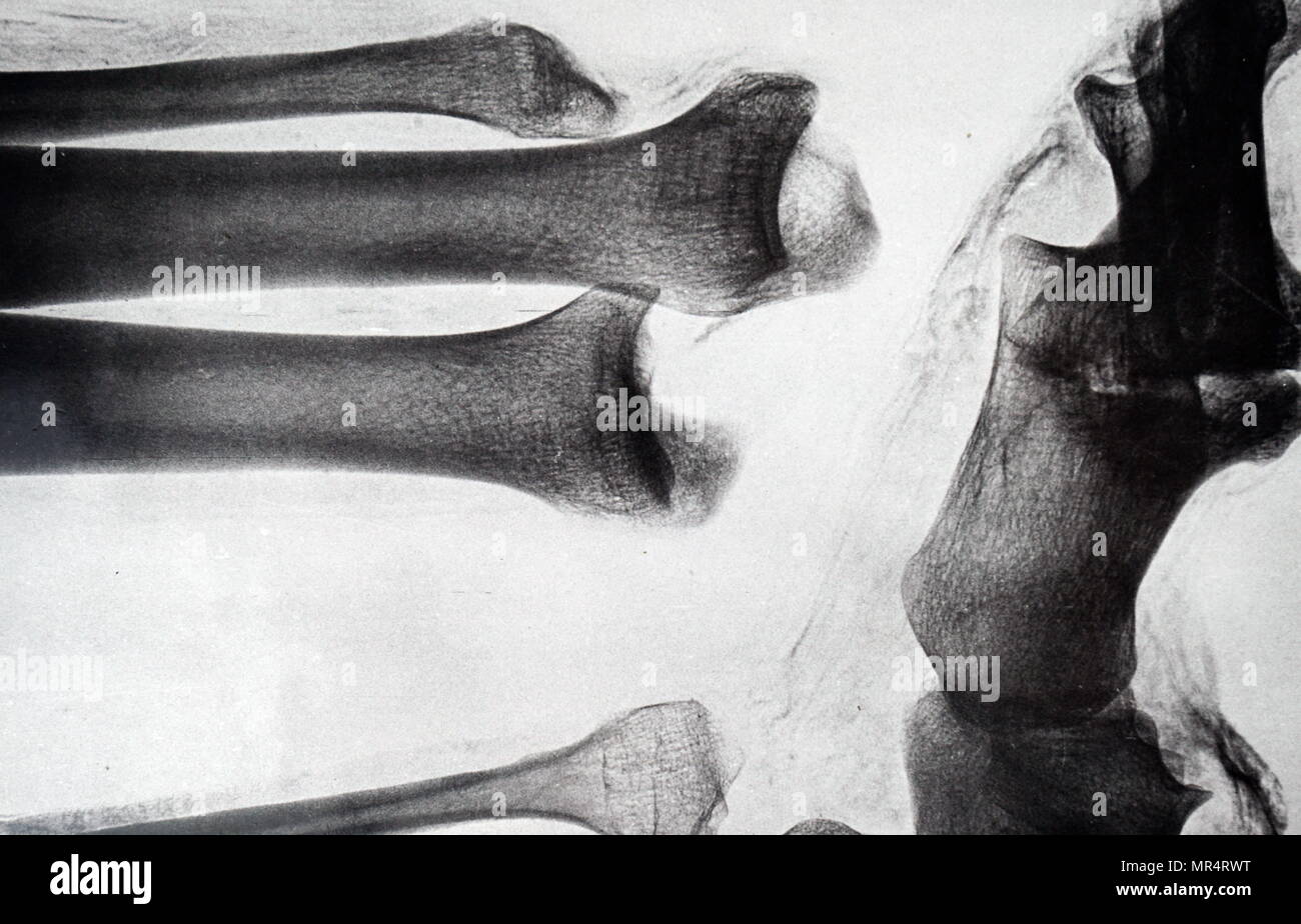 X-ray imaging of the leg of an Egyptian mummy. Dated 19th century Stock Photohttps://www.alamy.com/image-license-details/?v=1https://www.alamy.com/x-ray-imaging-of-the-leg-of-an-egyptian-mummy-dated-19th-century-image186347332.html
RMMR4RWT–X-ray imaging of the leg of an Egyptian mummy. Dated 19th century
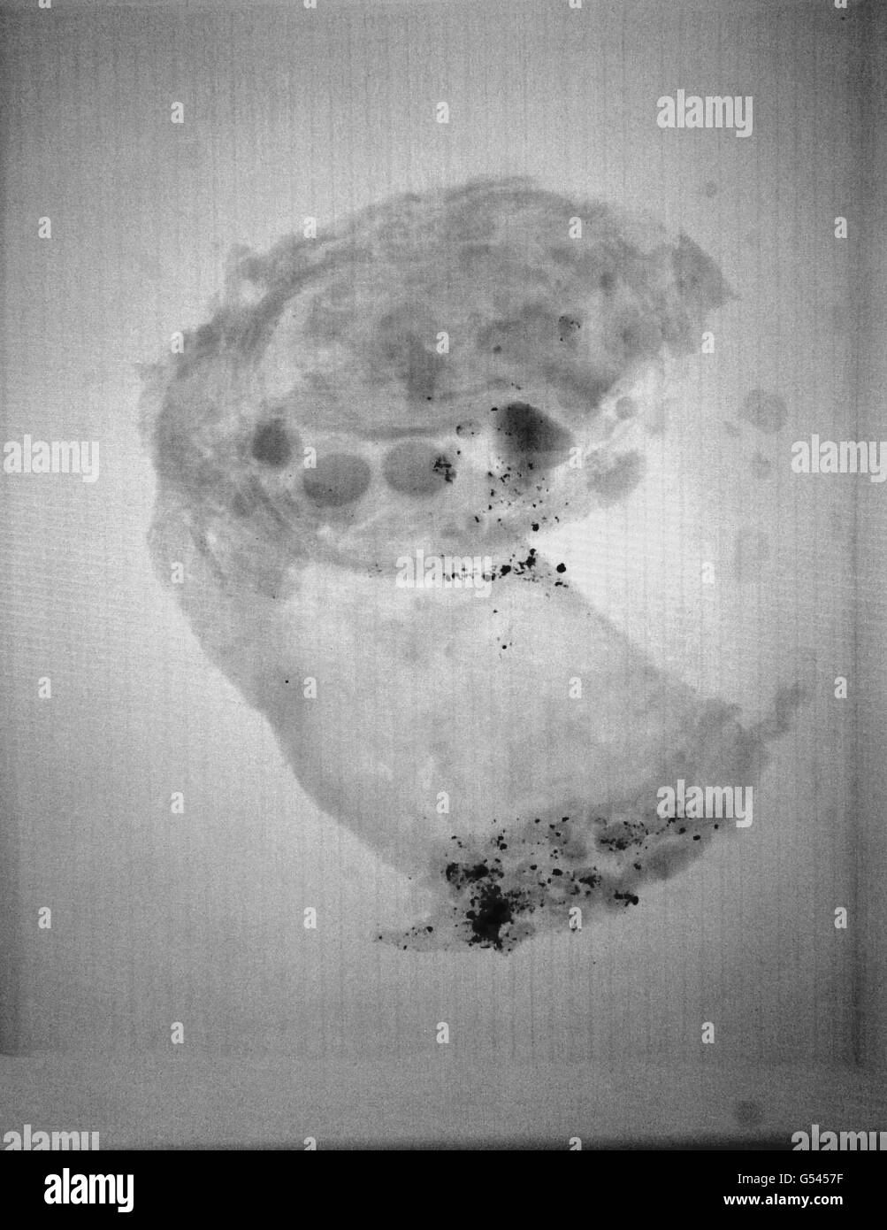 Bronze Age relics x-rayed Stock Photohttps://www.alamy.com/image-license-details/?v=1https://www.alamy.com/stock-photo-bronze-age-relics-x-rayed-106427427.html
RMG5457F–Bronze Age relics x-rayed
 West Dorset County Hospital, Dorchester, West Dorset, Dorset, 08/04/1987. A view of West Dorset County Hospital from a pathway at the south east, showing a sign to the main entrance. In the early 1980s plans were developed for a modern hospital to replace the old Dorchester Hospital, at a new site to the west of the town centre. A joint venture of Laing’s South West Region and Haden Young Limited were responsible for Phase I of the construction project, which was to provide 149 beds and included maternity and geriatric units, two operating theatres and a pathology and X-ray department. Stock Photohttps://www.alamy.com/image-license-details/?v=1https://www.alamy.com/west-dorset-county-hospital-dorchester-west-dorset-dorset-08041987-a-view-of-west-dorset-county-hospital-from-a-pathway-at-the-south-east-showing-a-sign-to-the-main-entrance-in-the-early-1980s-plans-were-developed-for-a-modern-hospital-to-replace-the-old-dorchester-hospital-at-a-new-site-to-the-west-of-the-town-centre-a-joint-venture-of-laingx2019s-south-west-region-and-haden-young-limited-were-responsible-for-phase-i-of-the-construction-project-which-was-to-provide-149-beds-and-included-maternity-and-geriatric-units-two-operating-theatres-and-a-pathology-and-x-ray-department-image462503387.html
RM2HTCRP3–West Dorset County Hospital, Dorchester, West Dorset, Dorset, 08/04/1987. A view of West Dorset County Hospital from a pathway at the south east, showing a sign to the main entrance. In the early 1980s plans were developed for a modern hospital to replace the old Dorchester Hospital, at a new site to the west of the town centre. A joint venture of Laing’s South West Region and Haden Young Limited were responsible for Phase I of the construction project, which was to provide 149 beds and included maternity and geriatric units, two operating theatres and a pathology and X-ray department.
 . American quarterly of roentgenology . the crude apparatus could bemanipulated at this undeveloped stage, the services of anexpert or a specialist were hardly considered necessary, asis the case to-day. Not having the time to devote to it him-self, the physician or the surgeon who realized the need ofwhatever X-ray work could then be accomplished, naturallyplaced it in the hands of someone under him. As a result,a large part of the early X-ray work, at least in this country,was, on the one hand, turned over to under assistants oreven individuals with no medical training, such as hospitalpharm Stock Photohttps://www.alamy.com/image-license-details/?v=1https://www.alamy.com/american-quarterly-of-roentgenology-the-crude-apparatus-could-bemanipulated-at-this-undeveloped-stage-the-services-of-anexpert-or-a-specialist-were-hardly-considered-necessary-asis-the-case-to-day-not-having-the-time-to-devote-to-it-him-self-the-physician-or-the-surgeon-who-realized-the-need-ofwhatever-x-ray-work-could-then-be-accomplished-naturallyplaced-it-in-the-hands-of-someone-under-him-as-a-resulta-large-part-of-the-early-x-ray-work-at-least-in-this-countrywas-on-the-one-hand-turned-over-to-under-assistants-oreven-individuals-with-no-medical-training-such-as-hospitalpharm-image376051686.html
RM2CRPHNA–. American quarterly of roentgenology . the crude apparatus could bemanipulated at this undeveloped stage, the services of anexpert or a specialist were hardly considered necessary, asis the case to-day. Not having the time to devote to it him-self, the physician or the surgeon who realized the need ofwhatever X-ray work could then be accomplished, naturallyplaced it in the hands of someone under him. As a result,a large part of the early X-ray work, at least in this country,was, on the one hand, turned over to under assistants oreven individuals with no medical training, such as hospitalpharm