Electron micrograph chloroplast Stock Photos and Images
(58)See electron micrograph chloroplast stock video clipsQuick filters:
Electron micrograph chloroplast Stock Photos and Images
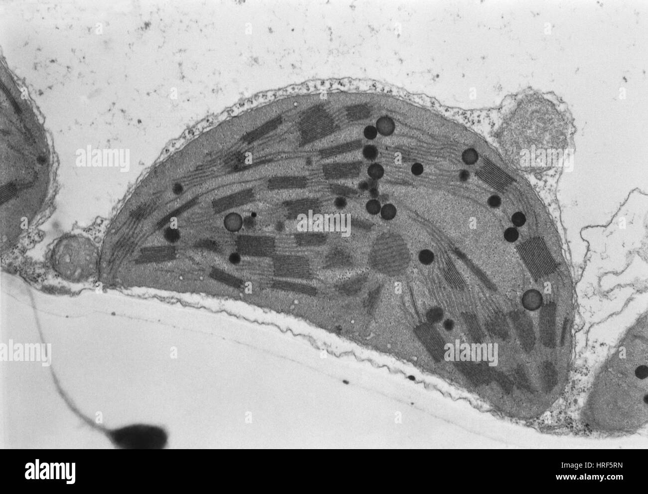 Chloroplast TEM Stock Photohttps://www.alamy.com/image-license-details/?v=1https://www.alamy.com/stock-photo-chloroplast-tem-134943529.html
RMHRF5RN–Chloroplast TEM
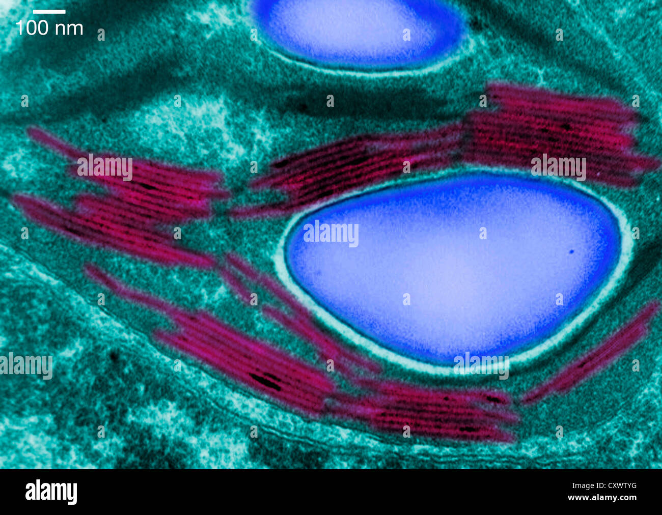 Transmission electron micrograph of a chloroplast Stock Photohttps://www.alamy.com/image-license-details/?v=1https://www.alamy.com/stock-photo-transmission-electron-micrograph-of-a-chloroplast-50970180.html
RFCXWTYG–Transmission electron micrograph of a chloroplast
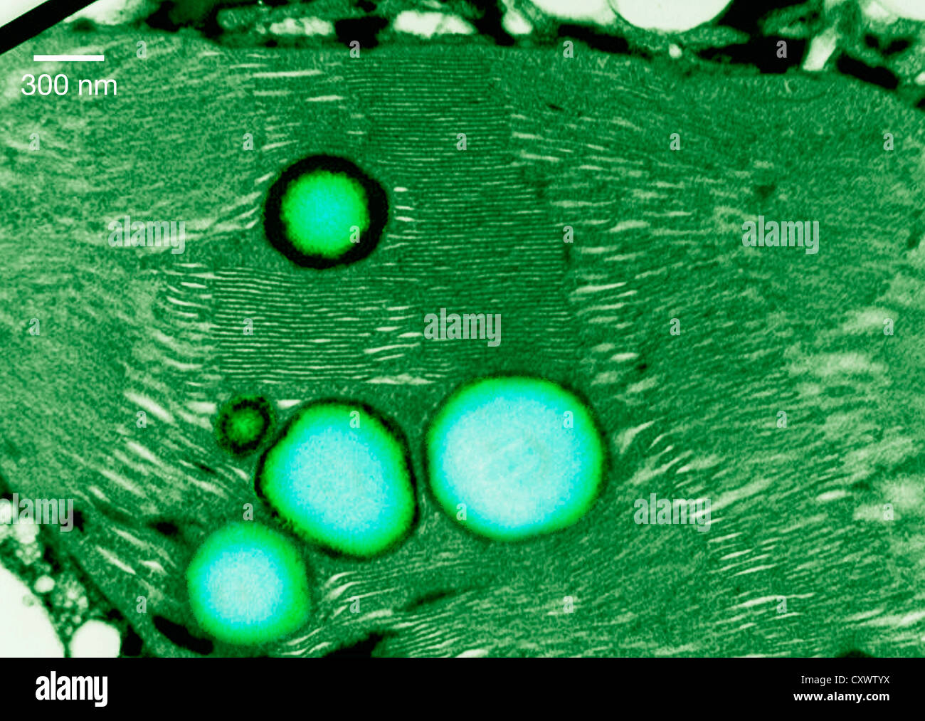 Transmission electron micrograph of a chloroplast Stock Photohttps://www.alamy.com/image-license-details/?v=1https://www.alamy.com/stock-photo-transmission-electron-micrograph-of-a-chloroplast-50970190.html
RFCXWTYX–Transmission electron micrograph of a chloroplast
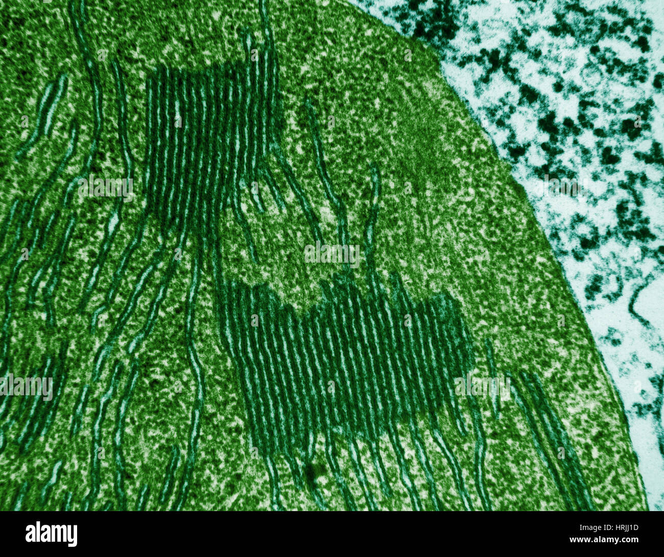
 Transmission electron micrograph of a chloroplast Stock Photohttps://www.alamy.com/image-license-details/?v=1https://www.alamy.com/stock-photo-transmission-electron-micrograph-of-a-chloroplast-50970177.html
RFCXWTYD–Transmission electron micrograph of a chloroplast
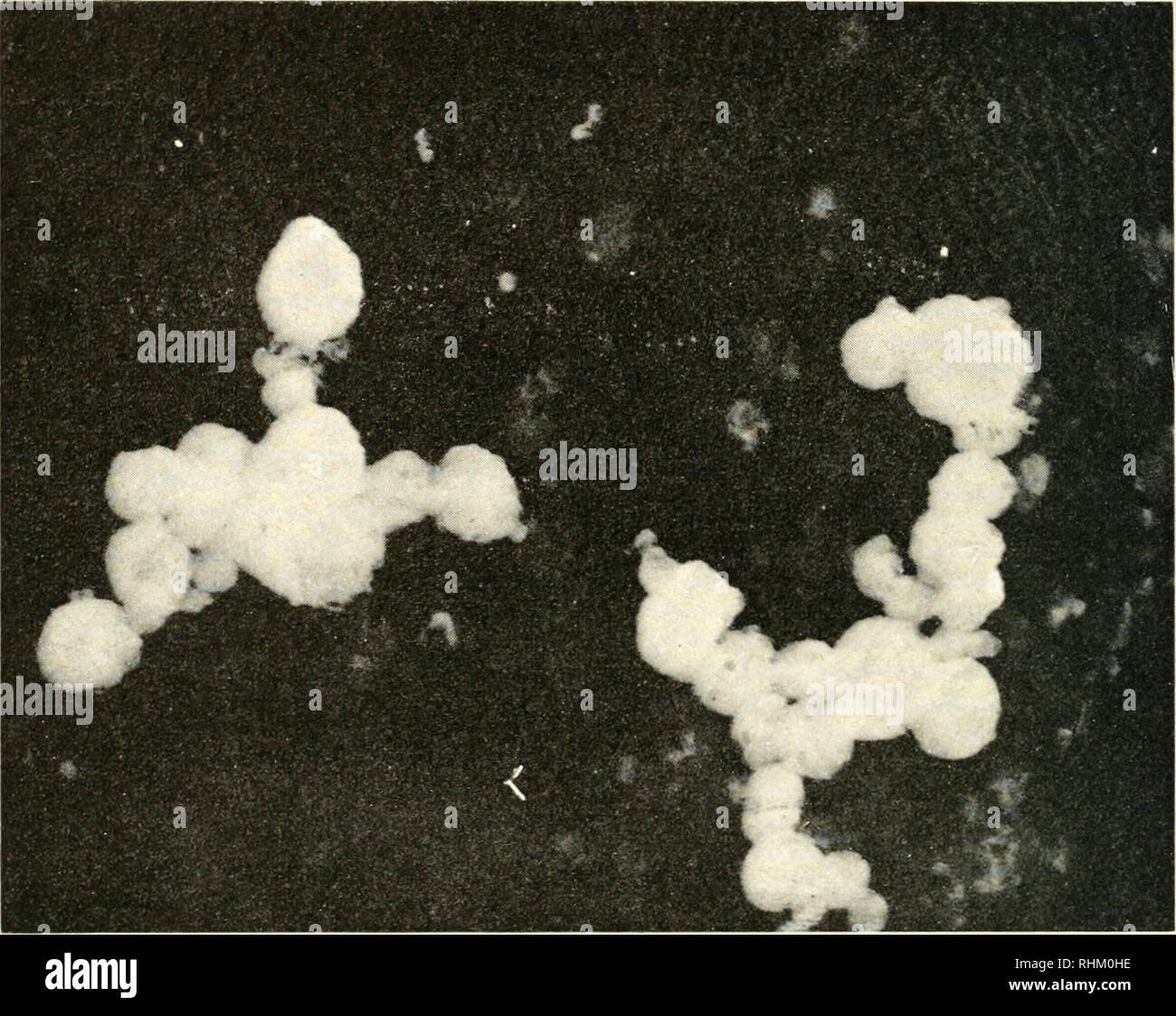 . Biological structure and function; proceedings. Biochemistry; Cytology. 364 DANIEL I. ARNON Table IV shows that, at the high Hght intensity at which cycHc photo- phosphorylation by purified grana was measured, the highest rates were obtained in the system catalyzed by phenazine methosulphate. Photo- phosphorylation in this system was not increased by the addition of an aqueous chloroplast extract. In contrast, photophosphorylation catalyzed. Fig. II. Electron micrograph of isolated spinach grana prepared for electron microscopy by a freeze-drying technique, i cm.- of leaf surface is estimate Stock Photohttps://www.alamy.com/image-license-details/?v=1https://www.alamy.com/biological-structure-and-function-proceedings-biochemistry-cytology-364-daniel-i-arnon-table-iv-shows-that-at-the-high-hght-intensity-at-which-cychc-photo-phosphorylation-by-purified-grana-was-measured-the-highest-rates-were-obtained-in-the-system-catalyzed-by-phenazine-methosulphate-photo-phosphorylation-in-this-system-was-not-increased-by-the-addition-of-an-aqueous-chloroplast-extract-in-contrast-photophosphorylation-catalyzed-fig-ii-electron-micrograph-of-isolated-spinach-grana-prepared-for-electron-microscopy-by-a-freeze-drying-technique-i-cm-of-leaf-surface-is-estimate-image234623466.html
RMRHM0HE–. Biological structure and function; proceedings. Biochemistry; Cytology. 364 DANIEL I. ARNON Table IV shows that, at the high Hght intensity at which cycHc photo- phosphorylation by purified grana was measured, the highest rates were obtained in the system catalyzed by phenazine methosulphate. Photo- phosphorylation in this system was not increased by the addition of an aqueous chloroplast extract. In contrast, photophosphorylation catalyzed. Fig. II. Electron micrograph of isolated spinach grana prepared for electron microscopy by a freeze-drying technique, i cm.- of leaf surface is estimate
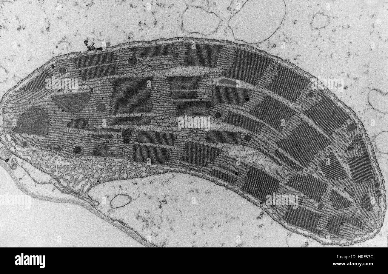 Corn Leaf Mesophyll with Chloroplast, TEM Stock Photohttps://www.alamy.com/image-license-details/?v=1https://www.alamy.com/stock-photo-corn-leaf-mesophyll-with-chloroplast-tem-134945424.html
RMHRF87C–Corn Leaf Mesophyll with Chloroplast, TEM
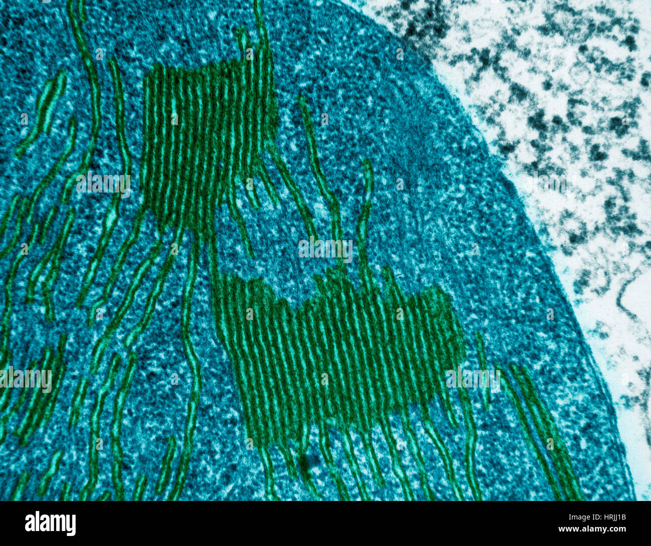
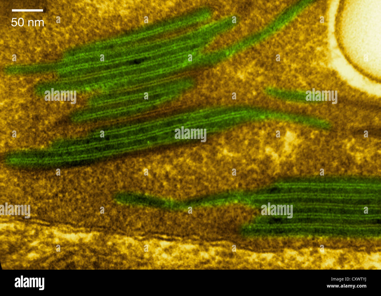 High magnification micrograph of a chloroplast Stock Photohttps://www.alamy.com/image-license-details/?v=1https://www.alamy.com/stock-photo-high-magnification-micrograph-of-a-chloroplast-50970182.html
RFCXWTYJ–High magnification micrograph of a chloroplast
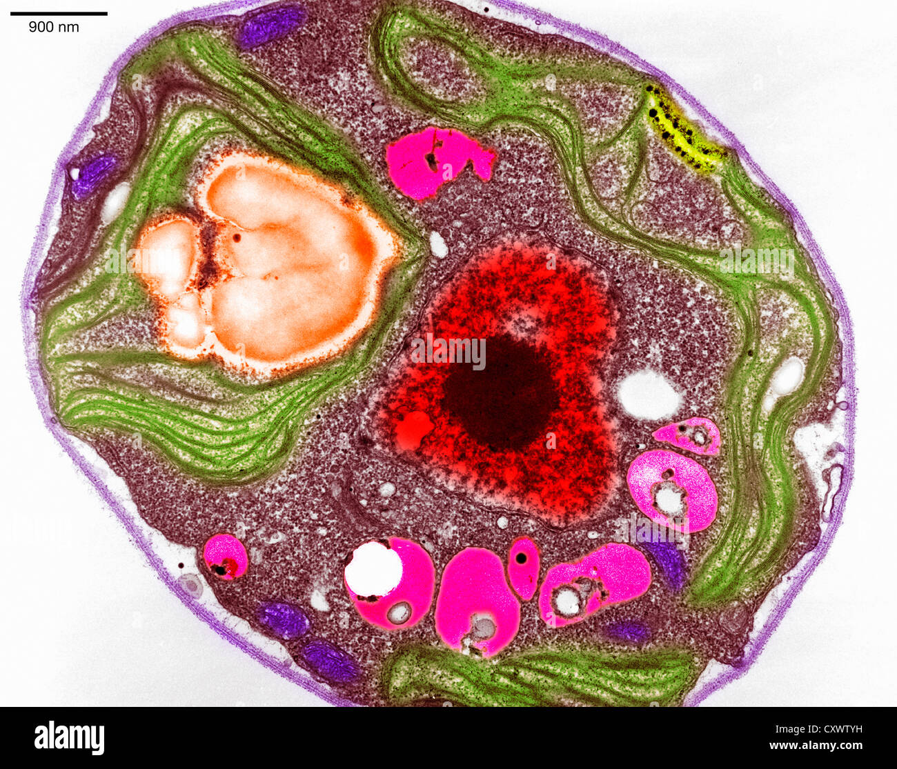 Scanning electron micrograph of algae Stock Photohttps://www.alamy.com/image-license-details/?v=1https://www.alamy.com/stock-photo-scanning-electron-micrograph-of-algae-50970181.html
RFCXWTYH–Scanning electron micrograph of algae
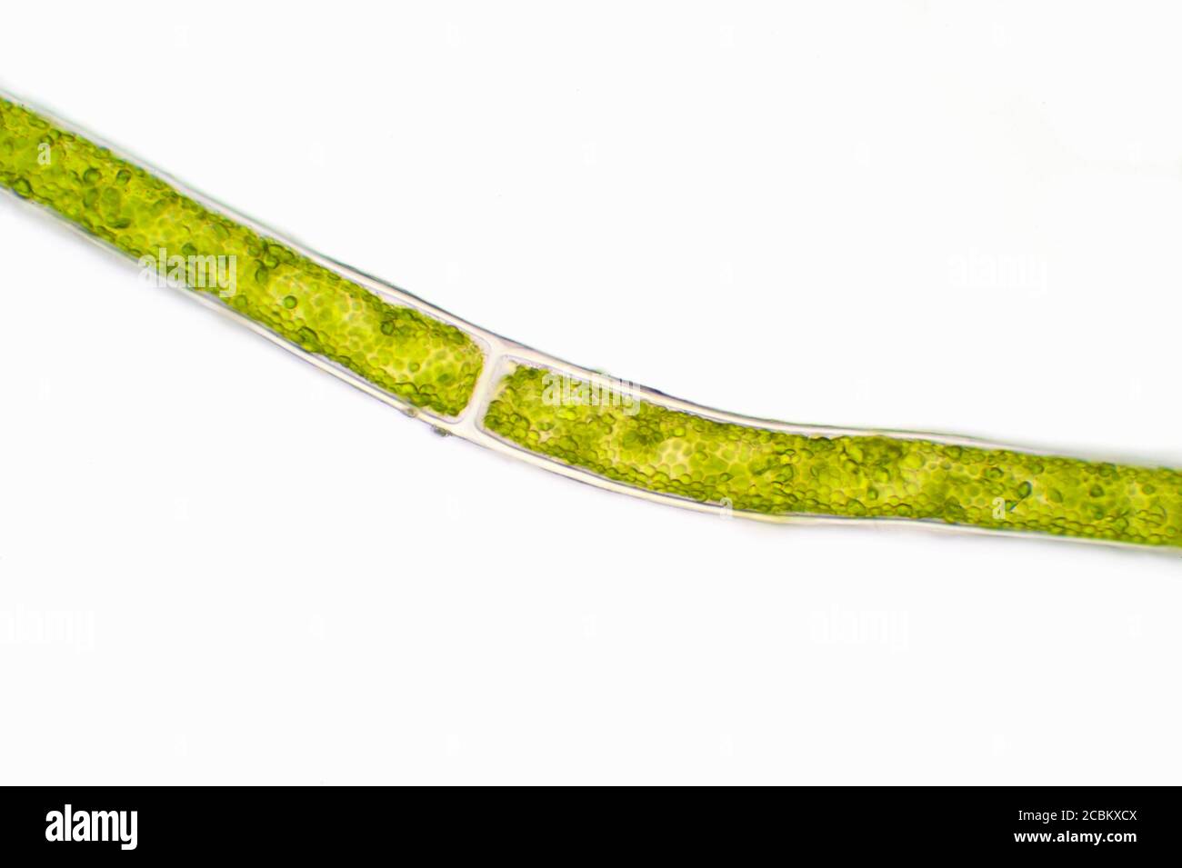 Microscopic view of green algae Stock Photohttps://www.alamy.com/image-license-details/?v=1https://www.alamy.com/microscopic-view-of-green-algae-image368616778.html
RF2CBKXCX–Microscopic view of green algae
![. Biological structure and function; proceedings. Biochemistry; Cytology. Fig. 2. Electron micrograph of a section of a maize chloroplast showing details of structure. The dense areas that resemble stacks of coins are the grana. The layers within each granum are called grana lamellae. The grana lamellae of different grana are inter-connected by stroma lamellae. Magnification 35 000 x (courtesy of Dr. A. E. Vatter). chloroplasts [38, 39] in the dark, it was converted to sugar phosphates. The hght and dark phases when carried out separately, yielded essentially the same final photosynthetic prod Stock Photo . Biological structure and function; proceedings. Biochemistry; Cytology. Fig. 2. Electron micrograph of a section of a maize chloroplast showing details of structure. The dense areas that resemble stacks of coins are the grana. The layers within each granum are called grana lamellae. The grana lamellae of different grana are inter-connected by stroma lamellae. Magnification 35 000 x (courtesy of Dr. A. E. Vatter). chloroplasts [38, 39] in the dark, it was converted to sugar phosphates. The hght and dark phases when carried out separately, yielded essentially the same final photosynthetic prod Stock Photo](https://c8.alamy.com/comp/RHM0M1/biological-structure-and-function-proceedings-biochemistry-cytology-fig-2-electron-micrograph-of-a-section-of-a-maize-chloroplast-showing-details-of-structure-the-dense-areas-that-resemble-stacks-of-coins-are-the-grana-the-layers-within-each-granum-are-called-grana-lamellae-the-grana-lamellae-of-different-grana-are-inter-connected-by-stroma-lamellae-magnification-35-000-x-courtesy-of-dr-a-e-vatter-chloroplasts-38-39-in-the-dark-it-was-converted-to-sugar-phosphates-the-hght-and-dark-phases-when-carried-out-separately-yielded-essentially-the-same-final-photosynthetic-prod-RHM0M1.jpg) . Biological structure and function; proceedings. Biochemistry; Cytology. Fig. 2. Electron micrograph of a section of a maize chloroplast showing details of structure. The dense areas that resemble stacks of coins are the grana. The layers within each granum are called grana lamellae. The grana lamellae of different grana are inter-connected by stroma lamellae. Magnification 35 000 x (courtesy of Dr. A. E. Vatter). chloroplasts [38, 39] in the dark, it was converted to sugar phosphates. The hght and dark phases when carried out separately, yielded essentially the same final photosynthetic prod Stock Photohttps://www.alamy.com/image-license-details/?v=1https://www.alamy.com/biological-structure-and-function-proceedings-biochemistry-cytology-fig-2-electron-micrograph-of-a-section-of-a-maize-chloroplast-showing-details-of-structure-the-dense-areas-that-resemble-stacks-of-coins-are-the-grana-the-layers-within-each-granum-are-called-grana-lamellae-the-grana-lamellae-of-different-grana-are-inter-connected-by-stroma-lamellae-magnification-35-000-x-courtesy-of-dr-a-e-vatter-chloroplasts-38-39-in-the-dark-it-was-converted-to-sugar-phosphates-the-hght-and-dark-phases-when-carried-out-separately-yielded-essentially-the-same-final-photosynthetic-prod-image234623537.html
. Biological structure and function; proceedings. Biochemistry; Cytology. Fig. 2. Electron micrograph of a section of a maize chloroplast showing details of structure. The dense areas that resemble stacks of coins are the grana. The layers within each granum are called grana lamellae. The grana lamellae of different grana are inter-connected by stroma lamellae. Magnification 35 000 x (courtesy of Dr. A. E. Vatter). chloroplasts [38, 39] in the dark, it was converted to sugar phosphates. The hght and dark phases when carried out separately, yielded essentially the same final photosynthetic prod Stock Photohttps://www.alamy.com/image-license-details/?v=1https://www.alamy.com/biological-structure-and-function-proceedings-biochemistry-cytology-fig-2-electron-micrograph-of-a-section-of-a-maize-chloroplast-showing-details-of-structure-the-dense-areas-that-resemble-stacks-of-coins-are-the-grana-the-layers-within-each-granum-are-called-grana-lamellae-the-grana-lamellae-of-different-grana-are-inter-connected-by-stroma-lamellae-magnification-35-000-x-courtesy-of-dr-a-e-vatter-chloroplasts-38-39-in-the-dark-it-was-converted-to-sugar-phosphates-the-hght-and-dark-phases-when-carried-out-separately-yielded-essentially-the-same-final-photosynthetic-prod-image234623537.htmlRMRHM0M1–. Biological structure and function; proceedings. Biochemistry; Cytology. Fig. 2. Electron micrograph of a section of a maize chloroplast showing details of structure. The dense areas that resemble stacks of coins are the grana. The layers within each granum are called grana lamellae. The grana lamellae of different grana are inter-connected by stroma lamellae. Magnification 35 000 x (courtesy of Dr. A. E. Vatter). chloroplasts [38, 39] in the dark, it was converted to sugar phosphates. The hght and dark phases when carried out separately, yielded essentially the same final photosynthetic prod
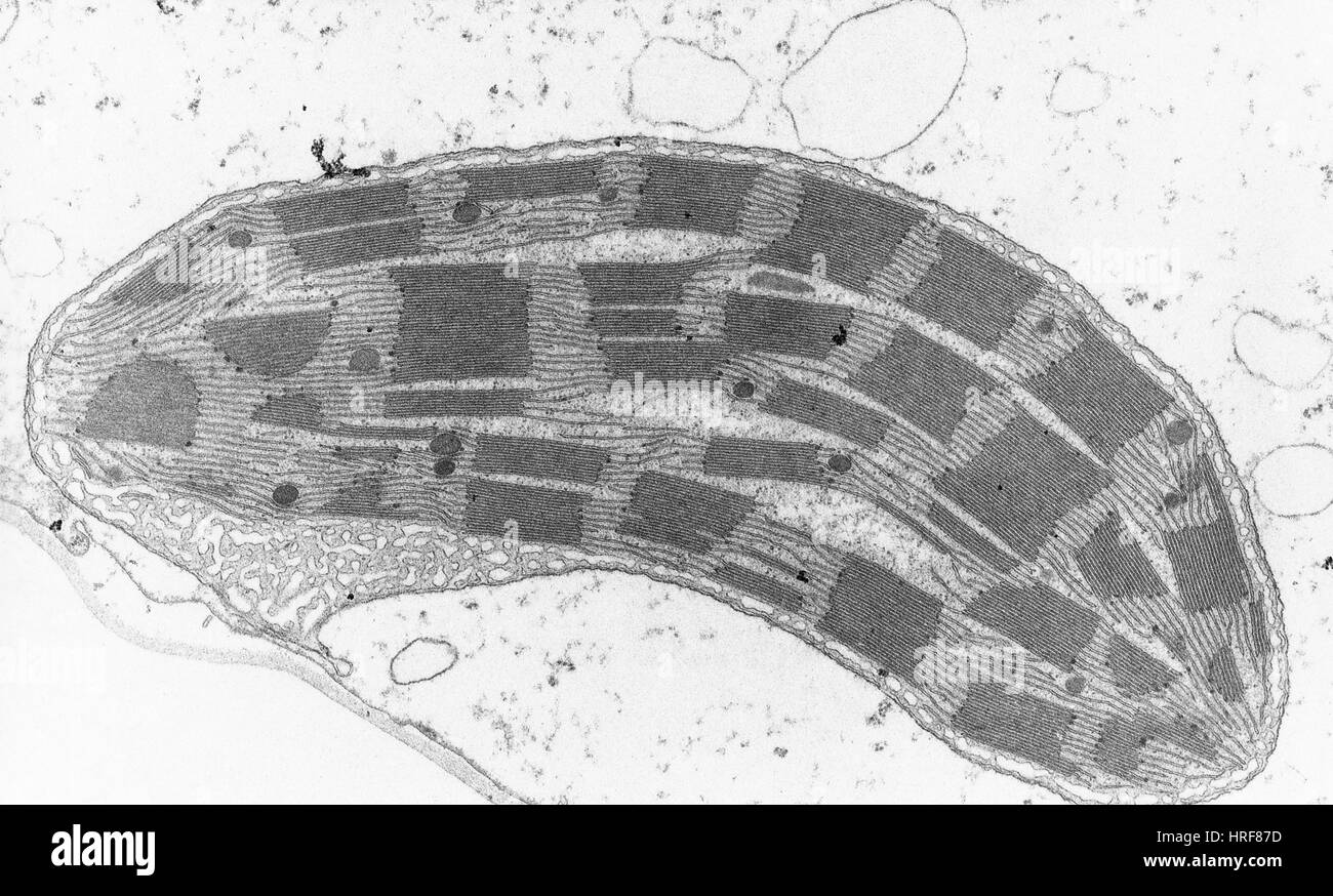 Corn Leaf Mesophyll with Chloroplast, TEM Stock Photohttps://www.alamy.com/image-license-details/?v=1https://www.alamy.com/stock-photo-corn-leaf-mesophyll-with-chloroplast-tem-134945425.html
RMHRF87D–Corn Leaf Mesophyll with Chloroplast, TEM
 . Biophysical science. Biophysics. Nucleus. (b) Figure I. (a) Three Chlorella cells. This diagram emphasizes that the single cup-shaped chloroplast occupies most of the cell. The pyrenoid is associated with starch and/or protein synthesis and/or storage, (b) A corn chloroplast. Sketch of an electron micrograph of a chloroplast from Zea mais. The dark regions are the grana. They are cylinders about 4,000 to 6,000 A in diameter and 5,000 to 8,000 A in height. After E. I. Rabinowitch, Photosynthesis, II, 2 (New York: Interscience Publishers, Inc., 1956), from Vatter, unpublished, modified. and mo Stock Photohttps://www.alamy.com/image-license-details/?v=1https://www.alamy.com/biophysical-science-biophysics-nucleus-b-figure-i-a-three-chlorella-cells-this-diagram-emphasizes-that-the-single-cup-shaped-chloroplast-occupies-most-of-the-cell-the-pyrenoid-is-associated-with-starch-andor-protein-synthesis-andor-storage-b-a-corn-chloroplast-sketch-of-an-electron-micrograph-of-a-chloroplast-from-zea-mais-the-dark-regions-are-the-grana-they-are-cylinders-about-4000-to-6000-a-in-diameter-and-5000-to-8000-a-in-height-after-e-i-rabinowitch-photosynthesis-ii-2-new-york-interscience-publishers-inc-1956-from-vatter-unpublished-modified-and-mo-image234599963.html
RMRHJXJ3–. Biophysical science. Biophysics. Nucleus. (b) Figure I. (a) Three Chlorella cells. This diagram emphasizes that the single cup-shaped chloroplast occupies most of the cell. The pyrenoid is associated with starch and/or protein synthesis and/or storage, (b) A corn chloroplast. Sketch of an electron micrograph of a chloroplast from Zea mais. The dark regions are the grana. They are cylinders about 4,000 to 6,000 A in diameter and 5,000 to 8,000 A in height. After E. I. Rabinowitch, Photosynthesis, II, 2 (New York: Interscience Publishers, Inc., 1956), from Vatter, unpublished, modified. and mo
 Corn Leaf Chloroplast, TEM Stock Photohttps://www.alamy.com/image-license-details/?v=1https://www.alamy.com/stock-photo-corn-leaf-chloroplast-tem-135017544.html
RMHRJG74–Corn Leaf Chloroplast, TEM
 . Biophysical science. Biophysics. (a) (b) Figure 2. Green grana of corn leaves, (a) A sketch of an electron micrograph of a section of granum within an intact chloroplast of Zea mais. There are about 50 such grana per chloroplast. Each granum has about 15 parallel lamellae which are about 400 A thick and about 4,000 A in diameter. (b) A sketch of an electron micrograph of a granum which is believed to be dissociated into separate disks. After E. I. Rabinowitch, Photosynthesis, II, 2 (New York: Interscience Publishers, Inc., 1956), from Vatter, unpublished, modified. Figure 2 shows the lamella Stock Photohttps://www.alamy.com/image-license-details/?v=1https://www.alamy.com/biophysical-science-biophysics-a-b-figure-2-green-grana-of-corn-leaves-a-a-sketch-of-an-electron-micrograph-of-a-section-of-granum-within-an-intact-chloroplast-of-zea-mais-there-are-about-50-such-grana-per-chloroplast-each-granum-has-about-15-parallel-lamellae-which-are-about-400-a-thick-and-about-4000-a-in-diameter-b-a-sketch-of-an-electron-micrograph-of-a-granum-which-is-believed-to-be-dissociated-into-separate-disks-after-e-i-rabinowitch-photosynthesis-ii-2-new-york-interscience-publishers-inc-1956-from-vatter-unpublished-modified-figure-2-shows-the-lamella-image234538559.html
RMRHG493–. Biophysical science. Biophysics. (a) (b) Figure 2. Green grana of corn leaves, (a) A sketch of an electron micrograph of a section of granum within an intact chloroplast of Zea mais. There are about 50 such grana per chloroplast. Each granum has about 15 parallel lamellae which are about 400 A thick and about 4,000 A in diameter. (b) A sketch of an electron micrograph of a granum which is believed to be dissociated into separate disks. After E. I. Rabinowitch, Photosynthesis, II, 2 (New York: Interscience Publishers, Inc., 1956), from Vatter, unpublished, modified. Figure 2 shows the lamella
 Corn Leaf Mesophyll with Chloroplast, TEM Stock Photohttps://www.alamy.com/image-license-details/?v=1https://www.alamy.com/stock-photo-corn-leaf-mesophyll-with-chloroplast-tem-135017543.html
RMHRJG73–Corn Leaf Mesophyll with Chloroplast, TEM
 . The Biological bulletin. Biology; Zoology; Biology; Marine Biology. ALGAL PATHOGEN IN A GORGONIAN CORAL 379 AL V •'..-.. *—.. FIGURE 10. Association of coral amoebocyte and algal filaments. A. Light micrograph of amoebocytes aggregated near an algal filament; note PAS-positive vesicles; scale bar = 50 nm. B. Low power electron micrograph of amoebocyte lysing and releasing contents near two algal filaments; scale bar = 1 ^m. c. Mesoglea coating an algal filament; scale bar = 1 ^m. AL, algae; AM, amoebocyte; CW, cell wall; CY, chloroplast; M, mesoglea. latter authors give familial characters t Stock Photohttps://www.alamy.com/image-license-details/?v=1https://www.alamy.com/the-biological-bulletin-biology-zoology-biology-marine-biology-algal-pathogen-in-a-gorgonian-coral-379-al-v-figure-10-association-of-coral-amoebocyte-and-algal-filaments-a-light-micrograph-of-amoebocytes-aggregated-near-an-algal-filament-note-pas-positive-vesicles-scale-bar-=-50-nm-b-low-power-electron-micrograph-of-amoebocyte-lysing-and-releasing-contents-near-two-algal-filaments-scale-bar-=-1-m-c-mesoglea-coating-an-algal-filament-scale-bar-=-1-m-al-algae-am-amoebocyte-cw-cell-wall-cy-chloroplast-m-mesoglea-latter-authors-give-familial-characters-t-image234644854.html
RMRHMYWA–. The Biological bulletin. Biology; Zoology; Biology; Marine Biology. ALGAL PATHOGEN IN A GORGONIAN CORAL 379 AL V •'..-.. *—.. FIGURE 10. Association of coral amoebocyte and algal filaments. A. Light micrograph of amoebocytes aggregated near an algal filament; note PAS-positive vesicles; scale bar = 50 nm. B. Low power electron micrograph of amoebocyte lysing and releasing contents near two algal filaments; scale bar = 1 ^m. c. Mesoglea coating an algal filament; scale bar = 1 ^m. AL, algae; AM, amoebocyte; CW, cell wall; CY, chloroplast; M, mesoglea. latter authors give familial characters t
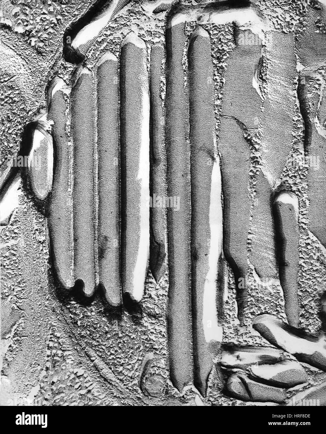 Thylakoid Stock Photohttps://www.alamy.com/image-license-details/?v=1https://www.alamy.com/stock-photo-thylakoid-134945594.html
RMHRF8DE–Thylakoid
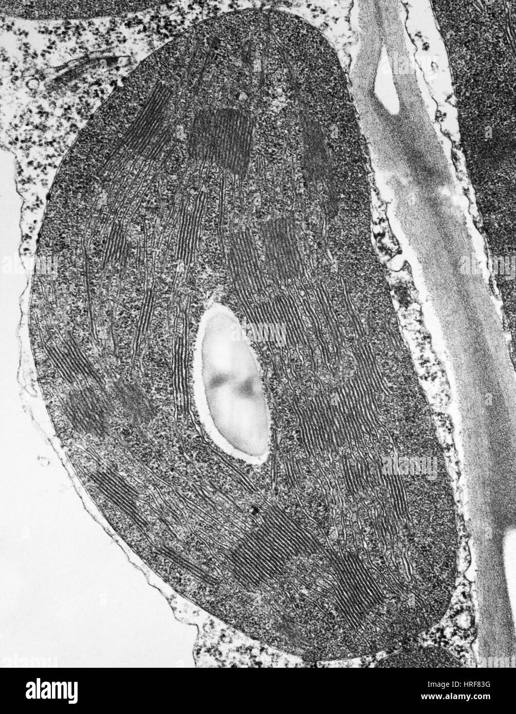 Tomato Chloroplast Stock Photohttps://www.alamy.com/image-license-details/?v=1https://www.alamy.com/stock-photo-tomato-chloroplast-134945316.html
RMHRF83G–Tomato Chloroplast
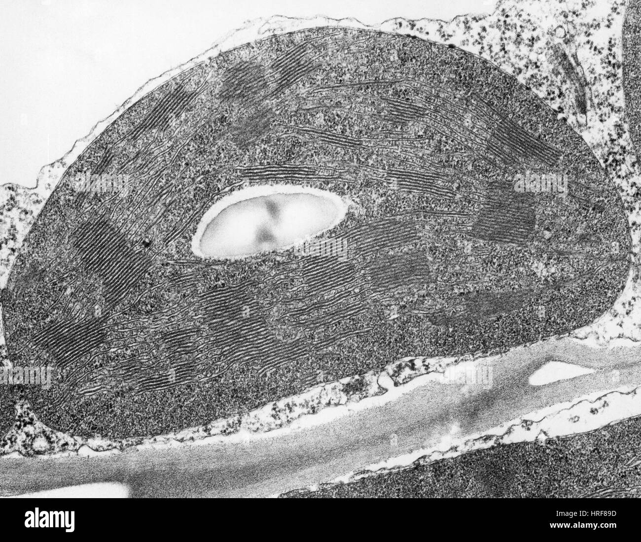 Tomato Chloroplast (TEM) Stock Photohttps://www.alamy.com/image-license-details/?v=1https://www.alamy.com/stock-photo-tomato-chloroplast-tem-134945481.html
RMHRF89D–Tomato Chloroplast (TEM)
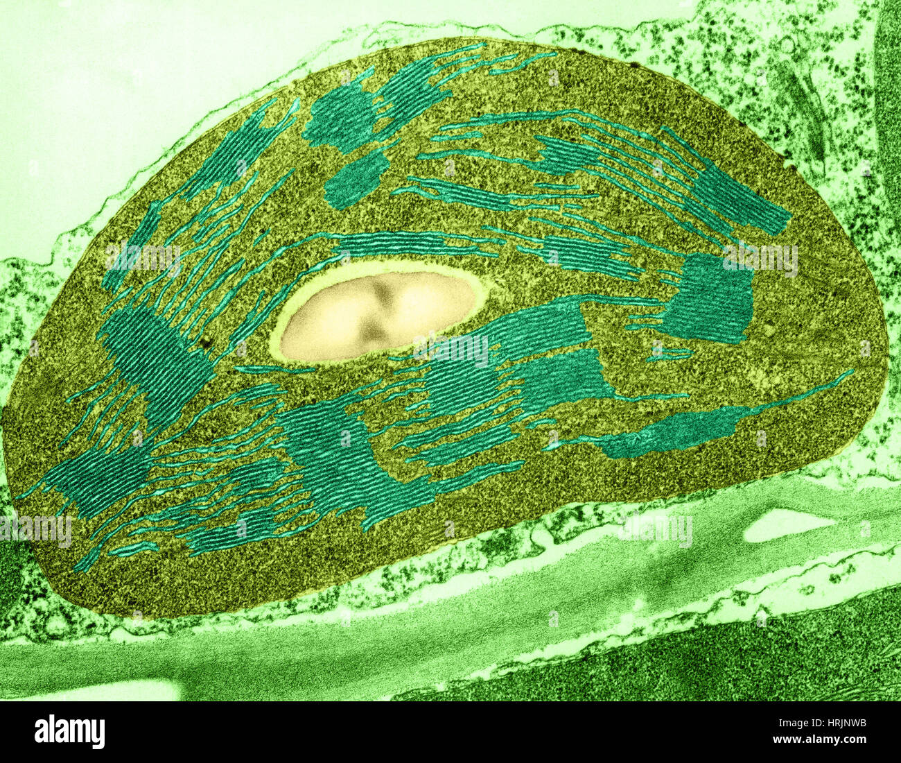 Tomato Chloroplast, TEM Stock Photohttps://www.alamy.com/image-license-details/?v=1https://www.alamy.com/stock-photo-tomato-chloroplast-tem-135021975.html
RMHRJNWB–Tomato Chloroplast, TEM
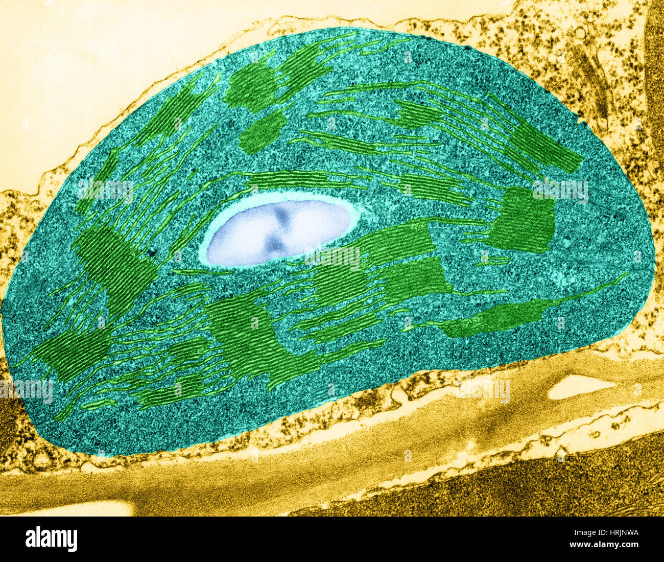 Tomato Chloroplast, TEM Stock Photohttps://www.alamy.com/image-license-details/?v=1https://www.alamy.com/stock-photo-tomato-chloroplast-tem-135021974.html
RMHRJNWA–Tomato Chloroplast, TEM
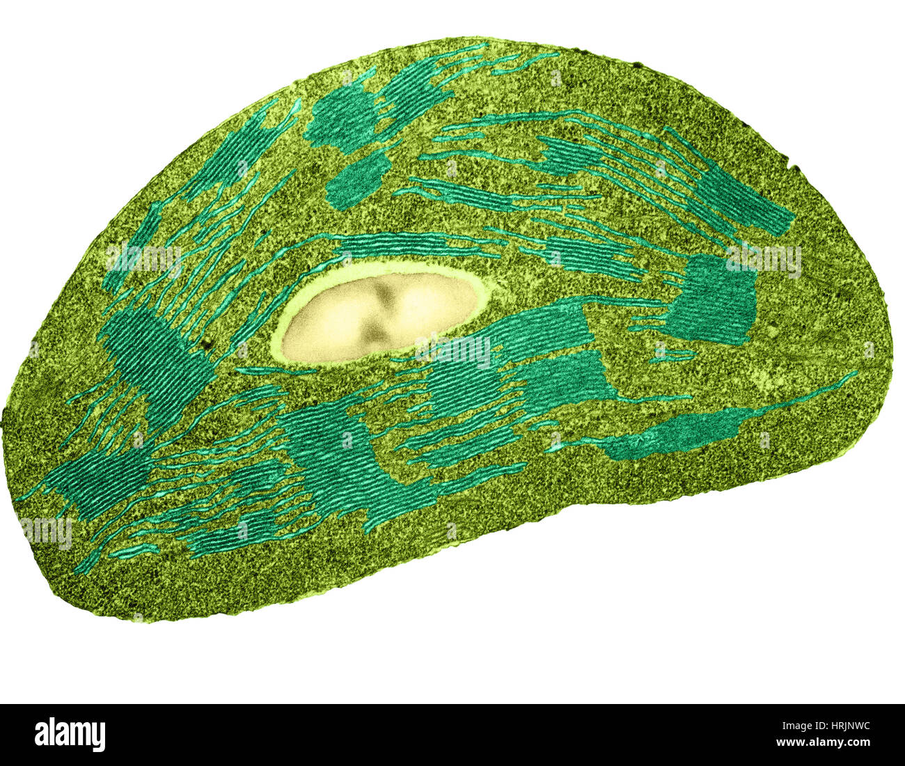 Tomato Chloroplast, TEM Stock Photohttps://www.alamy.com/image-license-details/?v=1https://www.alamy.com/stock-photo-tomato-chloroplast-tem-135021976.html
RMHRJNWC–Tomato Chloroplast, TEM
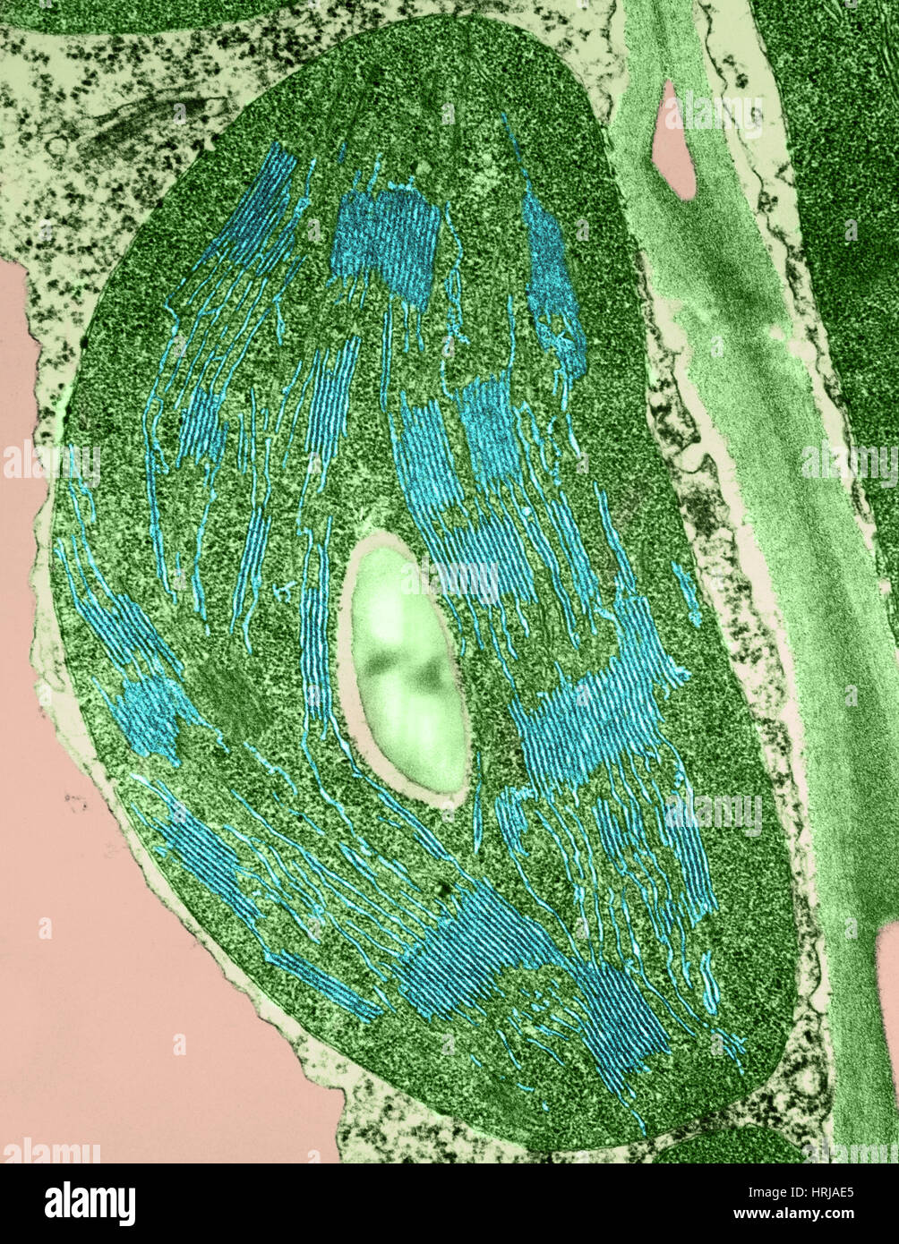 Tomato Chloroplast, TEM Stock Photohttps://www.alamy.com/image-license-details/?v=1https://www.alamy.com/stock-photo-tomato-chloroplast-tem-135013037.html
RMHRJAE5–Tomato Chloroplast, TEM
 Chloroplast DNA Stock Photohttps://www.alamy.com/image-license-details/?v=1https://www.alamy.com/stock-photo-chloroplast-dna-134986529.html
RMHRH4KD–Chloroplast DNA
 Chloroplast DNA Stock Photohttps://www.alamy.com/image-license-details/?v=1https://www.alamy.com/stock-photo-chloroplast-dna-134986534.html
RMHRH4KJ–Chloroplast DNA
 Chloroplast DNA Stock Photohttps://www.alamy.com/image-license-details/?v=1https://www.alamy.com/stock-photo-chloroplast-dna-134986533.html
RMHRH4KH–Chloroplast DNA
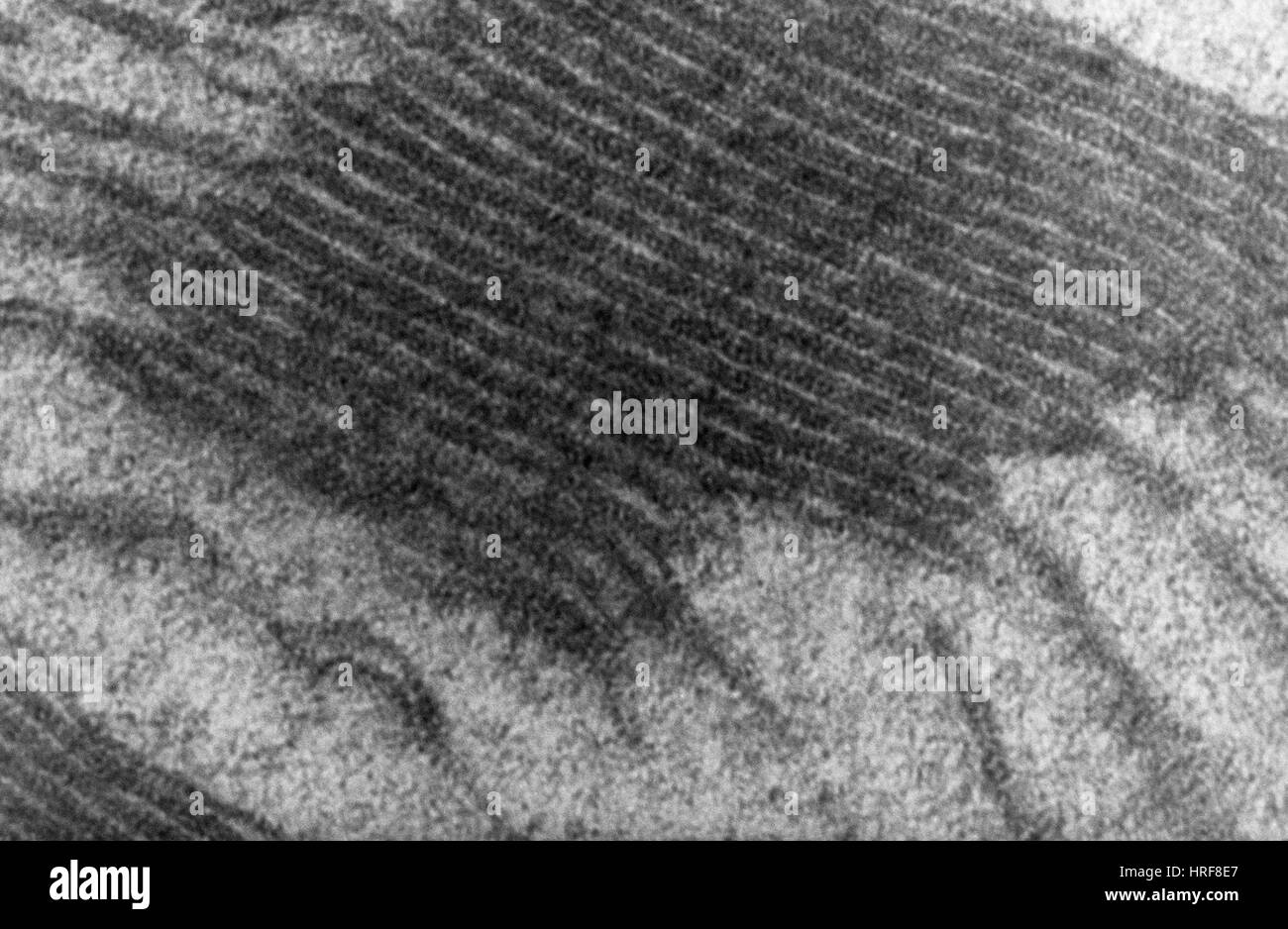 Chloroplast, TEM Stock Photohttps://www.alamy.com/image-license-details/?v=1https://www.alamy.com/stock-photo-chloroplast-tem-134945615.html
RMHRF8E7–Chloroplast, TEM
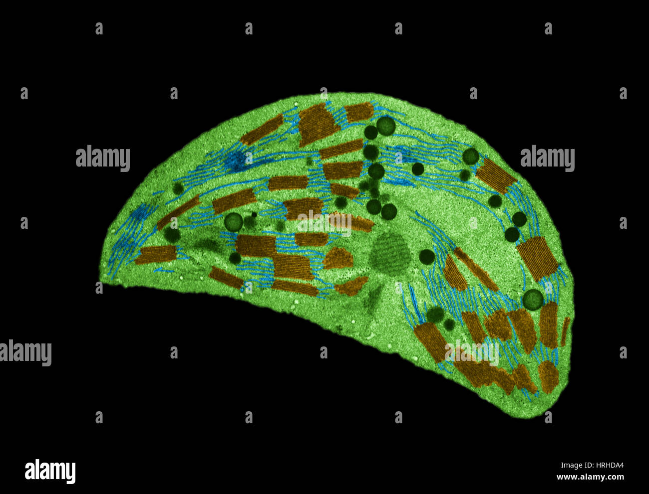 Chloroplast TEM Stock Photohttps://www.alamy.com/image-license-details/?v=1https://www.alamy.com/stock-photo-chloroplast-tem-134993324.html
RMHRHDA4–Chloroplast TEM
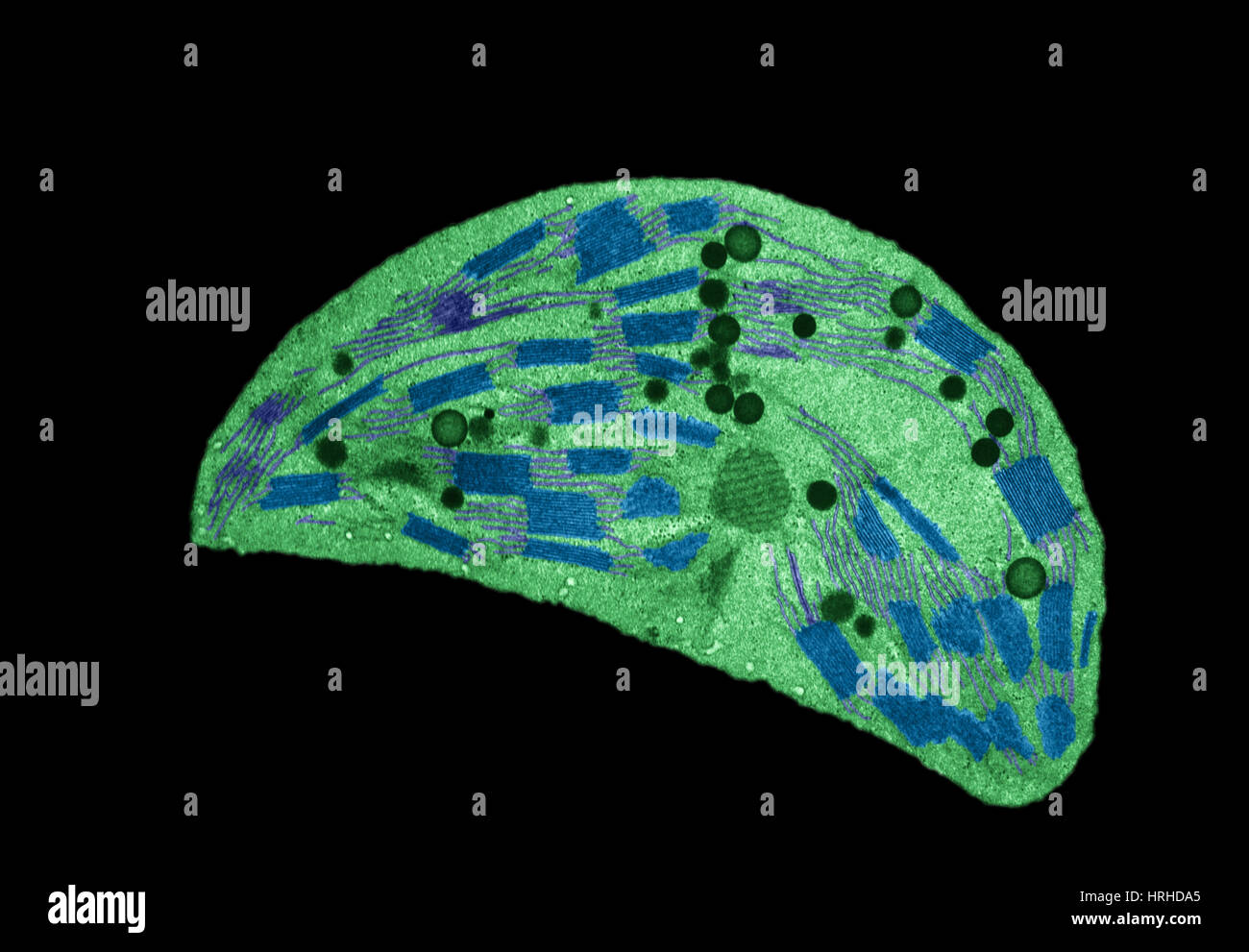 Chloroplast TEM Stock Photohttps://www.alamy.com/image-license-details/?v=1https://www.alamy.com/stock-photo-chloroplast-tem-134993325.html
RMHRHDA5–Chloroplast TEM
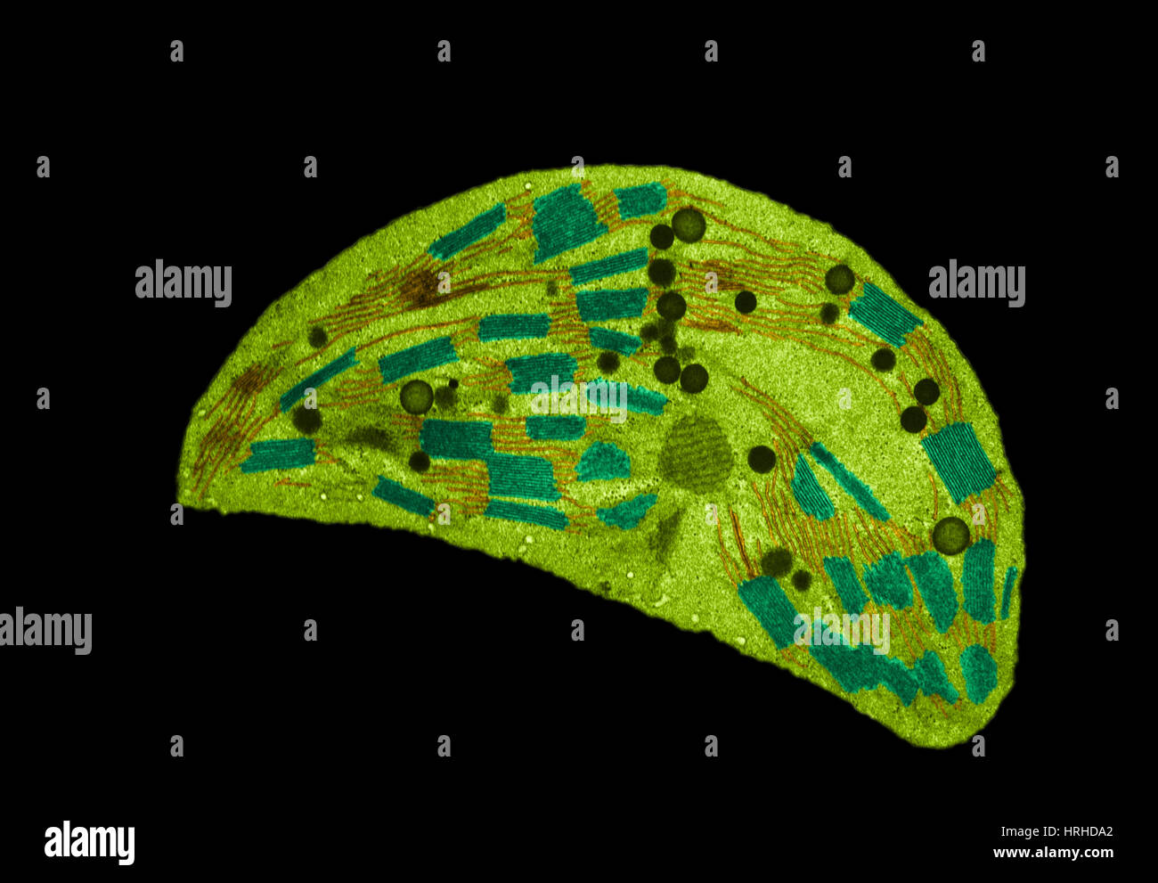 Chloroplast TEM Stock Photohttps://www.alamy.com/image-license-details/?v=1https://www.alamy.com/stock-photo-chloroplast-tem-134993322.html
RMHRHDA2–Chloroplast TEM
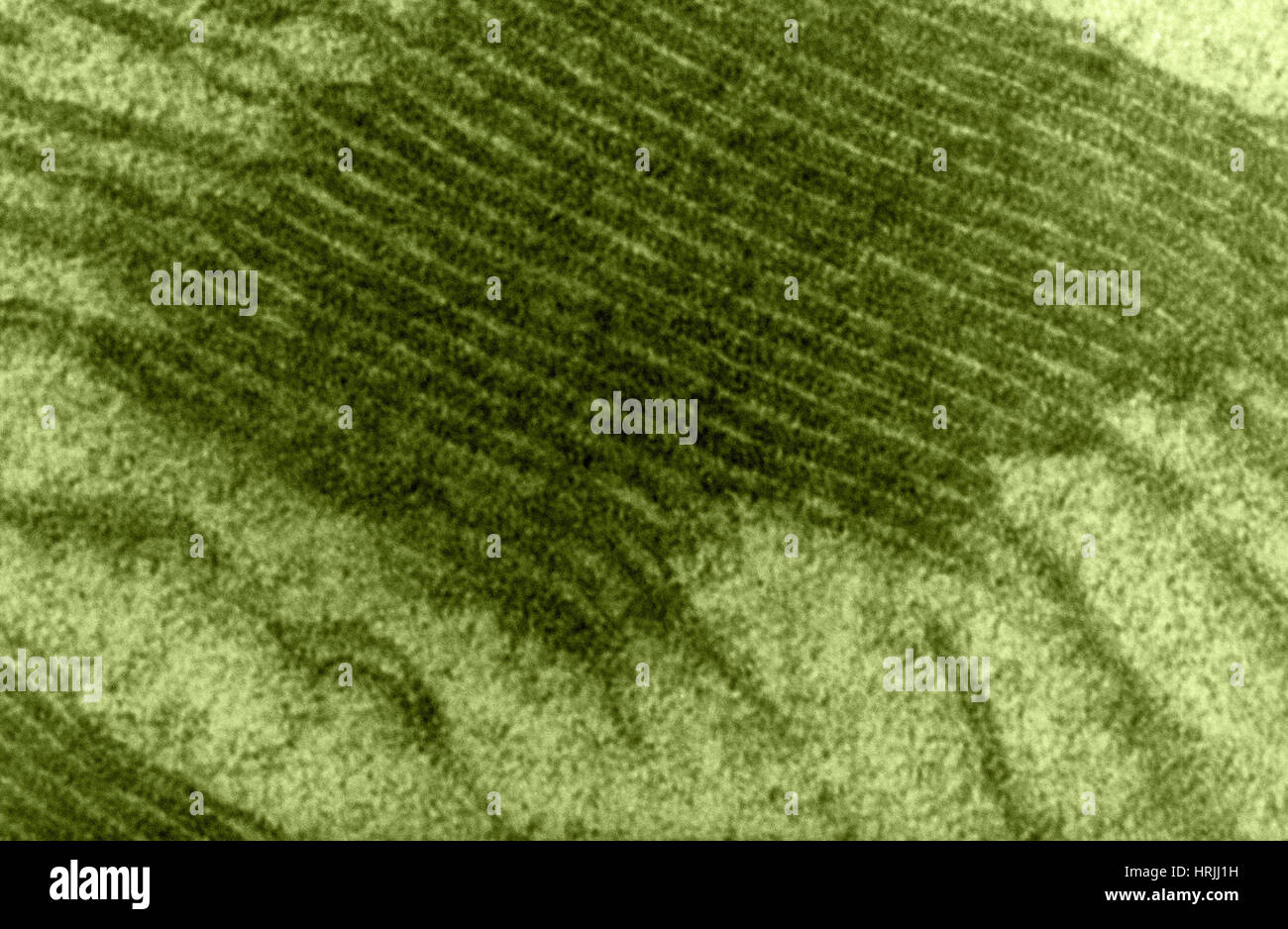 Chloroplast, TEM Stock Photohttps://www.alamy.com/image-license-details/?v=1https://www.alamy.com/stock-photo-chloroplast-tem-135018957.html
RMHRJJ1H–Chloroplast, TEM
 Chloroplast, TEM Stock Photohttps://www.alamy.com/image-license-details/?v=1https://www.alamy.com/stock-photo-chloroplast-tem-135018956.html
RMHRJJ1G–Chloroplast, TEM
 Chloroplast, TEM Stock Photohttps://www.alamy.com/image-license-details/?v=1https://www.alamy.com/stock-photo-chloroplast-tem-135018955.html
RMHRJJ1F–Chloroplast, TEM
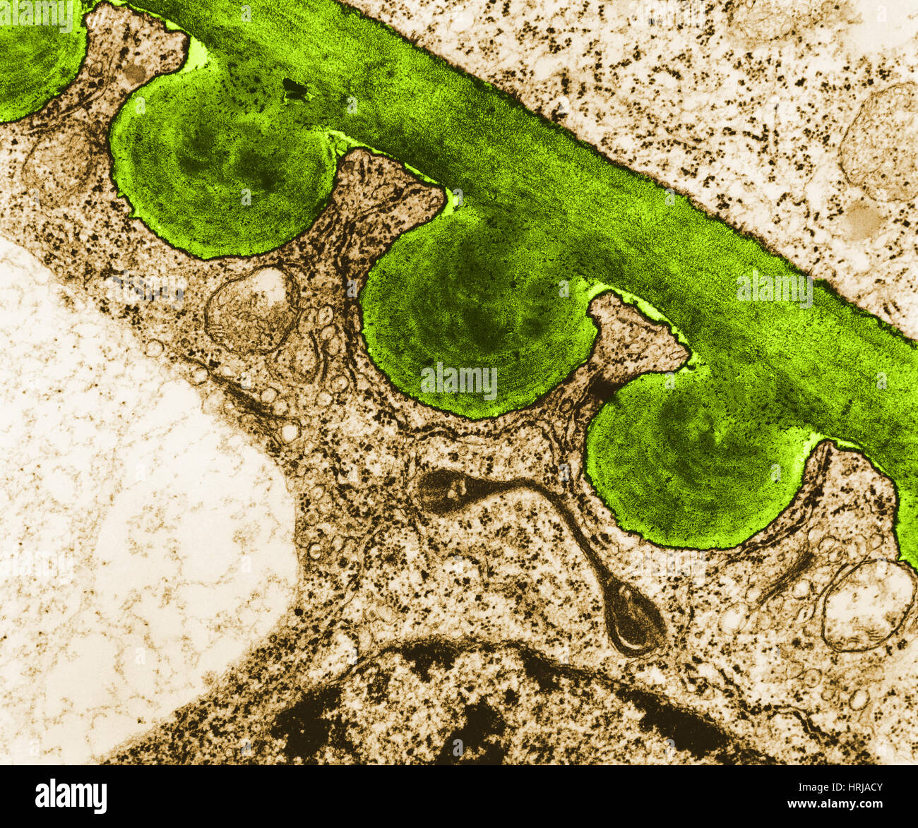 Tomato Chloroplast, TEM Stock Photohttps://www.alamy.com/image-license-details/?v=1https://www.alamy.com/stock-photo-tomato-chloroplast-tem-135013003.html
RMHRJACY–Tomato Chloroplast, TEM
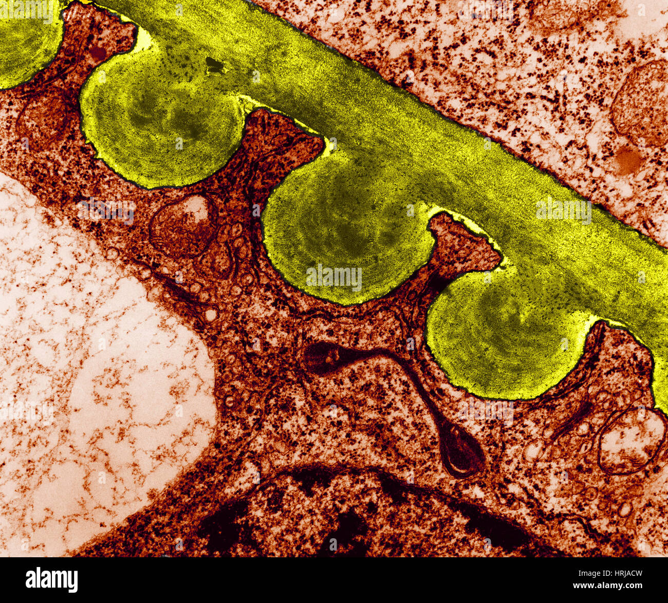 Tomato Chloroplast, TEM Stock Photohttps://www.alamy.com/image-license-details/?v=1https://www.alamy.com/stock-photo-tomato-chloroplast-tem-135013001.html
RMHRJACW–Tomato Chloroplast, TEM
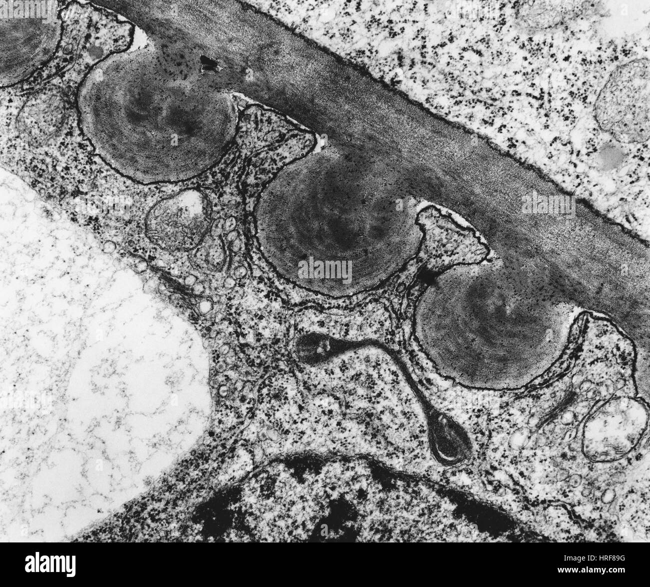 Tomato Chloroplast (TEM) Stock Photohttps://www.alamy.com/image-license-details/?v=1https://www.alamy.com/stock-photo-tomato-chloroplast-tem-134945484.html
RMHRF89G–Tomato Chloroplast (TEM)
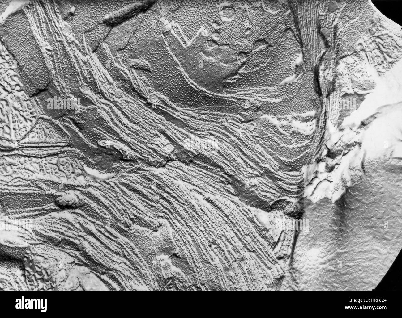 Chloroplast Membranes in Leaf Cell, TEM Stock Photohttps://www.alamy.com/image-license-details/?v=1https://www.alamy.com/stock-photo-chloroplast-membranes-in-leaf-cell-tem-134945276.html
RMHRF824–Chloroplast Membranes in Leaf Cell, TEM
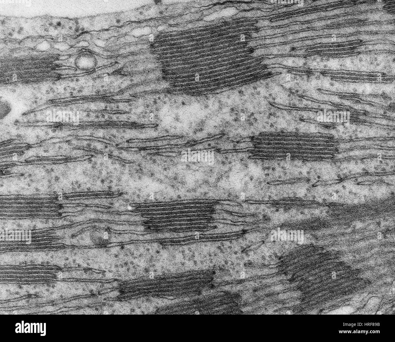 Chloroplast in Corn Leaf Cell, TEM Stock Photohttps://www.alamy.com/image-license-details/?v=1https://www.alamy.com/stock-photo-chloroplast-in-corn-leaf-cell-tem-134945479.html
RMHRF89B–Chloroplast in Corn Leaf Cell, TEM
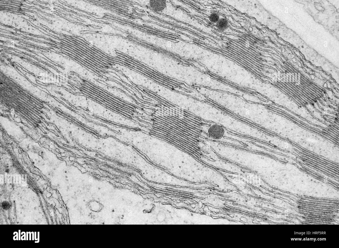 Chloroplast Grana TEM Stock Photohttps://www.alamy.com/image-license-details/?v=1https://www.alamy.com/stock-photo-chloroplast-grana-tem-134943531.html
RMHRF5RR–Chloroplast Grana TEM
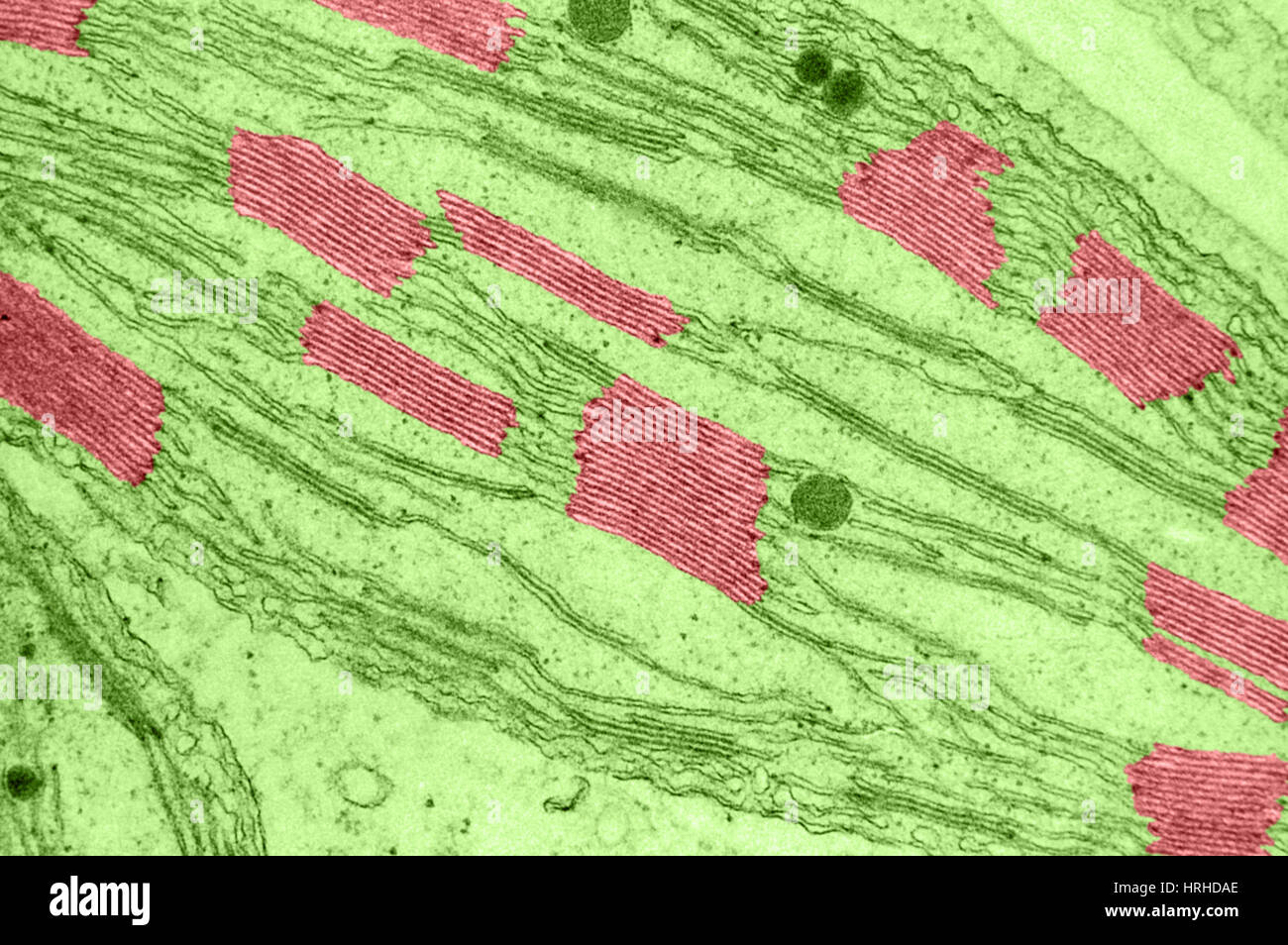 Chloroplast Grana TEM Stock Photohttps://www.alamy.com/image-license-details/?v=1https://www.alamy.com/stock-photo-chloroplast-grana-tem-134993334.html
RMHRHDAE–Chloroplast Grana TEM
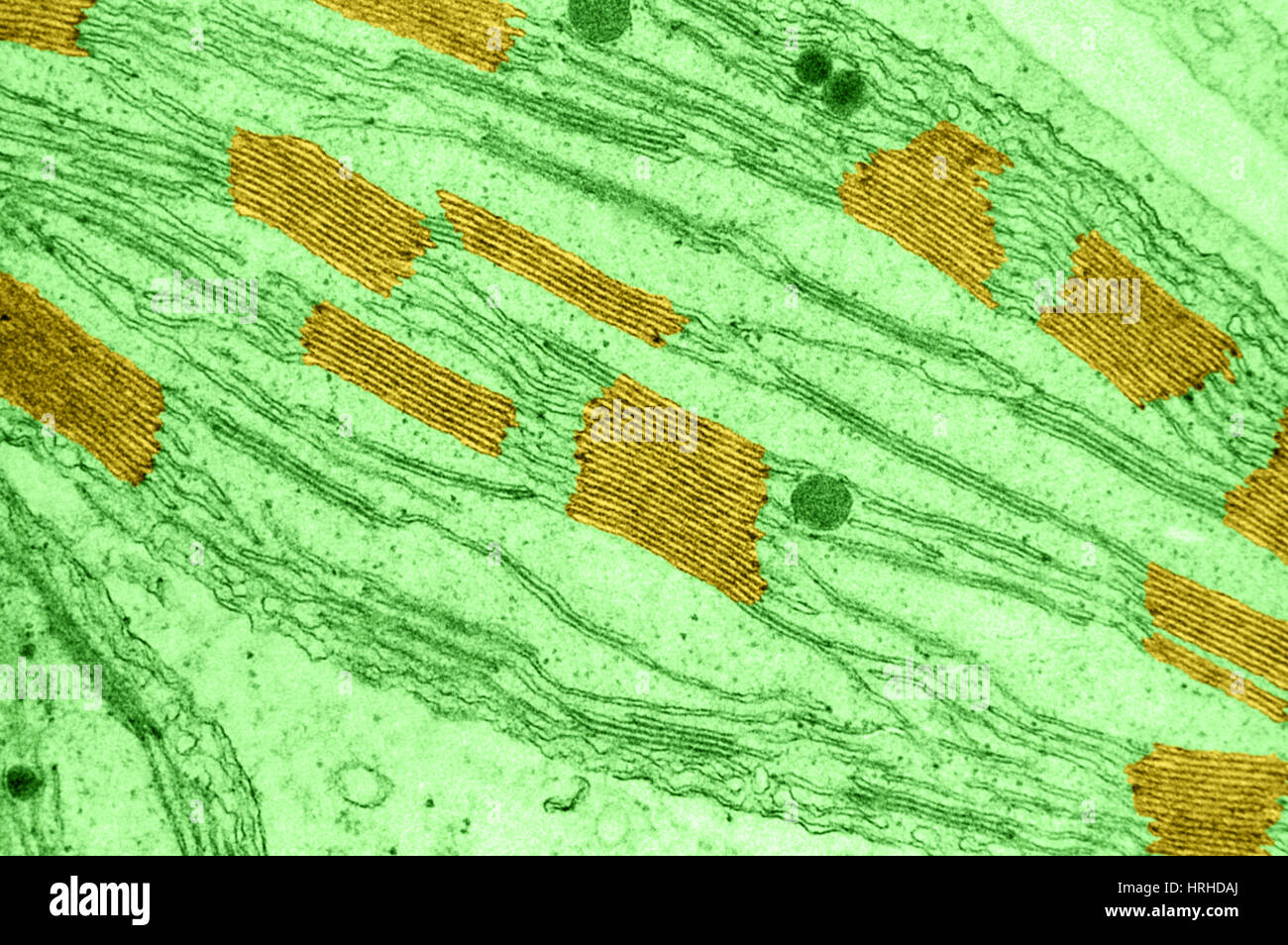 Chloroplast Grana TEM Stock Photohttps://www.alamy.com/image-license-details/?v=1https://www.alamy.com/stock-photo-chloroplast-grana-tem-134993338.html
RMHRHDAJ–Chloroplast Grana TEM
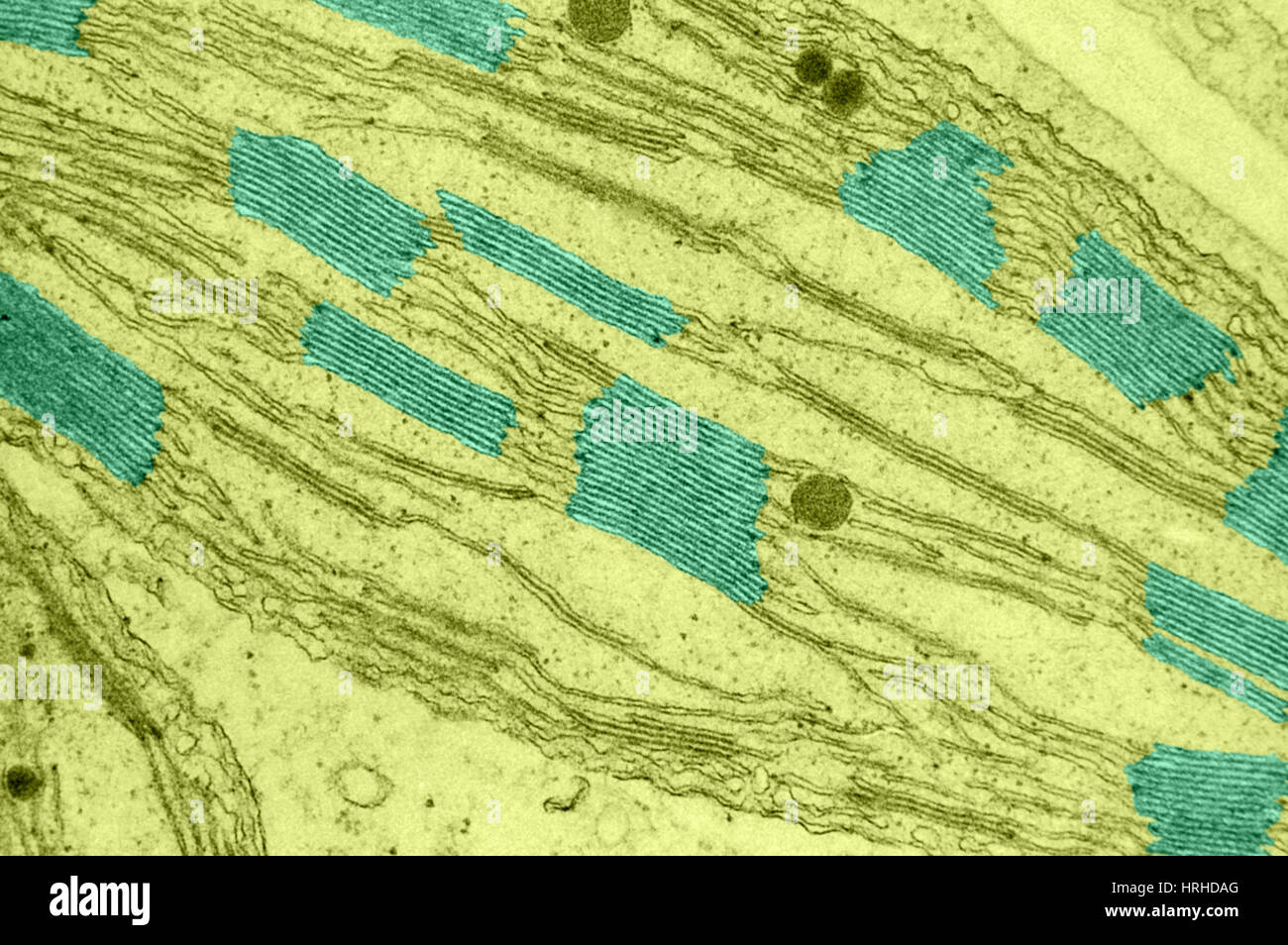 Chloroplast Grana TEM Stock Photohttps://www.alamy.com/image-license-details/?v=1https://www.alamy.com/stock-photo-chloroplast-grana-tem-134993336.html
RMHRHDAG–Chloroplast Grana TEM
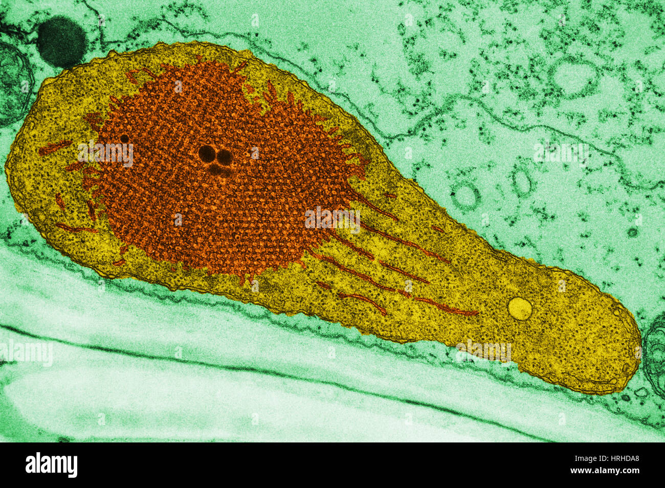 Developing Chloroplast and Etioplast TEM Stock Photohttps://www.alamy.com/image-license-details/?v=1https://www.alamy.com/stock-photo-developing-chloroplast-and-etioplast-tem-134993328.html
RMHRHDA8–Developing Chloroplast and Etioplast TEM
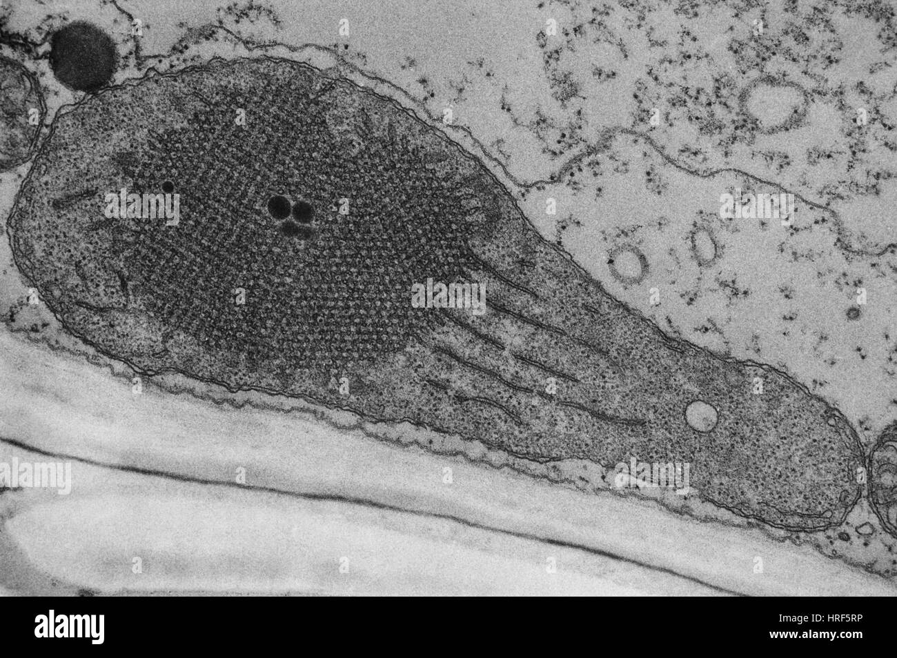 Developing Chloroplast and Etioplast TEM Stock Photohttps://www.alamy.com/image-license-details/?v=1https://www.alamy.com/stock-photo-developing-chloroplast-and-etioplast-tem-134943530.html
RMHRF5RP–Developing Chloroplast and Etioplast TEM
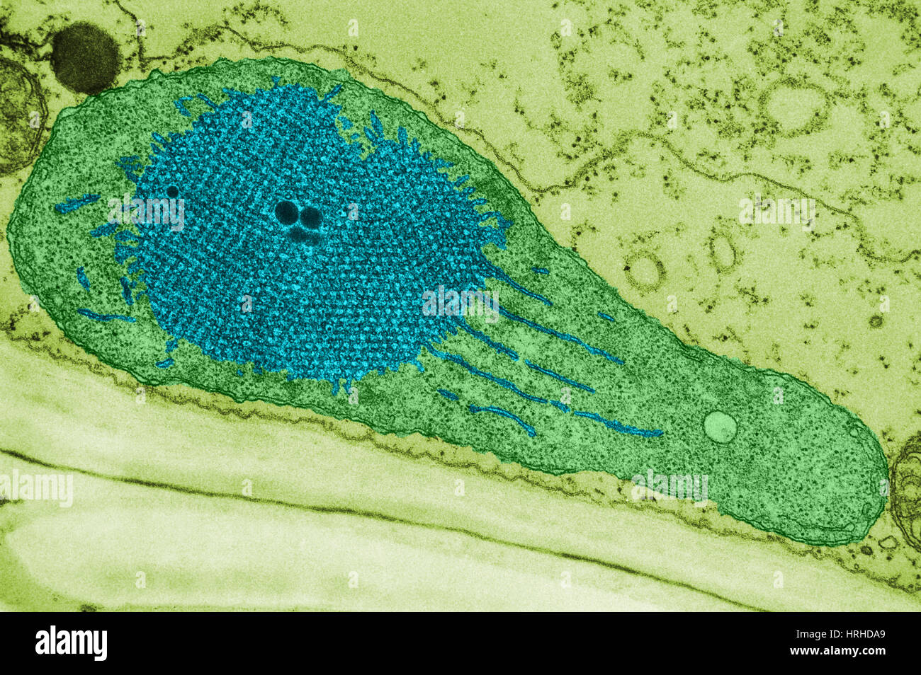 Developing Chloroplast and Etioplast TEM Stock Photohttps://www.alamy.com/image-license-details/?v=1https://www.alamy.com/stock-photo-developing-chloroplast-and-etioplast-tem-134993329.html
RMHRHDA9–Developing Chloroplast and Etioplast TEM
 Developing Chloroplast and Etioplast TEM Stock Photohttps://www.alamy.com/image-license-details/?v=1https://www.alamy.com/stock-photo-developing-chloroplast-and-etioplast-tem-134993331.html
RMHRHDAB–Developing Chloroplast and Etioplast TEM
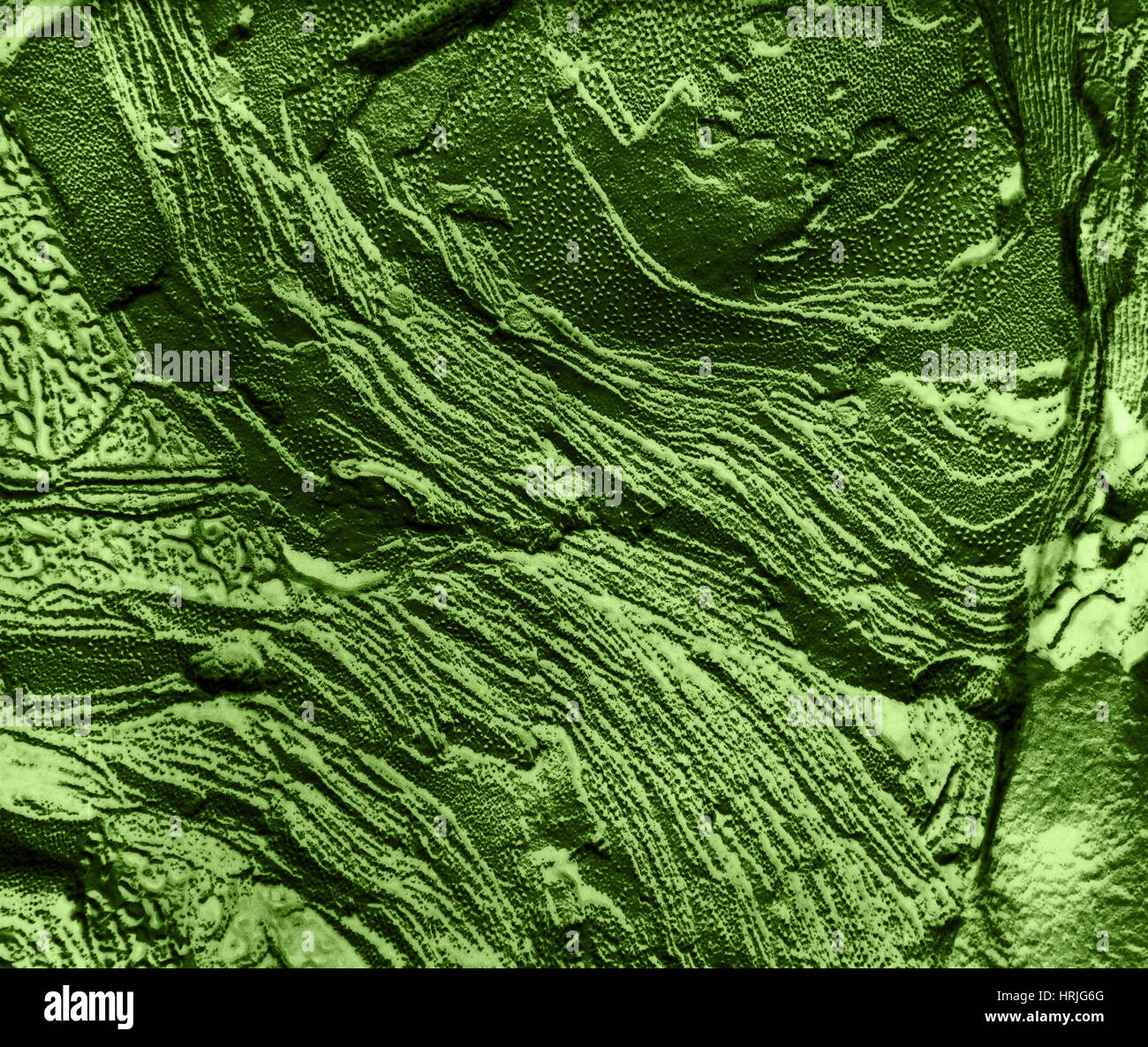 Chloroplast Membranes in Leaf Cell, TEM Stock Photohttps://www.alamy.com/image-license-details/?v=1https://www.alamy.com/stock-photo-chloroplast-membranes-in-leaf-cell-tem-135017528.html
RMHRJG6G–Chloroplast Membranes in Leaf Cell, TEM
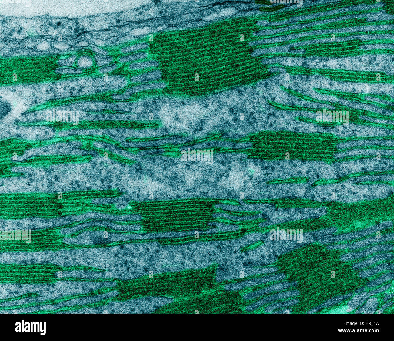 Chloroplast in Corn Leaf Cell, TEM Stock Photohttps://www.alamy.com/image-license-details/?v=1https://www.alamy.com/stock-photo-chloroplast-in-corn-leaf-cell-tem-135018950.html
RMHRJJ1A–Chloroplast in Corn Leaf Cell, TEM
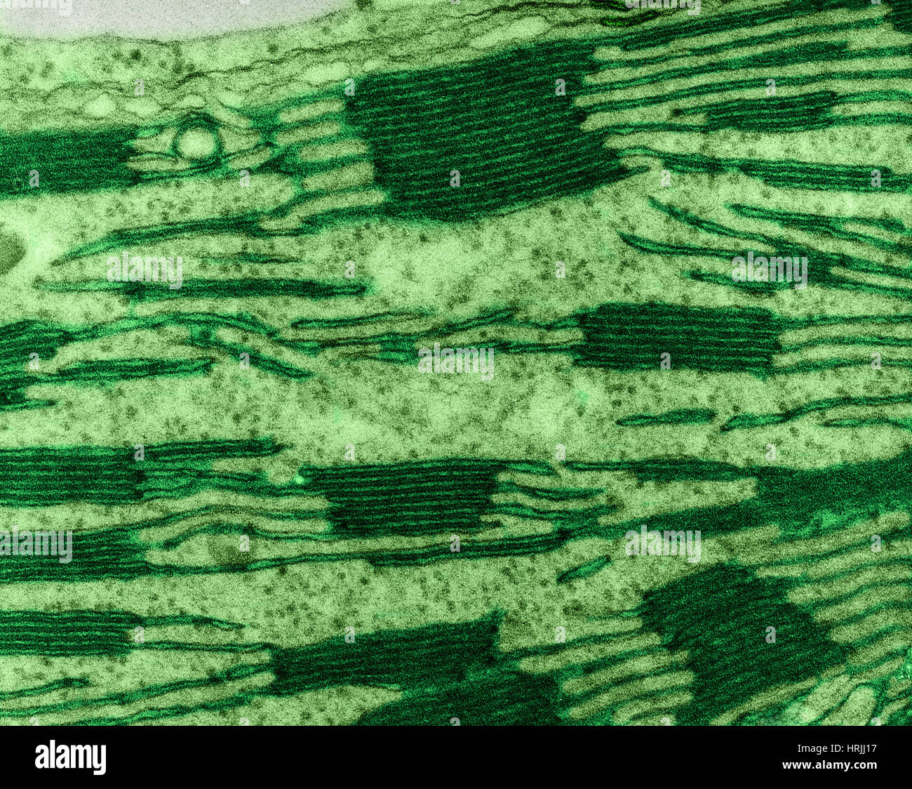 Chloroplast in Corn Leaf Cell, TEM Stock Photohttps://www.alamy.com/image-license-details/?v=1https://www.alamy.com/stock-photo-chloroplast-in-corn-leaf-cell-tem-135018947.html
RMHRJJ17–Chloroplast in Corn Leaf Cell, TEM
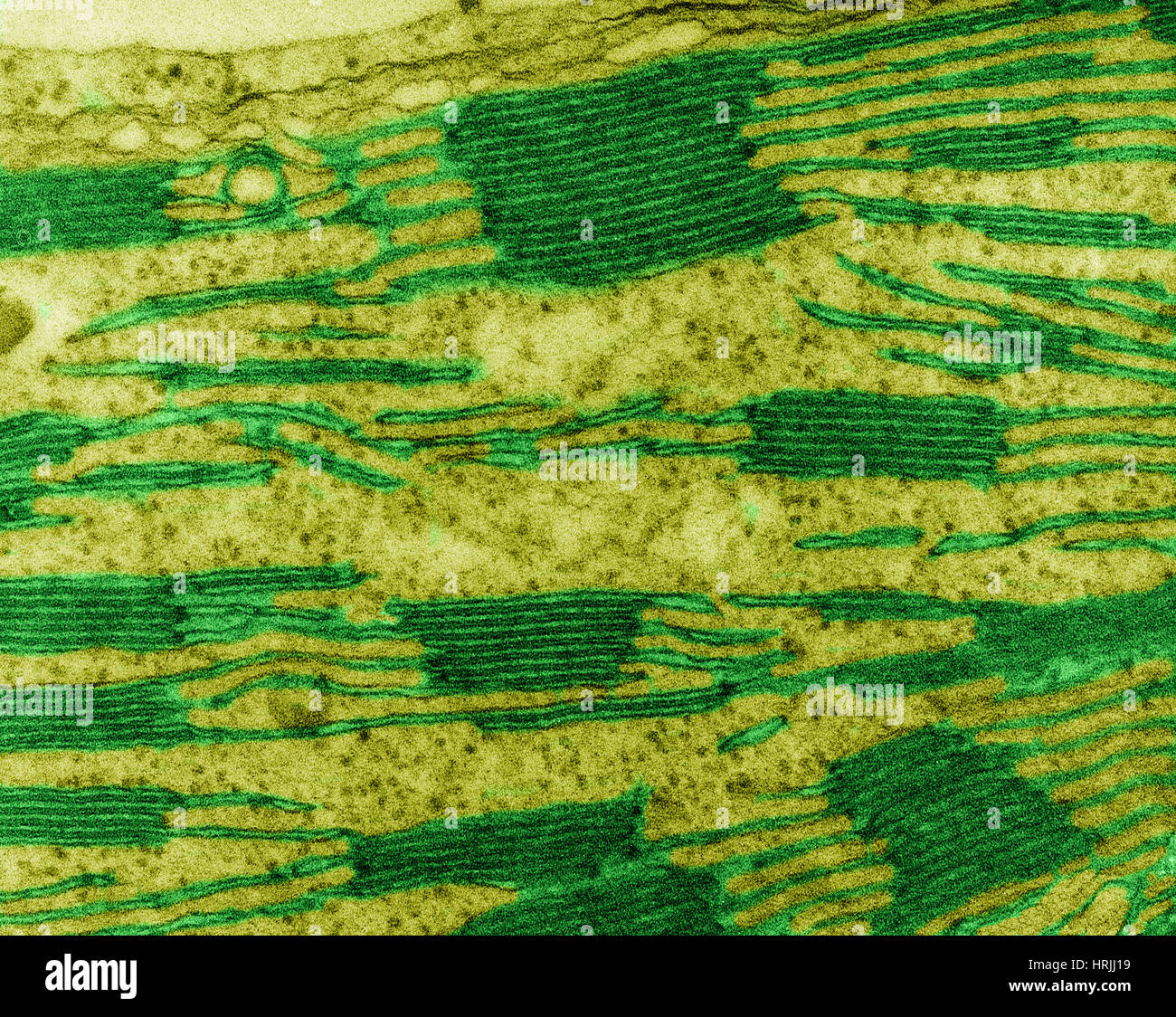 Chloroplast in Corn Leaf Cell, TEM Stock Photohttps://www.alamy.com/image-license-details/?v=1https://www.alamy.com/stock-photo-chloroplast-in-corn-leaf-cell-tem-135018949.html
RMHRJJ19–Chloroplast in Corn Leaf Cell, TEM
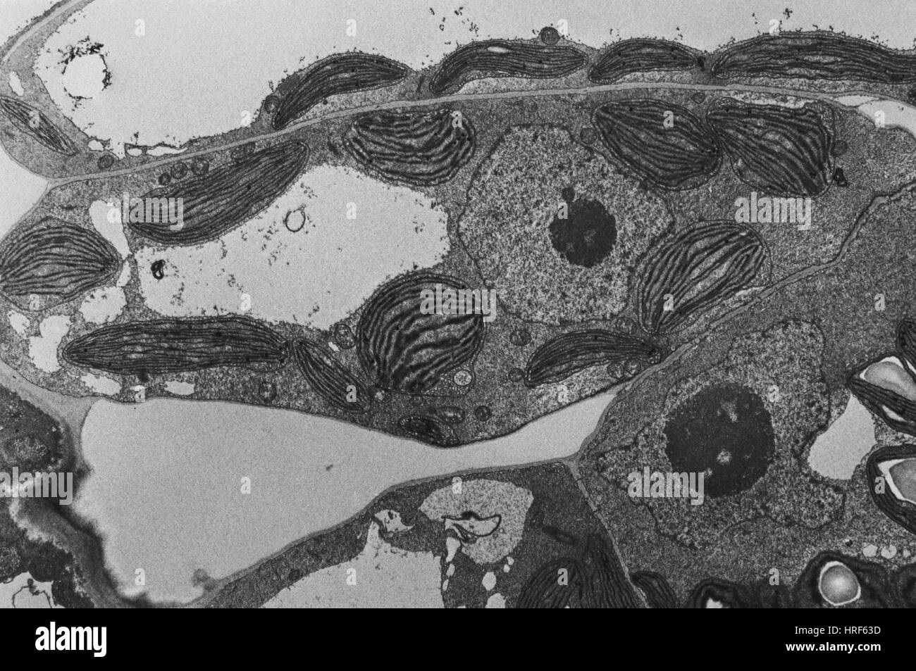 Chloroplasts TEM Stock Photohttps://www.alamy.com/image-license-details/?v=1https://www.alamy.com/stock-photo-chloroplasts-tem-134943745.html
RMHRF63D–Chloroplasts TEM
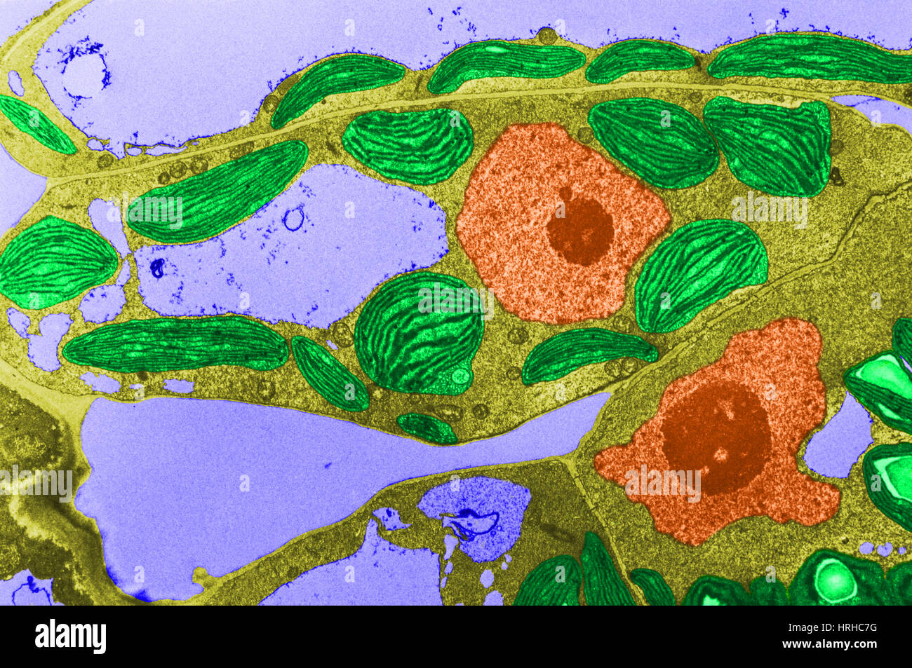 Chloroplasts TEM Stock Photohttps://www.alamy.com/image-license-details/?v=1https://www.alamy.com/stock-photo-chloroplasts-tem-134992468.html
RMHRHC7G–Chloroplasts TEM
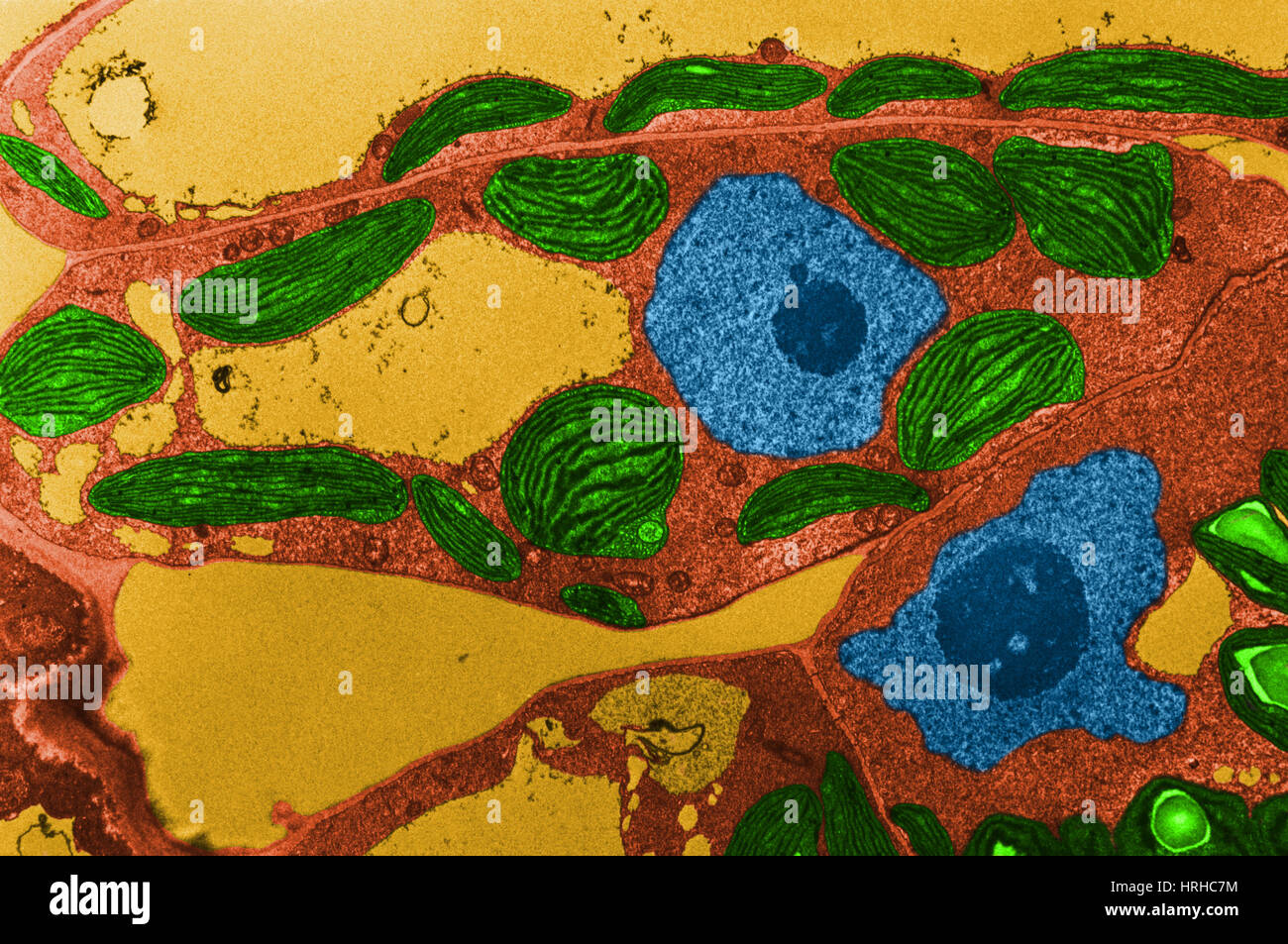 Chloroplasts TEM Stock Photohttps://www.alamy.com/image-license-details/?v=1https://www.alamy.com/stock-photo-chloroplasts-tem-134992472.html
RMHRHC7M–Chloroplasts TEM
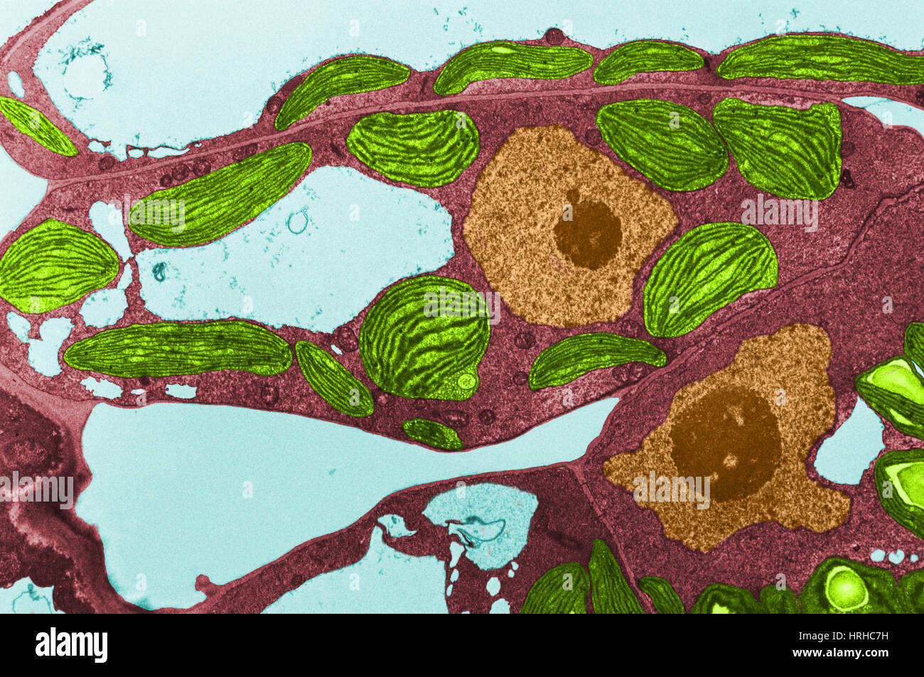 Chloroplasts TEM Stock Photohttps://www.alamy.com/image-license-details/?v=1https://www.alamy.com/stock-photo-chloroplasts-tem-134992469.html
RMHRHC7H–Chloroplasts TEM
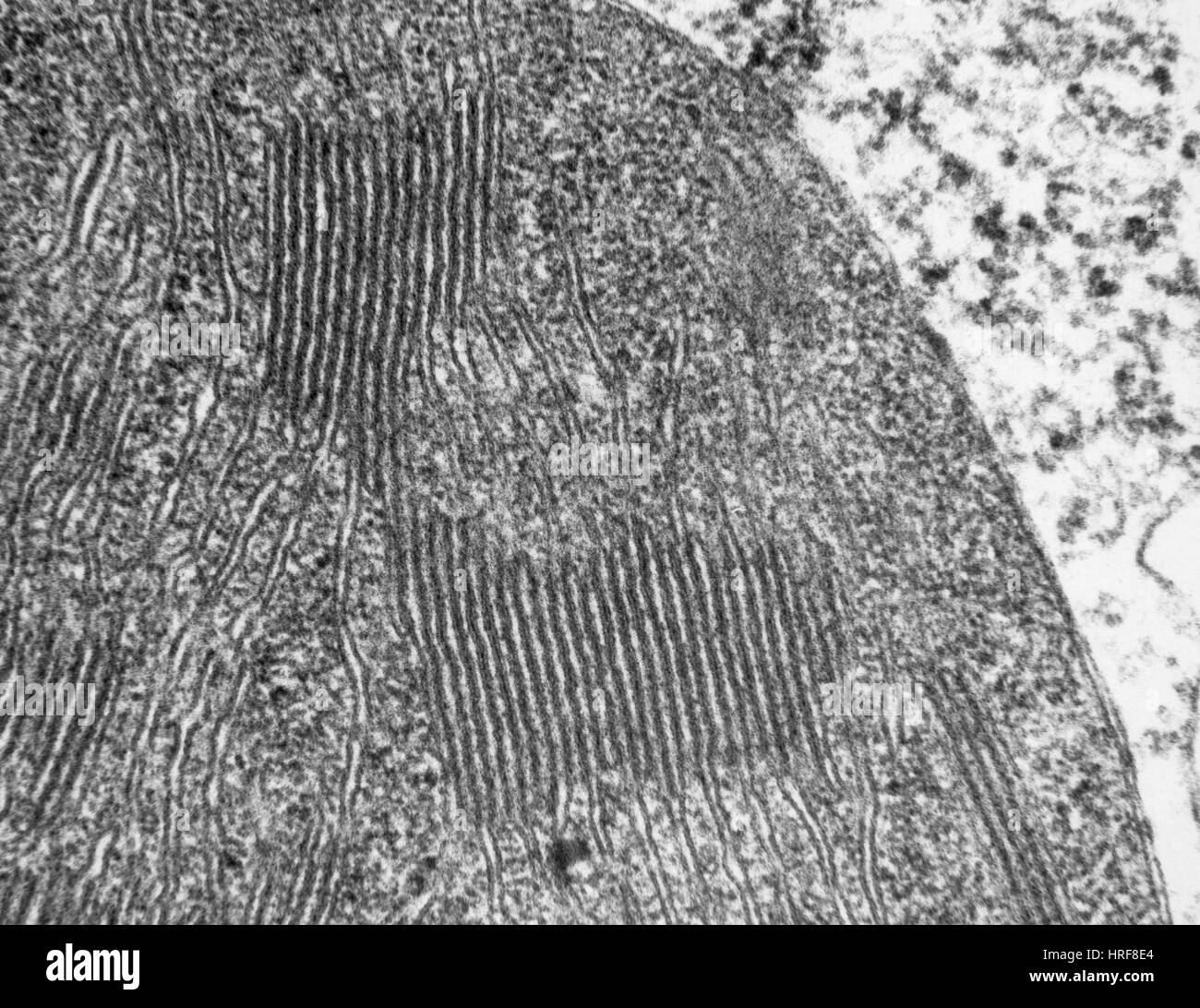 Chloroplasts in Tomato Leaf Cell, TEM Stock Photohttps://www.alamy.com/image-license-details/?v=1https://www.alamy.com/stock-photo-chloroplasts-in-tomato-leaf-cell-tem-134945612.html
RMHRF8E4–Chloroplasts in Tomato Leaf Cell, TEM
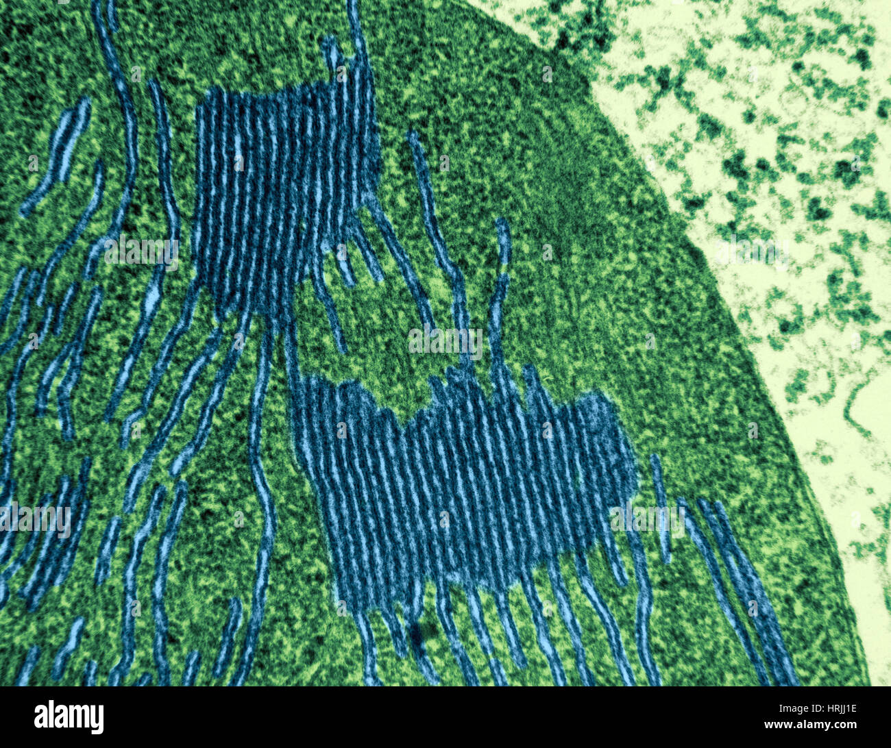 Chloroplasts in Tomato Leaf Cell, TEM Stock Photohttps://www.alamy.com/image-license-details/?v=1https://www.alamy.com/stock-photo-chloroplasts-in-tomato-leaf-cell-tem-135018954.html
RMHRJJ1E–Chloroplasts in Tomato Leaf Cell, TEM
