Quick filters:
Embryos cow Stock Photos and Images
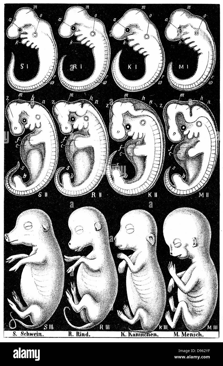 Haeckel's comparision of embryos of Pig, Cow, Rabbit and Man. Top row, all embryos show gill slit at O, demonstrating his Recapitulation theory: An embryo during development (Ontology) diplays whole evolutionary history of the species (Phylogeny). 'Ontoloogy recapitulates Phylogeny'. Stock Photohttps://www.alamy.com/image-license-details/?v=1https://www.alamy.com/stock-photo-haeckels-comparision-of-embryos-of-pig-cow-rabbit-and-man-top-row-57297059.html
RMD962YF–Haeckel's comparision of embryos of Pig, Cow, Rabbit and Man. Top row, all embryos show gill slit at O, demonstrating his Recapitulation theory: An embryo during development (Ontology) diplays whole evolutionary history of the species (Phylogeny). 'Ontoloogy recapitulates Phylogeny'.
 Cattle farming removing straws of embryos from flask containing liquid nitrogen for embryo transfer into recipient cows England Stock Photohttps://www.alamy.com/image-license-details/?v=1https://www.alamy.com/cattle-farming-removing-straws-of-embryos-from-flask-containing-liquid-image61853799.html
RMDGHK47–Cattle farming removing straws of embryos from flask containing liquid nitrogen for embryo transfer into recipient cows England
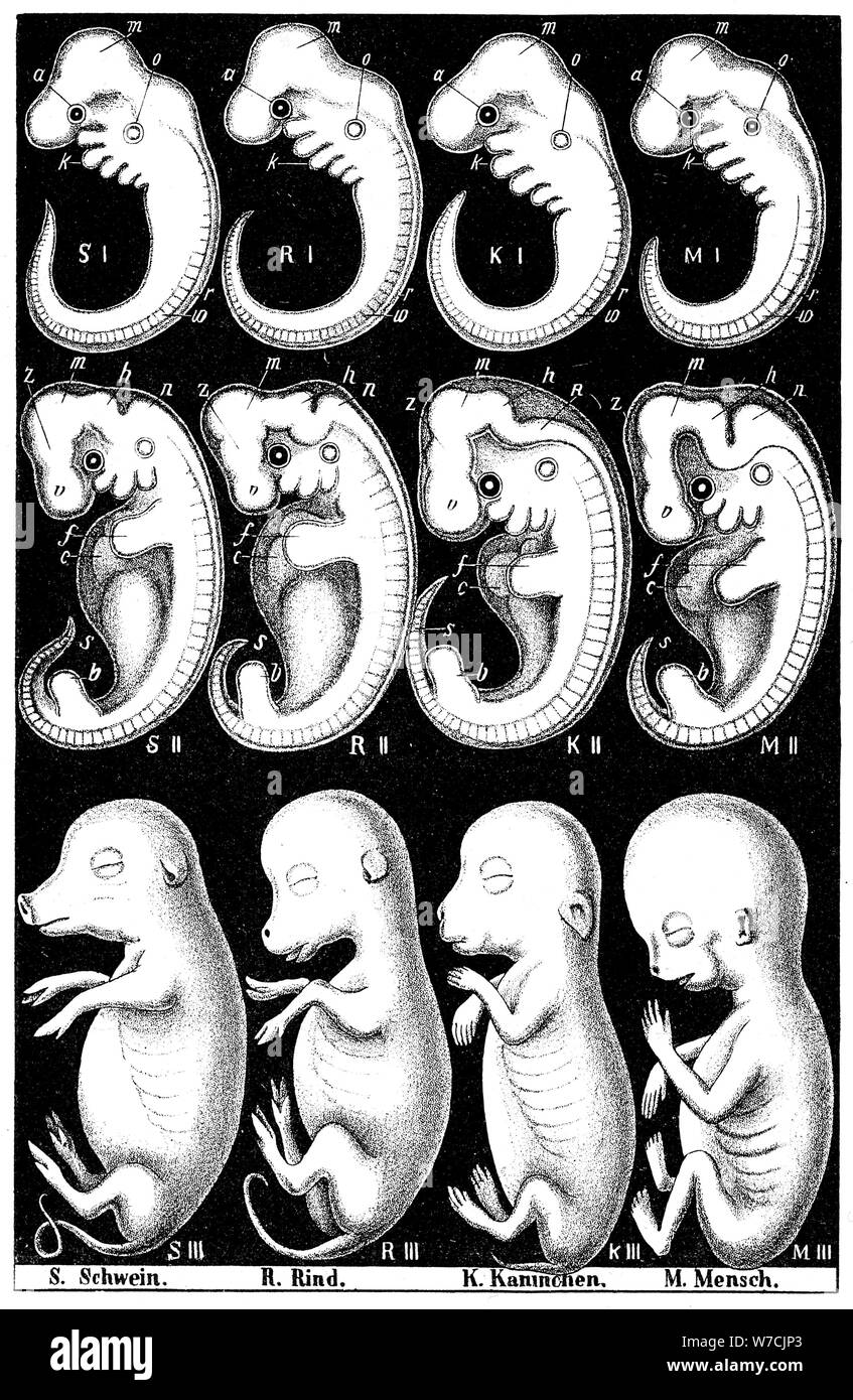 Haeckel's comparision of embryos of Pig, Cow, Rabbit and Man. Artist: Ernst Haeckel Stock Photohttps://www.alamy.com/image-license-details/?v=1https://www.alamy.com/haeckels-comparision-of-embryos-of-pig-cow-rabbit-and-man-artist-ernst-haeckel-image262736267.html
RMW7CJP3–Haeckel's comparision of embryos of Pig, Cow, Rabbit and Man. Artist: Ernst Haeckel
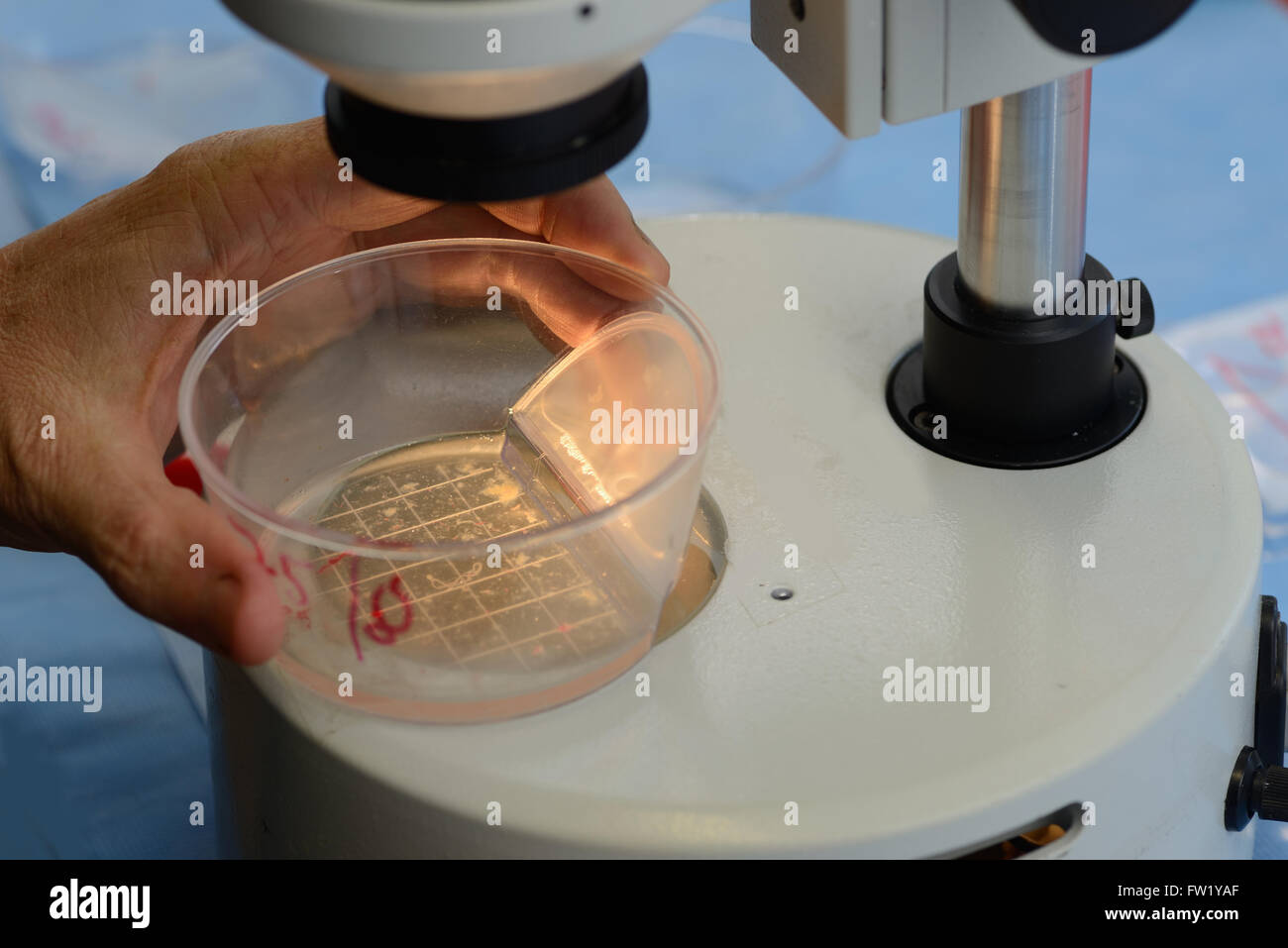 A technician searches for live calf embryos for implantation into a surrogate cow as part of an artificial breeding program, Wes Stock Photohttps://www.alamy.com/image-license-details/?v=1https://www.alamy.com/stock-photo-a-technician-searches-for-live-calf-embryos-for-implantation-into-101461655.html
RMFW1YAF–A technician searches for live calf embryos for implantation into a surrogate cow as part of an artificial breeding program, Wes
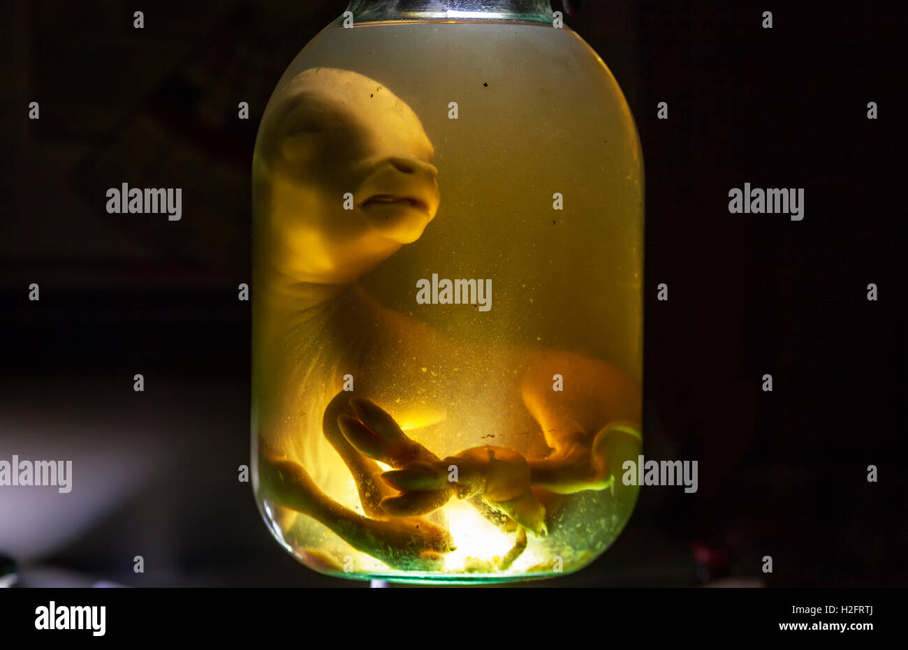 Animal embryos in formaldehyde Stock Photohttps://www.alamy.com/image-license-details/?v=1https://www.alamy.com/stock-photo-animal-embryos-in-formaldehyde-122049890.html
RFH2FRTJ–Animal embryos in formaldehyde
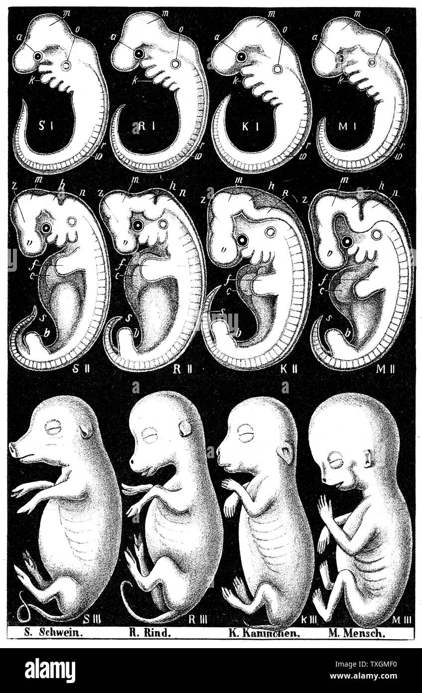 Haeckel's comparison of embryos of Pig, Cow, Rabbit and Man. Top row, all embryos show gill slit at O, demonstrating his RECAPITULATION theory: An embryo during development (Ontogeny) diplays whole evolutionary history of the species (Phylogeny). 'Ontogeny recapitulates Phylogeny' . From Ernst Haeckel 'The Evolution of Man', fifth edition, London, 1910 Stock Photohttps://www.alamy.com/image-license-details/?v=1https://www.alamy.com/haeckels-comparison-of-embryos-of-pig-cow-rabbit-and-man-top-row-all-embryos-show-gill-slit-at-o-demonstrating-his-recapitulation-theory-an-embryo-during-development-ontogeny-diplays-whole-evolutionary-history-of-the-species-phylogeny-ontogeny-recapitulates-phylogeny-from-ernst-haeckel-the-evolution-of-man-fifth-edition-london-1910-image257293540.html
RMTXGMF0–Haeckel's comparison of embryos of Pig, Cow, Rabbit and Man. Top row, all embryos show gill slit at O, demonstrating his RECAPITULATION theory: An embryo during development (Ontogeny) diplays whole evolutionary history of the species (Phylogeny). 'Ontogeny recapitulates Phylogeny' . From Ernst Haeckel 'The Evolution of Man', fifth edition, London, 1910
 . The development of the human body : a manual of human embryology. Embryology; Embryo, Non-Mammalian. THE INTERNAL EAR 435 anterior wall of the otocyst. The origin of the ganglionic mass has not yet been traced in the mammalia, but it has been observed that in cow embryos the geniculate ganglion is connected with the ecto- derm at the dorsal end of the first branchial cleft (Froriep), and it may perhaps be regarded as one of the epibranchial placodes (see p. 417), and in the lower vertebrates a union of the ganglion with a suprabranchial placode has been observed (Kupffer), this union. Fig. 2 Stock Photohttps://www.alamy.com/image-license-details/?v=1https://www.alamy.com/the-development-of-the-human-body-a-manual-of-human-embryology-embryology-embryo-non-mammalian-the-internal-ear-435-anterior-wall-of-the-otocyst-the-origin-of-the-ganglionic-mass-has-not-yet-been-traced-in-the-mammalia-but-it-has-been-observed-that-in-cow-embryos-the-geniculate-ganglion-is-connected-with-the-ecto-derm-at-the-dorsal-end-of-the-first-branchial-cleft-froriep-and-it-may-perhaps-be-regarded-as-one-of-the-epibranchial-placodes-see-p-417-and-in-the-lower-vertebrates-a-union-of-the-ganglion-with-a-suprabranchial-placode-has-been-observed-kupffer-this-union-fig-2-image215969273.html
RMPFA709–. The development of the human body : a manual of human embryology. Embryology; Embryo, Non-Mammalian. THE INTERNAL EAR 435 anterior wall of the otocyst. The origin of the ganglionic mass has not yet been traced in the mammalia, but it has been observed that in cow embryos the geniculate ganglion is connected with the ecto- derm at the dorsal end of the first branchial cleft (Froriep), and it may perhaps be regarded as one of the epibranchial placodes (see p. 417), and in the lower vertebrates a union of the ganglion with a suprabranchial placode has been observed (Kupffer), this union. Fig. 2
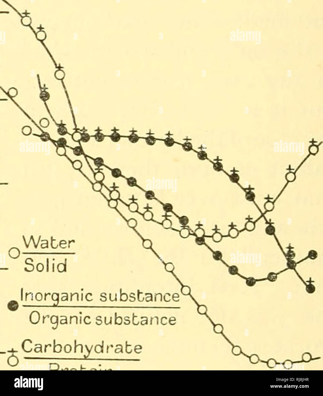 . Chemical embryology. Embryology. 9i6 GENERAL METABOLISM [PT. Ill not show such a fall in the case of the mammalian embryo. Possibly this is due to the fact that we have no analyses of mammalian embryos in the earlier stages, and the earlier, sharper part of the inorganic curve may thus have been missed. Inspection of Col. 13 of Table 108 shows that this decline in ash-content percentage dry weight is in fact more rapid at the beginning than at the end of development. Some fragmentary data for the Jersey cow embryo contained in the paper of Moulton, Trowbridge & Haigh, do not seem to show Stock Photohttps://www.alamy.com/image-license-details/?v=1https://www.alamy.com/chemical-embryology-embryology-9i6-general-metabolism-pt-ill-not-show-such-a-fall-in-the-case-of-the-mammalian-embryo-possibly-this-is-due-to-the-fact-that-we-have-no-analyses-of-mammalian-embryos-in-the-earlier-stages-and-the-earlier-sharper-part-of-the-inorganic-curve-may-thus-have-been-missed-inspection-of-col-13-of-table-108-shows-that-this-decline-in-ash-content-percentage-dry-weight-is-in-fact-more-rapid-at-the-beginning-than-at-the-end-of-development-some-fragmentary-data-for-the-jersey-cow-embryo-contained-in-the-paper-of-moulton-trowbridge-amp-haigh-do-not-seem-to-show-image234988819.html
RMRJ8JHR–. Chemical embryology. Embryology. 9i6 GENERAL METABOLISM [PT. Ill not show such a fall in the case of the mammalian embryo. Possibly this is due to the fact that we have no analyses of mammalian embryos in the earlier stages, and the earlier, sharper part of the inorganic curve may thus have been missed. Inspection of Col. 13 of Table 108 shows that this decline in ash-content percentage dry weight is in fact more rapid at the beginning than at the end of development. Some fragmentary data for the Jersey cow embryo contained in the paper of Moulton, Trowbridge & Haigh, do not seem to show
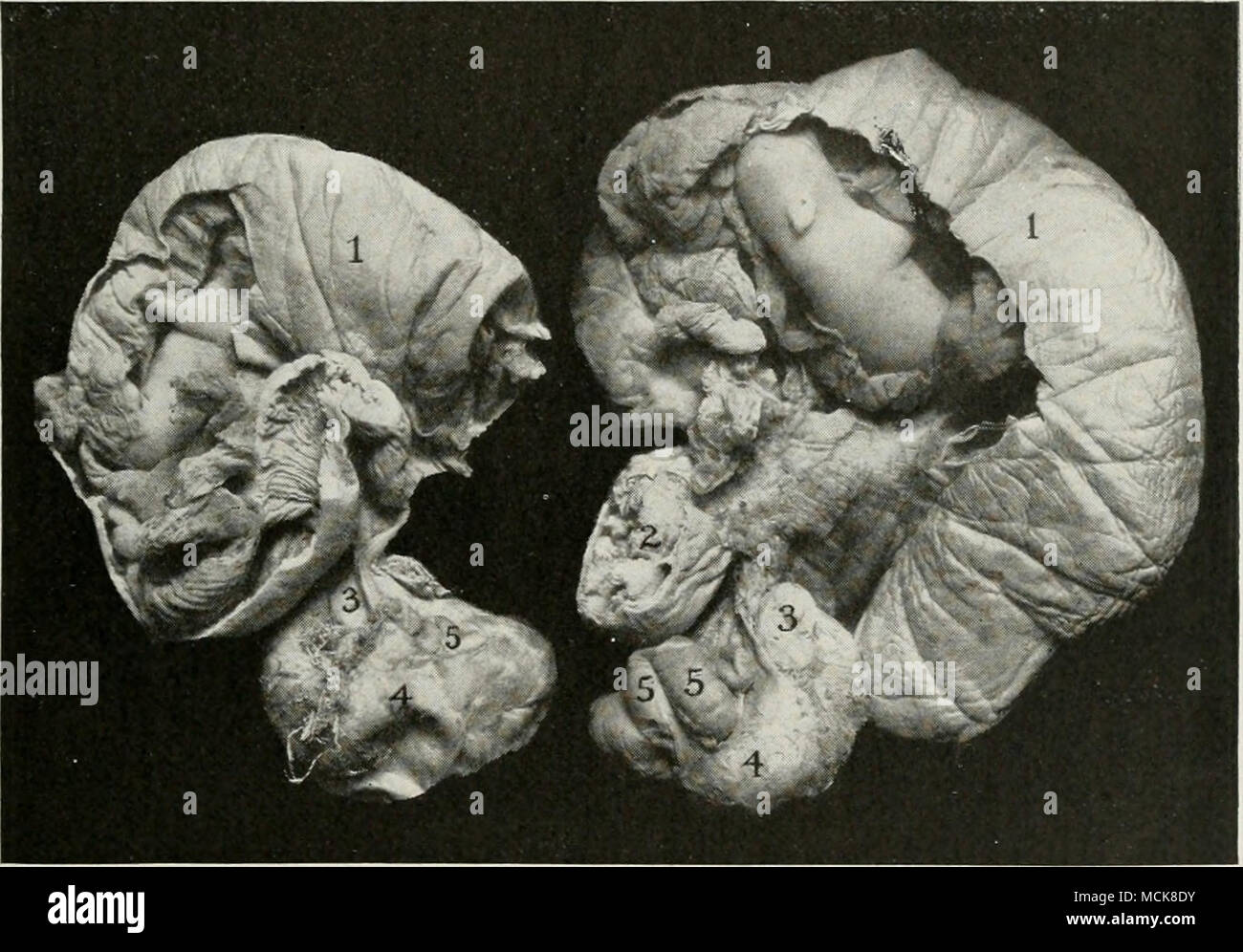 . Fig. 21S—Bilateral Hydrosalpinx Developed During Pregnancy. Sow. /, /, The cornua each contains a normal embryo " long ; 2, abscess in apex of horn, probably embryonic debris; j, ovary sectioned, showing small cysts ; 4, cystic distension of adherent pavilion ; 5, cystic oviduct. before the end of pregnancy the reproductive life of the sow has been permanently closed—unless one believes that the tubal infection was post-coital. The gravid uterus of the sow, the embryonic envelopes, and the embryos show essentially all the lesions already de- scribed for the cow, and it is only necessar Stock Photohttps://www.alamy.com/image-license-details/?v=1https://www.alamy.com/fig-21sbilateral-hydrosalpinx-developed-during-pregnancy-sow-the-cornua-each-contains-a-normal-embryo-quot-long-2-abscess-in-apex-of-horn-probably-embryonic-debris-j-ovary-sectioned-showing-small-cysts-4-cystic-distension-of-adherent-pavilion-5-cystic-oviduct-before-the-end-of-pregnancy-the-reproductive-life-of-the-sow-has-been-permanently-closedunless-one-believes-that-the-tubal-infection-was-post-coital-the-gravid-uterus-of-the-sow-the-embryonic-envelopes-and-the-embryos-show-essentially-all-the-lesions-already-de-scribed-for-the-cow-and-it-is-only-necessar-image179903303.html
RMMCK8DY–. Fig. 21S—Bilateral Hydrosalpinx Developed During Pregnancy. Sow. /, /, The cornua each contains a normal embryo " long ; 2, abscess in apex of horn, probably embryonic debris; j, ovary sectioned, showing small cysts ; 4, cystic distension of adherent pavilion ; 5, cystic oviduct. before the end of pregnancy the reproductive life of the sow has been permanently closed—unless one believes that the tubal infection was post-coital. The gravid uterus of the sow, the embryonic envelopes, and the embryos show essentially all the lesions already de- scribed for the cow, and it is only necessar
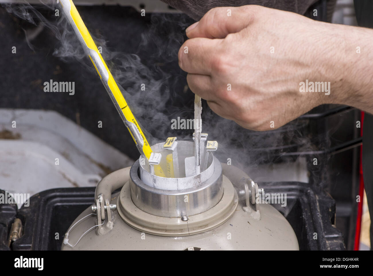 Cattle farming removing straws of embryos from flask containing liquid nitrogen for embryo transfer into recipient cows England Stock Photohttps://www.alamy.com/image-license-details/?v=1https://www.alamy.com/cattle-farming-removing-straws-of-embryos-from-flask-containing-liquid-image61853815.html
RMDGHK4R–Cattle farming removing straws of embryos from flask containing liquid nitrogen for embryo transfer into recipient cows England
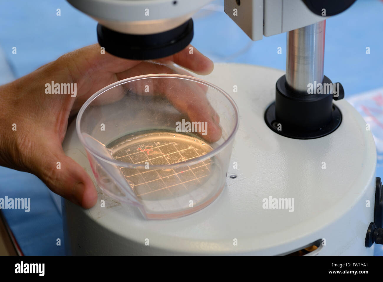 A technician searches for live calf embryos for implantation into a surrogate cow as part of an artificial breeding program, Wes Stock Photohttps://www.alamy.com/image-license-details/?v=1https://www.alamy.com/stock-photo-a-technician-searches-for-live-calf-embryos-for-implantation-into-101461641.html
RMFW1YA1–A technician searches for live calf embryos for implantation into a surrogate cow as part of an artificial breeding program, Wes
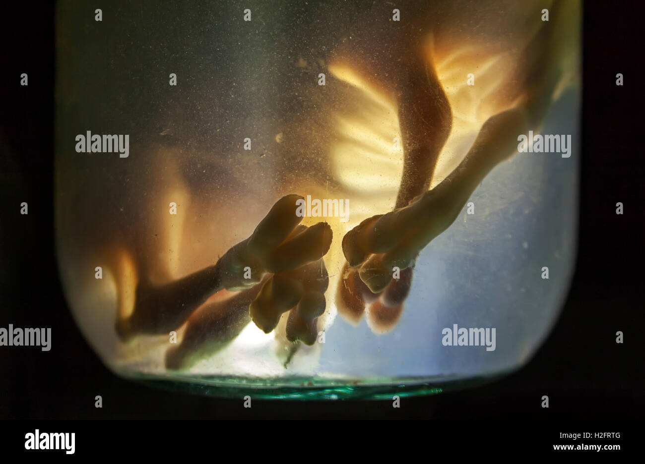 Animal embryos in formaldehyde Stock Photohttps://www.alamy.com/image-license-details/?v=1https://www.alamy.com/stock-photo-animal-embryos-in-formaldehyde-122049888.html
RFH2FRTG–Animal embryos in formaldehyde
 Haeckel's comparision of embryos of Pig, Cow, Rabbit and Man. Top row, all embryos show gill slit at O, demonstrating his Recapitulation theory: An embryo during development (Ontogeny) diplays whole evolutionary history of the species (Phylogeny). 'Ontogeny recapitulates Phylogeny' . From Ernst Haeckel 'The Evolution of Man', fifth edition 1910 London Stock Photohttps://www.alamy.com/image-license-details/?v=1https://www.alamy.com/haeckels-comparision-of-embryos-of-pig-cow-rabbit-and-man-top-row-all-embryos-show-gill-slit-at-o-demonstrating-his-recapitulation-theory-an-embryo-during-development-ontogeny-diplays-whole-evolutionary-history-of-the-species-phylogeny-ontogeny-recapitulates-phylogeny-from-ernst-haeckel-the-evolution-of-man-fifth-edition-1910-london-image257294652.html
RMTXGNXM–Haeckel's comparision of embryos of Pig, Cow, Rabbit and Man. Top row, all embryos show gill slit at O, demonstrating his Recapitulation theory: An embryo during development (Ontogeny) diplays whole evolutionary history of the species (Phylogeny). 'Ontogeny recapitulates Phylogeny' . From Ernst Haeckel 'The Evolution of Man', fifth edition 1910 London
 . Animal biology. Zoology; Biology. PARASITISM 173 In the uterus there develops in this egg a six-hooked embryo. As the proglottid becomes ripe and passes out of the body of the host with the feces, it carries with it thousands of embryos, still inclosed in the egg shells. 2. If this proglottid falls upon vegetation and is eaten by a cow, the eggs are freed from the proglottid in her intestine and the embryos escape.. Fig. 81.—Diagram showing the life history of Taenia saginata. A, the egg. X 550. B, egg containing six-hooked embryo. X 550. C, young eysticercus. X 30. D. cysticercus showing be Stock Photohttps://www.alamy.com/image-license-details/?v=1https://www.alamy.com/animal-biology-zoology-biology-parasitism-173-in-the-uterus-there-develops-in-this-egg-a-six-hooked-embryo-as-the-proglottid-becomes-ripe-and-passes-out-of-the-body-of-the-host-with-the-feces-it-carries-with-it-thousands-of-embryos-still-inclosed-in-the-egg-shells-2-if-this-proglottid-falls-upon-vegetation-and-is-eaten-by-a-cow-the-eggs-are-freed-from-the-proglottid-in-her-intestine-and-the-embryos-escape-fig-81diagram-showing-the-life-history-of-taenia-saginata-a-the-egg-x-550-b-egg-containing-six-hooked-embryo-x-550-c-young-eysticercus-x-30-d-cysticercus-showing-be-image236768737.html
RMRN5MX9–. Animal biology. Zoology; Biology. PARASITISM 173 In the uterus there develops in this egg a six-hooked embryo. As the proglottid becomes ripe and passes out of the body of the host with the feces, it carries with it thousands of embryos, still inclosed in the egg shells. 2. If this proglottid falls upon vegetation and is eaten by a cow, the eggs are freed from the proglottid in her intestine and the embryos escape.. Fig. 81.—Diagram showing the life history of Taenia saginata. A, the egg. X 550. B, egg containing six-hooked embryo. X 550. C, young eysticercus. X 30. D. cysticercus showing be
 . Fig. 218—Bilateral Hydrosalpinx Developed During Pregnancy. Sow. /, /, The cornua each contains a normal embryo 4" long ; 2, abscess in apex of horn, probably embryonic debris; j, ovary sectioned, showing small cysts ; 4, cystic distension of adherent pavilion ; 5, cystic oviduct. before the end of pregnancy the reproductive life of the sow has been permanently closed—unless one believes that the tubal infection was post-coital. The gravid uterus of the sow, the embryonic envelopes, and the embryos show essentially all the lesions already de- scribed for the cow, and it is only necessar Stock Photohttps://www.alamy.com/image-license-details/?v=1https://www.alamy.com/fig-218bilateral-hydrosalpinx-developed-during-pregnancy-sow-the-cornua-each-contains-a-normal-embryo-4quot-long-2-abscess-in-apex-of-horn-probably-embryonic-debris-j-ovary-sectioned-showing-small-cysts-4-cystic-distension-of-adherent-pavilion-5-cystic-oviduct-before-the-end-of-pregnancy-the-reproductive-life-of-the-sow-has-been-permanently-closedunless-one-believes-that-the-tubal-infection-was-post-coital-the-gravid-uterus-of-the-sow-the-embryonic-envelopes-and-the-embryos-show-essentially-all-the-lesions-already-de-scribed-for-the-cow-and-it-is-only-necessar-image179903589.html
RMMCK8T5–. Fig. 218—Bilateral Hydrosalpinx Developed During Pregnancy. Sow. /, /, The cornua each contains a normal embryo 4" long ; 2, abscess in apex of horn, probably embryonic debris; j, ovary sectioned, showing small cysts ; 4, cystic distension of adherent pavilion ; 5, cystic oviduct. before the end of pregnancy the reproductive life of the sow has been permanently closed—unless one believes that the tubal infection was post-coital. The gravid uterus of the sow, the embryonic envelopes, and the embryos show essentially all the lesions already de- scribed for the cow, and it is only necessar
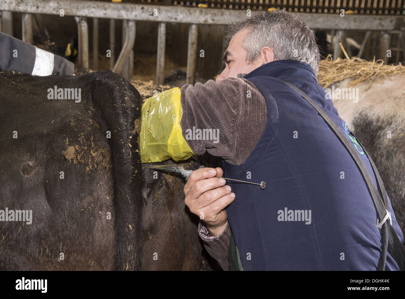 Cattle farming, inserting embryo transfer gun into recipient cow, England, April Stock Photohttps://www.alamy.com/image-license-details/?v=1https://www.alamy.com/cattle-farming-inserting-embryo-transfer-gun-into-recipient-cow-england-image61853811.html
RMDGHK4K–Cattle farming, inserting embryo transfer gun into recipient cow, England, April
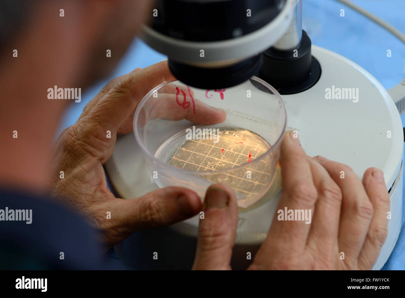 A technician searches for live calf embryos for implantation into a surrogate cow as part of an artificial breeding program, Wes Stock Photohttps://www.alamy.com/image-license-details/?v=1https://www.alamy.com/stock-photo-a-technician-searches-for-live-calf-embryos-for-implantation-into-101461715.html
RMFW1YCK–A technician searches for live calf embryos for implantation into a surrogate cow as part of an artificial breeding program, Wes
 . Agri-news. Agriculture. • I . I - •. Picina, a two year old full-blood Marchigiana heifer, owned by Danbeck Marchiniana Farms, Calmar, Alberta, with her 1 7 full-blood calves (11 heifers and six bulls) born from a single transplant collection performed by Alberta Livestock Transplants Ltd. Alberta Livestock Transplants Ltd. of Calgary, recently announced a new preg- nancy record for a single non-surgical bovine embryo collection. The donor cow is "Perla", a full-blood Romagnola female owned by Berg Cattle Co. of Millicent, Alberta. From ' ?M^ 30 embryos which were recovered non-sur Stock Photohttps://www.alamy.com/image-license-details/?v=1https://www.alamy.com/agri-news-agriculture-i-i-picina-a-two-year-old-full-blood-marchigiana-heifer-owned-by-danbeck-marchiniana-farms-calmar-alberta-with-her-1-7-full-blood-calves-11-heifers-and-six-bulls-born-from-a-single-transplant-collection-performed-by-alberta-livestock-transplants-ltd-alberta-livestock-transplants-ltd-of-calgary-recently-announced-a-new-preg-nancy-record-for-a-single-non-surgical-bovine-embryo-collection-the-donor-cow-is-quotperlaquot-a-full-blood-romagnola-female-owned-by-berg-cattle-co-of-millicent-alberta-from-m-30-embryos-which-were-recovered-non-sur-image237877011.html
RMRR06FF–. Agri-news. Agriculture. • I . I - •. Picina, a two year old full-blood Marchigiana heifer, owned by Danbeck Marchiniana Farms, Calmar, Alberta, with her 1 7 full-blood calves (11 heifers and six bulls) born from a single transplant collection performed by Alberta Livestock Transplants Ltd. Alberta Livestock Transplants Ltd. of Calgary, recently announced a new preg- nancy record for a single non-surgical bovine embryo collection. The donor cow is "Perla", a full-blood Romagnola female owned by Berg Cattle Co. of Millicent, Alberta. From ' ?M^ 30 embryos which were recovered non-sur
 . Fig. 21S—Bilateral Hydrosalpinx Developed During Pregnancy. Sow. /, /, The cornua each contains a normal embryo A," long ; 2, abscess in apex of horn, probably embryonic debris; j, ovary sectioned, showing small cysts ; 4, cystic distension of adherent pavilion ; 5, cystic oviduct. before the end of pregnancy the reproductive life of the sow has been permanently closed—unless one believes that the tubal infection was post-coital. The gravid uterus of the sow, the embryonic envelopes, and the embryos show essentially all the lesions already de- scribed for the cow, and it is only necessa Stock Photohttps://www.alamy.com/image-license-details/?v=1https://www.alamy.com/fig-21sbilateral-hydrosalpinx-developed-during-pregnancy-sow-the-cornua-each-contains-a-normal-embryo-aquot-long-2-abscess-in-apex-of-horn-probably-embryonic-debris-j-ovary-sectioned-showing-small-cysts-4-cystic-distension-of-adherent-pavilion-5-cystic-oviduct-before-the-end-of-pregnancy-the-reproductive-life-of-the-sow-has-been-permanently-closedunless-one-believes-that-the-tubal-infection-was-post-coital-the-gravid-uterus-of-the-sow-the-embryonic-envelopes-and-the-embryos-show-essentially-all-the-lesions-already-de-scribed-for-the-cow-and-it-is-only-necessa-image179902933.html
RMMCK80N–. Fig. 21S—Bilateral Hydrosalpinx Developed During Pregnancy. Sow. /, /, The cornua each contains a normal embryo A," long ; 2, abscess in apex of horn, probably embryonic debris; j, ovary sectioned, showing small cysts ; 4, cystic distension of adherent pavilion ; 5, cystic oviduct. before the end of pregnancy the reproductive life of the sow has been permanently closed—unless one believes that the tubal infection was post-coital. The gravid uterus of the sow, the embryonic envelopes, and the embryos show essentially all the lesions already de- scribed for the cow, and it is only necessa
 Cattle farming, bottle of anaesthetic for epidural into recipient cows during embryo transfer, England, April Stock Photohttps://www.alamy.com/image-license-details/?v=1https://www.alamy.com/cattle-farming-bottle-of-anaesthetic-for-epidural-into-recipient-cows-image61853791.html
RMDGHK3Y–Cattle farming, bottle of anaesthetic for epidural into recipient cows during embryo transfer, England, April
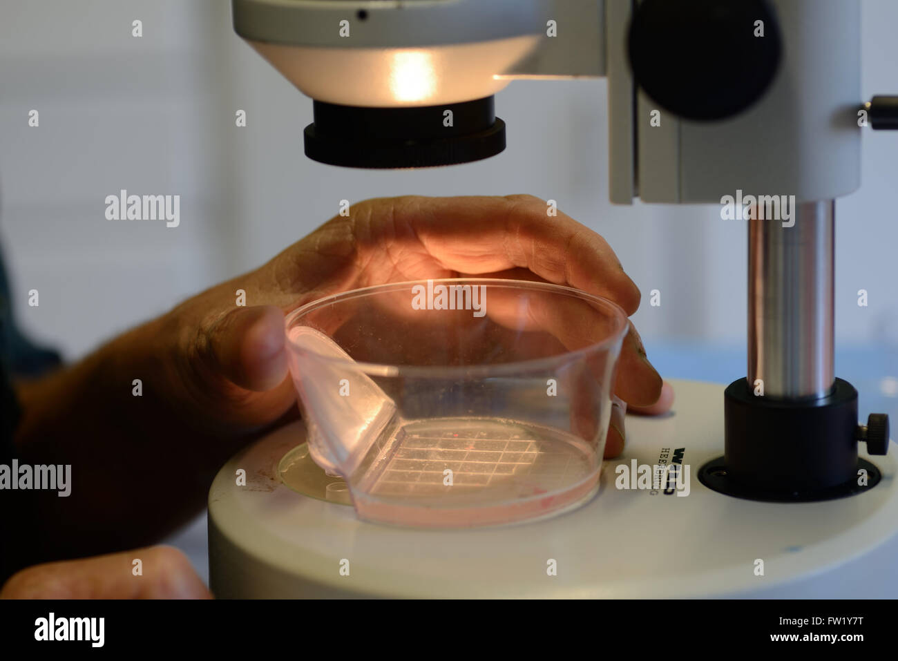 A technician searches for live calf embryos for implantation into a surrogate cow as part of an artificial breeding program, Wes Stock Photohttps://www.alamy.com/image-license-details/?v=1https://www.alamy.com/stock-photo-a-technician-searches-for-live-calf-embryos-for-implantation-into-101461580.html
RMFW1Y7T–A technician searches for live calf embryos for implantation into a surrogate cow as part of an artificial breeding program, Wes
 . The development of the human body : a manual of human embryology. Embryology; Embryo, Non-Mammalian. THE INTERNAL EAR 435 anterior wall of the otocyst. The origin of the ganglionic mass has not yet been traced in the mammalia, but it has been observed that in cow embryos the geniculate ganglion is connected with the ecto- derm at the dorsal end of the first branchial cleft (Froriep), and it may perhaps be regarded as one of the epibranchial placodes (see p. 417), and in the lower vertebrates a union of the ganglion with a suprabranchial placode has been observed (Kupffer), this union. Fig. 2 Stock Photohttps://www.alamy.com/image-license-details/?v=1https://www.alamy.com/the-development-of-the-human-body-a-manual-of-human-embryology-embryology-embryo-non-mammalian-the-internal-ear-435-anterior-wall-of-the-otocyst-the-origin-of-the-ganglionic-mass-has-not-yet-been-traced-in-the-mammalia-but-it-has-been-observed-that-in-cow-embryos-the-geniculate-ganglion-is-connected-with-the-ecto-derm-at-the-dorsal-end-of-the-first-branchial-cleft-froriep-and-it-may-perhaps-be-regarded-as-one-of-the-epibranchial-placodes-see-p-417-and-in-the-lower-vertebrates-a-union-of-the-ganglion-with-a-suprabranchial-placode-has-been-observed-kupffer-this-union-fig-2-image231665048.html
RMRCW73M–. The development of the human body : a manual of human embryology. Embryology; Embryo, Non-Mammalian. THE INTERNAL EAR 435 anterior wall of the otocyst. The origin of the ganglionic mass has not yet been traced in the mammalia, but it has been observed that in cow embryos the geniculate ganglion is connected with the ecto- derm at the dorsal end of the first branchial cleft (Froriep), and it may perhaps be regarded as one of the epibranchial placodes (see p. 417), and in the lower vertebrates a union of the ganglion with a suprabranchial placode has been observed (Kupffer), this union. Fig. 2
 . Fig. 25—Diagram of ovary of cow showing comparative sizes of the follicles shown in Figs. 22, 23, 24 The number of ovisacs rupturing at a given estrual period corresponds as a rule with the maximum number of possible fetuses. It is said that rarely two ova are contained in one ovisac. I have been unable to verify this statement, and have in all cases of twins in the cow observed two corpora lutea, sometimes both in one ovary but most frequently one in each. Sometimes a single fertilized ovum divides to con- stitute two embryos, which form identical twins in man, but this is not known to occu Stock Photohttps://www.alamy.com/image-license-details/?v=1https://www.alamy.com/fig-25diagram-of-ovary-of-cow-showing-comparative-sizes-of-the-follicles-shown-in-figs-22-23-24-the-number-of-ovisacs-rupturing-at-a-given-estrual-period-corresponds-as-a-rule-with-the-maximum-number-of-possible-fetuses-it-is-said-that-rarely-two-ova-are-contained-in-one-ovisac-i-have-been-unable-to-verify-this-statement-and-have-in-all-cases-of-twins-in-the-cow-observed-two-corpora-lutea-sometimes-both-in-one-ovary-but-most-frequently-one-in-each-sometimes-a-single-fertilized-ovum-divides-to-con-stitute-two-embryos-which-form-identical-twins-in-man-but-this-is-not-known-to-occu-image179903242.html
RMMCK8BP–. Fig. 25—Diagram of ovary of cow showing comparative sizes of the follicles shown in Figs. 22, 23, 24 The number of ovisacs rupturing at a given estrual period corresponds as a rule with the maximum number of possible fetuses. It is said that rarely two ova are contained in one ovisac. I have been unable to verify this statement, and have in all cases of twins in the cow observed two corpora lutea, sometimes both in one ovary but most frequently one in each. Sometimes a single fertilized ovum divides to con- stitute two embryos, which form identical twins in man, but this is not known to occu
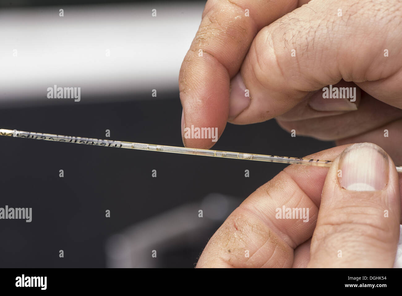 Cattle farming, straw containing embryo for embryo transfer to recipient cows, England, April Stock Photohttps://www.alamy.com/image-license-details/?v=1https://www.alamy.com/cattle-farming-straw-containing-embryo-for-embryo-transfer-to-recipient-image61853824.html
RMDGHK54–Cattle farming, straw containing embryo for embryo transfer to recipient cows, England, April
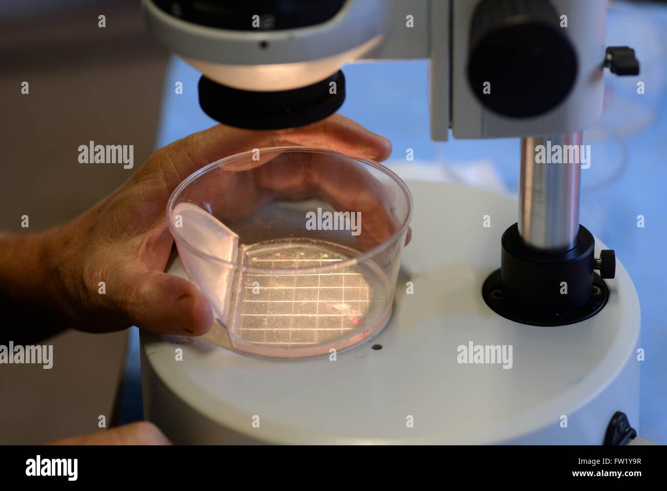 A technician searches for live calf embryos for implantation into a surrogate cow as part of an artificial breeding program, Wes Stock Photohttps://www.alamy.com/image-license-details/?v=1https://www.alamy.com/stock-photo-a-technician-searches-for-live-calf-embryos-for-implantation-into-101461635.html
RMFW1Y9R–A technician searches for live calf embryos for implantation into a surrogate cow as part of an artificial breeding program, Wes
 . Fig. 25—Diagram of ovary of cow showing comparative sizes of the follicles shown in Figs. 22, 2a, 24 The number of ovisacs rupturing at a given estrual period corresponds as a rule with the maximum number of possible fetuses. It is said that rarely two ova are contained in one ovisac. I have been unable to verify this statement, and have in all cases of twins in the cow observed two corpora lutea, sometimes both in one ovary but most frequently one in each. Sometimes a single fertilized ovum divides to con- stitute two embryos, which form identical twins in man, but this is not known to occu Stock Photohttps://www.alamy.com/image-license-details/?v=1https://www.alamy.com/fig-25diagram-of-ovary-of-cow-showing-comparative-sizes-of-the-follicles-shown-in-figs-22-2a-24-the-number-of-ovisacs-rupturing-at-a-given-estrual-period-corresponds-as-a-rule-with-the-maximum-number-of-possible-fetuses-it-is-said-that-rarely-two-ova-are-contained-in-one-ovisac-i-have-been-unable-to-verify-this-statement-and-have-in-all-cases-of-twins-in-the-cow-observed-two-corpora-lutea-sometimes-both-in-one-ovary-but-most-frequently-one-in-each-sometimes-a-single-fertilized-ovum-divides-to-con-stitute-two-embryos-which-form-identical-twins-in-man-but-this-is-not-known-to-occu-image179903578.html
RMMCK8RP–. Fig. 25—Diagram of ovary of cow showing comparative sizes of the follicles shown in Figs. 22, 2a, 24 The number of ovisacs rupturing at a given estrual period corresponds as a rule with the maximum number of possible fetuses. It is said that rarely two ova are contained in one ovisac. I have been unable to verify this statement, and have in all cases of twins in the cow observed two corpora lutea, sometimes both in one ovary but most frequently one in each. Sometimes a single fertilized ovum divides to con- stitute two embryos, which form identical twins in man, but this is not known to occu
 Cattle farming, end of embryo transfer gun, England, April Stock Photohttps://www.alamy.com/image-license-details/?v=1https://www.alamy.com/cattle-farming-end-of-embryo-transfer-gun-england-april-image61853805.html
RMDGHK4D–Cattle farming, end of embryo transfer gun, England, April
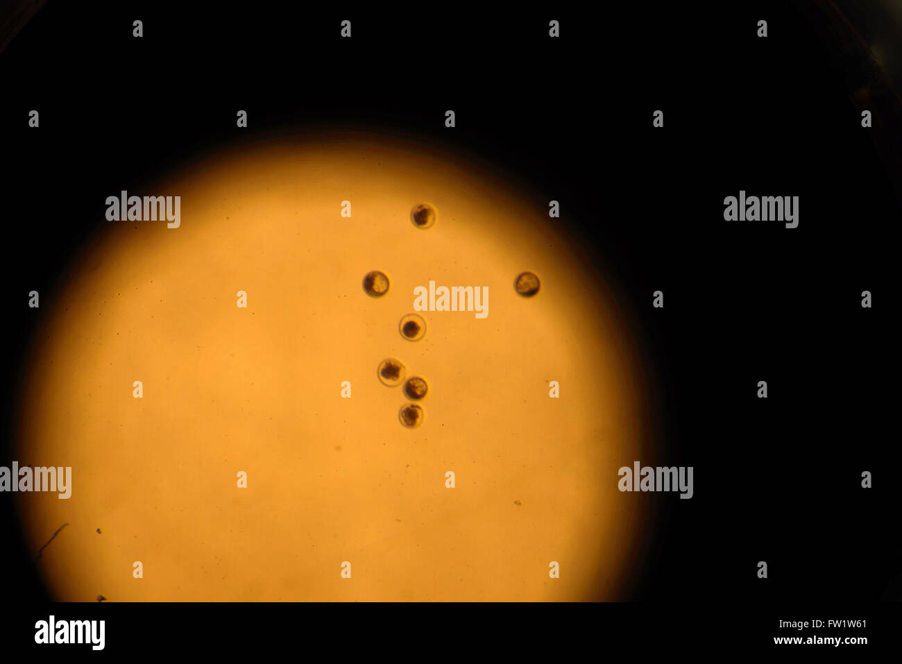 The view down a stereo microscope showing calf embryos flushed from a high-producing dairy cow as part of an artificial breeding Stock Photohttps://www.alamy.com/image-license-details/?v=1https://www.alamy.com/stock-photo-the-view-down-a-stereo-microscope-showing-calf-embryos-flushed-from-101459961.html
RMFW1W61–The view down a stereo microscope showing calf embryos flushed from a high-producing dairy cow as part of an artificial breeding
 . Fig. 25—Diagram of ovary of cow showing- comparative sizes of the follicles shown in Figs. 22, 2b, 24 The number of ovisacs rupturing at a given estrual period corresponds as a rule with the maximum number of possible fetuses. It is said that rarely two ova are contained in one ovisac. I have been unable to verify this statement, and have in all cases of twins in the cow observed two corpora lutea, sometimes both in one ovary but most frequently one in each. Sometimes a single fertilized ovum divides to con- stitute two embryos, which form identical twins in man, but this is not known to occ Stock Photohttps://www.alamy.com/image-license-details/?v=1https://www.alamy.com/fig-25diagram-of-ovary-of-cow-showing-comparative-sizes-of-the-follicles-shown-in-figs-22-2b-24-the-number-of-ovisacs-rupturing-at-a-given-estrual-period-corresponds-as-a-rule-with-the-maximum-number-of-possible-fetuses-it-is-said-that-rarely-two-ova-are-contained-in-one-ovisac-i-have-been-unable-to-verify-this-statement-and-have-in-all-cases-of-twins-in-the-cow-observed-two-corpora-lutea-sometimes-both-in-one-ovary-but-most-frequently-one-in-each-sometimes-a-single-fertilized-ovum-divides-to-con-stitute-two-embryos-which-form-identical-twins-in-man-but-this-is-not-known-to-occ-image179903869.html
RMMCK965–. Fig. 25—Diagram of ovary of cow showing- comparative sizes of the follicles shown in Figs. 22, 2b, 24 The number of ovisacs rupturing at a given estrual period corresponds as a rule with the maximum number of possible fetuses. It is said that rarely two ova are contained in one ovisac. I have been unable to verify this statement, and have in all cases of twins in the cow observed two corpora lutea, sometimes both in one ovary but most frequently one in each. Sometimes a single fertilized ovum divides to con- stitute two embryos, which form identical twins in man, but this is not known to occ
 Cattle farming filling syringe from bottle of anaesthetic for epidural into recipient cows during embryo transfer England April Stock Photohttps://www.alamy.com/image-license-details/?v=1https://www.alamy.com/cattle-farming-filling-syringe-from-bottle-of-anaesthetic-for-epidural-image61853788.html
RMDGHK3T–Cattle farming filling syringe from bottle of anaesthetic for epidural into recipient cows during embryo transfer England April
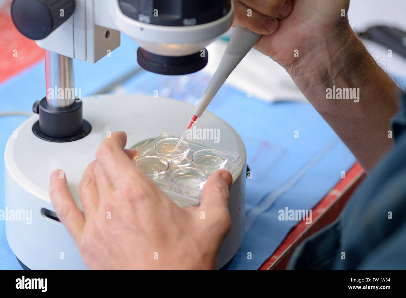 A technicianpicks up the calf embryos flushed from a high-producing dairy cow as part of an artificial breeding program, West Co Stock Photohttps://www.alamy.com/image-license-details/?v=1https://www.alamy.com/stock-photo-a-technicianpicks-up-the-calf-embryos-flushed-from-a-high-producing-101460020.html
RMFW1W84–A technicianpicks up the calf embryos flushed from a high-producing dairy cow as part of an artificial breeding program, West Co
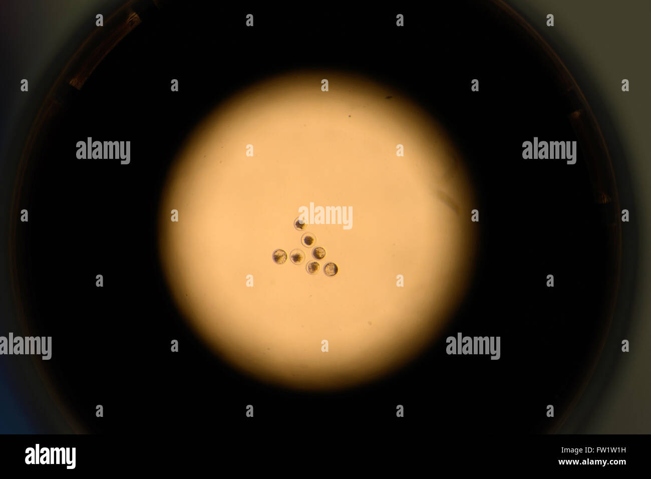 A view down a stereo microscope showing the calf embryos flushed from a high-producing dairy cow as part of an artificial breedi Stock Photohttps://www.alamy.com/image-license-details/?v=1https://www.alamy.com/stock-photo-a-view-down-a-stereo-microscope-showing-the-calf-embryos-flushed-from-101459837.html
RMFW1W1H–A view down a stereo microscope showing the calf embryos flushed from a high-producing dairy cow as part of an artificial breedi
 A technician uses a stereo microscope to locate the calf embryos flushed from a high-producing dairy cow as part of an artificia Stock Photohttps://www.alamy.com/image-license-details/?v=1https://www.alamy.com/stock-photo-a-technician-uses-a-stereo-microscope-to-locate-the-calf-embryos-flushed-101459635.html
RMFW1TPB–A technician uses a stereo microscope to locate the calf embryos flushed from a high-producing dairy cow as part of an artificia
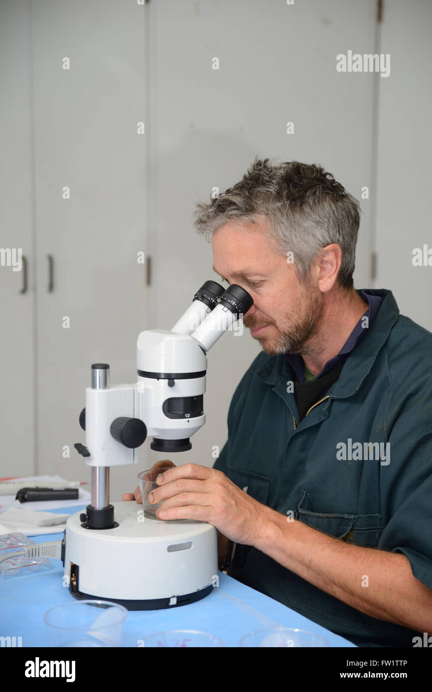 A technician uses a stereo microscope to locate the calf embryos flushed from a high-producing dairy cow as part of an artificia Stock Photohttps://www.alamy.com/image-license-details/?v=1https://www.alamy.com/stock-photo-a-technician-uses-a-stereo-microscope-to-locate-the-calf-embryos-flushed-101459702.html
RMFW1TTP–A technician uses a stereo microscope to locate the calf embryos flushed from a high-producing dairy cow as part of an artificia
 A technician uses a stereo microscope to locate the calf embryos flushed from a high-producing dairy cow as part of an artificia Stock Photohttps://www.alamy.com/image-license-details/?v=1https://www.alamy.com/stock-photo-a-technician-uses-a-stereo-microscope-to-locate-the-calf-embryos-flushed-101459770.html
RMFW1TY6–A technician uses a stereo microscope to locate the calf embryos flushed from a high-producing dairy cow as part of an artificia
 A technician uses a stereo microscope to locate the calf embryos flushed from a high-producing dairy cow as part of an artificia Stock Photohttps://www.alamy.com/image-license-details/?v=1https://www.alamy.com/stock-photo-a-technician-uses-a-stereo-microscope-to-locate-the-calf-embryos-flushed-101459480.html
RMFW1TGT–A technician uses a stereo microscope to locate the calf embryos flushed from a high-producing dairy cow as part of an artificia
 A technician flushes calf embryos from a high-producing dairy cow as part of an artificial breeding program, West Coast, New Zea Stock Photohttps://www.alamy.com/image-license-details/?v=1https://www.alamy.com/stock-photo-a-technician-flushes-calf-embryos-from-a-high-producing-dairy-cow-101458255.html
RMFW1R13–A technician flushes calf embryos from a high-producing dairy cow as part of an artificial breeding program, West Coast, New Zea
 A technician flushes calf embryos from a high-producing dairy cow as part of an artificial breeding program, West Coast, New Zea Stock Photohttps://www.alamy.com/image-license-details/?v=1https://www.alamy.com/stock-photo-a-technician-flushes-calf-embryos-from-a-high-producing-dairy-cow-101457978.html
RMFW1PK6–A technician flushes calf embryos from a high-producing dairy cow as part of an artificial breeding program, West Coast, New Zea
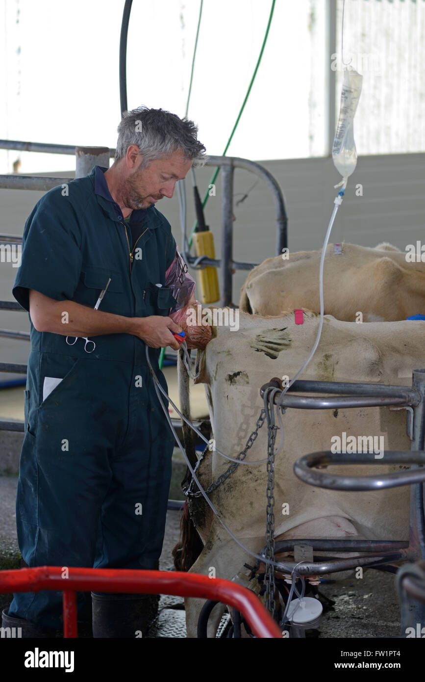 A technician flushes calf embryos from a high-producing dairy cow as part of an artificial breeding program, West Coast, New Zea Stock Photohttps://www.alamy.com/image-license-details/?v=1https://www.alamy.com/stock-photo-a-technician-flushes-calf-embryos-from-a-high-producing-dairy-cow-101458116.html
RMFW1PT4–A technician flushes calf embryos from a high-producing dairy cow as part of an artificial breeding program, West Coast, New Zea
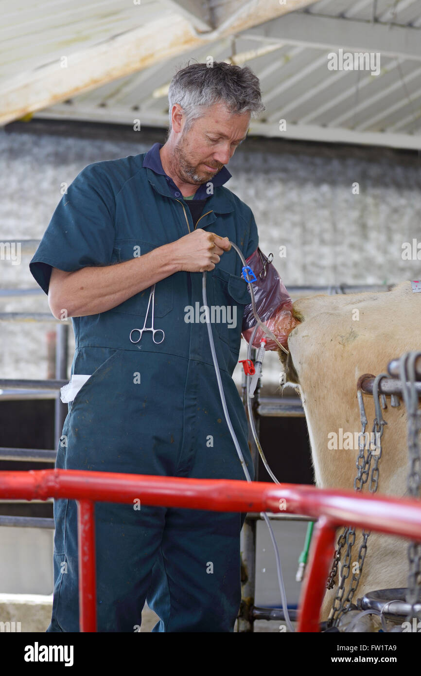 A technician uses saline solution from a drip bag to flush calf embryos from a high-producing dairy cow as part of an artificial Stock Photohttps://www.alamy.com/image-license-details/?v=1https://www.alamy.com/stock-photo-a-technician-uses-saline-solution-from-a-drip-bag-to-flush-calf-embryos-101459297.html
RMFW1TA9–A technician uses saline solution from a drip bag to flush calf embryos from a high-producing dairy cow as part of an artificial
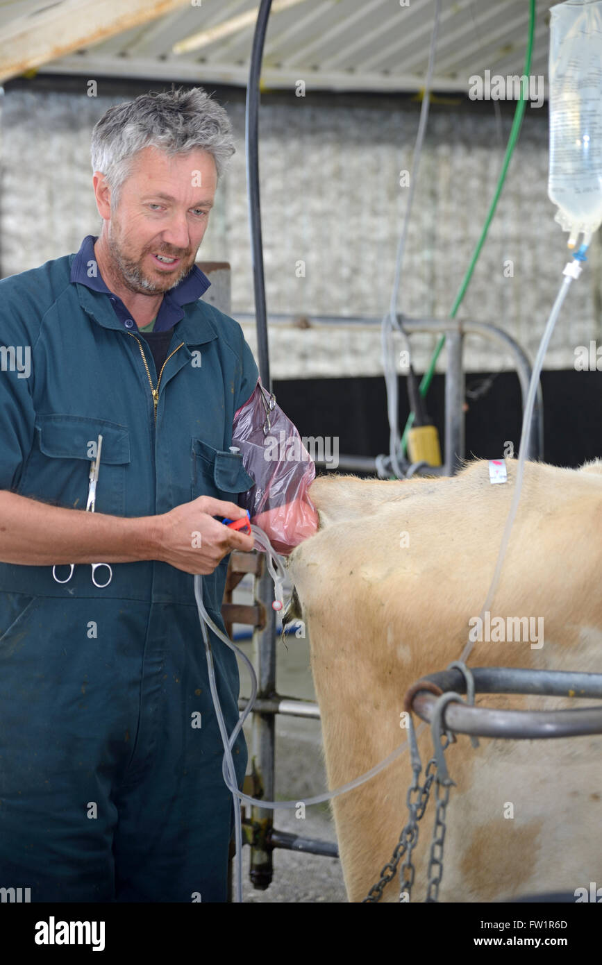 A technician uses saline solution from a drip bag to flush calf embryos from a high-producing dairy cow as part of an artificial Stock Photohttps://www.alamy.com/image-license-details/?v=1https://www.alamy.com/stock-photo-a-technician-uses-saline-solution-from-a-drip-bag-to-flush-calf-embryos-101458405.html
RMFW1R6D–A technician uses saline solution from a drip bag to flush calf embryos from a high-producing dairy cow as part of an artificial
 A technician uses saline solution from a drip bag to flush calf embryos from a high-producing dairy cow as part of an artificial Stock Photohttps://www.alamy.com/image-license-details/?v=1https://www.alamy.com/stock-photo-a-technician-uses-saline-solution-from-a-drip-bag-to-flush-calf-embryos-101458864.html
RMFW1RPT–A technician uses saline solution from a drip bag to flush calf embryos from a high-producing dairy cow as part of an artificial
 A technician uses saline solution from a drip bag to flush calf embryos from a high-producing dairy cow as part of an artificial Stock Photohttps://www.alamy.com/image-license-details/?v=1https://www.alamy.com/stock-photo-a-technician-uses-saline-solution-from-a-drip-bag-to-flush-calf-embryos-101458528.html
RMFW1RAT–A technician uses saline solution from a drip bag to flush calf embryos from a high-producing dairy cow as part of an artificial
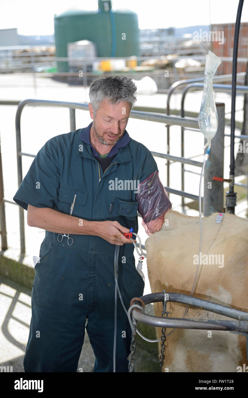 A technician uses saline solution from a drip bag to flush calf embryos from a high-producing dairy cow as part of an artificial Stock Photohttps://www.alamy.com/image-license-details/?v=1https://www.alamy.com/stock-photo-a-technician-uses-saline-solution-from-a-drip-bag-to-flush-calf-embryos-101459072.html
RMFW1T28–A technician uses saline solution from a drip bag to flush calf embryos from a high-producing dairy cow as part of an artificial
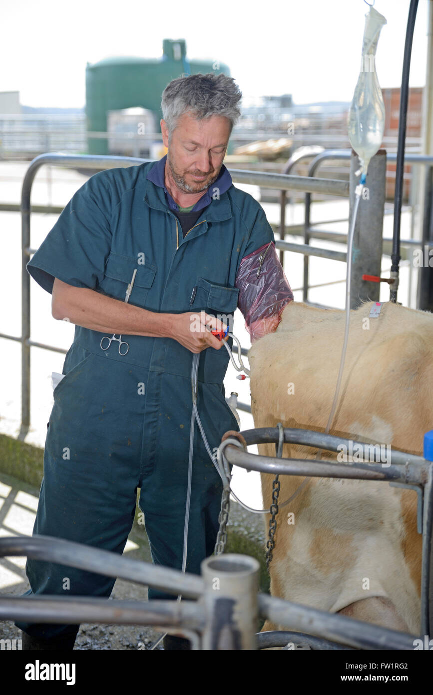 A technician uses saline solution from a drip bag to flush calf embryos from a high-producing dairy cow as part of an artificial Stock Photohttps://www.alamy.com/image-license-details/?v=1https://www.alamy.com/stock-photo-a-technician-uses-saline-solution-from-a-drip-bag-to-flush-calf-embryos-101458674.html
RMFW1RG2–A technician uses saline solution from a drip bag to flush calf embryos from a high-producing dairy cow as part of an artificial
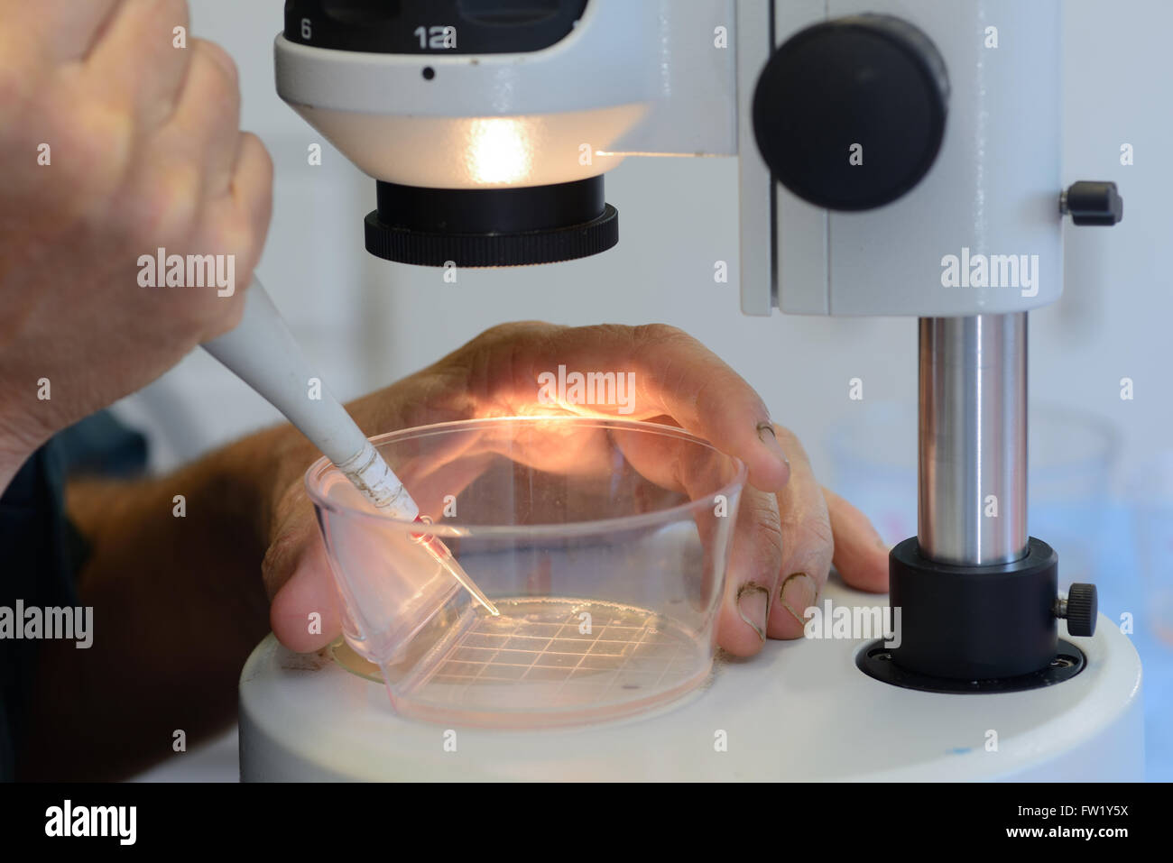 A technician steadies his hand to draw up a live calf embryo into a pipette, ready for implantation into a surrogate cow as part Stock Photohttps://www.alamy.com/image-license-details/?v=1https://www.alamy.com/stock-photo-a-technician-steadies-his-hand-to-draw-up-a-live-calf-embryo-into-101461526.html
RMFW1Y5X–A technician steadies his hand to draw up a live calf embryo into a pipette, ready for implantation into a surrogate cow as part
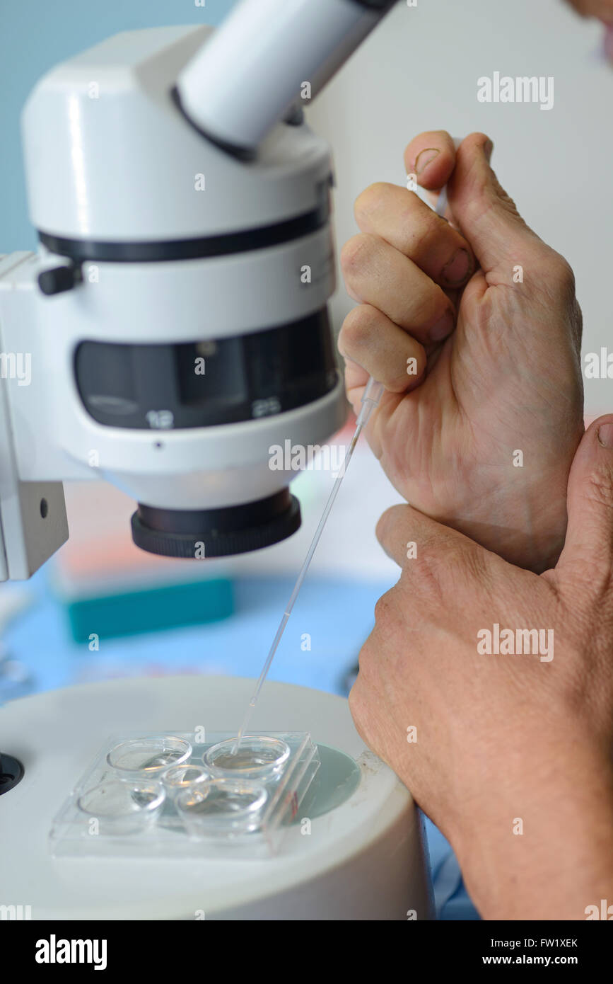 A technician steadies his hand to draw up a live calf embryo into a pipette, ready for implantation into a surrogate cow as part Stock Photohttps://www.alamy.com/image-license-details/?v=1https://www.alamy.com/stock-photo-a-technician-steadies-his-hand-to-draw-up-a-live-calf-embryo-into-101460987.html
RMFW1XEK–A technician steadies his hand to draw up a live calf embryo into a pipette, ready for implantation into a surrogate cow as part
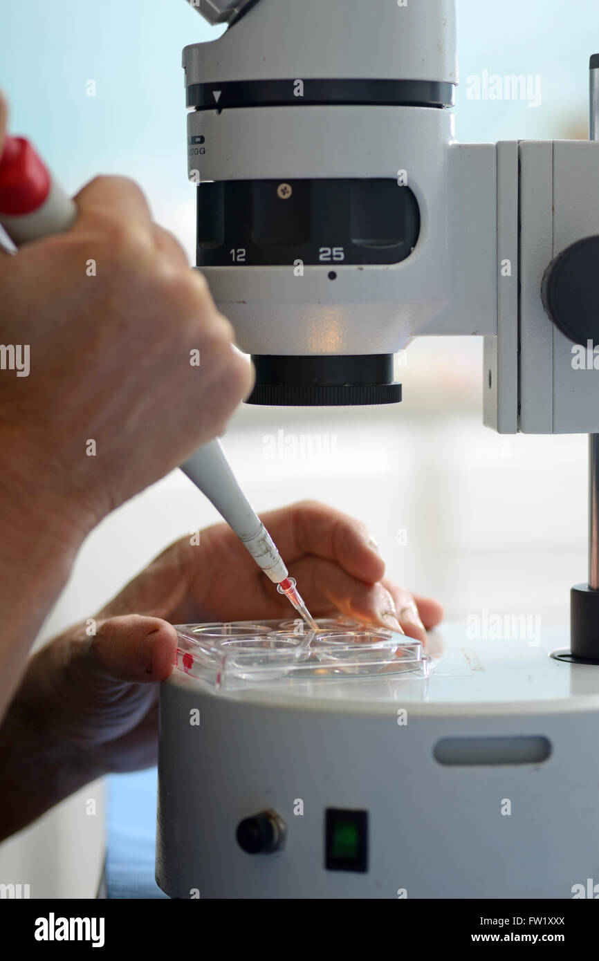 A technician draws up a live calf embryo into a pipette, ready for implantation into a surrogate cow as part of an artificial br Stock Photohttps://www.alamy.com/image-license-details/?v=1https://www.alamy.com/stock-photo-a-technician-draws-up-a-live-calf-embryo-into-a-pipette-ready-for-101461330.html
RMFW1XXX–A technician draws up a live calf embryo into a pipette, ready for implantation into a surrogate cow as part of an artificial br
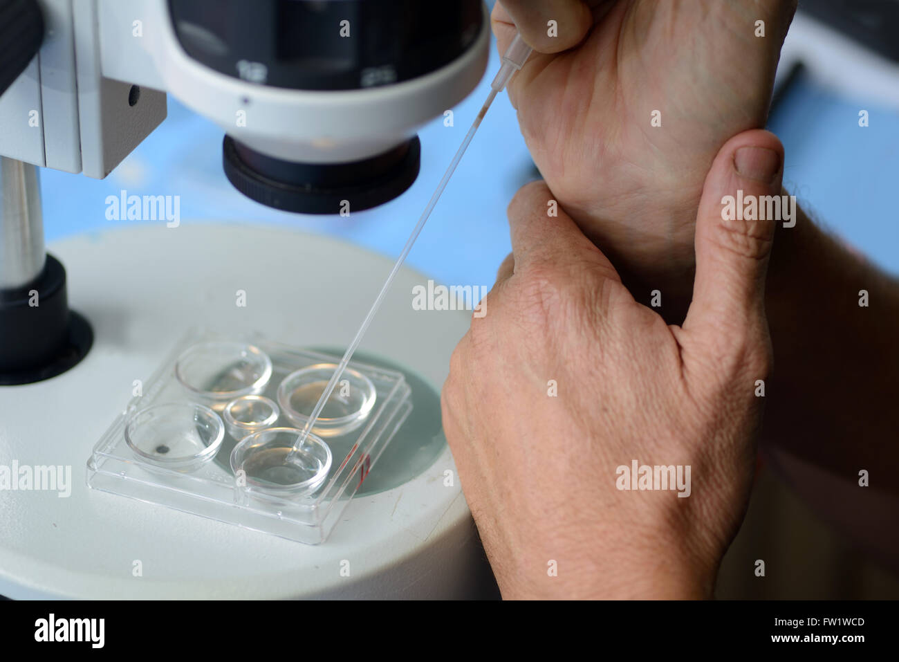 A technician draws up a live calf embryo into a pipette, ready for implantation into a surrogate cow as part of an artificial br Stock Photohttps://www.alamy.com/image-license-details/?v=1https://www.alamy.com/stock-photo-a-technician-draws-up-a-live-calf-embryo-into-a-pipette-ready-for-101460141.html
RMFW1WCD–A technician draws up a live calf embryo into a pipette, ready for implantation into a surrogate cow as part of an artificial br
 A technician steadies his hand to draw up a live calf embryo into a pipette, ready for implantation into a surrogate cow as part Stock Photohttps://www.alamy.com/image-license-details/?v=1https://www.alamy.com/stock-photo-a-technician-steadies-his-hand-to-draw-up-a-live-calf-embryo-into-101461459.html
RMFW1Y3F–A technician steadies his hand to draw up a live calf embryo into a pipette, ready for implantation into a surrogate cow as part
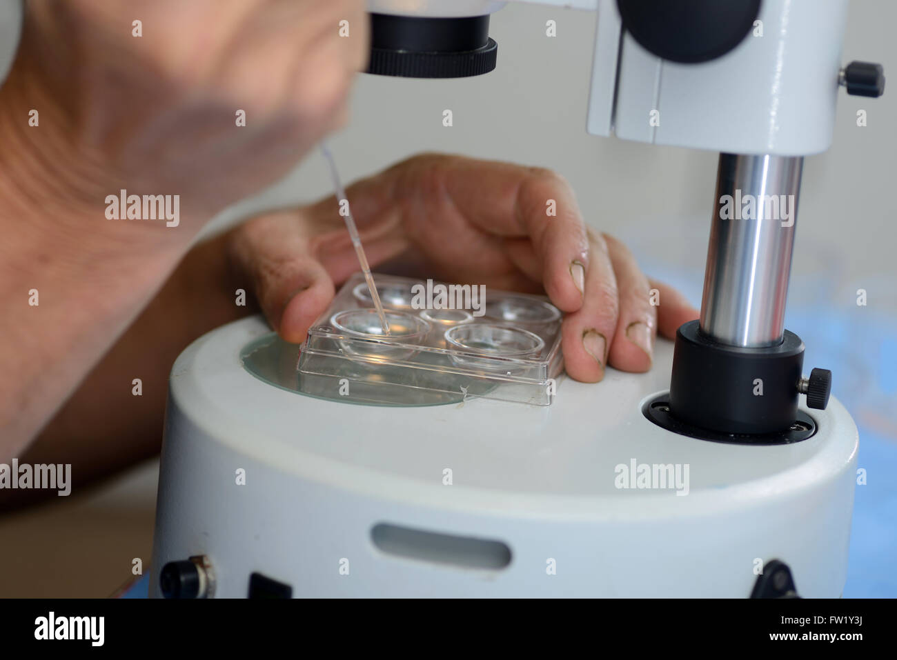 A technician draws up a live calf embryo into a pipette, ready for implantation into a surrogate cow as part of an artificial br Stock Photohttps://www.alamy.com/image-license-details/?v=1https://www.alamy.com/stock-photo-a-technician-draws-up-a-live-calf-embryo-into-a-pipette-ready-for-101461462.html
RMFW1Y3J–A technician draws up a live calf embryo into a pipette, ready for implantation into a surrogate cow as part of an artificial br
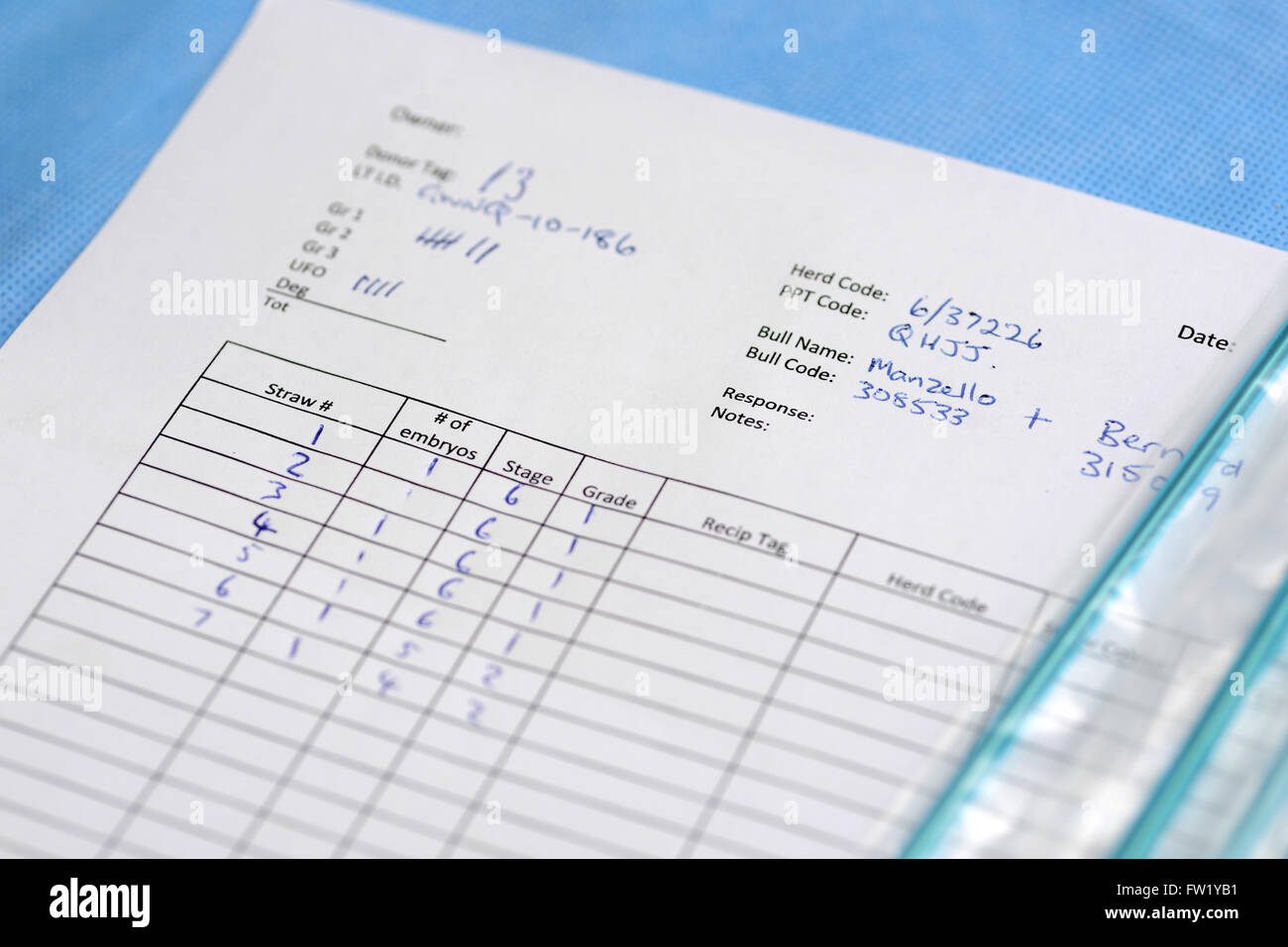 A technician records information about the farm's dairy breeding programme, West Coast, New Zealand Stock Photohttps://www.alamy.com/image-license-details/?v=1https://www.alamy.com/stock-photo-a-technician-records-information-about-the-farms-dairy-breeding-programme-101461669.html
RMFW1YB1–A technician records information about the farm's dairy breeding programme, West Coast, New Zealand
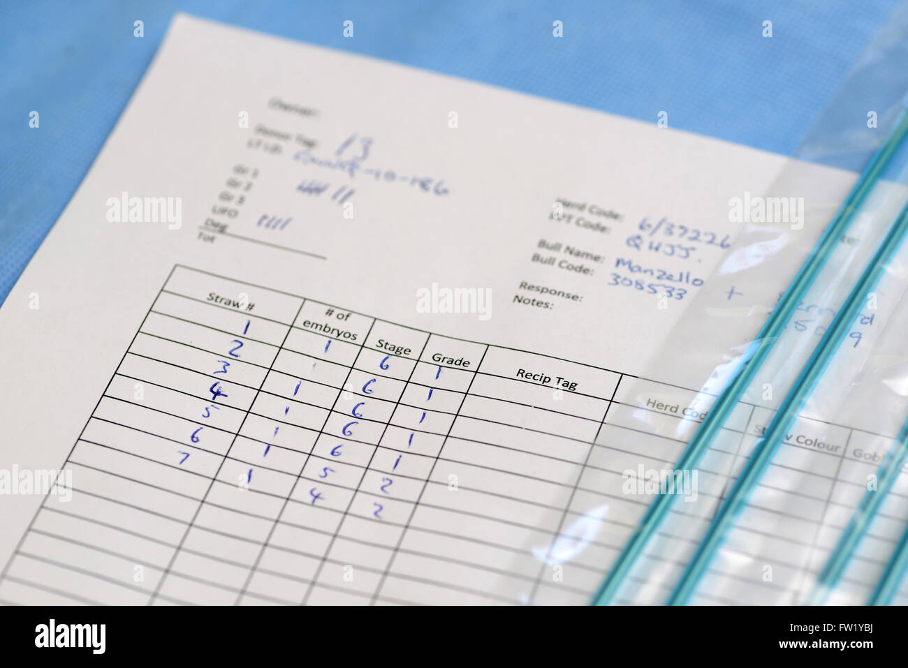 A technician records information about the farm's dairy breeding programme, West Coast, New Zealand Stock Photohttps://www.alamy.com/image-license-details/?v=1https://www.alamy.com/stock-photo-a-technician-records-information-about-the-farms-dairy-breeding-programme-101461686.html
RMFW1YBJ–A technician records information about the farm's dairy breeding programme, West Coast, New Zealand
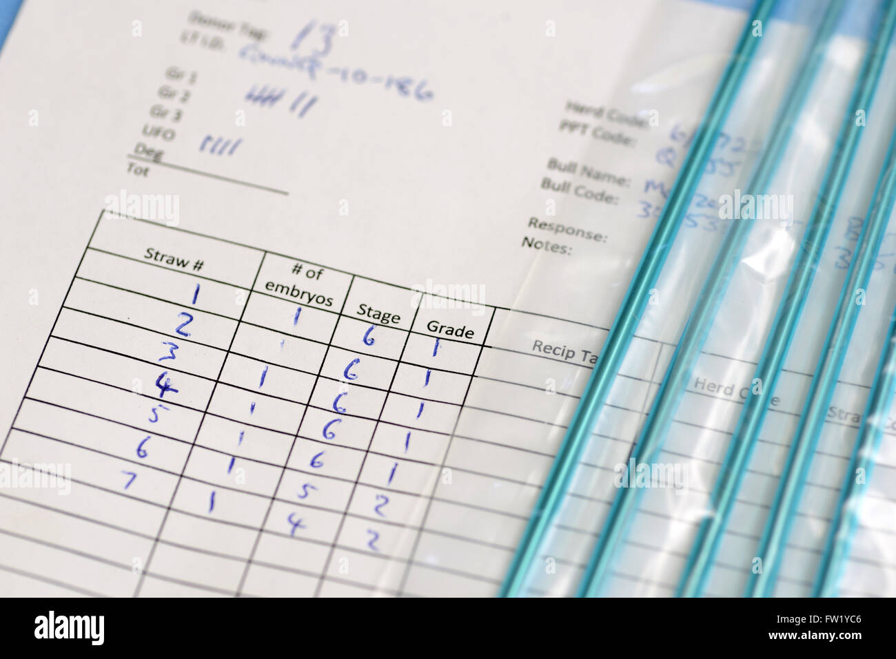 A technician records information about the farm's dairy breeding programme, West Coast, New Zealand Stock Photohttps://www.alamy.com/image-license-details/?v=1https://www.alamy.com/stock-photo-a-technician-records-information-about-the-farms-dairy-breeding-programme-101461702.html
RMFW1YC6–A technician records information about the farm's dairy breeding programme, West Coast, New Zealand