Quick filters:
Enlarged ventricles Stock Photos and Images
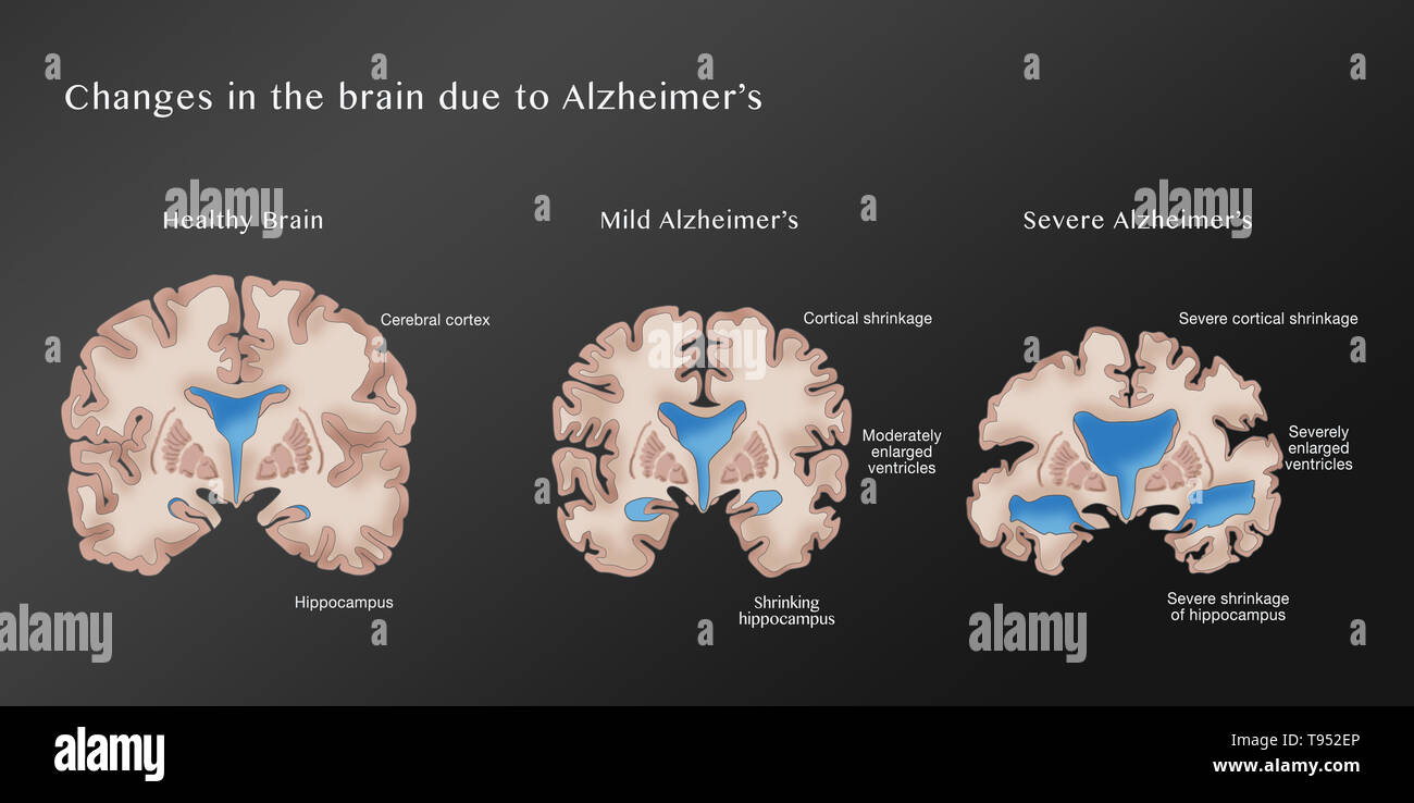 Illustration depicting the progression of Alzheimer's disease. On the left is a healthy brain. The middle brain displays cortical shrinkage, moderately enlarged ventricles, and a shrinking hippocampus, symptoms of mild Alzheimer's. The right brain shows severe cortical shrinkage, severely enlarged ventricles, and severe shrinkage of the hippocampus, indicative of severe Alzheimer's. Stock Photohttps://www.alamy.com/image-license-details/?v=1https://www.alamy.com/illustration-depicting-the-progression-of-alzheimers-disease-on-the-left-is-a-healthy-brain-the-middle-brain-displays-cortical-shrinkage-moderately-enlarged-ventricles-and-a-shrinking-hippocampus-symptoms-of-mild-alzheimers-the-right-brain-shows-severe-cortical-shrinkage-severely-enlarged-ventricles-and-severe-shrinkage-of-the-hippocampus-indicative-of-severe-alzheimers-image246588798.html
RFT952EP–Illustration depicting the progression of Alzheimer's disease. On the left is a healthy brain. The middle brain displays cortical shrinkage, moderately enlarged ventricles, and a shrinking hippocampus, symptoms of mild Alzheimer's. The right brain shows severe cortical shrinkage, severely enlarged ventricles, and severe shrinkage of the hippocampus, indicative of severe Alzheimer's.
 Enlarged ventricles of a child's brain, illustration Stock Photohttps://www.alamy.com/image-license-details/?v=1https://www.alamy.com/enlarged-ventricles-of-a-childs-brain-illustration-image572908578.html
RF2T826MJ–Enlarged ventricles of a child's brain, illustration
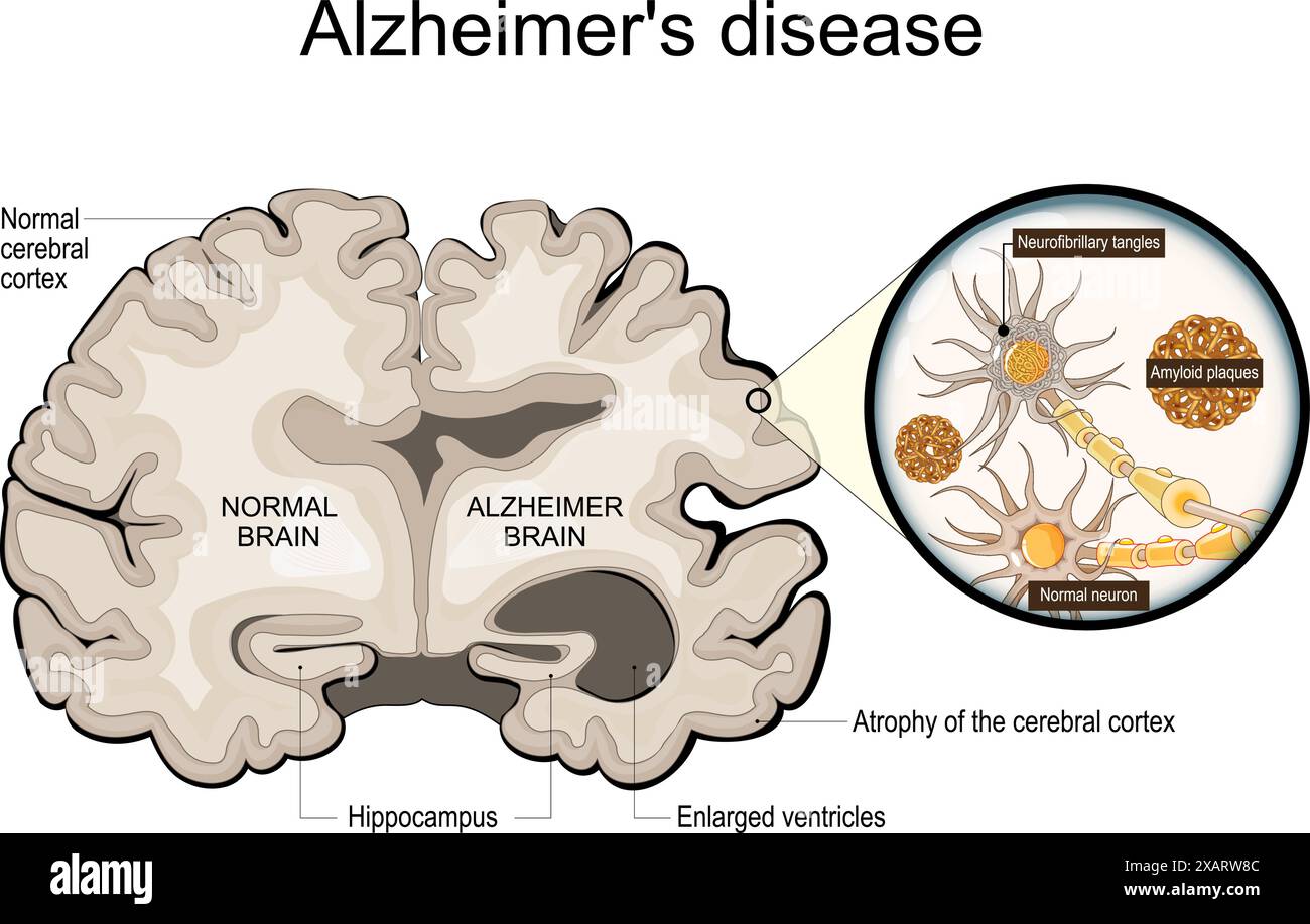 Alzheimer's disease. Neurodegeneration. Cross section of normal and Alzheimer brain, with Atrophy of the cerebral cortex, Enlarged ventricles and Hipp Stock Vectorhttps://www.alamy.com/image-license-details/?v=1https://www.alamy.com/alzheimers-disease-neurodegeneration-cross-section-of-normal-and-alzheimer-brain-with-atrophy-of-the-cerebral-cortex-enlarged-ventricles-and-hipp-image609034172.html
RF2XARW8C–Alzheimer's disease. Neurodegeneration. Cross section of normal and Alzheimer brain, with Atrophy of the cerebral cortex, Enlarged ventricles and Hipp
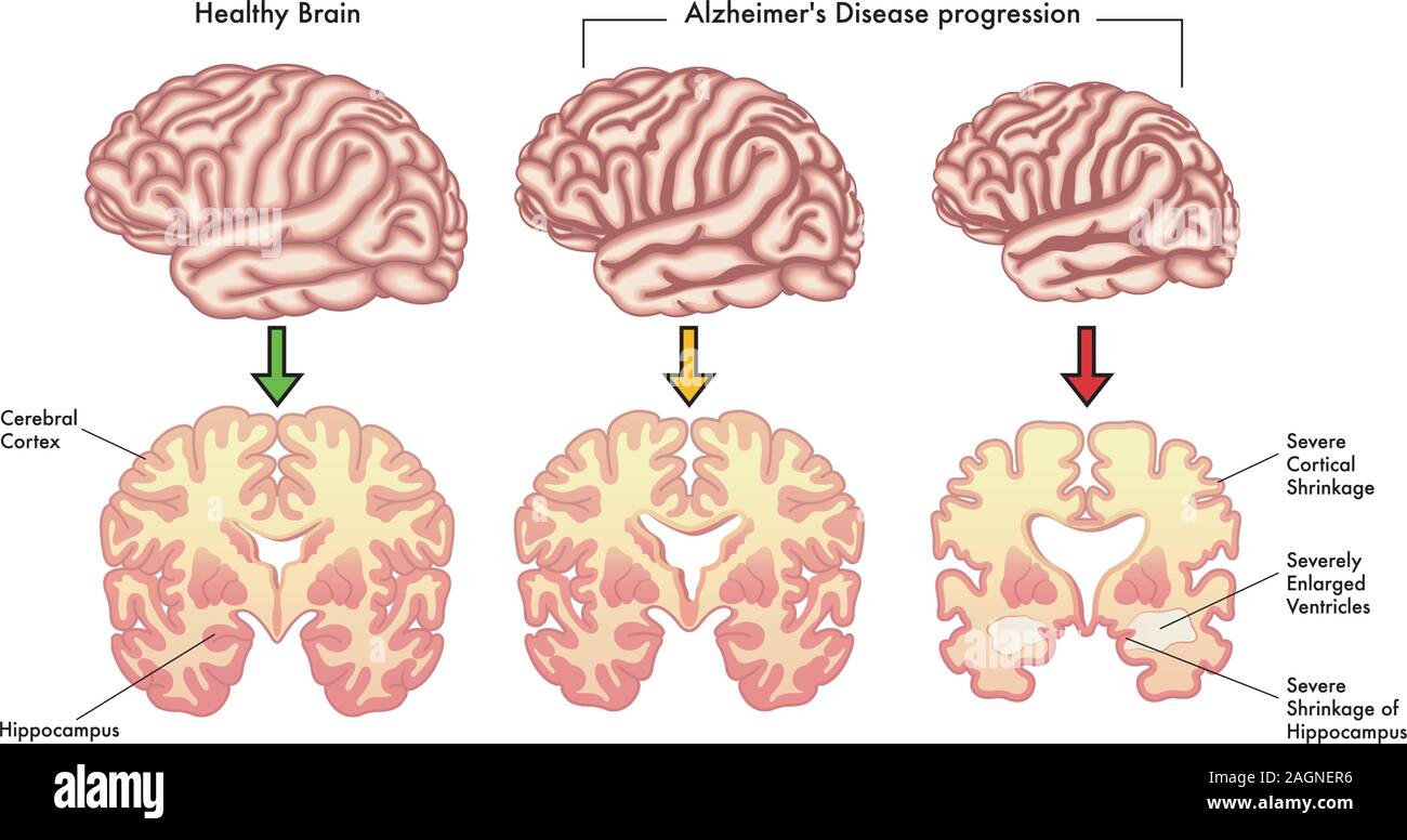 Medical illustration of the symptoms of Alzheimer's disease progression. Stock Vectorhttps://www.alamy.com/image-license-details/?v=1https://www.alamy.com/medical-illustration-of-the-symptoms-of-alzheimers-disease-progression-image337304106.html
RF2AGNER6–Medical illustration of the symptoms of Alzheimer's disease progression.
 Human heart - anterior view - showing the coronary arteries, aortic arch, left and right ventricles, left and right auricles, pulmary trunk, superior Stock Photohttps://www.alamy.com/image-license-details/?v=1https://www.alamy.com/stock-photo-human-heart-anterior-view-showing-the-coronary-arteries-aortic-arch-130806240.html
RFHGPMKC–Human heart - anterior view - showing the coronary arteries, aortic arch, left and right ventricles, left and right auricles, pulmary trunk, superior
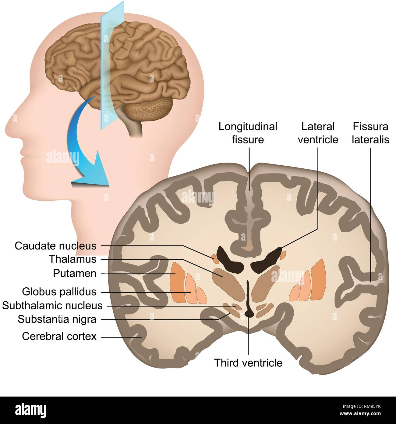 Coronal section of the human brain medical vector illustration Stock Vectorhttps://www.alamy.com/image-license-details/?v=1https://www.alamy.com/coronal-section-of-the-human-brain-medical-vector-illustration-image236208215.html
RFRM85YK–Coronal section of the human brain medical vector illustration
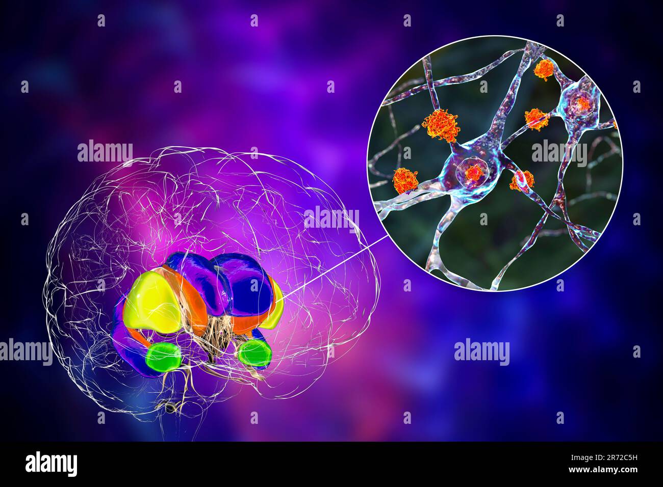 Dorsal striatum and its neurons in the Huntington's disease, computer illustration showing enlarged anterior horns of lateral ventricles, degeneration Stock Photohttps://www.alamy.com/image-license-details/?v=1https://www.alamy.com/dorsal-striatum-and-its-neurons-in-the-huntingtons-disease-computer-illustration-showing-enlarged-anterior-horns-of-lateral-ventricles-degeneration-image555087837.html
RF2R72C5H–Dorsal striatum and its neurons in the Huntington's disease, computer illustration showing enlarged anterior horns of lateral ventricles, degeneration
RF2YBMJR2–Huntington's chorea line black icon. Sign for web page, mobile app, button, logo. Vector isolated button. Editable stroke.
 The practice of obstetrics, designed for the use of students and practitioners of medicine . in thebrain or its membranes, causing enlargement of the skull. The serous effusion isgenerally confined to the ventricles, although it may be found in the intersticesof the pia mater, in the parenchyma of the cerebrum, or between the arachnoidand the dura mater. (See also Dystocia of Fetal Origin, Part V.) Frequency:This affection is rare, occurring once in about 3000 deliveries. Pathology: Theskull becomes enlarged, sometimes enormously so, by the pressure of the in-creasing quantity of the fluid, an Stock Photohttps://www.alamy.com/image-license-details/?v=1https://www.alamy.com/the-practice-of-obstetrics-designed-for-the-use-of-students-and-practitioners-of-medicine-in-thebrain-or-its-membranes-causing-enlargement-of-the-skull-the-serous-effusion-isgenerally-confined-to-the-ventricles-although-it-may-be-found-in-the-intersticesof-the-pia-mater-in-the-parenchyma-of-the-cerebrum-or-between-the-arachnoidand-the-dura-mater-see-also-dystocia-of-fetal-origin-part-v-frequencythis-affection-is-rare-occurring-once-in-about-3000-deliveries-pathology-theskull-becomes-enlarged-sometimes-enormously-so-by-the-pressure-of-the-in-creasing-quantity-of-the-fluid-an-image343292974.html
RM2AXE9KA–The practice of obstetrics, designed for the use of students and practitioners of medicine . in thebrain or its membranes, causing enlargement of the skull. The serous effusion isgenerally confined to the ventricles, although it may be found in the intersticesof the pia mater, in the parenchyma of the cerebrum, or between the arachnoidand the dura mater. (See also Dystocia of Fetal Origin, Part V.) Frequency:This affection is rare, occurring once in about 3000 deliveries. Pathology: Theskull becomes enlarged, sometimes enormously so, by the pressure of the in-creasing quantity of the fluid, an
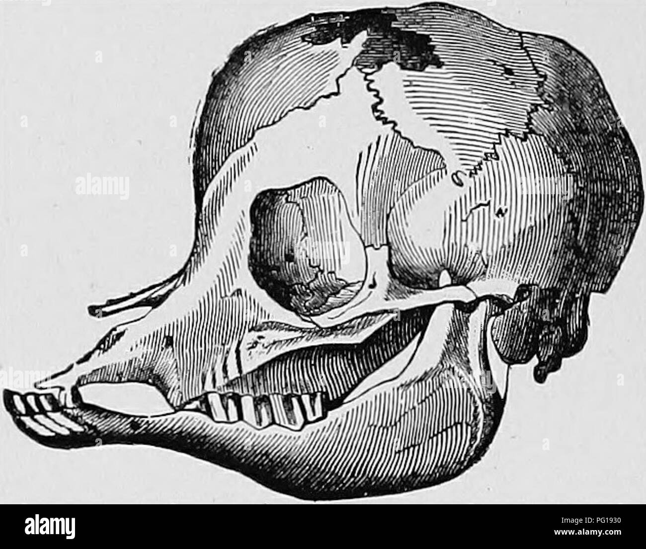 . Veterinary obstetrics; a compendium for the use of students and practitioners. Veterinary obstetrics. 88 VETERINARY OBSTETRICS. lungs being unprotected. This condition is known as. ^^Ectopia cordis." Often the foetus is born with the cranial cavity enlarged with fluid,—''Hydrocephalus"or ^'Hydrops capitis." First of all, fluid collects In the ventricles, and the diseased process may go on until. Fig. 37. Skull of Hvdrockphalic Calf: the Cranial Bonks are partially: destroyed and defective. the whole of the brain structure is displaced by fluid,, and only retained in position b Stock Photohttps://www.alamy.com/image-license-details/?v=1https://www.alamy.com/veterinary-obstetrics-a-compendium-for-the-use-of-students-and-practitioners-veterinary-obstetrics-88-veterinary-obstetrics-lungs-being-unprotected-this-condition-is-known-as-ectopia-cordisquot-often-the-foetus-is-born-with-the-cranial-cavity-enlarged-with-fluidhydrocephalusquotor-hydrops-capitisquot-first-of-all-fluid-collects-in-the-ventricles-and-the-diseased-process-may-go-on-until-fig-37-skull-of-hvdrockphalic-calf-the-cranial-bonks-are-partially-destroyed-and-defective-the-whole-of-the-brain-structure-is-displaced-by-fluid-and-only-retained-in-position-b-image216388004.html
RMPG1930–. Veterinary obstetrics; a compendium for the use of students and practitioners. Veterinary obstetrics. 88 VETERINARY OBSTETRICS. lungs being unprotected. This condition is known as. ^^Ectopia cordis." Often the foetus is born with the cranial cavity enlarged with fluid,—''Hydrocephalus"or ^'Hydrops capitis." First of all, fluid collects In the ventricles, and the diseased process may go on until. Fig. 37. Skull of Hvdrockphalic Calf: the Cranial Bonks are partially: destroyed and defective. the whole of the brain structure is displaced by fluid,, and only retained in position b
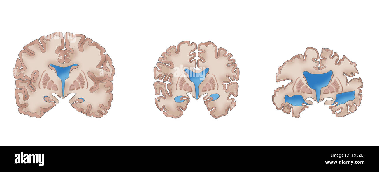 Illustration depicting the progression of Alzheimer's disease. On the left is a healthy brain. The middle brain displays cortical shrinkage, moderately enlarged ventricles, and a shrinking hippocampus, symptoms of mild Alzheimer's. The right brain shows severe cortical shrinkage, severely enlarged ventricles, and severe shrinkage of the hippocampus, indicative of severe Alzheimer's. Stock Photohttps://www.alamy.com/image-license-details/?v=1https://www.alamy.com/illustration-depicting-the-progression-of-alzheimers-disease-on-the-left-is-a-healthy-brain-the-middle-brain-displays-cortical-shrinkage-moderately-enlarged-ventricles-and-a-shrinking-hippocampus-symptoms-of-mild-alzheimers-the-right-brain-shows-severe-cortical-shrinkage-severely-enlarged-ventricles-and-severe-shrinkage-of-the-hippocampus-indicative-of-severe-alzheimers-image246588794.html
RFT952EJ–Illustration depicting the progression of Alzheimer's disease. On the left is a healthy brain. The middle brain displays cortical shrinkage, moderately enlarged ventricles, and a shrinking hippocampus, symptoms of mild Alzheimer's. The right brain shows severe cortical shrinkage, severely enlarged ventricles, and severe shrinkage of the hippocampus, indicative of severe Alzheimer's.
 Enlarged ventricles of a child's brain, illustration Stock Photohttps://www.alamy.com/image-license-details/?v=1https://www.alamy.com/enlarged-ventricles-of-a-childs-brain-illustration-image572908571.html
RF2T826MB–Enlarged ventricles of a child's brain, illustration
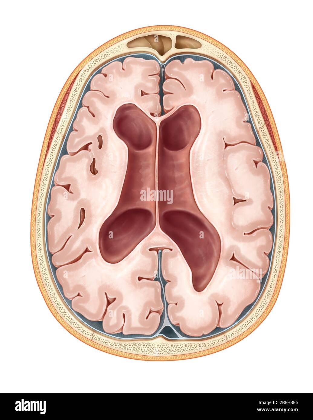 MRI Communicating Hydrocephalus (NPH) Stock Photohttps://www.alamy.com/image-license-details/?v=1https://www.alamy.com/mri-communicating-hydrocephalus-nph-image353194750.html
RM2BEHBE6–MRI Communicating Hydrocephalus (NPH)
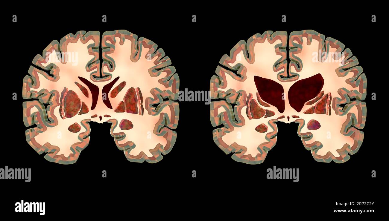 Coronal sections of a healthy brain and a brain in Huntington's disease showing enlarged anterior horns of the lateral ventricles, degeneration and at Stock Photohttps://www.alamy.com/image-license-details/?v=1https://www.alamy.com/coronal-sections-of-a-healthy-brain-and-a-brain-in-huntingtons-disease-showing-enlarged-anterior-horns-of-the-lateral-ventricles-degeneration-and-at-image555087763.html
RF2R72C2Y–Coronal sections of a healthy brain and a brain in Huntington's disease showing enlarged anterior horns of the lateral ventricles, degeneration and at
 American journal of ophthalmology. . ht, and the left eye did notturn in and down with the right; otherwise, motions of eyesnormal. The pupils were small, the left angle of the mouthparetic, the tongue turned to the left. There was slight rigidityof all four limbs, and the right were paralyzed. At autopsythe vessels were found contorted, the ventricles enlarged, thethe ependyma thickened, and a pair of small cysts the size ofpeas in the thalami. Also in the upper half of the pons anirregular haemorrhage, larger than a hazel-nut, in the regionof the left seventh nucleus, which spared the lower Stock Photohttps://www.alamy.com/image-license-details/?v=1https://www.alamy.com/american-journal-of-ophthalmology-ht-and-the-left-eye-did-notturn-in-and-down-with-the-right-otherwise-motions-of-eyesnormal-the-pupils-were-small-the-left-angle-of-the-mouthparetic-the-tongue-turned-to-the-left-there-was-slight-rigidityof-all-four-limbs-and-the-right-were-paralyzed-at-autopsythe-vessels-were-found-contorted-the-ventricles-enlarged-thethe-ependyma-thickened-and-a-pair-of-small-cysts-the-size-ofpeas-in-the-thalami-also-in-the-upper-half-of-the-pons-anirregular-haemorrhage-larger-than-a-hazel-nut-in-the-regionof-the-left-seventh-nucleus-which-spared-the-lower-image342974503.html
RM2AWYRDB–American journal of ophthalmology. . ht, and the left eye did notturn in and down with the right; otherwise, motions of eyesnormal. The pupils were small, the left angle of the mouthparetic, the tongue turned to the left. There was slight rigidityof all four limbs, and the right were paralyzed. At autopsythe vessels were found contorted, the ventricles enlarged, thethe ependyma thickened, and a pair of small cysts the size ofpeas in the thalami. Also in the upper half of the pons anirregular haemorrhage, larger than a hazel-nut, in the regionof the left seventh nucleus, which spared the lower
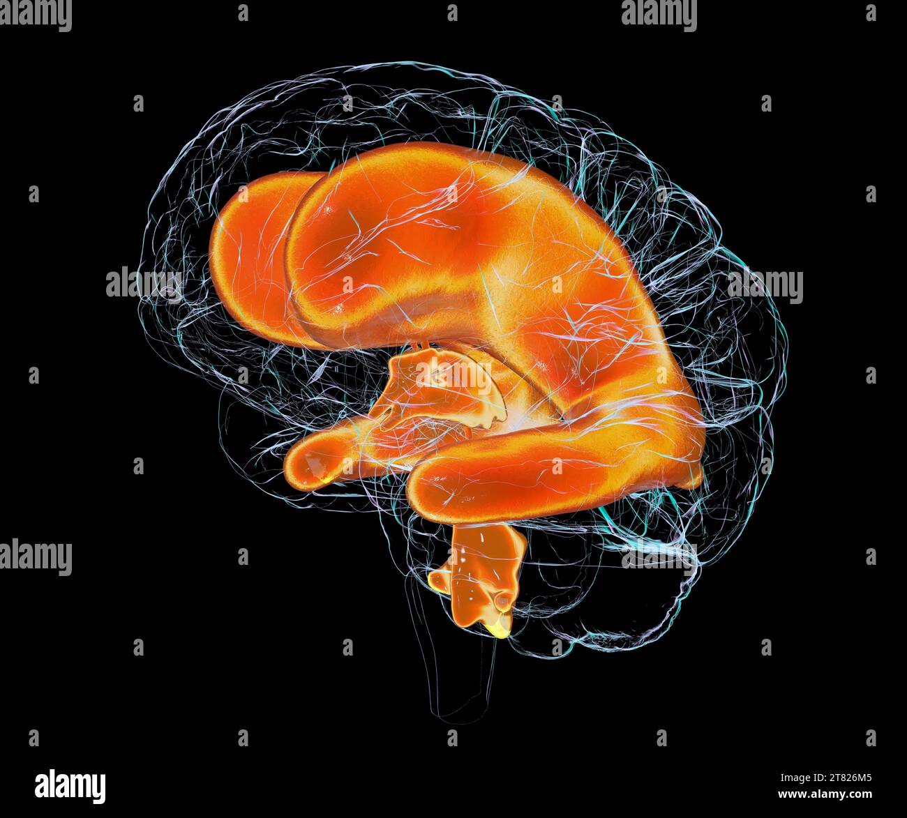 Enlarged ventricles of a child's brain, illustration Stock Photohttps://www.alamy.com/image-license-details/?v=1https://www.alamy.com/enlarged-ventricles-of-a-childs-brain-illustration-image572908565.html
RF2T826M5–Enlarged ventricles of a child's brain, illustration
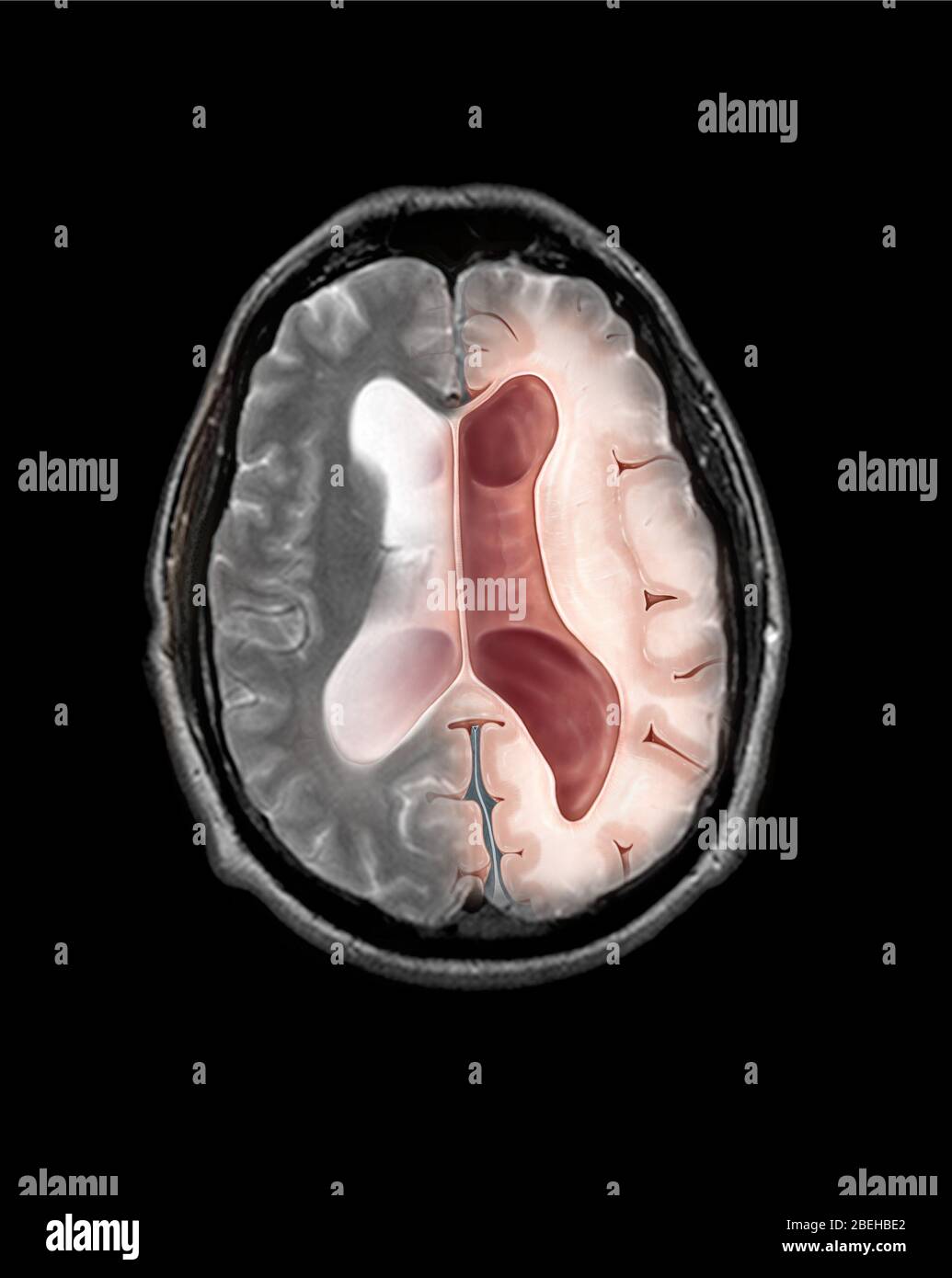 MRI Communicating Hydrocephalus (NPH) Stock Photohttps://www.alamy.com/image-license-details/?v=1https://www.alamy.com/mri-communicating-hydrocephalus-nph-image353194746.html
RM2BEHBE2–MRI Communicating Hydrocephalus (NPH)
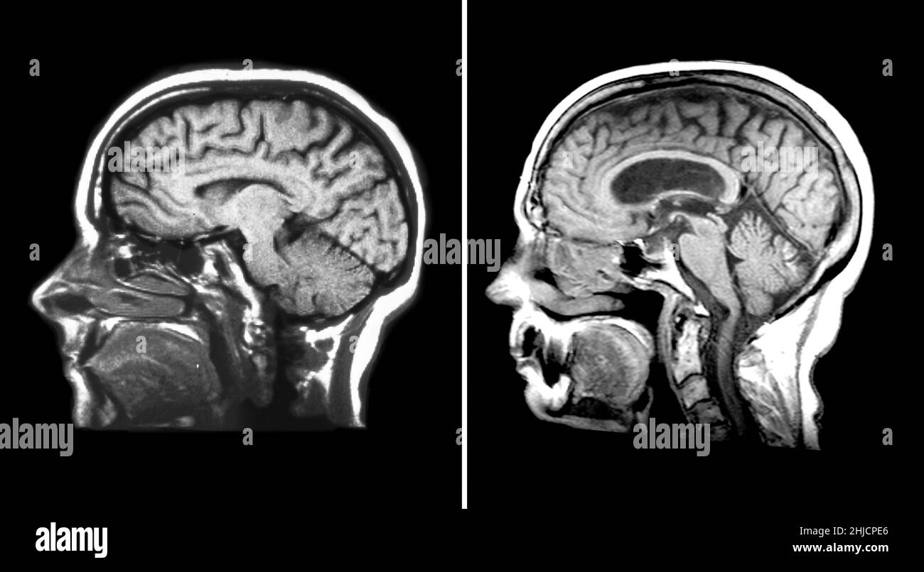 One the left is a sagittal (from the side) T1 weighted MRI showing normal anatomy of the brain. On the right is an MRI image of the brain showing enlargement of the lateral ventricles, a condition called hydrocephalus. This abnormality was treated by placing a ventricular shunt catheter into the enlarged lateral ventricles to divert the fluid away from the head. Stock Photohttps://www.alamy.com/image-license-details/?v=1https://www.alamy.com/one-the-left-is-a-sagittal-from-the-side-t1-weighted-mri-showing-normal-anatomy-of-the-brain-on-the-right-is-an-mri-image-of-the-brain-showing-enlargement-of-the-lateral-ventricles-a-condition-called-hydrocephalus-this-abnormality-was-treated-by-placing-a-ventricular-shunt-catheter-into-the-enlarged-lateral-ventricles-to-divert-the-fluid-away-from-the-head-image458814446.html
RM2HJCPE6–One the left is a sagittal (from the side) T1 weighted MRI showing normal anatomy of the brain. On the right is an MRI image of the brain showing enlargement of the lateral ventricles, a condition called hydrocephalus. This abnormality was treated by placing a ventricular shunt catheter into the enlarged lateral ventricles to divert the fluid away from the head.
 . Insanity, its classification, diagnosis, and treatment; a manual for students and practitioners of medicine. found most marked over those areas exhibiting cysticdegeneration. Probably the dilatations of the lymph spacein the posterior fissure of the spinal cord (Fig. 7), found inone case by the writer, are susceptible of a similar inter-pretation. The ventricles may be enlarged in advanced cases; oftenthey exhibit no change in dimensions, and a more charac-teristic pathological feature of the disease is the granularchange of their endyma or lining membrane.* This con- the surface, which is r Stock Photohttps://www.alamy.com/image-license-details/?v=1https://www.alamy.com/insanity-its-classification-diagnosis-and-treatment-a-manual-for-students-and-practitioners-of-medicine-found-most-marked-over-those-areas-exhibiting-cysticdegeneration-probably-the-dilatations-of-the-lymph-spacein-the-posterior-fissure-of-the-spinal-cord-fig-7-found-inone-case-by-the-writer-are-susceptible-of-a-similar-inter-pretation-the-ventricles-may-be-enlarged-in-advanced-cases-oftenthey-exhibit-no-change-in-dimensions-and-a-more-charac-teristic-pathological-feature-of-the-disease-is-the-granularchange-of-their-endyma-or-lining-membrane-this-con-the-surface-which-is-r-image336735756.html
RM2AFRHW0–. Insanity, its classification, diagnosis, and treatment; a manual for students and practitioners of medicine. found most marked over those areas exhibiting cysticdegeneration. Probably the dilatations of the lymph spacein the posterior fissure of the spinal cord (Fig. 7), found inone case by the writer, are susceptible of a similar inter-pretation. The ventricles may be enlarged in advanced cases; oftenthey exhibit no change in dimensions, and a more charac-teristic pathological feature of the disease is the granularchange of their endyma or lining membrane.* This con- the surface, which is r
 Baby with enlarged lateral ventricles, illustration Stock Photohttps://www.alamy.com/image-license-details/?v=1https://www.alamy.com/baby-with-enlarged-lateral-ventricles-illustration-image572908521.html
RF2T826JH–Baby with enlarged lateral ventricles, illustration
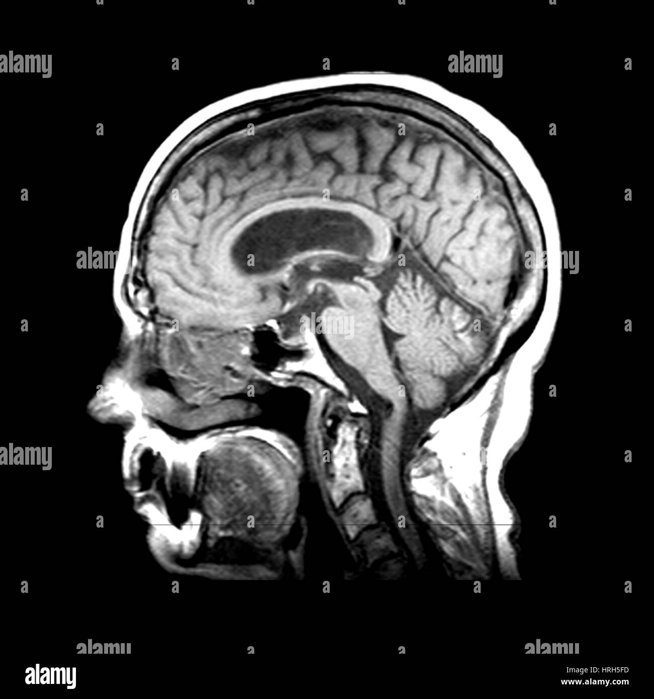 Hydrocephalus Stock Photohttps://www.alamy.com/image-license-details/?v=1https://www.alamy.com/stock-photo-hydrocephalus-134987201.html
RMHRH5FD–Hydrocephalus
 . The diseases of children : medical and surgical. compress the straightsinus which runs along at the base of the falx cerebri, and consequentlycheck the onward flow of blood in the veins of Galen and inferior longitudinalsinus. As the veins of Galen return the blood of the choroid plexus, it iseasy to understand how a chronic hydrocephalus may be thus produced.In these cases the lateral ventricles are distended with a clear fluid of lowspecific gravity, the third and fourth ventricles join in the dilatation, and theiter is also enlarged. In another class of case which occurs after a basalmeni Stock Photohttps://www.alamy.com/image-license-details/?v=1https://www.alamy.com/the-diseases-of-children-medical-and-surgical-compress-the-straightsinus-which-runs-along-at-the-base-of-the-falx-cerebri-and-consequentlycheck-the-onward-flow-of-blood-in-the-veins-of-galen-and-inferior-longitudinalsinus-as-the-veins-of-galen-return-the-blood-of-the-choroid-plexus-it-iseasy-to-understand-how-a-chronic-hydrocephalus-may-be-thus-producedin-these-cases-the-lateral-ventricles-are-distended-with-a-clear-fluid-of-lowspecific-gravity-the-third-and-fourth-ventricles-join-in-the-dilatation-and-theiter-is-also-enlarged-in-another-class-of-case-which-occurs-after-a-basalmeni-image337025327.html
RM2AG8R6R–. The diseases of children : medical and surgical. compress the straightsinus which runs along at the base of the falx cerebri, and consequentlycheck the onward flow of blood in the veins of Galen and inferior longitudinalsinus. As the veins of Galen return the blood of the choroid plexus, it iseasy to understand how a chronic hydrocephalus may be thus produced.In these cases the lateral ventricles are distended with a clear fluid of lowspecific gravity, the third and fourth ventricles join in the dilatation, and theiter is also enlarged. In another class of case which occurs after a basalmeni
 Baby with enlarged lateral ventricles, illustration Stock Photohttps://www.alamy.com/image-license-details/?v=1https://www.alamy.com/baby-with-enlarged-lateral-ventricles-illustration-image572908535.html
RF2T826K3–Baby with enlarged lateral ventricles, illustration
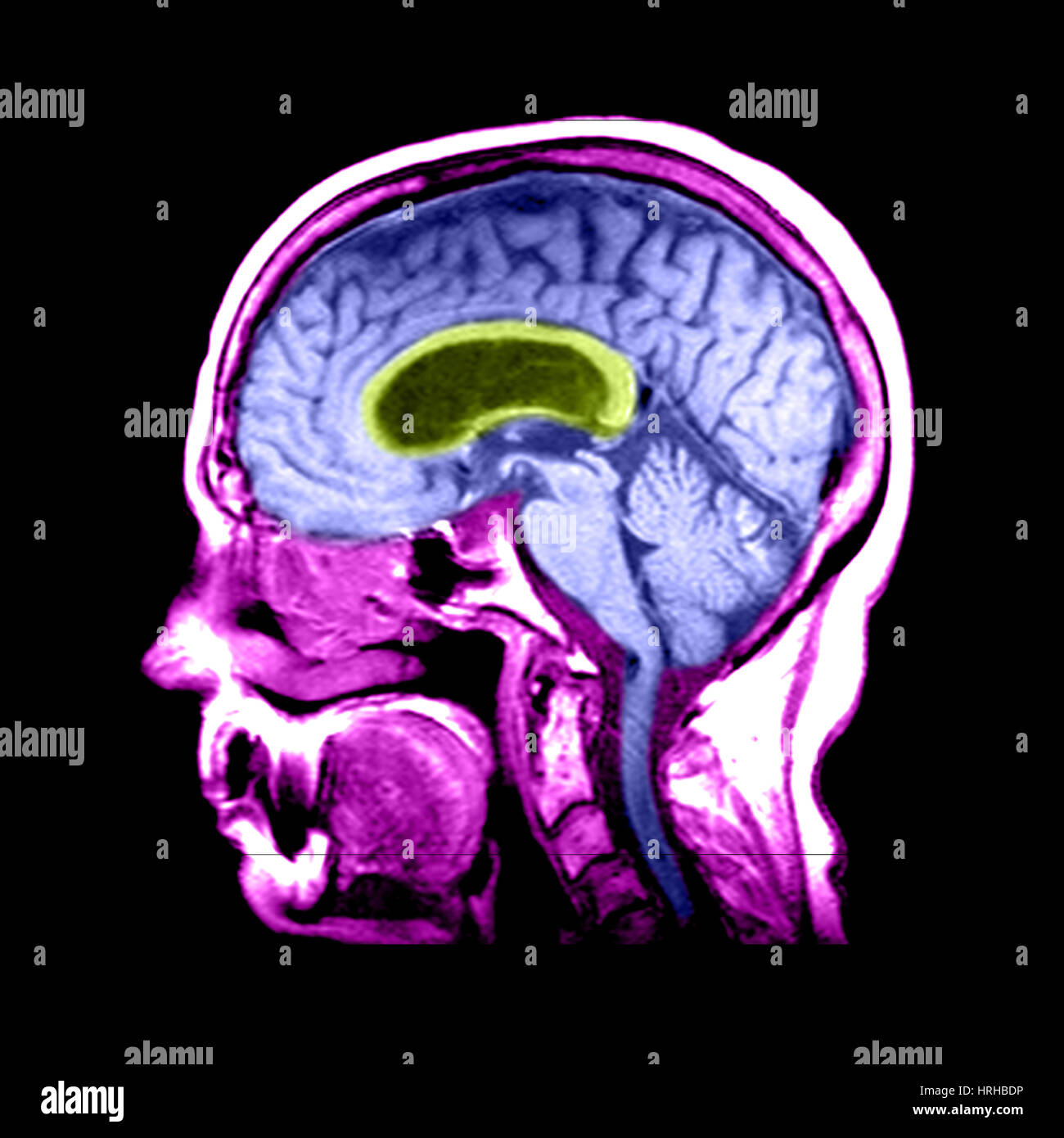 Hydrocephalus Stock Photohttps://www.alamy.com/image-license-details/?v=1https://www.alamy.com/stock-photo-hydrocephalus-134991858.html
RMHRHBDP–Hydrocephalus
 The physical signs of cardiac disease, for the use of clinical students . Increase to the right = enlargement of the right auricle. (3.) Increase to the left = dilatation of the left ventricle. (Butgreat enlargement of the right ventricle ultimately leads to thesame result).* • In caHes in which emphysema seems to be the primary mischief, it is no uncommonthing to find both riglit and left ventricles of the heart enlarged. It seems more likely,in these cases, that there is a degenerative tendency vi^hich has manifested itself in theheart as well as in the lungs tiian that the dilatation of tli Stock Photohttps://www.alamy.com/image-license-details/?v=1https://www.alamy.com/the-physical-signs-of-cardiac-disease-for-the-use-of-clinical-students-increase-to-the-right-=-enlargement-of-the-right-auricle-3-increase-to-the-left-=-dilatation-of-the-left-ventricle-butgreat-enlargement-of-the-right-ventricle-ultimately-leads-to-thesame-result-in-cahes-in-which-emphysema-seems-to-be-the-primary-mischief-it-is-no-uncommonthing-to-find-both-riglit-and-left-ventricles-of-the-heart-enlarged-it-seems-more-likelyin-these-cases-that-there-is-a-degenerative-tendency-vihich-has-manifested-itself-in-theheart-as-well-as-in-the-lungs-tiian-that-the-dilatation-of-tli-image338481352.html
RM2AJK4BM–The physical signs of cardiac disease, for the use of clinical students . Increase to the right = enlargement of the right auricle. (3.) Increase to the left = dilatation of the left ventricle. (Butgreat enlargement of the right ventricle ultimately leads to thesame result).* • In caHes in which emphysema seems to be the primary mischief, it is no uncommonthing to find both riglit and left ventricles of the heart enlarged. It seems more likely,in these cases, that there is a degenerative tendency vi^hich has manifested itself in theheart as well as in the lungs tiian that the dilatation of tli
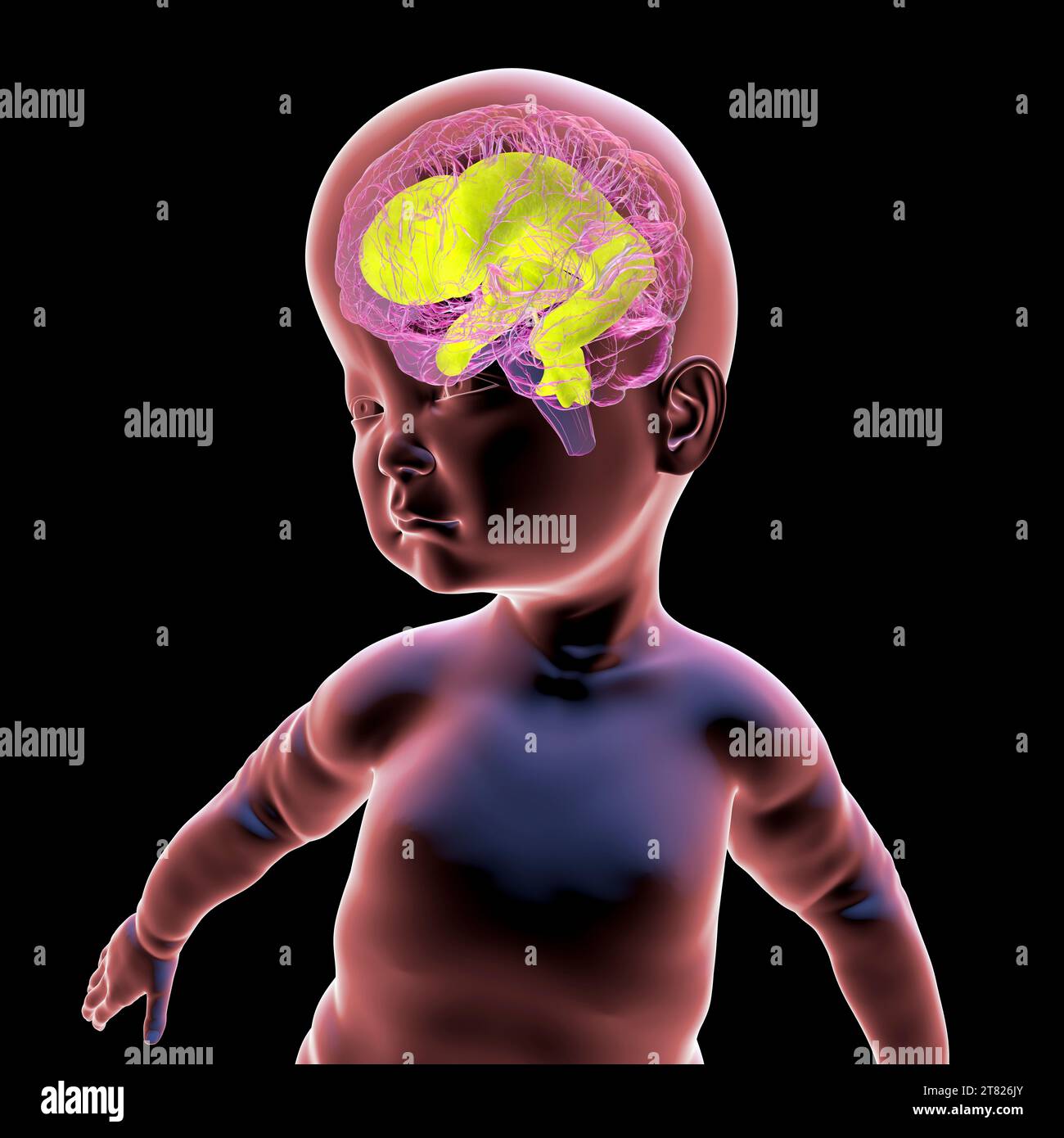 Baby with enlarged brain ventricles, illustration Stock Photohttps://www.alamy.com/image-license-details/?v=1https://www.alamy.com/baby-with-enlarged-brain-ventricles-illustration-image572908531.html
RF2T826JY–Baby with enlarged brain ventricles, illustration
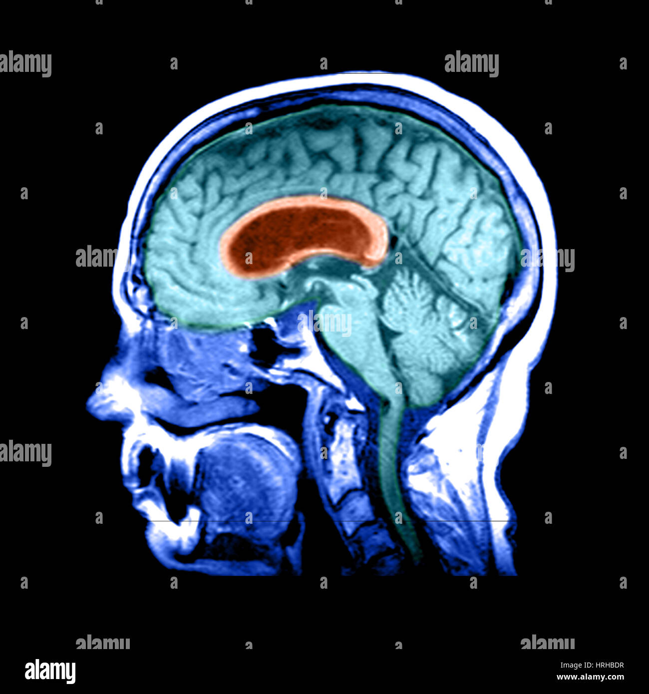 Hydrocephalus Stock Photohttps://www.alamy.com/image-license-details/?v=1https://www.alamy.com/stock-photo-hydrocephalus-134991859.html
RMHRHBDR–Hydrocephalus
 Quain's elements of anatomy . ted behind withthe fornix, a structure to be presently described, and in the rest of itslength with the septum lucidum, a vertical partition between the twolateral ventricles, which is included in the anterior bend of the corpuscallosum. On the sides the corpus callosum roofs in the body andanterior comu of the lateral ventricles. The enlarged posterior part orsplenium lies over the mesencephalon, with pia mater between. Fig. 300. — View OF THE CORPUSCALLOSUM FROM ABOVE (fromSappey after Fo-Yille). 4 The upper sur-face of the corpuscallosum has beenfully exposed b Stock Photohttps://www.alamy.com/image-license-details/?v=1https://www.alamy.com/quains-elements-of-anatomy-ted-behind-withthe-fornix-a-structure-to-be-presently-described-and-in-the-rest-of-itslength-with-the-septum-lucidum-a-vertical-partition-between-the-twolateral-ventricles-which-is-included-in-the-anterior-bend-of-the-corpuscallosum-on-the-sides-the-corpus-callosum-roofs-in-the-body-andanterior-comu-of-the-lateral-ventricles-the-enlarged-posterior-part-orsplenium-lies-over-the-mesencephalon-with-pia-mater-between-fig-300-view-of-the-corpuscallosum-from-above-fromsappey-after-fo-yille-4-the-upper-sur-face-of-the-corpuscallosum-has-beenfully-exposed-b-image342738496.html
RM2AWH2CG–Quain's elements of anatomy . ted behind withthe fornix, a structure to be presently described, and in the rest of itslength with the septum lucidum, a vertical partition between the twolateral ventricles, which is included in the anterior bend of the corpuscallosum. On the sides the corpus callosum roofs in the body andanterior comu of the lateral ventricles. The enlarged posterior part orsplenium lies over the mesencephalon, with pia mater between. Fig. 300. — View OF THE CORPUSCALLOSUM FROM ABOVE (fromSappey after Fo-Yille). 4 The upper sur-face of the corpuscallosum has beenfully exposed b
 Baby with enlarged lateral ventricles, illustration Stock Photohttps://www.alamy.com/image-license-details/?v=1https://www.alamy.com/baby-with-enlarged-lateral-ventricles-illustration-image572908516.html
RF2T826JC–Baby with enlarged lateral ventricles, illustration
 Hydrocephalus Stock Photohttps://www.alamy.com/image-license-details/?v=1https://www.alamy.com/stock-photo-hydrocephalus-134991861.html
RMHRHBDW–Hydrocephalus
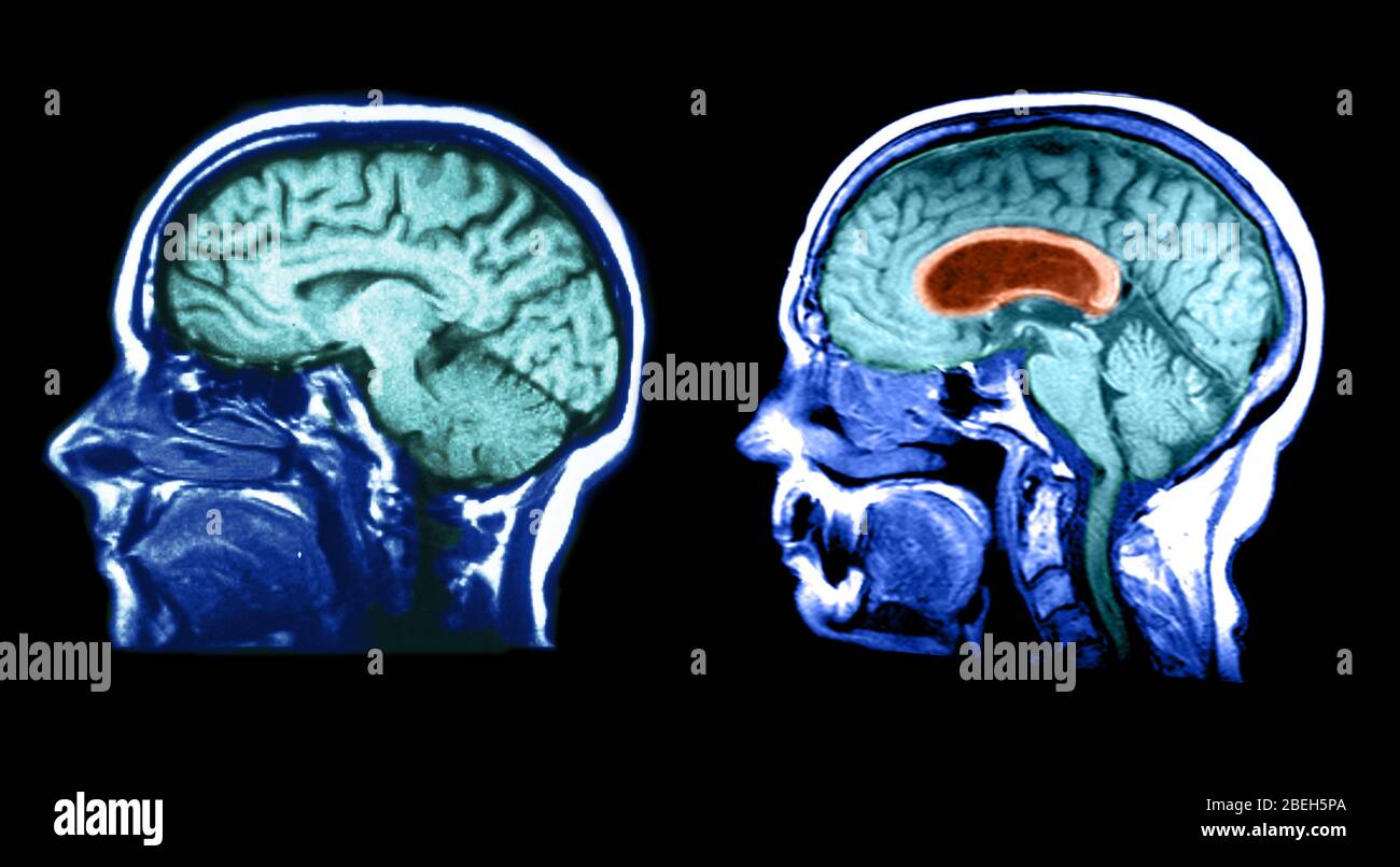 MRI of Normal Brain and Hydrocephalic Brain Stock Photohttps://www.alamy.com/image-license-details/?v=1https://www.alamy.com/mri-of-normal-brain-and-hydrocephalic-brain-image353190274.html
RM2BEH5PA–MRI of Normal Brain and Hydrocephalic Brain
 Clinical lectures on the principles and practice of medicine . ighing 6 lb. 2 oz., of a pale fawn color, considerablyindurated, in the second stage of cirrhosis. Abdomen contained two gallons and ahalf of amber-colored serum. Other organs healthy. Microscopic Examination.—The lymph filling up the ventricles of the larynxwas entirely composed of molecular fibres, included in a mass of coagulated molecularexudation. Commentary.—This man, while laboring under enlarged liver withascites, was apparently seized with an ordinary sore throat, having caughtcold, as it was afterwards ascertained, when v Stock Photohttps://www.alamy.com/image-license-details/?v=1https://www.alamy.com/clinical-lectures-on-the-principles-and-practice-of-medicine-ighing-6-lb-2-oz-of-a-pale-fawn-color-considerablyindurated-in-the-second-stage-of-cirrhosis-abdomen-contained-two-gallons-and-ahalf-of-amber-colored-serum-other-organs-healthy-microscopic-examinationthe-lymph-filling-up-the-ventricles-of-the-larynxwas-entirely-composed-of-molecular-fibres-included-in-a-mass-of-coagulated-molecularexudation-commentarythis-man-while-laboring-under-enlarged-liver-withascites-was-apparently-seized-with-an-ordinary-sore-throat-having-caughtcold-as-it-was-afterwards-ascertained-when-v-image338156999.html
RM2AJ4AKK–Clinical lectures on the principles and practice of medicine . ighing 6 lb. 2 oz., of a pale fawn color, considerablyindurated, in the second stage of cirrhosis. Abdomen contained two gallons and ahalf of amber-colored serum. Other organs healthy. Microscopic Examination.—The lymph filling up the ventricles of the larynxwas entirely composed of molecular fibres, included in a mass of coagulated molecularexudation. Commentary.—This man, while laboring under enlarged liver withascites, was apparently seized with an ordinary sore throat, having caughtcold, as it was afterwards ascertained, when v
 Baby with enlarged brain ventricles, illustration Stock Photohttps://www.alamy.com/image-license-details/?v=1https://www.alamy.com/baby-with-enlarged-brain-ventricles-illustration-image572908539.html
RF2T826K7–Baby with enlarged brain ventricles, illustration
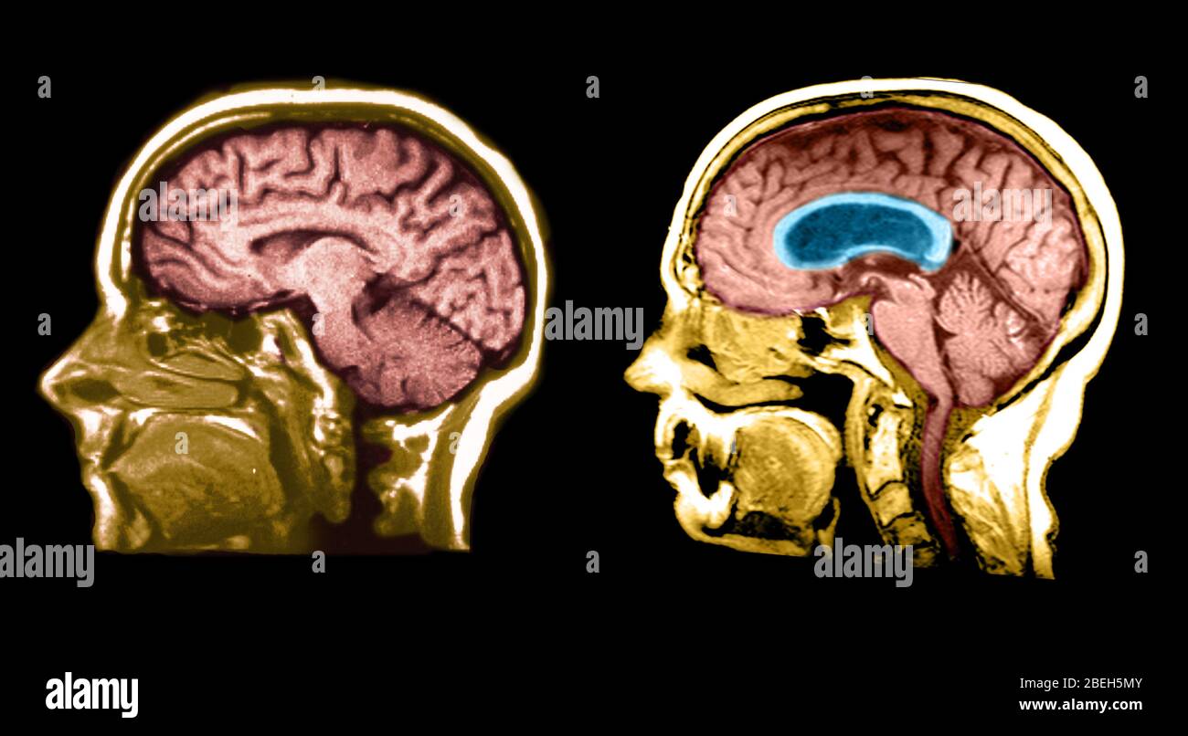 MRI of Normal Brain and Hydrocephalic Brain Stock Photohttps://www.alamy.com/image-license-details/?v=1https://www.alamy.com/mri-of-normal-brain-and-hydrocephalic-brain-image353190235.html
RM2BEH5MY–MRI of Normal Brain and Hydrocephalic Brain
 MRI Communicating Hydrocephalus (NPH) Stock Photohttps://www.alamy.com/image-license-details/?v=1https://www.alamy.com/stock-photo-mri-communicating-hydrocephalus-nph-134988156.html
RMHRH6NG–MRI Communicating Hydrocephalus (NPH)
 A text-book of physiology for medical students and physicians . idsurrounding the cord is infree communication withthat in the brain, as is indi-cated in the accompanyingschematic figure (Fig. 258).Within the brain itself thereare certain points at the an-gles and hollows of the differ-ent parts of the brain at whichthe subarachnoidal spaceis much enlarged, formingthe so-called cisternse, whichare in communication onewith another by means of theless conspicuous canals (seeFig. 259). The whole systemis also in direct communica-tion with the ventricles ofthe brain on the one hand,through the for Stock Photohttps://www.alamy.com/image-license-details/?v=1https://www.alamy.com/a-text-book-of-physiology-for-medical-students-and-physicians-idsurrounding-the-cord-is-infree-communication-withthat-in-the-brain-as-is-indi-cated-in-the-accompanyingschematic-figure-fig-258within-the-brain-itself-thereare-certain-points-at-the-an-gles-and-hollows-of-the-differ-ent-parts-of-the-brain-at-whichthe-subarachnoidal-spaceis-much-enlarged-formingthe-so-called-cisternse-whichare-in-communication-onewith-another-by-means-of-theless-conspicuous-canals-seefig-259-the-whole-systemis-also-in-direct-communica-tion-with-the-ventricles-ofthe-brain-on-the-one-handthrough-the-for-image342731810.html
RM2AWGNWP–A text-book of physiology for medical students and physicians . idsurrounding the cord is infree communication withthat in the brain, as is indi-cated in the accompanyingschematic figure (Fig. 258).Within the brain itself thereare certain points at the an-gles and hollows of the differ-ent parts of the brain at whichthe subarachnoidal spaceis much enlarged, formingthe so-called cisternse, whichare in communication onewith another by means of theless conspicuous canals (seeFig. 259). The whole systemis also in direct communica-tion with the ventricles ofthe brain on the one hand,through the for
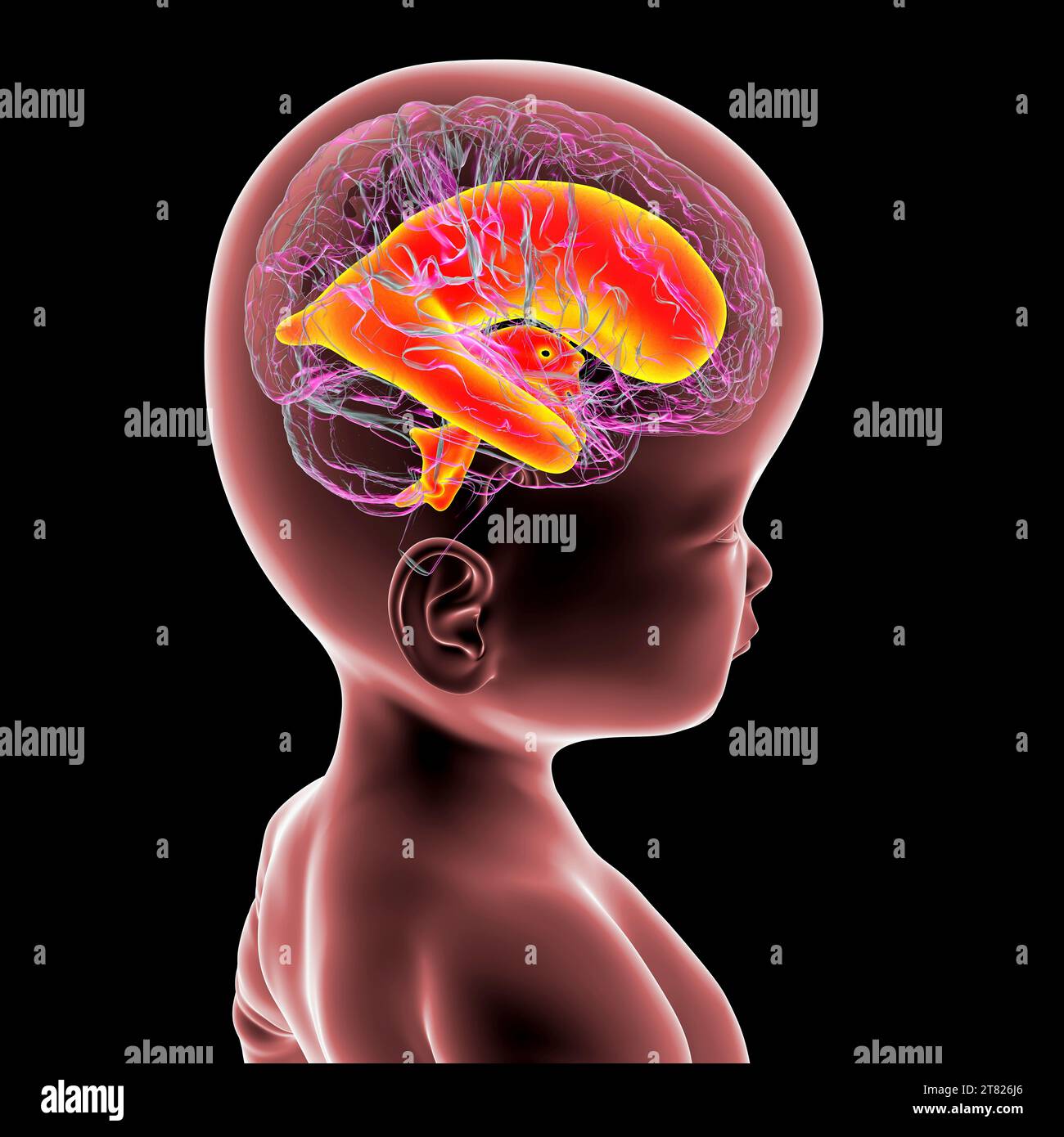 Baby with enlarged brain ventricles, illustration Stock Photohttps://www.alamy.com/image-license-details/?v=1https://www.alamy.com/baby-with-enlarged-brain-ventricles-illustration-image572908510.html
RF2T826J6–Baby with enlarged brain ventricles, illustration
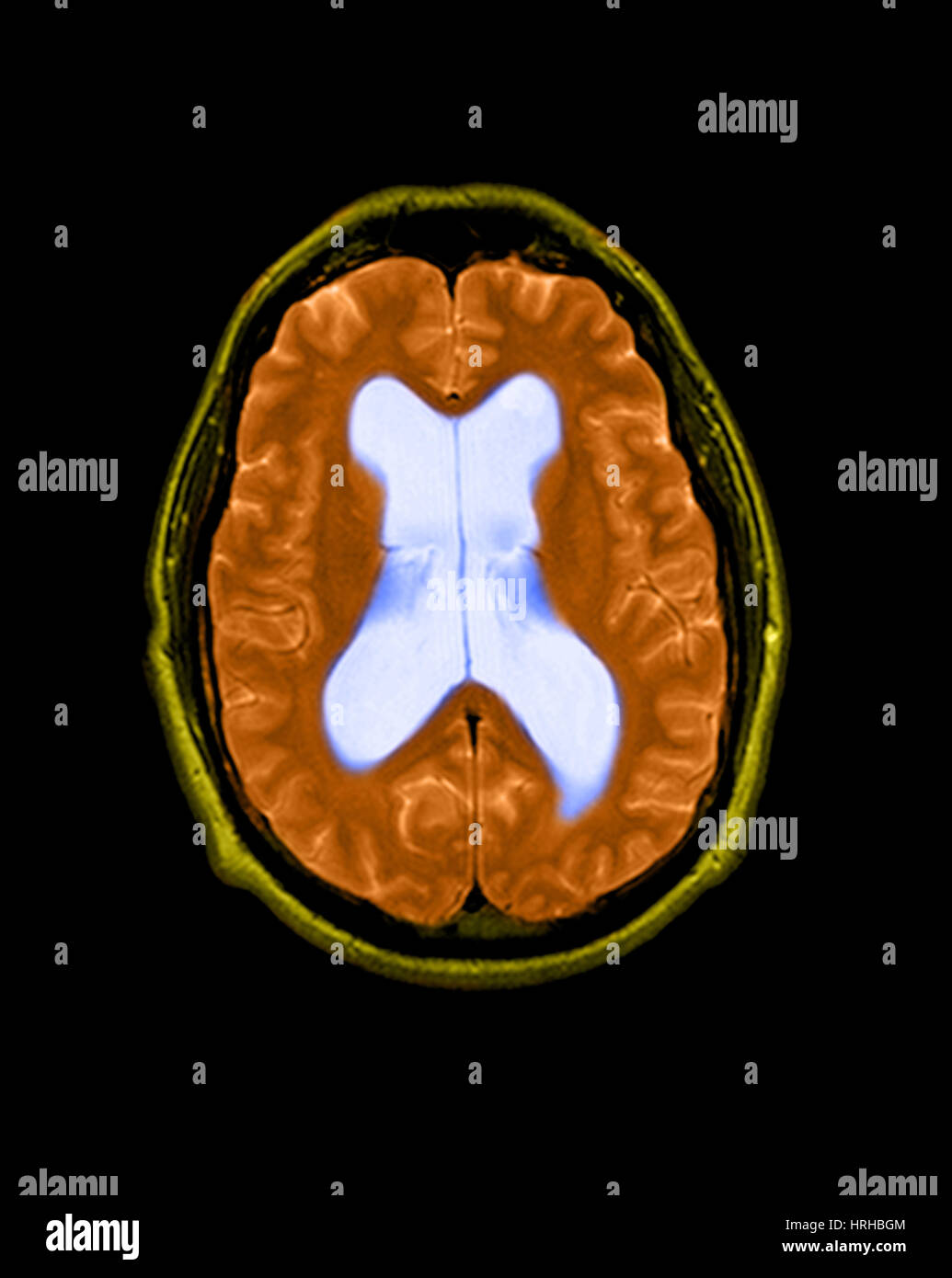 MRI Communicating Hydrocephalus (NPH) Stock Photohttps://www.alamy.com/image-license-details/?v=1https://www.alamy.com/stock-photo-mri-communicating-hydrocephalus-nph-134991940.html
RMHRHBGM–MRI Communicating Hydrocephalus (NPH)
 A text-book of physiology, for medical students and physicians . dsurrounding the cord is infree communication withthat in the brain, as is indi-cated in the accompanyingschematic figure (Fig. 249).Within the brain itself thereare certain points at the an-gles and hollows of the differ-ent parts of the brain at whichthe subarachnoidal spaceis much enlarged, formingthe so-called cisternal, whichare in communication onewith another by means of theless conspicuous canals (seeFig. 250). The whole systemis also in direct communica-tion with the ventricles ofthe brain on the one hand,through the for Stock Photohttps://www.alamy.com/image-license-details/?v=1https://www.alamy.com/a-text-book-of-physiology-for-medical-students-and-physicians-dsurrounding-the-cord-is-infree-communication-withthat-in-the-brain-as-is-indi-cated-in-the-accompanyingschematic-figure-fig-249within-the-brain-itself-thereare-certain-points-at-the-an-gles-and-hollows-of-the-differ-ent-parts-of-the-brain-at-whichthe-subarachnoidal-spaceis-much-enlarged-formingthe-so-called-cisternal-whichare-in-communication-onewith-another-by-means-of-theless-conspicuous-canals-seefig-250-the-whole-systemis-also-in-direct-communica-tion-with-the-ventricles-ofthe-brain-on-the-one-handthrough-the-for-image342916680.html
RM2AWW5M8–A text-book of physiology, for medical students and physicians . dsurrounding the cord is infree communication withthat in the brain, as is indi-cated in the accompanyingschematic figure (Fig. 249).Within the brain itself thereare certain points at the an-gles and hollows of the differ-ent parts of the brain at whichthe subarachnoidal spaceis much enlarged, formingthe so-called cisternal, whichare in communication onewith another by means of theless conspicuous canals (seeFig. 250). The whole systemis also in direct communica-tion with the ventricles ofthe brain on the one hand,through the for
 Enlarged lateral ventricles of the brain, illustration Stock Photohttps://www.alamy.com/image-license-details/?v=1https://www.alamy.com/enlarged-lateral-ventricles-of-the-brain-illustration-image572908824.html
RF2T8271C–Enlarged lateral ventricles of the brain, illustration
 MRI Communicating Hydrocephalus (NPH) Stock Photohttps://www.alamy.com/image-license-details/?v=1https://www.alamy.com/stock-photo-mri-communicating-hydrocephalus-nph-134991942.html
RMHRHBGP–MRI Communicating Hydrocephalus (NPH)
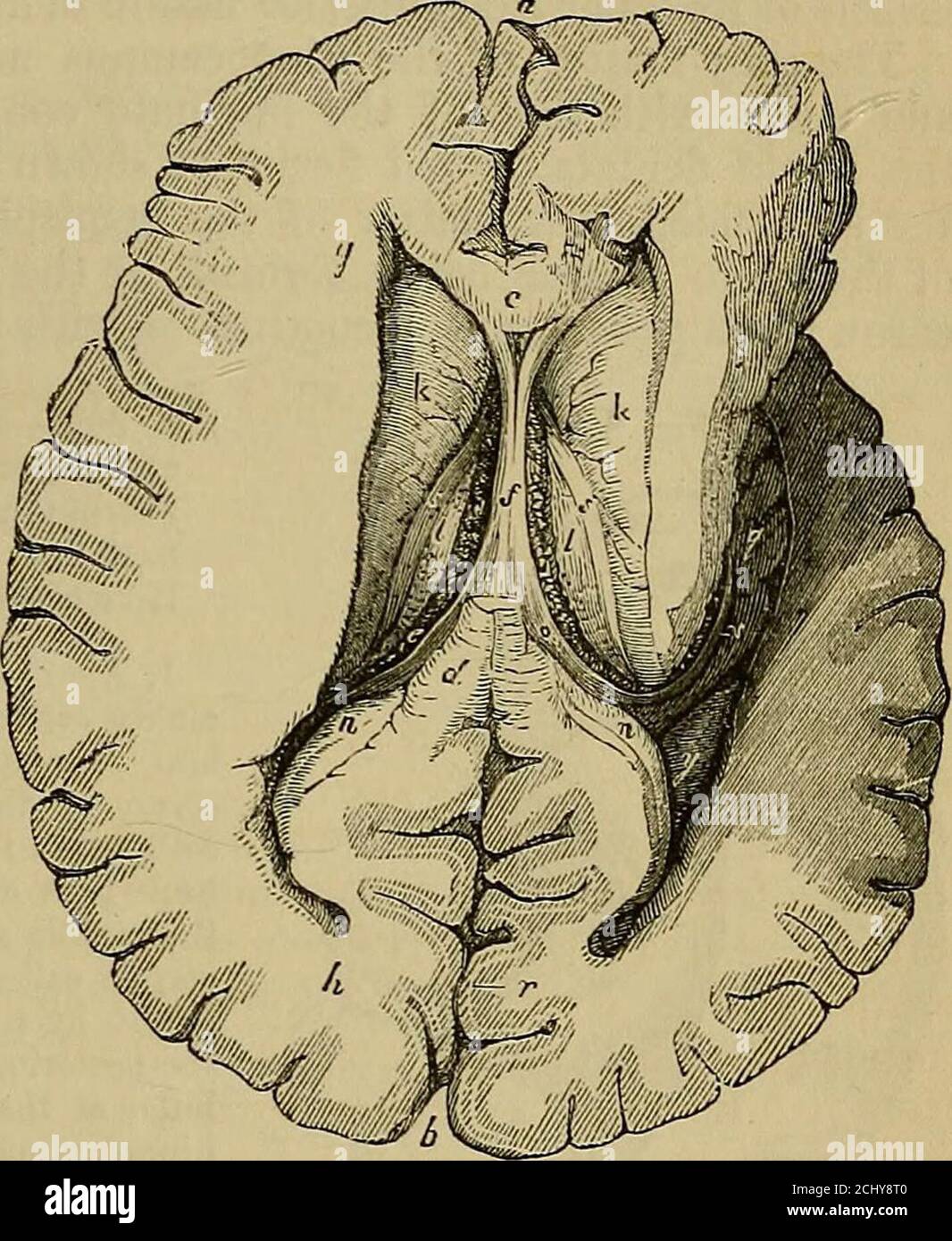 . Quain's elements of anatomy . enlarged anterior extremity of the nucleus caudatus, de-cending as it proceeds, and bounded below and externally by that body,in front by the reflected part of the corpus callosum, and mesially bythe septum lucidum. Fig. 301.—The lateral VENTRICLES OPENED BYEE3I0TAL OP THE MID-DLE PART OF THE CORPUSCALLOSDM, AND THE DE-SCENDING COENU EXPOSEDON THE EIGHT SIDE. h, a, h, anterior and pos-terior parts of the greatlongitudinal fissure; c,section of the anterior partof the corpus callosum ; d,posterior part of the same ;e, the left choroid plexus ;/, the fornix ; ff, Stock Photohttps://www.alamy.com/image-license-details/?v=1https://www.alamy.com/quains-elements-of-anatomy-enlarged-anterior-extremity-of-the-nucleus-caudatus-de-cending-as-it-proceeds-and-bounded-below-and-externally-by-that-bodyin-front-by-the-reflected-part-of-the-corpus-callosum-and-mesially-bythe-septum-lucidum-fig-301the-lateral-ventricles-opened-byee3i0tal-op-the-mid-dle-part-of-the-corpuscallosdm-and-the-de-scending-coenu-exposedon-the-eight-side-h-a-h-anterior-and-pos-terior-parts-of-the-greatlongitudinal-fissure-csection-of-the-anterior-partof-the-corpus-callosum-dposterior-part-of-the-same-e-the-left-choroid-plexus-the-fornix-ff-image372466528.html
RM2CHY8T0–. Quain's elements of anatomy . enlarged anterior extremity of the nucleus caudatus, de-cending as it proceeds, and bounded below and externally by that body,in front by the reflected part of the corpus callosum, and mesially bythe septum lucidum. Fig. 301.—The lateral VENTRICLES OPENED BYEE3I0TAL OP THE MID-DLE PART OF THE CORPUSCALLOSDM, AND THE DE-SCENDING COENU EXPOSEDON THE EIGHT SIDE. h, a, h, anterior and pos-terior parts of the greatlongitudinal fissure; c,section of the anterior partof the corpus callosum ; d,posterior part of the same ;e, the left choroid plexus ;/, the fornix ; ff,
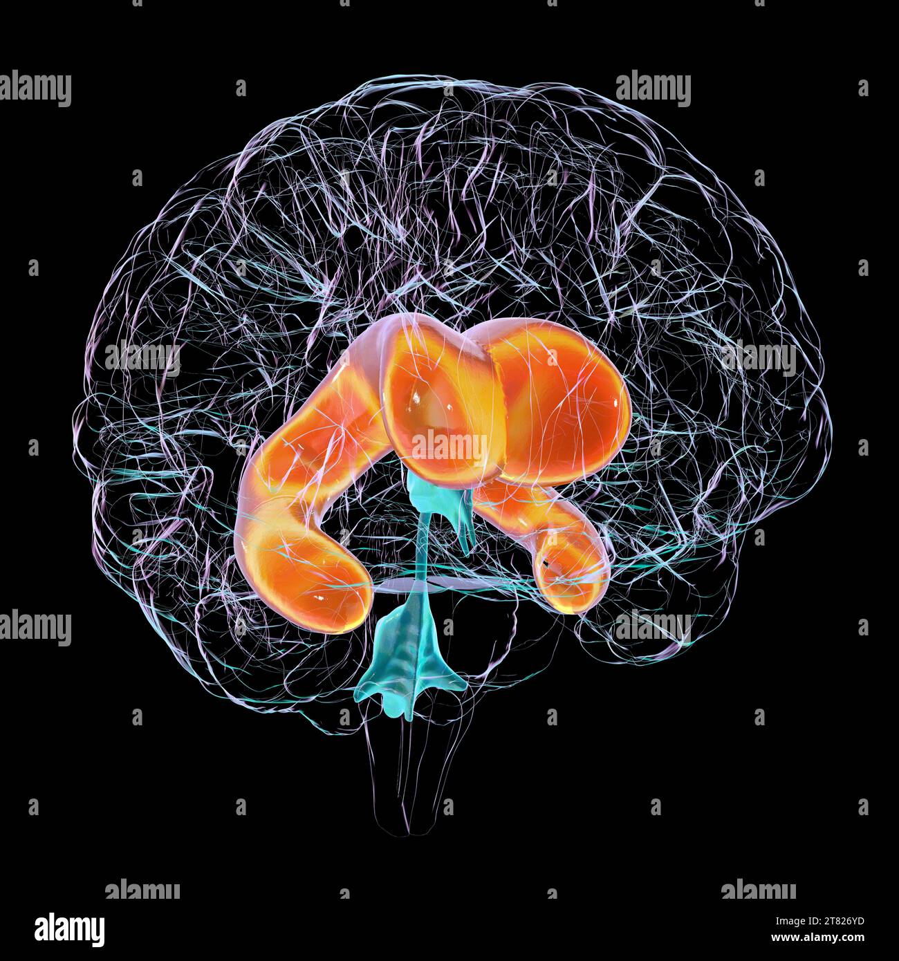 Enlarged lateral ventricles of the brain, illustration Stock Photohttps://www.alamy.com/image-license-details/?v=1https://www.alamy.com/enlarged-lateral-ventricles-of-the-brain-illustration-image572908769.html
RF2T826YD–Enlarged lateral ventricles of the brain, illustration
 MRI Communicating Hydrocephalus (NPH) Stock Photohttps://www.alamy.com/image-license-details/?v=1https://www.alamy.com/stock-photo-mri-communicating-hydrocephalus-nph-134991943.html
RMHRHBGR–MRI Communicating Hydrocephalus (NPH)
 . Veterinary obstetrics; a compendium for the use of students and practitioners. Veterinary obstetrics. 88 VETERINARY OBSTETRICS. lungs being unprotected. This condition is known as. ^^Ectopia cordis." Often the foetus is born with the cranial cavity enlarged with fluid,—''Hydrocephalus"or ^'Hydrops capitis." First of all, fluid collects In the ventricles, and the diseased process may go on until. Fig. 37. Skull of Hvdrockphalic Calf: the Cranial Bonks are partially: destroyed and defective. the whole of the brain structure is displaced by fluid,, and only retained in position b Stock Photohttps://www.alamy.com/image-license-details/?v=1https://www.alamy.com/veterinary-obstetrics-a-compendium-for-the-use-of-students-and-practitioners-veterinary-obstetrics-88-veterinary-obstetrics-lungs-being-unprotected-this-condition-is-known-as-ectopia-cordisquot-often-the-foetus-is-born-with-the-cranial-cavity-enlarged-with-fluidhydrocephalusquotor-hydrops-capitisquot-first-of-all-fluid-collects-in-the-ventricles-and-the-diseased-process-may-go-on-until-fig-37-skull-of-hvdrockphalic-calf-the-cranial-bonks-are-partially-destroyed-and-defective-the-whole-of-the-brain-structure-is-displaced-by-fluid-and-only-retained-in-position-b-image232003447.html
RMRDCJNB–. Veterinary obstetrics; a compendium for the use of students and practitioners. Veterinary obstetrics. 88 VETERINARY OBSTETRICS. lungs being unprotected. This condition is known as. ^^Ectopia cordis." Often the foetus is born with the cranial cavity enlarged with fluid,—''Hydrocephalus"or ^'Hydrops capitis." First of all, fluid collects In the ventricles, and the diseased process may go on until. Fig. 37. Skull of Hvdrockphalic Calf: the Cranial Bonks are partially: destroyed and defective. the whole of the brain structure is displaced by fluid,, and only retained in position b
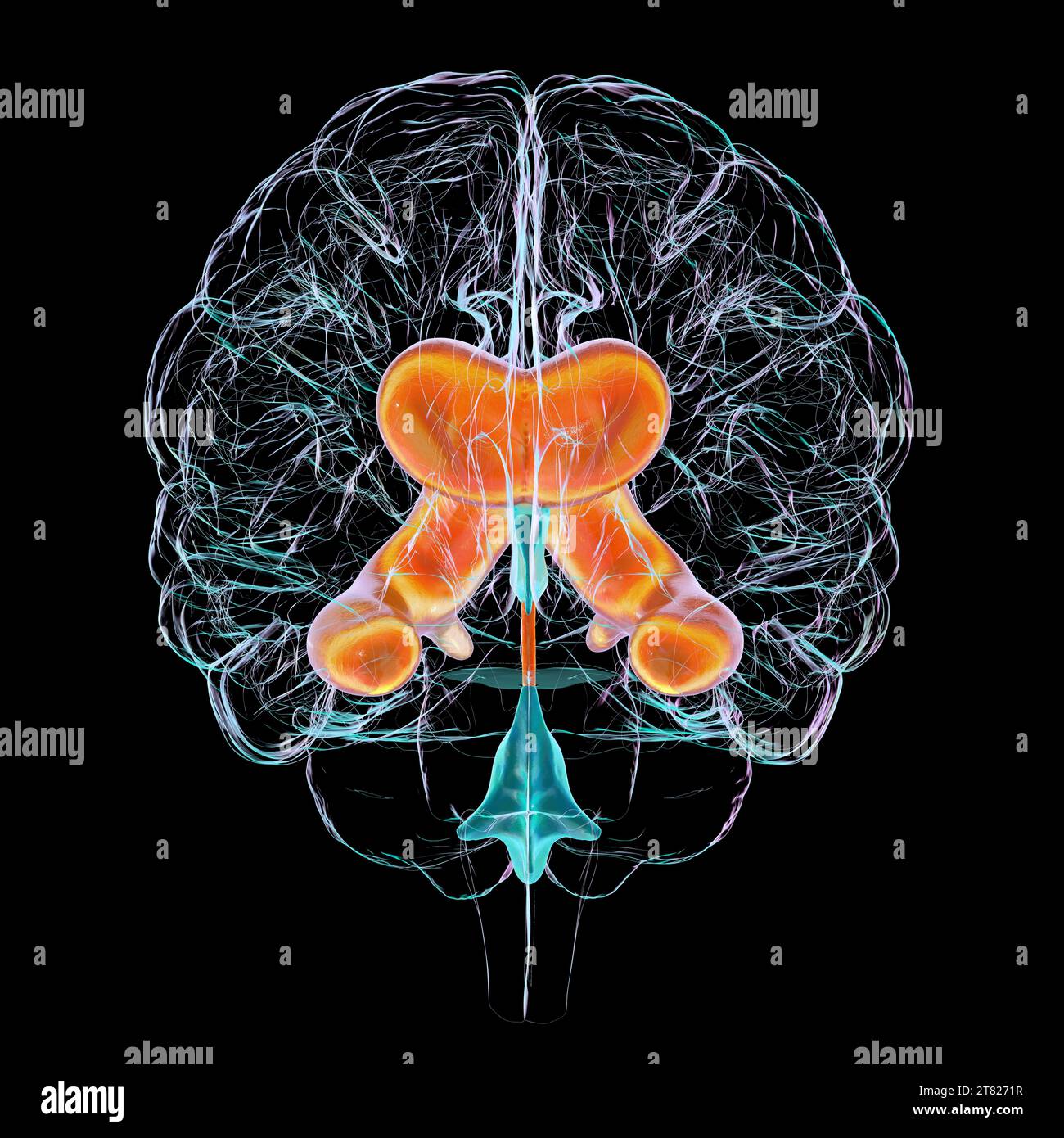 Enlarged lateral ventricles of the brain, illustration Stock Photohttps://www.alamy.com/image-license-details/?v=1https://www.alamy.com/enlarged-lateral-ventricles-of-the-brain-illustration-image572908835.html
RF2T8271R–Enlarged lateral ventricles of the brain, illustration
 Enlarged lateral ventricles of the brain, illustration Stock Photohttps://www.alamy.com/image-license-details/?v=1https://www.alamy.com/enlarged-lateral-ventricles-of-the-brain-illustration-image572908758.html
RF2T826Y2–Enlarged lateral ventricles of the brain, illustration
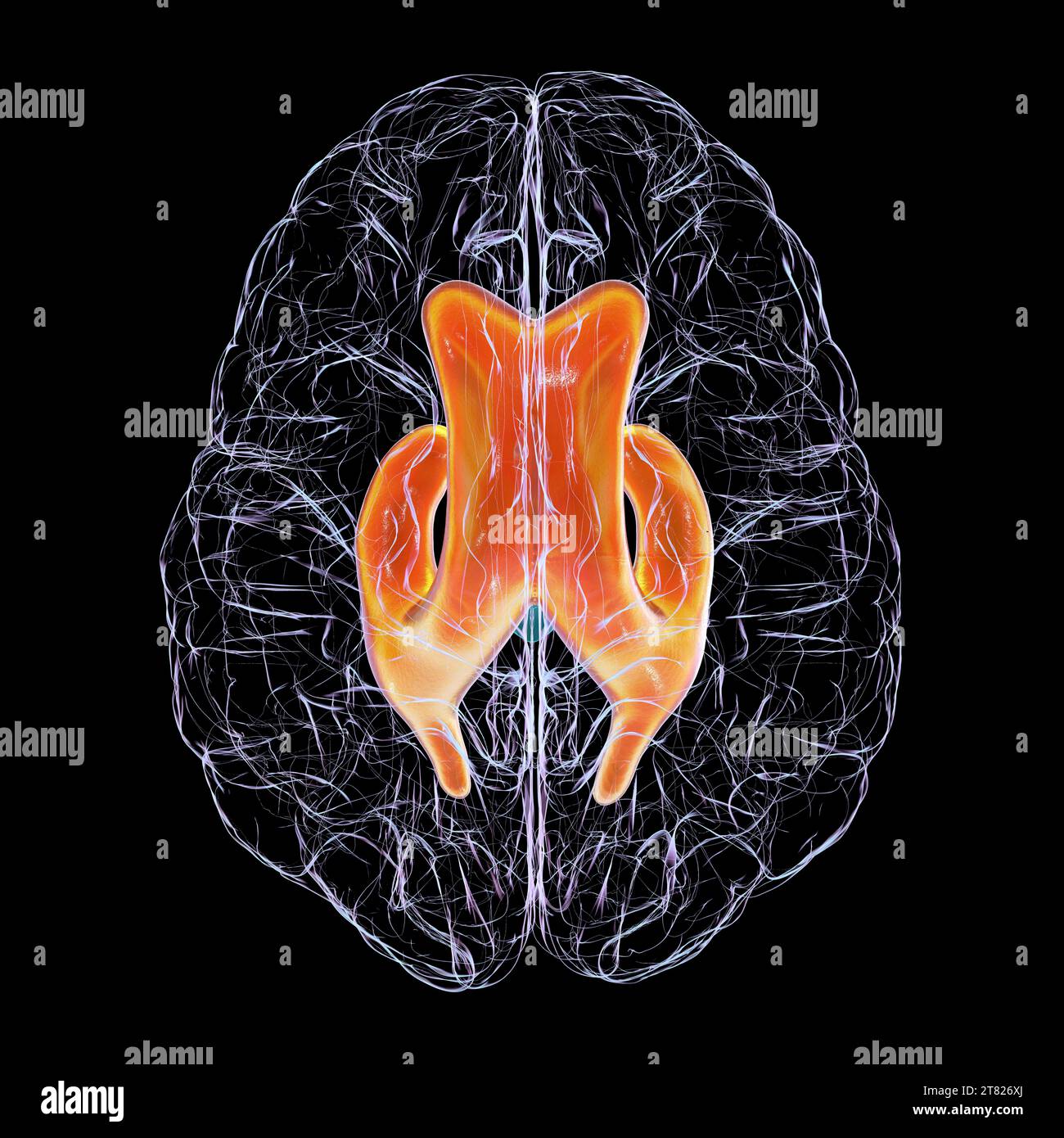 Enlarged lateral ventricles of the brain, illustration Stock Photohttps://www.alamy.com/image-license-details/?v=1https://www.alamy.com/enlarged-lateral-ventricles-of-the-brain-illustration-image572908746.html
RF2T826XJ–Enlarged lateral ventricles of the brain, illustration
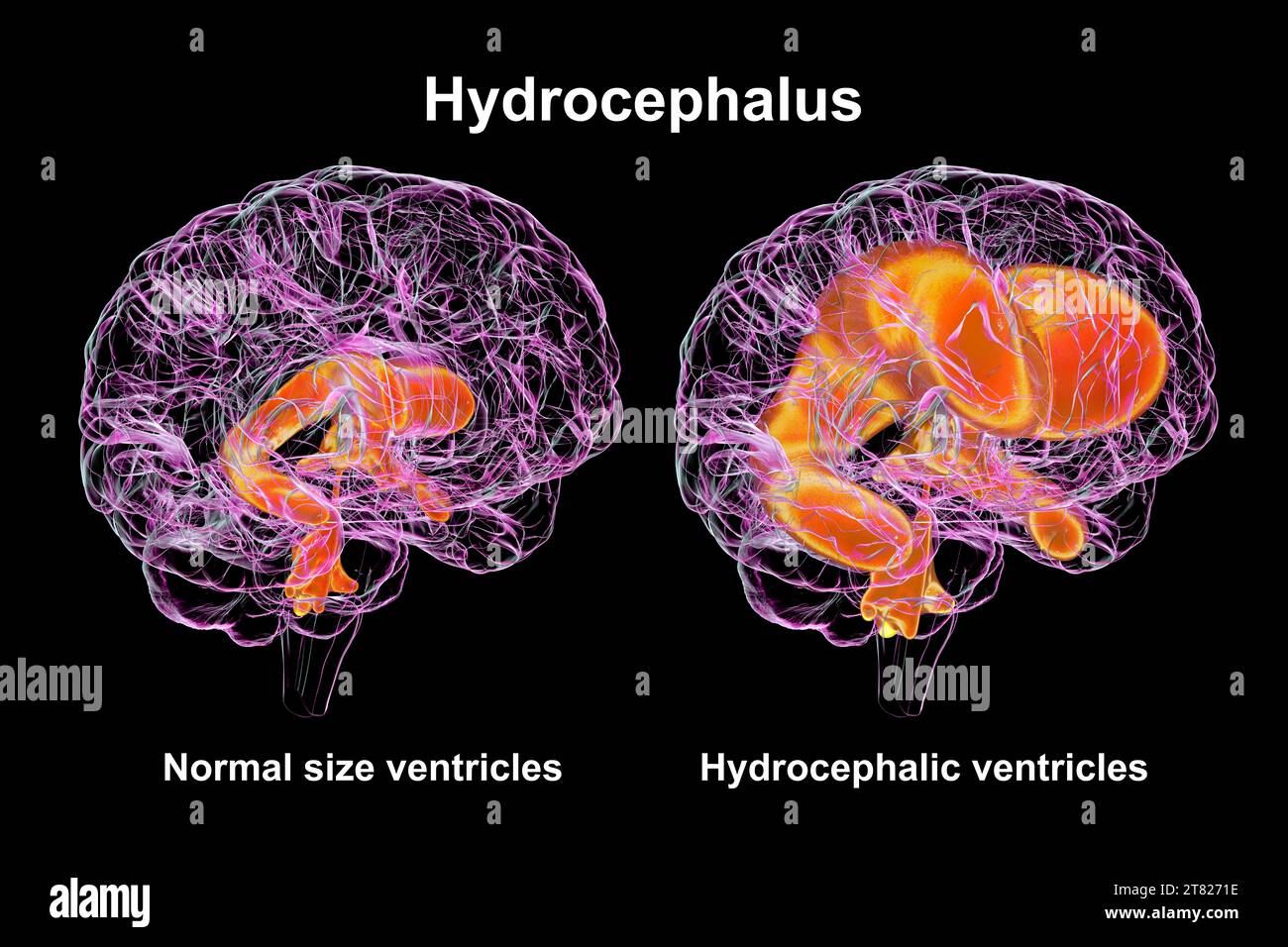 Enlarged and normal brain ventricles, illustration Stock Photohttps://www.alamy.com/image-license-details/?v=1https://www.alamy.com/enlarged-and-normal-brain-ventricles-illustration-image572908826.html
RF2T8271E–Enlarged and normal brain ventricles, illustration
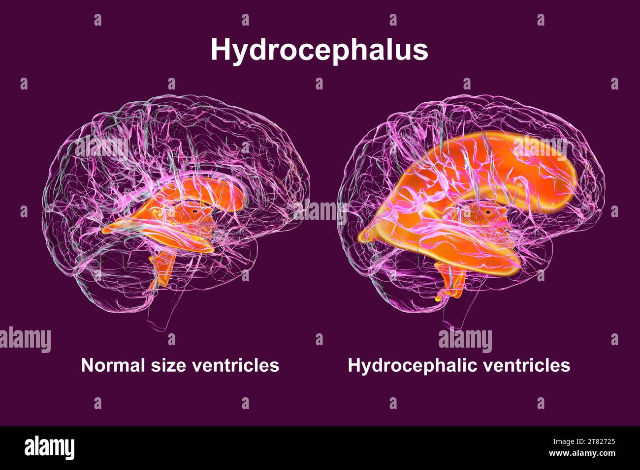 Enlarged and normal brain ventricles, illustration Stock Photohttps://www.alamy.com/image-license-details/?v=1https://www.alamy.com/enlarged-and-normal-brain-ventricles-illustration-image572908845.html
RF2T82725–Enlarged and normal brain ventricles, illustration
 Enlarged lateral ventricles of the brain, illustration Stock Photohttps://www.alamy.com/image-license-details/?v=1https://www.alamy.com/enlarged-lateral-ventricles-of-the-brain-illustration-image572908756.html
RF2T826Y0–Enlarged lateral ventricles of the brain, illustration
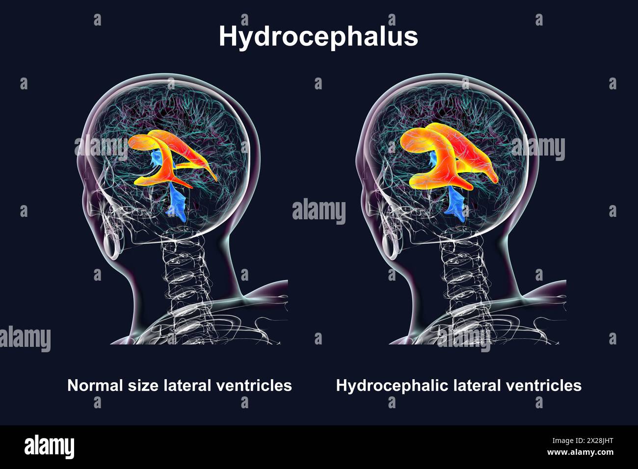 Enlarged and normal lateral ventricles, illustration Stock Photohttps://www.alamy.com/image-license-details/?v=1https://www.alamy.com/enlarged-and-normal-lateral-ventricles-illustration-image603782420.html
RF2X28JHT–Enlarged and normal lateral ventricles, illustration
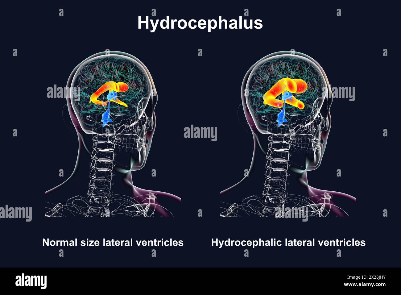 Enlarged and normal lateral ventricles, illustration Stock Photohttps://www.alamy.com/image-license-details/?v=1https://www.alamy.com/enlarged-and-normal-lateral-ventricles-illustration-image603782423.html
RF2X28JHY–Enlarged and normal lateral ventricles, illustration
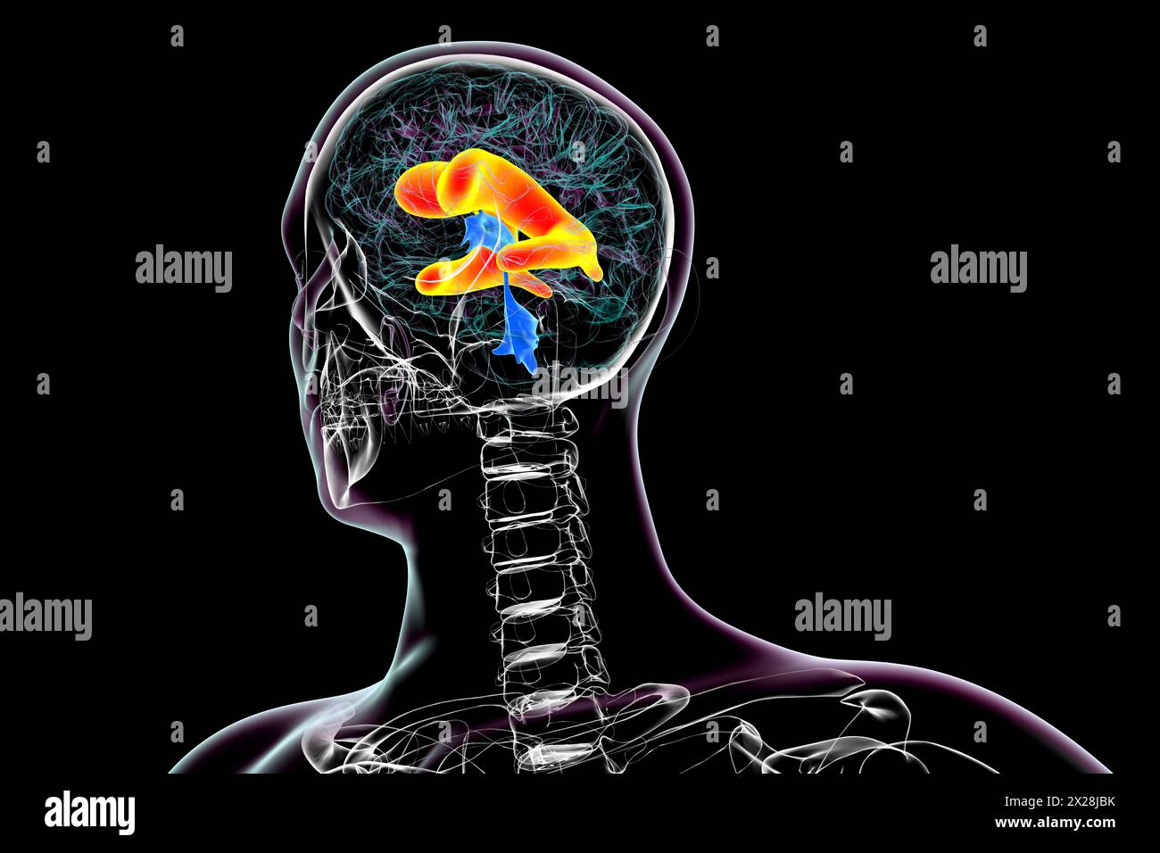 Enlarged lateral ventricles of the brain, illustration Stock Photohttps://www.alamy.com/image-license-details/?v=1https://www.alamy.com/enlarged-lateral-ventricles-of-the-brain-illustration-image603782247.html
RF2X28JBK–Enlarged lateral ventricles of the brain, illustration
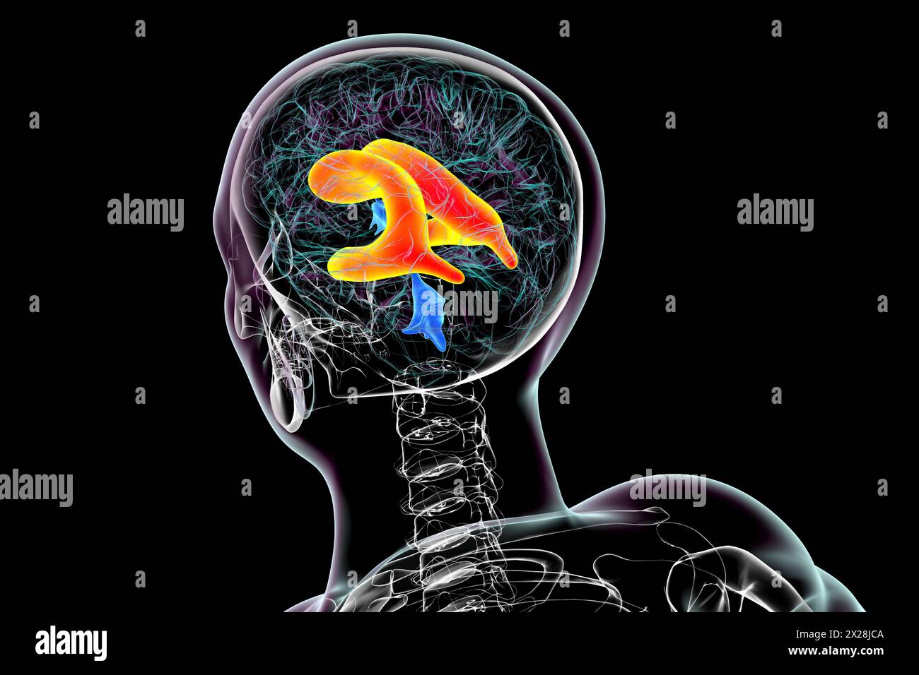 Enlarged lateral ventricles of the brain, illustration Stock Photohttps://www.alamy.com/image-license-details/?v=1https://www.alamy.com/enlarged-lateral-ventricles-of-the-brain-illustration-image603782266.html
RF2X28JCA–Enlarged lateral ventricles of the brain, illustration
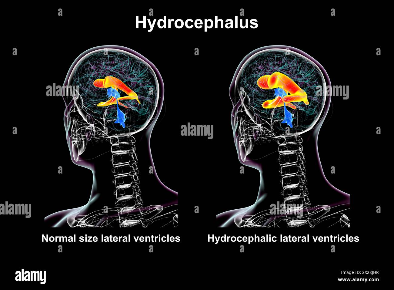 Enlarged and normal lateral ventricles, illustration Stock Photohttps://www.alamy.com/image-license-details/?v=1https://www.alamy.com/enlarged-and-normal-lateral-ventricles-illustration-image603782419.html
RF2X28JHR–Enlarged and normal lateral ventricles, illustration
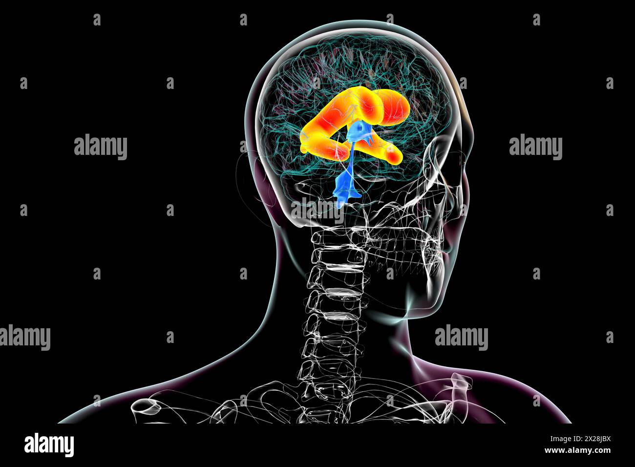 Enlarged lateral ventricles of the brain, illustration Stock Photohttps://www.alamy.com/image-license-details/?v=1https://www.alamy.com/enlarged-lateral-ventricles-of-the-brain-illustration-image603782254.html
RF2X28JBX–Enlarged lateral ventricles of the brain, illustration
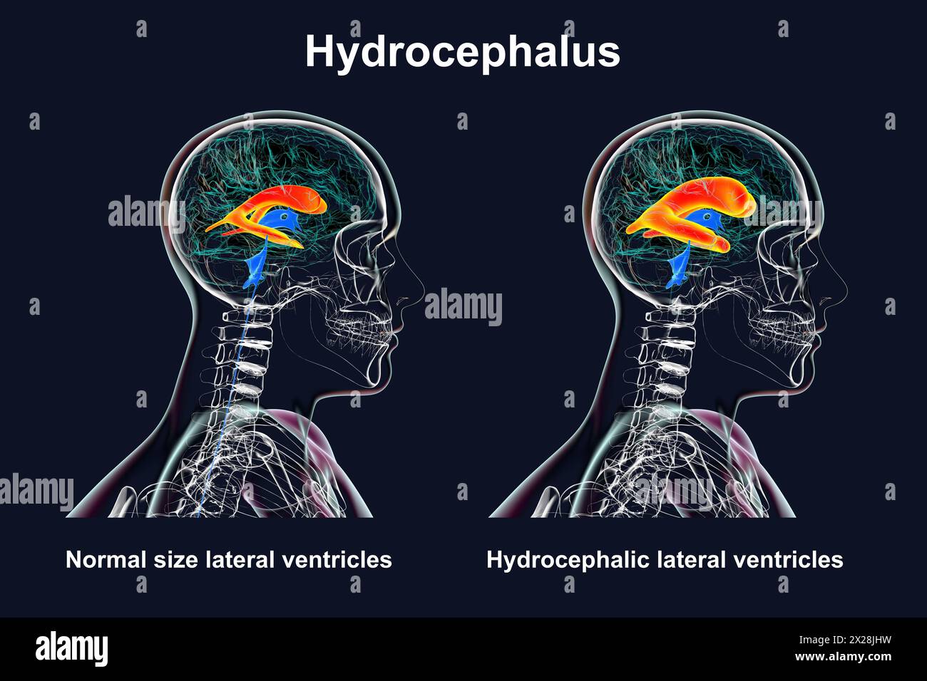 Enlarged and normal lateral ventricles, illustration Stock Photohttps://www.alamy.com/image-license-details/?v=1https://www.alamy.com/enlarged-and-normal-lateral-ventricles-illustration-image603782421.html
RF2X28JHW–Enlarged and normal lateral ventricles, illustration
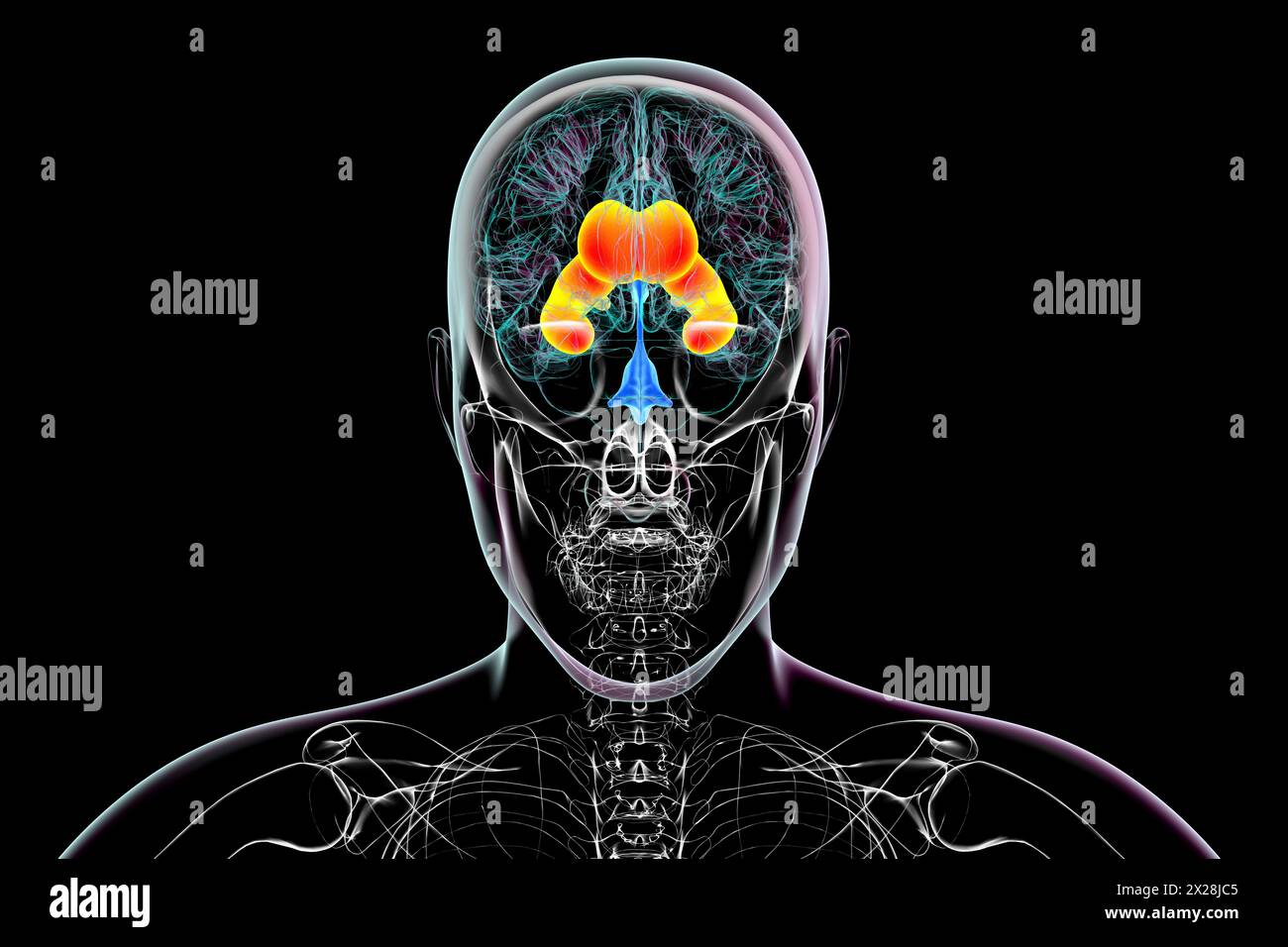 Enlarged lateral ventricles of the brain, illustration Stock Photohttps://www.alamy.com/image-license-details/?v=1https://www.alamy.com/enlarged-lateral-ventricles-of-the-brain-illustration-image603782261.html
RF2X28JC5–Enlarged lateral ventricles of the brain, illustration
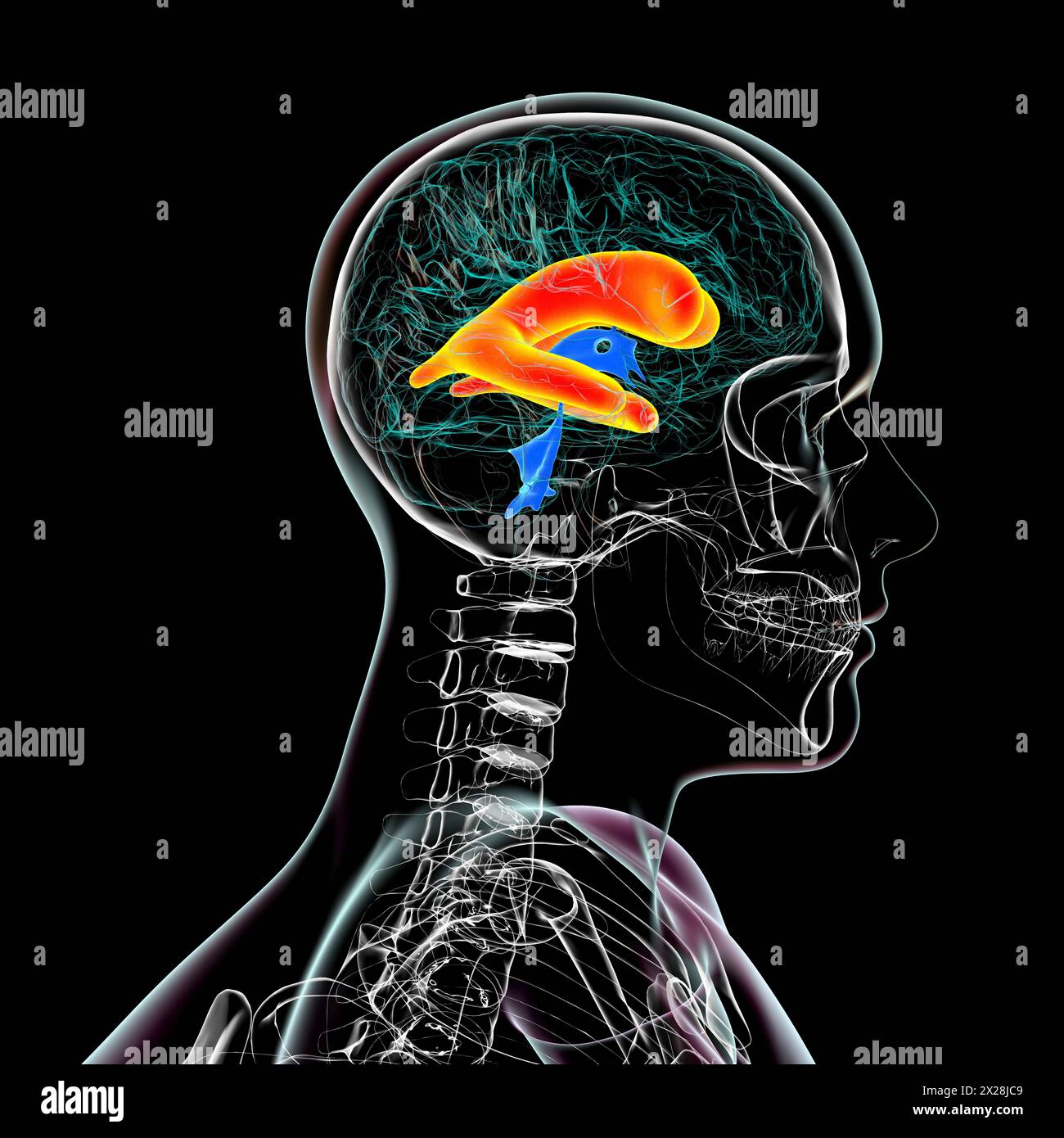 Enlarged lateral ventricles of the brain, illustration Stock Photohttps://www.alamy.com/image-license-details/?v=1https://www.alamy.com/enlarged-lateral-ventricles-of-the-brain-illustration-image603782265.html
RF2X28JC9–Enlarged lateral ventricles of the brain, illustration
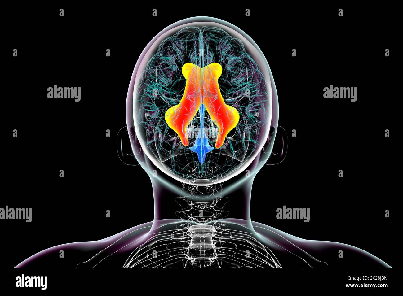 Enlarged lateral ventricles of the brain, illustration Stock Photohttps://www.alamy.com/image-license-details/?v=1https://www.alamy.com/enlarged-lateral-ventricles-of-the-brain-illustration-image603782249.html
RF2X28JBN–Enlarged lateral ventricles of the brain, illustration
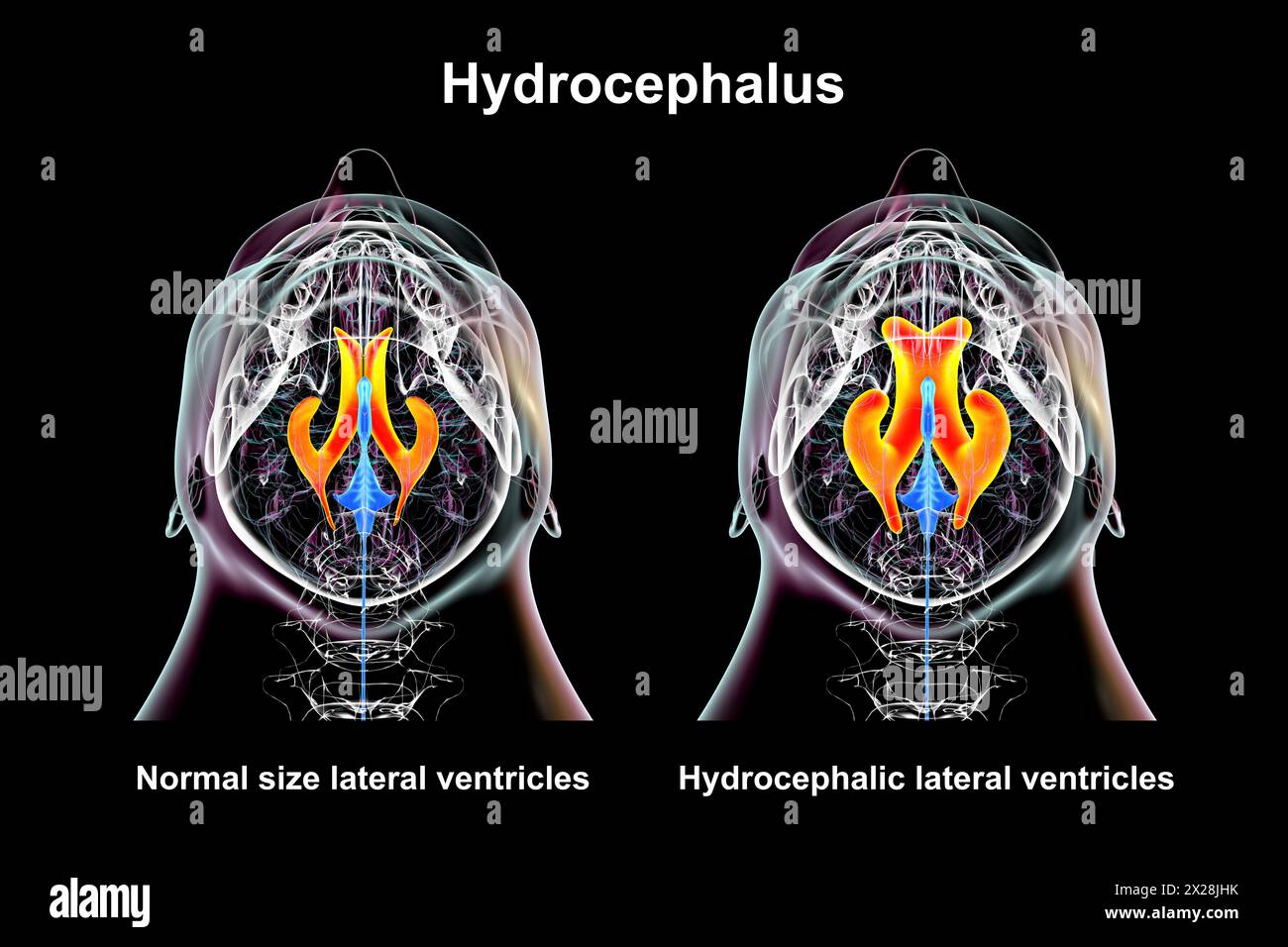 Enlarged and normal lateral ventricles, illustration Stock Photohttps://www.alamy.com/image-license-details/?v=1https://www.alamy.com/enlarged-and-normal-lateral-ventricles-illustration-image603782415.html
RF2X28JHK–Enlarged and normal lateral ventricles, illustration
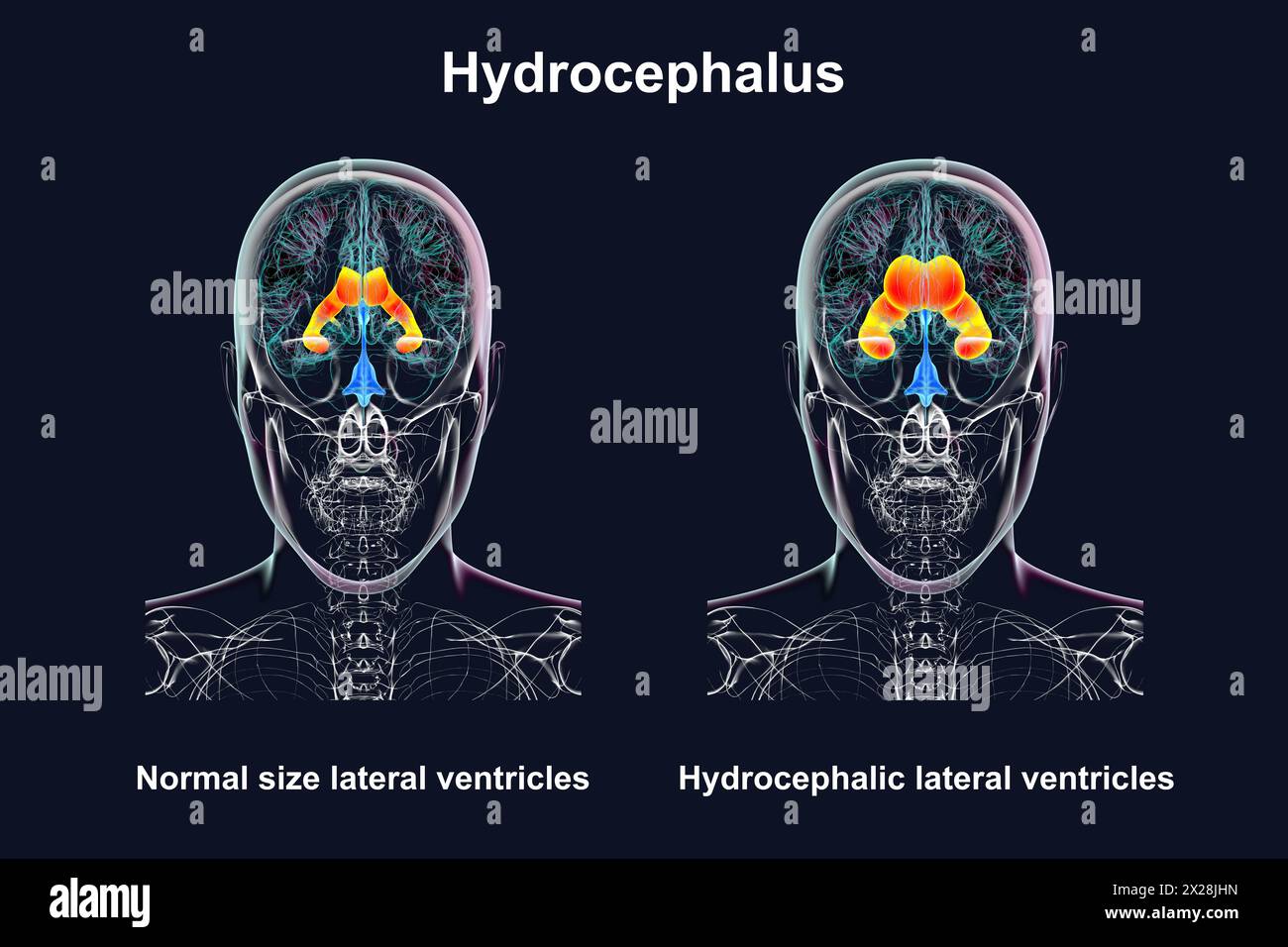 Enlarged and normal lateral ventricles, illustration Stock Photohttps://www.alamy.com/image-license-details/?v=1https://www.alamy.com/enlarged-and-normal-lateral-ventricles-illustration-image603782417.html
RF2X28JHN–Enlarged and normal lateral ventricles, illustration
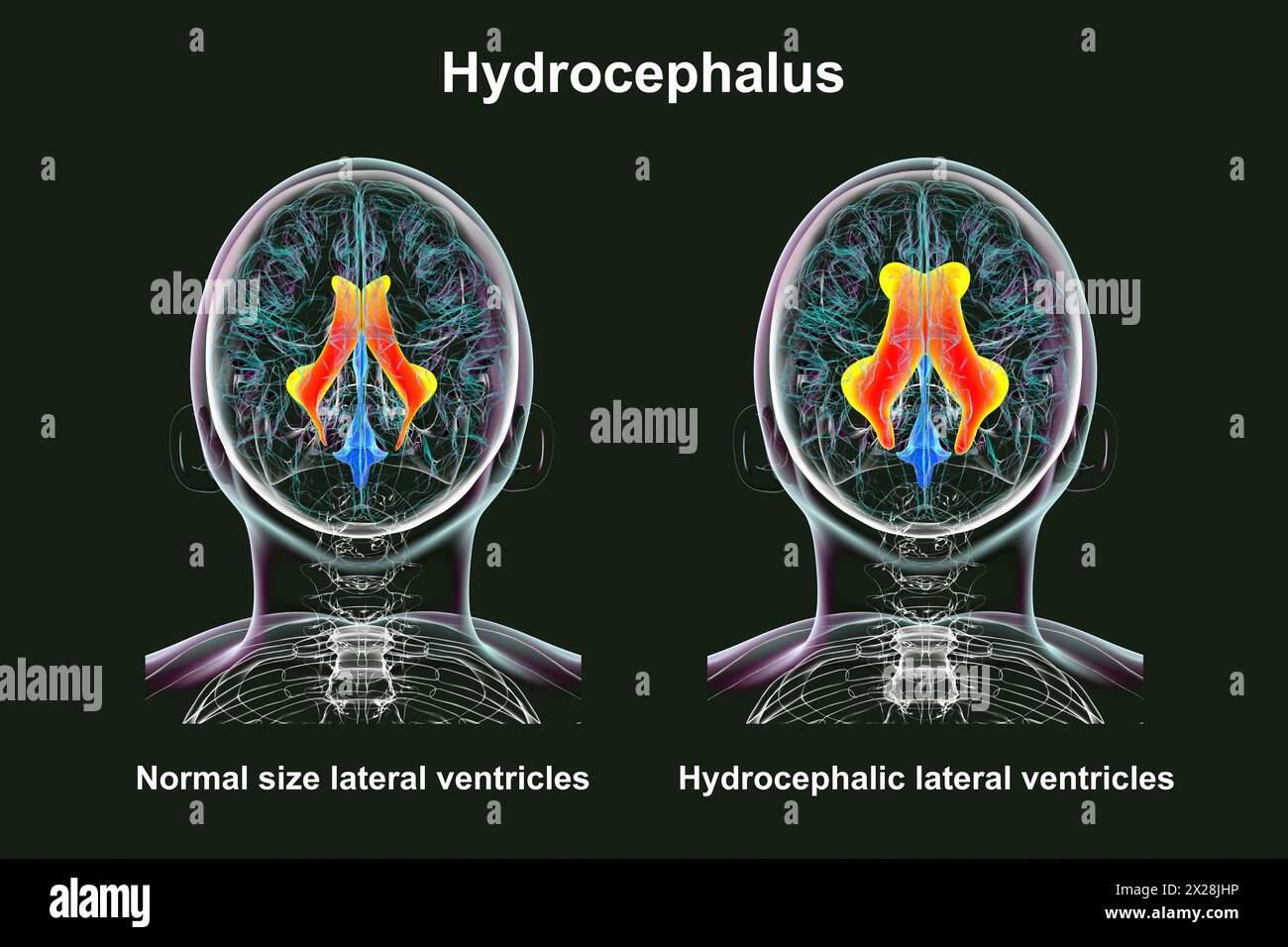 Enlarged and normal lateral ventricles, illustration Stock Photohttps://www.alamy.com/image-license-details/?v=1https://www.alamy.com/enlarged-and-normal-lateral-ventricles-illustration-image603782418.html
RF2X28JHP–Enlarged and normal lateral ventricles, illustration
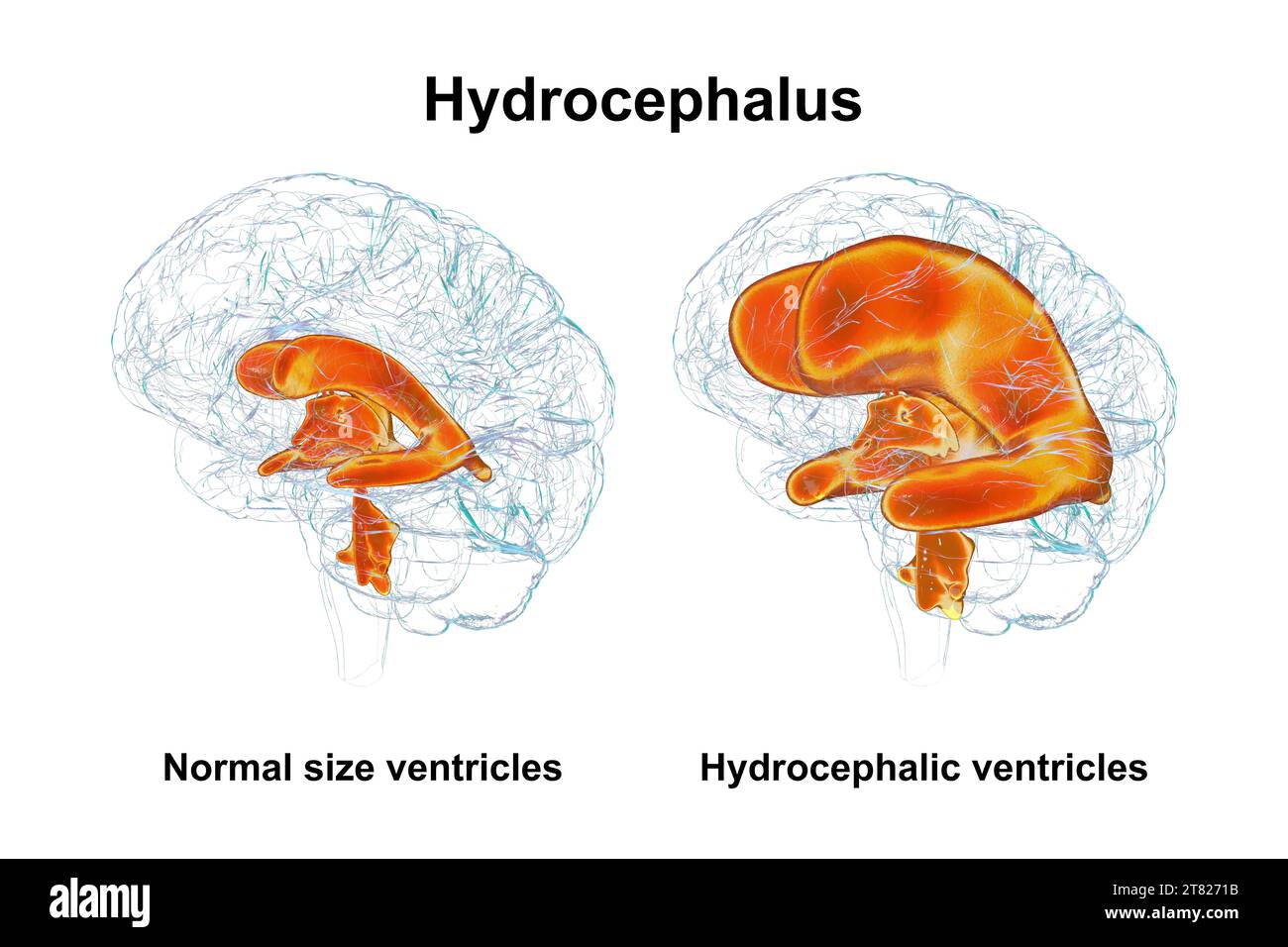 Enlarged and normal brain ventricles, illustration Stock Photohttps://www.alamy.com/image-license-details/?v=1https://www.alamy.com/enlarged-and-normal-brain-ventricles-illustration-image572908823.html
RF2T8271B–Enlarged and normal brain ventricles, illustration
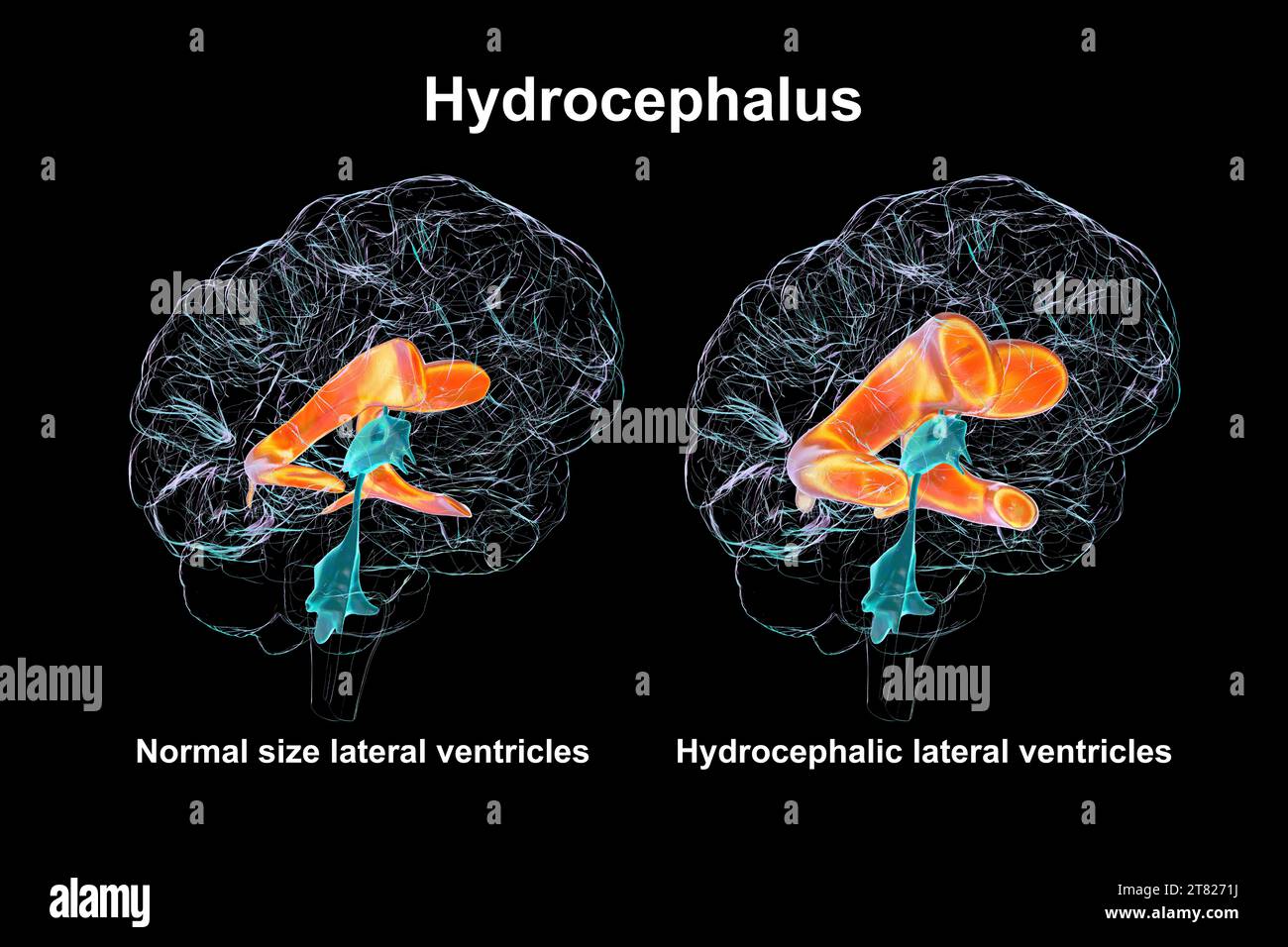 Enlarged and normal brain lateral ventricles, illustration Stock Photohttps://www.alamy.com/image-license-details/?v=1https://www.alamy.com/enlarged-and-normal-brain-lateral-ventricles-illustration-image572908830.html
RF2T8271J–Enlarged and normal brain lateral ventricles, illustration
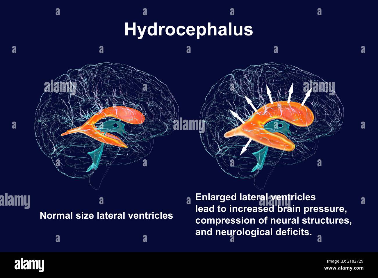 Enlarged and normal brain lateral ventricles, illustration Stock Photohttps://www.alamy.com/image-license-details/?v=1https://www.alamy.com/enlarged-and-normal-brain-lateral-ventricles-illustration-image572908849.html
RF2T82729–Enlarged and normal brain lateral ventricles, illustration
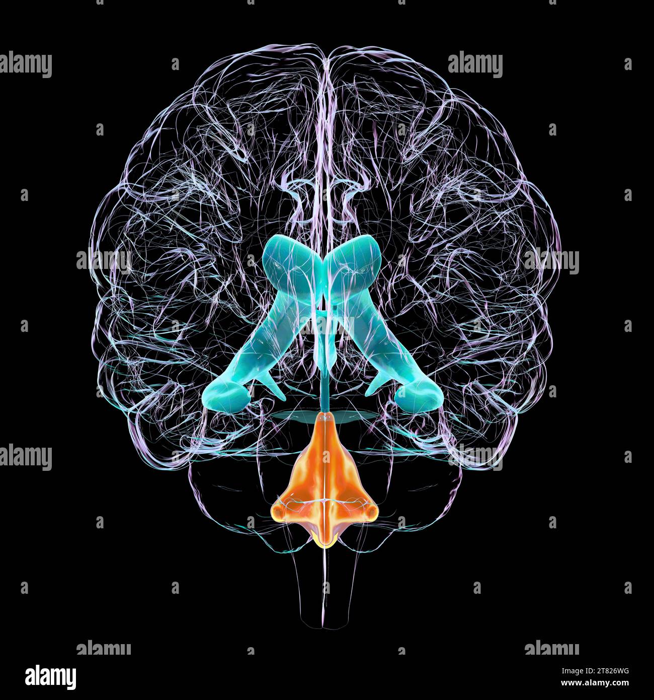 Enlarged fourth ventricle of the brain, illustration Stock Photohttps://www.alamy.com/image-license-details/?v=1https://www.alamy.com/enlarged-fourth-ventricle-of-the-brain-illustration-image572908716.html
RF2T826WG–Enlarged fourth ventricle of the brain, illustration
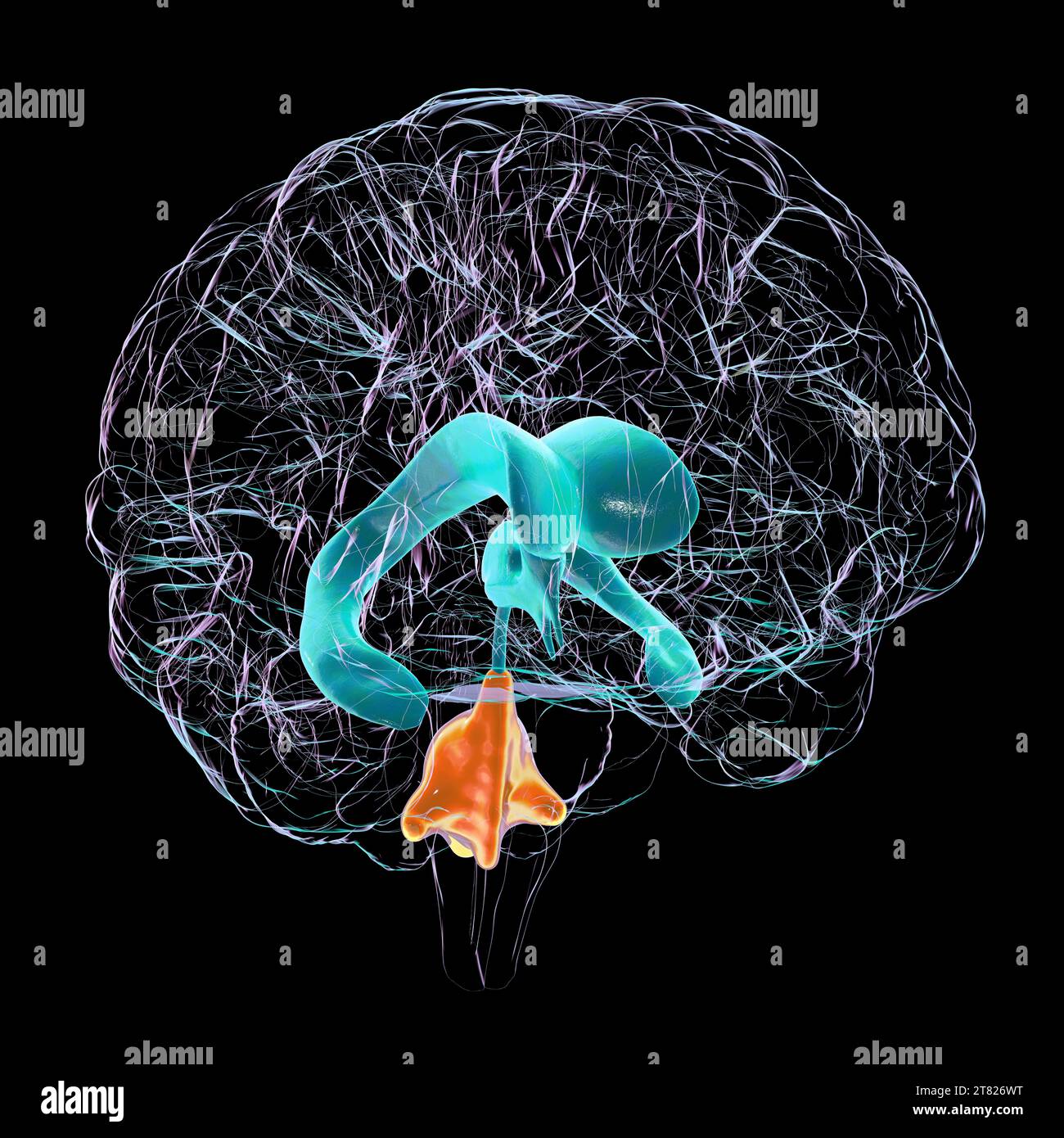 Enlarged fourth ventricle of the brain, illustration Stock Photohttps://www.alamy.com/image-license-details/?v=1https://www.alamy.com/enlarged-fourth-ventricle-of-the-brain-illustration-image572908724.html
RF2T826WT–Enlarged fourth ventricle of the brain, illustration
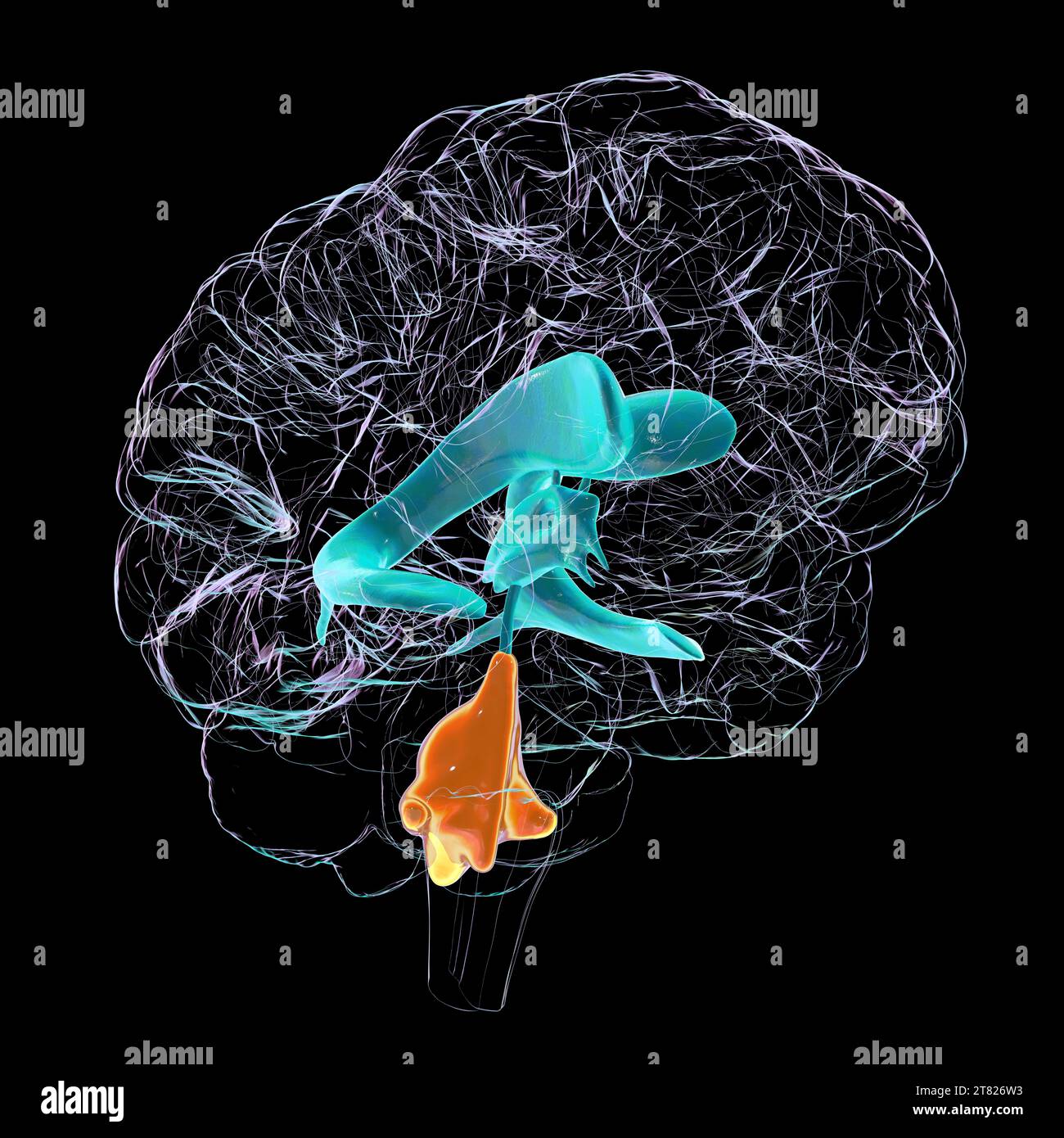 Enlarged fourth ventricle of the brain, illustration Stock Photohttps://www.alamy.com/image-license-details/?v=1https://www.alamy.com/enlarged-fourth-ventricle-of-the-brain-illustration-image572908703.html
RF2T826W3–Enlarged fourth ventricle of the brain, illustration
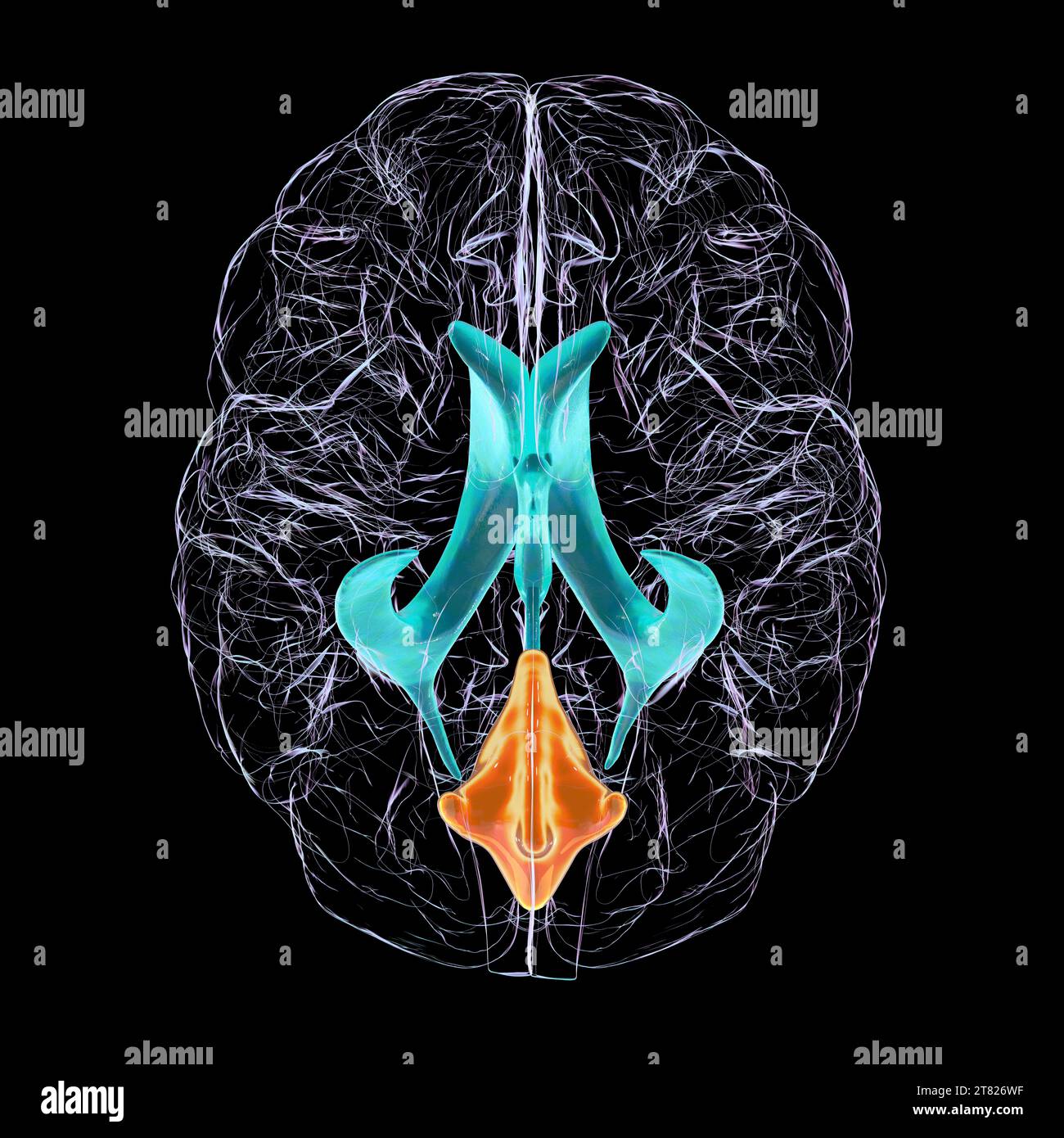 Enlarged fourth ventricle of the brain, illustration Stock Photohttps://www.alamy.com/image-license-details/?v=1https://www.alamy.com/enlarged-fourth-ventricle-of-the-brain-illustration-image572908715.html
RF2T826WF–Enlarged fourth ventricle of the brain, illustration
 Enlarged fourth ventricle of the brain, illustration Stock Photohttps://www.alamy.com/image-license-details/?v=1https://www.alamy.com/enlarged-fourth-ventricle-of-the-brain-illustration-image572908700.html
RF2T826W0–Enlarged fourth ventricle of the brain, illustration
 Enlarged lateral and third brain ventricles, illustration Stock Photohttps://www.alamy.com/image-license-details/?v=1https://www.alamy.com/enlarged-lateral-and-third-brain-ventricles-illustration-image603782274.html
RF2X28JCJ–Enlarged lateral and third brain ventricles, illustration
 Enlarged lateral ventricles of the brain, illustration Stock Photohttps://www.alamy.com/image-license-details/?v=1https://www.alamy.com/enlarged-lateral-ventricles-of-the-brain-illustration-image603782260.html
RF2X28JC4–Enlarged lateral ventricles of the brain, illustration
 Enlarged lateral and third brain ventricles, illustration Stock Photohttps://www.alamy.com/image-license-details/?v=1https://www.alamy.com/enlarged-lateral-and-third-brain-ventricles-illustration-image603782248.html
RF2X28JBM–Enlarged lateral and third brain ventricles, illustration
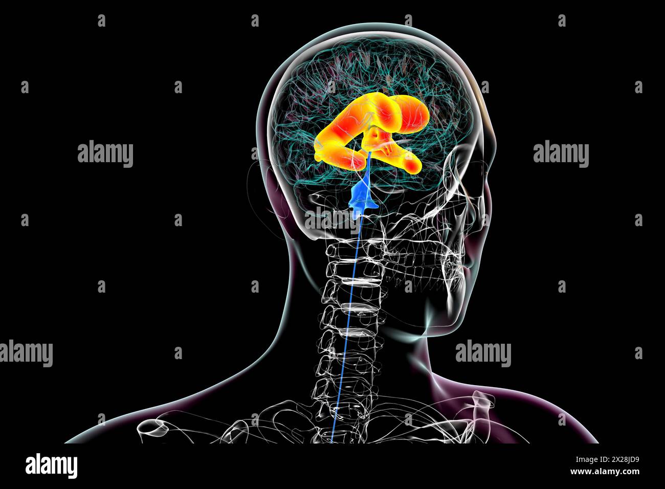 Enlarged lateral and third brain ventricles, illustration Stock Photohttps://www.alamy.com/image-license-details/?v=1https://www.alamy.com/enlarged-lateral-and-third-brain-ventricles-illustration-image603782293.html
RF2X28JD9–Enlarged lateral and third brain ventricles, illustration
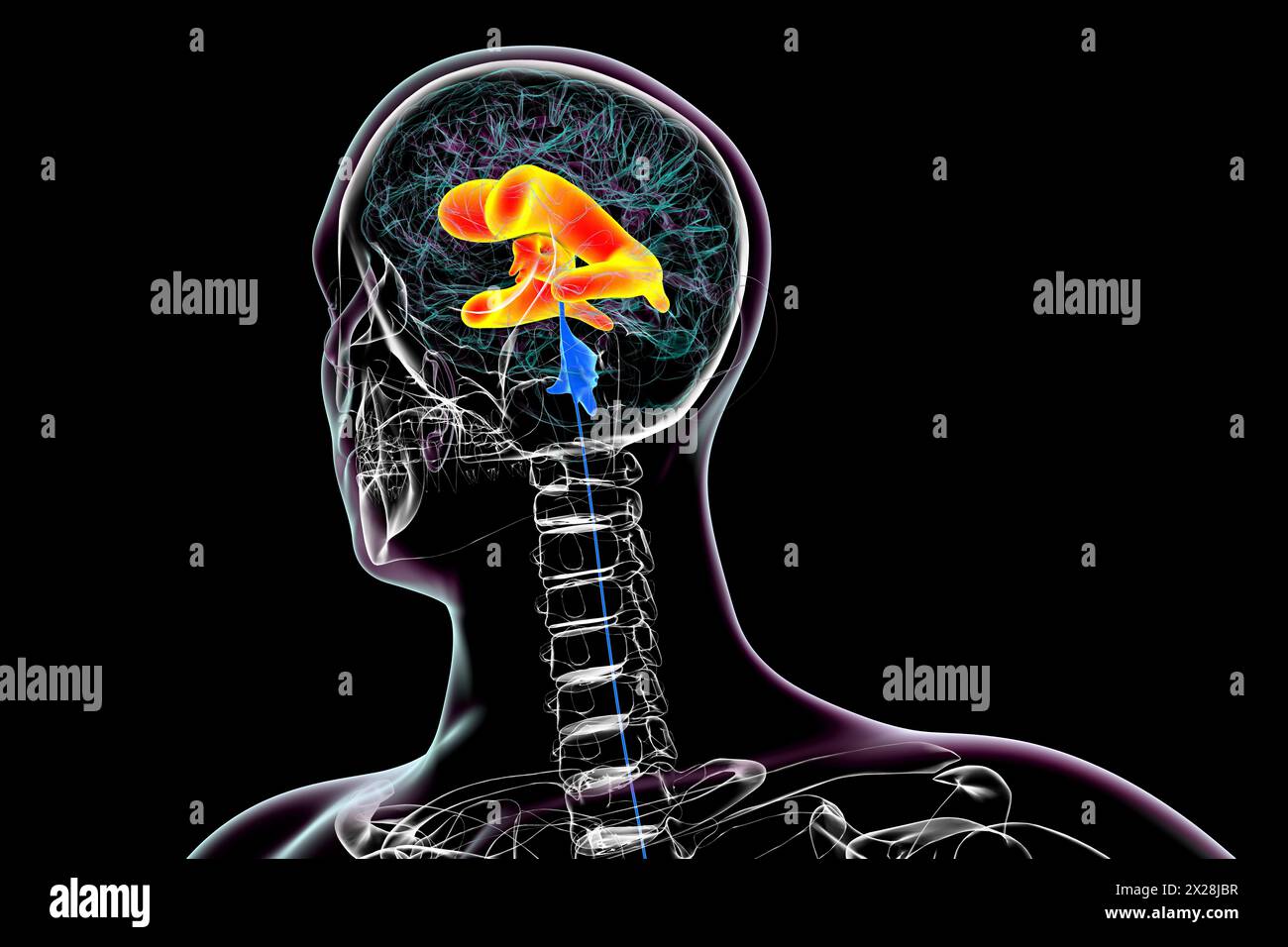 Enlarged lateral and third brain ventricles, illustration Stock Photohttps://www.alamy.com/image-license-details/?v=1https://www.alamy.com/enlarged-lateral-and-third-brain-ventricles-illustration-image603782251.html
RF2X28JBR–Enlarged lateral and third brain ventricles, illustration
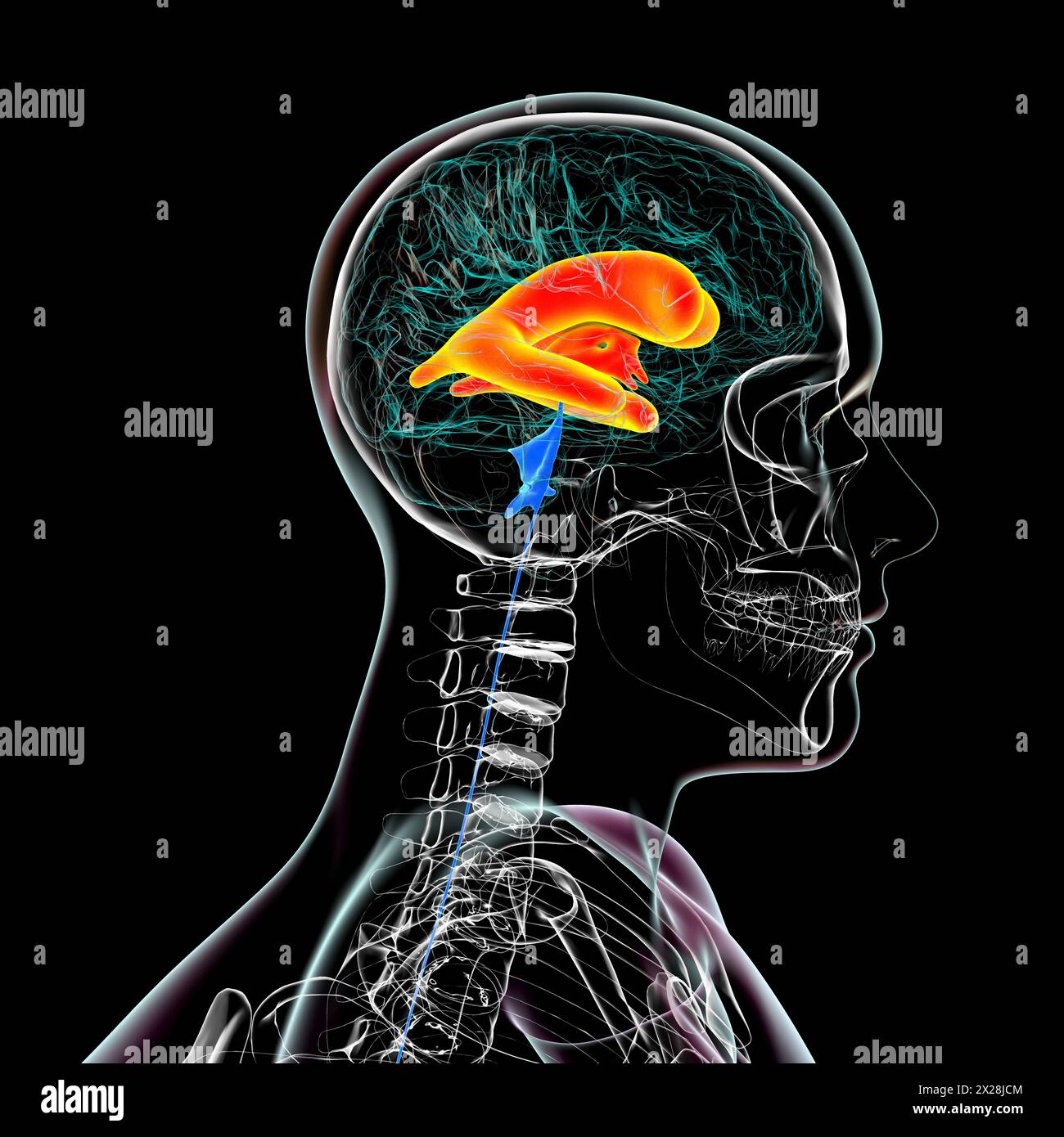 Enlarged lateral and third brain ventricles, illustration Stock Photohttps://www.alamy.com/image-license-details/?v=1https://www.alamy.com/enlarged-lateral-and-third-brain-ventricles-illustration-image603782276.html
RF2X28JCM–Enlarged lateral and third brain ventricles, illustration
 Enlarged lateral and third brain ventricles, illustration Stock Photohttps://www.alamy.com/image-license-details/?v=1https://www.alamy.com/enlarged-lateral-and-third-brain-ventricles-illustration-image603782256.html
RF2X28JC0–Enlarged lateral and third brain ventricles, illustration
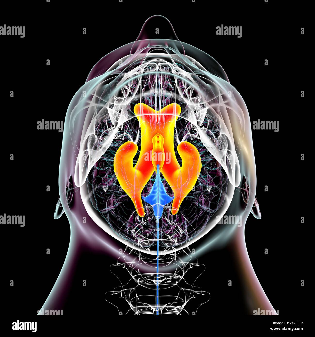 Enlarged lateral and third brain ventricles, illustration Stock Photohttps://www.alamy.com/image-license-details/?v=1https://www.alamy.com/enlarged-lateral-and-third-brain-ventricles-illustration-image603782279.html
RF2X28JCR–Enlarged lateral and third brain ventricles, illustration
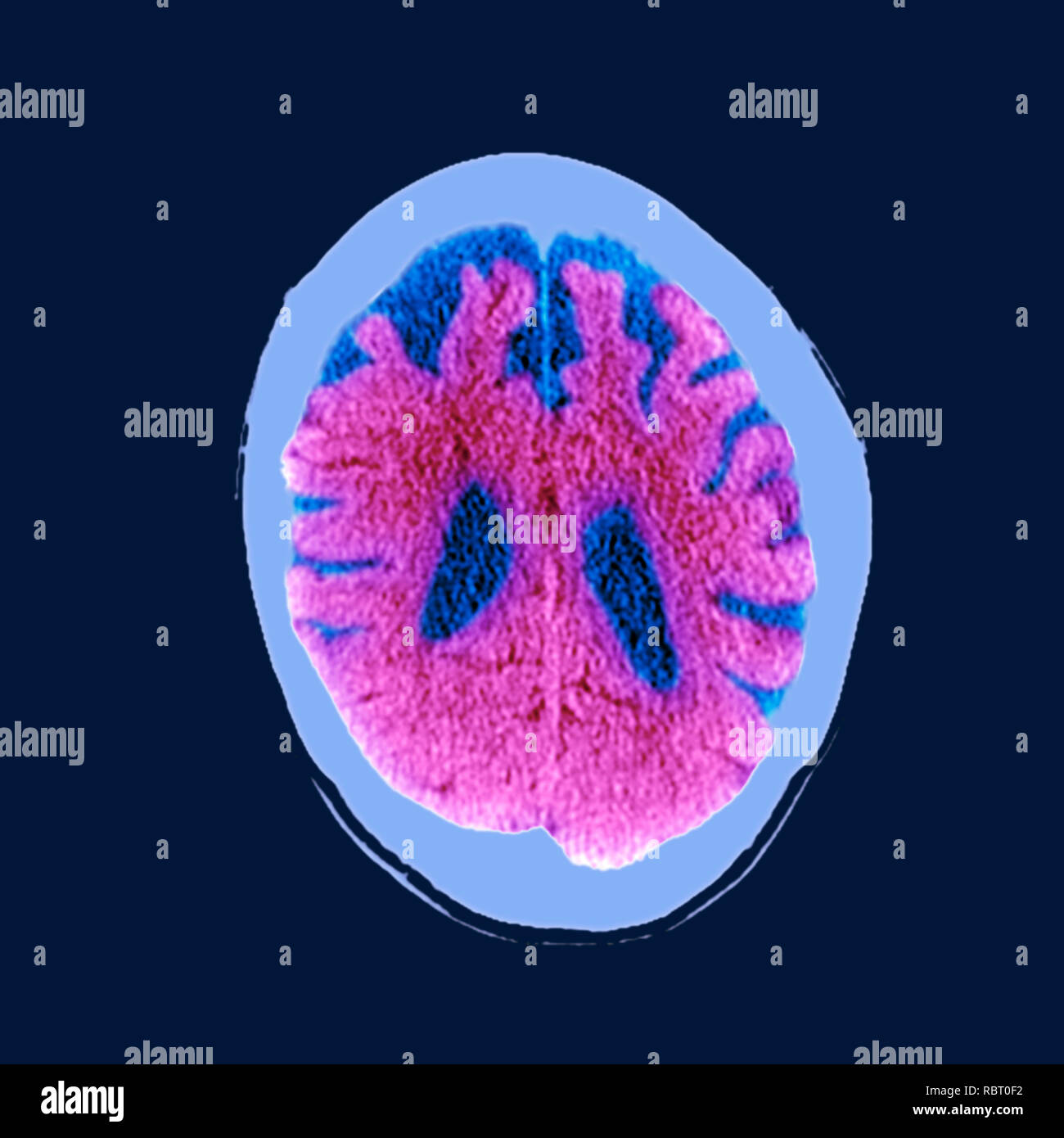 Brain in dementia. Coloured computed tomography (CT) scan of a section through the brain of an 89-year-old male patient with dementia. The brain has atrophied (shrunk), shown by the enlarged central ventricles (dark blue) and deep indentations around the brain's edges. Stock Photohttps://www.alamy.com/image-license-details/?v=1https://www.alamy.com/brain-in-dementia-coloured-computed-tomography-ct-scan-of-a-section-through-the-brain-of-an-89-year-old-male-patient-with-dementia-the-brain-has-atrophied-shrunk-shown-by-the-enlarged-central-ventricles-dark-blue-and-deep-indentations-around-the-brains-edges-image231023270.html
RFRBT0F2–Brain in dementia. Coloured computed tomography (CT) scan of a section through the brain of an 89-year-old male patient with dementia. The brain has atrophied (shrunk), shown by the enlarged central ventricles (dark blue) and deep indentations around the brain's edges.
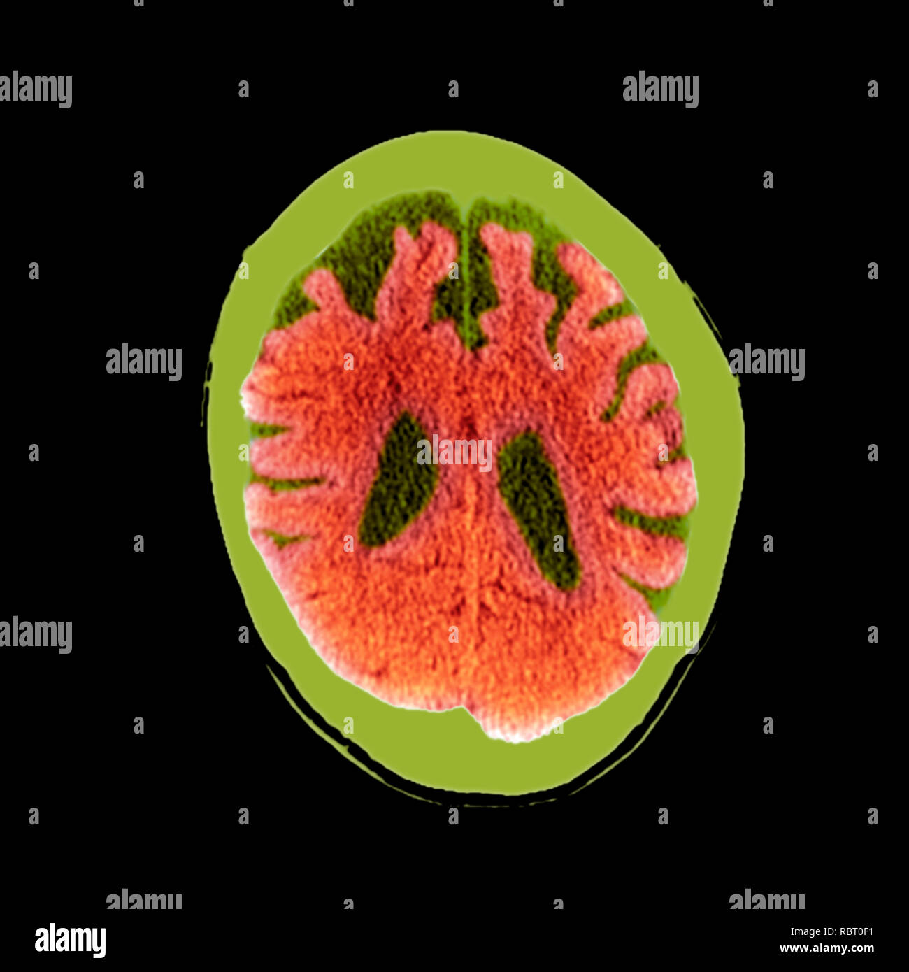 Brain in dementia. Coloured computed tomography (CT) scan of a section through the brain of an 89-year-old male patient with dementia. The brain has atrophied (shrunk), shown by the enlarged central ventricles (dark green) and deep indentations around the brain's edges. Stock Photohttps://www.alamy.com/image-license-details/?v=1https://www.alamy.com/brain-in-dementia-coloured-computed-tomography-ct-scan-of-a-section-through-the-brain-of-an-89-year-old-male-patient-with-dementia-the-brain-has-atrophied-shrunk-shown-by-the-enlarged-central-ventricles-dark-green-and-deep-indentations-around-the-brains-edges-image231023269.html
RFRBT0F1–Brain in dementia. Coloured computed tomography (CT) scan of a section through the brain of an 89-year-old male patient with dementia. The brain has atrophied (shrunk), shown by the enlarged central ventricles (dark green) and deep indentations around the brain's edges.
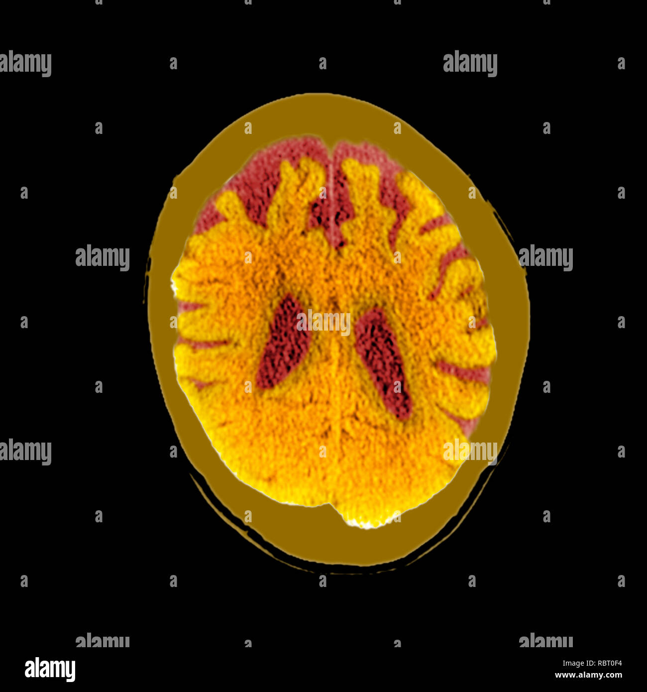 Brain in dementia. Coloured computed tomography (CT) scan of a section through the brain of an 89-year-old male patient with dementia. The brain has atrophied (shrunk), shown by the enlarged central ventricles (dark red) and deep indentations around the brain's edges. Stock Photohttps://www.alamy.com/image-license-details/?v=1https://www.alamy.com/brain-in-dementia-coloured-computed-tomography-ct-scan-of-a-section-through-the-brain-of-an-89-year-old-male-patient-with-dementia-the-brain-has-atrophied-shrunk-shown-by-the-enlarged-central-ventricles-dark-red-and-deep-indentations-around-the-brains-edges-image231023272.html
RFRBT0F4–Brain in dementia. Coloured computed tomography (CT) scan of a section through the brain of an 89-year-old male patient with dementia. The brain has atrophied (shrunk), shown by the enlarged central ventricles (dark red) and deep indentations around the brain's edges.
 Brain in dementia. Coloured computed tomography (CT) scan of a section through the brain of an 89-year-old male patient with dementia. The brain has atrophied (shrunk), shown by the enlarged central ventricles and deep indentations around the brain's edges. Stock Photohttps://www.alamy.com/image-license-details/?v=1https://www.alamy.com/brain-in-dementia-coloured-computed-tomography-ct-scan-of-a-section-through-the-brain-of-an-89-year-old-male-patient-with-dementia-the-brain-has-atrophied-shrunk-shown-by-the-enlarged-central-ventricles-and-deep-indentations-around-the-brains-edges-image231023268.html
RFRBT0F0–Brain in dementia. Coloured computed tomography (CT) scan of a section through the brain of an 89-year-old male patient with dementia. The brain has atrophied (shrunk), shown by the enlarged central ventricles and deep indentations around the brain's edges.
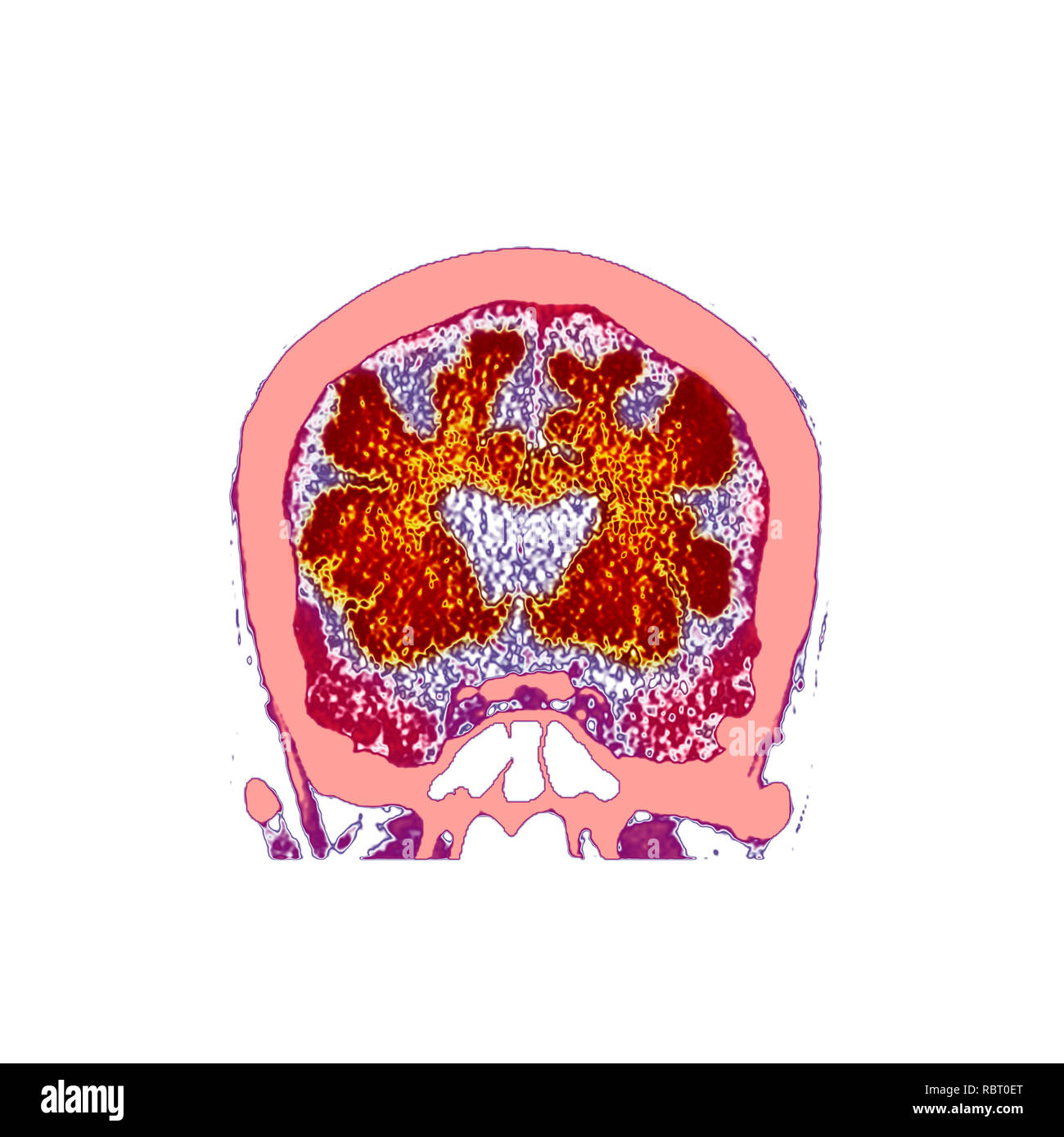 Brain in dementia. Coloured computed tomography (CT) scan of a section through the brain of an 89-year-old male patient with dementia. The brain has atrophied (shrunk), shown by the enlarged central ventricles and deep indentations around the brain's edges. Stock Photohttps://www.alamy.com/image-license-details/?v=1https://www.alamy.com/brain-in-dementia-coloured-computed-tomography-ct-scan-of-a-section-through-the-brain-of-an-89-year-old-male-patient-with-dementia-the-brain-has-atrophied-shrunk-shown-by-the-enlarged-central-ventricles-and-deep-indentations-around-the-brains-edges-image231023264.html
RFRBT0ET–Brain in dementia. Coloured computed tomography (CT) scan of a section through the brain of an 89-year-old male patient with dementia. The brain has atrophied (shrunk), shown by the enlarged central ventricles and deep indentations around the brain's edges.
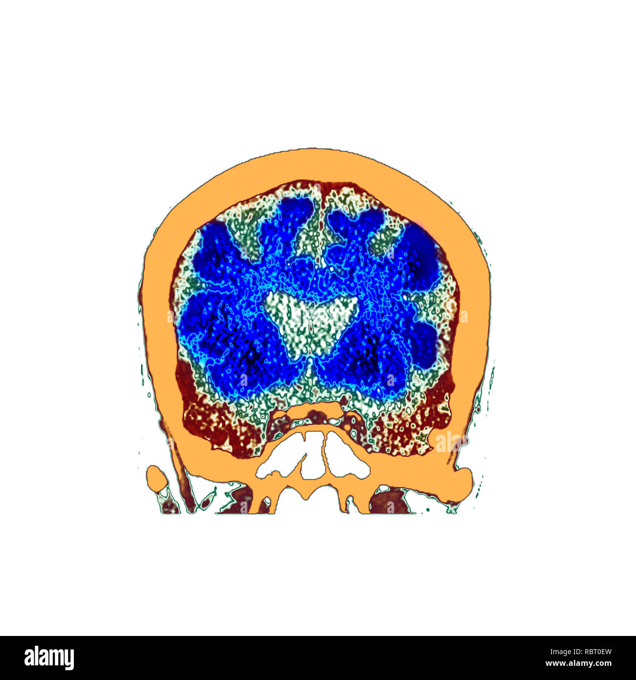 Brain in dementia. Coloured computed tomography (CT) scan of a section through the brain of an 89-year-old male patient with dementia. The brain has atrophied (shrunk), shown by the enlarged central ventricles and deep indentations around the brain's edges. Stock Photohttps://www.alamy.com/image-license-details/?v=1https://www.alamy.com/brain-in-dementia-coloured-computed-tomography-ct-scan-of-a-section-through-the-brain-of-an-89-year-old-male-patient-with-dementia-the-brain-has-atrophied-shrunk-shown-by-the-enlarged-central-ventricles-and-deep-indentations-around-the-brains-edges-image231023265.html
RFRBT0EW–Brain in dementia. Coloured computed tomography (CT) scan of a section through the brain of an 89-year-old male patient with dementia. The brain has atrophied (shrunk), shown by the enlarged central ventricles and deep indentations around the brain's edges.
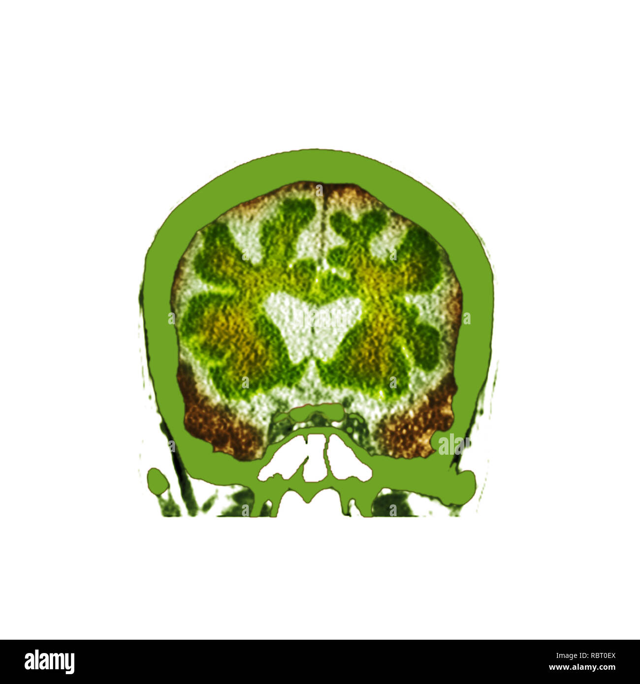 Brain in dementia. Coloured computed tomography (CT) scan of a section through the brain of an 89-year-old male patient with dementia. The brain has atrophied (shrunk), shown by the enlarged central ventricles and deep indentations around the brain's edges. Stock Photohttps://www.alamy.com/image-license-details/?v=1https://www.alamy.com/brain-in-dementia-coloured-computed-tomography-ct-scan-of-a-section-through-the-brain-of-an-89-year-old-male-patient-with-dementia-the-brain-has-atrophied-shrunk-shown-by-the-enlarged-central-ventricles-and-deep-indentations-around-the-brains-edges-image231023266.html
RFRBT0EX–Brain in dementia. Coloured computed tomography (CT) scan of a section through the brain of an 89-year-old male patient with dementia. The brain has atrophied (shrunk), shown by the enlarged central ventricles and deep indentations around the brain's edges.
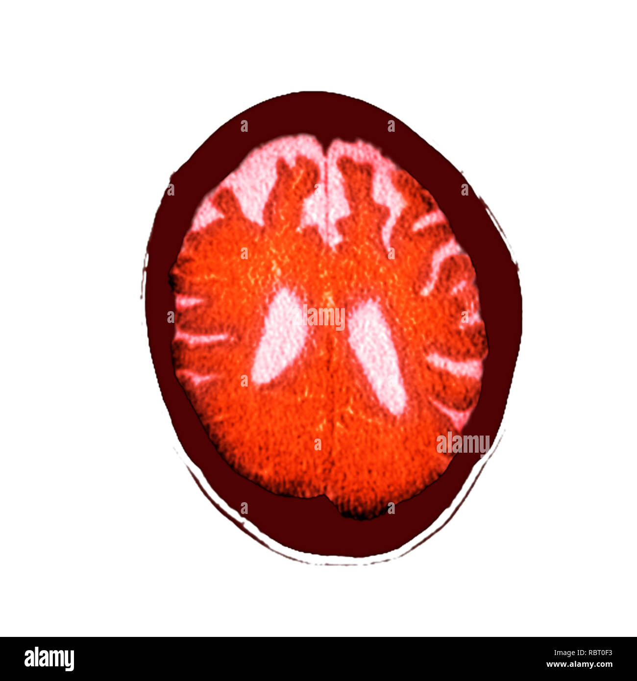 Brain in dementia. Coloured computed tomography (CT) scan of a section through the brain of an 89-year-old male patient with dementia. The brain has atrophied (shrunk), shown by the enlarged central ventricles (white) and deep indentations around the brain's edges. Stock Photohttps://www.alamy.com/image-license-details/?v=1https://www.alamy.com/brain-in-dementia-coloured-computed-tomography-ct-scan-of-a-section-through-the-brain-of-an-89-year-old-male-patient-with-dementia-the-brain-has-atrophied-shrunk-shown-by-the-enlarged-central-ventricles-white-and-deep-indentations-around-the-brains-edges-image231023271.html
RFRBT0F3–Brain in dementia. Coloured computed tomography (CT) scan of a section through the brain of an 89-year-old male patient with dementia. The brain has atrophied (shrunk), shown by the enlarged central ventricles (white) and deep indentations around the brain's edges.
 Brain tumour causing hydrocephalus, illustration Stock Photohttps://www.alamy.com/image-license-details/?v=1https://www.alamy.com/brain-tumour-causing-hydrocephalus-illustration-image603782297.html
RF2X28JDD–Brain tumour causing hydrocephalus, illustration
 Brain tumour causing hydrocephalus, illustration Stock Photohttps://www.alamy.com/image-license-details/?v=1https://www.alamy.com/brain-tumour-causing-hydrocephalus-illustration-image603782307.html
RF2X28JDR–Brain tumour causing hydrocephalus, illustration
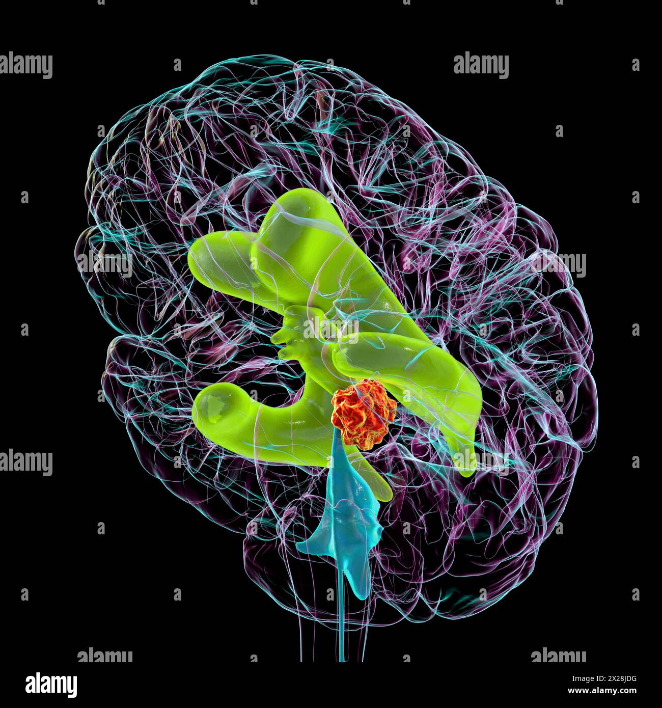 Brain tumour causing hydrocephalus, illustration Stock Photohttps://www.alamy.com/image-license-details/?v=1https://www.alamy.com/brain-tumour-causing-hydrocephalus-illustration-image603782300.html
RF2X28JDG–Brain tumour causing hydrocephalus, illustration
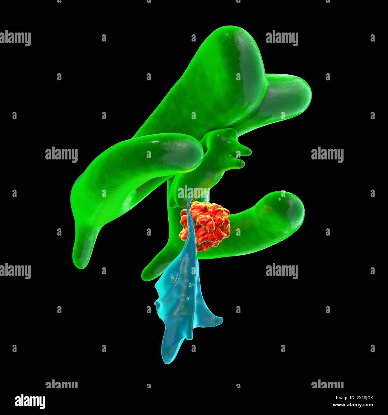 Brain tumour causing hydrocephalus, illustration Stock Photohttps://www.alamy.com/image-license-details/?v=1https://www.alamy.com/brain-tumour-causing-hydrocephalus-illustration-image603782303.html
RF2X28JDK–Brain tumour causing hydrocephalus, illustration
 Brain tumour causing hydrocephalus, illustration Stock Photohttps://www.alamy.com/image-license-details/?v=1https://www.alamy.com/brain-tumour-causing-hydrocephalus-illustration-image603782294.html
RF2X28JDA–Brain tumour causing hydrocephalus, illustration
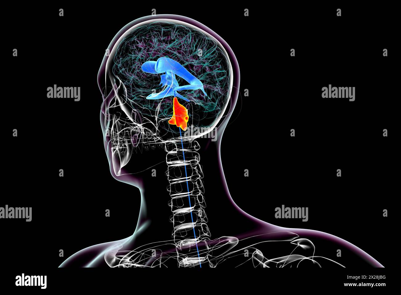 Enlargement of the fourth brain ventricle, illustration Stock Photohttps://www.alamy.com/image-license-details/?v=1https://www.alamy.com/enlargement-of-the-fourth-brain-ventricle-illustration-image603782244.html
RF2X28JBG–Enlargement of the fourth brain ventricle, illustration
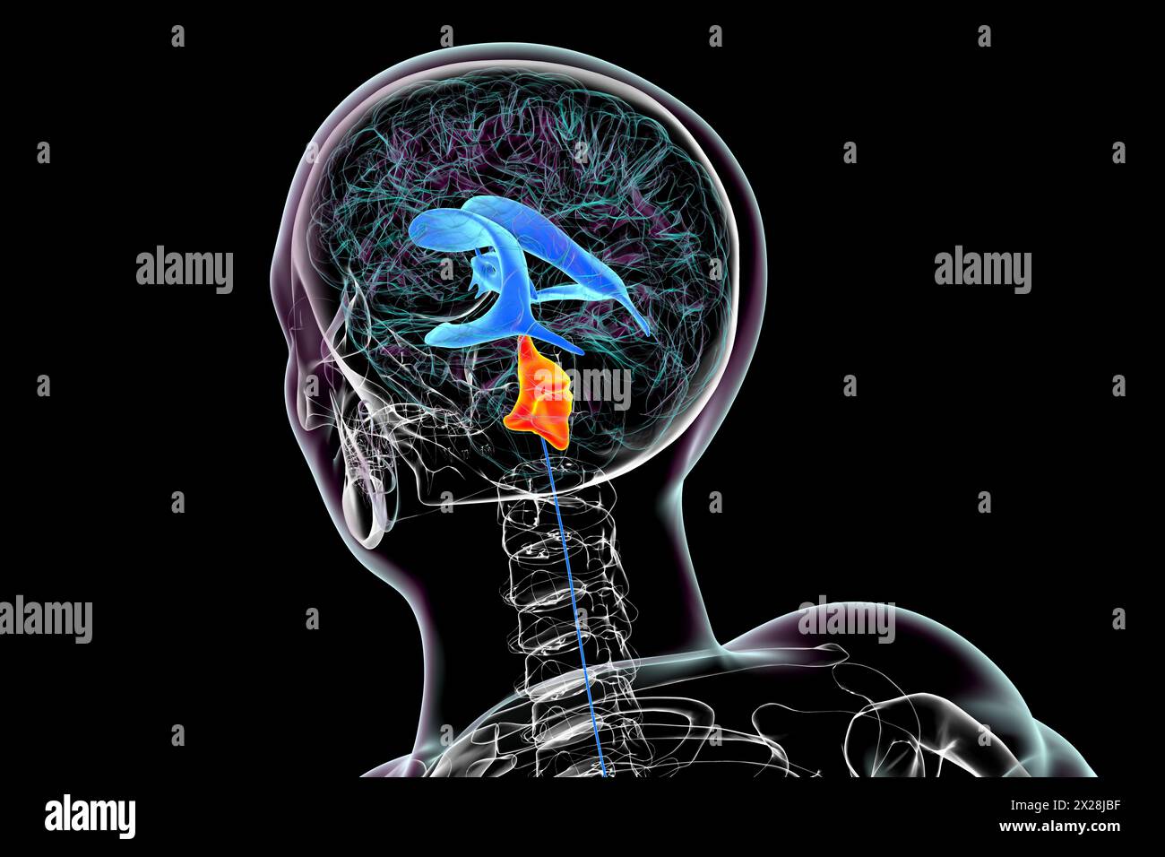 Enlargement of the fourth brain ventricle, illustration Stock Photohttps://www.alamy.com/image-license-details/?v=1https://www.alamy.com/enlargement-of-the-fourth-brain-ventricle-illustration-image603782243.html
RF2X28JBF–Enlargement of the fourth brain ventricle, illustration
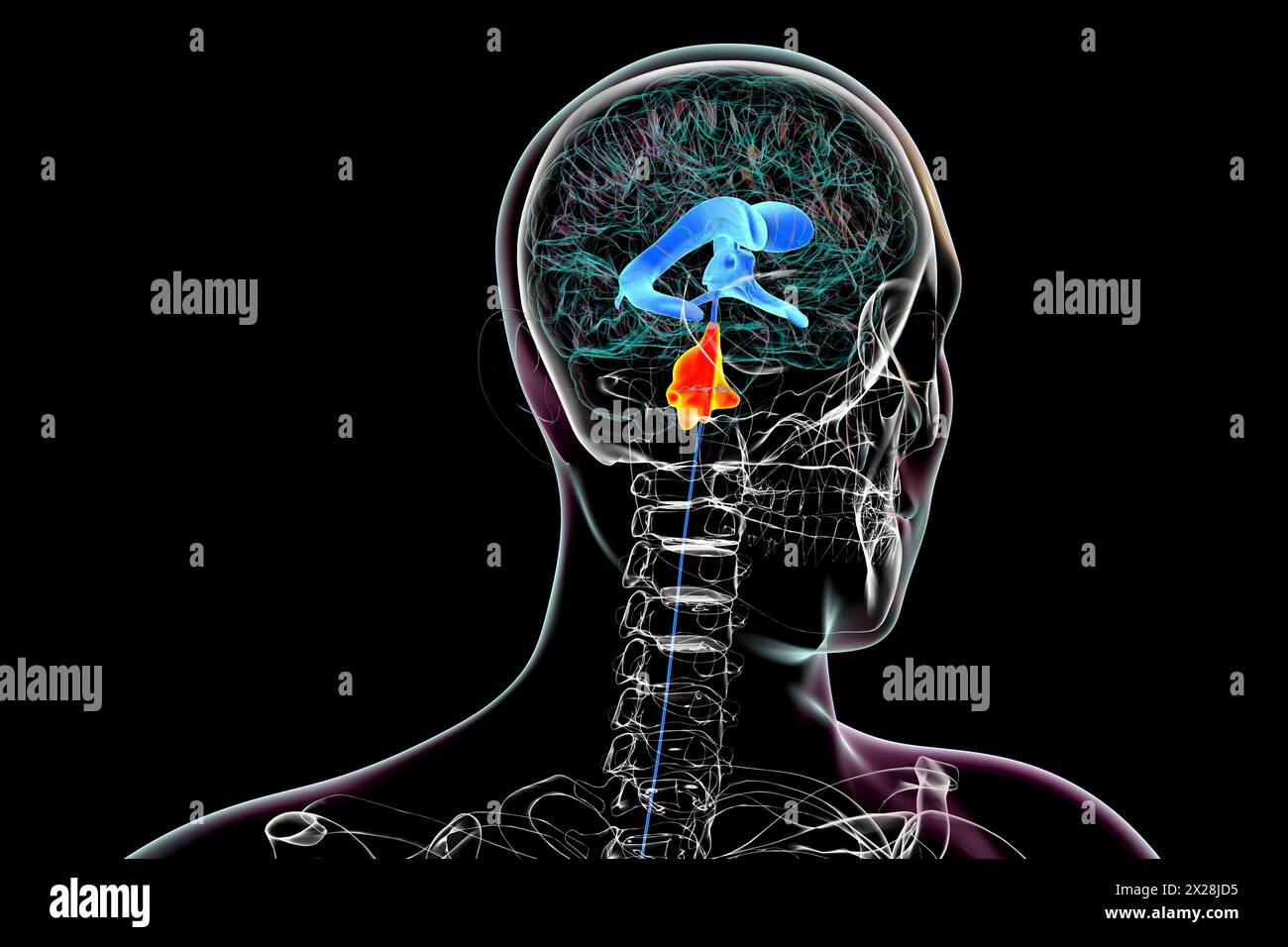 Enlargement of the fourth brain ventricle, illustration Stock Photohttps://www.alamy.com/image-license-details/?v=1https://www.alamy.com/enlargement-of-the-fourth-brain-ventricle-illustration-image603782289.html
RF2X28JD5–Enlargement of the fourth brain ventricle, illustration
 Brain tumour causing hydrocephalus, illustration Stock Photohttps://www.alamy.com/image-license-details/?v=1https://www.alamy.com/brain-tumour-causing-hydrocephalus-illustration-image603782309.html
RF2X28JDW–Brain tumour causing hydrocephalus, illustration
 Brain tumour causing hydrocephalus, illustration Stock Photohttps://www.alamy.com/image-license-details/?v=1https://www.alamy.com/brain-tumour-causing-hydrocephalus-illustration-image603782270.html
RF2X28JCE–Brain tumour causing hydrocephalus, illustration
