Enzyme protein Cut Out Stock Images
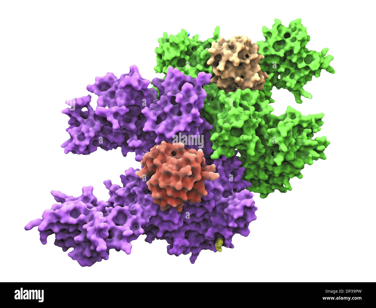 Ubiquitin activating enzyme protein E1 Stock Photohttps://www.alamy.com/image-license-details/?v=1https://www.alamy.com/ubiquitin-activating-enzyme-protein-e1-image65227089.html
RFDP39PW–Ubiquitin activating enzyme protein E1
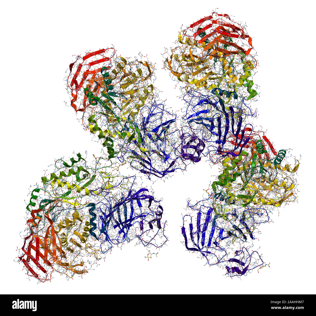 Sucrase-isomaltase enzyme structure Stock Photohttps://www.alamy.com/image-license-details/?v=1https://www.alamy.com/sucrase-isomaltase-enzyme-structure-image333530631.html
RF2AAHHM7–Sucrase-isomaltase enzyme structure
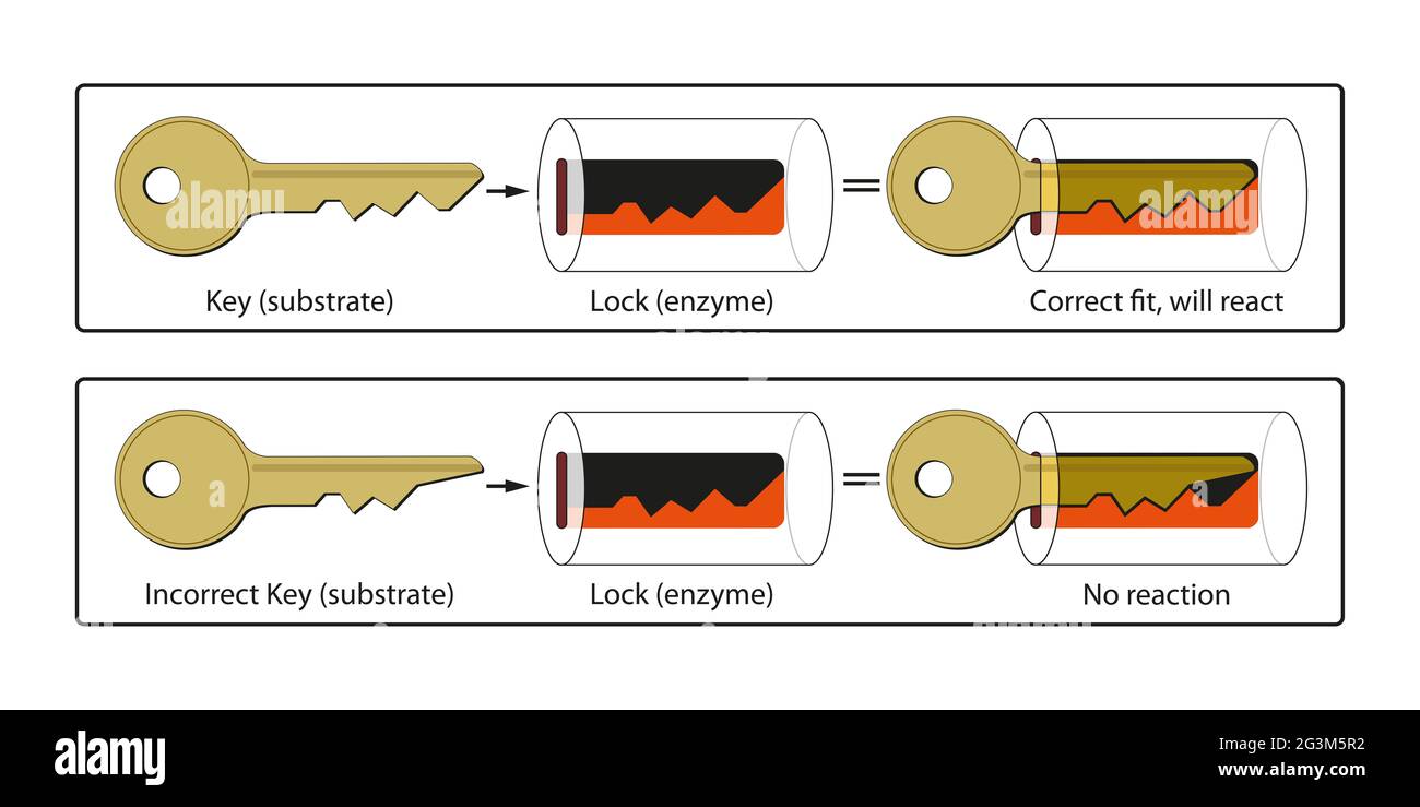 The basic mechanism by which enzymes catalyze chemical reactions begins with the binding of the substrate to the active site on the enzyme Stock Photohttps://www.alamy.com/image-license-details/?v=1https://www.alamy.com/the-basic-mechanism-by-which-enzymes-catalyze-chemical-reactions-begins-with-the-binding-of-the-substrate-to-the-active-site-on-the-enzyme-image432546774.html
RF2G3M5R2–The basic mechanism by which enzymes catalyze chemical reactions begins with the binding of the substrate to the active site on the enzyme
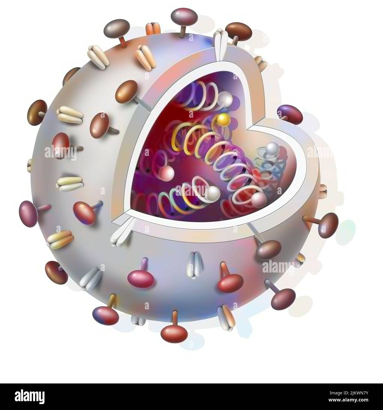 Schematic of any virus with DNA strands inside. Stock Photohttps://www.alamy.com/image-license-details/?v=1https://www.alamy.com/schematic-of-any-virus-with-dna-strands-inside-image476923887.html
RF2JKWN7Y–Schematic of any virus with DNA strands inside.
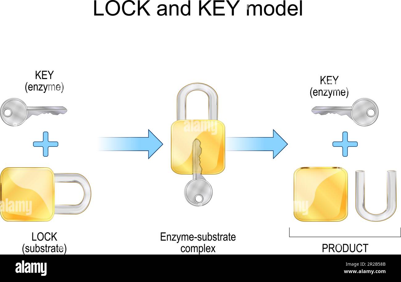 Lock and key model. Enzyme, substrate, products, and chemical mechanism. Degradation. metabolic processes. Vector illustration Stock Vectorhttps://www.alamy.com/image-license-details/?v=1https://www.alamy.com/lock-and-key-model-enzyme-substrate-products-and-chemical-mechanism-degradation-metabolic-processes-vector-illustration-image552206715.html
RF2R2B58B–Lock and key model. Enzyme, substrate, products, and chemical mechanism. Degradation. metabolic processes. Vector illustration
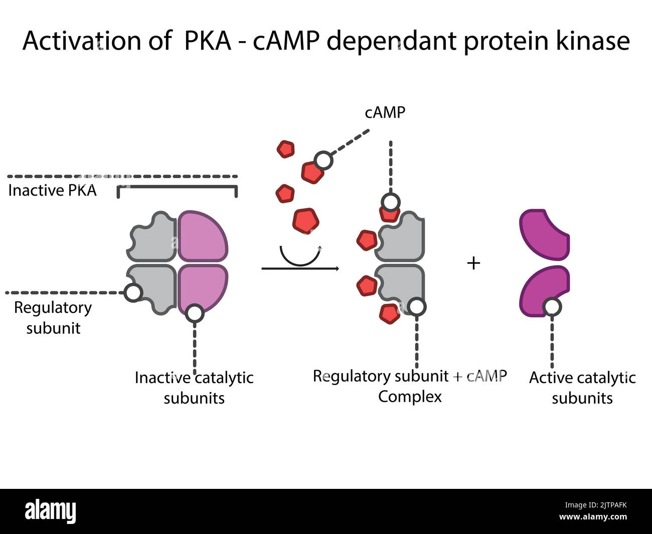 Activation of PKA (Protein Kinase A) via cyclic AMP in GPCR Gs signaling schematic diagram. Stock Vectorhttps://www.alamy.com/image-license-details/?v=1https://www.alamy.com/activation-of-pka-protein-kinase-a-via-cyclic-amp-in-gpcr-gs-signaling-schematic-diagram-image479922903.html
RF2JTPAFK–Activation of PKA (Protein Kinase A) via cyclic AMP in GPCR Gs signaling schematic diagram.
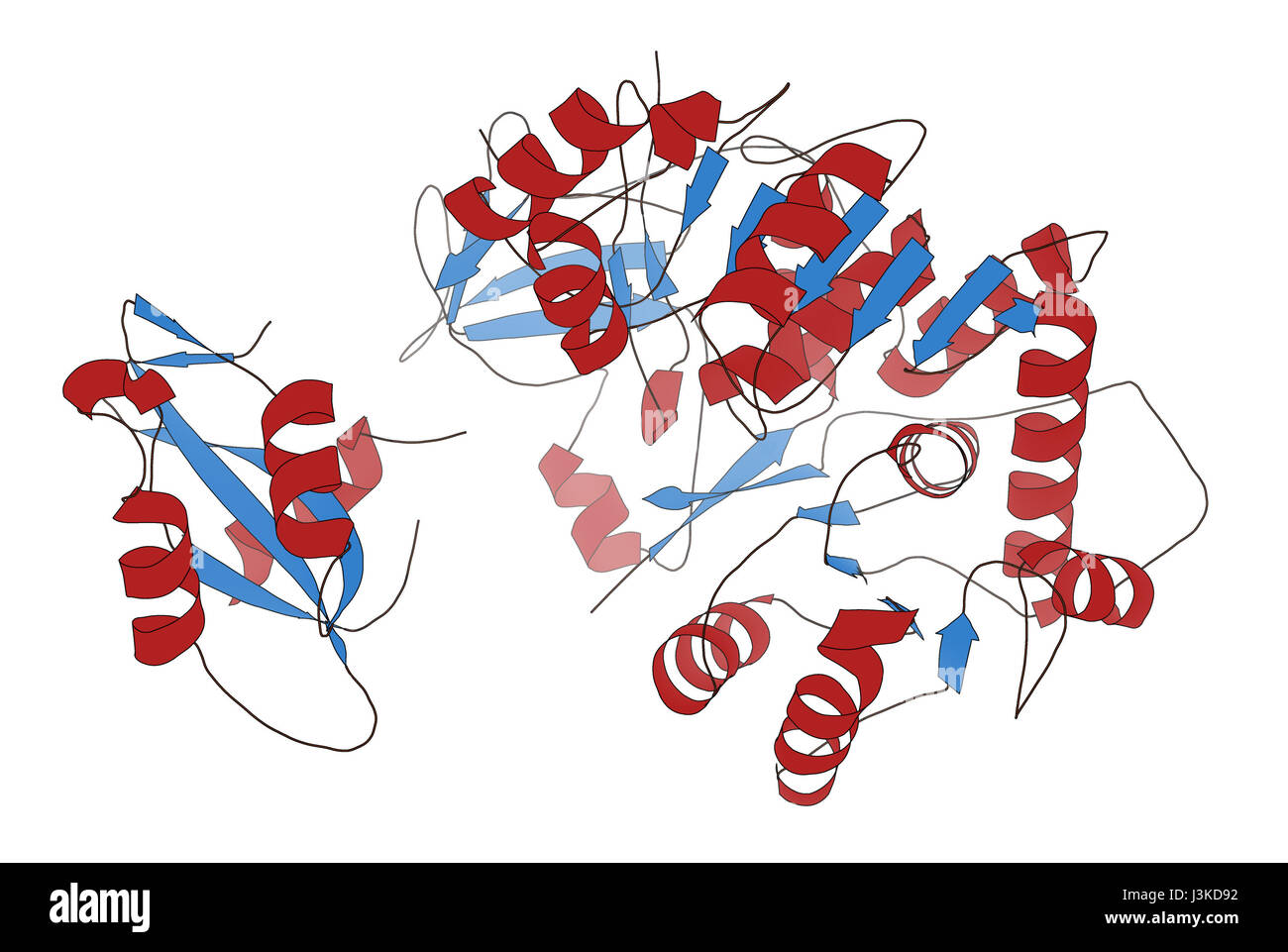 Firefly luciferase enzyme. Protein responsible for the bioluminescence of fireflies. Often used as reporter in biotechnology and genetic engineering. Stock Photohttps://www.alamy.com/image-license-details/?v=1https://www.alamy.com/stock-photo-firefly-luciferase-enzyme-protein-responsible-for-the-bioluminescence-139954446.html
RFJ3KD92–Firefly luciferase enzyme. Protein responsible for the bioluminescence of fireflies. Often used as reporter in biotechnology and genetic engineering.
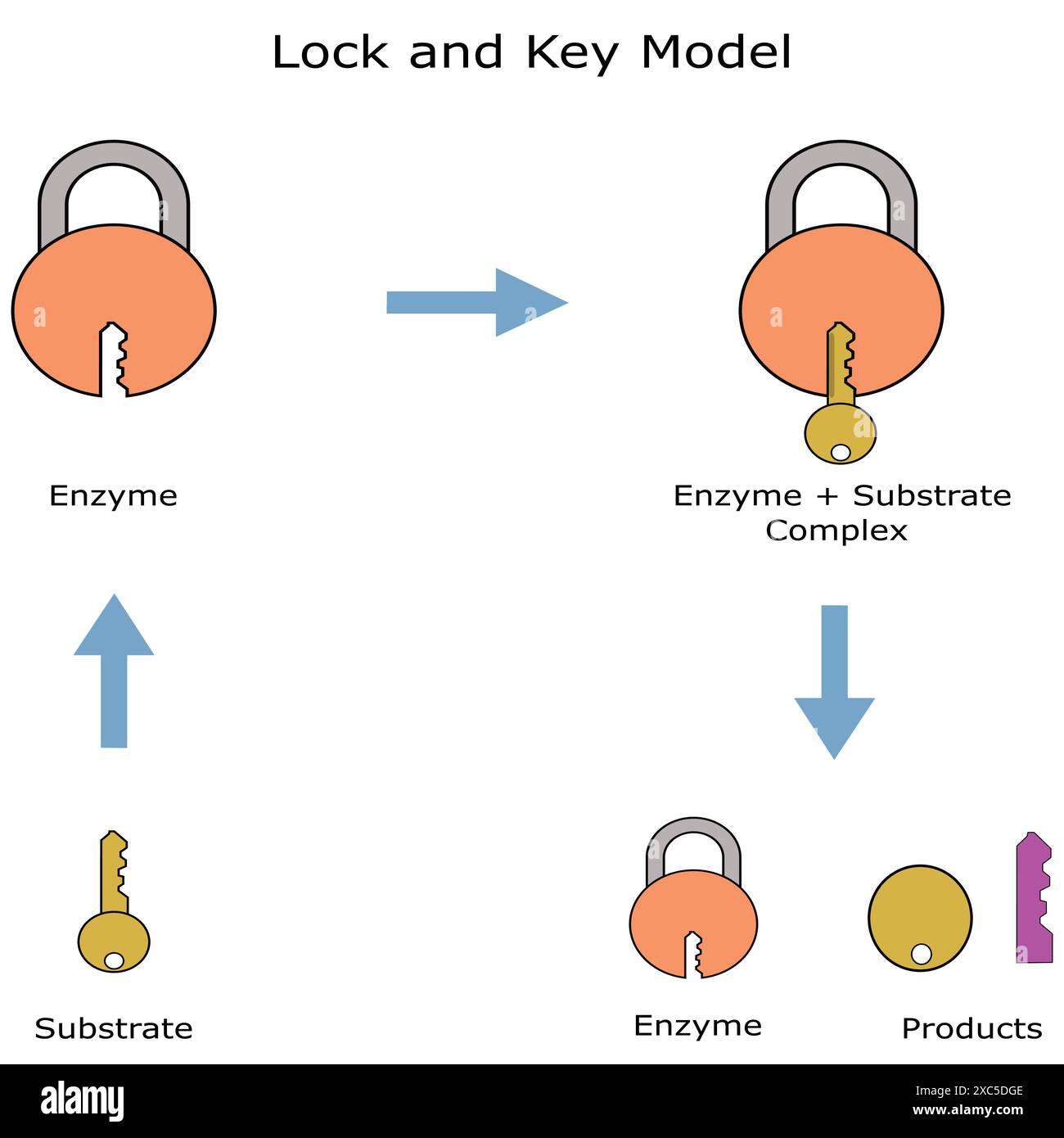 Key and lock hypothesis. Mechanism of action of the enzyme. Stock Vectorhttps://www.alamy.com/image-license-details/?v=1https://www.alamy.com/key-and-lock-hypothesis-mechanism-of-action-of-the-enzyme-image609859166.html
RF2XC5DGE–Key and lock hypothesis. Mechanism of action of the enzyme.
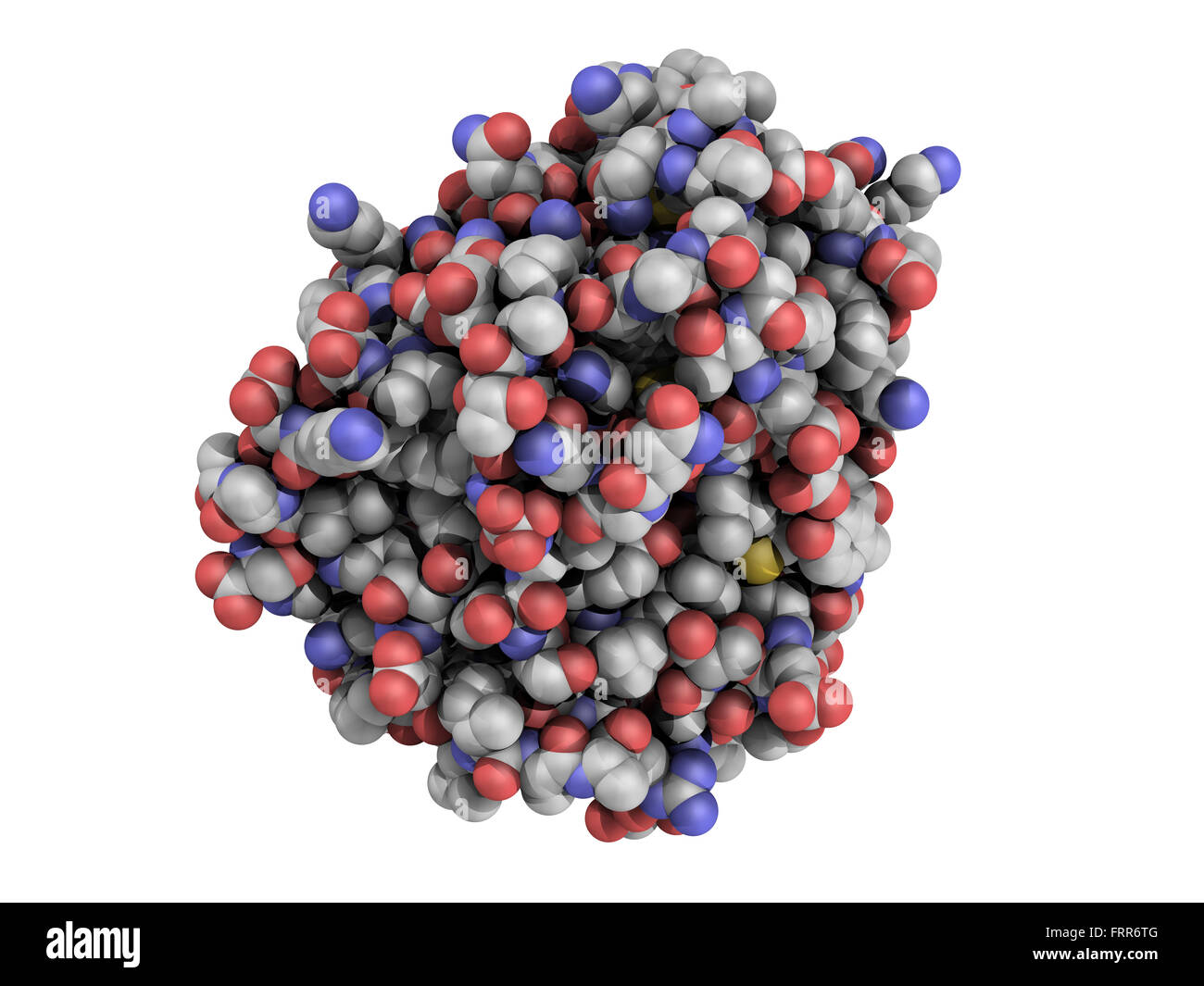 Trypsin digestive enzyme molecule (human), chemical structure. Trypsin is an enzyme that contributes to the digestion of protein Stock Photohttps://www.alamy.com/image-license-details/?v=1https://www.alamy.com/stock-photo-trypsin-digestive-enzyme-molecule-human-chemical-structure-trypsin-100699216.html
RFFRR6TG–Trypsin digestive enzyme molecule (human), chemical structure. Trypsin is an enzyme that contributes to the digestion of protein
 Human Mitochondrial PheRS Stock Photohttps://www.alamy.com/image-license-details/?v=1https://www.alamy.com/human-mitochondrial-phers-image416784511.html
RM2F624W3–Human Mitochondrial PheRS
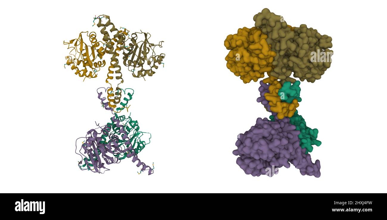 Structure of ubiquitin carboxyl-terminal hydrolase isozyme L5 (Uch37) tetramer. 3D cartoon and Gaussian surface models, PDB 3ihr Stock Photohttps://www.alamy.com/image-license-details/?v=1https://www.alamy.com/structure-of-ubiquitin-carboxyl-terminal-hydrolase-isozyme-l5-uch37-tetramer-3d-cartoon-and-gaussian-surface-models-pdb-3ihr-image463849341.html
RF2HXJ4FW–Structure of ubiquitin carboxyl-terminal hydrolase isozyme L5 (Uch37) tetramer. 3D cartoon and Gaussian surface models, PDB 3ihr
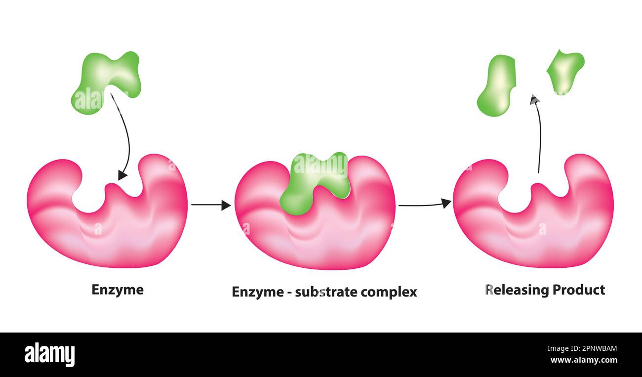 lock and key mechanism of enzyme catalysis Stock Vectorhttps://www.alamy.com/image-license-details/?v=1https://www.alamy.com/lock-and-key-mechanism-of-enzyme-catalysis-image546986908.html
RF2PNWBAM–lock and key mechanism of enzyme catalysis
 3D image of Protochlorophyllide skeletal formula - molecular chemical structure of Monovinyl protochlorophyllide isolated on white background Stock Photohttps://www.alamy.com/image-license-details/?v=1https://www.alamy.com/3d-image-of-protochlorophyllide-skeletal-formula-molecular-chemical-structure-of-monovinyl-protochlorophyllide-isolated-on-white-background-image500161682.html
RF2M1M996–3D image of Protochlorophyllide skeletal formula - molecular chemical structure of Monovinyl protochlorophyllide isolated on white background
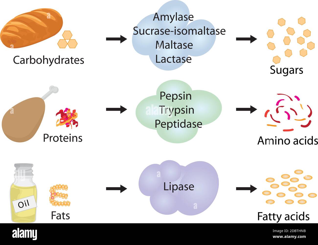 Enzymes braking down food into nutrients. Digestive systems work Stock Photohttps://www.alamy.com/image-license-details/?v=1https://www.alamy.com/enzymes-braking-down-food-into-nutrients-digestive-systems-work-image385930087.html
RF2DBTHNB–Enzymes braking down food into nutrients. Digestive systems work
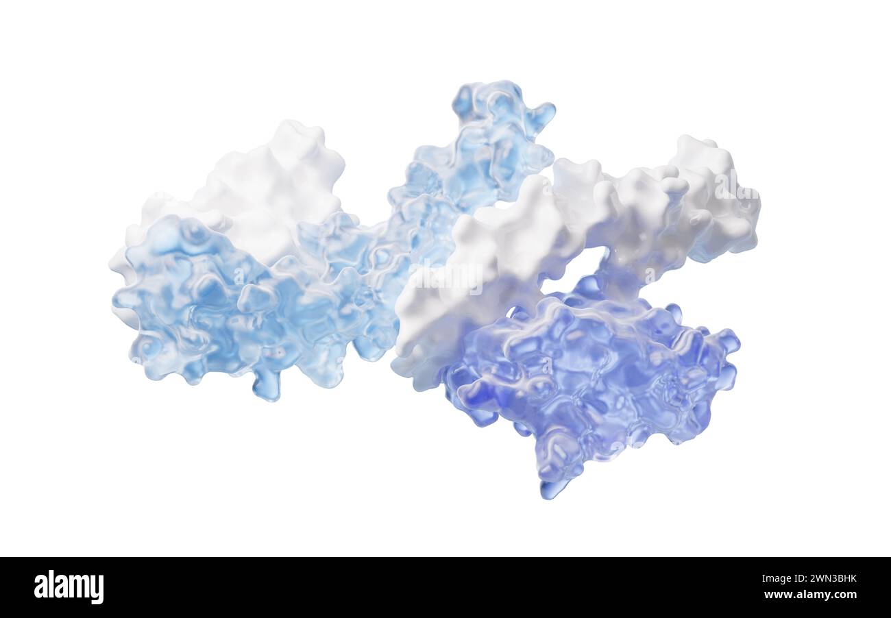 Protein structure with biological concept, 3d rendering. 3D illustration. Stock Photohttps://www.alamy.com/image-license-details/?v=1https://www.alamy.com/protein-structure-with-biological-concept-3d-rendering-3d-illustration-image598135263.html
RF2WN3BHK–Protein structure with biological concept, 3d rendering. 3D illustration.
 A bottle of Probiotic Yogurt Drink isolated on white background Stock Photohttps://www.alamy.com/image-license-details/?v=1https://www.alamy.com/a-bottle-of-probiotic-yogurt-drink-isolated-on-white-background-image424985715.html
RF2FKBNH7–A bottle of Probiotic Yogurt Drink isolated on white background
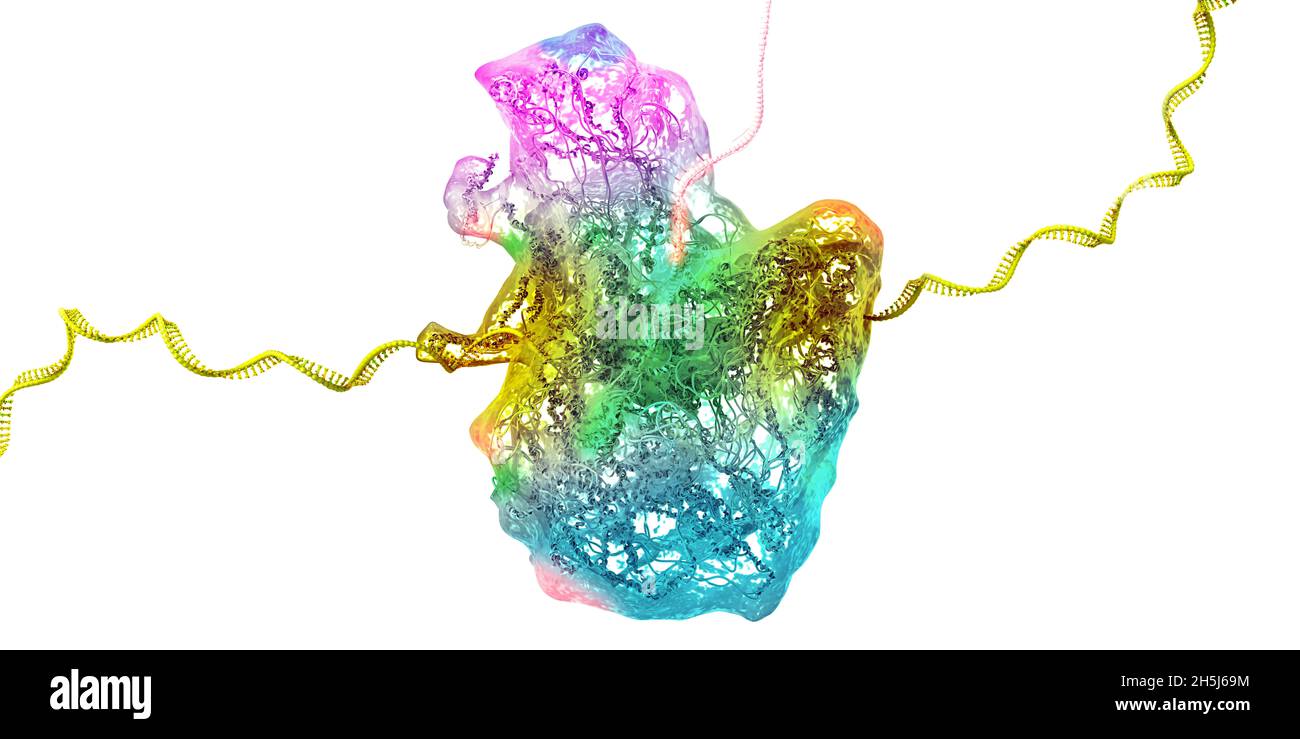 Ribosome as part of an biological cell constructing messenger rna molecule - 3d illustration Stock Photohttps://www.alamy.com/image-license-details/?v=1https://www.alamy.com/ribosome-as-part-of-an-biological-cell-constructing-messenger-rna-molecule-3d-illustration-image450942960.html
RF2H5J69M–Ribosome as part of an biological cell constructing messenger rna molecule - 3d illustration
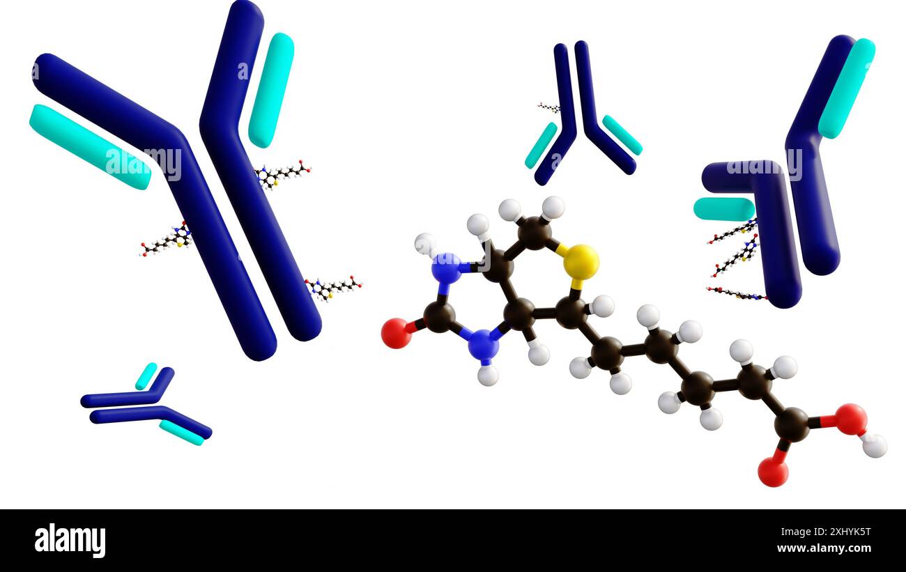 3D rendering of biotinylated antibody, an engineered protein designed to bind to specific molecules. Stock Photohttps://www.alamy.com/image-license-details/?v=1https://www.alamy.com/3d-rendering-of-biotinylated-antibody-an-engineered-protein-designed-to-bind-to-specific-molecules-image613419796.html
RF2XHYK5T–3D rendering of biotinylated antibody, an engineered protein designed to bind to specific molecules.
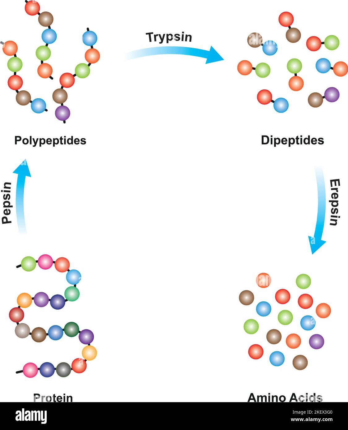 Scientific Designing of Protein digestion. Pepsin, Trypsin and Erepsin Enzymes Effect on Protein Molecule. Colorful Symbols. Vector Illustration. Stock Vectorhttps://www.alamy.com/image-license-details/?v=1https://www.alamy.com/scientific-designing-of-protein-digestion-pepsin-trypsin-and-erepsin-enzymes-effect-on-protein-molecule-colorful-symbols-vector-illustration-image491069040.html
RF2KEX3G0–Scientific Designing of Protein digestion. Pepsin, Trypsin and Erepsin Enzymes Effect on Protein Molecule. Colorful Symbols. Vector Illustration.
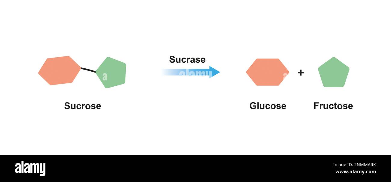 Sucrase enzyme, illustration Stock Photohttps://www.alamy.com/image-license-details/?v=1https://www.alamy.com/sucrase-enzyme-illustration-image529051703.html
RF2NMMARK–Sucrase enzyme, illustration
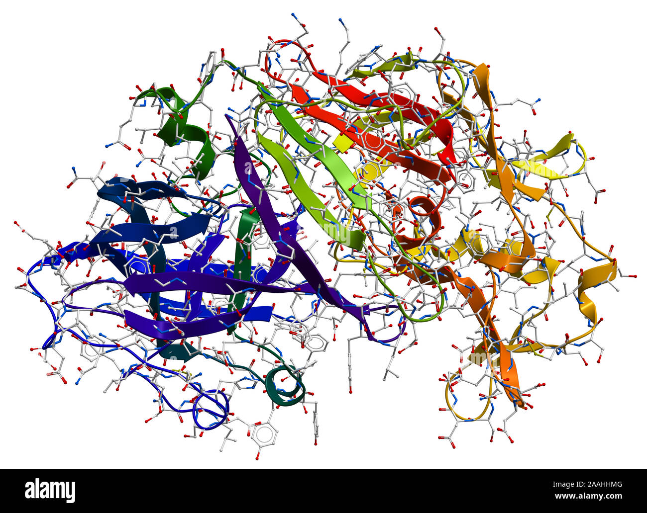 Enzyme pepsin 3D model Stock Photohttps://www.alamy.com/image-license-details/?v=1https://www.alamy.com/enzyme-pepsin-3d-model-image333530640.html
RF2AAHHMG–Enzyme pepsin 3D model
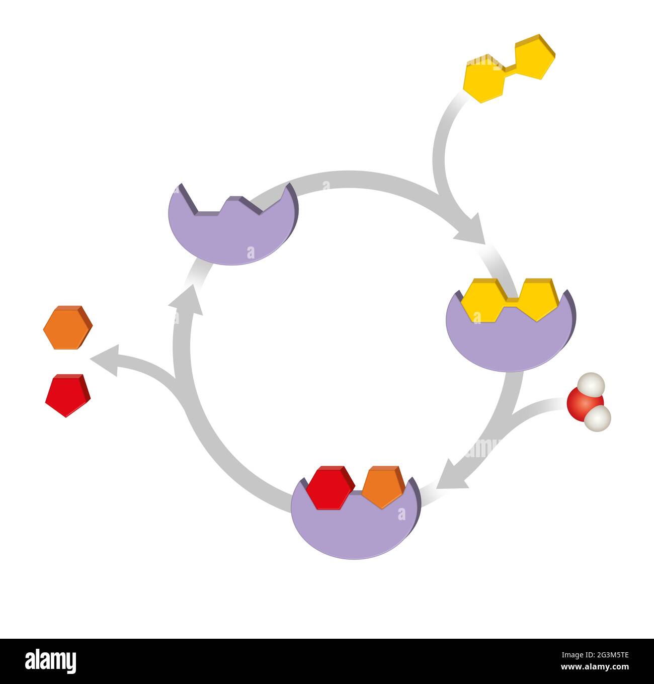 Enzyme function. Macromolecular biological catalysts Stock Photohttps://www.alamy.com/image-license-details/?v=1https://www.alamy.com/enzyme-function-macromolecular-biological-catalysts-image432546814.html
RF2G3M5TE–Enzyme function. Macromolecular biological catalysts
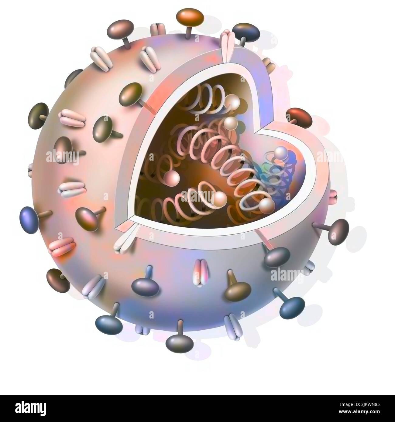 Schematic of any virus with DNA strands inside. Stock Photohttps://www.alamy.com/image-license-details/?v=1https://www.alamy.com/schematic-of-any-virus-with-dna-strands-inside-image476923893.html
RF2JKWN85–Schematic of any virus with DNA strands inside.
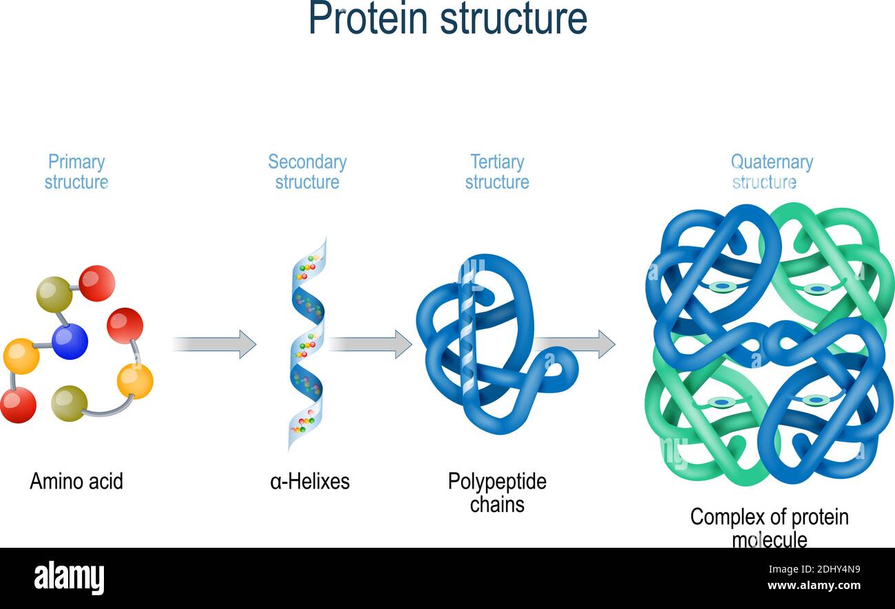 Levels of protein structure from amino acids to Complex of protein molecule. Protein is a polymer (polypeptide) Stock Vectorhttps://www.alamy.com/image-license-details/?v=1https://www.alamy.com/levels-of-protein-structure-from-amino-acids-to-complex-of-protein-molecule-protein-is-a-polymerpolypeptide-image389673685.html
RF2DHY4N9–Levels of protein structure from amino acids to Complex of protein molecule. Protein is a polymer (polypeptide)
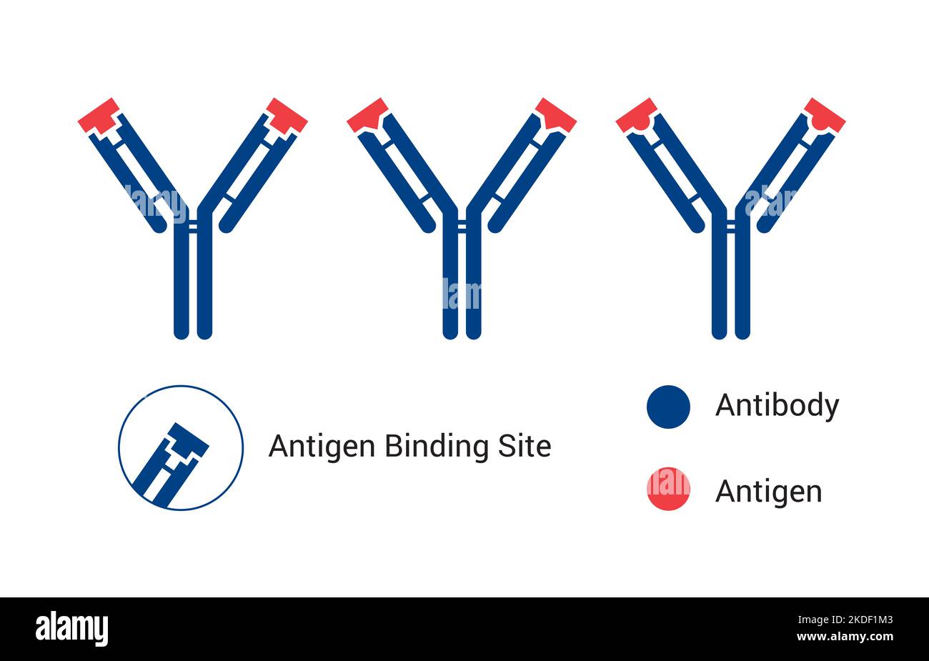 Antibody structure of immunoglobulin with enzymes papain and pepsin, the basic structure of an antibody, showing the light and heavy chains Stock Photohttps://www.alamy.com/image-license-details/?v=1https://www.alamy.com/antibody-structure-of-immunoglobulin-with-enzymes-papain-and-pepsin-the-basic-structure-of-an-antibody-showing-the-light-and-heavy-chains-image490211459.html
RF2KDF1M3–Antibody structure of immunoglobulin with enzymes papain and pepsin, the basic structure of an antibody, showing the light and heavy chains
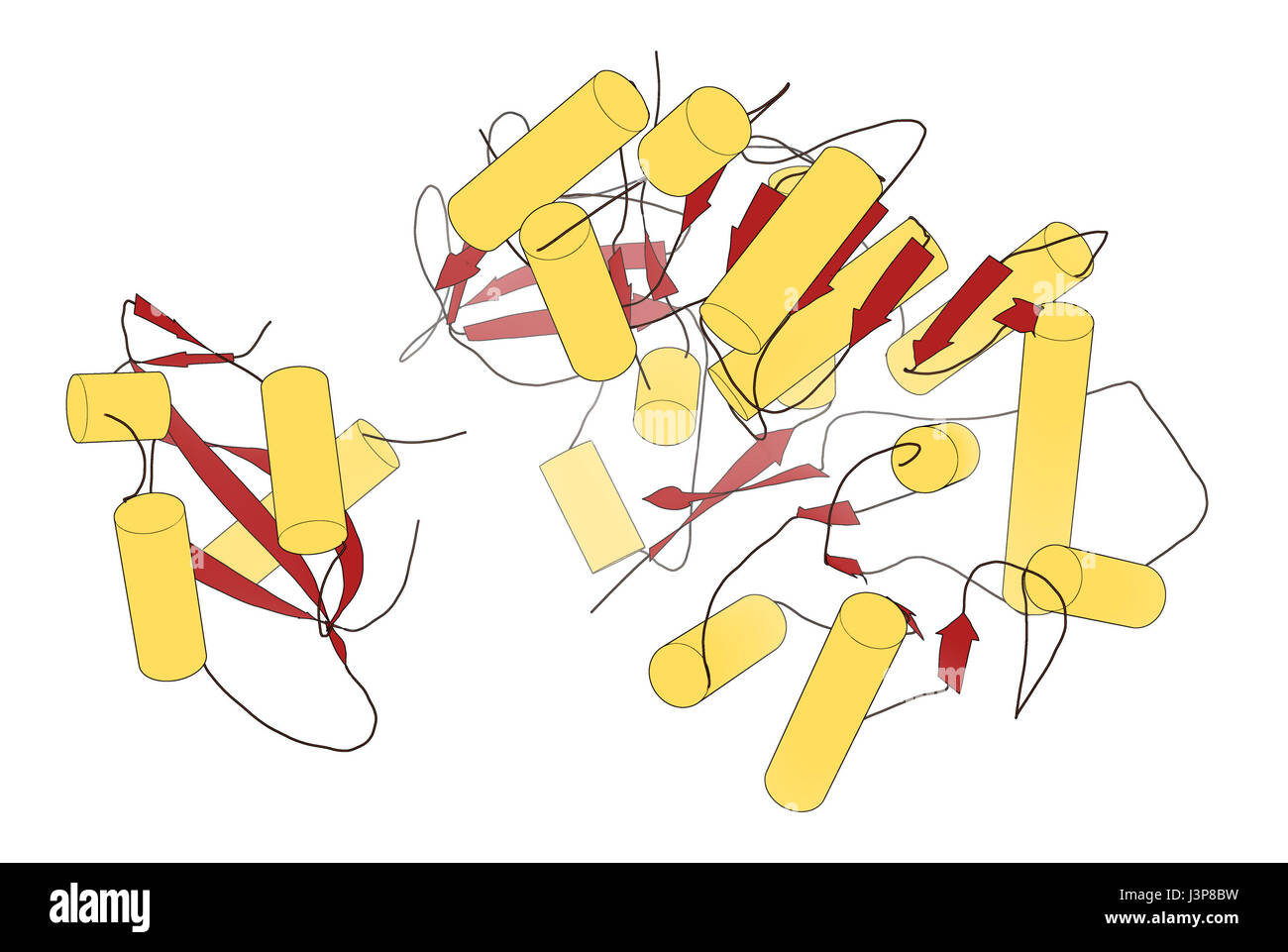 Firefly luciferase enzyme. Protein responsible for the bioluminescence of fireflies. Often used as reporter in biotechnology and genetic engineering. Stock Photohttps://www.alamy.com/image-license-details/?v=1https://www.alamy.com/stock-photo-firefly-luciferase-enzyme-protein-responsible-for-the-bioluminescence-140016461.html
RFJ3P8BW–Firefly luciferase enzyme. Protein responsible for the bioluminescence of fireflies. Often used as reporter in biotechnology and genetic engineering.
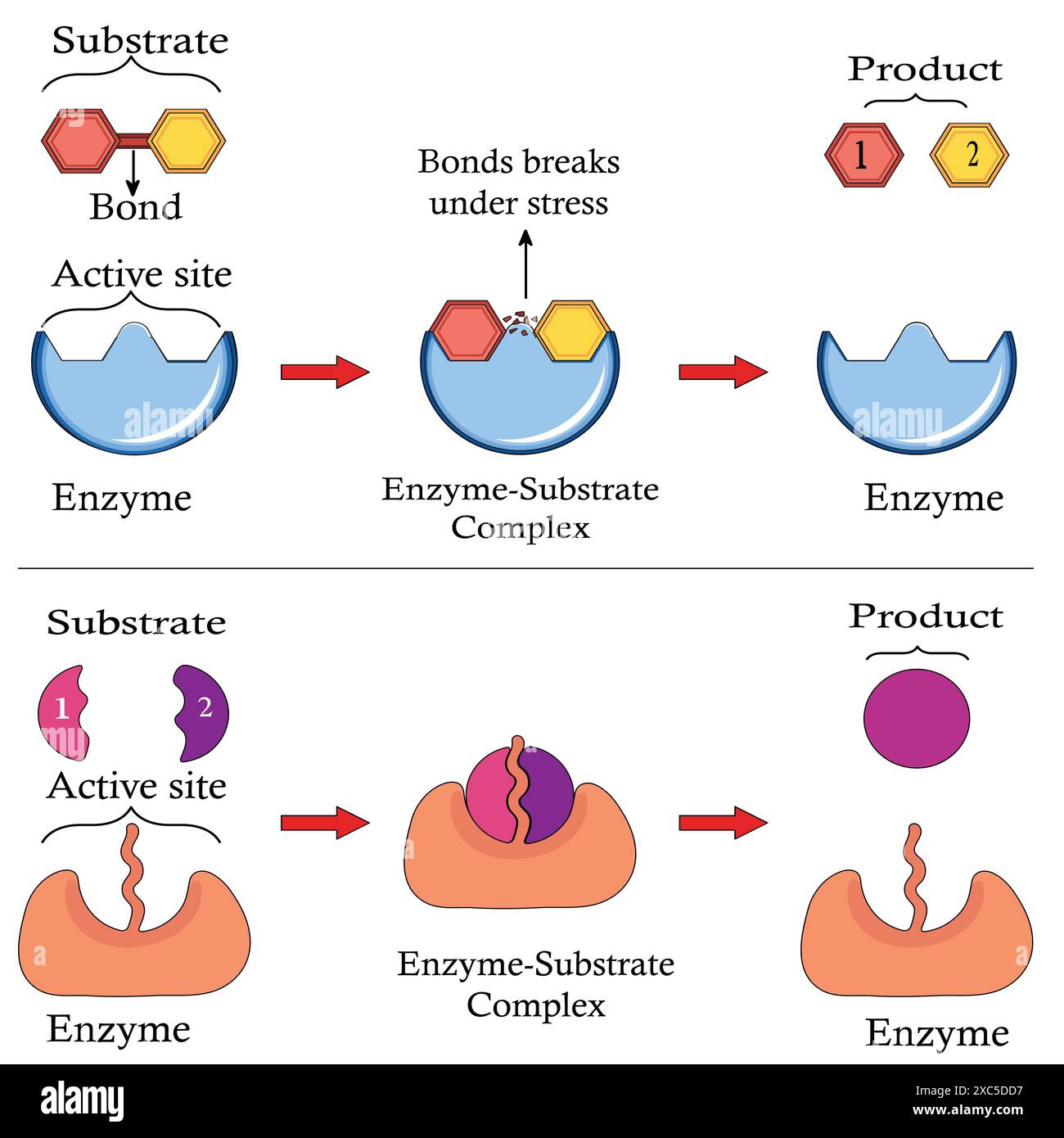 Mechanism of Action of Enzymes. Substrate reactants enter active site of enzyme. Chemical reaction creates products. Stock Vectorhttps://www.alamy.com/image-license-details/?v=1https://www.alamy.com/mechanism-of-action-of-enzymes-substrate-reactants-enter-active-site-of-enzyme-chemical-reaction-creates-products-image609859075.html
RF2XC5DD7–Mechanism of Action of Enzymes. Substrate reactants enter active site of enzyme. Chemical reaction creates products.
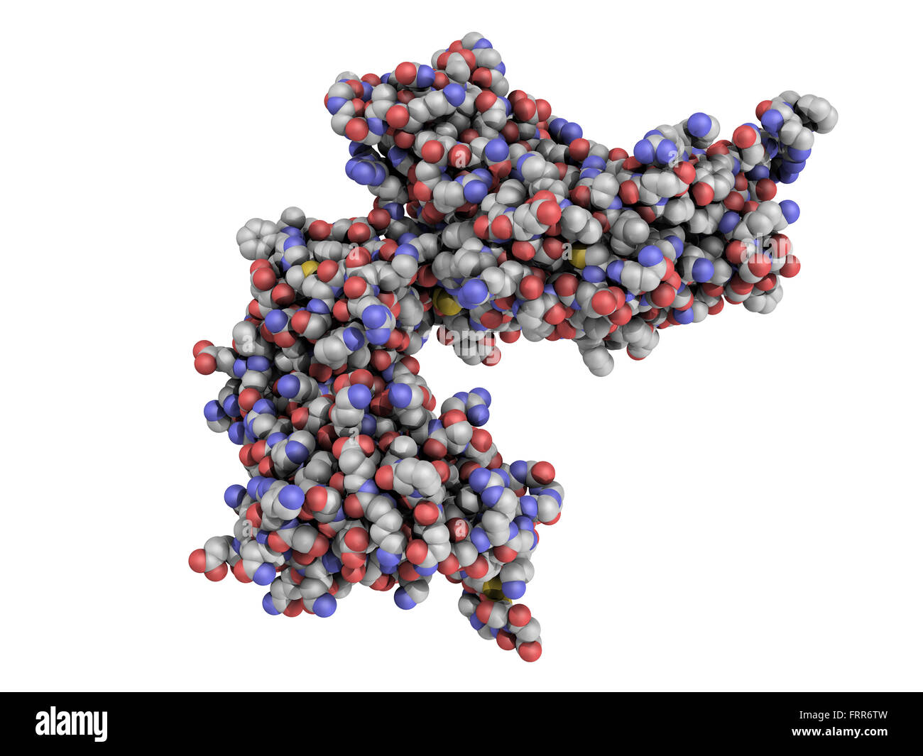 cyclic ADP ribose hydrolase (CD38) protein. CD38 is an enzyme present on the cell surface of many immune cells and it is a progn Stock Photohttps://www.alamy.com/image-license-details/?v=1https://www.alamy.com/stock-photo-cyclic-adp-ribose-hydrolase-cd38-protein-cd38-is-an-enzyme-present-100699225.html
RFFRR6TW–cyclic ADP ribose hydrolase (CD38) protein. CD38 is an enzyme present on the cell surface of many immune cells and it is a progn
 Human Mitochondrial PheRS Stock Photohttps://www.alamy.com/image-license-details/?v=1https://www.alamy.com/human-mitochondrial-phers-image416784513.html
RM2F624W5–Human Mitochondrial PheRS
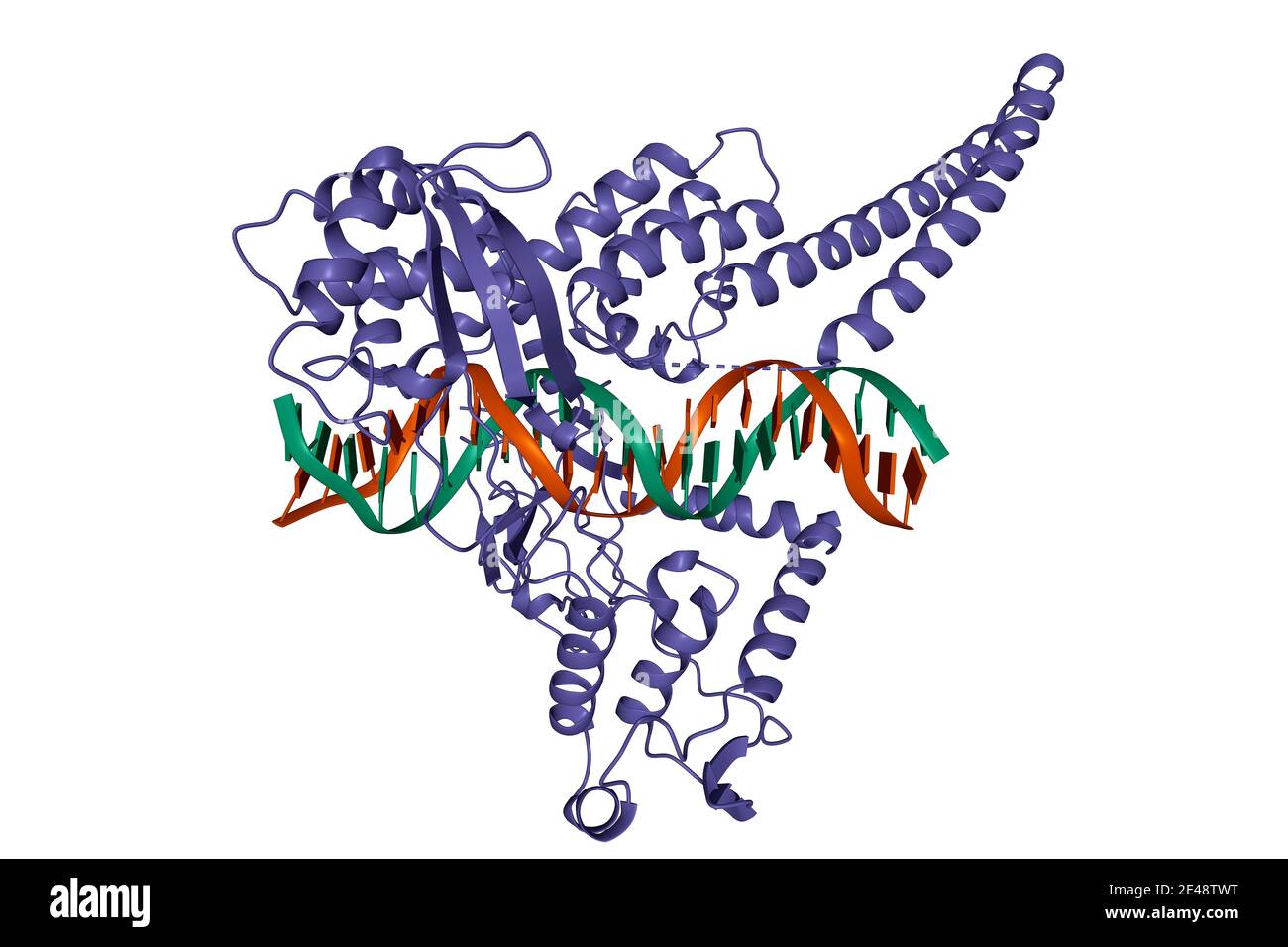 Structure of the topoisomerase I - DNA complex, 3D cartoon model, white background Stock Photohttps://www.alamy.com/image-license-details/?v=1https://www.alamy.com/structure-of-the-topoisomerase-i-dna-complex-3d-cartoon-model-white-background-image398492244.html
RF2E48TWT–Structure of the topoisomerase I - DNA complex, 3D cartoon model, white background
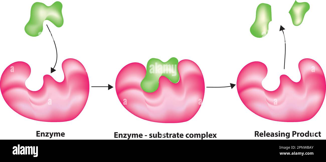 Enzymes function in human body Stock Vectorhttps://www.alamy.com/image-license-details/?v=1https://www.alamy.com/enzymes-function-in-human-body-image546986915.html
RF2PNWBAY–Enzymes function in human body
 3D image of Cyclic adenosine monophosphate skeletal formula - molecular chemical structure of second messenger cAMP isolated on white background Stock Photohttps://www.alamy.com/image-license-details/?v=1https://www.alamy.com/3d-image-of-cyclic-adenosine-monophosphate-skeletal-formula-molecular-chemical-structure-of-second-messenger-camp-isolated-on-white-background-image500386433.html
RF2M22G01–3D image of Cyclic adenosine monophosphate skeletal formula - molecular chemical structure of second messenger cAMP isolated on white background
 This infographic illustrates the HIV replication cycle, which begins when HIV fuses with the surface of the host cell. A capsid containing the viruss genome and proteins then enters the cell. The shell of the capsid disintegrates and the HIV protein called reverse transcriptase transcribes the viral RNA into DNA. The viral DNA is transported across the nucleus, where the HIV protein integrase integrates the HIV DNA into the hosts DNA. The hosts normal transcription machinery transcribes HIV DNA into multiple copies of new HIV RNA. Some of this RNA becomes the genome of a new virus, while the c Stock Photohttps://www.alamy.com/image-license-details/?v=1https://www.alamy.com/this-infographic-illustrates-the-hiv-replication-cycle-which-begins-when-hiv-fuses-with-the-surface-of-the-host-cell-a-capsid-containing-the-viruss-genome-and-proteins-then-enters-the-cell-the-shell-of-the-capsid-disintegrates-and-the-hiv-protein-called-reverse-transcriptase-transcribes-the-viral-rna-into-dna-the-viral-dna-is-transported-across-the-nucleus-where-the-hiv-protein-integrase-integrates-the-hiv-dna-into-the-hosts-dna-the-hosts-normal-transcription-machinery-transcribes-hiv-dna-into-multiple-copies-of-new-hiv-rna-some-of-this-rna-becomes-the-genome-of-a-new-virus-while-the-c-image627780476.html
RM2YD9TB8–This infographic illustrates the HIV replication cycle, which begins when HIV fuses with the surface of the host cell. A capsid containing the viruss genome and proteins then enters the cell. The shell of the capsid disintegrates and the HIV protein called reverse transcriptase transcribes the viral RNA into DNA. The viral DNA is transported across the nucleus, where the HIV protein integrase integrates the HIV DNA into the hosts DNA. The hosts normal transcription machinery transcribes HIV DNA into multiple copies of new HIV RNA. Some of this RNA becomes the genome of a new virus, while the c
 Protein structure with biological concept, 3d rendering. 3D illustration. Stock Photohttps://www.alamy.com/image-license-details/?v=1https://www.alamy.com/protein-structure-with-biological-concept-3d-rendering-3d-illustration-image598135279.html
RF2WN3BJ7–Protein structure with biological concept, 3d rendering. 3D illustration.
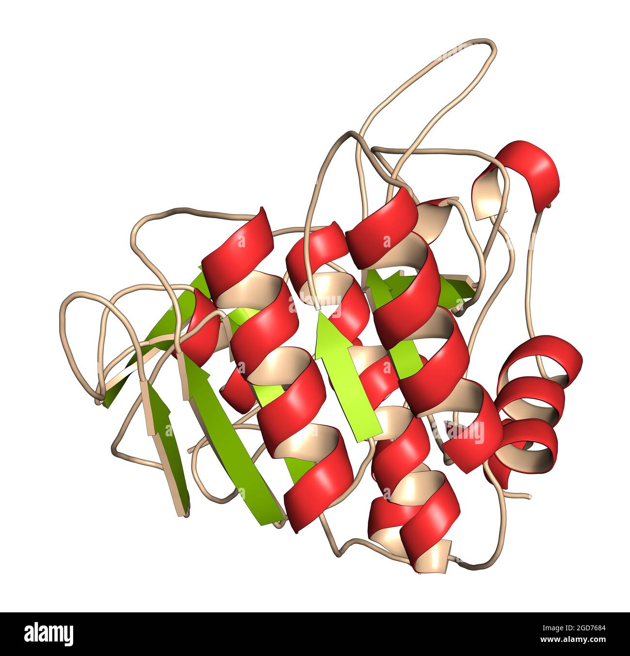 Nattokinase enzyme. Protein produced by Bacillus natto, present Stock Photohttps://www.alamy.com/image-license-details/?v=1https://www.alamy.com/nattokinase-enzyme-protein-produced-by-bacillus-natto-present-image438408324.html
RF2GD7684–Nattokinase enzyme. Protein produced by Bacillus natto, present
RF2YFMXC0–Organic Health Supplement Icons Featuring Enzyme, Probiotic, Minerals, Amino Acids, Super Fruits, Collagen, Vitamins, Vegan, Protein, and USDA Organic
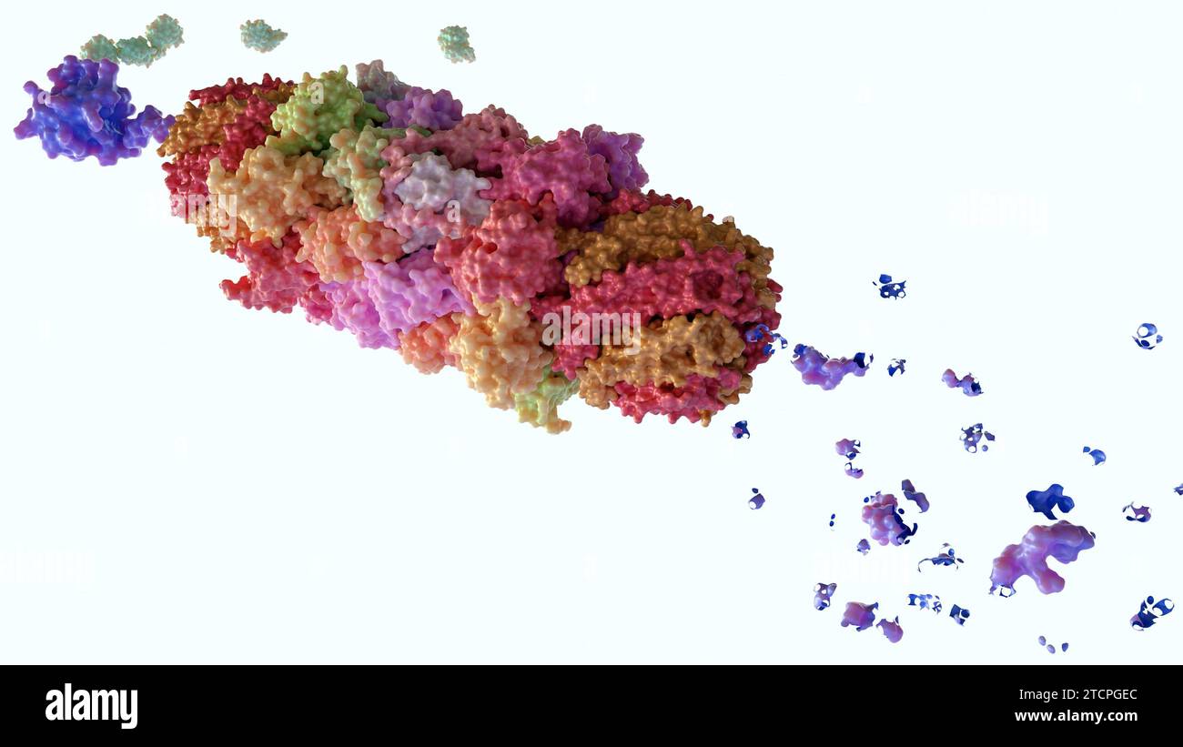 Proteasomes are molecular machines for breaking down proteins called proteolysis and ubiquitin to the target protein, 3d rendering Stock Photohttps://www.alamy.com/image-license-details/?v=1https://www.alamy.com/proteasomes-are-molecular-machines-for-breaking-down-proteins-called-proteolysis-and-ubiquitin-to-the-target-protein-3d-rendering-image575813908.html
RF2TCPGEC–Proteasomes are molecular machines for breaking down proteins called proteolysis and ubiquitin to the target protein, 3d rendering
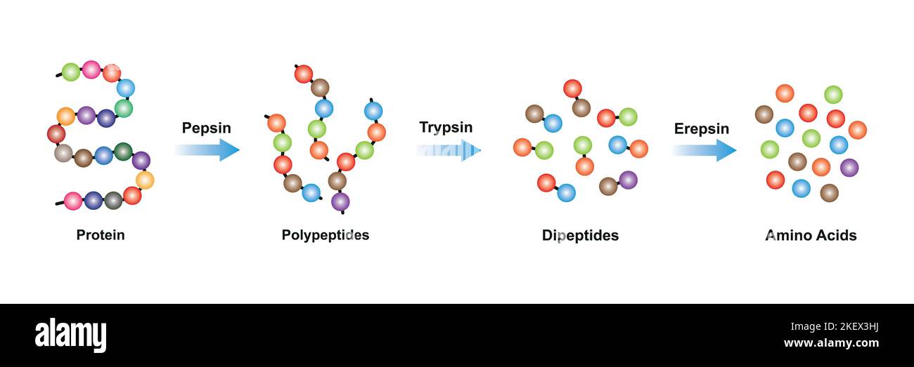 Scientific Designing of Protein digestion. Pepsin, Trypsin and Erepsin Enzymes Effect on Protein Molecule. Colorful Symbols. Vector Illustration. Stock Vectorhttps://www.alamy.com/image-license-details/?v=1https://www.alamy.com/scientific-designing-of-protein-digestion-pepsin-trypsin-and-erepsin-enzymes-effect-on-protein-molecule-colorful-symbols-vector-illustration-image491069086.html
RF2KEX3HJ–Scientific Designing of Protein digestion. Pepsin, Trypsin and Erepsin Enzymes Effect on Protein Molecule. Colorful Symbols. Vector Illustration.
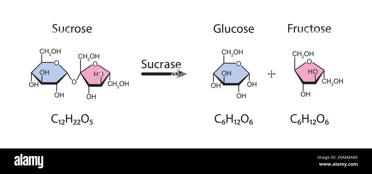 Sucrase enzyme, illustration Stock Photohttps://www.alamy.com/image-license-details/?v=1https://www.alamy.com/sucrase-enzyme-illustration-image529051684.html
RF2NMMAR0–Sucrase enzyme, illustration
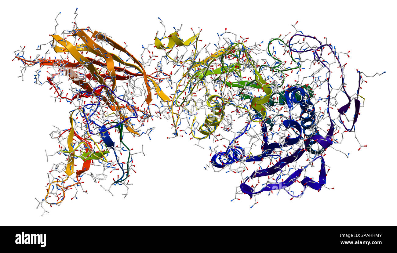 Enzyme pancreatic lipase-colipase complex Stock Photohttps://www.alamy.com/image-license-details/?v=1https://www.alamy.com/enzyme-pancreatic-lipase-colipase-complex-image333530651.html
RF2AAHHMY–Enzyme pancreatic lipase-colipase complex
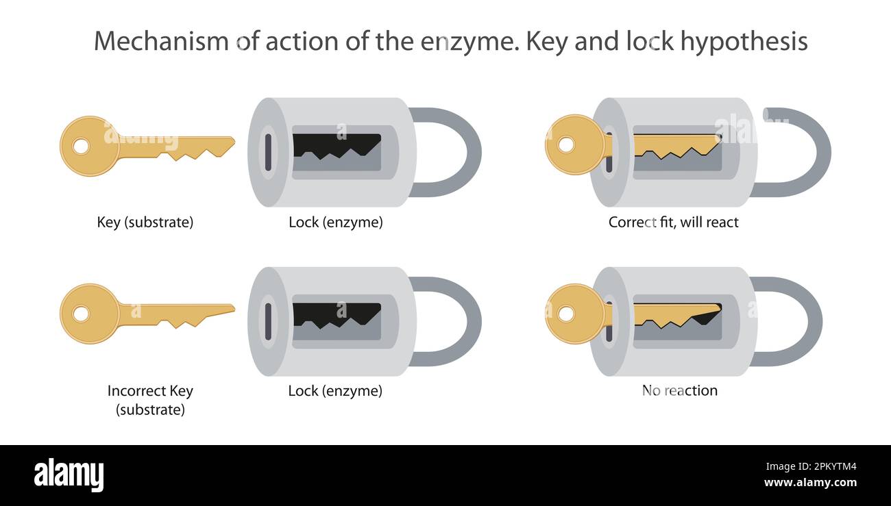 Mechanism of action of the enzyme. Key and lock hypothesis Stock Photohttps://www.alamy.com/image-license-details/?v=1https://www.alamy.com/mechanism-of-action-of-the-enzyme-key-and-lock-hypothesis-image545811956.html
RF2PKYTM4–Mechanism of action of the enzyme. Key and lock hypothesis
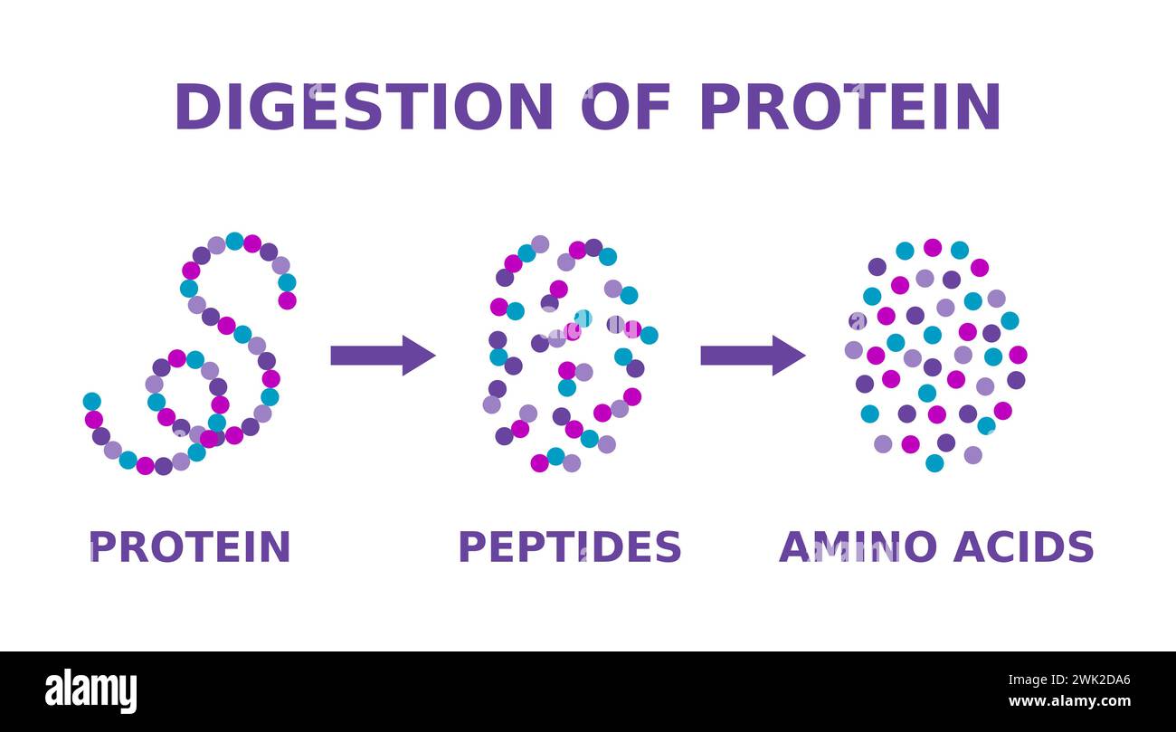 Digestion of protein. Breaking the complex molecule first into peptides then into individual amino acids. The pepsins are enzymes. Vector Stock Vectorhttps://www.alamy.com/image-license-details/?v=1https://www.alamy.com/digestion-of-protein-breaking-the-complex-molecule-first-into-peptides-then-into-individual-amino-acids-the-pepsins-are-enzymes-vector-image596885358.html
RF2WK2DA6–Digestion of protein. Breaking the complex molecule first into peptides then into individual amino acids. The pepsins are enzymes. Vector
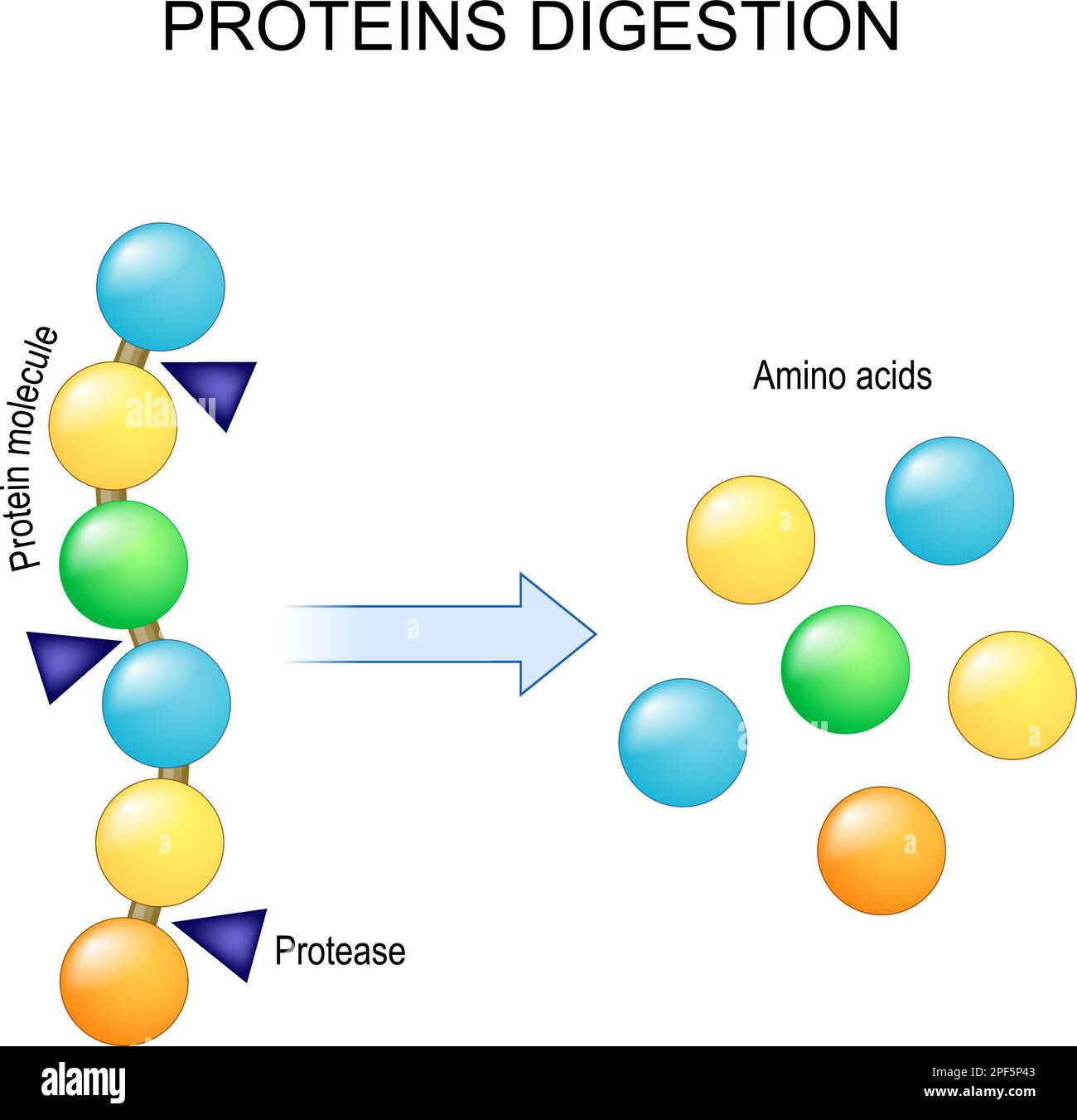 Protein digestion. Enzymes proteases are digestion breaks the protein into single amino acids, which are absorbed into the blood. Vector illustration Stock Vectorhttps://www.alamy.com/image-license-details/?v=1https://www.alamy.com/protein-digestion-enzymes-proteases-are-digestion-breaks-the-protein-into-single-amino-acids-which-are-absorbed-into-the-blood-vector-illustration-image542868371.html
RF2PF5P43–Protein digestion. Enzymes proteases are digestion breaks the protein into single amino acids, which are absorbed into the blood. Vector illustration
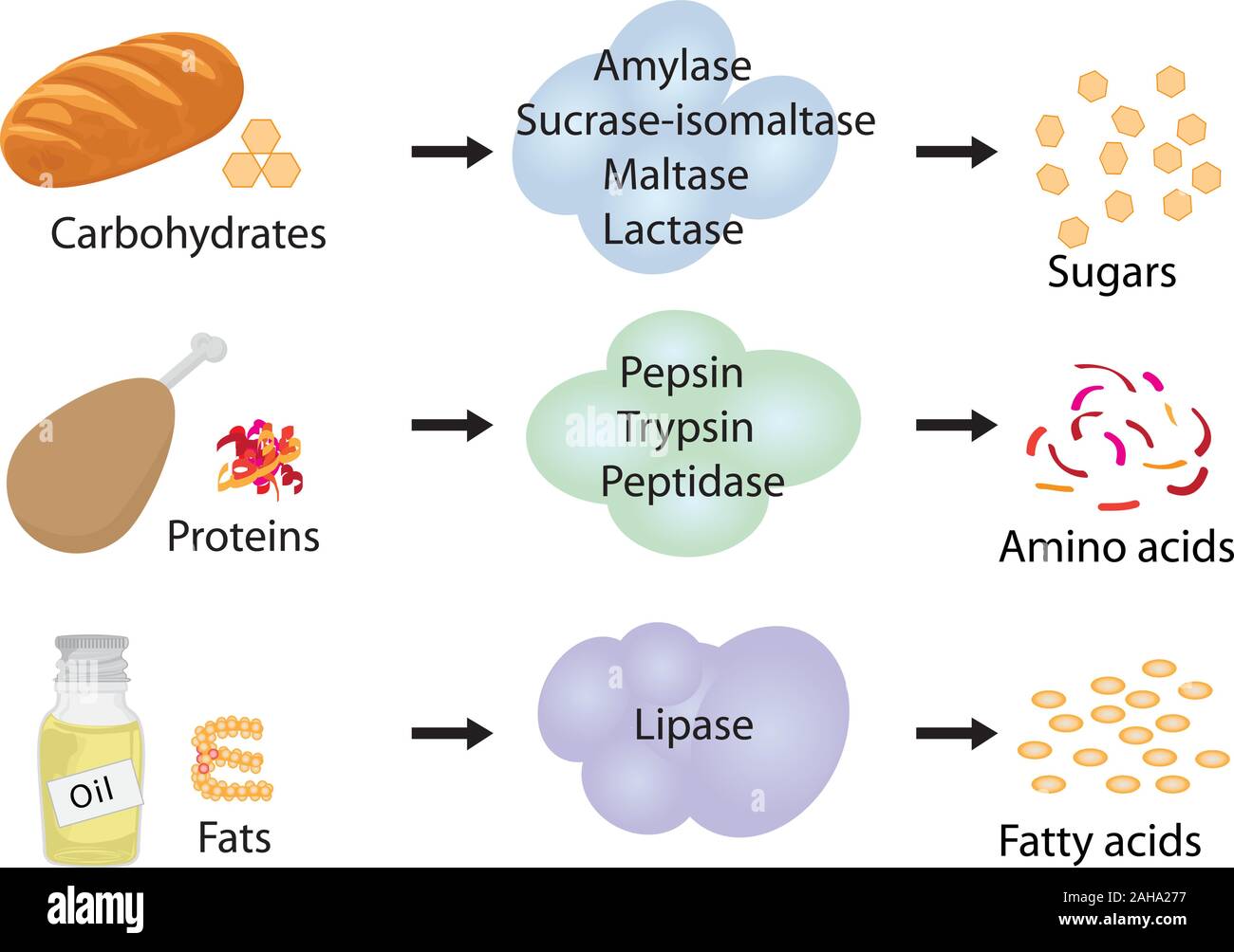 How enzymes work. Fermentation and digestion food Stock Vectorhttps://www.alamy.com/image-license-details/?v=1https://www.alamy.com/how-enzymes-work-fermentation-and-digestion-food-image337667435.html
RF2AHA277–How enzymes work. Fermentation and digestion food
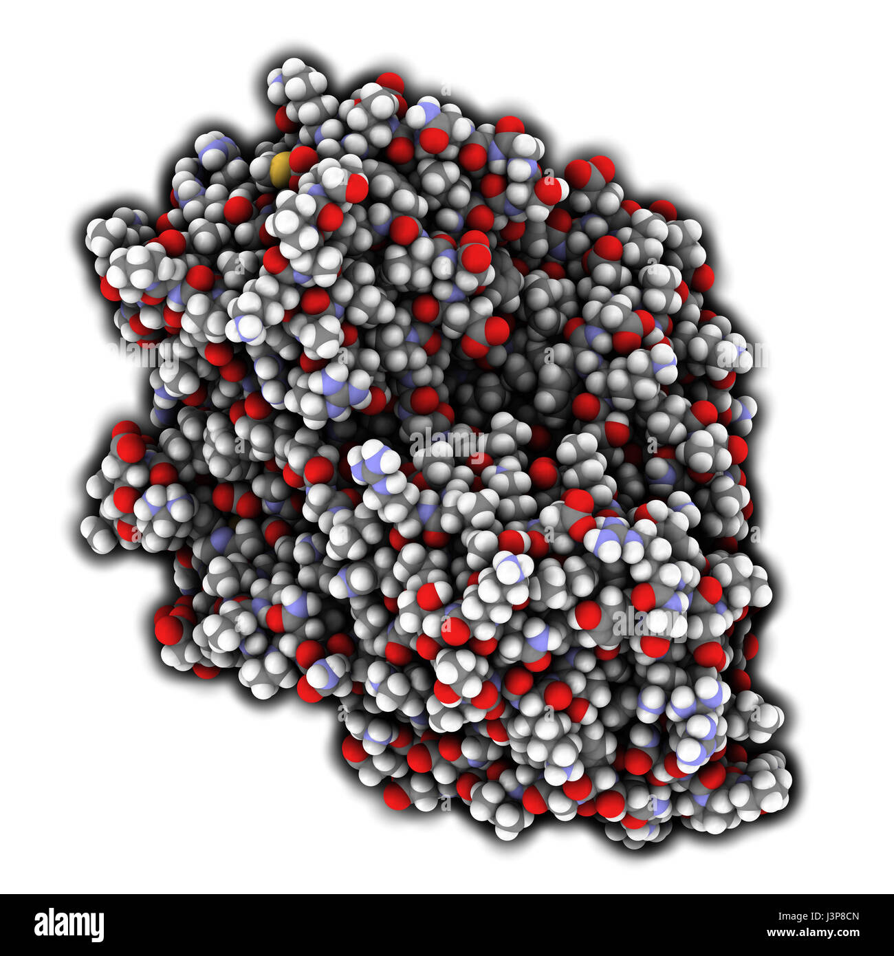 Firefly luciferase enzyme. Protein responsible for the bioluminescence of fireflies. Often used as reporter in biotechnology and genetic engineering. Stock Photohttps://www.alamy.com/image-license-details/?v=1https://www.alamy.com/stock-photo-firefly-luciferase-enzyme-protein-responsible-for-the-bioluminescence-140016485.html
RFJ3P8CN–Firefly luciferase enzyme. Protein responsible for the bioluminescence of fireflies. Often used as reporter in biotechnology and genetic engineering.
 VIRUS Stock Photohttps://www.alamy.com/image-license-details/?v=1https://www.alamy.com/stock-photo-virus-49289090.html
RMCT58MJ–VIRUS
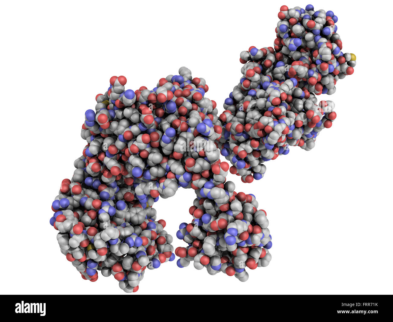 Taq polymerase PCR enzyme: a heat-stable DNA polymerase protein that is the key component that makes the polymerase chain reacti Stock Photohttps://www.alamy.com/image-license-details/?v=1https://www.alamy.com/stock-photo-taq-polymerase-pcr-enzyme-a-heat-stable-dna-polymerase-protein-that-100699359.html
RFFRR71K–Taq polymerase PCR enzyme: a heat-stable DNA polymerase protein that is the key component that makes the polymerase chain reacti
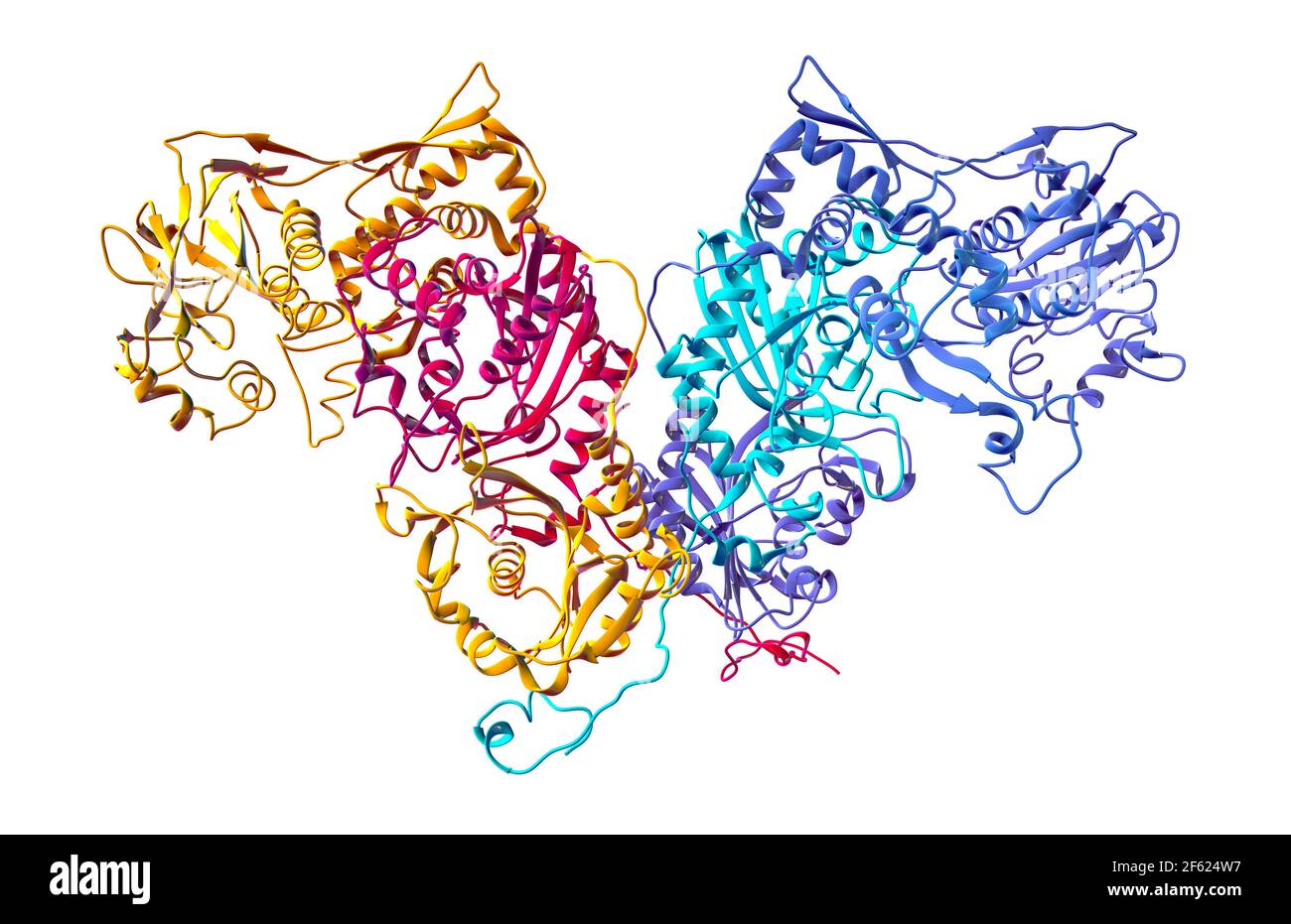 Human Cytoplasmic PheRS Stock Photohttps://www.alamy.com/image-license-details/?v=1https://www.alamy.com/human-cytoplasmic-phers-image416784515.html
RM2F624W7–Human Cytoplasmic PheRS
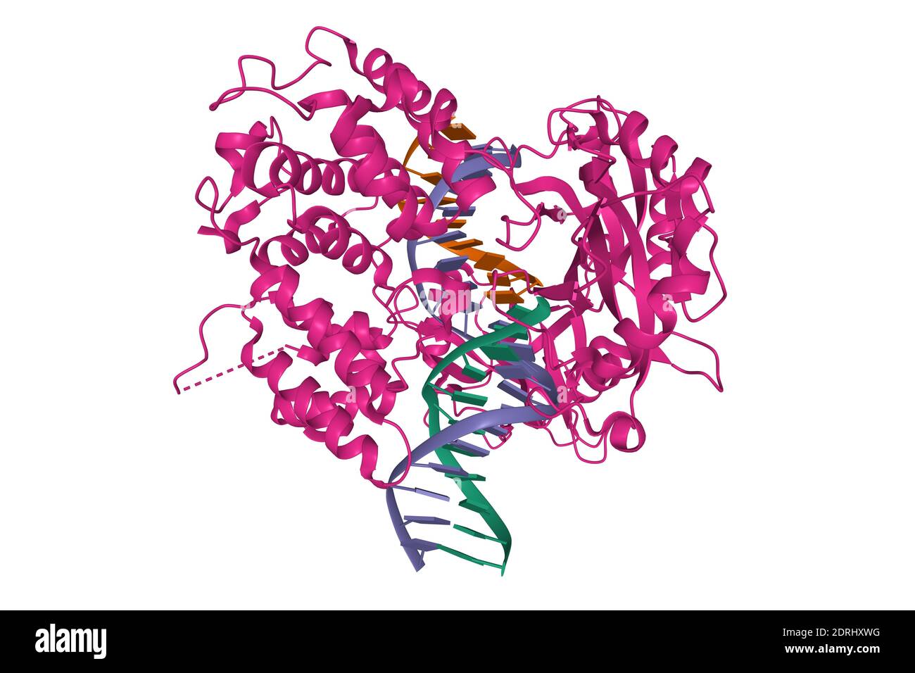 Crystal structure of human DNA ligase I bound to 5'-adenylated, nicked DNA, 3D cartoon model, white background Stock Photohttps://www.alamy.com/image-license-details/?v=1https://www.alamy.com/crystal-structure-of-human-dna-ligase-i-bound-to-5-adenylated-nicked-dna-3d-cartoon-model-white-background-image393159468.html
RF2DRHXWG–Crystal structure of human DNA ligase I bound to 5'-adenylated, nicked DNA, 3D cartoon model, white background
 3D Anatomy of lysosome Stock Photohttps://www.alamy.com/image-license-details/?v=1https://www.alamy.com/3d-anatomy-of-lysosome-image476853344.html
RF2JKPF8G–3D Anatomy of lysosome
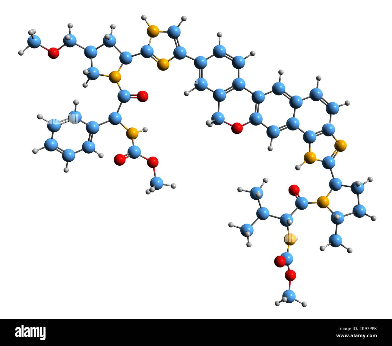 3D image of Velpatasvir skeletal formula - molecular chemical structure of NS5A inhibitor isolated on white background Stock Photohttps://www.alamy.com/image-license-details/?v=1https://www.alamy.com/3d-image-of-velpatasvir-skeletal-formula-molecular-chemical-structure-of-ns5a-inhibitor-isolated-on-white-background-image487593755.html
RF2K97PPK–3D image of Velpatasvir skeletal formula - molecular chemical structure of NS5A inhibitor isolated on white background
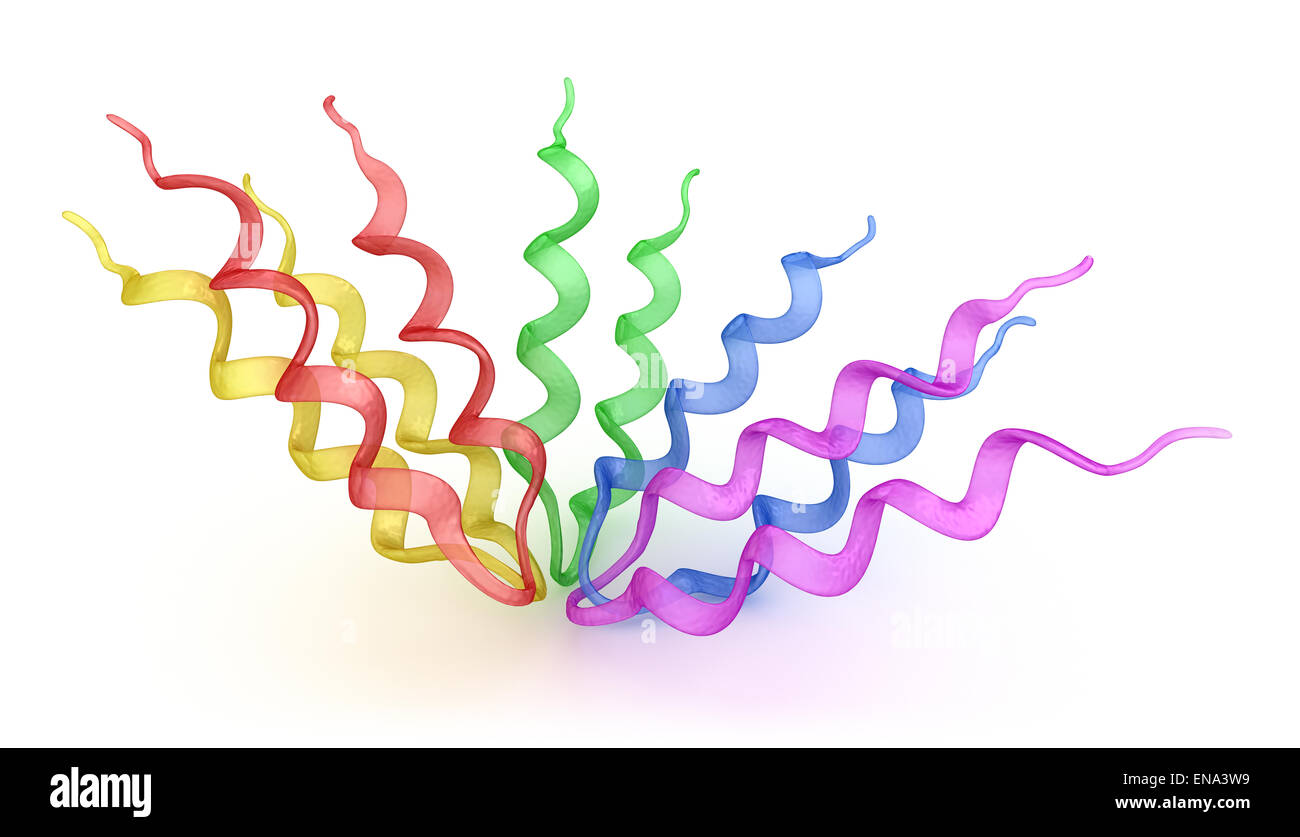 Protein 3D model Stock Photohttps://www.alamy.com/image-license-details/?v=1https://www.alamy.com/stock-photo-protein-3d-model-81971829.html
RFENA3W9–Protein 3D model
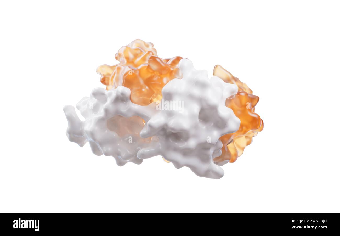 Protein structure with biological concept, 3d rendering. 3D illustration. Stock Photohttps://www.alamy.com/image-license-details/?v=1https://www.alamy.com/protein-structure-with-biological-concept-3d-rendering-3d-illustration-image598135293.html
RF2WN3BJN–Protein structure with biological concept, 3d rendering. 3D illustration.
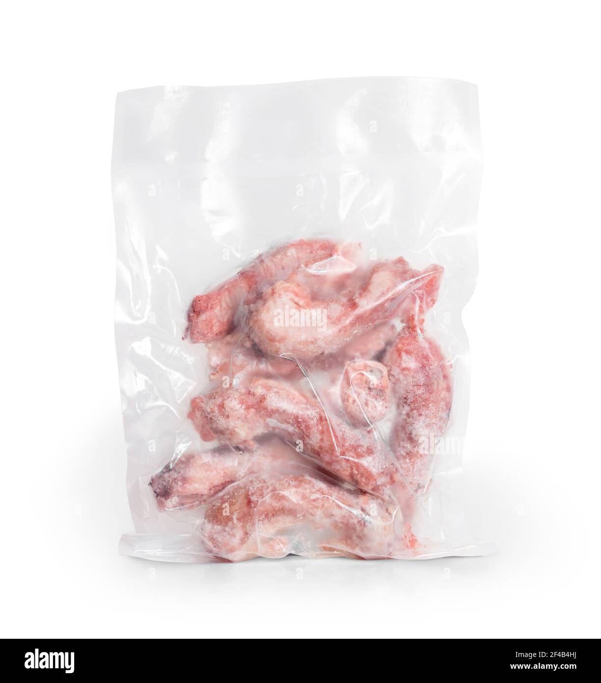 1 lb frozen chicken necks in vacuum sealed package. Many raw large chicken necks. Concept for raw food diet for cats, dogs and pets. Bones prevent pla Stock Photohttps://www.alamy.com/image-license-details/?v=1https://www.alamy.com/1-lb-frozen-chicken-necks-in-vacuum-sealed-package-many-raw-large-chicken-necks-concept-for-raw-food-diet-for-cats-dogs-and-pets-bones-prevent-pla-image415752558.html
RF2F4B4HJ–1 lb frozen chicken necks in vacuum sealed package. Many raw large chicken necks. Concept for raw food diet for cats, dogs and pets. Bones prevent pla
 abstract medical graphic Stock Photohttps://www.alamy.com/image-license-details/?v=1https://www.alamy.com/stock-photo-abstract-medical-graphic-74589639.html
RFE99RRK–abstract medical graphic
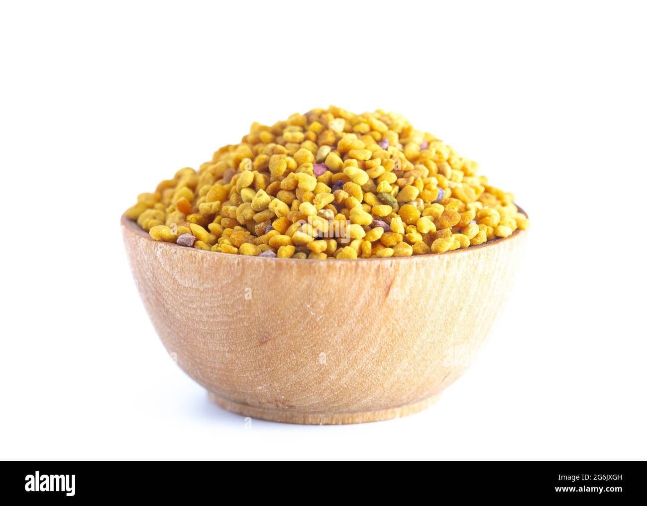 A Bowl of Pellets of Yellow Bee Pollen Stock Photohttps://www.alamy.com/image-license-details/?v=1https://www.alamy.com/a-bowl-of-pellets-of-yellow-bee-pollen-image434363121.html
RF2G6JXGH–A Bowl of Pellets of Yellow Bee Pollen
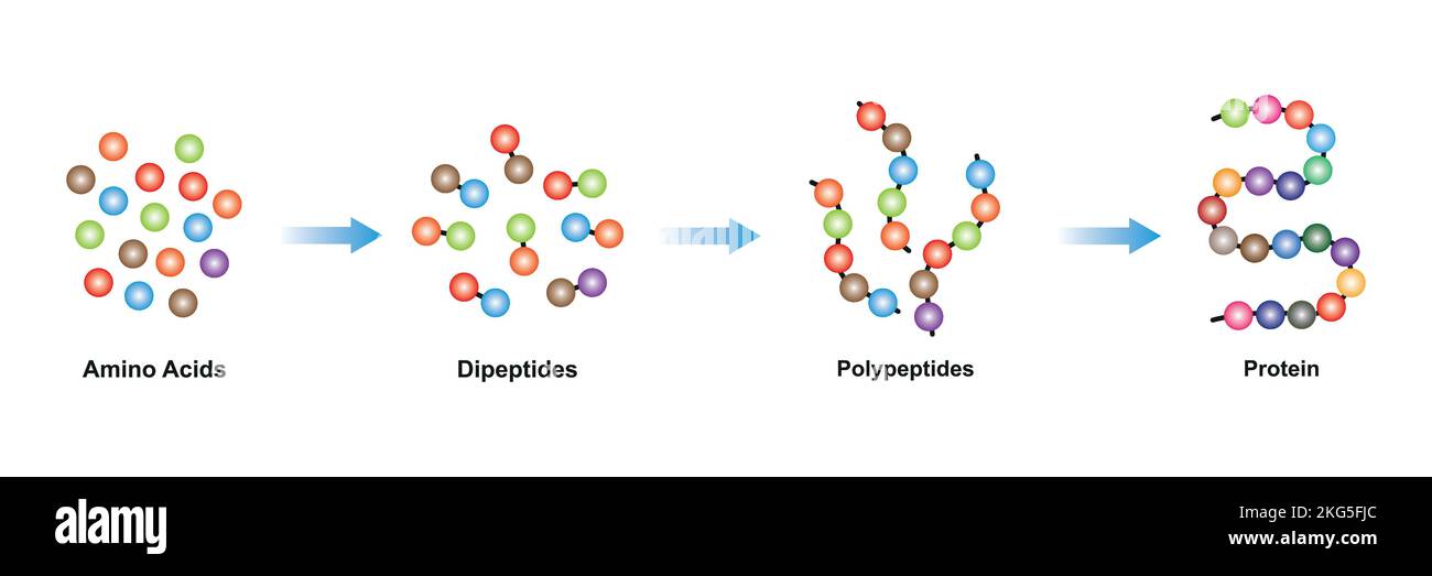 Scientific Designing of Protein Molecule Formation Levels. Colorful Symbols. Vector Illustration. Stock Vectorhttps://www.alamy.com/image-license-details/?v=1https://www.alamy.com/scientific-designing-of-protein-molecule-formation-levels-colorful-symbols-vector-illustration-image491846836.html
RF2KG5FJC–Scientific Designing of Protein Molecule Formation Levels. Colorful Symbols. Vector Illustration.
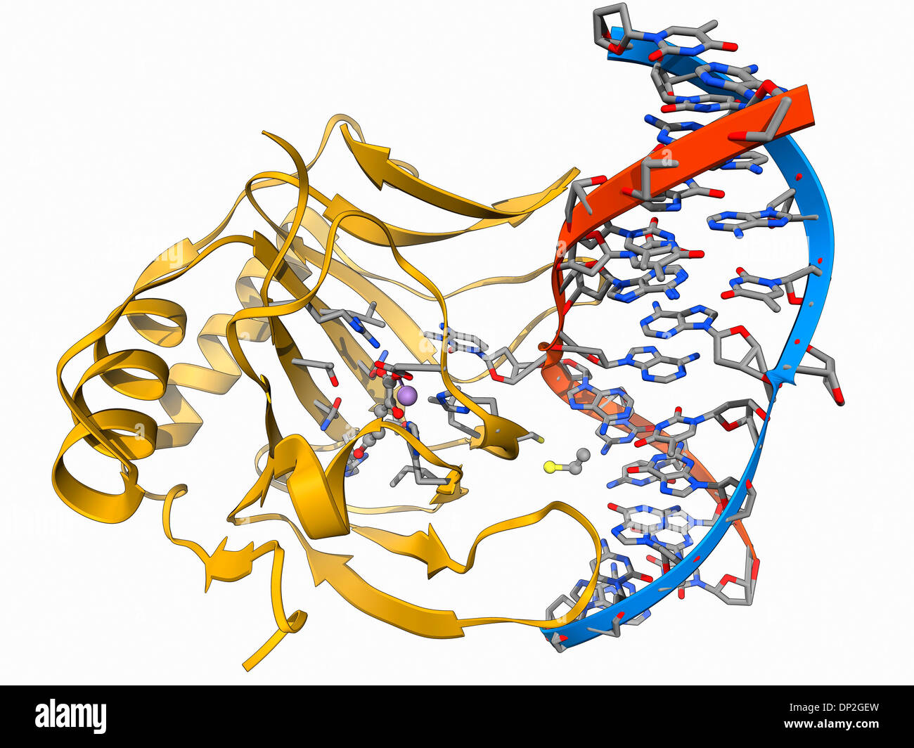 DNA repair enzyme, molecular model Stock Photohttps://www.alamy.com/image-license-details/?v=1https://www.alamy.com/dna-repair-enzyme-molecular-model-image65210401.html
RFDP2GEW–DNA repair enzyme, molecular model
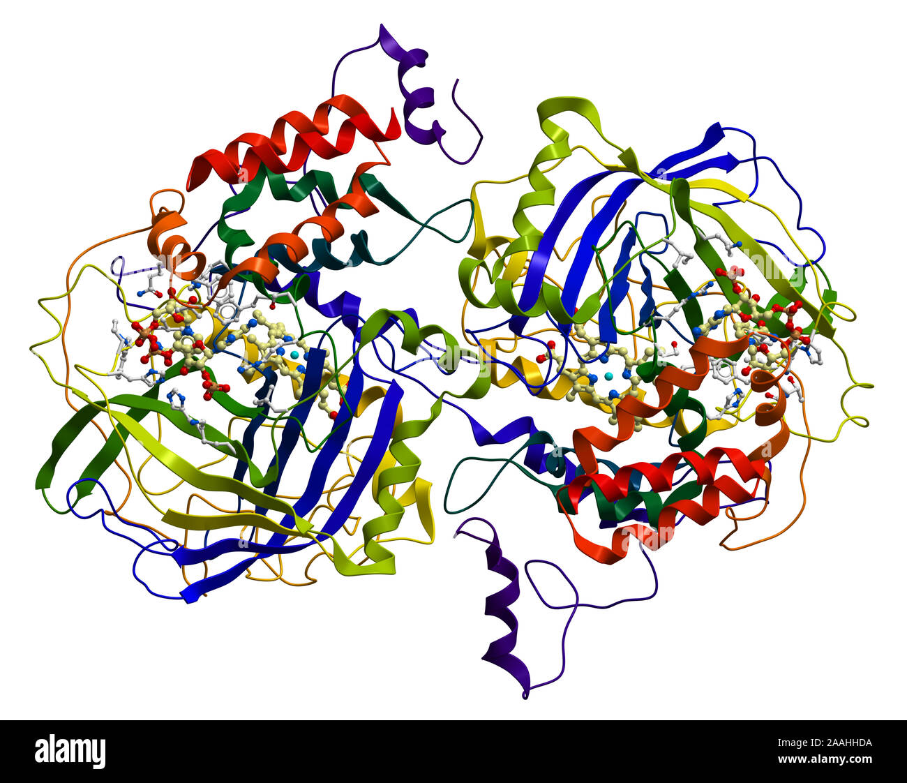 Enzyme Catalase, a very important antioxidant in organism Stock Photohttps://www.alamy.com/image-license-details/?v=1https://www.alamy.com/enzyme-catalase-a-very-important-antioxidant-in-organism-image333530438.html
RF2AAHHDA–Enzyme Catalase, a very important antioxidant in organism
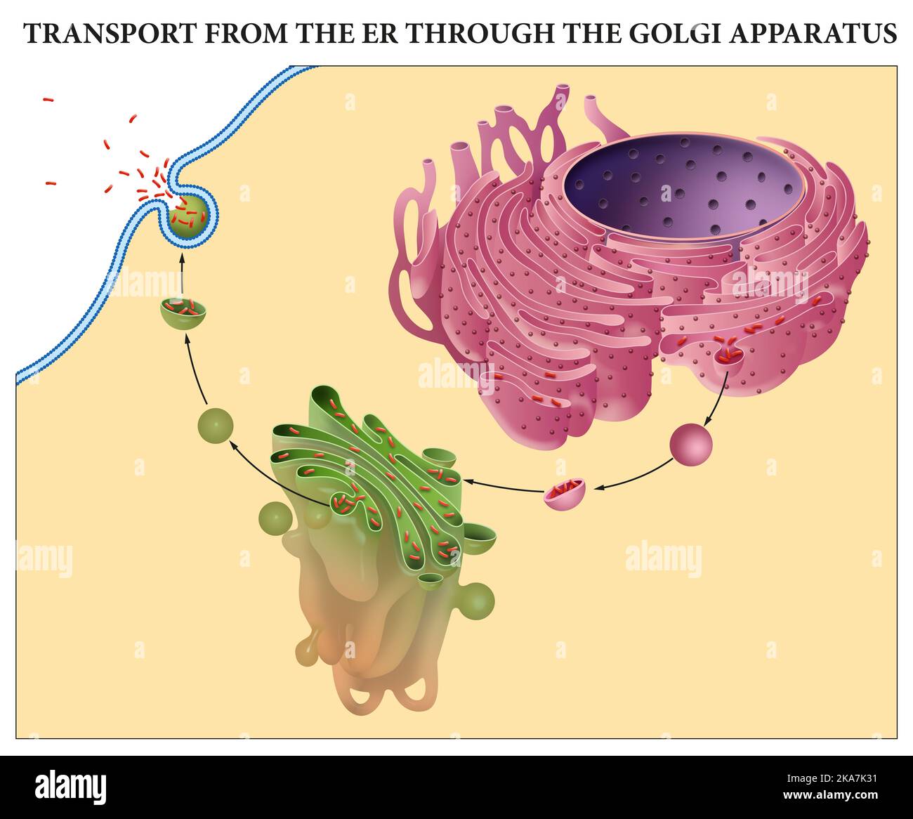 Transport from the ER through the Golgi Apparatus Stock Photohttps://www.alamy.com/image-license-details/?v=1https://www.alamy.com/transport-from-the-er-through-the-golgi-apparatus-image488205509.html
RF2KA7K31–Transport from the ER through the Golgi Apparatus
 Bee pollen grains in white ceramic bowl and apple flowers on white background. Stock Photohttps://www.alamy.com/image-license-details/?v=1https://www.alamy.com/bee-pollen-grains-in-white-ceramic-bowl-and-apple-flowers-on-white-background-image366156138.html
RF2C7KRTX–Bee pollen grains in white ceramic bowl and apple flowers on white background.
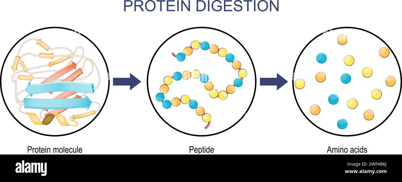 Protein Digestion. Enzymes proteases and peptidases are digestion breaks the protein into smaller peptide chains and into single amino acids, which ar Stock Vectorhttps://www.alamy.com/image-license-details/?v=1https://www.alamy.com/protein-digestion-enzymes-proteases-and-peptidases-are-digestion-breaks-the-protein-into-smaller-peptide-chains-and-into-single-amino-acids-which-ar-image594468970.html
RF2WF4B6J–Protein Digestion. Enzymes proteases and peptidases are digestion breaks the protein into smaller peptide chains and into single amino acids, which ar
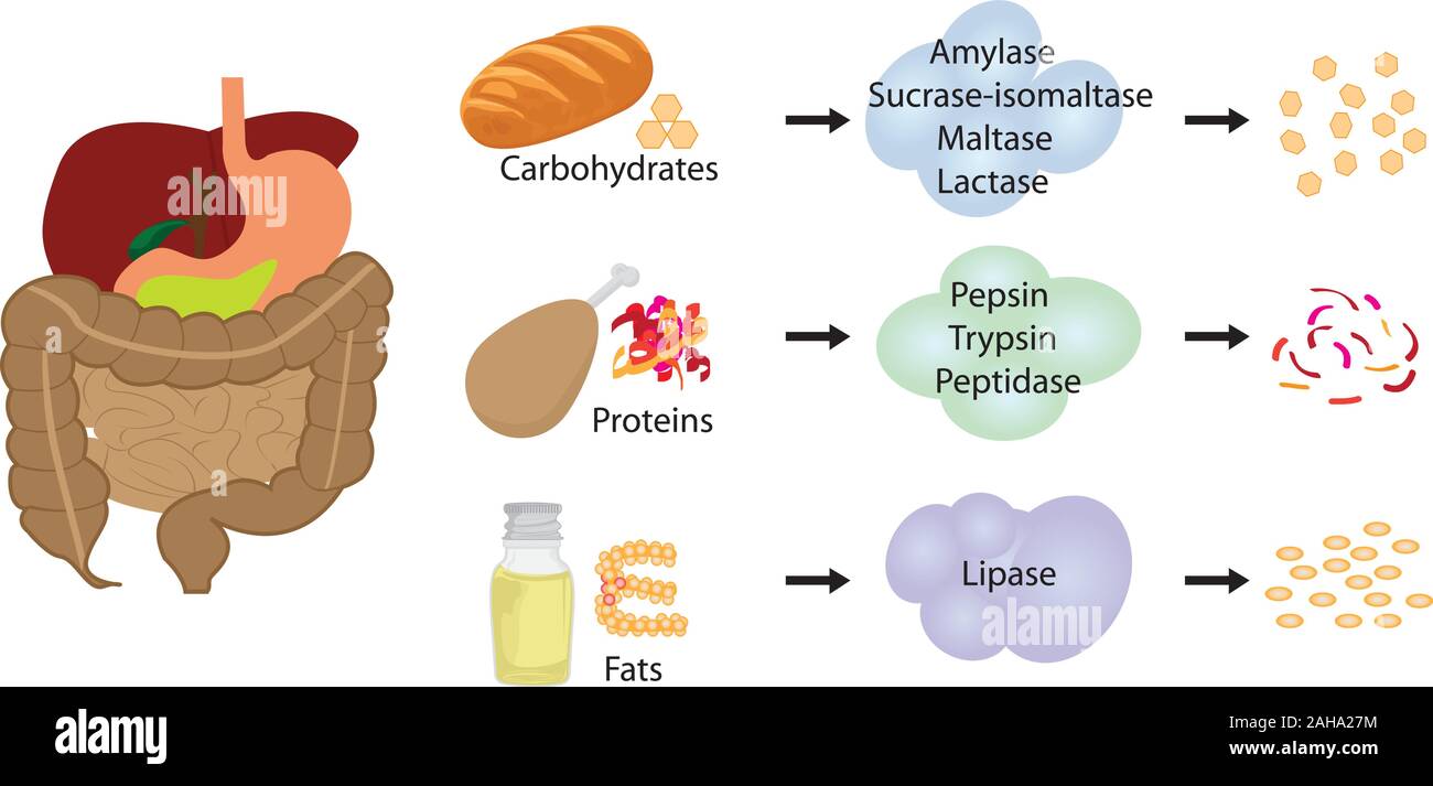 Enzymes braking down food into nutrients. Digestive systems work vector illustrative infographics Stock Vectorhttps://www.alamy.com/image-license-details/?v=1https://www.alamy.com/enzymes-braking-down-food-into-nutrients-digestive-systems-work-vector-illustrative-infographics-image337667448.html
RF2AHA27M–Enzymes braking down food into nutrients. Digestive systems work vector illustrative infographics
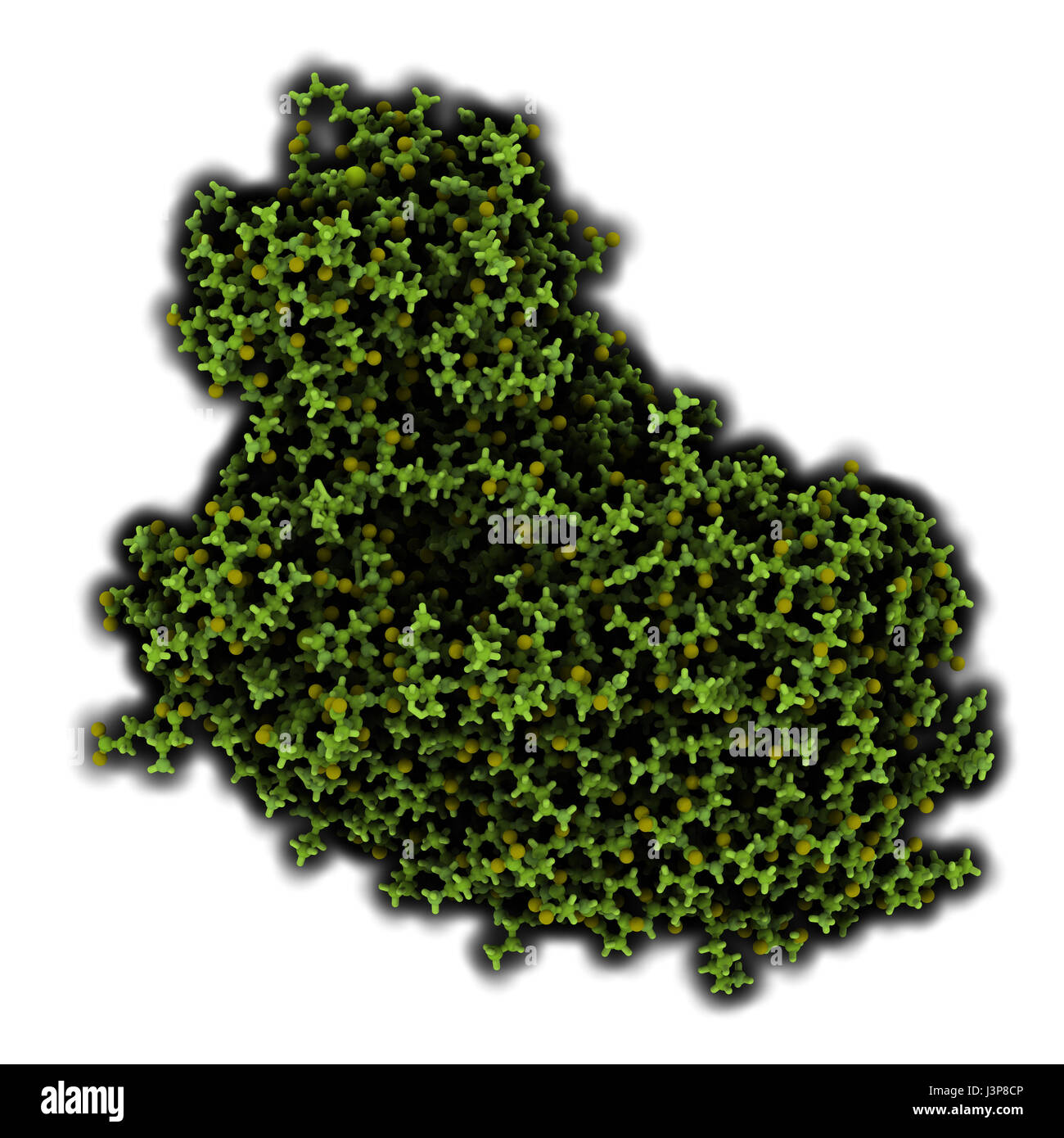 Firefly luciferase enzyme. Protein responsible for the bioluminescence of fireflies. Often used as reporter in biotechnology and genetic engineering. Stock Photohttps://www.alamy.com/image-license-details/?v=1https://www.alamy.com/stock-photo-firefly-luciferase-enzyme-protein-responsible-for-the-bioluminescence-140016486.html
RFJ3P8CP–Firefly luciferase enzyme. Protein responsible for the bioluminescence of fireflies. Often used as reporter in biotechnology and genetic engineering.
RF2XHH7MC–Collagen icon Black line art vector in black and white outline set collection sign
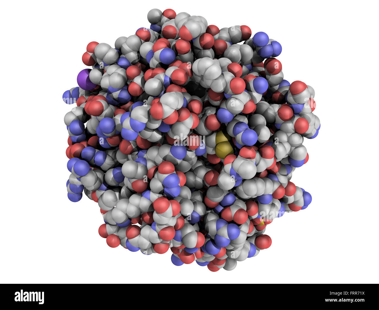 Structure of thrombin blood-clotting enzyme: Human alpha-thrombin molecule is a key protein in the blood coagulation cascade. Co Stock Photohttps://www.alamy.com/image-license-details/?v=1https://www.alamy.com/stock-photo-structure-of-thrombin-blood-clotting-enzyme-human-alpha-thrombin-molecule-100699366.html
RFFRR71X–Structure of thrombin blood-clotting enzyme: Human alpha-thrombin molecule is a key protein in the blood coagulation cascade. Co
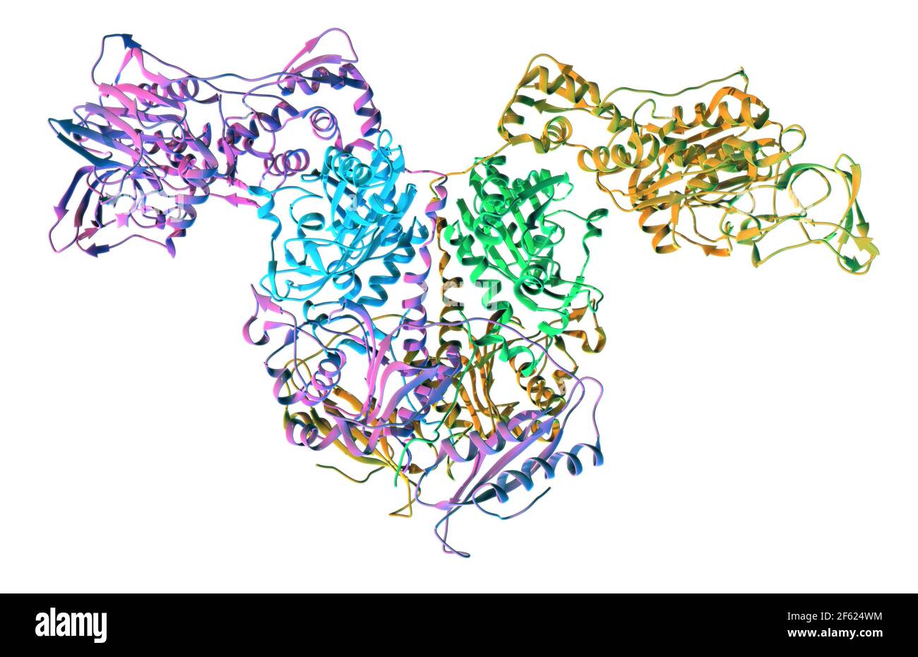 Bacterial T. thermophilus PheRS Stock Photohttps://www.alamy.com/image-license-details/?v=1https://www.alamy.com/bacterial-t-thermophilus-phers-image416784528.html
RM2F624WM–Bacterial T. thermophilus PheRS
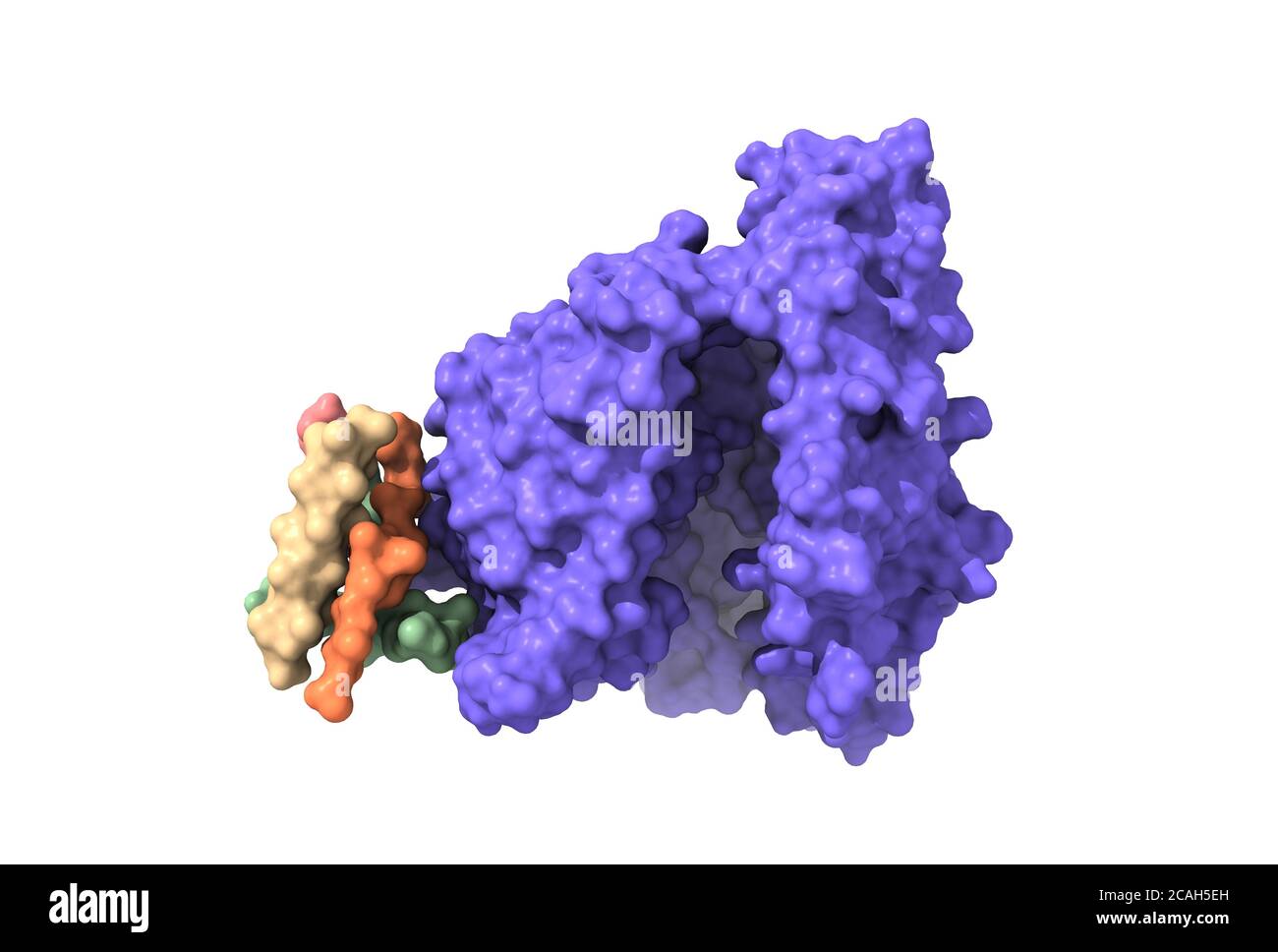 Structure of the human Angiotensin Converting Enzyme-Related Carboxypeptidase (ACE2), a receptor of SARS-CoV-2 spike glycoprotein, 3D surface model Stock Photohttps://www.alamy.com/image-license-details/?v=1https://www.alamy.com/structure-of-the-human-angiotensin-converting-enzyme-related-carboxypeptidase-ace2-a-receptor-of-sars-cov-2-spike-glycoprotein-3d-surface-model-image367941801.html
RF2CAH5EH–Structure of the human Angiotensin Converting Enzyme-Related Carboxypeptidase (ACE2), a receptor of SARS-CoV-2 spike glycoprotein, 3D surface model
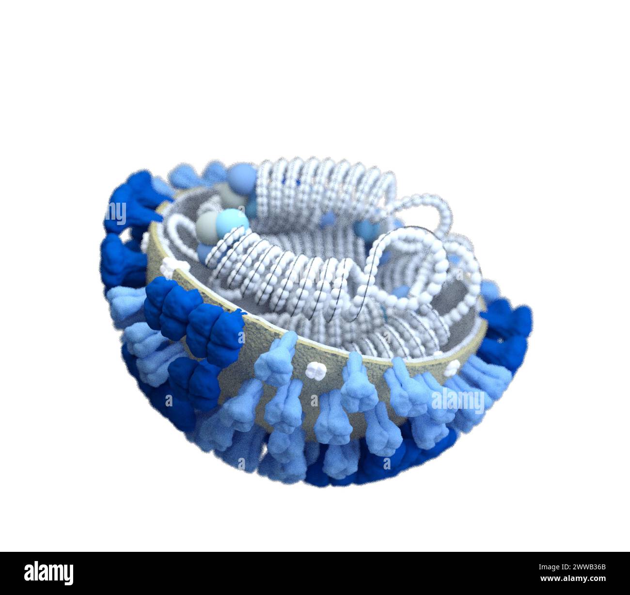 This illustration depicted a 3D computer-generated rendering of a half-sliced influenza (flu) virus with a grey surface membrane. Stock Photohttps://www.alamy.com/image-license-details/?v=1https://www.alamy.com/this-illustration-depicted-a-3d-computer-generated-rendering-of-a-half-sliced-influenza-flu-virus-with-a-grey-surface-membrane-image600762915.html
RM2WWB36B–This illustration depicted a 3D computer-generated rendering of a half-sliced influenza (flu) virus with a grey surface membrane.
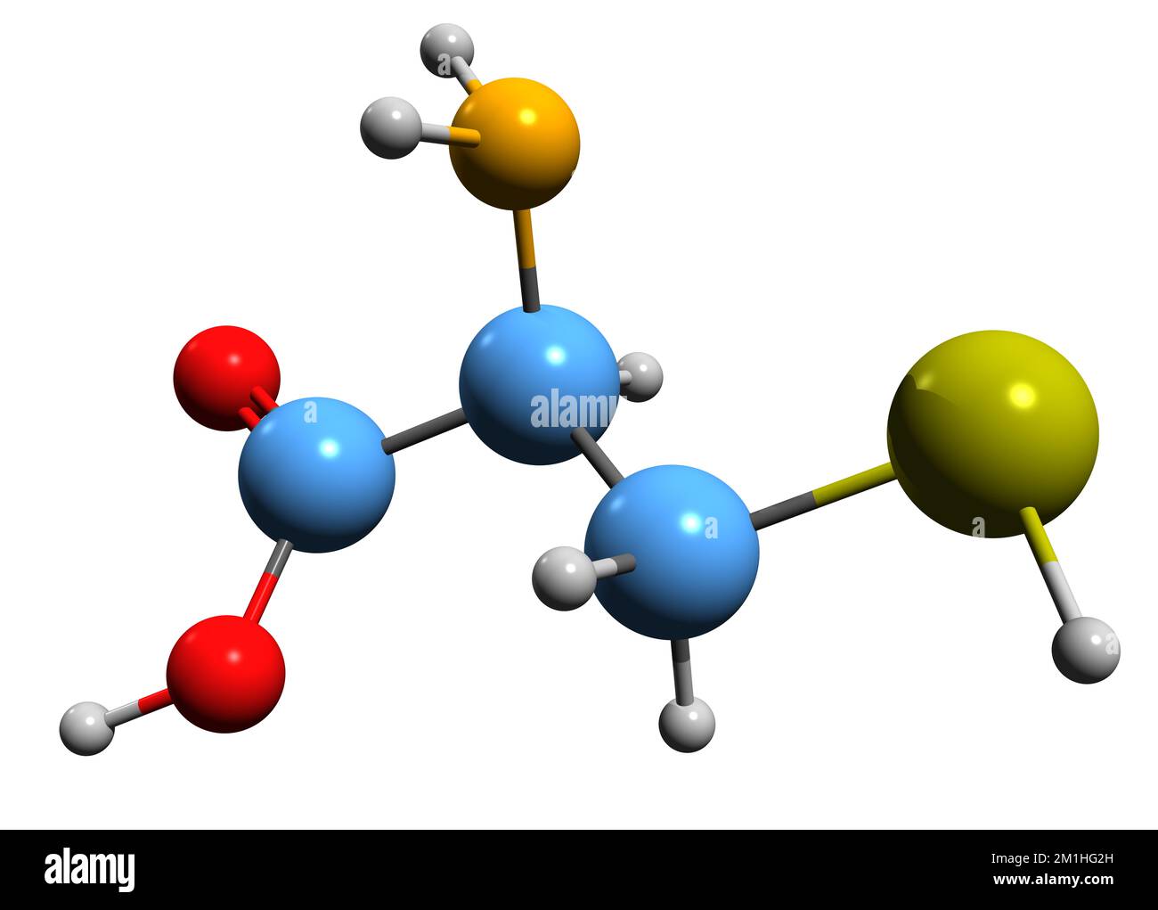 3D image of Cysteine skeletal formula - molecular chemical structure of proteinogenic amino acid isolated on white background Stock Photohttps://www.alamy.com/image-license-details/?v=1https://www.alamy.com/3d-image-of-cysteine-skeletal-formula-molecular-chemical-structure-of-proteinogenic-amino-acid-isolated-on-white-background-image500101129.html
RF2M1HG2H–3D image of Cysteine skeletal formula - molecular chemical structure of proteinogenic amino acid isolated on white background
 Protein 3D model over white Stock Photohttps://www.alamy.com/image-license-details/?v=1https://www.alamy.com/stock-photo-protein-3d-model-over-white-81970561.html
RFENA281–Protein 3D model over white
 Protein structure with biological concept, 3d rendering. 3D illustration. Stock Photohttps://www.alamy.com/image-license-details/?v=1https://www.alamy.com/protein-structure-with-biological-concept-3d-rendering-3d-illustration-image598135256.html
RF2WN3BHC–Protein structure with biological concept, 3d rendering. 3D illustration.
RF2R5RHT7–Molecule set icon template color editable
 Biomarker word cloud concept. Collage made of words about biomarker. Vector illustration Stock Vectorhttps://www.alamy.com/image-license-details/?v=1https://www.alamy.com/biomarker-word-cloud-concept-collage-made-of-words-about-biomarker-vector-illustration-image269545556.html
RFWJET30–Biomarker word cloud concept. Collage made of words about biomarker. Vector illustration
 A Pile of Pellets of Yellow Bee Pollen Stock Photohttps://www.alamy.com/image-license-details/?v=1https://www.alamy.com/a-pile-of-pellets-of-yellow-bee-pollen-image434363162.html
RF2G6JXJ2–A Pile of Pellets of Yellow Bee Pollen
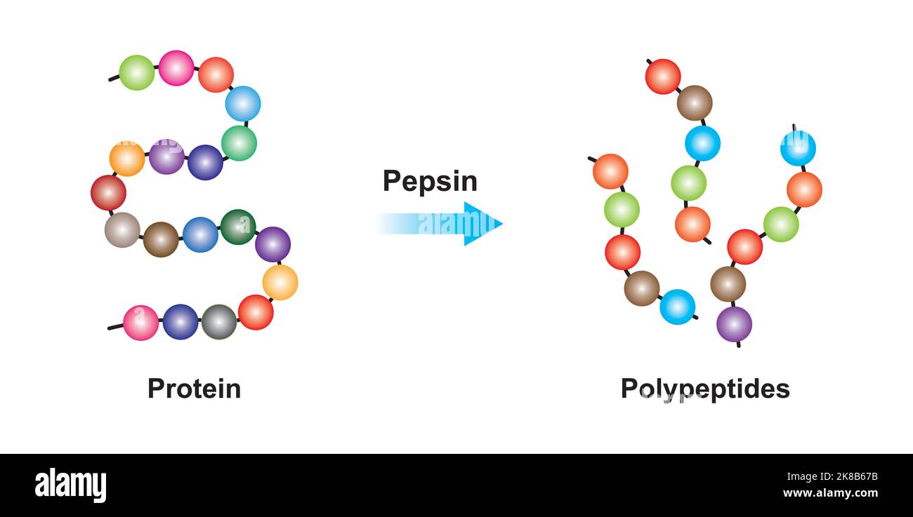 Scientific Designing of Pepsin Enzyme Effect on Protein Molecule. Vector Illustration. Stock Vectorhttps://www.alamy.com/image-license-details/?v=1https://www.alamy.com/scientific-designing-of-pepsin-enzyme-effect-on-protein-molecule-vector-illustration-image487053935.html
RF2K8B67B–Scientific Designing of Pepsin Enzyme Effect on Protein Molecule. Vector Illustration.
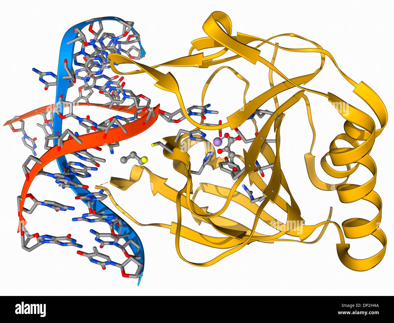 DNA repair enzyme, molecular model Stock Photohttps://www.alamy.com/image-license-details/?v=1https://www.alamy.com/dna-repair-enzyme-molecular-model-image65210890.html
RFDP2H4A–DNA repair enzyme, molecular model
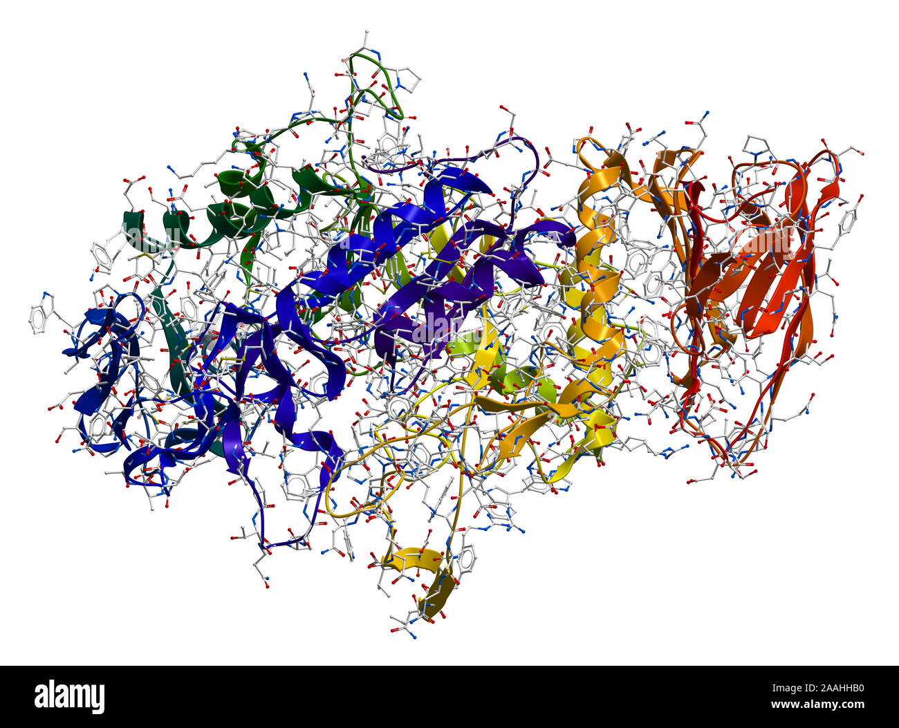 Alpha-Amylase, an enzyme that hydrolyses polysaccharides, such as starch and glycogen, to glucose and maltose. Stock Photohttps://www.alamy.com/image-license-details/?v=1https://www.alamy.com/alpha-amylase-an-enzyme-that-hydrolyses-polysaccharides-such-as-starch-and-glycogen-to-glucose-and-maltose-image333530372.html
RF2AAHHB0–Alpha-Amylase, an enzyme that hydrolyses polysaccharides, such as starch and glycogen, to glucose and maltose.
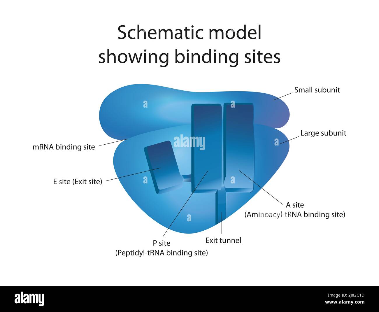 A structural view on the mechanism of the ribosome Stock Photohttps://www.alamy.com/image-license-details/?v=1https://www.alamy.com/a-structural-view-on-the-mechanism-of-the-ribosome-image469650537.html
RF2J82C1D–A structural view on the mechanism of the ribosome
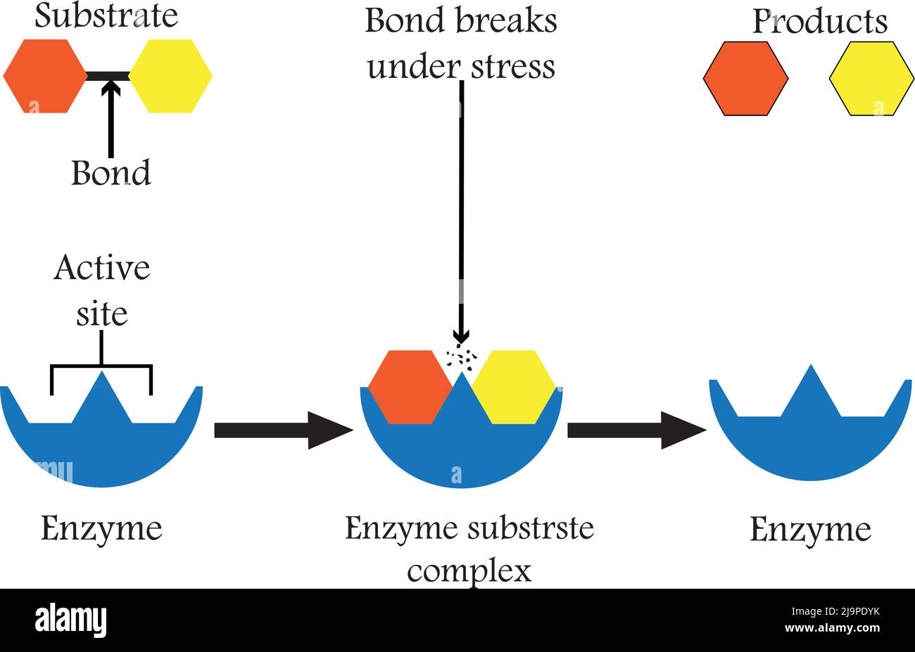 Enzymes that break down food compounds into their basic building blocks, to facilitate its absorption in the body, and we call them digestive enzymes. Stock Vectorhttps://www.alamy.com/image-license-details/?v=1https://www.alamy.com/enzymes-that-break-down-food-compounds-into-their-basic-building-blocks-to-facilitate-its-absorption-in-the-body-and-we-call-them-digestive-enzymes-image470705751.html
RF2J9PDYK–Enzymes that break down food compounds into their basic building blocks, to facilitate its absorption in the body, and we call them digestive enzymes.
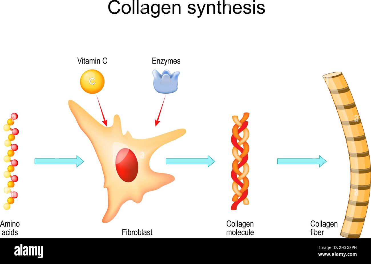 Collagen synthesis with Vitamin C and Enzymes. From Fibroblast and Amino acids to Collagen fiber that comprises molecules of protein. Vector Stock Vectorhttps://www.alamy.com/image-license-details/?v=1https://www.alamy.com/collagen-synthesis-with-vitamin-c-and-enzymes-from-fibroblast-and-amino-acids-to-collagen-fiber-that-comprises-molecules-of-protein-vector-image449671673.html
RF2H3G8PH–Collagen synthesis with Vitamin C and Enzymes. From Fibroblast and Amino acids to Collagen fiber that comprises molecules of protein. Vector
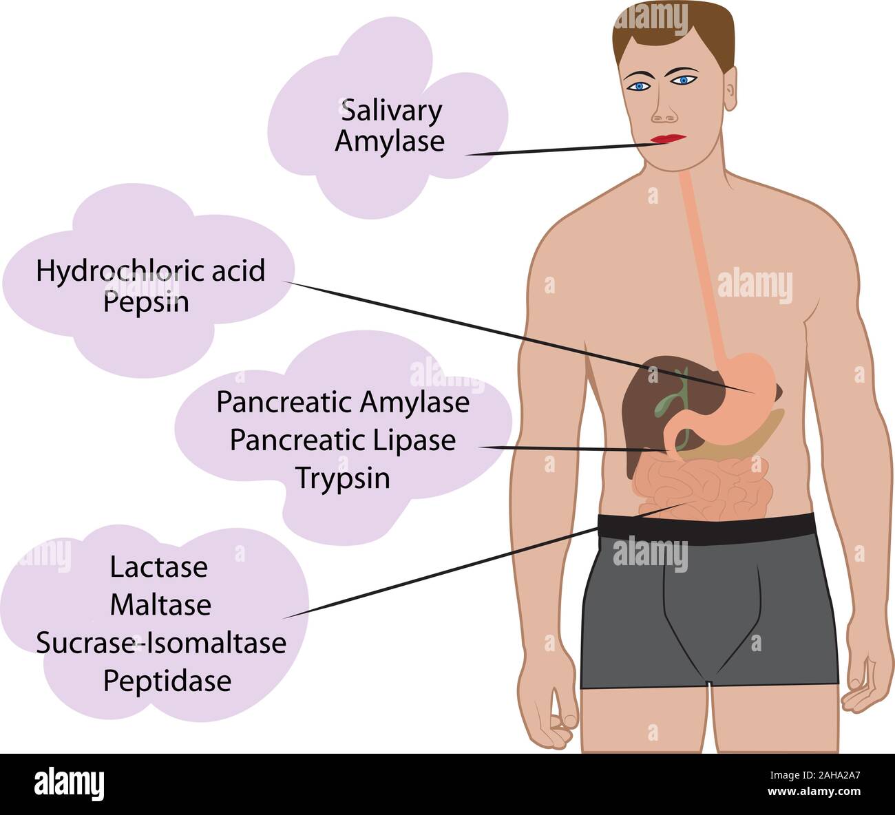 Enzymes braking down food. Digestive systems work vector illustrative infographics Stock Vectorhttps://www.alamy.com/image-license-details/?v=1https://www.alamy.com/enzymes-braking-down-food-digestive-systems-work-vector-illustrative-infographics-image337667519.html
RF2AHA2A7–Enzymes braking down food. Digestive systems work vector illustrative infographics
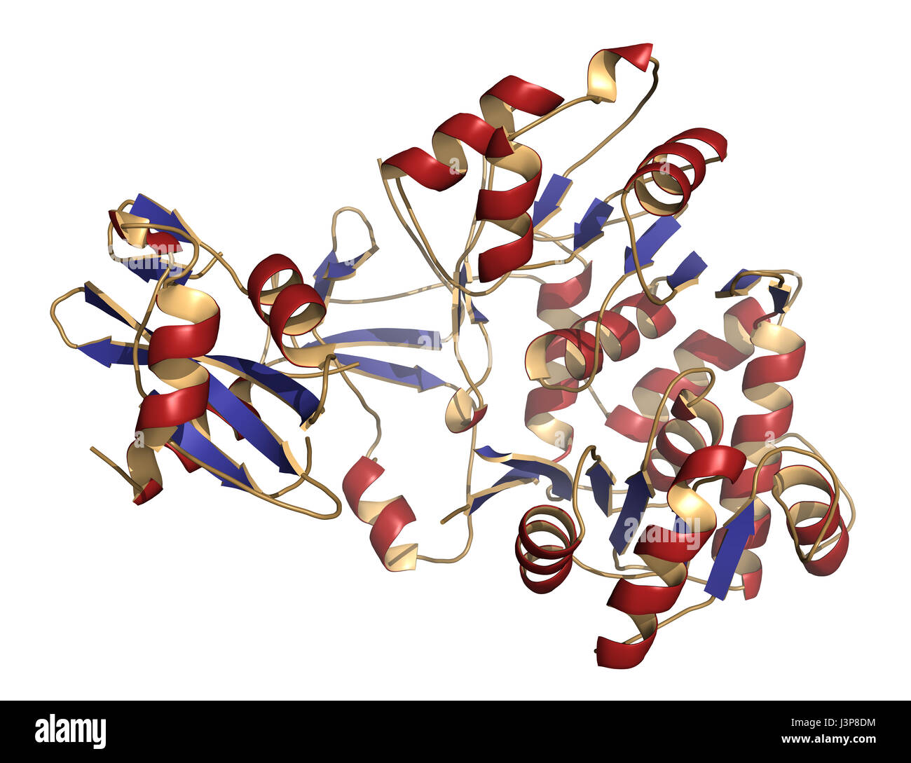 Firefly luciferase enzyme. Protein responsible for the bioluminescence of fireflies. Often used as reporter in biotechnology and genetic engineering. Stock Photohttps://www.alamy.com/image-license-details/?v=1https://www.alamy.com/stock-photo-firefly-luciferase-enzyme-protein-responsible-for-the-bioluminescence-140016512.html
RFJ3P8DM–Firefly luciferase enzyme. Protein responsible for the bioluminescence of fireflies. Often used as reporter in biotechnology and genetic engineering.
RF2XGC8D0–Collagen icon vector logo set collection for web app ui
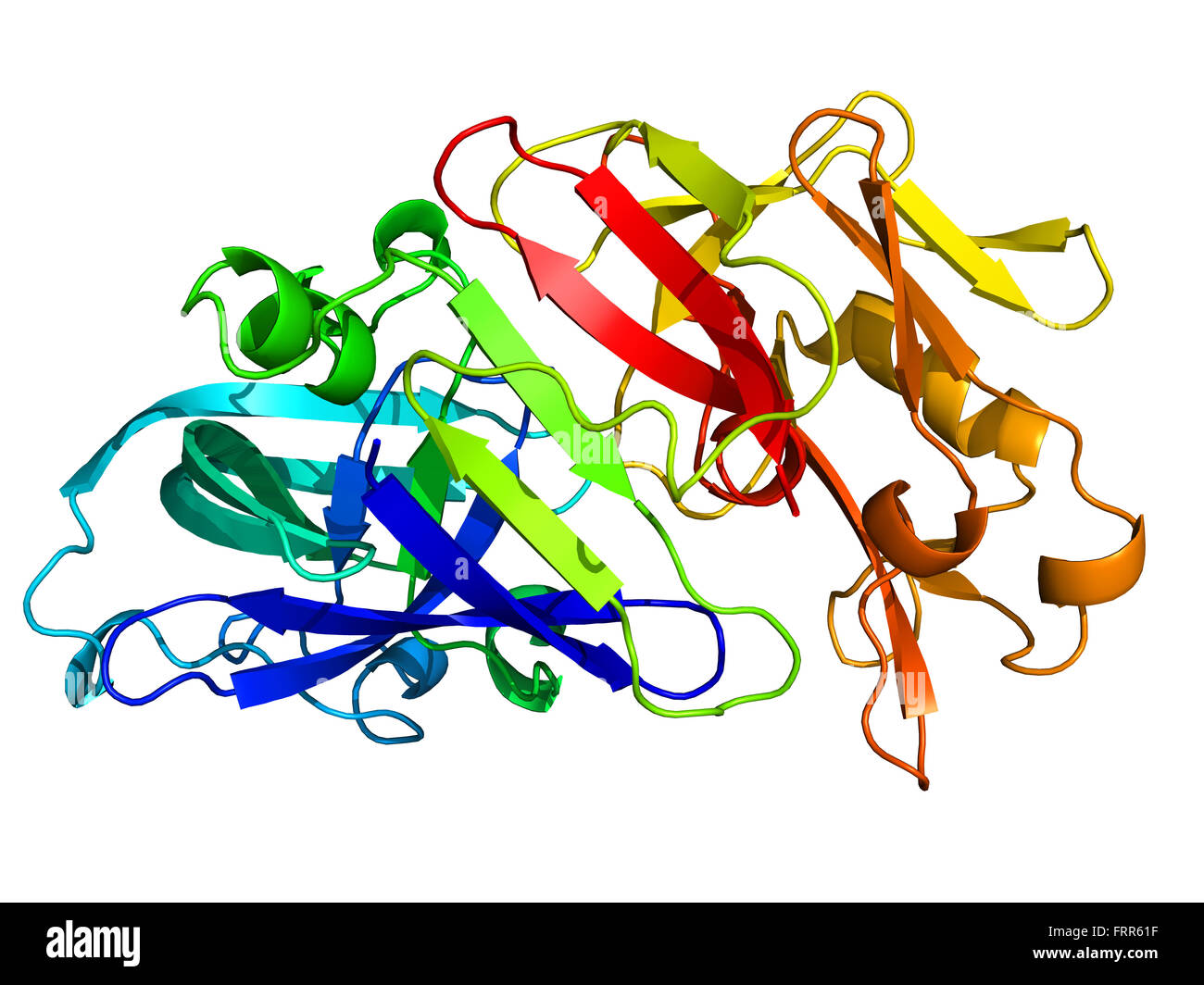 Pepsin 3D model. An enzyme that digests food proteins into peptides Stock Photohttps://www.alamy.com/image-license-details/?v=1https://www.alamy.com/stock-photo-pepsin-3d-model-an-enzyme-that-digests-food-proteins-into-peptides-100698571.html
RFFRR61F–Pepsin 3D model. An enzyme that digests food proteins into peptides
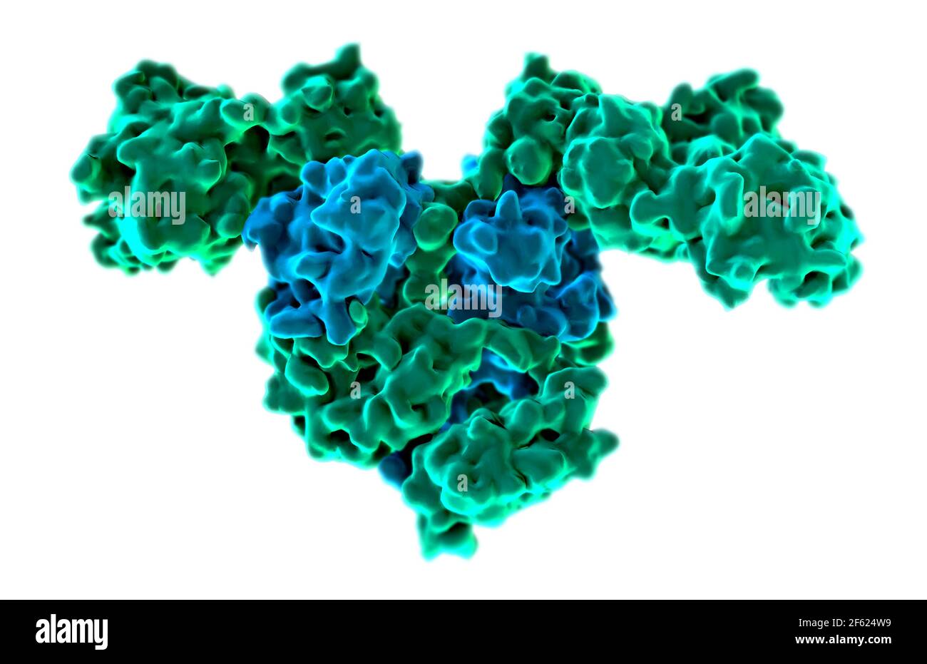 Bacterial T. thermophilus PheRS Stock Photohttps://www.alamy.com/image-license-details/?v=1https://www.alamy.com/bacterial-t-thermophilus-phers-image416784517.html
RM2F624W9–Bacterial T. thermophilus PheRS
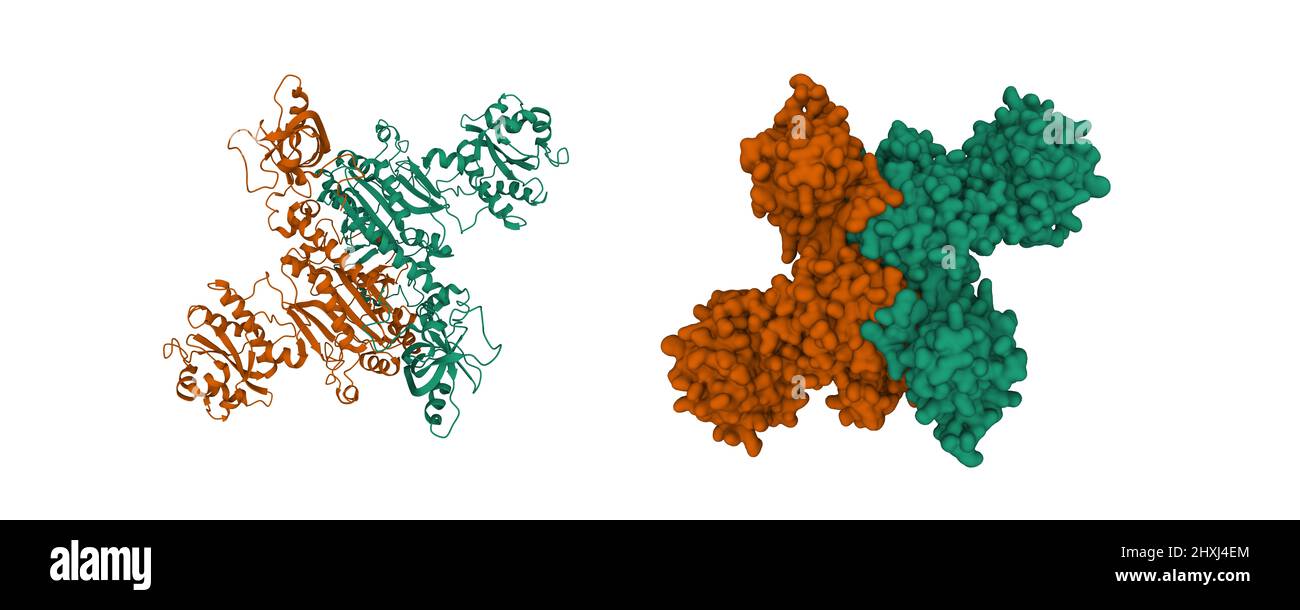 Structure of human mitochondrial aspartyl-tRNA synthetase homodimer. 3D cartoon and Gaussian surface models, chain id color scheme, PDB 4ah6 Stock Photohttps://www.alamy.com/image-license-details/?v=1https://www.alamy.com/structure-of-human-mitochondrial-aspartyl-trna-synthetase-homodimer-3d-cartoon-and-gaussian-surface-models-chain-id-color-scheme-pdb-4ah6-image463849308.html
RF2HXJ4EM–Structure of human mitochondrial aspartyl-tRNA synthetase homodimer. 3D cartoon and Gaussian surface models, chain id color scheme, PDB 4ah6
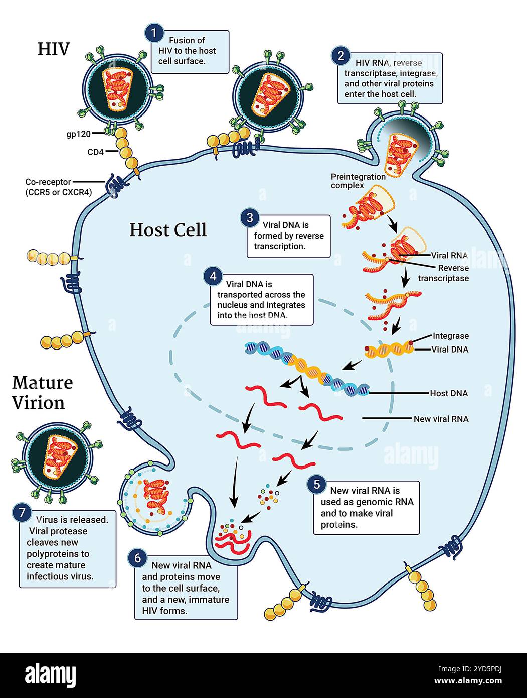 This infographic illustrates the HIV replication cycle, which begins when HIV fuses with the surface of the host cell. Stock Photohttps://www.alamy.com/image-license-details/?v=1https://www.alamy.com/this-infographic-illustrates-the-hiv-replication-cycle-which-begins-when-hiv-fuses-with-the-surface-of-the-host-cell-image627691166.html
RM2YD5PDJ–This infographic illustrates the HIV replication cycle, which begins when HIV fuses with the surface of the host cell.
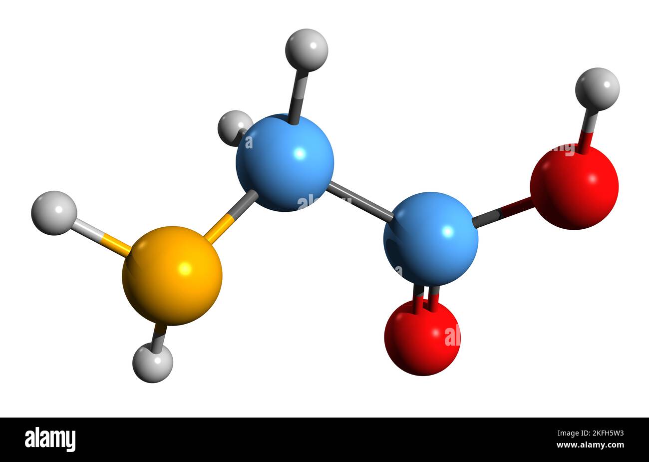 3D image of Glycine skeletal formula - molecular chemical structure of amino acid isolated on white background Stock Photohttps://www.alamy.com/image-license-details/?v=1https://www.alamy.com/3d-image-of-glycine-skeletal-formula-molecular-chemical-structure-of-amino-acid-isolated-on-white-background-image491487951.html
RF2KFH5W3–3D image of Glycine skeletal formula - molecular chemical structure of amino acid isolated on white background
 Protein 3D model over white Stock Photohttps://www.alamy.com/image-license-details/?v=1https://www.alamy.com/stock-photo-protein-3d-model-over-white-82210025.html
RFENMYM9–Protein 3D model over white
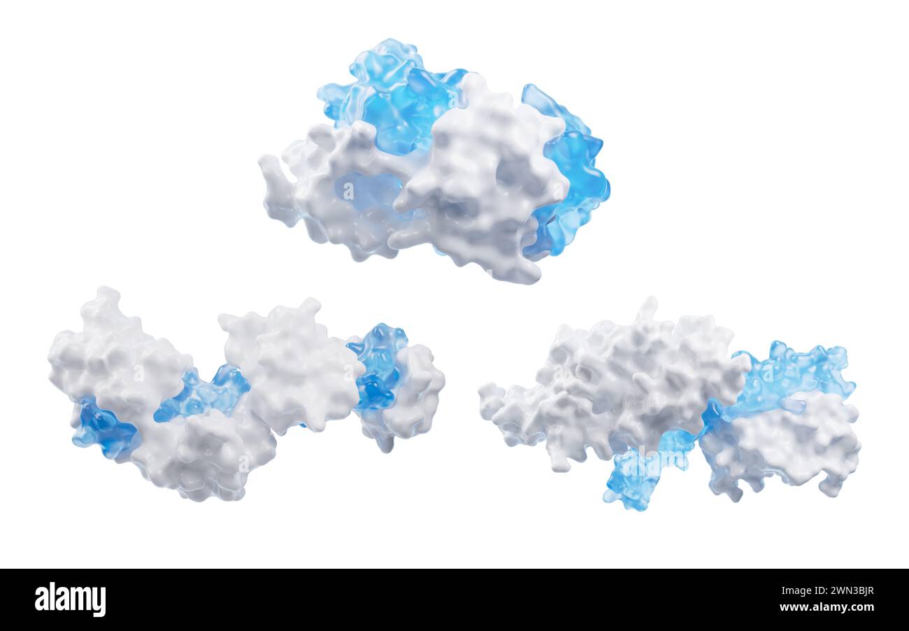 Protein structure with biological concept, 3d rendering. 3D illustration. Stock Photohttps://www.alamy.com/image-license-details/?v=1https://www.alamy.com/protein-structure-with-biological-concept-3d-rendering-3d-illustration-image598135295.html
RF2WN3BJR–Protein structure with biological concept, 3d rendering. 3D illustration.
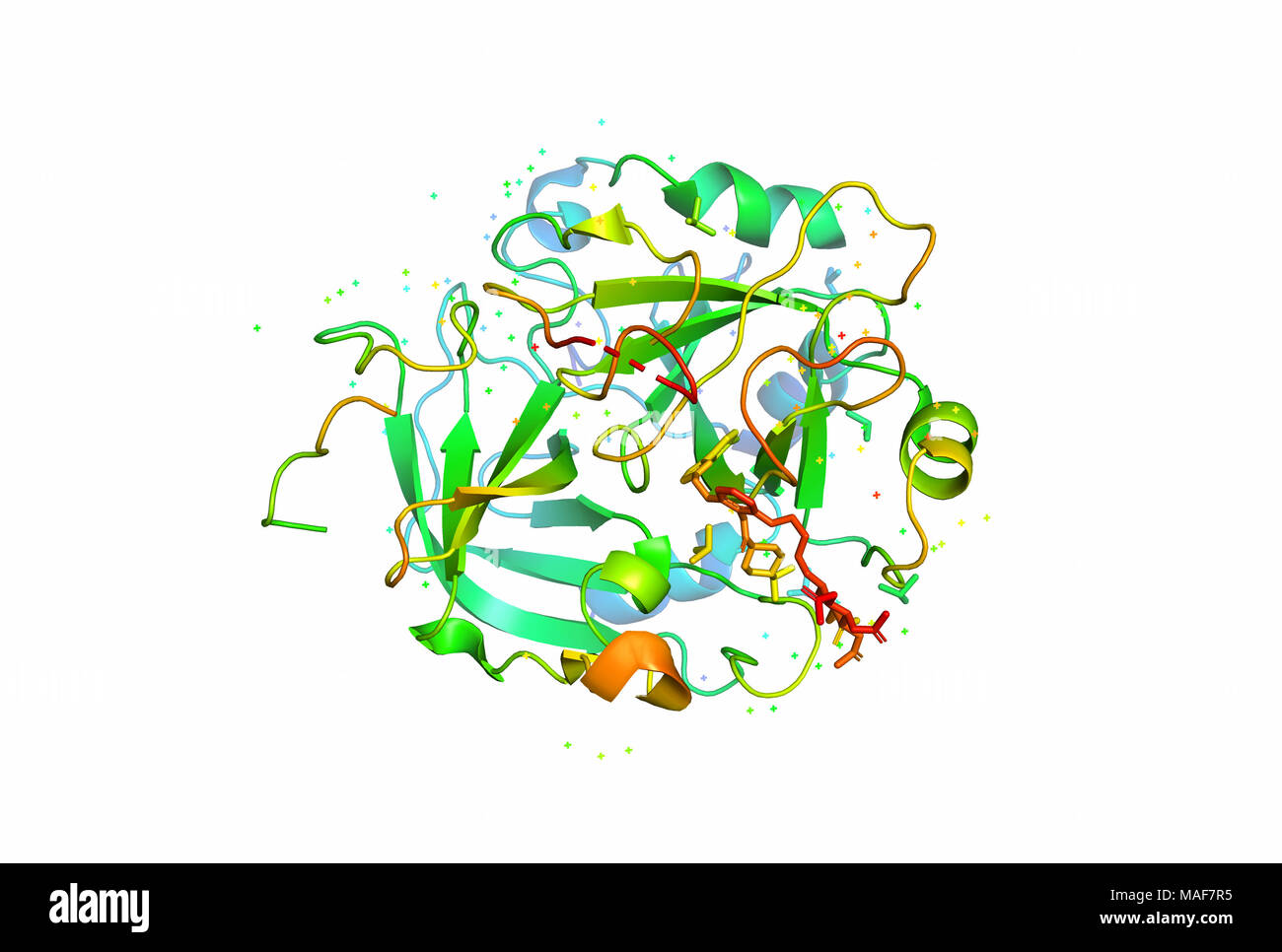 3D model of a protein molecule. The spatial oriented structure of the macromolecule. Stock Photohttps://www.alamy.com/image-license-details/?v=1https://www.alamy.com/3d-model-of-a-protein-molecule-the-spatial-oriented-structure-of-the-macromolecule-image178585657.html
RFMAF7R5–3D model of a protein molecule. The spatial oriented structure of the macromolecule.
 Digestion word cloud concept. Collage made of words about digestion. Vector illustration Stock Vectorhttps://www.alamy.com/image-license-details/?v=1https://www.alamy.com/digestion-word-cloud-concept-collage-made-of-words-about-digestion-vector-illustration-image262247161.html
RFW6JAX1–Digestion word cloud concept. Collage made of words about digestion. Vector illustration
 A Bowl of Pellets of Yellow Bee Pollen Stock Photohttps://www.alamy.com/image-license-details/?v=1https://www.alamy.com/a-bowl-of-pellets-of-yellow-bee-pollen-image240054272.html
RFRXFBJT–A Bowl of Pellets of Yellow Bee Pollen
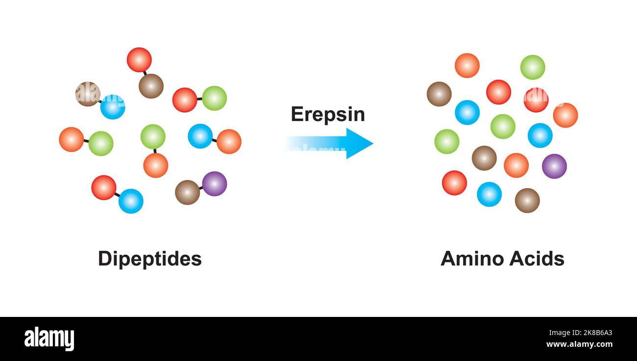 Scientific Designing of Erepsin Enzyme Effect on Dipeptide Molecule. Vector Illustration. Stock Vectorhttps://www.alamy.com/image-license-details/?v=1https://www.alamy.com/scientific-designing-of-erepsin-enzyme-effect-on-dipeptide-molecule-vector-illustration-image487054011.html
RF2K8B6A3–Scientific Designing of Erepsin Enzyme Effect on Dipeptide Molecule. Vector Illustration.
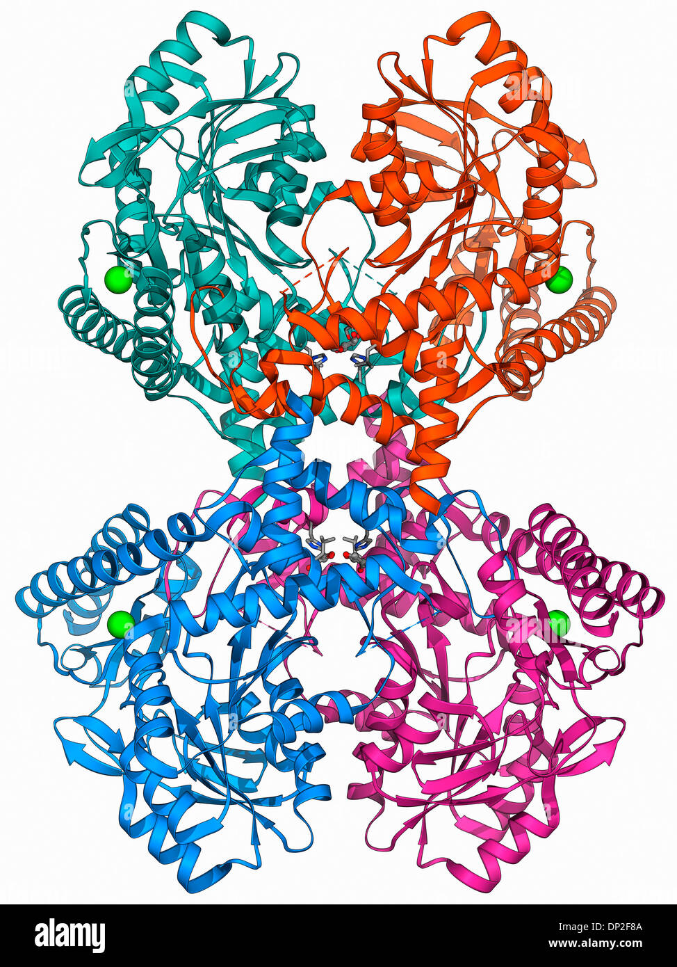 Selenocysteine synthase enzyme molecule Stock Photohttps://www.alamy.com/image-license-details/?v=1https://www.alamy.com/selenocysteine-synthase-enzyme-molecule-image65209434.html
RFDP2F8A–Selenocysteine synthase enzyme molecule
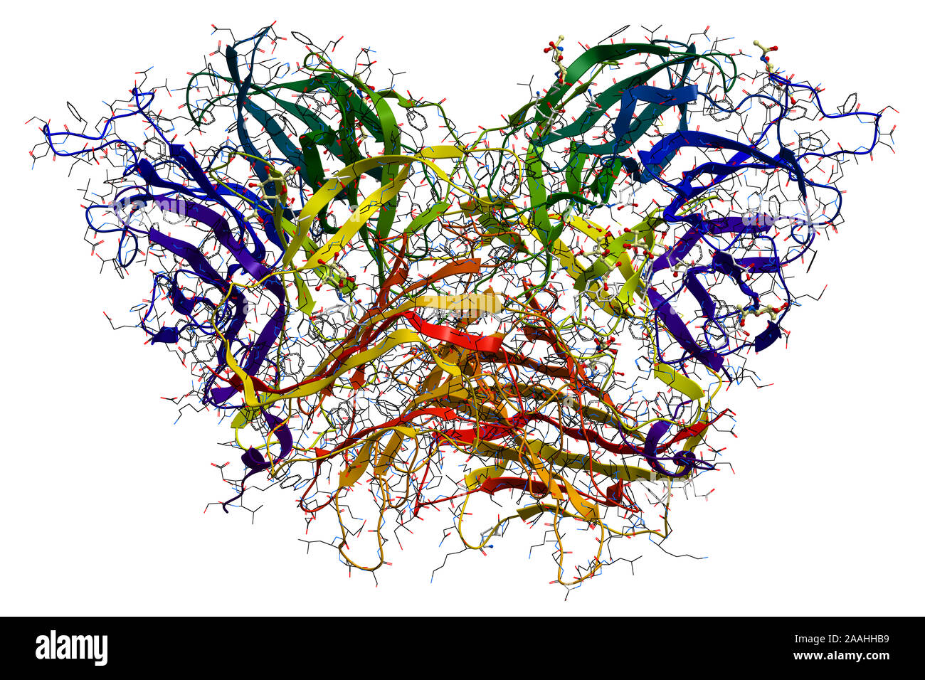 Invertase, an enzyme that catalyzes the hydrolysis (breakdown) of sucrose (table sugar). 3D molecular structure Stock Photohttps://www.alamy.com/image-license-details/?v=1https://www.alamy.com/invertase-an-enzyme-that-catalyzes-the-hydrolysis-breakdown-of-sucrose-table-sugar-3d-molecular-structure-image333530381.html
RF2AAHHB9–Invertase, an enzyme that catalyzes the hydrolysis (breakdown) of sucrose (table sugar). 3D molecular structure
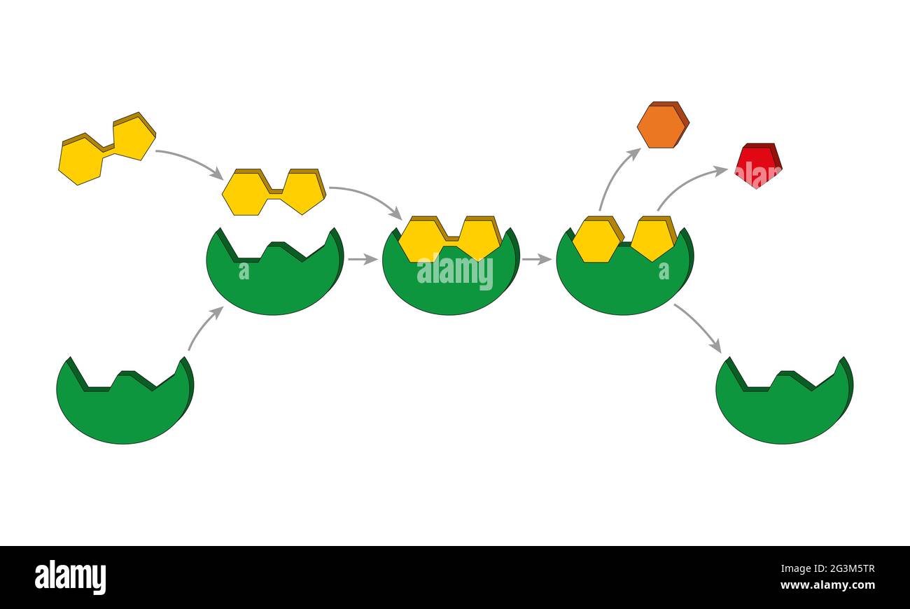 Lock and key model. Enzymes are proteins that act as biological catalysts (biocatalysts). Catalysts accelerate chemical reactions Stock Photohttps://www.alamy.com/image-license-details/?v=1https://www.alamy.com/lock-and-key-model-enzymes-are-proteins-that-act-as-biological-catalysts-biocatalysts-catalysts-accelerate-chemical-reactions-image432546823.html
RF2G3M5TR–Lock and key model. Enzymes are proteins that act as biological catalysts (biocatalysts). Catalysts accelerate chemical reactions
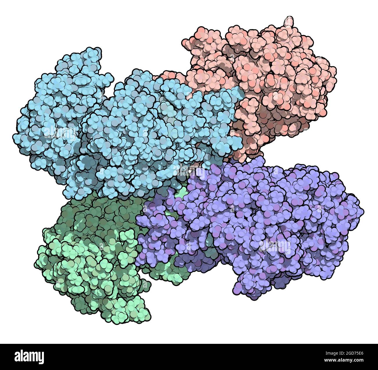 Glucose-6-phosphate dehydrogenase (G6PD) protein. 3D Illustration. Stock Photohttps://www.alamy.com/image-license-details/?v=1https://www.alamy.com/glucose-6-phosphate-dehydrogenase-g6pd-protein-3d-illustration-image438407710.html
RF2GD75E6–Glucose-6-phosphate dehydrogenase (G6PD) protein. 3D Illustration.
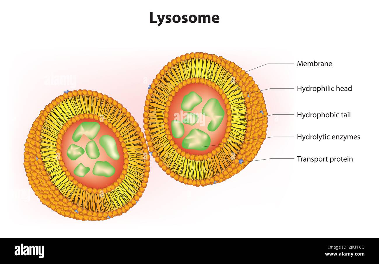
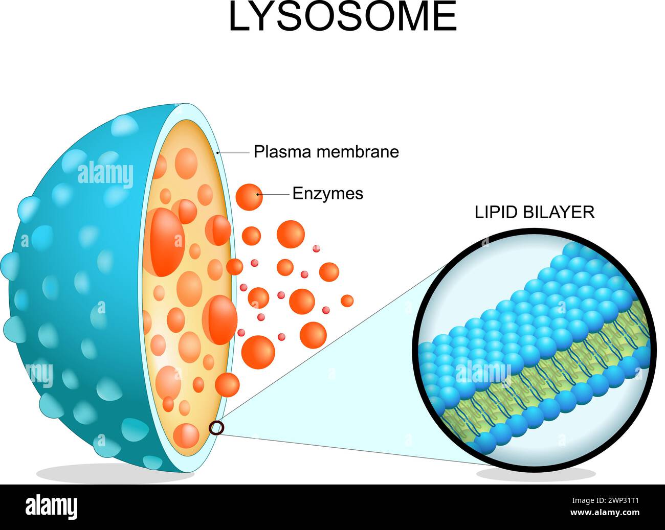 Lysosome anatomy. Cross section of a cell organelle. Close-up of a Lipid bilayer membrane, hydrolytic enzymes, transport proteins. Autophagy. Vector i Stock Vectorhttps://www.alamy.com/image-license-details/?v=1https://www.alamy.com/lysosome-anatomy-cross-section-of-a-cell-organelle-close-up-of-a-lipid-bilayer-membrane-hydrolytic-enzymes-transport-proteins-autophagy-vector-i-image598742257.html
RF2WP31T1–Lysosome anatomy. Cross section of a cell organelle. Close-up of a Lipid bilayer membrane, hydrolytic enzymes, transport proteins. Autophagy. Vector i