Extensor carpi ulnaris muscle Stock Photos and Images
(1,191)See extensor carpi ulnaris muscle stock video clipsQuick filters:
Extensor carpi ulnaris muscle Stock Photos and Images
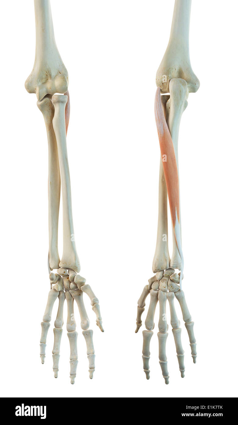 Human extensor carpi ulnaris muscle computer artwork. Stock Photohttps://www.alamy.com/image-license-details/?v=1https://www.alamy.com/human-extensor-carpi-ulnaris-muscle-computer-artwork-image69879395.html
RFE1K7TK–Human extensor carpi ulnaris muscle computer artwork.
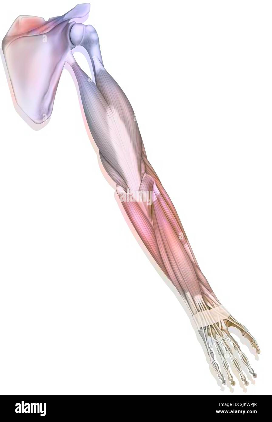 The muscles of the upper right limb in posterior view. Stock Photohttps://www.alamy.com/image-license-details/?v=1https://www.alamy.com/the-muscles-of-the-upper-right-limb-in-posterior-view-image476924975.html
RF2JKWPJR–The muscles of the upper right limb in posterior view.
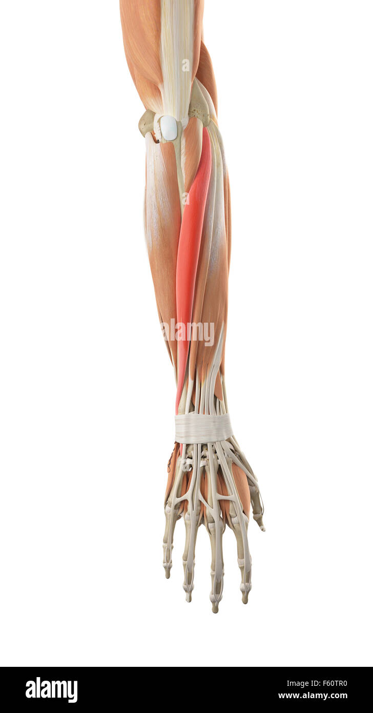 medically accurate illustration of the extensor carpi ulnaris Stock Photohttps://www.alamy.com/image-license-details/?v=1https://www.alamy.com/stock-photo-medically-accurate-illustration-of-the-extensor-carpi-ulnaris-89759236.html
RFF60TR0–medically accurate illustration of the extensor carpi ulnaris
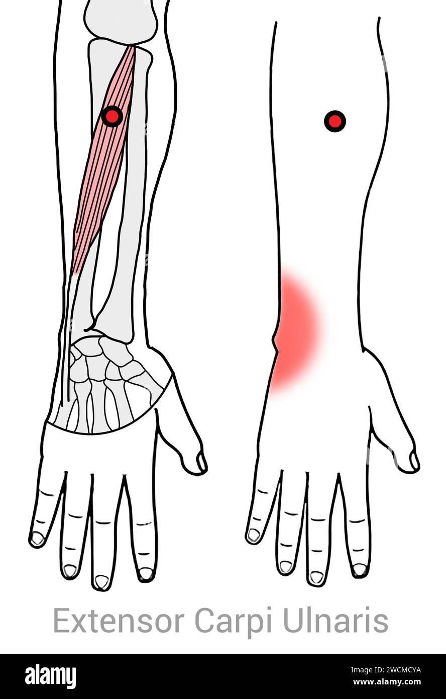 Extensor Carpi Ulnaris: Myofascial trigger points and associated pain locations Stock Photohttps://www.alamy.com/image-license-details/?v=1https://www.alamy.com/extensor-carpi-ulnaris-myofascial-trigger-points-and-associated-pain-locations-image592977598.html
RF2WCMCYA–Extensor Carpi Ulnaris: Myofascial trigger points and associated pain locations
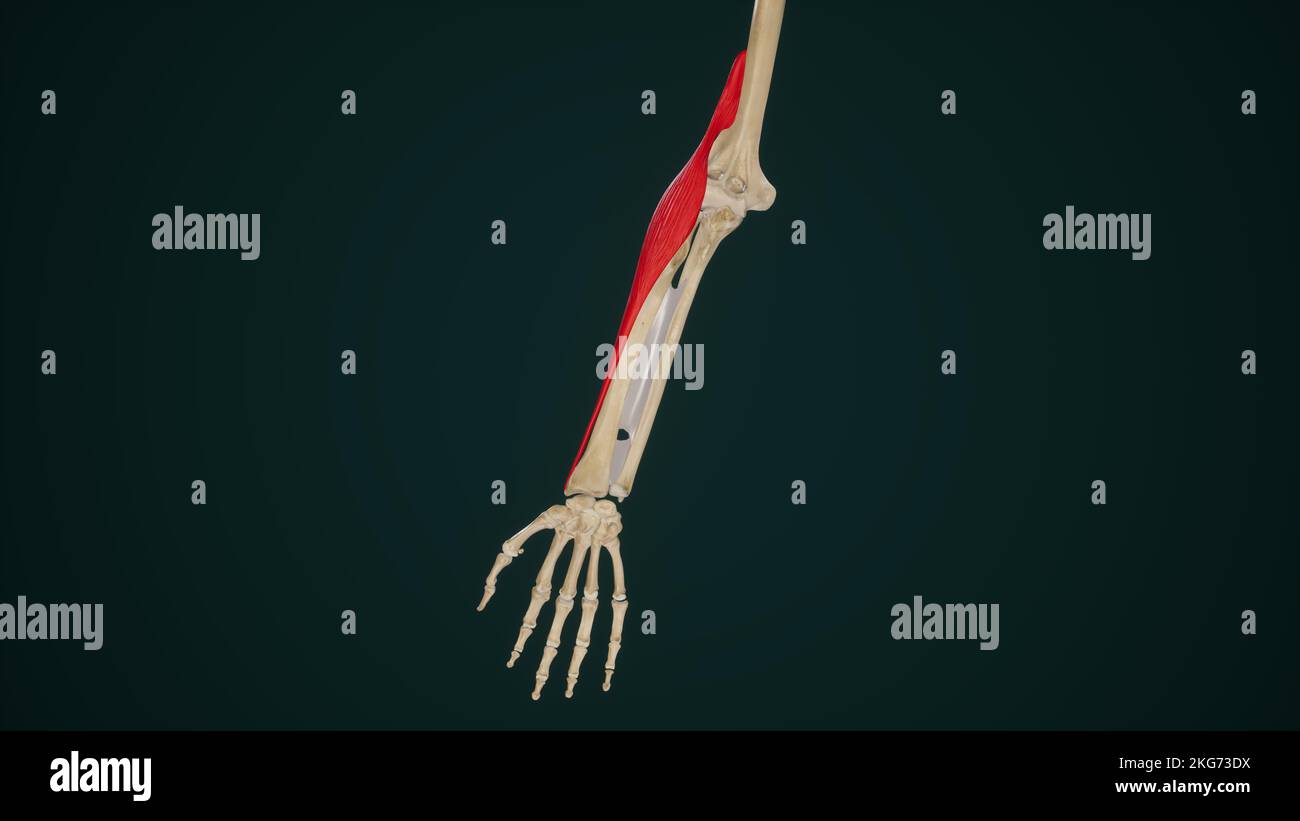 Brachioradialis Muscle Stock Photohttps://www.alamy.com/image-license-details/?v=1https://www.alamy.com/brachioradialis-muscle-image491881206.html
RF2KG73DX–Brachioradialis Muscle
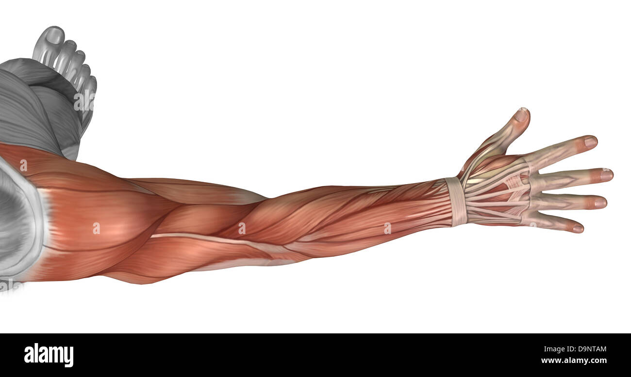 Muscle anatomy of the human arm, posterior view. Stock Photohttps://www.alamy.com/image-license-details/?v=1https://www.alamy.com/stock-photo-muscle-anatomy-of-the-human-arm-posterior-view-57643116.html
RFD9NTAM–Muscle anatomy of the human arm, posterior view.
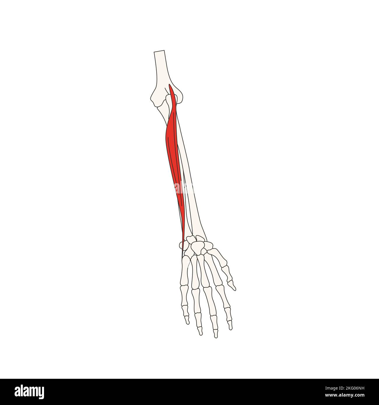 human anatomy drawing extensor carpi ulnaris Stock Photohttps://www.alamy.com/image-license-details/?v=1https://www.alamy.com/human-anatomy-drawing-extensor-carpi-ulnaris-image491730109.html
RF2KG06NH–human anatomy drawing extensor carpi ulnaris
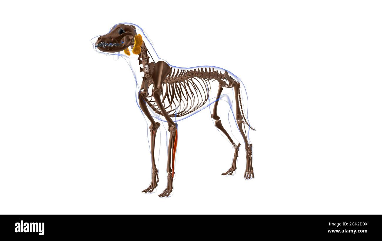 Extensor Carpi Ulnaris muscle Dog muscle Anatomy For Medical Concept 3D Illustration Stock Photohttps://www.alamy.com/image-license-details/?v=1https://www.alamy.com/extensor-carpi-ulnaris-muscle-dog-muscle-anatomy-for-medical-concept-3d-illustration-image441991786.html
RF2GK2D0X–Extensor Carpi Ulnaris muscle Dog muscle Anatomy For Medical Concept 3D Illustration
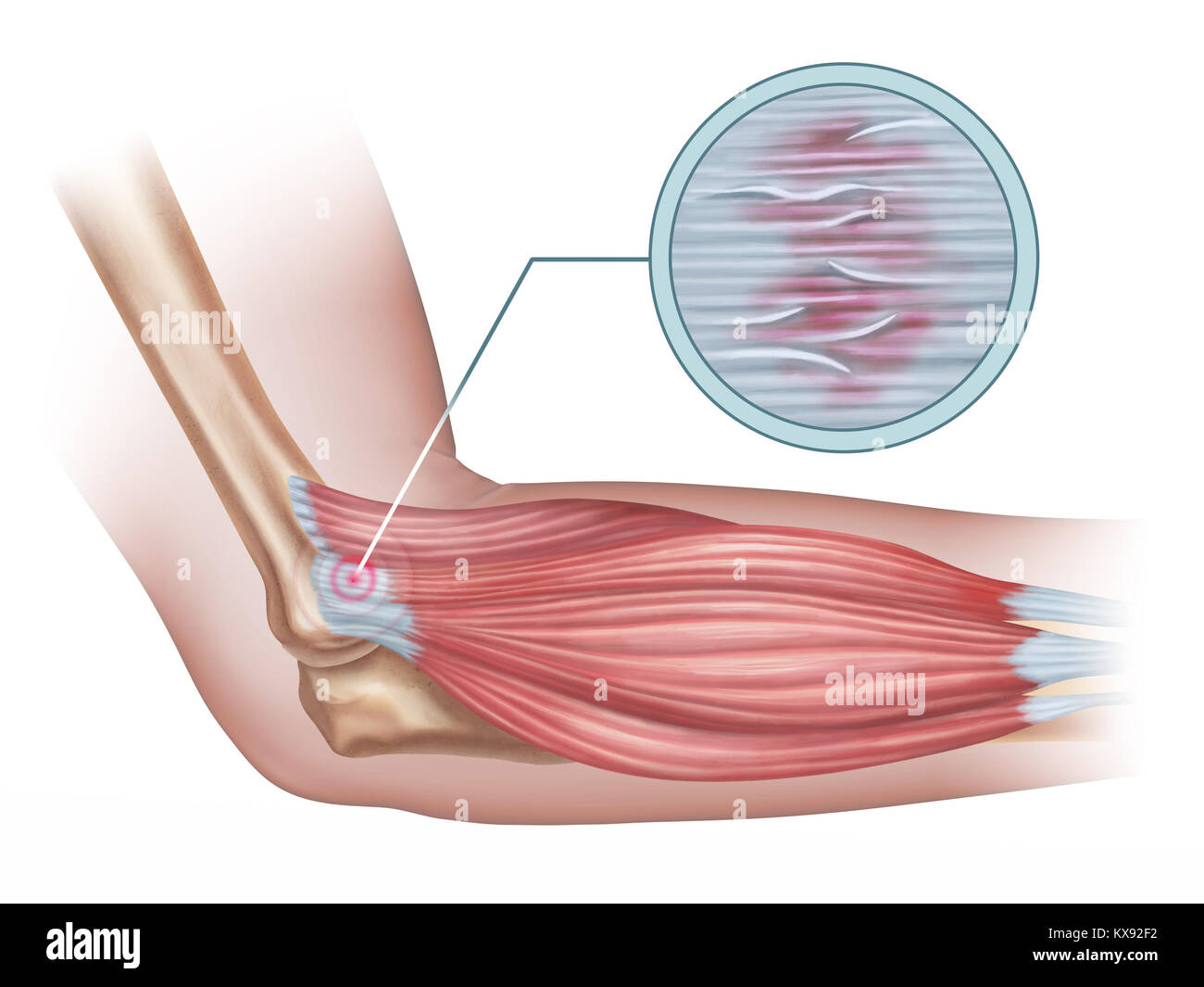 Tennis elbow diagram showing a detail of the damaged tendon tissue. Digital illustration. Stock Photohttps://www.alamy.com/image-license-details/?v=1https://www.alamy.com/stock-photo-tennis-elbow-diagram-showing-a-detail-of-the-damaged-tendon-tissue-171073926.html
RFKX92F2–Tennis elbow diagram showing a detail of the damaged tendon tissue. Digital illustration.
 Extensor carpi ulnaris muscle Stock Photohttps://www.alamy.com/image-license-details/?v=1https://www.alamy.com/extensor-carpi-ulnaris-muscle-image259928764.html
RMW2TNP4–Extensor carpi ulnaris muscle
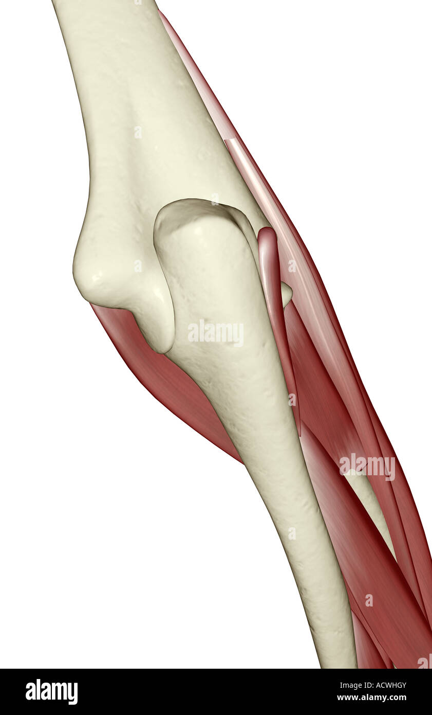 Muscles of the lower arm Stock Photohttps://www.alamy.com/image-license-details/?v=1https://www.alamy.com/stock-photo-muscles-of-the-lower-arm-13236698.html
RFACWHGY–Muscles of the lower arm
 Hand muscles system human body system. Arm Muscular anatomy Stock Vectorhttps://www.alamy.com/image-license-details/?v=1https://www.alamy.com/hand-muscles-system-human-body-system-arm-muscular-anatomy-image229585723.html
RFR9EEX3–Hand muscles system human body system. Arm Muscular anatomy
RMW9GYEN–Archive image from page 420 of Cunningham's Text-book of anatomy (1914). Cunningham's Text-book of anatomy cunninghamstextb00cunn Year: 1914 ( MUSCLES ON ANTEEIOE AND MEDIAL ASPECTS OF EOEEAEM. 387 the forearm between the two heads of origin of the muscle), and the ulnar artery. The tendon serves as a guide to the artery in the distal half of the forearm. Biceps brachii Medial inter- muscular septum Brachialis Medial epicondyle Lacertus fibrosus supinator muscle Pronator teres Flexor carpi radialis Palmaris LONGUS Flexor carpi ULNARIS Extensor CARPI RADIALIS LONGUS Brachio- RADIAL1S Flexor
 . An atlas of human anatomy for students and physicians. Anatomy. THE MUSCLES OF THE UPPER EXTREMITY 331 The radius Radius Canals for the tendonsâ of the extensor secundi intemodii poUicis muscle' of the extensor communis digitorum and extensor indicis muscles of the extensor "I'Tiimi digiti muscle of the extensor carpi ulnaris muscle â Tendon of the extensor carpi ulnaris muscle Dorsal interosseous muscles Mm. interossei dorsales Dorsal aponeuroses of the extensor tendons Aponeuroses tendinum extensorum digitorum. Canals for the tendonsâ of the extensor carpi radialis longior and extenso Stock Photohttps://www.alamy.com/image-license-details/?v=1https://www.alamy.com/an-atlas-of-human-anatomy-for-students-and-physicians-anatomy-the-muscles-of-the-upper-extremity-331-the-radius-radius-canals-for-the-tendons-of-the-extensor-secundi-intemodii-pouicis-muscle-of-the-extensor-communis-digitorum-and-extensor-indicis-muscles-of-the-extensor-quotitiimi-digiti-muscle-of-the-extensor-carpi-ulnaris-muscle-tendon-of-the-extensor-carpi-ulnaris-muscle-dorsal-interosseous-muscles-mm-interossei-dorsales-dorsal-aponeuroses-of-the-extensor-tendons-aponeuroses-tendinum-extensorum-digitorum-canals-for-the-tendons-of-the-extensor-carpi-radialis-longior-and-extenso-image235400158.html
RMRJYB8E–. An atlas of human anatomy for students and physicians. Anatomy. THE MUSCLES OF THE UPPER EXTREMITY 331 The radius Radius Canals for the tendonsâ of the extensor secundi intemodii poUicis muscle' of the extensor communis digitorum and extensor indicis muscles of the extensor "I'Tiimi digiti muscle of the extensor carpi ulnaris muscle â Tendon of the extensor carpi ulnaris muscle Dorsal interosseous muscles Mm. interossei dorsales Dorsal aponeuroses of the extensor tendons Aponeuroses tendinum extensorum digitorum. Canals for the tendonsâ of the extensor carpi radialis longior and extenso
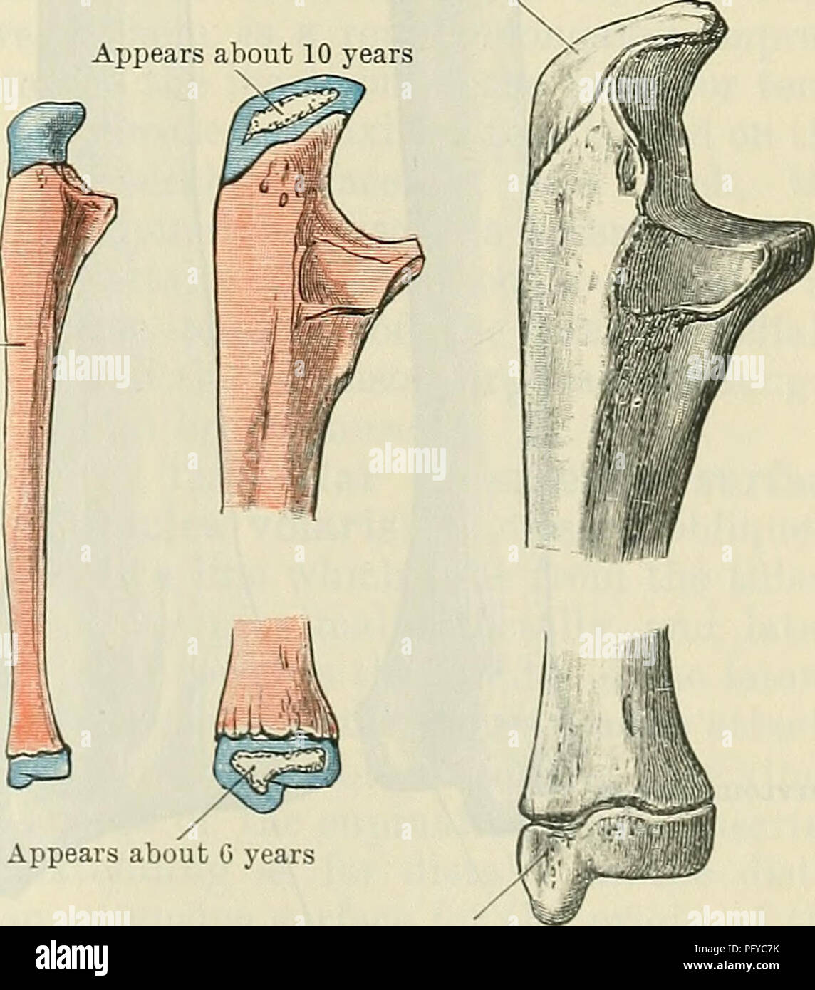 . Cunningham's Text-book of anatomy. Anatomy. THE ULNA. 213 posterior surface is subdivided by a faint longitudinal ridge, the bone between which and the interosseous crest furnishes origins for the abductor pollicis longus, extensor pollicis longus, and extensor indicis proprius muscles, in order proximo- distally. The surface of bone between the dorsal margin and the afore-mentioned longitudinal line is smooth and overlain by the extensor carpi ulnaris muscle, which does not arise from it. The distal extremity of the ulna presents a rounded head (capitulum ulnse), from which, on its medial a Stock Photohttps://www.alamy.com/image-license-details/?v=1https://www.alamy.com/cunninghams-text-book-of-anatomy-anatomy-the-ulna-213-posterior-surface-is-subdivided-by-a-faint-longitudinal-ridge-the-bone-between-which-and-the-interosseous-crest-furnishes-origins-for-the-abductor-pollicis-longus-extensor-pollicis-longus-and-extensor-indicis-proprius-muscles-in-order-proximo-distally-the-surface-of-bone-between-the-dorsal-margin-and-the-afore-mentioned-longitudinal-line-is-smooth-and-overlain-by-the-extensor-carpi-ulnaris-muscle-which-does-not-arise-from-it-the-distal-extremity-of-the-ulna-presents-a-rounded-head-capitulum-ulnse-from-which-on-its-medial-a-image216346583.html
RMPFYC7K–. Cunningham's Text-book of anatomy. Anatomy. THE ULNA. 213 posterior surface is subdivided by a faint longitudinal ridge, the bone between which and the interosseous crest furnishes origins for the abductor pollicis longus, extensor pollicis longus, and extensor indicis proprius muscles, in order proximo- distally. The surface of bone between the dorsal margin and the afore-mentioned longitudinal line is smooth and overlain by the extensor carpi ulnaris muscle, which does not arise from it. The distal extremity of the ulna presents a rounded head (capitulum ulnse), from which, on its medial a
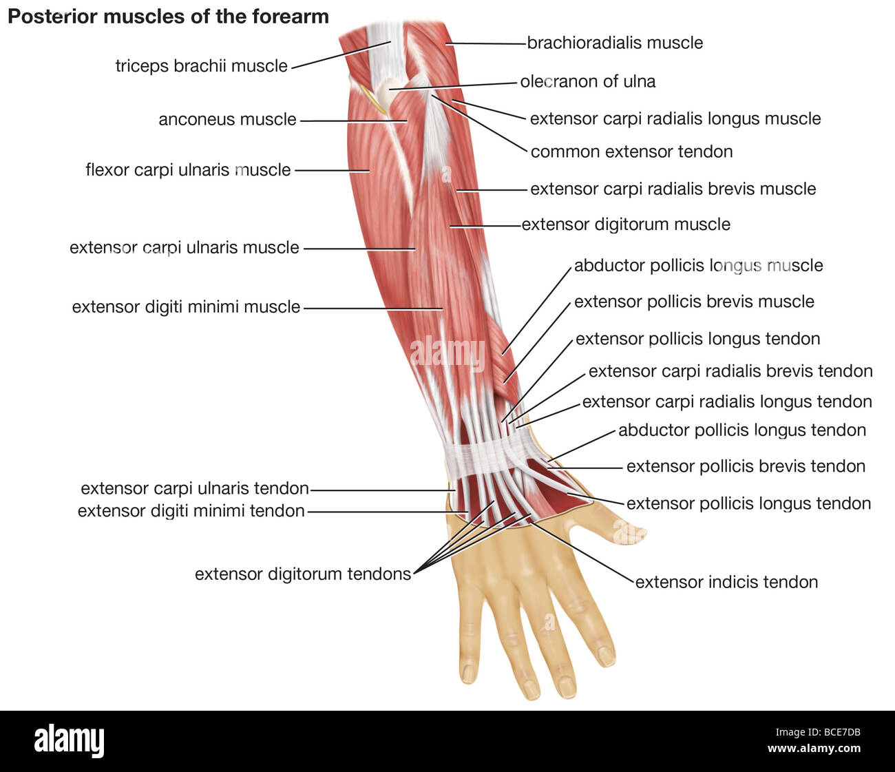 The posterior view of the muscles of the human forearm. Stock Photohttps://www.alamy.com/image-license-details/?v=1https://www.alamy.com/stock-photo-the-posterior-view-of-the-muscles-of-the-human-forearm-24899431.html
RMBCE7DB–The posterior view of the muscles of the human forearm.
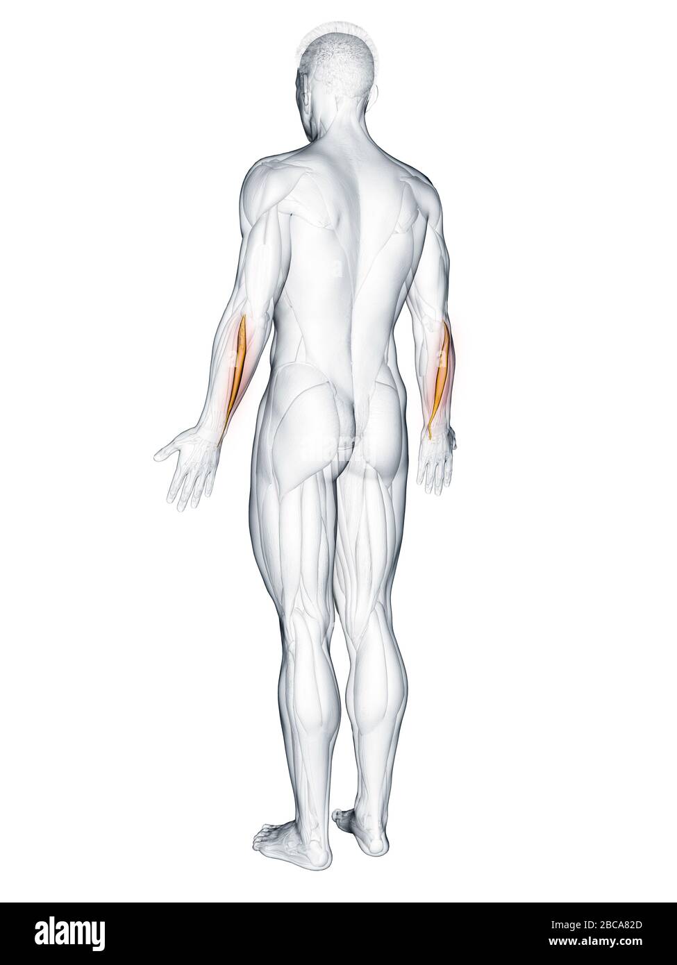 Extensor carpi ulnaris muscle, illustration. Stock Photohttps://www.alamy.com/image-license-details/?v=1https://www.alamy.com/extensor-carpi-ulnaris-muscle-illustration-image351809093.html
RF2BCA82D–Extensor carpi ulnaris muscle, illustration.
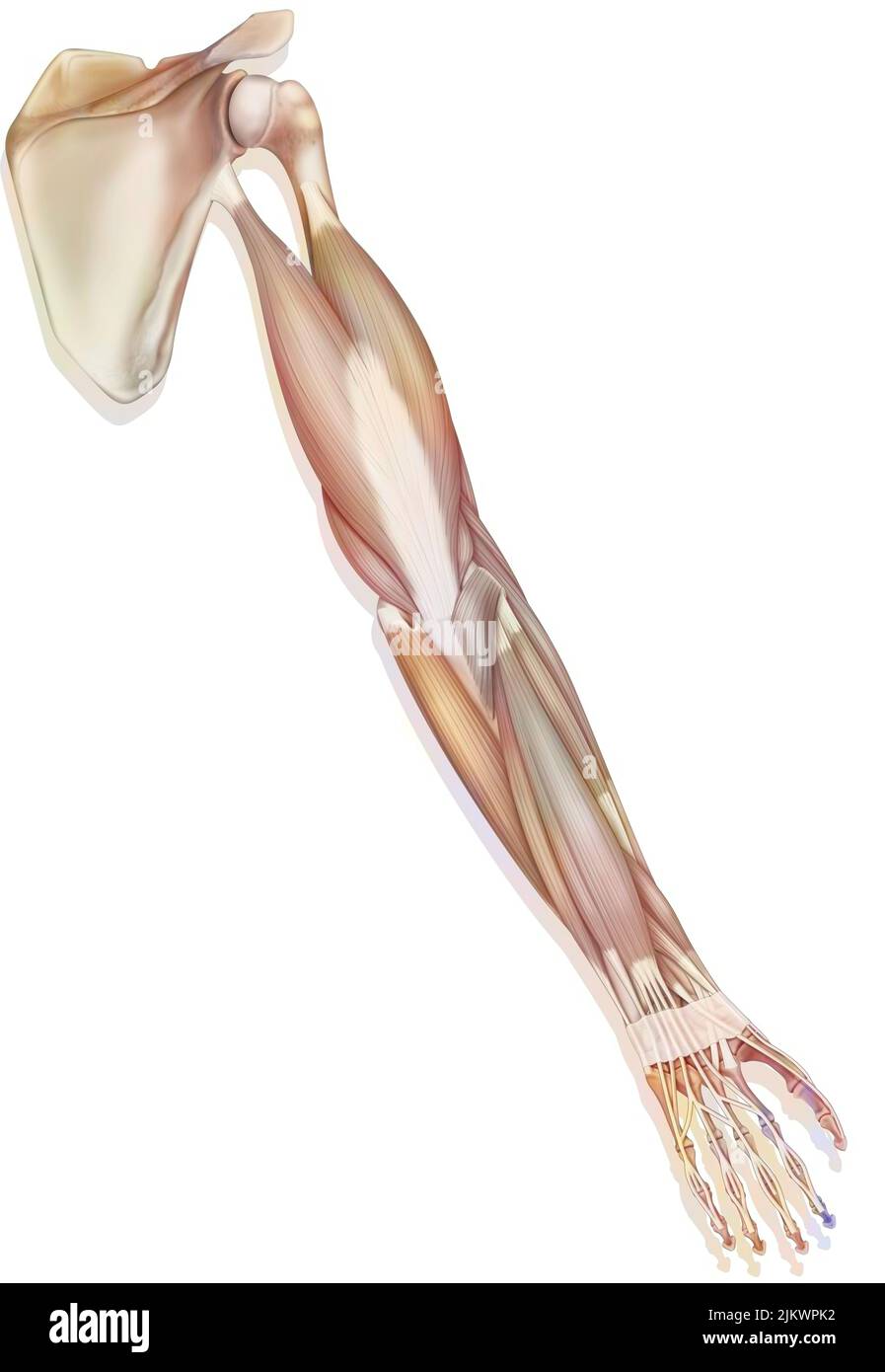 The muscles of the upper right limb in posterior view. Stock Photohttps://www.alamy.com/image-license-details/?v=1https://www.alamy.com/the-muscles-of-the-upper-right-limb-in-posterior-view-image476924982.html
RF2JKWPK2–The muscles of the upper right limb in posterior view.
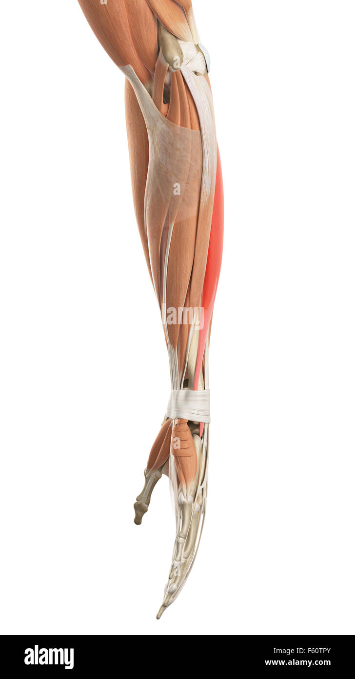 medically accurate illustration of the extensor carpi ulnaris Stock Photohttps://www.alamy.com/image-license-details/?v=1https://www.alamy.com/stock-photo-medically-accurate-illustration-of-the-extensor-carpi-ulnaris-89759235.html
RFF60TPY–medically accurate illustration of the extensor carpi ulnaris
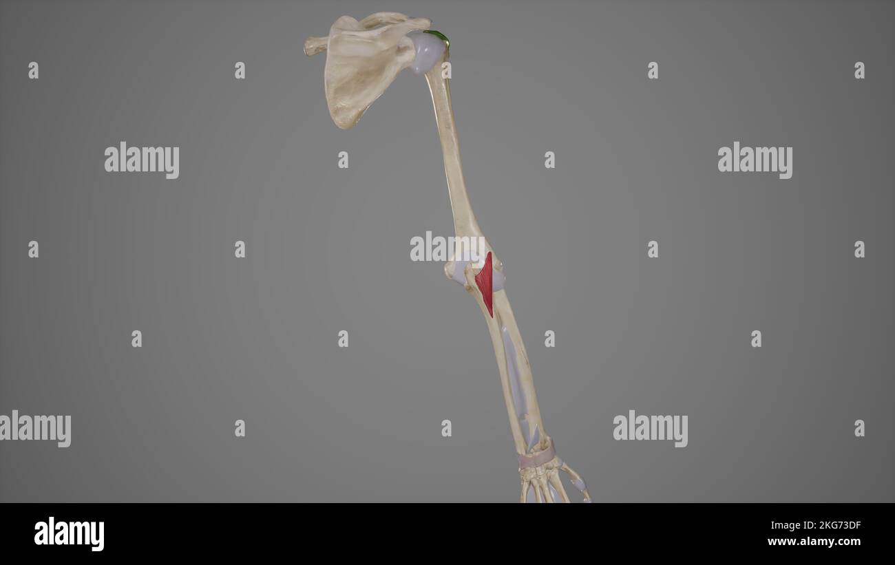 Anconeus Muscle Stock Photohttps://www.alamy.com/image-license-details/?v=1https://www.alamy.com/anconeus-muscle-image491881195.html
RF2KG73DF–Anconeus Muscle
 Muscle anatomy of human arm and hand. Stock Photohttps://www.alamy.com/image-license-details/?v=1https://www.alamy.com/stock-photo-muscle-anatomy-of-human-arm-and-hand-59361164.html
RFDCG3NG–Muscle anatomy of human arm and hand.
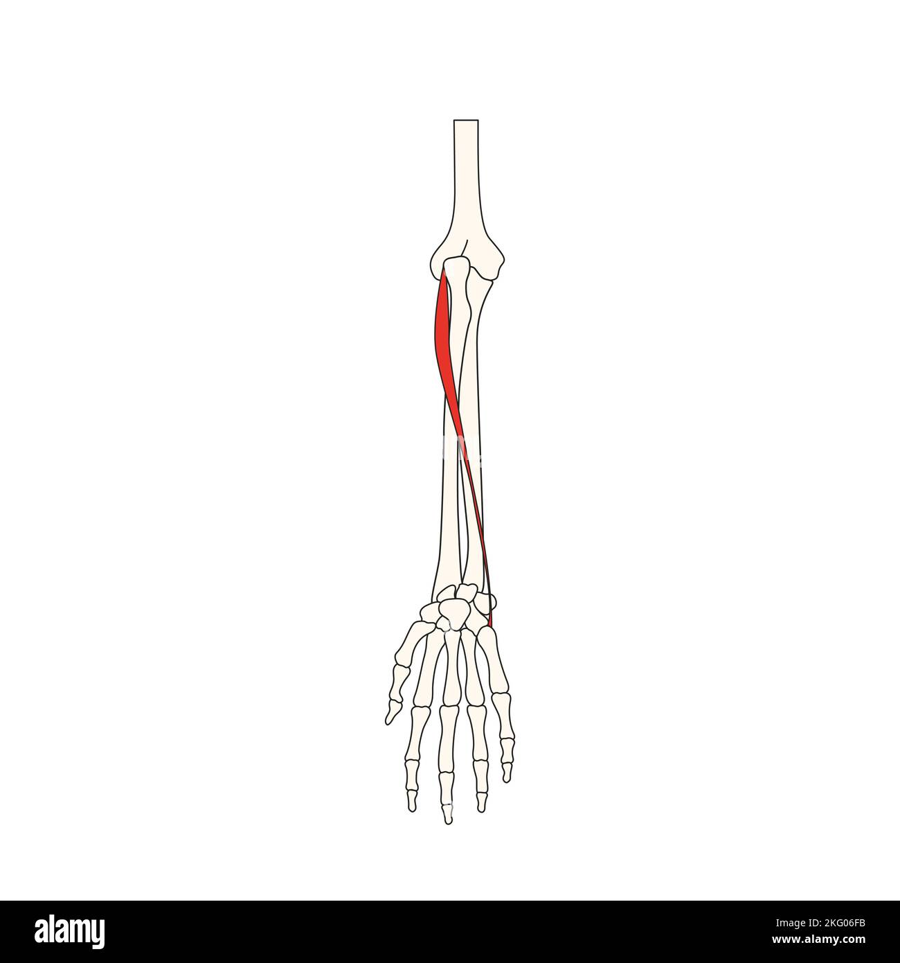 human anatomy drawing extensor carpi ulnaris Stock Photohttps://www.alamy.com/image-license-details/?v=1https://www.alamy.com/human-anatomy-drawing-extensor-carpi-ulnaris-image491729935.html
RF2KG06FB–human anatomy drawing extensor carpi ulnaris
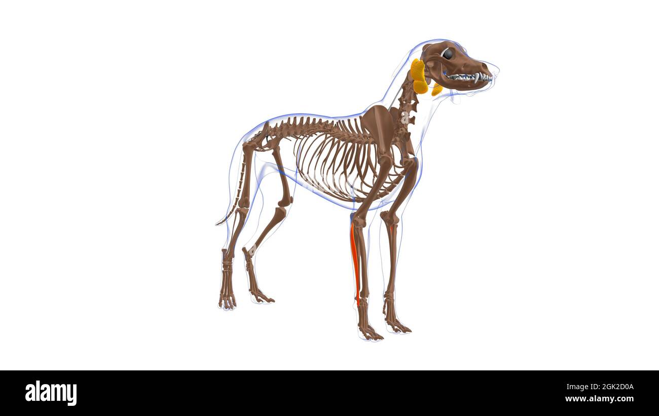 Extensor Carpi Ulnaris muscle Dog muscle Anatomy For Medical Concept 3D Illustration Stock Photohttps://www.alamy.com/image-license-details/?v=1https://www.alamy.com/extensor-carpi-ulnaris-muscle-dog-muscle-anatomy-for-medical-concept-3d-illustration-image441991770.html
RF2GK2D0A–Extensor Carpi Ulnaris muscle Dog muscle Anatomy For Medical Concept 3D Illustration
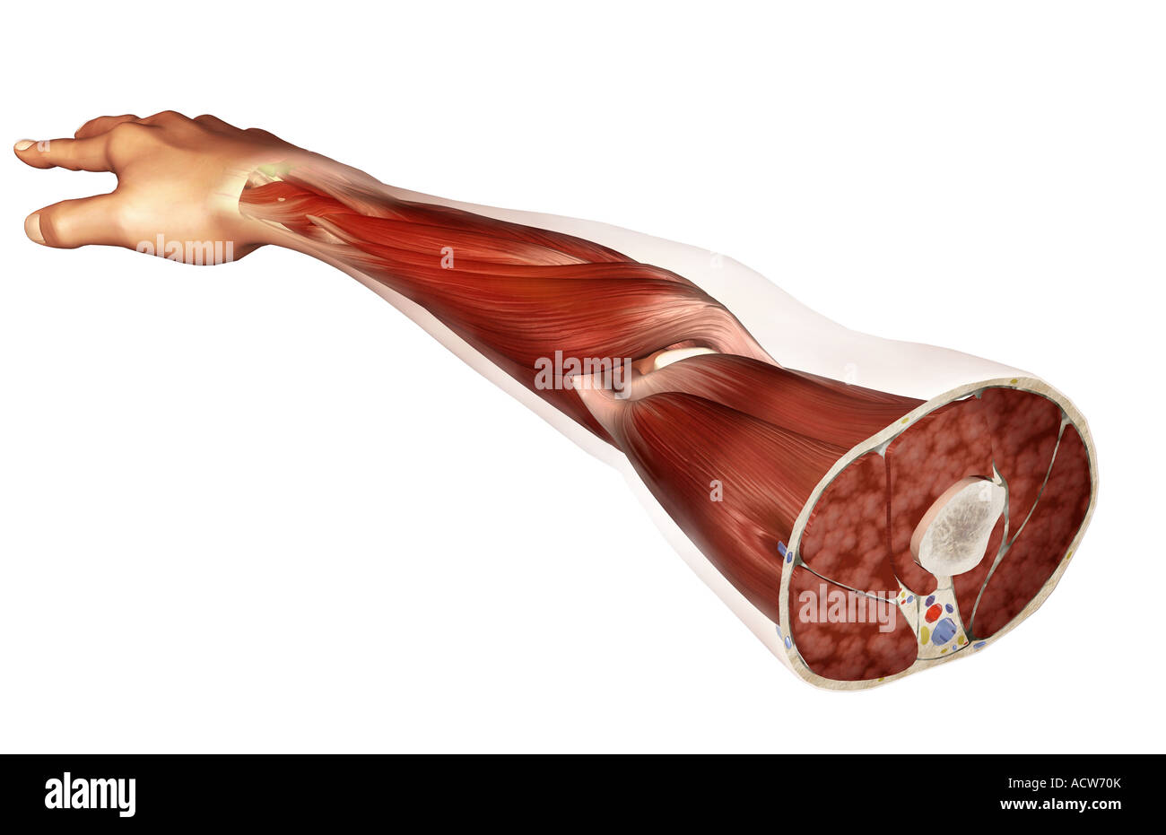 Transverse section of the arm Stock Photohttps://www.alamy.com/image-license-details/?v=1https://www.alamy.com/stock-photo-transverse-section-of-the-arm-13233138.html
RFACW70K–Transverse section of the arm
 . Cunningham's Text-book of anatomy. Anatomy. THE ULNA. 213 posterior surface is subdivided by a faint longitudinal ridge, the bone between which and the interosseous crest furnishes origins for the abductor pollicis longus, extensor pollicis longus, and extensor indicis proprius muscles, in order proximo- distally. The surface of bone between the dorsal margin and the afore-mentioned longitudinal line is smooth and overlain by the extensor carpi ulnaris muscle, which does not arise from it. The distal extremity of the ulna presents a rounded head (capitulum ulnse), from which, on its medial a Stock Photohttps://www.alamy.com/image-license-details/?v=1https://www.alamy.com/cunninghams-text-book-of-anatomy-anatomy-the-ulna-213-posterior-surface-is-subdivided-by-a-faint-longitudinal-ridge-the-bone-between-which-and-the-interosseous-crest-furnishes-origins-for-the-abductor-pollicis-longus-extensor-pollicis-longus-and-extensor-indicis-proprius-muscles-in-order-proximo-distally-the-surface-of-bone-between-the-dorsal-margin-and-the-afore-mentioned-longitudinal-line-is-smooth-and-overlain-by-the-extensor-carpi-ulnaris-muscle-which-does-not-arise-from-it-the-distal-extremity-of-the-ulna-presents-a-rounded-head-capitulum-ulnse-from-which-on-its-medial-a-image231857363.html
RMRD60C3–. Cunningham's Text-book of anatomy. Anatomy. THE ULNA. 213 posterior surface is subdivided by a faint longitudinal ridge, the bone between which and the interosseous crest furnishes origins for the abductor pollicis longus, extensor pollicis longus, and extensor indicis proprius muscles, in order proximo- distally. The surface of bone between the dorsal margin and the afore-mentioned longitudinal line is smooth and overlain by the extensor carpi ulnaris muscle, which does not arise from it. The distal extremity of the ulna presents a rounded head (capitulum ulnse), from which, on its medial a
RMPFY56P–. Cunningham's Text-book of anatomy. Anatomy. MUSCLES ON ANTEEIOE AND MEDIAL ASPECTS OF EOEEAEM. 387 the forearm between the two heads of origin of the muscle), and the ulnar artery. The tendon serves as a guide to the artery in the distal half of the forearm. Biceps brachii Medial inter- muscular septum Brachialis Medial epicondyle Lacertus fibrosus supinator muscle Pronator teres Flexor carpi radialis Palmaris LONGUS Flexor carpi ULNARIS Extensor CARPI RADIALIS LONGUS Brachio- RADIAL1S. Flexor digi- torum sublimis Flexor pollicis longus Brachioradialis (tendon) Flexor carpi radialis (tendon)
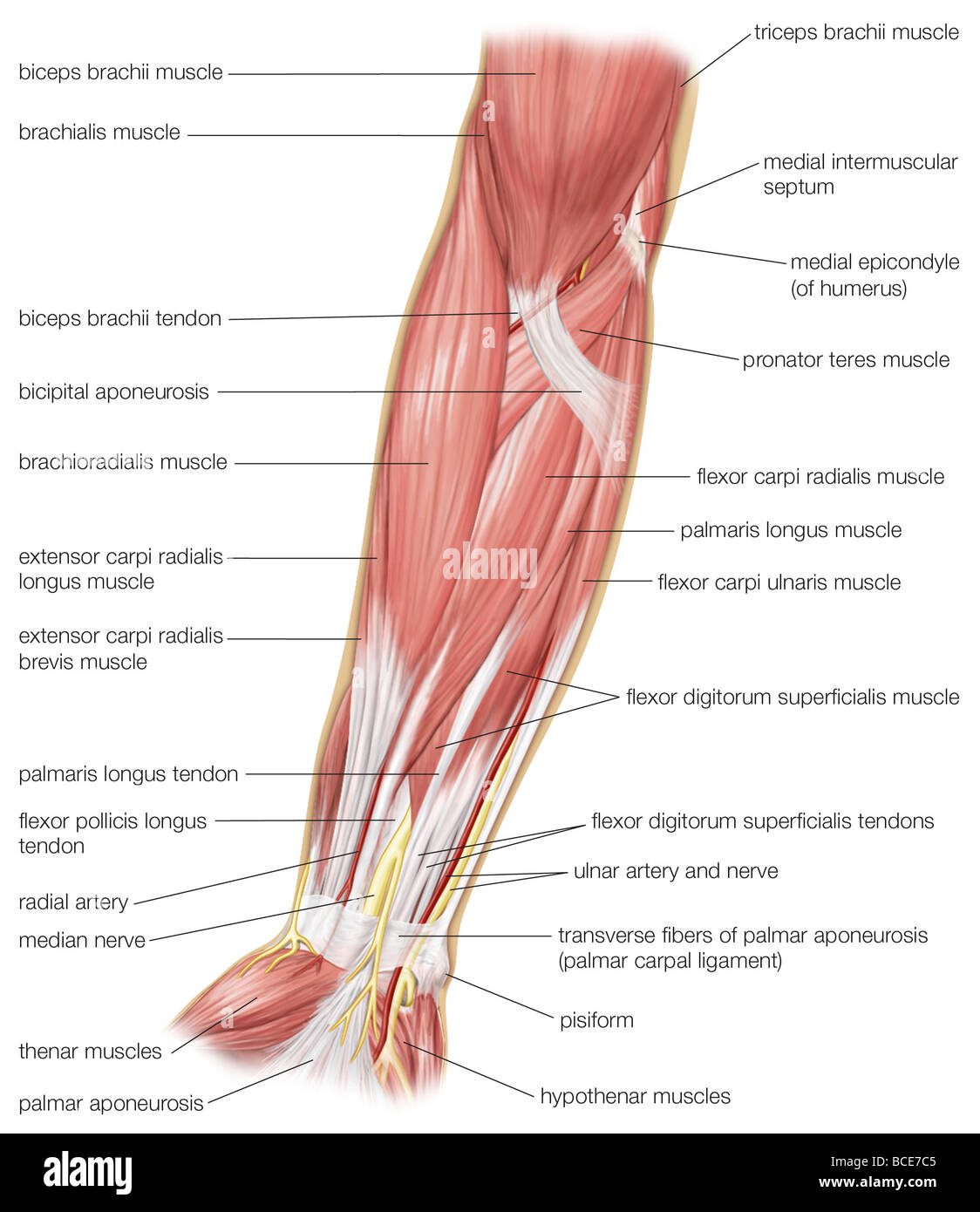 The anterior view of the muscles of the human forearm. Stock Photohttps://www.alamy.com/image-license-details/?v=1https://www.alamy.com/stock-photo-the-anterior-view-of-the-muscles-of-the-human-forearm-24899397.html
RMBCE7C5–The anterior view of the muscles of the human forearm.
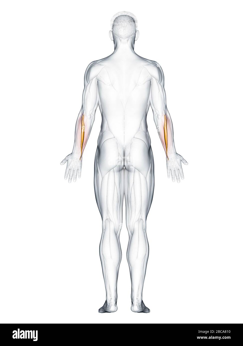 Extensor carpi ulnaris muscle, illustration. Stock Photohttps://www.alamy.com/image-license-details/?v=1https://www.alamy.com/extensor-carpi-ulnaris-muscle-illustration-image351809052.html
RF2BCA810–Extensor carpi ulnaris muscle, illustration.
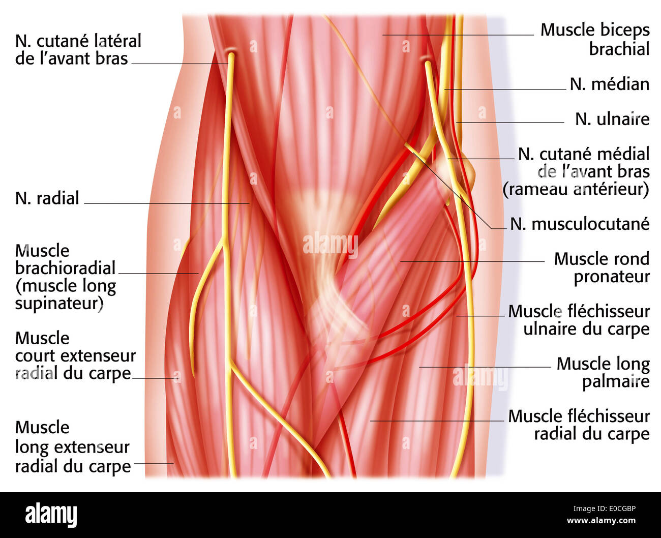 Elbow, anatomy Stock Photohttps://www.alamy.com/image-license-details/?v=1https://www.alamy.com/elbow-anatomy-image69117770.html
RME0CGBP–Elbow, anatomy
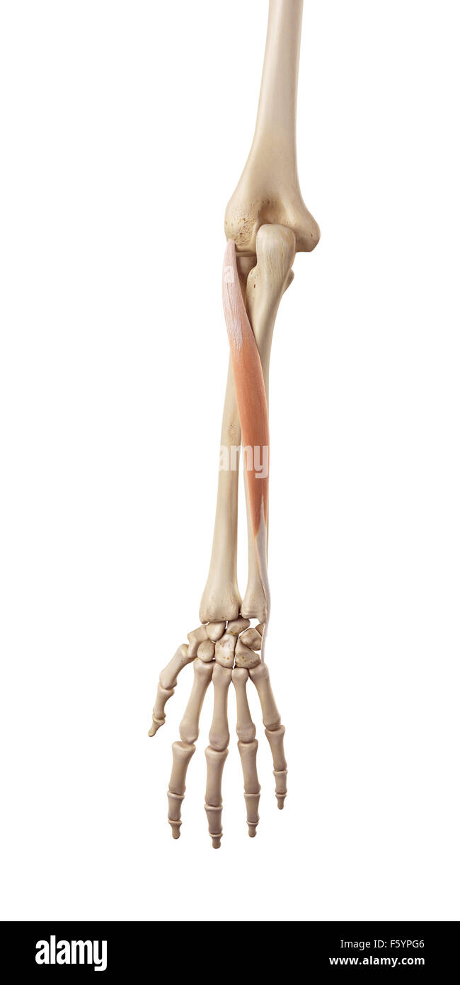 medical accurate illustration of the extensor carpi ulnaris Stock Photohttps://www.alamy.com/image-license-details/?v=1https://www.alamy.com/stock-photo-medical-accurate-illustration-of-the-extensor-carpi-ulnaris-89735526.html
RFF5YPG6–medical accurate illustration of the extensor carpi ulnaris
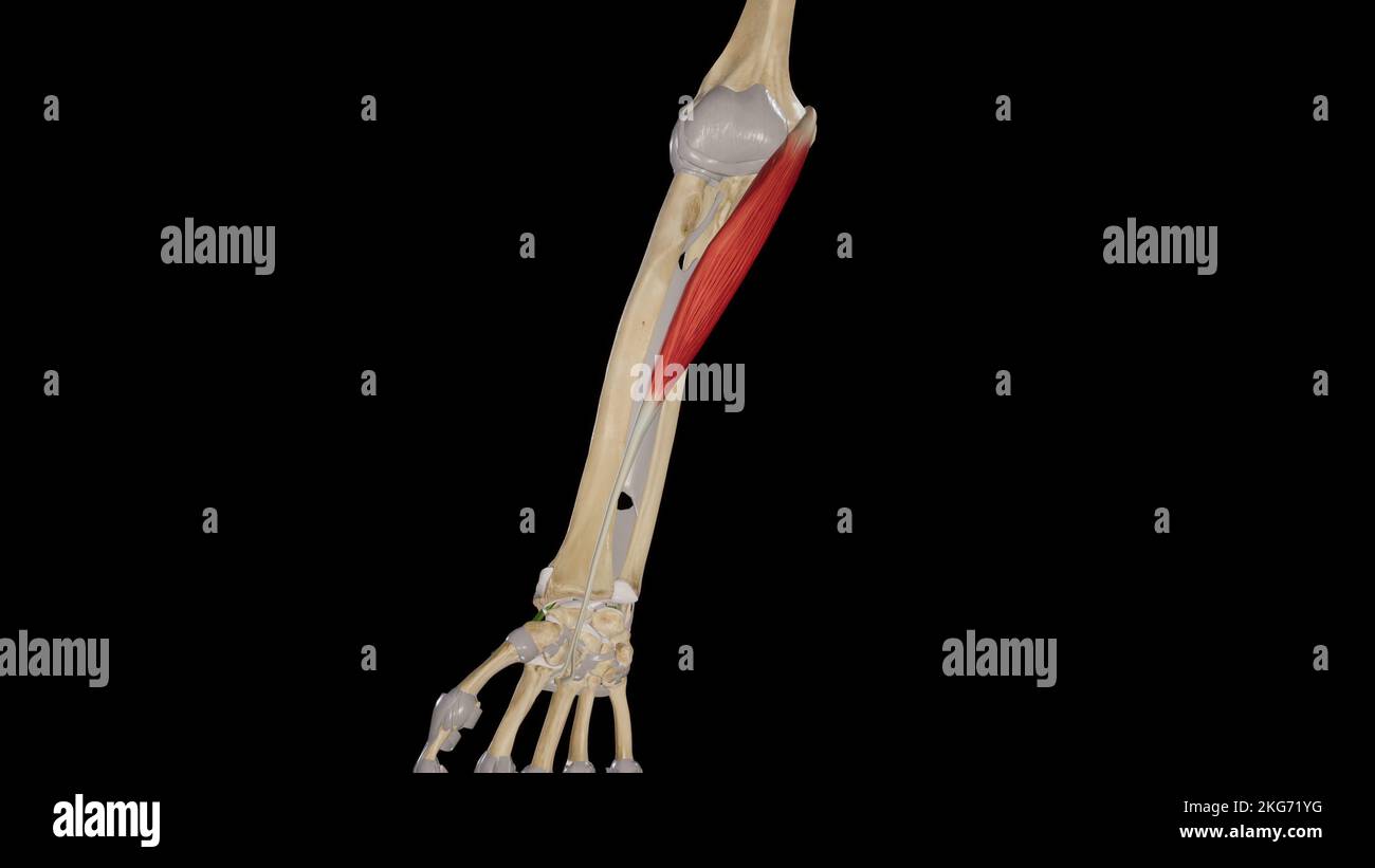 Flexor Carpi Radialis Muscle Stock Photohttps://www.alamy.com/image-license-details/?v=1https://www.alamy.com/flexor-carpi-radialis-muscle-image491880020.html
RF2KG71YG–Flexor Carpi Radialis Muscle
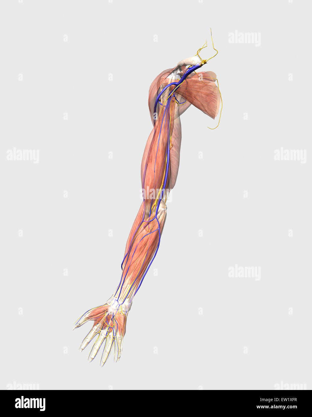 Medical illustration of human arm muscles, veins and nerves. Stock Photohttps://www.alamy.com/image-license-details/?v=1https://www.alamy.com/stock-photo-medical-illustration-of-human-arm-muscles-veins-and-nerves-84250651.html
RFEW1XFR–Medical illustration of human arm muscles, veins and nerves.
 Extensor Carpi Ulnaris muscle Dog muscle Anatomy For Medical Concept 3D Illustration Stock Photohttps://www.alamy.com/image-license-details/?v=1https://www.alamy.com/extensor-carpi-ulnaris-muscle-dog-muscle-anatomy-for-medical-concept-3d-illustration-image441991803.html
RF2GK2D1F–Extensor Carpi Ulnaris muscle Dog muscle Anatomy For Medical Concept 3D Illustration
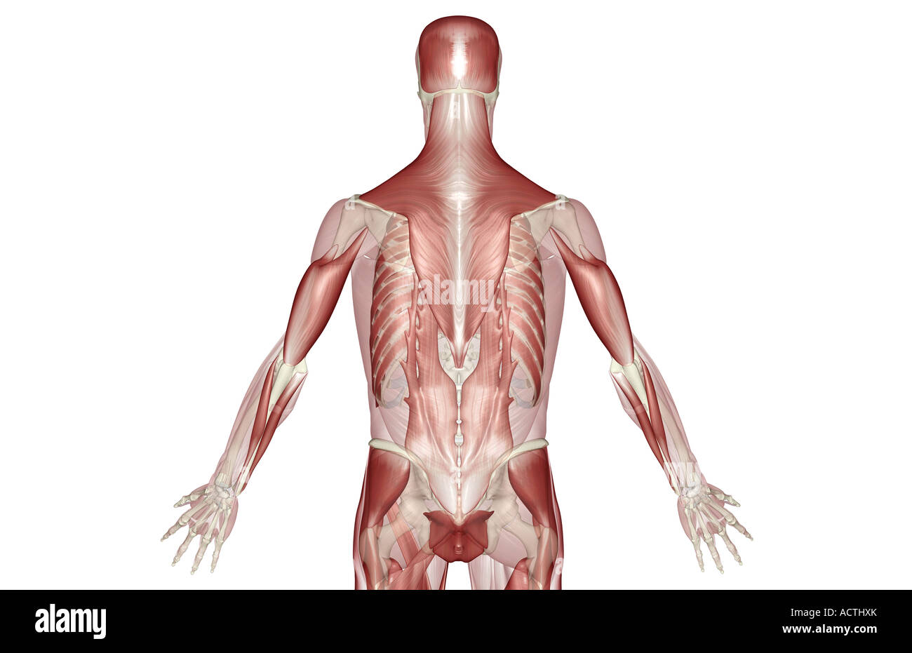 Muscles of the back Stock Photohttps://www.alamy.com/image-license-details/?v=1https://www.alamy.com/stock-photo-muscles-of-the-back-13227402.html
RFACTHXK–Muscles of the back
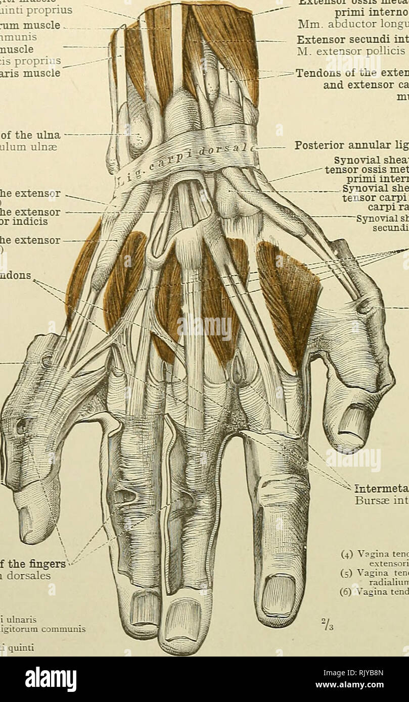 . An atlas of human anatomy for students and physicians. Anatomy. 330 THE MUSCLES OF THE UPPER EXTREMITY Extensor Tninimi digiti muscle M. extensor digiti quinti proprius Extensor communis digitorum muscle . M. extensor digitorum communis Extensor indicis muscle M. extensor indicis proprius Extensor carpi ulnaris muscle Head of the ulna Capitulum ulnse Synovial sheath of the tendon of the extensor carpi ulnaris muscle u' Synovial sheath of the tendons of the extensor communis di^torum and extensor indicis muscles t; i Synovial sheath of the tendon of the extensor minimi digiti muscle (j) Vin Stock Photohttps://www.alamy.com/image-license-details/?v=1https://www.alamy.com/an-atlas-of-human-anatomy-for-students-and-physicians-anatomy-330-the-muscles-of-the-upper-extremity-extensor-tninimi-digiti-muscle-m-extensor-digiti-quinti-proprius-extensor-communis-digitorum-muscle-m-extensor-digitorum-communis-extensor-indicis-muscle-m-extensor-indicis-proprius-extensor-carpi-ulnaris-muscle-head-of-the-ulna-capitulum-ulnse-synovial-sheath-of-the-tendon-of-the-extensor-carpi-ulnaris-muscle-u-synovial-sheath-of-the-tendons-of-the-extensor-communis-ditorum-and-extensor-indicis-muscles-t-i-synovial-sheath-of-the-tendon-of-the-extensor-minimi-digiti-muscle-j-vin-image235400165.html
RMRJYB8N–. An atlas of human anatomy for students and physicians. Anatomy. 330 THE MUSCLES OF THE UPPER EXTREMITY Extensor Tninimi digiti muscle M. extensor digiti quinti proprius Extensor communis digitorum muscle . M. extensor digitorum communis Extensor indicis muscle M. extensor indicis proprius Extensor carpi ulnaris muscle Head of the ulna Capitulum ulnse Synovial sheath of the tendon of the extensor carpi ulnaris muscle u' Synovial sheath of the tendons of the extensor communis di^torum and extensor indicis muscles t; i Synovial sheath of the tendon of the extensor minimi digiti muscle (j) Vin
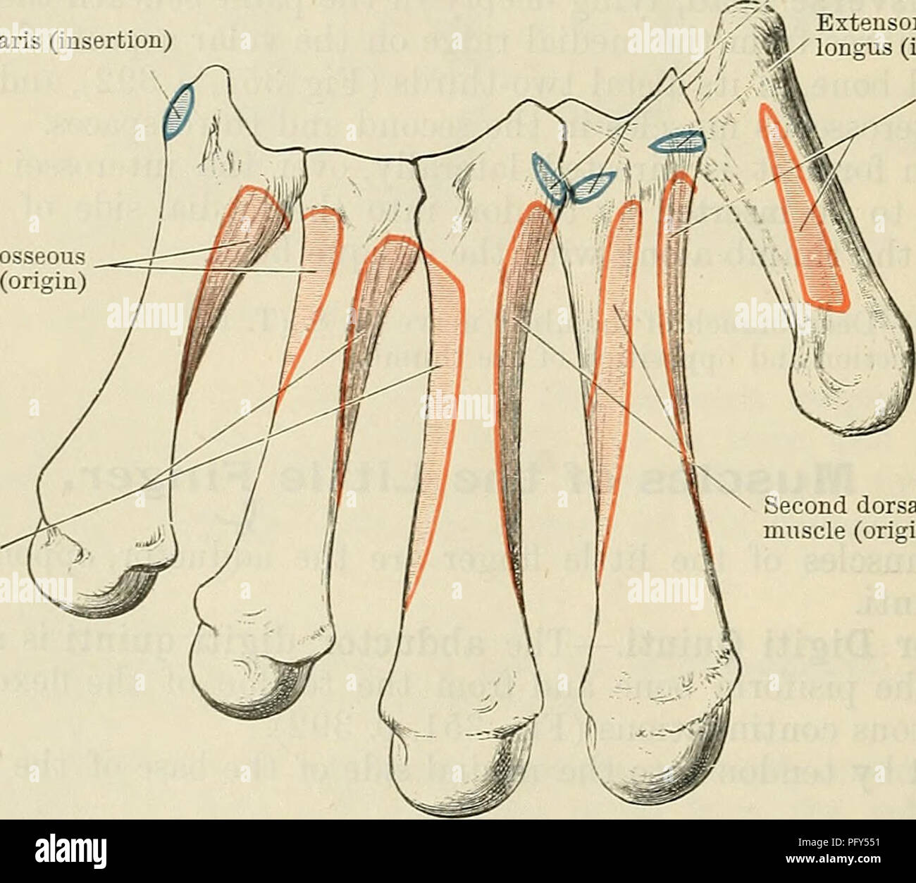 . Cunningham's Text-book of anatomy. Anatomy. Fig. 352.—The Volar Interosseous Muscles (Right Side). V1, first; V2, second ; and V3, third volar interosseous muscles. The Interosseous Muscles. The interosseous muscles of the hand occupy the spaces between the metacarpal bones. They are arranged in two sets, volar and dorsal. Mm. Interossei Volares.—The volar (O.T. palmar) interossei are three in Extensor carpi ulnaris (insertion) Fourth dorsal interosseous muscle (origin) Third dorsal inter- osseous muscle (origin). Extensor carpi radialis brevis (insertion) Extensor carpi radialis longus (ins Stock Photohttps://www.alamy.com/image-license-details/?v=1https://www.alamy.com/cunninghams-text-book-of-anatomy-anatomy-fig-352the-volar-interosseous-muscles-right-side-v1-first-v2-second-and-v3-third-volar-interosseous-muscles-the-interosseous-muscles-the-interosseous-muscles-of-the-hand-occupy-the-spaces-between-the-metacarpal-bones-they-are-arranged-in-two-sets-volar-and-dorsal-mm-interossei-volaresthe-volar-ot-palmar-interossei-are-three-in-extensor-carpi-ulnaris-insertion-fourth-dorsal-interosseous-muscle-origin-third-dorsal-inter-osseous-muscle-origin-extensor-carpi-radialis-brevis-insertion-extensor-carpi-radialis-longus-ins-image216341021.html
RMPFY551–. Cunningham's Text-book of anatomy. Anatomy. Fig. 352.—The Volar Interosseous Muscles (Right Side). V1, first; V2, second ; and V3, third volar interosseous muscles. The Interosseous Muscles. The interosseous muscles of the hand occupy the spaces between the metacarpal bones. They are arranged in two sets, volar and dorsal. Mm. Interossei Volares.—The volar (O.T. palmar) interossei are three in Extensor carpi ulnaris (insertion) Fourth dorsal interosseous muscle (origin) Third dorsal inter- osseous muscle (origin). Extensor carpi radialis brevis (insertion) Extensor carpi radialis longus (ins
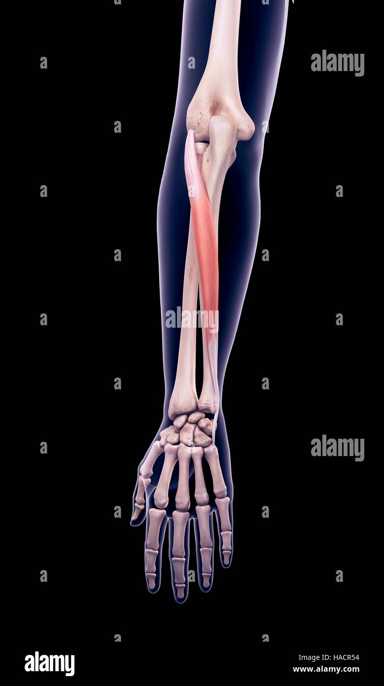 Illustration of the extensor carpi ulnaris muscle. Stock Photohttps://www.alamy.com/image-license-details/?v=1https://www.alamy.com/stock-photo-illustration-of-the-extensor-carpi-ulnaris-muscle-126900736.html
RFHACR54–Illustration of the extensor carpi ulnaris muscle.
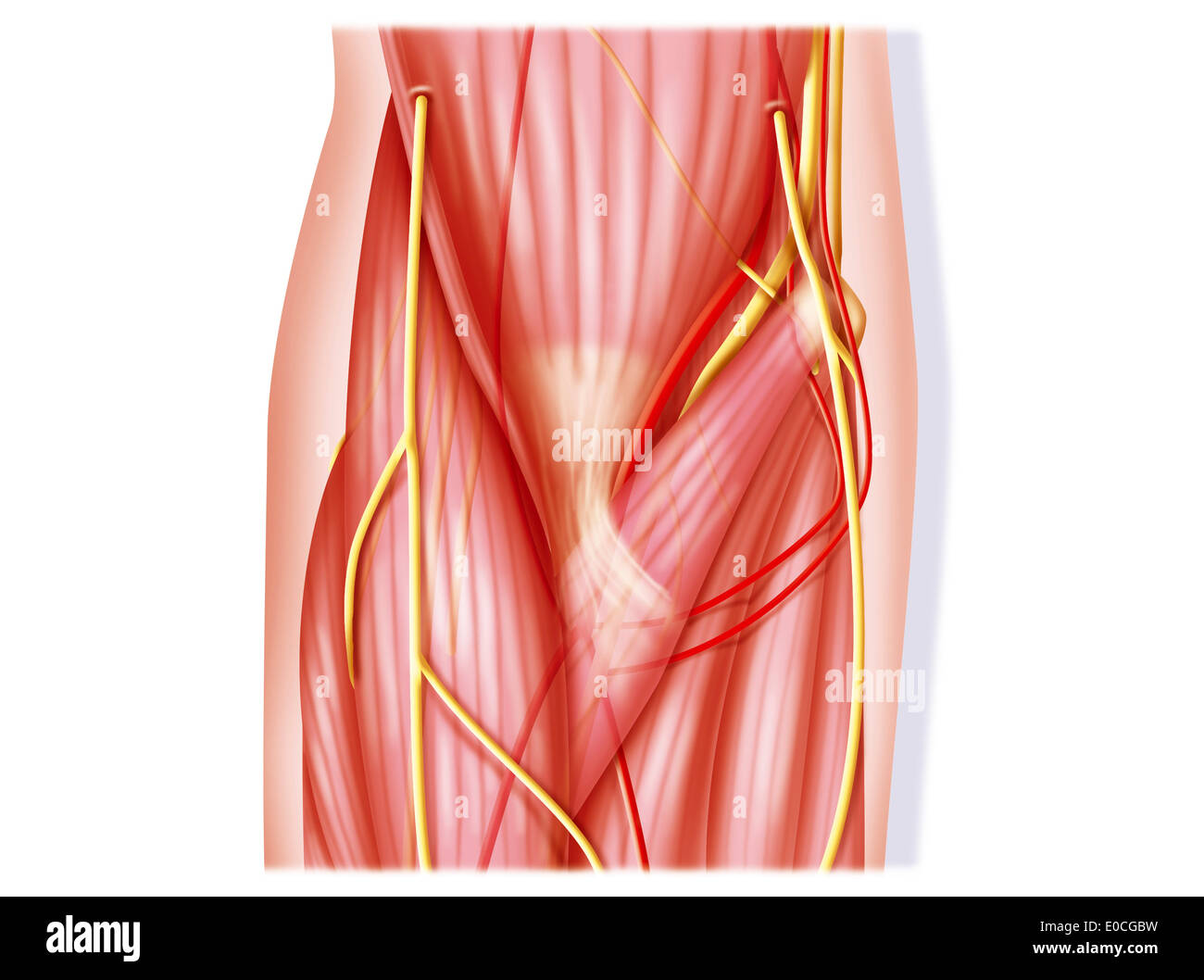 Elbow, anatomy Stock Photohttps://www.alamy.com/image-license-details/?v=1https://www.alamy.com/elbow-anatomy-image69117773.html
RME0CGBW–Elbow, anatomy
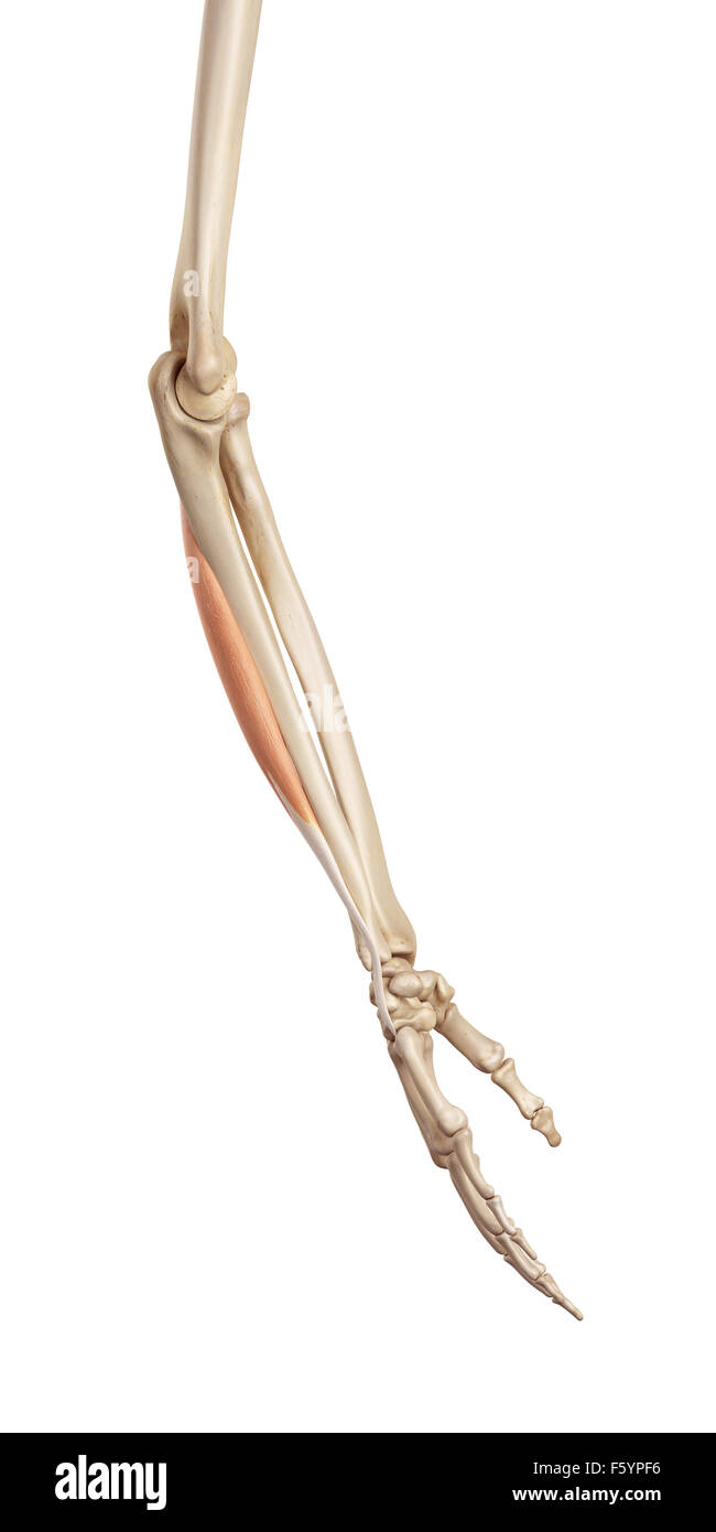 medical accurate illustration of the extensor carpi ulnaris Stock Photohttps://www.alamy.com/image-license-details/?v=1https://www.alamy.com/stock-photo-medical-accurate-illustration-of-the-extensor-carpi-ulnaris-89735498.html
RFF5YPF6–medical accurate illustration of the extensor carpi ulnaris
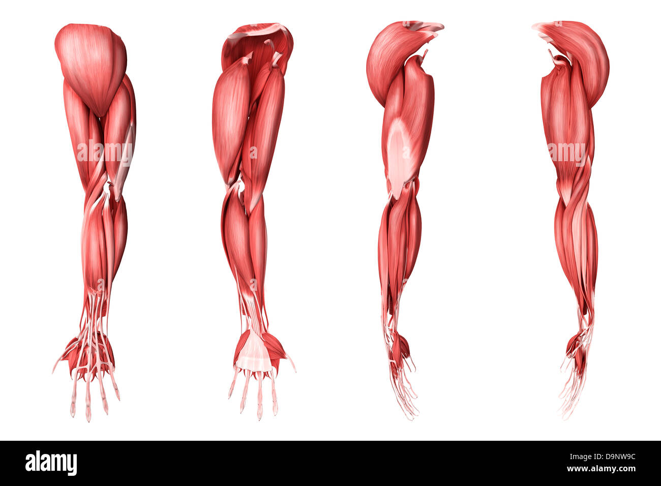 Medical illustration of human arm muscles, four side views. Stock Photohttps://www.alamy.com/image-license-details/?v=1https://www.alamy.com/stock-photo-medical-illustration-of-human-arm-muscles-four-side-views-57643864.html
RFD9NW9C–Medical illustration of human arm muscles, four side views.
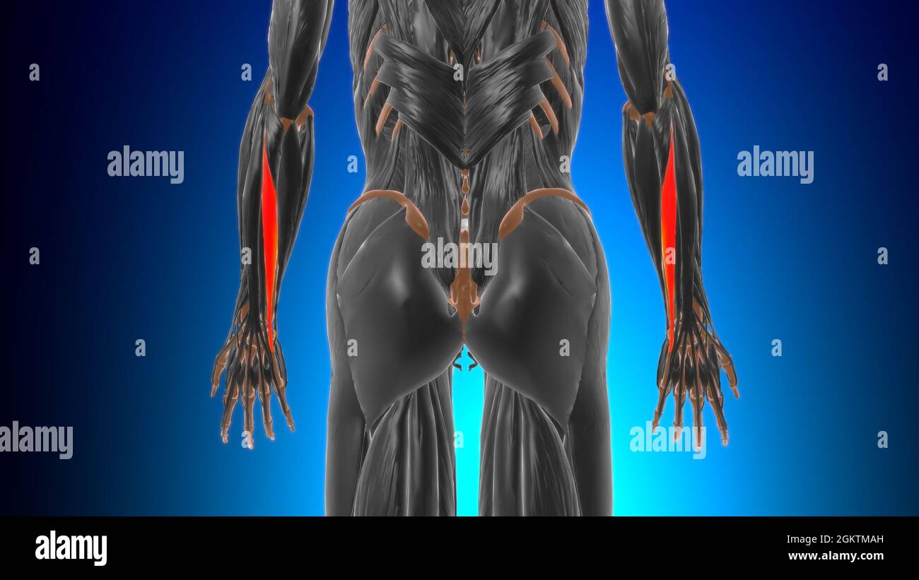 Extensor carpi ulnaris Muscle Anatomy For Medical Concept 3D Illustration Stock Photohttps://www.alamy.com/image-license-details/?v=1https://www.alamy.com/extensor-carpi-ulnaris-muscle-anatomy-for-medical-concept-3d-illustration-image442480489.html
RF2GKTMAH–Extensor carpi ulnaris Muscle Anatomy For Medical Concept 3D Illustration
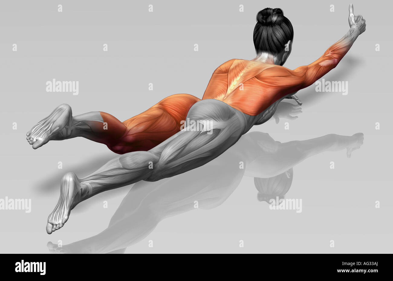 Arm-leg extensions (Part 1 of 2) Stock Photohttps://www.alamy.com/image-license-details/?v=1https://www.alamy.com/stock-photo-arm-leg-extensions-part-1-of-2-14078633.html
RFAG33AJ–Arm-leg extensions (Part 1 of 2)
![. Atlas of applied (topographical) human anatomy for students and practitioners. Anatomy. Extensor ]Iiniini Digiti Muscle ^ Flexor Profundus Uigitorum ^luscle Extensor Carpi Ulnaris Muscle Abductor Minimi Digiti Muscle Opponens Minimi Digitt iluscle -—Ulnar Nerve — Deep Division ^ Opponens Minimi Digiti Muscle ""Hook of Unciform Bone Ulnar Nerve — Superficial Division I'almaris Brcvis Muscle Palmar Fasci. Ulnar Artery Fig. 103. Transverse Section through Carpus (Left): Distal Aspect. Nat. 8ize. Extensor Indicis ^lusclcajiEj Deep Palmar Arch Extensor Tendon to jrd Finger Deep Divisio Stock Photo . Atlas of applied (topographical) human anatomy for students and practitioners. Anatomy. Extensor ]Iiniini Digiti Muscle ^ Flexor Profundus Uigitorum ^luscle Extensor Carpi Ulnaris Muscle Abductor Minimi Digiti Muscle Opponens Minimi Digitt iluscle -—Ulnar Nerve — Deep Division ^ Opponens Minimi Digiti Muscle ""Hook of Unciform Bone Ulnar Nerve — Superficial Division I'almaris Brcvis Muscle Palmar Fasci. Ulnar Artery Fig. 103. Transverse Section through Carpus (Left): Distal Aspect. Nat. 8ize. Extensor Indicis ^lusclcajiEj Deep Palmar Arch Extensor Tendon to jrd Finger Deep Divisio Stock Photo](https://c8.alamy.com/comp/RJYCR0/atlas-of-applied-topographical-human-anatomy-for-students-and-practitioners-anatomy-extensor-iiniini-digiti-muscle-flexor-profundus-uigitorum-luscle-extensor-carpi-ulnaris-muscle-abductor-minimi-digiti-muscle-opponens-minimi-digitt-iluscle-ulnar-nerve-deep-division-opponens-minimi-digiti-muscle-quotquothook-of-unciform-bone-ulnar-nerve-superficial-division-ialmaris-brcvis-muscle-palmar-fasci-ulnar-artery-fig-103-transverse-section-through-carpus-left-distal-aspect-nat-8ize-extensor-indicis-lusclcajiej-deep-palmar-arch-extensor-tendon-to-jrd-finger-deep-divisio-RJYCR0.jpg) . Atlas of applied (topographical) human anatomy for students and practitioners. Anatomy. Extensor ]Iiniini Digiti Muscle ^ Flexor Profundus Uigitorum ^luscle Extensor Carpi Ulnaris Muscle Abductor Minimi Digiti Muscle Opponens Minimi Digitt iluscle -—Ulnar Nerve — Deep Division ^ Opponens Minimi Digiti Muscle ""Hook of Unciform Bone Ulnar Nerve — Superficial Division I'almaris Brcvis Muscle Palmar Fasci. Ulnar Artery Fig. 103. Transverse Section through Carpus (Left): Distal Aspect. Nat. 8ize. Extensor Indicis ^lusclcajiEj Deep Palmar Arch Extensor Tendon to jrd Finger Deep Divisio Stock Photohttps://www.alamy.com/image-license-details/?v=1https://www.alamy.com/atlas-of-applied-topographical-human-anatomy-for-students-and-practitioners-anatomy-extensor-iiniini-digiti-muscle-flexor-profundus-uigitorum-luscle-extensor-carpi-ulnaris-muscle-abductor-minimi-digiti-muscle-opponens-minimi-digitt-iluscle-ulnar-nerve-deep-division-opponens-minimi-digiti-muscle-quotquothook-of-unciform-bone-ulnar-nerve-superficial-division-ialmaris-brcvis-muscle-palmar-fasci-ulnar-artery-fig-103-transverse-section-through-carpus-left-distal-aspect-nat-8ize-extensor-indicis-lusclcajiej-deep-palmar-arch-extensor-tendon-to-jrd-finger-deep-divisio-image235401348.html
. Atlas of applied (topographical) human anatomy for students and practitioners. Anatomy. Extensor ]Iiniini Digiti Muscle ^ Flexor Profundus Uigitorum ^luscle Extensor Carpi Ulnaris Muscle Abductor Minimi Digiti Muscle Opponens Minimi Digitt iluscle -—Ulnar Nerve — Deep Division ^ Opponens Minimi Digiti Muscle ""Hook of Unciform Bone Ulnar Nerve — Superficial Division I'almaris Brcvis Muscle Palmar Fasci. Ulnar Artery Fig. 103. Transverse Section through Carpus (Left): Distal Aspect. Nat. 8ize. Extensor Indicis ^lusclcajiEj Deep Palmar Arch Extensor Tendon to jrd Finger Deep Divisio Stock Photohttps://www.alamy.com/image-license-details/?v=1https://www.alamy.com/atlas-of-applied-topographical-human-anatomy-for-students-and-practitioners-anatomy-extensor-iiniini-digiti-muscle-flexor-profundus-uigitorum-luscle-extensor-carpi-ulnaris-muscle-abductor-minimi-digiti-muscle-opponens-minimi-digitt-iluscle-ulnar-nerve-deep-division-opponens-minimi-digiti-muscle-quotquothook-of-unciform-bone-ulnar-nerve-superficial-division-ialmaris-brcvis-muscle-palmar-fasci-ulnar-artery-fig-103-transverse-section-through-carpus-left-distal-aspect-nat-8ize-extensor-indicis-lusclcajiej-deep-palmar-arch-extensor-tendon-to-jrd-finger-deep-divisio-image235401348.htmlRMRJYCR0–. Atlas of applied (topographical) human anatomy for students and practitioners. Anatomy. Extensor ]Iiniini Digiti Muscle ^ Flexor Profundus Uigitorum ^luscle Extensor Carpi Ulnaris Muscle Abductor Minimi Digiti Muscle Opponens Minimi Digitt iluscle -—Ulnar Nerve — Deep Division ^ Opponens Minimi Digiti Muscle ""Hook of Unciform Bone Ulnar Nerve — Superficial Division I'almaris Brcvis Muscle Palmar Fasci. Ulnar Artery Fig. 103. Transverse Section through Carpus (Left): Distal Aspect. Nat. 8ize. Extensor Indicis ^lusclcajiEj Deep Palmar Arch Extensor Tendon to jrd Finger Deep Divisio
 . Cunningham's Text-book of anatomy. Anatomy. Flexor carpi ulnaris Abductor pollicis longus """ Extensor indicis proprius. Extensor pollicis BREVIS Extensor pollicis longus Dorsal carpal ligament... Extensor carpi ^ radialis longus i '" Extensor carpi ) ... RADIALIS BREVIS f Extensor carpi ) l'1-NARIS / Triceps brachii TENDON Brachio- , radialis Origin of superficial. extensor MUSCLES Annular liga- ment OF RADIOS V x Ancon.-eus r— Extensor carpi RADIALIS LONGUS"j" Dorsal margin- of ulna Extensor carpi . radialis brevis Supinator. muscle Abductor pollicis longus D Stock Photohttps://www.alamy.com/image-license-details/?v=1https://www.alamy.com/cunninghams-text-book-of-anatomy-anatomy-flexor-carpi-ulnaris-abductor-pollicis-longus-quotquotquot-extensor-indicis-proprius-extensor-pollicis-brevis-extensor-pollicis-longus-dorsal-carpal-ligament-extensor-carpi-radialis-longus-i-quot-extensor-carpi-radialis-brevis-f-extensor-carpi-l1-naris-triceps-brachii-tendon-brachio-radialis-origin-of-superficial-extensor-muscles-annular-liga-ment-of-radios-v-x-ancon-eus-r-extensor-carpi-radialis-longusquotjquot-dorsal-margin-of-ulna-extensor-carpi-radialis-brevis-supinator-muscle-abductor-pollicis-longus-d-image216340999.html
RMPFY547–. Cunningham's Text-book of anatomy. Anatomy. Flexor carpi ulnaris Abductor pollicis longus """ Extensor indicis proprius. Extensor pollicis BREVIS Extensor pollicis longus Dorsal carpal ligament... Extensor carpi ^ radialis longus i '" Extensor carpi ) ... RADIALIS BREVIS f Extensor carpi ) l'1-NARIS / Triceps brachii TENDON Brachio- , radialis Origin of superficial. extensor MUSCLES Annular liga- ment OF RADIOS V x Ancon.-eus r— Extensor carpi RADIALIS LONGUS"j" Dorsal margin- of ulna Extensor carpi . radialis brevis Supinator. muscle Abductor pollicis longus D
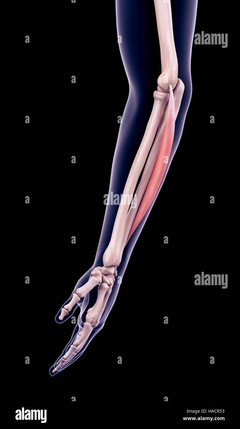 Illustration of the extensor carpi ulnaris muscle. Stock Photohttps://www.alamy.com/image-license-details/?v=1https://www.alamy.com/stock-photo-illustration-of-the-extensor-carpi-ulnaris-muscle-126900735.html
RFHACR53–Illustration of the extensor carpi ulnaris muscle.
 3d rendered illustration of the dog muscle anatomy - extensor carpi ulnaris Stock Photohttps://www.alamy.com/image-license-details/?v=1https://www.alamy.com/3d-rendered-illustration-of-the-dog-muscle-anatomy-extensor-carpi-ulnaris-image333863743.html
RF2AB4PH3–3d rendered illustration of the dog muscle anatomy - extensor carpi ulnaris
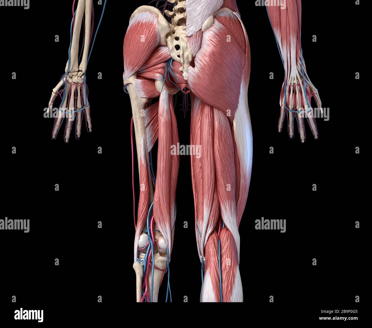 Low section rear view of human limbs, hip and muscular system with arteries and veins. Stock Photohttps://www.alamy.com/image-license-details/?v=1https://www.alamy.com/low-section-rear-view-of-human-limbs-hip-and-muscular-system-with-arteries-and-veins-image350068997.html
RF2B9F0G5–Low section rear view of human limbs, hip and muscular system with arteries and veins.
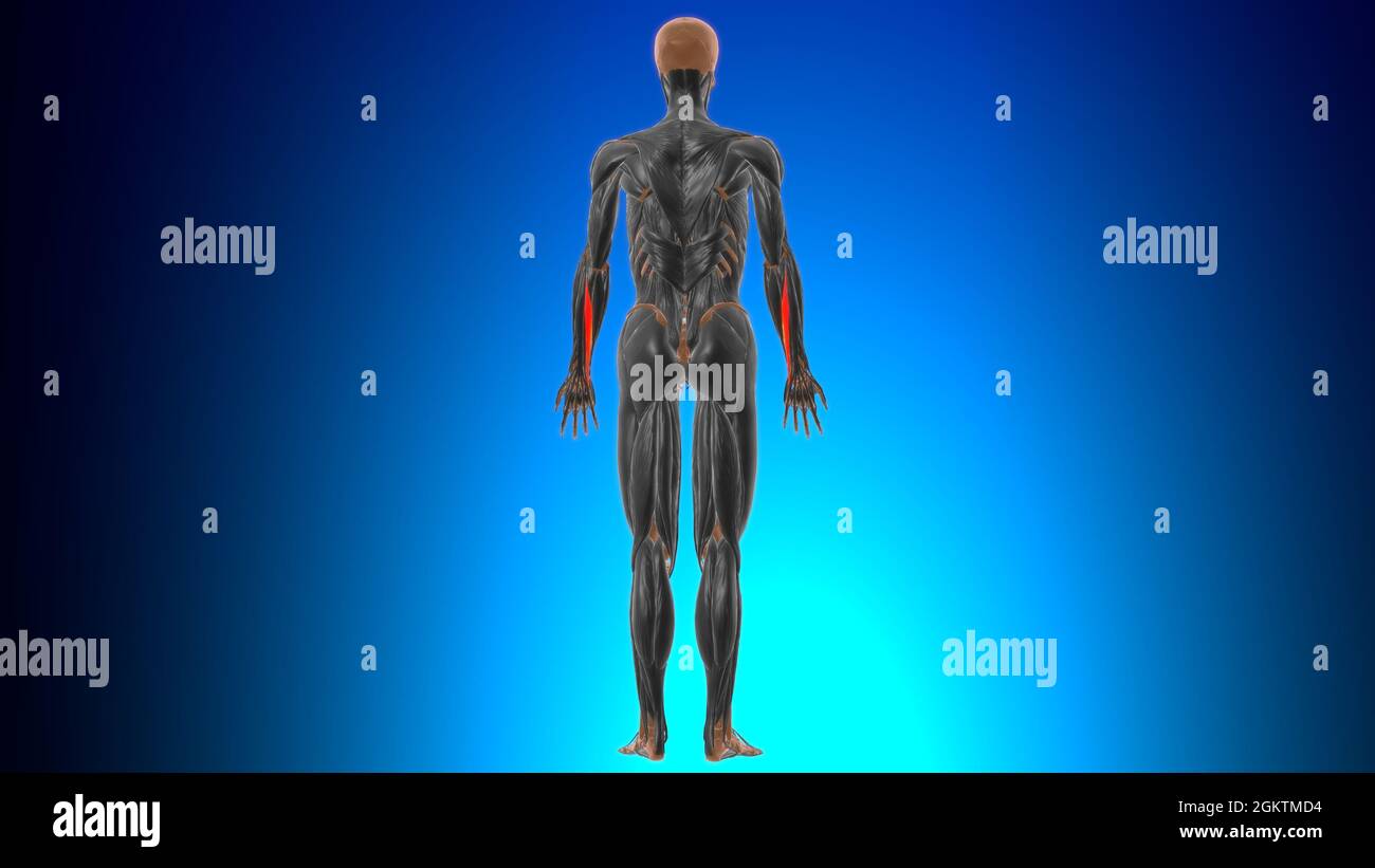 Extensor carpi ulnaris Muscle Anatomy For Medical Concept 3D Illustration Stock Photohttps://www.alamy.com/image-license-details/?v=1https://www.alamy.com/extensor-carpi-ulnaris-muscle-anatomy-for-medical-concept-3d-illustration-image442480560.html
RF2GKTMD4–Extensor carpi ulnaris Muscle Anatomy For Medical Concept 3D Illustration
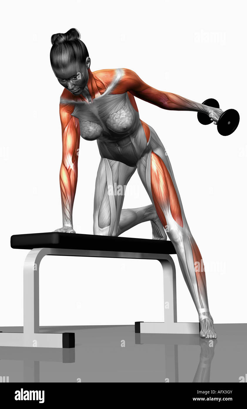 Dumbbell kickback exercise (Part 1 of 2) Stock Photohttps://www.alamy.com/image-license-details/?v=1https://www.alamy.com/stock-photo-dumbbell-kickback-exercise-part-1-of-2-14031674.html
RFAFX3GY–Dumbbell kickback exercise (Part 1 of 2)
RMRJYB9P–. An atlas of human anatomy for students and physicians. Anatomy. 326 THE MUSCLES OE THE UPPER EXTREMITY Triceps extensor cubiti muscle (external head) M. triceps brachii (caput laterale External intermuscular septum Septum intermusculare laterale Supinator radii longus muscle M. brachioradialis External condyle Epicondylus lateralis Extensor carpi radialis longior muscle M. extensor carpi radialis longus Anconeus muscle - M. anconaeus Extensor communis digitorum muscle M. extensor digitorum communis Extensor carpi ulnaris muscle Extensor minimi digiti muscle M. extensor digiti quinti proprms
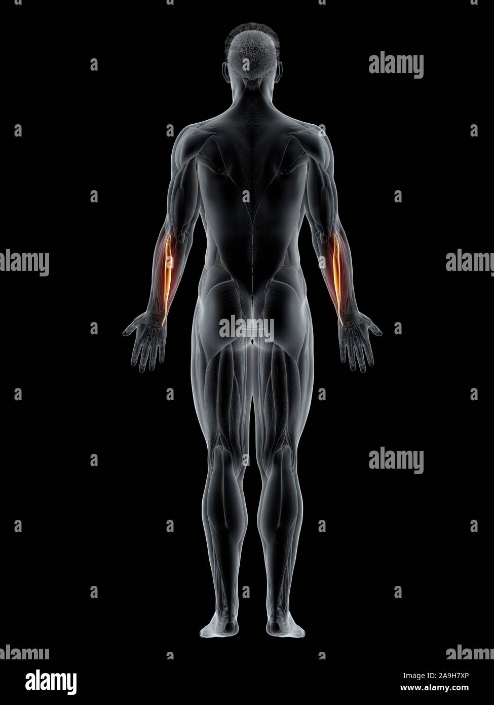 Extensor carpi ulnaris muscle, illustration Stock Photohttps://www.alamy.com/image-license-details/?v=1https://www.alamy.com/extensor-carpi-ulnaris-muscle-illustration-image332908318.html
RF2A9H7XP–Extensor carpi ulnaris muscle, illustration
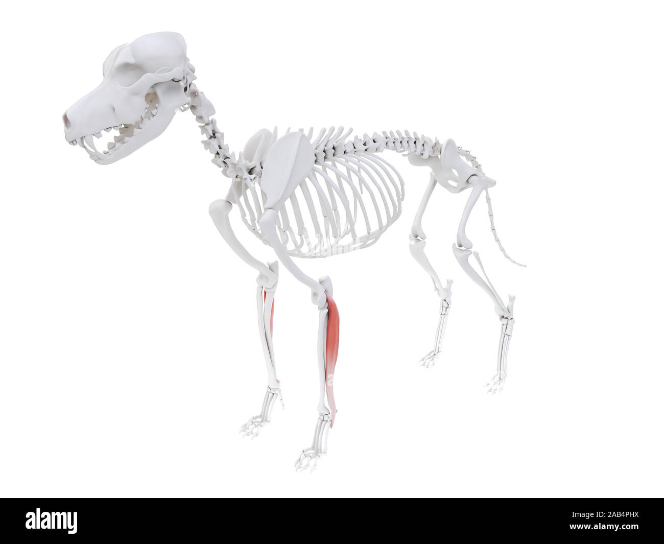 3d rendered illustration of the dog muscle anatomy - extensor carpi ulnaris Stock Photohttps://www.alamy.com/image-license-details/?v=1https://www.alamy.com/3d-rendered-illustration-of-the-dog-muscle-anatomy-extensor-carpi-ulnaris-image333863766.html
RF2AB4PHX–3d rendered illustration of the dog muscle anatomy - extensor carpi ulnaris
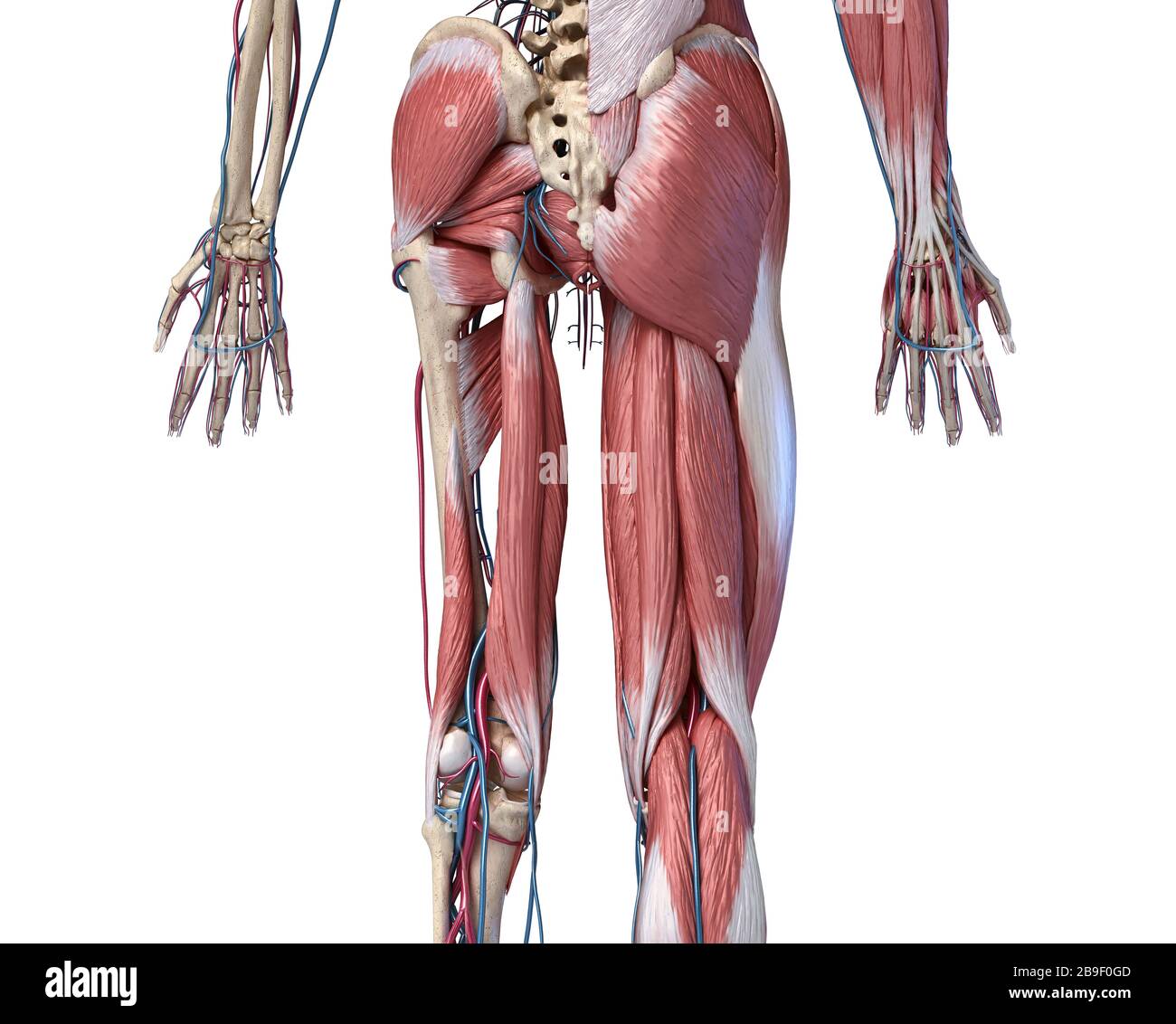 Low section rear view of human limbs, hip and muscular system with arteries and veins. Stock Photohttps://www.alamy.com/image-license-details/?v=1https://www.alamy.com/low-section-rear-view-of-human-limbs-hip-and-muscular-system-with-arteries-and-veins-image350069005.html
RF2B9F0GD–Low section rear view of human limbs, hip and muscular system with arteries and veins.
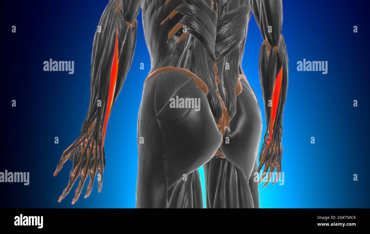 Extensor carpi ulnaris Muscle Anatomy For Medical Concept 3D Illustration Stock Photohttps://www.alamy.com/image-license-details/?v=1https://www.alamy.com/extensor-carpi-ulnaris-muscle-anatomy-for-medical-concept-3d-illustration-image442480532.html
RF2GKTMC4–Extensor carpi ulnaris Muscle Anatomy For Medical Concept 3D Illustration
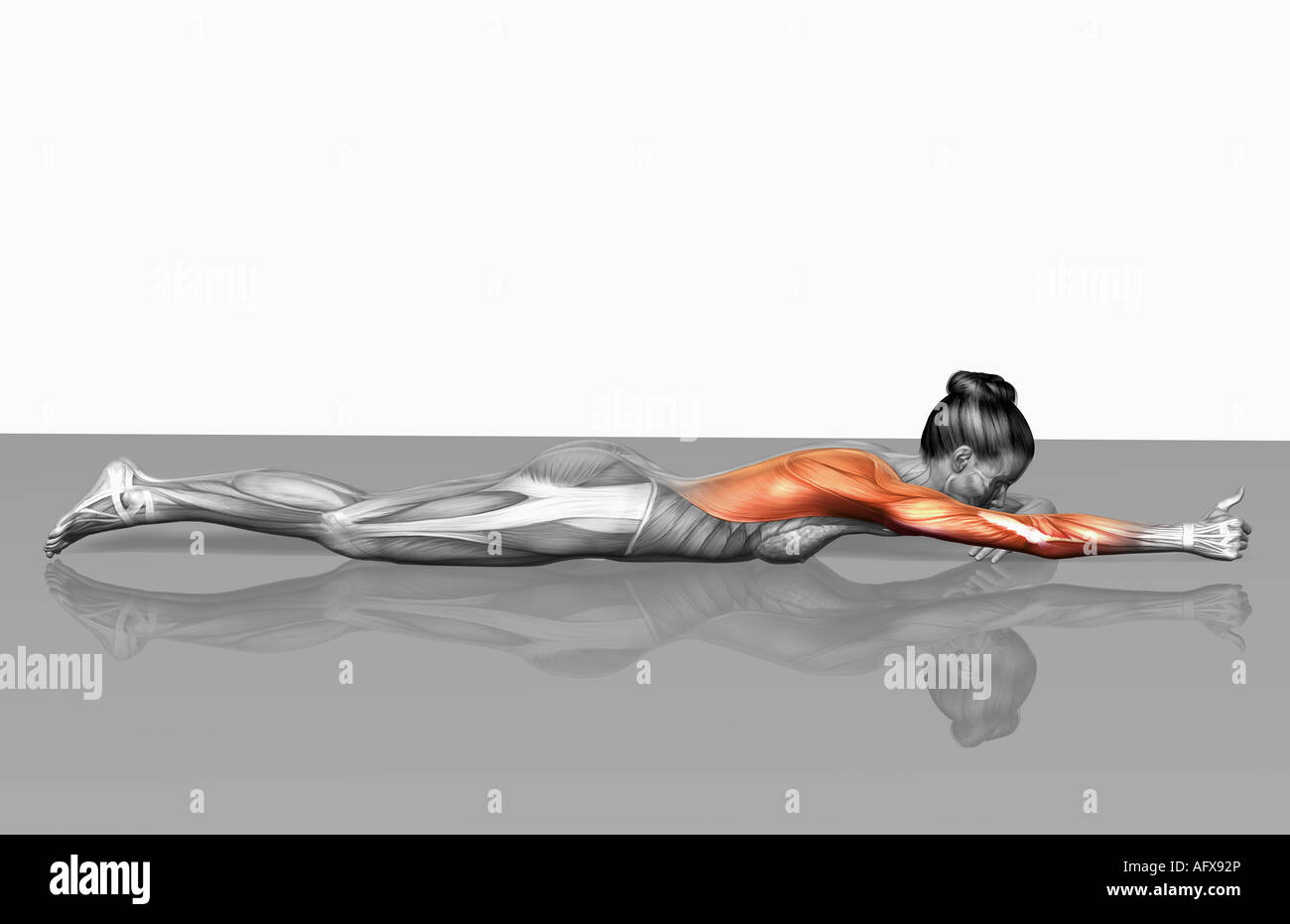 Arm-leg extensions (Part 2 of 2) Stock Photohttps://www.alamy.com/image-license-details/?v=1https://www.alamy.com/stock-photo-arm-leg-extensions-part-2-of-2-14033517.html
RFAFX92P–Arm-leg extensions (Part 2 of 2)
 . An atlas of human anatomy for students and physicians. Anatomy. 328 THE MUSCLES OF THE UPPER EXTREMITY Triceps extensor cubiti muscle (internal or deep head) //j M- triceps brachii (caput mediale) i Anconeus muscle M. anconaeus Ulna Ulna Extensor seoundi internodii pollicis or ejrtensor longus pollicis muscle' M. extensor pollicis longus Extensor indicts muscle - M. extensor indicis proprius Styloid process of the ulna Processus styloideus ulnae' Posterior annular ligament of the wrist Extensor carpi ulnaris muscle M. extensor carpi ulnaris. Capitellum of the humerus Capitulum humeri Externa Stock Photohttps://www.alamy.com/image-license-details/?v=1https://www.alamy.com/an-atlas-of-human-anatomy-for-students-and-physicians-anatomy-328-the-muscles-of-the-upper-extremity-triceps-extensor-cubiti-muscle-internal-or-deep-head-j-m-triceps-brachii-caput-mediale-i-anconeus-muscle-m-anconaeus-ulna-ulna-extensor-seoundi-internodii-pollicis-or-ejrtensor-longus-pollicis-muscle-m-extensor-pollicis-longus-extensor-indicts-muscle-m-extensor-indicis-proprius-styloid-process-of-the-ulna-processus-styloideus-ulnae-posterior-annular-ligament-of-the-wrist-extensor-carpi-ulnaris-muscle-m-extensor-carpi-ulnaris-capitellum-of-the-humerus-capitulum-humeri-externa-image235400173.html
RMRJYB91–. An atlas of human anatomy for students and physicians. Anatomy. 328 THE MUSCLES OF THE UPPER EXTREMITY Triceps extensor cubiti muscle (internal or deep head) //j M- triceps brachii (caput mediale) i Anconeus muscle M. anconaeus Ulna Ulna Extensor seoundi internodii pollicis or ejrtensor longus pollicis muscle' M. extensor pollicis longus Extensor indicts muscle - M. extensor indicis proprius Styloid process of the ulna Processus styloideus ulnae' Posterior annular ligament of the wrist Extensor carpi ulnaris muscle M. extensor carpi ulnaris. Capitellum of the humerus Capitulum humeri Externa
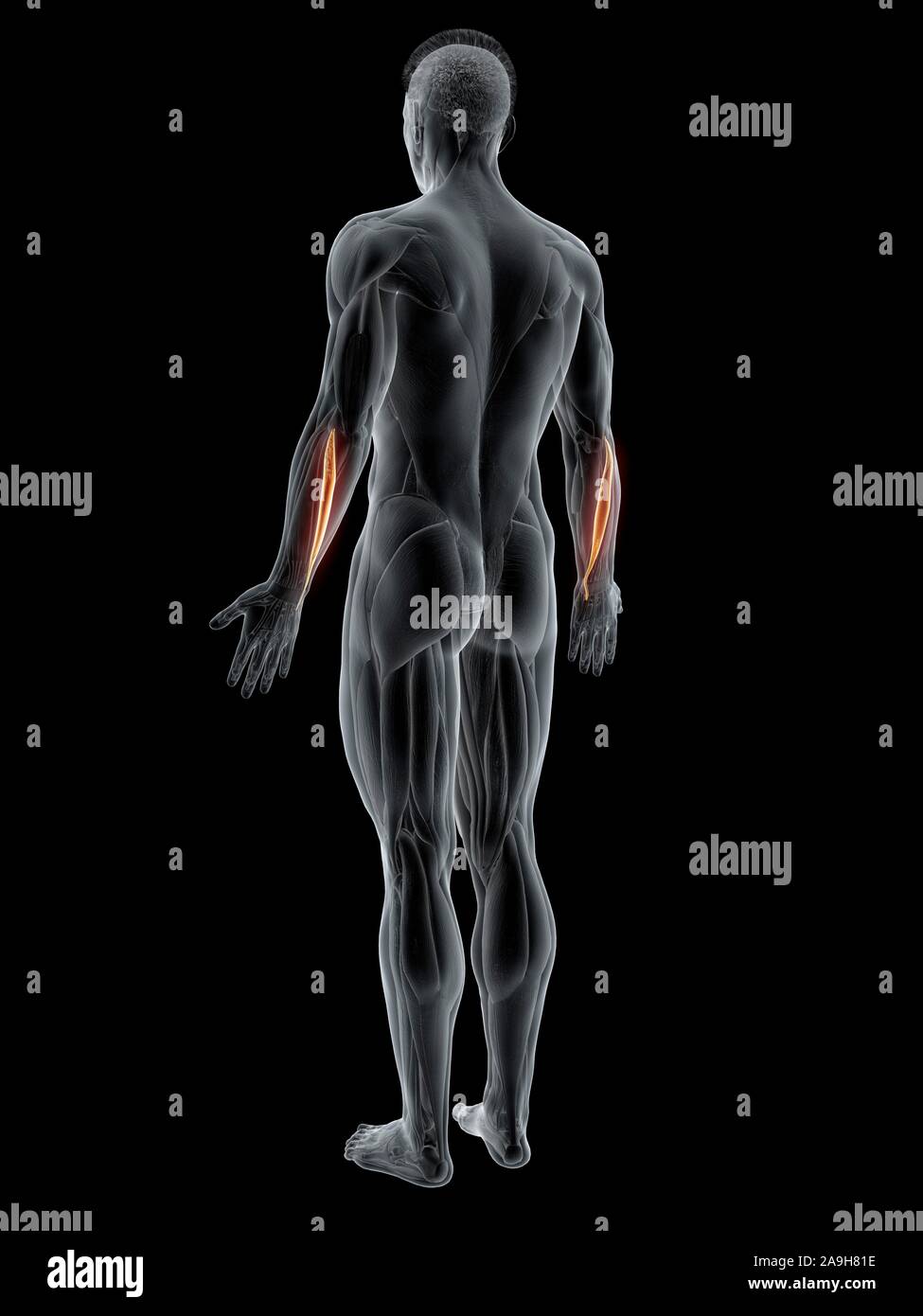 Extensor carpi ulnaris muscle, illustration Stock Photohttps://www.alamy.com/image-license-details/?v=1https://www.alamy.com/extensor-carpi-ulnaris-muscle-illustration-image332908394.html
RF2A9H81E–Extensor carpi ulnaris muscle, illustration
 medically accurate muscle illustration of the extensor carpi ulnaris Stock Photohttps://www.alamy.com/image-license-details/?v=1https://www.alamy.com/stock-photo-medically-accurate-muscle-illustration-of-the-extensor-carpi-ulnaris-89751045.html
RFF60EAD–medically accurate muscle illustration of the extensor carpi ulnaris
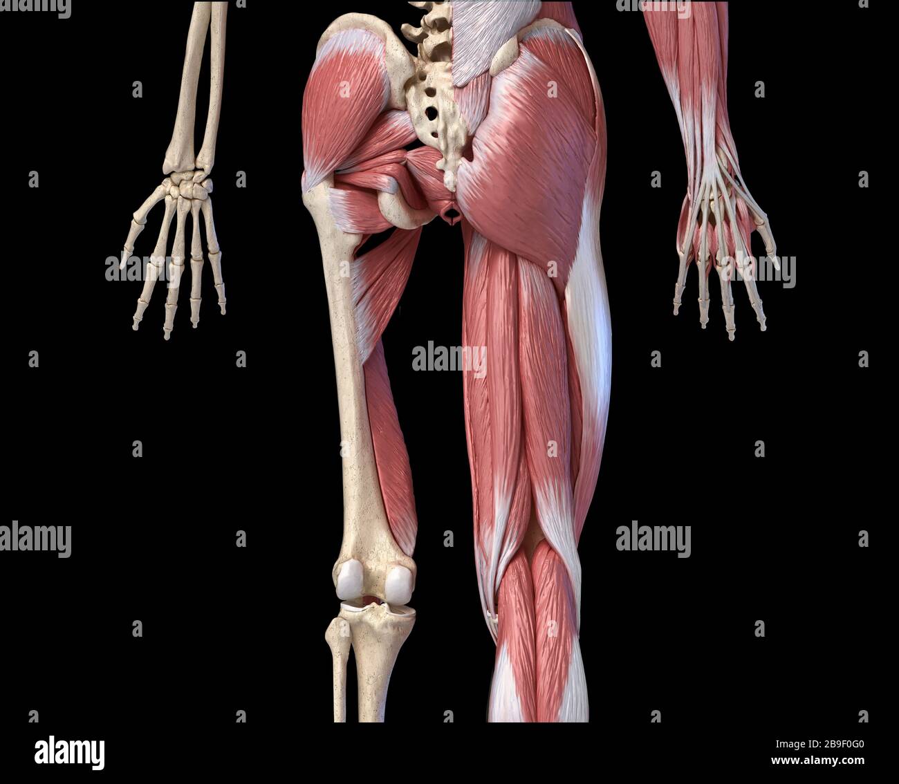 Low section rear view of human limbs, hip and muscular system, on black background. Stock Photohttps://www.alamy.com/image-license-details/?v=1https://www.alamy.com/low-section-rear-view-of-human-limbs-hip-and-muscular-system-on-black-background-image350068992.html
RF2B9F0G0–Low section rear view of human limbs, hip and muscular system, on black background.
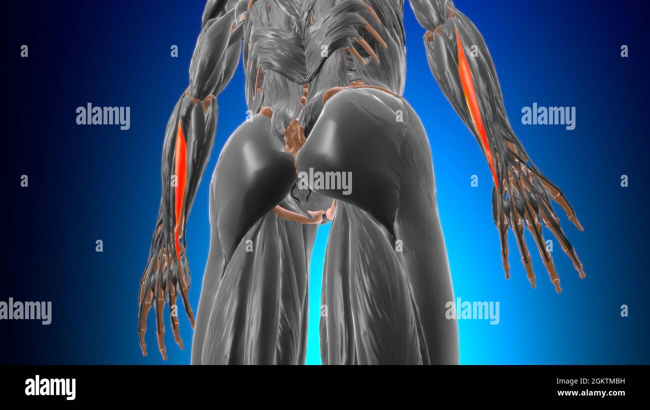 Extensor carpi ulnaris Muscle Anatomy For Medical Concept 3D Illustration Stock Photohttps://www.alamy.com/image-license-details/?v=1https://www.alamy.com/extensor-carpi-ulnaris-muscle-anatomy-for-medical-concept-3d-illustration-image442480517.html
RF2GKTMBH–Extensor carpi ulnaris Muscle Anatomy For Medical Concept 3D Illustration
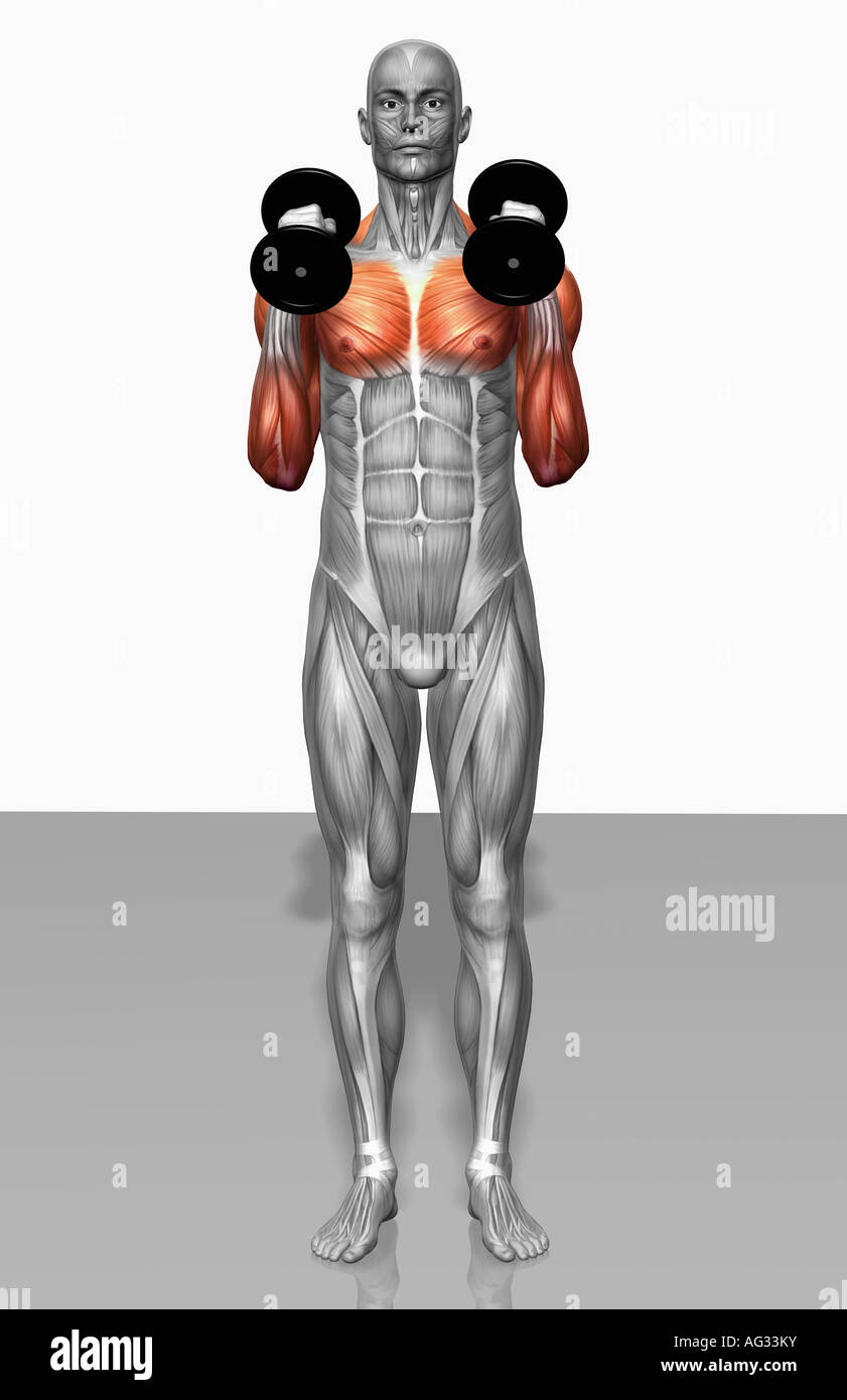 Hammer curl exercise (Part 1 of 2) Stock Photohttps://www.alamy.com/image-license-details/?v=1https://www.alamy.com/stock-photo-hammer-curl-exercise-part-1-of-2-14078750.html
RFAG33KY–Hammer curl exercise (Part 1 of 2)
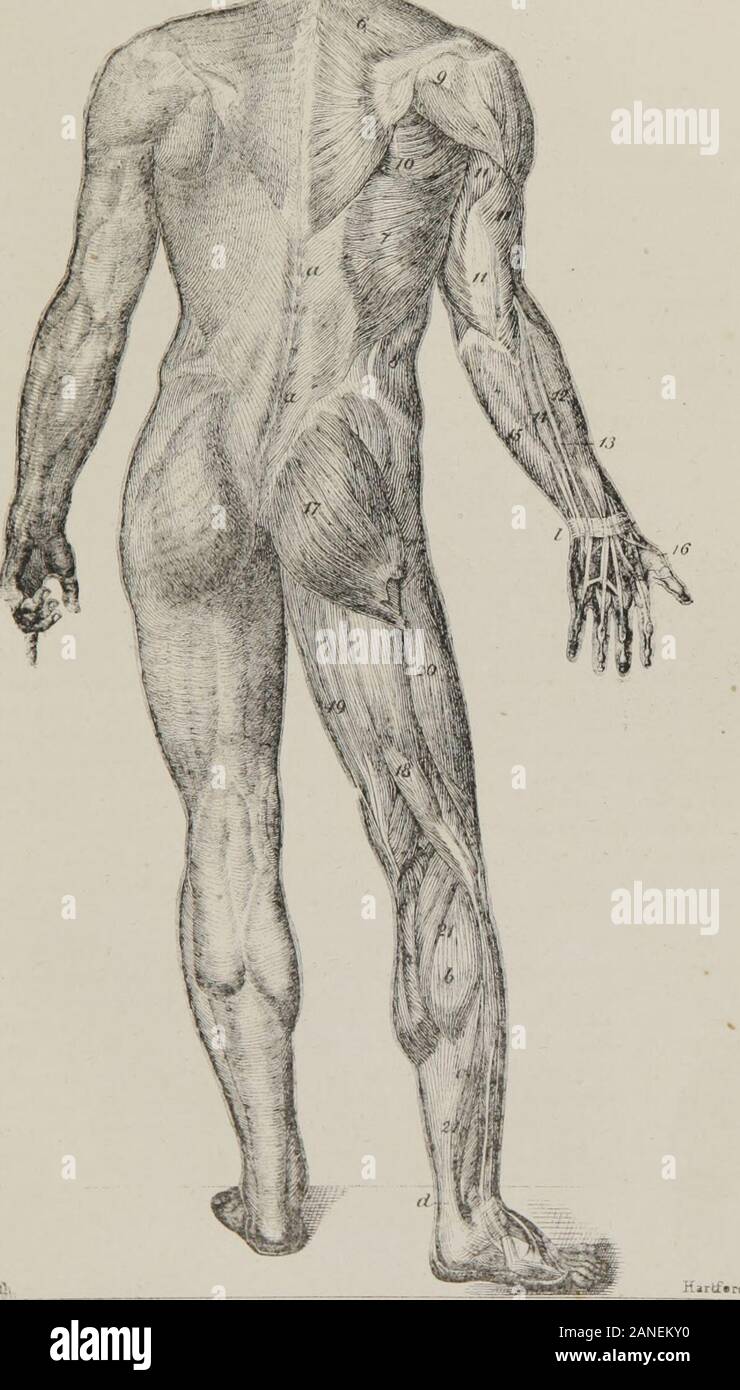 Class-book of physiology : for the use of schools and families : comprising the structure and functions of the organs of man, illustrated by comparative reference to those of inferior animals . 4, The extensor carpi ulnaris.—This muscle extends the hand on thewrist. 15, Flexor carpi ulnaris. 10, Tendon of the long extensor of the thumb. /, Annular ligament of the carpus. Muscles of the Inferior Extremities.—17, The gluteus maximus.—This large, thickmuscle is of greater volume than any other in the body, and is the principal agent in pre-serving its equilibrium. It extends from the iliac bones Stock Photohttps://www.alamy.com/image-license-details/?v=1https://www.alamy.com/class-book-of-physiology-for-the-use-of-schools-and-families-comprising-the-structure-and-functions-of-the-organs-of-man-illustrated-by-comparative-reference-to-those-of-inferior-animals-4-the-extensor-carpi-ulnaristhis-muscle-extends-the-hand-on-thewrist-15-flexor-carpi-ulnaris-10-tendon-of-the-long-extensor-of-the-thumb-annular-ligament-of-the-carpus-muscles-of-the-inferior-extremities17-the-gluteus-maximusthis-large-thickmuscle-is-of-greater-volume-than-any-other-in-the-body-and-is-the-principal-agent-in-pre-serving-its-equilibrium-it-extends-from-the-iliac-bones-image340227748.html
RM2ANEKY0–Class-book of physiology : for the use of schools and families : comprising the structure and functions of the organs of man, illustrated by comparative reference to those of inferior animals . 4, The extensor carpi ulnaris.—This muscle extends the hand on thewrist. 15, Flexor carpi ulnaris. 10, Tendon of the long extensor of the thumb. /, Annular ligament of the carpus. Muscles of the Inferior Extremities.—17, The gluteus maximus.—This large, thickmuscle is of greater volume than any other in the body, and is the principal agent in pre-serving its equilibrium. It extends from the iliac bones
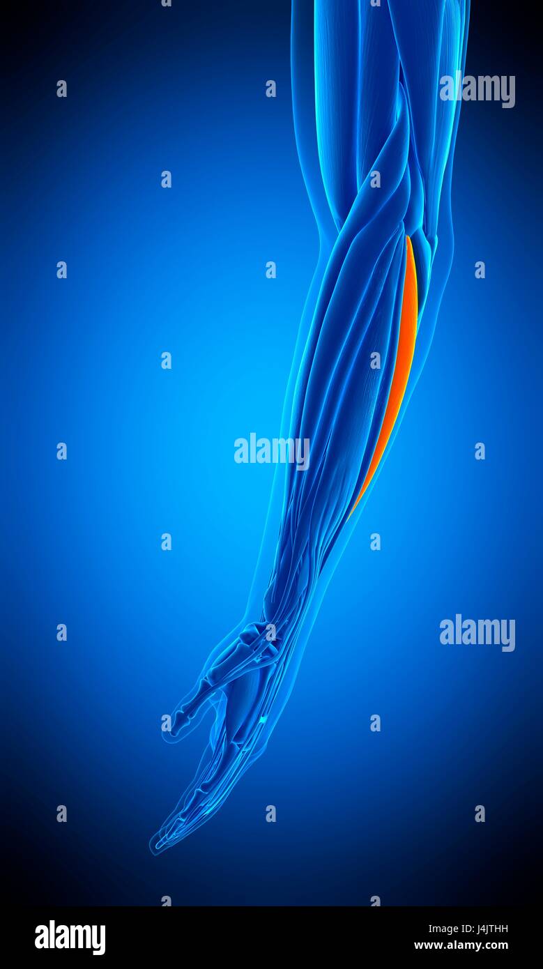 Illustration of the extensor carpi ulnaris muscle. Stock Photohttps://www.alamy.com/image-license-details/?v=1https://www.alamy.com/stock-photo-illustration-of-the-extensor-carpi-ulnaris-muscle-140556013.html
RFJ4JTHH–Illustration of the extensor carpi ulnaris muscle.
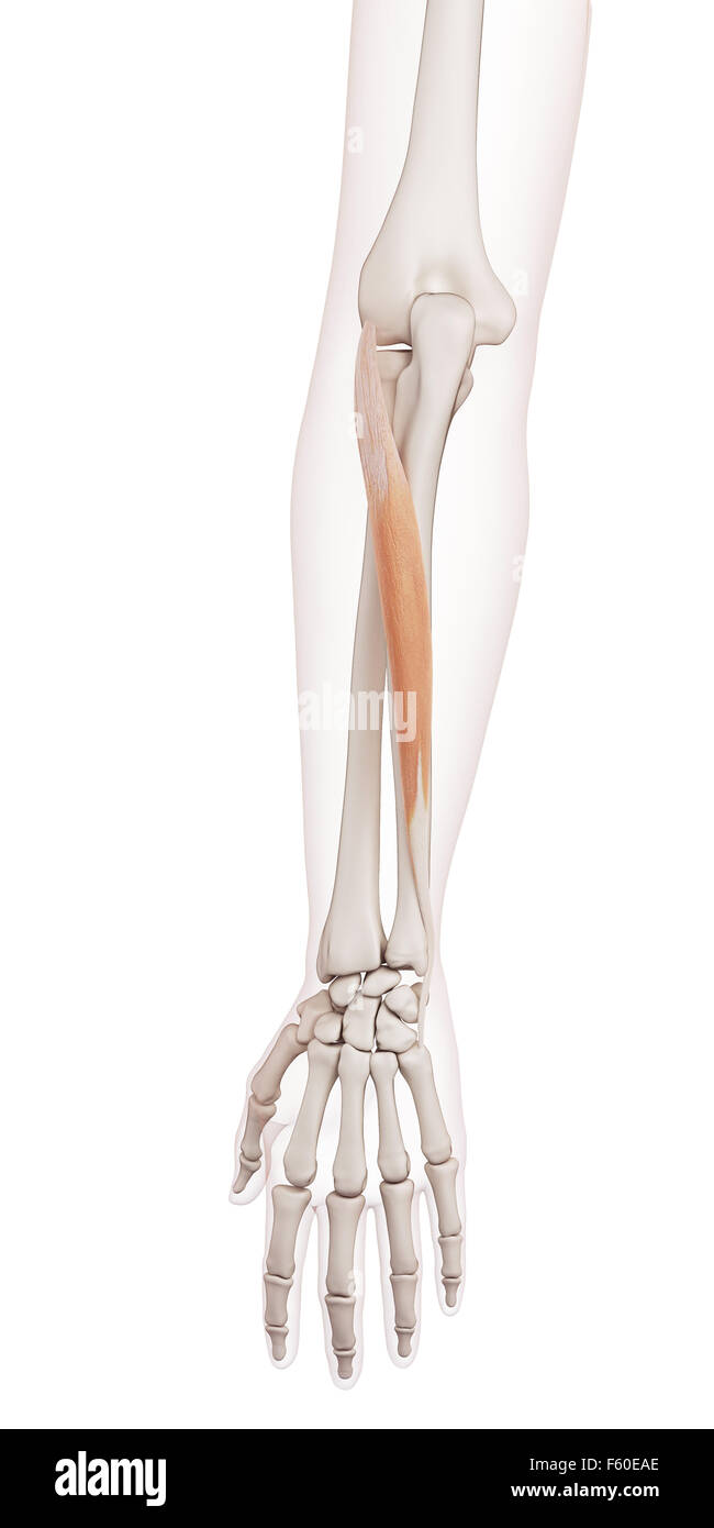 medically accurate muscle illustration of the extensor carpi ulnaris Stock Photohttps://www.alamy.com/image-license-details/?v=1https://www.alamy.com/stock-photo-medically-accurate-muscle-illustration-of-the-extensor-carpi-ulnaris-89751046.html
RFF60EAE–medically accurate muscle illustration of the extensor carpi ulnaris
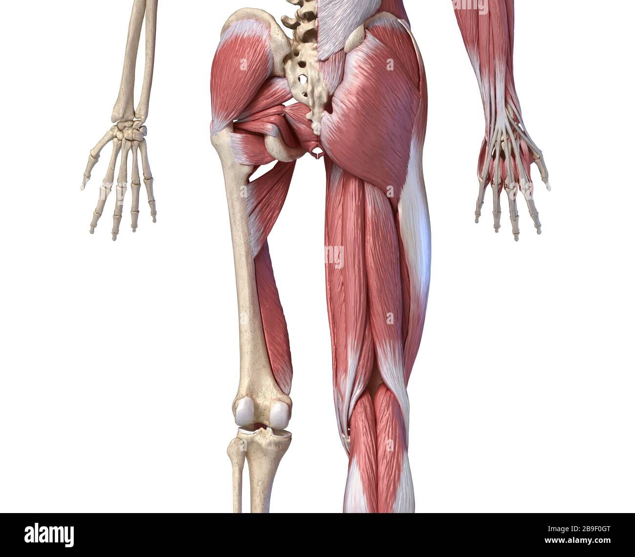 Low section rear view of human limbs, hip and muscular system, on white background. Stock Photohttps://www.alamy.com/image-license-details/?v=1https://www.alamy.com/low-section-rear-view-of-human-limbs-hip-and-muscular-system-on-white-background-image350069016.html
RF2B9F0GT–Low section rear view of human limbs, hip and muscular system, on white background.
 Extensor carpi ulnaris Muscle Anatomy For Medical Concept 3D Illustration Stock Photohttps://www.alamy.com/image-license-details/?v=1https://www.alamy.com/extensor-carpi-ulnaris-muscle-anatomy-for-medical-concept-3d-illustration-image442480551.html
RF2GKTMCR–Extensor carpi ulnaris Muscle Anatomy For Medical Concept 3D Illustration
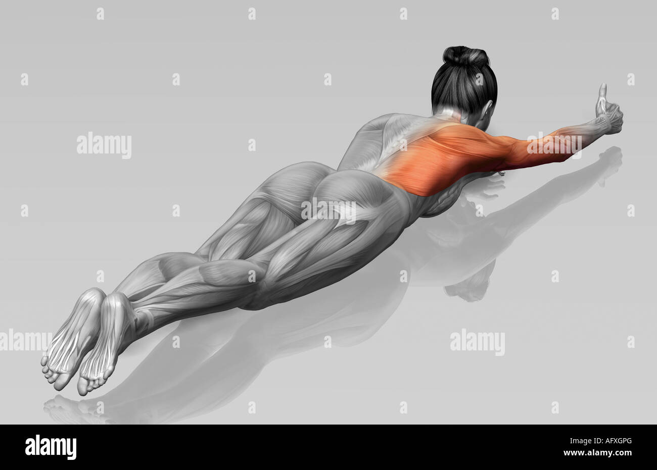 Arm-leg extensions (Part 2 of 2) Stock Photohttps://www.alamy.com/image-license-details/?v=1https://www.alamy.com/stock-photo-arm-leg-extensions-part-2-of-2-14036103.html
RFAFXGPG–Arm-leg extensions (Part 2 of 2)
 Anatomy and physiology : designed for academies and families . Fig. 70. The superficial layer of muscles on the posterior aspect of the fore-arro.l,The lower part of the biceps muscle. 2, Part of the brnehialis anticus. 3, The lowerpart of the triceps inserted into the olecranon. 4, The supinator LongUS. 5, The ex-tensor carpi radialia longior. 6, The extensor carpi radialis brevior. 7, The tendons ofinsertion of these two muscles 8, The extensor communis digitorum. 9. The extensorminimi digiti. 10, The extensor carpi ulnaris. 11, The anconeus. 12. Tart of theflexor carpi ulnaris 13, The exten Stock Photohttps://www.alamy.com/image-license-details/?v=1https://www.alamy.com/anatomy-and-physiology-designed-for-academies-and-families-fig-70-the-superficial-layer-of-muscles-on-the-posterior-aspect-of-the-fore-arrolthe-lower-part-of-the-biceps-muscle-2-part-of-the-brnehialis-anticus-3-the-lowerpart-of-the-triceps-inserted-into-the-olecranon-4-the-supinator-longus-5-the-ex-tensor-carpi-radialia-longior-6-the-extensor-carpi-radialis-brevior-7-the-tendons-ofinsertion-of-these-two-muscles-8-the-extensor-communis-digitorum-9-the-extensorminimi-digiti-10-the-extensor-carpi-ulnaris-11-the-anconeus-12-tart-of-theflexor-carpi-ulnaris-13-the-exten-image339934655.html
RM2AN1A3B–Anatomy and physiology : designed for academies and families . Fig. 70. The superficial layer of muscles on the posterior aspect of the fore-arro.l,The lower part of the biceps muscle. 2, Part of the brnehialis anticus. 3, The lowerpart of the triceps inserted into the olecranon. 4, The supinator LongUS. 5, The ex-tensor carpi radialia longior. 6, The extensor carpi radialis brevior. 7, The tendons ofinsertion of these two muscles 8, The extensor communis digitorum. 9. The extensorminimi digiti. 10, The extensor carpi ulnaris. 11, The anconeus. 12. Tart of theflexor carpi ulnaris 13, The exten
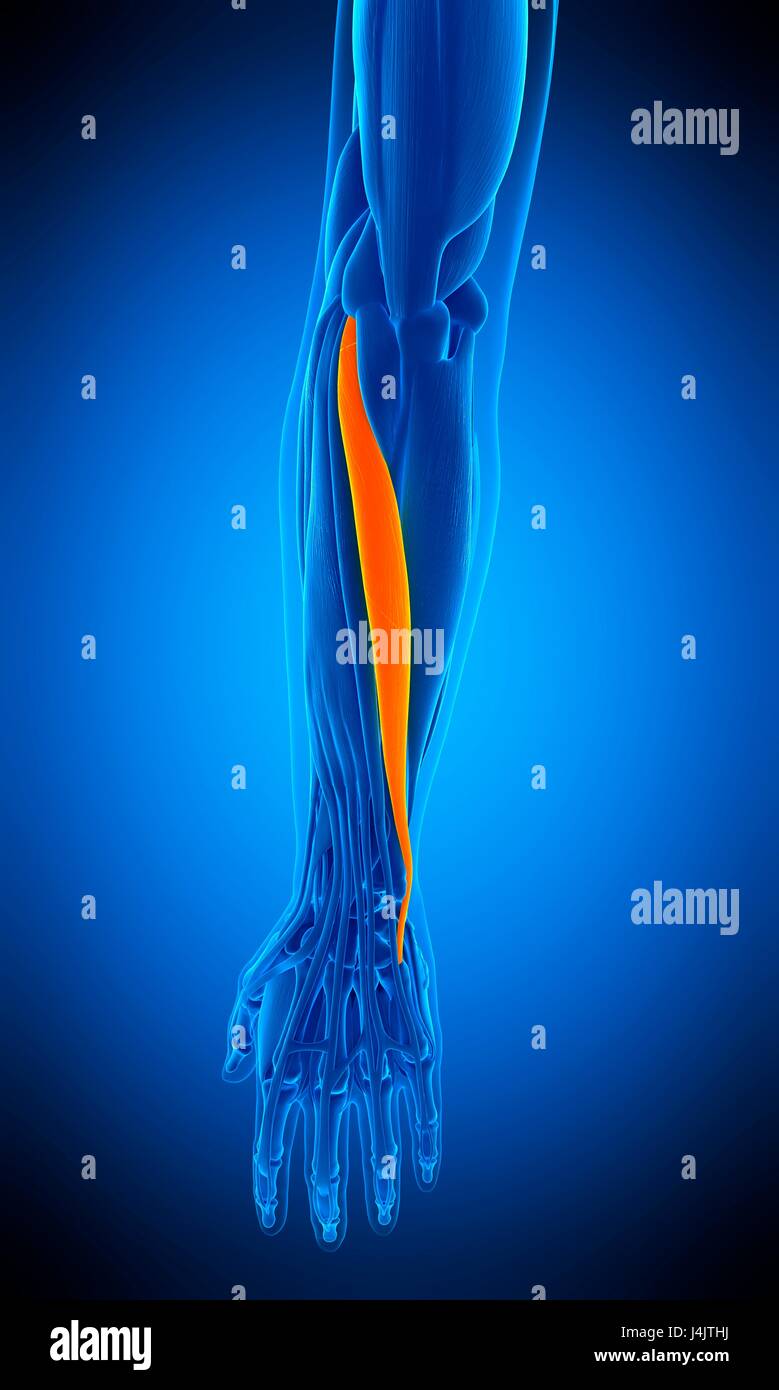 Illustration of the extensor carpi ulnaris muscle. Stock Photohttps://www.alamy.com/image-license-details/?v=1https://www.alamy.com/stock-photo-illustration-of-the-extensor-carpi-ulnaris-muscle-140556014.html
RFJ4JTHJ–Illustration of the extensor carpi ulnaris muscle.
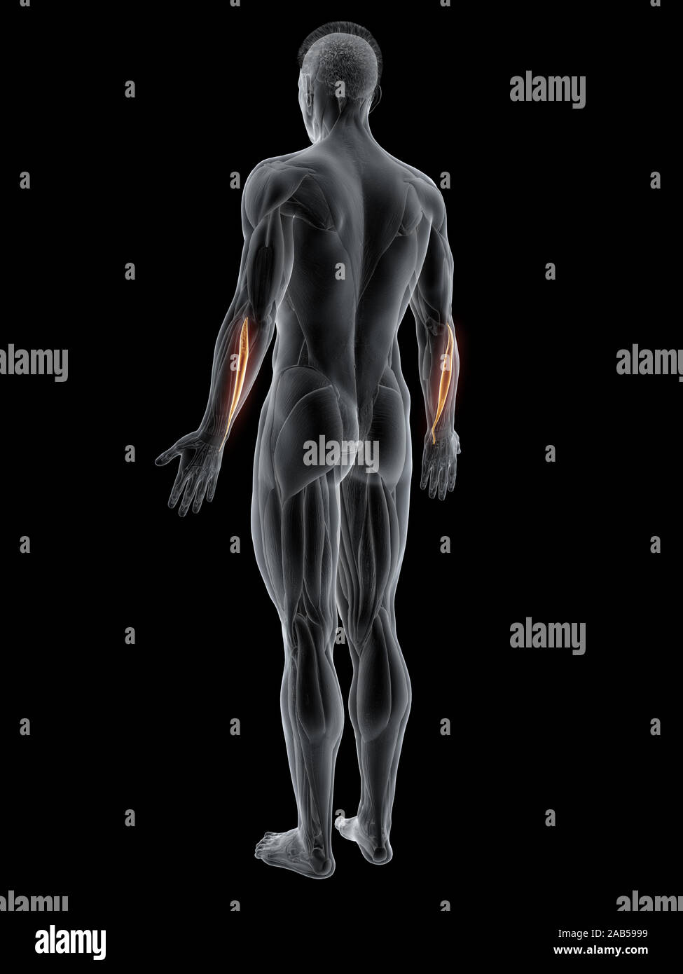 3d rendered muscle illustration of the extensor carpi ulnaris Stock Photohttps://www.alamy.com/image-license-details/?v=1https://www.alamy.com/3d-rendered-muscle-illustration-of-the-extensor-carpi-ulnaris-image333875285.html
RF2AB5999–3d rendered muscle illustration of the extensor carpi ulnaris
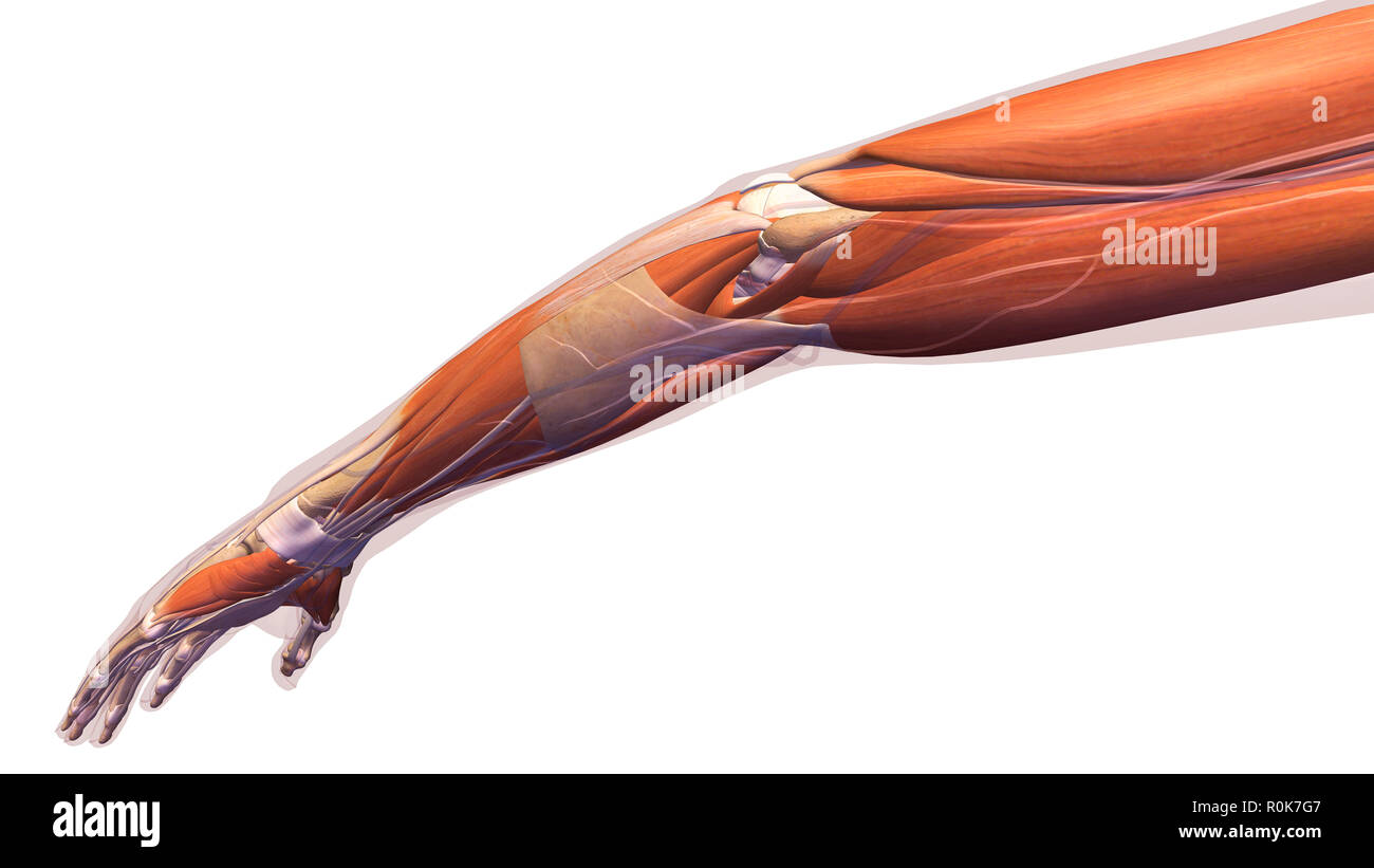 Female arm, elbow muscles and tendons on white background. Stock Photohttps://www.alamy.com/image-license-details/?v=1https://www.alamy.com/female-arm-elbow-muscles-and-tendons-on-white-background-image224157815.html
RFR0K7G7–Female arm, elbow muscles and tendons on white background.
 Extensor Carpi Ulnaris muscle Dog muscle Anatomy For Medical Concept 3D Illustration Stock Photohttps://www.alamy.com/image-license-details/?v=1https://www.alamy.com/extensor-carpi-ulnaris-muscle-dog-muscle-anatomy-for-medical-concept-3d-illustration-image439390504.html
RF2GERY20–Extensor Carpi Ulnaris muscle Dog muscle Anatomy For Medical Concept 3D Illustration
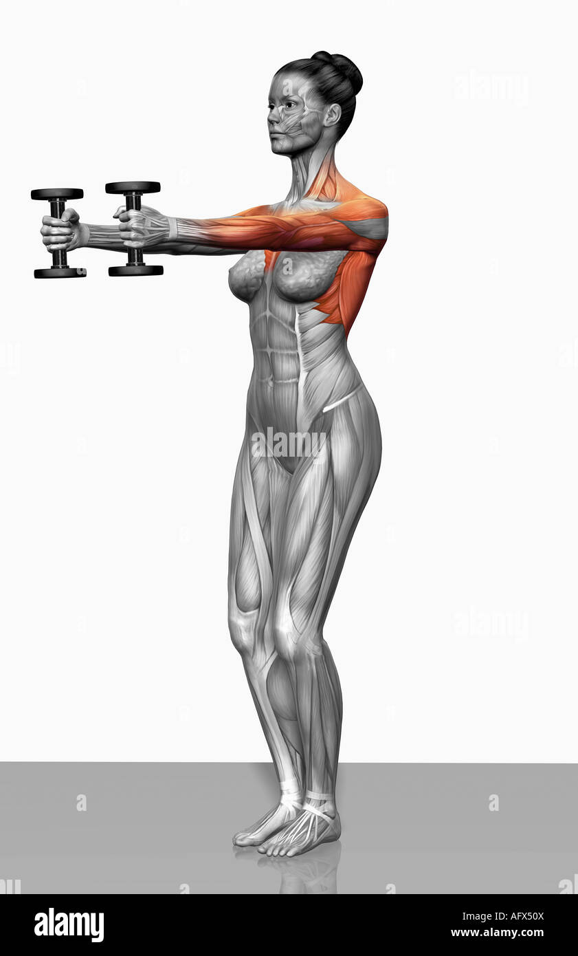 Forward raise exercise Stock Photohttps://www.alamy.com/image-license-details/?v=1https://www.alamy.com/stock-photo-forward-raise-exercise-14032153.html
RFAFX50X–Forward raise exercise
 Modelling; a guide for teachers and students . TRICEPS CUBITI (long HEAD) SUPPLLMEINTAL MUSCLE. OFLATI3S>IMU5 DORi>i TRICEPS CUBITI (external head) FLEXOR OIGITORUM PROFUNDUS : ANTERIOR ULNAR (FLEXOR CARPI ULNARIS) POSTERIOR ULNAR- (EXTENSOR CARPI ULNARIS)EXTENSOR MINIM! OIGITI - TENDON OFFLEXOR CARPI RADIALI5 TENDON OFFLEXOR DIGITORUM 5UBLIMIS TENDON OFFLEXOR DIGITORUM PROFUNDUS — SUSPENSORY LIGAMENT OF FETLOCK Fig. 134.—Fore-leg. Posterior Aspect. Myology. ATTACHMENTS OF MUSCLES. Triceps cubiti (long head) (origin) scapula: (insertion) olecranon process. Supple-mental MUSCLE OF LATissi Stock Photohttps://www.alamy.com/image-license-details/?v=1https://www.alamy.com/modelling-a-guide-for-teachers-and-students-triceps-cubiti-long-head-suppllmeintal-muscle-oflati3sgtimu5-dorigti-triceps-cubiti-external-head-flexor-oigitorum-profundus-anterior-ulnar-flexor-carpi-ulnaris-posterior-ulnar-extensor-carpi-ulnarisextensor-minim!-oigiti-tendon-offlexor-carpi-radiali5-tendon-offlexor-digitorum-5ublimis-tendon-offlexor-digitorum-profundus-suspensory-ligament-of-fetlock-fig-134fore-leg-posterior-aspect-myology-attachments-of-muscles-triceps-cubiti-long-head-origin-scapula-insertion-olecranon-process-supple-mental-muscle-of-latissi-image338448346.html
RM2AJHJ8X–Modelling; a guide for teachers and students . TRICEPS CUBITI (long HEAD) SUPPLLMEINTAL MUSCLE. OFLATI3S>IMU5 DORi>i TRICEPS CUBITI (external head) FLEXOR OIGITORUM PROFUNDUS : ANTERIOR ULNAR (FLEXOR CARPI ULNARIS) POSTERIOR ULNAR- (EXTENSOR CARPI ULNARIS)EXTENSOR MINIM! OIGITI - TENDON OFFLEXOR CARPI RADIALI5 TENDON OFFLEXOR DIGITORUM 5UBLIMIS TENDON OFFLEXOR DIGITORUM PROFUNDUS — SUSPENSORY LIGAMENT OF FETLOCK Fig. 134.—Fore-leg. Posterior Aspect. Myology. ATTACHMENTS OF MUSCLES. Triceps cubiti (long head) (origin) scapula: (insertion) olecranon process. Supple-mental MUSCLE OF LATissi
 Extensor carpi ulnaris muscle, illustration Stock Photohttps://www.alamy.com/image-license-details/?v=1https://www.alamy.com/extensor-carpi-ulnaris-muscle-illustration-image267278550.html
RFWERGEE–Extensor carpi ulnaris muscle, illustration
 3d rendered muscle illustration of the extensor carpi ulnaris Stock Photohttps://www.alamy.com/image-license-details/?v=1https://www.alamy.com/3d-rendered-muscle-illustration-of-the-extensor-carpi-ulnaris-image333874889.html
RF2AB58R5–3d rendered muscle illustration of the extensor carpi ulnaris
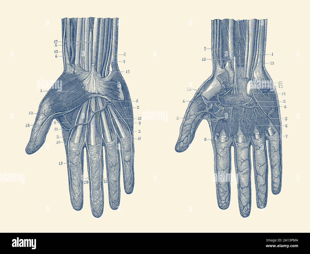 Dual view of the human hand, showcasing the muscles, bones and veins throughout. Stock Photohttps://www.alamy.com/image-license-details/?v=1https://www.alamy.com/dual-view-of-the-human-hand-showcasing-the-muscles-bones-and-veins-throughout-image327695322.html
RF2A13PMA–Dual view of the human hand, showcasing the muscles, bones and veins throughout.
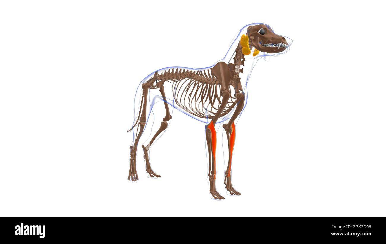 Extensor Carpi Radialis muscle Dog muscle Anatomy For Medical Concept 3D Illustration Stock Photohttps://www.alamy.com/image-license-details/?v=1https://www.alamy.com/extensor-carpi-radialis-muscle-dog-muscle-anatomy-for-medical-concept-3d-illustration-image441991766.html
RF2GK2D06–Extensor Carpi Radialis muscle Dog muscle Anatomy For Medical Concept 3D Illustration
 Bench press (Part 1 of 2) Stock Photohttps://www.alamy.com/image-license-details/?v=1https://www.alamy.com/stock-photo-bench-press-part-1-of-2-14078681.html
RFAG33EJ–Bench press (Part 1 of 2)
 The hydropathic encyclopedia: a system of hydropathy and hygiene .. . rated, and perforatingmuscles. G ABDUCTOR POLLTCIS MANUS—abductor of the thumb. H PALMARIS BREVIS—short muscle ot the palm. K EXTENSOR POLLICIS—extending muscle of the thumb. K EXTENSOR PR1MI INTURNODII—extensor of the first fingo» L EXTENSOR CARPI RADIALIS BREVIS—short radial exten-sor of the wrist. M EXTENSOR CARPI RADIALIS LONGUS—long radial exton-sor of tho wrist. N EXTENSOR DIOITORUM—extensor of the fingers. 0 EXTENSOR CARPI ULNARIS—ulnar extensor of the wrist P ANCONEUS—muscle of the elbow. Q EXTENSOR SECUNDI INTERNODI Stock Photohttps://www.alamy.com/image-license-details/?v=1https://www.alamy.com/the-hydropathic-encyclopedia-a-system-of-hydropathy-and-hygiene-rated-and-perforatingmuscles-g-abductor-polltcis-manusabductor-of-the-thumb-h-palmaris-brevisshort-muscle-ot-the-palm-k-extensor-pollicisextending-muscle-of-the-thumb-k-extensor-pr1mi-inturnodiiextensor-of-the-first-fingo-l-extensor-carpi-radialis-brevisshort-radial-exten-sor-of-the-wrist-m-extensor-carpi-radialis-longuslong-radial-exton-sor-of-tho-wrist-n-extensor-dioitorumextensor-of-the-fingers-0-extensor-carpi-ulnarisulnar-extensor-of-the-wrist-p-anconeusmuscle-of-the-elbow-q-extensor-secundi-internodi-image340117469.html
RM2AN9K8D–The hydropathic encyclopedia: a system of hydropathy and hygiene .. . rated, and perforatingmuscles. G ABDUCTOR POLLTCIS MANUS—abductor of the thumb. H PALMARIS BREVIS—short muscle ot the palm. K EXTENSOR POLLICIS—extending muscle of the thumb. K EXTENSOR PR1MI INTURNODII—extensor of the first fingo» L EXTENSOR CARPI RADIALIS BREVIS—short radial exten-sor of the wrist. M EXTENSOR CARPI RADIALIS LONGUS—long radial exton-sor of tho wrist. N EXTENSOR DIOITORUM—extensor of the fingers. 0 EXTENSOR CARPI ULNARIS—ulnar extensor of the wrist P ANCONEUS—muscle of the elbow. Q EXTENSOR SECUNDI INTERNODI
 Extensor carpi ulnaris muscle, illustration Stock Photohttps://www.alamy.com/image-license-details/?v=1https://www.alamy.com/extensor-carpi-ulnaris-muscle-illustration-image267278559.html
RFWERGER–Extensor carpi ulnaris muscle, illustration
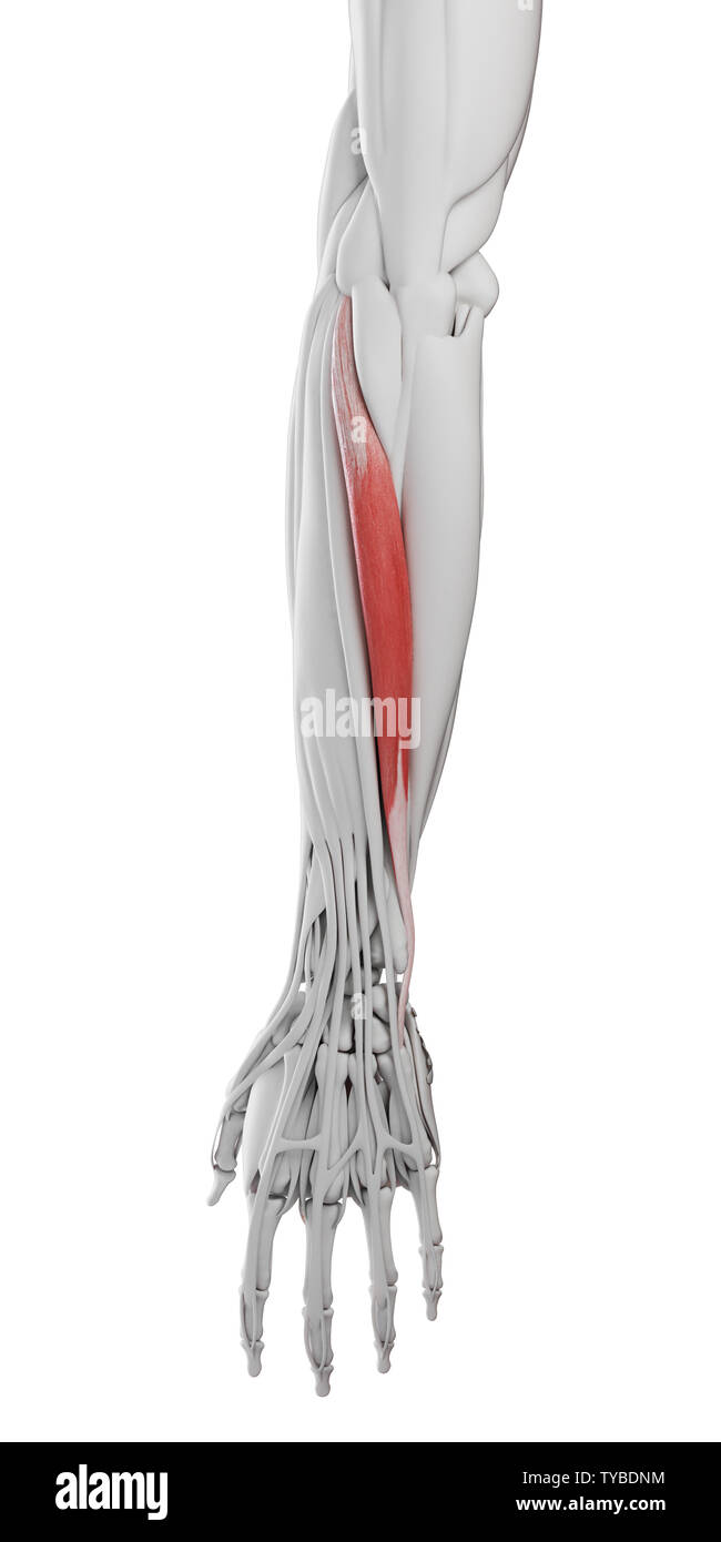 3d rendered medically accurate illustration of the extensor carpi ulnaris Stock Photohttps://www.alamy.com/image-license-details/?v=1https://www.alamy.com/3d-rendered-medically-accurate-illustration-of-the-extensor-carpi-ulnaris-image257793136.html
RFTYBDNM–3d rendered medically accurate illustration of the extensor carpi ulnaris
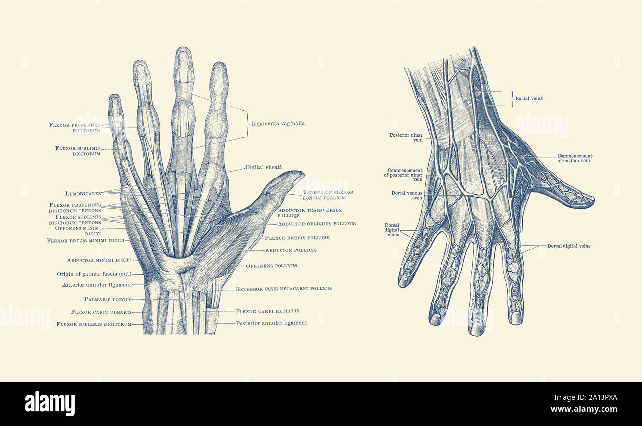 Dual-view diagram of the human hand, showcasing ligaments, muscles and veins. Stock Photohttps://www.alamy.com/image-license-details/?v=1https://www.alamy.com/dual-view-diagram-of-the-human-hand-showcasing-ligaments-muscles-and-veins-image327695490.html
RF2A13PXA–Dual-view diagram of the human hand, showcasing ligaments, muscles and veins.
 Flexor Carpi Ulnaris muscle Dog muscle Anatomy For Medical Concept 3D Illustration Stock Photohttps://www.alamy.com/image-license-details/?v=1https://www.alamy.com/flexor-carpi-ulnaris-muscle-dog-muscle-anatomy-for-medical-concept-3d-illustration-image441991179.html
RF2GK2C77–Flexor Carpi Ulnaris muscle Dog muscle Anatomy For Medical Concept 3D Illustration
 Cable rear raise Stock Photohttps://www.alamy.com/image-license-details/?v=1https://www.alamy.com/stock-photo-cable-rear-raise-14031530.html
RFAFX34Y–Cable rear raise
 Modelling; a guide for teachers and students . INTER055EUS MUSCLES TENDONS OFFLEXOR DIG. SUBLIMIS Fig. 102.—Fore-leg. Posterior Aspect. Myology. ATTACHMENTS OF MUSCLES. Accessory muscle of latissimus dorsi (origin) from tendon of latissimus dorsi: (insertion)olecranon process, and aponeurosis of leg. Triceps cubiti (long head) (o.) scapula: (i.) ole-cranon process. Triceps cubiti (internal head) (o.) humerus : (i.) olecranon process. Anconeus(o.) humerus: (i.) olecranon process. Extensor carpi ulnaris (posterior ulnar) (o.) externalcondyle of humerus : (i.) external metacarpal. Flexor digitoru Stock Photohttps://www.alamy.com/image-license-details/?v=1https://www.alamy.com/modelling-a-guide-for-teachers-and-students-inter055eus-muscles-tendons-offlexor-dig-sublimis-fig-102fore-leg-posterior-aspect-myology-attachments-of-muscles-accessory-muscle-of-latissimus-dorsi-origin-from-tendon-of-latissimus-dorsi-insertionolecranon-process-and-aponeurosis-of-leg-triceps-cubiti-long-head-o-scapula-i-ole-cranon-process-triceps-cubiti-internal-head-o-humerus-i-olecranon-process-anconeuso-humerus-i-olecranon-process-extensor-carpi-ulnaris-posterior-ulnar-o-externalcondyle-of-humerus-i-external-metacarpal-flexor-digitoru-image338466293.html
RM2AJJD5W–Modelling; a guide for teachers and students . INTER055EUS MUSCLES TENDONS OFFLEXOR DIG. SUBLIMIS Fig. 102.—Fore-leg. Posterior Aspect. Myology. ATTACHMENTS OF MUSCLES. Accessory muscle of latissimus dorsi (origin) from tendon of latissimus dorsi: (insertion)olecranon process, and aponeurosis of leg. Triceps cubiti (long head) (o.) scapula: (i.) ole-cranon process. Triceps cubiti (internal head) (o.) humerus : (i.) olecranon process. Anconeus(o.) humerus: (i.) olecranon process. Extensor carpi ulnaris (posterior ulnar) (o.) externalcondyle of humerus : (i.) external metacarpal. Flexor digitoru
 Extensor carpi ulnaris muscle, illustration Stock Photohttps://www.alamy.com/image-license-details/?v=1https://www.alamy.com/extensor-carpi-ulnaris-muscle-illustration-image267278532.html
RFWERGDT–Extensor carpi ulnaris muscle, illustration
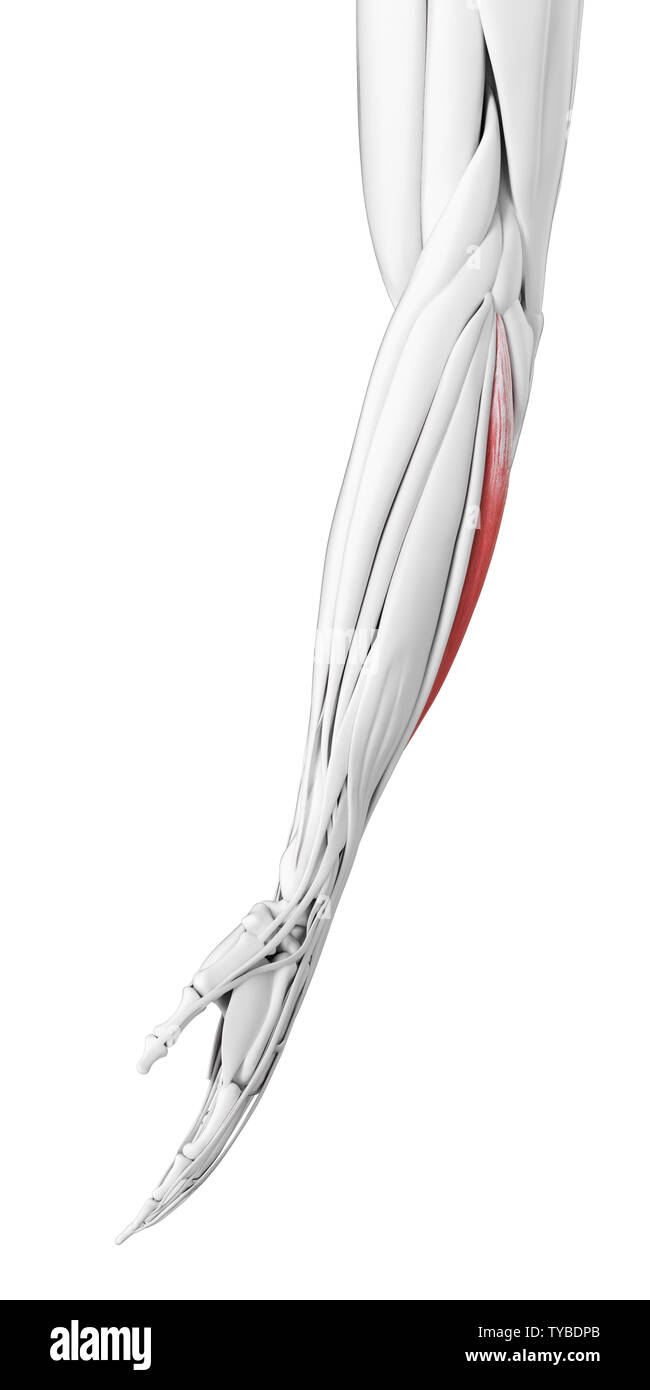 3d rendered medically accurate illustration of the extensor carpi ulnaris Stock Photohttps://www.alamy.com/image-license-details/?v=1https://www.alamy.com/3d-rendered-medically-accurate-illustration-of-the-extensor-carpi-ulnaris-image257793155.html
RFTYBDPB–3d rendered medically accurate illustration of the extensor carpi ulnaris
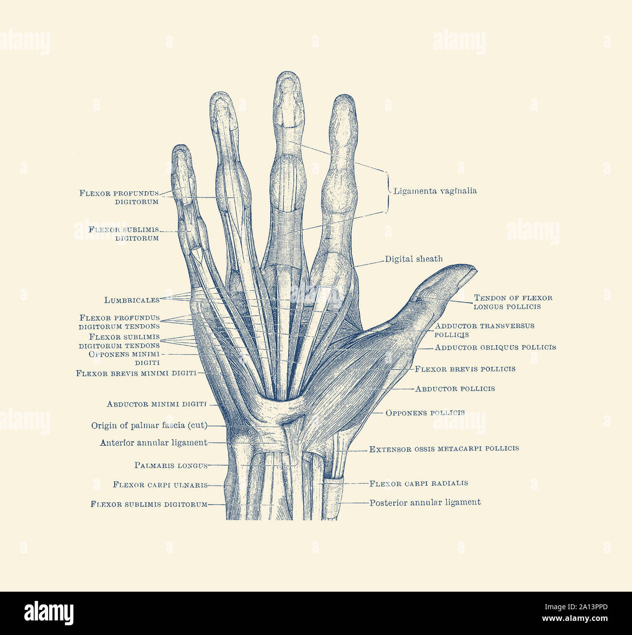 Diagram depicting the bones, ligaments and muscles throughout the hand and fingers. Stock Photohttps://www.alamy.com/image-license-details/?v=1https://www.alamy.com/diagram-depicting-the-bones-ligaments-and-muscles-throughout-the-hand-and-fingers-image327695381.html
RF2A13PPD–Diagram depicting the bones, ligaments and muscles throughout the hand and fingers.
 Flexor Carpi Ulnaris muscle Dog muscle Anatomy For Medical Concept 3D Illustration Stock Photohttps://www.alamy.com/image-license-details/?v=1https://www.alamy.com/flexor-carpi-ulnaris-muscle-dog-muscle-anatomy-for-medical-concept-3d-illustration-image441991361.html
RF2GK2CDN–Flexor Carpi Ulnaris muscle Dog muscle Anatomy For Medical Concept 3D Illustration
 Engravings of the arteries : illustrating the second volume of the Anatomy of the human body, by J Bell ; and serving as an introduction to the surgery of the arteries . .N. 33 EXPLANATION PLATE VI. Read the Text from page 340 to page 402. OF THE ARTERIES OF THE ARM. Fig. I. a. The Scapula. b. The Pectoral Muscle held up.c The Deltoid Muscle. d. The Biceps Muscle. E. The CORACO-BRACHIALIS MUSCLE. f. The Triceps extensor Muscle. g. The Teres Major. h. The Tendon of the lesser Pectoral Mus-cle. i. The Supinator longus. k. The Extensor Carpi Radialis. l. The Flexor Carpi Ulnaris. m. The Palmaris Stock Photohttps://www.alamy.com/image-license-details/?v=1https://www.alamy.com/engravings-of-the-arteries-illustrating-the-second-volume-of-the-anatomy-of-the-human-body-by-j-bell-and-serving-as-an-introduction-to-the-surgery-of-the-arteries-n-33-explanation-plate-vi-read-the-text-from-page-340-to-page-402-of-the-arteries-of-the-arm-fig-i-a-the-scapula-b-the-pectoral-muscle-held-upc-the-deltoid-muscle-d-the-biceps-muscle-e-the-coraco-brachialis-muscle-f-the-triceps-extensor-muscle-g-the-teres-major-h-the-tendon-of-the-lesser-pectoral-mus-cle-i-the-supinator-longus-k-the-extensor-carpi-radialis-l-the-flexor-carpi-ulnaris-m-the-palmaris-image338293360.html
RM2AJAGHM–Engravings of the arteries : illustrating the second volume of the Anatomy of the human body, by J Bell ; and serving as an introduction to the surgery of the arteries . .N. 33 EXPLANATION PLATE VI. Read the Text from page 340 to page 402. OF THE ARTERIES OF THE ARM. Fig. I. a. The Scapula. b. The Pectoral Muscle held up.c The Deltoid Muscle. d. The Biceps Muscle. E. The CORACO-BRACHIALIS MUSCLE. f. The Triceps extensor Muscle. g. The Teres Major. h. The Tendon of the lesser Pectoral Mus-cle. i. The Supinator longus. k. The Extensor Carpi Radialis. l. The Flexor Carpi Ulnaris. m. The Palmaris
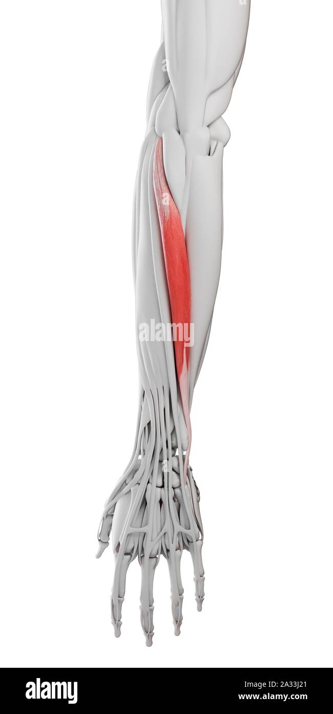 Extensor carpi ulnaris muscle, illustration Stock Photohttps://www.alamy.com/image-license-details/?v=1https://www.alamy.com/extensor-carpi-ulnaris-muscle-illustration-image328920985.html
RF2A33J21–Extensor carpi ulnaris muscle, illustration
 3d rendered medically accurate illustration of the Extensor Carpi Ulnaris Stock Photohttps://www.alamy.com/image-license-details/?v=1https://www.alamy.com/3d-rendered-medically-accurate-illustration-of-the-extensor-carpi-ulnaris-image257881792.html
RFTYFET0–3d rendered medically accurate illustration of the Extensor Carpi Ulnaris
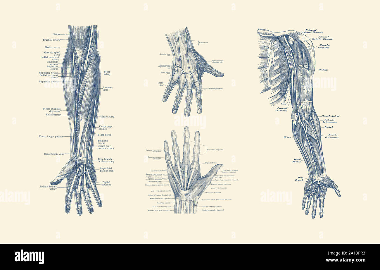 Multi-view diagram showcasing ligaments, muscles and veins throughout hand, arm and fingers. Stock Photohttps://www.alamy.com/image-license-details/?v=1https://www.alamy.com/multi-view-diagram-showcasing-ligaments-muscles-and-veins-throughout-hand-arm-and-fingers-image327695399.html
RF2A13PR3–Multi-view diagram showcasing ligaments, muscles and veins throughout hand, arm and fingers.
 Flexor Carpi Ulnaris muscle Dog muscle Anatomy For Medical Concept 3D Illustration Stock Photohttps://www.alamy.com/image-license-details/?v=1https://www.alamy.com/flexor-carpi-ulnaris-muscle-dog-muscle-anatomy-for-medical-concept-3d-illustration-image441991360.html
RF2GK2CDM–Flexor Carpi Ulnaris muscle Dog muscle Anatomy For Medical Concept 3D Illustration
 The electro-therapeutic guide, or, A thousand questions asked and answered . rve. 7 Biceps muscle. 8 Musculo-spiral nerve. 9 Brachialis internus. 10 Triceps muscle. 11 Median nerve. 12 Supinator longus.1,3 Exten. carpi ulnaris. 14 Supinator brevis. 15 Exten. digit, communis. 16 Exten. digit, minimi. 17 Extensor indicis. (a). 18 Extensor indicis. (b). 19 Exten. oss metacar. poll. 20 Exten. prim inter, poll. 21 Exten. sec. inter, poll. 22 Rectus abdominus. 23 Tensor vaginae femoris 24 Crural nerves. 25 Quadriceps, (common point). 26 Rectus femoris muscle. 27 Vastus externus muscle. 28 Peroneal n Stock Photohttps://www.alamy.com/image-license-details/?v=1https://www.alamy.com/the-electro-therapeutic-guide-or-a-thousand-questions-asked-and-answered-rve-7-biceps-muscle-8-musculo-spiral-nerve-9-brachialis-internus-10-triceps-muscle-11-median-nerve-12-supinator-longus13-exten-carpi-ulnaris-14-supinator-brevis-15-exten-digit-communis-16-exten-digit-minimi-17-extensor-indicis-a-18-extensor-indicis-b-19-exten-oss-metacar-poll-20-exten-prim-inter-poll-21-exten-sec-inter-poll-22-rectus-abdominus-23-tensor-vaginae-femoris-24-crural-nerves-25-quadriceps-common-point-26-rectus-femoris-muscle-27-vastus-externus-muscle-28-peroneal-n-image340286733.html
RM2ANHB5H–The electro-therapeutic guide, or, A thousand questions asked and answered . rve. 7 Biceps muscle. 8 Musculo-spiral nerve. 9 Brachialis internus. 10 Triceps muscle. 11 Median nerve. 12 Supinator longus.1,3 Exten. carpi ulnaris. 14 Supinator brevis. 15 Exten. digit, communis. 16 Exten. digit, minimi. 17 Extensor indicis. (a). 18 Extensor indicis. (b). 19 Exten. oss metacar. poll. 20 Exten. prim inter, poll. 21 Exten. sec. inter, poll. 22 Rectus abdominus. 23 Tensor vaginae femoris 24 Crural nerves. 25 Quadriceps, (common point). 26 Rectus femoris muscle. 27 Vastus externus muscle. 28 Peroneal n
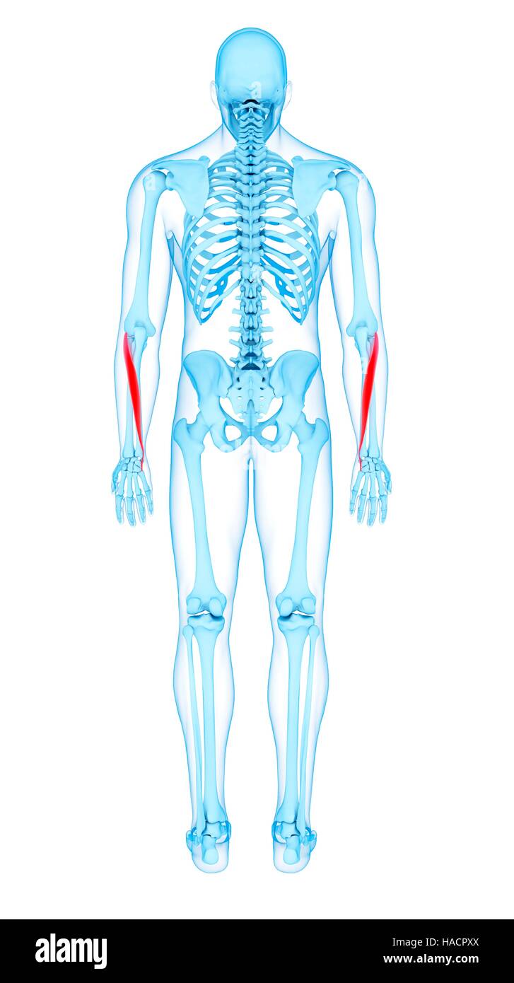 Illustration of the extensor carpi ulnaris muscles. Stock Photohttps://www.alamy.com/image-license-details/?v=1https://www.alamy.com/stock-photo-illustration-of-the-extensor-carpi-ulnaris-muscles-126900562.html
RFHACPXX–Illustration of the extensor carpi ulnaris muscles.
 3d rendered medically accurate illustration of the Extensor Carpi Ulnaris Stock Photohttps://www.alamy.com/image-license-details/?v=1https://www.alamy.com/3d-rendered-medically-accurate-illustration-of-the-extensor-carpi-ulnaris-image257881875.html
RFTYFEXY–3d rendered medically accurate illustration of the Extensor Carpi Ulnaris
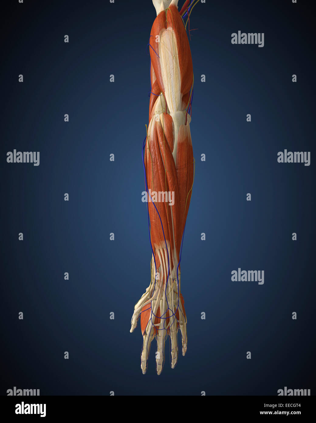 Human arm with bone, muscles and nerves. Stock Photohttps://www.alamy.com/image-license-details/?v=1https://www.alamy.com/stock-photo-human-arm-with-bone-muscles-and-nerves-77723300.html
RFEECGT4–Human arm with bone, muscles and nerves.
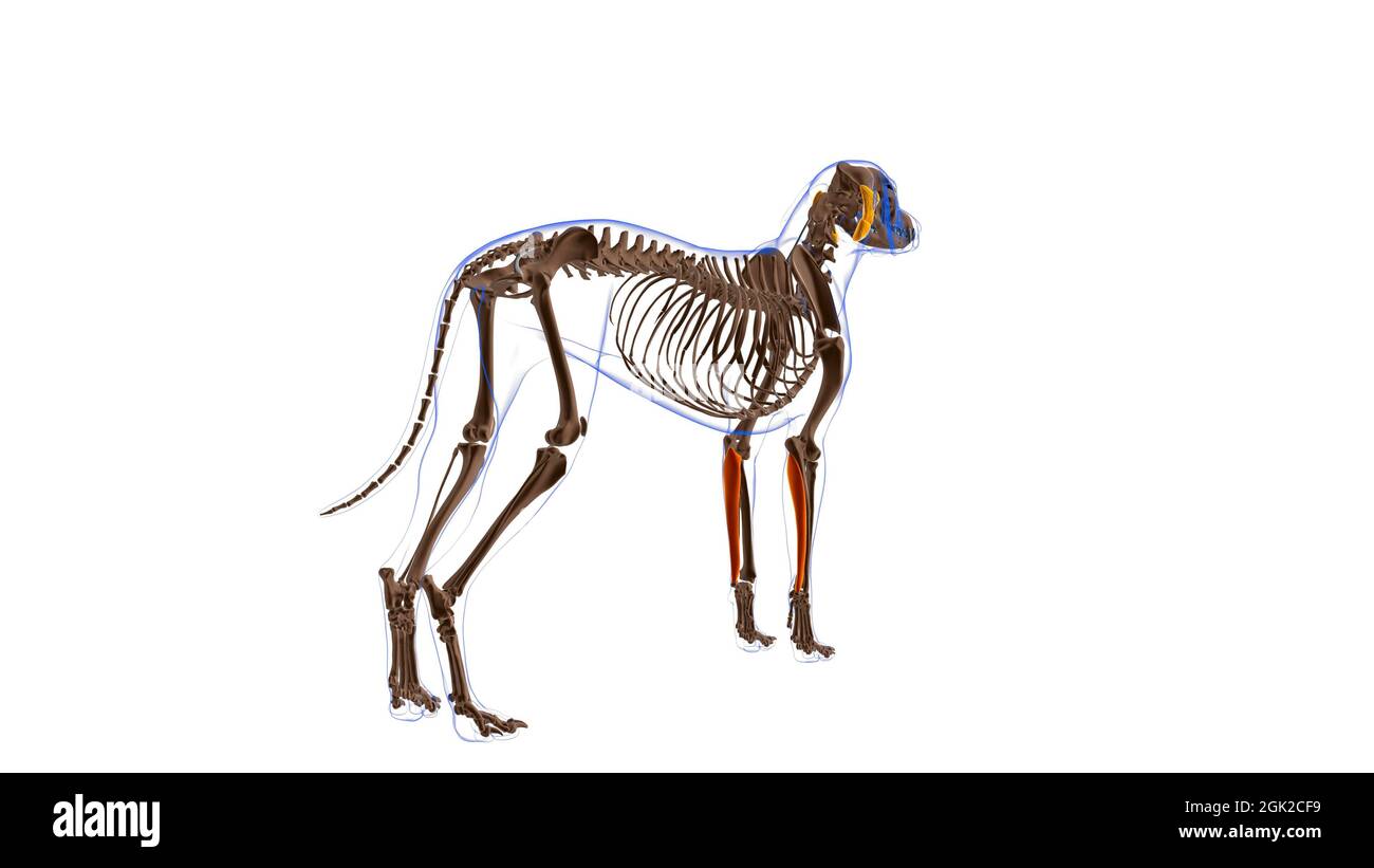 Flexor Carpi Ulnaris muscle Dog muscle Anatomy For Medical Concept 3D Illustration Stock Photohttps://www.alamy.com/image-license-details/?v=1https://www.alamy.com/flexor-carpi-ulnaris-muscle-dog-muscle-anatomy-for-medical-concept-3d-illustration-image441991405.html
RF2GK2CF9–Flexor Carpi Ulnaris muscle Dog muscle Anatomy For Medical Concept 3D Illustration