Quick filters:
Fibular notch Stock Photos and Images
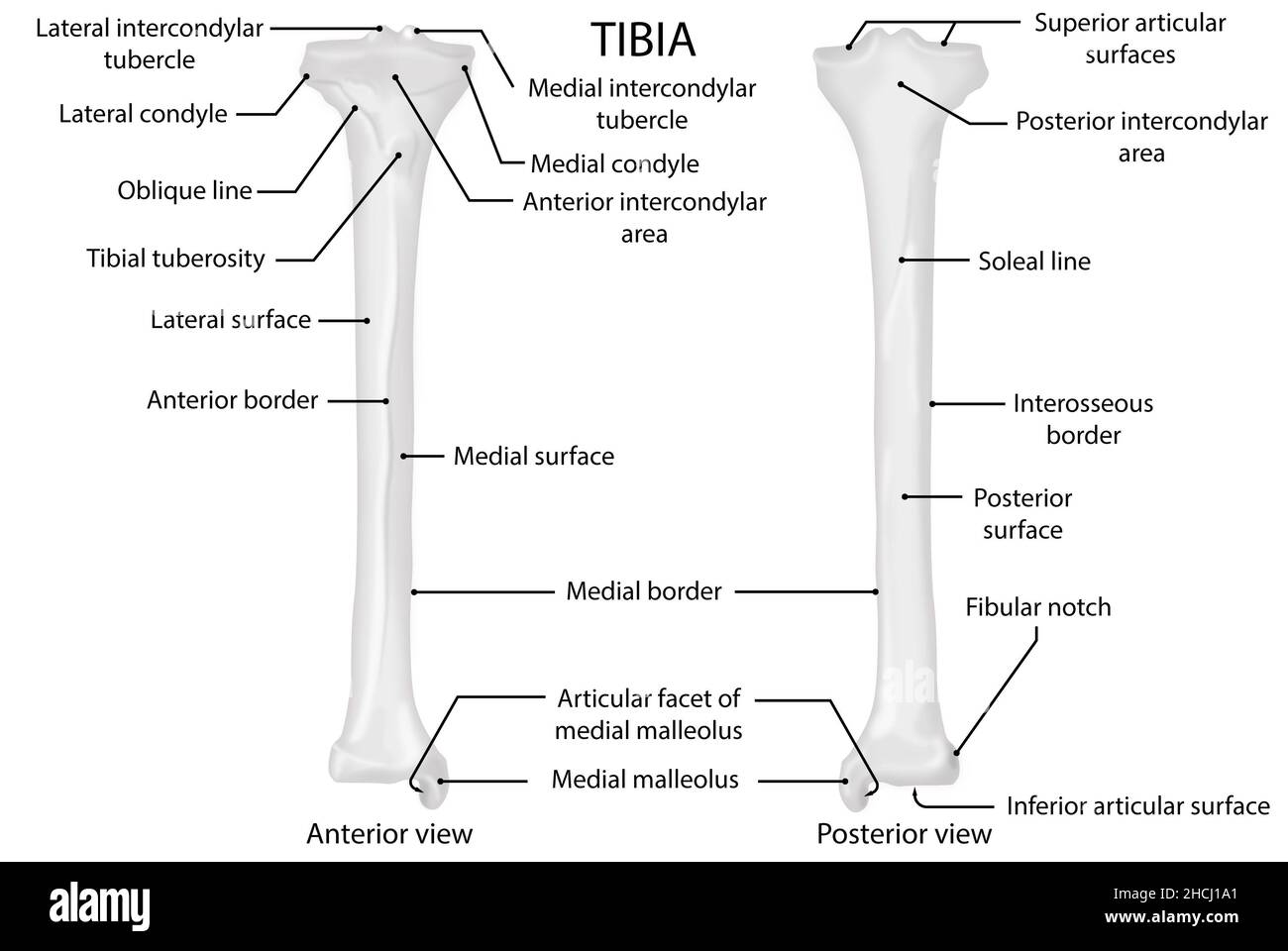 Tibia, anterior and posterior view, human anatomy Stock Photohttps://www.alamy.com/image-license-details/?v=1https://www.alamy.com/tibia-anterior-and-posterior-view-human-anatomy-image455241641.html
RF2HCJ1A1–Tibia, anterior and posterior view, human anatomy
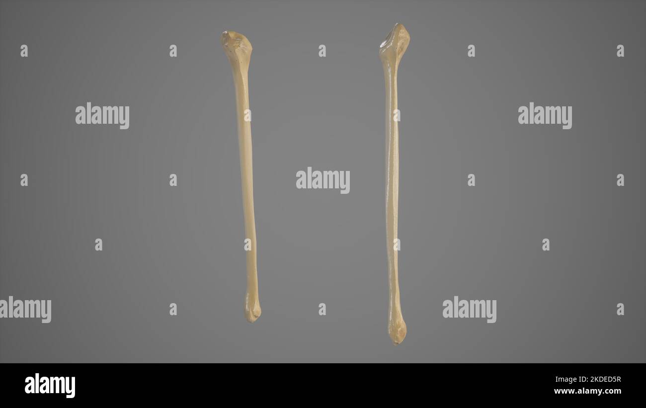 Anterior and Posterior View of Fibula Stock Photohttps://www.alamy.com/image-license-details/?v=1https://www.alamy.com/anterior-and-posterior-view-of-fibula-image490198515.html
RF2KDED5R–Anterior and Posterior View of Fibula
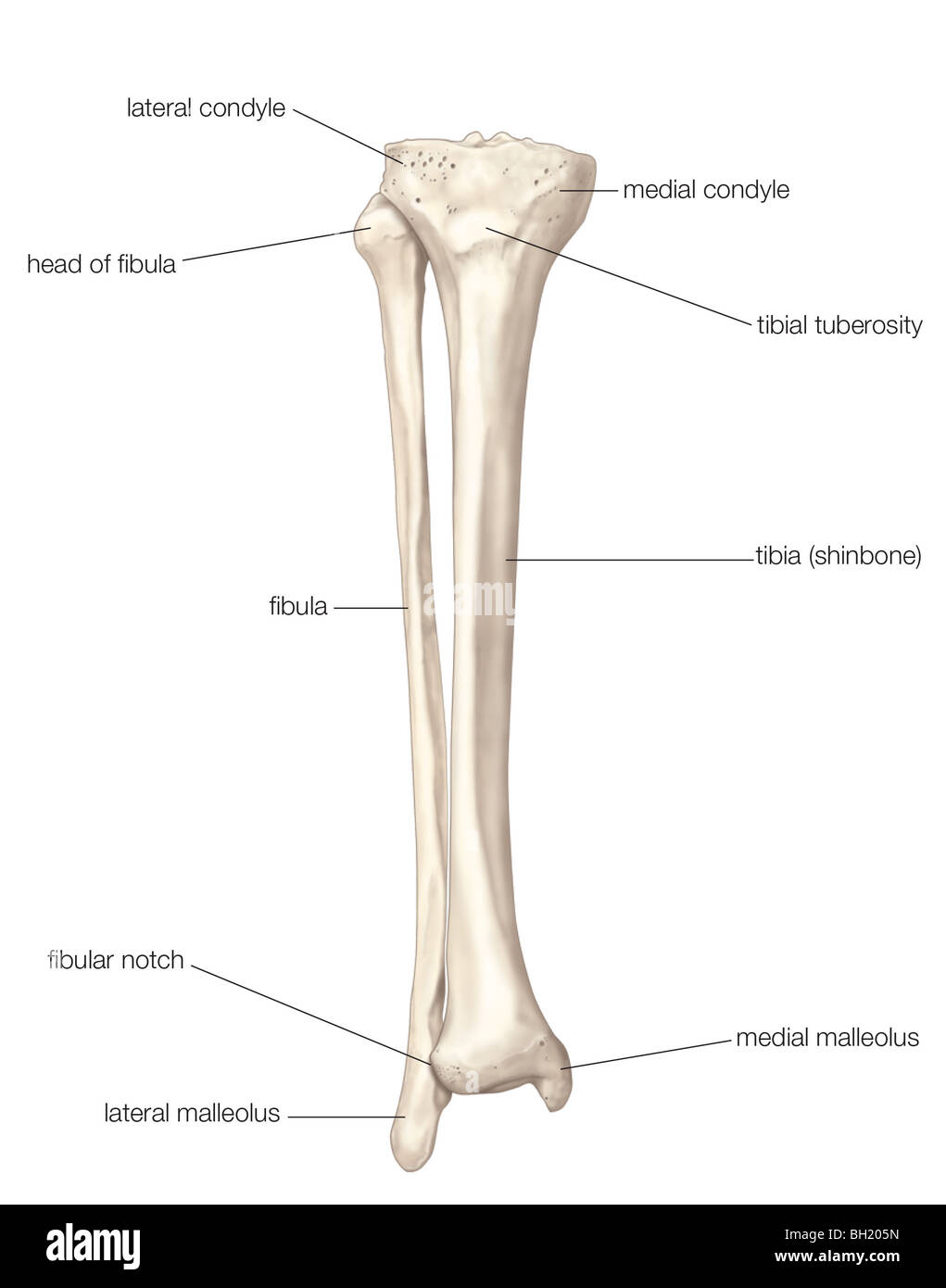 Fibula and tibia Stock Photohttps://www.alamy.com/image-license-details/?v=1https://www.alamy.com/stock-photo-fibula-and-tibia-27703585.html
RMBH205N–Fibula and tibia
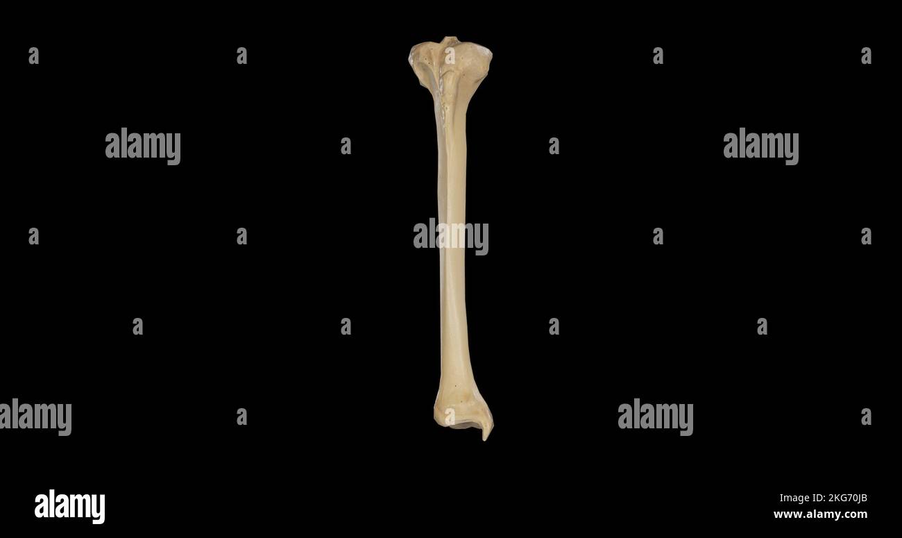 Anterior view of Right Tibia Stock Photohttps://www.alamy.com/image-license-details/?v=1https://www.alamy.com/anterior-view-of-right-tibia-image491878979.html
RF2KG70JB–Anterior view of Right Tibia
 . Atlas and text-book of human anatomy. Anatomy -- Atlases. Shaft -^ t. Fibular * notch Inferior articular surface F/o. 144. Infernal malleolus. Please note that these images are extracted from scanned page images that may have been digitally enhanced for readability - coloration and appearance of these illustrations may not perfectly resemble the original work.. Sobotta, Johannes, 1869-1945; McMurrich, J. Playfair (James Playfair), 1859-1939. Philadelphia, Saunders Stock Photohttps://www.alamy.com/image-license-details/?v=1https://www.alamy.com/atlas-and-text-book-of-human-anatomy-anatomy-atlases-shaft-t-fibular-notch-inferior-articular-surface-fo-144-infernal-malleolus-please-note-that-these-images-are-extracted-from-scanned-page-images-that-may-have-been-digitally-enhanced-for-readability-coloration-and-appearance-of-these-illustrations-may-not-perfectly-resemble-the-original-work-sobotta-johannes-1869-1945-mcmurrich-j-playfair-james-playfair-1859-1939-philadelphia-saunders-image235396847.html
RMRJY727–. Atlas and text-book of human anatomy. Anatomy -- Atlases. Shaft -^ t. Fibular * notch Inferior articular surface F/o. 144. Infernal malleolus. Please note that these images are extracted from scanned page images that may have been digitally enhanced for readability - coloration and appearance of these illustrations may not perfectly resemble the original work.. Sobotta, Johannes, 1869-1945; McMurrich, J. Playfair (James Playfair), 1859-1939. Philadelphia, Saunders
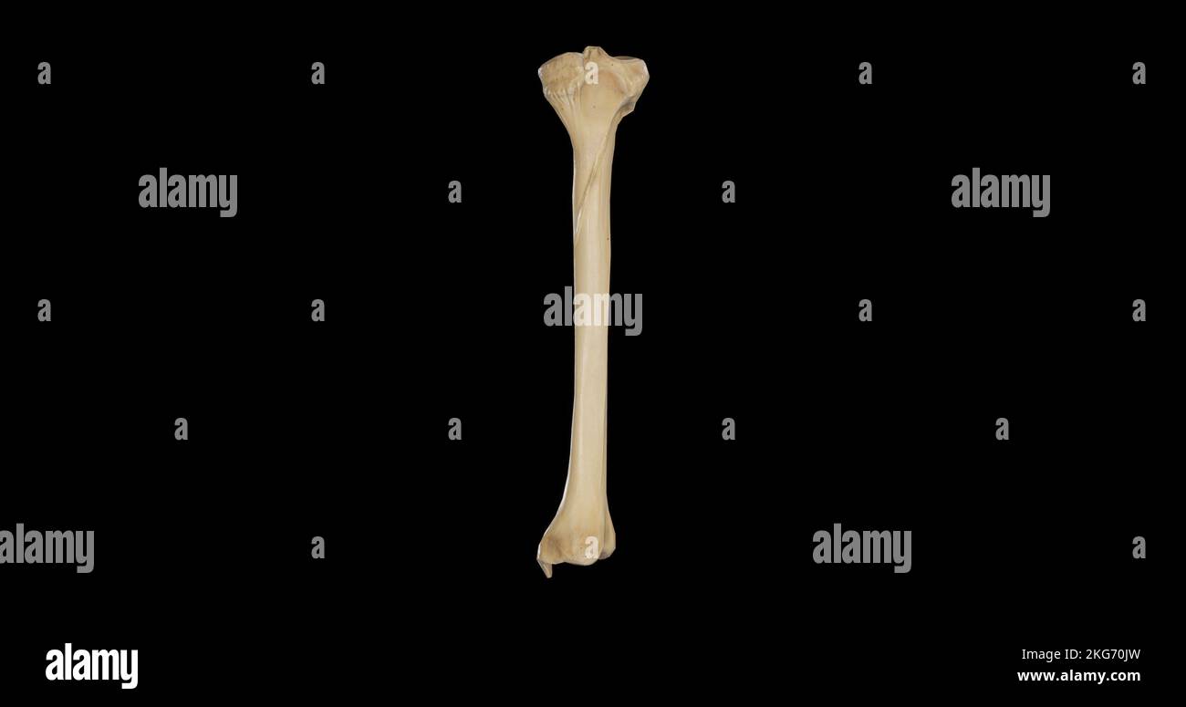 Posterior view of Right Tibia Stock Photohttps://www.alamy.com/image-license-details/?v=1https://www.alamy.com/posterior-view-of-right-tibia-image491878993.html
RF2KG70JW–Posterior view of Right Tibia
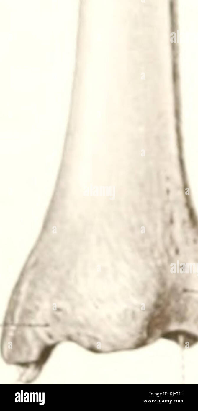 . Atlas and text-book of human anatomy. Anatomy -- Atlases. Fibular * notch Inferior articular surface F/o. 144. Infernal malleolus. Groove on internal malleolus Articular surface of malleolus Fisf. 145 surface Interosseous ridge ^ Nutrient foramen Posterior surface Interosseous ridge External surface Fibular notch Inferior artiailar surface Fig. 140. Inferior extremity A** Inferior articular surface External surface Articular surface of malleolus. Please note that these images are extracted from scanned page images that may have been digitally enhanced for readability - coloration and appeara Stock Photohttps://www.alamy.com/image-license-details/?v=1https://www.alamy.com/atlas-and-text-book-of-human-anatomy-anatomy-atlases-fibular-notch-inferior-articular-surface-fo-144-infernal-malleolus-groove-on-internal-malleolus-articular-surface-of-malleolus-fisf-145-surface-interosseous-ridge-nutrient-foramen-posterior-surface-interosseous-ridge-external-surface-fibular-notch-inferior-artiailar-surface-fig-140-inferior-extremity-a-inferior-articular-surface-external-surface-articular-surface-of-malleolus-please-note-that-these-images-are-extracted-from-scanned-page-images-that-may-have-been-digitally-enhanced-for-readability-coloration-and-appeara-image235396813.html
RMRJY711–. Atlas and text-book of human anatomy. Anatomy -- Atlases. Fibular * notch Inferior articular surface F/o. 144. Infernal malleolus. Groove on internal malleolus Articular surface of malleolus Fisf. 145 surface Interosseous ridge ^ Nutrient foramen Posterior surface Interosseous ridge External surface Fibular notch Inferior artiailar surface Fig. 140. Inferior extremity A** Inferior articular surface External surface Articular surface of malleolus. Please note that these images are extracted from scanned page images that may have been digitally enhanced for readability - coloration and appeara
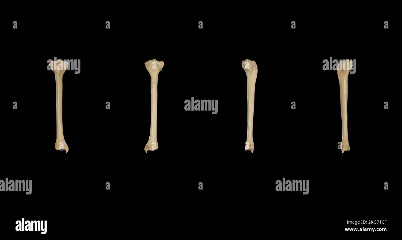 Right Tibia-Multiple Views Stock Photohttps://www.alamy.com/image-license-details/?v=1https://www.alamy.com/right-tibia-multiple-views-image491879599.html
RF2KG71CF–Right Tibia-Multiple Views
 Nervous and mental diseases . are the following : The gluteal point over the sciatic notch, thetrochanteric point above the trochanter major, a tract correspondingto the nerve-trunk on the posterior aspect of the thigh, a popliteal pointin the ham at the division of the nerve, a fibular point where the exter-nal popliteal is superficial to the neck of the fibula, and a point on thedorsum of the foot. Less frequently we find lumbar points just abovethe sacrum, an iliac point at the middle of the iliac crest, a patellar pointover the knee-cap, points in the calf, points behind the malleoli, andp Stock Photohttps://www.alamy.com/image-license-details/?v=1https://www.alamy.com/nervous-and-mental-diseases-are-the-following-the-gluteal-point-over-the-sciatic-notch-thetrochanteric-point-above-the-trochanter-major-a-tract-correspondingto-the-nerve-trunk-on-the-posterior-aspect-of-the-thigh-a-popliteal-pointin-the-ham-at-the-division-of-the-nerve-a-fibular-point-where-the-exter-nal-popliteal-is-superficial-to-the-neck-of-the-fibula-and-a-point-on-thedorsum-of-the-foot-less-frequently-we-find-lumbar-points-just-abovethe-sacrum-an-iliac-point-at-the-middle-of-the-iliac-crest-a-patellar-pointover-the-knee-cap-points-in-the-calf-points-behind-the-malleoli-andp-image342951417.html
RM2AWXP0W–Nervous and mental diseases . are the following : The gluteal point over the sciatic notch, thetrochanteric point above the trochanter major, a tract correspondingto the nerve-trunk on the posterior aspect of the thigh, a popliteal pointin the ham at the division of the nerve, a fibular point where the exter-nal popliteal is superficial to the neck of the fibula, and a point on thedorsum of the foot. Less frequently we find lumbar points just abovethe sacrum, an iliac point at the middle of the iliac crest, a patellar pointover the knee-cap, points in the calf, points behind the malleoli, andp
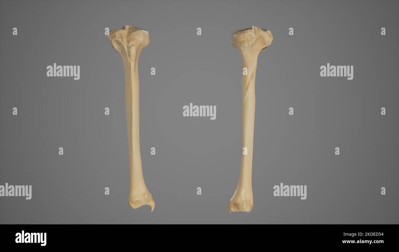 Anterior and Posterior View of Tibia Stock Photohttps://www.alamy.com/image-license-details/?v=1https://www.alamy.com/anterior-and-posterior-view-of-tibia-image490198496.html
RF2KDED54–Anterior and Posterior View of Tibia
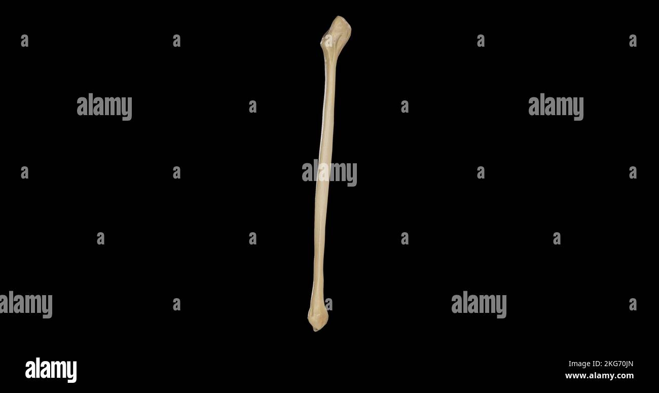 Posterior view of Right Fibula Stock Photohttps://www.alamy.com/image-license-details/?v=1https://www.alamy.com/posterior-view-of-right-fibula-image491878989.html
RF2KG70JN–Posterior view of Right Fibula
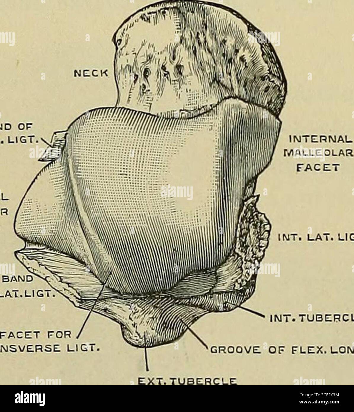 . Quain's Elements of anatomy. e outer marginis convex, and inclined inwards pos-teriorly, thus making the surfacenarrower behind than iu front. Be-tween the upper and the external sur-faces posteriorly is a narrow trian-gular facet which plays againstthe transverse tibio-fibular ligament.The capsule of the articulation isdivided into the following liga-ments :— The internal lateral or deltoidligament (fig. 221, 1) is a broadlayer of fibres, which radiate fromthe internal malleolus to the tarsalbones. The hinder part is thickand short, and descends from the notch at the lower border of the mal Stock Photohttps://www.alamy.com/image-license-details/?v=1https://www.alamy.com/quains-elements-of-anatomy-e-outer-marginis-convex-and-inclined-inwards-pos-teriorly-thus-making-the-surfacenarrower-behind-than-iu-front-be-tween-the-upper-and-the-external-sur-faces-posteriorly-is-a-narrow-trian-gular-facet-which-plays-againstthe-transverse-tibio-fibular-ligamentthe-capsule-of-the-articulation-isdivided-into-the-following-liga-ments-the-internal-lateral-or-deltoidligament-fig-221-1-is-a-broadlayer-of-fibres-which-radiate-fromthe-internal-malleolus-to-the-tarsalbones-the-hinder-part-is-thickand-short-and-descends-from-the-notch-at-the-lower-border-of-the-mal-image370702744.html
RM2CF2Y3M–. Quain's Elements of anatomy. e outer marginis convex, and inclined inwards pos-teriorly, thus making the surfacenarrower behind than iu front. Be-tween the upper and the external sur-faces posteriorly is a narrow trian-gular facet which plays againstthe transverse tibio-fibular ligament.The capsule of the articulation isdivided into the following liga-ments :— The internal lateral or deltoidligament (fig. 221, 1) is a broadlayer of fibres, which radiate fromthe internal malleolus to the tarsalbones. The hinder part is thickand short, and descends from the notch at the lower border of the mal
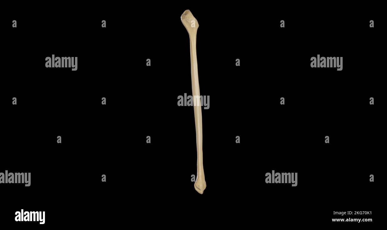 Anterior view of Right Fibula Stock Photohttps://www.alamy.com/image-license-details/?v=1https://www.alamy.com/anterior-view-of-right-fibula-image491878997.html
RF2KG70K1–Anterior view of Right Fibula
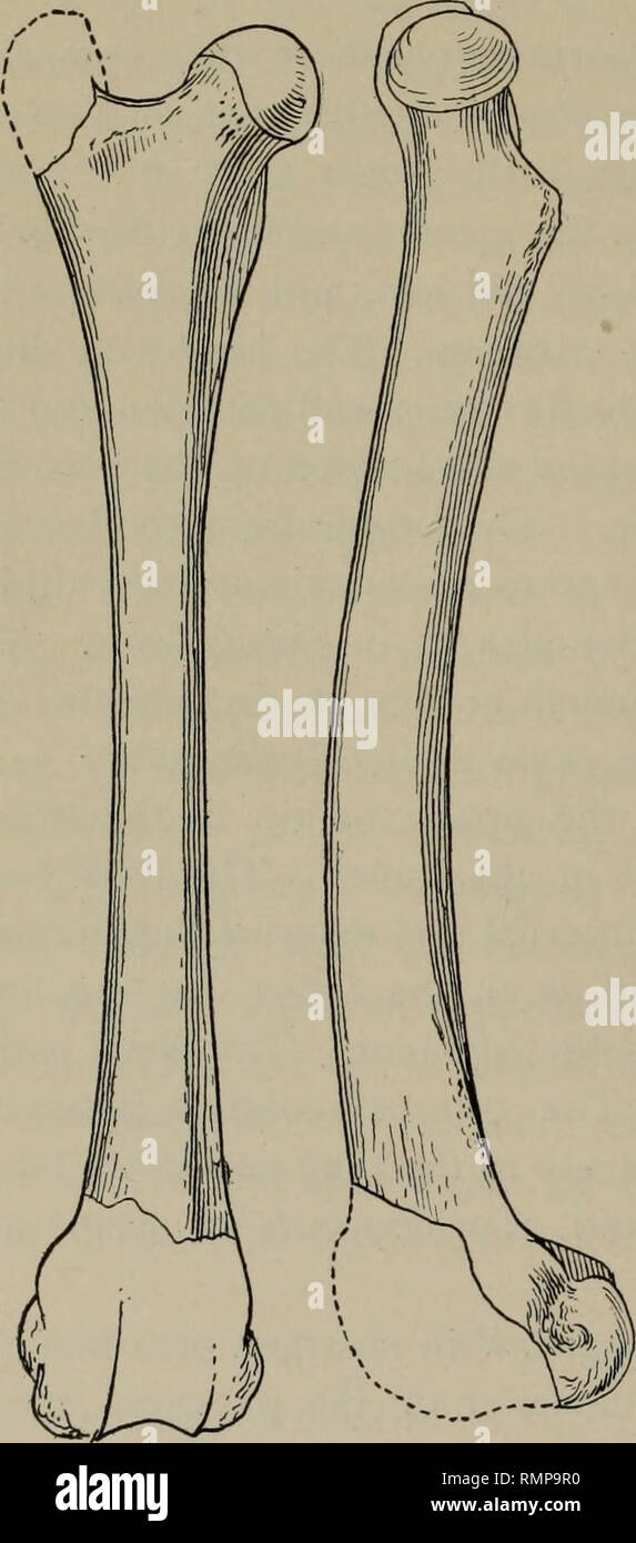 . Annals of the Carnegie Museum. Carnegie Museum; Carnegie Museum of Natural History; Natural history. 296 Annals of the Carnegie Museum. than in Oxydactylus, and the fossa below the popliteal notch is con- sequently very much shallower and more nearly like that found in the llama and the camel. The external face has a deeper fossa, which is. Fig. 9. Anterior and tibial views of femur of Stenomylus gracilis. Type No. 1610. 'I/, Fig. 10. Fibular view of tibia of Stenomylus gracilis. Type No. 1610. |nat. size. bounded above by the overhanging border of the head, posteriorly by the external borde Stock Photohttps://www.alamy.com/image-license-details/?v=1https://www.alamy.com/annals-of-the-carnegie-museum-carnegie-museum-carnegie-museum-of-natural-history-natural-history-296-annals-of-the-carnegie-museum-than-in-oxydactylus-and-the-fossa-below-the-popliteal-notch-is-con-sequently-very-much-shallower-and-more-nearly-like-that-found-in-the-llama-and-the-camel-the-external-face-has-a-deeper-fossa-which-is-fig-9-anterior-and-tibial-views-of-femur-of-stenomylus-gracilis-type-no-1610-i-fig-10-fibular-view-of-tibia-of-stenomylus-gracilis-type-no-1610-nat-size-bounded-above-by-the-overhanging-border-of-the-head-posteriorly-by-the-external-borde-image236518548.html
RMRMP9R0–. Annals of the Carnegie Museum. Carnegie Museum; Carnegie Museum of Natural History; Natural history. 296 Annals of the Carnegie Museum. than in Oxydactylus, and the fossa below the popliteal notch is con- sequently very much shallower and more nearly like that found in the llama and the camel. The external face has a deeper fossa, which is. Fig. 9. Anterior and tibial views of femur of Stenomylus gracilis. Type No. 1610. 'I/, Fig. 10. Fibular view of tibia of Stenomylus gracilis. Type No. 1610. |nat. size. bounded above by the overhanging border of the head, posteriorly by the external borde
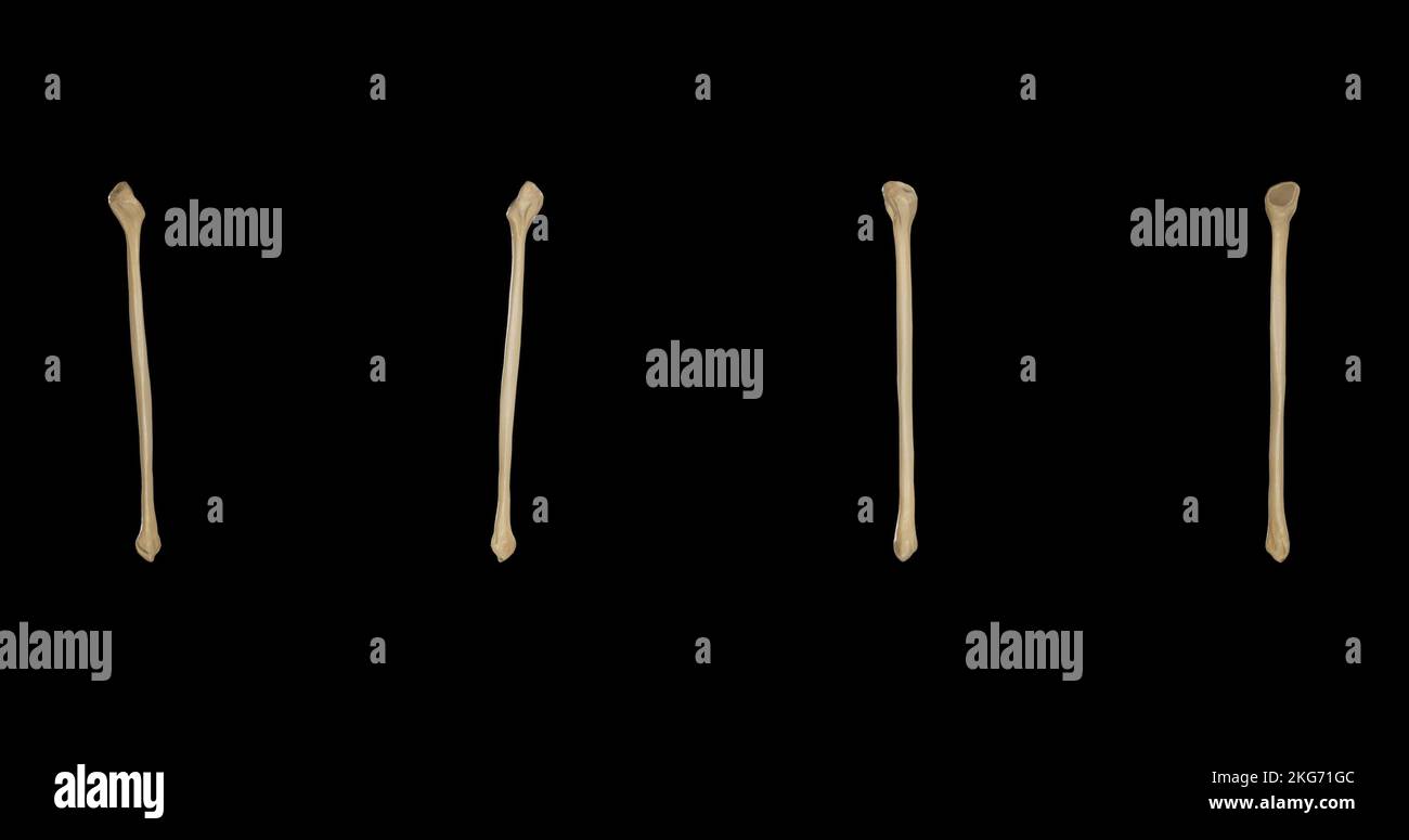 Right Fibula-Multiple Views Stock Photohttps://www.alamy.com/image-license-details/?v=1https://www.alamy.com/right-fibula-multiple-views-image491879708.html
RF2KG71GC–Right Fibula-Multiple Views
 . Anatomy in a nutshell : a treatise on human anatomy in its relation to osteopathy. Human anatomy; Osteopathic medicine; Osteopathic Medicine; Anatomy. PLATE L. POPLITEAL NOTCH. EXTERNAL FIBRO-CARTILAGE. POST. CRUCIAL LIGAMENT. CAPSULE. STYLOID PROCESS. POST. TIBIO-FIBULAR LIGAMENT , SOLEUS (0) ' Plexor longus hallucis <o). flexor surface of fibula NUTRIENT FORAMEN 'ERONEUS BREVIS (0).. INT. FIBRO CARTILAGE? XAPSUL'E. SEMIMEMBRANOSUS) POPLITEUS (I). OBLIQUE LINE. SOLEUS (0). NUTRIENT FORAMEN. TIBIALIS POSTICUS (0). FLEXOR LONGUS DIGITORUM (0). POST. TIBIO-FIBULAR i IGAMENT, EXT. LATERAL L Stock Photohttps://www.alamy.com/image-license-details/?v=1https://www.alamy.com/anatomy-in-a-nutshell-a-treatise-on-human-anatomy-in-its-relation-to-osteopathy-human-anatomy-osteopathic-medicine-osteopathic-medicine-anatomy-plate-l-popliteal-notch-external-fibro-cartilage-post-crucial-ligament-capsule-styloid-process-post-tibio-fibular-ligament-soleus-0-plexor-longus-hallucis-lto-flexor-surface-of-fibula-nutrient-foramen-eroneus-brevis-0-int-fibro-cartilage-xapsule-semimembranosus-popliteus-i-oblique-line-soleus-0-nutrient-foramen-tibialis-posticus-0-flexor-longus-digitorum-0-post-tibio-fibular-i-igament-ext-lateral-l-image236816186.html
RMRN7WCX–. Anatomy in a nutshell : a treatise on human anatomy in its relation to osteopathy. Human anatomy; Osteopathic medicine; Osteopathic Medicine; Anatomy. PLATE L. POPLITEAL NOTCH. EXTERNAL FIBRO-CARTILAGE. POST. CRUCIAL LIGAMENT. CAPSULE. STYLOID PROCESS. POST. TIBIO-FIBULAR LIGAMENT , SOLEUS (0) ' Plexor longus hallucis <o). flexor surface of fibula NUTRIENT FORAMEN 'ERONEUS BREVIS (0).. INT. FIBRO CARTILAGE? XAPSUL'E. SEMIMEMBRANOSUS) POPLITEUS (I). OBLIQUE LINE. SOLEUS (0). NUTRIENT FORAMEN. TIBIALIS POSTICUS (0). FLEXOR LONGUS DIGITORUM (0). POST. TIBIO-FIBULAR i IGAMENT, EXT. LATERAL L