Quick filters:
Fluorescence microscopy Stock Photos and Images
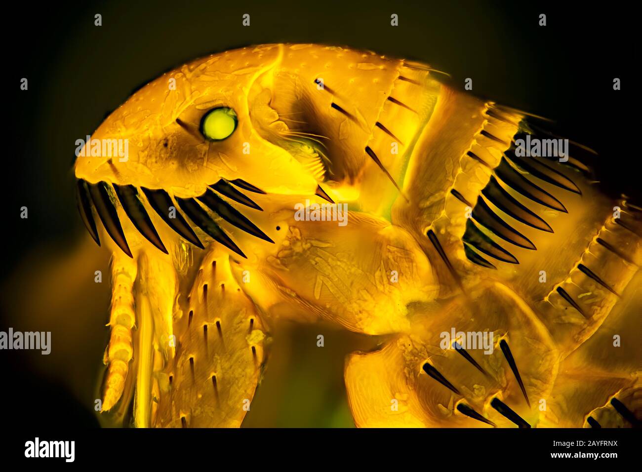 Cat flea (Ctenocephalides felis), Head of a Cat flea, fluorescence microscopy, Germany Stock Photohttps://www.alamy.com/image-license-details/?v=1https://www.alamy.com/cat-flea-ctenocephalides-felis-head-of-a-cat-flea-fluorescence-microscopy-germany-image343940630.html
RM2AYFRNX–Cat flea (Ctenocephalides felis), Head of a Cat flea, fluorescence microscopy, Germany
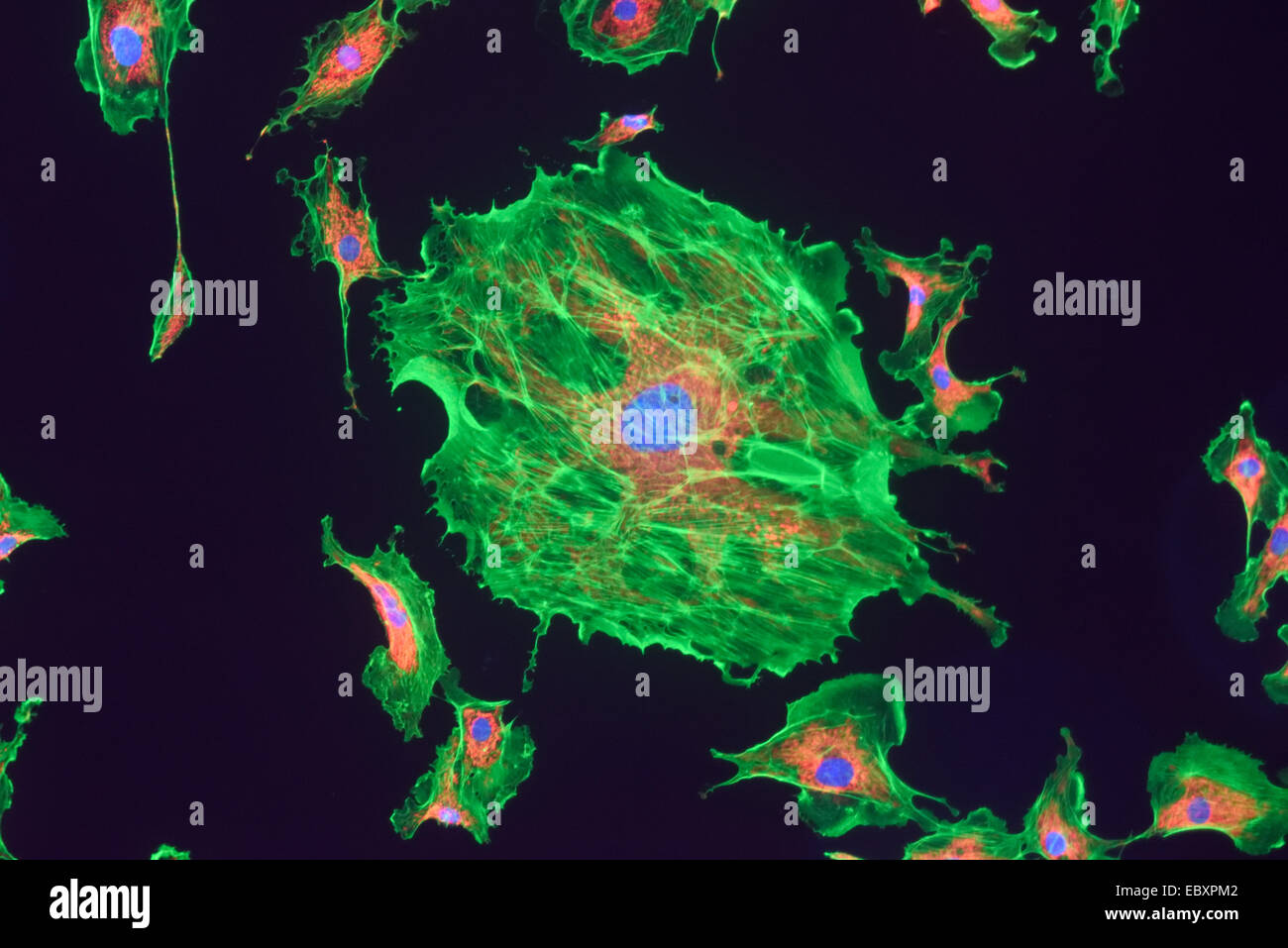 Microfilaments, mitochondria, and nuclei in fibroblast cells Stock Photohttps://www.alamy.com/image-license-details/?v=1https://www.alamy.com/stock-photo-microfilaments-mitochondria-and-nuclei-in-fibroblast-cells-76191250.html
RMEBXPM2–Microfilaments, mitochondria, and nuclei in fibroblast cells
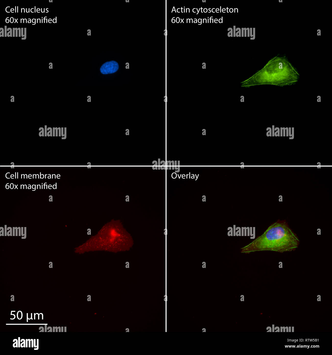 Single human osteosarcoma cell stained for epifluorescence Stock Photohttps://www.alamy.com/image-license-details/?v=1https://www.alamy.com/single-human-osteosarcoma-cell-stained-for-epifluorescence-image239039557.html
RMRTW5B1–Single human osteosarcoma cell stained for epifluorescence
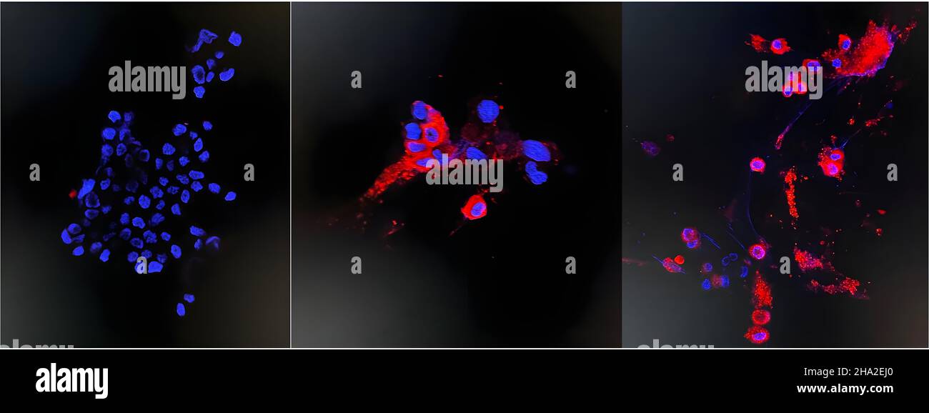 Omicron variant Stock Photohttps://www.alamy.com/image-license-details/?v=1https://www.alamy.com/omicron-variant-image453671512.html
RM2HA2EJ0–Omicron variant
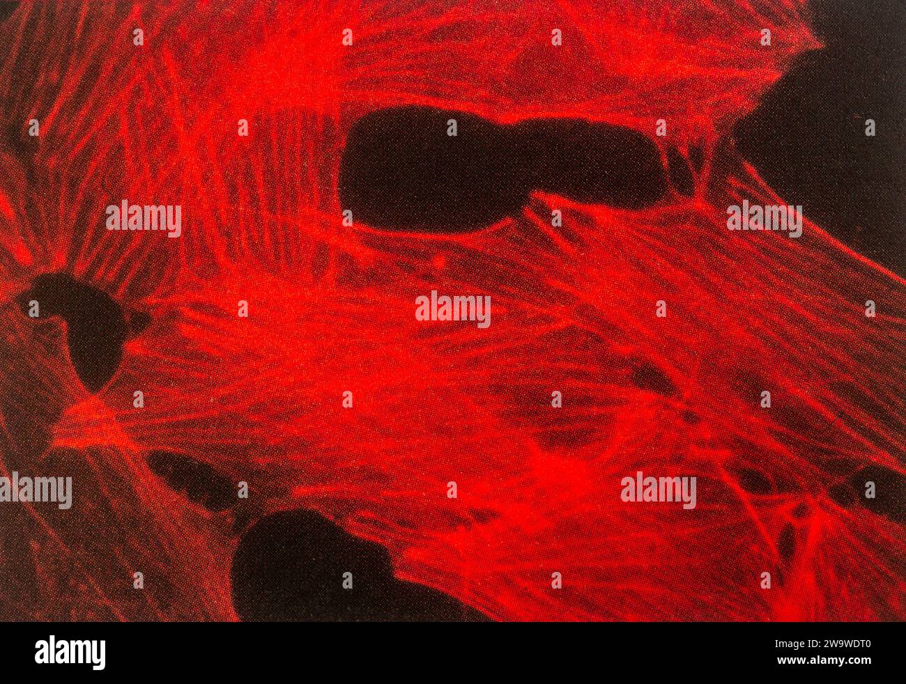 Fluoresence microscopy of cells, specifically fibroblasts, stained with fluorescently labelled phalloidin showing actin stress fibres Stock Photohttps://www.alamy.com/image-license-details/?v=1https://www.alamy.com/fluoresence-microscopy-of-cells-specifically-fibroblasts-stained-with-fluorescently-labelled-phalloidin-showing-actin-stress-fibres-image591244080.html
RM2W9WDT0–Fluoresence microscopy of cells, specifically fibroblasts, stained with fluorescently labelled phalloidin showing actin stress fibres
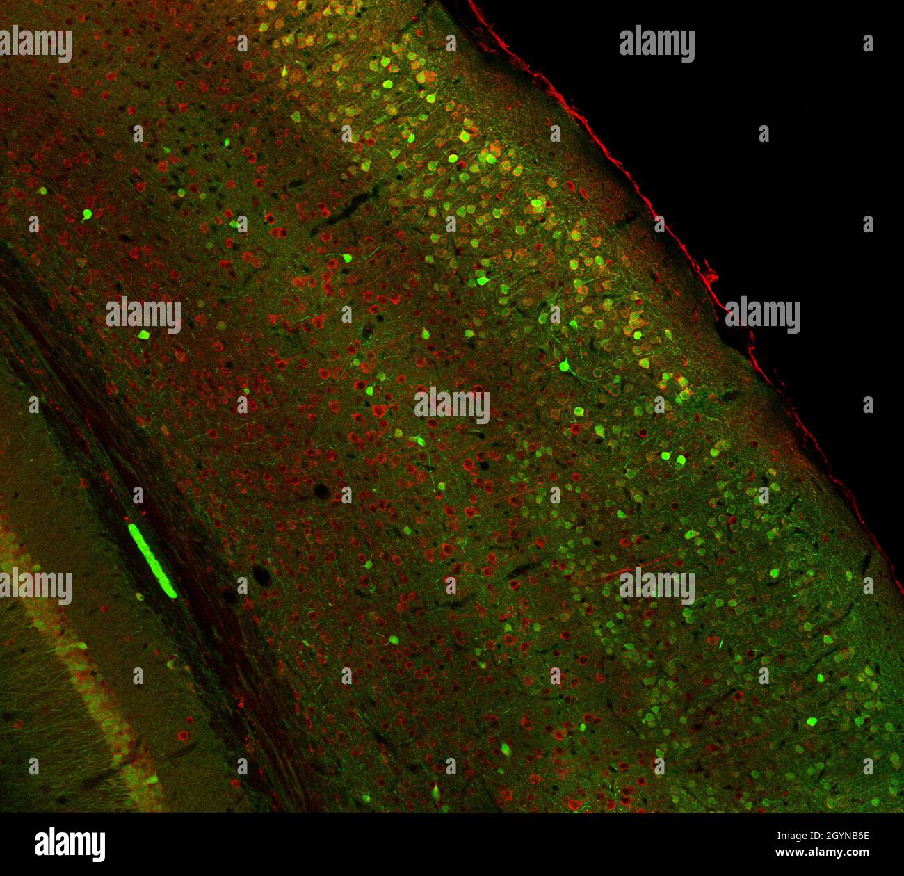 Confocal laser scanning microscopy image of immunofluorescence- labelled cells in the cerebral cortex of the mouse brain Stock Photohttps://www.alamy.com/image-license-details/?v=1https://www.alamy.com/confocal-laser-scanning-microscopy-image-of-immunofluorescence-labelled-cells-in-the-cerebral-cortex-of-the-mouse-brain-image447324710.html
RF2GYNB6E–Confocal laser scanning microscopy image of immunofluorescence- labelled cells in the cerebral cortex of the mouse brain
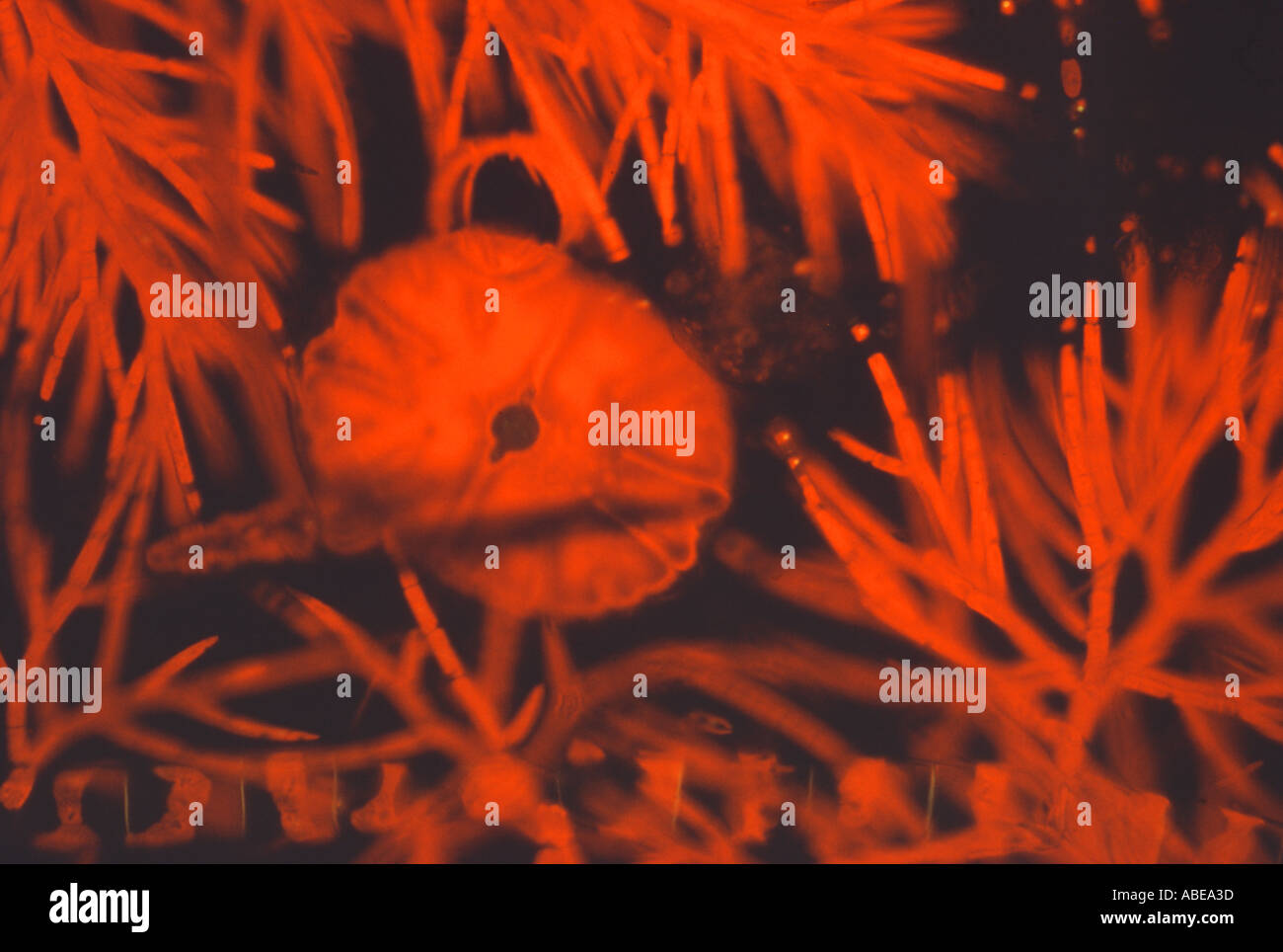 Green algae Live Blue Violet Fluorescence Stock Photohttps://www.alamy.com/image-license-details/?v=1https://www.alamy.com/green-algae-live-blue-violet-fluorescence-image780861.html
RMABEA3D–Green algae Live Blue Violet Fluorescence
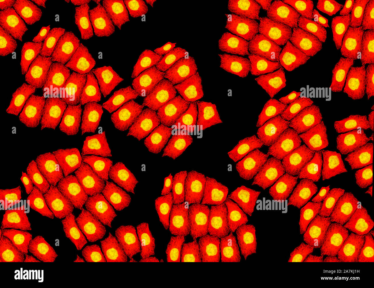 Confocal Fluorescence Microscopy of CRISPR-generated knockout cells. Stock Photohttps://www.alamy.com/image-license-details/?v=1https://www.alamy.com/confocal-fluorescence-microscopy-of-crispr-generated-knockout-cells-image331730829.html
RM2A7KJ1H–Confocal Fluorescence Microscopy of CRISPR-generated knockout cells.
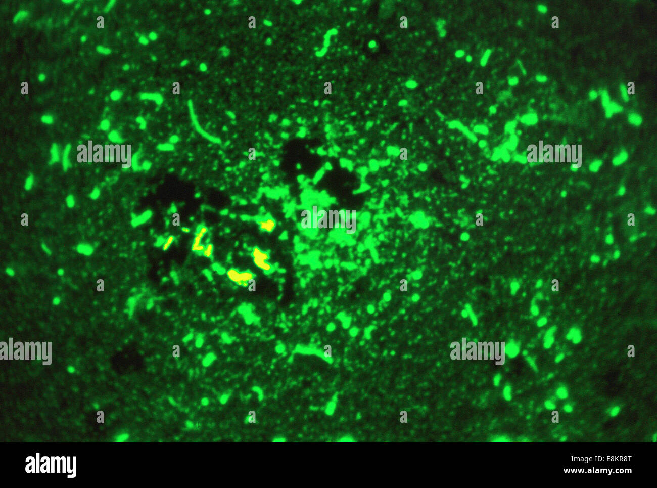 This fluorescent acid-fast stained (Smithwick) photomicrograph revealed presence of Mycobacterium tuberculosis bacteria in Stock Photohttps://www.alamy.com/image-license-details/?v=1https://www.alamy.com/stock-photo-this-fluorescent-acid-fast-stained-smithwick-photomicrograph-revealed-74194088.html
RME8KR8T–This fluorescent acid-fast stained (Smithwick) photomicrograph revealed presence of Mycobacterium tuberculosis bacteria in
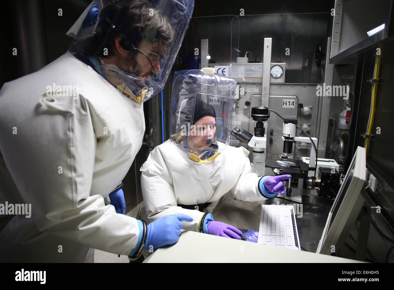 Virologists Lisa Oestereich and Toni Rieger (left) work with a fluorescence microscope in Hamburg, Germany in 2013. Eric Betzig, Stefan W Hell and William E Moerner won the Nobel Prize in Chemistry Wednesday for their work in the development of super-resolved fluorescence microscopy. DPA/CHRISTIAN CHARISIUS (ARCHIVE) Stock Photohttps://www.alamy.com/image-license-details/?v=1https://www.alamy.com/stock-photo-virologists-lisa-oestereich-and-toni-rieger-left-work-with-a-fluorescence-74132385.html
RME8H0H5–Virologists Lisa Oestereich and Toni Rieger (left) work with a fluorescence microscope in Hamburg, Germany in 2013. Eric Betzig, Stefan W Hell and William E Moerner won the Nobel Prize in Chemistry Wednesday for their work in the development of super-resolved fluorescence microscopy. DPA/CHRISTIAN CHARISIUS (ARCHIVE)
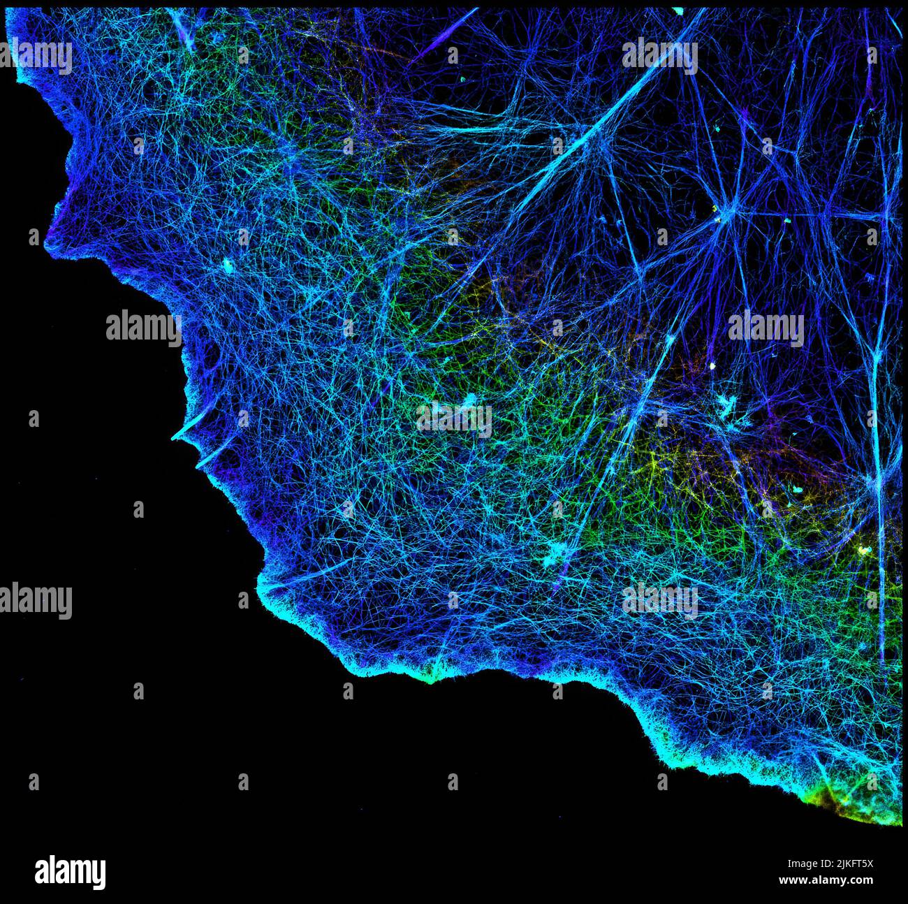 Actin is an essential protein in the skeleton of a cell (cytoskeleton). It forms a dense network of fine filaments in the cell. Here, the researchers used a technique called stochastic optical reconstruction microscopy (STORM) to visualize the actin network in a cell in three dimensions. Actin strands were labeled with a dye called Alexa Fluor 647-phalloidin. This image appears in a study published by Nature Methods, which reports how researchers use STORM to visualize the cytoskeleton. Stock Photohttps://www.alamy.com/image-license-details/?v=1https://www.alamy.com/actin-is-an-essential-protein-in-the-skeleton-of-a-cell-cytoskeleton-it-forms-a-dense-network-of-fine-filaments-in-the-cell-here-the-researchers-used-a-technique-called-stochastic-optical-reconstruction-microscopy-storm-to-visualize-the-actin-network-in-a-cell-in-three-dimensions-actin-strands-were-labeled-with-a-dye-called-alexa-fluor-647-phalloidin-this-image-appears-in-a-study-published-by-nature-methods-which-reports-how-researchers-use-storm-to-visualize-the-cytoskeleton-image476706662.html
RM2JKFT5X–Actin is an essential protein in the skeleton of a cell (cytoskeleton). It forms a dense network of fine filaments in the cell. Here, the researchers used a technique called stochastic optical reconstruction microscopy (STORM) to visualize the actin network in a cell in three dimensions. Actin strands were labeled with a dye called Alexa Fluor 647-phalloidin. This image appears in a study published by Nature Methods, which reports how researchers use STORM to visualize the cytoskeleton.
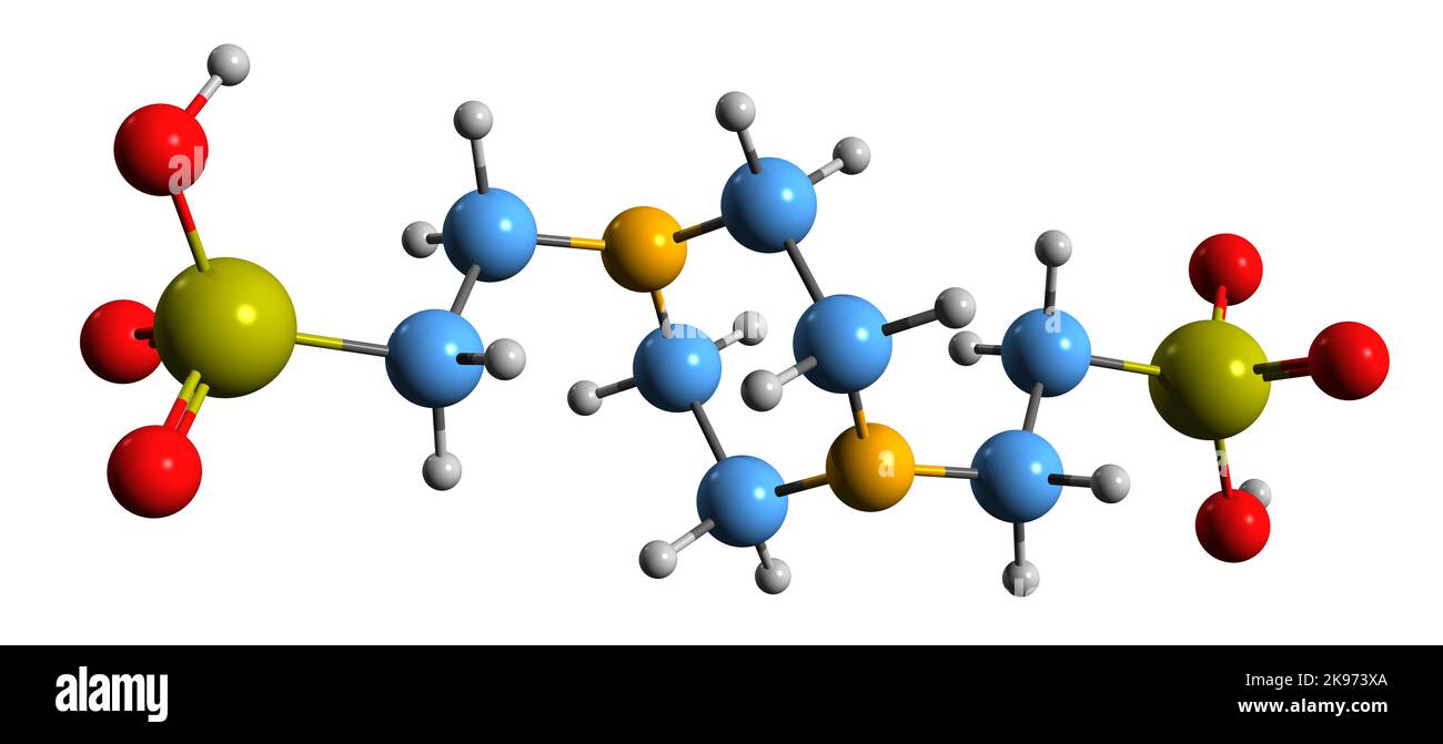 3D image of PIPES skeletal formula - molecular chemical structure of buffering agent isolated on white background Stock Photohttps://www.alamy.com/image-license-details/?v=1https://www.alamy.com/3d-image-of-pipes-skeletal-formula-molecular-chemical-structure-of-buffering-agent-isolated-on-white-background-image487578962.html
RF2K973XA–3D image of PIPES skeletal formula - molecular chemical structure of buffering agent isolated on white background
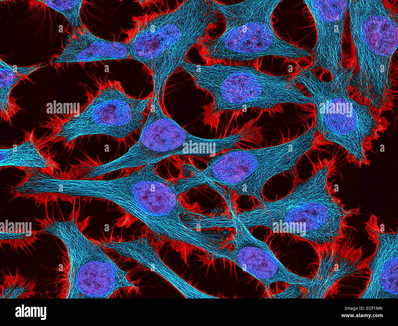 Multiphoton fluorescence image of HeLa cells stained with the actin binding toxin phalloidin (red), microtubules (cyan) and cell Stock Photohttps://www.alamy.com/image-license-details/?v=1https://www.alamy.com/stock-photo-multiphoton-fluorescence-image-of-hela-cells-stained-with-the-actin-76547987.html
RFECF1MK–Multiphoton fluorescence image of HeLa cells stained with the actin binding toxin phalloidin (red), microtubules (cyan) and cell
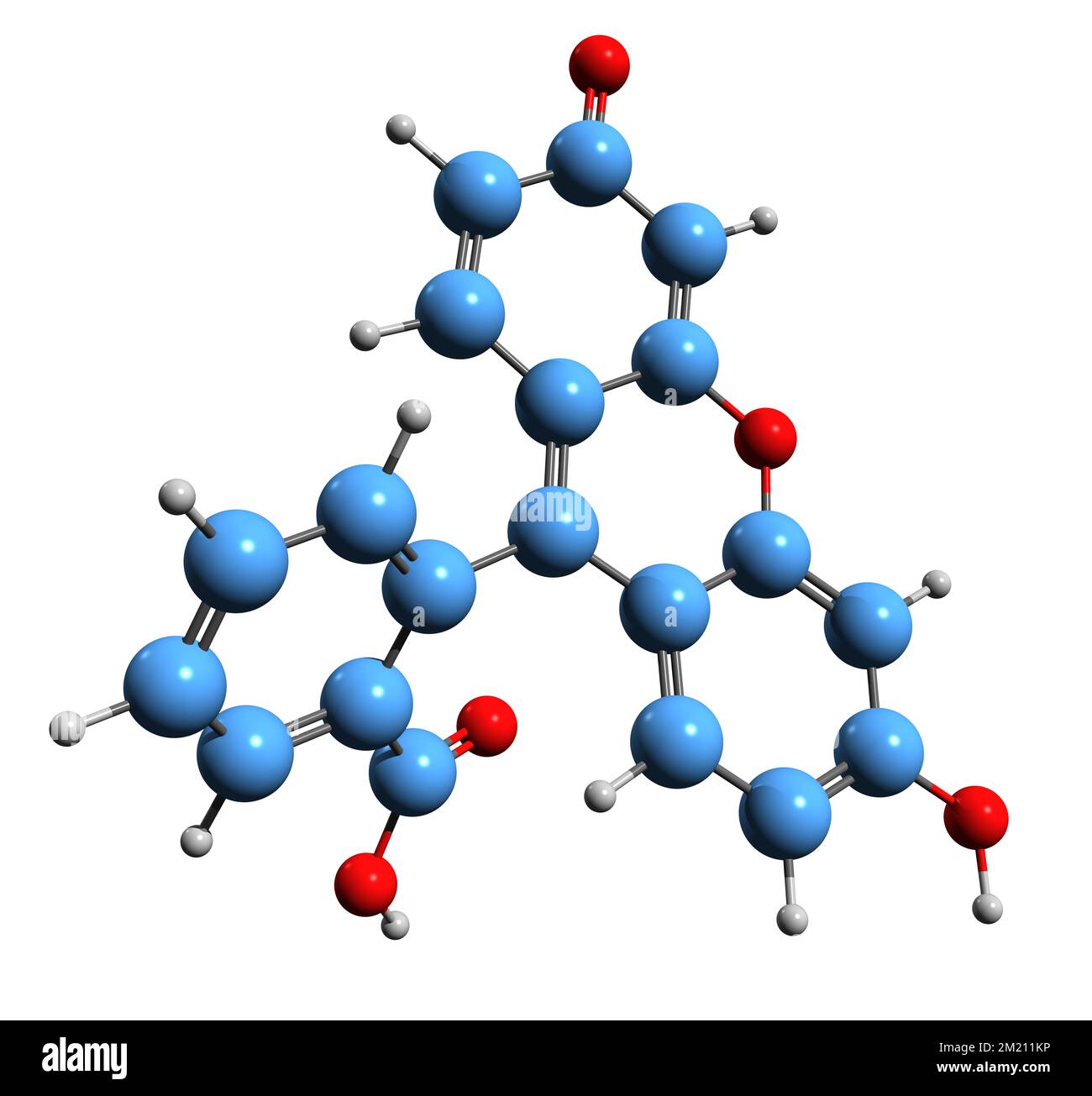 3D image of Fluorescein skeletal formula - molecular chemical structure of resorcinolphthalein isolated on white background Stock Photohttps://www.alamy.com/image-license-details/?v=1https://www.alamy.com/3d-image-of-fluorescein-skeletal-formula-molecular-chemical-structure-of-resorcinolphthalein-isolated-on-white-background-image500353274.html
RF2M211KP–3D image of Fluorescein skeletal formula - molecular chemical structure of resorcinolphthalein isolated on white background
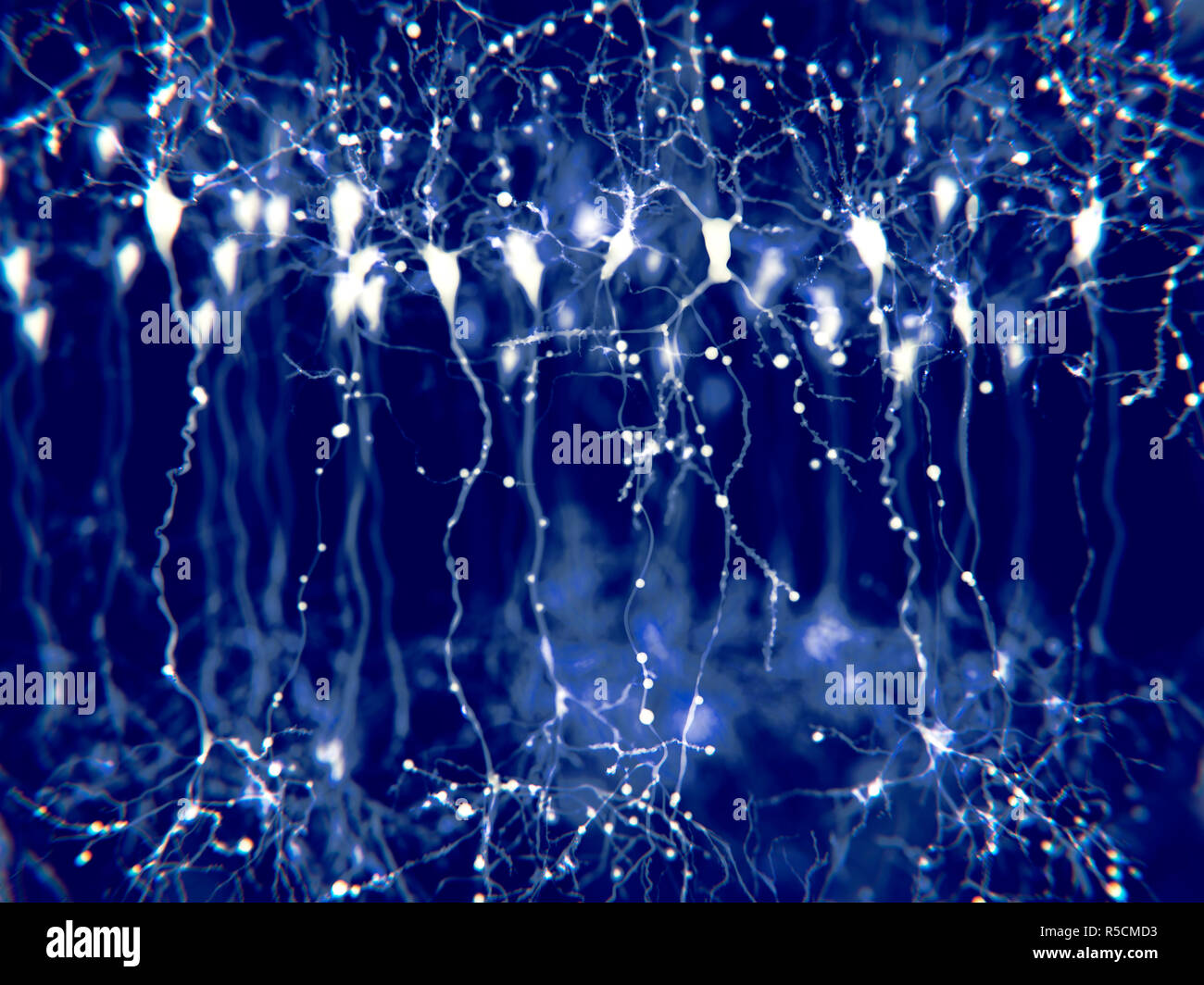 Pyramidal neurons in the cerebral cortex, illustration. Pyramidal neurons are found in certain areas of the brain including the cerebral cortex, the hippocampus, and the amygdala. Here, the illustration shows the synaptic signals highlighted using a microscopy fluorescence technique. Stock Photohttps://www.alamy.com/image-license-details/?v=1https://www.alamy.com/pyramidal-neurons-in-the-cerebral-cortex-illustration-pyramidal-neurons-are-found-in-certain-areas-of-the-brain-including-the-cerebral-cortex-the-hippocampus-and-the-amygdala-here-the-illustration-shows-the-synaptic-signals-highlighted-using-a-microscopy-fluorescence-technique-image227087535.html
RFR5CMD3–Pyramidal neurons in the cerebral cortex, illustration. Pyramidal neurons are found in certain areas of the brain including the cerebral cortex, the hippocampus, and the amygdala. Here, the illustration shows the synaptic signals highlighted using a microscopy fluorescence technique.
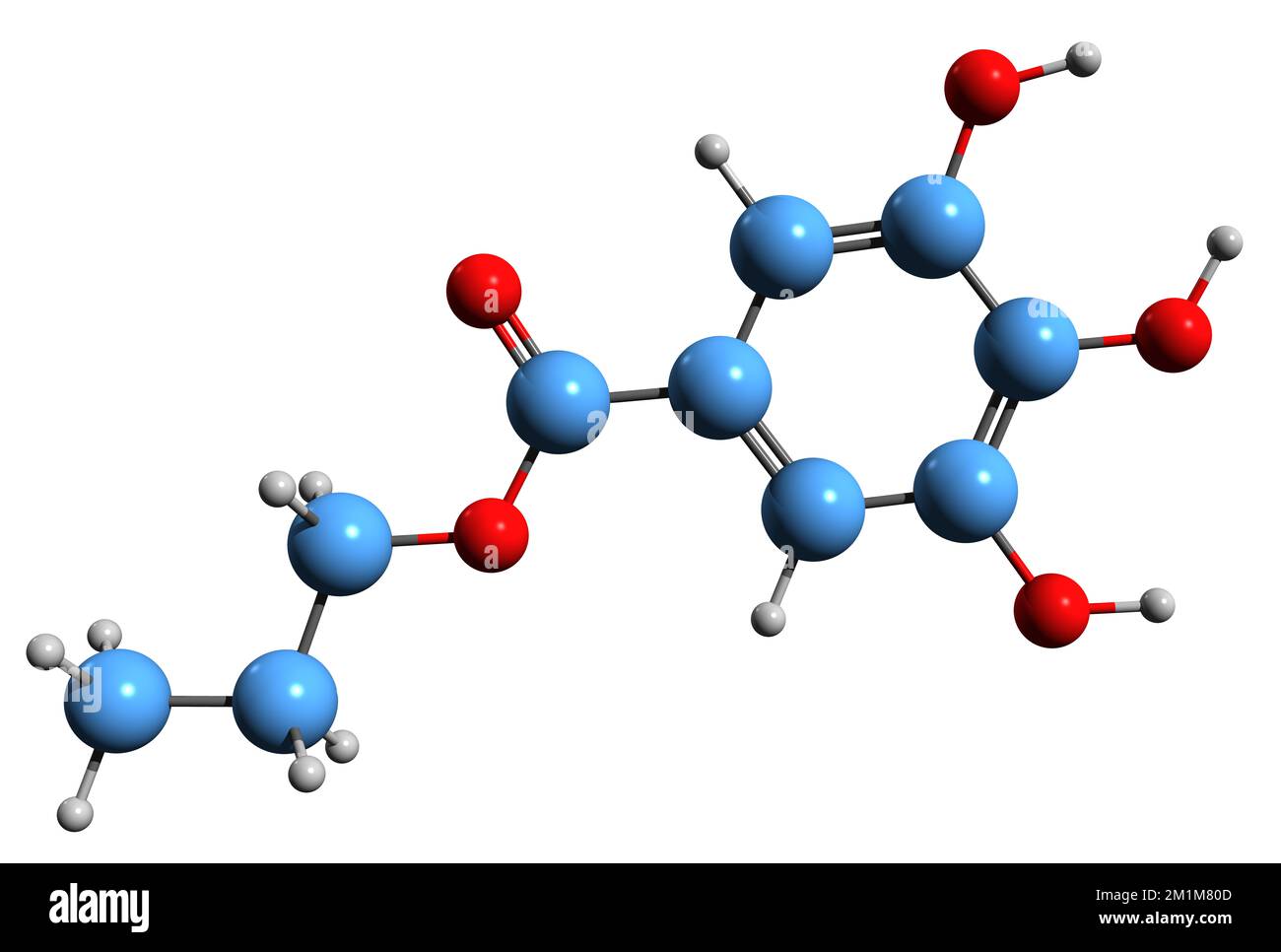 3D image of Propyl gallate skeletal formula - molecular chemical structure of Propyl trihydroxybenzoate isolated on white background Stock Photohttps://www.alamy.com/image-license-details/?v=1https://www.alamy.com/3d-image-of-propyl-gallate-skeletal-formula-molecular-chemical-structure-of-propyl-trihydroxybenzoate-isolated-on-white-background-image500160653.html
RF2M1M80D–3D image of Propyl gallate skeletal formula - molecular chemical structure of Propyl trihydroxybenzoate isolated on white background
 Scorpion fluoresce when exposed to ultraviolet (UV) light. Arthropods Workshop taught by the entomologist and environmental disseminator Sergi Romeu V Stock Photohttps://www.alamy.com/image-license-details/?v=1https://www.alamy.com/scorpion-fluoresce-when-exposed-to-ultraviolet-uv-light-arthropods-workshop-taught-by-the-entomologist-and-environmental-disseminator-sergi-romeu-v-image600448462.html
RM2WTTP3X–Scorpion fluoresce when exposed to ultraviolet (UV) light. Arthropods Workshop taught by the entomologist and environmental disseminator Sergi Romeu V
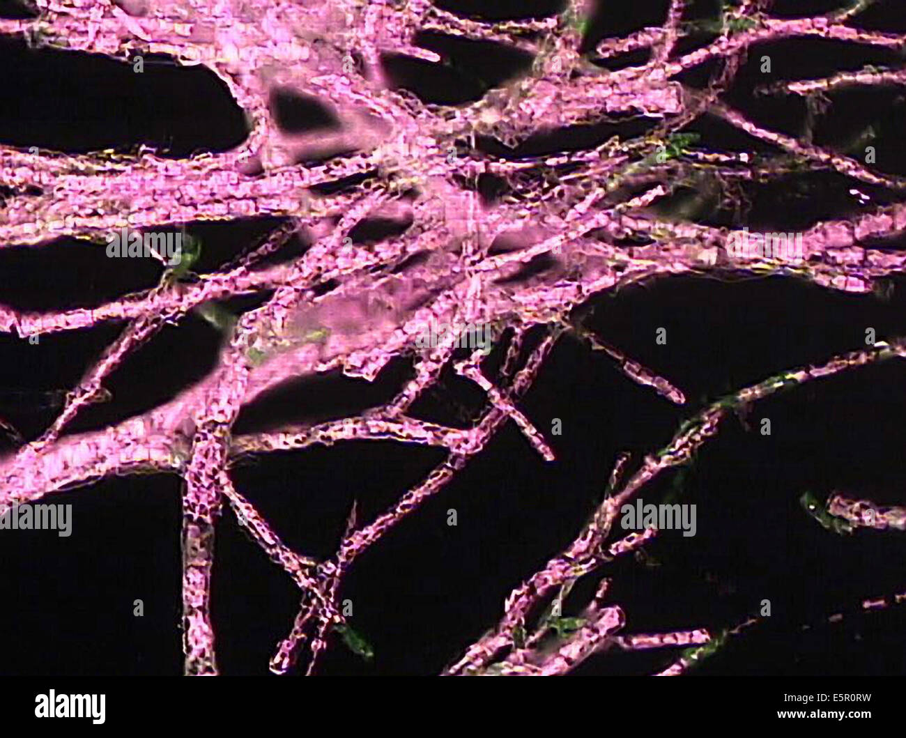 Photomicrograph of Ectocarpus siliculosus, This brown alga produces natural fluorescence when exposed to ultraviolet. Stock Photohttps://www.alamy.com/image-license-details/?v=1https://www.alamy.com/stock-photo-photomicrograph-of-ectocarpus-siliculosus-this-brown-alga-produces-72420317.html
RME5R0RW–Photomicrograph of Ectocarpus siliculosus, This brown alga produces natural fluorescence when exposed to ultraviolet.
 Microscope, liquid samples and portable computer with microscopic image on observation table, set up in modern laboratory Stock Photohttps://www.alamy.com/image-license-details/?v=1https://www.alamy.com/microscope-liquid-samples-and-portable-computer-with-microscopic-image-on-observation-table-set-up-in-modern-laboratory-image211068303.html
RFP7AYNK–Microscope, liquid samples and portable computer with microscopic image on observation table, set up in modern laboratory
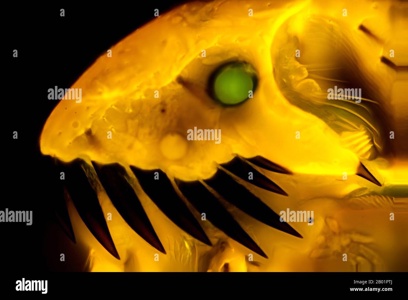 Cat flea (Ctenocephalides felis), Head of a Cat flea, fluorescence microscopy, Germany Stock Photohttps://www.alamy.com/image-license-details/?v=1https://www.alamy.com/cat-flea-ctenocephalides-felis-head-of-a-cat-flea-fluorescence-microscopy-germany-image344247250.html
RM2B01PTJ–Cat flea (Ctenocephalides felis), Head of a Cat flea, fluorescence microscopy, Germany
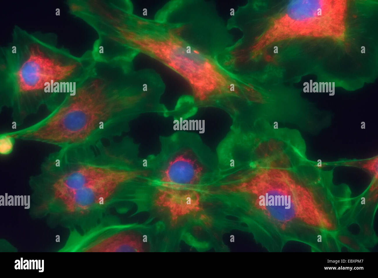 Microfilaments, mitochondria, and nuclei in fibroblast cells Stock Photohttps://www.alamy.com/image-license-details/?v=1https://www.alamy.com/stock-photo-microfilaments-mitochondria-and-nuclei-in-fibroblast-cells-76191255.html
RMEBXPM7–Microfilaments, mitochondria, and nuclei in fibroblast cells
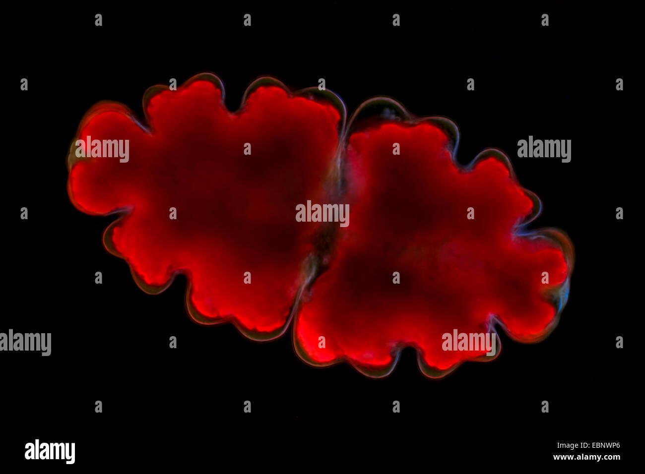 Euastrum (Euastrum spec.), fluorescence microscope Stock Photohttps://www.alamy.com/image-license-details/?v=1https://www.alamy.com/stock-photo-euastrum-euastrum-spec-fluorescence-microscope-76083902.html
RMEBNWP6–Euastrum (Euastrum spec.), fluorescence microscope
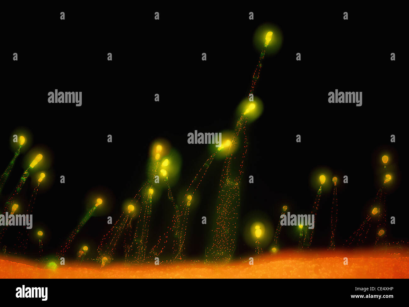 Tobacco (Nicotiana benthamiana) leaf hairs, fluorescence microscopy, objective lens 20x Stock Photohttps://www.alamy.com/image-license-details/?v=1https://www.alamy.com/stock-photo-tobacco-nicotiana-benthamiana-leaf-hairs-fluorescence-microscopy-objective-43134610.html
RMCE4XHP–Tobacco (Nicotiana benthamiana) leaf hairs, fluorescence microscopy, objective lens 20x
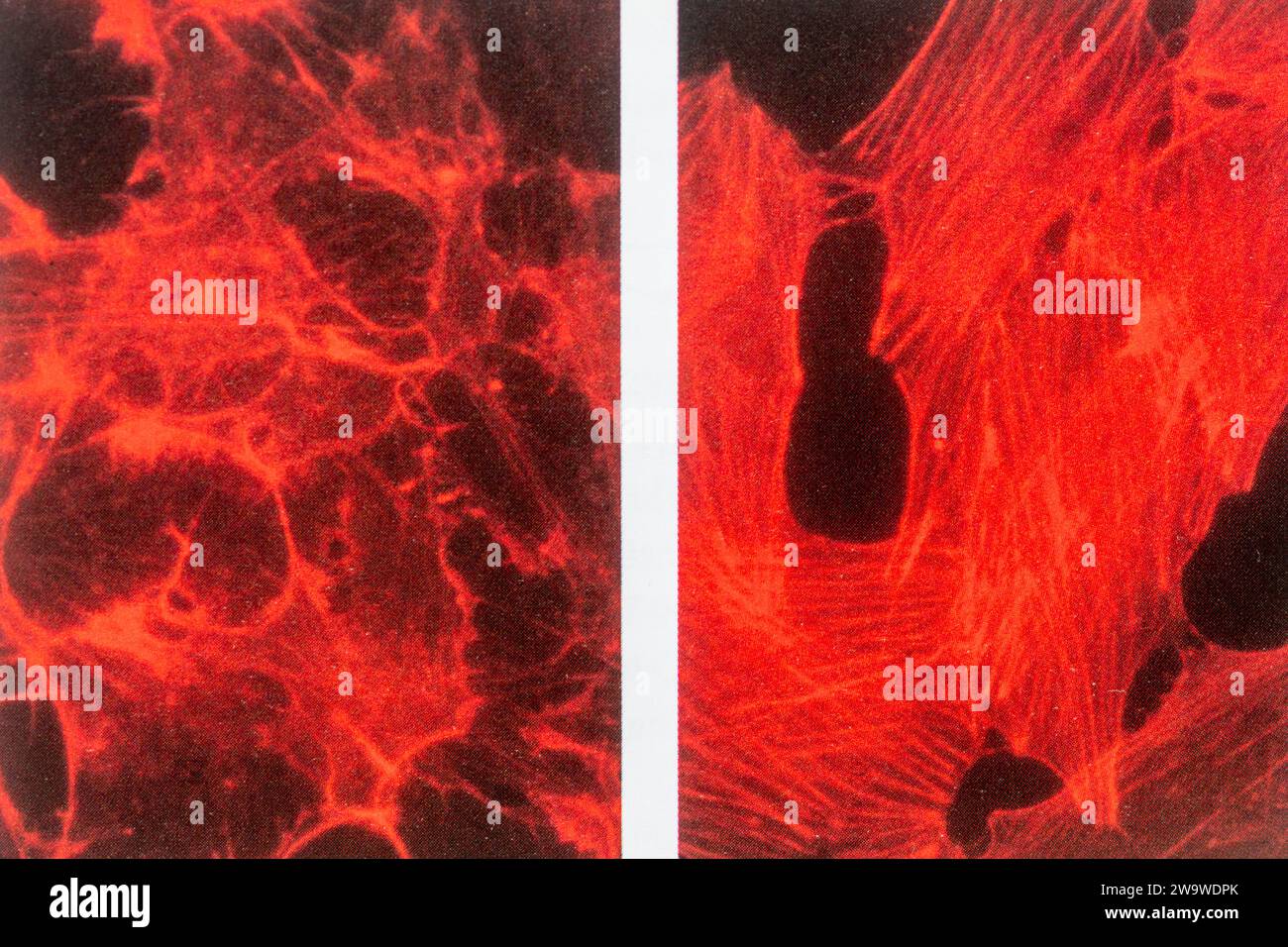 Fluoresence microscopy of cells, specifically fibroblasts, stained with fluorescently labelled phalloidin showing actin stress fibres Stock Photohttps://www.alamy.com/image-license-details/?v=1https://www.alamy.com/fluoresence-microscopy-of-cells-specifically-fibroblasts-stained-with-fluorescently-labelled-phalloidin-showing-actin-stress-fibres-image591244043.html
RM2W9WDPK–Fluoresence microscopy of cells, specifically fibroblasts, stained with fluorescently labelled phalloidin showing actin stress fibres
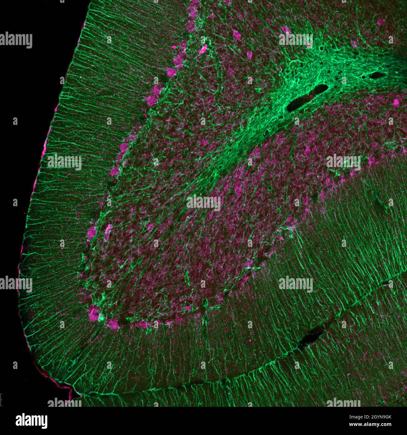 Sagittal section of mouse cerebellum labelled with immunofluorescence and visualized with confocal laser scanning microscopy. Large Purkinje cells and Stock Photohttps://www.alamy.com/image-license-details/?v=1https://www.alamy.com/sagittal-section-of-mouse-cerebellum-labelled-with-immunofluorescence-and-visualized-with-confocal-laser-scanning-microscopy-large-purkinje-cells-and-image447323427.html
RF2GYN9GK–Sagittal section of mouse cerebellum labelled with immunofluorescence and visualized with confocal laser scanning microscopy. Large Purkinje cells and
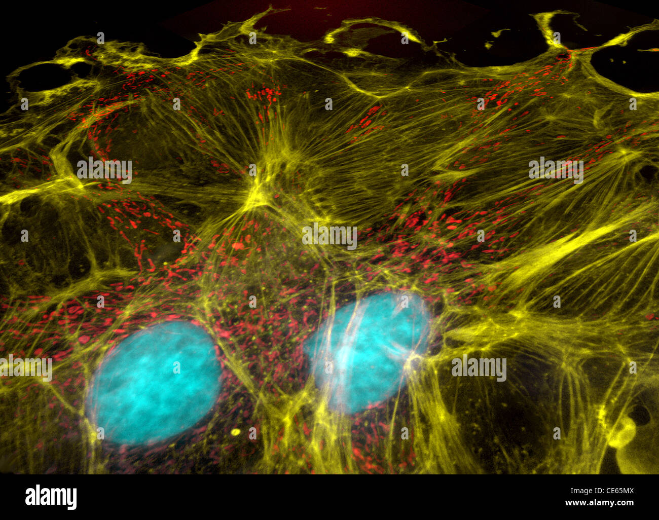 Fibroblast cells, fluorescence microscopy, nuclei, mitochondria, and microfilaments Stock Photohttps://www.alamy.com/image-license-details/?v=1https://www.alamy.com/stock-photo-fibroblast-cells-fluorescence-microscopy-nuclei-mitochondria-and-microfilaments-43162138.html
RMCE65MX–Fibroblast cells, fluorescence microscopy, nuclei, mitochondria, and microfilaments
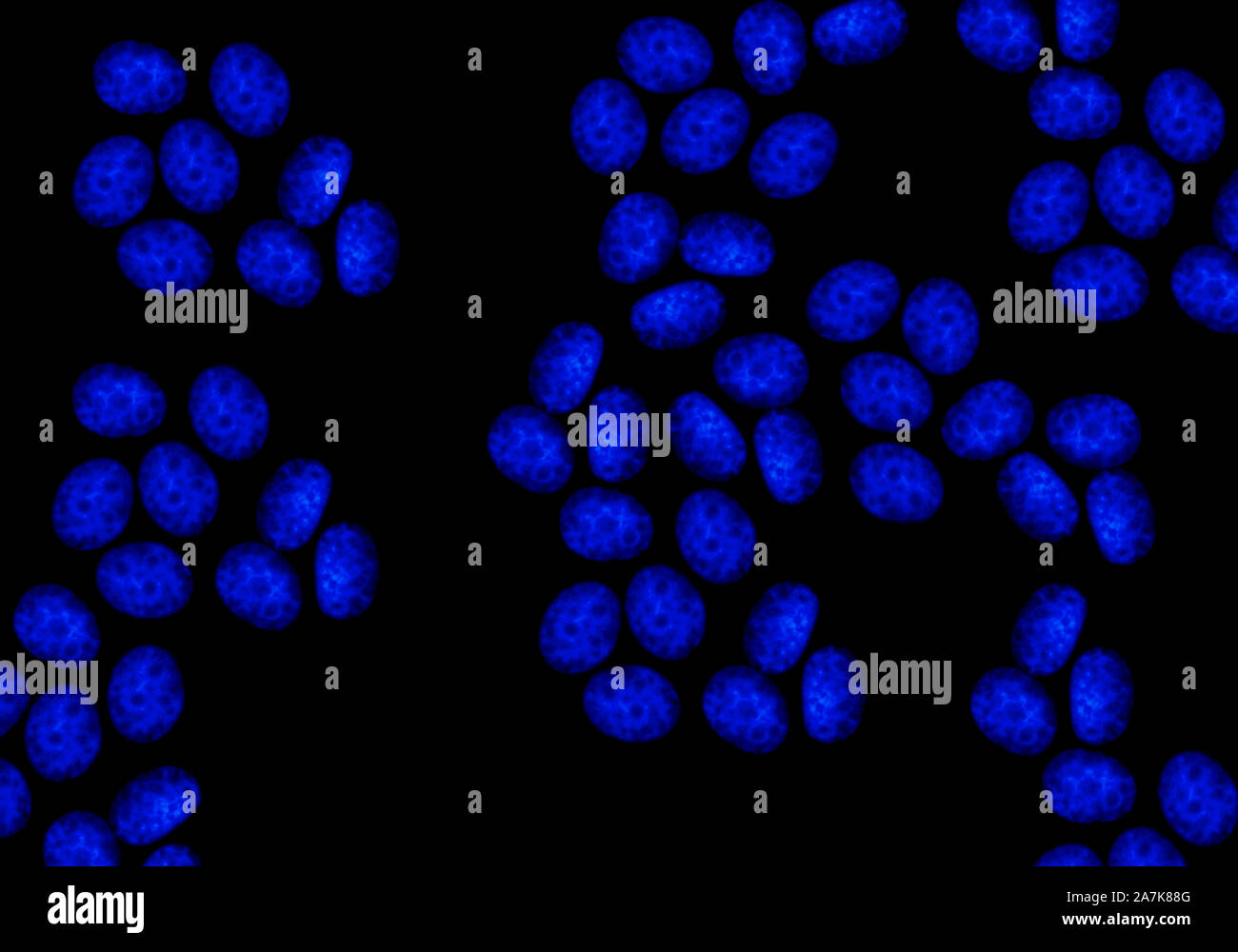 Confocal Fluorescence Microscopy of CRISPR-generated knockout cells Stock Photohttps://www.alamy.com/image-license-details/?v=1https://www.alamy.com/confocal-fluorescence-microscopy-of-crispr-generated-knockout-cells-image331723184.html
RM2A7K88G–Confocal Fluorescence Microscopy of CRISPR-generated knockout cells
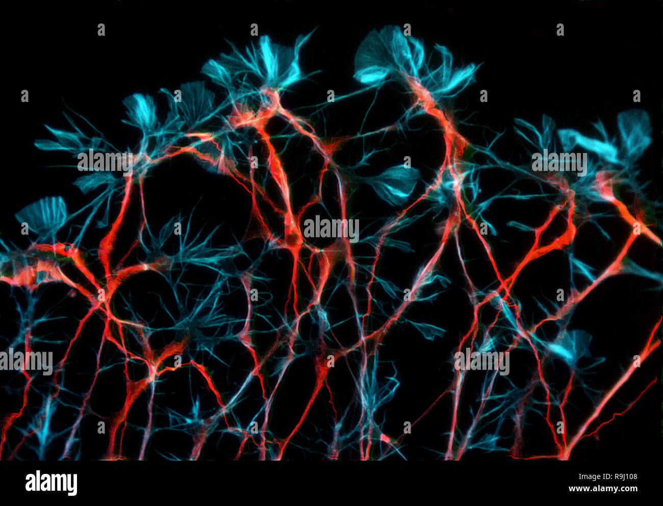 Neurons from rat embryonic dorsal root ganglion Stock Photohttps://www.alamy.com/image-license-details/?v=1https://www.alamy.com/neurons-from-rat-embryonic-dorsal-root-ganglion-image229662616.html
RMR9J108–Neurons from rat embryonic dorsal root ganglion
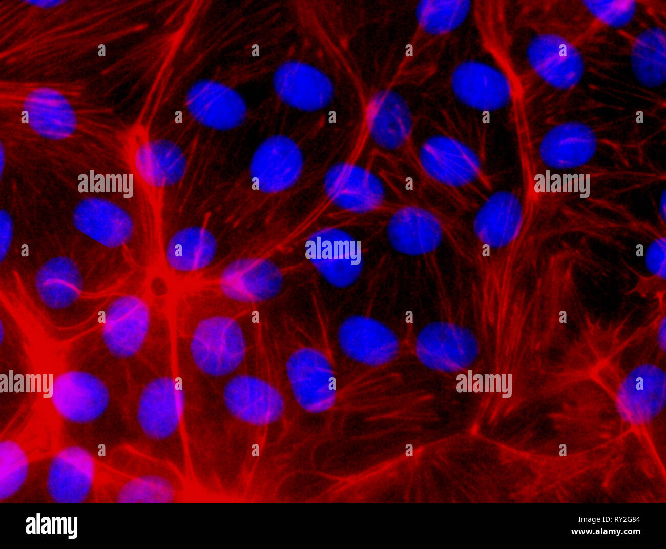 Confocal Fluorescence Microscopy of CRISPR generated knockout cells. CRISPR (Clustered Regularly Interspaced Short Palindromic Repeats) gene editing Stock Photohttps://www.alamy.com/image-license-details/?v=1https://www.alamy.com/confocal-fluorescence-microscopy-of-crispr-generated-knockout-cells-crispr-clustered-regularly-interspaced-short-palindromic-repeats-gene-editing-image240387172.html
RMRY2G84–Confocal Fluorescence Microscopy of CRISPR generated knockout cells. CRISPR (Clustered Regularly Interspaced Short Palindromic Repeats) gene editing
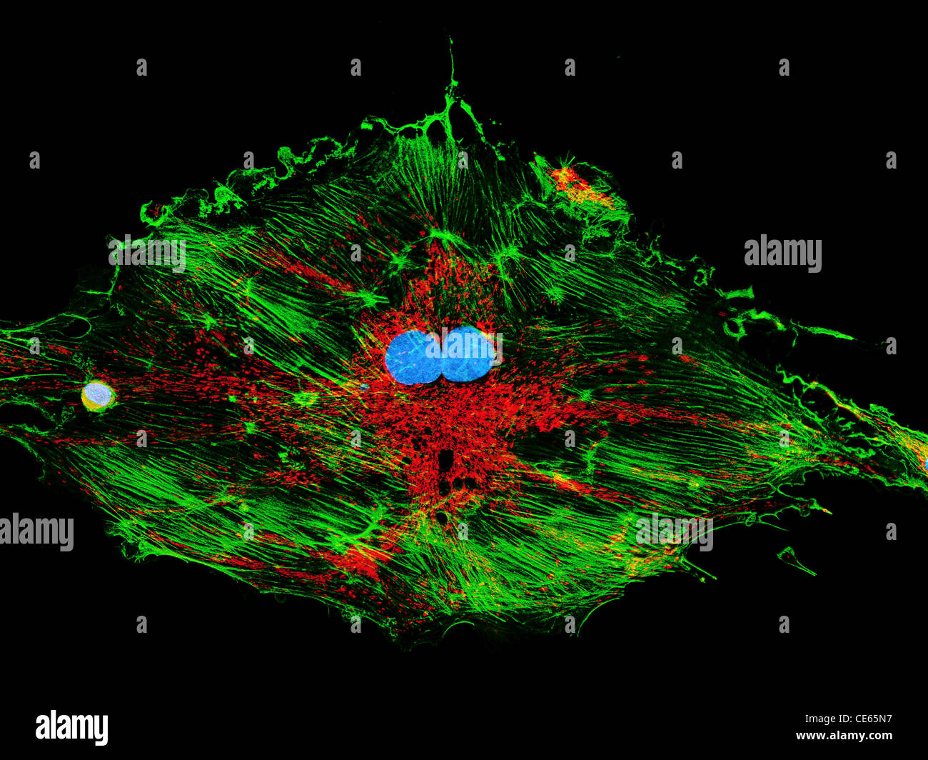 Fibroblast cells, fluorescence microscopy, nuclei, mitochondria, and microfilaments Stock Photohttps://www.alamy.com/image-license-details/?v=1https://www.alamy.com/stock-photo-fibroblast-cells-fluorescence-microscopy-nuclei-mitochondria-and-microfilaments-43162147.html
RMCE65N7–Fibroblast cells, fluorescence microscopy, nuclei, mitochondria, and microfilaments
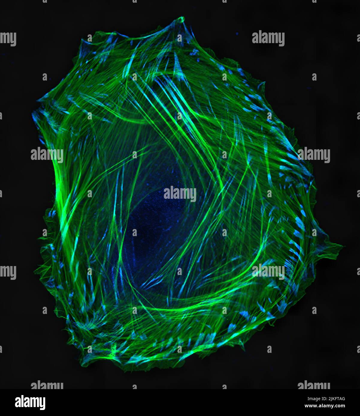 Embryonic smooth muscle cell. Immunofluorescence-labeled actin cytoskeleton (green) and vinculin in cell adhesions (blue). Confocal laser scanning microscopy. Stock Photohttps://www.alamy.com/image-license-details/?v=1https://www.alamy.com/embryonic-smooth-muscle-cell-immunofluorescence-labeled-actin-cytoskeleton-green-and-vinculin-in-cell-adhesions-blue-confocal-laser-scanning-microscopy-image476706792.html
RM2JKFTAG–Embryonic smooth muscle cell. Immunofluorescence-labeled actin cytoskeleton (green) and vinculin in cell adhesions (blue). Confocal laser scanning microscopy.
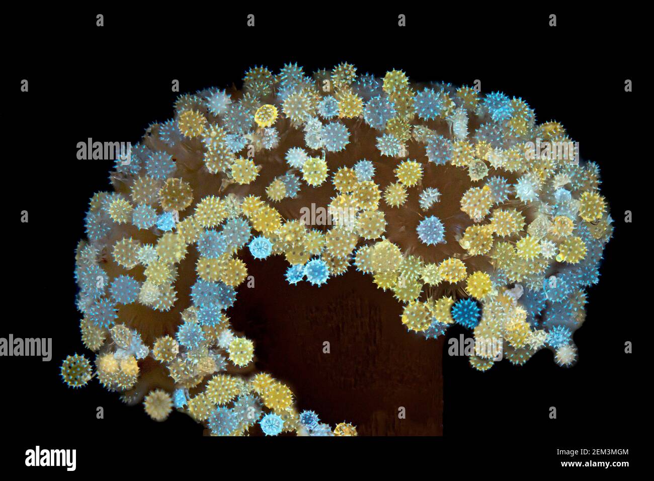 hibiscus (Hibiscus spec.), pollen of hibiscus in the anther, fluorescence, UV-light irradiation, light microscope image, magnification x34 related to Stock Photohttps://www.alamy.com/image-license-details/?v=1https://www.alamy.com/hibiscus-hibiscus-spec-pollen-of-hibiscus-in-the-anther-fluorescence-uv-light-irradiation-light-microscope-image-magnification-x34-related-to-image408213588.html
RM2EM3MGM–hibiscus (Hibiscus spec.), pollen of hibiscus in the anther, fluorescence, UV-light irradiation, light microscope image, magnification x34 related to
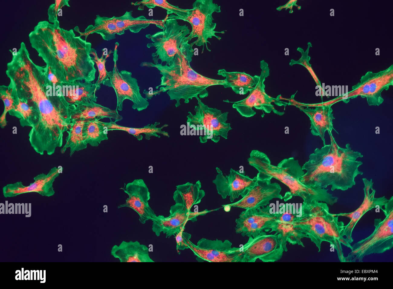 Microfilaments, mitochondria, and nuclei in fibroblast cells Stock Photohttps://www.alamy.com/image-license-details/?v=1https://www.alamy.com/stock-photo-microfilaments-mitochondria-and-nuclei-in-fibroblast-cells-76191252.html
RMEBXPM4–Microfilaments, mitochondria, and nuclei in fibroblast cells
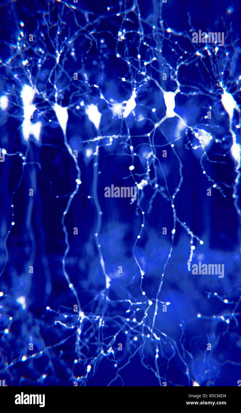 Pyramidal neurons in the cerebral cortex, illustration. Pyramidal neurons are found in certain areas of the brain including the cerebral cortex, the hippocampus, and the amygdala. Here, the illustration shows the synaptic signals highlighted using a microscopy fluorescence technique. Stock Photohttps://www.alamy.com/image-license-details/?v=1https://www.alamy.com/pyramidal-neurons-in-the-cerebral-cortex-illustration-pyramidal-neurons-are-found-in-certain-areas-of-the-brain-including-the-cerebral-cortex-the-hippocampus-and-the-amygdala-here-the-illustration-shows-the-synaptic-signals-highlighted-using-a-microscopy-fluorescence-technique-image227087536.html
RFR5CMD4–Pyramidal neurons in the cerebral cortex, illustration. Pyramidal neurons are found in certain areas of the brain including the cerebral cortex, the hippocampus, and the amygdala. Here, the illustration shows the synaptic signals highlighted using a microscopy fluorescence technique.
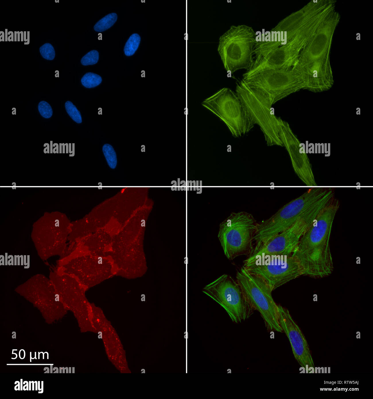 multiple human osteosarcoma cells stained for epifluorescence Stock Photohttps://www.alamy.com/image-license-details/?v=1https://www.alamy.com/multiple-human-osteosarcoma-cells-stained-for-epifluorescence-image239039546.html
RMRTW5AJ–multiple human osteosarcoma cells stained for epifluorescence
 Scorpion fluoresce when exposed to ultraviolet (UV) light. Arthropods Workshop taught by the entomologist and environmental disseminator Sergi Romeu V Stock Photohttps://www.alamy.com/image-license-details/?v=1https://www.alamy.com/scorpion-fluoresce-when-exposed-to-ultraviolet-uv-light-arthropods-workshop-taught-by-the-entomologist-and-environmental-disseminator-sergi-romeu-v-image600448341.html
RM2WTTNYH–Scorpion fluoresce when exposed to ultraviolet (UV) light. Arthropods Workshop taught by the entomologist and environmental disseminator Sergi Romeu V
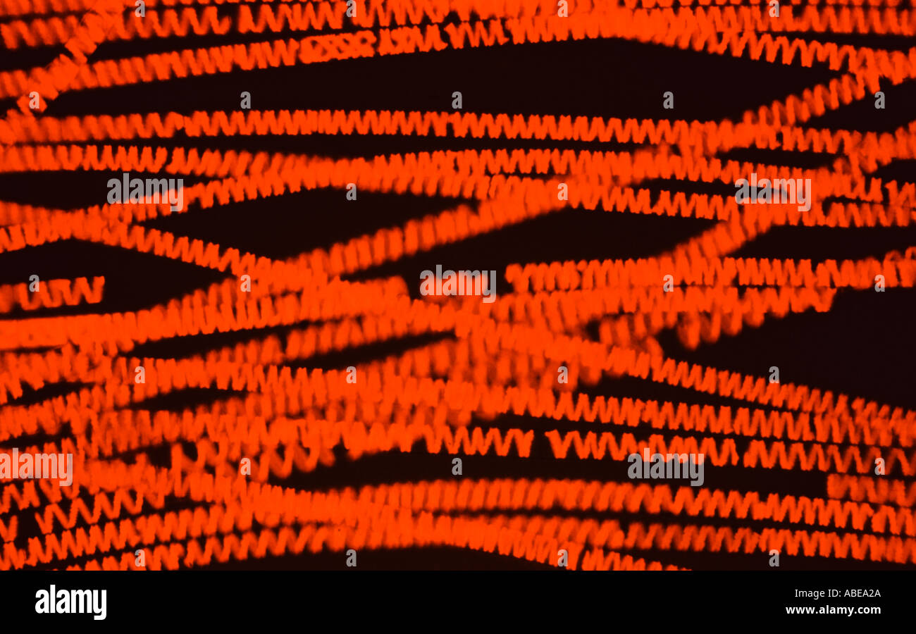 Spirogyra Live Blue Violet Fluorescence Stock Photohttps://www.alamy.com/image-license-details/?v=1https://www.alamy.com/spirogyra-live-blue-violet-fluorescence-image780842.html
RMABEA2A–Spirogyra Live Blue Violet Fluorescence
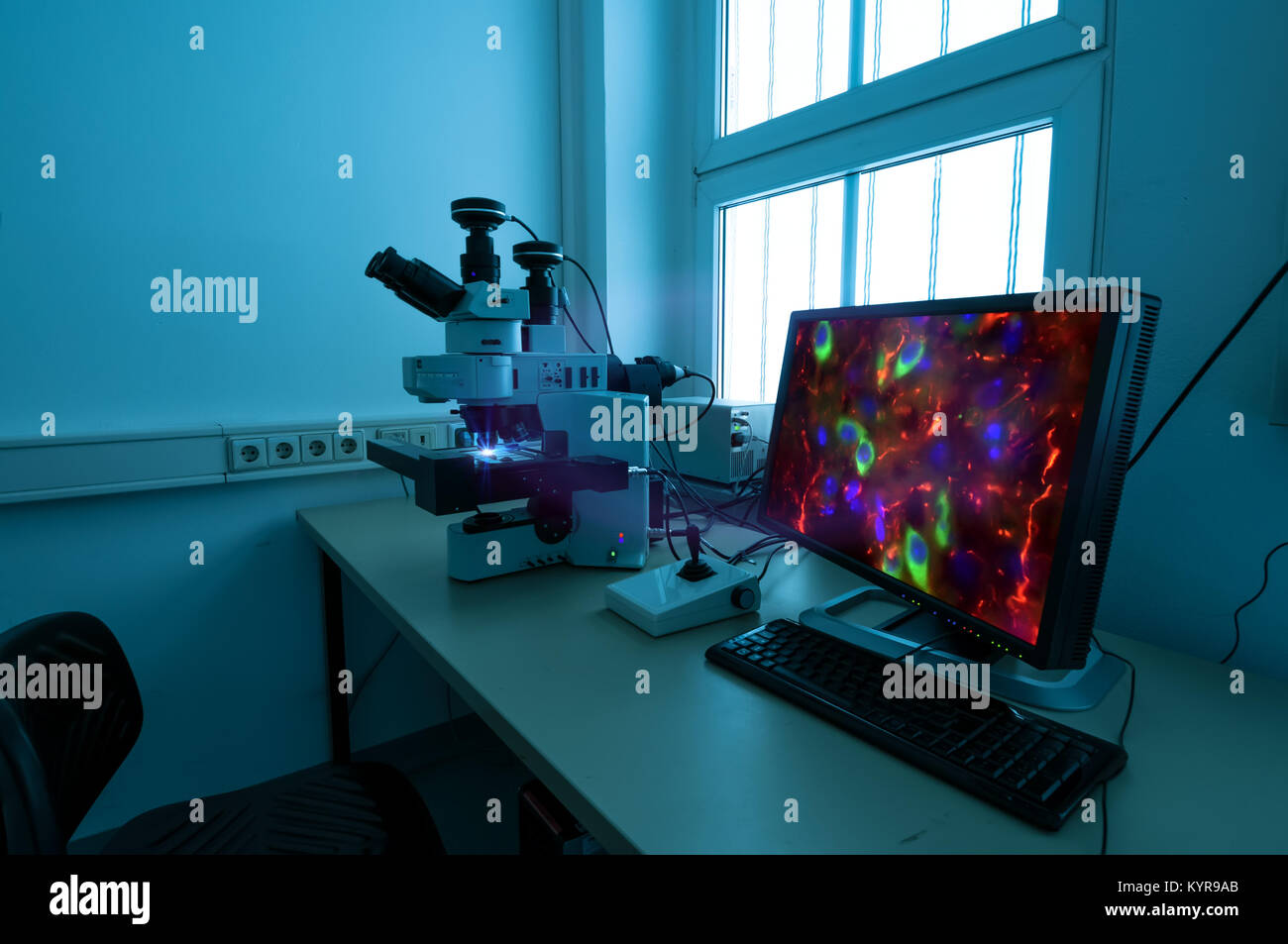 Modern fluorescent microstope working station Stock Photohttps://www.alamy.com/image-license-details/?v=1https://www.alamy.com/stock-photo-modern-fluorescent-microstope-working-station-172001267.html
RFKYR9AB–Modern fluorescent microstope working station
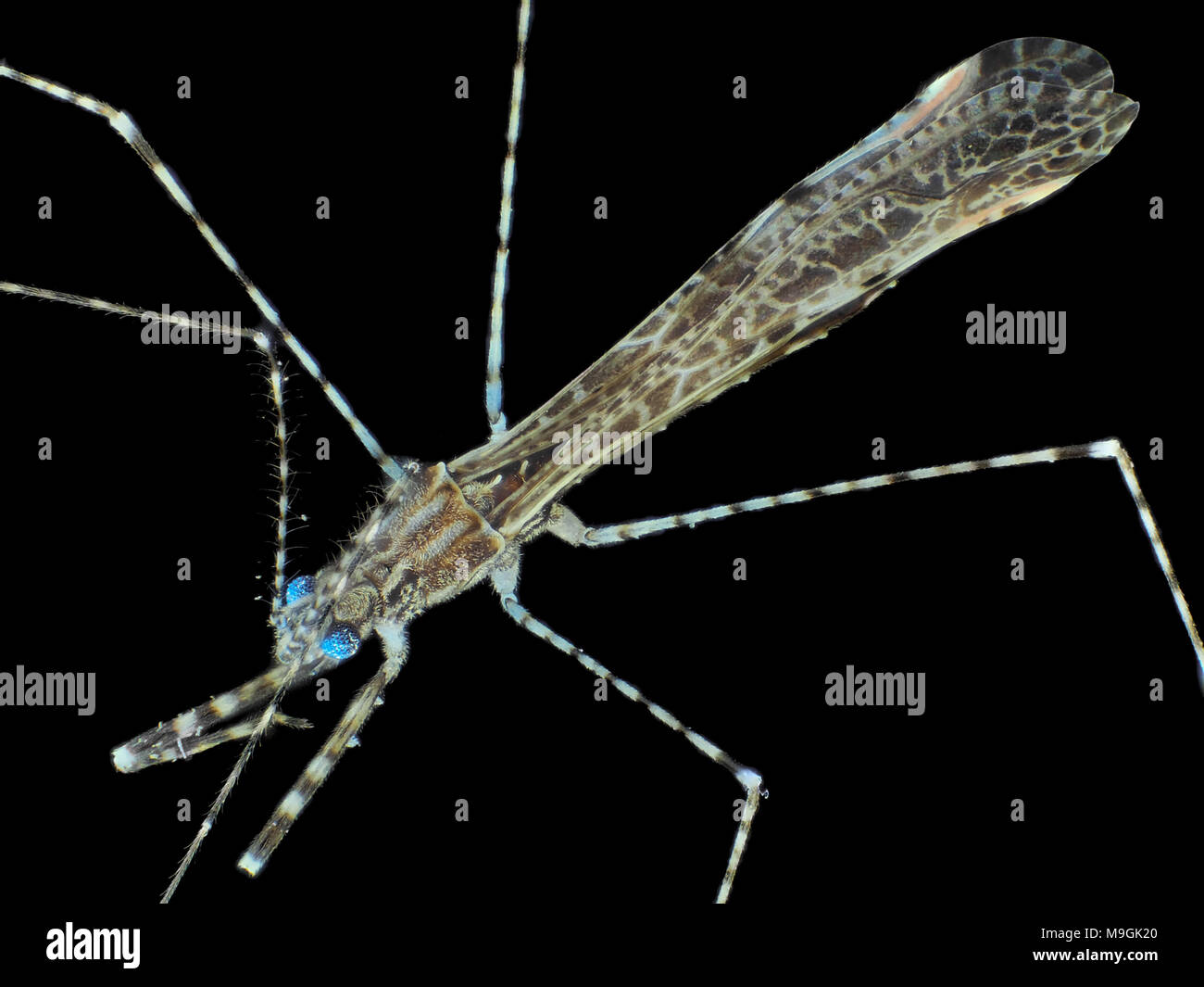 Extreme macro photo (under the microscope) of a tiny thread-legged bug, in visible plus UV light Stock Photohttps://www.alamy.com/image-license-details/?v=1https://www.alamy.com/extreme-macro-photo-under-the-microscope-of-a-tiny-thread-legged-bug-in-visible-plus-uv-light-image178001768.html
RMM9GK20–Extreme macro photo (under the microscope) of a tiny thread-legged bug, in visible plus UV light
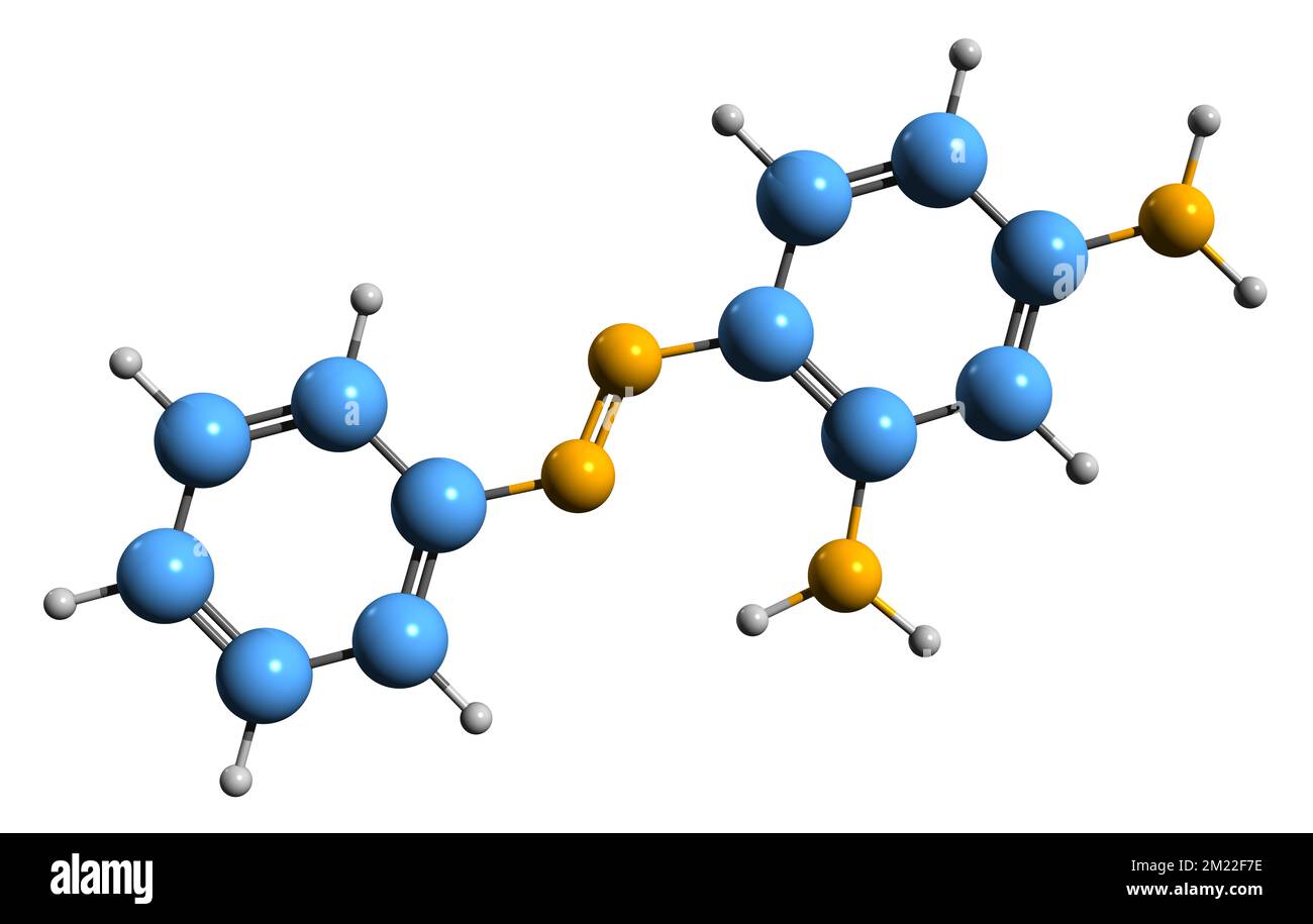 3D image of Chrysoidine skeletal formula - molecular chemical structure of Basic Orange 2 isolated on white background Stock Photohttps://www.alamy.com/image-license-details/?v=1https://www.alamy.com/3d-image-of-chrysoidine-skeletal-formula-molecular-chemical-structure-of-basic-orange-2-isolated-on-white-background-image500385858.html
RF2M22F7E–3D image of Chrysoidine skeletal formula - molecular chemical structure of Basic Orange 2 isolated on white background
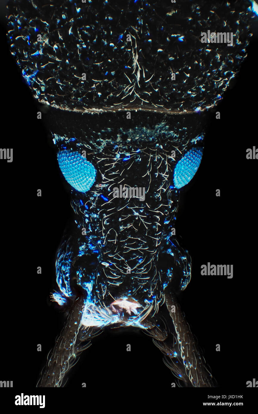 Weevil beetle (likely Larinus carlinae) with fluorescent blue eyes, reflected ultraviolet light micrograph, 26x magnification when printed 10cm tall Stock Photohttps://www.alamy.com/image-license-details/?v=1https://www.alamy.com/weevil-beetle-likely-larinus-carlinae-with-fluorescent-blue-eyes-reflected-image153950655.html
RMJXD1HK–Weevil beetle (likely Larinus carlinae) with fluorescent blue eyes, reflected ultraviolet light micrograph, 26x magnification when printed 10cm tall
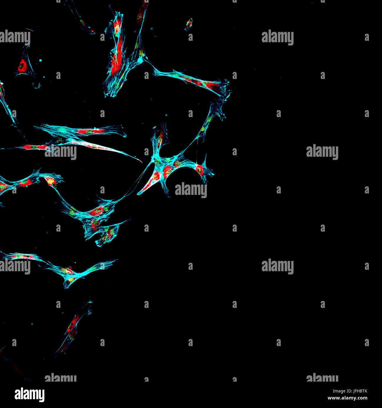 Immunofluorescence of multiple human tumor metastatic cells growing in tissue culture for research purposes Stock Photohttps://www.alamy.com/image-license-details/?v=1https://www.alamy.com/stock-photo-immunofluorescence-of-multiple-human-tumor-metastatic-cells-growing-147285283.html
RFJFHBTK–Immunofluorescence of multiple human tumor metastatic cells growing in tissue culture for research purposes
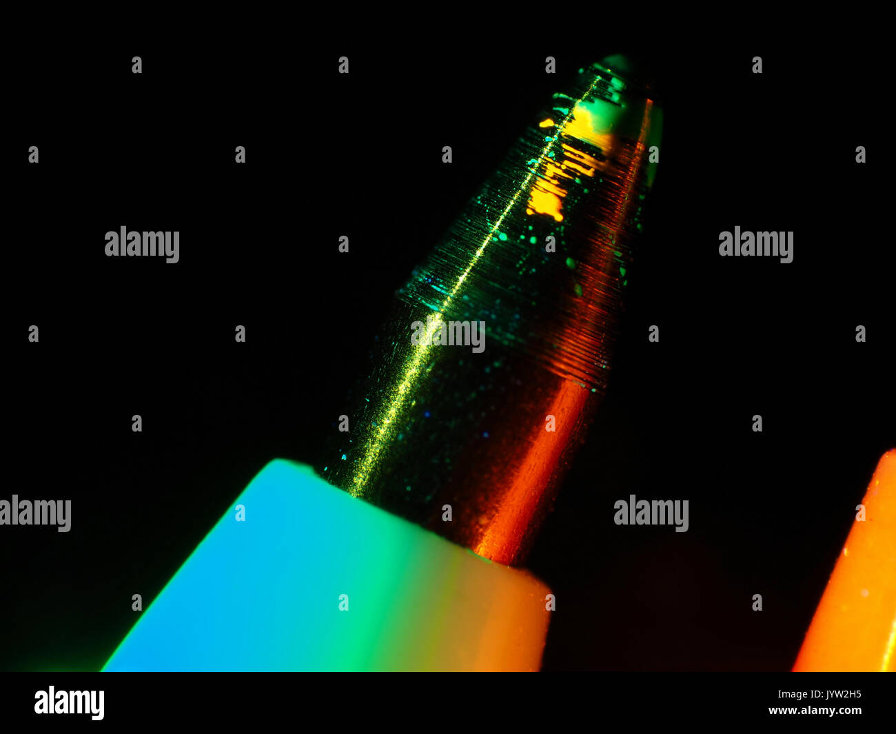 Ultraviolet reflected light micrograph of a fluorescent ball pen tip, pictured area is about 8.5mm wide Stock Photohttps://www.alamy.com/image-license-details/?v=1https://www.alamy.com/ultraviolet-reflected-light-micrograph-of-a-fluorescent-ball-pen-tip-image154829505.html
RMJYW2H5–Ultraviolet reflected light micrograph of a fluorescent ball pen tip, pictured area is about 8.5mm wide
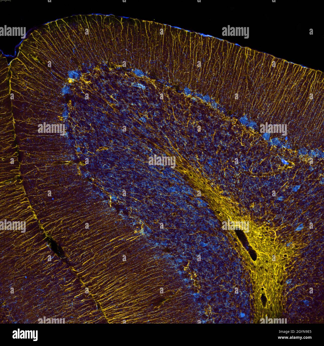 Sagittal section of mouse cerebellum labelled with immunofluorescence and visualized with confocal laser scanning microscopy. Large Purkinje cells and Stock Photohttps://www.alamy.com/image-license-details/?v=1https://www.alamy.com/sagittal-section-of-mouse-cerebellum-labelled-with-immunofluorescence-and-visualized-with-confocal-laser-scanning-microscopy-large-purkinje-cells-and-image447323357.html
RF2GYN9E5–Sagittal section of mouse cerebellum labelled with immunofluorescence and visualized with confocal laser scanning microscopy. Large Purkinje cells and
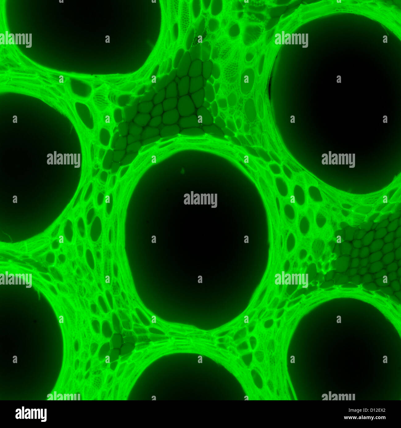 micrograph plant tissue, stem of pumpkin,with green fluorescence Stock Photohttps://www.alamy.com/image-license-details/?v=1https://www.alamy.com/stock-photo-micrograph-plant-tissue-stem-of-pumpkinwith-green-fluorescence-52301370.html
RFD12EX2–micrograph plant tissue, stem of pumpkin,with green fluorescence
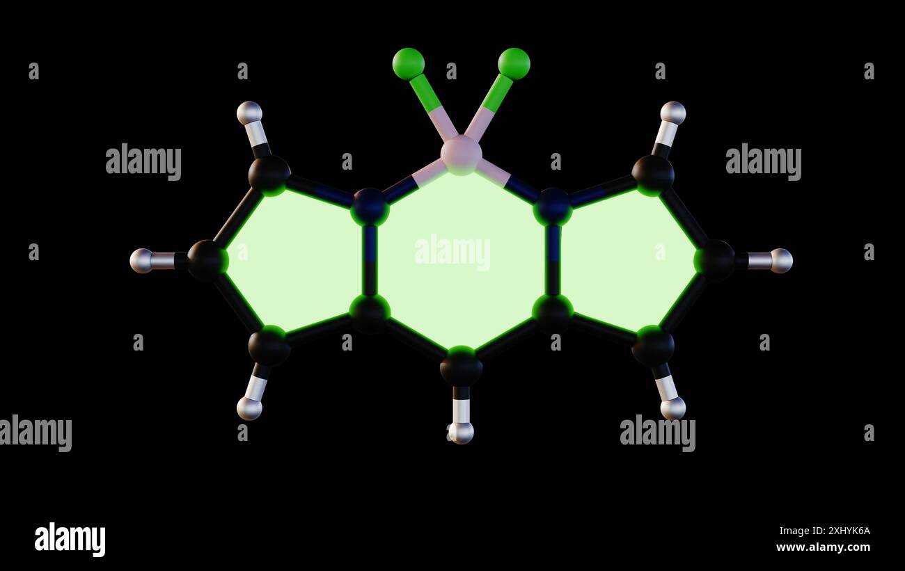 3D rendering of BODIPY: A bright dye for research, glowing for cell imaging. Stock Photohttps://www.alamy.com/image-license-details/?v=1https://www.alamy.com/3d-rendering-of-bodipy-a-bright-dye-for-research-glowing-for-cell-imaging-image613419810.html
RF2XHYK6A–3D rendering of BODIPY: A bright dye for research, glowing for cell imaging.
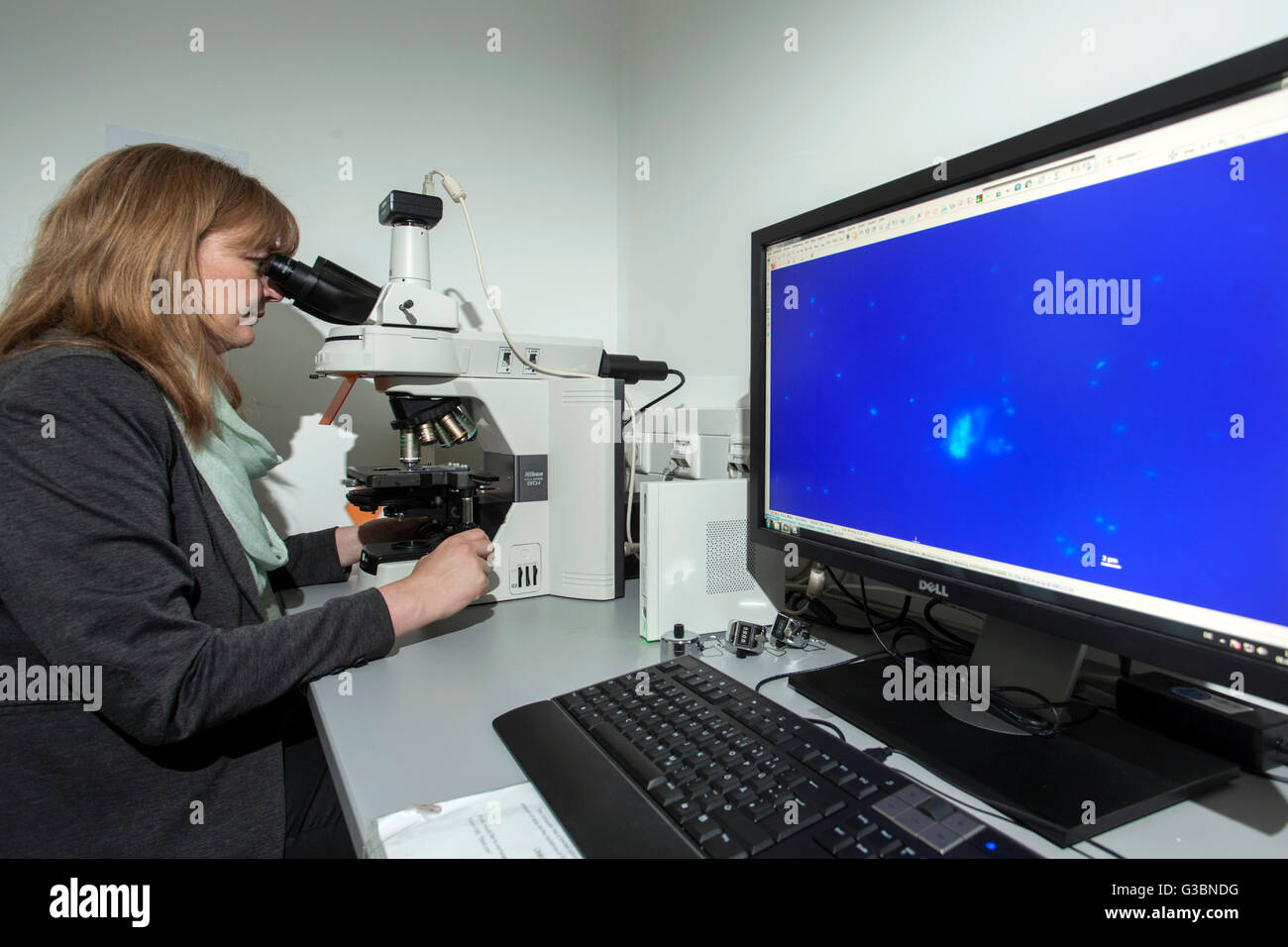 Researcher at a fluorescence microscope. Stock Photohttps://www.alamy.com/image-license-details/?v=1https://www.alamy.com/stock-photo-researcher-at-a-fluorescence-microscope-105364492.html
RMG3BNDG–Researcher at a fluorescence microscope.
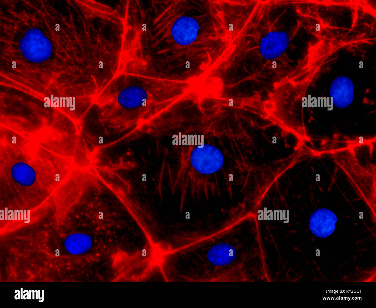 Confocal Fluorescence Microscopy of CRISPR generated knockout cells. CRISPR (Clustered Regularly Interspaced Short Palindromic Repeats) gene editing Stock Photohttps://www.alamy.com/image-license-details/?v=1https://www.alamy.com/confocal-fluorescence-microscopy-of-crispr-generated-knockout-cells-crispr-clustered-regularly-interspaced-short-palindromic-repeats-gene-editing-image240387416.html
RMRY2GGT–Confocal Fluorescence Microscopy of CRISPR generated knockout cells. CRISPR (Clustered Regularly Interspaced Short Palindromic Repeats) gene editing
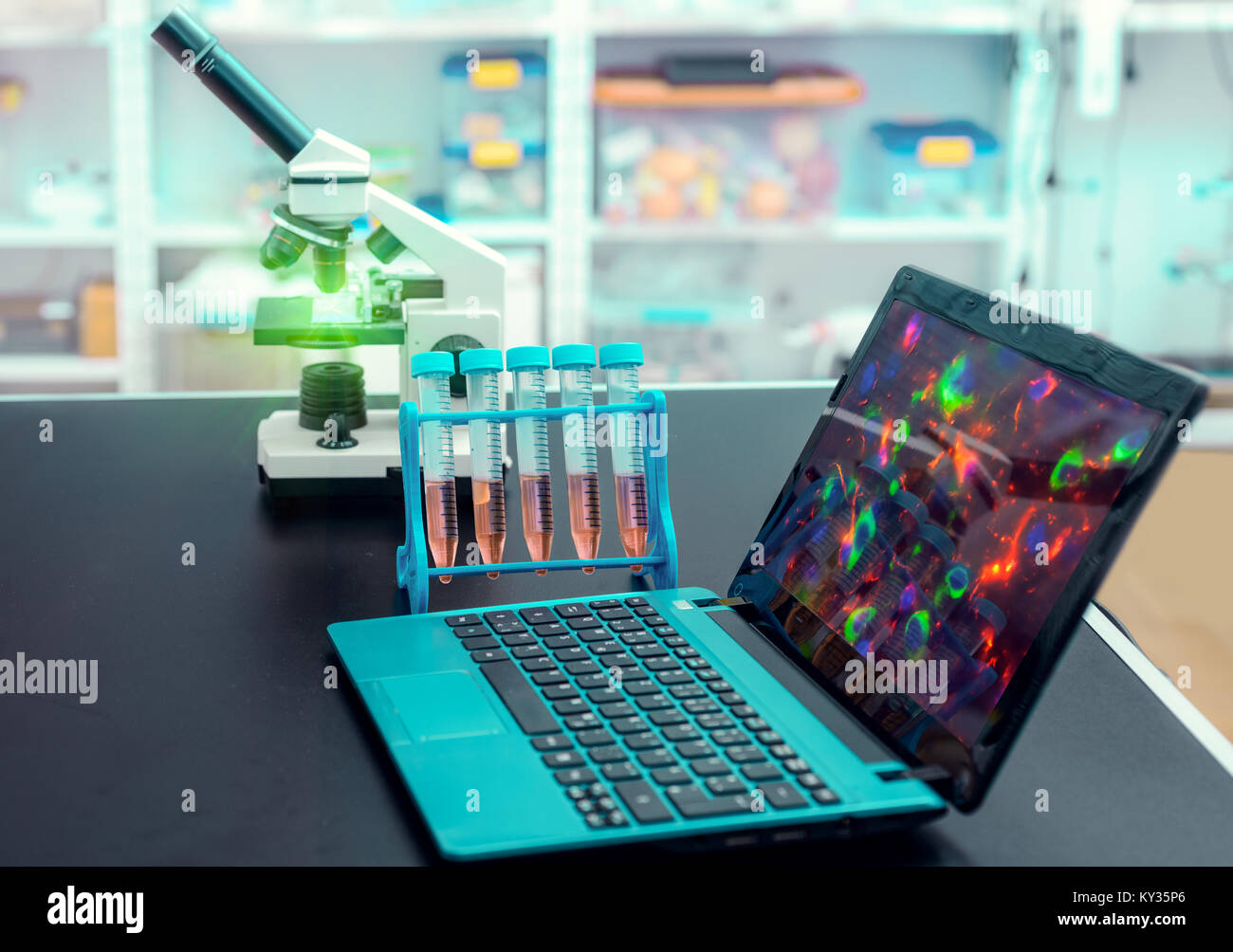 Microscope and portable computer with microscopic image on observation table, in modern laboratory Stock Photohttps://www.alamy.com/image-license-details/?v=1https://www.alamy.com/stock-photo-microscope-and-portable-computer-with-microscopic-image-on-observation-171559422.html
RFKY35P6–Microscope and portable computer with microscopic image on observation table, in modern laboratory
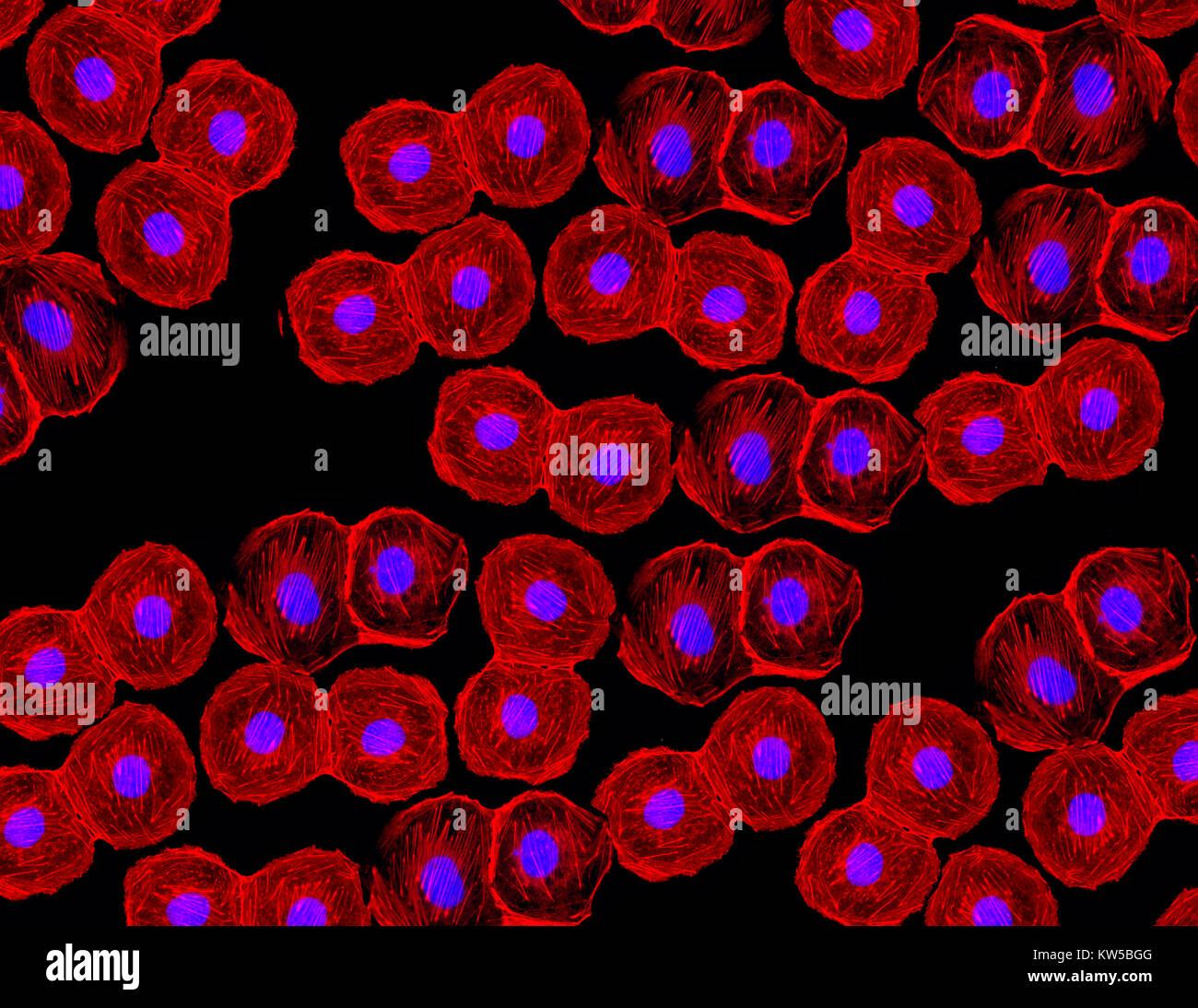 Fluorescent image of human stem cells stained with monoclonal antibodies markers under the microscopy showing nuclei in blue and microtubules in red Stock Photohttps://www.alamy.com/image-license-details/?v=1https://www.alamy.com/stock-photo-fluorescent-image-of-human-stem-cells-stained-with-monoclonal-antibodies-170378560.html
RMKW5BGG–Fluorescent image of human stem cells stained with monoclonal antibodies markers under the microscopy showing nuclei in blue and microtubules in red
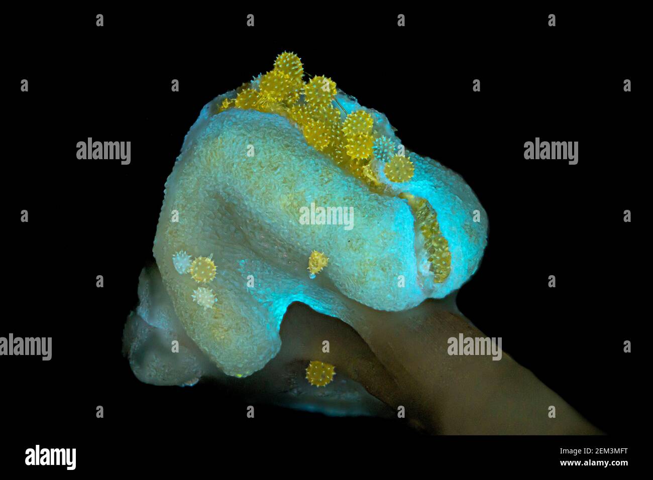 hibiscus (Hibiscus spec.), pollen of hibiscus, fluorescence, UV-light irradiation, light microscope image, magnification x24 related to 35 mm, Stock Photohttps://www.alamy.com/image-license-details/?v=1https://www.alamy.com/hibiscus-hibiscus-spec-pollen-of-hibiscus-fluorescence-uv-light-irradiation-light-microscope-image-magnification-x24-related-to-35-mm-image408213564.html
RM2EM3MFT–hibiscus (Hibiscus spec.), pollen of hibiscus, fluorescence, UV-light irradiation, light microscope image, magnification x24 related to 35 mm,
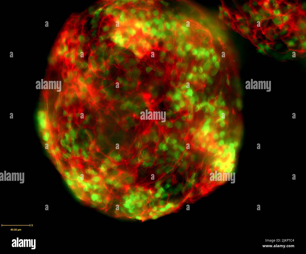 CRISPR/Cas9 engineering was used in mouse embryonic stem cells to insert an in-frame GFP tag with the motor neuron-specific transcription factor HB9. These cells are differentiated into motor neurons. The resulting motor neuron nuclei are labeled with the GFP reporter (green) and counterstained with antibodies against the neuronal marker Tuj1 (red). Stock Photohttps://www.alamy.com/image-license-details/?v=1https://www.alamy.com/crisprcas9-engineering-was-used-in-mouse-embryonic-stem-cells-to-insert-an-in-frame-gfp-tag-with-the-motor-neuron-specific-transcription-factor-hb9-these-cells-are-differentiated-into-motor-neurons-the-resulting-motor-neuron-nuclei-are-labeled-with-the-gfp-reporter-green-and-counterstained-with-antibodies-against-the-neuronal-marker-tuj1-red-image476706836.html
RM2JKFTC4–CRISPR/Cas9 engineering was used in mouse embryonic stem cells to insert an in-frame GFP tag with the motor neuron-specific transcription factor HB9. These cells are differentiated into motor neurons. The resulting motor neuron nuclei are labeled with the GFP reporter (green) and counterstained with antibodies against the neuronal marker Tuj1 (red).
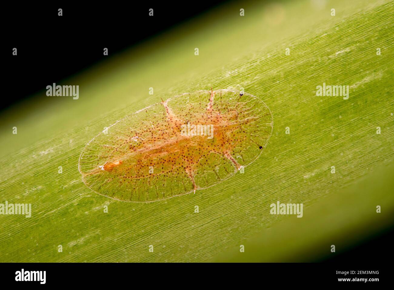 scale insects and mealy bugs (Coccoidea), fluorescence image, light microscope image, magnification x16 related to 35 mm, Germany Stock Photohttps://www.alamy.com/image-license-details/?v=1https://www.alamy.com/scale-insects-and-mealy-bugs-coccoidea-fluorescence-image-light-microscope-image-magnification-x16-related-to-35-mm-germany-image408213724.html
RM2EM3MNG–scale insects and mealy bugs (Coccoidea), fluorescence image, light microscope image, magnification x16 related to 35 mm, Germany
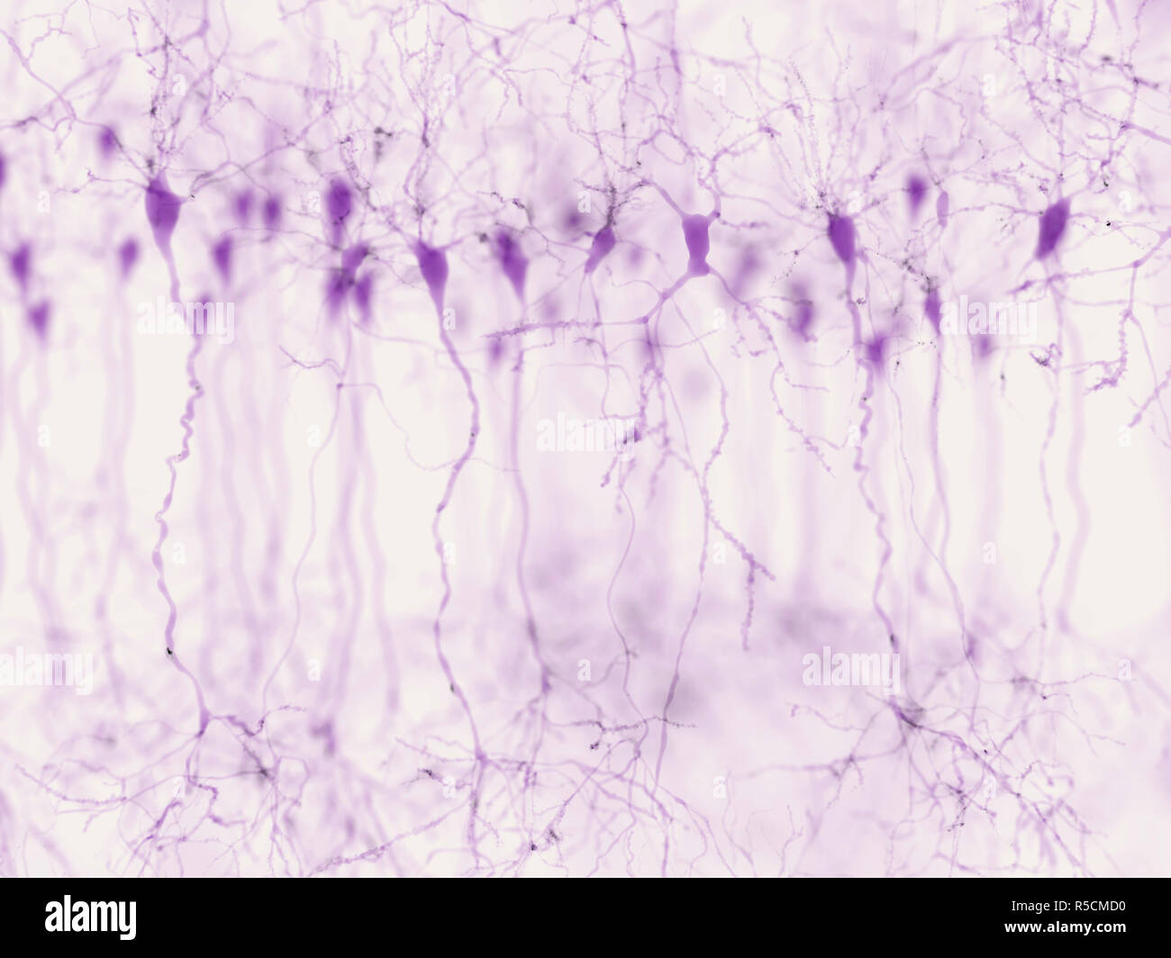 Pyramidal neurons in the cerebral cortex, illustration. Pyramidal neurons are found in certain areas of the brain including the cerebral cortex, the hippocampus, and the amygdala. Here, the illustration shows the synaptic signals highlighted using a microscopy fluorescence technique. Stock Photohttps://www.alamy.com/image-license-details/?v=1https://www.alamy.com/pyramidal-neurons-in-the-cerebral-cortex-illustration-pyramidal-neurons-are-found-in-certain-areas-of-the-brain-including-the-cerebral-cortex-the-hippocampus-and-the-amygdala-here-the-illustration-shows-the-synaptic-signals-highlighted-using-a-microscopy-fluorescence-technique-image227087532.html
RFR5CMD0–Pyramidal neurons in the cerebral cortex, illustration. Pyramidal neurons are found in certain areas of the brain including the cerebral cortex, the hippocampus, and the amygdala. Here, the illustration shows the synaptic signals highlighted using a microscopy fluorescence technique.
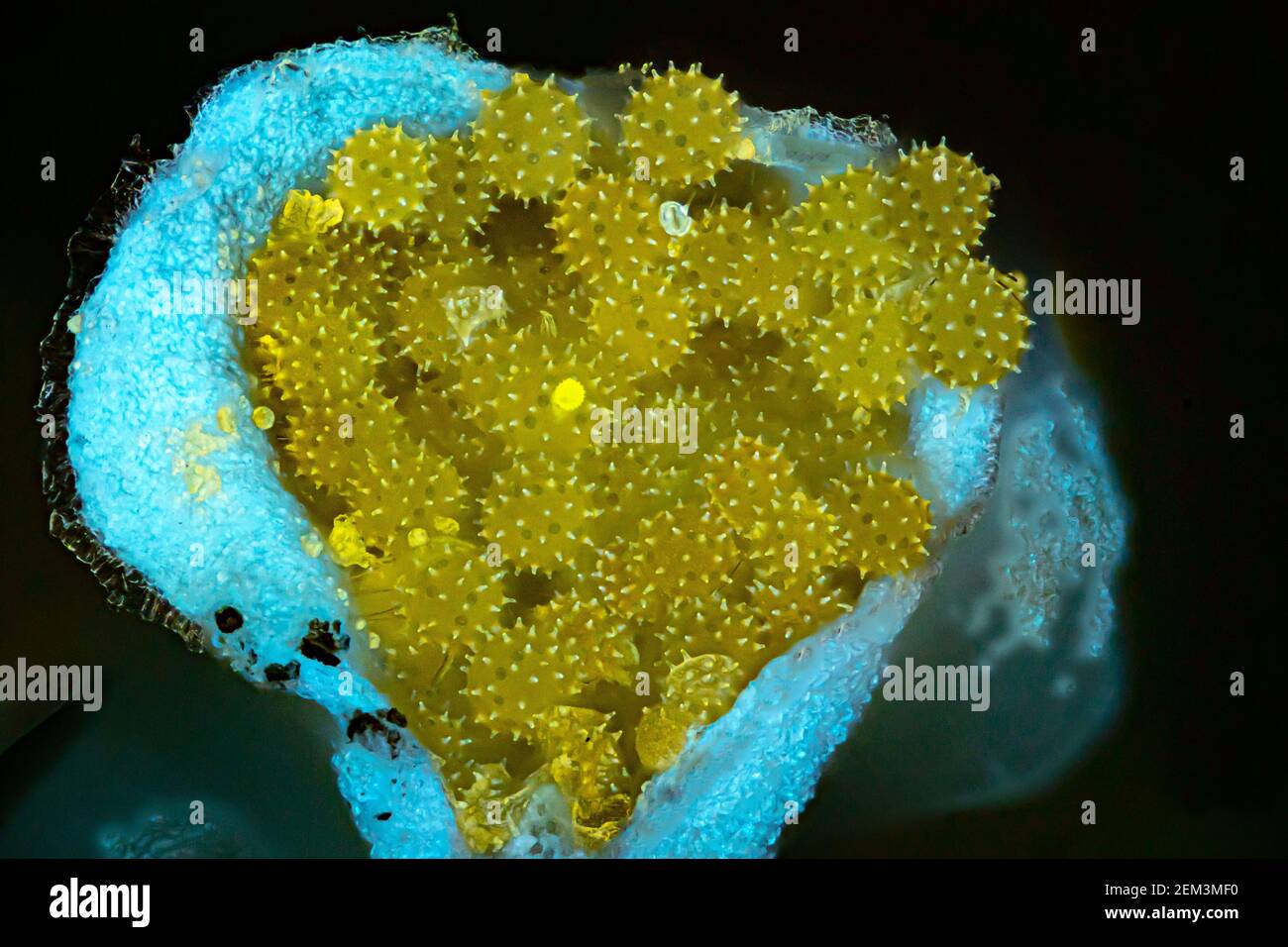 hibiscus (Hibiscus spec.), pollen of hibiscus in the anther, fluorescence, UV-light irradiation, light microscope image, magnification x34 related to Stock Photohttps://www.alamy.com/image-license-details/?v=1https://www.alamy.com/hibiscus-hibiscus-spec-pollen-of-hibiscus-in-the-anther-fluorescence-uv-light-irradiation-light-microscope-image-magnification-x34-related-to-image408213540.html
RM2EM3MF0–hibiscus (Hibiscus spec.), pollen of hibiscus in the anther, fluorescence, UV-light irradiation, light microscope image, magnification x34 related to
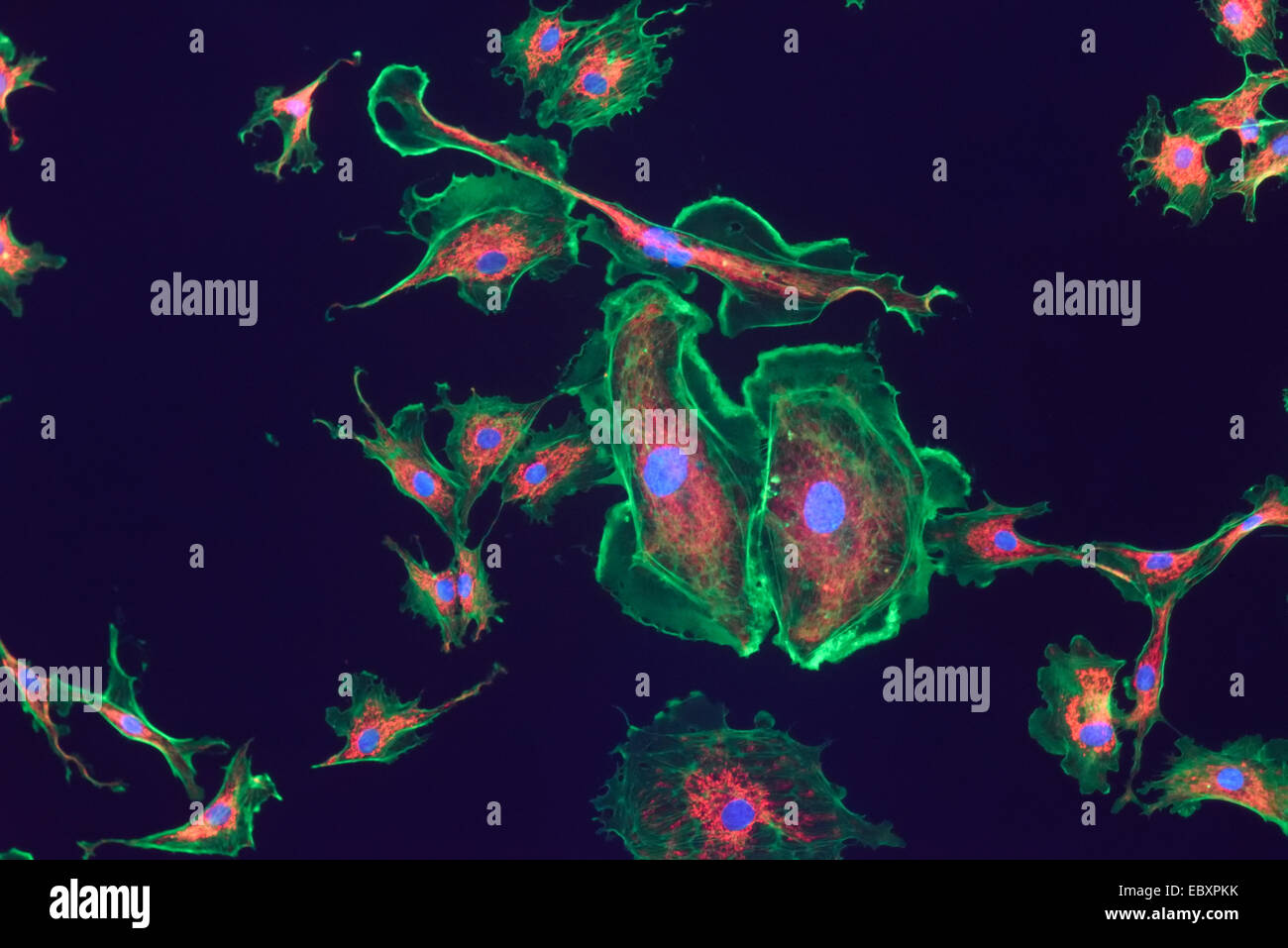 Microfilaments, mitochondria, and nuclei in fibroblast cells Stock Photohttps://www.alamy.com/image-license-details/?v=1https://www.alamy.com/stock-photo-microfilaments-mitochondria-and-nuclei-in-fibroblast-cells-76191239.html
RMEBXPKK–Microfilaments, mitochondria, and nuclei in fibroblast cells
 Scorpion fluoresce when exposed to ultraviolet (UV) light. Arthropods Workshop taught by the entomologist and environmental disseminator Sergi Romeu V Stock Photohttps://www.alamy.com/image-license-details/?v=1https://www.alamy.com/scorpion-fluoresce-when-exposed-to-ultraviolet-uv-light-arthropods-workshop-taught-by-the-entomologist-and-environmental-disseminator-sergi-romeu-v-image600448291.html
RM2WTTNWR–Scorpion fluoresce when exposed to ultraviolet (UV) light. Arthropods Workshop taught by the entomologist and environmental disseminator Sergi Romeu V
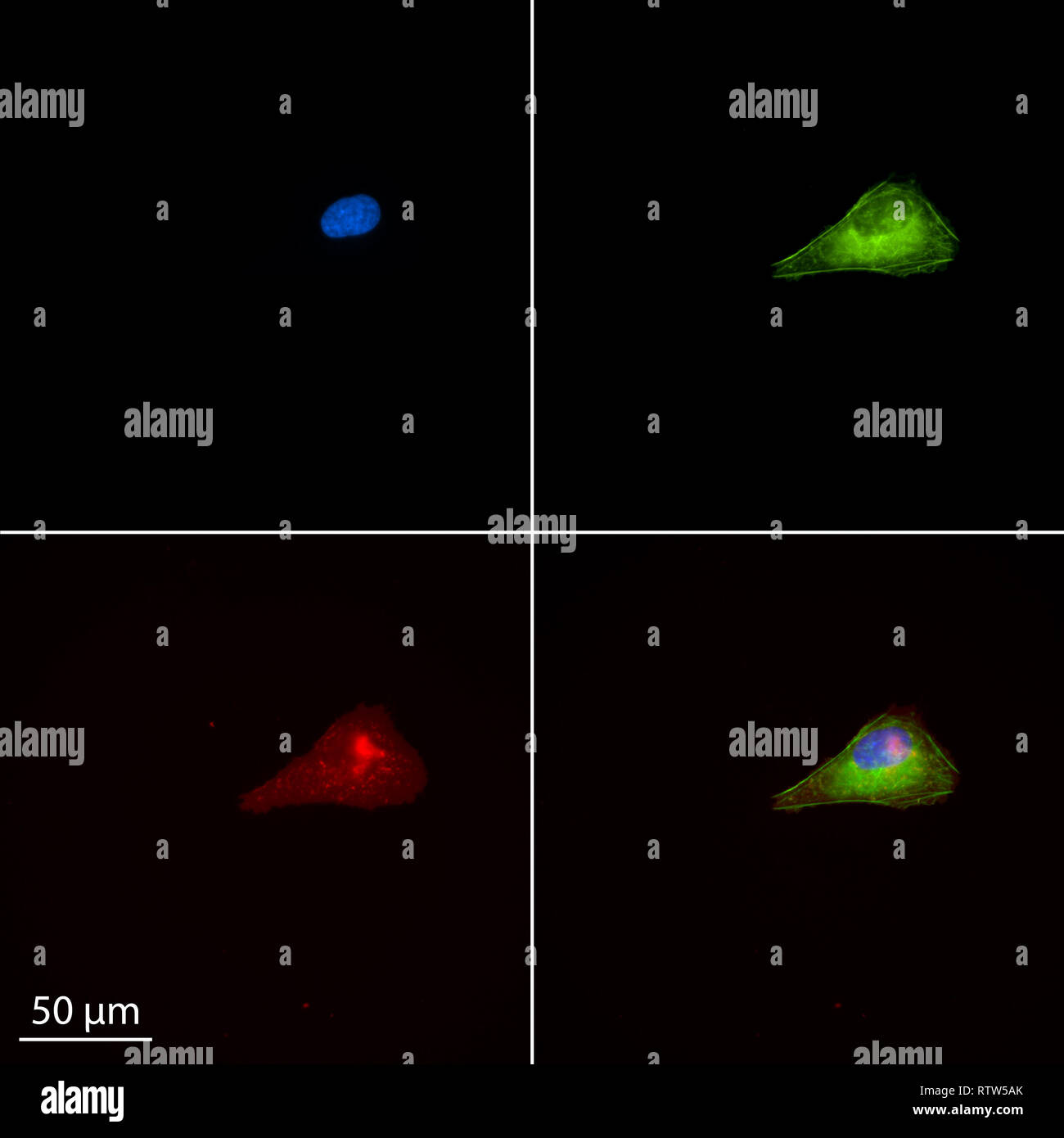 Single human osteosarcoma cell stained for epifluorescence Stock Photohttps://www.alamy.com/image-license-details/?v=1https://www.alamy.com/single-human-osteosarcoma-cell-stained-for-epifluorescence-image239039547.html
RMRTW5AK–Single human osteosarcoma cell stained for epifluorescence
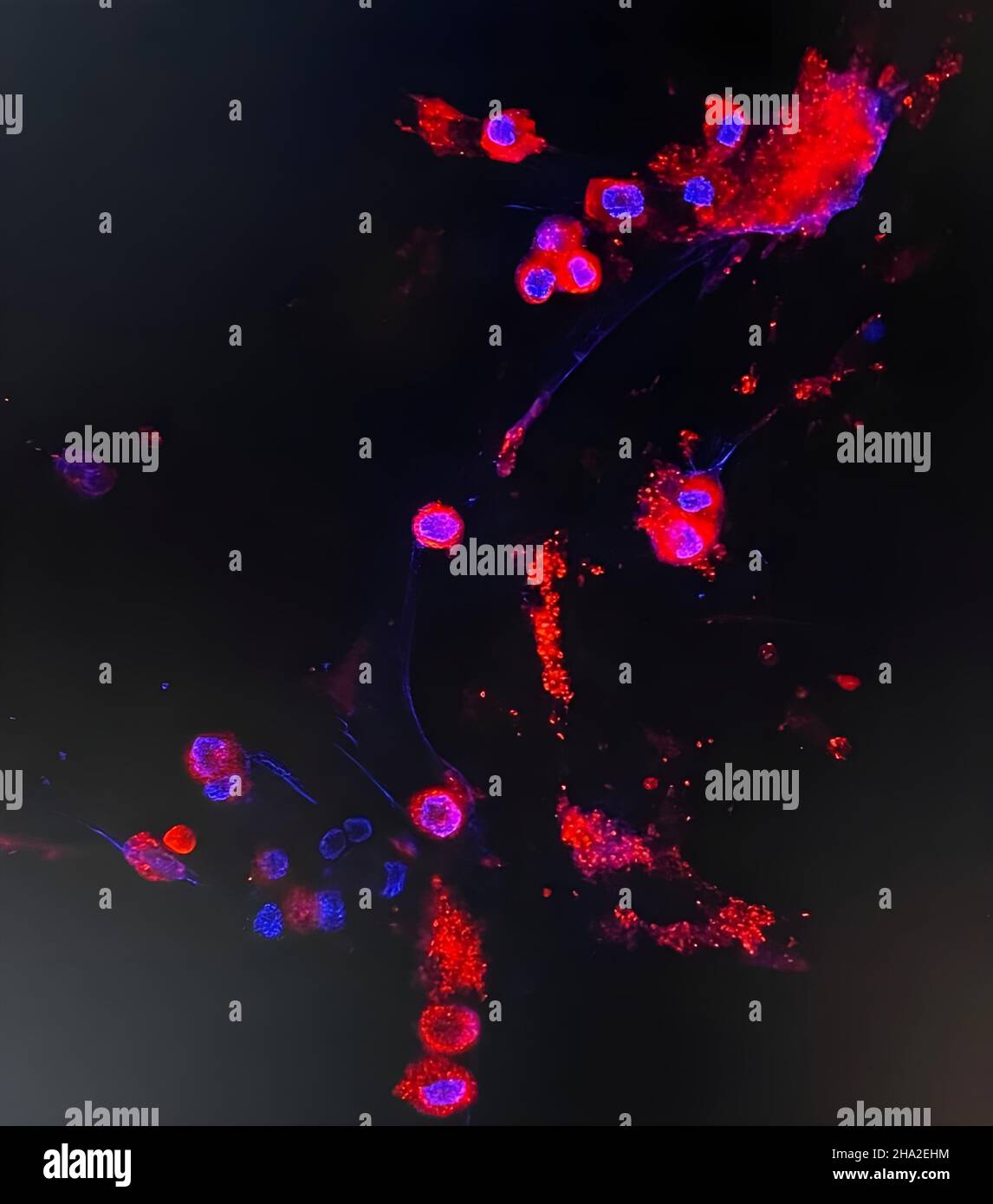 Omicron variant Stock Photohttps://www.alamy.com/image-license-details/?v=1https://www.alamy.com/omicron-variant-image453671504.html
RM2HA2EHM–Omicron variant
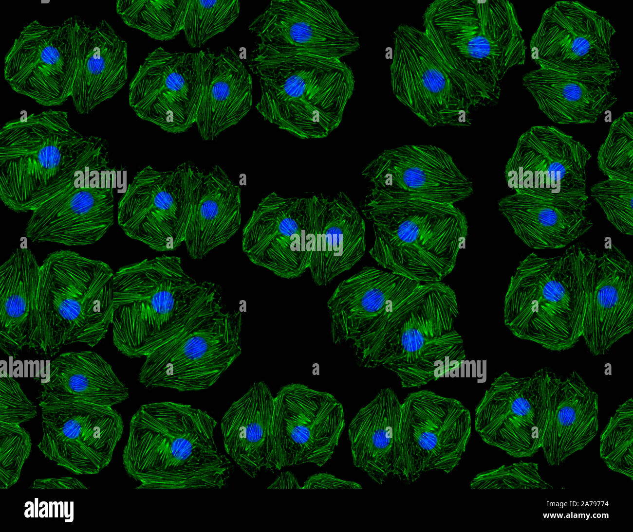 Fluorescent image of human stem cells stained with monoclonal antibodies markers under the microscopy nuclei in blue and actin microfilaments in green Stock Photohttps://www.alamy.com/image-license-details/?v=1https://www.alamy.com/fluorescent-image-of-human-stem-cells-stained-with-monoclonal-antibodies-markers-under-the-microscopy-nuclei-in-blue-and-actin-microfilaments-in-green-image331502840.html
RM2A79774–Fluorescent image of human stem cells stained with monoclonal antibodies markers under the microscopy nuclei in blue and actin microfilaments in green
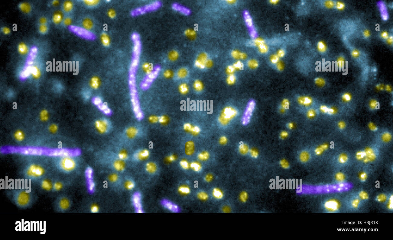 Quantum Dots bound to E.coli Bacteria Stock Photohttps://www.alamy.com/image-license-details/?v=1https://www.alamy.com/stock-photo-quantum-dots-bound-to-ecoli-bacteria-135022886.html
RMHRJR1X–Quantum Dots bound to E.coli Bacteria
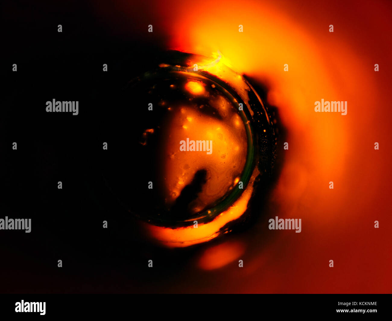 Ultraviolet reflected light micrograph of a red fluorescent ball pen tip, pictured area is about 1.7mm wide Stock Photohttps://www.alamy.com/image-license-details/?v=1https://www.alamy.com/stock-image-ultraviolet-reflected-light-micrograph-of-a-red-fluorescent-ball-pen-162703310.html
RMKCKNME–Ultraviolet reflected light micrograph of a red fluorescent ball pen tip, pictured area is about 1.7mm wide
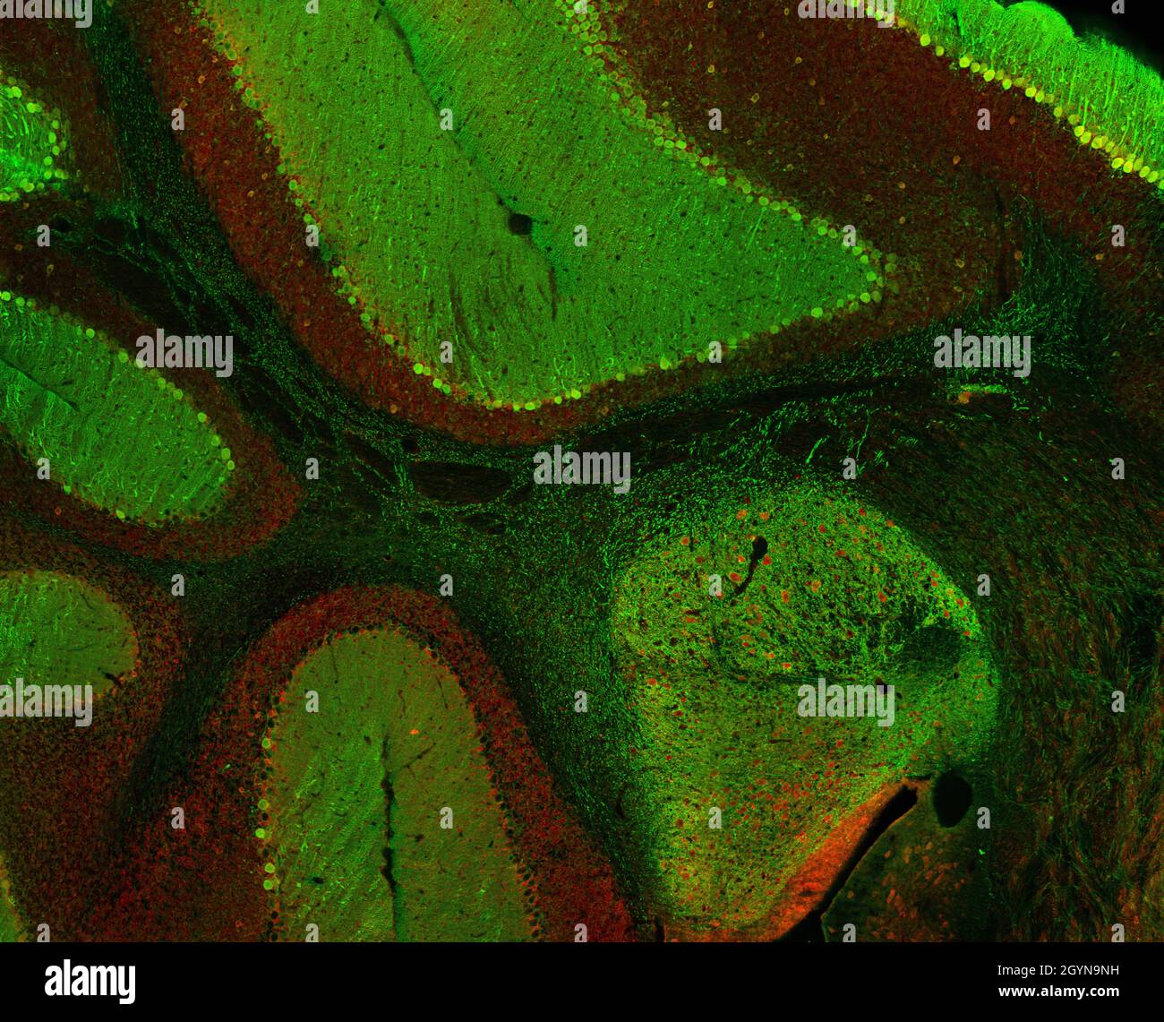 Sagittal section of mouse cerebellum labelled with immunofluorescence and visualized with confocal laser scanning microscopy. Large Purkinje cells. Stock Photohttps://www.alamy.com/image-license-details/?v=1https://www.alamy.com/sagittal-section-of-mouse-cerebellum-labelled-with-immunofluorescence-and-visualized-with-confocal-laser-scanning-microscopy-large-purkinje-cells-image447323565.html
RF2GYN9NH–Sagittal section of mouse cerebellum labelled with immunofluorescence and visualized with confocal laser scanning microscopy. Large Purkinje cells.
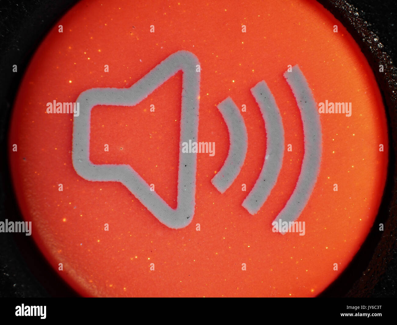 Fluorescent red alarm button from car key fob, ultraviolet + visible light micrograph, 12x magnification when printed 10cm wide Stock Photohttps://www.alamy.com/image-license-details/?v=1https://www.alamy.com/fluorescent-red-alarm-button-from-car-key-fob-ultraviolet-visible-image154419884.html
RFJY6C3T–Fluorescent red alarm button from car key fob, ultraviolet + visible light micrograph, 12x magnification when printed 10cm wide
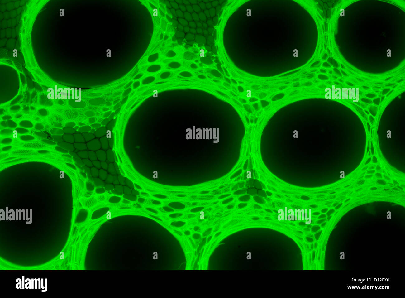 micrograph plant tissue, stem of pumpkin,with green fluorescence Stock Photohttps://www.alamy.com/image-license-details/?v=1https://www.alamy.com/stock-photo-micrograph-plant-tissue-stem-of-pumpkinwith-green-fluorescence-52301368.html
RFD12EX0–micrograph plant tissue, stem of pumpkin,with green fluorescence
 3D rendering of BODIPY: A bright dye for research, glowing for cell imaging. Stock Photohttps://www.alamy.com/image-license-details/?v=1https://www.alamy.com/3d-rendering-of-bodipy-a-bright-dye-for-research-glowing-for-cell-imaging-image613419803.html
RF2XHYK63–3D rendering of BODIPY: A bright dye for research, glowing for cell imaging.
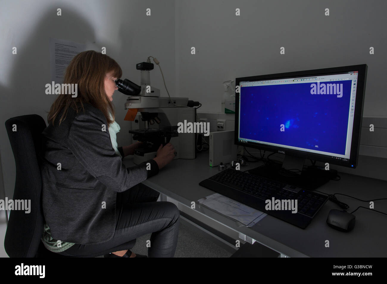 Researcher at a fluorescence microscope. Stock Photohttps://www.alamy.com/image-license-details/?v=1https://www.alamy.com/stock-photo-researcher-at-a-fluorescence-microscope-105364473.html
RMG3BNCW–Researcher at a fluorescence microscope.
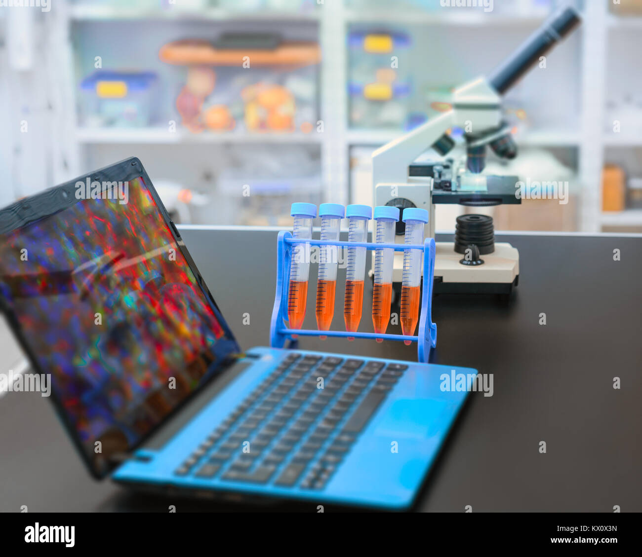 Microscope and laptop with digital microscopic image in research facility Stock Photohttps://www.alamy.com/image-license-details/?v=1https://www.alamy.com/stock-photo-microscope-and-laptop-with-digital-microscopic-image-in-research-facility-170894857.html
RFKX0X3N–Microscope and laptop with digital microscopic image in research facility
 Scientist examining samples under a microscope in a laboratory Stock Photohttps://www.alamy.com/image-license-details/?v=1https://www.alamy.com/scientist-examining-samples-under-a-microscope-in-a-laboratory-image600441632.html
RF2WTTDC0–Scientist examining samples under a microscope in a laboratory
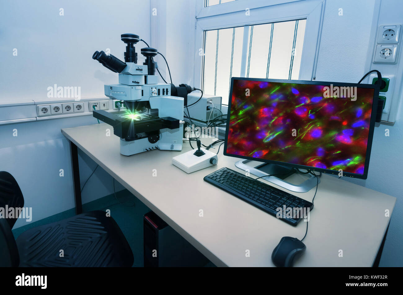 Modern microscope station with cell stained with antibody on the screen Stock Photohttps://www.alamy.com/image-license-details/?v=1https://www.alamy.com/stock-photo-modern-microscope-station-with-cell-stained-with-antibody-on-the-screen-170591423.html
RFKWF32R–Modern microscope station with cell stained with antibody on the screen
 Obtention of HepG2 cell images through fluorescence microscopy in the ´colony forming assay´ to determine the toxicity of the sample, Cell Culture Laboratory, Unit of Health Technology, Tecnalia Research & Innovation, Miramon Technology Park, San Sebastian, Donostia, Gipuzkoa, Basque Country, Spain. Stock Photohttps://www.alamy.com/image-license-details/?v=1https://www.alamy.com/obtention-of-hepg2-cell-images-through-fluorescence-microscopy-in-the-colony-forming-assay-to-determine-the-toxicity-of-the-sample-cell-culture-laboratory-unit-of-health-technology-tecnalia-research-innovation-miramon-technology-park-san-sebastian-donostia-gipuzkoa-basque-country-spain-image602037435.html
RM2WYD4TY–Obtention of HepG2 cell images through fluorescence microscopy in the ´colony forming assay´ to determine the toxicity of the sample, Cell Culture Laboratory, Unit of Health Technology, Tecnalia Research & Innovation, Miramon Technology Park, San Sebastian, Donostia, Gipuzkoa, Basque Country, Spain.
 A microscopic instrument Stock Vectorhttps://www.alamy.com/image-license-details/?v=1https://www.alamy.com/a-microscopic-instrument-image446416420.html
RF2GX80KG–A microscopic instrument
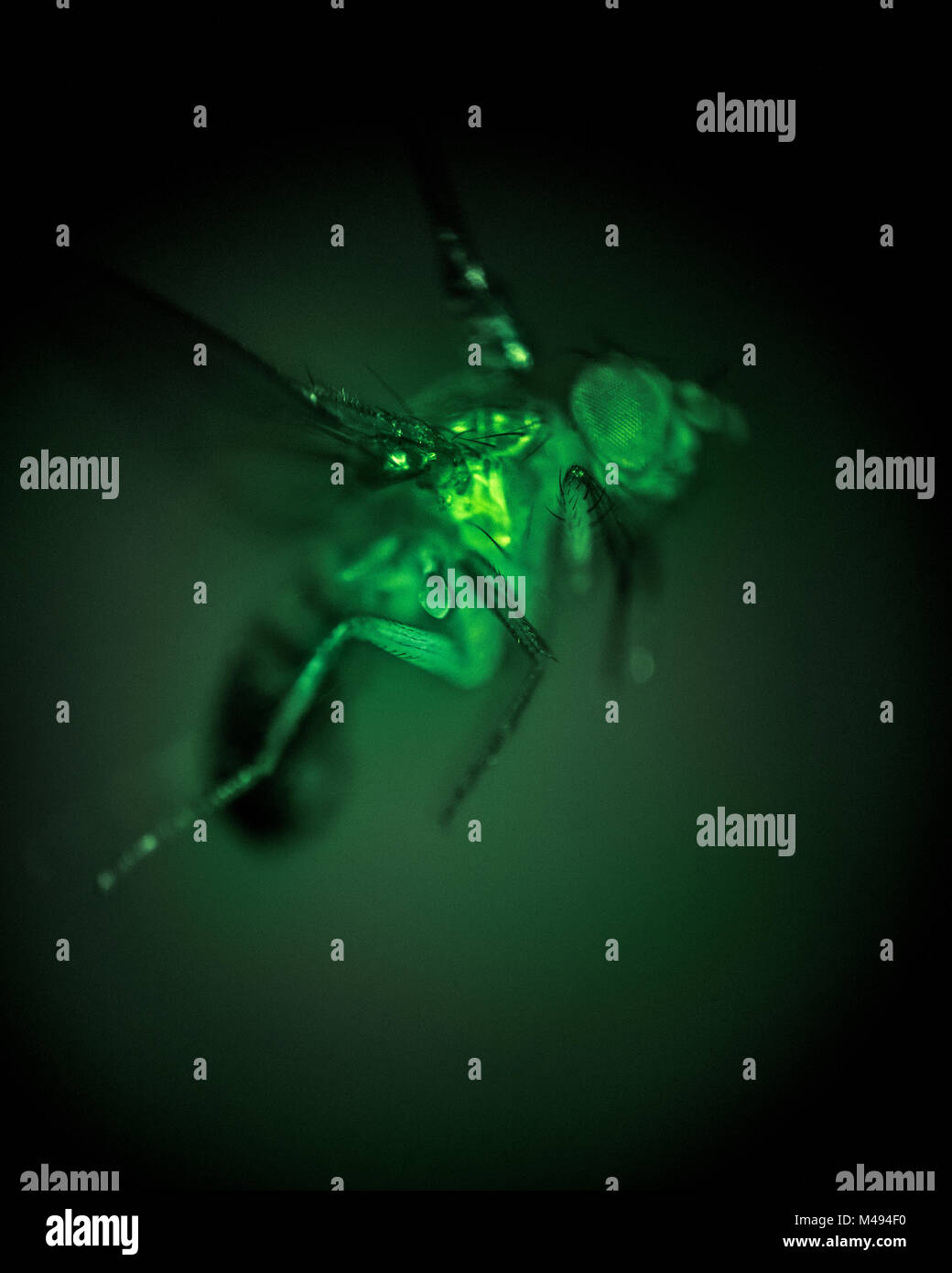 High speed exposure of a Fruit fly (Drosophila melanogaster) taken through a microscope. In this image flight muscles express a genetically engineered protein called GCaMP, which fluoresces green in the presence of calcium ions. Because the concentration of calcium increases in muscles as they are activated, the fluorescence provides a direct measurement of the muscle activity. California Institute of Technology, USA, December 2016. Stock Photohttps://www.alamy.com/image-license-details/?v=1https://www.alamy.com/stock-photo-high-speed-exposure-of-a-fruit-fly-drosophila-melanogaster-taken-through-174763428.html
RMM494F0–High speed exposure of a Fruit fly (Drosophila melanogaster) taken through a microscope. In this image flight muscles express a genetically engineered protein called GCaMP, which fluoresces green in the presence of calcium ions. Because the concentration of calcium increases in muscles as they are activated, the fluorescence provides a direct measurement of the muscle activity. California Institute of Technology, USA, December 2016.
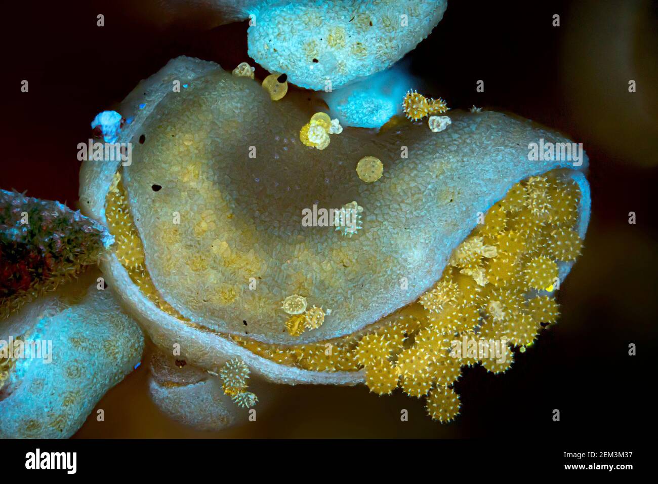 hibiscus (Hibiscus spec.), pollen of hibiscus in the anther, fluorescence, UV-light irradiation, light microscope image, magnification x34 related to Stock Photohttps://www.alamy.com/image-license-details/?v=1https://www.alamy.com/hibiscus-hibiscus-spec-pollen-of-hibiscus-in-the-anther-fluorescence-uv-light-irradiation-light-microscope-image-magnification-x34-related-to-image408213211.html
RM2EM3M37–hibiscus (Hibiscus spec.), pollen of hibiscus in the anther, fluorescence, UV-light irradiation, light microscope image, magnification x34 related to
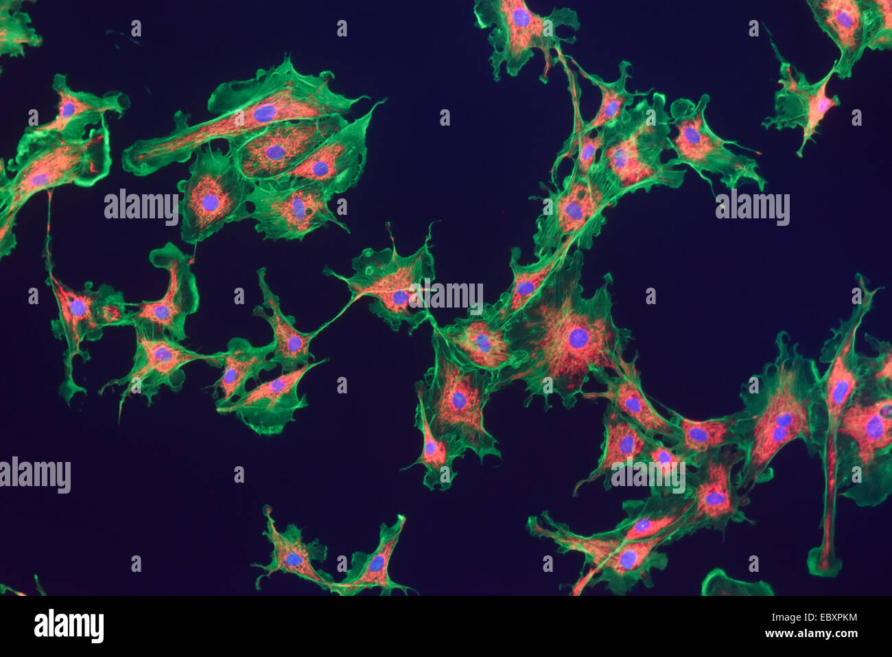 Microfilaments, mitochondria, and nuclei in fibroblast cells Stock Photohttps://www.alamy.com/image-license-details/?v=1https://www.alamy.com/stock-photo-microfilaments-mitochondria-and-nuclei-in-fibroblast-cells-76191240.html
RMEBXPKM–Microfilaments, mitochondria, and nuclei in fibroblast cells
 Scorpion fluoresce when exposed to ultraviolet (UV) light. Arthropods Workshop taught by the entomologist and environmental disseminator Sergi Romeu V Stock Photohttps://www.alamy.com/image-license-details/?v=1https://www.alamy.com/scorpion-fluoresce-when-exposed-to-ultraviolet-uv-light-arthropods-workshop-taught-by-the-entomologist-and-environmental-disseminator-sergi-romeu-v-image600448463.html
RM2WTTP3Y–Scorpion fluoresce when exposed to ultraviolet (UV) light. Arthropods Workshop taught by the entomologist and environmental disseminator Sergi Romeu V
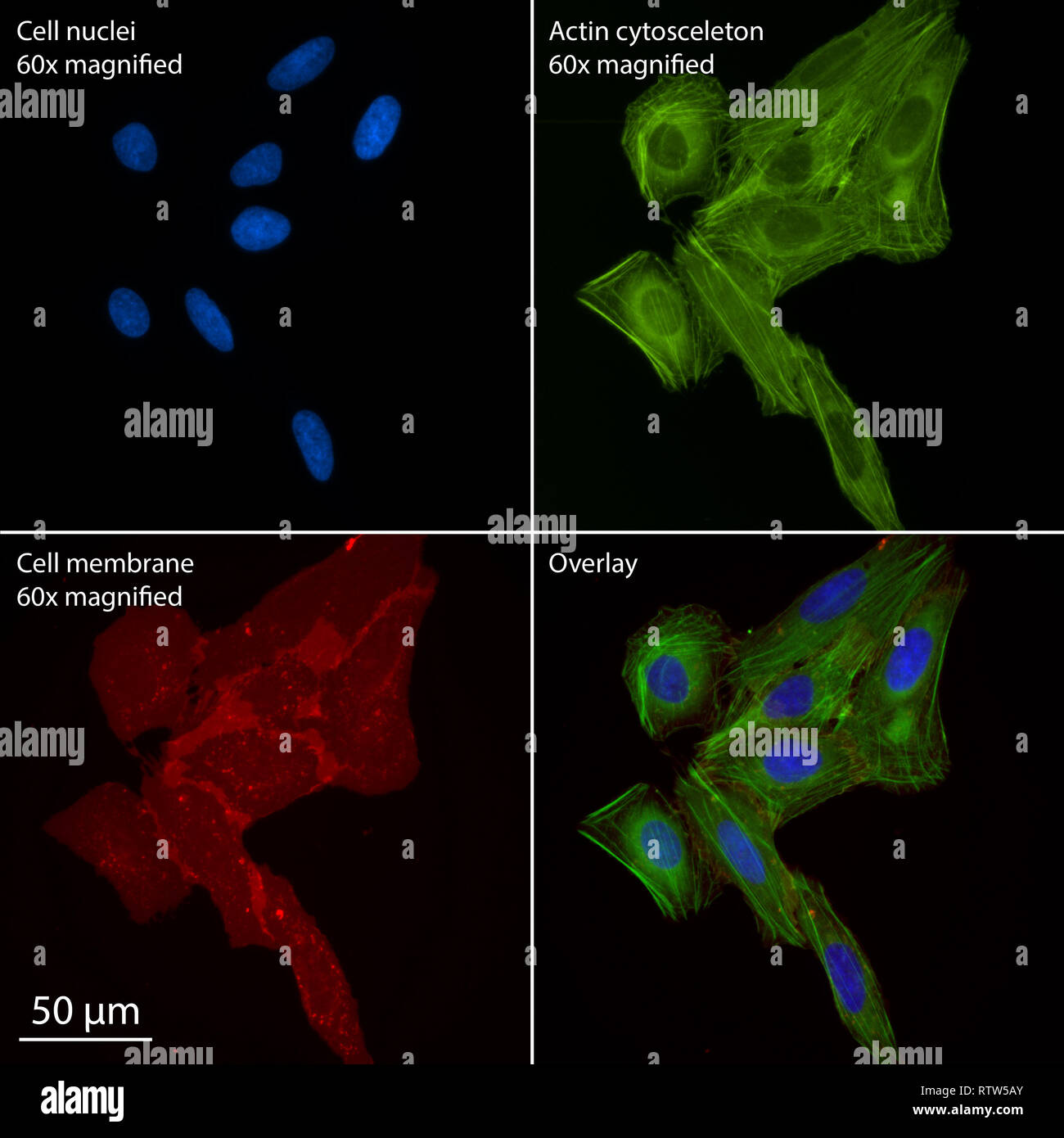 multiple human osteosarcoma cells stained for epifluorescence Stock Photohttps://www.alamy.com/image-license-details/?v=1https://www.alamy.com/multiple-human-osteosarcoma-cells-stained-for-epifluorescence-image239039555.html
RMRTW5AY–multiple human osteosarcoma cells stained for epifluorescence
 Omicron variant Stock Photohttps://www.alamy.com/image-license-details/?v=1https://www.alamy.com/omicron-variant-image453671508.html
RM2HA2EHT–Omicron variant
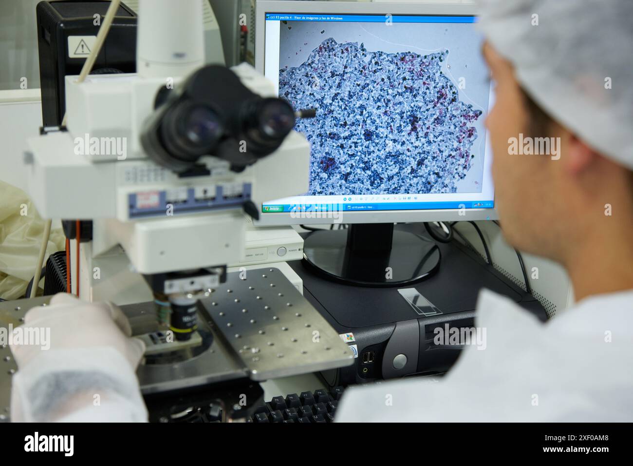 Obtention of HepG2 cell images through fluorescence microscopy in the ´colony forming assay´ to determine the toxicity of the sample, Cell Culture Lab Stock Photohttps://www.alamy.com/image-license-details/?v=1https://www.alamy.com/obtention-of-hepg2-cell-images-through-fluorescence-microscopy-in-the-colony-forming-assay-to-determine-the-toxicity-of-the-sample-cell-culture-lab-image611591128.html
RM2XF0AM8–Obtention of HepG2 cell images through fluorescence microscopy in the ´colony forming assay´ to determine the toxicity of the sample, Cell Culture Lab
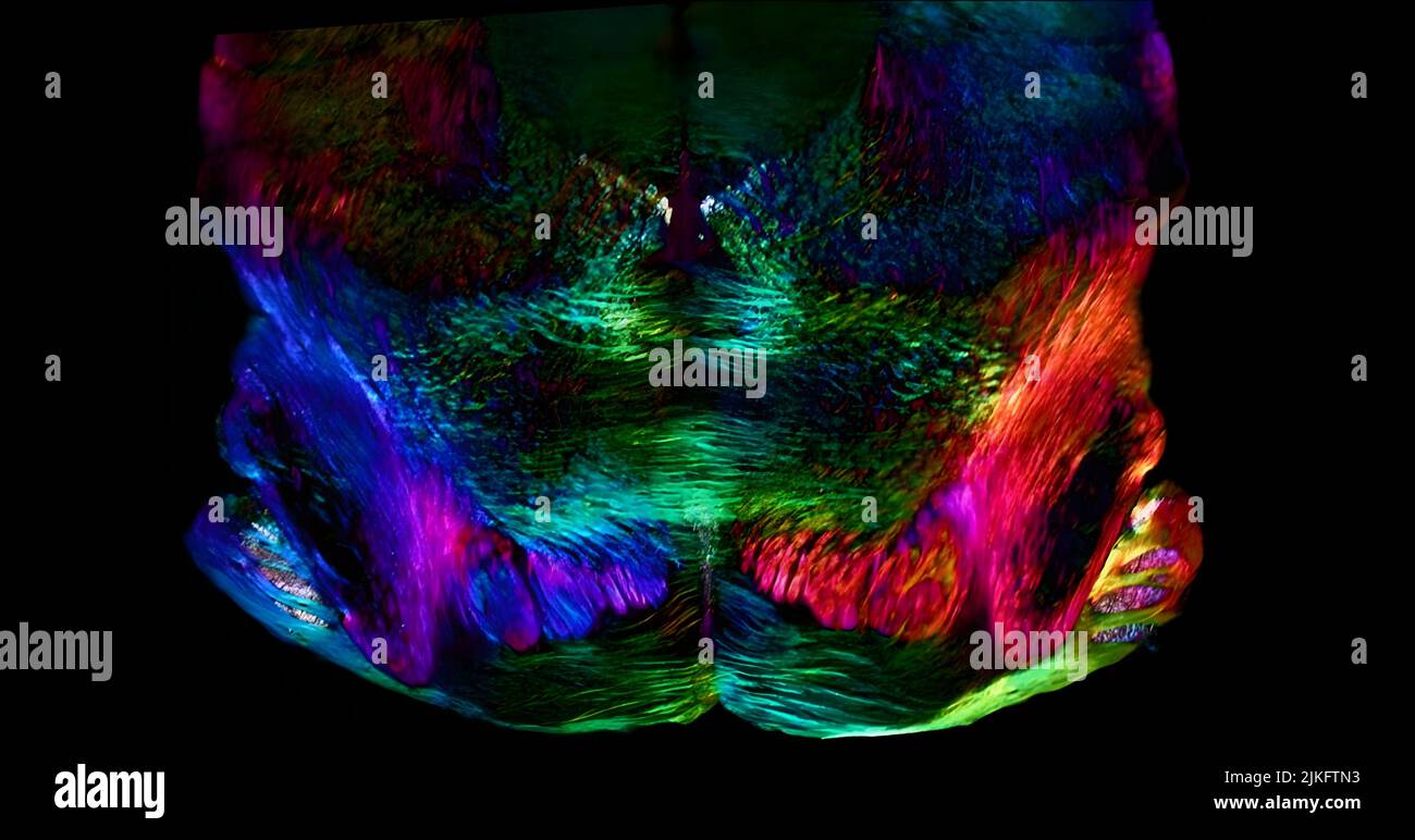 In this image, the researchers used a newly developed polarized light microscope to plot the spatial orientation of neurons in a thin section of the mouse midbrain. Neurons that stretch horizontally displayed in green, while those oriented at a 45 degree angle are pinkish red and those at 225 degrees are purplish blue. These colors do not involve staining or labeling the cells with fluorescent markers: the colors are emitted strictly from light interacting with the physical orientation of each neuron. Stock Photohttps://www.alamy.com/image-license-details/?v=1https://www.alamy.com/in-this-image-the-researchers-used-a-newly-developed-polarized-light-microscope-to-plot-the-spatial-orientation-of-neurons-in-a-thin-section-of-the-mouse-midbrain-neurons-that-stretch-horizontally-displayed-in-green-while-those-oriented-at-a-45-degree-angle-are-pinkish-red-and-those-at-225-degrees-are-purplish-blue-these-colors-do-not-involve-staining-or-labeling-the-cells-with-fluorescent-markers-the-colors-are-emitted-strictly-from-light-interacting-with-the-physical-orientation-of-each-neuron-image476707087.html
RM2JKFTN3–In this image, the researchers used a newly developed polarized light microscope to plot the spatial orientation of neurons in a thin section of the mouse midbrain. Neurons that stretch horizontally displayed in green, while those oriented at a 45 degree angle are pinkish red and those at 225 degrees are purplish blue. These colors do not involve staining or labeling the cells with fluorescent markers: the colors are emitted strictly from light interacting with the physical orientation of each neuron.
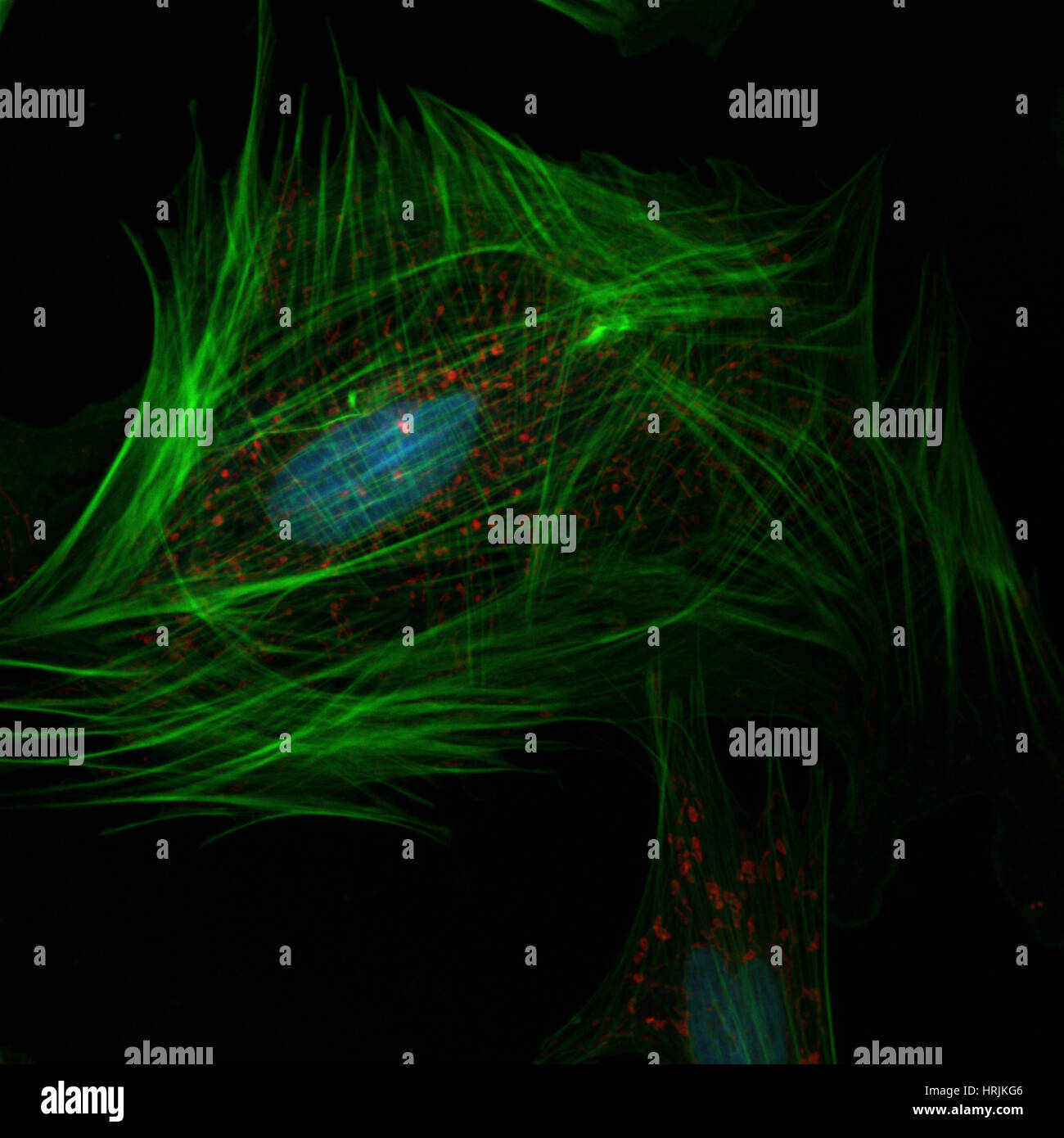 Fibroblast, Confocal Microscopy Stock Photohttps://www.alamy.com/image-license-details/?v=1https://www.alamy.com/stock-photo-fibroblast-confocal-microscopy-135020150.html
RMHRJKG6–Fibroblast, Confocal Microscopy
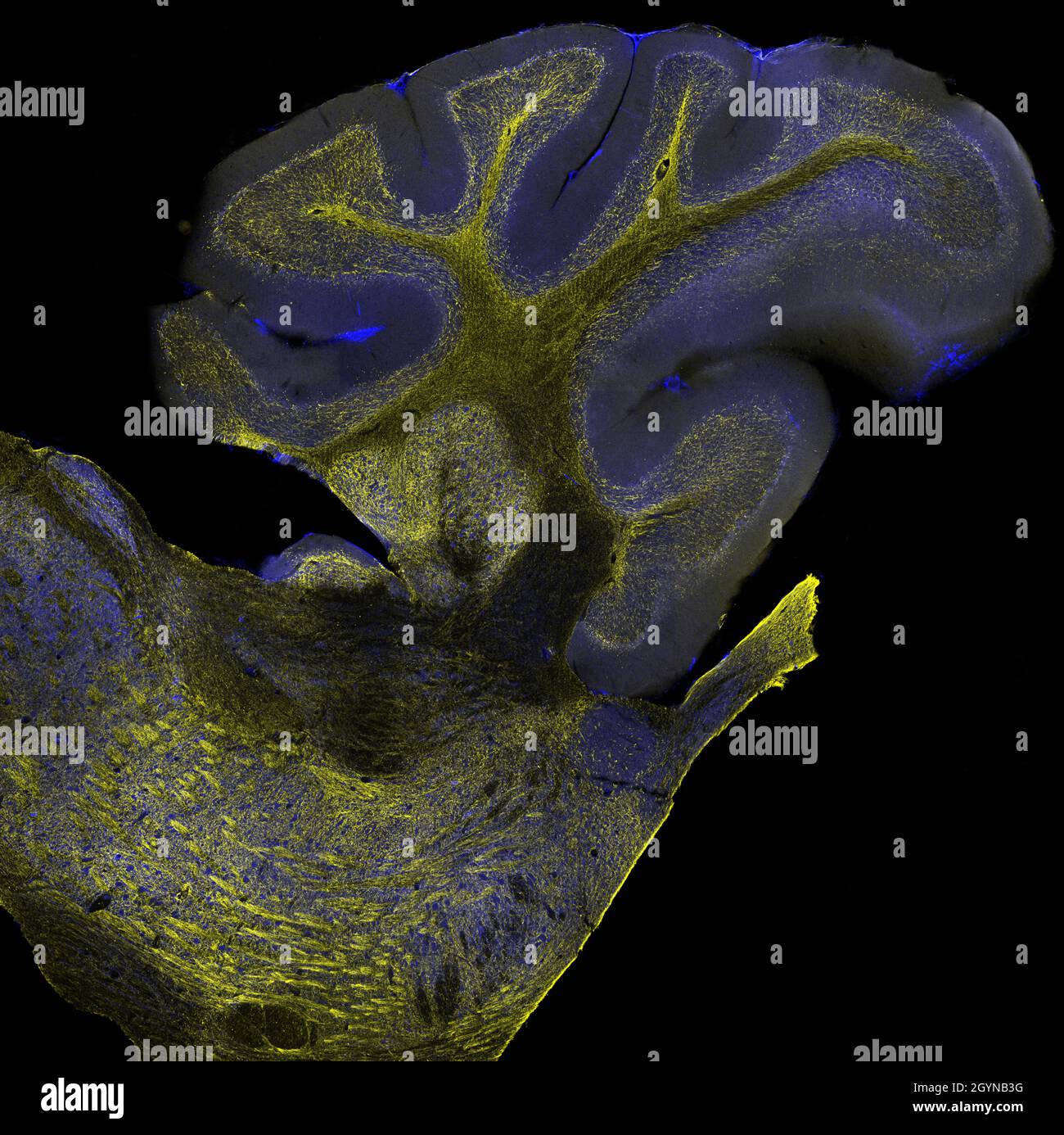 Sagittal section of mouse cerebellum labelled with immunofluorescence and visualized with confocal laser scanning microscopy. Large Purkinje cells Stock Photohttps://www.alamy.com/image-license-details/?v=1https://www.alamy.com/sagittal-section-of-mouse-cerebellum-labelled-with-immunofluorescence-and-visualized-with-confocal-laser-scanning-microscopy-large-purkinje-cells-image447324628.html
RF2GYNB3G–Sagittal section of mouse cerebellum labelled with immunofluorescence and visualized with confocal laser scanning microscopy. Large Purkinje cells
 Number 12 from fluorescent plastic 12-sided dice, ultraviolet + visible light micrograph, 12x magnification when printed 10cm wide Stock Photohttps://www.alamy.com/image-license-details/?v=1https://www.alamy.com/number-12-from-fluorescent-plastic-12-sided-dice-ultraviolet-visible-image154419883.html
RFJY6C3R–Number 12 from fluorescent plastic 12-sided dice, ultraviolet + visible light micrograph, 12x magnification when printed 10cm wide
 micrograph plant tissue, stem of pumpkin,with red fluorescence Stock Photohttps://www.alamy.com/image-license-details/?v=1https://www.alamy.com/stock-photo-micrograph-plant-tissue-stem-of-pumpkinwith-red-fluorescence-52301260.html
RFD12EP4–micrograph plant tissue, stem of pumpkin,with red fluorescence
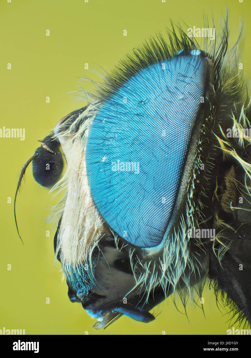 Hoverfly (family Syrphidae) with fluorescent blue eyes, reflected visible + ultraviolet light micrograph, 24x magnification with printed 10cm tall Stock Photohttps://www.alamy.com/image-license-details/?v=1https://www.alamy.com/hoverfly-family-syrphidae-with-fluorescent-blue-eyes-reflected-visible-image153950635.html
RMJXD1GY–Hoverfly (family Syrphidae) with fluorescent blue eyes, reflected visible + ultraviolet light micrograph, 24x magnification with printed 10cm tall
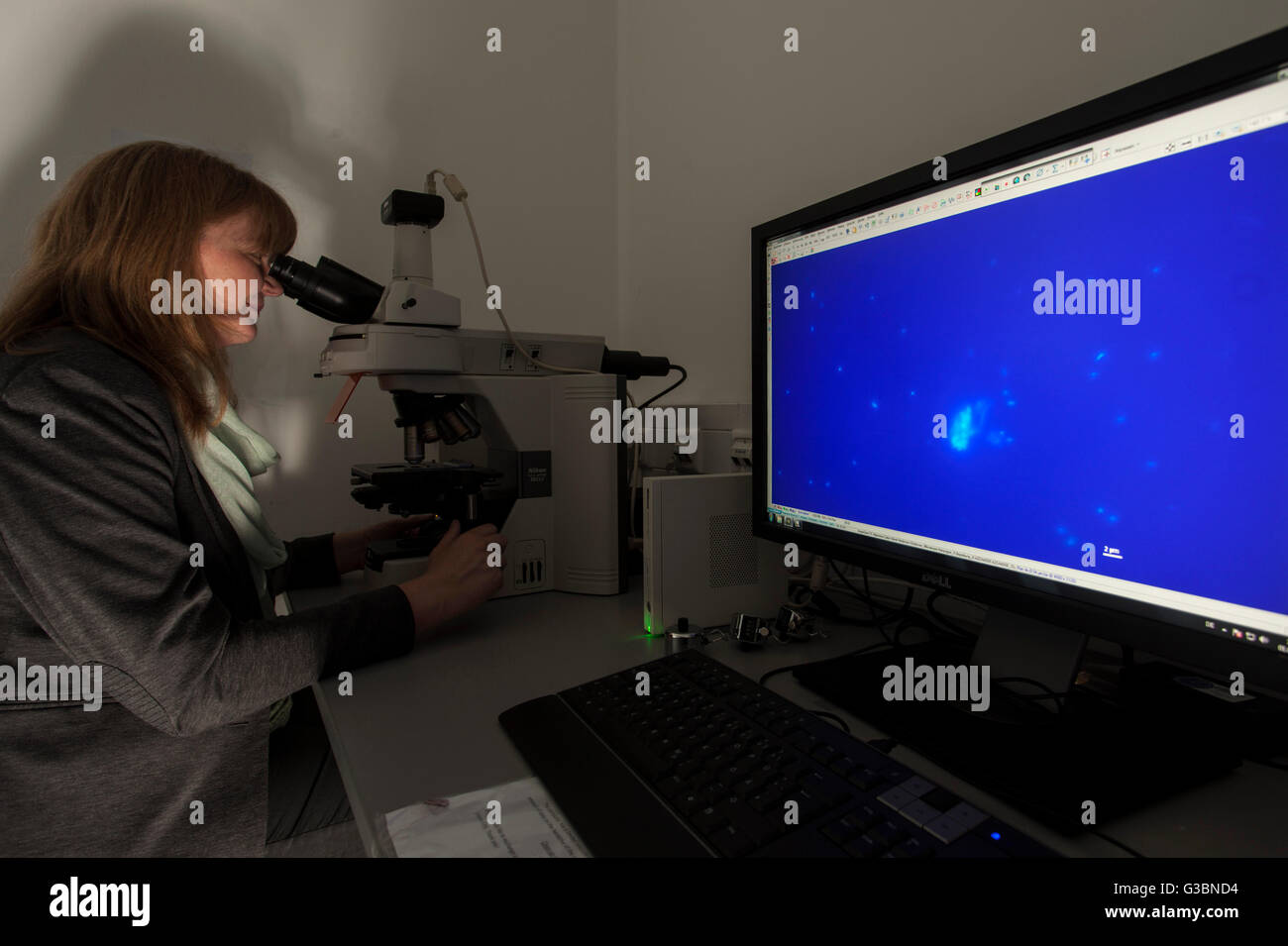 Researcher at a fluorescence microscope. Stock Photohttps://www.alamy.com/image-license-details/?v=1https://www.alamy.com/stock-photo-researcher-at-a-fluorescence-microscope-105364480.html
RMG3BND4–Researcher at a fluorescence microscope.
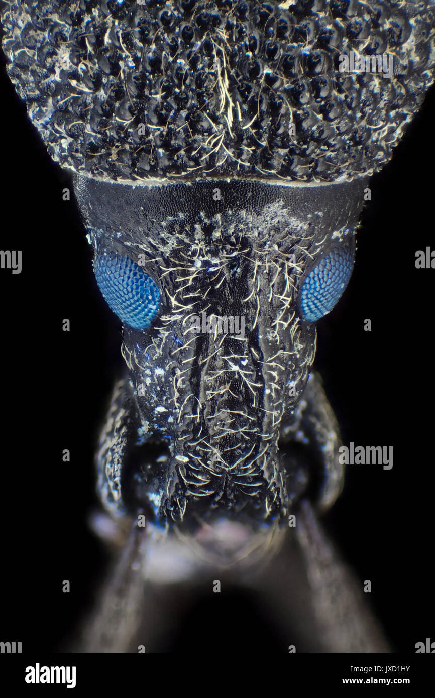 Weevil beetle (likely Larinus carlinae) with fluorescent eyes, reflected visible + ultraviolet micrograph, 31x magnification when printed 10cm tall Stock Photohttps://www.alamy.com/image-license-details/?v=1https://www.alamy.com/weevil-beetle-likely-larinus-carlinae-with-fluorescent-eyes-reflected-image153950663.html
RMJXD1HY–Weevil beetle (likely Larinus carlinae) with fluorescent eyes, reflected visible + ultraviolet micrograph, 31x magnification when printed 10cm tall
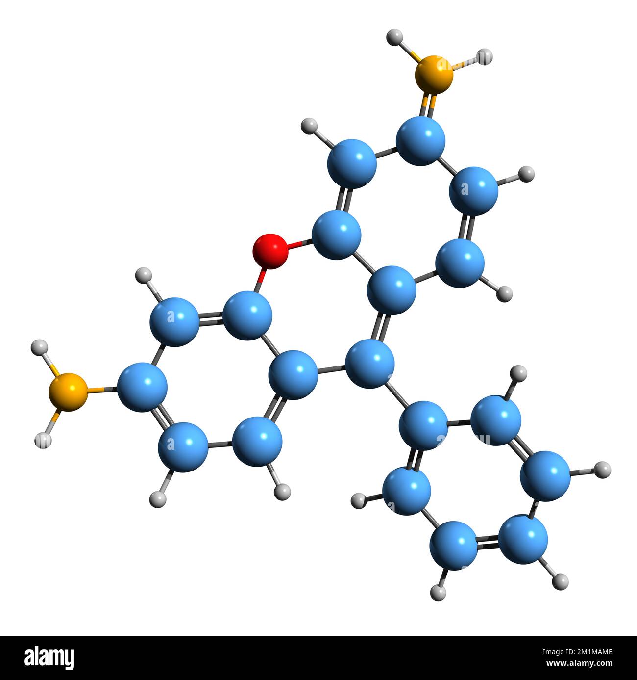 3D image of Rhodamine skeletal formula - molecular chemical structure of triarylmethane dye isolated on white background Stock Photohttps://www.alamy.com/image-license-details/?v=1https://www.alamy.com/3d-image-of-rhodamine-skeletal-formula-molecular-chemical-structure-of-triarylmethane-dye-isolated-on-white-background-image500162782.html
RF2M1MAME–3D image of Rhodamine skeletal formula - molecular chemical structure of triarylmethane dye isolated on white background
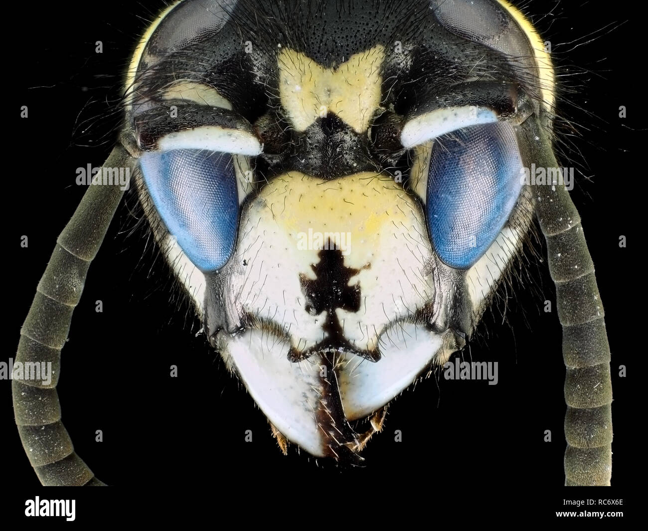 Extreme macro shot (micrograph) of a wasp (Vespula sp.) with fluorescent eyes, in visible and ultraviolet light Stock Photohttps://www.alamy.com/image-license-details/?v=1https://www.alamy.com/extreme-macro-shot-micrograph-of-a-wasp-vespula-sp-with-fluorescent-eyes-in-visible-and-ultraviolet-light-image231262934.html
RMRC6X6E–Extreme macro shot (micrograph) of a wasp (Vespula sp.) with fluorescent eyes, in visible and ultraviolet light
 Modern fluorescent microstope working station, text space Stock Photohttps://www.alamy.com/image-license-details/?v=1https://www.alamy.com/stock-photo-modern-fluorescent-microstope-working-station-text-space-172512600.html
RFM0JHG8–Modern fluorescent microstope working station, text space
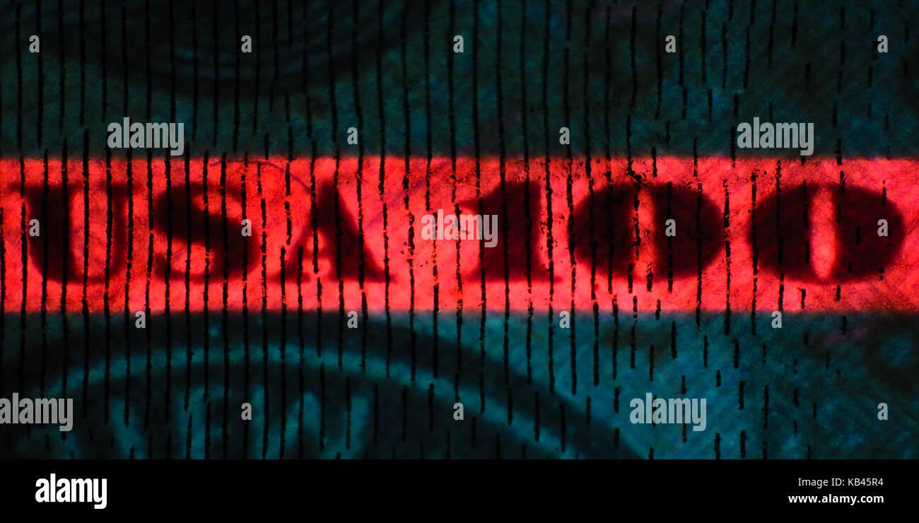 Ultraviolet light micrograph of a fluorescent security thread on US $100 bill (year 2009 design), pictured area is about 12mm wide Stock Photohttps://www.alamy.com/image-license-details/?v=1https://www.alamy.com/stock-image-ultraviolet-light-micrograph-of-a-fluorescent-security-thread-on-us-161746904.html
RMKB45R4–Ultraviolet light micrograph of a fluorescent security thread on US $100 bill (year 2009 design), pictured area is about 12mm wide
 Watching polymeric nanofibers, images of Atomic Force Microscope, AFM In the foreground the Axio Observer, Inverted microscope for fluorescence microscopy Cleanroom Materials and Tissue Unit CEIT Center of Studies and Technical Research University of Navarra, Donostia, Gipuzkoa, Basque Country, Spain. Stock Photohttps://www.alamy.com/image-license-details/?v=1https://www.alamy.com/watching-polymeric-nanofibers-images-of-atomic-force-microscope-afm-in-the-foreground-the-axio-observer-inverted-microscope-for-fluorescence-microscopy-cleanroom-materials-and-tissue-unit-ceit-center-of-studies-and-technical-research-university-of-navarra-donostia-gipuzkoa-basque-country-spain-image602045277.html
RM2WYDEW1–Watching polymeric nanofibers, images of Atomic Force Microscope, AFM In the foreground the Axio Observer, Inverted microscope for fluorescence microscopy Cleanroom Materials and Tissue Unit CEIT Center of Studies and Technical Research University of Navarra, Donostia, Gipuzkoa, Basque Country, Spain.
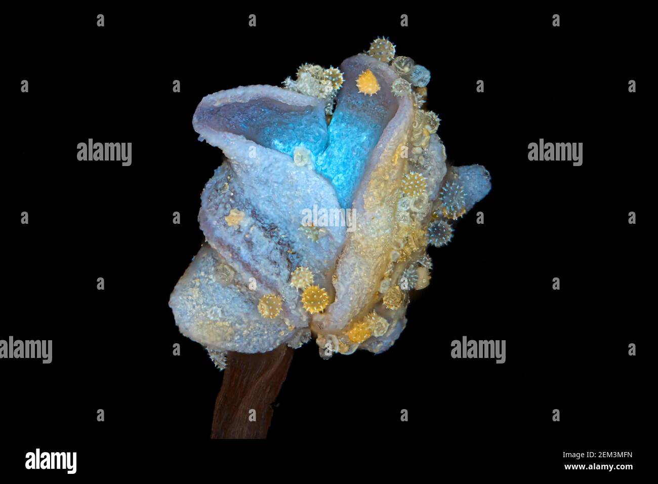 hibiscus (Hibiscus spec.), pollen grains of hibiscus in the anther, fluorescence, UV-light irradiation, light microscope image, magnification x20 Stock Photohttps://www.alamy.com/image-license-details/?v=1https://www.alamy.com/hibiscus-hibiscus-spec-pollen-grains-of-hibiscus-in-the-anther-fluorescence-uv-light-irradiation-light-microscope-image-magnification-x20-image408213561.html
RM2EM3MFN–hibiscus (Hibiscus spec.), pollen grains of hibiscus in the anther, fluorescence, UV-light irradiation, light microscope image, magnification x20
 Microfilaments, mitochondria, and nuclei in fibroblast cells Stock Photohttps://www.alamy.com/image-license-details/?v=1https://www.alamy.com/stock-photo-microfilaments-mitochondria-and-nuclei-in-fibroblast-cells-76191254.html
RMEBXPM6–Microfilaments, mitochondria, and nuclei in fibroblast cells
 Gloved hand holds bar-coded liquid sample in microscopy room Stock Photohttps://www.alamy.com/image-license-details/?v=1https://www.alamy.com/stock-photo-gloved-hand-holds-bar-coded-liquid-sample-in-microscopy-room-171559350.html
RFKY35KJ–Gloved hand holds bar-coded liquid sample in microscopy room
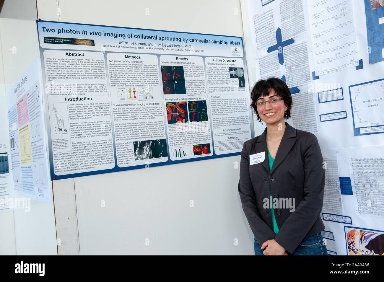 A researcher poses with a poster describing a complex scientific experiment at the Johns Hopkins University in Baltimore, Maryland, April 9, 2009. From the Homewood Photography collection. () Stock Photohttps://www.alamy.com/image-license-details/?v=1https://www.alamy.com/a-researcher-poses-with-a-poster-describing-a-complex-scientific-experiment-at-the-johns-hopkins-university-in-baltimore-maryland-april-9-2009-from-the-homewood-photography-collection-image333146918.html
RM2AA0486–A researcher poses with a poster describing a complex scientific experiment at the Johns Hopkins University in Baltimore, Maryland, April 9, 2009. From the Homewood Photography collection. ()
 Researcher studying hepatocytes in research on hepatitis, laboratory of the Centre de Reference National des Hepatites virales, Stock Photohttps://www.alamy.com/image-license-details/?v=1https://www.alamy.com/stock-photo-researcher-studying-hepatocytes-in-research-on-hepatitis-laboratory-72441962.html
RME5T0CX–Researcher studying hepatocytes in research on hepatitis, laboratory of the Centre de Reference National des Hepatites virales,
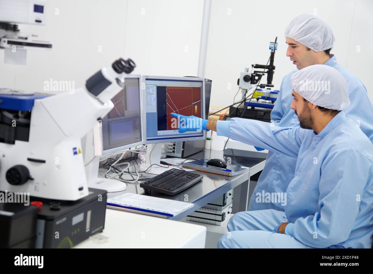 Watching polymeric nano fibers Atomic force microscope AFM In the foreground the Axio Observer, Inverted microscope for fluorescence microscopy Cl Stock Photohttps://www.alamy.com/image-license-details/?v=1https://www.alamy.com/watching-polymeric-nano-fibers-atomic-force-microscope-afm-in-the-foreground-the-axio-observer-inverted-microscope-for-fluorescence-microscopy-cl-image610392728.html
RM2XD1P48–Watching polymeric nano fibers Atomic force microscope AFM In the foreground the Axio Observer, Inverted microscope for fluorescence microscopy Cl
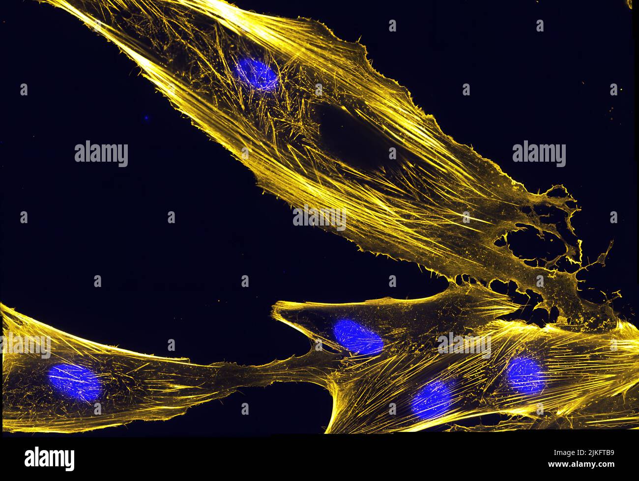 Immunofluorescence image of actin bundles in muscle precursor cells called myoblasts. Actin is labeled with fluorescently labeled phalloidin, which is a toxin from the fungus Amanita phalloides. Nuclei are shown in blue. Stock Photohttps://www.alamy.com/image-license-details/?v=1https://www.alamy.com/immunofluorescence-image-of-actin-bundles-in-muscle-precursor-cells-called-myoblasts-actin-is-labeled-with-fluorescently-labeled-phalloidin-which-is-a-toxin-from-the-fungus-amanita-phalloides-nuclei-are-shown-in-blue-image476706813.html
RM2JKFTB9–Immunofluorescence image of actin bundles in muscle precursor cells called myoblasts. Actin is labeled with fluorescently labeled phalloidin, which is a toxin from the fungus Amanita phalloides. Nuclei are shown in blue.
