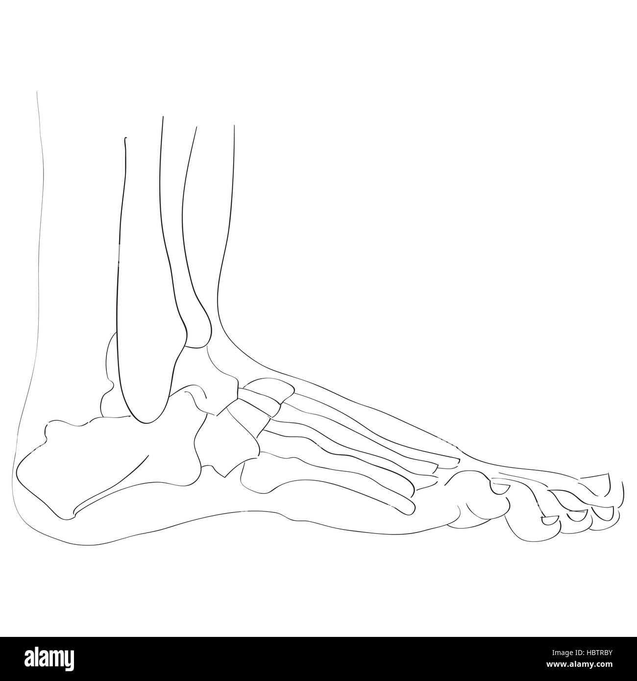
Foot bones anatomy Black & White Stock Photos
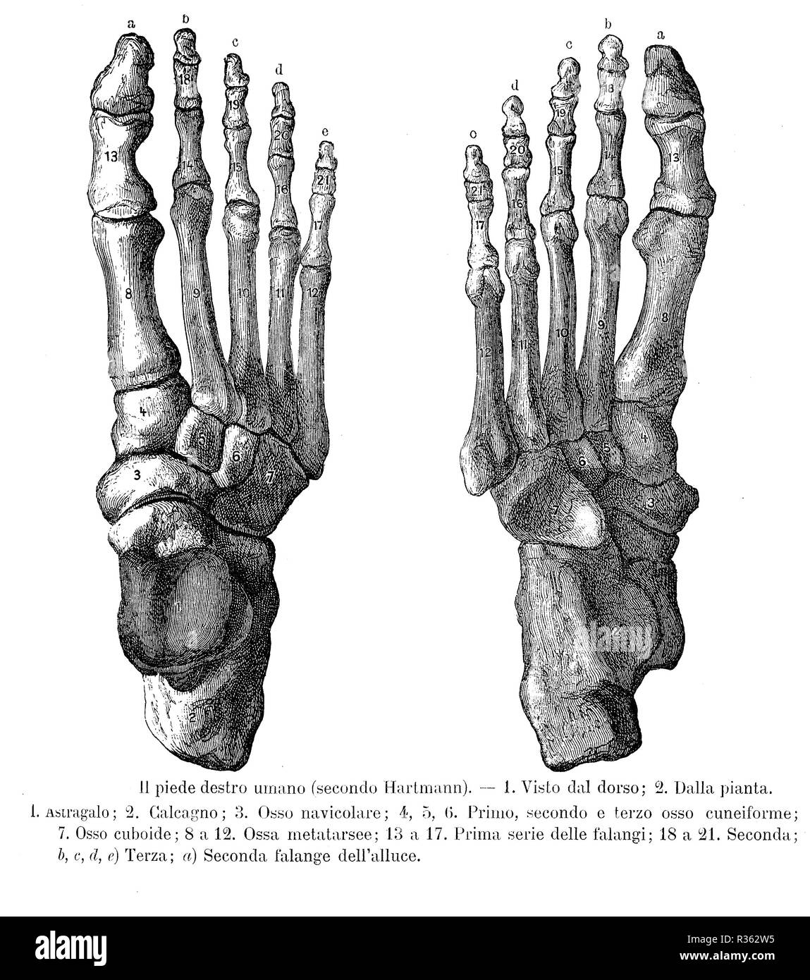 Vintage illustration of anatomy, right foot bones, dorsalis and sole view with Italian anatomical descriptions Stock Photohttps://www.alamy.com/image-license-details/?v=1https://www.alamy.com/vintage-illustration-of-anatomy-right-foot-bones-dorsalis-and-sole-view-with-italian-anatomical-descriptions-image225712737.html
RFR362W5–Vintage illustration of anatomy, right foot bones, dorsalis and sole view with Italian anatomical descriptions
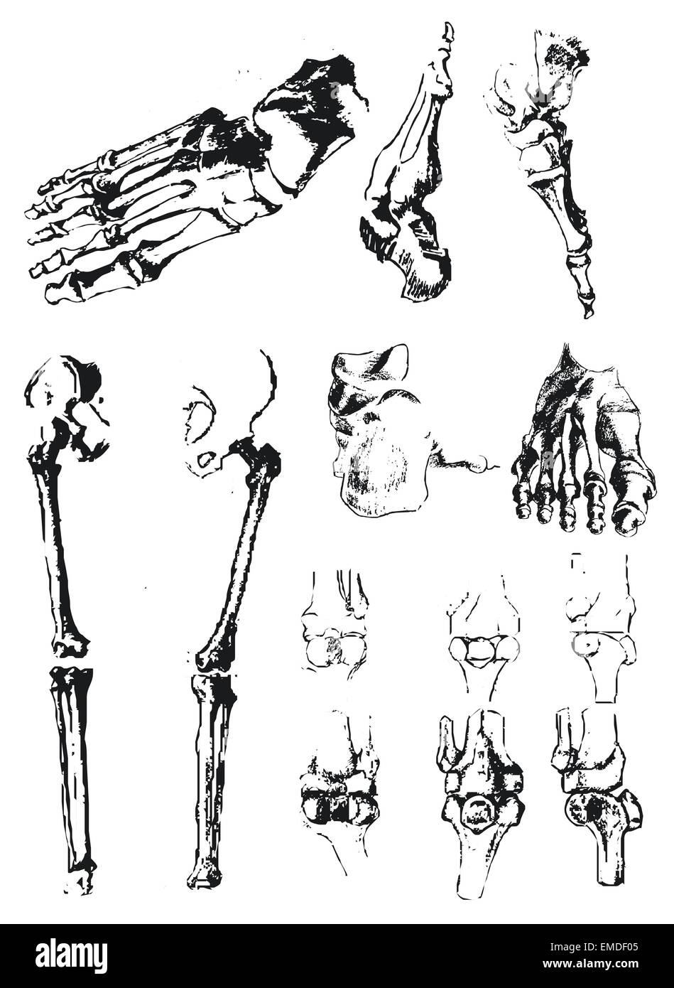 Hand drawn foot bones and patella Stock Vectorhttps://www.alamy.com/image-license-details/?v=1https://www.alamy.com/stock-photo-hand-drawn-foot-bones-and-patella-81431733.html
RFEMDF05–Hand drawn foot bones and patella
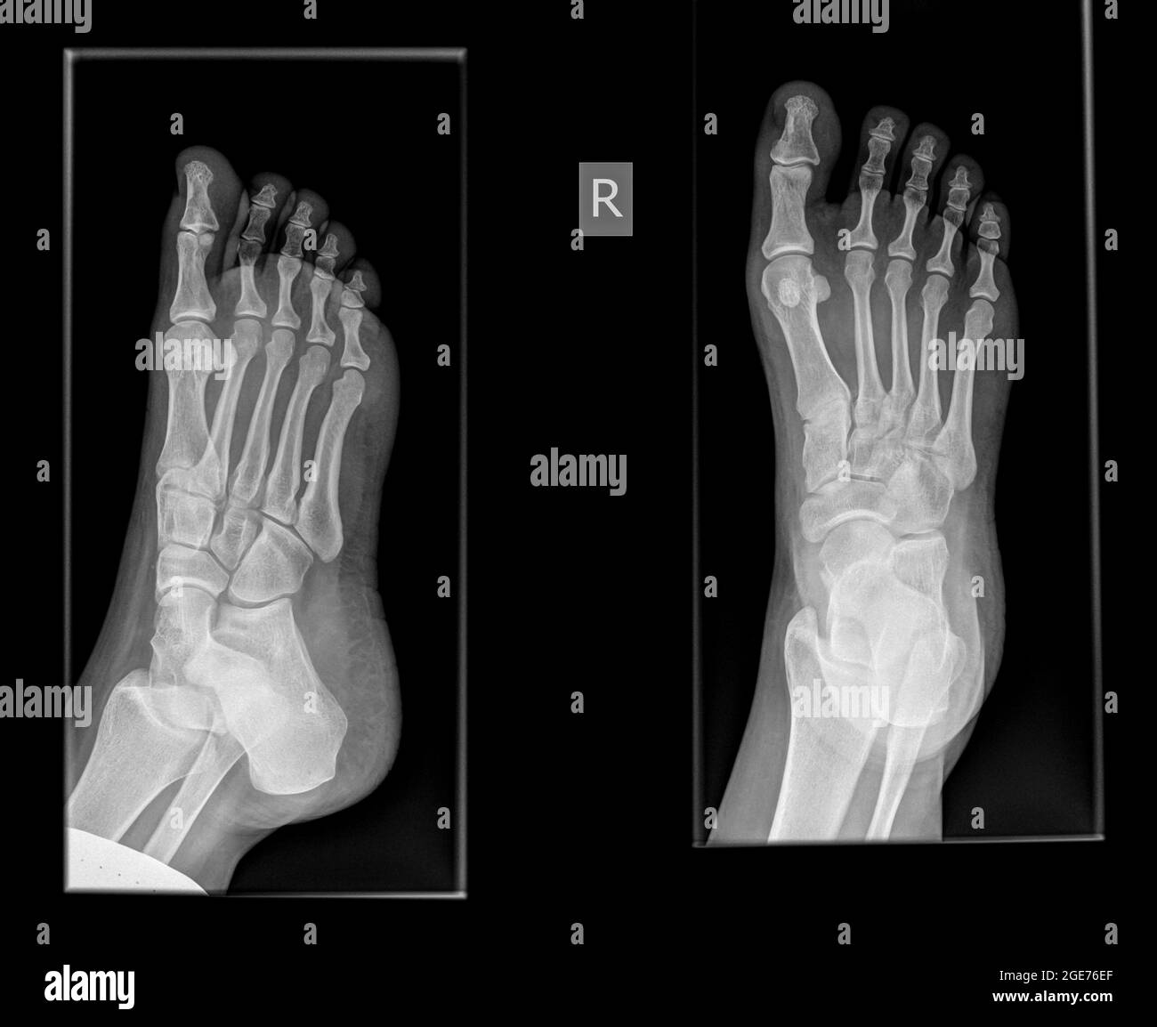 x-ray of a foot showing a fracture in the proximal phalanx of the big toe on the righ foot of a 30 year old female patient Stock Photohttps://www.alamy.com/image-license-details/?v=1https://www.alamy.com/x-ray-of-a-foot-showing-a-fracture-in-the-proximal-phalanx-of-the-big-toe-on-the-righ-foot-of-a-30-year-old-female-patient-image439023159.html
RM2GE76EF–x-ray of a foot showing a fracture in the proximal phalanx of the big toe on the righ foot of a 30 year old female patient
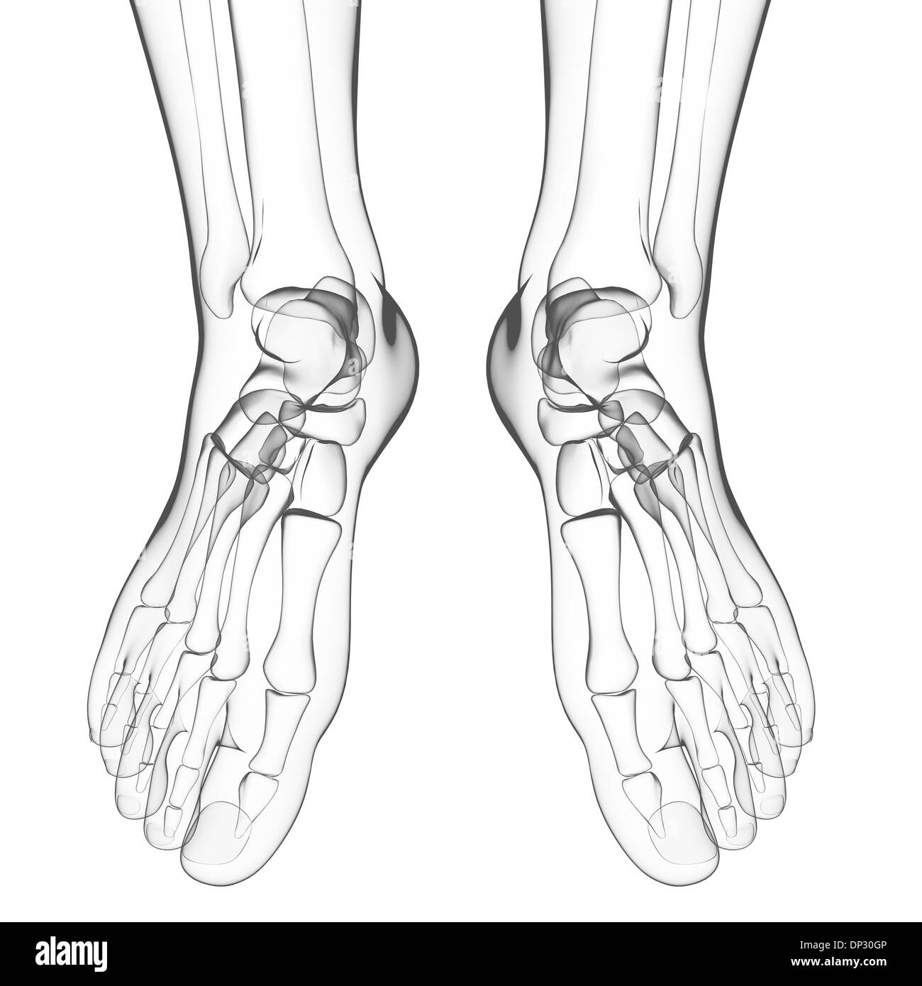 Human foot bones, artwork Stock Photohttps://www.alamy.com/image-license-details/?v=1https://www.alamy.com/human-foot-bones-artwork-image65219862.html
RFDP30GP–Human foot bones, artwork
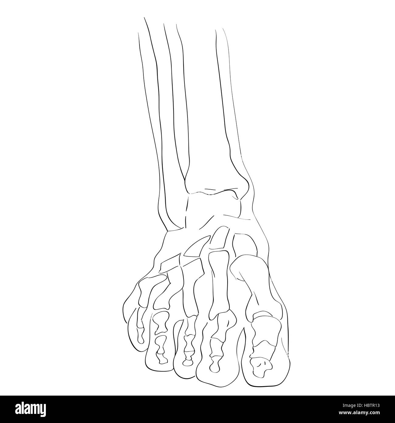 front view foot bones Stock Photohttps://www.alamy.com/image-license-details/?v=1https://www.alamy.com/stock-photo-front-view-foot-bones-127778703.html
RMHBTR13–front view foot bones
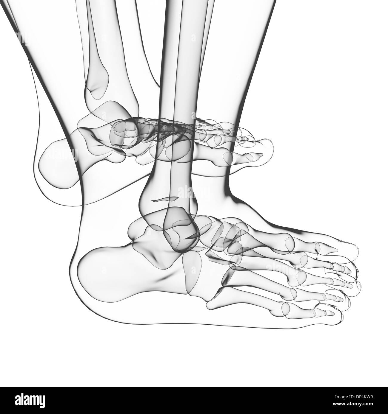 Human foot bones, artwork Stock Photohttps://www.alamy.com/image-license-details/?v=1https://www.alamy.com/human-foot-bones-artwork-image65256963.html
RFDP4KWR–Human foot bones, artwork
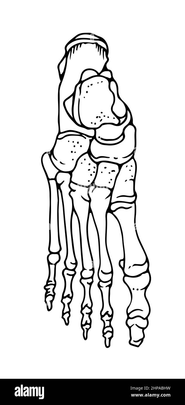 Bones of the human foot, hand drawn vector illustration isolated on a white background, orthopedics medicine anatomy sketch Stock Vectorhttps://www.alamy.com/image-license-details/?v=1https://www.alamy.com/bones-of-the-human-foot-hand-drawn-vector-illustration-isolated-on-a-white-background-orthopedics-medicine-anatomy-sketch-image461220645.html
RF2HPABHW–Bones of the human foot, hand drawn vector illustration isolated on a white background, orthopedics medicine anatomy sketch
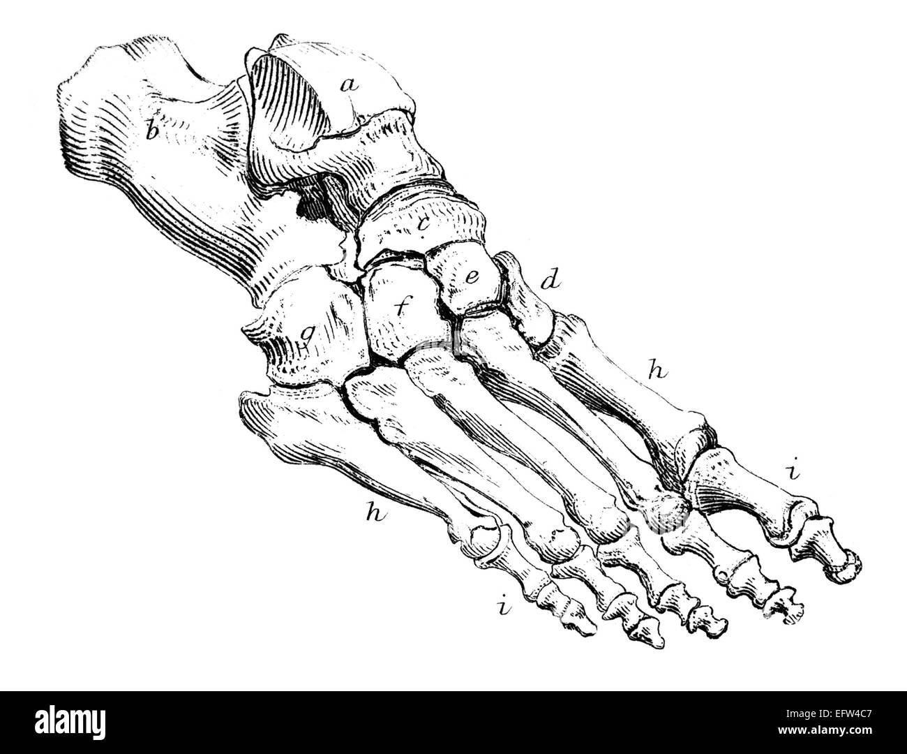 photographed from a book titled the 'National Encyclopedia', published in London in 1881. Copyright has expired on this artwork. Stock Photohttps://www.alamy.com/image-license-details/?v=1https://www.alamy.com/stock-photo-photographed-from-a-book-titled-the-national-encyclopedia-published-78613591.html
RFEFW4C7–photographed from a book titled the 'National Encyclopedia', published in London in 1881. Copyright has expired on this artwork.
RF2CDJC4P–Foot Anatomy Glyph Icons
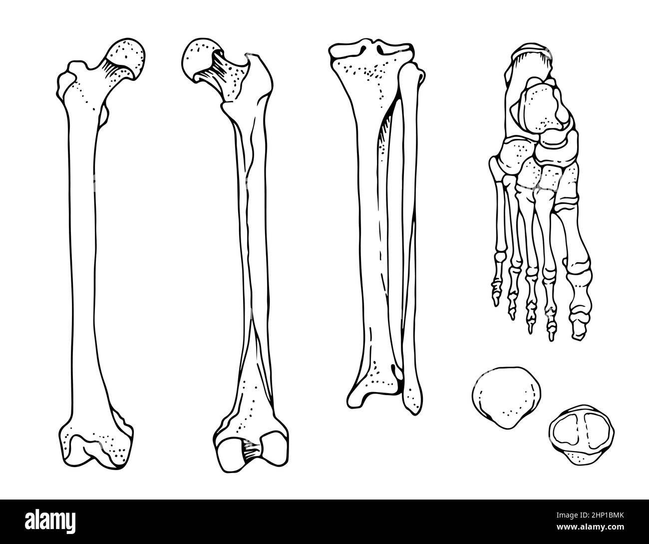 Human foot bones, femur, tibia and fibula, foot, patella, vector hand drawn illustration isolated on a white background, orthopedics anatomy set Stock Vectorhttps://www.alamy.com/image-license-details/?v=1https://www.alamy.com/human-foot-bones-femur-tibia-and-fibula-foot-patella-vector-hand-drawn-illustration-isolated-on-a-white-background-orthopedics-anatomy-set-image461023155.html
RF2HP1BMK–Human foot bones, femur, tibia and fibula, foot, patella, vector hand drawn illustration isolated on a white background, orthopedics anatomy set
 Skeletons in a row doing yoga with one foot up under their arms sitting in a row - black and white isolated against black background Stock Photohttps://www.alamy.com/image-license-details/?v=1https://www.alamy.com/skeletons-in-a-row-doing-yoga-with-one-foot-up-under-their-arms-sitting-in-a-row-black-and-white-isolated-against-black-background-image439446529.html
RF2GEXEEW–Skeletons in a row doing yoga with one foot up under their arms sitting in a row - black and white isolated against black background
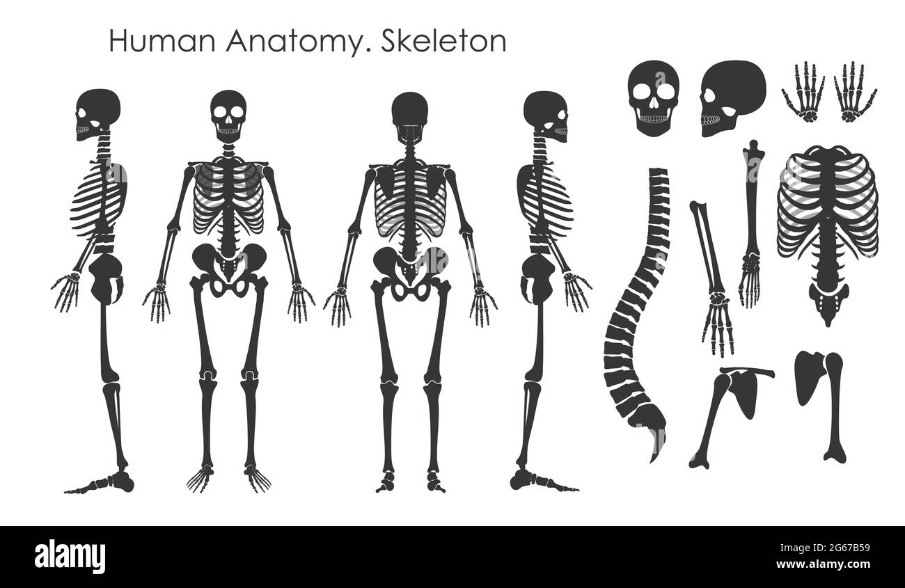 Vector illustration set of human bones skeleton in silhouette style isolated on white background. Human anatomy concept, skeleton in different Stock Vectorhttps://www.alamy.com/image-license-details/?v=1https://www.alamy.com/vector-illustration-set-of-human-bones-skeleton-in-silhouette-style-isolated-on-white-background-human-anatomy-concept-skeleton-in-different-image434109573.html
RF2G67B59–Vector illustration set of human bones skeleton in silhouette style isolated on white background. Human anatomy concept, skeleton in different
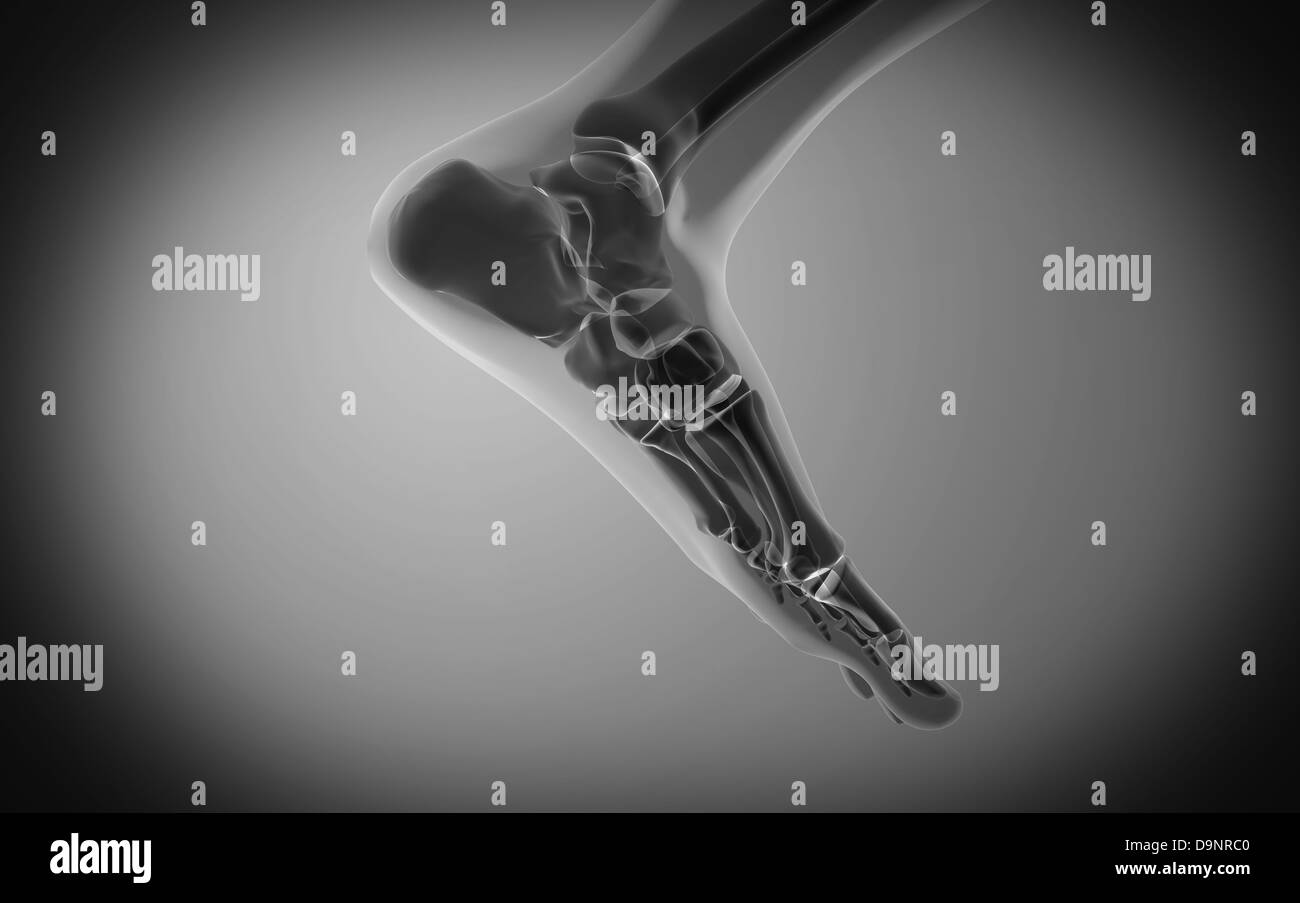 X-ray view of human foot. Stock Photohttps://www.alamy.com/image-license-details/?v=1https://www.alamy.com/stock-photo-x-ray-view-of-human-foot-57642368.html
RFD9NRC0–X-ray view of human foot.
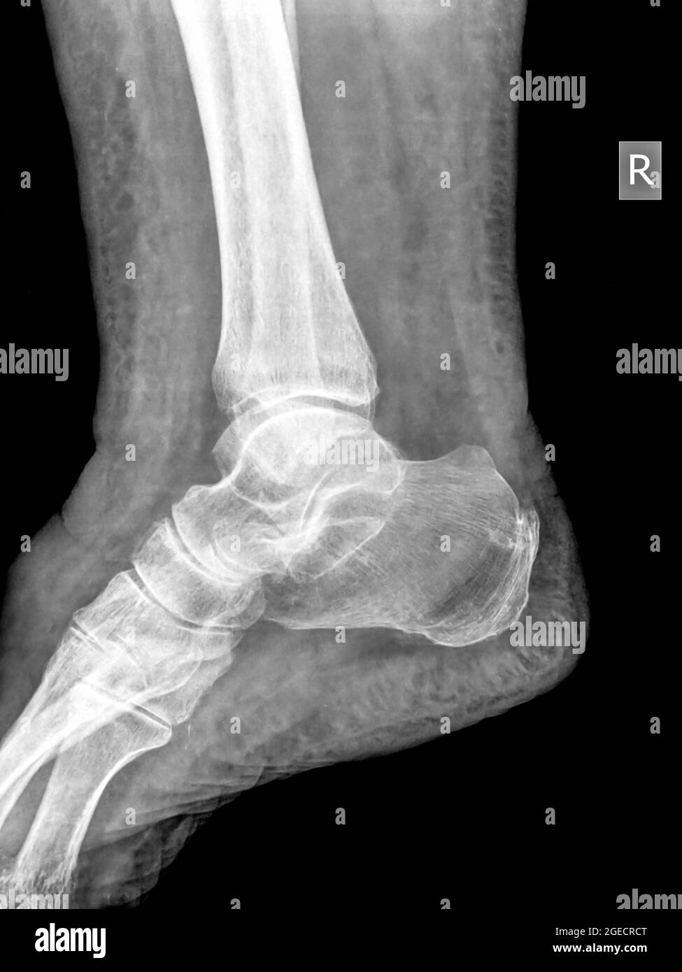 Healthy ankle joint x-ray of a 79 year old female patient. No fracture nor sprain can be seen Stock Photohttps://www.alamy.com/image-license-details/?v=1https://www.alamy.com/healthy-ankle-joint-x-ray-of-a-79-year-old-female-patient-no-fracture-nor-sprain-can-be-seen-image439146200.html
RM2GECRCT–Healthy ankle joint x-ray of a 79 year old female patient. No fracture nor sprain can be seen
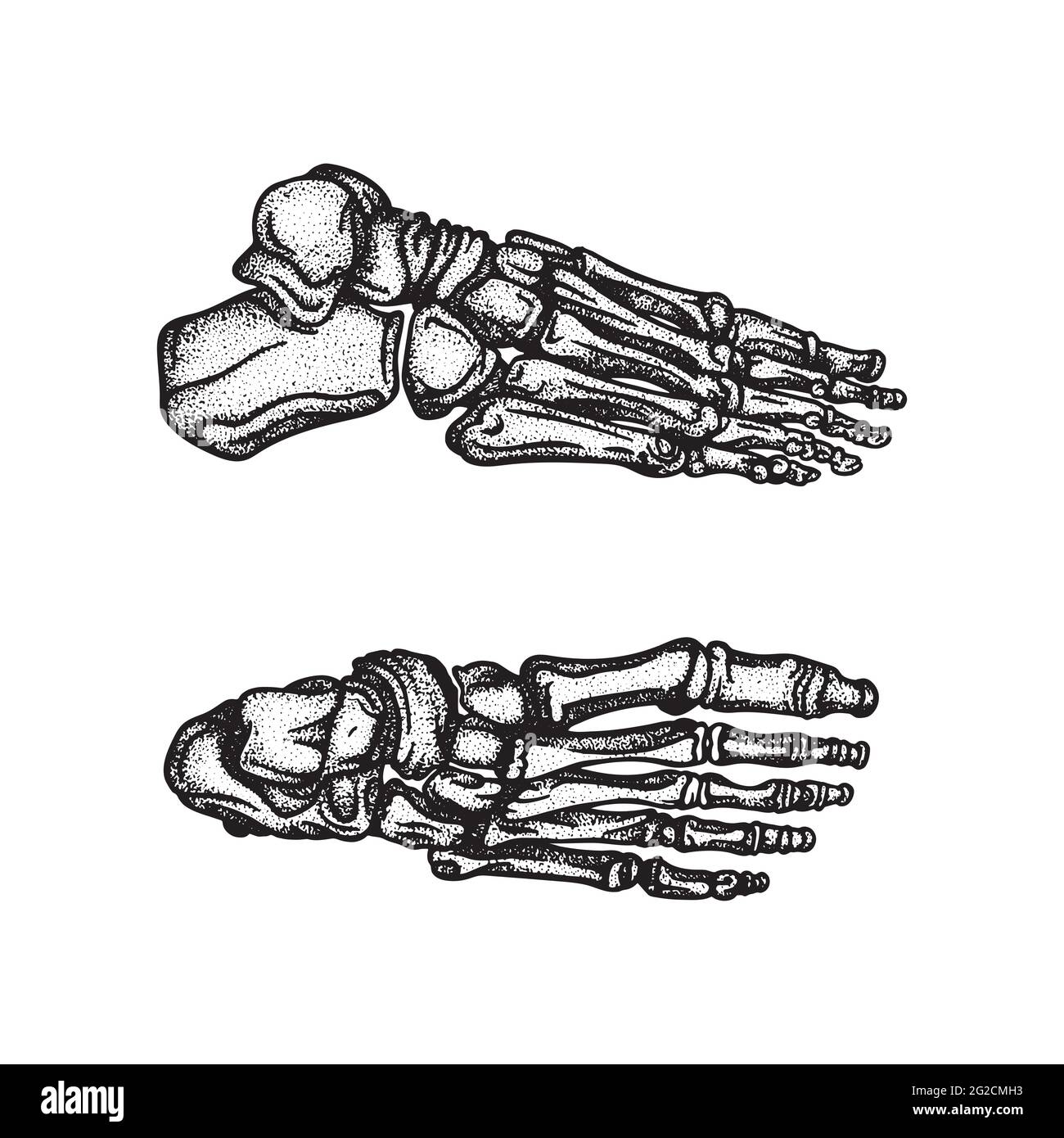 Foot. Foot bones top and side hand drawn vector illustrations set. Part of human skeleton graphic. Stock Vectorhttps://www.alamy.com/image-license-details/?v=1https://www.alamy.com/foot-foot-bones-top-and-side-hand-drawn-vector-illustrations-set-part-of-human-skeleton-graphic-image431768095.html
RF2G2CMH3–Foot. Foot bones top and side hand drawn vector illustrations set. Part of human skeleton graphic.
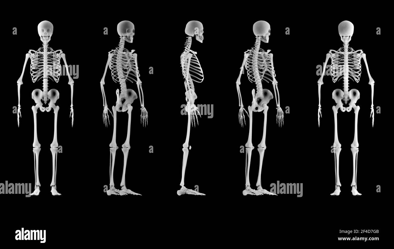 X-ray view of a human skeleton. Medical examination and body scan. Human anatomy and body bones. 360 degree view of a skeleton. 3d render Stock Photohttps://www.alamy.com/image-license-details/?v=1https://www.alamy.com/x-ray-view-of-a-human-skeleton-medical-examination-and-body-scan-human-anatomy-and-body-bones-360-degree-view-of-a-skeleton-3d-render-image415798779.html
RF2F4D7GB–X-ray view of a human skeleton. Medical examination and body scan. Human anatomy and body bones. 360 degree view of a skeleton. 3d render
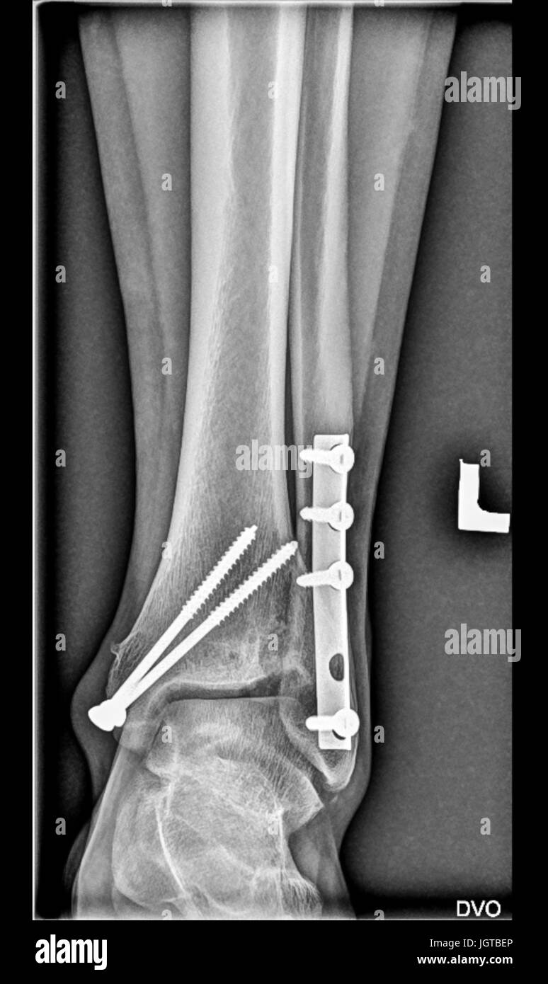 Foot medical xray, lower limb bones, broken ankle, tibia fibula with screws, fingers joints Stock Photohttps://www.alamy.com/image-license-details/?v=1https://www.alamy.com/stock-photo-foot-medical-xray-lower-limb-bones-broken-ankle-tibia-fibula-with-148053326.html
RFJGTBEP–Foot medical xray, lower limb bones, broken ankle, tibia fibula with screws, fingers joints
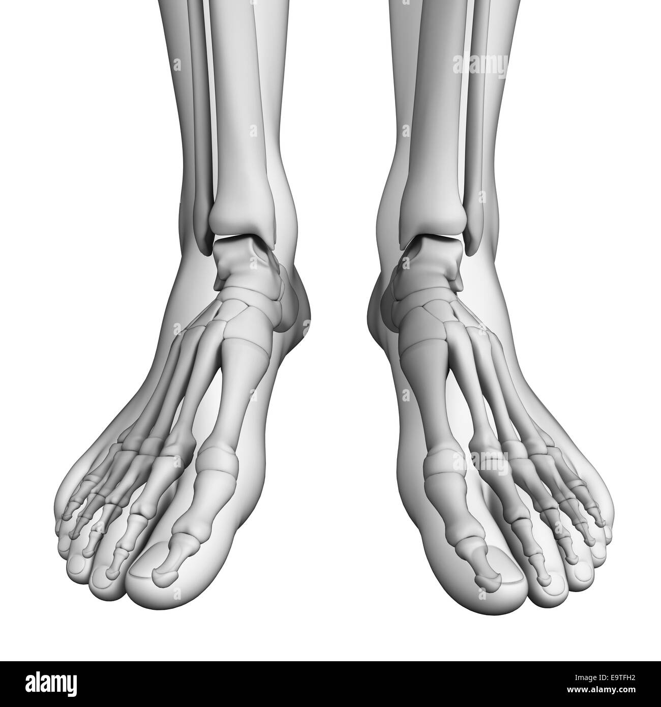 Illustration of human foot artwork Stock Photohttps://www.alamy.com/image-license-details/?v=1https://www.alamy.com/stock-photo-illustration-of-human-foot-artwork-74912462.html
RFE9TFH2–Illustration of human foot artwork
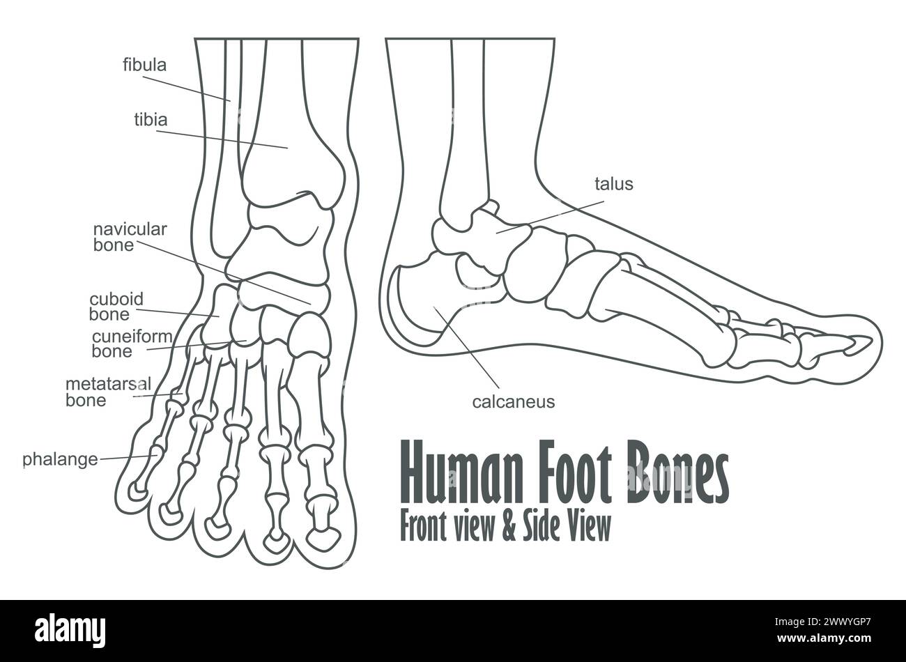 Human Foot Bones Front And Side View Anatomy, Vector Illustration Stock Vectorhttps://www.alamy.com/image-license-details/?v=1https://www.alamy.com/human-foot-bones-front-and-side-view-anatomy-vector-illustration-image601124783.html
RF2WWYGP7–Human Foot Bones Front And Side View Anatomy, Vector Illustration
 The Tarsal Bones of the Foot are located in the midfoot and the hind foot areas of the human foot, vintage line drawing or engraving illustration. Stock Vectorhttps://www.alamy.com/image-license-details/?v=1https://www.alamy.com/the-tarsal-bones-of-the-foot-are-located-in-the-midfoot-and-the-hind-foot-areas-of-the-human-foot-vintage-line-drawing-or-engraving-illustration-image359321248.html
RF2BTGDWM–The Tarsal Bones of the Foot are located in the midfoot and the hind foot areas of the human foot, vintage line drawing or engraving illustration.
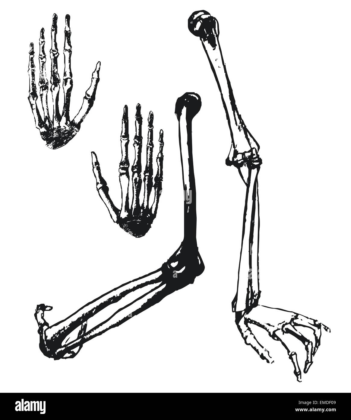 Hand drawn humerus, ulna and hand bones Stock Vectorhttps://www.alamy.com/image-license-details/?v=1https://www.alamy.com/stock-photo-hand-drawn-humerus-ulna-and-hand-bones-81431737.html
RFEMDF09–Hand drawn humerus, ulna and hand bones
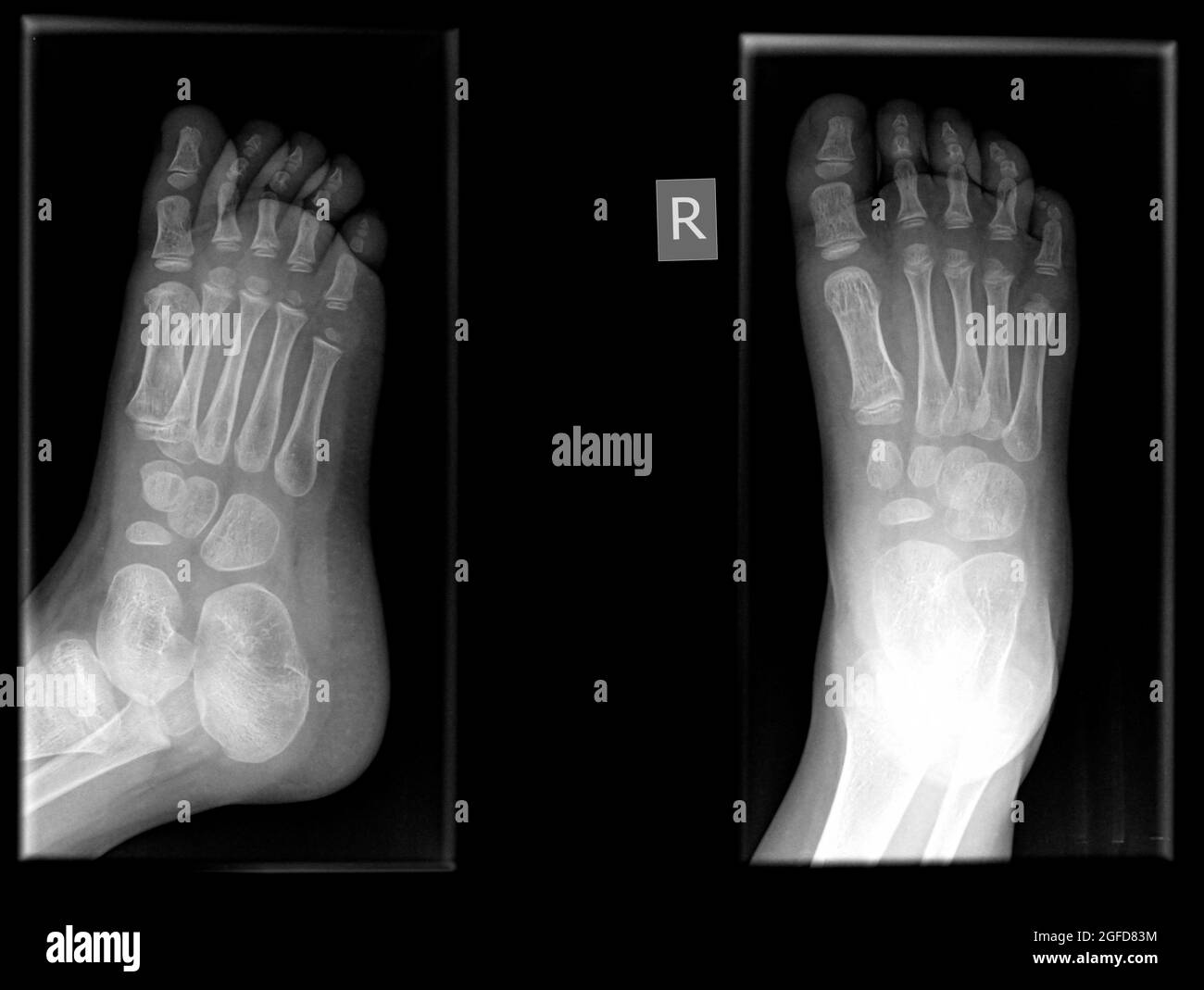 Fracture in the first metatarsal bone of the right foot of a 3 year old male patient Stock Photohttps://www.alamy.com/image-license-details/?v=1https://www.alamy.com/fracture-in-the-first-metatarsal-bone-of-the-right-foot-of-a-3-year-old-male-patient-image439770792.html
RM2GFD83M–Fracture in the first metatarsal bone of the right foot of a 3 year old male patient
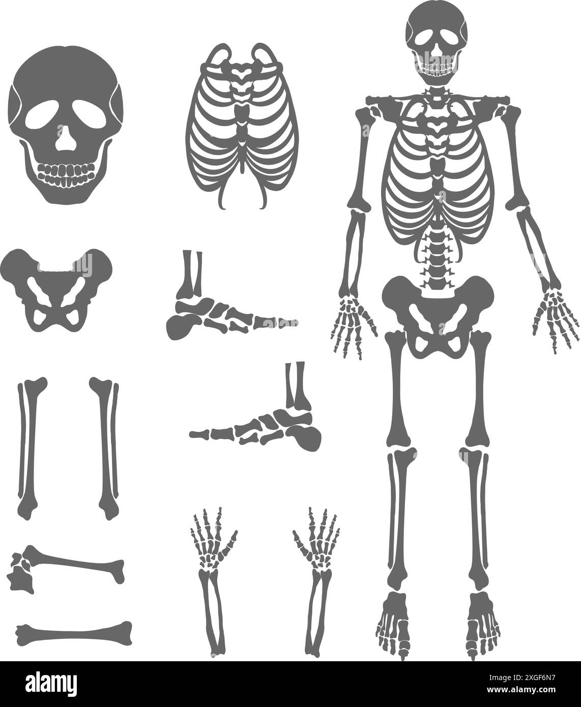 Human bones skeleton silhouette collection set Stock Vectorhttps://www.alamy.com/image-license-details/?v=1https://www.alamy.com/human-bones-skeleton-silhouette-collection-set-image612531955.html
RF2XGF6N7–Human bones skeleton silhouette collection set
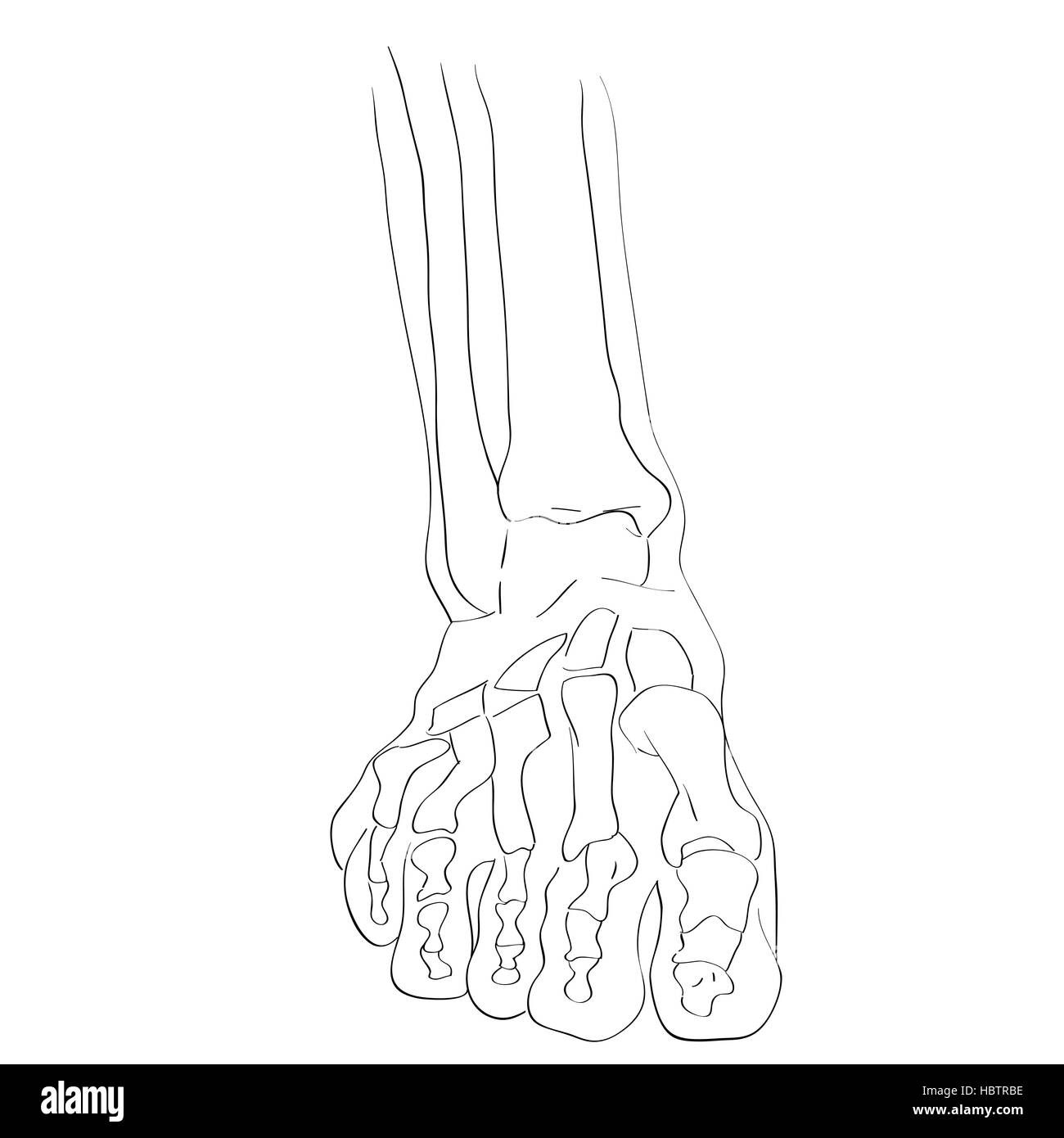 front view foot bones Stock Photohttps://www.alamy.com/image-license-details/?v=1https://www.alamy.com/stock-photo-front-view-foot-bones-127778994.html
RMHBTRBE–front view foot bones
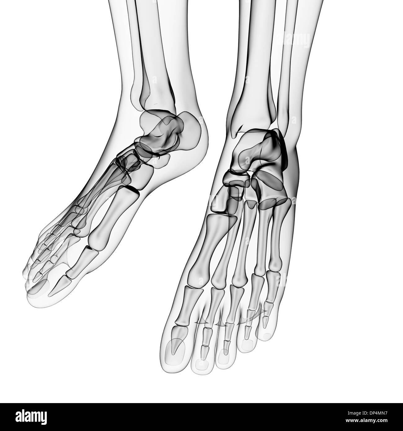 Human foot bones, artwork Stock Photohttps://www.alamy.com/image-license-details/?v=1https://www.alamy.com/human-foot-bones-artwork-image65257619.html
RFDP4MN7–Human foot bones, artwork
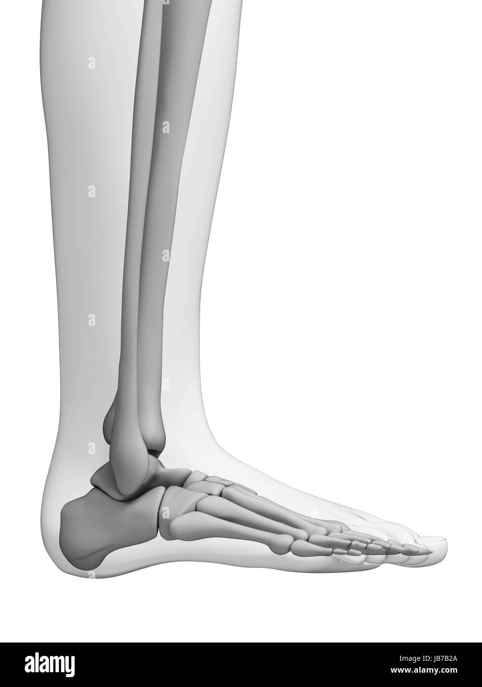 3d rendered illustration - foot anatomy Stock Photohttps://www.alamy.com/image-license-details/?v=1https://www.alamy.com/stock-photo-3d-rendered-illustration-foot-anatomy-144606514.html
RFJB7B2A–3d rendered illustration - foot anatomy
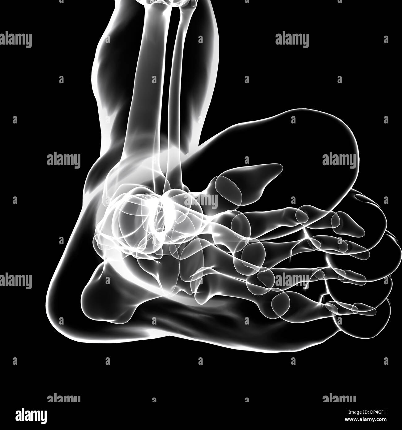 Human foot bones, artwork Stock Photohttps://www.alamy.com/image-license-details/?v=1https://www.alamy.com/human-foot-bones-artwork-image65254325.html
RFDP4GFH–Human foot bones, artwork
RF2CDJCHC–Foot Anatomy Line Icon
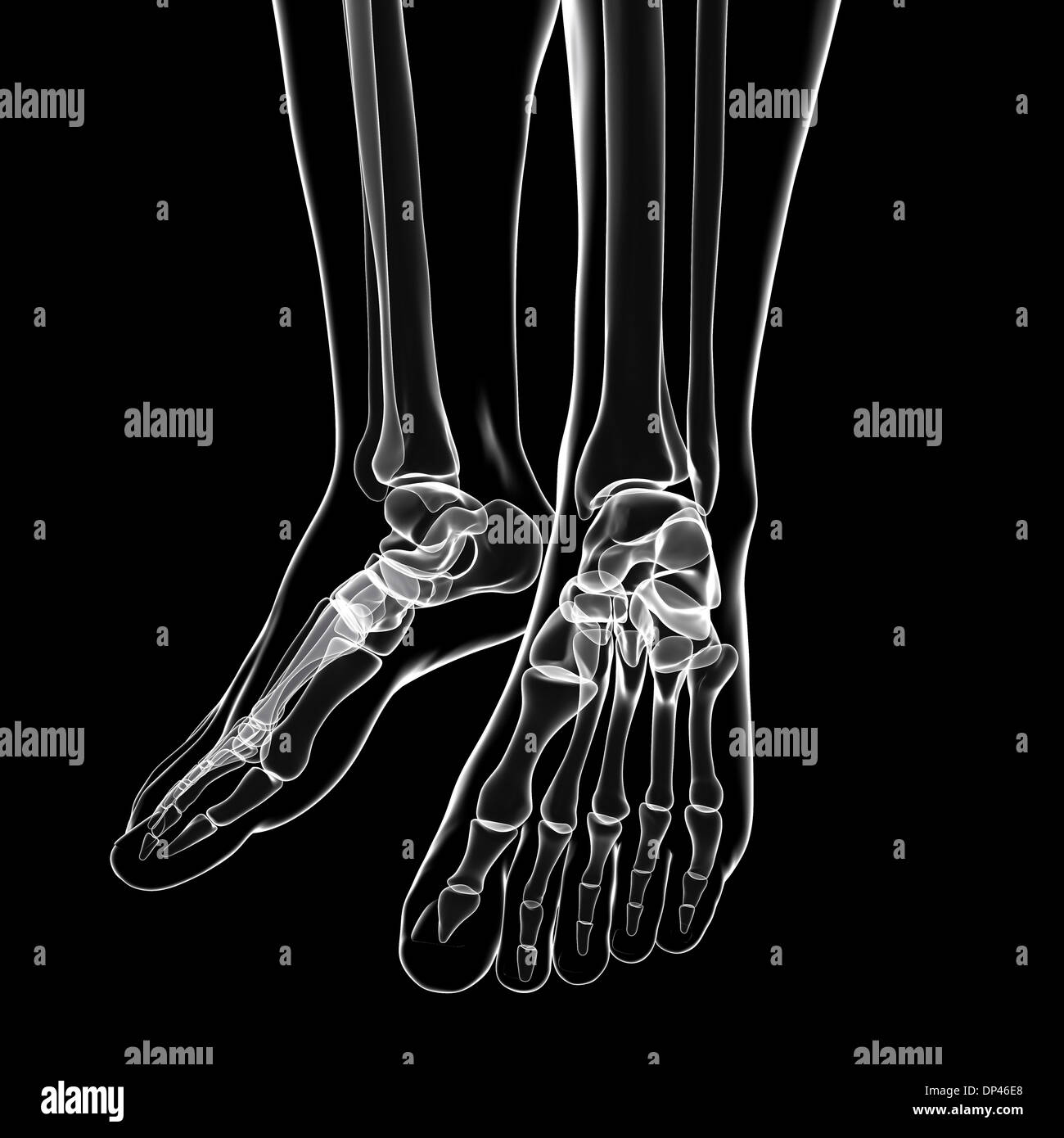 Human foot bones, artwork Stock Photohttps://www.alamy.com/image-license-details/?v=1https://www.alamy.com/human-foot-bones-artwork-image65246448.html
RFDP46E8–Human foot bones, artwork
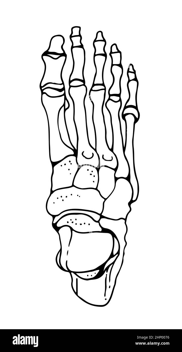 Bones of the human foot, vector hand drawn illustration isolated on a white background, orthopedics medicine anatomy sketch Stock Vectorhttps://www.alamy.com/image-license-details/?v=1https://www.alamy.com/bones-of-the-human-foot-vector-hand-drawn-illustration-isolated-on-a-white-background-orthopedics-medicine-anatomy-sketch-image460992202.html
RF2HP0076–Bones of the human foot, vector hand drawn illustration isolated on a white background, orthopedics medicine anatomy sketch
 A typical representation of the bones of the fore foot of existing Perissodactyle; Rhinoceros (Rhinoceros Sumairensis). They are usually nose-horned a Stock Vectorhttps://www.alamy.com/image-license-details/?v=1https://www.alamy.com/a-typical-representation-of-the-bones-of-the-fore-foot-of-existing-perissodactyle-rhinoceros-rhinoceros-sumairensis-they-are-usually-nose-horned-a-image367213317.html
RF2C9C099–A typical representation of the bones of the fore foot of existing Perissodactyle; Rhinoceros (Rhinoceros Sumairensis). They are usually nose-horned a
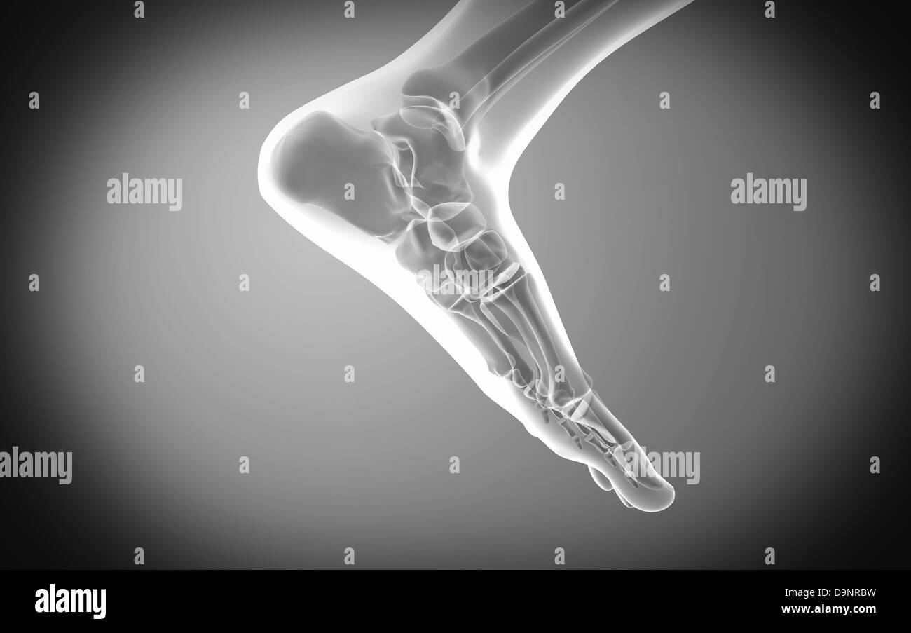 X-ray view of human foot. Stock Photohttps://www.alamy.com/image-license-details/?v=1https://www.alamy.com/stock-photo-x-ray-view-of-human-foot-57642365.html
RFD9NRBW–X-ray view of human foot.
 A typical representation of the bones of fore foot of existing Artiodactyle. Red Deer (Cervus elaphus), characterized by a four-chambered stomach and Stock Vectorhttps://www.alamy.com/image-license-details/?v=1https://www.alamy.com/a-typical-representation-of-the-bones-of-fore-foot-of-existing-artiodactyle-red-deer-cervus-elaphus-characterized-by-a-four-chambered-stomach-and-image359325594.html
RF2BTGKCX–A typical representation of the bones of fore foot of existing Artiodactyle. Red Deer (Cervus elaphus), characterized by a four-chambered stomach and
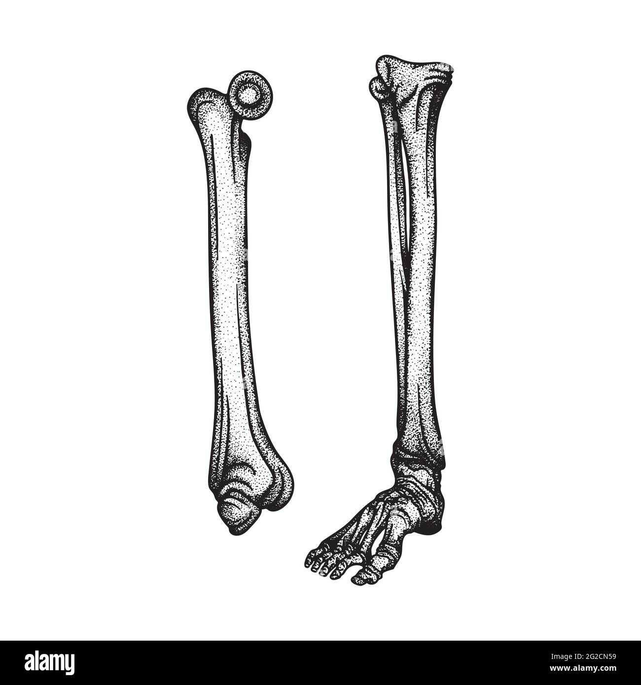 Foot. Foot bones top and side hand drawn vector illustrations set. Part of human skeleton graphic. Stock Vectorhttps://www.alamy.com/image-license-details/?v=1https://www.alamy.com/foot-foot-bones-top-and-side-hand-drawn-vector-illustrations-set-part-of-human-skeleton-graphic-image431768549.html
RF2G2CN59–Foot. Foot bones top and side hand drawn vector illustrations set. Part of human skeleton graphic.
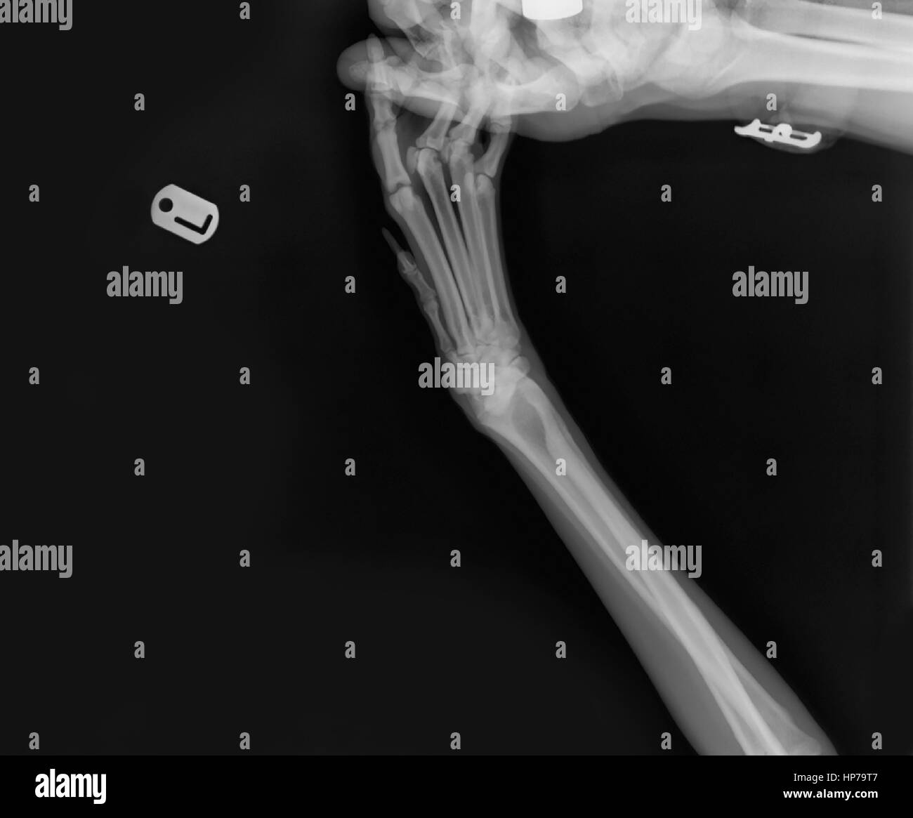 X-ray of a dog's front left leg at a veterinary surgery The technician's hand holding the paw can be seen Stock Photohttps://www.alamy.com/image-license-details/?v=1https://www.alamy.com/stock-photo-x-ray-of-a-dogs-front-left-leg-at-a-veterinary-surgery-the-technicians-134156407.html
RMHP79T7–X-ray of a dog's front left leg at a veterinary surgery The technician's hand holding the paw can be seen
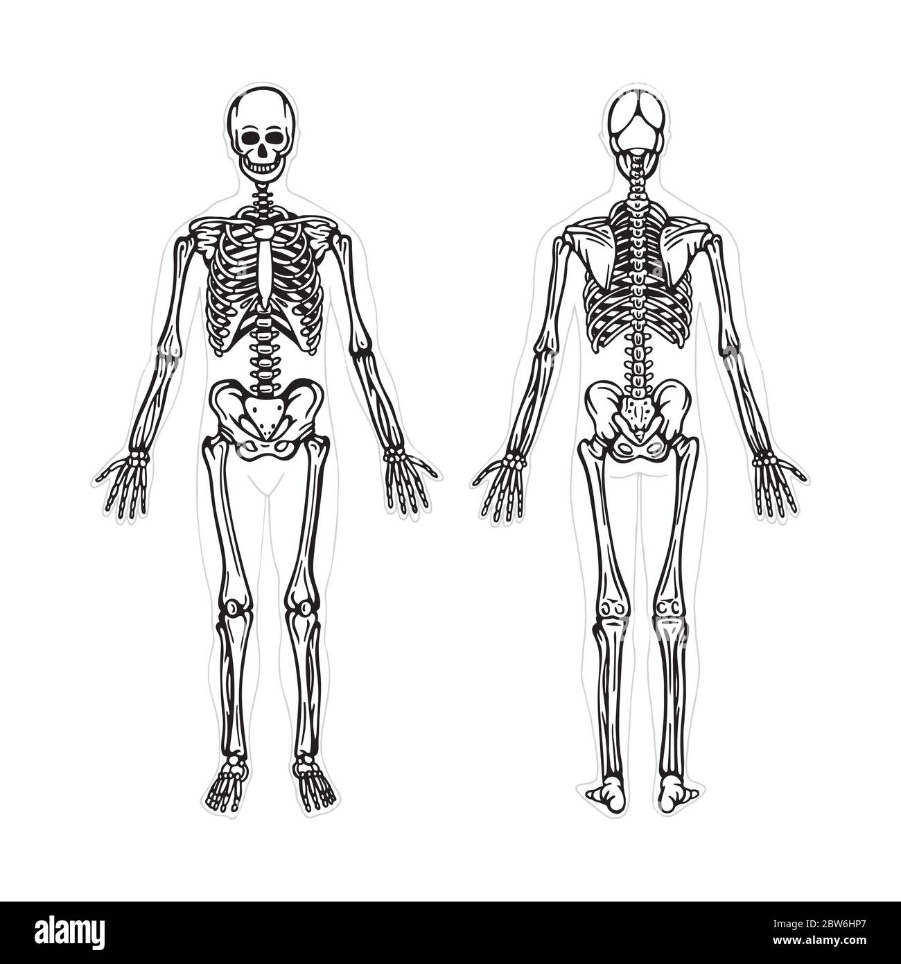 Skeleton. Human skeleton hand drawn vector illustration. Human skeleton front and back view. Bony system. Part of set. Stock Vectorhttps://www.alamy.com/image-license-details/?v=1https://www.alamy.com/skeleton-human-skeleton-hand-drawn-vector-illustration-human-skeleton-front-and-back-view-bony-system-part-of-set-image359719423.html
RF2BW6HP7–Skeleton. Human skeleton hand drawn vector illustration. Human skeleton front and back view. Bony system. Part of set.
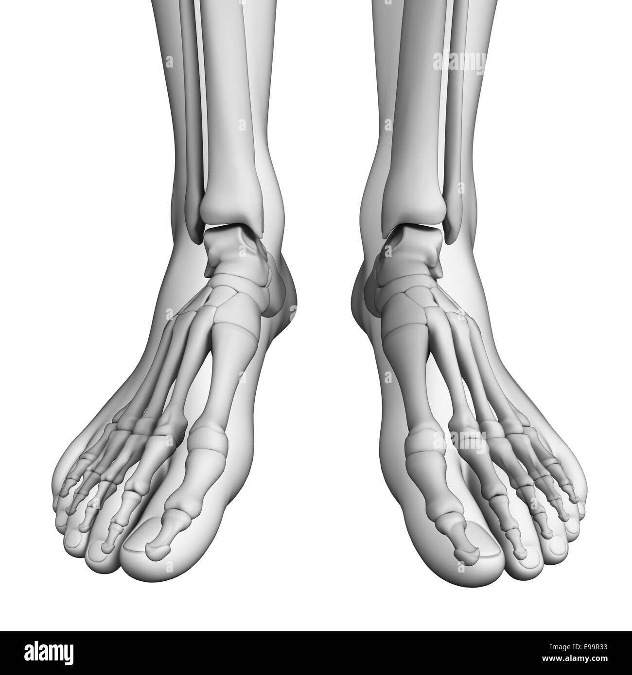 Illustration of human foot artwork Stock Photohttps://www.alamy.com/image-license-details/?v=1https://www.alamy.com/stock-photo-illustration-of-human-foot-artwork-74589063.html
RFE99R33–Illustration of human foot artwork
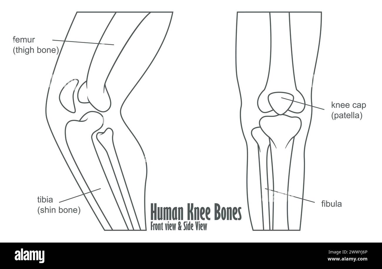 Human Knee Bones Front And Side View Anatomy, Vector Illustration Stock Vectorhttps://www.alamy.com/image-license-details/?v=1https://www.alamy.com/human-knee-bones-front-and-side-view-anatomy-vector-illustration-image601125918.html
RF2WWYJ6P–Human Knee Bones Front And Side View Anatomy, Vector Illustration
 X-ray view of human foot Stock Photohttps://www.alamy.com/image-license-details/?v=1https://www.alamy.com/x-ray-view-of-human-foot-image619197108.html
RM2XYAT6C–X-ray view of human foot
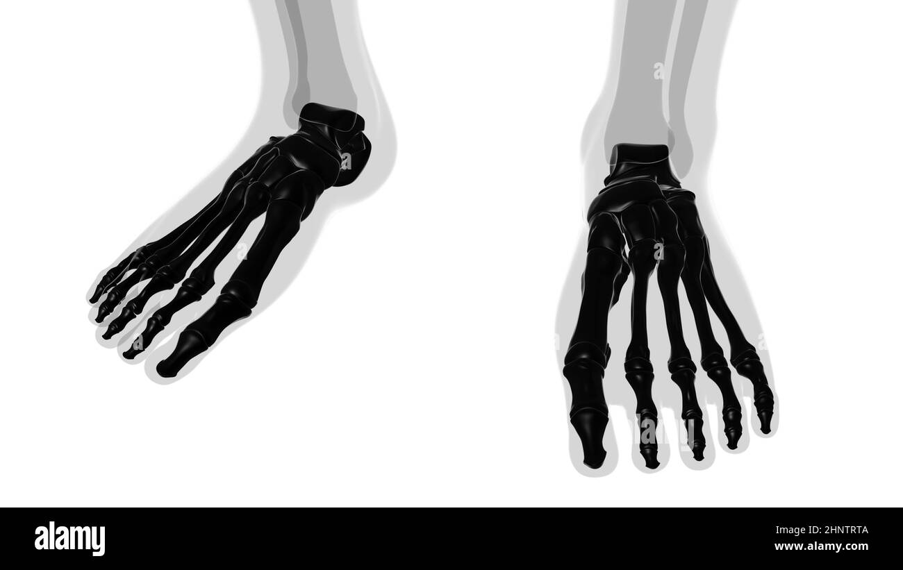 Human Skeleton Foot bones Anatomy For Medical Concept 3D Illustration Stock Photohttps://www.alamy.com/image-license-details/?v=1https://www.alamy.com/human-skeleton-foot-bones-anatomy-for-medical-concept-3d-illustration-image460922906.html
RF2HNTRTA–Human Skeleton Foot bones Anatomy For Medical Concept 3D Illustration
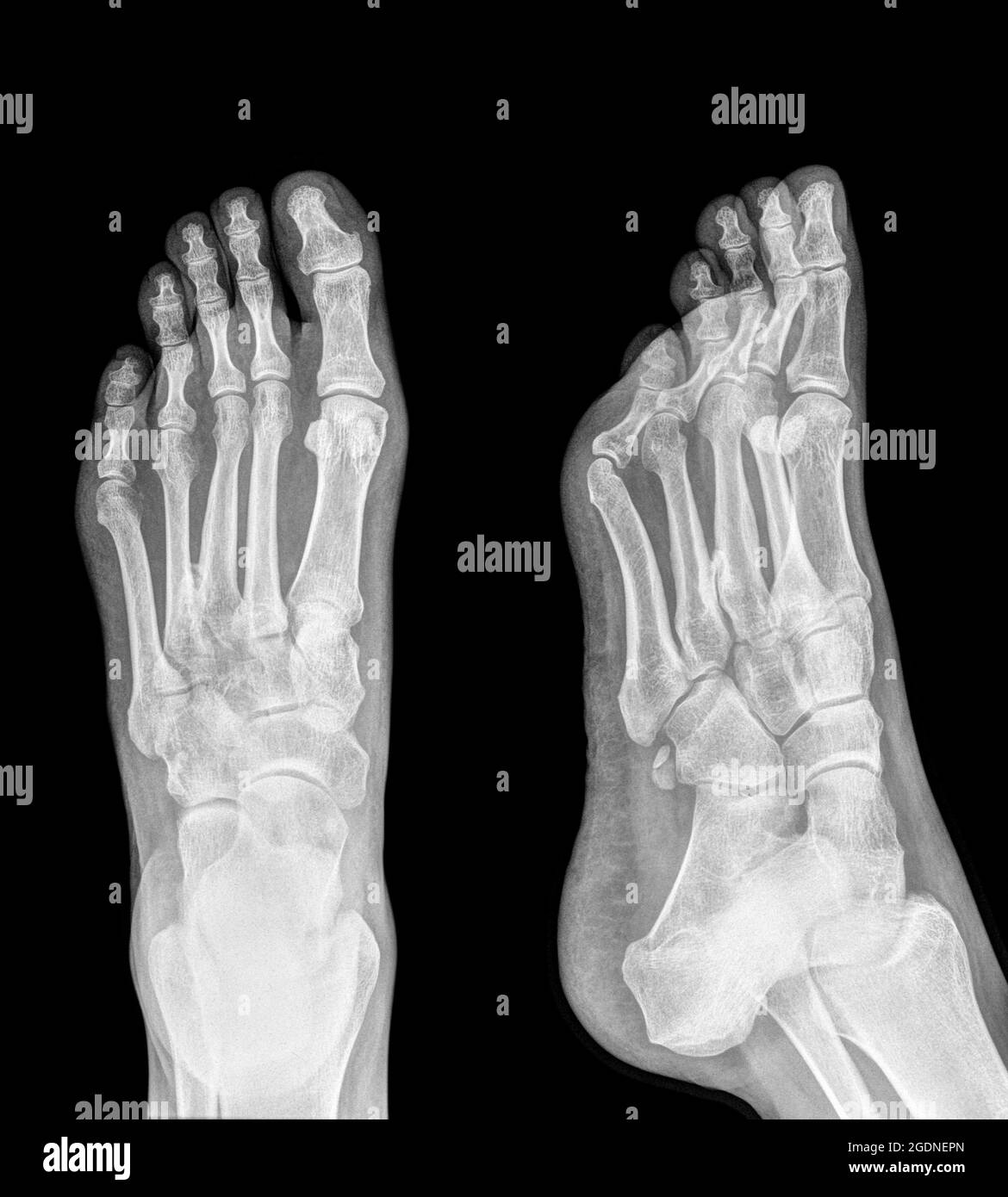 X-ray of a foot showing Plantar fasciitis (also known as Plantar fasciopathy or Jogger's heel) is a common painful enthesopathy of the heel and planta Stock Photohttps://www.alamy.com/image-license-details/?v=1https://www.alamy.com/x-ray-of-a-foot-showing-plantar-fasciitis-also-known-as-plantar-fasciopathy-or-joggers-heel-is-a-common-painful-enthesopathy-of-the-heel-and-planta-image438722333.html
RM2GDNEPN–X-ray of a foot showing Plantar fasciitis (also known as Plantar fasciopathy or Jogger's heel) is a common painful enthesopathy of the heel and planta
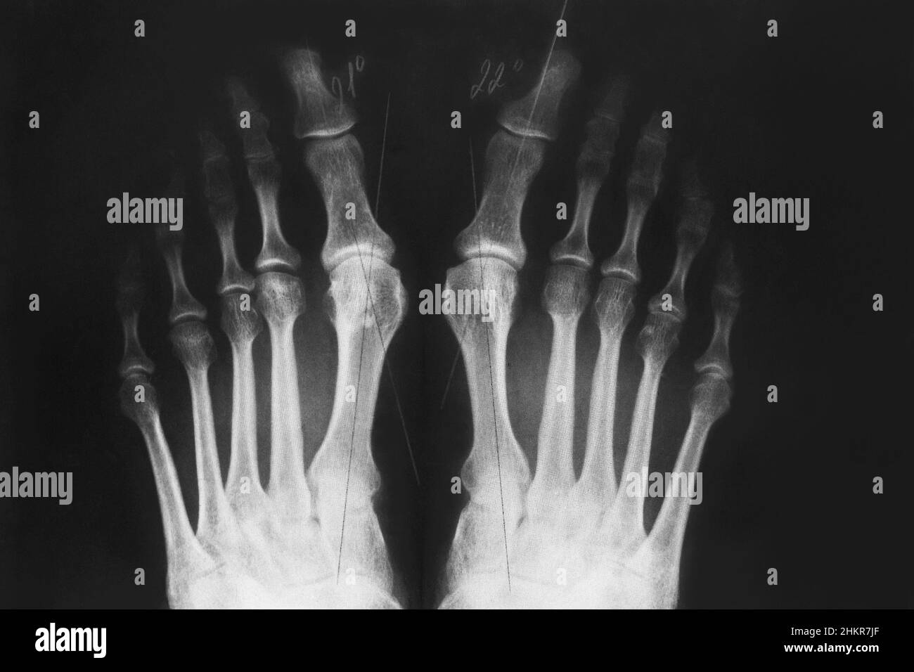 X-ray of the feet, valgus deformity of the toe or a bone on a finger, 3rd degree. Orthopedic disease. Stock Photohttps://www.alamy.com/image-license-details/?v=1https://www.alamy.com/x-ray-of-the-feet-valgus-deformity-of-the-toe-or-a-bone-on-a-finger-3rd-degree-orthopedic-disease-image459658935.html
RF2HKR7JF–X-ray of the feet, valgus deformity of the toe or a bone on a finger, 3rd degree. Orthopedic disease.
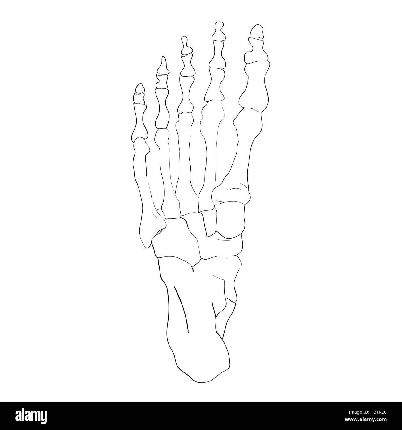 below view foot bones Stock Photohttps://www.alamy.com/image-license-details/?v=1https://www.alamy.com/stock-photo-below-view-foot-bones-127778728.html
RMHBTR20–below view foot bones
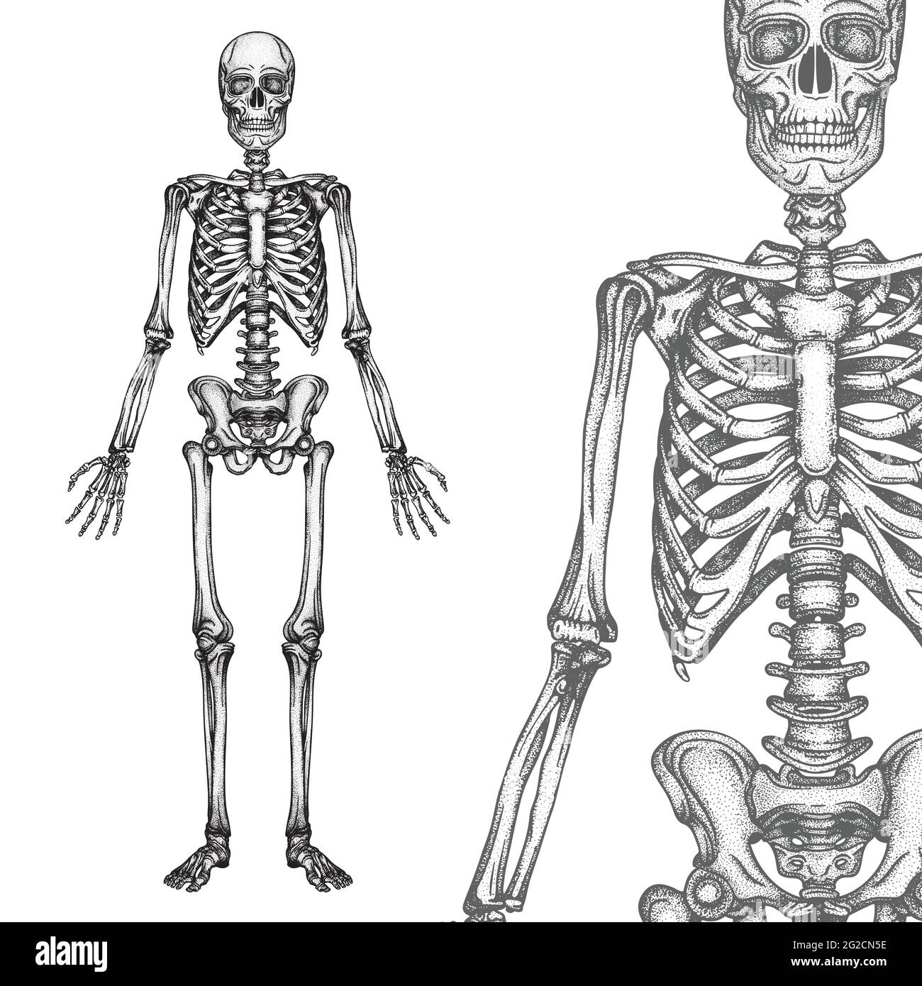 Human skeleton hand drawn vector illustrations set. Part of human skeleton graphic. Stock Vectorhttps://www.alamy.com/image-license-details/?v=1https://www.alamy.com/human-skeleton-hand-drawn-vector-illustrations-set-part-of-human-skeleton-graphic-image431768554.html
RF2G2CN5E–Human skeleton hand drawn vector illustrations set. Part of human skeleton graphic.
RF2P9X5NE–bone joints icon vector illustration symbol design
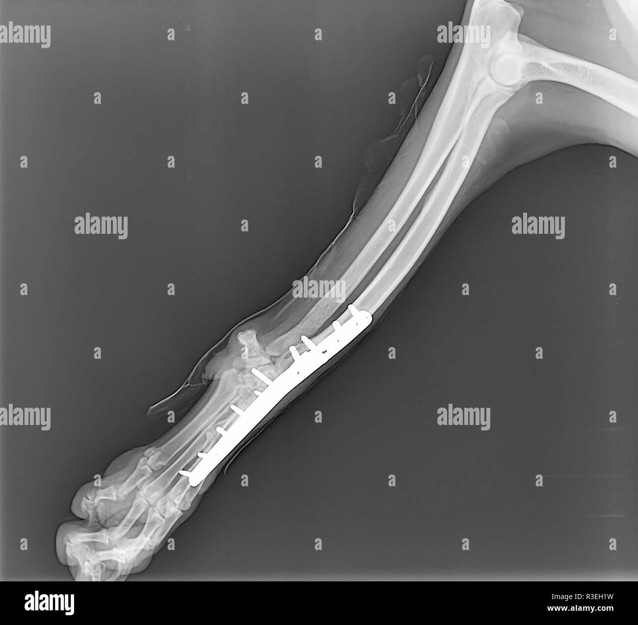 X-ray of a dog's front right leg at a veterinary surgery. Metal fixture and screws can be seen Stock Photohttps://www.alamy.com/image-license-details/?v=1https://www.alamy.com/x-ray-of-a-dogs-front-right-leg-at-a-veterinary-surgery-metal-fixture-and-screws-can-be-seen-image225899461.html
RFR3EH1W–X-ray of a dog's front right leg at a veterinary surgery. Metal fixture and screws can be seen
RF2B072TF–Orthopedic and spine vector black symbols. human bones icons. Hand and leg, skull and joint knee illustration
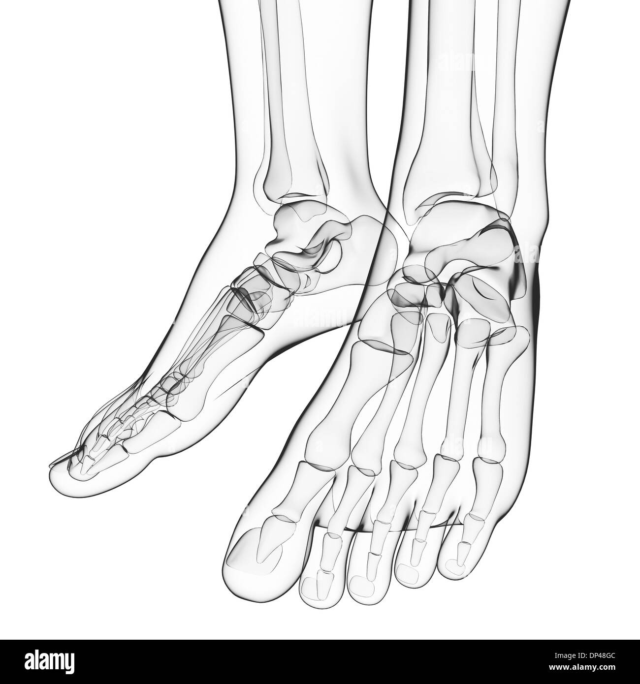 Human foot bones, artwork Stock Photohttps://www.alamy.com/image-license-details/?v=1https://www.alamy.com/human-foot-bones-artwork-image65248076.html
RFDP48GC–Human foot bones, artwork
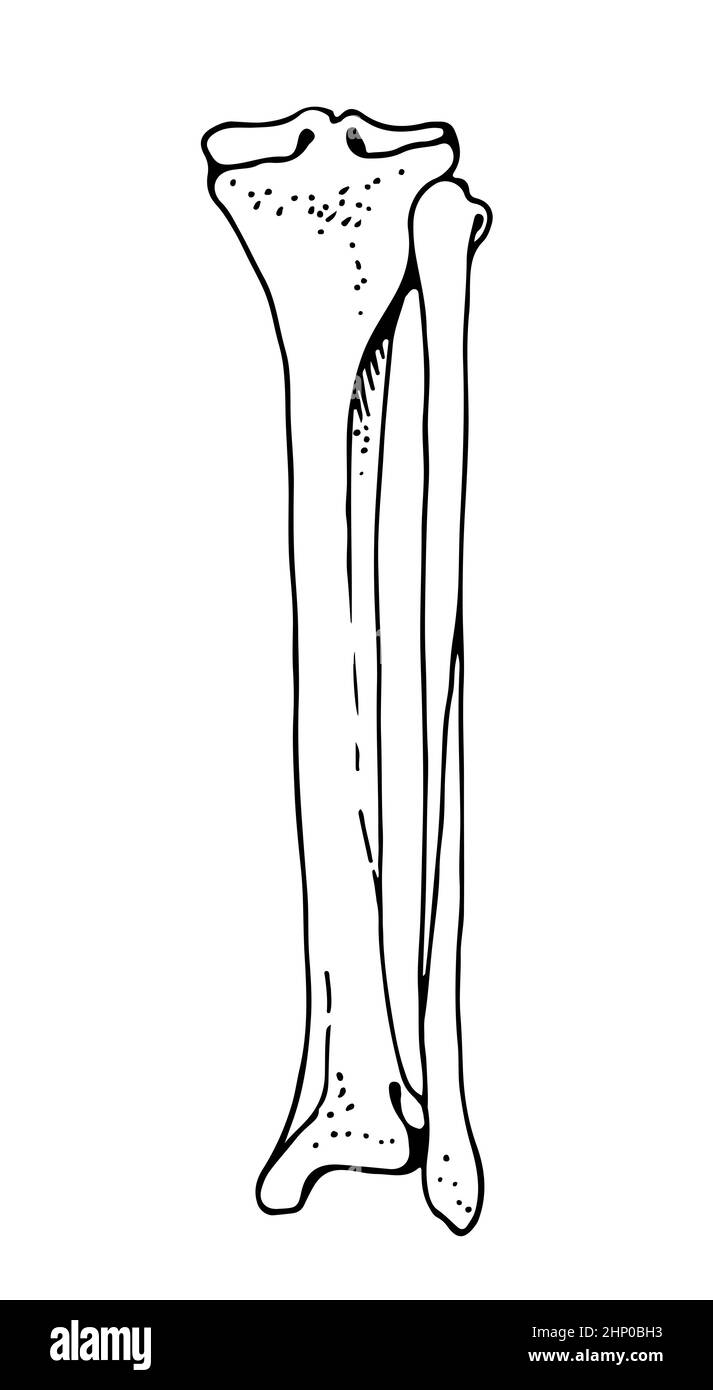 Tibia and fibula human bones, vector hand drawn illustration isolated on a white background, orthopedics medicine anatomy sketch Stock Vectorhttps://www.alamy.com/image-license-details/?v=1https://www.alamy.com/tibia-and-fibula-human-bones-vector-hand-drawn-illustration-isolated-on-a-white-background-orthopedics-medicine-anatomy-sketch-image461001103.html
RF2HP0BH3–Tibia and fibula human bones, vector hand drawn illustration isolated on a white background, orthopedics medicine anatomy sketch
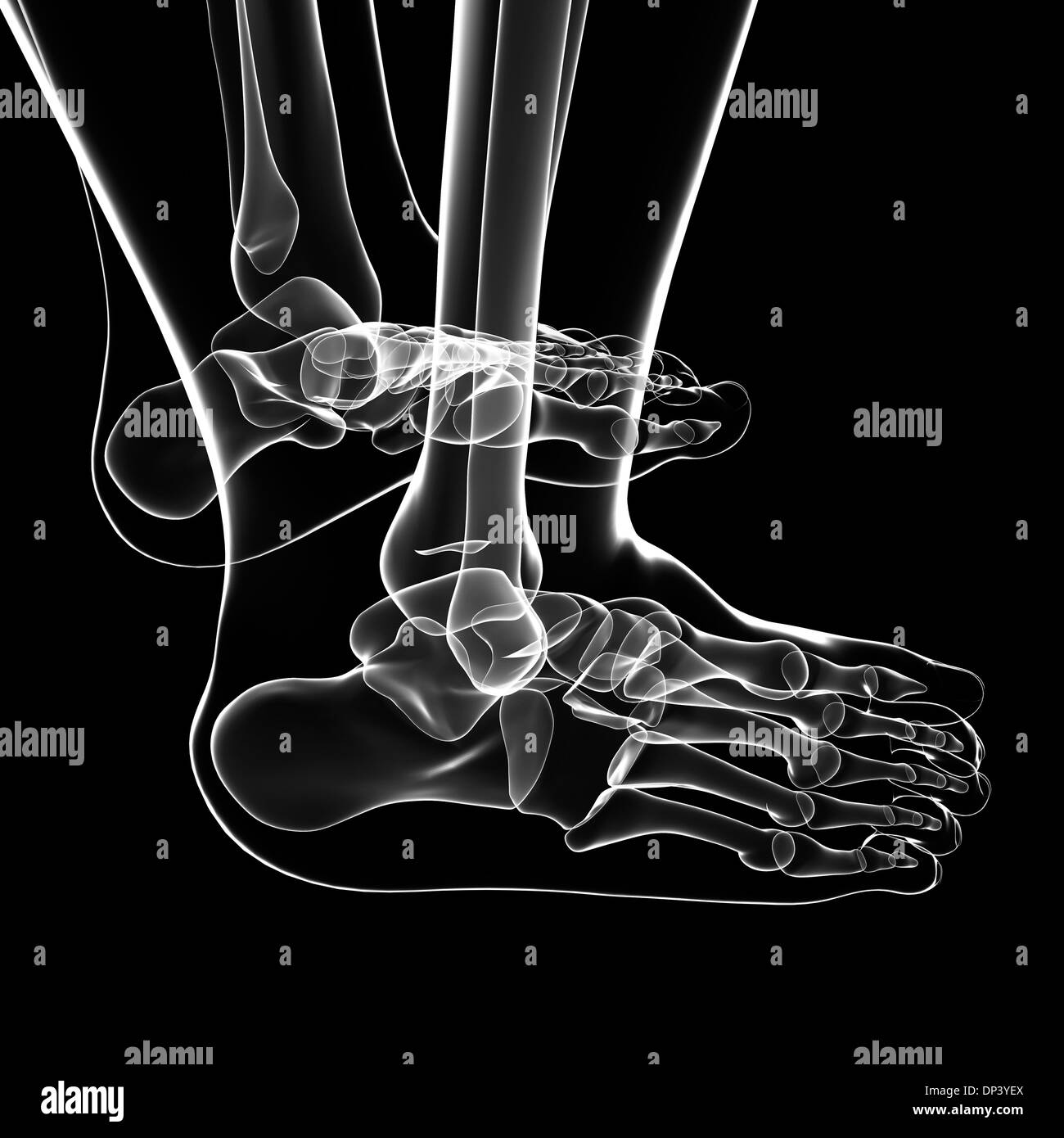 Human foot bones, artwork Stock Photohttps://www.alamy.com/image-license-details/?v=1https://www.alamy.com/human-foot-bones-artwork-image65240978.html
RFDP3YEX–Human foot bones, artwork
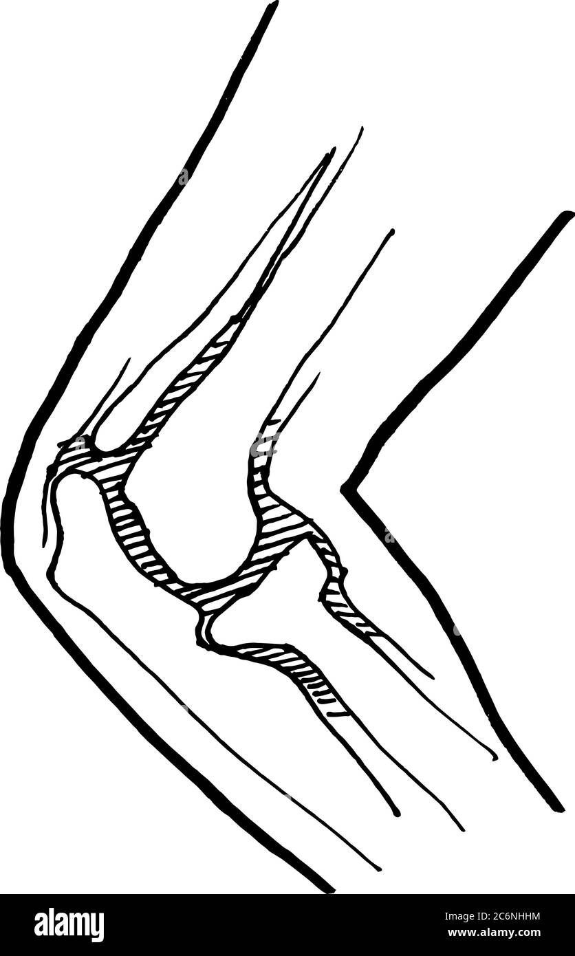 Contour vector outline drawing of human injured knee bones. Medical design editable template Stock Vectorhttps://www.alamy.com/image-license-details/?v=1https://www.alamy.com/contour-vector-outline-drawing-of-human-injured-knee-bones-medical-design-editable-template-image365580480.html
RF2C6NHHM–Contour vector outline drawing of human injured knee bones. Medical design editable template
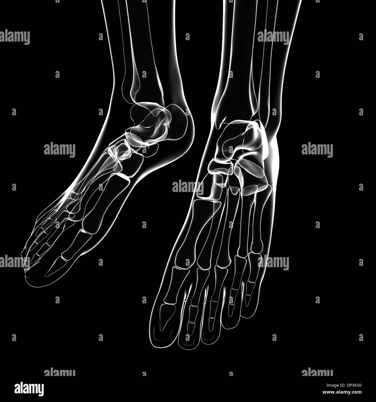 Human foot bones, artwork Stock Photohttps://www.alamy.com/image-license-details/?v=1https://www.alamy.com/human-foot-bones-artwork-image65235016.html
RFDP3KX0–Human foot bones, artwork
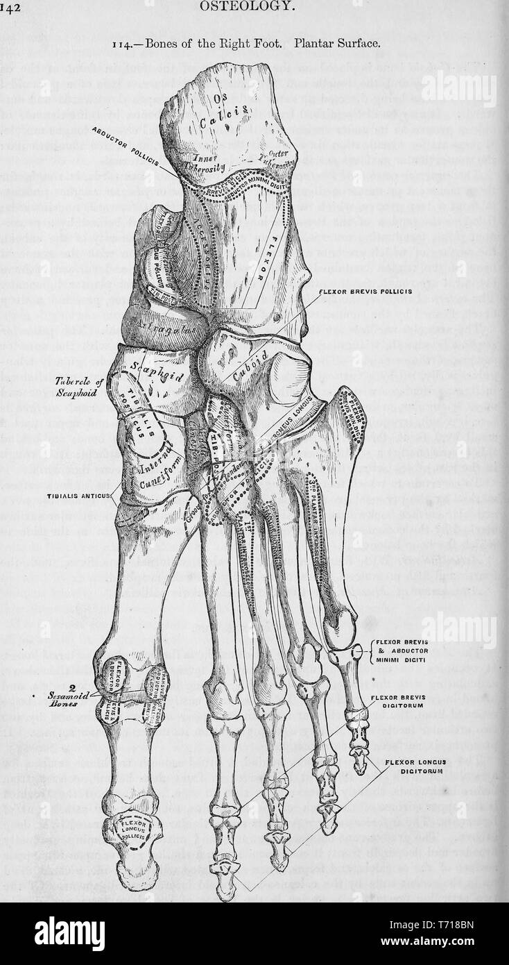 Illustrated planar surface of right foot bones, from the book 'Anatomy, descriptive and surgical' by Henry Gray, Henry Vandyke Carter, and John Guise Westmacott, 1860. Courtesy Internet Archive. () Stock Photohttps://www.alamy.com/image-license-details/?v=1https://www.alamy.com/illustrated-planar-surface-of-right-foot-bones-from-the-book-anatomy-descriptive-and-surgical-by-henry-gray-henry-vandyke-carter-and-john-guise-westmacott-1860-courtesy-internet-archive-image245276297.html
RMT718BN–Illustrated planar surface of right foot bones, from the book 'Anatomy, descriptive and surgical' by Henry Gray, Henry Vandyke Carter, and John Guise Westmacott, 1860. Courtesy Internet Archive. ()
 Human leg and foot bones, computer artwork. Stock Photohttps://www.alamy.com/image-license-details/?v=1https://www.alamy.com/stock-photo-human-leg-and-foot-bones-computer-artwork-73691516.html
RFE7TX7T–Human leg and foot bones, computer artwork.
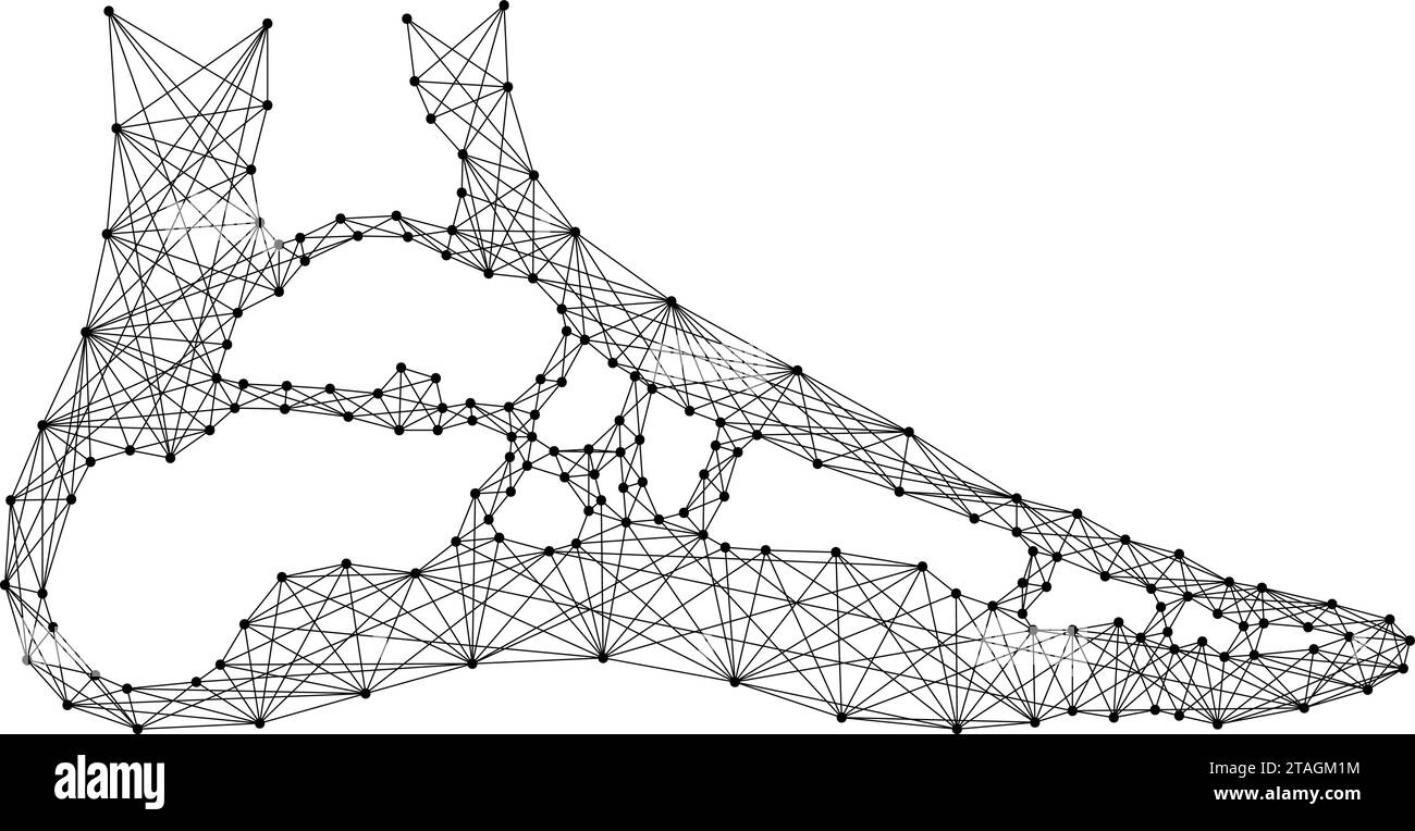 Foot of human leg in section, from abstract futuristic polygonal black lines and dots. Vector illustration. Stock Vectorhttps://www.alamy.com/image-license-details/?v=1https://www.alamy.com/foot-of-human-leg-in-section-from-abstract-futuristic-polygonal-black-lines-and-dots-vector-illustration-image574455664.html
RF2TAGM1M–Foot of human leg in section, from abstract futuristic polygonal black lines and dots. Vector illustration.
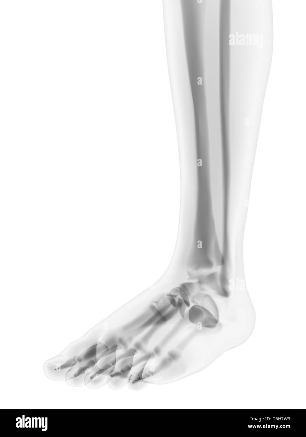 Foot bones, artwork Stock Photohttps://www.alamy.com/image-license-details/?v=1https://www.alamy.com/stock-photo-foot-bones-artwork-55698415.html
RFD6H7W3–Foot bones, artwork
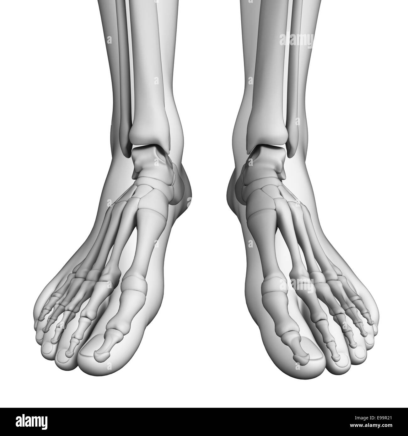 Illustration of human foot artwork Stock Photohttps://www.alamy.com/image-license-details/?v=1https://www.alamy.com/stock-photo-illustration-of-human-foot-artwork-74589033.html
RFE99R21–Illustration of human foot artwork
 X-ray view of human foot Stock Photohttps://www.alamy.com/image-license-details/?v=1https://www.alamy.com/x-ray-view-of-human-foot-image619199304.html
RM2XYAY0T–X-ray view of human foot
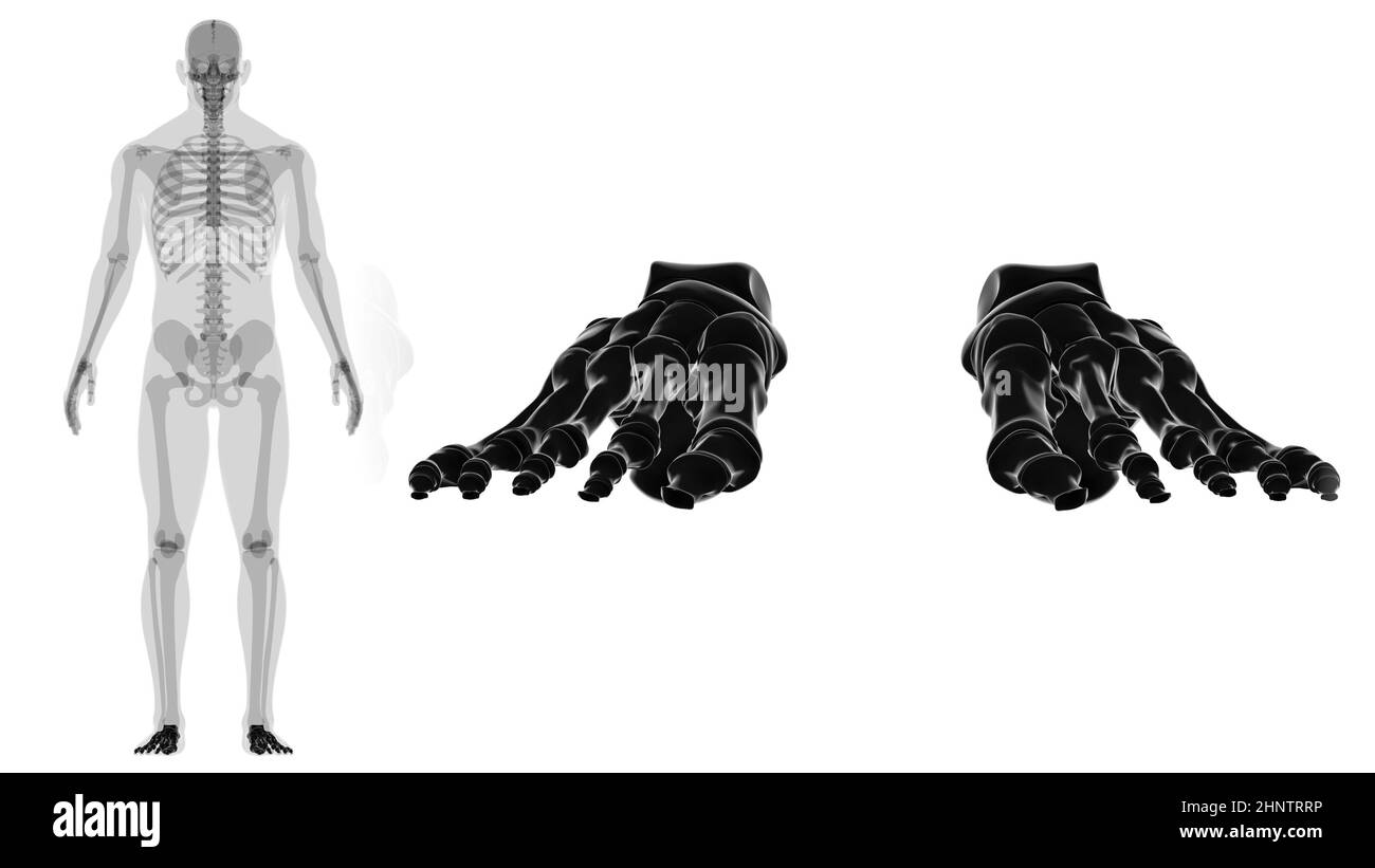 Human Skeleton Foot bones Anatomy For Medical Concept 3D Illustration Stock Photohttps://www.alamy.com/image-license-details/?v=1https://www.alamy.com/human-skeleton-foot-bones-anatomy-for-medical-concept-3d-illustration-image460922890.html
RF2HNTRRP–Human Skeleton Foot bones Anatomy For Medical Concept 3D Illustration
 x-ray of a foot showing a fracture in the 5th metatarsalia of a 31 year old male Stock Photohttps://www.alamy.com/image-license-details/?v=1https://www.alamy.com/x-ray-of-a-foot-showing-a-fracture-in-the-5th-metatarsalia-of-a-31-year-old-male-image439021264.html
RM2GE742T–x-ray of a foot showing a fracture in the 5th metatarsalia of a 31 year old male
 Bone Leg isometric isolated. 3D Bones anatomy. Human Skeleton system Stock Vectorhttps://www.alamy.com/image-license-details/?v=1https://www.alamy.com/bone-leg-isometric-isolated-3d-bones-anatomy-human-skeleton-system-image226337596.html
RFR46FWG–Bone Leg isometric isolated. 3D Bones anatomy. Human Skeleton system
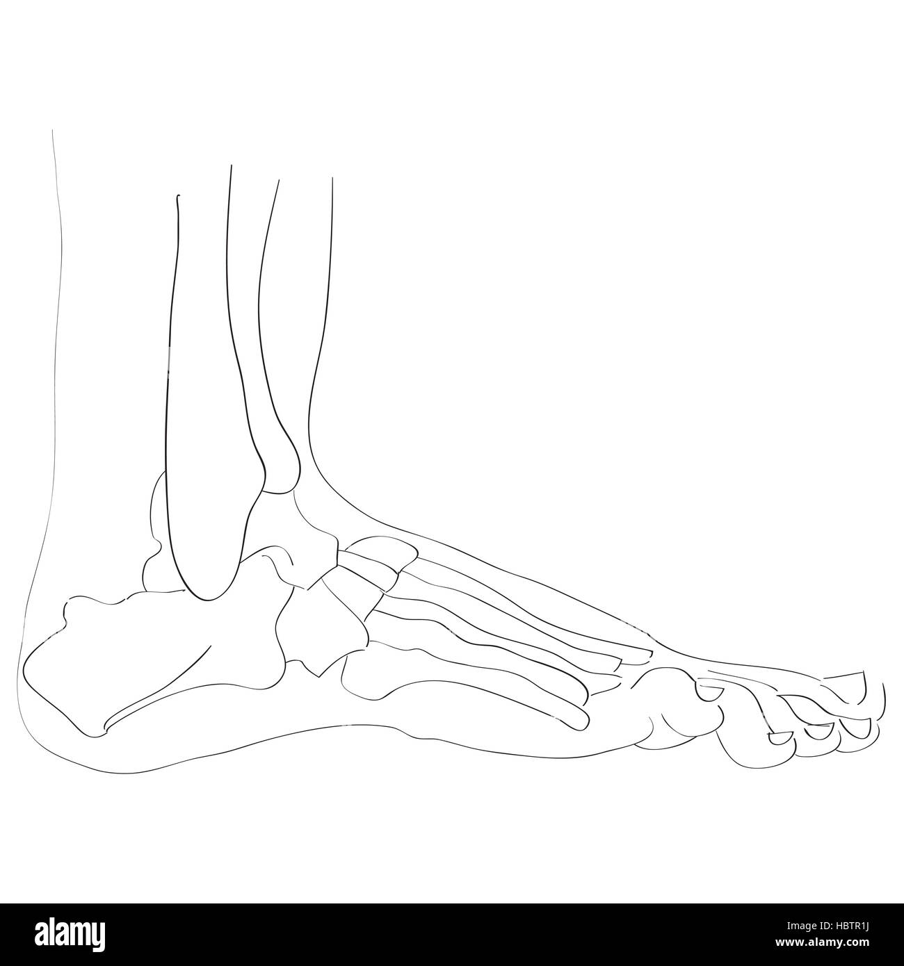 lateral view foot bones Stock Photohttps://www.alamy.com/image-license-details/?v=1https://www.alamy.com/stock-photo-lateral-view-foot-bones-127778718.html
RMHBTR1J–lateral view foot bones
 Silhouette of a human skeleton disassembled into bones isolated on a white background. Isometric view. Vector illustration. Stock Vectorhttps://www.alamy.com/image-license-details/?v=1https://www.alamy.com/silhouette-of-a-human-skeleton-disassembled-into-bones-isolated-on-a-white-background-isometric-view-vector-illustration-image627027720.html
RF2YC3G74–Silhouette of a human skeleton disassembled into bones isolated on a white background. Isometric view. Vector illustration.
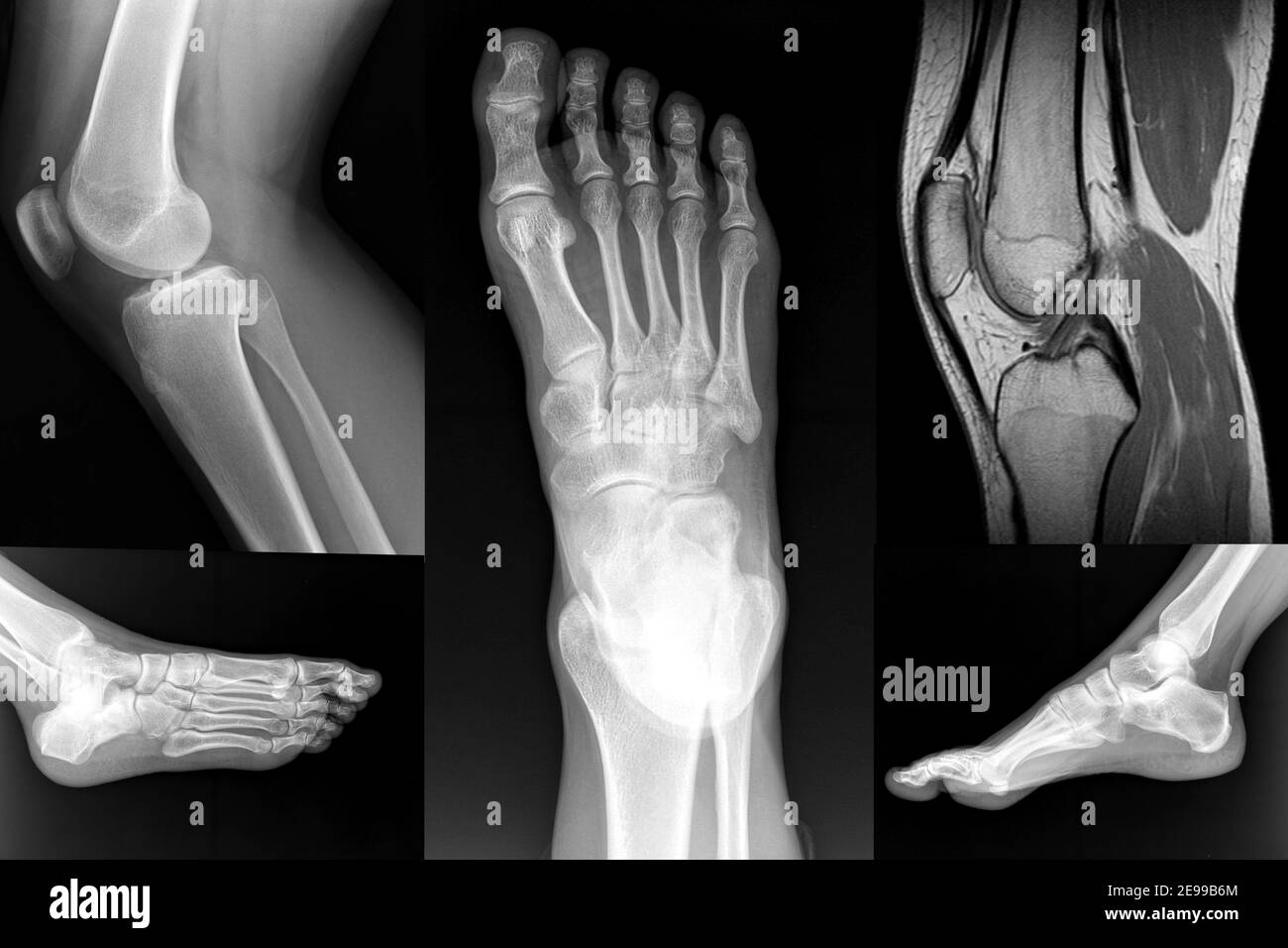 Orthopedic Collage of Foot and Knee XRay Stock Photohttps://www.alamy.com/image-license-details/?v=1https://www.alamy.com/orthopedic-collage-of-foot-and-knee-xray-image401576748.html
RF2E99B6M–Orthopedic Collage of Foot and Knee XRay
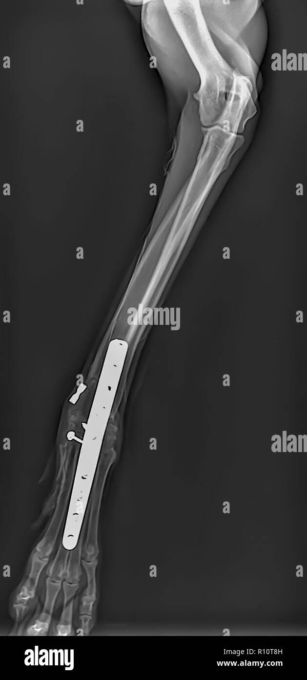 X-ray of a dog's front right leg at a veterinary surgery. Metal fixture and screws can be seen Stock Photohttps://www.alamy.com/image-license-details/?v=1https://www.alamy.com/x-ray-of-a-dogs-front-right-leg-at-a-veterinary-surgery-metal-fixture-and-screws-can-be-seen-image224368497.html
RMR10T8H–X-ray of a dog's front right leg at a veterinary surgery. Metal fixture and screws can be seen
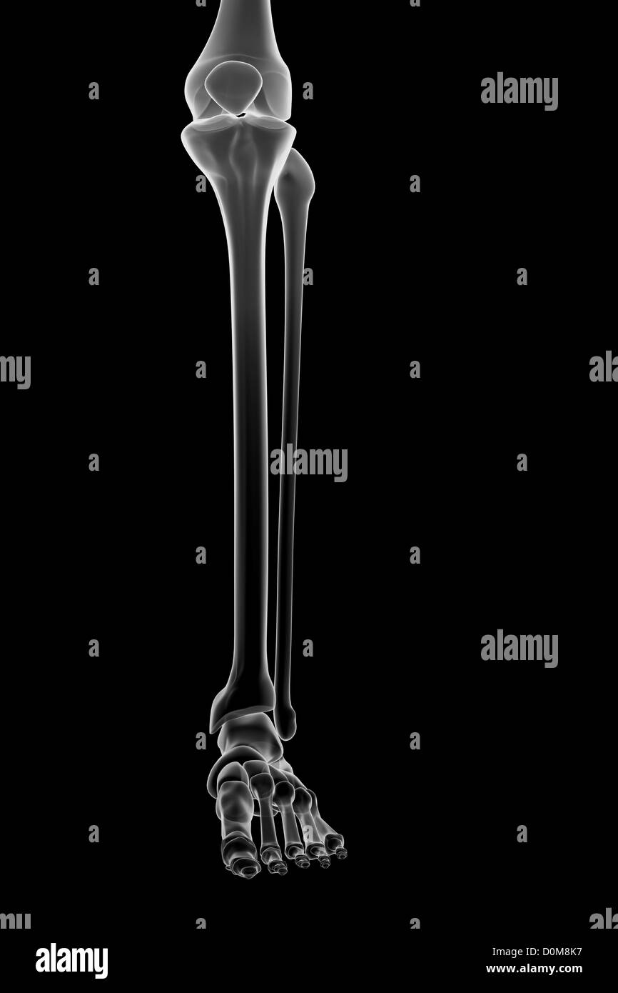 Stylized bones of the lower left leg, ankle joint and foot. Stock Photohttps://www.alamy.com/image-license-details/?v=1https://www.alamy.com/stock-photo-stylized-bones-of-the-lower-left-leg-ankle-joint-and-foot-52076955.html
RMD0M8K7–Stylized bones of the lower left leg, ankle joint and foot.
 The bones are fastened together kept in place and their movements limited by tough, vintage line drawing or engraving illustration. Stock Vectorhttps://www.alamy.com/image-license-details/?v=1https://www.alamy.com/the-bones-are-fastened-together-kept-in-place-and-their-movements-limited-by-tough-vintage-line-drawing-or-engraving-illustration-image348639541.html
RF2B75W85–The bones are fastened together kept in place and their movements limited by tough, vintage line drawing or engraving illustration.
RF2J16P07–Bone joint icon, articular connection of bones for body movement
 Anterior Region of the Leg, showing muscles and bones, vintage engraved illustration. Usual Medicine Dictionary by Dr Labarthe - Stock Vectorhttps://www.alamy.com/image-license-details/?v=1https://www.alamy.com/stock-photo-anterior-region-of-the-leg-showing-muscles-and-bones-vintage-engraved-84418999.html
RFEW9H87–Anterior Region of the Leg, showing muscles and bones, vintage engraved illustration. Usual Medicine Dictionary by Dr Labarthe -
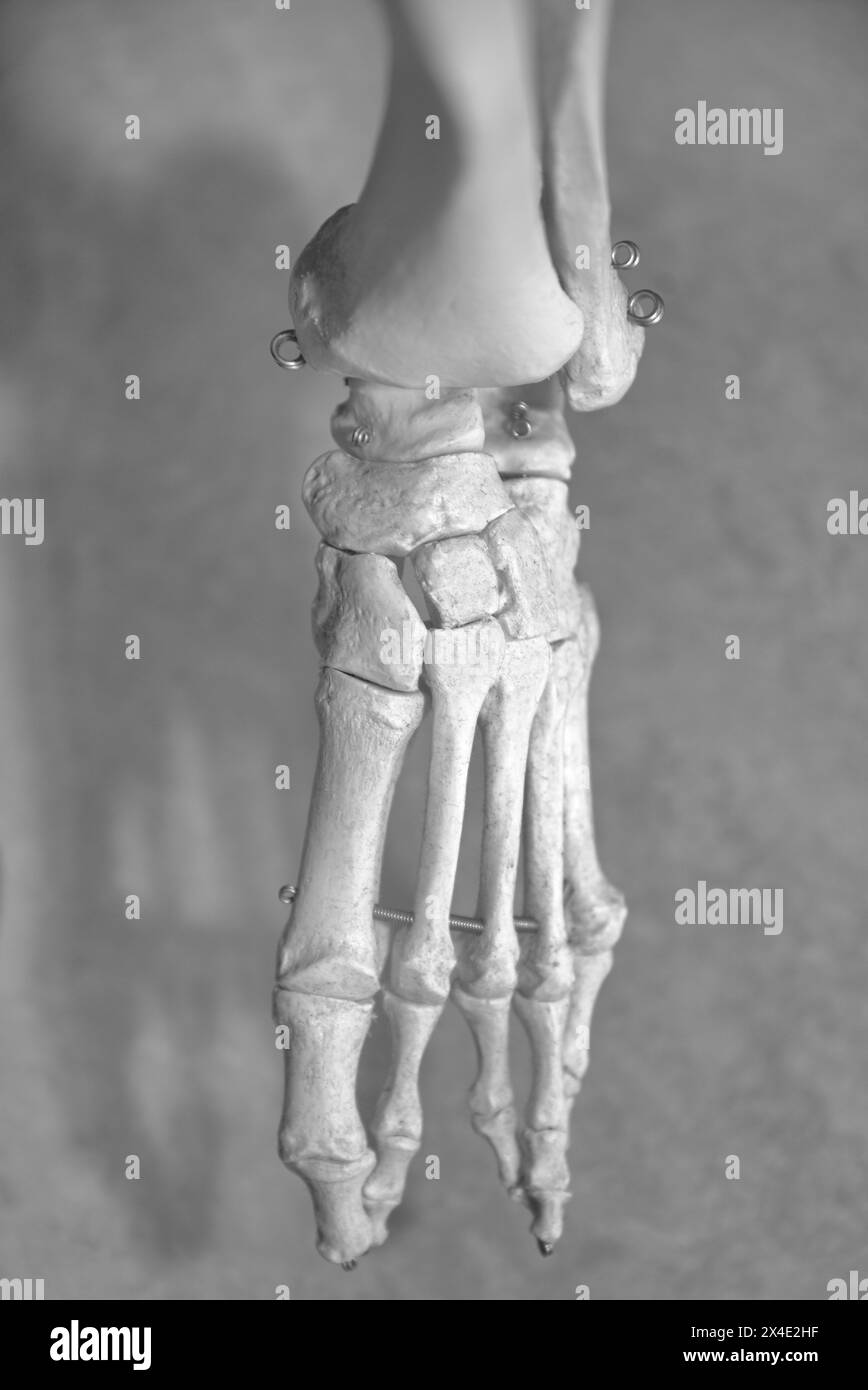 Skeletal Part - Bones Of The Toes, Fibula, Tibia And Ankle Joint Stock Photohttps://www.alamy.com/image-license-details/?v=1https://www.alamy.com/skeletal-part-bones-of-the-toes-fibula-tibia-and-ankle-joint-image605130891.html
RM2X4E2HF–Skeletal Part - Bones Of The Toes, Fibula, Tibia And Ankle Joint
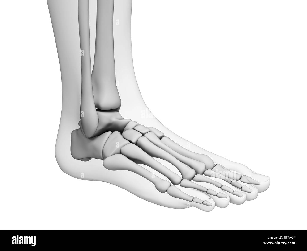 3d rendered illustration - foot anatomy Stock Photohttps://www.alamy.com/image-license-details/?v=1https://www.alamy.com/stock-photo-3d-rendered-illustration-foot-anatomy-144606127.html
RFJB7AGF–3d rendered illustration - foot anatomy
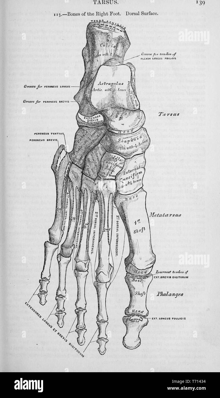 Black and white print showing the bones of a human right foot, from the dorsal perspective, illustrated by Henry Vandyke Carter, and published in Henry Gray's medical volume 'Anatomy, descriptive and surgical', 1860. Courtesy Internet Archive. () Stock Photohttps://www.alamy.com/image-license-details/?v=1https://www.alamy.com/black-and-white-print-showing-the-bones-of-a-human-right-foot-from-the-dorsal-perspective-illustrated-by-henry-vandyke-carter-and-published-in-henry-grays-medical-volume-anatomy-descriptive-and-surgical-1860-courtesy-internet-archive-image245272920.html
RMT71434–Black and white print showing the bones of a human right foot, from the dorsal perspective, illustrated by Henry Vandyke Carter, and published in Henry Gray's medical volume 'Anatomy, descriptive and surgical', 1860. Courtesy Internet Archive. ()
RF2HMGBR1–Leg bones icon design template vector isolated
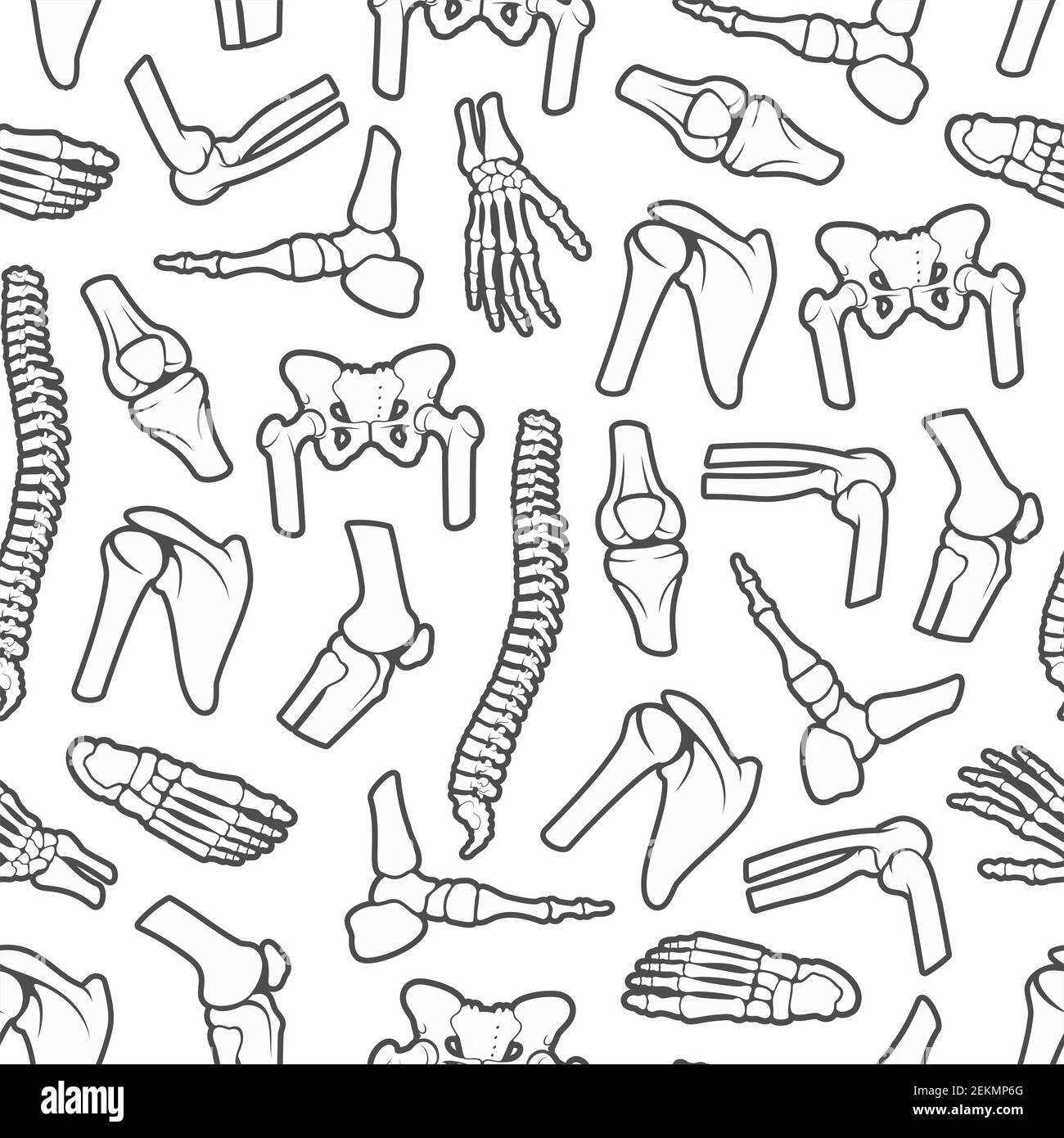 Bones and joints monochrome seamless pattern. Vector human spine pelvis, shoulder scapula, wrist with fingers and foot ankle. Orthopedic background, a Stock Vectorhttps://www.alamy.com/image-license-details/?v=1https://www.alamy.com/bones-and-joints-monochrome-seamless-pattern-vector-human-spine-pelvis-shoulder-scapula-wrist-with-fingers-and-foot-ankle-orthopedic-background-a-image407973400.html
RF2EKMP6G–Bones and joints monochrome seamless pattern. Vector human spine pelvis, shoulder scapula, wrist with fingers and foot ankle. Orthopedic background, a
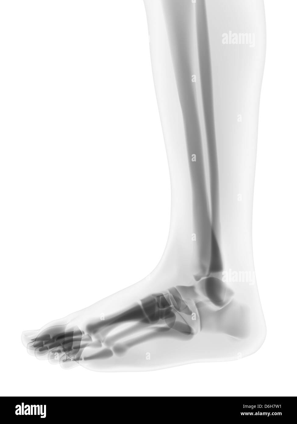 Foot bones, artwork Stock Photohttps://www.alamy.com/image-license-details/?v=1https://www.alamy.com/stock-photo-foot-bones-artwork-55698413.html
RFD6H7W1–Foot bones, artwork
RF2EEHH98–Human joints and body parts bones sketch icons. Vector isolated set of spine pelvis, shoulder scapula or elbow, leg knee and foot ankle, arm and hand
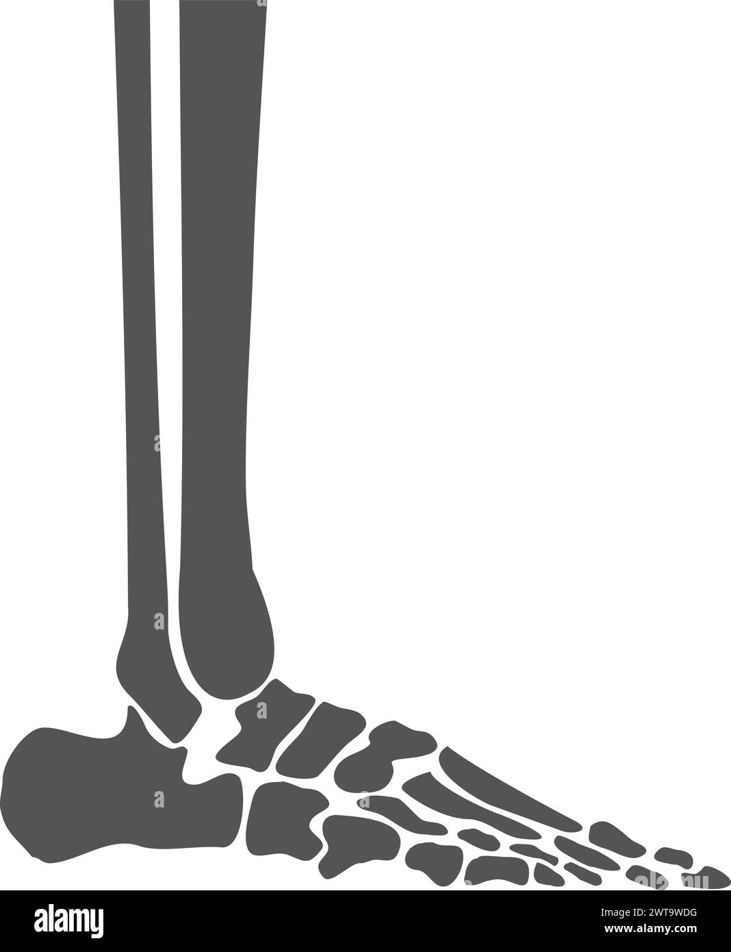 Human leg bones. Feet anatomy side view Stock Vectorhttps://www.alamy.com/image-license-details/?v=1https://www.alamy.com/human-leg-bones-feet-anatomy-side-view-image600121804.html
RF2WT9WDG–Human leg bones. Feet anatomy side view
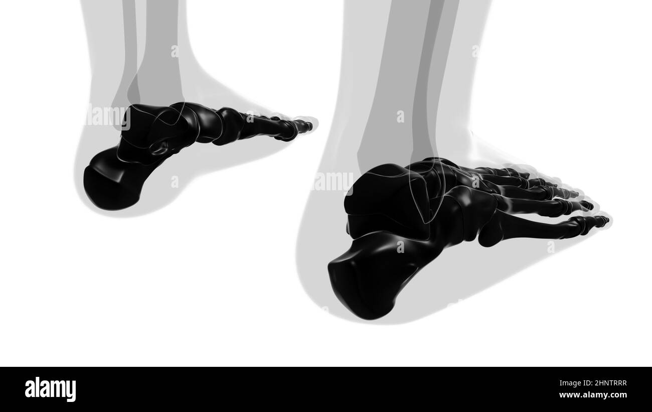 Human Skeleton Foot bones Anatomy For Medical Concept 3D Illustration Stock Photohttps://www.alamy.com/image-license-details/?v=1https://www.alamy.com/human-skeleton-foot-bones-anatomy-for-medical-concept-3d-illustration-image460922891.html
RF2HNTRRR–Human Skeleton Foot bones Anatomy For Medical Concept 3D Illustration
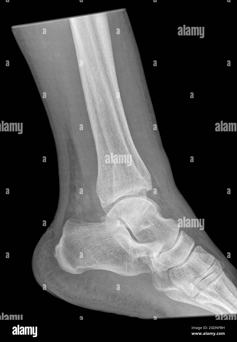 Fracture of the distal tibia and fibula. X-ray of a 57 year old male Stock Photohttps://www.alamy.com/image-license-details/?v=1https://www.alamy.com/fracture-of-the-distal-tibia-and-fibula-x-ray-of-a-57-year-old-male-image438722805.html
RM2GDNFBH–Fracture of the distal tibia and fibula. X-ray of a 57 year old male
RFR46FB3–Bone Leg pixel art. Bones anatomy 8 bit. Pixelate Human Skeleton system 16bit. Old game computer graphics style
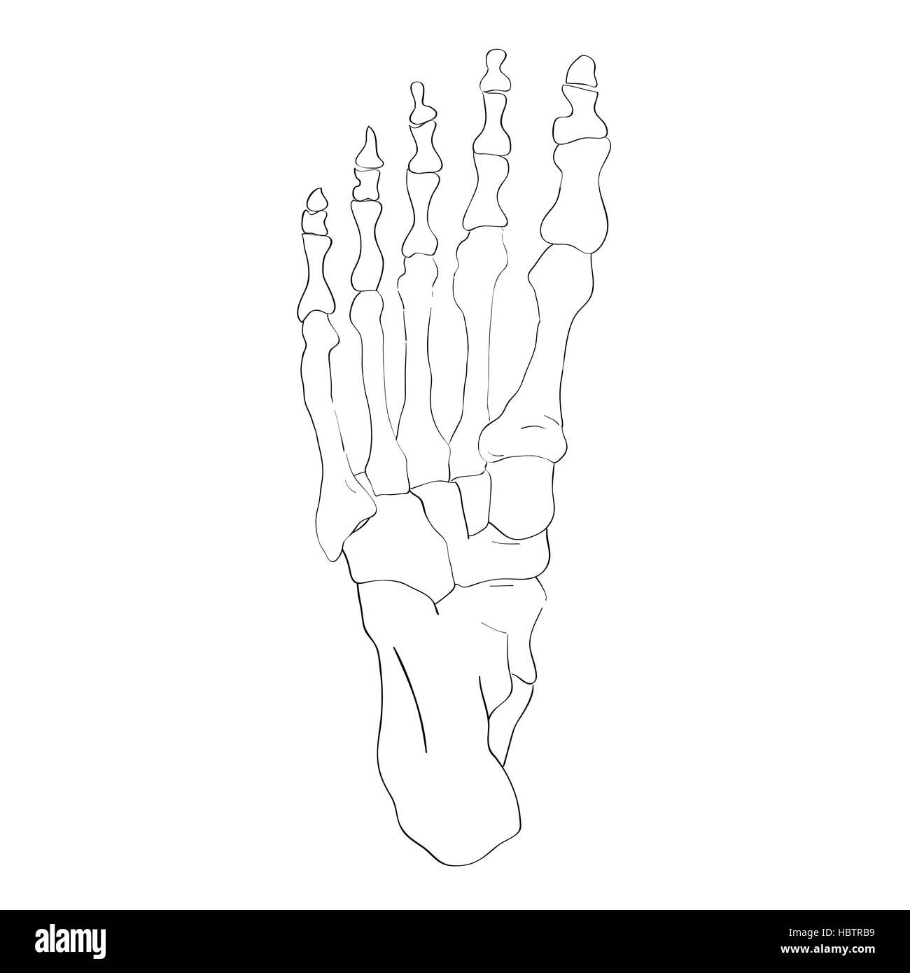 below view foot bones Stock Photohttps://www.alamy.com/image-license-details/?v=1https://www.alamy.com/stock-photo-below-view-foot-bones-127778989.html
RMHBTRB9–below view foot bones
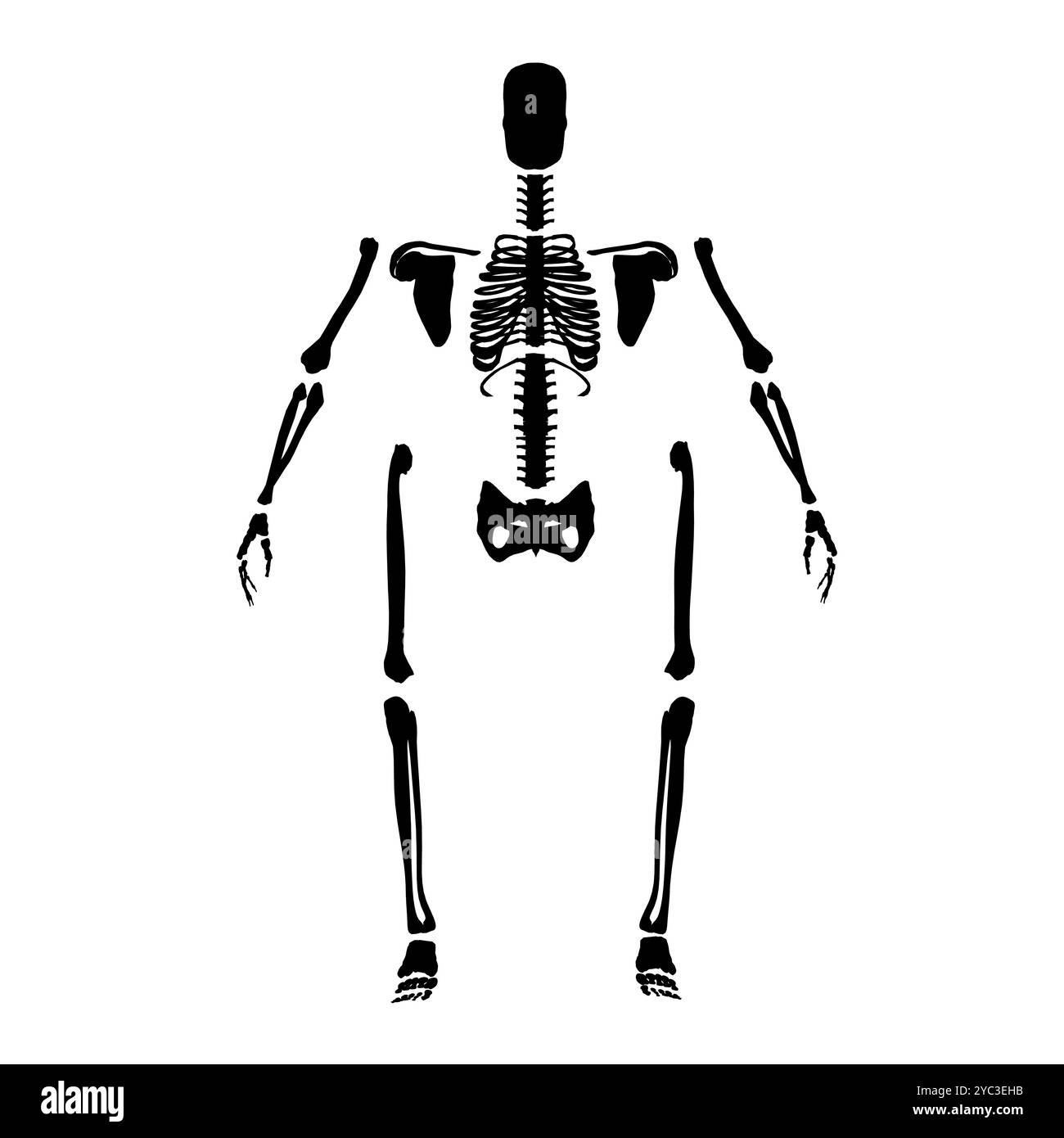 Silhouette of a human skeleton disassembled into bones isolated on a white background. Front view. Vector illustration. Stock Vectorhttps://www.alamy.com/image-license-details/?v=1https://www.alamy.com/silhouette-of-a-human-skeleton-disassembled-into-bones-isolated-on-a-white-background-front-view-vector-illustration-image627026439.html
RF2YC3EHB–Silhouette of a human skeleton disassembled into bones isolated on a white background. Front view. Vector illustration.
 Pig Skull and Bones. Concept pig skeleton head for pirate flag Stock Vectorhttps://www.alamy.com/image-license-details/?v=1https://www.alamy.com/pig-skull-and-bones-concept-pig-skeleton-head-for-pirate-flag-image630719774.html
RF2YJ3NE6–Pig Skull and Bones. Concept pig skeleton head for pirate flag
 human leg foot bone anatomy medical sketch Stock Vectorhttps://www.alamy.com/image-license-details/?v=1https://www.alamy.com/stock-photo-human-leg-foot-bone-anatomy-medical-sketch-140113611.html
RFJ3XM9F–human leg foot bone anatomy medical sketch
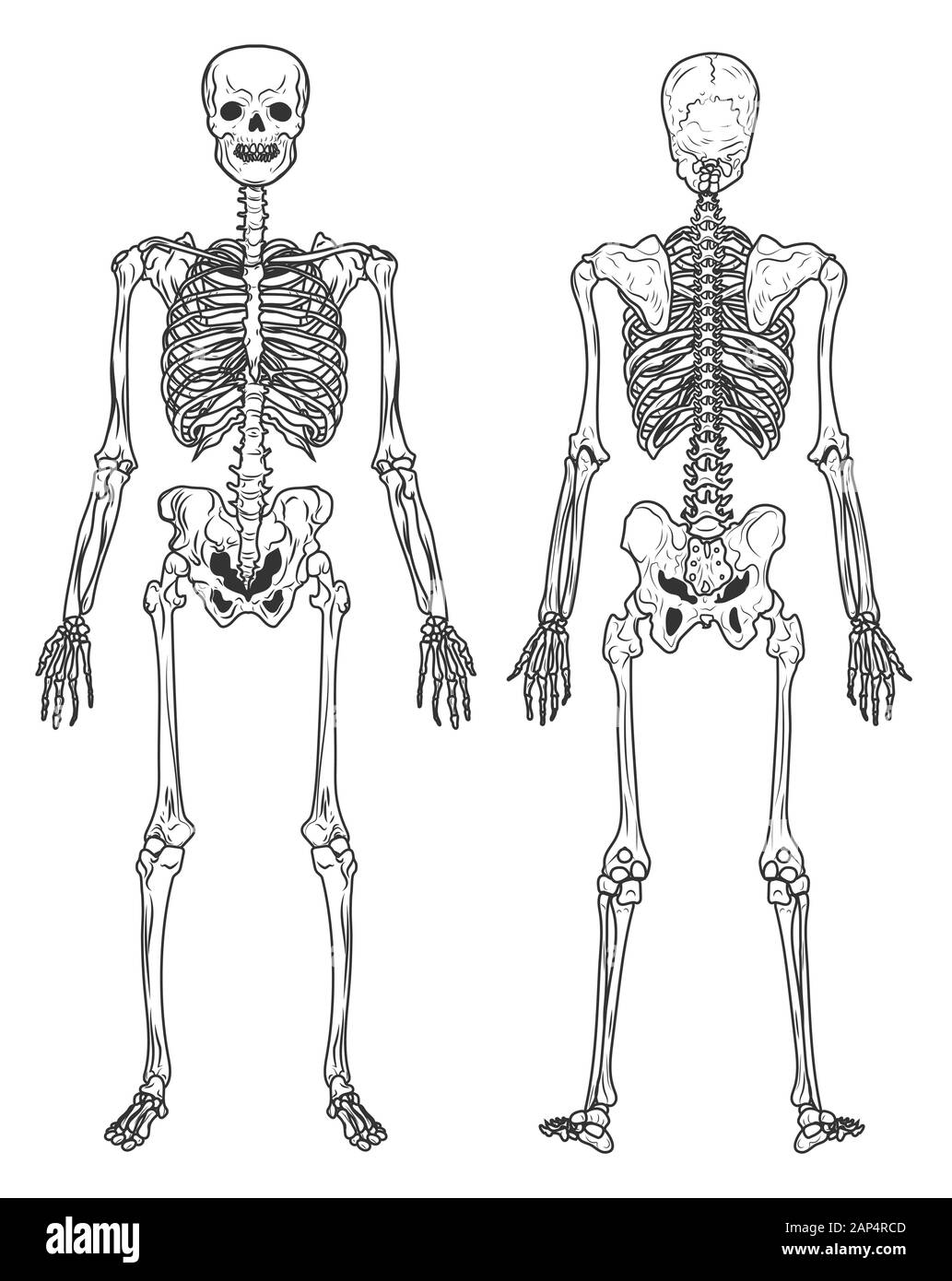 Skeleton structure back and front view, human bones Stock Vectorhttps://www.alamy.com/image-license-details/?v=1https://www.alamy.com/skeleton-structure-back-and-front-view-human-bones-image340625613.html
RF2AP4RCD–Skeleton structure back and front view, human bones
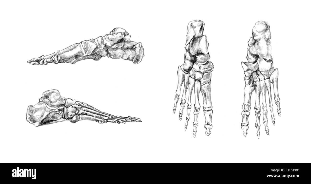 Bones of foot, Hand drawn medical illustration drawing with imitation of lithography Stock Photohttps://www.alamy.com/image-license-details/?v=1https://www.alamy.com/stock-photo-bones-of-foot-hand-drawn-medical-illustration-drawing-with-imitation-129446906.html
RFHEGPRP–Bones of foot, Hand drawn medical illustration drawing with imitation of lithography
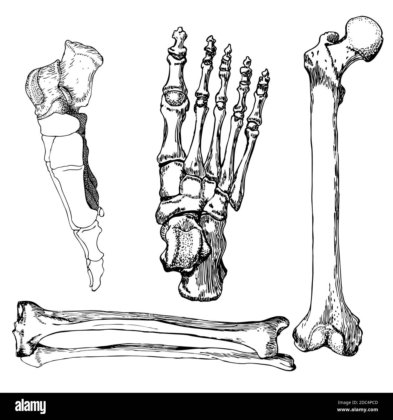 Set of human leg bones with foot. Hand drawn vector illustration. Isolated on white. Stock Vectorhttps://www.alamy.com/image-license-details/?v=1https://www.alamy.com/set-of-human-leg-bones-with-foot-hand-drawn-vector-illustration-isolated-on-white-image386109373.html
RF2DC4PCD–Set of human leg bones with foot. Hand drawn vector illustration. Isolated on white.
 Anterior Region of the Leg, showing muscles and bones, vintage engraved illustration. Usual Medicine Dictionary by Dr Labarthe - Stock Vectorhttps://www.alamy.com/image-license-details/?v=1https://www.alamy.com/stock-photo-anterior-region-of-the-leg-showing-muscles-and-bones-vintage-engraved-84406684.html
RFEW91GC–Anterior Region of the Leg, showing muscles and bones, vintage engraved illustration. Usual Medicine Dictionary by Dr Labarthe -
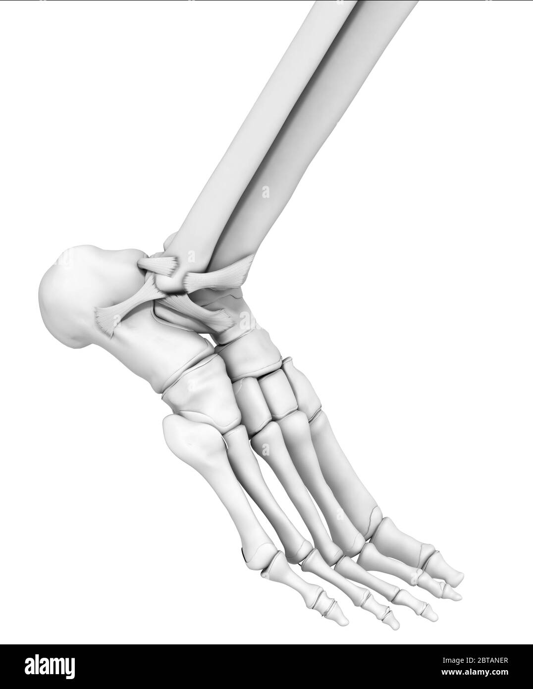 3D illustration showing ankle joint, medical mock up on white background Stock Photohttps://www.alamy.com/image-license-details/?v=1https://www.alamy.com/3d-illustration-showing-ankle-joint-medical-mock-up-on-white-background-image359195503.html
RF2BTANER–3D illustration showing ankle joint, medical mock up on white background
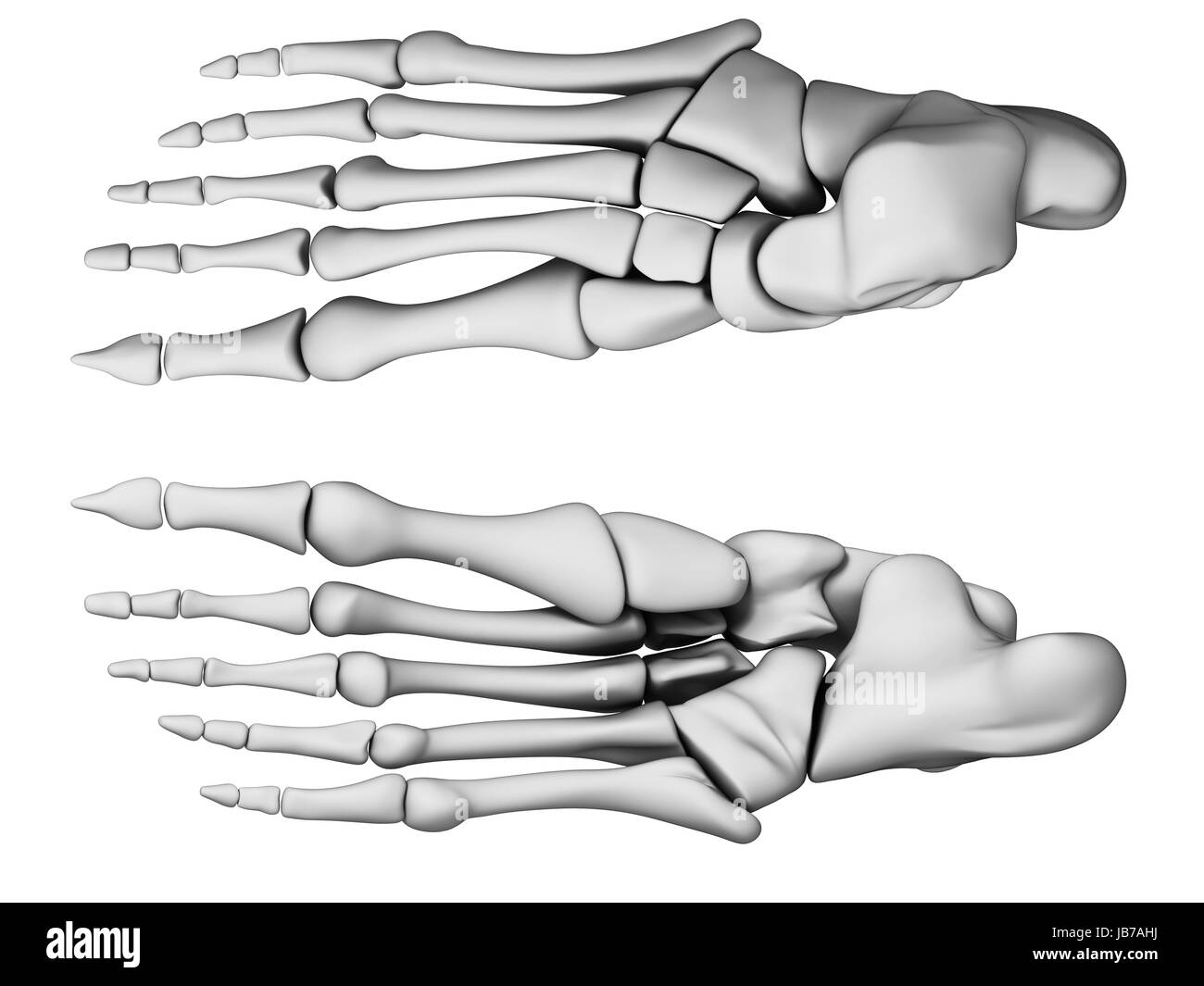 3d rendered illustration - foot anatomy Stock Photohttps://www.alamy.com/image-license-details/?v=1https://www.alamy.com/stock-photo-3d-rendered-illustration-foot-anatomy-144606158.html
RFJB7AHJ–3d rendered illustration - foot anatomy
 A Skeleton stood on a bridge, Droitwich Spa, Worcester, United Kingdom,02/03/2020, Skeleton stood on a bridge, fun for halloween ideal for posters a Stock Photohttps://www.alamy.com/image-license-details/?v=1https://www.alamy.com/a-skeleton-stood-on-a-bridge-droitwich-spa-worcester-united-kingdom02032020-skeleton-stood-on-a-bridge-fun-for-halloween-ideal-for-posters-a-image345742883.html
RF2B2DXG3–A Skeleton stood on a bridge, Droitwich Spa, Worcester, United Kingdom,02/03/2020, Skeleton stood on a bridge, fun for halloween ideal for posters a
 transparent female skeleton - foot bones Stock Photohttps://www.alamy.com/image-license-details/?v=1https://www.alamy.com/stock-photo-transparent-female-skeleton-foot-bones-144564848.html
RFJB5DX8–transparent female skeleton - foot bones
 19th Century book illustration, taken from 9th edition (1875) of Encyclopaedia Britannica, of muscular system of foot Stock Photohttps://www.alamy.com/image-license-details/?v=1https://www.alamy.com/stock-photo-19th-century-book-illustration-taken-from-9th-edition-1875-of-encyclopaedia-35628527.html
RMC1Y0FB–19th Century book illustration, taken from 9th edition (1875) of Encyclopaedia Britannica, of muscular system of foot
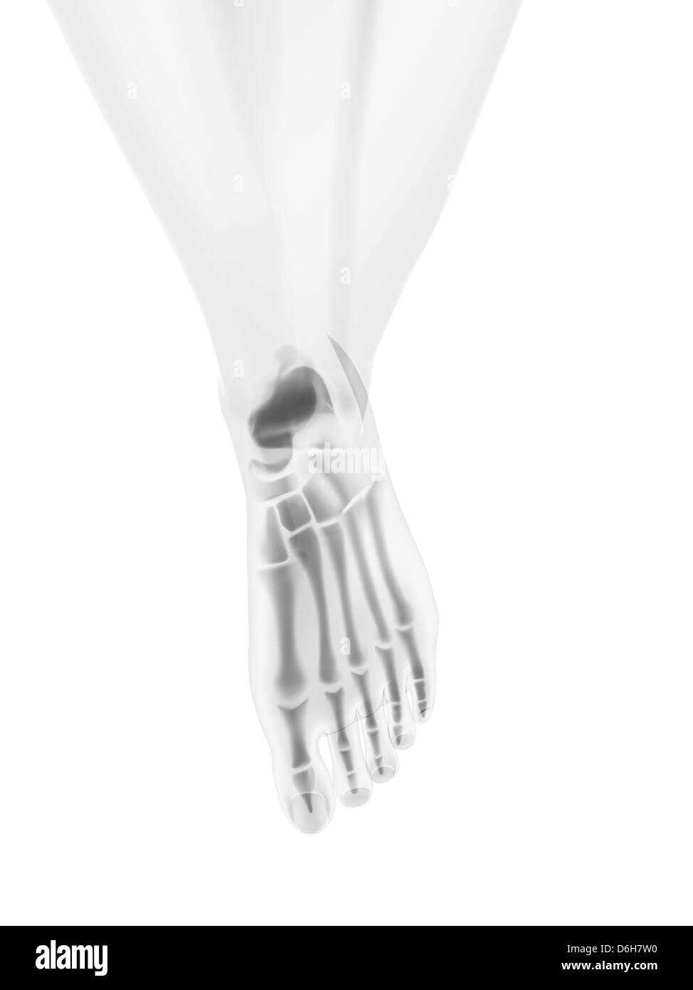 Foot bones, artwork Stock Photohttps://www.alamy.com/image-license-details/?v=1https://www.alamy.com/stock-photo-foot-bones-artwork-55698412.html
RFD6H7W0–Foot bones, artwork
 Organic unity and kinship of beings, Gorilla, vintage engraved illustration. Earth before man – 1886. Stock Vectorhttps://www.alamy.com/image-license-details/?v=1https://www.alamy.com/stock-photo-organic-unity-and-kinship-of-beings-gorilla-vintage-engraved-illustration-84299935.html
RFEW45BY–Organic unity and kinship of beings, Gorilla, vintage engraved illustration. Earth before man – 1886.
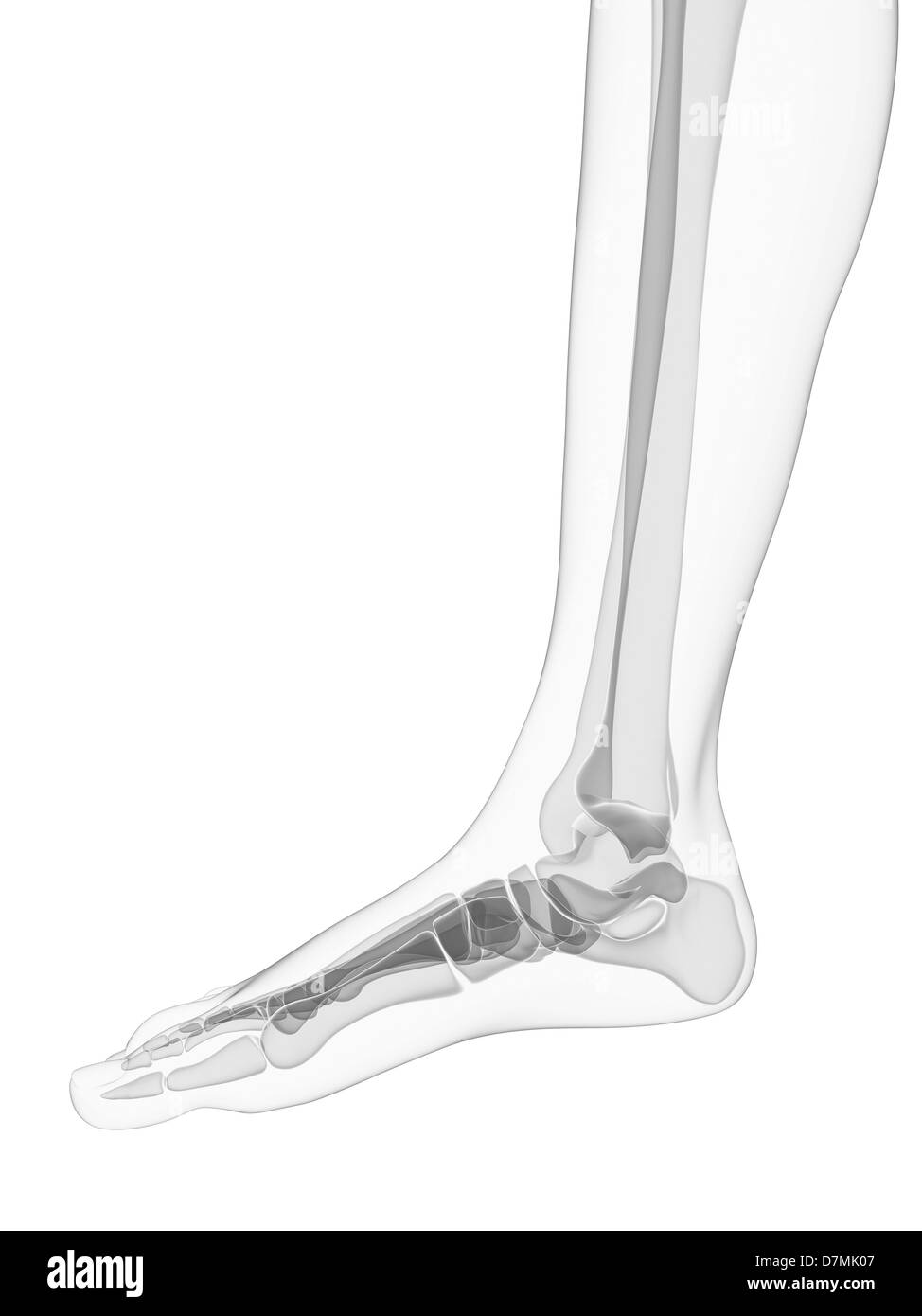 Foot bones, artwork Stock Photohttps://www.alamy.com/image-license-details/?v=1https://www.alamy.com/stock-photo-foot-bones-artwork-56387639.html
RFD7MK07–Foot bones, artwork
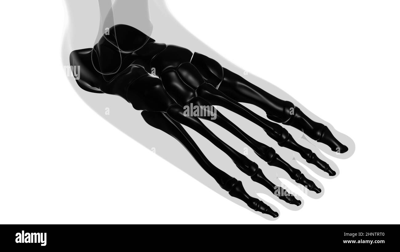 Human Skeleton Foot bones Anatomy For Medical Concept 3D Illustration Stock Photohttps://www.alamy.com/image-license-details/?v=1https://www.alamy.com/human-skeleton-foot-bones-anatomy-for-medical-concept-3d-illustration-image460922896.html
RF2HNTRT0–Human Skeleton Foot bones Anatomy For Medical Concept 3D Illustration
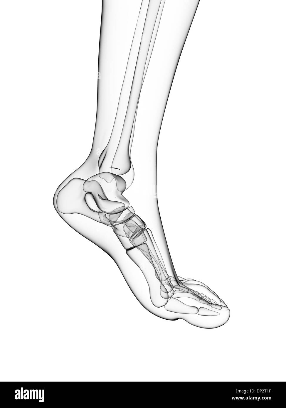 Bones of the foot, artwork Stock Photohttps://www.alamy.com/image-license-details/?v=1https://www.alamy.com/bones-of-the-foot-artwork-image65216306.html
RFDP2T1P–Bones of the foot, artwork
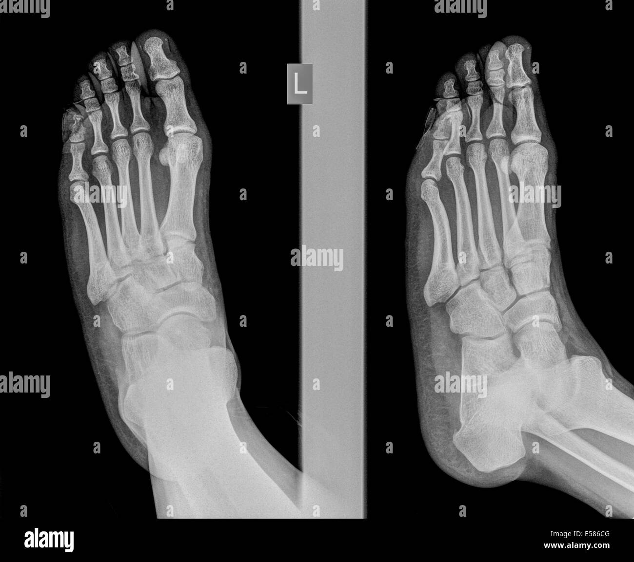 x-ray of a foot showing a fracture in the intermediate phalanx of the small toe on the left foot of a 30 year old male patient Stock Photohttps://www.alamy.com/image-license-details/?v=1https://www.alamy.com/stock-photo-x-ray-of-a-foot-showing-a-fracture-in-the-intermediate-phalanx-of-72095424.html
RME586CG–x-ray of a foot showing a fracture in the intermediate phalanx of the small toe on the left foot of a 30 year old male patient
