Quick filters:
Fractured clavicle Stock Photos and Images
 x-ray showing fractured clavicle Stock Photohttps://www.alamy.com/image-license-details/?v=1https://www.alamy.com/x-ray-showing-fractured-clavicle-image65494877.html
RFDPFFAN–x-ray showing fractured clavicle
 X-ray of shoulder of a 40 year old male patient with fractured clavicle front view Stock Photohttps://www.alamy.com/image-license-details/?v=1https://www.alamy.com/x-ray-of-shoulder-of-a-40-year-old-male-patient-with-fractured-clavicle-image68333212.html
RFDY4RKT–X-ray of shoulder of a 40 year old male patient with fractured clavicle front view
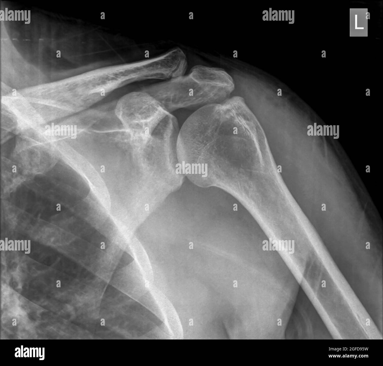 Shoulder x-ray of a 40 year old male patient with a fractured clavicle front view Stock Photohttps://www.alamy.com/image-license-details/?v=1https://www.alamy.com/shoulder-x-ray-of-a-40-year-old-male-patient-with-a-fractured-clavicle-front-view-image439771637.html
RM2GFD95W–Shoulder x-ray of a 40 year old male patient with a fractured clavicle front view
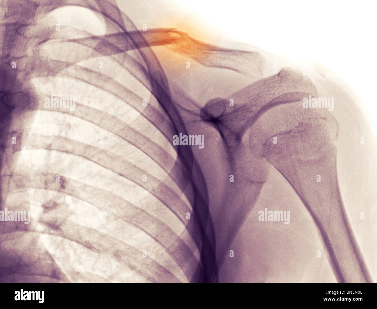 shoulder x-ray showing fractured clavicle Stock Photohttps://www.alamy.com/image-license-details/?v=1https://www.alamy.com/stock-photo-shoulder-x-ray-showing-fractured-clavicle-30441950.html
RFBNEN0E–shoulder x-ray showing fractured clavicle
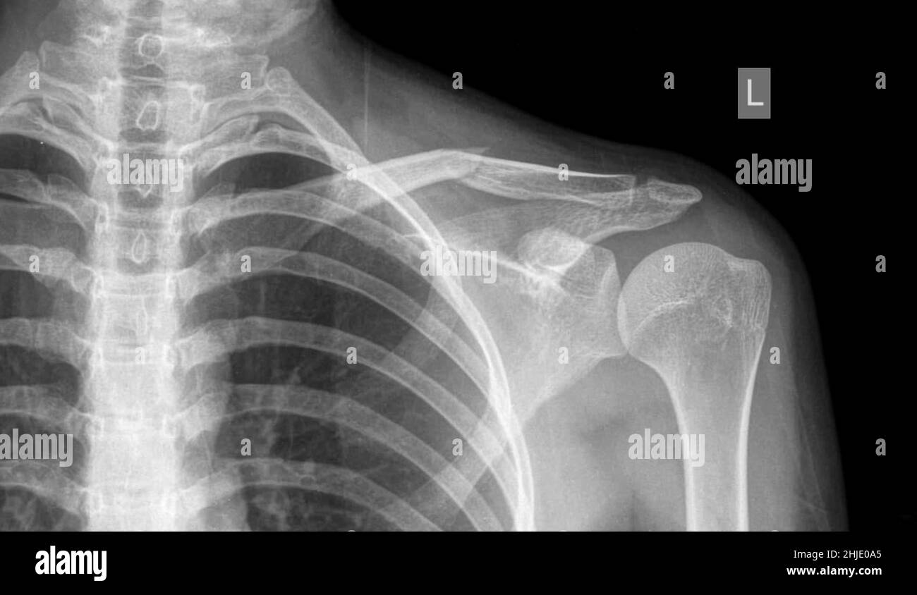 Fractured collar bone, X-ray Stock Photohttps://www.alamy.com/image-license-details/?v=1https://www.alamy.com/fractured-collar-bone-x-ray-image458840989.html
RF2HJE0A5–Fractured collar bone, X-ray
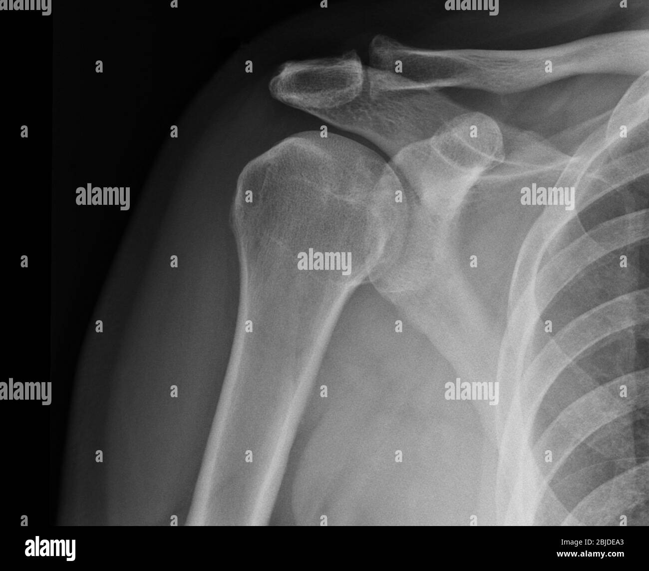 X-ray shoulder radiograph show state of injury Stock Photohttps://www.alamy.com/image-license-details/?v=1https://www.alamy.com/x-ray-shoulder-radiograph-show-state-of-injury-image355567803.html
RF2BJDEA3–X-ray shoulder radiograph show state of injury
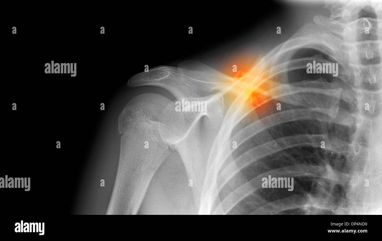 Fractured collarbone, X-ray Stock Photohttps://www.alamy.com/image-license-details/?v=1https://www.alamy.com/fractured-collarbone-x-ray-image65258172.html
RFDP4ND0–Fractured collarbone, X-ray
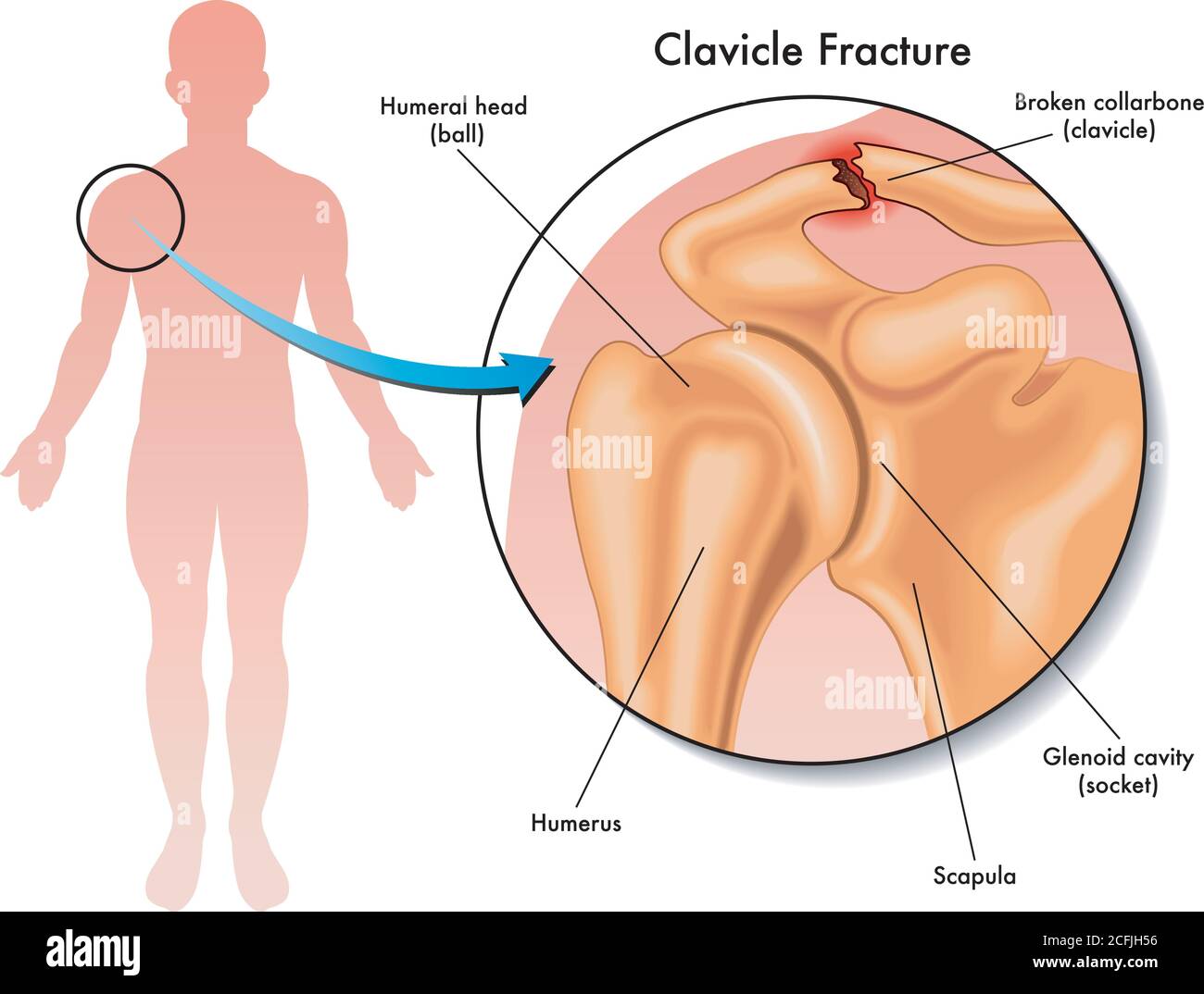 Medical illustration of a clavicle fracture and its location in the human body, with annotations. Stock Vectorhttps://www.alamy.com/image-license-details/?v=1https://www.alamy.com/medical-illustration-of-a-clavicle-fracture-and-its-location-in-the-human-body-with-annotations-image371046178.html
RF2CFJH56–Medical illustration of a clavicle fracture and its location in the human body, with annotations.
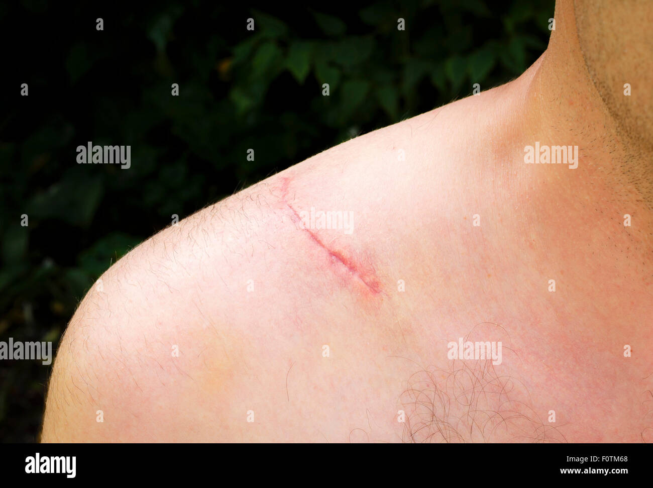 Closeup of surgical scar on the shoulder of a man following ORIF operation to repair a broken clavicle Stock Photohttps://www.alamy.com/image-license-details/?v=1https://www.alamy.com/stock-photo-closeup-of-surgical-scar-on-the-shoulder-of-a-man-following-orif-operation-86594544.html
RFF0TM68–Closeup of surgical scar on the shoulder of a man following ORIF operation to repair a broken clavicle
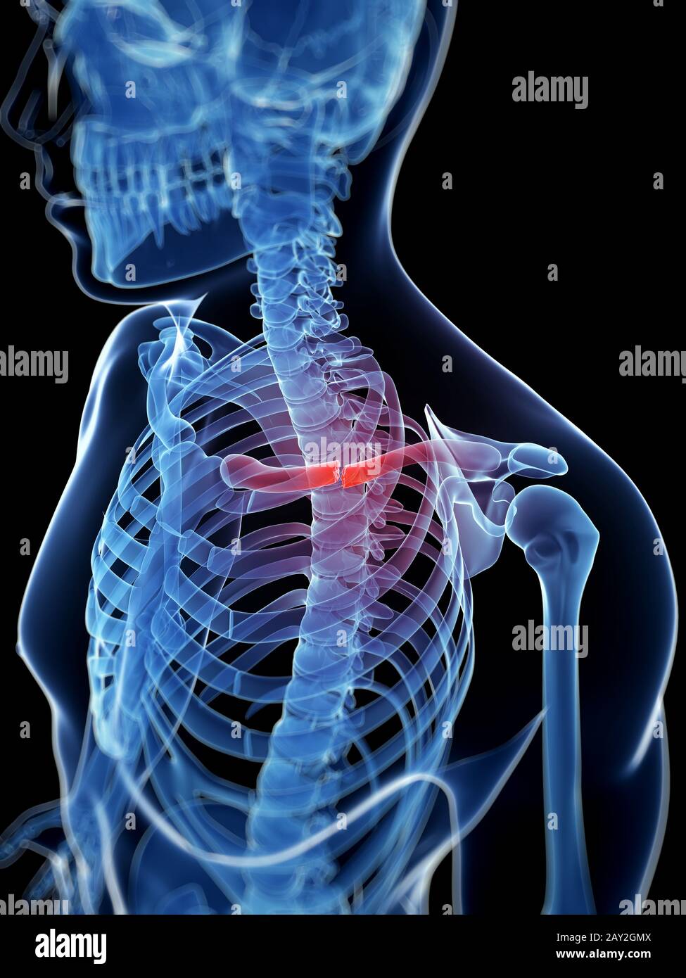 medical illustration of a broken clavicle Stock Photohttps://www.alamy.com/image-license-details/?v=1https://www.alamy.com/medical-illustration-of-a-broken-clavicle-image343649738.html
RM2AY2GMX–medical illustration of a broken clavicle
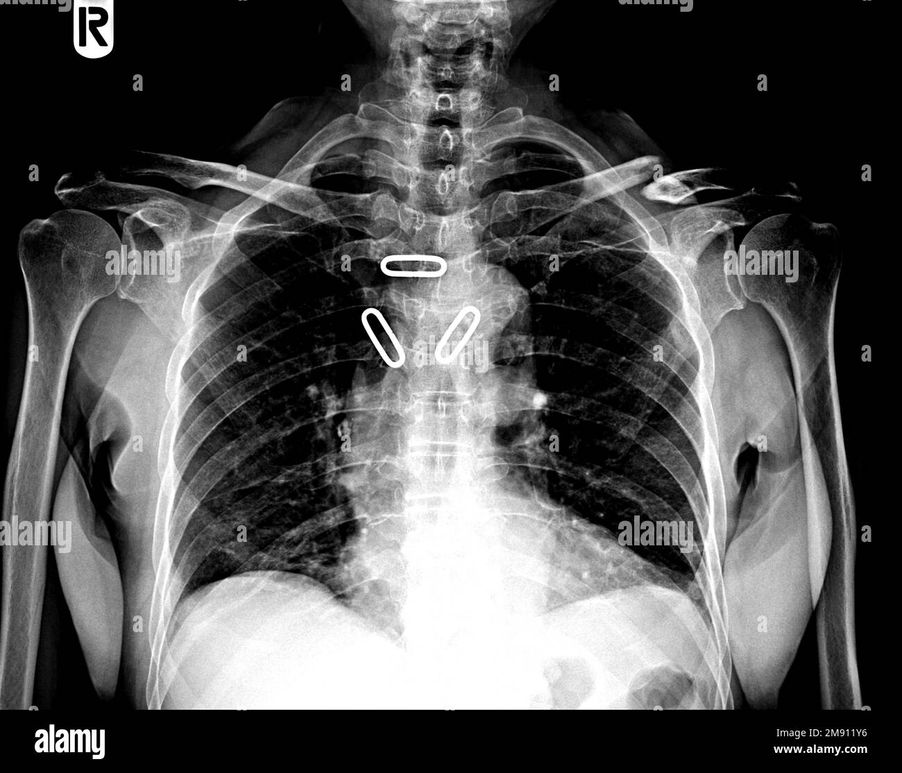 Figure of 8 shape Clavicle Splint. X ray clavicle AP view Stock Photohttps://www.alamy.com/image-license-details/?v=1https://www.alamy.com/figure-of-8-shape-clavicle-splint-x-ray-clavicle-ap-view-image504656074.html
RF2M911Y6–Figure of 8 shape Clavicle Splint. X ray clavicle AP view
 September 20, 2015: Dallas Cowboys quarterback Tony Romo (9) heads into the locker room with a fractured left clavicle during the NFL game between the Dallas Cowboys and the Philadelphia Eagles at Lincoln Financial Field in Philadelphia, Pennsylvania. The Dallas Cowboys won 20-10. Christopher Szagola/CSM Stock Photohttps://www.alamy.com/image-license-details/?v=1https://www.alamy.com/stock-photo-september-20-2015-dallas-cowboys-quarterback-tony-romo-9-heads-into-87702104.html
RMF2K4X0–September 20, 2015: Dallas Cowboys quarterback Tony Romo (9) heads into the locker room with a fractured left clavicle during the NFL game between the Dallas Cowboys and the Philadelphia Eagles at Lincoln Financial Field in Philadelphia, Pennsylvania. The Dallas Cowboys won 20-10. Christopher Szagola/CSM
 Fractured Clavicle Stock Vectorhttps://www.alamy.com/image-license-details/?v=1https://www.alamy.com/stock-photo-fractured-clavicle-78697421.html
RFEG0YA5–Fractured Clavicle
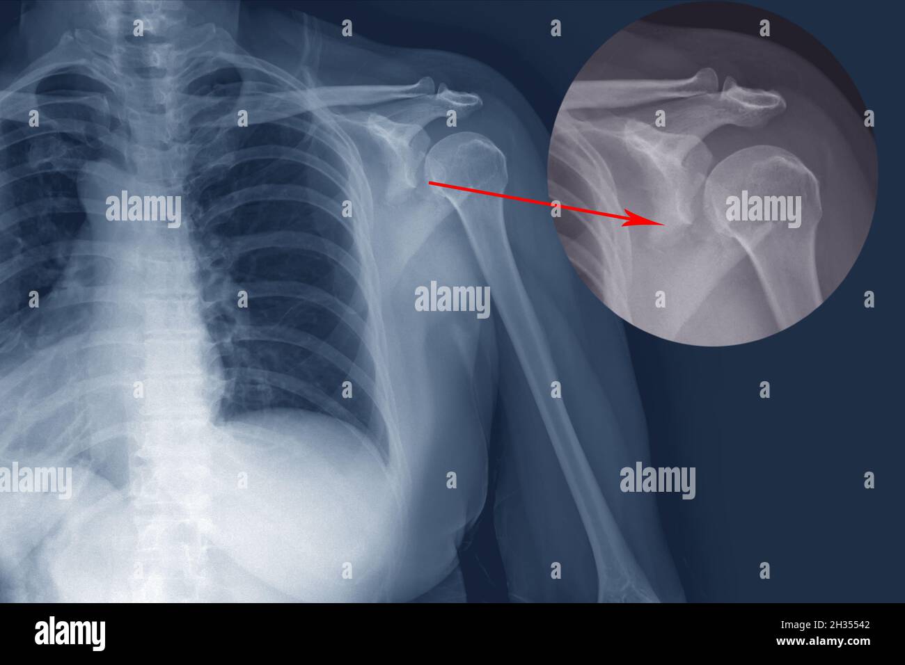 X-ray shoulder Fracture posterior half of glenoid with posterior dislocation of the bone fragment. Irregular transverse fracture at body of scapula. Stock Photohttps://www.alamy.com/image-license-details/?v=1https://www.alamy.com/x-ray-shoulder-fracture-posterior-half-of-glenoid-with-posterior-dislocation-of-the-bone-fragment-irregular-transverse-fracture-at-body-of-scapula-image449427330.html
RF2H35542–X-ray shoulder Fracture posterior half of glenoid with posterior dislocation of the bone fragment. Irregular transverse fracture at body of scapula.
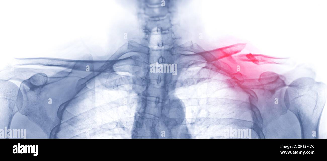 X-ray of Clavicle AP view showing fracture of left Clavicle bone. Stock Photohttps://www.alamy.com/image-license-details/?v=1https://www.alamy.com/x-ray-of-clavicle-ap-view-showing-fracture-of-left-clavicle-bone-image551406392.html
RF2R12MDC–X-ray of Clavicle AP view showing fracture of left Clavicle bone.
 Plain x Ray PXR of left shoulder of skeletally immature female patient child showing lateral one third fracture clavicle, broken lateral part of the c Stock Photohttps://www.alamy.com/image-license-details/?v=1https://www.alamy.com/plain-x-ray-pxr-of-left-shoulder-of-skeletally-immature-female-patient-child-showing-lateral-one-third-fracture-clavicle-broken-lateral-part-of-the-c-image553643193.html
RF2R4MHF5–Plain x Ray PXR of left shoulder of skeletally immature female patient child showing lateral one third fracture clavicle, broken lateral part of the c
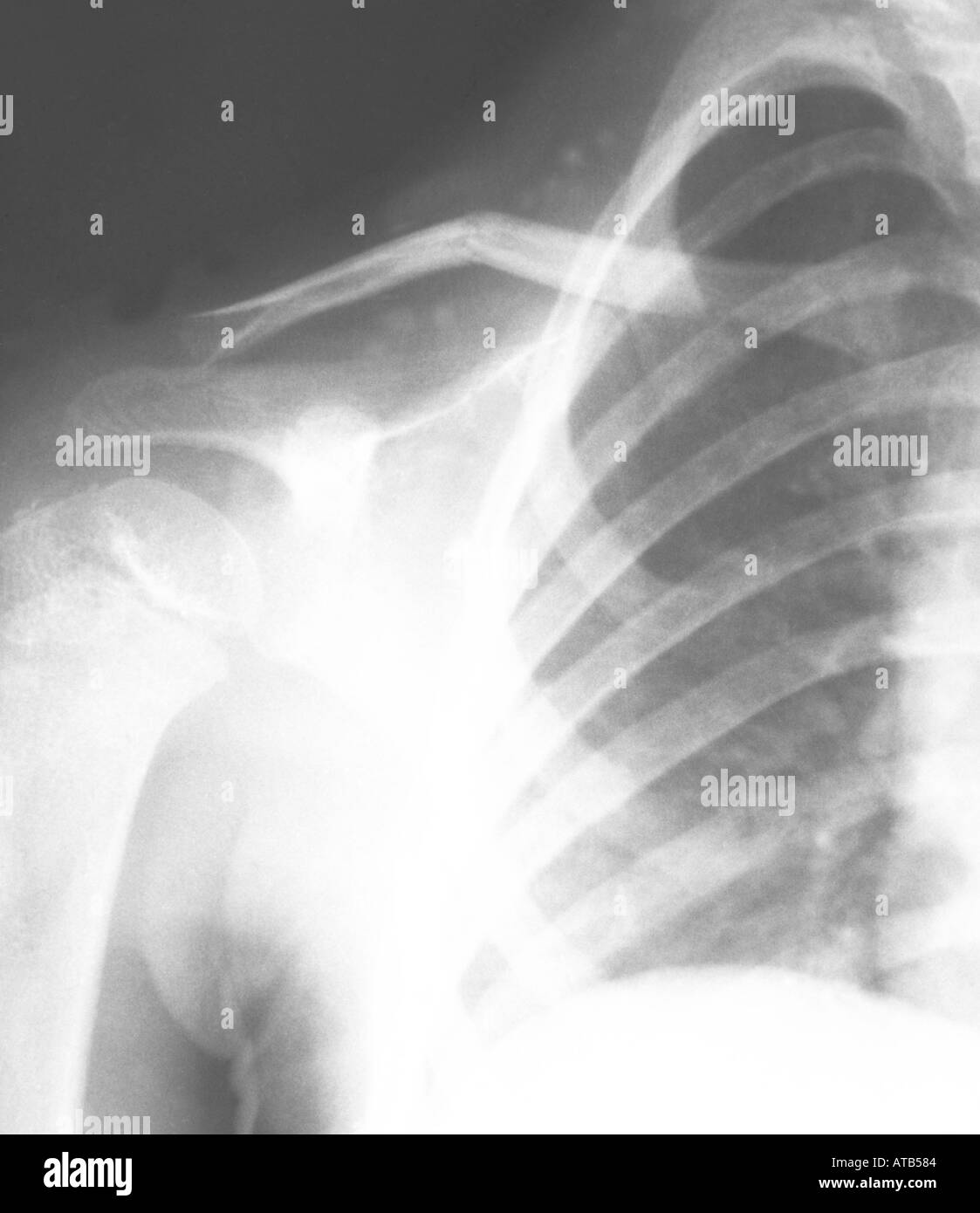 Shoulder x ray fractured clavicle Stock Photohttps://www.alamy.com/image-license-details/?v=1https://www.alamy.com/shoulder-x-ray-fractured-clavicle-image1684867.html
RMATB584–Shoulder x ray fractured clavicle
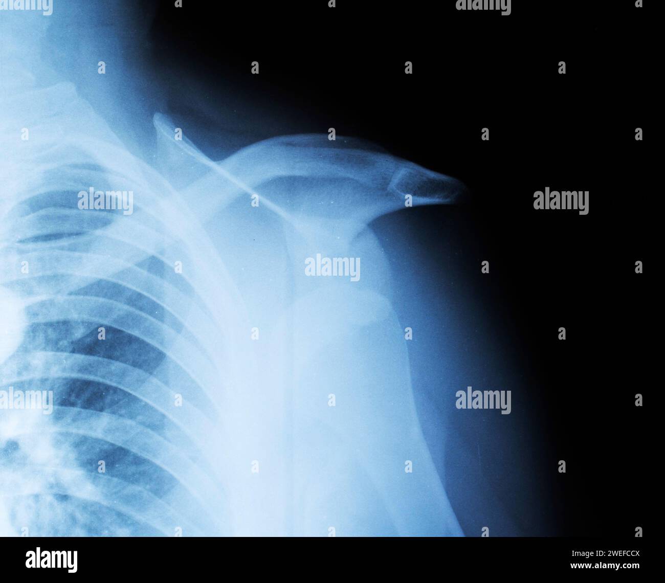 X-ray of a fractured humeral head and shoulder dislocation. Clavicle fracture. Traumatology and orthopedics. Stock Photohttps://www.alamy.com/image-license-details/?v=1https://www.alamy.com/x-ray-of-a-fractured-humeral-head-and-shoulder-dislocation-clavicle-fracture-traumatology-and-orthopedics-image594096746.html
RF2WEFCCX–X-ray of a fractured humeral head and shoulder dislocation. Clavicle fracture. Traumatology and orthopedics.
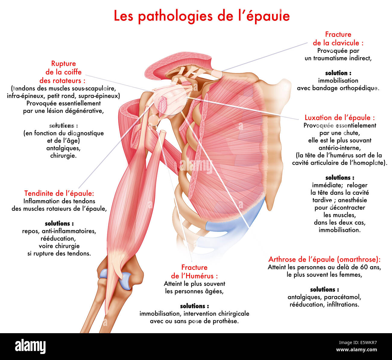 Shoulder, illustration Stock Photohttps://www.alamy.com/image-license-details/?v=1https://www.alamy.com/stock-photo-shoulder-illustration-72479099.html
RME5WKR7–Shoulder, illustration
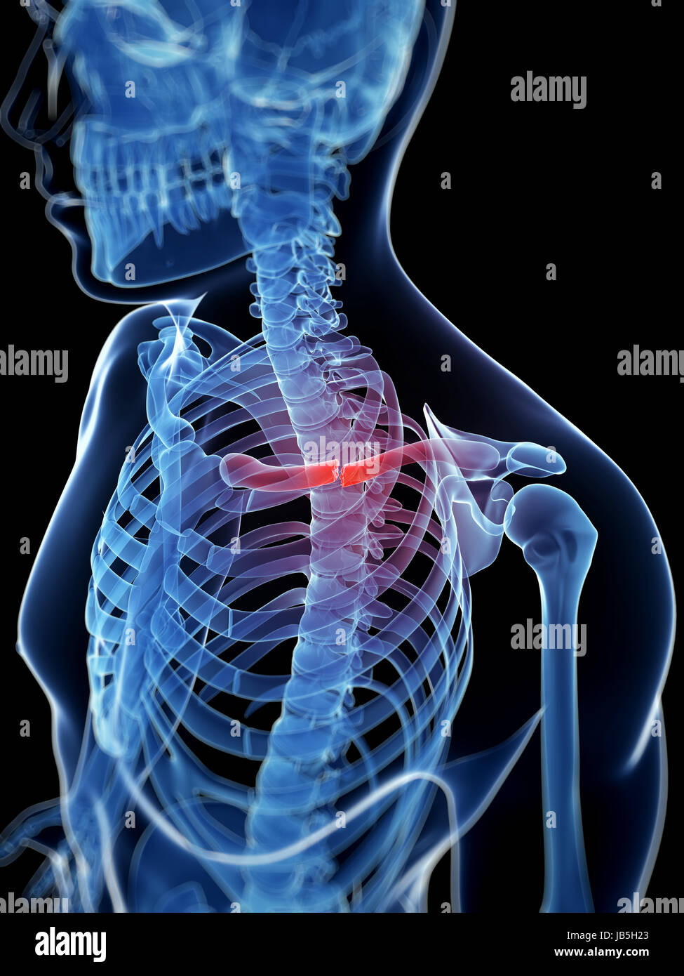 medical illustration of a broken clavicle Stock Photohttps://www.alamy.com/image-license-details/?v=1https://www.alamy.com/stock-photo-medical-illustration-of-a-broken-clavicle-144567307.html
RFJB5H23–medical illustration of a broken clavicle
 Woman wearing a splint. Stock Photohttps://www.alamy.com/image-license-details/?v=1https://www.alamy.com/stock-photo-woman-wearing-a-splint-72436911.html
RME5RP0F–Woman wearing a splint.
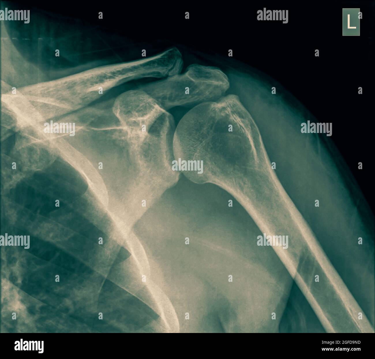 Shoulder x-ray of a 40 year old male patient with a fractured clavicle front view Stock Photohttps://www.alamy.com/image-license-details/?v=1https://www.alamy.com/shoulder-x-ray-of-a-40-year-old-male-patient-with-a-fractured-clavicle-front-view-image439772073.html
RM2GFD9ND–Shoulder x-ray of a 40 year old male patient with a fractured clavicle front view
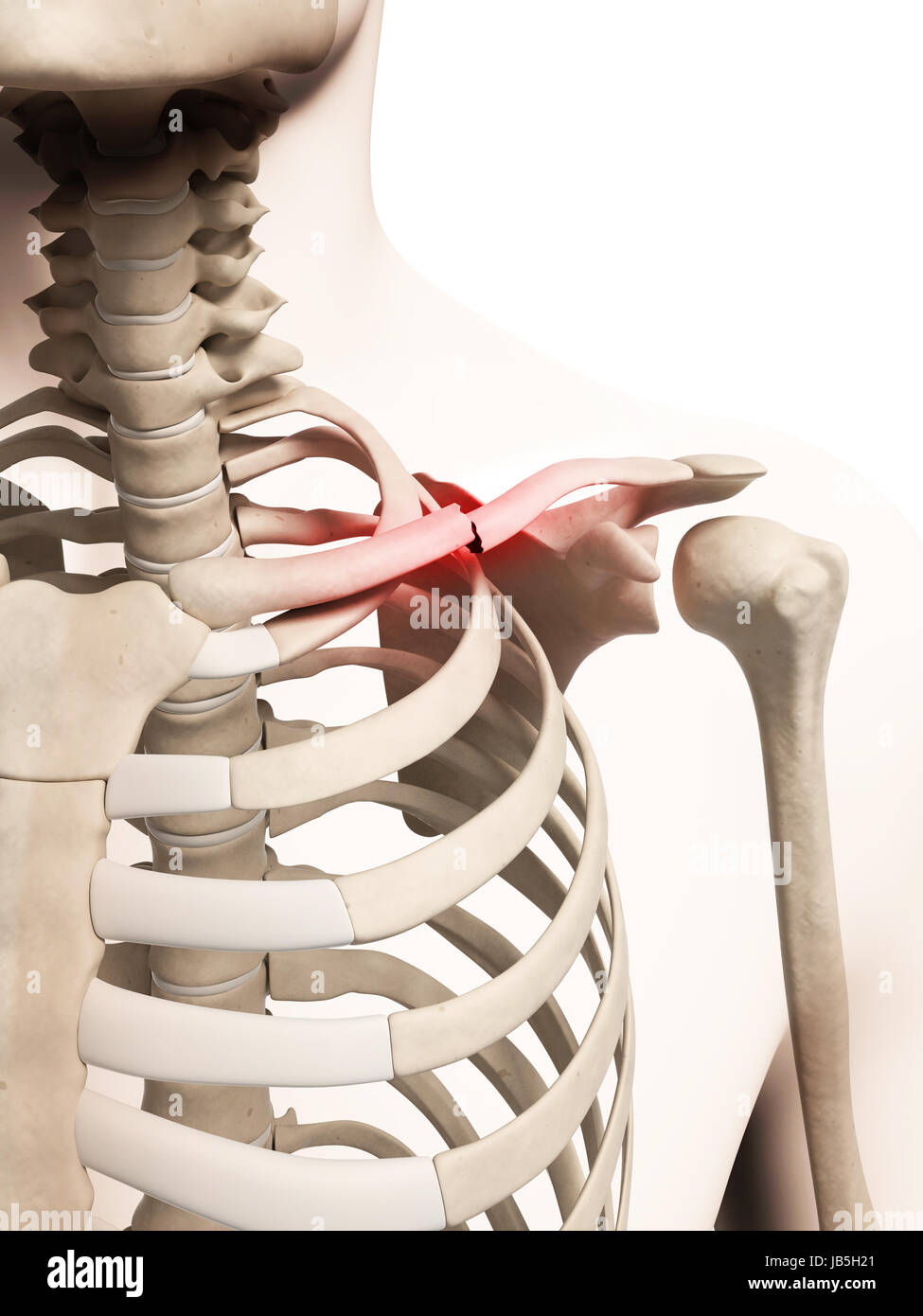 medical illustration of a broken clavicle Stock Photohttps://www.alamy.com/image-license-details/?v=1https://www.alamy.com/stock-photo-medical-illustration-of-a-broken-clavicle-144567305.html
RFJB5H21–medical illustration of a broken clavicle
 X-rays image of the painful or injury shoulder joint, shoulder dislocation. Medicine Stock Photohttps://www.alamy.com/image-license-details/?v=1https://www.alamy.com/x-rays-image-of-the-painful-or-injury-shoulder-joint-shoulder-dislocation-medicine-image344978132.html
RF2B1733G–X-rays image of the painful or injury shoulder joint, shoulder dislocation. Medicine
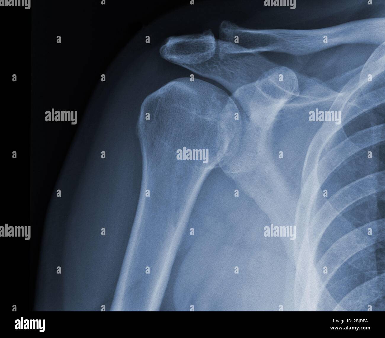 X-ray shoulder radiograph show state of injury Stock Photohttps://www.alamy.com/image-license-details/?v=1https://www.alamy.com/x-ray-shoulder-radiograph-show-state-of-injury-image355567801.html
RF2BJDEA1–X-ray shoulder radiograph show state of injury
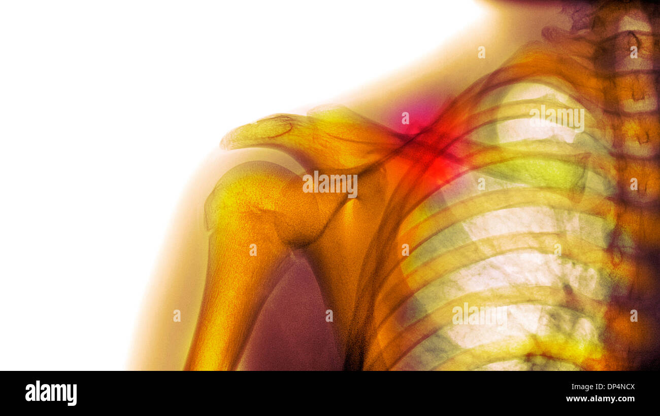 Fractured collarbone, X-ray Stock Photohttps://www.alamy.com/image-license-details/?v=1https://www.alamy.com/fractured-collarbone-x-ray-image65258170.html
RFDP4NCX–Fractured collarbone, X-ray
 MOUNTAIN DOCTOR Stock Photohttps://www.alamy.com/image-license-details/?v=1https://www.alamy.com/stock-photo-mountain-doctor-139737044.html
RMJ39G0M–MOUNTAIN DOCTOR
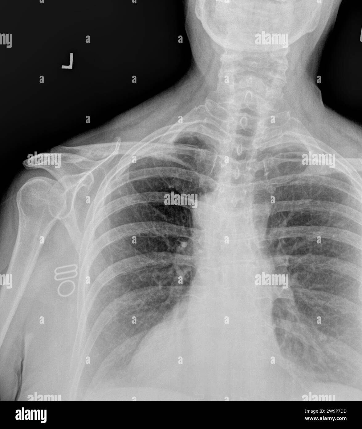 Film x ray or radiograph of a normal adult cervical vertebrae and left shoulder. anterior view showing normal bone structure of humerus, head glenohum Stock Photohttps://www.alamy.com/image-license-details/?v=1https://www.alamy.com/film-x-ray-or-radiograph-of-a-normal-adult-cervical-vertebrae-and-left-shoulder-anterior-view-showing-normal-bone-structure-of-humerus-head-glenohum-image591173225.html
RF2W9P7DD–Film x ray or radiograph of a normal adult cervical vertebrae and left shoulder. anterior view showing normal bone structure of humerus, head glenohum
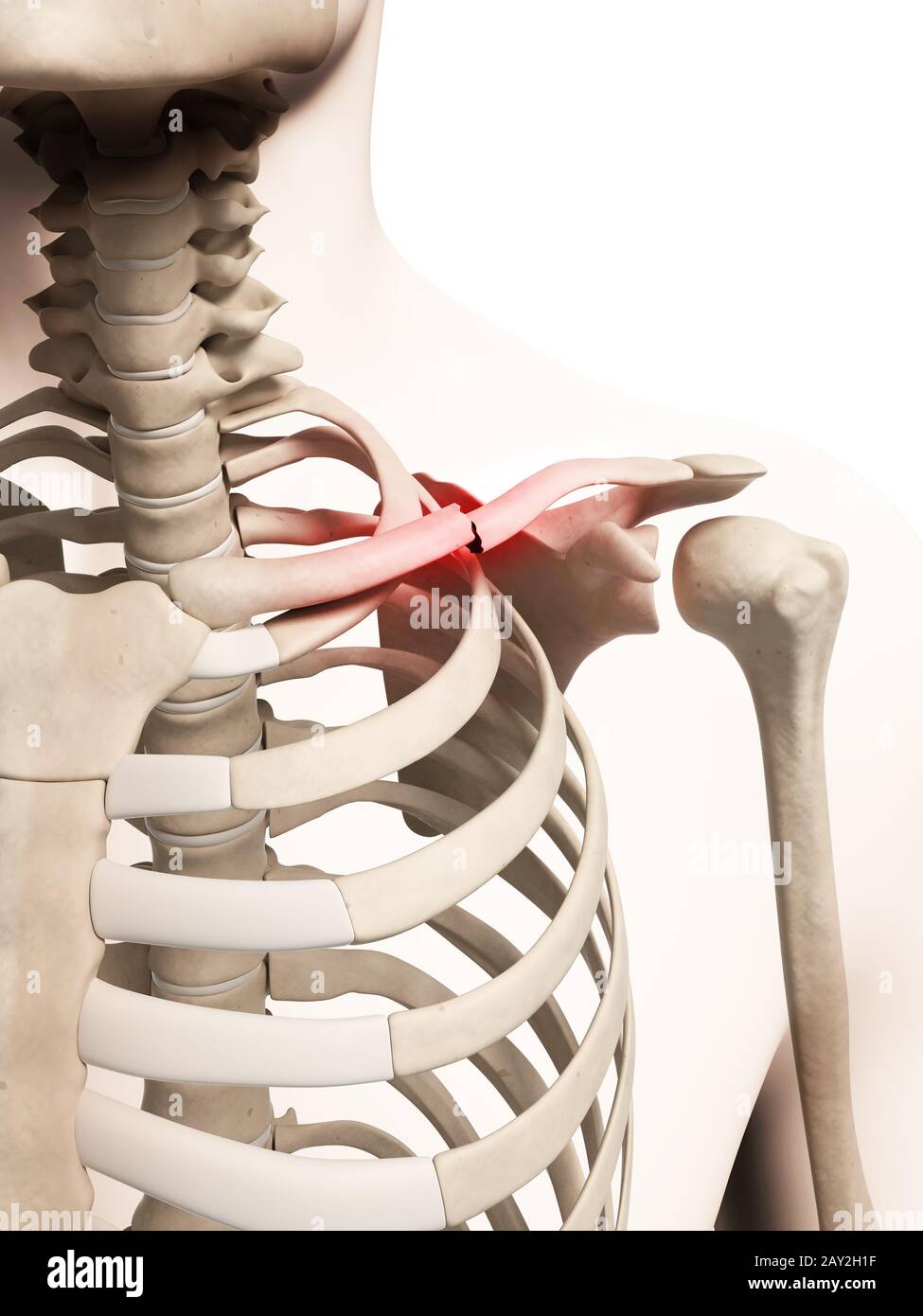 medical illustration of a broken clavicle Stock Photohttps://www.alamy.com/image-license-details/?v=1https://www.alamy.com/medical-illustration-of-a-broken-clavicle-image343649979.html
RM2AY2H1F–medical illustration of a broken clavicle
 FRACTURED COLLARBONE, X-RAY Stock Photohttps://www.alamy.com/image-license-details/?v=1https://www.alamy.com/stock-photo-fractured-collarbone-x-ray-53857912.html
RMD3HC8T–FRACTURED COLLARBONE, X-RAY
 September 20, 2015: Dallas Cowboys quarterback Tony Romo (9) looks on after suffering a fractured left clavicle following the NFL game between the Dallas Cowboys and the Philadelphia Eagles at Lincoln Financial Field in Philadelphia, Pennsylvania. The Dallas Cowboys won 20-10. Christopher Szagola/CSM Stock Photohttps://www.alamy.com/image-license-details/?v=1https://www.alamy.com/stock-photo-september-20-2015-dallas-cowboys-quarterback-tony-romo-9-looks-on-87702099.html
RMF2K4WR–September 20, 2015: Dallas Cowboys quarterback Tony Romo (9) looks on after suffering a fractured left clavicle following the NFL game between the Dallas Cowboys and the Philadelphia Eagles at Lincoln Financial Field in Philadelphia, Pennsylvania. The Dallas Cowboys won 20-10. Christopher Szagola/CSM
 X-ray of the cervical vertebrae. X-ray image of the cervical spine. Stock Photohttps://www.alamy.com/image-license-details/?v=1https://www.alamy.com/x-ray-of-the-cervical-vertebrae-x-ray-image-of-the-cervical-spine-image448643671.html
RM2H1WDG7–X-ray of the cervical vertebrae. X-ray image of the cervical spine.
 X-ray of chest heart stimulator Stock Photohttps://www.alamy.com/image-license-details/?v=1https://www.alamy.com/x-ray-of-chest-heart-stimulator-image363839188.html
RF2C3X8GM–X-ray of chest heart stimulator
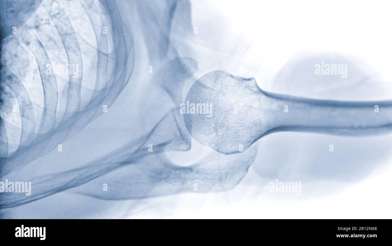 X-ray Shoulder joint shoulder transaxillary view for diagnosis fracture of shoulder joint. Stock Photohttps://www.alamy.com/image-license-details/?v=1https://www.alamy.com/x-ray-shoulder-joint-shoulder-transaxillary-view-for-diagnosis-fracture-of-shoulder-joint-image551406976.html
RF2R12N68–X-ray Shoulder joint shoulder transaxillary view for diagnosis fracture of shoulder joint.
 Plain x Ray PXR of left shoulder of skeletally immature female patient child showing lateral one third fracture clavicle, broken lateral part of the c Stock Photohttps://www.alamy.com/image-license-details/?v=1https://www.alamy.com/plain-x-ray-pxr-of-left-shoulder-of-skeletally-immature-female-patient-child-showing-lateral-one-third-fracture-clavicle-broken-lateral-part-of-the-c-image553592272.html
RF2R4J8GG–Plain x Ray PXR of left shoulder of skeletally immature female patient child showing lateral one third fracture clavicle, broken lateral part of the c
 The international encyclopaedia of surgery; a systematic treatise on the theory and practice of surgery . Dr. Sayres dressing for fractured clavicle ;application of first strip. Fig. 603. Fig. 604.. Stock Photohttps://www.alamy.com/image-license-details/?v=1https://www.alamy.com/the-international-encyclopaedia-of-surgery-a-systematic-treatise-on-the-theory-and-practice-of-surgery-dr-sayres-dressing-for-fractured-clavicle-application-of-first-strip-fig-603-fig-604-image339135310.html
RM2AKMXFA–The international encyclopaedia of surgery; a systematic treatise on the theory and practice of surgery . Dr. Sayres dressing for fractured clavicle ;application of first strip. Fig. 603. Fig. 604..
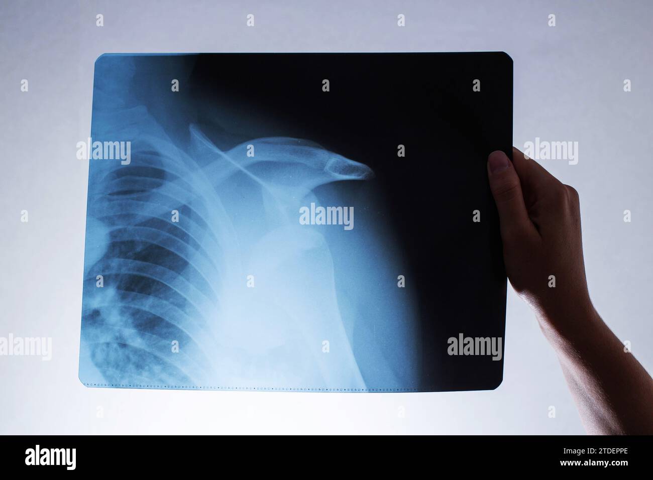 A woman's hand holds an x-ray of a patient with a dislocated shoulder and a fractured collarbone, osteoporosis. Surgery to reconstruct the shoulder jo Stock Photohttps://www.alamy.com/image-license-details/?v=1https://www.alamy.com/a-womans-hand-holds-an-x-ray-of-a-patient-with-a-dislocated-shoulder-and-a-fractured-collarbone-osteoporosis-surgery-to-reconstruct-the-shoulder-jo-image576257878.html
RF2TDEPPE–A woman's hand holds an x-ray of a patient with a dislocated shoulder and a fractured collarbone, osteoporosis. Surgery to reconstruct the shoulder jo
 collection OA knee x-ray image Stock Photohttps://www.alamy.com/image-license-details/?v=1https://www.alamy.com/collection-oa-knee-x-ray-image-image215366883.html
RFPEAPJB–collection OA knee x-ray image
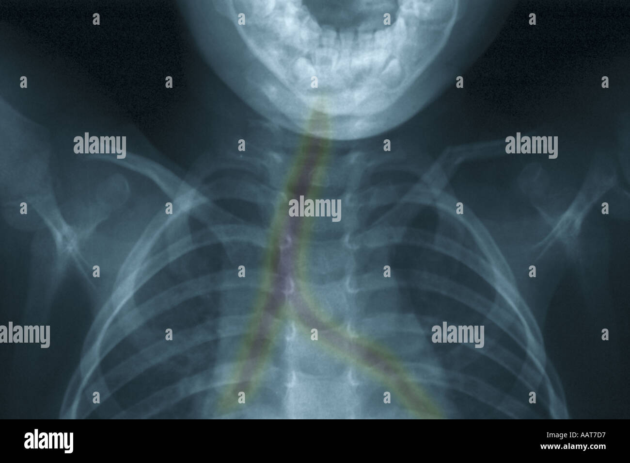 colorized xray showing fractured collarbone Stock Photohttps://www.alamy.com/image-license-details/?v=1https://www.alamy.com/colorized-xray-showing-fractured-collarbone-image2361302.html
RMAAT7D7–colorized xray showing fractured collarbone
 Doctor visiting female patient wearing a splint, at home. Stock Photohttps://www.alamy.com/image-license-details/?v=1https://www.alamy.com/stock-photo-doctor-visiting-female-patient-wearing-a-splint-at-home-72436910.html
RME5RP0E–Doctor visiting female patient wearing a splint, at home.
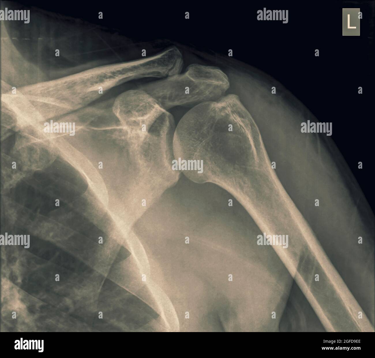 Shoulder x-ray of a 40 year old male patient with a fractured clavicle front view Stock Photohttps://www.alamy.com/image-license-details/?v=1https://www.alamy.com/shoulder-x-ray-of-a-40-year-old-male-patient-with-a-fractured-clavicle-front-view-image439771878.html
RM2GFD9EE–Shoulder x-ray of a 40 year old male patient with a fractured clavicle front view
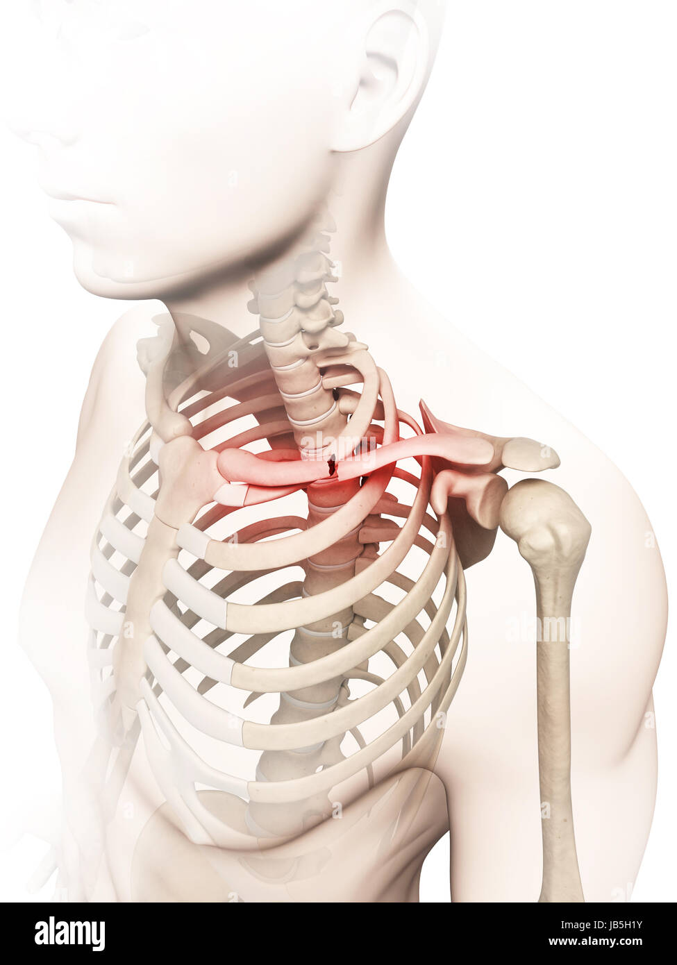 medical illustration of a broken clavicle Stock Photohttps://www.alamy.com/image-license-details/?v=1https://www.alamy.com/stock-photo-medical-illustration-of-a-broken-clavicle-144567303.html
RFJB5H1Y–medical illustration of a broken clavicle
 Lung cancer, pleural effusion 3d Illustration isolated black. Stock Photohttps://www.alamy.com/image-license-details/?v=1https://www.alamy.com/lung-cancer-pleural-effusion-3d-illustration-isolated-black-image182183561.html
RFMGB4YN–Lung cancer, pleural effusion 3d Illustration isolated black.
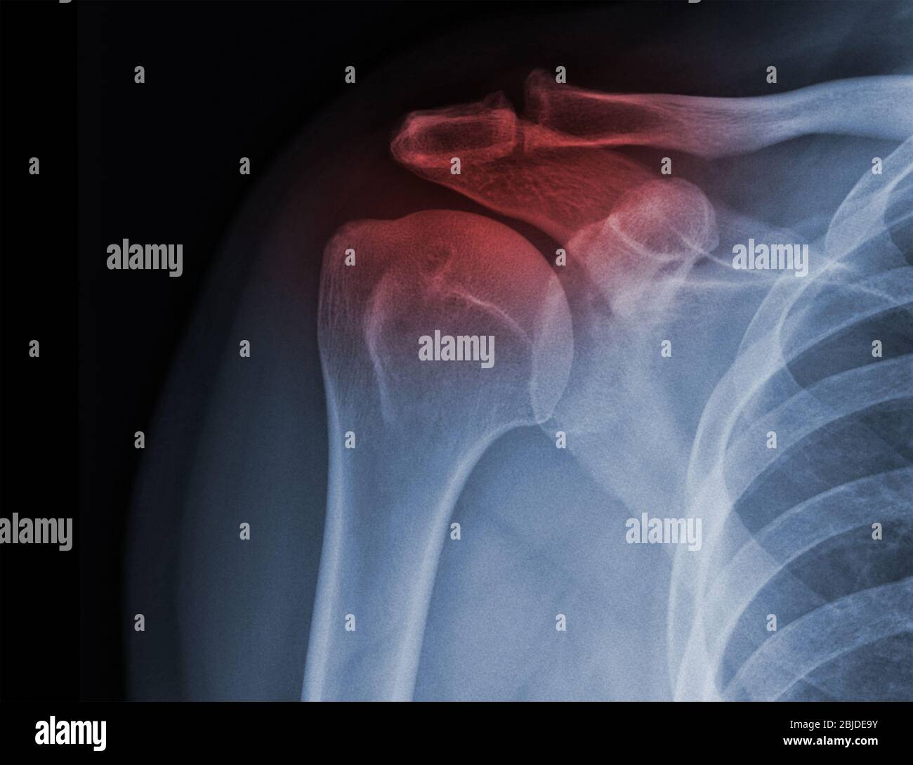 X-ray shoulder radiograph show state of injury Stock Photohttps://www.alamy.com/image-license-details/?v=1https://www.alamy.com/x-ray-shoulder-radiograph-show-state-of-injury-image355567799.html
RF2BJDE9Y–X-ray shoulder radiograph show state of injury
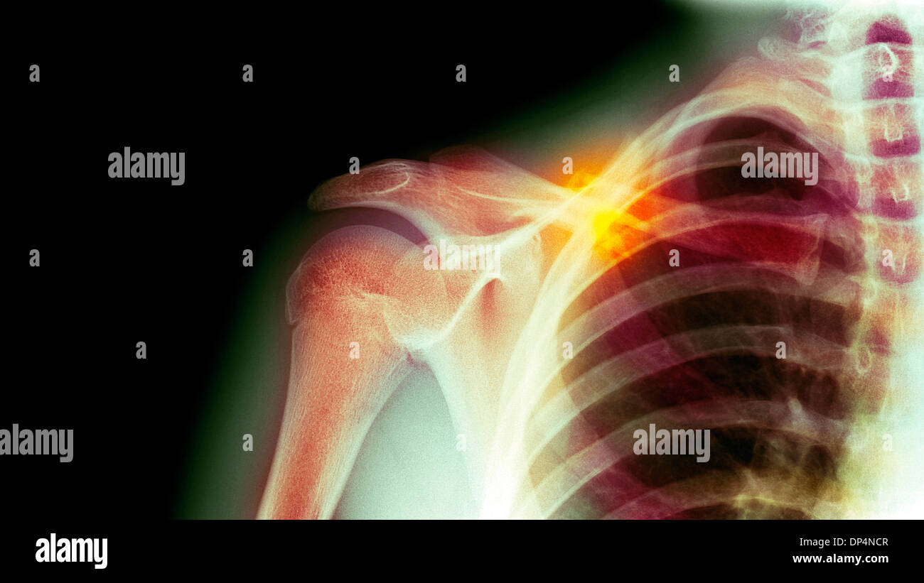 Fractured collarbone, X-ray Stock Photohttps://www.alamy.com/image-license-details/?v=1https://www.alamy.com/fractured-collarbone-x-ray-image65258167.html
RFDP4NCR–Fractured collarbone, X-ray
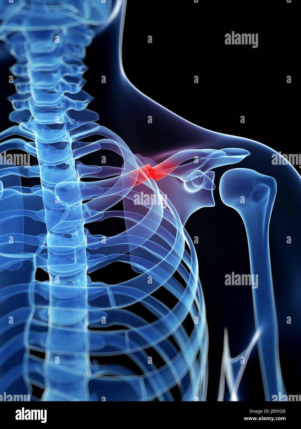 medical illustration of a broken clavicle Stock Photohttps://www.alamy.com/image-license-details/?v=1https://www.alamy.com/stock-photo-medical-illustration-of-a-broken-clavicle-144567312.html
RFJB5H28–medical illustration of a broken clavicle
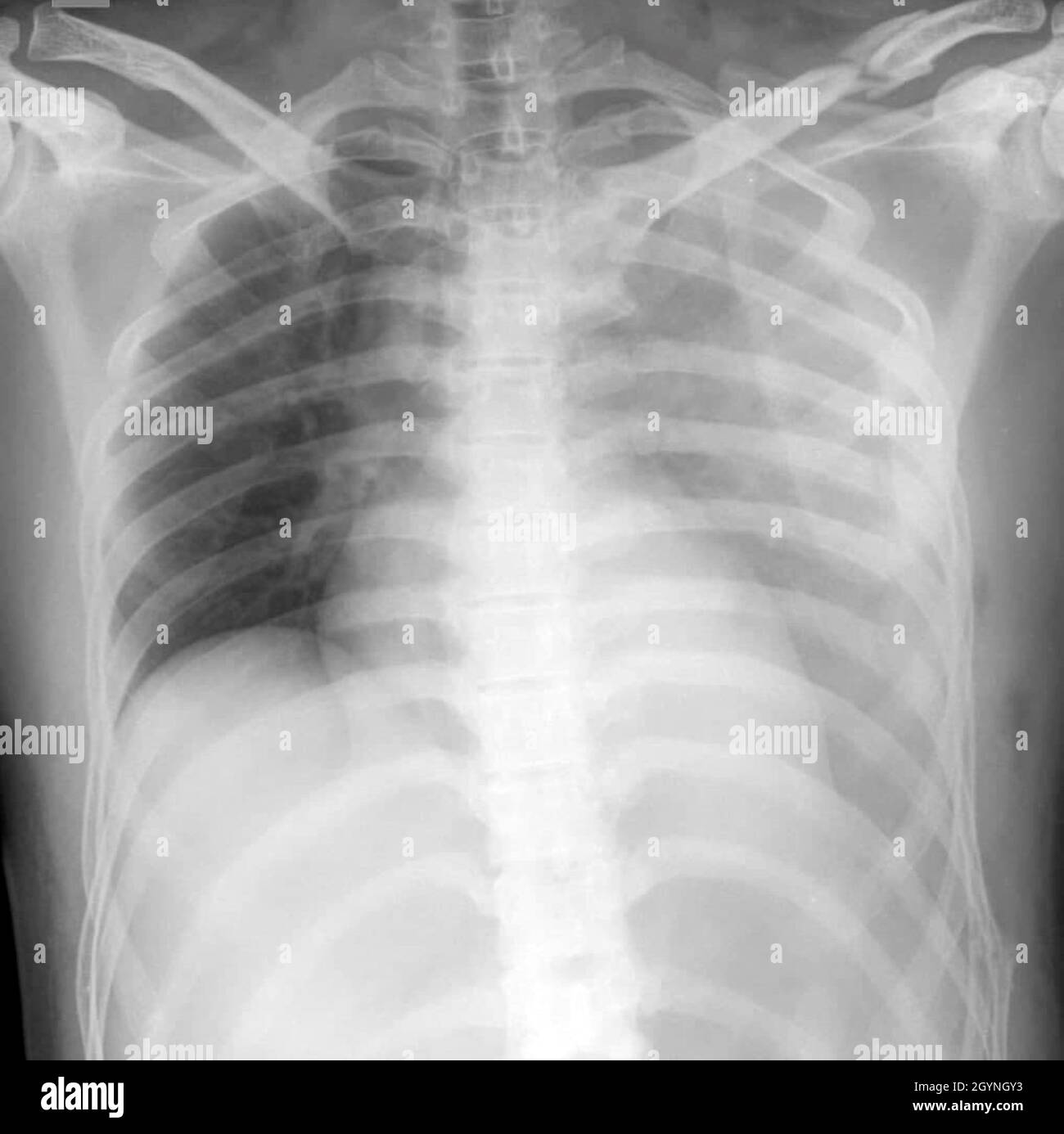 Haemothorax and multiple fractures , X-ray Stock Photohttps://www.alamy.com/image-license-details/?v=1https://www.alamy.com/haemothorax-and-multiple-fractures-x-ray-image447329207.html
RF2GYNGY3–Haemothorax and multiple fractures , X-ray
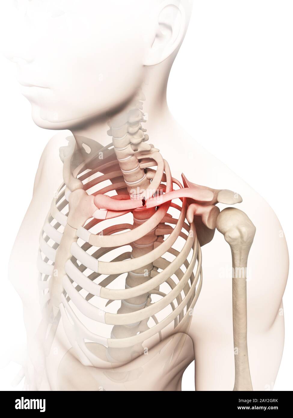 medical illustration of a broken clavicle Stock Photohttps://www.alamy.com/image-license-details/?v=1https://www.alamy.com/medical-illustration-of-a-broken-clavicle-image343649815.html
RM2AY2GRK–medical illustration of a broken clavicle
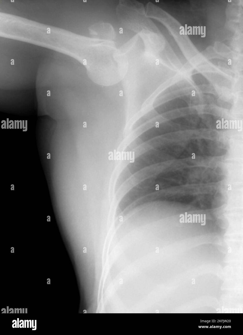 Dislocated shoulder, X-ray Stock Photohttps://www.alamy.com/image-license-details/?v=1https://www.alamy.com/dislocated-shoulder-x-ray-image491524936.html
RF2KFJW20–Dislocated shoulder, X-ray
 September 20, 2015: Dallas Cowboys quarterback Tony Romo (9) gets helped off by the medical staff as he suffered a fractured left clavicle during the NFL game between the Dallas Cowboys and the Philadelphia Eagles at Lincoln Financial Field in Philadelphia, Pennsylvania. The Dallas Cowboys won 20-10. Christopher Szagola/CSM Stock Photohttps://www.alamy.com/image-license-details/?v=1https://www.alamy.com/stock-photo-september-20-2015-dallas-cowboys-quarterback-tony-romo-9-gets-helped-87702095.html
RMF2K4WK–September 20, 2015: Dallas Cowboys quarterback Tony Romo (9) gets helped off by the medical staff as he suffered a fractured left clavicle during the NFL game between the Dallas Cowboys and the Philadelphia Eagles at Lincoln Financial Field in Philadelphia, Pennsylvania. The Dallas Cowboys won 20-10. Christopher Szagola/CSM
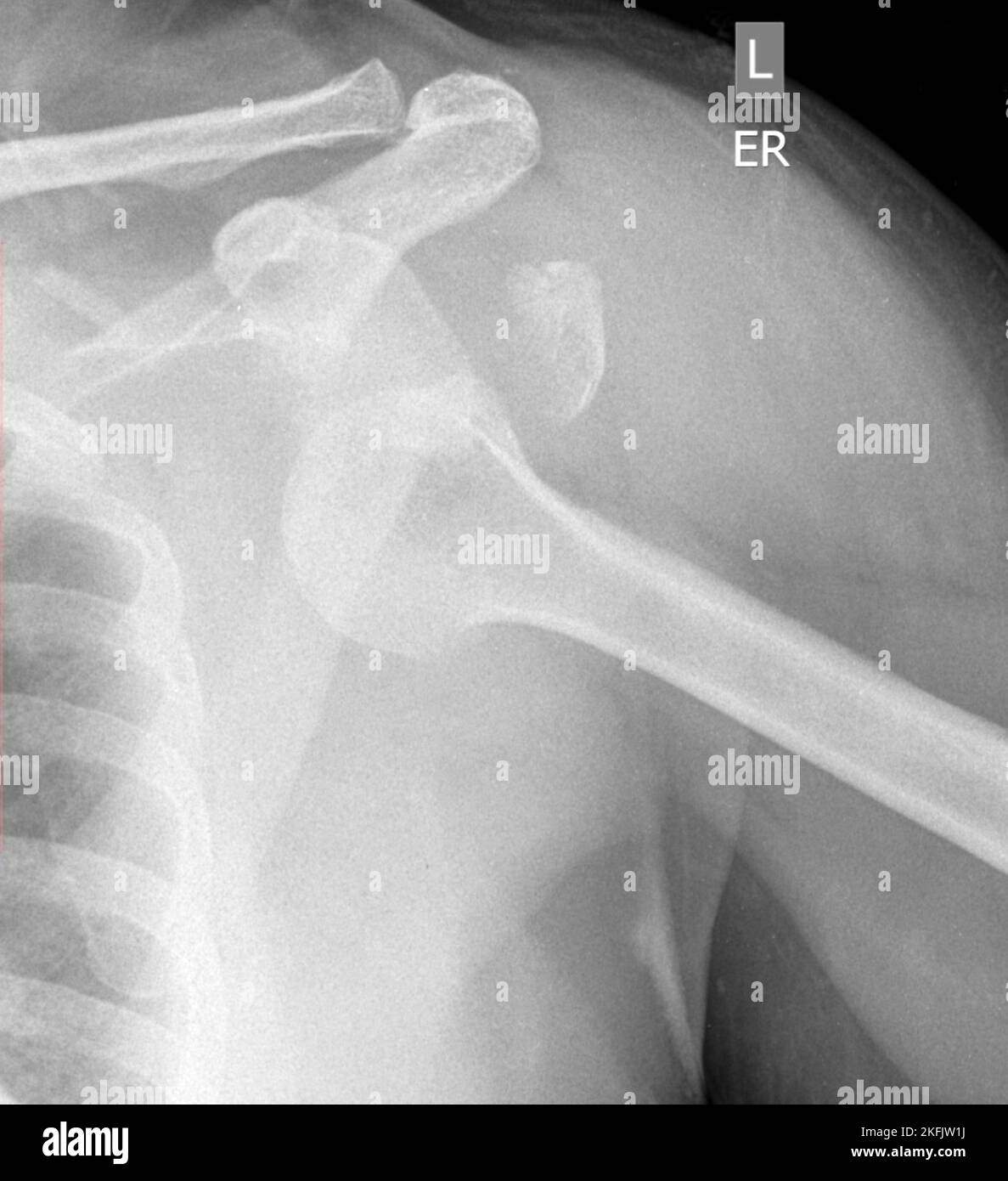 Dislocated shoulder, X-ray Stock Photohttps://www.alamy.com/image-license-details/?v=1https://www.alamy.com/dislocated-shoulder-x-ray-image491524926.html
RF2KFJW1J–Dislocated shoulder, X-ray
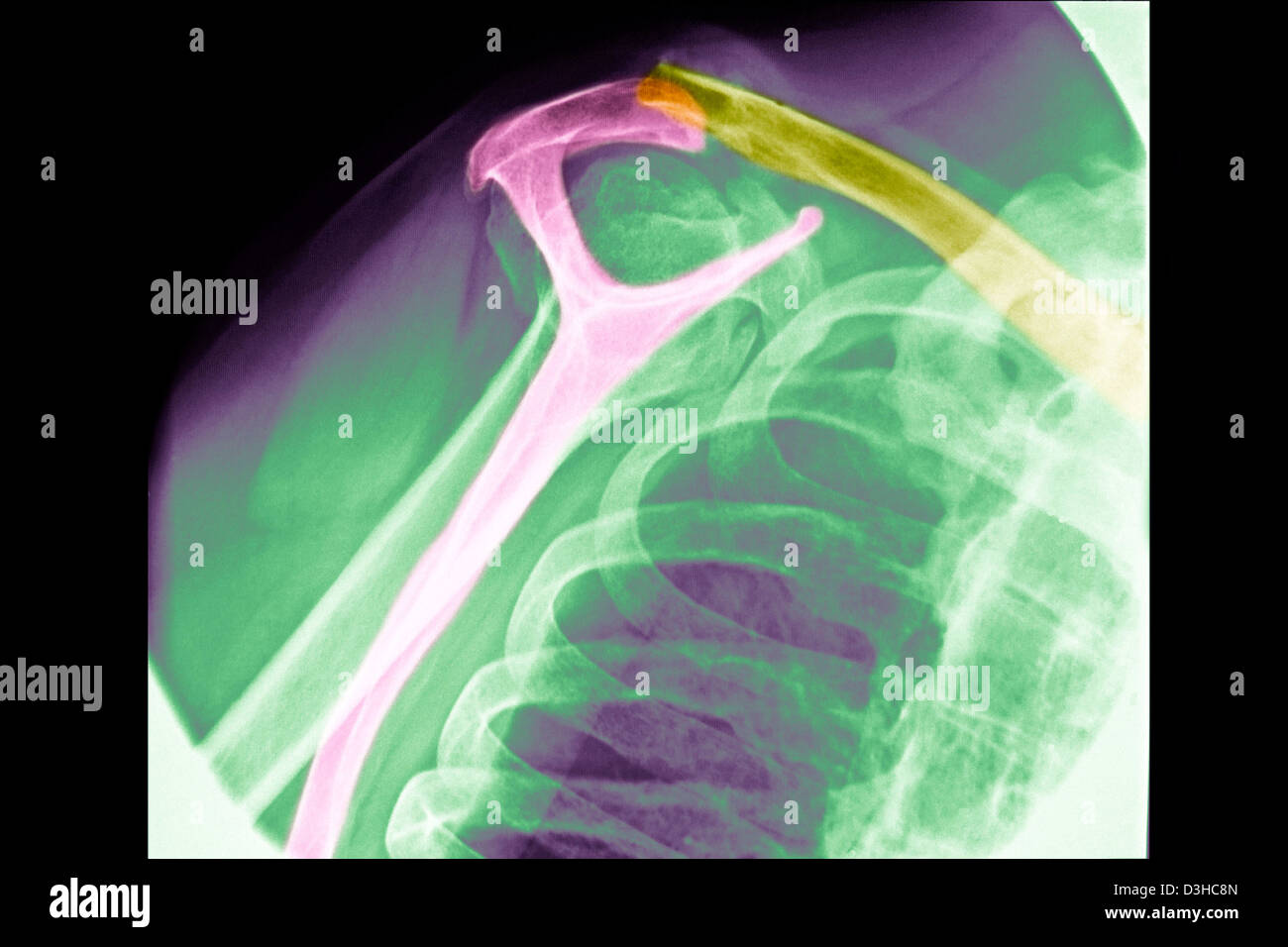 FRACTURED COLLARBONE, X-RAY Stock Photohttps://www.alamy.com/image-license-details/?v=1https://www.alamy.com/stock-photo-fractured-collarbone-x-ray-53857909.html
RMD3HC8N–FRACTURED COLLARBONE, X-RAY
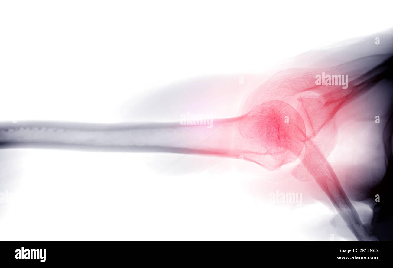 X-ray Shoulder joint shoulder transaxillary view for diagnosis fracture of shoulder joint. Stock Photohttps://www.alamy.com/image-license-details/?v=1https://www.alamy.com/x-ray-shoulder-joint-shoulder-transaxillary-view-for-diagnosis-fracture-of-shoulder-joint-image551406973.html
RF2R12N65–X-ray Shoulder joint shoulder transaxillary view for diagnosis fracture of shoulder joint.
 Plain x Ray PXR of left shoulder of skeletally immature female patient child showing lateral one third fracture clavicle, broken lateral part of the c Stock Photohttps://www.alamy.com/image-license-details/?v=1https://www.alamy.com/plain-x-ray-pxr-of-left-shoulder-of-skeletally-immature-female-patient-child-showing-lateral-one-third-fracture-clavicle-broken-lateral-part-of-the-c-image553590855.html
RF2R4J6NY–Plain x Ray PXR of left shoulder of skeletally immature female patient child showing lateral one third fracture clavicle, broken lateral part of the c
 X-ray of the cervical vertebrae. X-ray image of the cervical spine. Stock Photohttps://www.alamy.com/image-license-details/?v=1https://www.alamy.com/x-ray-of-the-cervical-vertebrae-x-ray-image-of-the-cervical-spine-image448643746.html
RM2H1WDJX–X-ray of the cervical vertebrae. X-ray image of the cervical spine.
 Atlas and epitome of traumatic fractures and dislocations . een thesternal and niidclle third of the bone. The fragments present markedoverriding. The sternal fragment is displaced upward by thepressure of the outer fragment and the pull of the sternocleidomastoidmuscle. In the illustration the sternocleidomastoid muscle is readilyrecognized ; behind, the outer boundary of the neck is formed by thetrapezius muscle ; the deltoid is dissected out and the clavicular por-tion of the pectoralis major has been in part removed. Within thefenestra thus produced we see the fractured clavicle with the s Stock Photohttps://www.alamy.com/image-license-details/?v=1https://www.alamy.com/atlas-and-epitome-of-traumatic-fractures-and-dislocations-een-thesternal-and-niidclle-third-of-the-bone-the-fragments-present-markedoverriding-the-sternal-fragment-is-displaced-upward-by-thepressure-of-the-outer-fragment-and-the-pull-of-the-sternocleidomastoidmuscle-in-the-illustration-the-sternocleidomastoid-muscle-is-readilyrecognized-behind-the-outer-boundary-of-the-neck-is-formed-by-thetrapezius-muscle-the-deltoid-is-dissected-out-and-the-clavicular-por-tion-of-the-pectoralis-major-has-been-in-part-removed-within-thefenestra-thus-produced-we-see-the-fractured-clavicle-with-the-s-image340283231.html
RM2ANH6MF–Atlas and epitome of traumatic fractures and dislocations . een thesternal and niidclle third of the bone. The fragments present markedoverriding. The sternal fragment is displaced upward by thepressure of the outer fragment and the pull of the sternocleidomastoidmuscle. In the illustration the sternocleidomastoid muscle is readilyrecognized ; behind, the outer boundary of the neck is formed by thetrapezius muscle ; the deltoid is dissected out and the clavicular por-tion of the pectoralis major has been in part removed. Within thefenestra thus produced we see the fractured clavicle with the s
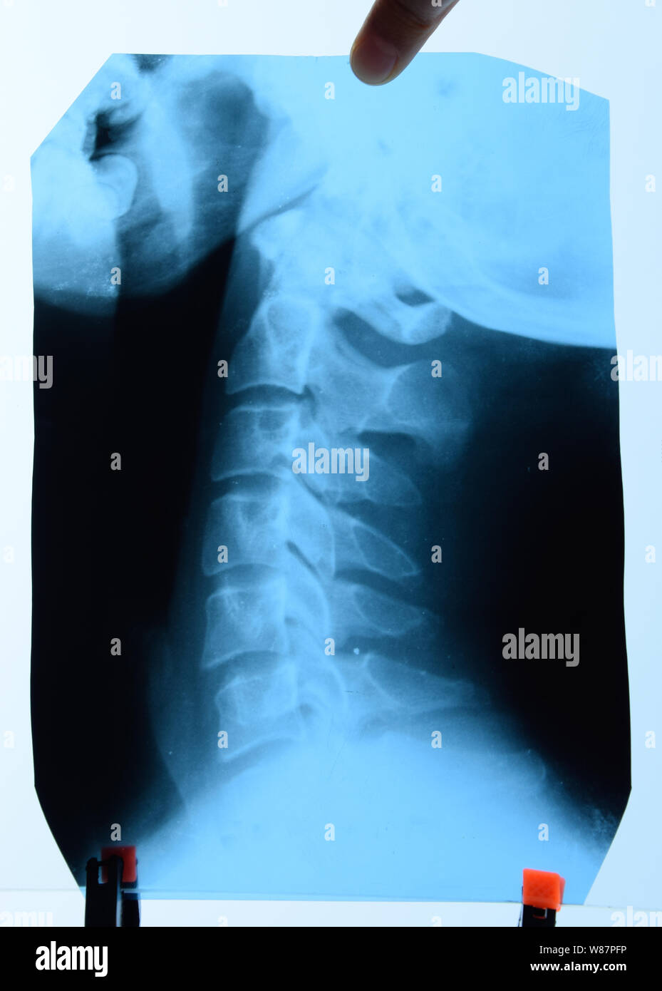 X-ray of the cervical vertebrae. X-ray image of the cervical spine. Stock Photohttps://www.alamy.com/image-license-details/?v=1https://www.alamy.com/x-ray-of-the-cervical-vertebrae-x-ray-image-of-the-cervical-spine-image263244122.html
RFW87PFP–X-ray of the cervical vertebrae. X-ray image of the cervical spine.
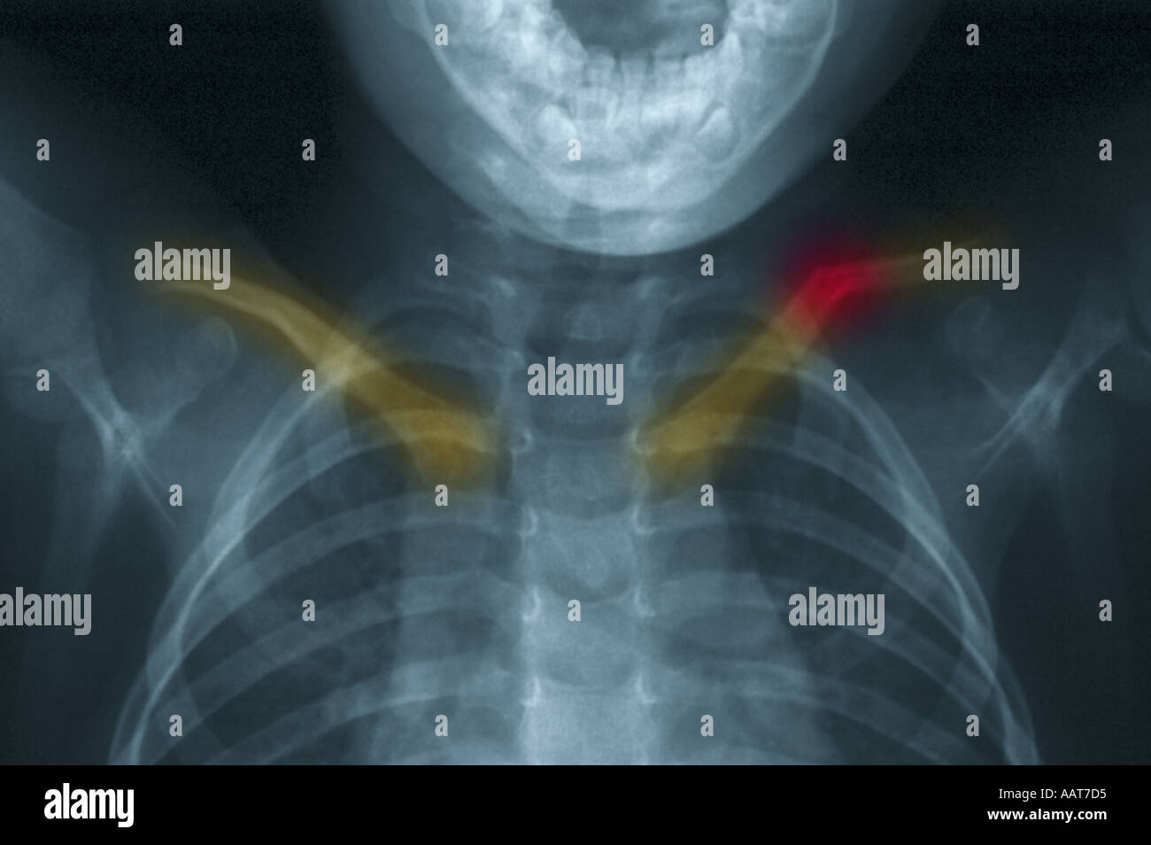 colorized xray showing fractured collarbone Stock Photohttps://www.alamy.com/image-license-details/?v=1https://www.alamy.com/colorized-xray-showing-fractured-collarbone-image2361300.html
RMAAT7D5–colorized xray showing fractured collarbone
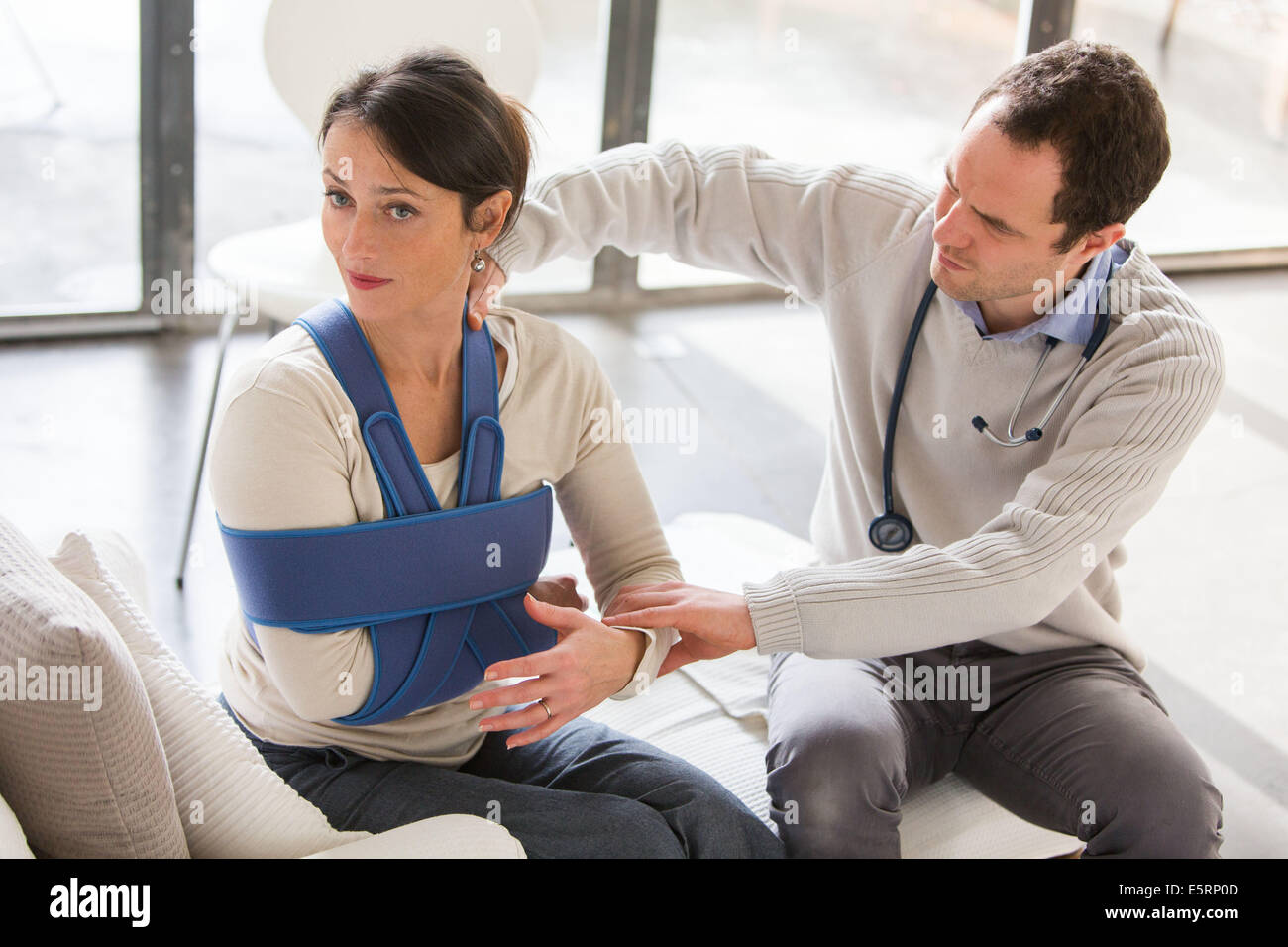 Doctor visiting female patient wearing a splint, at home. Stock Photohttps://www.alamy.com/image-license-details/?v=1https://www.alamy.com/stock-photo-doctor-visiting-female-patient-wearing-a-splint-at-home-72436909.html
RME5RP0D–Doctor visiting female patient wearing a splint, at home.
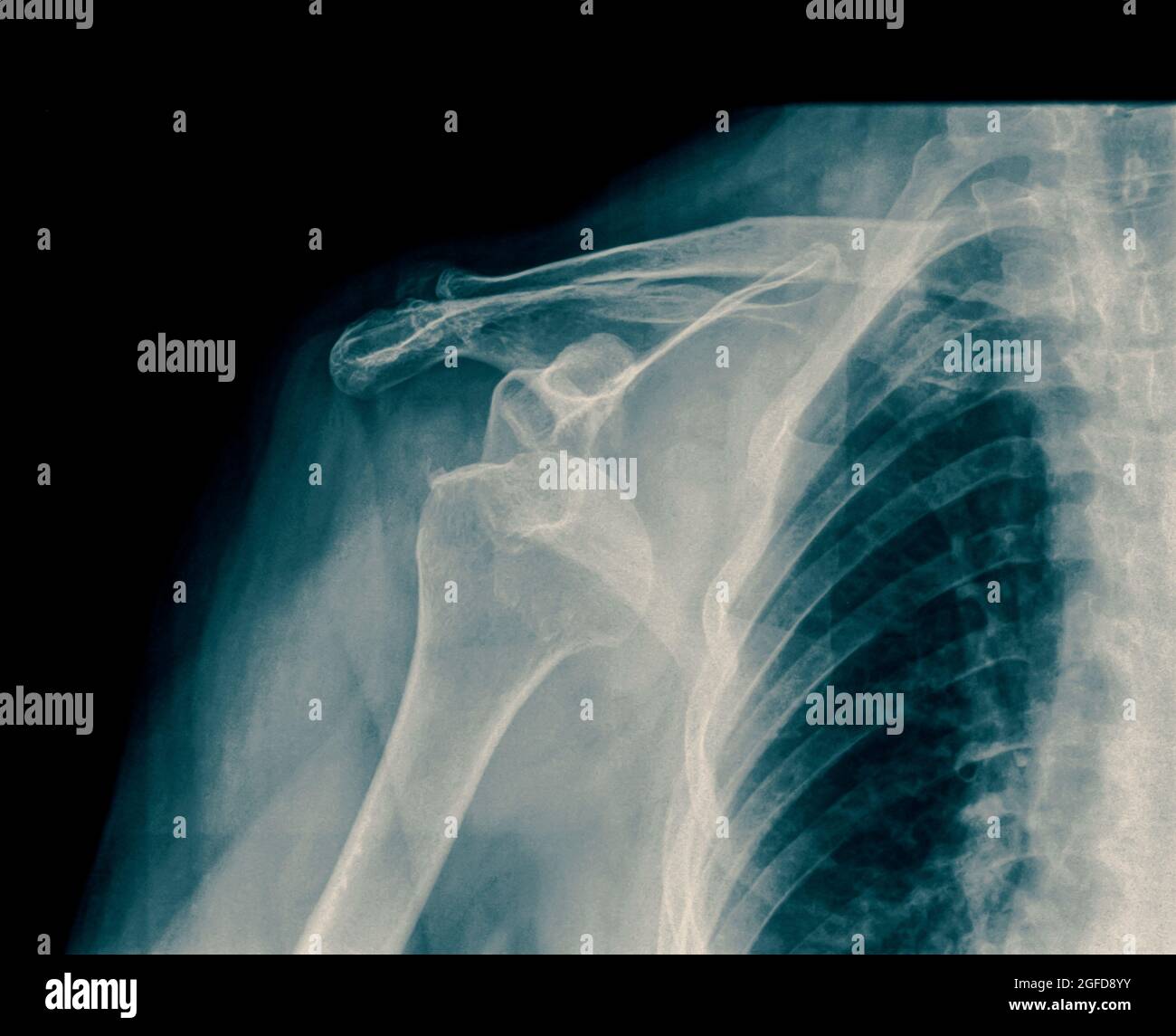 x-ray Dislocated shoulder on a 75 year old female Stock Photohttps://www.alamy.com/image-license-details/?v=1https://www.alamy.com/x-ray-dislocated-shoulder-on-a-75-year-old-female-image439771471.html
RM2GFD8YY–x-ray Dislocated shoulder on a 75 year old female
 Woman wearing a splint Stock Photohttps://www.alamy.com/image-license-details/?v=1https://www.alamy.com/stock-photo-woman-wearing-a-splint-57723088.html
RMD9WEAT–Woman wearing a splint
 3d Illustration of Lung cancer, pleural effusion isolated black. Stock Photohttps://www.alamy.com/image-license-details/?v=1https://www.alamy.com/3d-illustration-of-lung-cancer-pleural-effusion-isolated-black-image182183567.html
RFMGB4YY–3d Illustration of Lung cancer, pleural effusion isolated black.
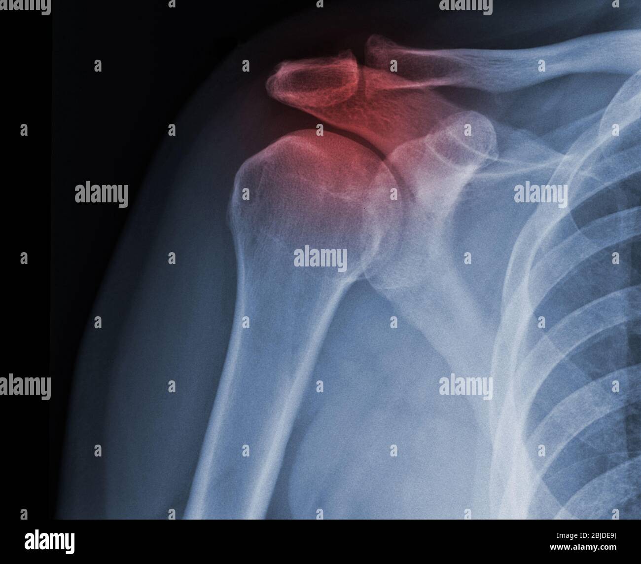 X-ray shoulder radiograph show state of injury Stock Photohttps://www.alamy.com/image-license-details/?v=1https://www.alamy.com/x-ray-shoulder-radiograph-show-state-of-injury-image355567790.html
RF2BJDE9J–X-ray shoulder radiograph show state of injury
 medical illustration of a broken clavicle Stock Photohttps://www.alamy.com/image-license-details/?v=1https://www.alamy.com/medical-illustration-of-a-broken-clavicle-image343649683.html
RM2AY2GJY–medical illustration of a broken clavicle
 September 20, 2015: Dallas Cowboys quarterback Tony Romo (9) gets helped off by the medical staff as he suffered a fractured left clavicle during the NFL game between the Dallas Cowboys and the Philadelphia Eagles at Lincoln Financial Field in Philadelphia, Pennsylvania. The Dallas Cowboys won 20-10. Christopher Szagola/CSM Stock Photohttps://www.alamy.com/image-license-details/?v=1https://www.alamy.com/stock-photo-september-20-2015-dallas-cowboys-quarterback-tony-romo-9-gets-helped-87721409.html
RMF2M1FD–September 20, 2015: Dallas Cowboys quarterback Tony Romo (9) gets helped off by the medical staff as he suffered a fractured left clavicle during the NFL game between the Dallas Cowboys and the Philadelphia Eagles at Lincoln Financial Field in Philadelphia, Pennsylvania. The Dallas Cowboys won 20-10. Christopher Szagola/CSM
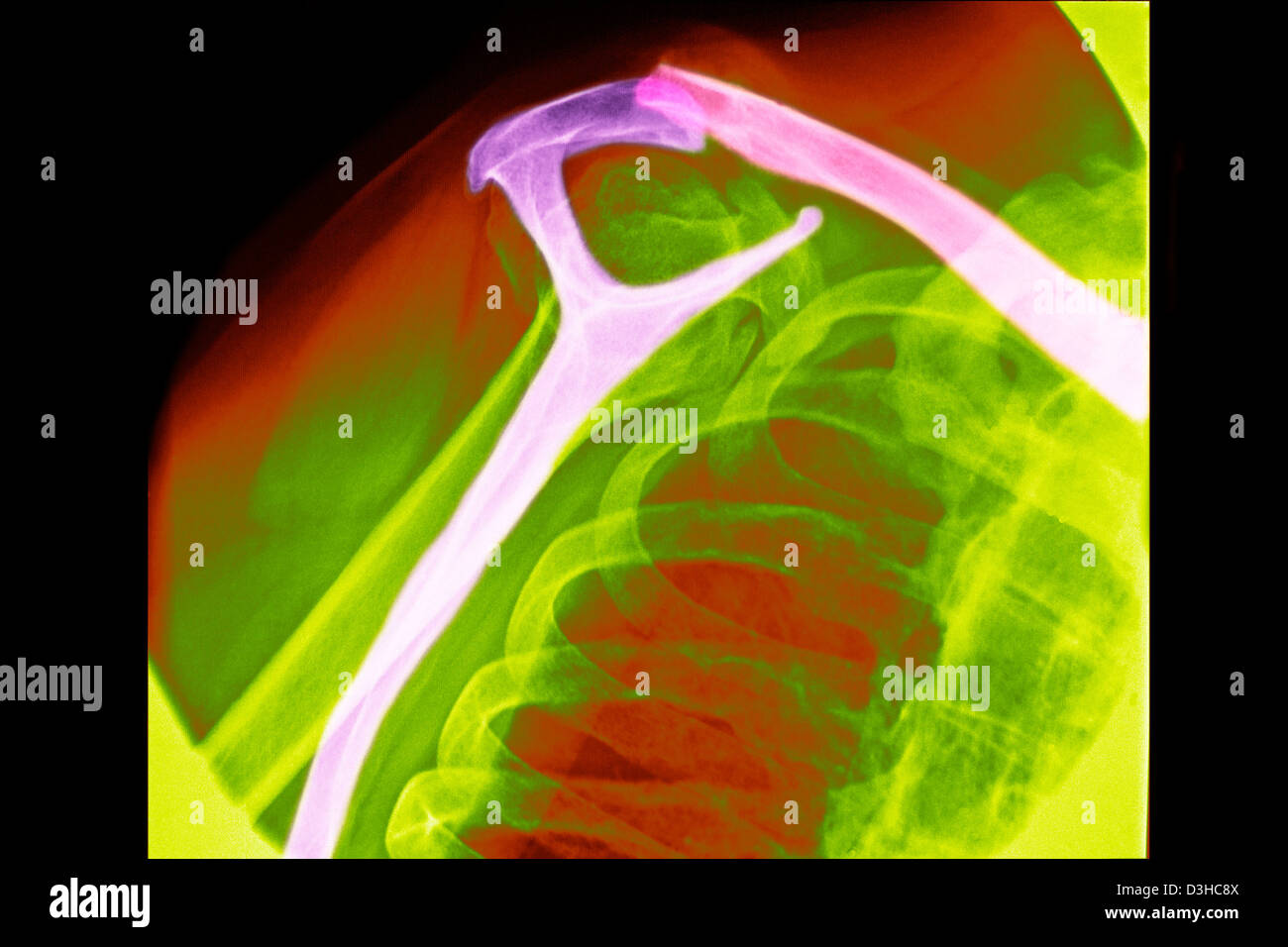 FRACTURED COLLARBONE, X-RAY Stock Photohttps://www.alamy.com/image-license-details/?v=1https://www.alamy.com/stock-photo-fractured-collarbone-x-ray-53857914.html
RMD3HC8X–FRACTURED COLLARBONE, X-RAY
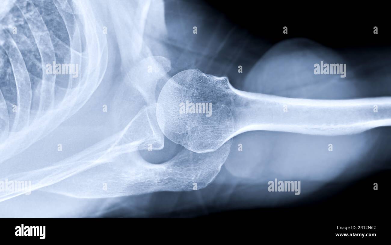 X-ray Shoulder joint shoulder transaxillary view for diagnosis fracture of shoulder joint. Stock Photohttps://www.alamy.com/image-license-details/?v=1https://www.alamy.com/x-ray-shoulder-joint-shoulder-transaxillary-view-for-diagnosis-fracture-of-shoulder-joint-image551406970.html
RF2R12N62–X-ray Shoulder joint shoulder transaxillary view for diagnosis fracture of shoulder joint.
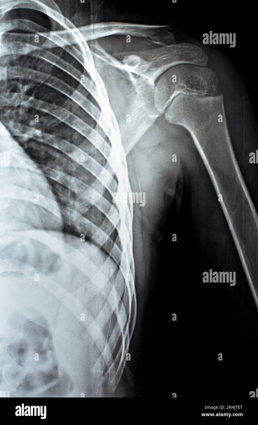 Plain x Ray PXR of left shoulder of skeletally immature female patient child showing lateral one third fracture clavicle, broken lateral part of the c Stock Photohttps://www.alamy.com/image-license-details/?v=1https://www.alamy.com/plain-x-ray-pxr-of-left-shoulder-of-skeletally-immature-female-patient-child-showing-lateral-one-third-fracture-clavicle-broken-lateral-part-of-the-c-image553604768.html
RF2R4JTET–Plain x Ray PXR of left shoulder of skeletally immature female patient child showing lateral one third fracture clavicle, broken lateral part of the c
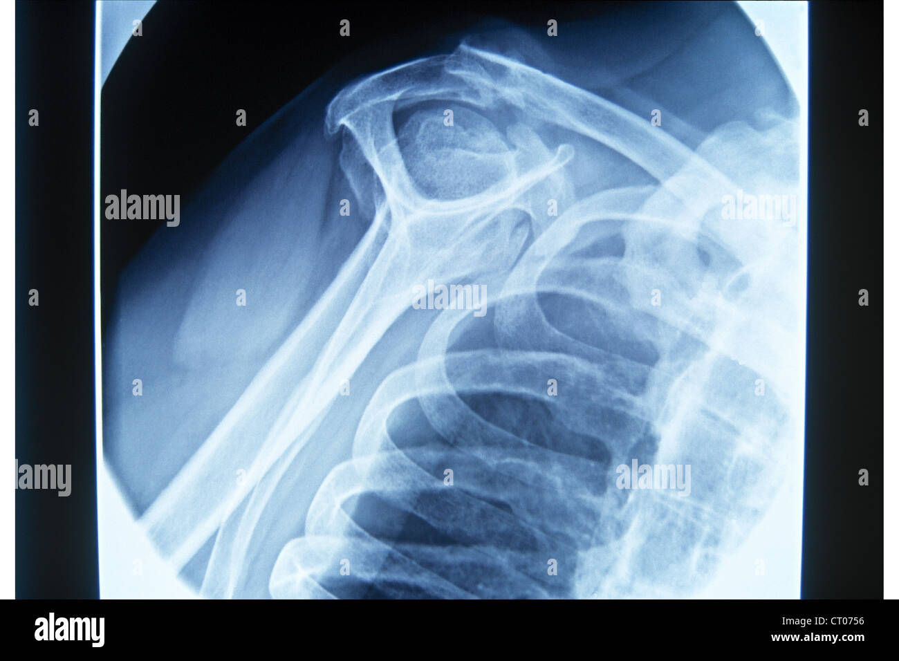 FRACTURED COLLARBONE, X-RAY Stock Photohttps://www.alamy.com/image-license-details/?v=1https://www.alamy.com/stock-photo-fractured-collarbone-x-ray-49178114.html
RMCT0756–FRACTURED COLLARBONE, X-RAY
![. The principles and practice of modern surgery. bone may be dislocated forwards by blows on the shoulder. It canreadily be felt on the anterior surface of the sternum. The tj-eatment isin all respects the same as for fractured clavicle. Dislocation of this * [This figure is intended to represent dislocation of the sternal extreinity of the clavicleand dislocation forwards of the shoulder-joint on its left side; and dislocation of the acro-mial end of the clavicle, and dislocation of the shoulder downvs^ards on its right side.] 23 266 DISLOCATIONS OF THE SHOULDER, end of the bone backwards has Stock Photo . The principles and practice of modern surgery. bone may be dislocated forwards by blows on the shoulder. It canreadily be felt on the anterior surface of the sternum. The tj-eatment isin all respects the same as for fractured clavicle. Dislocation of this * [This figure is intended to represent dislocation of the sternal extreinity of the clavicleand dislocation forwards of the shoulder-joint on its left side; and dislocation of the acro-mial end of the clavicle, and dislocation of the shoulder downvs^ards on its right side.] 23 266 DISLOCATIONS OF THE SHOULDER, end of the bone backwards has Stock Photo](https://c8.alamy.com/comp/2AG53A6/the-principles-and-practice-of-modern-surgery-bone-may-be-dislocated-forwards-by-blows-on-the-shoulder-it-canreadily-be-felt-on-the-anterior-surface-of-the-sternum-the-tj-eatment-isin-all-respects-the-same-as-for-fractured-clavicle-dislocation-of-this-this-figure-is-intended-to-represent-dislocation-of-the-sternal-extreinity-of-the-clavicleand-dislocation-forwards-of-the-shoulder-joint-on-its-left-side-and-dislocation-of-the-acro-mial-end-of-the-clavicle-and-dislocation-of-the-shoulder-downvsards-on-its-right-side-23-266-dislocations-of-the-shoulder-end-of-the-bone-backwards-has-2AG53A6.jpg) . The principles and practice of modern surgery. bone may be dislocated forwards by blows on the shoulder. It canreadily be felt on the anterior surface of the sternum. The tj-eatment isin all respects the same as for fractured clavicle. Dislocation of this * [This figure is intended to represent dislocation of the sternal extreinity of the clavicleand dislocation forwards of the shoulder-joint on its left side; and dislocation of the acro-mial end of the clavicle, and dislocation of the shoulder downvs^ards on its right side.] 23 266 DISLOCATIONS OF THE SHOULDER, end of the bone backwards has Stock Photohttps://www.alamy.com/image-license-details/?v=1https://www.alamy.com/the-principles-and-practice-of-modern-surgery-bone-may-be-dislocated-forwards-by-blows-on-the-shoulder-it-canreadily-be-felt-on-the-anterior-surface-of-the-sternum-the-tj-eatment-isin-all-respects-the-same-as-for-fractured-clavicle-dislocation-of-this-this-figure-is-intended-to-represent-dislocation-of-the-sternal-extreinity-of-the-clavicleand-dislocation-forwards-of-the-shoulder-joint-on-its-left-side-and-dislocation-of-the-acro-mial-end-of-the-clavicle-and-dislocation-of-the-shoulder-downvsards-on-its-right-side-23-266-dislocations-of-the-shoulder-end-of-the-bone-backwards-has-image336943886.html
. The principles and practice of modern surgery. bone may be dislocated forwards by blows on the shoulder. It canreadily be felt on the anterior surface of the sternum. The tj-eatment isin all respects the same as for fractured clavicle. Dislocation of this * [This figure is intended to represent dislocation of the sternal extreinity of the clavicleand dislocation forwards of the shoulder-joint on its left side; and dislocation of the acro-mial end of the clavicle, and dislocation of the shoulder downvs^ards on its right side.] 23 266 DISLOCATIONS OF THE SHOULDER, end of the bone backwards has Stock Photohttps://www.alamy.com/image-license-details/?v=1https://www.alamy.com/the-principles-and-practice-of-modern-surgery-bone-may-be-dislocated-forwards-by-blows-on-the-shoulder-it-canreadily-be-felt-on-the-anterior-surface-of-the-sternum-the-tj-eatment-isin-all-respects-the-same-as-for-fractured-clavicle-dislocation-of-this-this-figure-is-intended-to-represent-dislocation-of-the-sternal-extreinity-of-the-clavicleand-dislocation-forwards-of-the-shoulder-joint-on-its-left-side-and-dislocation-of-the-acro-mial-end-of-the-clavicle-and-dislocation-of-the-shoulder-downvsards-on-its-right-side-23-266-dislocations-of-the-shoulder-end-of-the-bone-backwards-has-image336943886.htmlRM2AG53A6–. The principles and practice of modern surgery. bone may be dislocated forwards by blows on the shoulder. It canreadily be felt on the anterior surface of the sternum. The tj-eatment isin all respects the same as for fractured clavicle. Dislocation of this * [This figure is intended to represent dislocation of the sternal extreinity of the clavicleand dislocation forwards of the shoulder-joint on its left side; and dislocation of the acro-mial end of the clavicle, and dislocation of the shoulder downvs^ards on its right side.] 23 266 DISLOCATIONS OF THE SHOULDER, end of the bone backwards has
 X-ray of the cervical vertebrae. X-ray image of the cervical spine. Stock Photohttps://www.alamy.com/image-license-details/?v=1https://www.alamy.com/x-ray-of-the-cervical-vertebrae-x-ray-image-of-the-cervical-spine-image263243075.html
RFW87N6B–X-ray of the cervical vertebrae. X-ray image of the cervical spine.
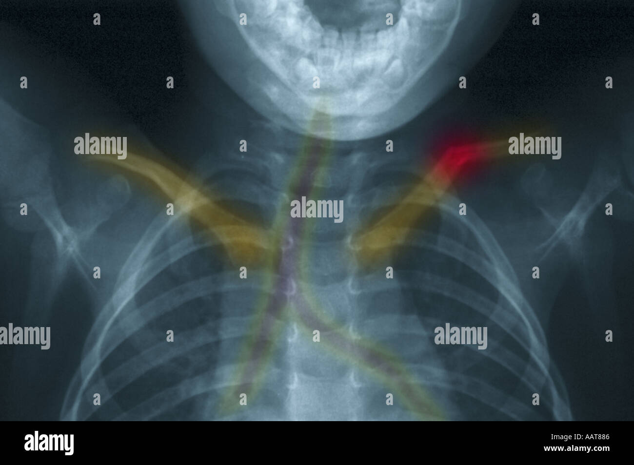 upper body chest xray of a 3 year old boy showing a fractured collarbone highlighted in red Stock Photohttps://www.alamy.com/image-license-details/?v=1https://www.alamy.com/upper-body-chest-xray-of-a-3-year-old-boy-showing-a-fractured-collarbone-image2361477.html
RMAAT886–upper body chest xray of a 3 year old boy showing a fractured collarbone highlighted in red
 Doctor visiting female patient wearing a splint, at home. Stock Photohttps://www.alamy.com/image-license-details/?v=1https://www.alamy.com/stock-photo-doctor-visiting-female-patient-wearing-a-splint-at-home-72436908.html
RME5RP0C–Doctor visiting female patient wearing a splint, at home.
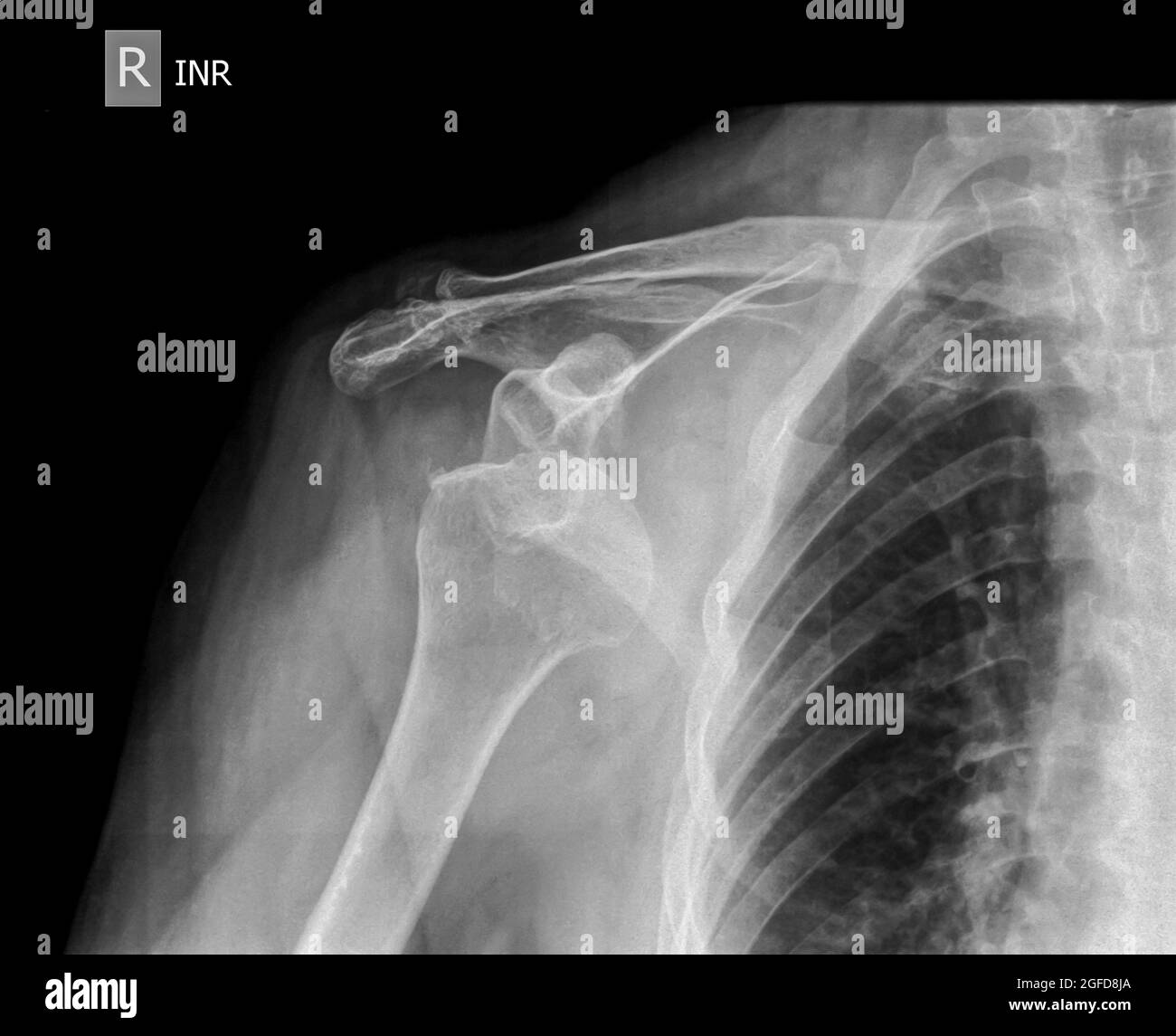 x-ray Dislocated shoulder on a 75 year old female Stock Photohttps://www.alamy.com/image-license-details/?v=1https://www.alamy.com/x-ray-dislocated-shoulder-on-a-75-year-old-female-image439771202.html
RM2GFD8JA–x-ray Dislocated shoulder on a 75 year old female
 Woman helping elderly man doing paperwork. Stock Photohttps://www.alamy.com/image-license-details/?v=1https://www.alamy.com/stock-photo-woman-helping-elderly-man-doing-paperwork-72440782.html
RME5RXXP–Woman helping elderly man doing paperwork.
 Doctor visiting female patient wearing a splint, at home Stock Photohttps://www.alamy.com/image-license-details/?v=1https://www.alamy.com/stock-photo-doctor-visiting-female-patient-wearing-a-splint-at-home-57723077.html
RMD9WEAD–Doctor visiting female patient wearing a splint, at home
 X-ray shoulder radiograph show state of injury Stock Photohttps://www.alamy.com/image-license-details/?v=1https://www.alamy.com/x-ray-shoulder-radiograph-show-state-of-injury-image355567795.html
RF2BJDE9R–X-ray shoulder radiograph show state of injury
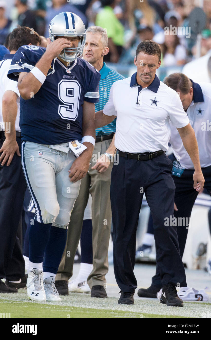 September 20, 2015: Dallas Cowboys quarterback Tony Romo (9) gets helped off by the medical staff as he suffered a fractured left clavicle during the NFL game between the Dallas Cowboys and the Philadelphia Eagles at Lincoln Financial Field in Philadelphia, Pennsylvania. The Dallas Cowboys won 20-10. Christopher Szagola/CSM Stock Photohttps://www.alamy.com/image-license-details/?v=1https://www.alamy.com/stock-photo-september-20-2015-dallas-cowboys-quarterback-tony-romo-9-gets-helped-87721393.html
RMF2M1EW–September 20, 2015: Dallas Cowboys quarterback Tony Romo (9) gets helped off by the medical staff as he suffered a fractured left clavicle during the NFL game between the Dallas Cowboys and the Philadelphia Eagles at Lincoln Financial Field in Philadelphia, Pennsylvania. The Dallas Cowboys won 20-10. Christopher Szagola/CSM
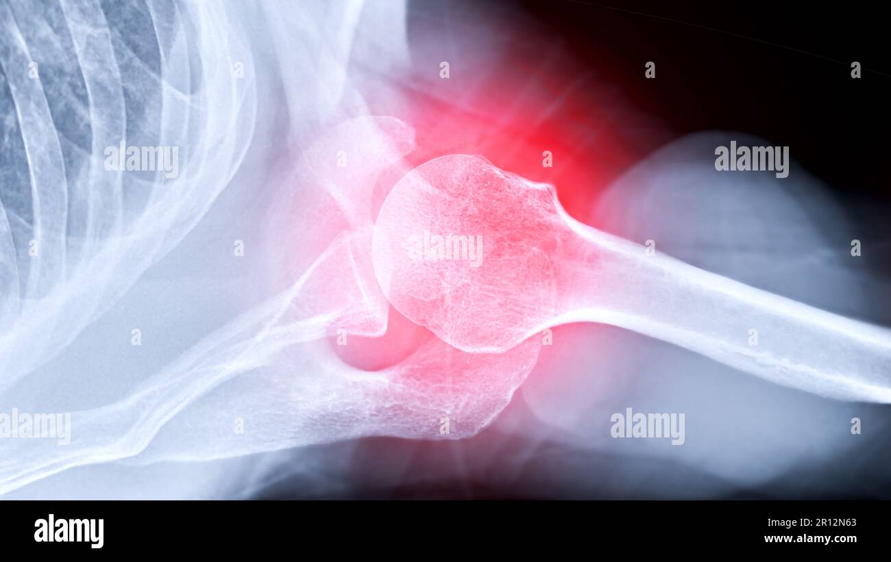 X-ray Shoulder joint shoulder transaxillary view for diagnosis fracture of shoulder joint. Stock Photohttps://www.alamy.com/image-license-details/?v=1https://www.alamy.com/x-ray-shoulder-joint-shoulder-transaxillary-view-for-diagnosis-fracture-of-shoulder-joint-image551406971.html
RF2R12N63–X-ray Shoulder joint shoulder transaxillary view for diagnosis fracture of shoulder joint.
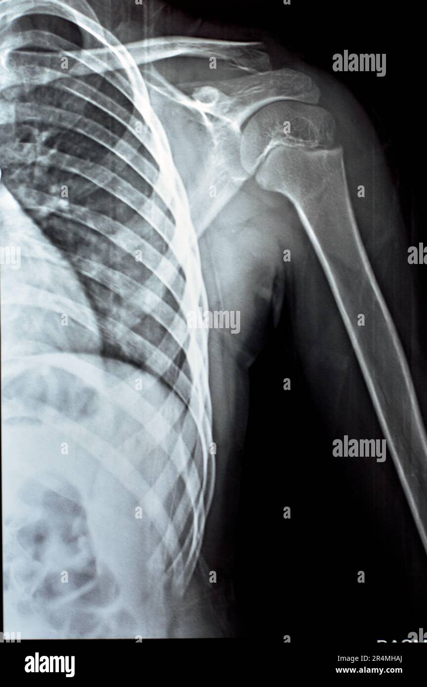 Plain x Ray PXR of left shoulder of skeletally immature female patient child showing lateral one third fracture clavicle, broken lateral part of the c Stock Photohttps://www.alamy.com/image-license-details/?v=1https://www.alamy.com/plain-x-ray-pxr-of-left-shoulder-of-skeletally-immature-female-patient-child-showing-lateral-one-third-fracture-clavicle-broken-lateral-part-of-the-c-image553643066.html
RF2R4MHAJ–Plain x Ray PXR of left shoulder of skeletally immature female patient child showing lateral one third fracture clavicle, broken lateral part of the c
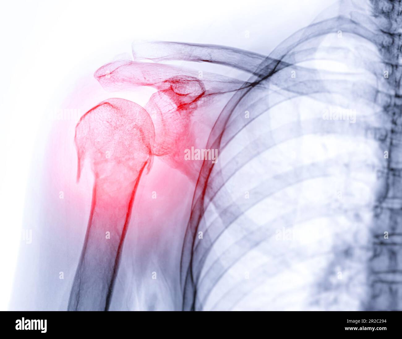 X-ray of Shoulder joint showing fracture of humerus bone. Stock Photohttps://www.alamy.com/image-license-details/?v=1https://www.alamy.com/x-ray-of-shoulder-joint-showing-fracture-of-humerus-bone-image552226336.html
RF2R2C294–X-ray of Shoulder joint showing fracture of humerus bone.
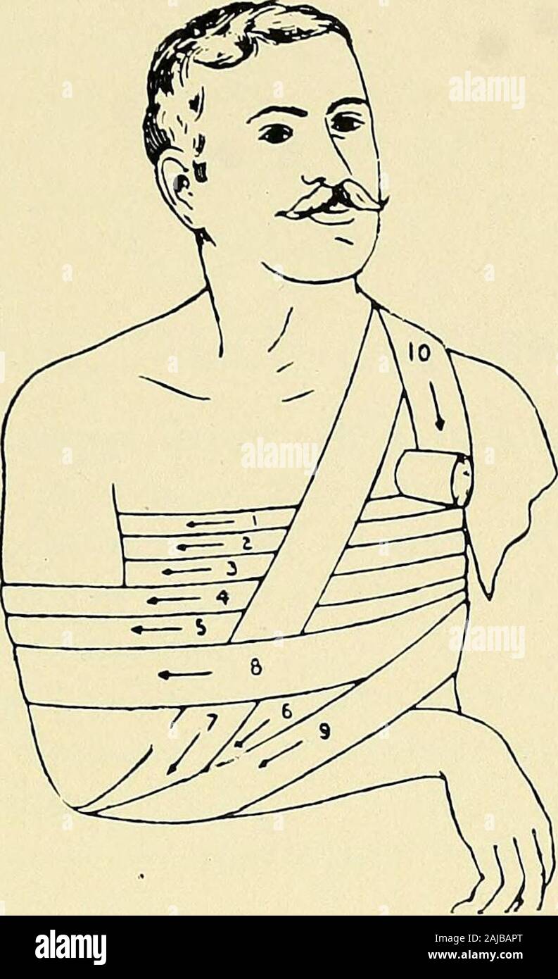 The principles and practice of bandaging : by Gwilym G Davis . ow of the sameside and continue to make figure 8 turns of the healthy axilla andaffected arm, the point of crossing being on the external extremityof the clavicle. The bandage is ended by oblique or horizontalcircular turns, whichever are thought best. (See fig. 93.) Gerdy says lie prefers the above bandage to the more complicated one ofDesaiilt for the treatment of fractured clavicle. Dr. Dulles (Medical News)has recently advocated the use of a portion of this bandage for the same affec-tion. He employs the horizontal circular tur Stock Photohttps://www.alamy.com/image-license-details/?v=1https://www.alamy.com/the-principles-and-practice-of-bandaging-by-gwilym-g-davis-ow-of-the-sameside-and-continue-to-make-figure-8-turns-of-the-healthy-axilla-andaffected-arm-the-point-of-crossing-being-on-the-external-extremityof-the-clavicle-the-bandage-is-ended-by-oblique-or-horizontalcircular-turns-whichever-are-thought-best-see-fig-93-gerdy-says-lie-prefers-the-above-bandage-to-the-more-complicated-one-ofdesaiilt-for-the-treatment-of-fractured-clavicle-dr-dulles-medical-newshas-recently-advocated-the-use-of-a-portion-of-this-bandage-for-the-same-affec-tion-he-employs-the-horizontal-circular-tur-image338310752.html
RM2AJBAPT–The principles and practice of bandaging : by Gwilym G Davis . ow of the sameside and continue to make figure 8 turns of the healthy axilla andaffected arm, the point of crossing being on the external extremityof the clavicle. The bandage is ended by oblique or horizontalcircular turns, whichever are thought best. (See fig. 93.) Gerdy says lie prefers the above bandage to the more complicated one ofDesaiilt for the treatment of fractured clavicle. Dr. Dulles (Medical News)has recently advocated the use of a portion of this bandage for the same affec-tion. He employs the horizontal circular tur
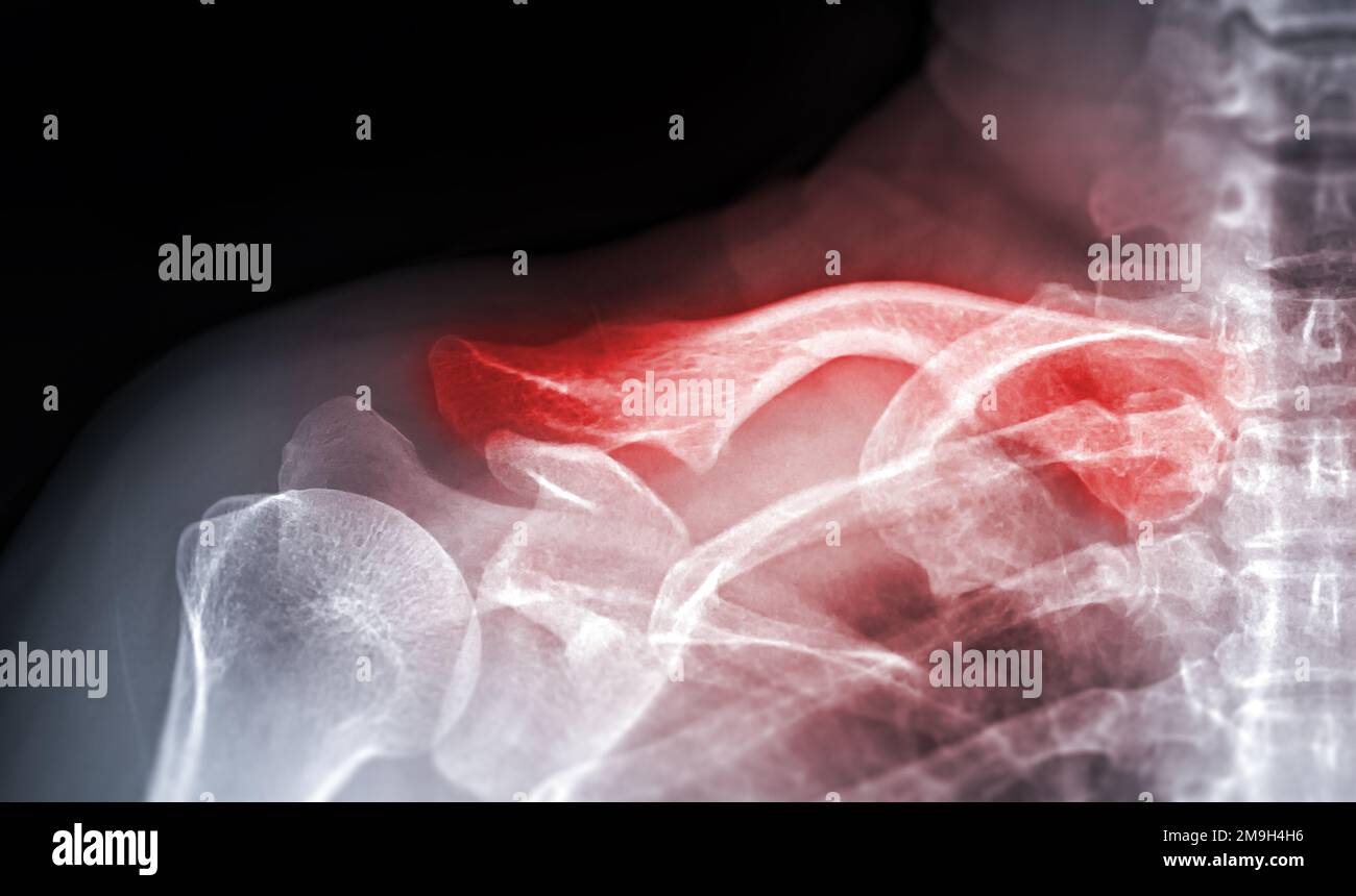 X-ray of Clavicle AP view for diagnosis fracture of Clavicle bone. Stock Photohttps://www.alamy.com/image-license-details/?v=1https://www.alamy.com/x-ray-of-clavicle-ap-view-for-diagnosis-fracture-of-clavicle-bone-image505009378.html
RF2M9H4H6–X-ray of Clavicle AP view for diagnosis fracture of Clavicle bone.
 X-ray of the cervical vertebrae. X-ray image of the cervical spine. Stock Photohttps://www.alamy.com/image-license-details/?v=1https://www.alamy.com/x-ray-of-the-cervical-vertebrae-x-ray-image-of-the-cervical-spine-image261525894.html
RFW5DEXE–X-ray of the cervical vertebrae. X-ray image of the cervical spine.
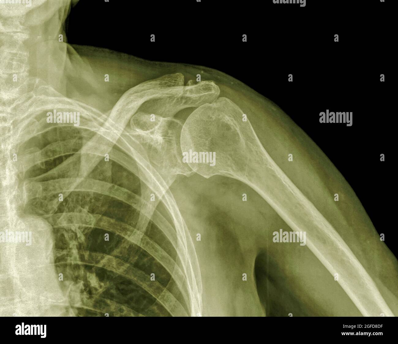 X-ray of a left shoulder of an 89 year old male patient Stock Photohttps://www.alamy.com/image-license-details/?v=1https://www.alamy.com/x-ray-of-a-left-shoulder-of-an-89-year-old-male-patient-image439771067.html
RM2GFD8DF–X-ray of a left shoulder of an 89 year old male patient
 Elderly man wearing a splint. Stock Photohttps://www.alamy.com/image-license-details/?v=1https://www.alamy.com/stock-photo-elderly-man-wearing-a-splint-72440779.html
RME5RXXK–Elderly man wearing a splint.
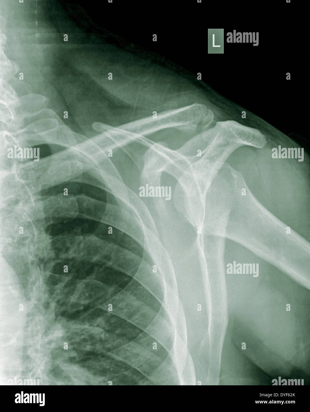 Shoulder x-ray of a 40 year old male patient with a fractured clavicle front view Stock Photohttps://www.alamy.com/image-license-details/?v=1https://www.alamy.com/shoulder-x-ray-of-a-40-year-old-male-patient-with-a-fractured-clavicle-image68560875.html
RMDYF62K–Shoulder x-ray of a 40 year old male patient with a fractured clavicle front view
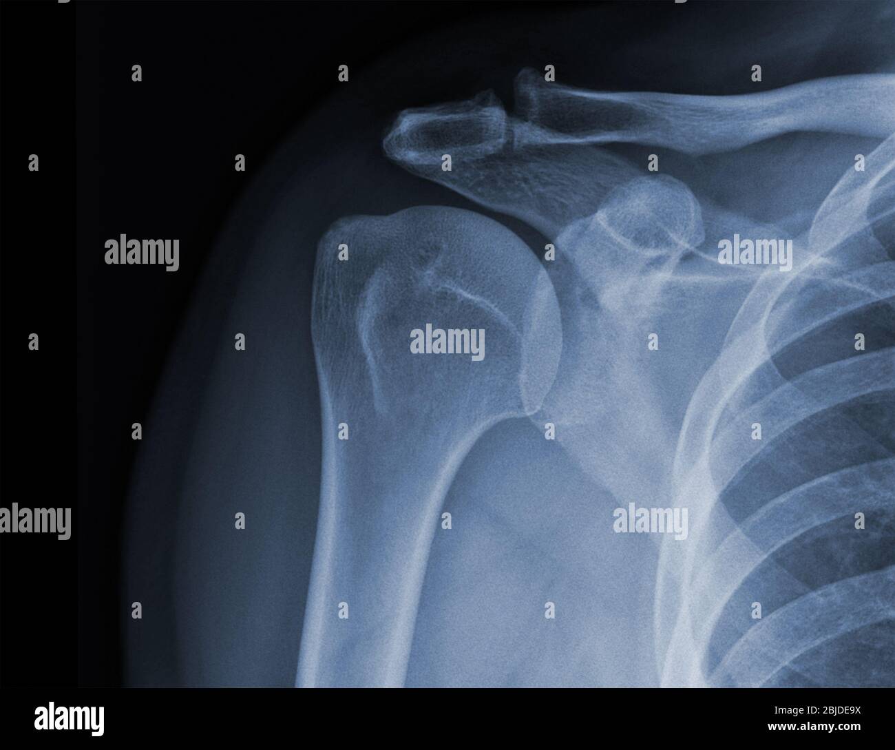 X-ray shoulder radiograph show state of injury Stock Photohttps://www.alamy.com/image-license-details/?v=1https://www.alamy.com/x-ray-shoulder-radiograph-show-state-of-injury-image355567798.html
RF2BJDE9X–X-ray shoulder radiograph show state of injury
 Doctor visiting female patient wearing a splint, at home Stock Photohttps://www.alamy.com/image-license-details/?v=1https://www.alamy.com/stock-photo-doctor-visiting-female-patient-wearing-a-splint-at-home-57723081.html
RMD9WEAH–Doctor visiting female patient wearing a splint, at home
 September 20, 2015: Dallas Cowboys quarterback Tony Romo (9) gets helped off by the medical staff as he suffered a fractured left clavicle during the NFL game between the Dallas Cowboys and the Philadelphia Eagles at Lincoln Financial Field in Philadelphia, Pennsylvania. The Dallas Cowboys won 20-10. Christopher Szagola/CSM Stock Photohttps://www.alamy.com/image-license-details/?v=1https://www.alamy.com/stock-photo-september-20-2015-dallas-cowboys-quarterback-tony-romo-9-gets-helped-87721395.html
RMF2M1EY–September 20, 2015: Dallas Cowboys quarterback Tony Romo (9) gets helped off by the medical staff as he suffered a fractured left clavicle during the NFL game between the Dallas Cowboys and the Philadelphia Eagles at Lincoln Financial Field in Philadelphia, Pennsylvania. The Dallas Cowboys won 20-10. Christopher Szagola/CSM
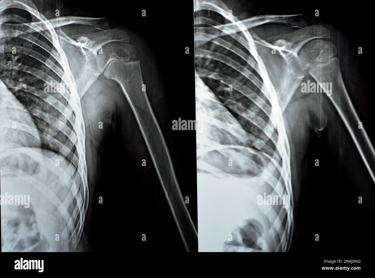 Plain x Ray PXR of left shoulder of skeletally immature female patient child showing lateral one third fracture clavicle, broken lateral part of the c Stock Photohttps://www.alamy.com/image-license-details/?v=1https://www.alamy.com/plain-x-ray-pxr-of-left-shoulder-of-skeletally-immature-female-patient-child-showing-lateral-one-third-fracture-clavicle-broken-lateral-part-of-the-c-image553590844.html
RF2R4J6NG–Plain x Ray PXR of left shoulder of skeletally immature female patient child showing lateral one third fracture clavicle, broken lateral part of the c
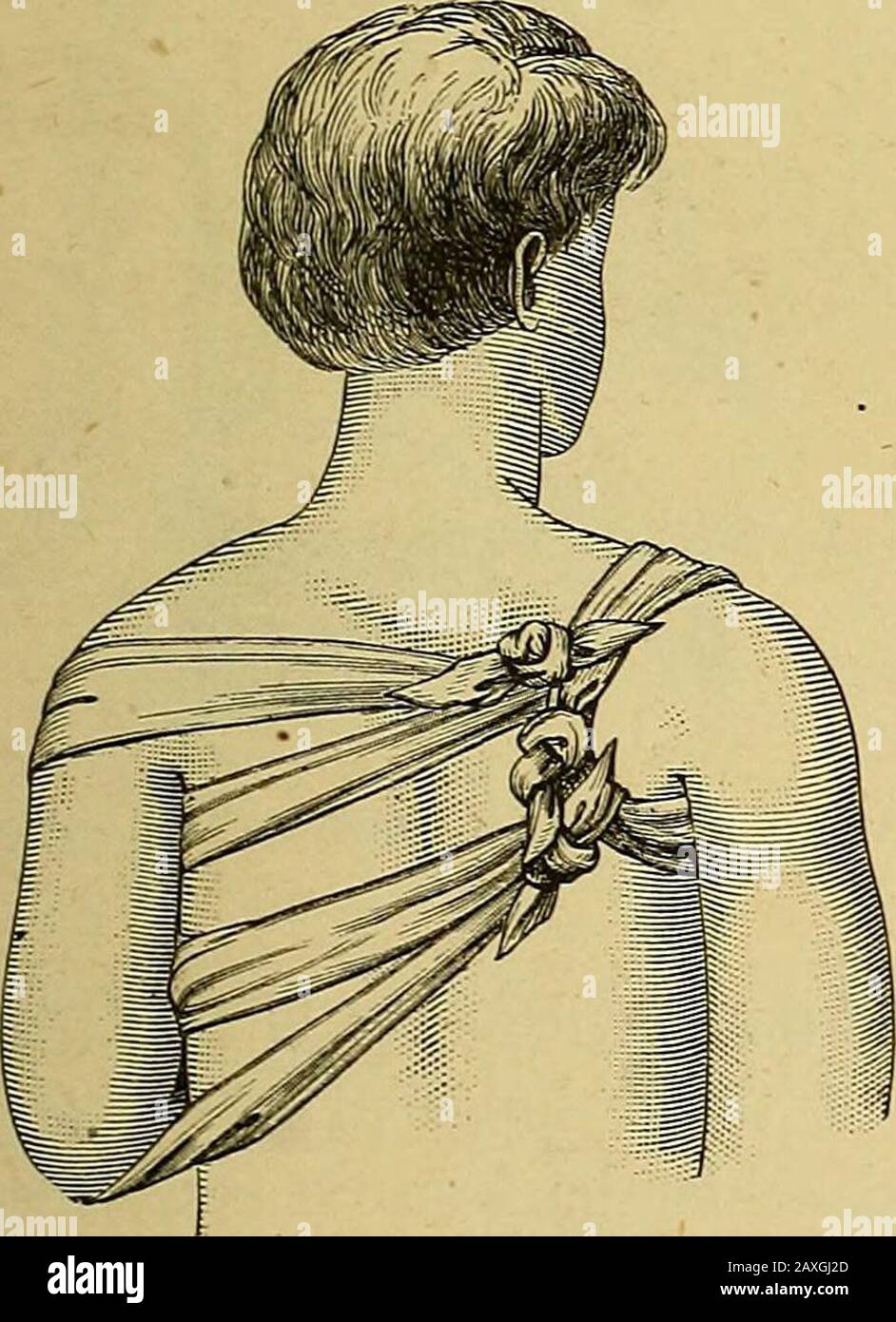 The surgeon's handbook on the treatment of wounded in war : a prize essay . 1st part. 2nd part.DESAULTS bandage for fractured clavicle. 3 rd part. 79 f. In fracture of the clavicle, the displacement of the fragmentscan be easily, if not permanently reduced, by a. Desaults bandage for fractured clavicle. It has indeed goneout of fashion, but it is an excellent exercise, in which the individualturns are employed for all shoulder dressings. The first bandage fixes a wedge-shaped cushion in the axilla ofthe abducted arm by turns, which pass round the chest (fig. 167). After the arm has been lowere Stock Photohttps://www.alamy.com/image-license-details/?v=1https://www.alamy.com/the-surgeons-handbook-on-the-treatment-of-wounded-in-war-a-prize-essay-1st-part-2nd-partdesaults-bandage-for-fractured-clavicle-3-rd-part-79-f-in-fracture-of-the-clavicle-the-displacement-of-the-fragmentscan-be-easily-if-not-permanently-reduced-by-a-desaults-bandage-for-fractured-clavicle-it-has-indeed-goneout-of-fashion-but-it-is-an-excellent-exercise-in-which-the-individualturns-are-employed-for-all-shoulder-dressings-the-first-bandage-fixes-a-wedge-shaped-cushion-in-the-axilla-ofthe-abducted-arm-by-turns-which-pass-round-the-chest-fig-167-after-the-arm-has-been-lowere-image343343461.html
RM2AXGJ2D–The surgeon's handbook on the treatment of wounded in war : a prize essay . 1st part. 2nd part.DESAULTS bandage for fractured clavicle. 3 rd part. 79 f. In fracture of the clavicle, the displacement of the fragmentscan be easily, if not permanently reduced, by a. Desaults bandage for fractured clavicle. It has indeed goneout of fashion, but it is an excellent exercise, in which the individualturns are employed for all shoulder dressings. The first bandage fixes a wedge-shaped cushion in the axilla ofthe abducted arm by turns, which pass round the chest (fig. 167). After the arm has been lowere
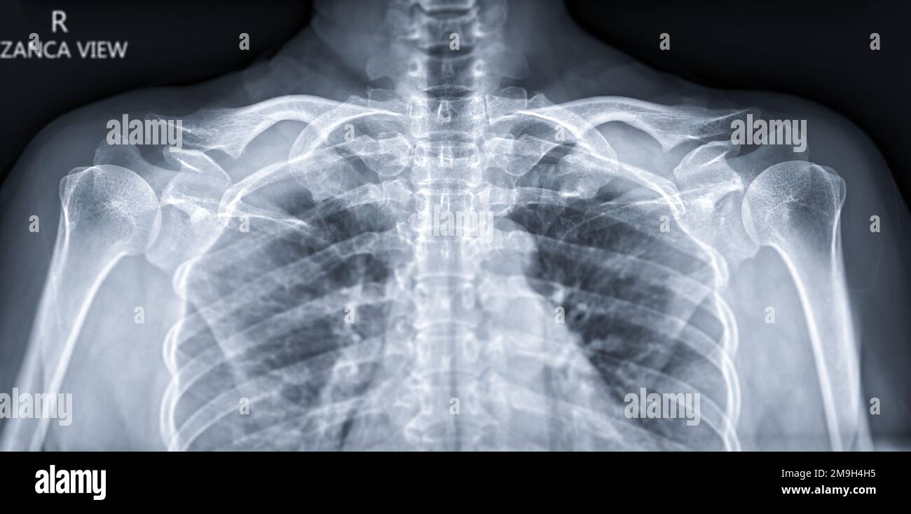 X-ray of both Clavicle Zanca view for diagnosis fracture of Clavicle . Stock Photohttps://www.alamy.com/image-license-details/?v=1https://www.alamy.com/x-ray-of-both-clavicle-zanca-view-for-diagnosis-fracture-of-clavicle-image505009377.html
RF2M9H4H5–X-ray of both Clavicle Zanca view for diagnosis fracture of Clavicle .
 X-ray of the cervical vertebrae. X-ray image of the cervical spine. Stock Photohttps://www.alamy.com/image-license-details/?v=1https://www.alamy.com/x-ray-of-the-cervical-vertebrae-x-ray-image-of-the-cervical-spine-image261526180.html
RFW5DF8M–X-ray of the cervical vertebrae. X-ray image of the cervical spine.
 X-ray of Clavicle bone showing fracture of Clavicle bone . Stock Photohttps://www.alamy.com/image-license-details/?v=1https://www.alamy.com/x-ray-of-clavicle-bone-showing-fracture-of-clavicle-bone-image552226702.html
RF2R2C2P6–X-ray of Clavicle bone showing fracture of Clavicle bone .
 Elderly man wearing a splint. Stock Photohttps://www.alamy.com/image-license-details/?v=1https://www.alamy.com/stock-photo-elderly-man-wearing-a-splint-72440780.html
RME5RXXM–Elderly man wearing a splint.
 Doctor visiting female patient wearing a splint, at home Stock Photohttps://www.alamy.com/image-license-details/?v=1https://www.alamy.com/stock-photo-doctor-visiting-female-patient-wearing-a-splint-at-home-57723084.html
RMD9WEAM–Doctor visiting female patient wearing a splint, at home
 September 20, 2015: Dallas Cowboys quarterback Tony Romo (9) gets looked over by the medical staff as he suffered a fractured left clavicle as his teammates react during the NFL game between the Dallas Cowboys and the Philadelphia Eagles at Lincoln Financial Field in Philadelphia, Pennsylvania. The Dallas Cowboys won 20-10. Christopher Szagola/CSM Stock Photohttps://www.alamy.com/image-license-details/?v=1https://www.alamy.com/stock-photo-september-20-2015-dallas-cowboys-quarterback-tony-romo-9-gets-looked-87702098.html
RMF2K4WP–September 20, 2015: Dallas Cowboys quarterback Tony Romo (9) gets looked over by the medical staff as he suffered a fractured left clavicle as his teammates react during the NFL game between the Dallas Cowboys and the Philadelphia Eagles at Lincoln Financial Field in Philadelphia, Pennsylvania. The Dallas Cowboys won 20-10. Christopher Szagola/CSM
 Plain x Ray PXR of left shoulder of skeletally immature female patient child showing lateral one third fracture clavicle, broken lateral part of the c Stock Photohttps://www.alamy.com/image-license-details/?v=1https://www.alamy.com/plain-x-ray-pxr-of-left-shoulder-of-skeletally-immature-female-patient-child-showing-lateral-one-third-fracture-clavicle-broken-lateral-part-of-the-c-image553604638.html
RF2R4JTA6–Plain x Ray PXR of left shoulder of skeletally immature female patient child showing lateral one third fracture clavicle, broken lateral part of the c
 The surgeon's handbook on the treatment of wounded in war : a prize essay . MIDDELDORPFS triangular cushion.Fig. 167. Fig. 168. MIDDELDORPFS triangle.Fig. 169.. 1st part. 2nd part.DESAULTS bandage for fractured clavicle. 3 rd part. 79 f. In fracture of the clavicle, the displacement of the fragmentscan be easily, if not permanently reduced, by a. Desaults bandage for fractured clavicle. It has indeed goneout of fashion, but it is an excellent exercise, in which the individualturns are employed for all shoulder dressings. The first bandage fixes a wedge-shaped cushion in the axilla ofthe abduct Stock Photohttps://www.alamy.com/image-license-details/?v=1https://www.alamy.com/the-surgeons-handbook-on-the-treatment-of-wounded-in-war-a-prize-essay-middeldorpfs-triangular-cushionfig-167-fig-168-middeldorpfs-trianglefig-169-1st-part-2nd-partdesaults-bandage-for-fractured-clavicle-3-rd-part-79-f-in-fracture-of-the-clavicle-the-displacement-of-the-fragmentscan-be-easily-if-not-permanently-reduced-by-a-desaults-bandage-for-fractured-clavicle-it-has-indeed-goneout-of-fashion-but-it-is-an-excellent-exercise-in-which-the-individualturns-are-employed-for-all-shoulder-dressings-the-first-bandage-fixes-a-wedge-shaped-cushion-in-the-axilla-ofthe-abduct-image343343691.html
RM2AXGJAK–The surgeon's handbook on the treatment of wounded in war : a prize essay . MIDDELDORPFS triangular cushion.Fig. 167. Fig. 168. MIDDELDORPFS triangle.Fig. 169.. 1st part. 2nd part.DESAULTS bandage for fractured clavicle. 3 rd part. 79 f. In fracture of the clavicle, the displacement of the fragmentscan be easily, if not permanently reduced, by a. Desaults bandage for fractured clavicle. It has indeed goneout of fashion, but it is an excellent exercise, in which the individualturns are employed for all shoulder dressings. The first bandage fixes a wedge-shaped cushion in the axilla ofthe abduct