Quick filters:
Frontal lobe and brain Stock Photos and Images
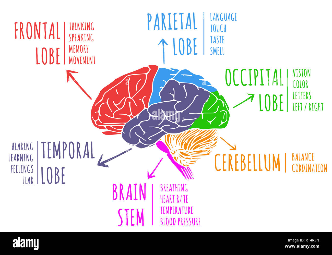 Illustration of human's brain region and function Stock Photohttps://www.alamy.com/image-license-details/?v=1https://www.alamy.com/illustration-of-humans-brain-region-and-function-image238592473.html
RFRT4R3N–Illustration of human's brain region and function
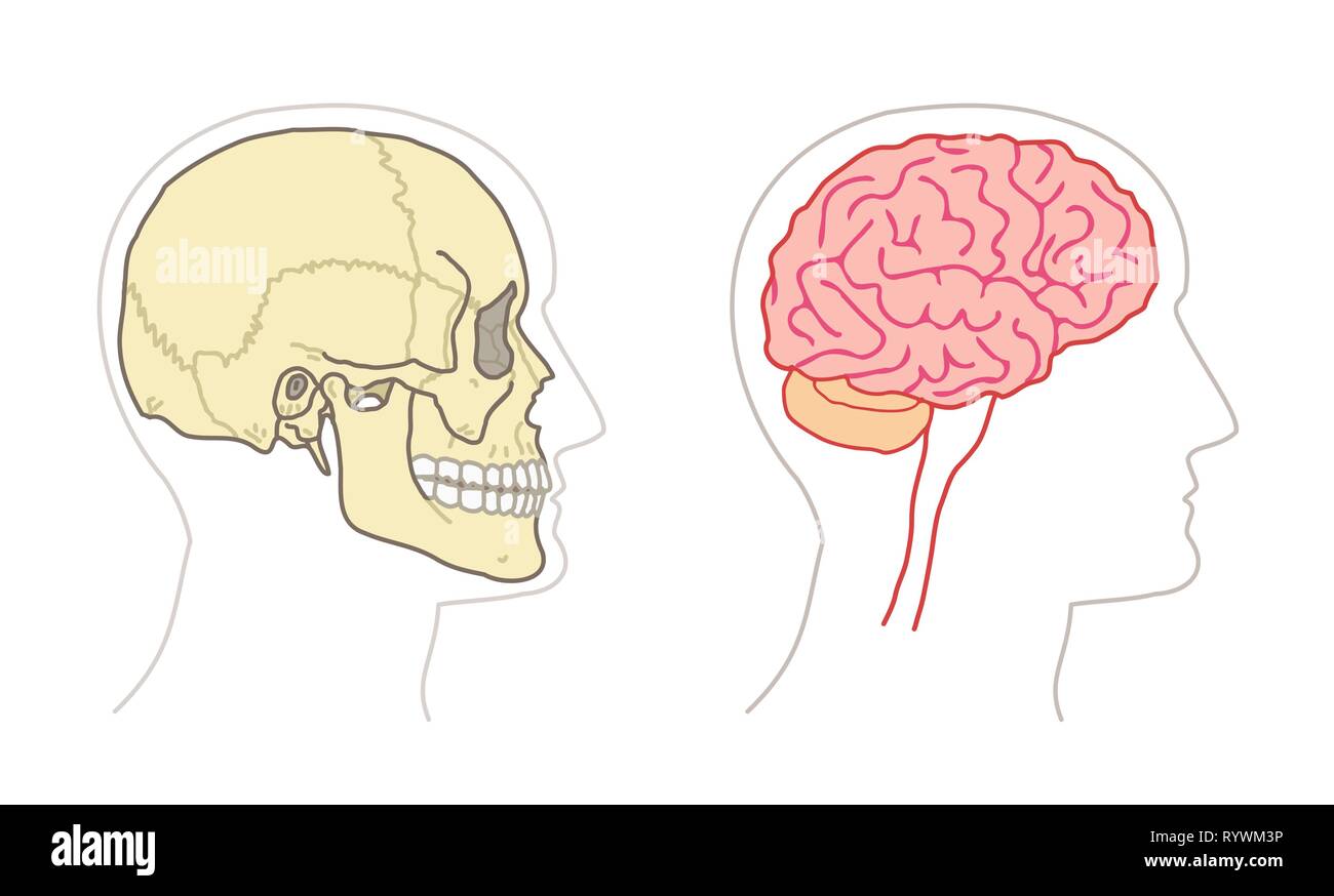 Human Anatomy drawings - BRAIN and SKULL side views Stock Vectorhttps://www.alamy.com/image-license-details/?v=1https://www.alamy.com/human-anatomy-drawings-brain-and-skull-side-views-image240895082.html
RFRYWM3P–Human Anatomy drawings - BRAIN and SKULL side views
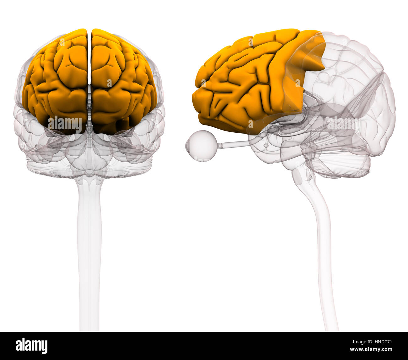 Frontal Lobe Brain Anatomy - 3d illustration Stock Photohttps://www.alamy.com/image-license-details/?v=1https://www.alamy.com/stock-photo-frontal-lobe-brain-anatomy-3d-illustration-133675333.html
RFHNDC71–Frontal Lobe Brain Anatomy - 3d illustration
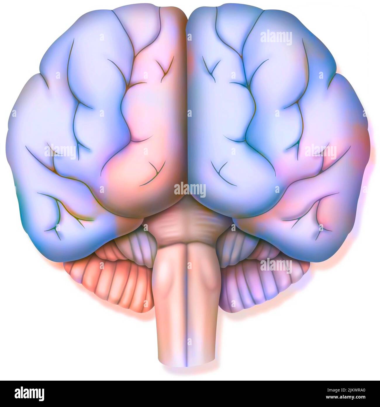 Brain, with the two cerebral hemispheres, the cerebellum and the brainstem. Stock Photohttps://www.alamy.com/image-license-details/?v=1https://www.alamy.com/brain-with-the-two-cerebral-hemispheres-the-cerebellum-and-the-brainstem-image476925512.html
RF2JKWRA0–Brain, with the two cerebral hemispheres, the cerebellum and the brainstem.
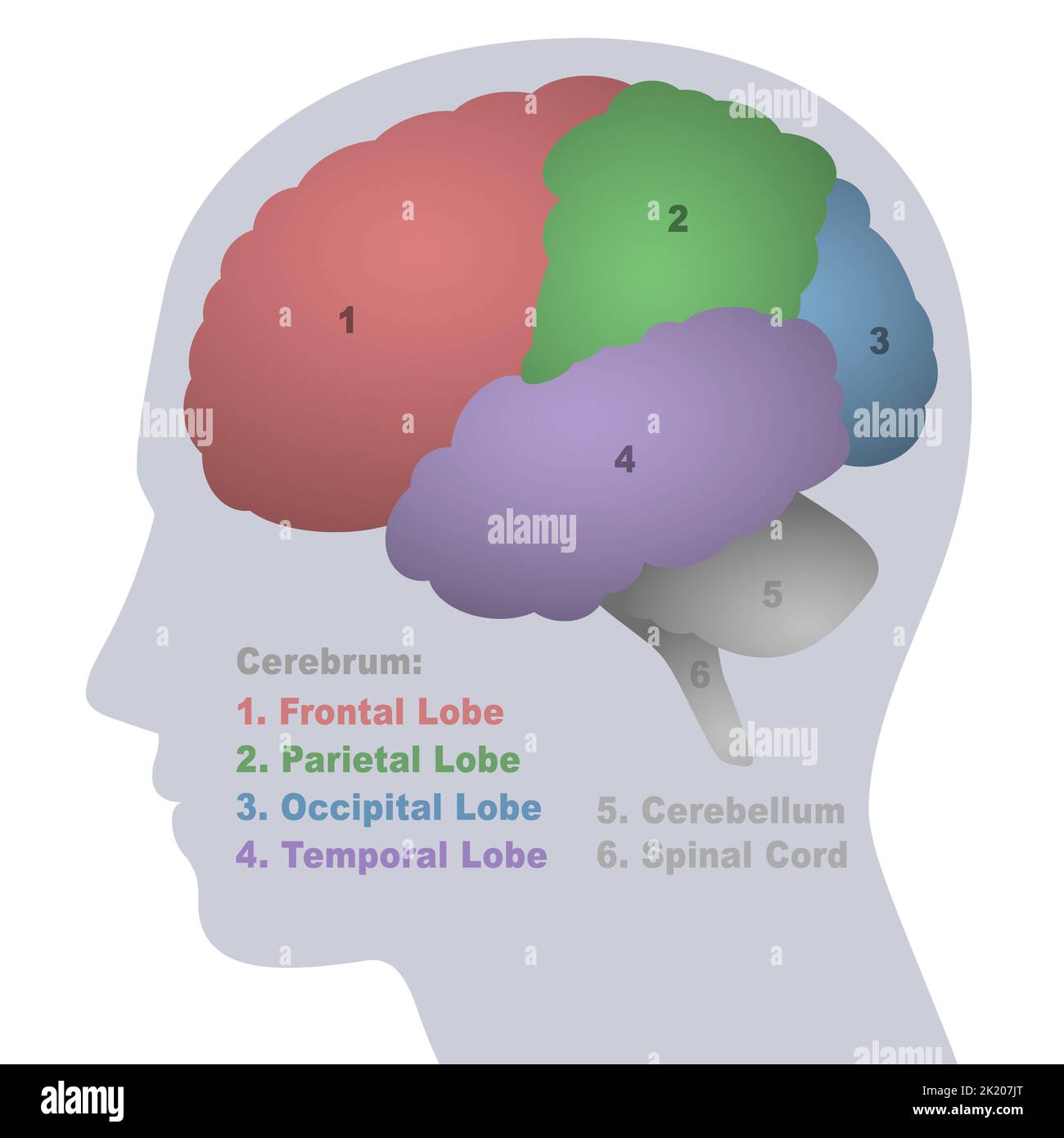 Brain lobes, anatomical regions of the cerebrum, frontal, parietal, occipital and temporal lobe, cerebellum and spinal cord, profile view. Stock Photohttps://www.alamy.com/image-license-details/?v=1https://www.alamy.com/brain-lobes-anatomical-regions-of-the-cerebrum-frontal-parietal-occipital-and-temporal-lobe-cerebellum-and-spinal-cord-profile-view-image483125632.html
RF2K207JT–Brain lobes, anatomical regions of the cerebrum, frontal, parietal, occipital and temporal lobe, cerebellum and spinal cord, profile view.
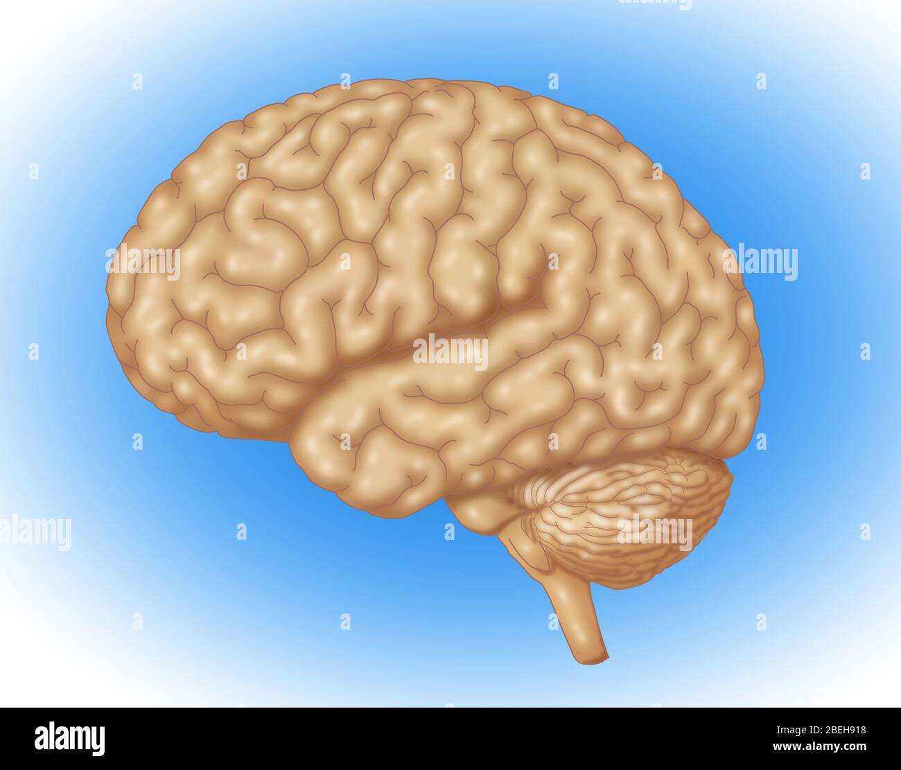 Illustration of a normal human brain. Stock Photohttps://www.alamy.com/image-license-details/?v=1https://www.alamy.com/illustration-of-a-normal-human-brain-image353192820.html
RM2BEH918–Illustration of a normal human brain.
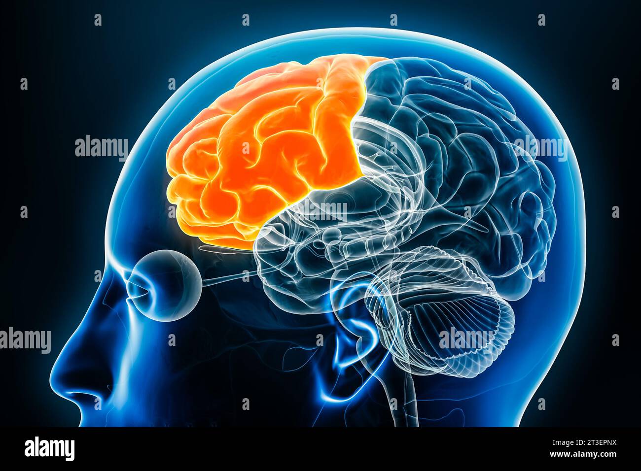 Frontal lobe of the cerebral cortex profile view close-up 3D rendering illustration. Human brain anatomy, neurology, neuroscience, medical and healthc Stock Photohttps://www.alamy.com/image-license-details/?v=1https://www.alamy.com/frontal-lobe-of-the-cerebral-cortex-profile-view-close-up-3d-rendering-illustration-human-brain-anatomy-neurology-neuroscience-medical-and-healthc-image570111302.html
RF2T3EPNX–Frontal lobe of the cerebral cortex profile view close-up 3D rendering illustration. Human brain anatomy, neurology, neuroscience, medical and healthc
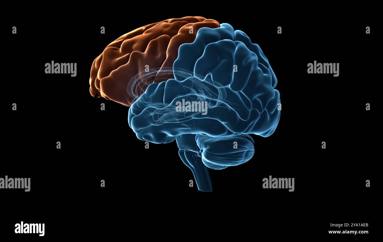 Illustration of the frontal lobe (highlighted in orange) of the human brain. Parts of this area are responsible for higher cognitive functions such as decision making, personality expression and moderating social behaviour. Stock Photohttps://www.alamy.com/image-license-details/?v=1https://www.alamy.com/illustration-of-the-frontal-lobe-highlighted-in-orange-of-the-human-brain-parts-of-this-area-are-responsible-for-higher-cognitive-functions-such-as-decision-making-personality-expression-and-moderating-social-behaviour-image625750003.html
RF2YA1AEB–Illustration of the frontal lobe (highlighted in orange) of the human brain. Parts of this area are responsible for higher cognitive functions such as decision making, personality expression and moderating social behaviour.
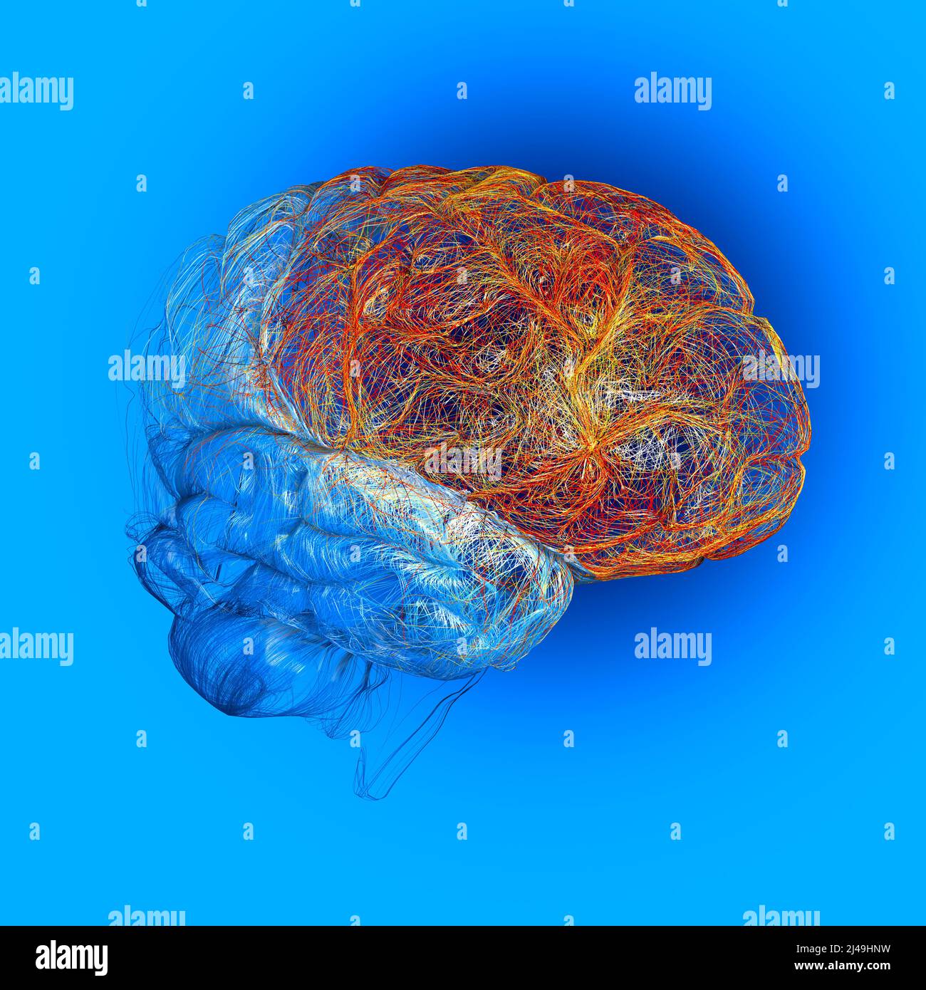 Brain, the frontal lobe is located at the front of each cerebral hemisphere, it contains most of the dopamine neurons in the cerebral cortex Stock Photohttps://www.alamy.com/image-license-details/?v=1https://www.alamy.com/brain-the-frontal-lobe-is-located-at-the-front-of-each-cerebral-hemisphere-it-contains-most-of-the-dopamine-neurons-in-the-cerebral-cortex-image467350069.html
RF2J49HNW–Brain, the frontal lobe is located at the front of each cerebral hemisphere, it contains most of the dopamine neurons in the cerebral cortex
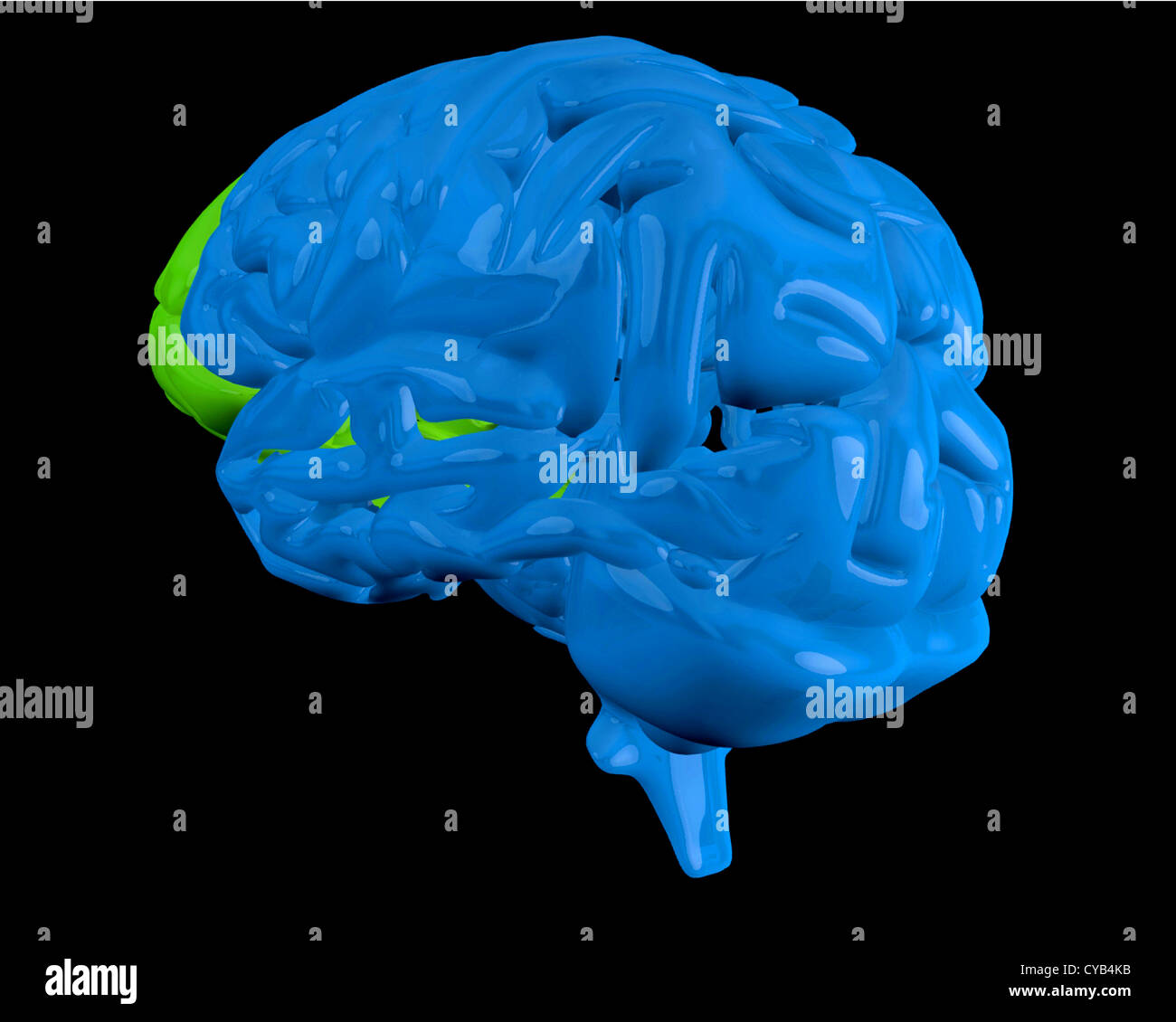 Blue brain with highlighted frontal lobe Stock Photohttps://www.alamy.com/image-license-details/?v=1https://www.alamy.com/stock-photo-blue-brain-with-highlighted-frontal-lobe-51261599.html
RFCYB4KB–Blue brain with highlighted frontal lobe
 Brain metal accumulation concept and human mind lobe as a thinking organ made of metallic material as a neurology and neuroscience symbol. Stock Photohttps://www.alamy.com/image-license-details/?v=1https://www.alamy.com/brain-metal-accumulation-concept-and-human-mind-lobe-as-a-thinking-organ-made-of-metallic-material-as-a-neurology-and-neuroscience-symbol-image425403367.html
RF2FM2P9B–Brain metal accumulation concept and human mind lobe as a thinking organ made of metallic material as a neurology and neuroscience symbol.
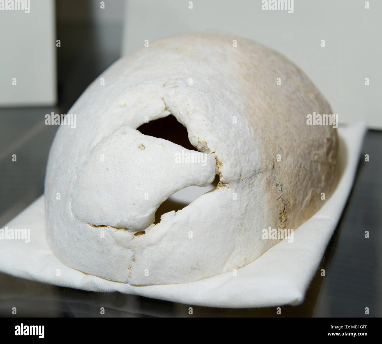 Phineas Gage skull, on display at Harvard Medical School. Hole in the cranium shows where tamping iron went through his frontal lobe in 1848. Stock Photohttps://www.alamy.com/image-license-details/?v=1https://www.alamy.com/phineas-gage-skull-on-display-at-harvard-medical-school-hole-in-the-cranium-shows-where-tamping-iron-went-through-his-frontal-lobe-in-1848-image178899834.html
RMMB1GFP–Phineas Gage skull, on display at Harvard Medical School. Hole in the cranium shows where tamping iron went through his frontal lobe in 1848.
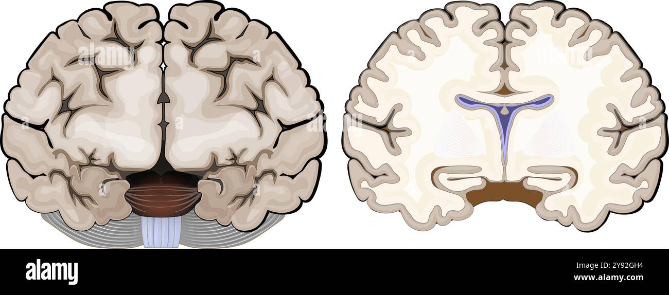 Brain anatomy. Frontal view and cross section of a human brain. Close-up of Hippocampus, and ventricles. Cerebral cortex. Vector illustration Stock Vectorhttps://www.alamy.com/image-license-details/?v=1https://www.alamy.com/brain-anatomy-frontal-view-and-cross-section-of-a-human-brain-close-up-of-hippocampus-and-ventricles-cerebral-cortex-vector-illustration-image625162080.html
RF2Y92GH4–Brain anatomy. Frontal view and cross section of a human brain. Close-up of Hippocampus, and ventricles. Cerebral cortex. Vector illustration
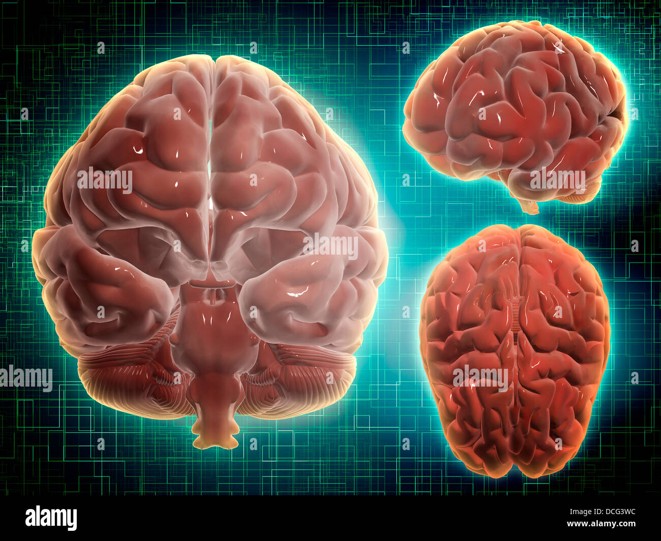 Conceptual image of human brain at different angles. Stock Photohttps://www.alamy.com/image-license-details/?v=1https://www.alamy.com/stock-photo-conceptual-image-of-human-brain-at-different-angles-59361272.html
RFDCG3WC–Conceptual image of human brain at different angles.
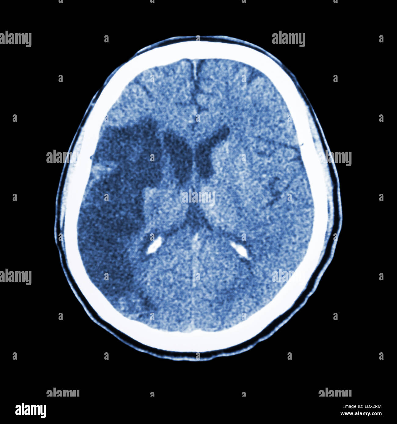 CT brain : show Ischemic stroke (hypodensity at right frontal-parietal lobe) Stock Photohttps://www.alamy.com/image-license-details/?v=1https://www.alamy.com/stock-photo-ct-brain-show-ischemic-stroke-hypodensity-at-right-frontal-parietal-77404984.html
RFEDX2RM–CT brain : show Ischemic stroke (hypodensity at right frontal-parietal lobe)
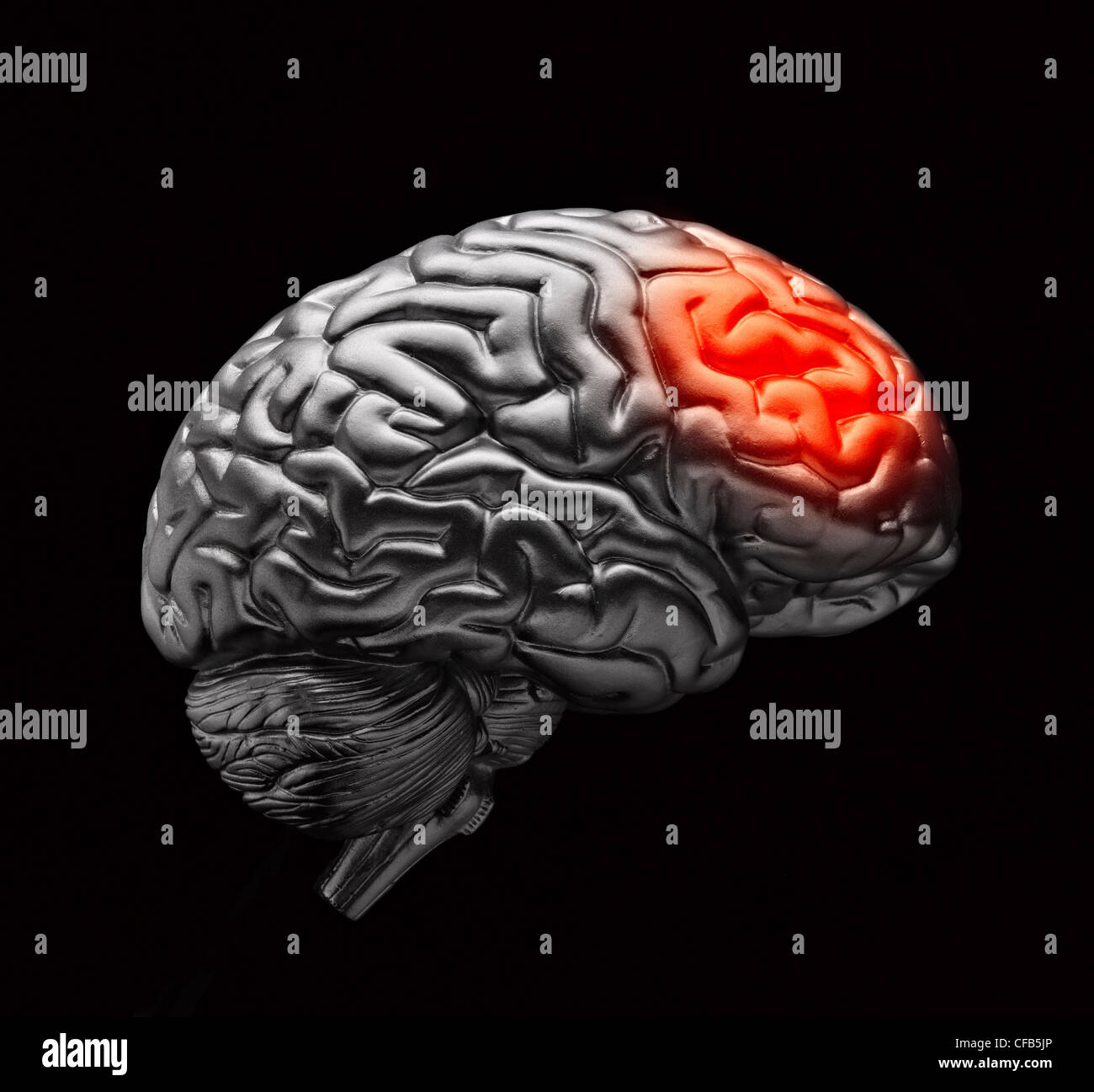 Brain with simulated headache pain Stock Photohttps://www.alamy.com/image-license-details/?v=1https://www.alamy.com/stock-photo-brain-with-simulated-headache-pain-43886494.html
RMCFB5JP–Brain with simulated headache pain
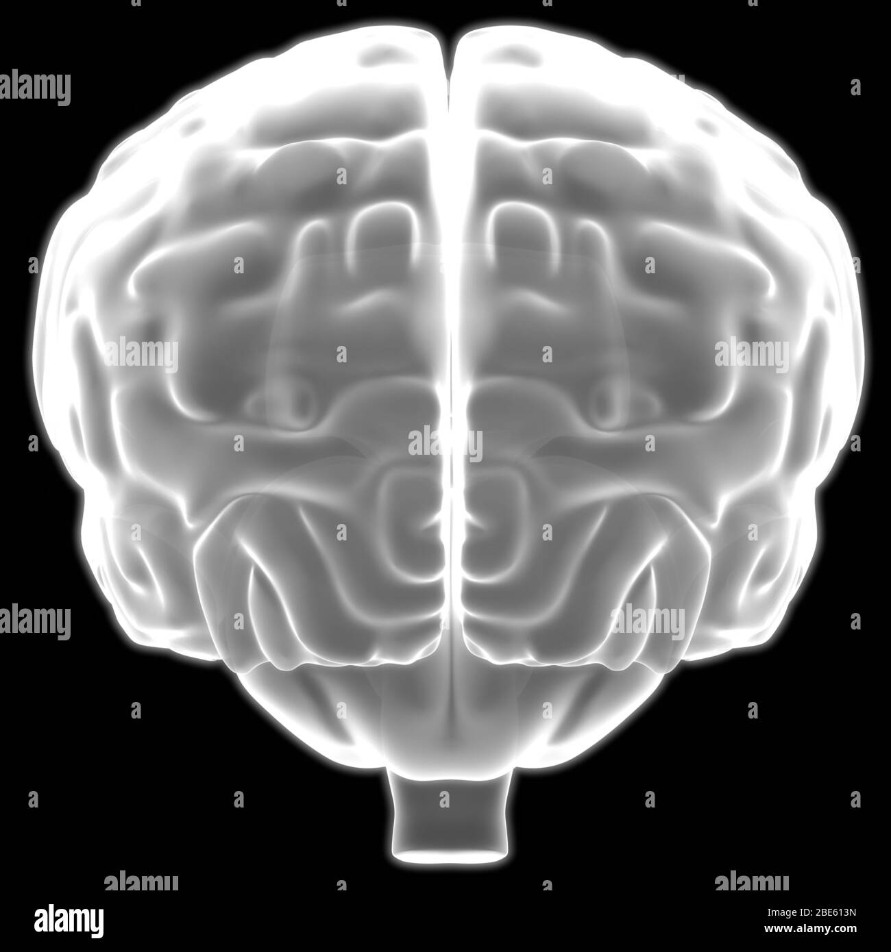 Brain is a Part of Human Body Central Nervous System Anatomy. 3D Stock Photohttps://www.alamy.com/image-license-details/?v=1https://www.alamy.com/brain-is-a-part-of-human-body-central-nervous-system-anatomy-3d-image352945145.html
RF2BE613N–Brain is a Part of Human Body Central Nervous System Anatomy. 3D
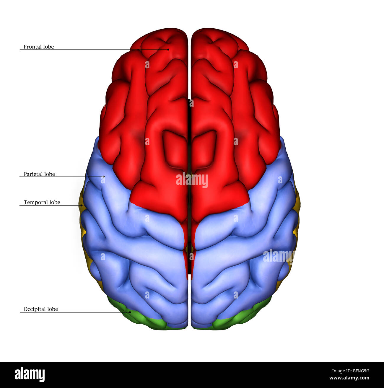 Illustration of the surface of the human brain seen from above Stock Photohttps://www.alamy.com/image-license-details/?v=1https://www.alamy.com/stock-photo-illustration-of-the-surface-of-the-human-brain-seen-from-above-26903900.html
RMBFNG5G–Illustration of the surface of the human brain seen from above
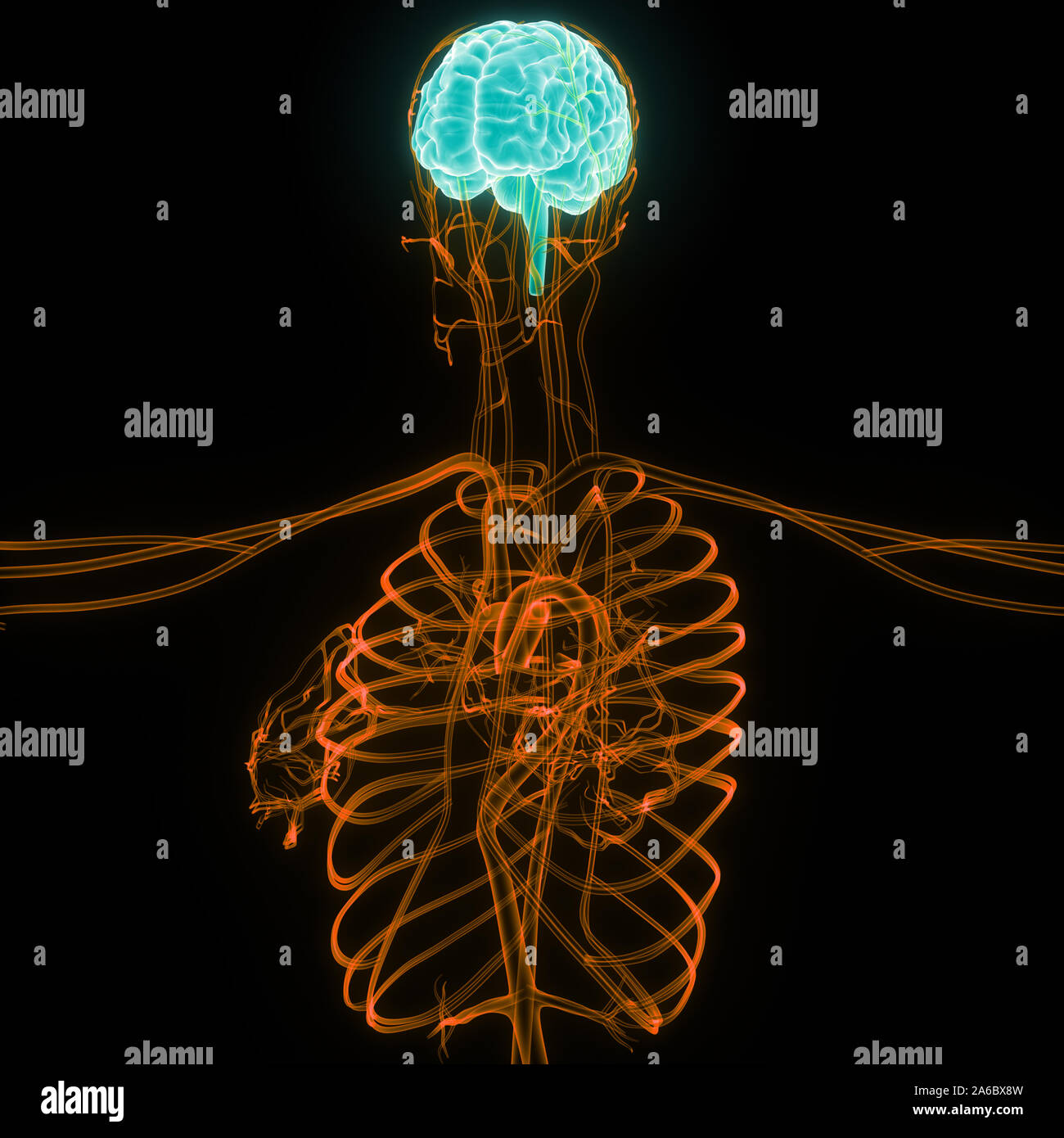 Central Organ of Human Nervous System Brain Anatomy Stock Photohttps://www.alamy.com/image-license-details/?v=1https://www.alamy.com/central-organ-of-human-nervous-system-brain-anatomy-image330947033.html
RF2A6BX8W–Central Organ of Human Nervous System Brain Anatomy
 Closeup image of human brain model. The brain is smooth and gray, with the different lobes and gyri clearly visible. Medical and healthcare concept. Stock Photohttps://www.alamy.com/image-license-details/?v=1https://www.alamy.com/closeup-image-of-human-brain-model-the-brain-is-smooth-and-gray-with-the-different-lobes-and-gyri-clearly-visible-medical-and-healthcare-concept-image569082209.html
RF2T1RX4H–Closeup image of human brain model. The brain is smooth and gray, with the different lobes and gyri clearly visible. Medical and healthcare concept.
 Cross-section of a cow brain showing the convoluted tissue with a probe pointing to the frontal lobe Stock Photohttps://www.alamy.com/image-license-details/?v=1https://www.alamy.com/cross-section-of-a-cow-brain-showing-the-convoluted-tissue-with-a-image69690568.html
RFE1AK0T–Cross-section of a cow brain showing the convoluted tissue with a probe pointing to the frontal lobe
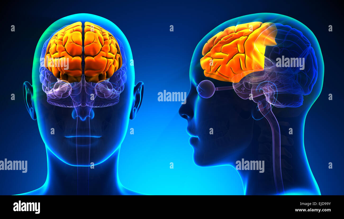 Female Frontal Lobe Brain Anatomy - blue concept Stock Photohttps://www.alamy.com/image-license-details/?v=1https://www.alamy.com/stock-photo-female-frontal-lobe-brain-anatomy-blue-concept-80197991.html
RFEJD99Y–Female Frontal Lobe Brain Anatomy - blue concept
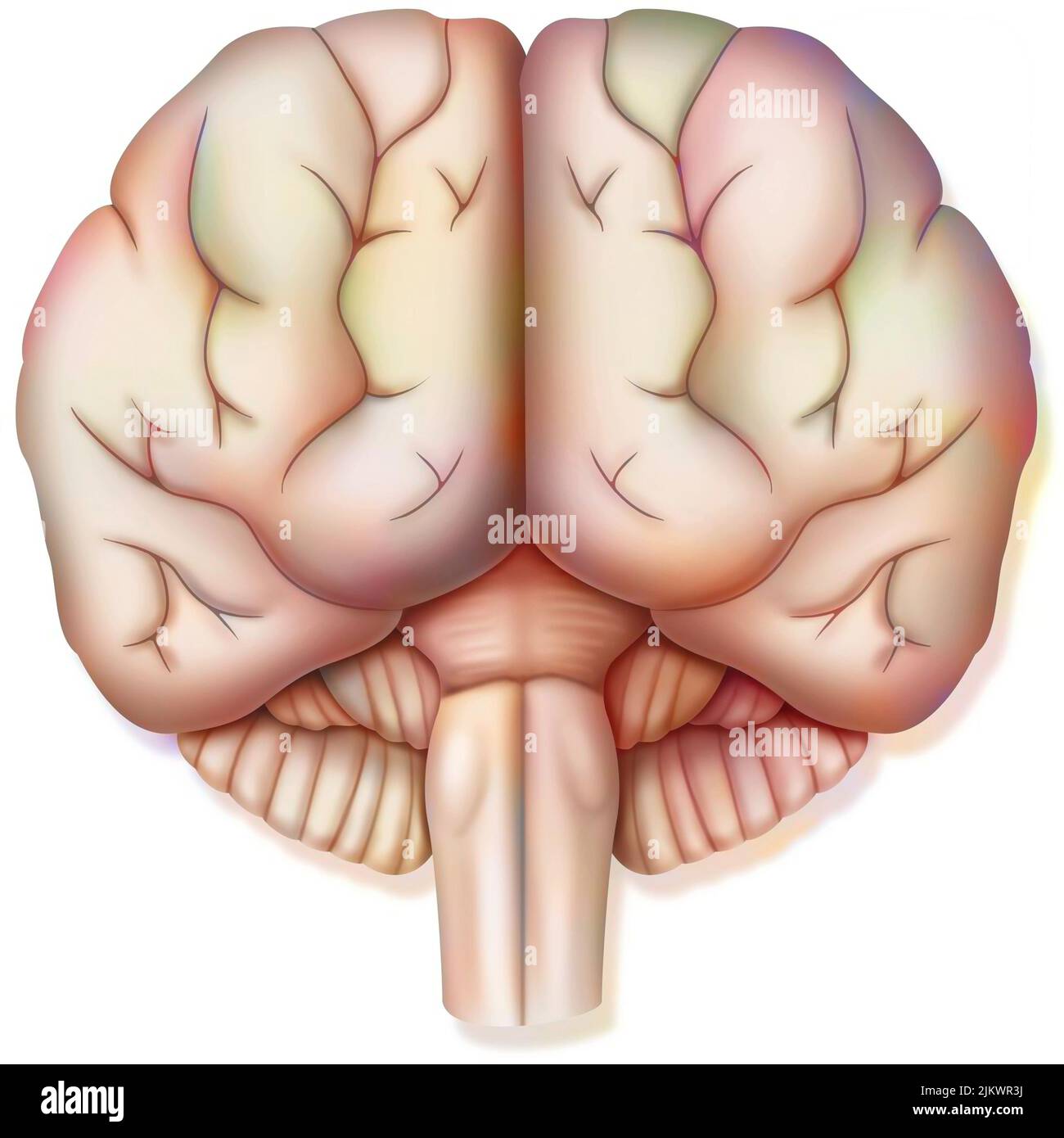 Brain, with the two cerebral hemispheres, the cerebellum and the brainstem. Stock Photohttps://www.alamy.com/image-license-details/?v=1https://www.alamy.com/brain-with-the-two-cerebral-hemispheres-the-cerebellum-and-the-brainstem-image476925334.html
RF2JKWR3J–Brain, with the two cerebral hemispheres, the cerebellum and the brainstem.
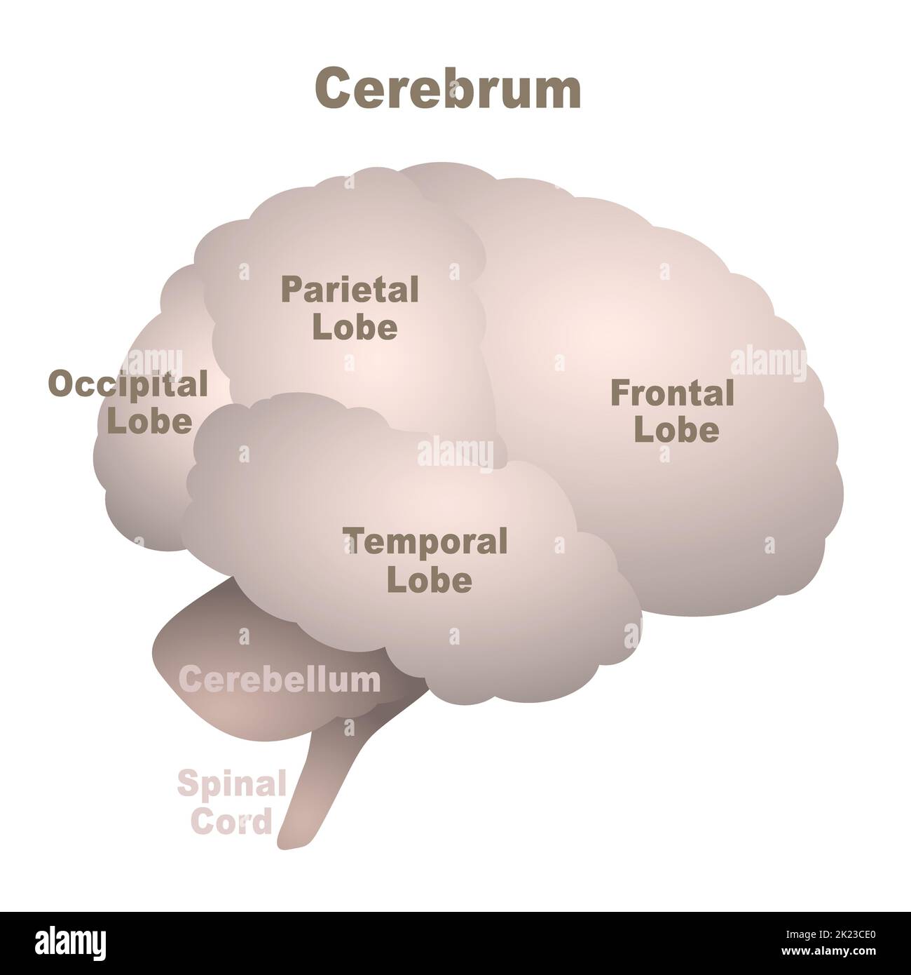 Brain lobes map, cerebrum with frontal, parietal, occipital and temporal lobe, plus cerebellum and spinal cord, anatomical regions of the human brain. Stock Photohttps://www.alamy.com/image-license-details/?v=1https://www.alamy.com/brain-lobes-map-cerebrum-with-frontal-parietal-occipital-and-temporal-lobe-plus-cerebellum-and-spinal-cord-anatomical-regions-of-the-human-brain-image483195272.html
RF2K23CE0–Brain lobes map, cerebrum with frontal, parietal, occipital and temporal lobe, plus cerebellum and spinal cord, anatomical regions of the human brain.
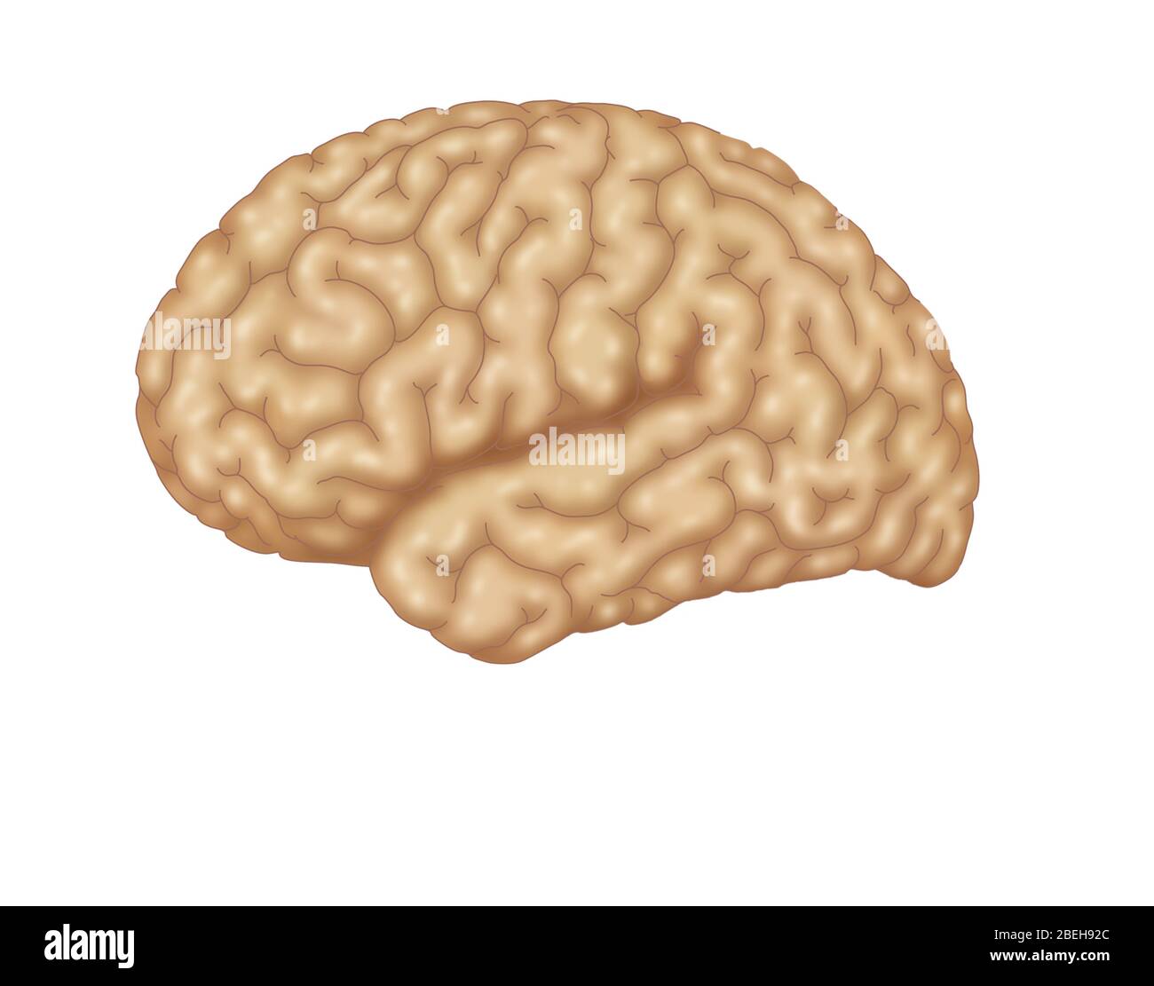 Illustration of a normal human brain. Stock Photohttps://www.alamy.com/image-license-details/?v=1https://www.alamy.com/illustration-of-a-normal-human-brain-image353192852.html
RM2BEH92C–Illustration of a normal human brain.
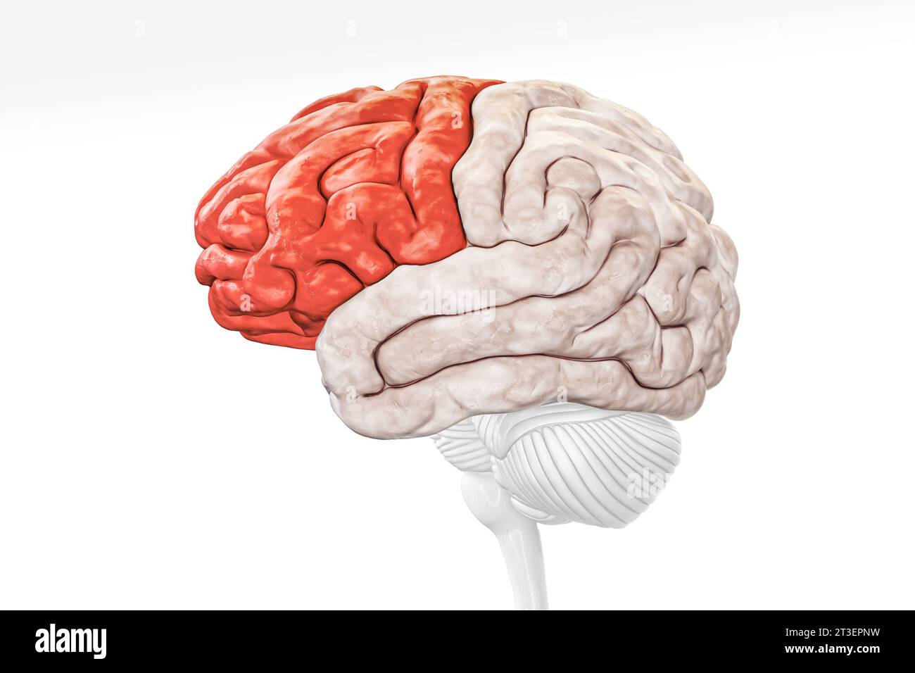 Cerebral cortex frontal lobe in red color profile view isolated on white background 3D rendering illustration. Human brain anatomy, neurology, neurosc Stock Photohttps://www.alamy.com/image-license-details/?v=1https://www.alamy.com/cerebral-cortex-frontal-lobe-in-red-color-profile-view-isolated-on-white-background-3d-rendering-illustration-human-brain-anatomy-neurology-neurosc-image570111301.html
RF2T3EPNW–Cerebral cortex frontal lobe in red color profile view isolated on white background 3D rendering illustration. Human brain anatomy, neurology, neurosc
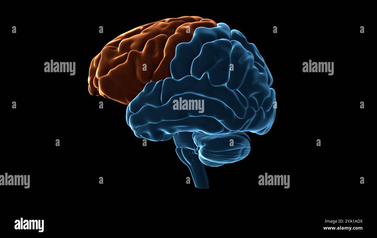 Illustration of the frontal lobe (highlighted in orange) of the human brain. Parts of this area are responsible for higher cognitive functions such as decision making, personality expression and moderating social behaviour. Stock Photohttps://www.alamy.com/image-license-details/?v=1https://www.alamy.com/illustration-of-the-frontal-lobe-highlighted-in-orange-of-the-human-brain-parts-of-this-area-are-responsible-for-higher-cognitive-functions-such-as-decision-making-personality-expression-and-moderating-social-behaviour-image625749990.html
RF2YA1ADX–Illustration of the frontal lobe (highlighted in orange) of the human brain. Parts of this area are responsible for higher cognitive functions such as decision making, personality expression and moderating social behaviour.
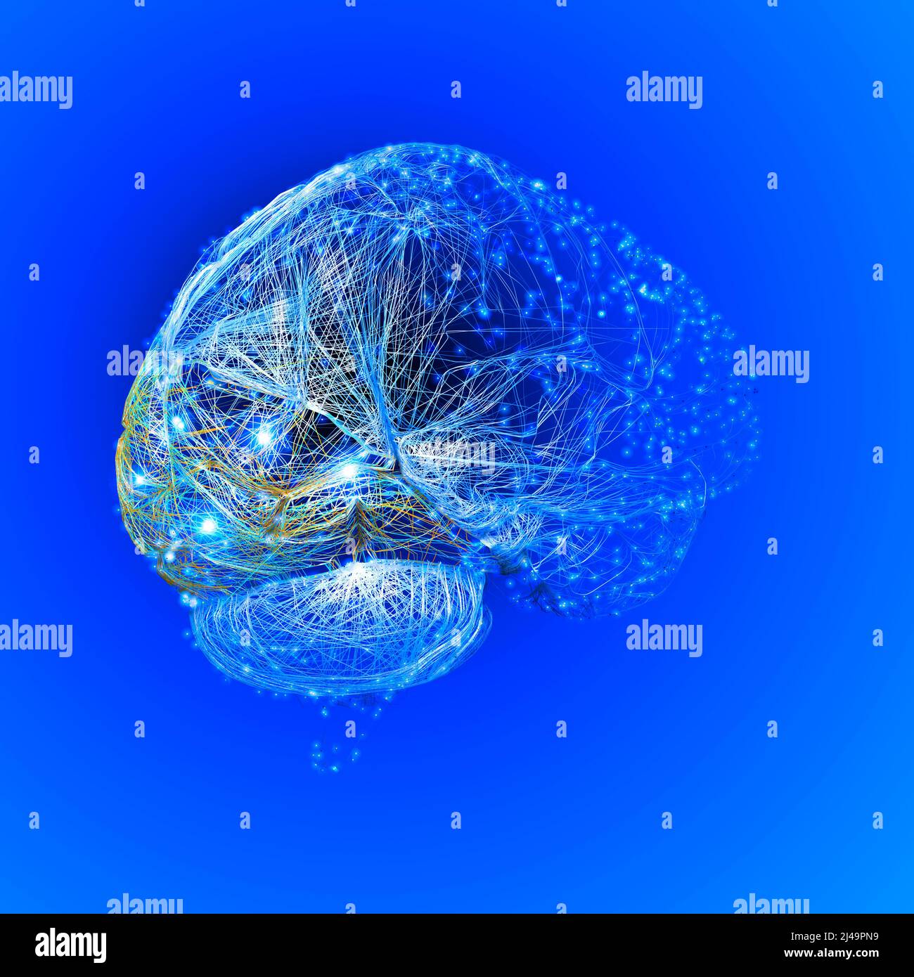 Brain, the occipital lobe is one of the four major lobes of the cerebral cortex, it is the visual processing center. 3d rendering Stock Photohttps://www.alamy.com/image-license-details/?v=1https://www.alamy.com/brain-the-occipital-lobe-is-one-of-the-four-major-lobes-of-the-cerebral-cortex-it-is-the-visual-processing-center-3d-rendering-image467353973.html
RF2J49PN9–Brain, the occipital lobe is one of the four major lobes of the cerebral cortex, it is the visual processing center. 3d rendering
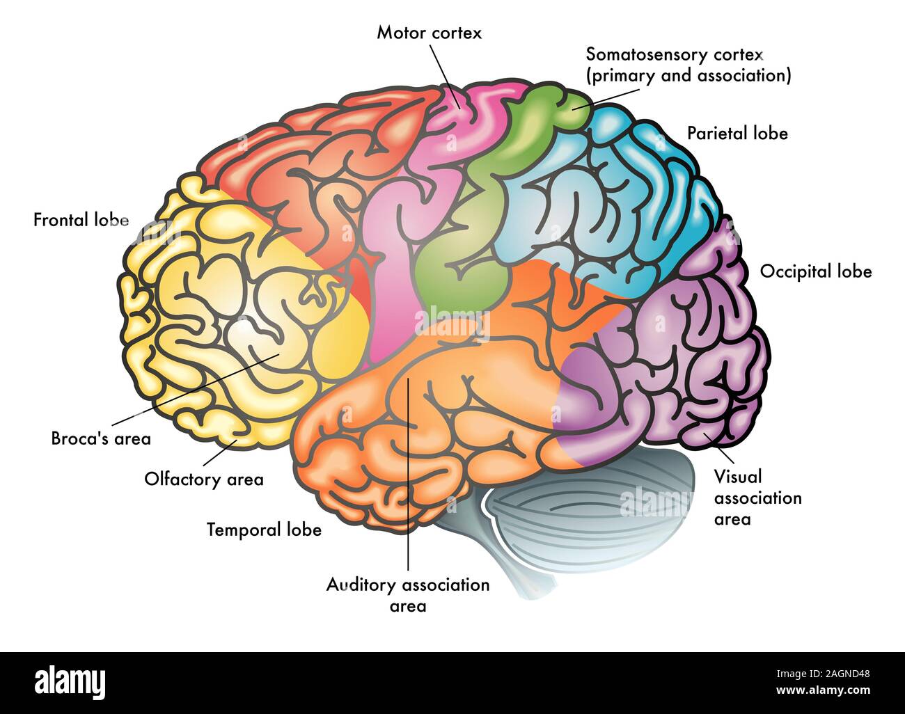 medical colorful illustration of a human brain with different functional areas highlighted with different colors Stock Photohttps://www.alamy.com/image-license-details/?v=1https://www.alamy.com/medical-colorful-illustration-of-a-human-brain-with-different-functional-areas-highlighted-with-different-colors-image337302792.html
RF2AGND48–medical colorful illustration of a human brain with different functional areas highlighted with different colors
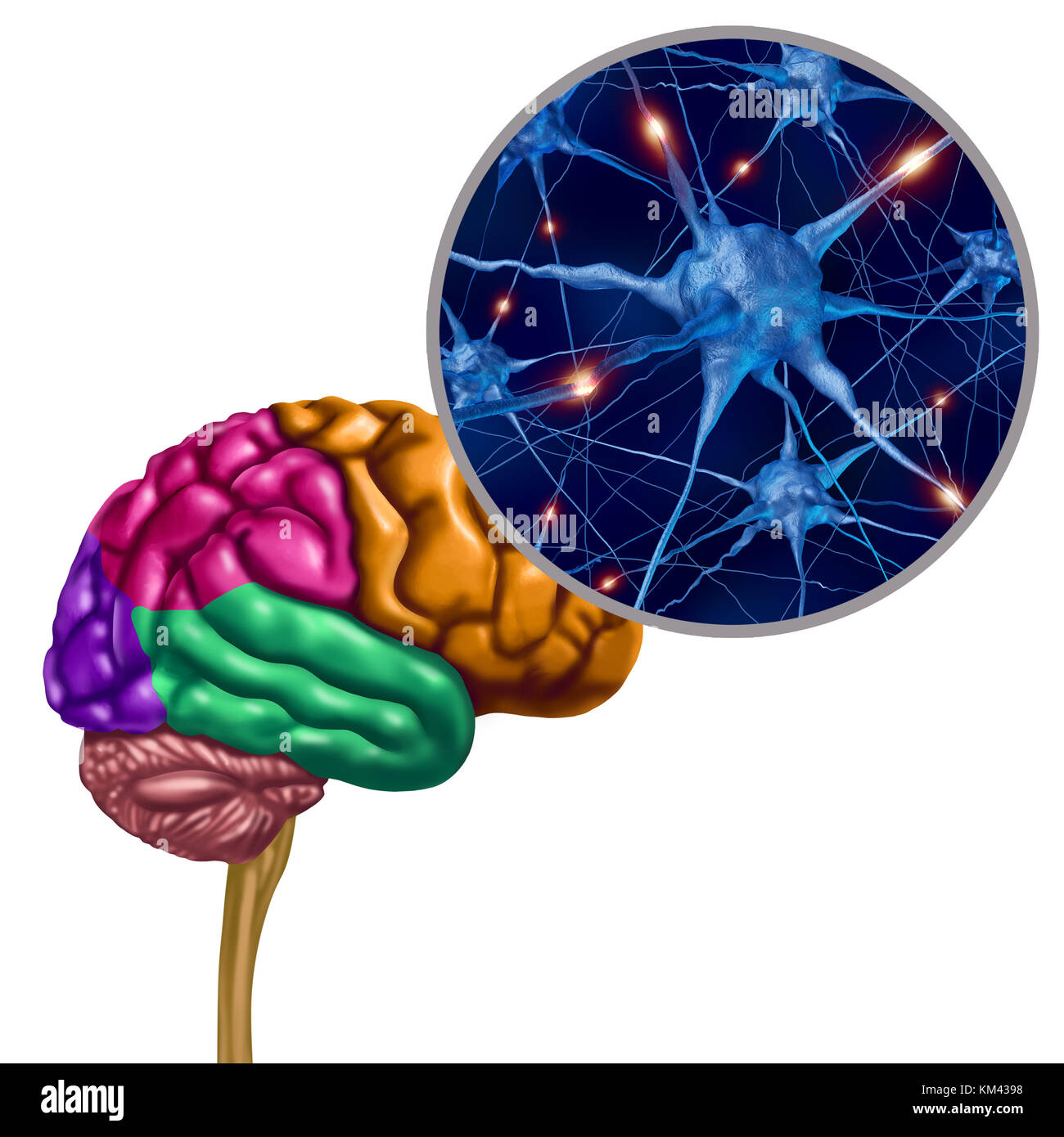 Brain lobe active neurons as a human thinking ogan with neuron magnification with 3D illustration elements. Stock Photohttps://www.alamy.com/image-license-details/?v=1https://www.alamy.com/stock-image-brain-lobe-active-neurons-as-a-human-thinking-ogan-with-neuron-magnification-167276852.html
RFKM4398–Brain lobe active neurons as a human thinking ogan with neuron magnification with 3D illustration elements.
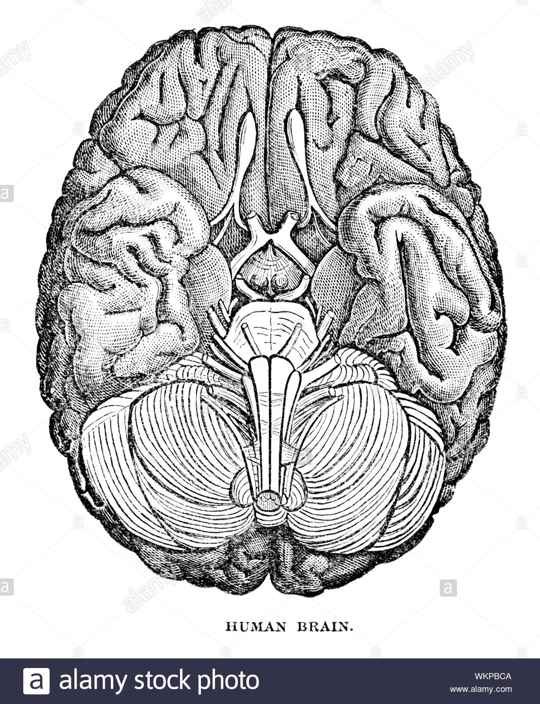 Human brain, vintage illustration from 1884 Stock Photohttps://www.alamy.com/image-license-details/?v=1https://www.alamy.com/human-brain-vintage-illustration-from-1884-image270325898.html
RMWKPBCA–Human brain, vintage illustration from 1884
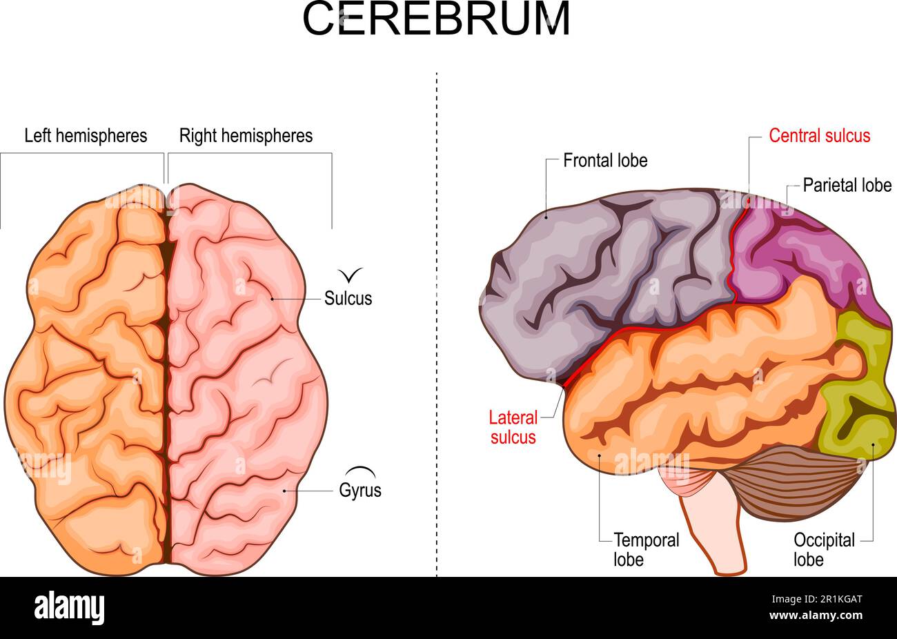 Human brain structure. Hemispheres and lobes of the cerebral cortex. frontal, temporal, occipital, and parietal lobes. lateral and superior view Stock Vectorhttps://www.alamy.com/image-license-details/?v=1https://www.alamy.com/human-brain-structure-hemispheres-and-lobes-of-the-cerebral-cortex-frontal-temporal-occipital-and-parietal-lobes-lateral-and-superior-view-image551776368.html
RF2R1KGAT–Human brain structure. Hemispheres and lobes of the cerebral cortex. frontal, temporal, occipital, and parietal lobes. lateral and superior view
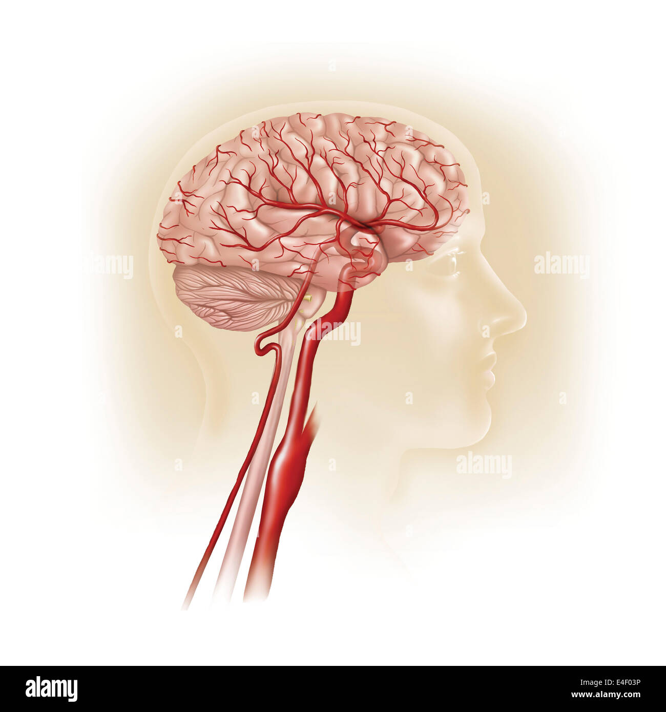 Side view of human brain showing internal carotid artery. Stock Photohttps://www.alamy.com/image-license-details/?v=1https://www.alamy.com/stock-photo-side-view-of-human-brain-showing-internal-carotid-artery-71629482.html
RME4F03P–Side view of human brain showing internal carotid artery.
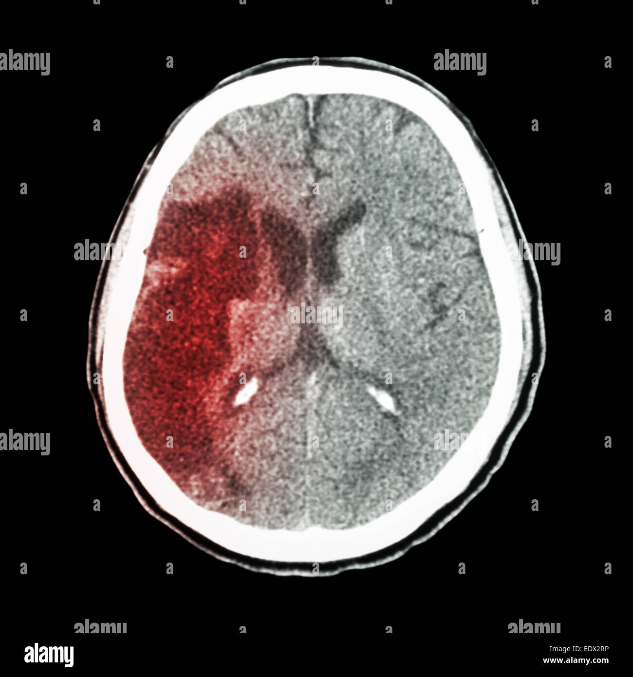 CT brain : show Ischemic stroke (hypodensity at right frontal-parietal lobe) Stock Photohttps://www.alamy.com/image-license-details/?v=1https://www.alamy.com/stock-photo-ct-brain-show-ischemic-stroke-hypodensity-at-right-frontal-parietal-77404986.html
RFEDX2RP–CT brain : show Ischemic stroke (hypodensity at right frontal-parietal lobe)
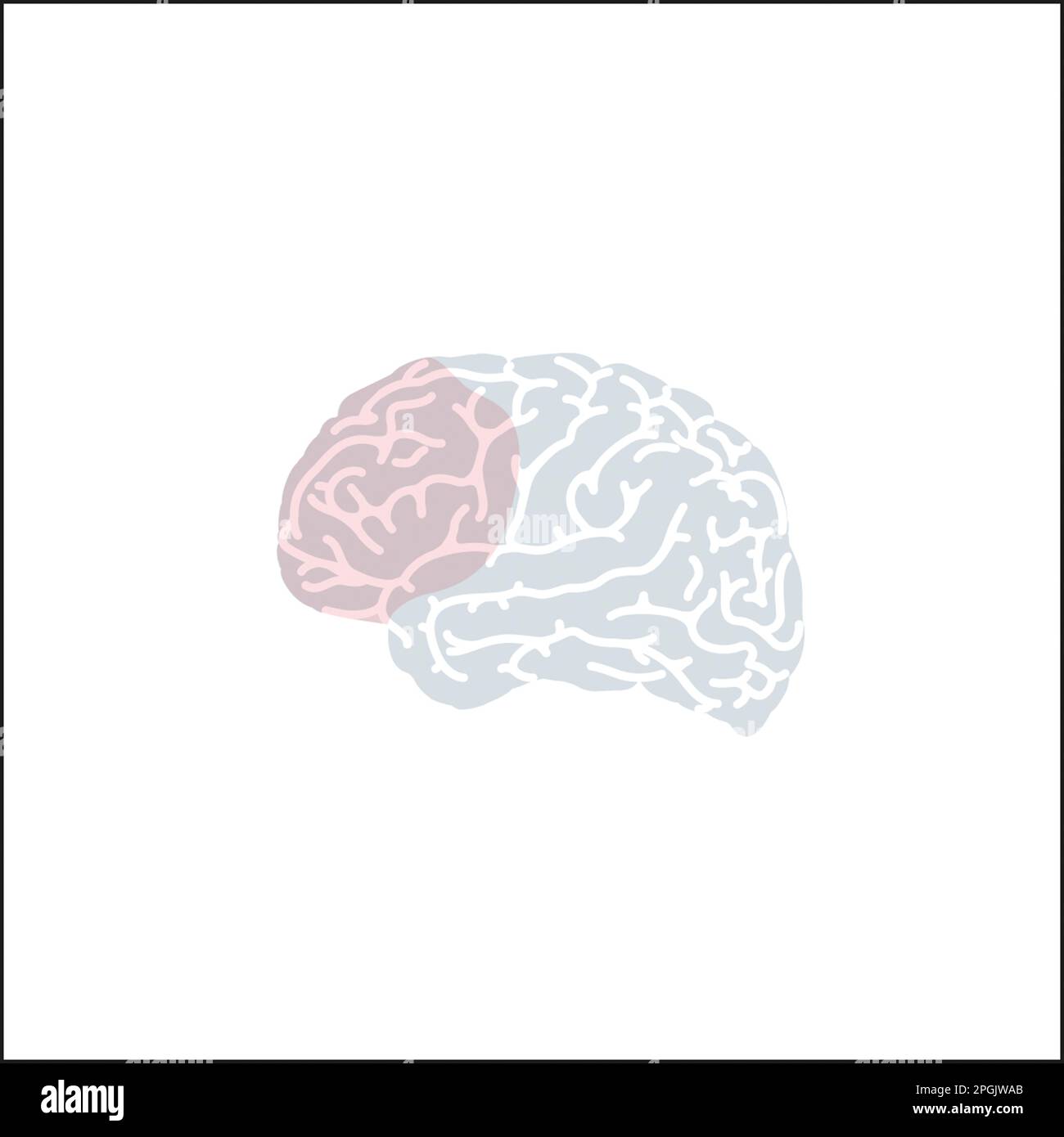 Prefrontal Cortex Stock Photohttps://www.alamy.com/image-license-details/?v=1https://www.alamy.com/prefrontal-cortex-image543770931.html
RM2PGJWAB–Prefrontal Cortex
 Brain is a Part of Human Body Central Nervous System Anatomy. 3D Stock Photohttps://www.alamy.com/image-license-details/?v=1https://www.alamy.com/brain-is-a-part-of-human-body-central-nervous-system-anatomy-3d-image352945138.html
RF2BE613E–Brain is a Part of Human Body Central Nervous System Anatomy. 3D
 Colored areas of the Brain,front view Stock Photohttps://www.alamy.com/image-license-details/?v=1https://www.alamy.com/stock-photo-colored-areas-of-the-brainfront-view-43697506.html
RFCF2GH6–Colored areas of the Brain,front view
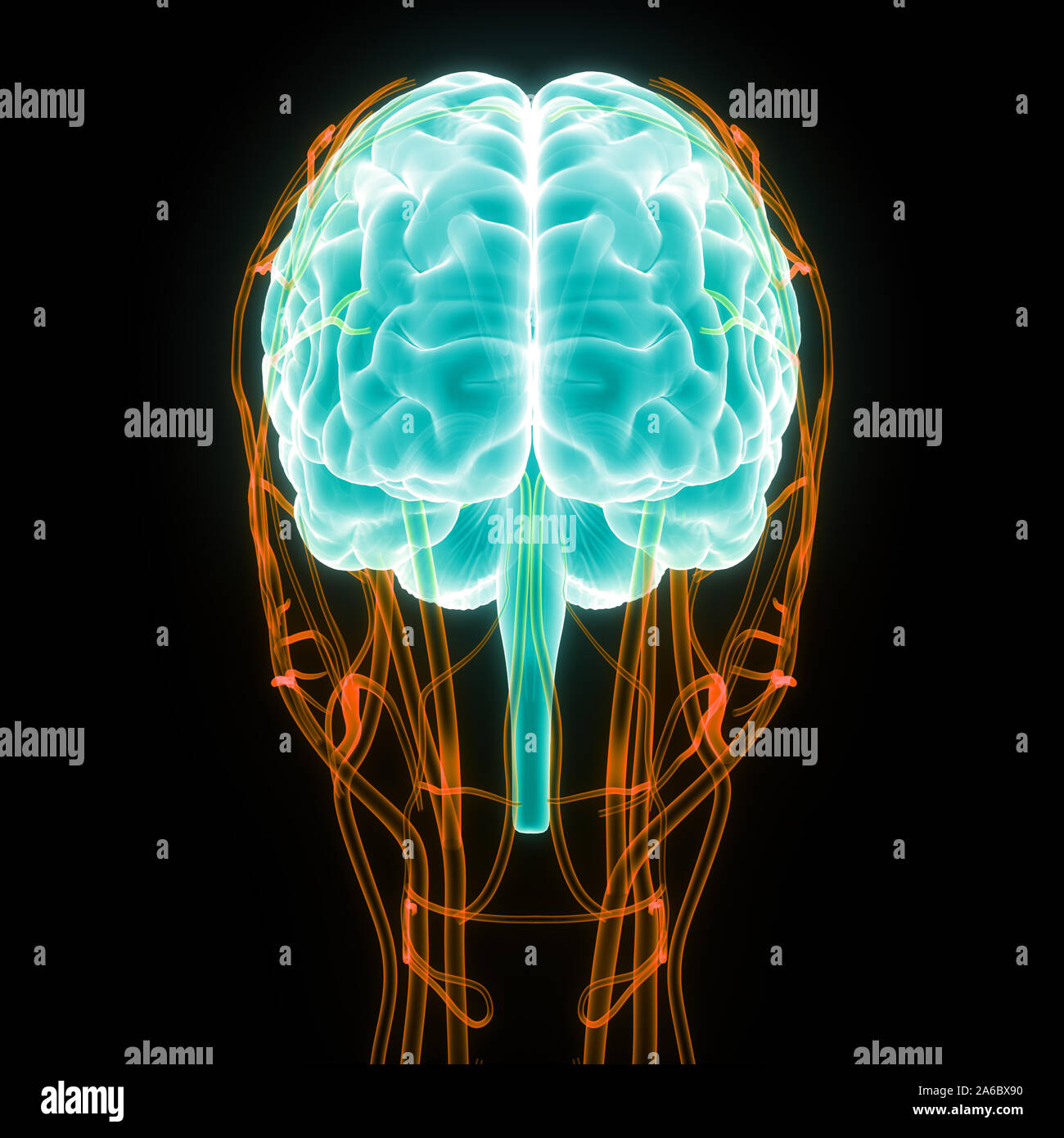 Central Organ of Human Nervous System Brain Anatomy Stock Photohttps://www.alamy.com/image-license-details/?v=1https://www.alamy.com/central-organ-of-human-nervous-system-brain-anatomy-image330947036.html
RF2A6BX90–Central Organ of Human Nervous System Brain Anatomy
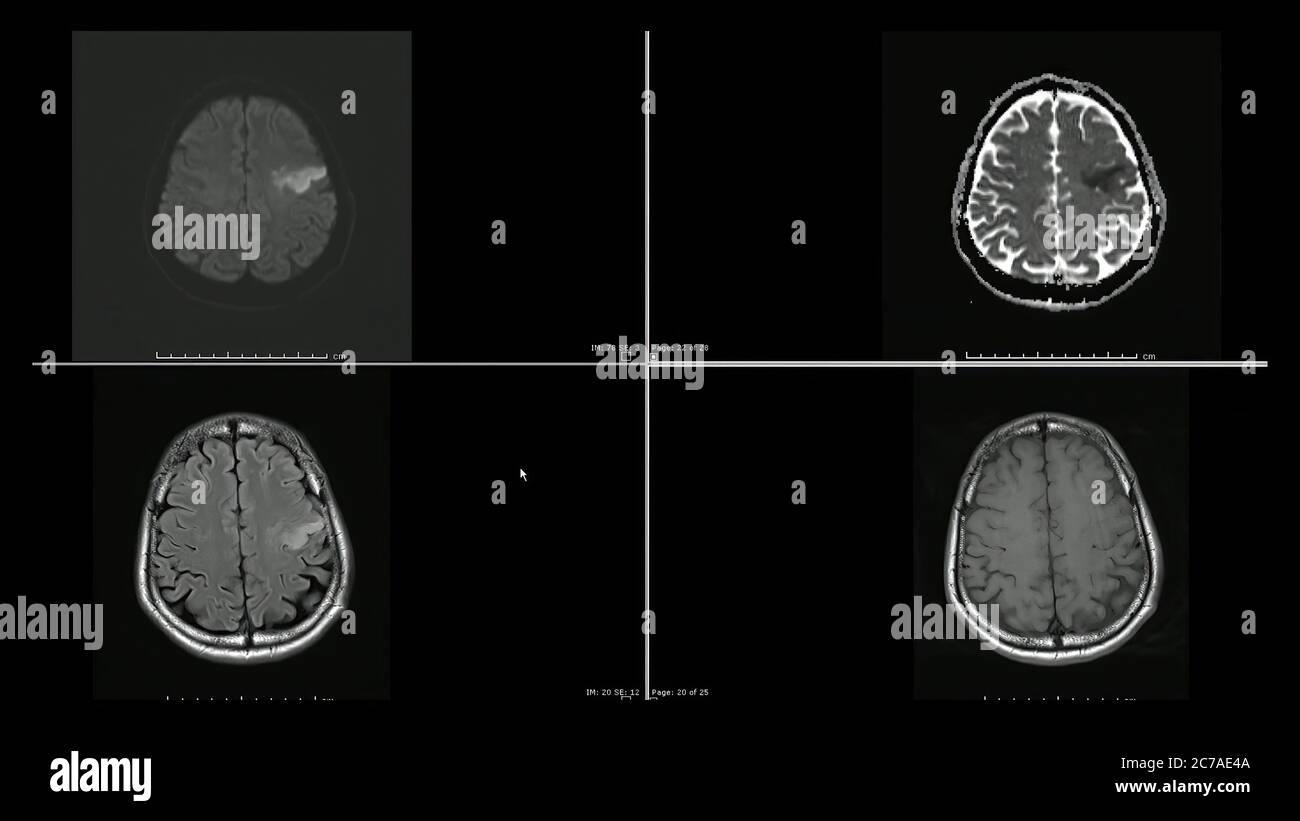 Magnetic resonance images (MRI) of brain infarction in the left frontal lobe superior cuts Stock Photohttps://www.alamy.com/image-license-details/?v=1https://www.alamy.com/magnetic-resonance-images-mri-of-brain-infarction-in-the-left-frontal-lobe-superior-cuts-image365950938.html
RF2C7AE4A–Magnetic resonance images (MRI) of brain infarction in the left frontal lobe superior cuts
 Connections between putamen and frontal lobe in a Parkinson's patient brain Stock Photohttps://www.alamy.com/image-license-details/?v=1https://www.alamy.com/stock-photo-connections-between-putamen-and-frontal-lobe-in-a-parkinsons-patient-87046647.html
RFF1H8TR–Connections between putamen and frontal lobe in a Parkinson's patient brain
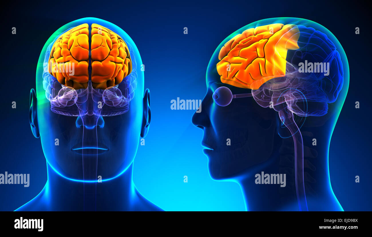 Male Frontal Lobe Brain Anatomy - blue concept Stock Photohttps://www.alamy.com/image-license-details/?v=1https://www.alamy.com/stock-photo-male-frontal-lobe-brain-anatomy-blue-concept-80198046.html
RFEJD9BX–Male Frontal Lobe Brain Anatomy - blue concept
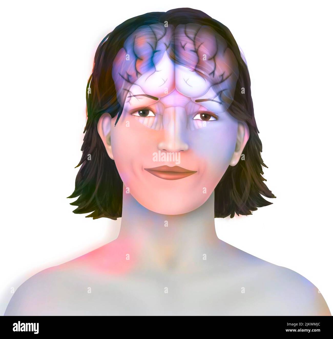 Brain: cerebral hemispheres with longitudinal fissure, cerebellum and brainstem). Stock Photohttps://www.alamy.com/image-license-details/?v=1https://www.alamy.com/brain-cerebral-hemispheres-with-longitudinal-fissure-cerebellum-and-brainstem-image476923396.html
RF2JKWMJC–Brain: cerebral hemispheres with longitudinal fissure, cerebellum and brainstem).
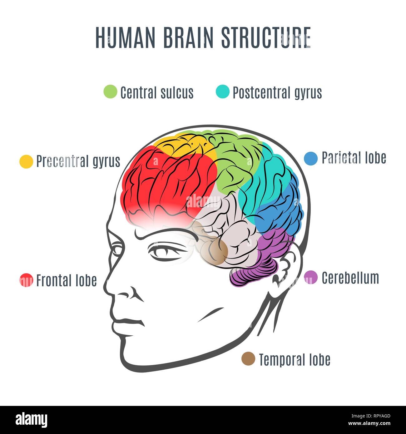 Structure of the human brain. Human head with brain inside. Human brain main parts. Vector illustration. Stock Vectorhttps://www.alamy.com/image-license-details/?v=1https://www.alamy.com/structure-of-the-human-brain-human-head-with-brain-inside-human-brain-main-parts-vector-illustration-image237858221.html
RFRPYAGD–Structure of the human brain. Human head with brain inside. Human brain main parts. Vector illustration.
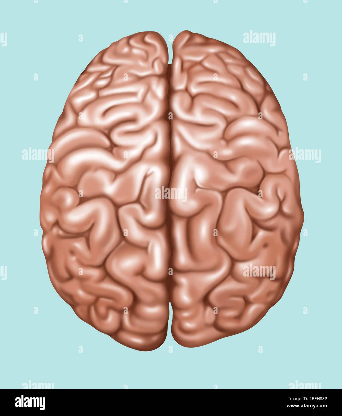 Top View of Normal Brain, Illustration Stock Photohttps://www.alamy.com/image-license-details/?v=1https://www.alamy.com/top-view-of-normal-brain-illustration-image353192246.html
RF2BEH88P–Top View of Normal Brain, Illustration
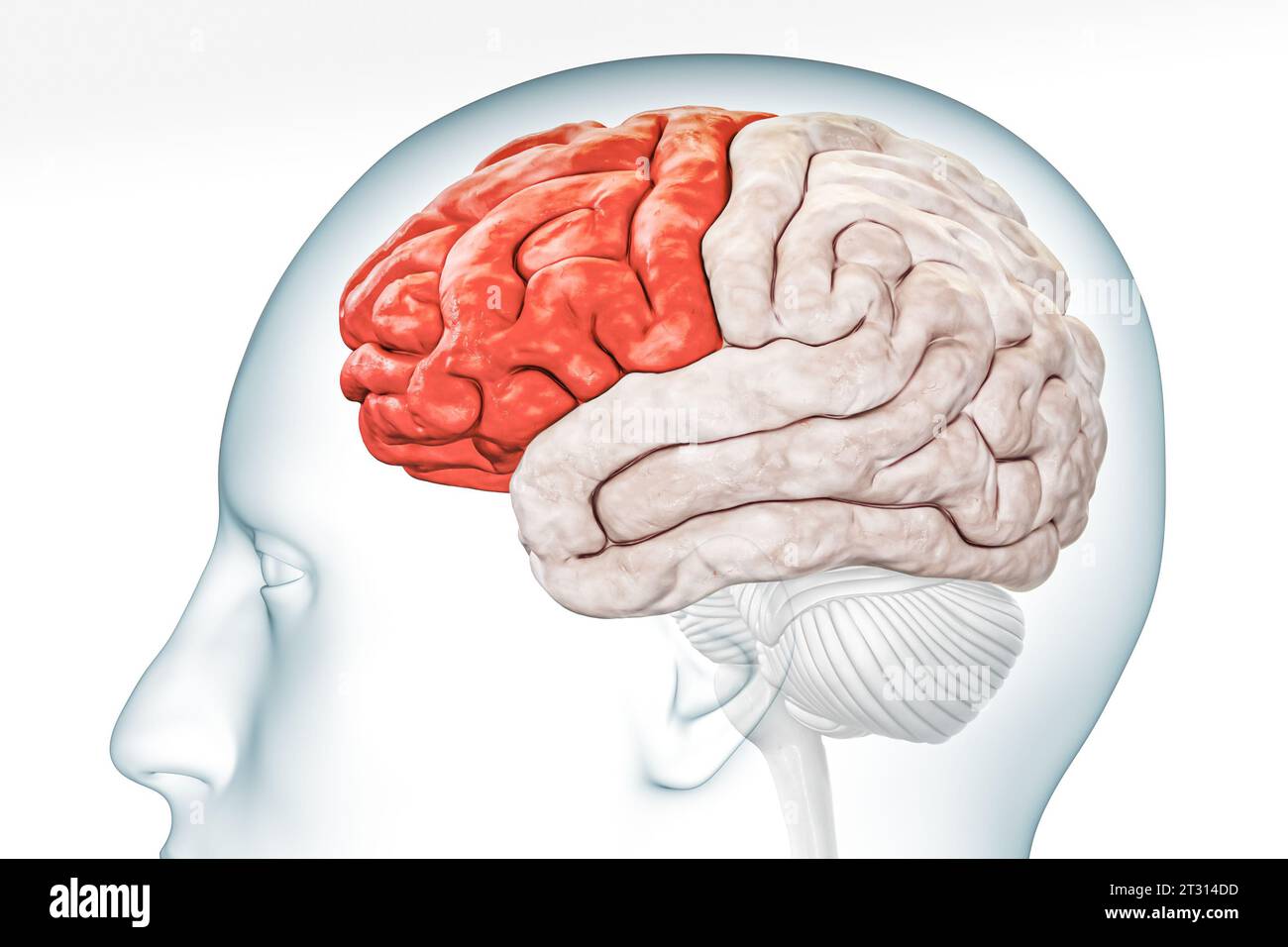 Cerebral cortex frontal lobe in red color profile view with body isolated on white background 3D rendering illustration. Human brain anatomy, neurolog Stock Photohttps://www.alamy.com/image-license-details/?v=1https://www.alamy.com/cerebral-cortex-frontal-lobe-in-red-color-profile-view-with-body-isolated-on-white-background-3d-rendering-illustration-human-brain-anatomy-neurolog-image569811577.html
RF2T314DD–Cerebral cortex frontal lobe in red color profile view with body isolated on white background 3D rendering illustration. Human brain anatomy, neurolog
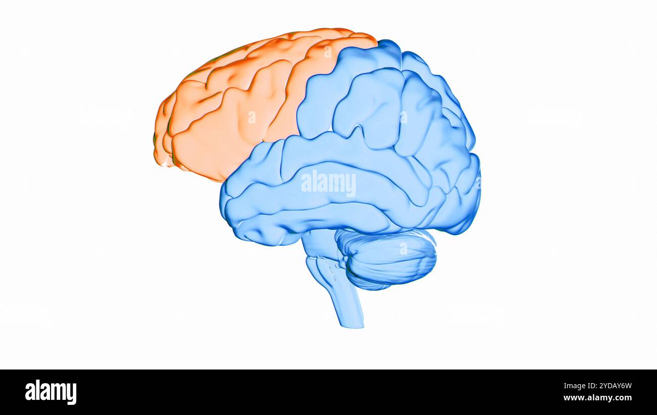 Illustration of the frontal lobe (orange) of the human brain. Parts of this area are responsible for higher cognitive functions such as decision making, personality expression and moderating social behaviour. Stock Photohttps://www.alamy.com/image-license-details/?v=1https://www.alamy.com/illustration-of-the-frontal-lobe-orange-of-the-human-brain-parts-of-this-area-are-responsible-for-higher-cognitive-functions-such-as-decision-making-personality-expression-and-moderating-social-behaviour-image627804657.html
RF2YDAY6W–Illustration of the frontal lobe (orange) of the human brain. Parts of this area are responsible for higher cognitive functions such as decision making, personality expression and moderating social behaviour.
 Brain, the temporal lobe is one of the four major lobes of the cerebral cortex, consist of structures that are vital for declarative or long-term memo Stock Photohttps://www.alamy.com/image-license-details/?v=1https://www.alamy.com/brain-the-temporal-lobe-is-one-of-the-four-major-lobes-of-the-cerebral-cortex-consist-of-structures-that-are-vital-for-declarative-or-long-term-memo-image467342077.html
RF2J497GD–Brain, the temporal lobe is one of the four major lobes of the cerebral cortex, consist of structures that are vital for declarative or long-term memo
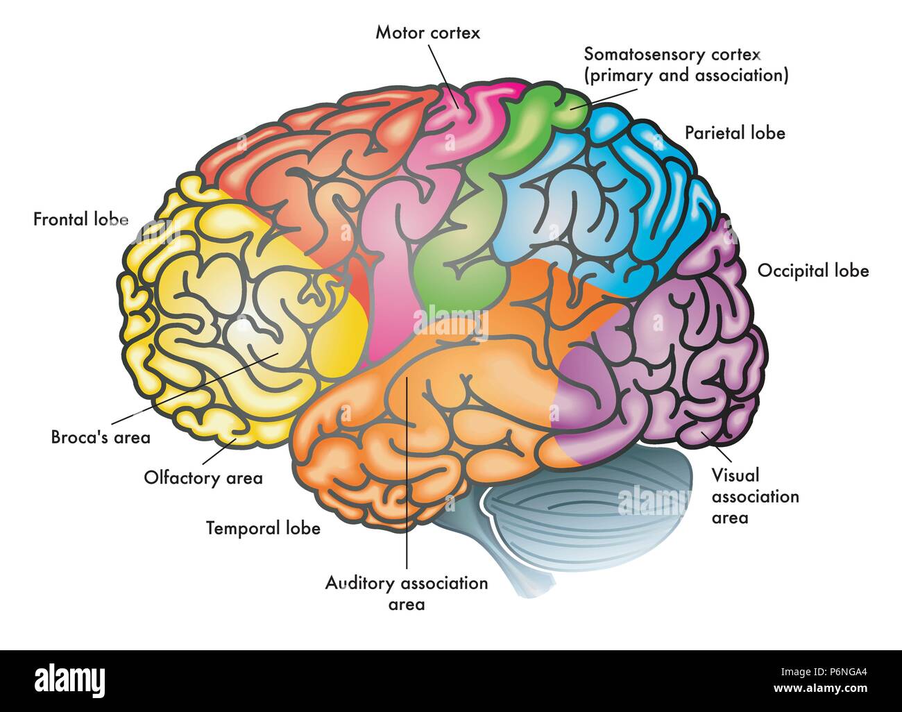 vector medical colorful illustration of a human brain with different functional areas highlighted with different colors Stock Vectorhttps://www.alamy.com/image-license-details/?v=1https://www.alamy.com/vector-medical-colorful-illustration-of-a-human-brain-with-different-functional-areas-highlighted-with-different-colors-image210686172.html
RFP6NGA4–vector medical colorful illustration of a human brain with different functional areas highlighted with different colors
 Human brain neurology and active neurons connections as a nervous system anatomy and neurological activity. Stock Photohttps://www.alamy.com/image-license-details/?v=1https://www.alamy.com/human-brain-neurology-and-active-neurons-connections-as-a-nervous-system-anatomy-and-neurological-activity-image453968185.html
RF2HAG11D–Human brain neurology and active neurons connections as a nervous system anatomy and neurological activity.
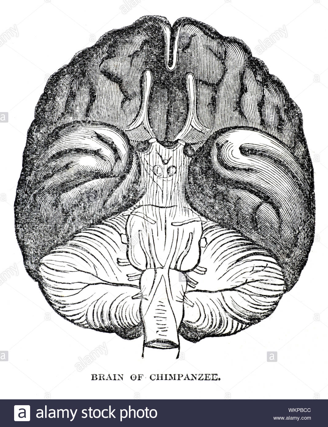 Brain of Chimpanzee, vintage illustration from 1884 Stock Photohttps://www.alamy.com/image-license-details/?v=1https://www.alamy.com/brain-of-chimpanzee-vintage-illustration-from-1884-image270325900.html
RMWKPBCC–Brain of Chimpanzee, vintage illustration from 1884
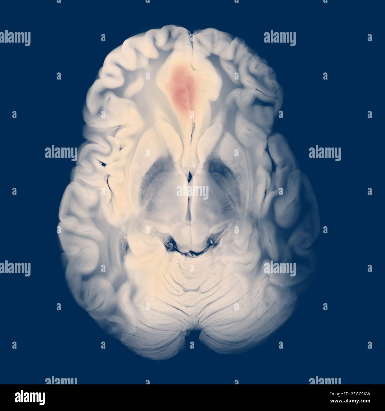 Axial Section Of The Brain With A Swollen Right Frontal Lobe Hematoma Stock Photohttps://www.alamy.com/image-license-details/?v=1https://www.alamy.com/axial-section-of-the-brain-with-a-swollen-right-frontal-lobe-hematoma-image405936941.html
RF2EGC0KW–Axial Section Of The Brain With A Swollen Right Frontal Lobe Hematoma
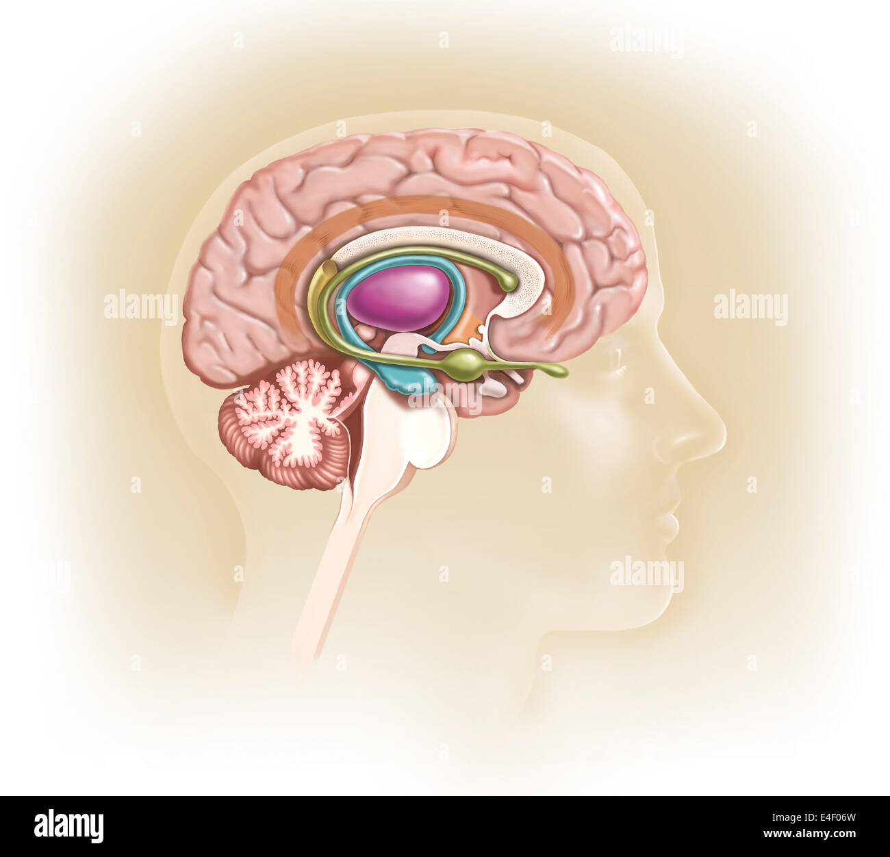 Sagittal view of human brain showing the limbic system. Stock Photohttps://www.alamy.com/image-license-details/?v=1https://www.alamy.com/stock-photo-sagittal-view-of-human-brain-showing-the-limbic-system-71629569.html
RME4F06W–Sagittal view of human brain showing the limbic system.
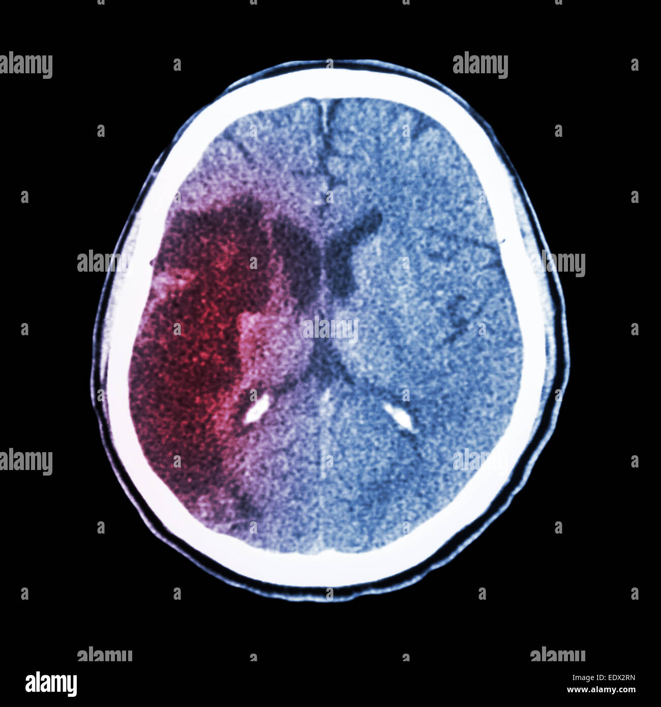 CT brain : show Ischemic stroke (hypodensity at right frontal-parietal lobe) Stock Photohttps://www.alamy.com/image-license-details/?v=1https://www.alamy.com/stock-photo-ct-brain-show-ischemic-stroke-hypodensity-at-right-frontal-parietal-77404985.html
RFEDX2RN–CT brain : show Ischemic stroke (hypodensity at right frontal-parietal lobe)
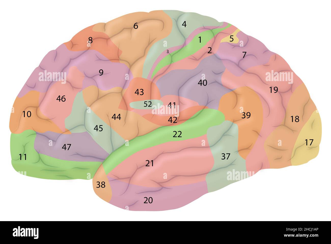 Lateral view of the brain with Brodmann areas Stock Photohttps://www.alamy.com/image-license-details/?v=1https://www.alamy.com/lateral-view-of-the-brain-with-brodmann-areas-image455241662.html
RF2HCJ1AP–Lateral view of the brain with Brodmann areas
 Brain is a Part of Human Body Central Nervous System Anatomy. 3D Stock Photohttps://www.alamy.com/image-license-details/?v=1https://www.alamy.com/brain-is-a-part-of-human-body-central-nervous-system-anatomy-3d-image352946400.html
RF2BE62MG–Brain is a Part of Human Body Central Nervous System Anatomy. 3D
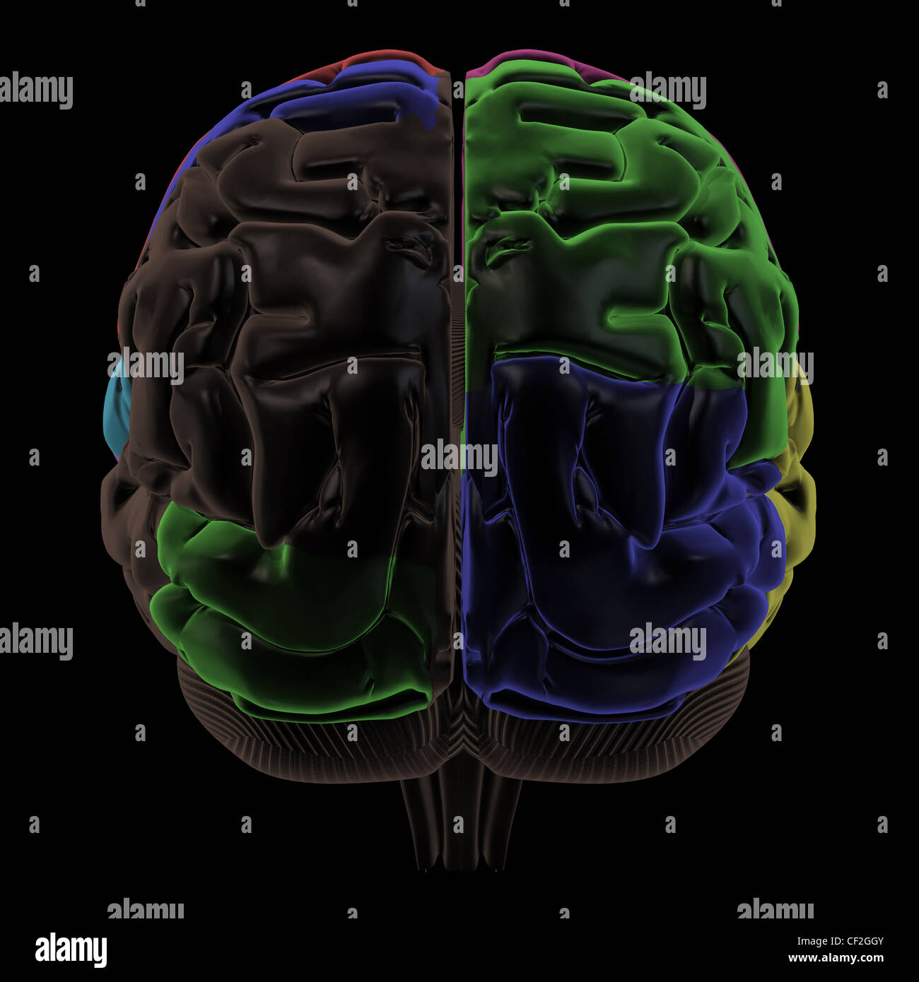 Colored areas of the Brain, back view Stock Photohttps://www.alamy.com/image-license-details/?v=1https://www.alamy.com/stock-photo-colored-areas-of-the-brain-back-view-43697499.html
RFCF2GGY–Colored areas of the Brain, back view
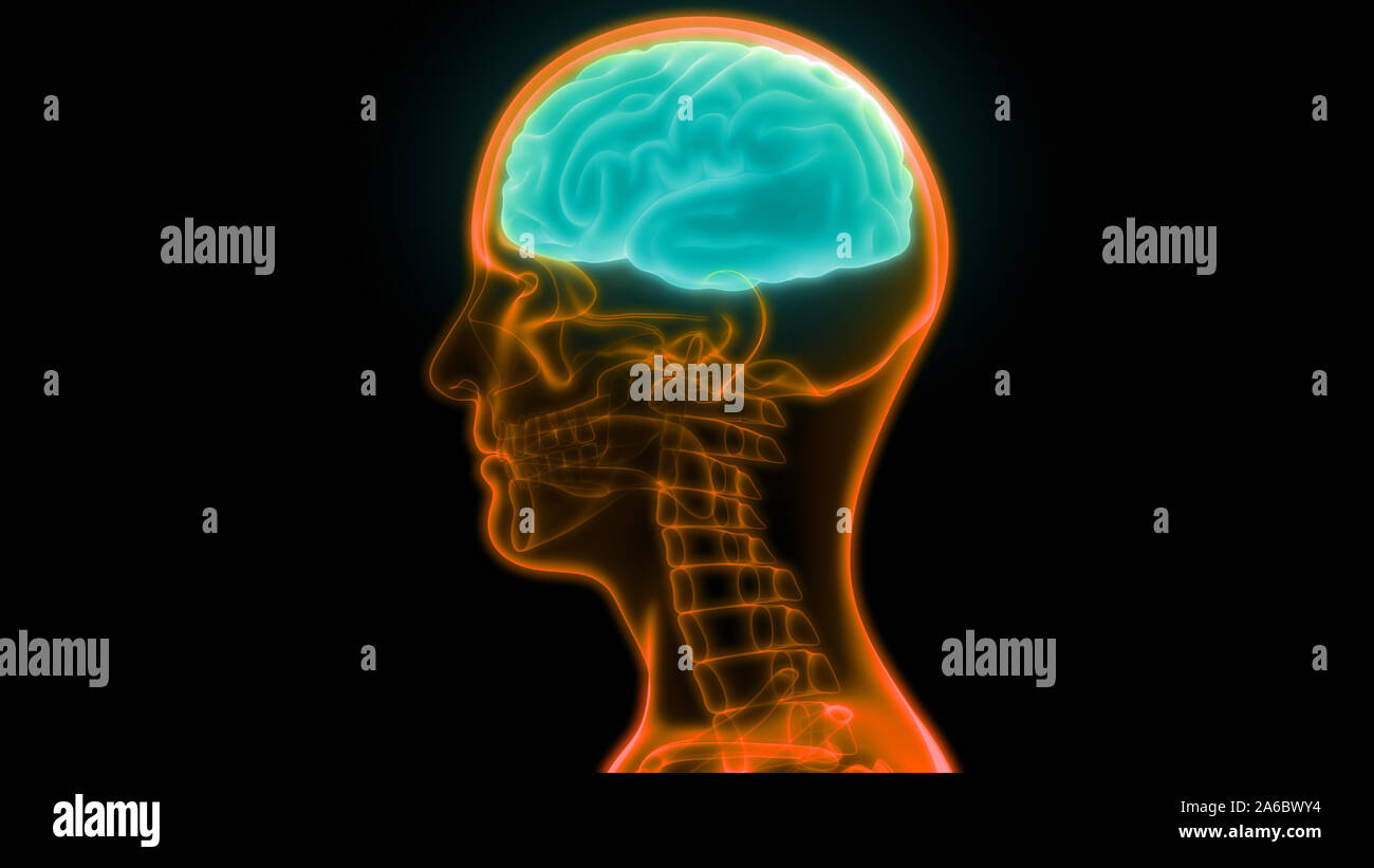 Central Organ of Human Nervous System Brain Anatomy Stock Photohttps://www.alamy.com/image-license-details/?v=1https://www.alamy.com/central-organ-of-human-nervous-system-brain-anatomy-image330946760.html
RF2A6BWY4–Central Organ of Human Nervous System Brain Anatomy
 Human brain, anatomy, anatomical logo design. Brain hemispheres, external cerebral connections in the frontal lobe. Communication, psychology. Stock Vectorhttps://www.alamy.com/image-license-details/?v=1https://www.alamy.com/human-brain-anatomy-anatomical-logo-design-brain-hemispheres-external-cerebral-connections-in-the-frontal-lobe-communication-psychology-image469284439.html
RF2J7DN2F–Human brain, anatomy, anatomical logo design. Brain hemispheres, external cerebral connections in the frontal lobe. Communication, psychology.
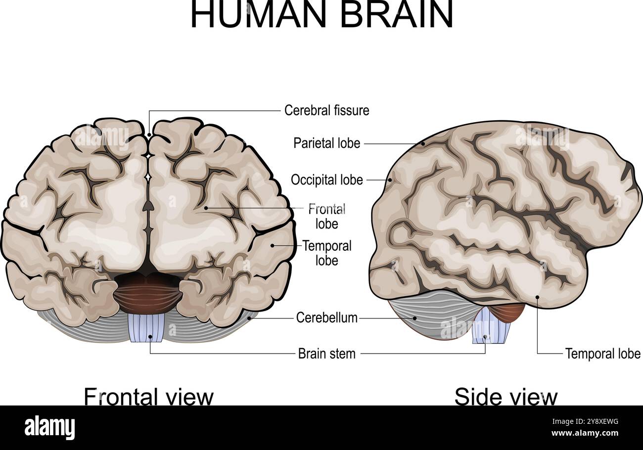 Human brain anatomy. Cerebral hemispheres, Cerebral cortex, Frontal, Parietal, Temporal, Occipital lobes, Cerebellum and Brainstem, Cerebral fissure. Stock Vectorhttps://www.alamy.com/image-license-details/?v=1https://www.alamy.com/human-brain-anatomy-cerebral-hemispheres-cerebral-cortex-frontal-parietal-temporal-occipital-lobes-cerebellum-and-brainstem-cerebral-fissure-image625072940.html
RF2Y8XEWG–Human brain anatomy. Cerebral hemispheres, Cerebral cortex, Frontal, Parietal, Temporal, Occipital lobes, Cerebellum and Brainstem, Cerebral fissure.
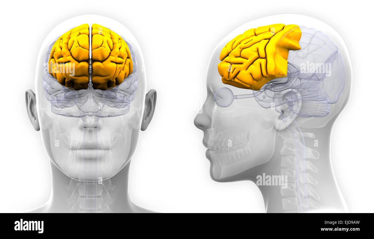 Female Frontal Lobe Brain Anatomy - isolated on white Stock Photohttps://www.alamy.com/image-license-details/?v=1https://www.alamy.com/stock-photo-female-frontal-lobe-brain-anatomy-isolated-on-white-80198017.html
RFEJD9AW–Female Frontal Lobe Brain Anatomy - isolated on white
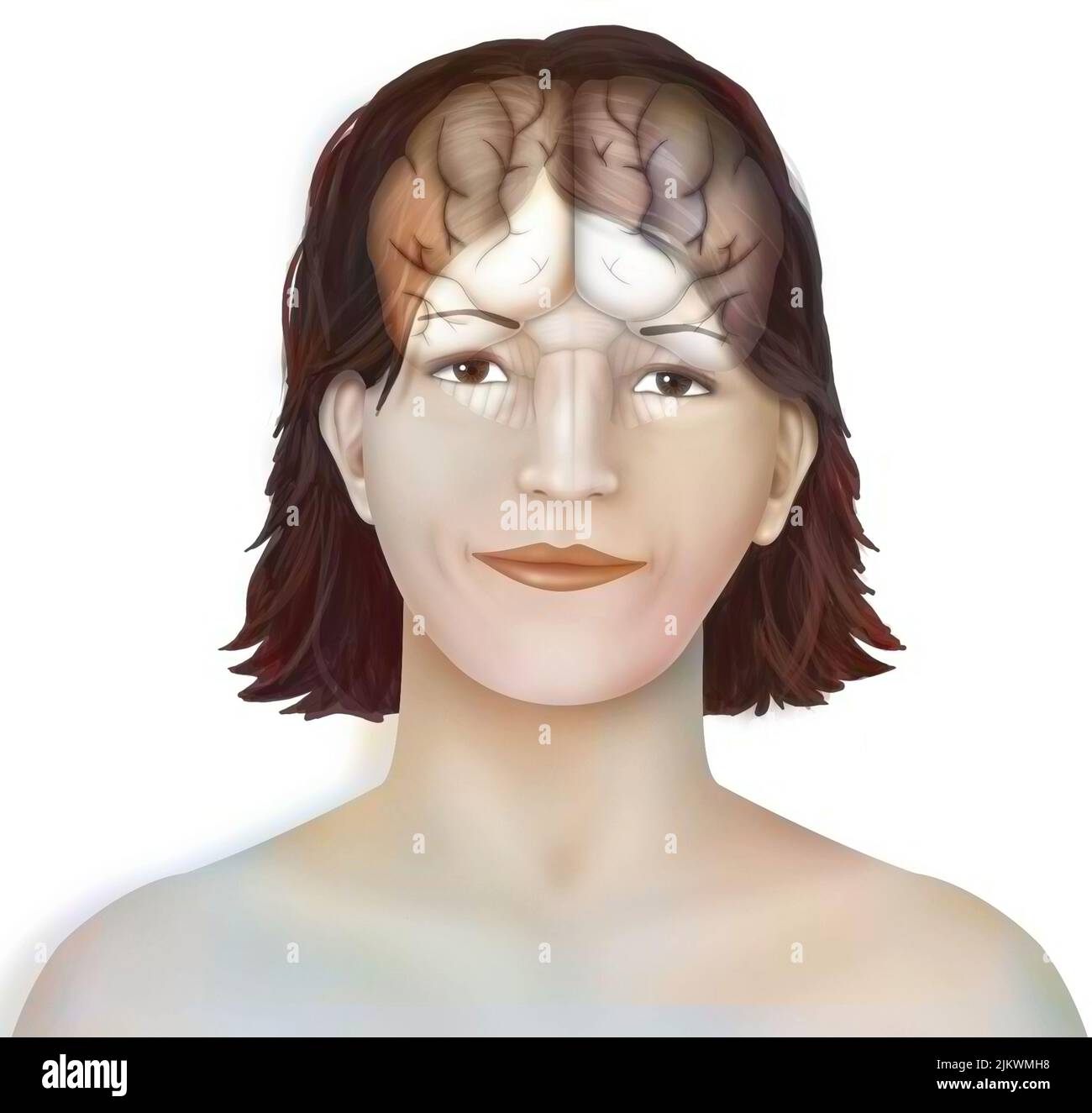 Brain: cerebral hemispheres with longitudinal fissure, cerebellum and brainstem). Stock Photohttps://www.alamy.com/image-license-details/?v=1https://www.alamy.com/brain-cerebral-hemispheres-with-longitudinal-fissure-cerebellum-and-brainstem-image476923364.html
RF2JKWMH8–Brain: cerebral hemispheres with longitudinal fissure, cerebellum and brainstem).
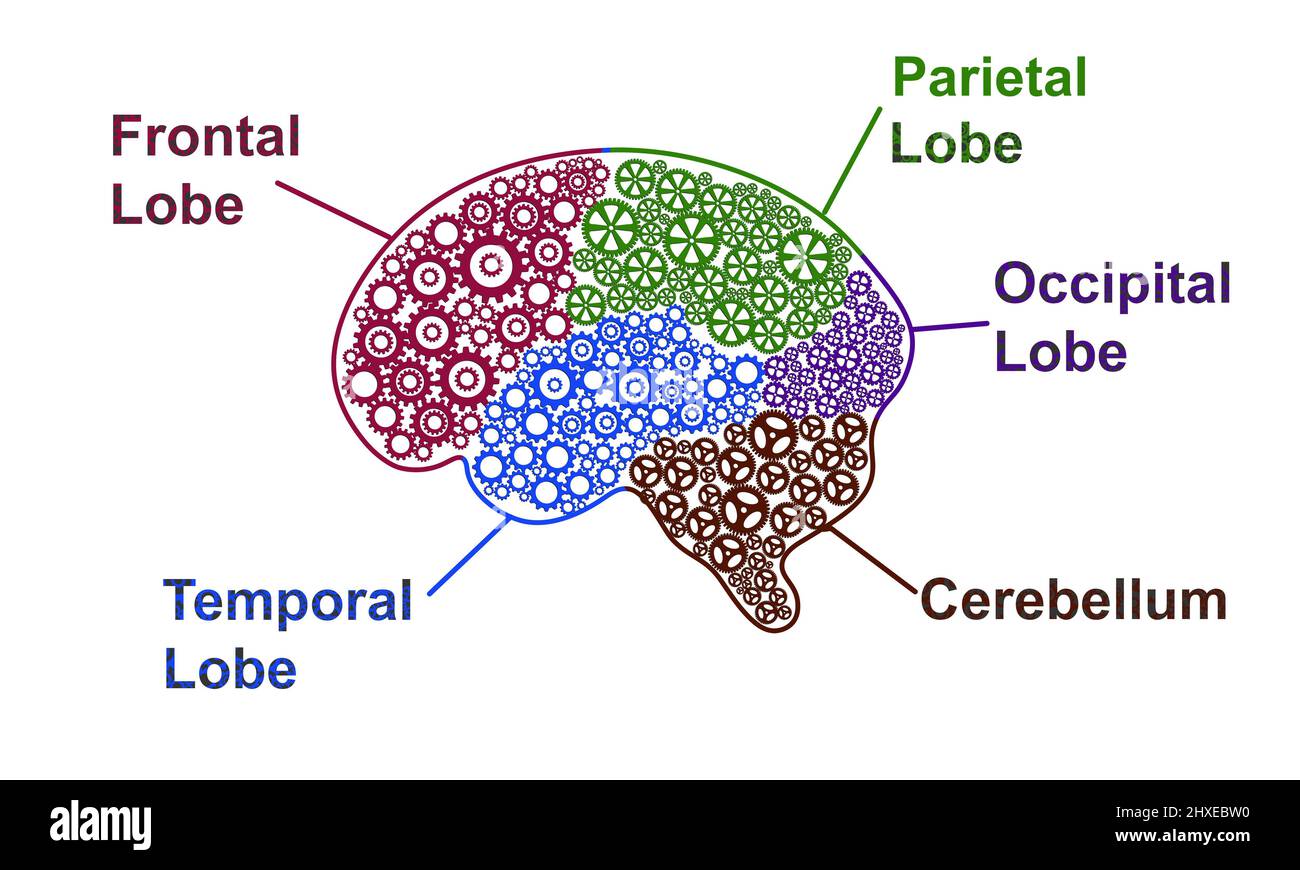 Five Parts of human brain anatomy. Brain Lobes Gear Abstract with text infographic on white background. Science and creativity Concept Stock Photohttps://www.alamy.com/image-license-details/?v=1https://www.alamy.com/five-parts-of-human-brain-anatomy-brain-lobes-gear-abstract-with-text-infographic-on-white-background-science-and-creativity-concept-image463767276.html
RF2HXEBW0–Five Parts of human brain anatomy. Brain Lobes Gear Abstract with text infographic on white background. Science and creativity Concept
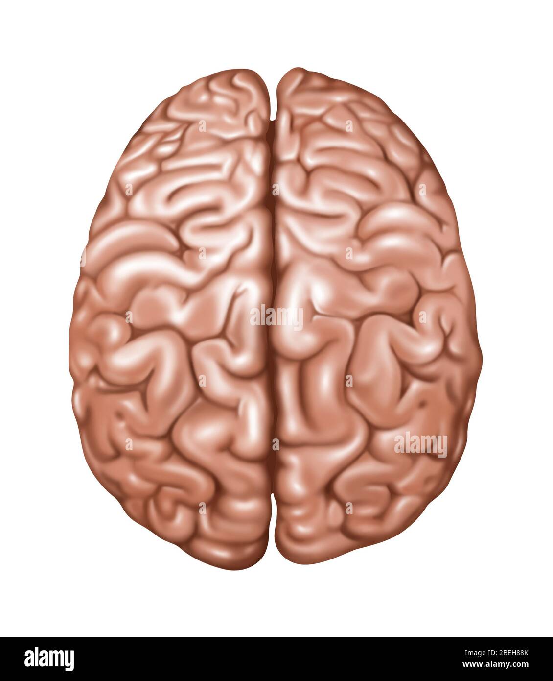 Top View of Normal Brain, Illustration Stock Photohttps://www.alamy.com/image-license-details/?v=1https://www.alamy.com/top-view-of-normal-brain-illustration-image353192243.html
RF2BEH88K–Top View of Normal Brain, Illustration
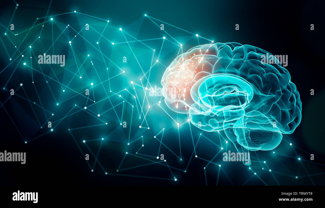 Human brain activity with plexus lines.. External cerebral connections in the frontal lobe. Communication, psychology, artificial intelligence or AI, Stock Photohttps://www.alamy.com/image-license-details/?v=1https://www.alamy.com/human-brain-activity-with-plexus-lines-external-cerebral-connections-in-the-frontal-lobe-communication-psychology-artificial-intelligence-or-ai-image255543128.html
RFTRMYT8–Human brain activity with plexus lines.. External cerebral connections in the frontal lobe. Communication, psychology, artificial intelligence or AI,
 Illustration of the frontal lobe (green) of the human brain. Parts of this area are responsible for higher cognitive functions such as decision making, personality expression and moderating social behaviour. Stock Photohttps://www.alamy.com/image-license-details/?v=1https://www.alamy.com/illustration-of-the-frontal-lobe-green-of-the-human-brain-parts-of-this-area-are-responsible-for-higher-cognitive-functions-such-as-decision-making-personality-expression-and-moderating-social-behaviour-image627804674.html
RF2YDAY7E–Illustration of the frontal lobe (green) of the human brain. Parts of this area are responsible for higher cognitive functions such as decision making, personality expression and moderating social behaviour.
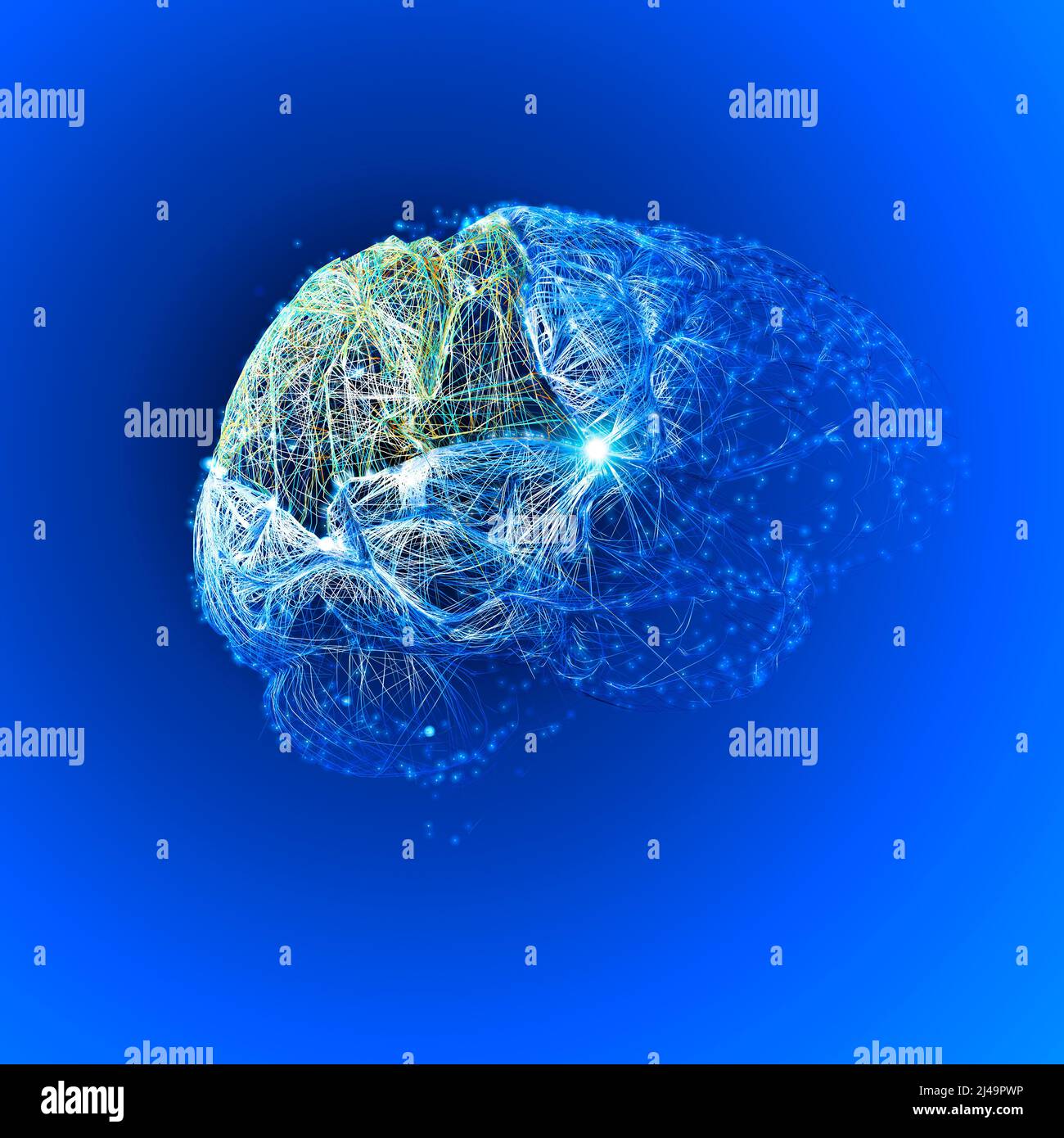 Brain, the parietal lobe is vital for sensory perception and integration, including the management of taste, hearing, sight, touch, and smell Stock Photohttps://www.alamy.com/image-license-details/?v=1https://www.alamy.com/brain-the-parietal-lobe-is-vital-for-sensory-perception-and-integration-including-the-management-of-taste-hearing-sight-touch-and-smell-image467354098.html
RF2J49PWP–Brain, the parietal lobe is vital for sensory perception and integration, including the management of taste, hearing, sight, touch, and smell
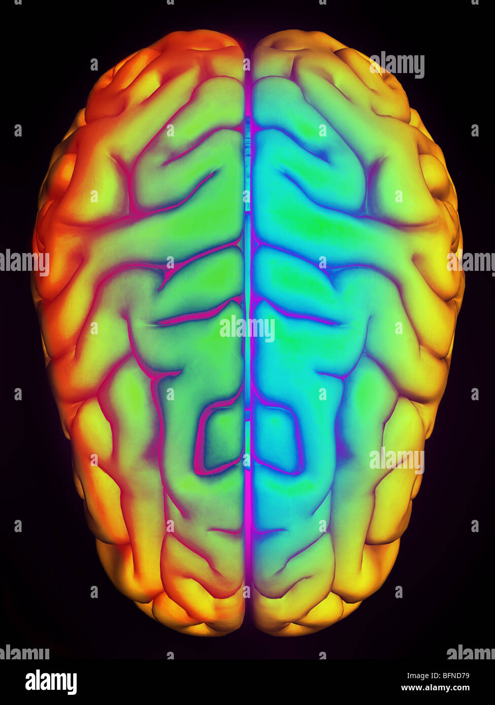 Illustration of the human brain seen from above Stock Photohttps://www.alamy.com/image-license-details/?v=1https://www.alamy.com/stock-photo-illustration-of-the-human-brain-seen-from-above-26901597.html
RMBFND79–Illustration of the human brain seen from above
 Screen Time damaging the brain psychology and technology health affects due to internet addiction and cognitive damage physiological effects. Stock Photohttps://www.alamy.com/image-license-details/?v=1https://www.alamy.com/screen-time-damaging-the-brain-psychology-and-technology-health-affects-due-to-internet-addiction-and-cognitive-damage-physiological-effects-image228501755.html
RFR7N48Y–Screen Time damaging the brain psychology and technology health affects due to internet addiction and cognitive damage physiological effects.
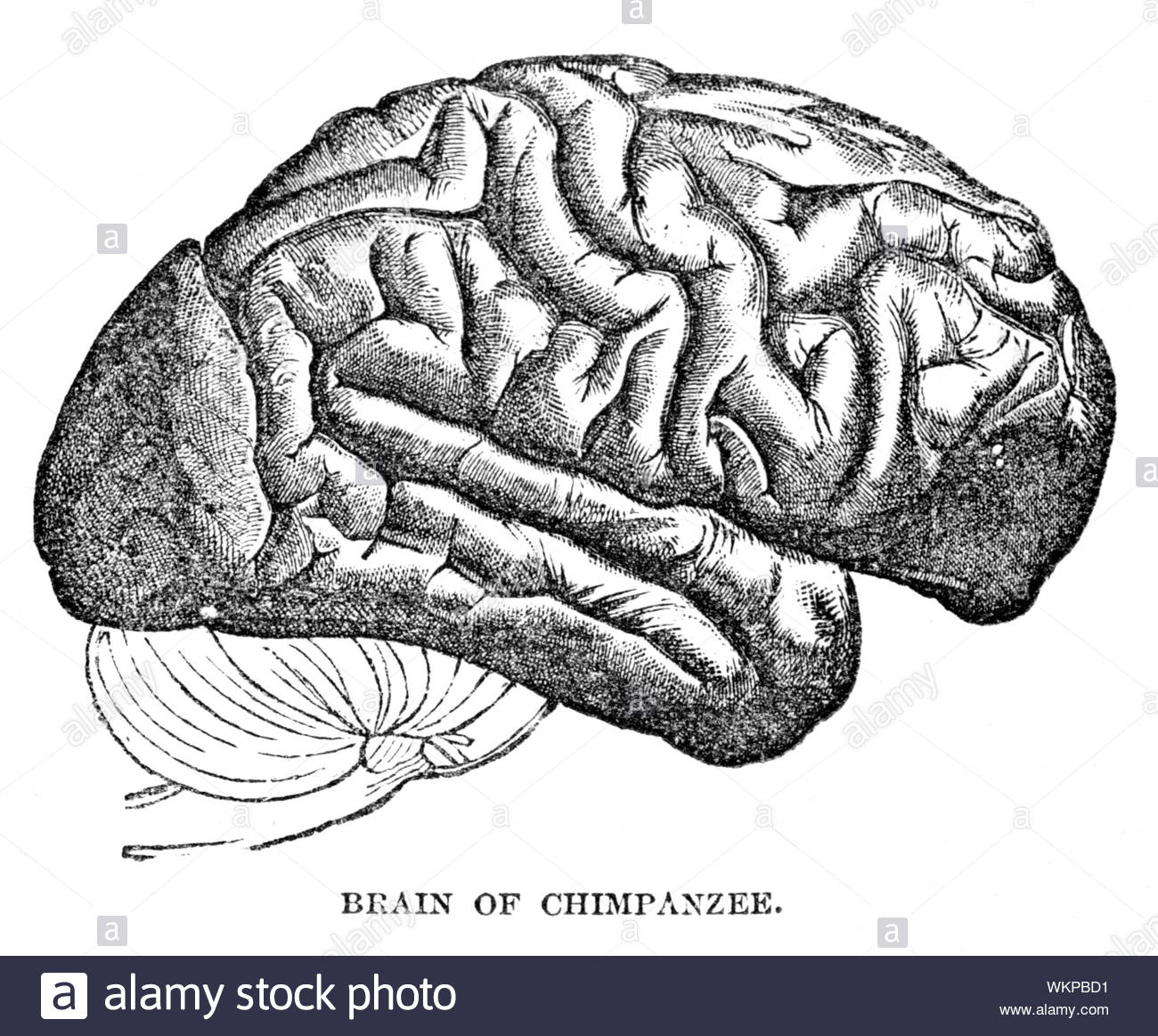 Brain of Chimpanzee, vintage illustration from 1884 Stock Photohttps://www.alamy.com/image-license-details/?v=1https://www.alamy.com/brain-of-chimpanzee-vintage-illustration-from-1884-image270325917.html
RMWKPBD1–Brain of Chimpanzee, vintage illustration from 1884
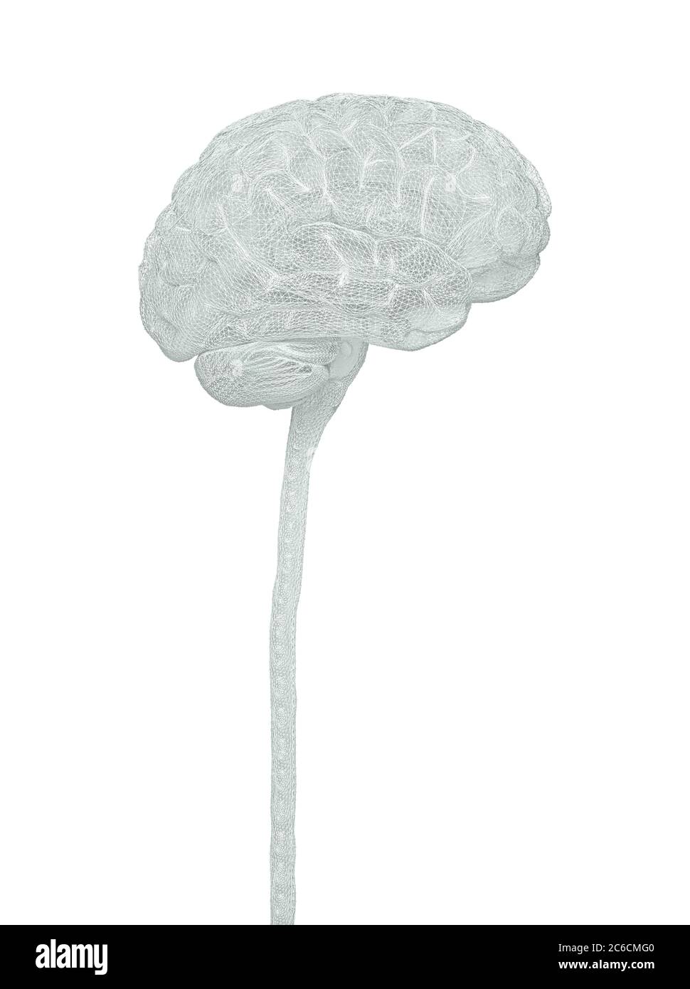 Central nervous system. Brain and spinal cord with clipping path included. Conceptual brain 3D illustration. Stock Photohttps://www.alamy.com/image-license-details/?v=1https://www.alamy.com/central-nervous-system-brain-and-spinal-cord-with-clipping-path-included-conceptual-brain-3d-illustration-image365385216.html
RF2C6CMG0–Central nervous system. Brain and spinal cord with clipping path included. Conceptual brain 3D illustration.
 Brain surface anatomy. Stock Photohttps://www.alamy.com/image-license-details/?v=1https://www.alamy.com/stock-photo-brain-surface-anatomy-111740765.html
RFGDP6DH–Brain surface anatomy.
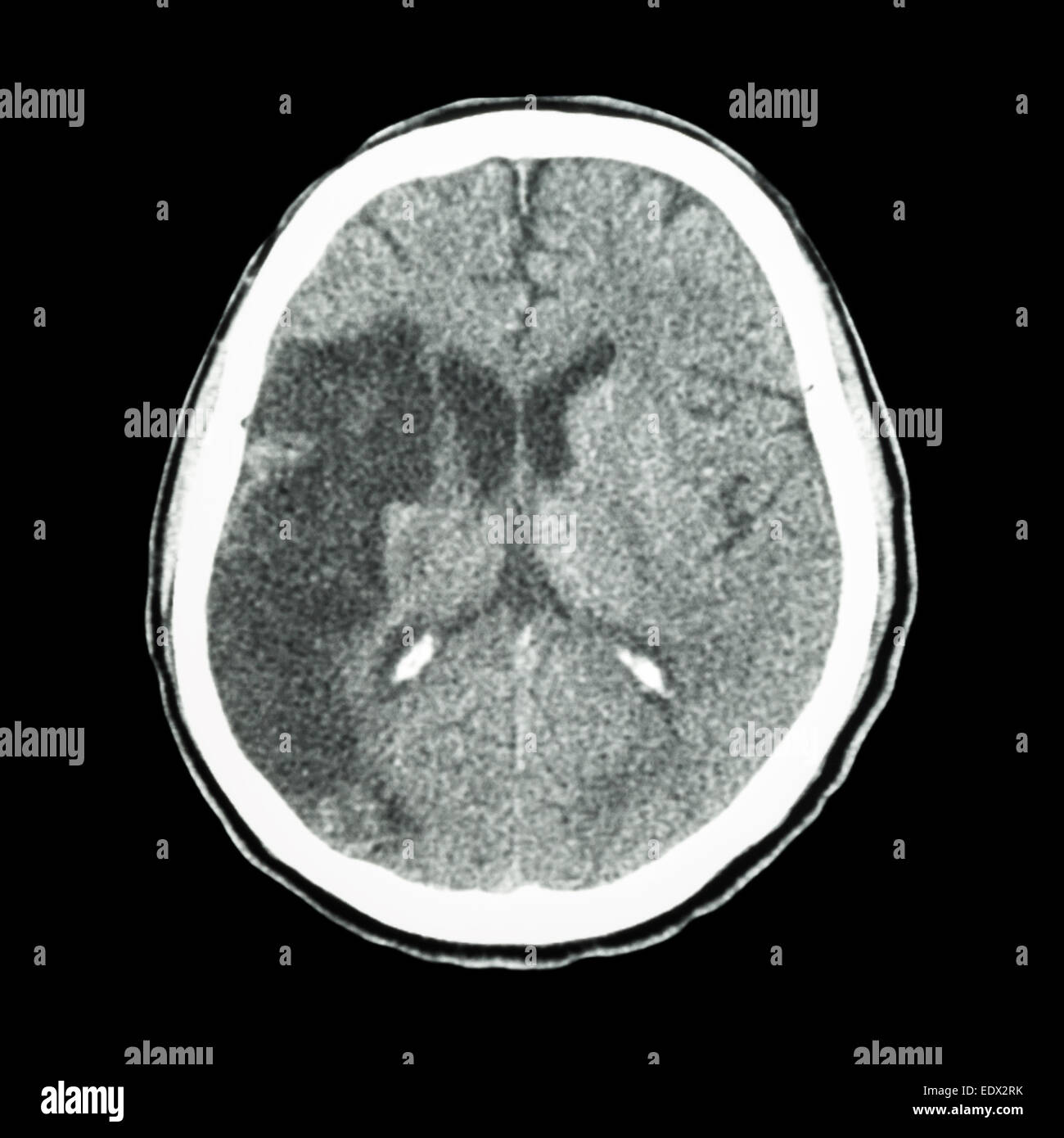 CT brain : show Ischemic stroke (hypodensity at right frontal-parietal lobe) Stock Photohttps://www.alamy.com/image-license-details/?v=1https://www.alamy.com/stock-photo-ct-brain-show-ischemic-stroke-hypodensity-at-right-frontal-parietal-77404983.html
RFEDX2RK–CT brain : show Ischemic stroke (hypodensity at right frontal-parietal lobe)
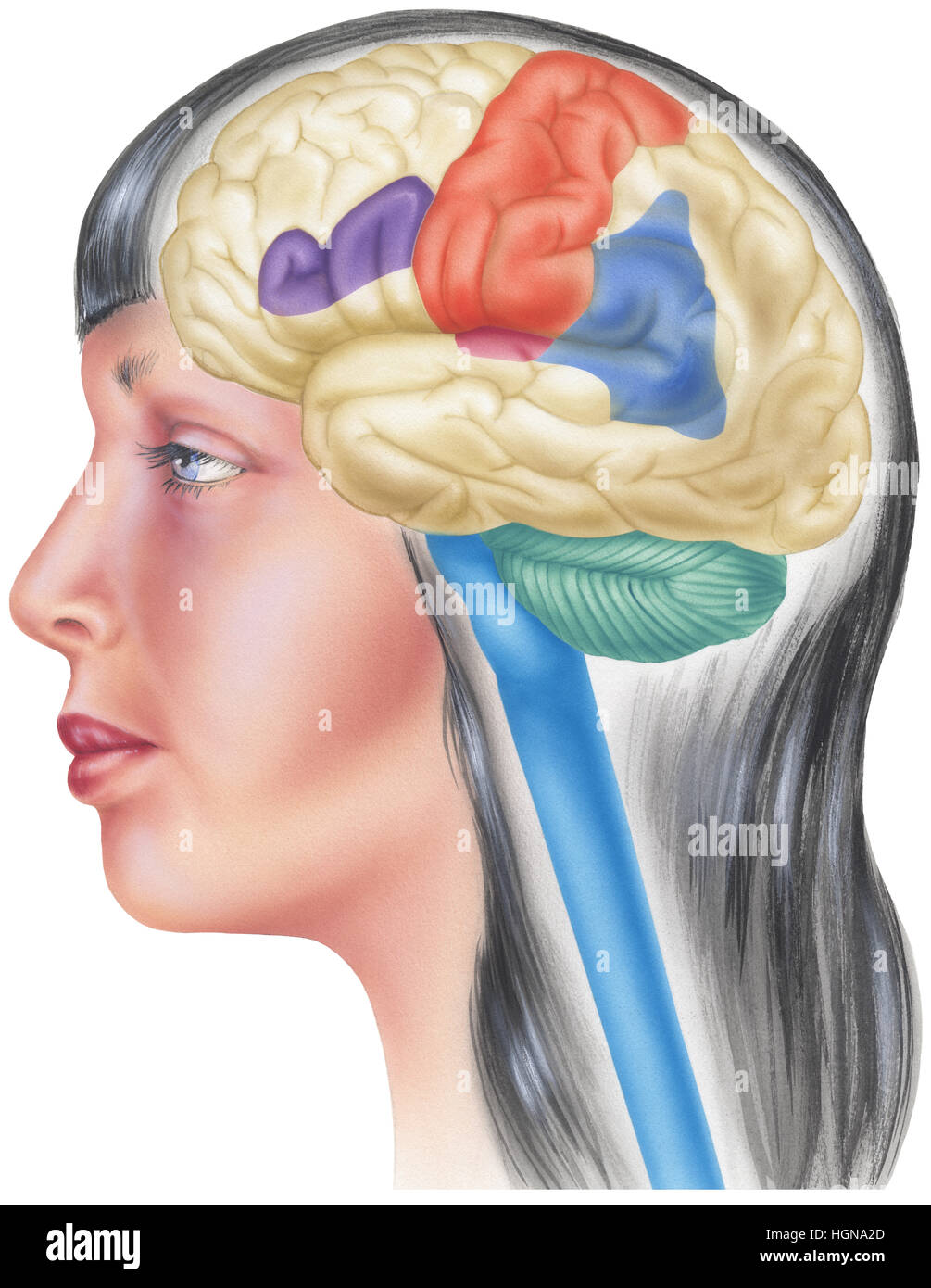 Side view of the human brain in the skull of a young woman. Shown are the parietal lobes, sensory cortex, angular gyrus, Broca's area, frontal lobes a Stock Photohttps://www.alamy.com/image-license-details/?v=1https://www.alamy.com/stock-photo-side-view-of-the-human-brain-in-the-skull-of-a-young-woman-shown-are-130775973.html
RFHGNA2D–Side view of the human brain in the skull of a young woman. Shown are the parietal lobes, sensory cortex, angular gyrus, Broca's area, frontal lobes a
 Brain is a Part of Human Body Central Nervous System Anatomy. 3D Stock Photohttps://www.alamy.com/image-license-details/?v=1https://www.alamy.com/brain-is-a-part-of-human-body-central-nervous-system-anatomy-3d-image352945124.html
RF2BE6130–Brain is a Part of Human Body Central Nervous System Anatomy. 3D
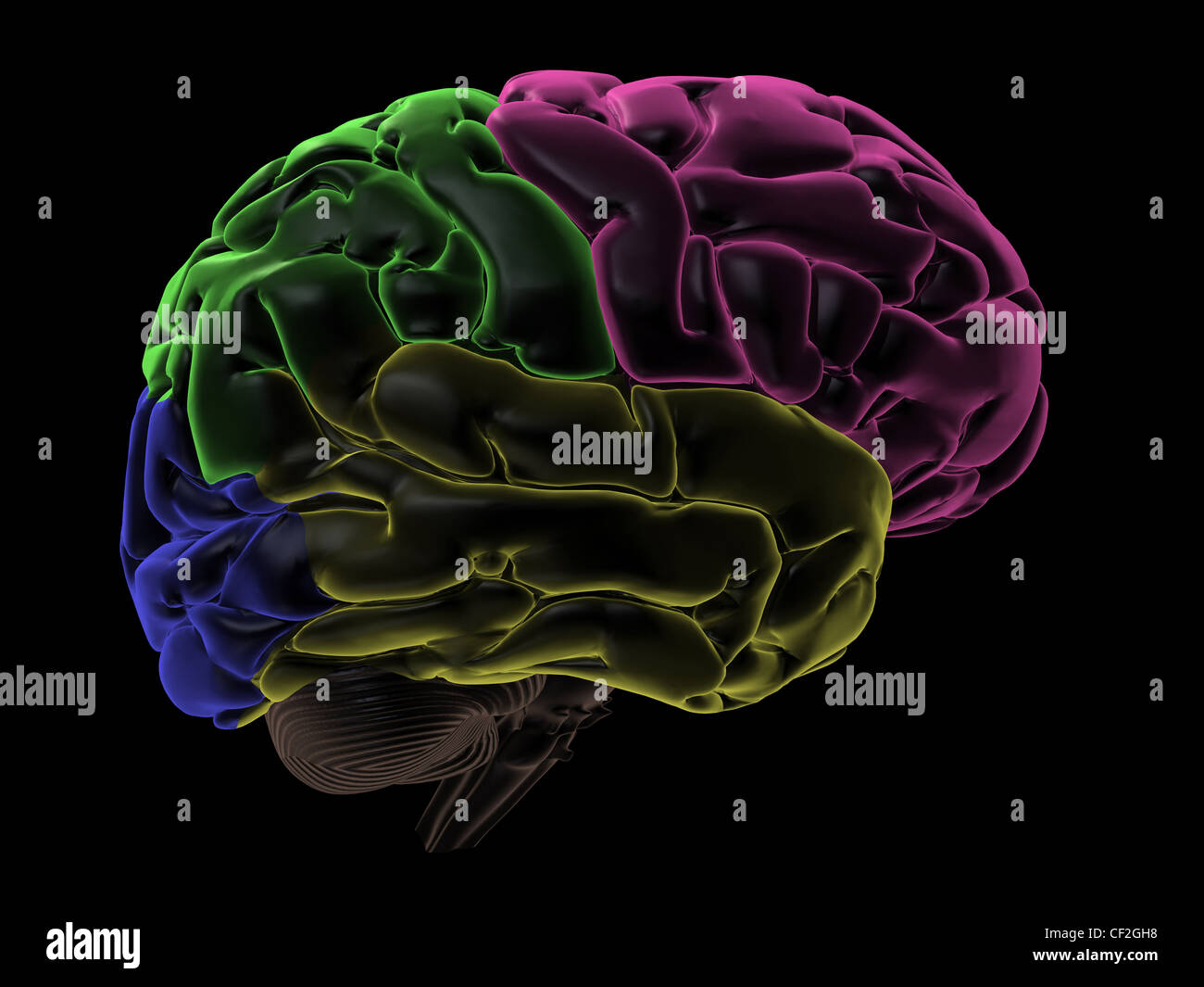 Coloured areas of the Brain, right hemisphere Stock Photohttps://www.alamy.com/image-license-details/?v=1https://www.alamy.com/stock-photo-coloured-areas-of-the-brain-right-hemisphere-43697508.html
RFCF2GH8–Coloured areas of the Brain, right hemisphere
 Central Organ of Human Nervous System Brain Anatomy Stock Photohttps://www.alamy.com/image-license-details/?v=1https://www.alamy.com/central-organ-of-human-nervous-system-brain-anatomy-image330946766.html
RF2A6BWYA–Central Organ of Human Nervous System Brain Anatomy
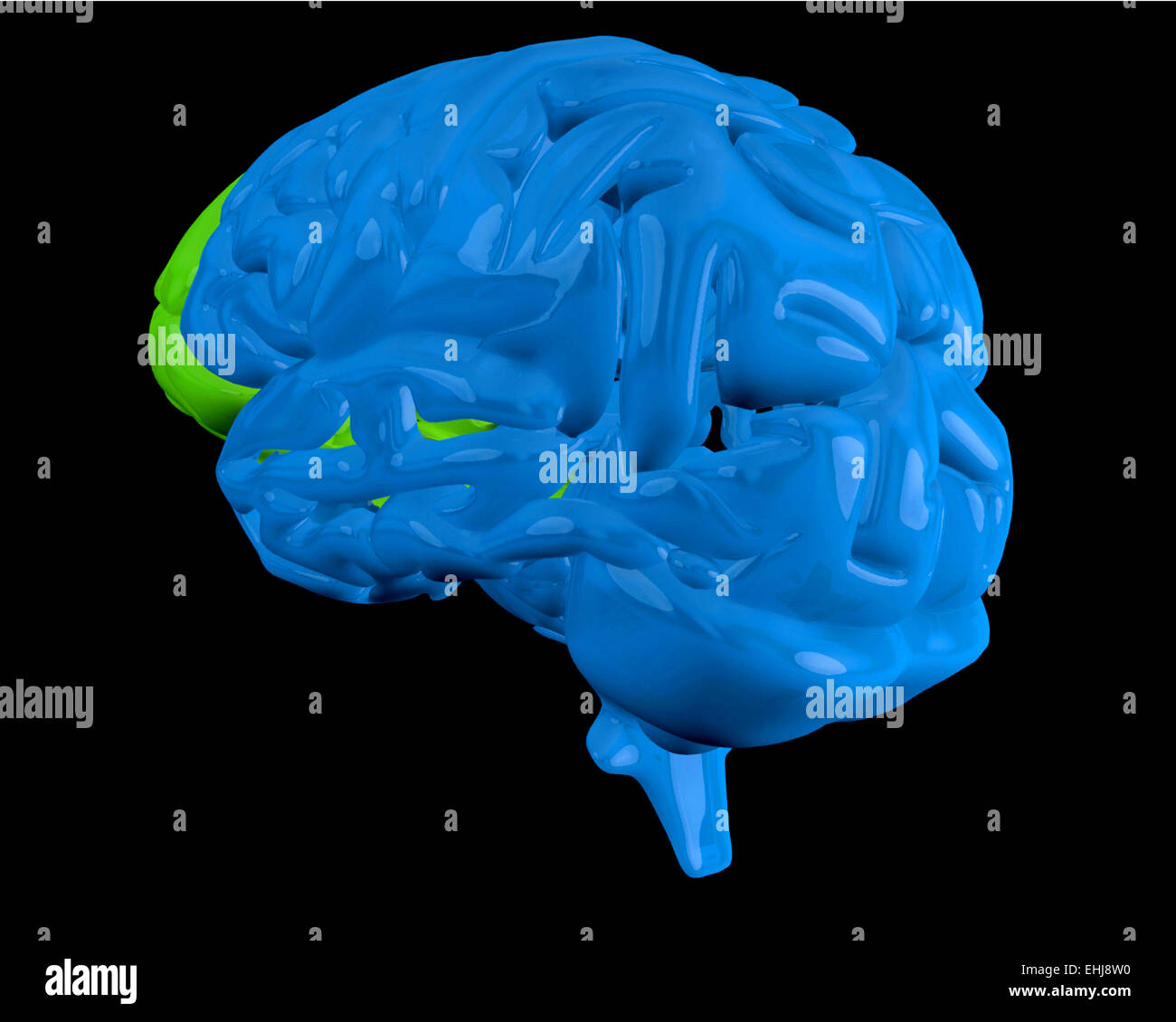 Blue brain with highlighted frontal lobe Stock Photohttps://www.alamy.com/image-license-details/?v=1https://www.alamy.com/stock-photo-blue-brain-with-highlighted-frontal-lobe-79692732.html
RFEHJ8W0–Blue brain with highlighted frontal lobe
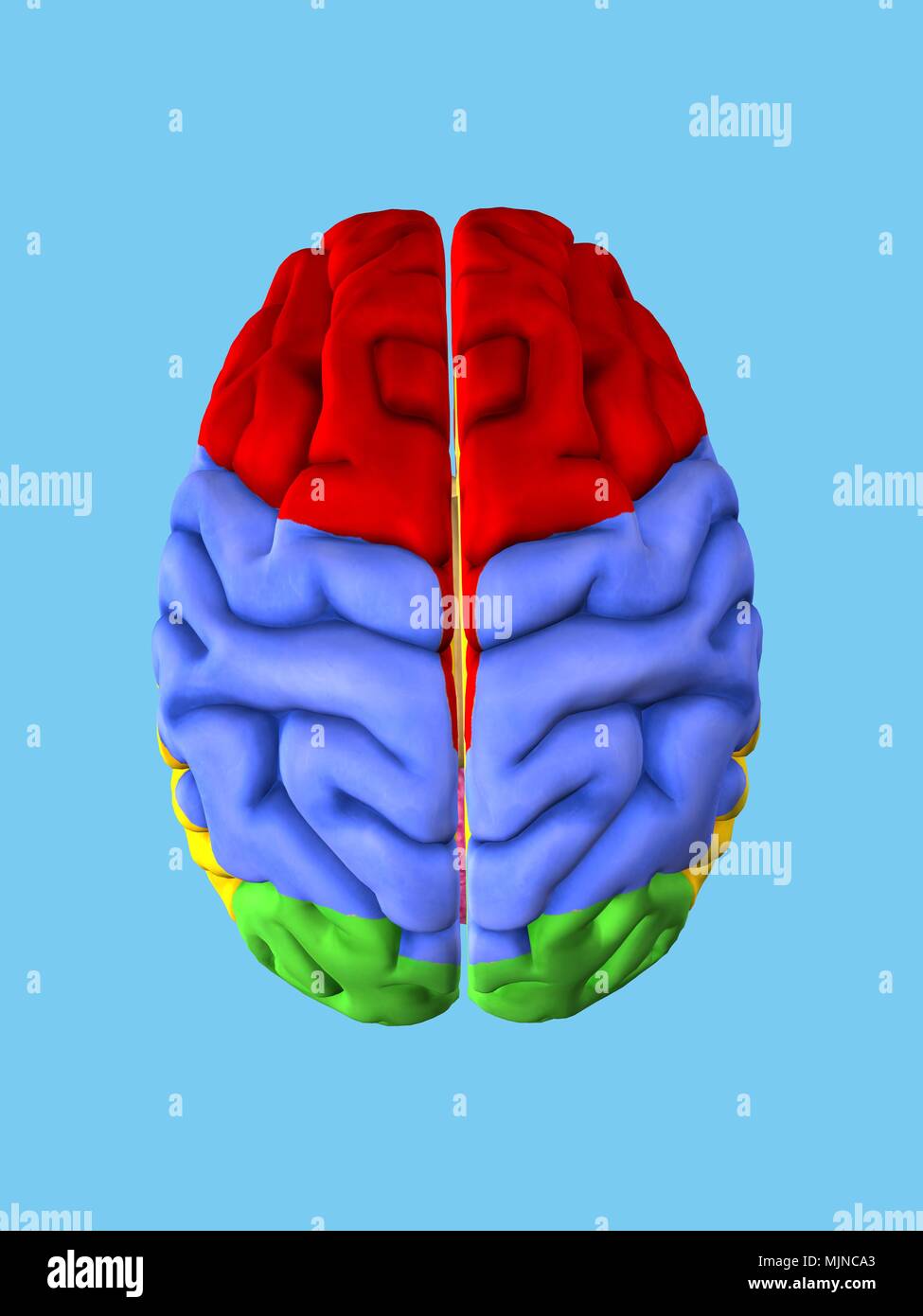 Regions of the Brain Stock Photohttps://www.alamy.com/image-license-details/?v=1https://www.alamy.com/regions-of-the-brain-image183638171.html
RFMJNCA3–Regions of the Brain
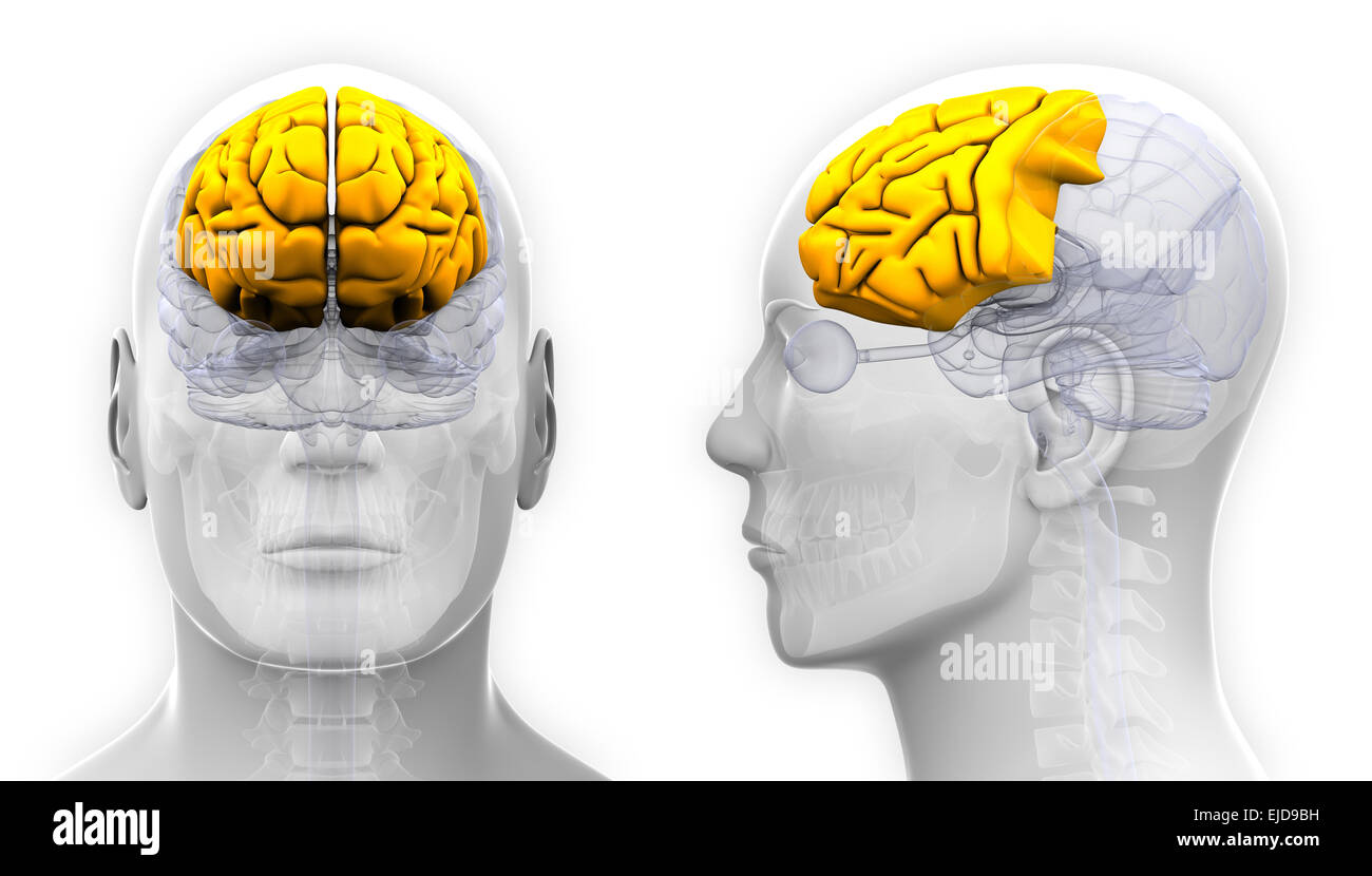 Male Frontal Lobe Brain Anatomy - isolated on white Stock Photohttps://www.alamy.com/image-license-details/?v=1https://www.alamy.com/stock-photo-male-frontal-lobe-brain-anatomy-isolated-on-white-80198037.html
RFEJD9BH–Male Frontal Lobe Brain Anatomy - isolated on white
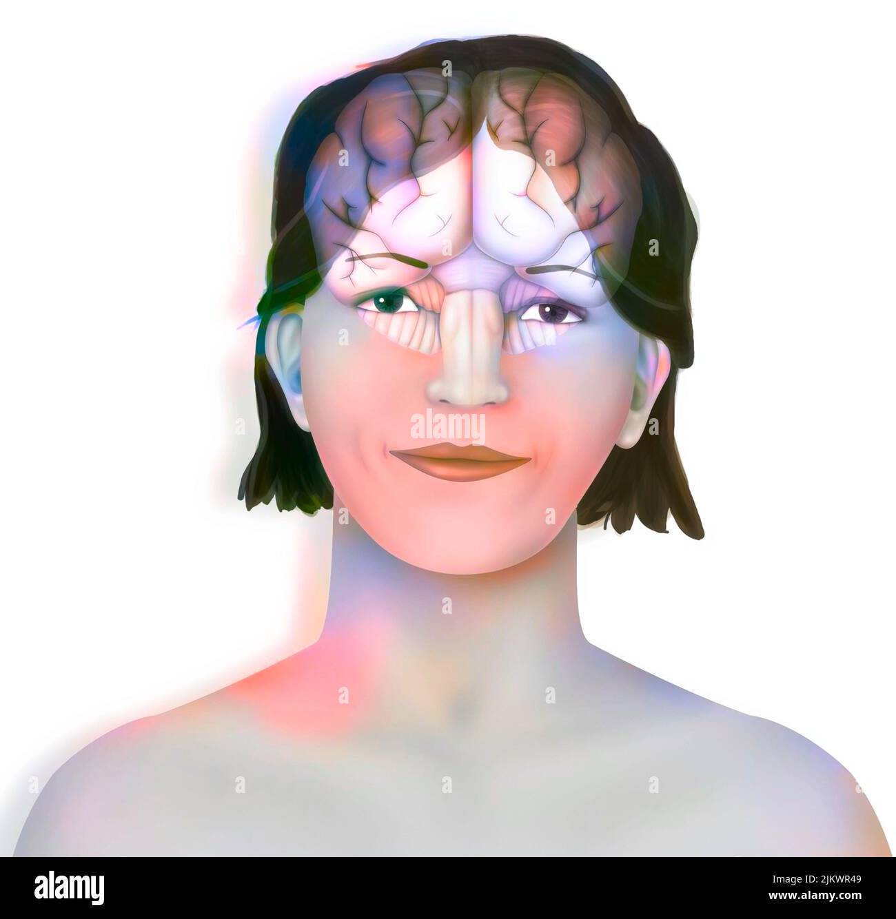 Brain (right and left cerebral hemispheres, cerebellum and brainstem) in a woman's face. Stock Photohttps://www.alamy.com/image-license-details/?v=1https://www.alamy.com/brain-right-and-left-cerebral-hemispheres-cerebellum-and-brainstem-in-a-womans-face-image476925353.html
RF2JKWR49–Brain (right and left cerebral hemispheres, cerebellum and brainstem) in a woman's face.
 Head with brain - 3d Illustration of pain Stock Photohttps://www.alamy.com/image-license-details/?v=1https://www.alamy.com/head-with-brain-3d-illustration-of-pain-image261300653.html
RFW537J5–Head with brain - 3d Illustration of pain
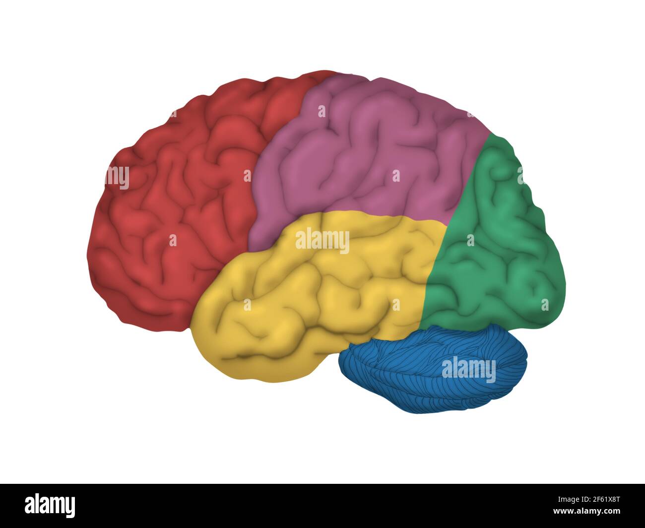 Human Brain, Lateral View Stock Photohttps://www.alamy.com/image-license-details/?v=1https://www.alamy.com/human-brain-lateral-view-image416779352.html
RM2F61X8T–Human Brain, Lateral View
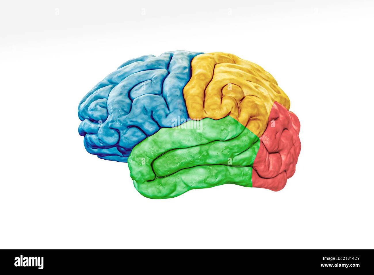 Cerebral cortex lobes in color profile view isolated on white background accurate 3D rendering illustration. Human brain anatomy, neurology, neuroscie Stock Photohttps://www.alamy.com/image-license-details/?v=1https://www.alamy.com/cerebral-cortex-lobes-in-color-profile-view-isolated-on-white-background-accurate-3d-rendering-illustration-human-brain-anatomy-neurology-neuroscie-image569811591.html
RF2T314DY–Cerebral cortex lobes in color profile view isolated on white background accurate 3D rendering illustration. Human brain anatomy, neurology, neuroscie
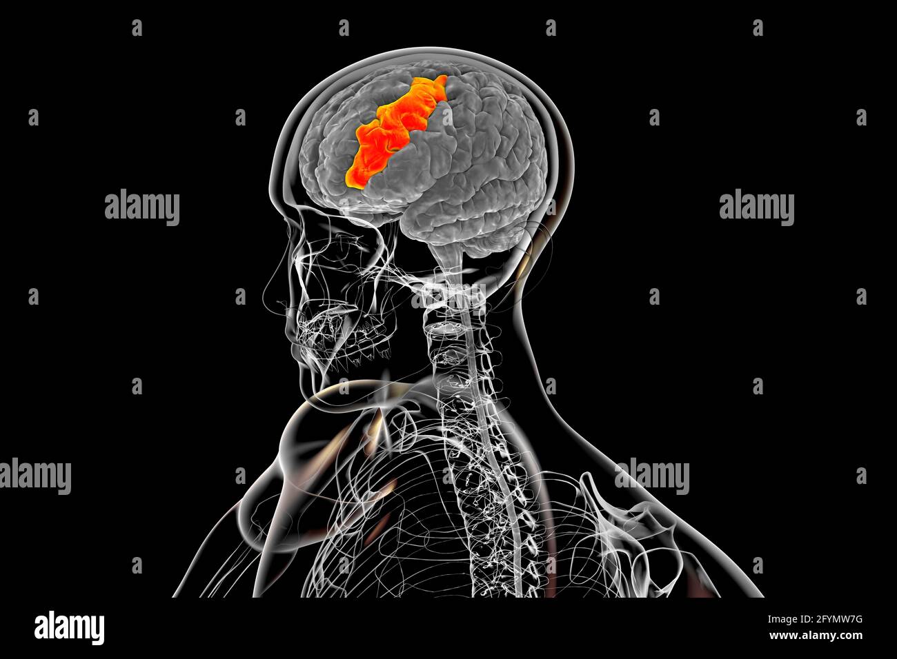 Brain with highlighted middle frontal gyrus, illustration Stock Photohttps://www.alamy.com/image-license-details/?v=1https://www.alamy.com/brain-with-highlighted-middle-frontal-gyrus-illustration-image430103396.html
RF2FYMW7G–Brain with highlighted middle frontal gyrus, illustration
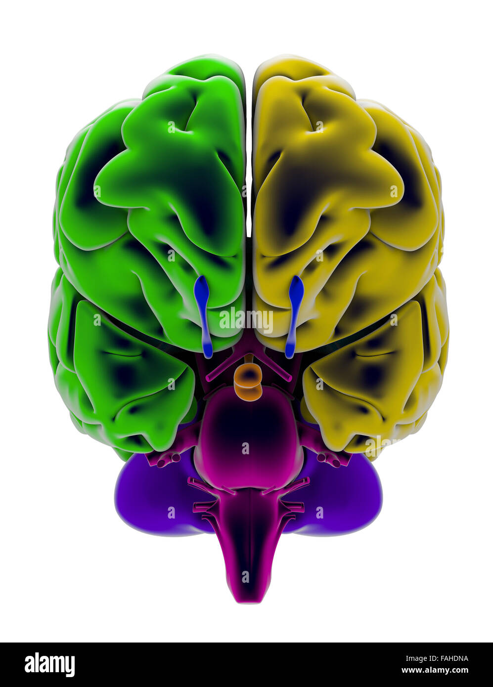 Brain, section, division, cutting parts, anatomy study Stock Photohttps://www.alamy.com/image-license-details/?v=1https://www.alamy.com/stock-photo-brain-section-division-cutting-parts-anatomy-study-92582374.html
RFFAHDNA–Brain, section, division, cutting parts, anatomy study
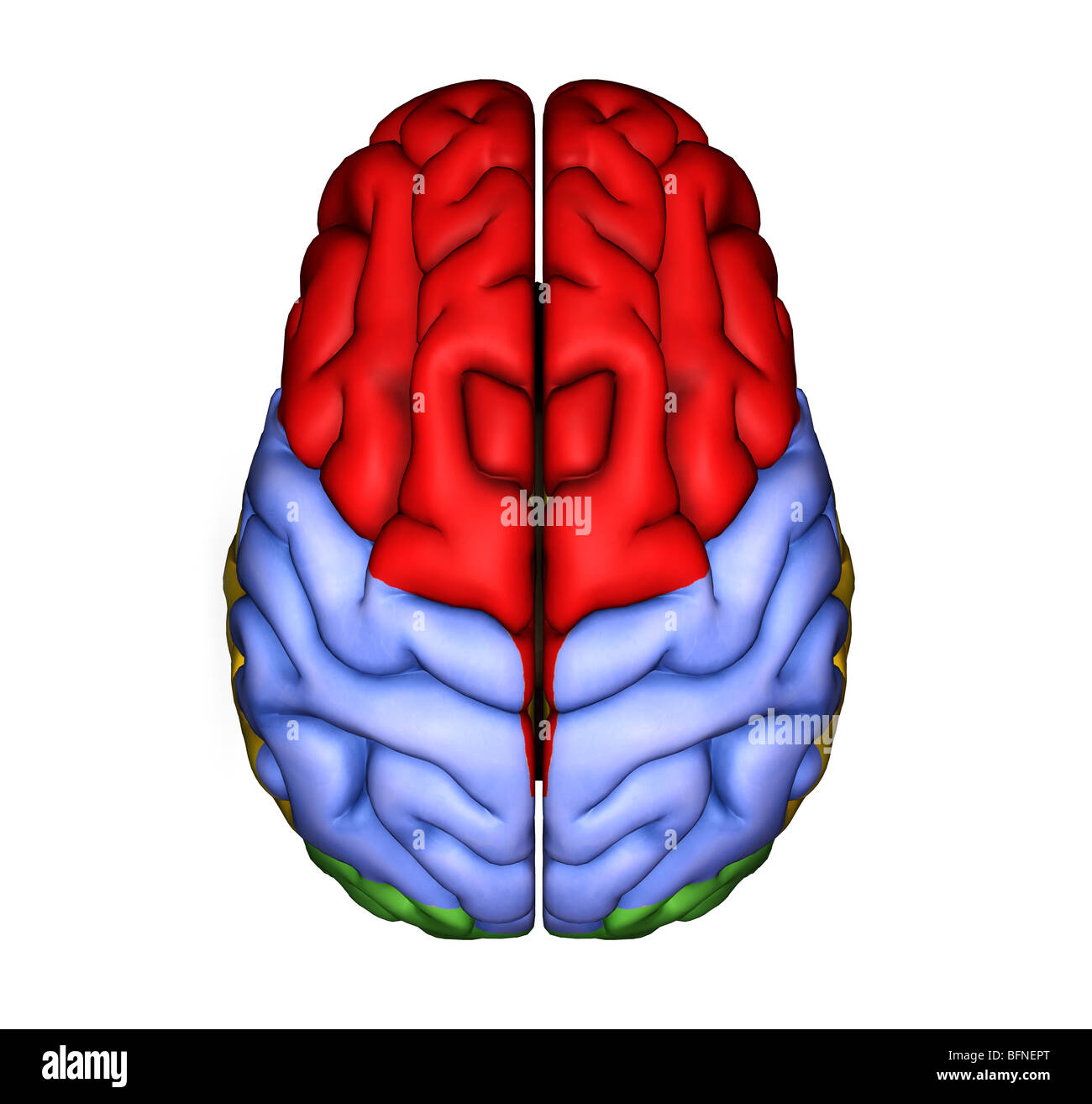 Illustration of the surface of the human brain seen from above Stock Photohttps://www.alamy.com/image-license-details/?v=1https://www.alamy.com/stock-photo-illustration-of-the-surface-of-the-human-brain-seen-from-above-26902816.html
RMBFNEPT–Illustration of the surface of the human brain seen from above
 Neural Pathways and brain intelligence as a cognitive function and psychiatric or psychological behavior or neurological disorder symbol as a human th Stock Photohttps://www.alamy.com/image-license-details/?v=1https://www.alamy.com/neural-pathways-and-brain-intelligence-as-a-cognitive-function-and-psychiatric-or-psychological-behavior-or-neurological-disorder-symbol-as-a-human-th-image620790300.html
RF2Y1YCA4–Neural Pathways and brain intelligence as a cognitive function and psychiatric or psychological behavior or neurological disorder symbol as a human th
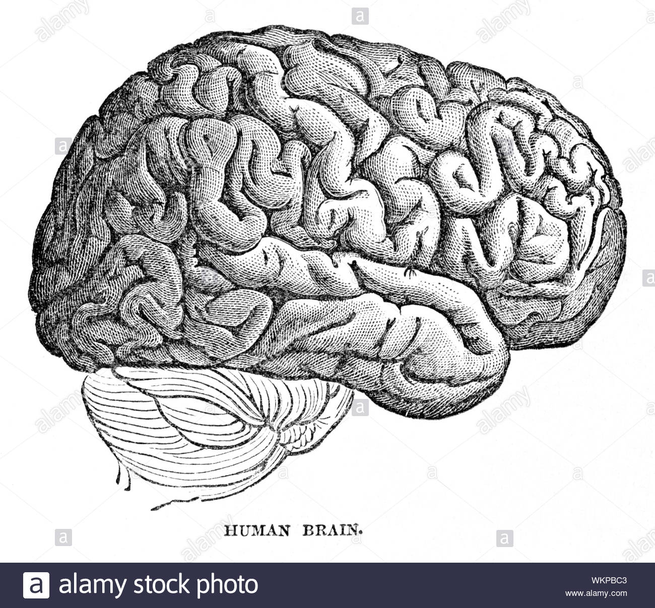 Human brain, vintage illustration from 1884 Stock Photohttps://www.alamy.com/image-license-details/?v=1https://www.alamy.com/human-brain-vintage-illustration-from-1884-image270325891.html
RMWKPBC3–Human brain, vintage illustration from 1884
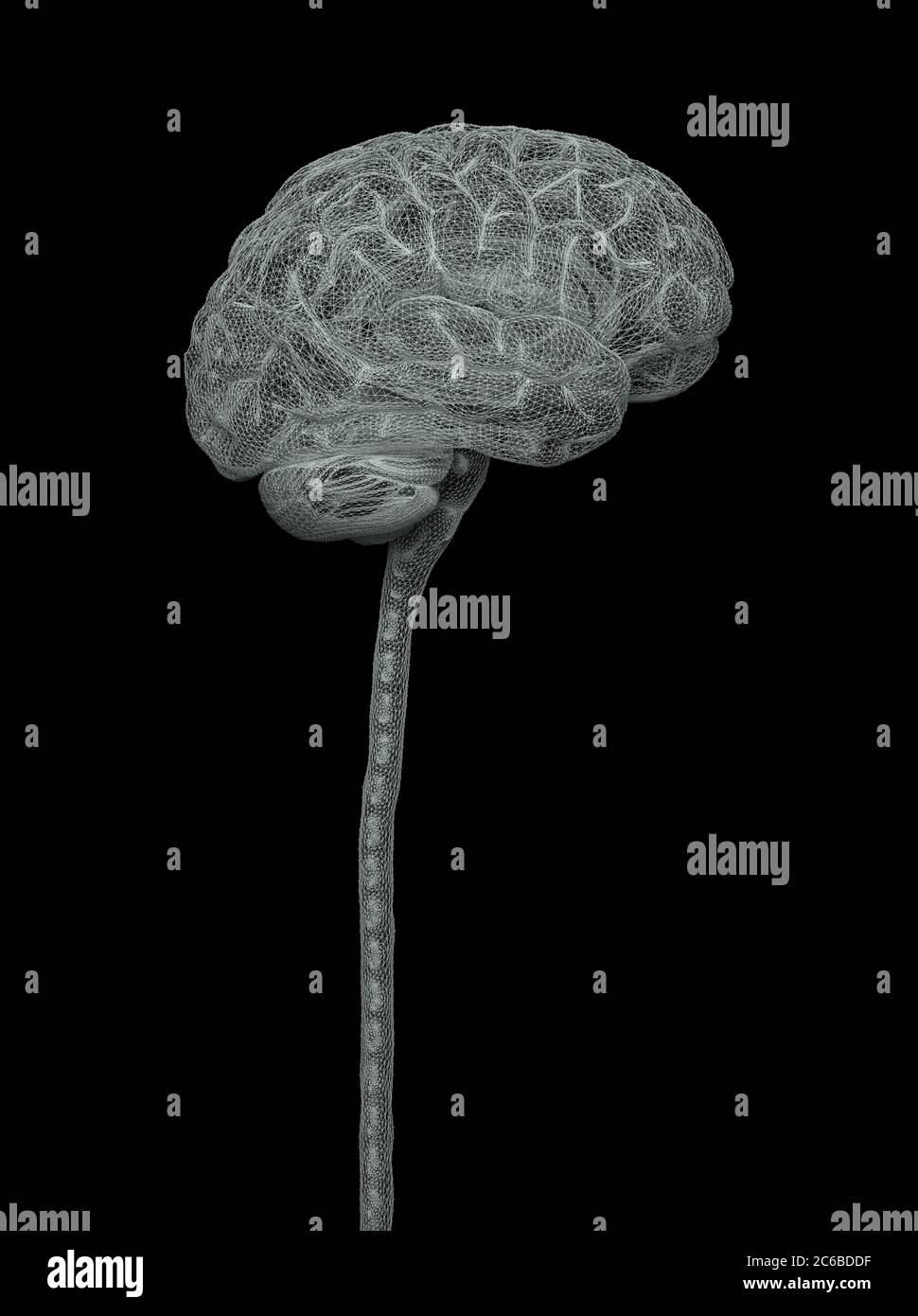 Central nervous system. Brain and spinal cord with clipping path included. Conceptual brain 3D illustration. Stock Photohttps://www.alamy.com/image-license-details/?v=1https://www.alamy.com/central-nervous-system-brain-and-spinal-cord-with-clipping-path-included-conceptual-brain-3d-illustration-image365357707.html
RF2C6BDDF–Central nervous system. Brain and spinal cord with clipping path included. Conceptual brain 3D illustration.
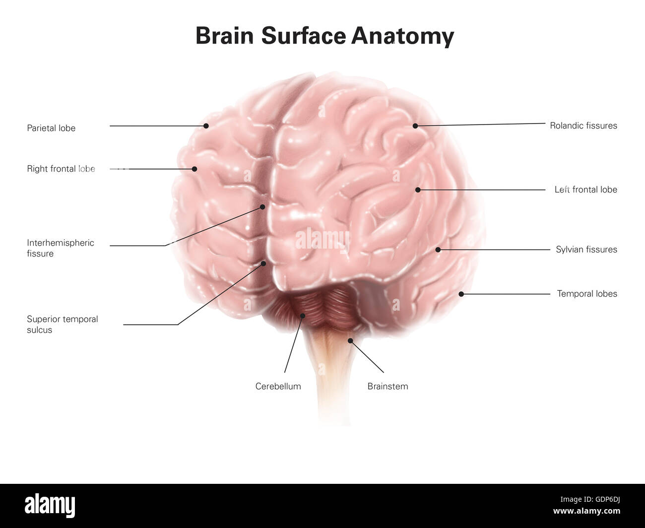 Brain surface anatomy, with labels. Stock Photohttps://www.alamy.com/image-license-details/?v=1https://www.alamy.com/stock-photo-brain-surface-anatomy-with-labels-111740766.html
RFGDP6DJ–Brain surface anatomy, with labels.
 Ischemic stroke : ( CT of brain show cerebral infarction at left frontal - temporal - parietal lobe ) ( nervous system backgroun Stock Photohttps://www.alamy.com/image-license-details/?v=1https://www.alamy.com/stock-photo-ischemic-stroke-ct-of-brain-show-cerebral-infarction-at-left-frontal-82644054.html
RFEPCN9A–Ischemic stroke : ( CT of brain show cerebral infarction at left frontal - temporal - parietal lobe ) ( nervous system backgroun
 vector medical illustration of the consequences of whiplash on the brain Stock Vectorhttps://www.alamy.com/image-license-details/?v=1https://www.alamy.com/vector-medical-illustration-of-the-consequences-of-whiplash-on-the-brain-image212143133.html
RFP93XMD–vector medical illustration of the consequences of whiplash on the brain
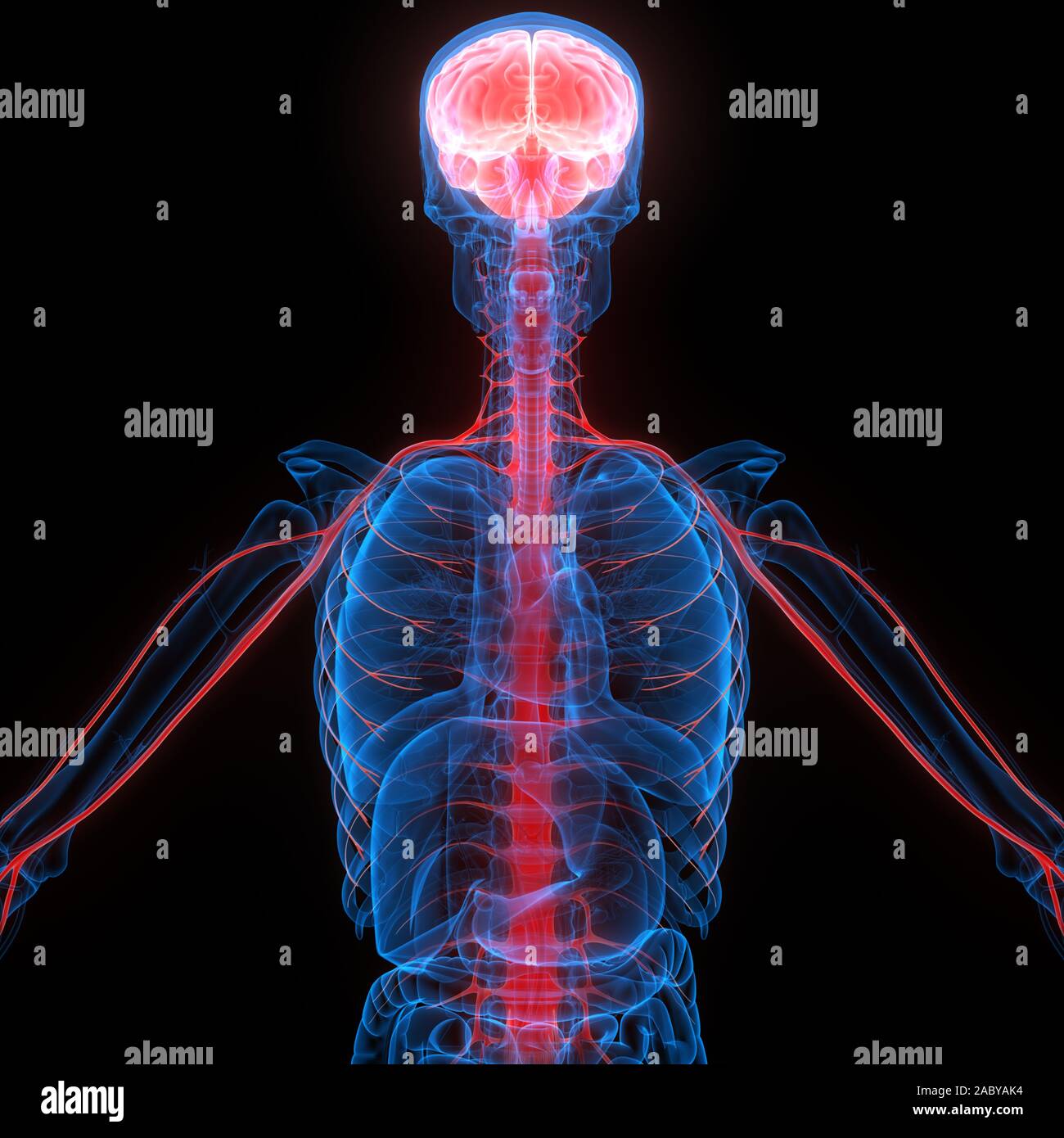 Human Internal Organ Brain with Nervous System Anatomy X-ray 3D rendering Stock Photohttps://www.alamy.com/image-license-details/?v=1https://www.alamy.com/human-internal-organ-brain-with-nervous-system-anatomy-x-ray-3d-rendering-image334359288.html
RF2ABYAK4–Human Internal Organ Brain with Nervous System Anatomy X-ray 3D rendering
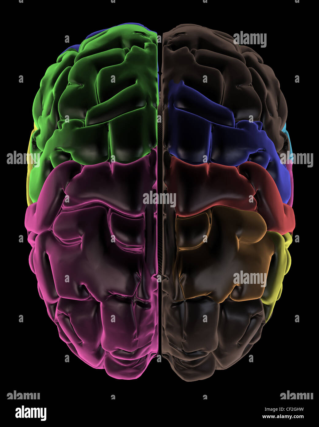 Coloured areas of the Brain, top view Stock Photohttps://www.alamy.com/image-license-details/?v=1https://www.alamy.com/stock-photo-coloured-areas-of-the-brain-top-view-43697525.html
RFCF2GHW–Coloured areas of the Brain, top view
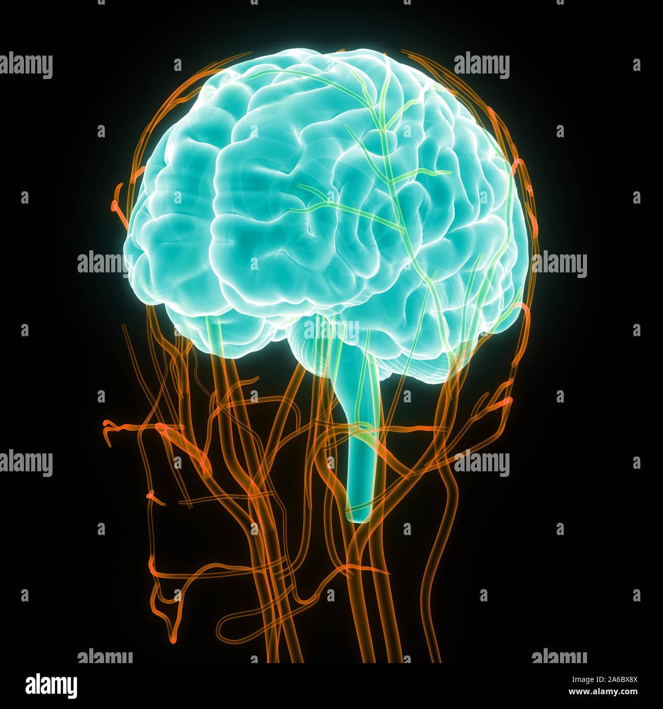 Central Organ of Human Nervous System Brain Anatomy Stock Photohttps://www.alamy.com/image-license-details/?v=1https://www.alamy.com/central-organ-of-human-nervous-system-brain-anatomy-image330947034.html
RF2A6BX8X–Central Organ of Human Nervous System Brain Anatomy
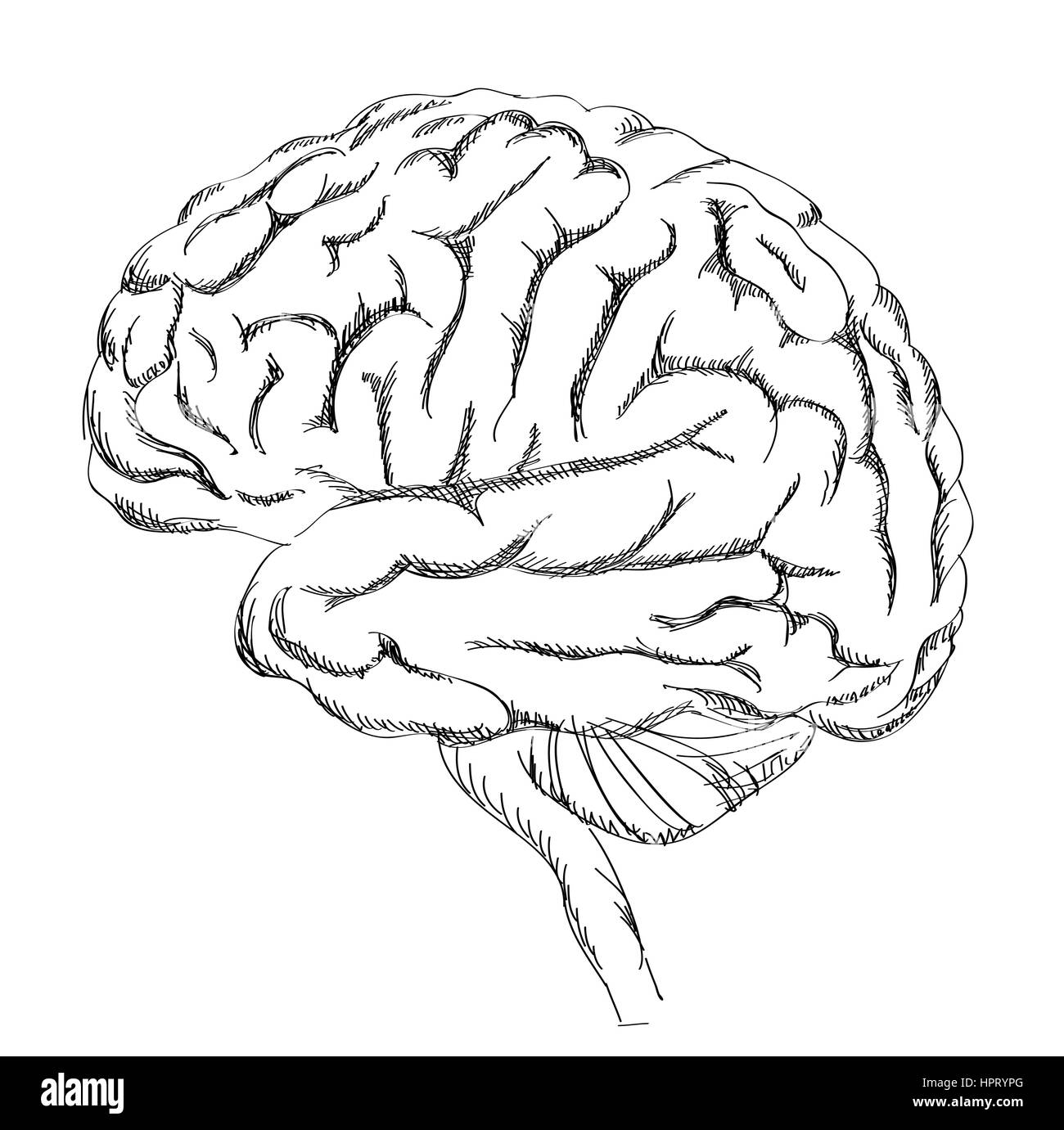 Brain anatomy. Human brain lateral view. Sketch illustration isolated on white background. Stock Vectorhttps://www.alamy.com/image-license-details/?v=1https://www.alamy.com/stock-photo-brain-anatomy-human-brain-lateral-view-sketch-illustration-isolated-134521704.html
RFHPRYPG–Brain anatomy. Human brain lateral view. Sketch illustration isolated on white background.
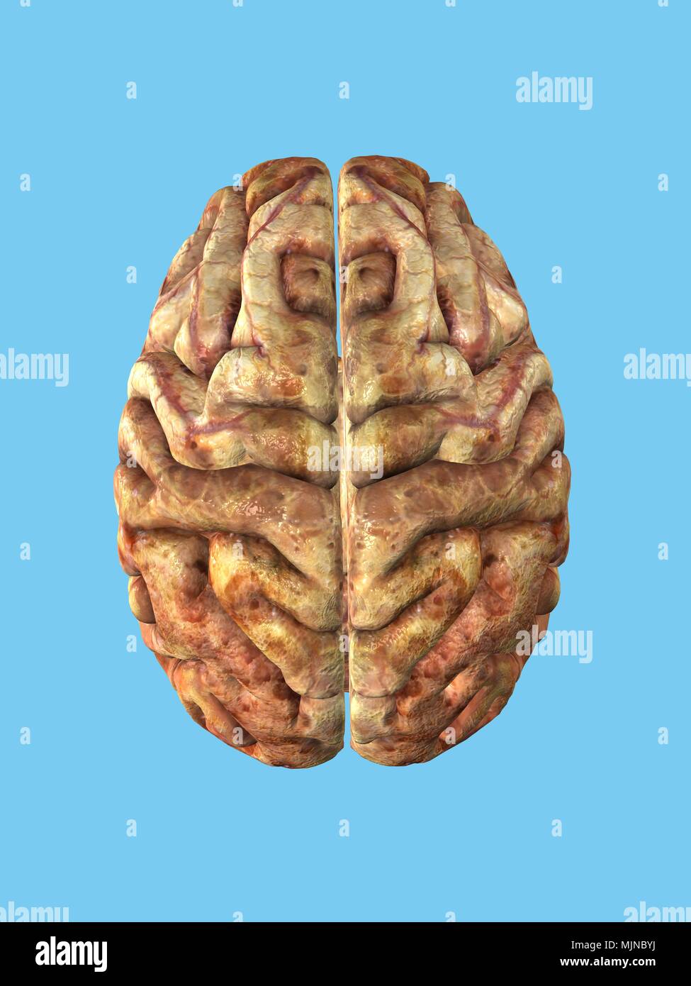 Human Brain Stock Photohttps://www.alamy.com/image-license-details/?v=1https://www.alamy.com/human-brain-image183637878.html
RMMJNBYJ–Human Brain
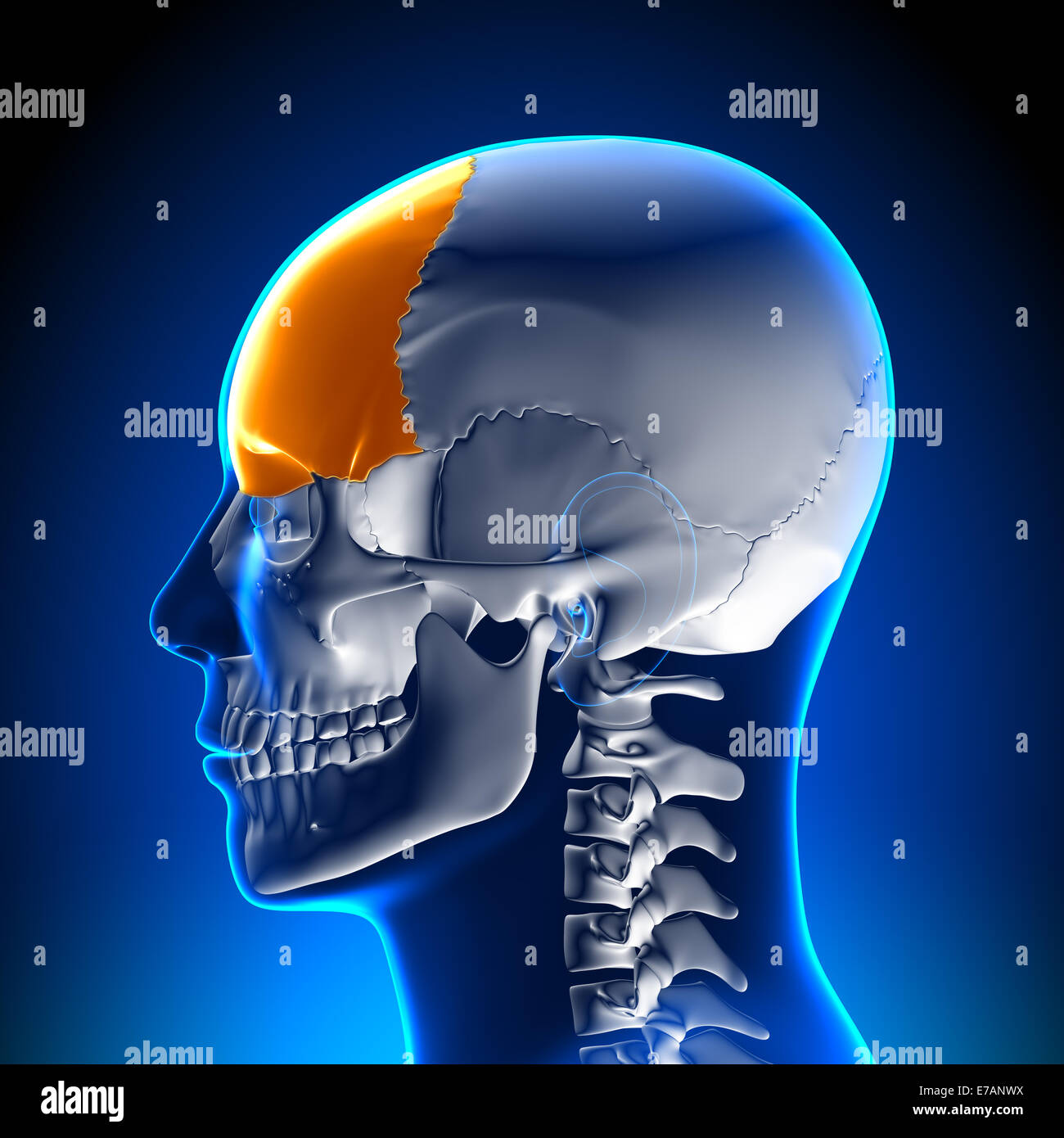 Frontal bone - skull Stock Photohttps://www.alamy.com/image-license-details/?v=1https://www.alamy.com/stock-photo-frontal-bone-skull-73380774.html
RFE7ANWX–Frontal bone - skull
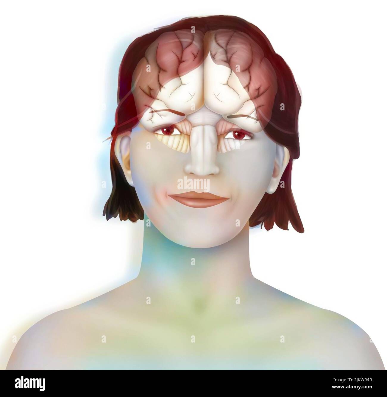 Brain (right and left cerebral hemispheres, cerebellum and brainstem) in a woman's face. Stock Photohttps://www.alamy.com/image-license-details/?v=1https://www.alamy.com/brain-right-and-left-cerebral-hemispheres-cerebellum-and-brainstem-in-a-womans-face-image476925367.html
RF2JKWR4R–Brain (right and left cerebral hemispheres, cerebellum and brainstem) in a woman's face.
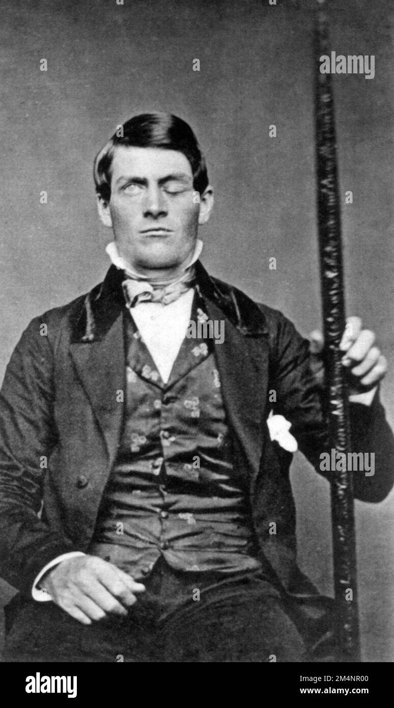 Phineas Gage. Photograph of Phineas P. Gage (1823–1860), an American railroad construction foreman known for his improbable survival of an accident in which a large iron rod was driven completely through his head, destroying much of his brain's left frontal lobe. Stock Photohttps://www.alamy.com/image-license-details/?v=1https://www.alamy.com/phineas-gage-photograph-of-phineas-p-gage-18231860-an-american-railroad-construction-foreman-known-for-his-improbable-survival-of-an-accident-in-which-a-large-iron-rod-was-driven-completely-through-his-head-destroying-much-of-his-brains-left-frontal-lobe-image502038320.html
RM2M4NR00–Phineas Gage. Photograph of Phineas P. Gage (1823–1860), an American railroad construction foreman known for his improbable survival of an accident in which a large iron rod was driven completely through his head, destroying much of his brain's left frontal lobe.