Quick filters:
Ganglion cells Stock Photos and Images
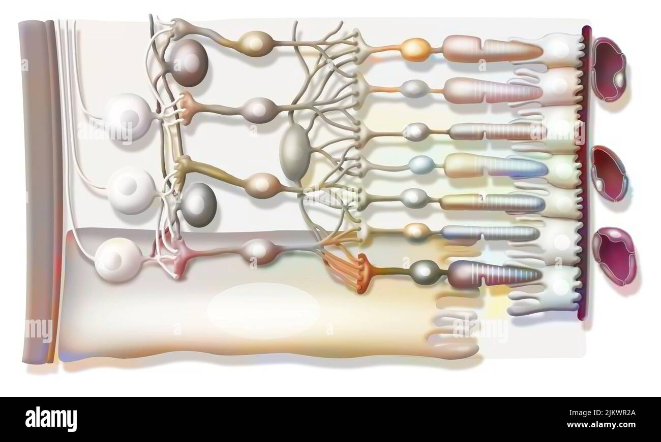 Zoom on the structure of the retina with vitreous body, internal limiting membrane, ganglion cells. Stock Photohttps://www.alamy.com/image-license-details/?v=1https://www.alamy.com/zoom-on-the-structure-of-the-retina-with-vitreous-body-internal-limiting-membrane-ganglion-cells-image476925298.html
RF2JKWR2A–Zoom on the structure of the retina with vitreous body, internal limiting membrane, ganglion cells.
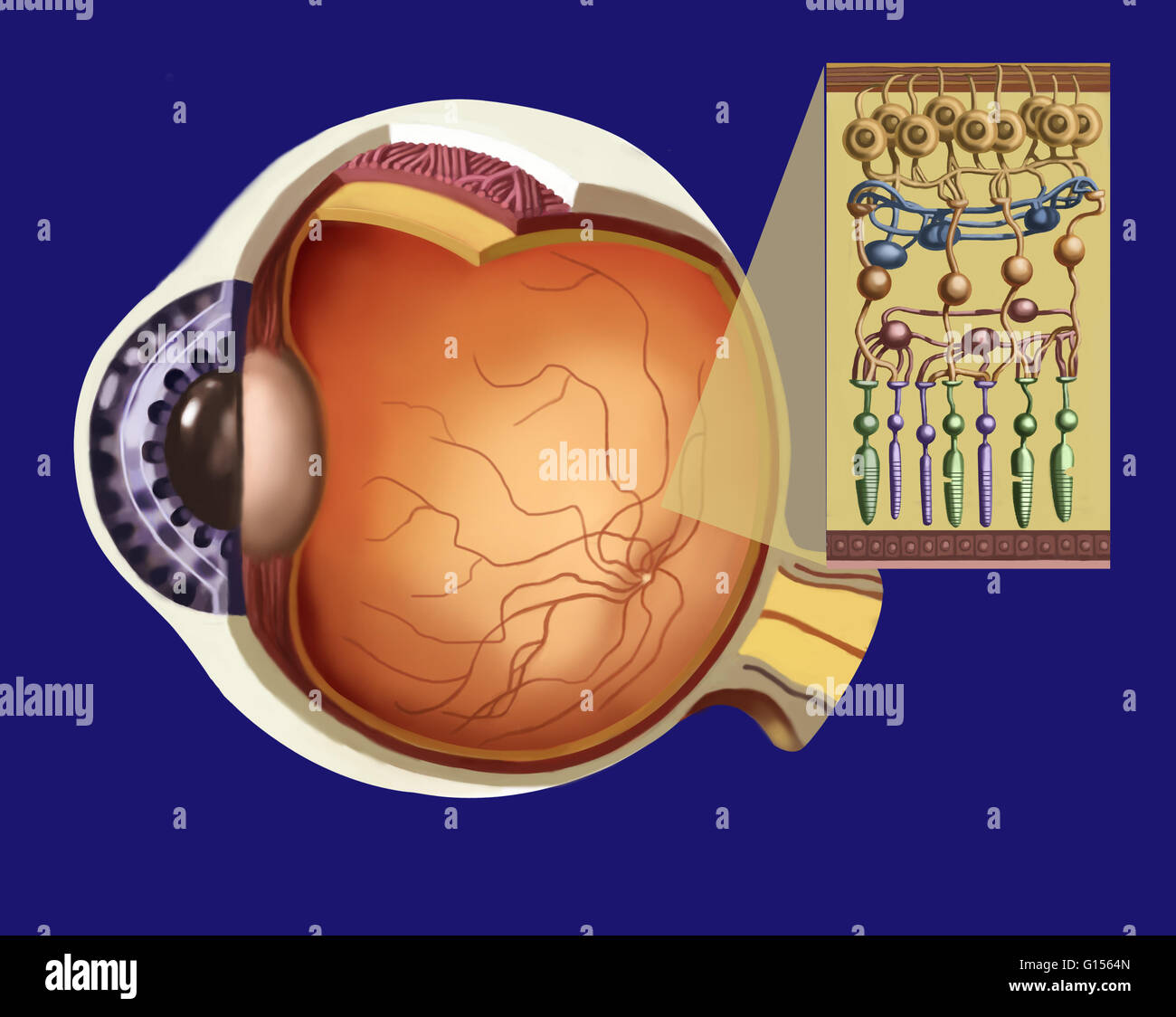 Illustration showing the structure of the retina as an insert to the larger eye structure. From top to bottom: optic nerve fiber (reddish brown strip at top), ganglion cells (in brown), amacrine cells (in dark blue), bipolar cells (in brownish-orange), ho Stock Photohttps://www.alamy.com/image-license-details/?v=1https://www.alamy.com/stock-photo-illustration-showing-the-structure-of-the-retina-as-an-insert-to-the-103991461.html
RMG1564N–Illustration showing the structure of the retina as an insert to the larger eye structure. From top to bottom: optic nerve fiber (reddish brown strip at top), ganglion cells (in brown), amacrine cells (in dark blue), bipolar cells (in brownish-orange), ho
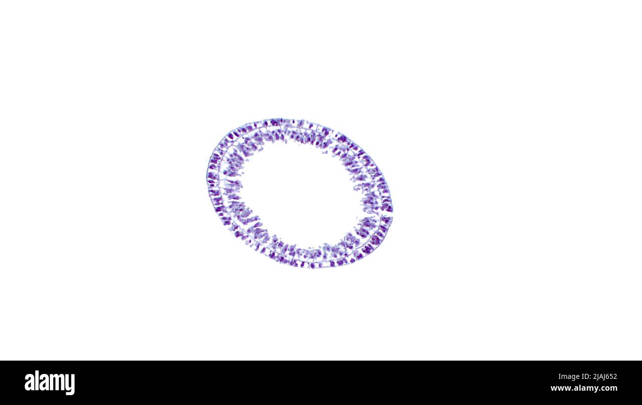 Hydra, light micrograph. Cross-section of the body showing the two layers of cells of the body wall. Educational material for lesson of zoology. Stock Photohttps://www.alamy.com/image-license-details/?v=1https://www.alamy.com/hydra-light-micrograph-cross-section-of-the-body-showing-the-two-layers-of-cells-of-the-body-wall-educational-material-for-lesson-of-zoology-image471226478.html
RF2JAJ652–Hydra, light micrograph. Cross-section of the body showing the two layers of cells of the body wall. Educational material for lesson of zoology.
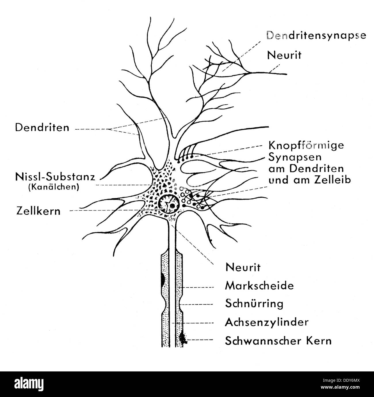 medicine, anatomy, nerve cell, schematic diagram of a ganglion cell, drawing, 20th century, 20th century, graphic, graphics, neural, neuritic, nerve pathway, nerve tract, nerve pathways, nerve tracts, neuron, neurons, ganglion, ganglia, dendrite, dendrites, synapse, cell nuclei, nuclei, nucleuses, Nissl body, myelin sheath, myelin sheath gap, axon, Schwann cell, medicine, medicines, nerve cell, nerve cells, schematic diagram, scheme, schemes, historic, historical, Additional-Rights-Clearences-Not Available Stock Photohttps://www.alamy.com/image-license-details/?v=1https://www.alamy.com/medicine-anatomy-nerve-cell-schematic-diagram-of-a-ganglion-cell-drawing-image60219626.html
RMDDY6MX–medicine, anatomy, nerve cell, schematic diagram of a ganglion cell, drawing, 20th century, 20th century, graphic, graphics, neural, neuritic, nerve pathway, nerve tract, nerve pathways, nerve tracts, neuron, neurons, ganglion, ganglia, dendrite, dendrites, synapse, cell nuclei, nuclei, nucleuses, Nissl body, myelin sheath, myelin sheath gap, axon, Schwann cell, medicine, medicines, nerve cell, nerve cells, schematic diagram, scheme, schemes, historic, historical, Additional-Rights-Clearences-Not Available
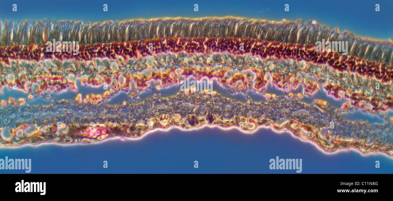 Darkfield photomicrograph of an eye retina section showing structure including rods and cone cells Stock Photohttps://www.alamy.com/image-license-details/?v=1https://www.alamy.com/stock-photo-darkfield-photomicrograph-of-an-eye-retina-section-showing-structure-35074048.html
RMC11N8G–Darkfield photomicrograph of an eye retina section showing structure including rods and cone cells
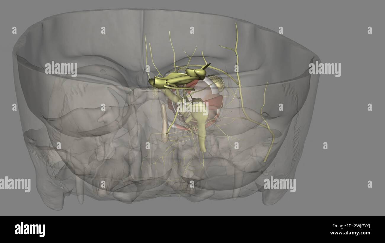 The eye light reflex, is regulated by three structures: the retina, the pretectum, and the (rods), bipolar cells, and retinal ganglion cells Stock Photohttps://www.alamy.com/image-license-details/?v=1https://www.alamy.com/the-eye-light-reflex-is-regulated-by-three-structures-the-retina-the-pretectum-and-the-rods-bipolar-cells-and-retinal-ganglion-cells-image596589494.html
RF2WJGYYJ–The eye light reflex, is regulated by three structures: the retina, the pretectum, and the (rods), bipolar cells, and retinal ganglion cells
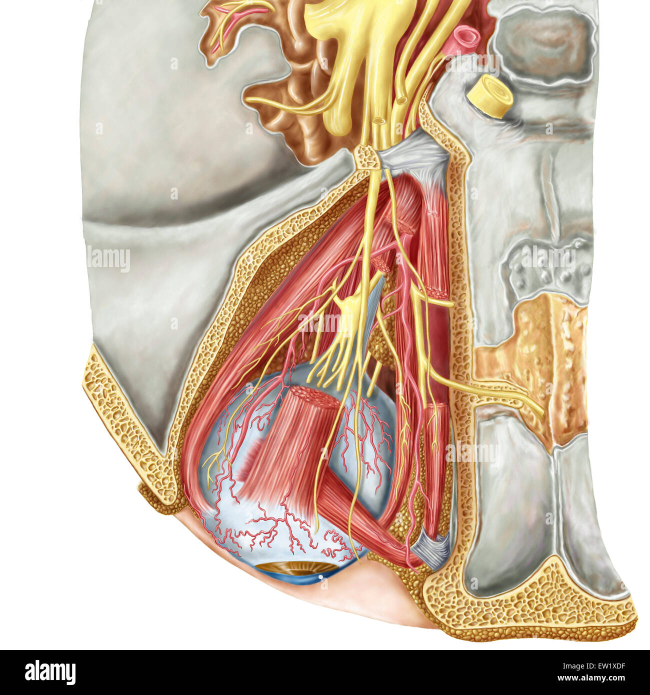 Orbital cut showing abducent nerve with ciliary ganglion and oculomotor nerve. Stock Photohttps://www.alamy.com/image-license-details/?v=1https://www.alamy.com/stock-photo-orbital-cut-showing-abducent-nerve-with-ciliary-ganglion-and-oculomotor-84250587.html
RFEW1XDF–Orbital cut showing abducent nerve with ciliary ganglion and oculomotor nerve.
 Archive image from page 177 of The anatomy, physiology, morphology and. The anatomy, physiology, morphology and development of the blow-fly (Calliphora erythrocephala.) A study in the comparative anatomy and morphology of insects; with plates and illustrations executed directly from the drawings of the author; CUbiodiversity4765349-9875 Year: 1890 ( 490 THE NERVOUS SYSTEM. continuity of the structure in question with the mantle layer renders them in the highest degree improbable. In my pre- parations the continuity of this layer with the ganglion cells of the pyramidal ganglion on the one han Stock Photohttps://www.alamy.com/image-license-details/?v=1https://www.alamy.com/archive-image-from-page-177-of-the-anatomy-physiology-morphology-and-the-anatomy-physiology-morphology-and-development-of-the-blow-fly-calliphora-erythrocephala-a-study-in-the-comparative-anatomy-and-morphology-of-insects-with-plates-and-illustrations-executed-directly-from-the-drawings-of-the-author-cubiodiversity4765349-9875-year-1890-490-the-nervous-system-continuity-of-the-structure-in-question-with-the-mantle-layer-renders-them-in-the-highest-degree-improbable-in-my-pre-parations-the-continuity-of-this-layer-with-the-ganglion-cells-of-the-pyramidal-ganglion-on-the-one-han-image264048281.html
RMW9GC7N–Archive image from page 177 of The anatomy, physiology, morphology and. The anatomy, physiology, morphology and development of the blow-fly (Calliphora erythrocephala.) A study in the comparative anatomy and morphology of insects; with plates and illustrations executed directly from the drawings of the author; CUbiodiversity4765349-9875 Year: 1890 ( 490 THE NERVOUS SYSTEM. continuity of the structure in question with the mantle layer renders them in the highest degree improbable. In my pre- parations the continuity of this layer with the ganglion cells of the pyramidal ganglion on the one han
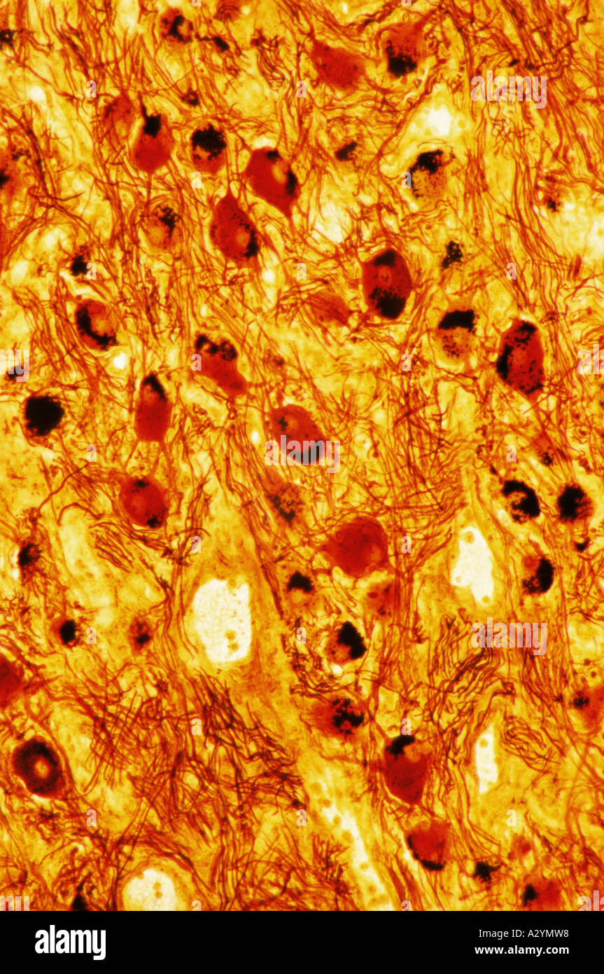 Ganglion Stock Photohttps://www.alamy.com/image-license-details/?v=1https://www.alamy.com/ganglion-image6069911.html
RFA2YMW8–Ganglion
 General physiology; an outline of the science of life . Fig. 238.—Curve of tetanus of a fatigued muscle of a frog. individual fibrillae were enormously enlarged in the fatigued, incomparison with the resting, muscle. It would lead us too far toconsider in detail the significance of these changes. Hodge (92),G. Mann (94), and Lugaro (95), have recently made known distinctmicroscopic phenomena of fatigue in the ganglion-cells of mammals,. Stock Photohttps://www.alamy.com/image-license-details/?v=1https://www.alamy.com/general-physiology-an-outline-of-the-science-of-life-fig-238curve-of-tetanus-of-a-fatigued-muscle-of-a-frog-individual-fibrillae-were-enormously-enlarged-in-the-fatigued-incomparison-with-the-resting-muscle-it-would-lead-us-too-far-toconsider-in-detail-the-significance-of-these-changes-hodge-92g-mann-94-and-lugaro-95-have-recently-made-known-distinctmicroscopic-phenomena-of-fatigue-in-the-ganglion-cells-of-mammals-image338487394.html
RM2AJKC3E–General physiology; an outline of the science of life . Fig. 238.—Curve of tetanus of a fatigued muscle of a frog. individual fibrillae were enormously enlarged in the fatigued, incomparison with the resting, muscle. It would lead us too far toconsider in detail the significance of these changes. Hodge (92),G. Mann (94), and Lugaro (95), have recently made known distinctmicroscopic phenomena of fatigue in the ganglion-cells of mammals,.
 Enterocyte, illustration Stock Photohttps://www.alamy.com/image-license-details/?v=1https://www.alamy.com/enterocyte-illustration-image595938765.html
RF2WHF9Y9–Enterocyte, illustration
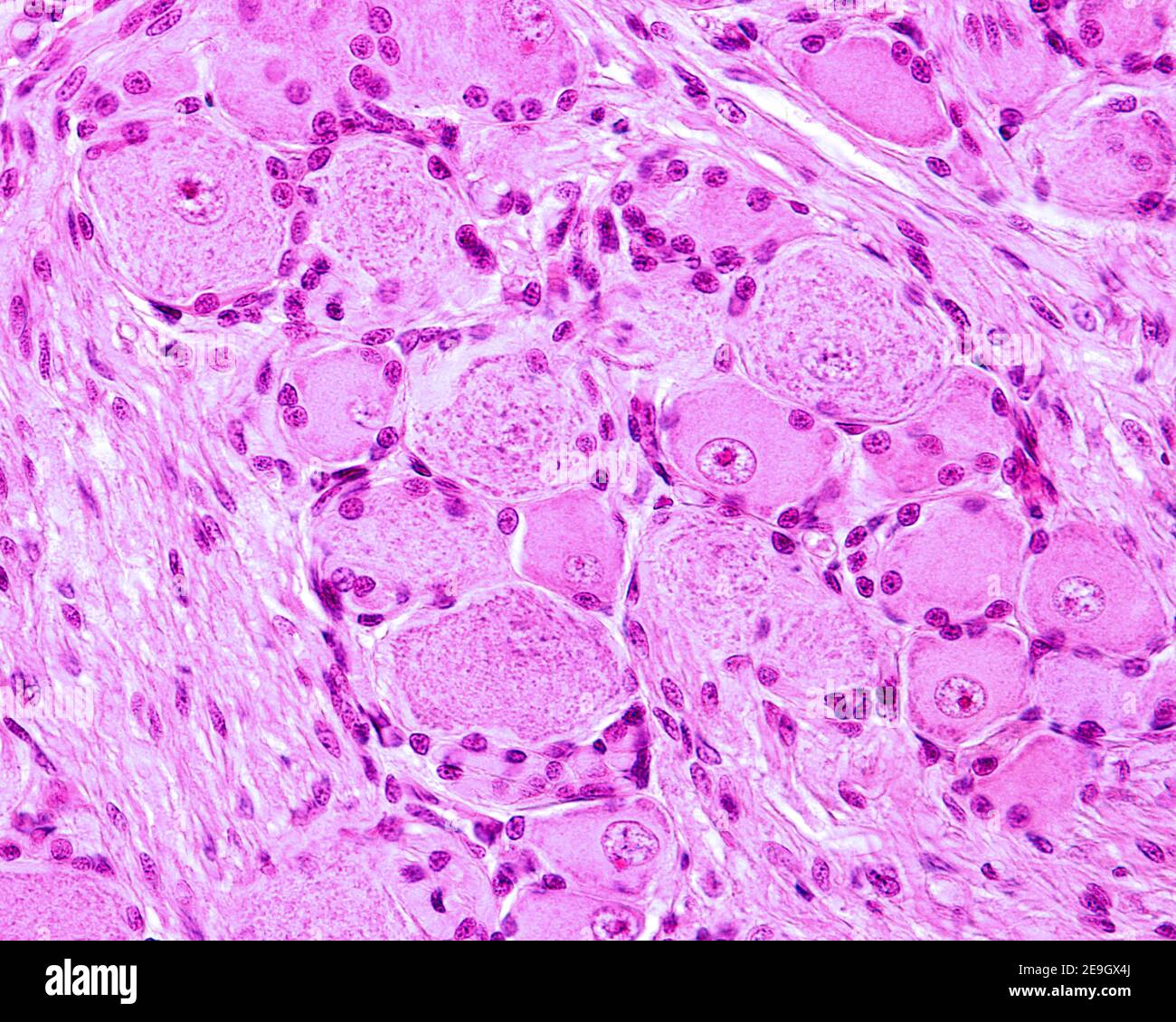 Pseudounipolar neurons of rounded soma of a dorsal root ganglion. Each cell body is surrounded by small dense nuclei corresponding to glia cells Stock Photohttps://www.alamy.com/image-license-details/?v=1https://www.alamy.com/pseudounipolar-neurons-of-rounded-soma-of-a-dorsal-root-ganglion-each-cell-body-is-surrounded-by-small-dense-nuclei-corresponding-to-glia-cells-image401742114.html
RF2E9GX4J–Pseudounipolar neurons of rounded soma of a dorsal root ganglion. Each cell body is surrounded by small dense nuclei corresponding to glia cells
 Orbital cut showing abducent nerve with ciliary ganglion and oculomotor nerve Stock Photohttps://www.alamy.com/image-license-details/?v=1https://www.alamy.com/orbital-cut-showing-abducent-nerve-with-ciliary-ganglion-and-oculomotor-nerve-image619199621.html
RM2XYAYC5–Orbital cut showing abducent nerve with ciliary ganglion and oculomotor nerve
 Spinal ganglion cross section. Light microscope X40. at 10 cm wide Stock Photohttps://www.alamy.com/image-license-details/?v=1https://www.alamy.com/spinal-ganglion-cross-section-light-microscope-x40-at-10-cm-wide-image575506878.html
RF2TC8GW2–Spinal ganglion cross section. Light microscope X40. at 10 cm wide
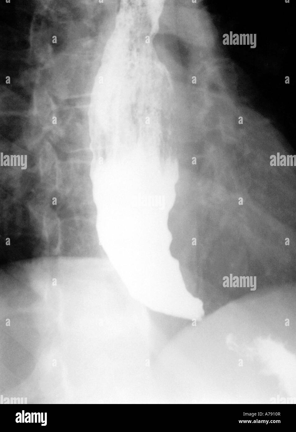 An x-ray image showing a barium study of achalasia. Stock Photohttps://www.alamy.com/image-license-details/?v=1https://www.alamy.com/stock-photo-an-x-ray-image-showing-a-barium-study-of-achalasia-11763478.html
RMA7910R–An x-ray image showing a barium study of achalasia.
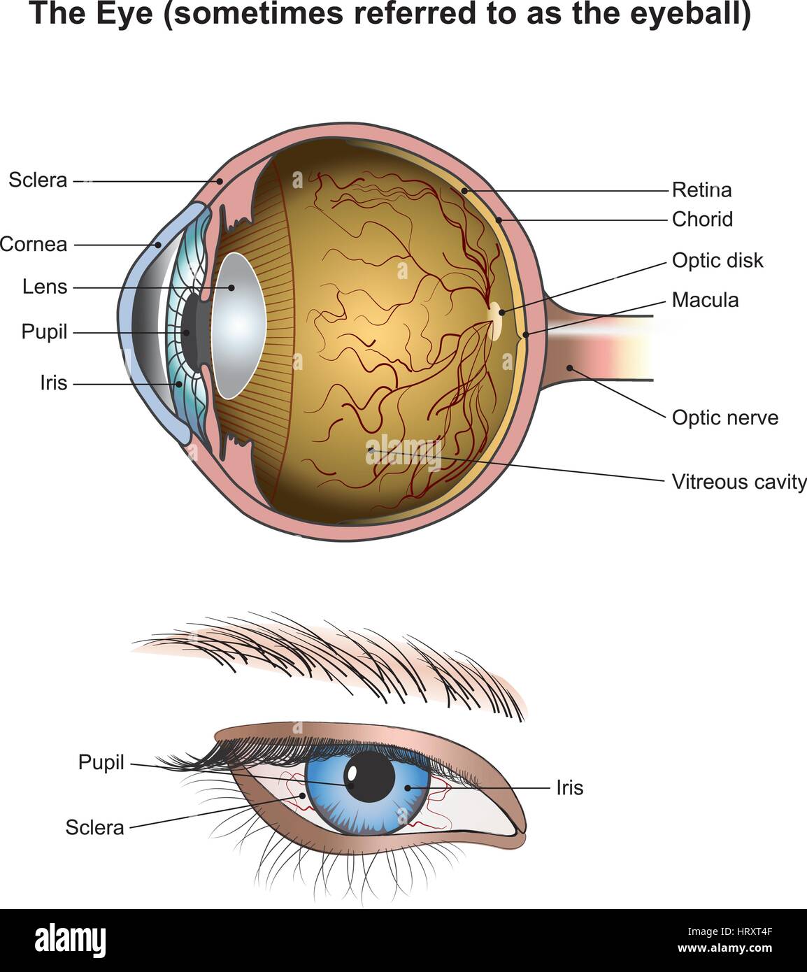 Eyes are the organs of vision. They detect light and convert it into electro-chemical impulses in neurons. In higher organisms, the eye is a complex o Stock Vectorhttps://www.alamy.com/image-license-details/?v=1https://www.alamy.com/stock-photo-eyes-are-the-organs-of-vision-they-detect-light-and-convert-it-into-135199359.html
RFHRXT4F–Eyes are the organs of vision. They detect light and convert it into electro-chemical impulses in neurons. In higher organisms, the eye is a complex o
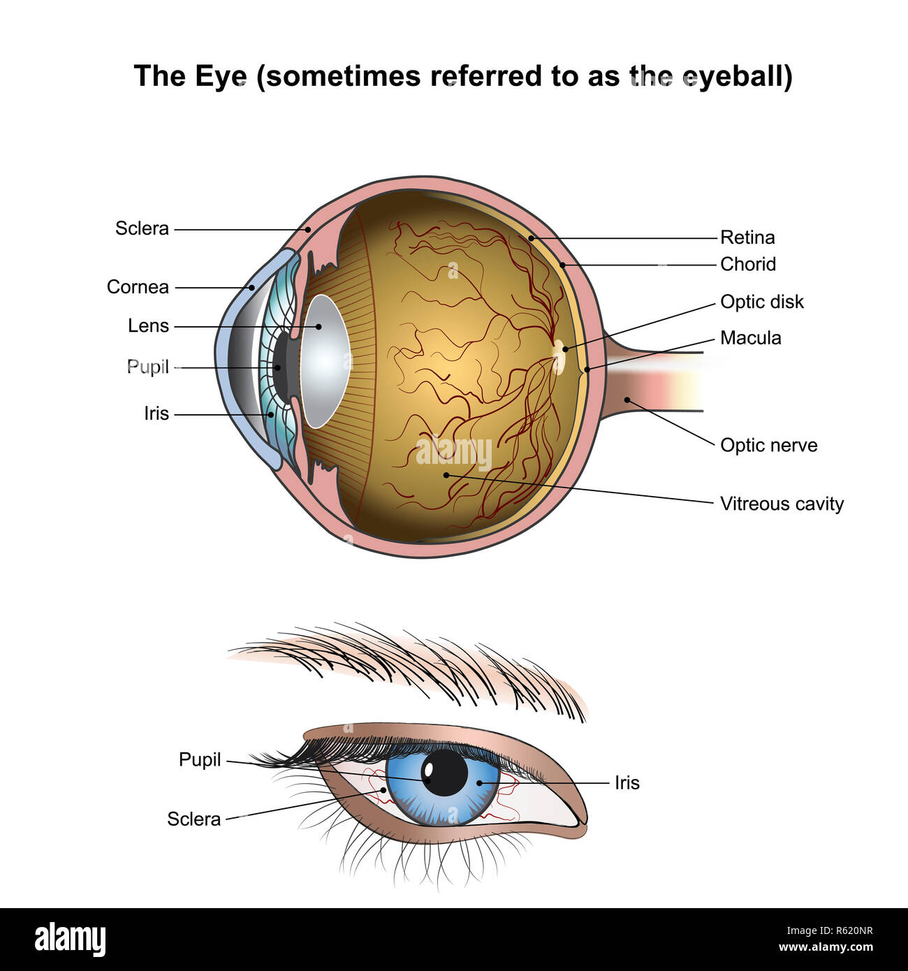 The Eyes. Illustration anatomy. Stock Photohttps://www.alamy.com/image-license-details/?v=1https://www.alamy.com/the-eyes-illustration-anatomy-image227467235.html
RFR620NR–The Eyes. Illustration anatomy.
 This illustration shows nerve cells from spinal ganglia, vintage line drawing or engraving illustration. Stock Vectorhttps://www.alamy.com/image-license-details/?v=1https://www.alamy.com/this-illustration-shows-nerve-cells-from-spinal-ganglia-vintage-line-drawing-or-engraving-illustration-image244565349.html
RFT5TWGN–This illustration shows nerve cells from spinal ganglia, vintage line drawing or engraving illustration.
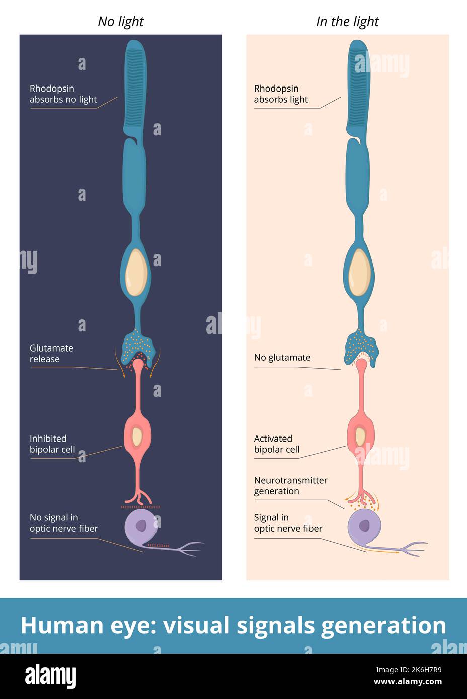 Human eye: visual signals generation. Schematic representation of nerve signal generation from rhodopsin to bipolar cell and Ganglion cell. Stock Vectorhttps://www.alamy.com/image-license-details/?v=1https://www.alamy.com/human-eye-visual-signals-generation-schematic-representation-of-nerve-signal-generation-from-rhodopsin-to-bipolar-cell-and-ganglion-cell-image485957565.html
RF2K6H7R9–Human eye: visual signals generation. Schematic representation of nerve signal generation from rhodopsin to bipolar cell and Ganglion cell.
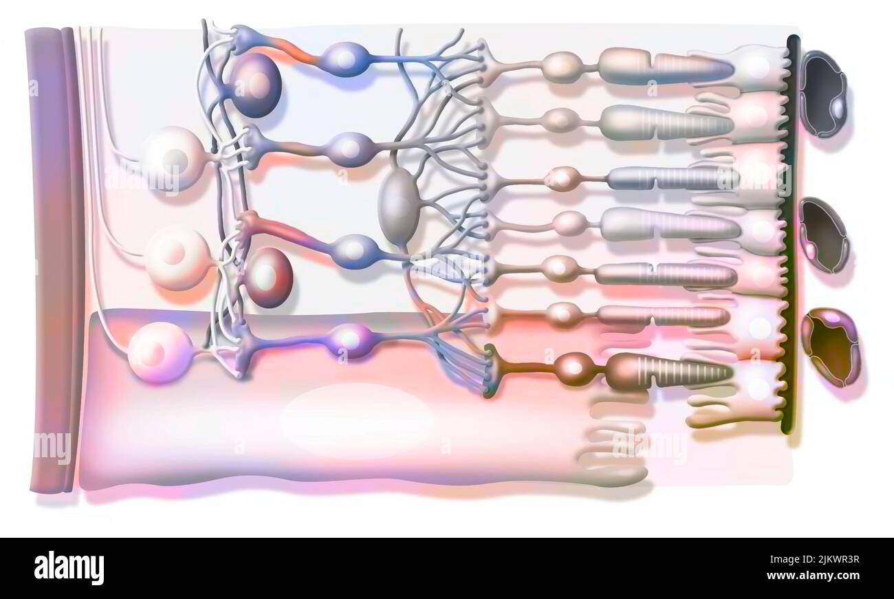 Zoom on the structure of the retina with vitreous body, internal limiting membrane, ganglion cells. Stock Photohttps://www.alamy.com/image-license-details/?v=1https://www.alamy.com/zoom-on-the-structure-of-the-retina-with-vitreous-body-internal-limiting-membrane-ganglion-cells-image476925339.html
RF2JKWR3R–Zoom on the structure of the retina with vitreous body, internal limiting membrane, ganglion cells.
 Illustration showing the structure of the retina. From top to bottom: optic nerve fiber (reddish brown strip at top), ganglion cells (in brown), amacrine cells (in dark blue), bipolar cells (in brownish-orange), horizontal cells (in dark pink), Muller gli Stock Photohttps://www.alamy.com/image-license-details/?v=1https://www.alamy.com/stock-photo-illustration-showing-the-structure-of-the-retina-from-top-to-bottom-103991420.html
RMG15638–Illustration showing the structure of the retina. From top to bottom: optic nerve fiber (reddish brown strip at top), ganglion cells (in brown), amacrine cells (in dark blue), bipolar cells (in brownish-orange), horizontal cells (in dark pink), Muller gli
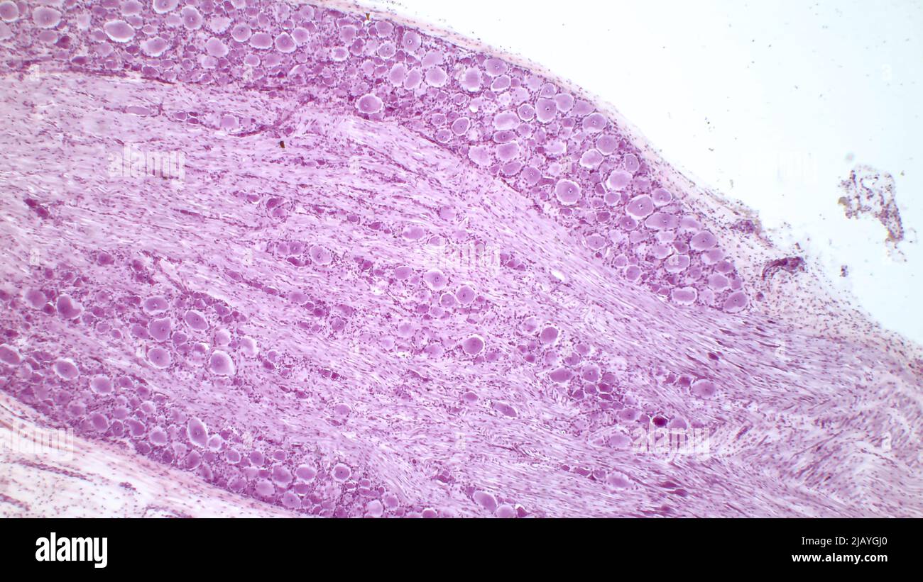 Dorsal root ganglion. Pseudounipolar neurons of a dorsal root ganglion. Hematoxlyn and eosin stain. Magnification: x40. Stock Photohttps://www.alamy.com/image-license-details/?v=1https://www.alamy.com/dorsal-root-ganglion-pseudounipolar-neurons-of-a-dorsal-root-ganglion-hematoxlyn-and-eosin-stain-magnification-x40-image471432248.html
RF2JAYGJ0–Dorsal root ganglion. Pseudounipolar neurons of a dorsal root ganglion. Hematoxlyn and eosin stain. Magnification: x40.
 A close up of an eye Stock Photohttps://www.alamy.com/image-license-details/?v=1https://www.alamy.com/stock-photo-a-close-up-of-an-eye-41979808.html
RFCC89JT–A close up of an eye
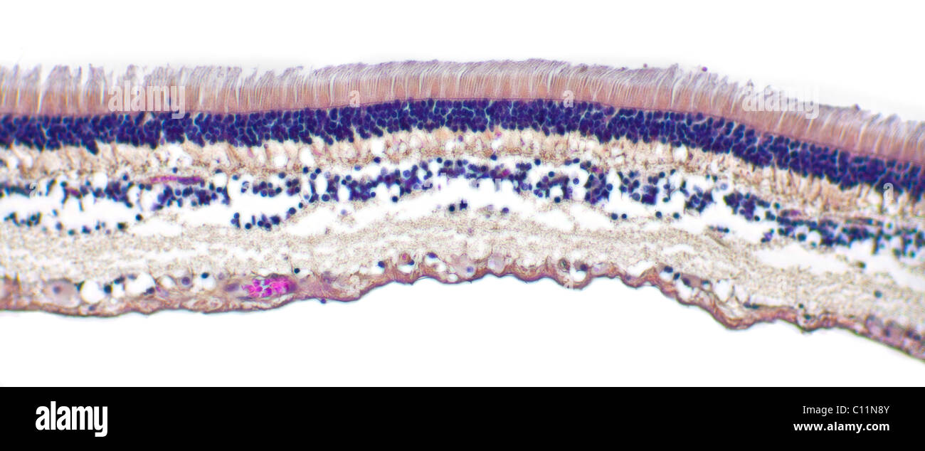 Brightfield photomicrograph of an eye retina section showing structure including rods and cone cells Stock Photohttps://www.alamy.com/image-license-details/?v=1https://www.alamy.com/stock-photo-brightfield-photomicrograph-of-an-eye-retina-section-showing-structure-35074059.html
RMC11N8Y–Brightfield photomicrograph of an eye retina section showing structure including rods and cone cells
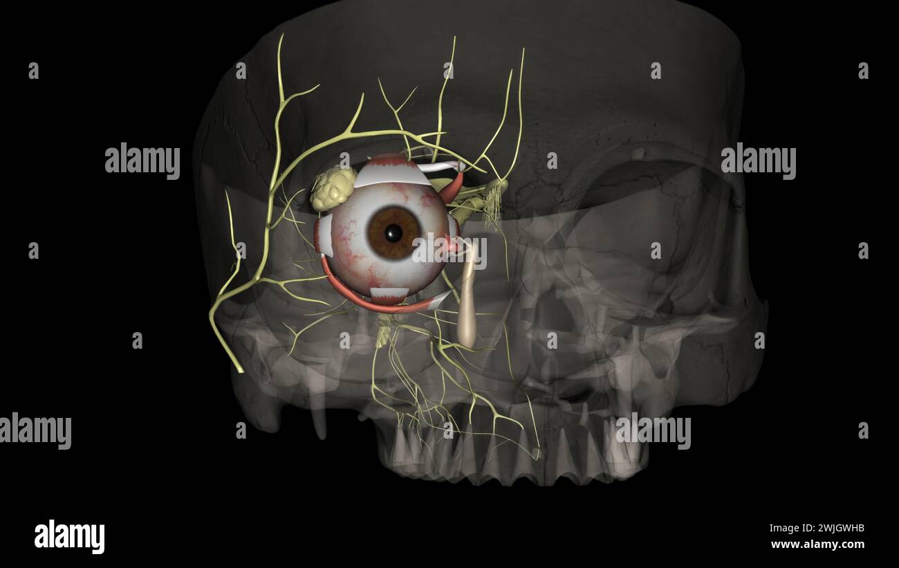 The eye light reflex, is regulated by three structures: the retina, the pretectum, and the (rods), bipolar cells, and retinal ganglion cells Stock Photohttps://www.alamy.com/image-license-details/?v=1https://www.alamy.com/the-eye-light-reflex-is-regulated-by-three-structures-the-retina-the-pretectum-and-the-rods-bipolar-cells-and-retinal-ganglion-cells-image596587639.html
RF2WJGWHB–The eye light reflex, is regulated by three structures: the retina, the pretectum, and the (rods), bipolar cells, and retinal ganglion cells
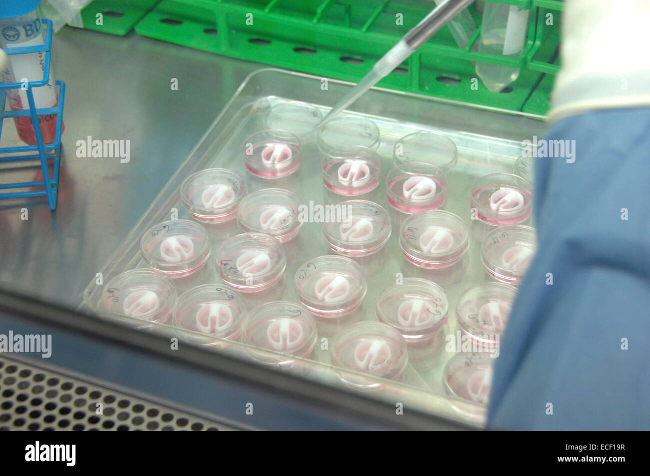 Primary cultures of superior cervical ganglia cells. Stock Photohttps://www.alamy.com/image-license-details/?v=1https://www.alamy.com/stock-photo-primary-cultures-of-superior-cervical-ganglia-cells-76547683.html
RFECF19R–Primary cultures of superior cervical ganglia cells.
 Archive image from page 64 of Cunningham's Text-book of anatomy (1914). Cunningham's Text-book of anatomy cunninghamstextb00cunn Year: 1914 ( Roof-plate Surface ectoderm Spinal ganglion (2) Sympathetic ganglion Chromaffin clls Ependyma cells Posterior nerve Posterior nerve-root Anterior nerve-root Sympathetic ganglion Chromaffin cells Basal lamina with neuroblasts (3) Gut Anterior nerve-root Sympathetic ganglion - Chromaffin cells Roots of sympathetic ganglion Sympathetic nerve Secondary sympathetic ganglion Fig. 44.—-Diagrams illustrating the formation of (1) the rudiments of the primit Stock Photohttps://www.alamy.com/image-license-details/?v=1https://www.alamy.com/archive-image-from-page-64-of-cunninghams-text-book-of-anatomy-1914-cunninghams-text-book-of-anatomy-cunninghamstextb00cunn-year-1914-roof-plate-surface-ectoderm-spinal-ganglion-2-sympathetic-ganglion-chromaffin-clls-ependyma-cells-posterior-nerve-posterior-nerve-root-anterior-nerve-root-sympathetic-ganglion-chromaffin-cells-basal-lamina-with-neuroblasts-3-gut-anterior-nerve-root-sympathetic-ganglion-chromaffin-cells-roots-of-sympathetic-ganglion-sympathetic-nerve-secondary-sympathetic-ganglion-fig-44-diagrams-illustrating-the-formation-of-1-the-rudiments-of-the-primit-image264034954.html
RMW9FR7P–Archive image from page 64 of Cunningham's Text-book of anatomy (1914). Cunningham's Text-book of anatomy cunninghamstextb00cunn Year: 1914 ( Roof-plate Surface ectoderm Spinal ganglion (2) Sympathetic ganglion Chromaffin clls Ependyma cells Posterior nerve Posterior nerve-root Anterior nerve-root Sympathetic ganglion Chromaffin cells Basal lamina with neuroblasts (3) Gut Anterior nerve-root Sympathetic ganglion - Chromaffin cells Roots of sympathetic ganglion Sympathetic nerve Secondary sympathetic ganglion Fig. 44.—-Diagrams illustrating the formation of (1) the rudiments of the primit
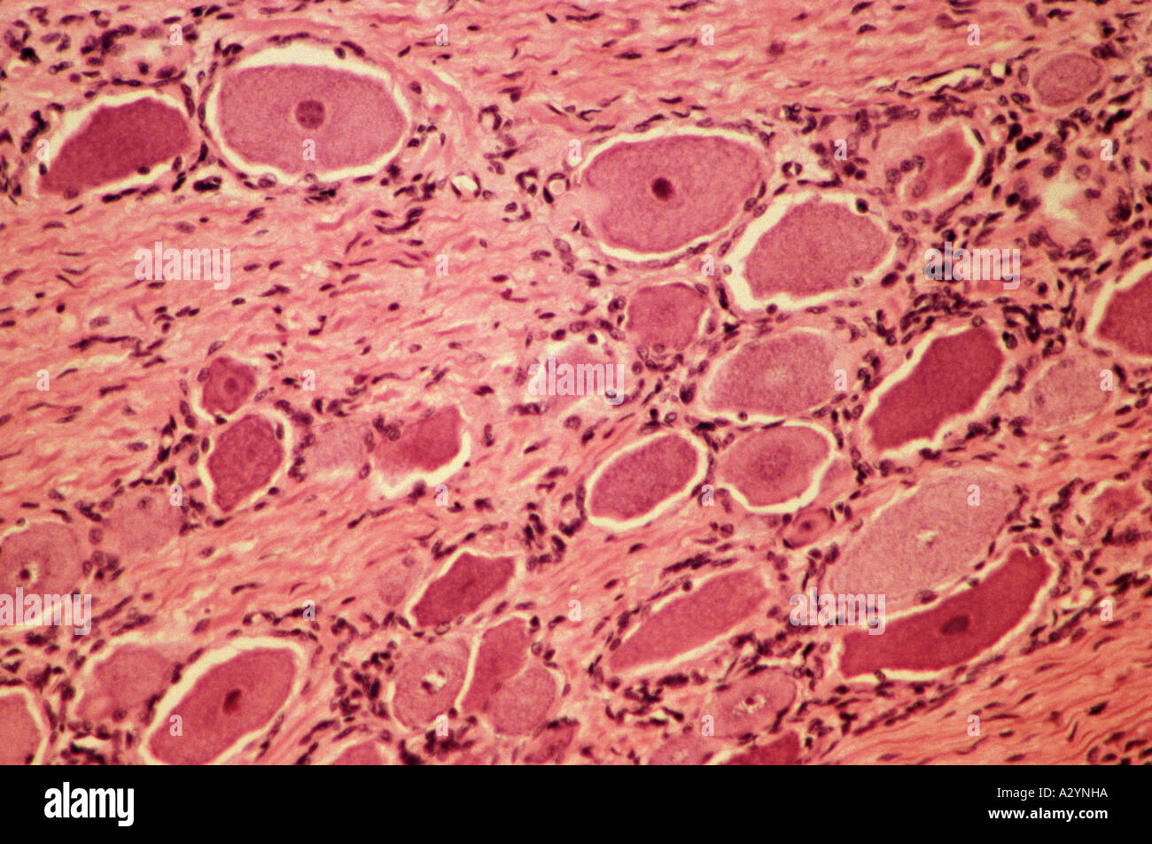 Spinal ganglion Stock Photohttps://www.alamy.com/image-license-details/?v=1https://www.alamy.com/spinal-ganglion-image6069977.html
RFA2YNHA–Spinal ganglion
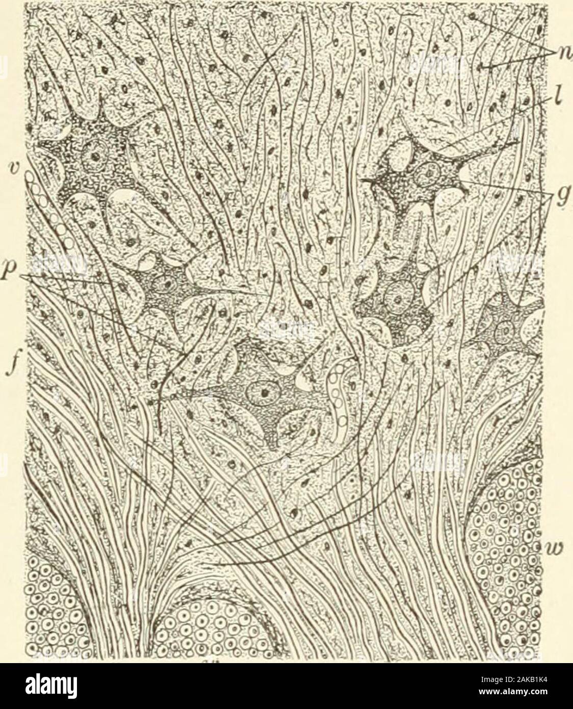 Textbook of normal histology: including an account of the development of the tissues and of the organs . om the processes of the ganglion-cells, but also the universallypresent substantia spongiosa, the modified neuroglia of the graymatter, which contributes additional nuclei and fibrils of its own.The recent advances in our knowledge concerning the processes ofnerve-cells have introduced new elements of complexity, for it mustbe remembered that, in addition to the richly-branched protoplasmicprocesses, the axis-cylinders contribute numerous fibrils both as thecollateral fibres and as the net- Stock Photohttps://www.alamy.com/image-license-details/?v=1https://www.alamy.com/textbook-of-normal-histology-including-an-account-of-the-development-of-the-tissues-and-of-the-organs-om-the-processes-of-the-ganglion-cells-but-also-the-universallypresent-substantia-spongiosa-the-modified-neuroglia-of-the-graymatter-which-contributes-additional-nuclei-and-fibrils-of-its-ownthe-recent-advances-in-our-knowledge-concerning-the-processes-ofnerve-cells-have-introduced-new-elements-of-complexity-for-it-mustbe-remembered-that-in-addition-to-the-richly-branched-protoplasmicprocesses-the-axis-cylinders-contribute-numerous-fibrils-both-as-thecollateral-fibres-and-as-the-net-image338918248.html
RM2AKB1K4–Textbook of normal histology: including an account of the development of the tissues and of the organs . om the processes of the ganglion-cells, but also the universallypresent substantia spongiosa, the modified neuroglia of the graymatter, which contributes additional nuclei and fibrils of its own.The recent advances in our knowledge concerning the processes ofnerve-cells have introduced new elements of complexity, for it mustbe remembered that, in addition to the richly-branched protoplasmicprocesses, the axis-cylinders contribute numerous fibrils both as thecollateral fibres and as the net-
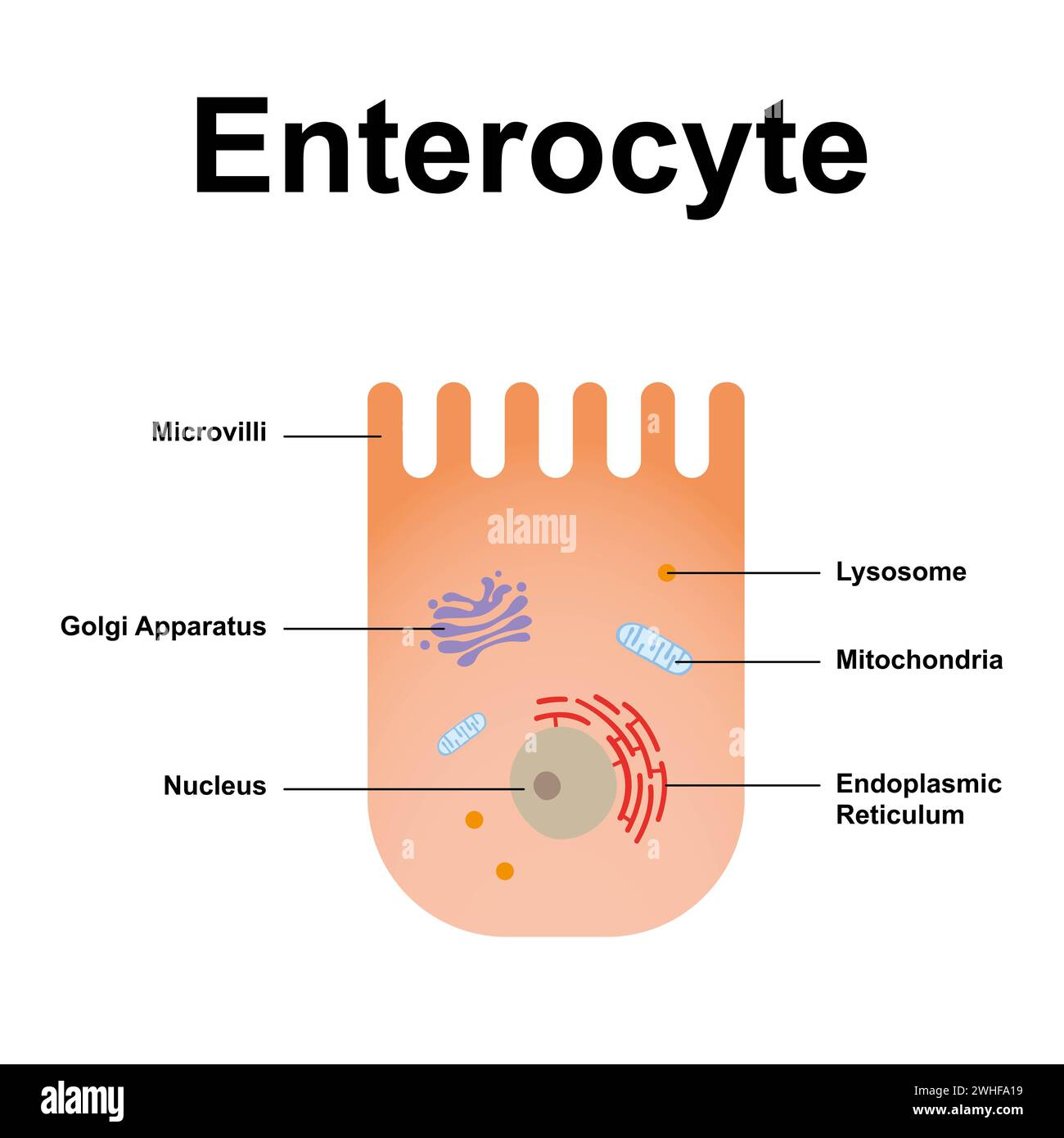 Enterocyte, illustration Stock Photohttps://www.alamy.com/image-license-details/?v=1https://www.alamy.com/enterocyte-illustration-image595938821.html
RF2WHFA19–Enterocyte, illustration
 . The anatomy, physiology, morphology and development of the blow-fly (Calliphora erythrocephala.) A study in the comparative anatomy and morphology of insects; with plates and illustrations executed directly from the drawings of the author;. Blowflies. 490 THE NERVOUS SYSTEM. continuity of the structure in question with the mantle layer renders them in the highest degree improbable. In my pre- parations the continuity of this layer with the ganglion cells of the pyramidal ganglion on the one hand, and with those of the antennal ganglia on the other, is indubitable, and its continuity with my Stock Photohttps://www.alamy.com/image-license-details/?v=1https://www.alamy.com/the-anatomy-physiology-morphology-and-development-of-the-blow-fly-calliphora-erythrocephala-a-study-in-the-comparative-anatomy-and-morphology-of-insects-with-plates-and-illustrations-executed-directly-from-the-drawings-of-the-author-blowflies-490-the-nervous-system-continuity-of-the-structure-in-question-with-the-mantle-layer-renders-them-in-the-highest-degree-improbable-in-my-pre-parations-the-continuity-of-this-layer-with-the-ganglion-cells-of-the-pyramidal-ganglion-on-the-one-hand-and-with-those-of-the-antennal-ganglia-on-the-other-is-indubitable-and-its-continuity-with-my-image216287541.html
RMPFTMY1–. The anatomy, physiology, morphology and development of the blow-fly (Calliphora erythrocephala.) A study in the comparative anatomy and morphology of insects; with plates and illustrations executed directly from the drawings of the author;. Blowflies. 490 THE NERVOUS SYSTEM. continuity of the structure in question with the mantle layer renders them in the highest degree improbable. In my pre- parations the continuity of this layer with the ganglion cells of the pyramidal ganglion on the one hand, and with those of the antennal ganglia on the other, is indubitable, and its continuity with my
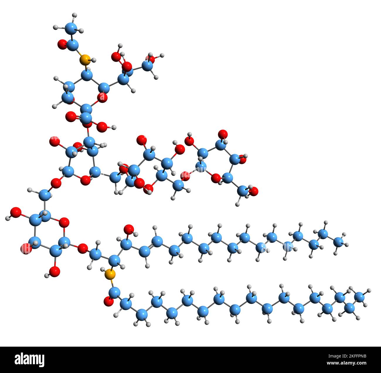 3D image of Ganglioside skeletal formula - molecular chemical structure of glycosphingolipid isolated on white background Stock Photohttps://www.alamy.com/image-license-details/?v=1https://www.alamy.com/3d-image-of-ganglioside-skeletal-formula-molecular-chemical-structure-of-glycosphingolipid-isolated-on-white-background-image491457271.html
RF2KFFPNB–3D image of Ganglioside skeletal formula - molecular chemical structure of glycosphingolipid isolated on white background
 Spinal ganglion cross section. Light microscope X40 at 10 cm wide. Stock Photohttps://www.alamy.com/image-license-details/?v=1https://www.alamy.com/spinal-ganglion-cross-section-light-microscope-x40-at-10-cm-wide-image575506881.html
RF2TC8GW5–Spinal ganglion cross section. Light microscope X40 at 10 cm wide.
![Infography on the lymphatic system. [Adobe InDesign (.indd); 3543x4842]. Stock Photo Infography on the lymphatic system. [Adobe InDesign (.indd); 3543x4842]. Stock Photo](https://c8.alamy.com/comp/2NEBFRM/infography-on-the-lymphatic-system-adobe-indesign-indd-3543x4842-2NEBFRM.jpg) Infography on the lymphatic system. [Adobe InDesign (.indd); 3543x4842]. Stock Photohttps://www.alamy.com/image-license-details/?v=1https://www.alamy.com/infography-on-the-lymphatic-system-adobe-indesign-indd-3543x4842-image525170120.html
Infography on the lymphatic system. [Adobe InDesign (.indd); 3543x4842]. Stock Photohttps://www.alamy.com/image-license-details/?v=1https://www.alamy.com/infography-on-the-lymphatic-system-adobe-indesign-indd-3543x4842-image525170120.htmlRM2NEBFRM–Infography on the lymphatic system. [Adobe InDesign (.indd); 3543x4842].
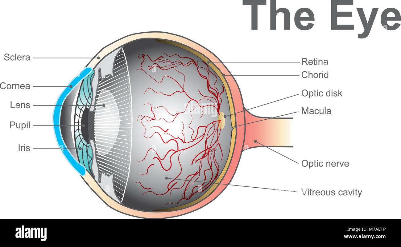 Eyes are the organs of vision. They detect light and convert it into electro-chemical impulses in neurons. Anatomy body human. Stock Vectorhttps://www.alamy.com/image-license-details/?v=1https://www.alamy.com/stock-photo-eyes-are-the-organs-of-vision-they-detect-light-and-convert-it-into-176637462.html
RFM7AETP–Eyes are the organs of vision. They detect light and convert it into electro-chemical impulses in neurons. Anatomy body human.
 The Eyes. Stock Photohttps://www.alamy.com/image-license-details/?v=1https://www.alamy.com/the-eyes-image227573384.html
RFR66T4T–The Eyes.
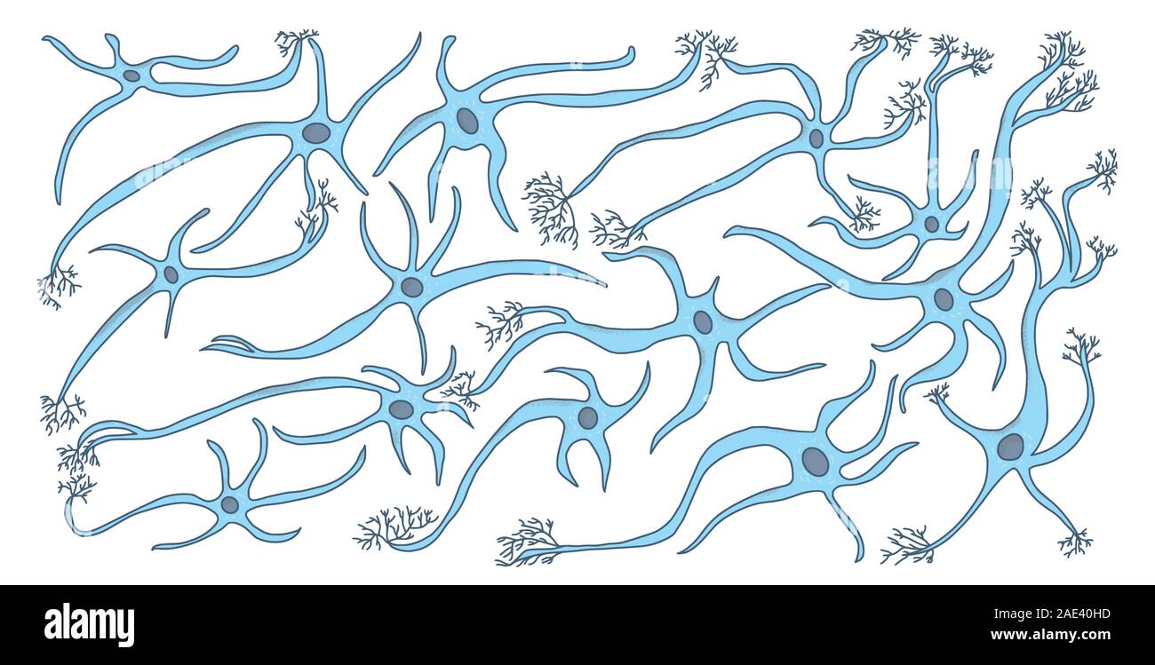 Neuron cells set. Collection of brain neurocyte. Vector illustartion. Stock Vectorhttps://www.alamy.com/image-license-details/?v=1https://www.alamy.com/neuron-cells-set-collection-of-brain-neurocyte-vector-illustartion-image335690473.html
RF2AE40HD–Neuron cells set. Collection of brain neurocyte. Vector illustartion.
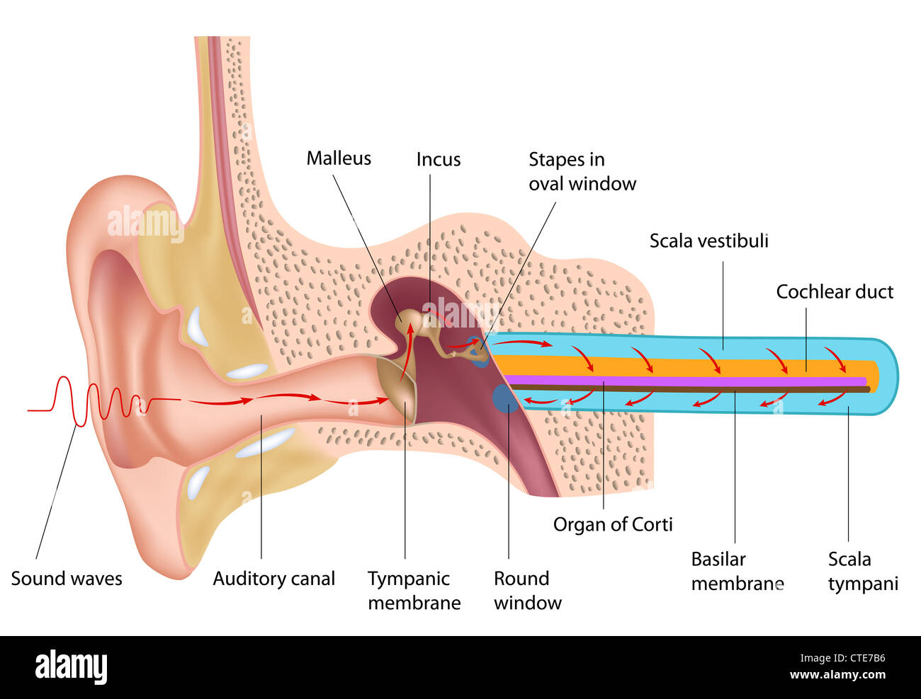 Mechanism of hearing Stock Photohttps://www.alamy.com/image-license-details/?v=1https://www.alamy.com/stock-photo-mechanism-of-hearing-49485610.html
RFCTE7B6–Mechanism of hearing
 Retinal ganglion cells are neurons that transmit information about light from the eye to the brain via a structure called the optic nerve. Here, new neurons (green) and their supporting cells, called astrocytes (red), have been created in a Petri dish from stem cells. Making retinal ganglion cells and astrocytes from stem cells could one day help doctors rewire optic nerves damaged by glaucoma. Stock Photohttps://www.alamy.com/image-license-details/?v=1https://www.alamy.com/retinal-ganglion-cells-are-neurons-that-transmit-information-about-light-from-the-eye-to-the-brain-via-a-structure-called-the-optic-nerve-here-new-neurons-green-and-their-supporting-cells-called-astrocytes-red-have-been-created-in-a-petri-dish-from-stem-cells-making-retinal-ganglion-cells-and-astrocytes-from-stem-cells-could-one-day-help-doctors-rewire-optic-nerves-damaged-by-glaucoma-image476707082.html
RM2JKFTMX–Retinal ganglion cells are neurons that transmit information about light from the eye to the brain via a structure called the optic nerve. Here, new neurons (green) and their supporting cells, called astrocytes (red), have been created in a Petri dish from stem cells. Making retinal ganglion cells and astrocytes from stem cells could one day help doctors rewire optic nerves damaged by glaucoma.
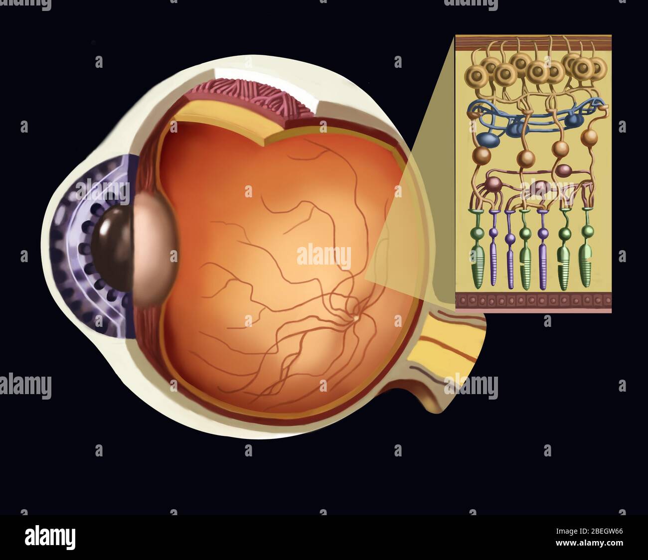 Rods And Cones Stock Photohttps://www.alamy.com/image-license-details/?v=1https://www.alamy.com/rods-and-cones-image353183550.html
RM2BEGW66–Rods And Cones
 Pseudounipolar neurons. Pseudounipolar neurons of rounded soma of a dorsal root ganglion.Сell body is surrounded by glia cells called satellite cells. Stock Photohttps://www.alamy.com/image-license-details/?v=1https://www.alamy.com/pseudounipolar-neurons-pseudounipolar-neurons-of-rounded-soma-of-a-dorsal-root-ganglionell-body-is-surrounded-by-glia-cells-called-satellite-cells-image471432281.html
RF2JAYGK5–Pseudounipolar neurons. Pseudounipolar neurons of rounded soma of a dorsal root ganglion.Сell body is surrounded by glia cells called satellite cells.
 Illustration of Pseudo-hypertrophic Infantile Paralysis circa 1881 Stock Photohttps://www.alamy.com/image-license-details/?v=1https://www.alamy.com/stock-photo-illustration-of-pseudo-hypertrophic-infantile-paralysis-circa-1881-37371272.html
RFC4PBC8–Illustration of Pseudo-hypertrophic Infantile Paralysis circa 1881
 . The elementary nervous system . 8ecFt!o°n 0? •^?»c°ord%tfrrvveerrt8e! brate (salamander); c, central canal; e, epithelium; g, grey substance com- posed of ganglion cells and fibrillar material; u>, whits substance or nerve Stock Photohttps://www.alamy.com/image-license-details/?v=1https://www.alamy.com/the-elementary-nervous-system-8ecft!on-0-cordtfrrvveerrt8e!-brate-salamander-c-central-canal-e-epithelium-g-grey-substance-com-posed-of-ganglion-cells-and-fibrillar-material-ugt-whits-substance-or-nerve-image178403760.html
RMMA6YPT–. The elementary nervous system . 8ecFt!o°n 0? •^?»c°ord%tfrrvveerrt8e! brate (salamander); c, central canal; e, epithelium; g, grey substance com- posed of ganglion cells and fibrillar material; u>, whits substance or nerve
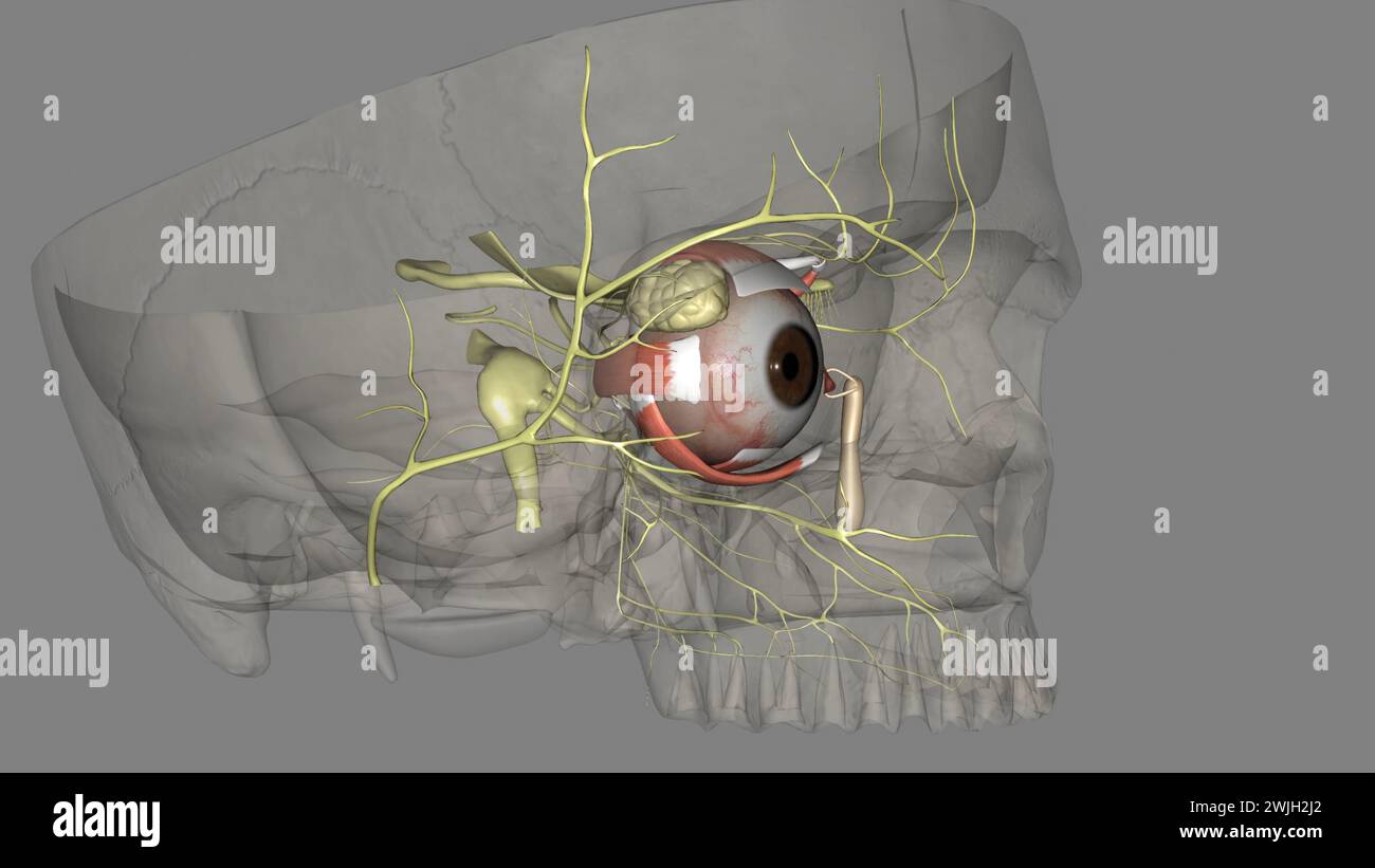 The eye light reflex, is regulated by three structures: the retina, the pretectum, and the (rods), bipolar cells, and retinal ganglion cells Stock Photohttps://www.alamy.com/image-license-details/?v=1https://www.alamy.com/the-eye-light-reflex-is-regulated-by-three-structures-the-retina-the-pretectum-and-the-rods-bipolar-cells-and-retinal-ganglion-cells-image596591578.html
RF2WJH2J2–The eye light reflex, is regulated by three structures: the retina, the pretectum, and the (rods), bipolar cells, and retinal ganglion cells
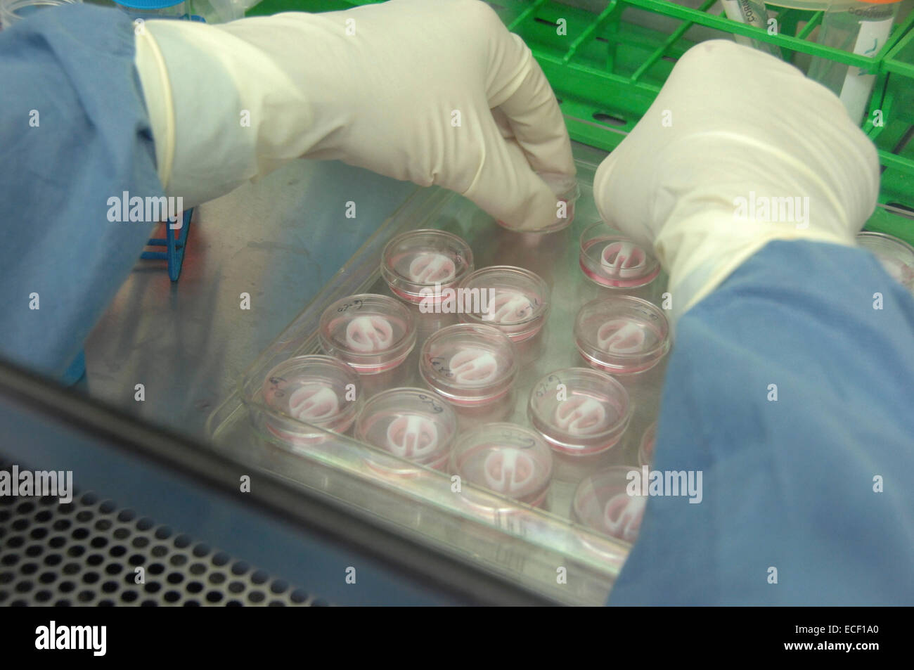 Primary cultures of superior cervical ganglia cells. Stock Photohttps://www.alamy.com/image-license-details/?v=1https://www.alamy.com/stock-photo-primary-cultures-of-superior-cervical-ganglia-cells-76547688.html
RFECF1A0–Primary cultures of superior cervical ganglia cells.
 Archive image from page 468 of The development of the human. The development of the human body : a manual of human embryology . developmentofhum00mcmu Year: 1914 THE RETINA 457 while the inner layer, that nearest the marginal velum, has larger nuclei and is presumably composed of the ganglion cells. Little is as yet known concerning the further differentiation of the nervous elements of the human retina, but the history of some of them has been traced in the cat, in which, as in other mammals, the histogenetic processes take place at a relatively later period than in man. Of the histogenesis Stock Photohttps://www.alamy.com/image-license-details/?v=1https://www.alamy.com/archive-image-from-page-468-of-the-development-of-the-human-the-development-of-the-human-body-a-manual-of-human-embryology-developmentofhum00mcmu-year-1914-the-retina-457-while-the-inner-layer-that-nearest-the-marginal-velum-has-larger-nuclei-and-is-presumably-composed-of-the-ganglion-cells-little-is-as-yet-known-concerning-the-further-differentiation-of-the-nervous-elements-of-the-human-retina-but-the-history-of-some-of-them-has-been-traced-in-the-cat-in-which-as-in-other-mammals-the-histogenetic-processes-take-place-at-a-relatively-later-period-than-in-man-of-the-histogenesis-image259032105.html
RMW1BX2H–Archive image from page 468 of The development of the human. The development of the human body : a manual of human embryology . developmentofhum00mcmu Year: 1914 THE RETINA 457 while the inner layer, that nearest the marginal velum, has larger nuclei and is presumably composed of the ganglion cells. Little is as yet known concerning the further differentiation of the nervous elements of the human retina, but the history of some of them has been traced in the cat, in which, as in other mammals, the histogenetic processes take place at a relatively later period than in man. Of the histogenesis
 The brain as an organ of mind . Fig 10.—Three bipolar Ganglion Cells from the fifth nerve of the Pike (Striekerafter Bidder). Fig. 11.—Three bipolar Ganglion Cells from the auditory nerve of the Pike : a.entirely enclosed within the medullary sheath ; b, entirely, and c, partially, exposed,to show that these ganglion cells are only expansions of the axis band. the visceral nerves, and tha-t something hke it also exists, 42 THE STRUCTURE OF. ^ Stock Photohttps://www.alamy.com/image-license-details/?v=1https://www.alamy.com/the-brain-as-an-organ-of-mind-fig-10three-bipolar-ganglion-cells-from-the-fifth-nerve-of-the-pike-striekerafter-bidder-fig-11three-bipolar-ganglion-cells-from-the-auditory-nerve-of-the-pike-aentirely-enclosed-within-the-medullary-sheath-b-entirely-and-c-partially-exposedto-show-that-these-ganglion-cells-are-only-expansions-of-the-axis-band-the-visceral-nerves-and-tha-t-something-hke-it-also-exists-42-the-structure-of-image338079412.html
RM2AJ0RMM–The brain as an organ of mind . Fig 10.—Three bipolar Ganglion Cells from the fifth nerve of the Pike (Striekerafter Bidder). Fig. 11.—Three bipolar Ganglion Cells from the auditory nerve of the Pike : a.entirely enclosed within the medullary sheath ; b, entirely, and c, partially, exposed,to show that these ganglion cells are only expansions of the axis band. the visceral nerves, and tha-t something hke it also exists, 42 THE STRUCTURE OF. ^
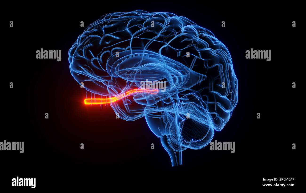 Optic nerve, illustration. Stock Photohttps://www.alamy.com/image-license-details/?v=1https://www.alamy.com/optic-nerve-illustration-image559787263.html
RF2REMEA7–Optic nerve, illustration.
 . A manual of zoology. Zoology. 112 GENERAL PRINCIPLES OF ZOOLOGY Diffuse Nervous System.—The diffuse type is certainly the most primitive; it sliows the two elements, nerve libres and ganglion cells, distributed through the whole body, or, at least, through certain layers of it. The skin of the body, the ectoderm, is one of the fundamental elements in the nervous svstem, since it is related to the external world, and hence receives the sensory impressions, so important for the de'elop- ment of nervous tissue. The corals and hydroid polyps are examples, c. Fig. 77.—Third abdominal gani^Iion o Stock Photohttps://www.alamy.com/image-license-details/?v=1https://www.alamy.com/a-manual-of-zoology-zoology-112-general-principles-of-zoology-diffuse-nervous-systemthe-diffuse-type-is-certainly-the-most-primitive-it-sliows-the-two-elements-nerve-libres-and-ganglion-cells-distributed-through-the-whole-body-or-at-least-through-certain-layers-of-it-the-skin-of-the-body-the-ectoderm-is-one-of-the-fundamental-elements-in-the-nervous-svstem-since-it-is-related-to-the-external-world-and-hence-receives-the-sensory-impressions-so-important-for-the-deelop-ment-of-nervous-tissue-the-corals-and-hydroid-polyps-are-examples-c-fig-77third-abdominal-ganiiion-o-image216442739.html
RMPG3PWR–. A manual of zoology. Zoology. 112 GENERAL PRINCIPLES OF ZOOLOGY Diffuse Nervous System.—The diffuse type is certainly the most primitive; it sliows the two elements, nerve libres and ganglion cells, distributed through the whole body, or, at least, through certain layers of it. The skin of the body, the ectoderm, is one of the fundamental elements in the nervous svstem, since it is related to the external world, and hence receives the sensory impressions, so important for the de'elop- ment of nervous tissue. The corals and hydroid polyps are examples, c. Fig. 77.—Third abdominal gani^Iion o
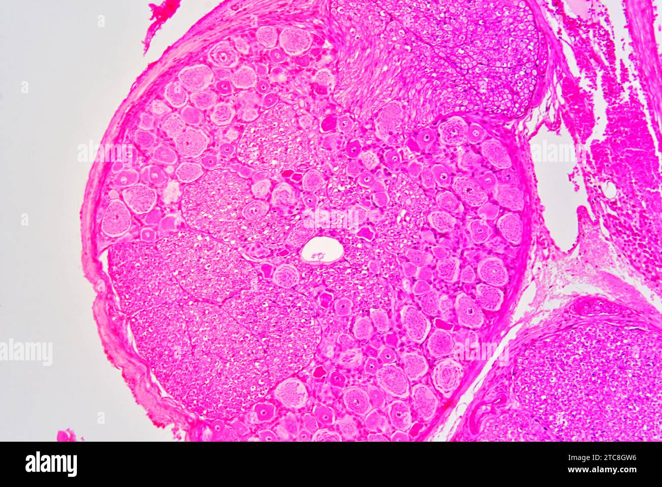 Spinal ganglion cross section. Light microscope X150 at 10 cm wide. Stock Photohttps://www.alamy.com/image-license-details/?v=1https://www.alamy.com/spinal-ganglion-cross-section-light-microscope-x150-at-10-cm-wide-image575506882.html
RF2TC8GW6–Spinal ganglion cross section. Light microscope X150 at 10 cm wide.
![Infography on nodes, also called lymph nodes, and their immune functions. [Adobe InDesign (.indd); 4795x3543]. Stock Photo Infography on nodes, also called lymph nodes, and their immune functions. [Adobe InDesign (.indd); 4795x3543]. Stock Photo](https://c8.alamy.com/comp/2NEC9EF/infography-on-nodes-also-called-lymph-nodes-and-their-immune-functions-adobe-indesign-indd-4795x3543-2NEC9EF.jpg) Infography on nodes, also called lymph nodes, and their immune functions. [Adobe InDesign (.indd); 4795x3543]. Stock Photohttps://www.alamy.com/image-license-details/?v=1https://www.alamy.com/infography-on-nodes-also-called-lymph-nodes-and-their-immune-functions-adobe-indesign-indd-4795x3543-image525187111.html
Infography on nodes, also called lymph nodes, and their immune functions. [Adobe InDesign (.indd); 4795x3543]. Stock Photohttps://www.alamy.com/image-license-details/?v=1https://www.alamy.com/infography-on-nodes-also-called-lymph-nodes-and-their-immune-functions-adobe-indesign-indd-4795x3543-image525187111.htmlRM2NEC9EF–Infography on nodes, also called lymph nodes, and their immune functions. [Adobe InDesign (.indd); 4795x3543].
 Human eye Stock Vectorhttps://www.alamy.com/image-license-details/?v=1https://www.alamy.com/human-eye-image445645830.html
RF2GW0WPE–Human eye
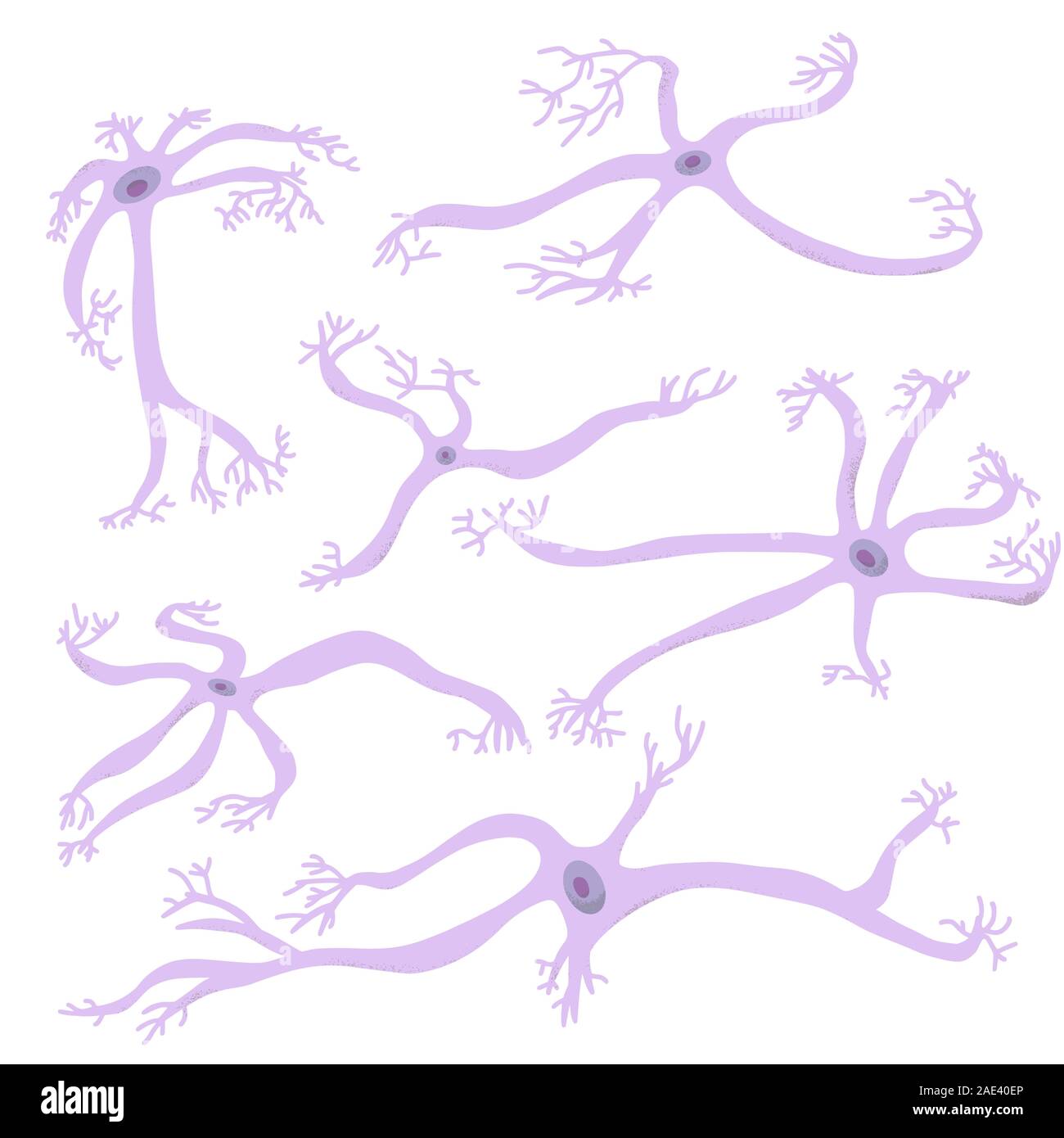 Neuron cells set. Collection of brain neurocyte. Vector illustartion. Stock Vectorhttps://www.alamy.com/image-license-details/?v=1https://www.alamy.com/neuron-cells-set-collection-of-brain-neurocyte-vector-illustartion-image335690398.html
RF2AE40EP–Neuron cells set. Collection of brain neurocyte. Vector illustartion.
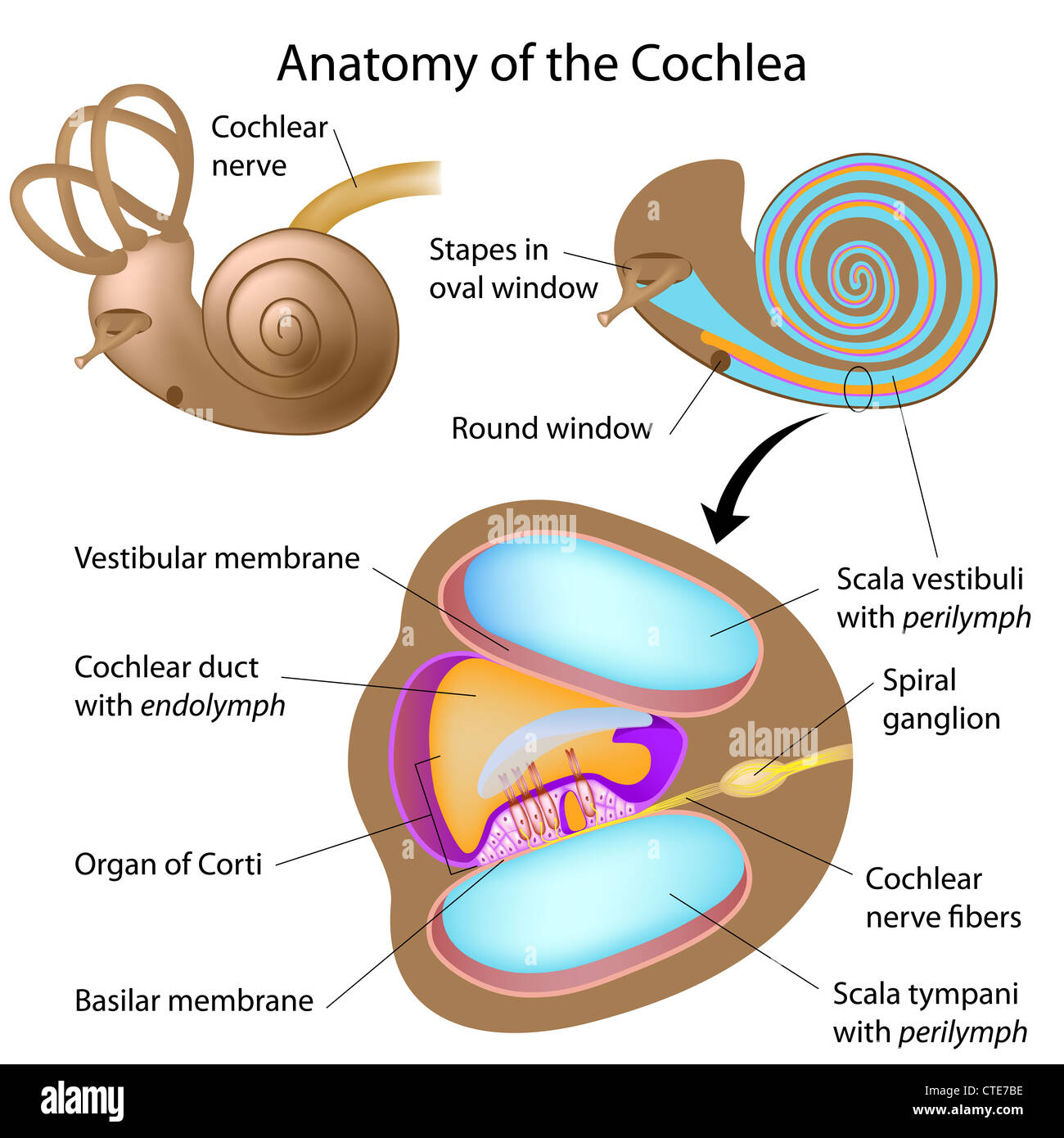 Anatomy of the cochlea of human ear Stock Photohttps://www.alamy.com/image-license-details/?v=1https://www.alamy.com/stock-photo-anatomy-of-the-cochlea-of-human-ear-49485618.html
RFCTE7BE–Anatomy of the cochlea of human ear
 Retina Stock Photohttps://www.alamy.com/image-license-details/?v=1https://www.alamy.com/stock-photo-retina-70170152.html
RME24EMT–Retina
 Retina, Fluorescent LSM Stock Photohttps://www.alamy.com/image-license-details/?v=1https://www.alamy.com/stock-photo-retina-fluorescent-lsm-135017881.html
RMHRJGK5–Retina, Fluorescent LSM
 Retina. Light microscopy of the frog (Pelophylax ridibundus) retina. Hematoxlyn and eosin stain. Stock Photohttps://www.alamy.com/image-license-details/?v=1https://www.alamy.com/retina-light-microscopy-of-the-frog-pelophylax-ridibundus-retina-hematoxlyn-and-eosin-stain-image471094111.html
RF2JAC59K–Retina. Light microscopy of the frog (Pelophylax ridibundus) retina. Hematoxlyn and eosin stain.
 Illustration of the Human Eye copyright 1904 Stock Photohttps://www.alamy.com/image-license-details/?v=1https://www.alamy.com/stock-photo-illustration-of-the-human-eye-copyright-1904-37149004.html
RFC4C7X4–Illustration of the Human Eye copyright 1904
 . The elementary nervous system . m Fio. 47.—Diagram of a com- plex type of receptor-effector sys- tem such as is seen in many parts of sea-anemones. It consists not only of receptors r, with their nerve-nets, and of muscle cells m, but also of the so-called ganglion cells g in the nerve-net. Stock Photohttps://www.alamy.com/image-license-details/?v=1https://www.alamy.com/the-elementary-nervous-system-m-fio-47diagram-of-a-com-plex-type-of-receptor-effector-sys-tem-such-as-is-seen-in-many-parts-of-sea-anemones-it-consists-not-only-of-receptors-r-with-their-nerve-nets-and-of-muscle-cells-m-but-also-of-the-so-called-ganglion-cells-g-in-the-nerve-net-image178403746.html
RMMA6YPA–. The elementary nervous system . m Fio. 47.—Diagram of a com- plex type of receptor-effector sys- tem such as is seen in many parts of sea-anemones. It consists not only of receptors r, with their nerve-nets, and of muscle cells m, but also of the so-called ganglion cells g in the nerve-net.
 The eye light reflex, is regulated by three structures: the retina, the pretectum, and the (rods), bipolar cells, and retinal ganglion cells Stock Photohttps://www.alamy.com/image-license-details/?v=1https://www.alamy.com/the-eye-light-reflex-is-regulated-by-three-structures-the-retina-the-pretectum-and-the-rods-bipolar-cells-and-retinal-ganglion-cells-image596589508.html
RF2WJH004–The eye light reflex, is regulated by three structures: the retina, the pretectum, and the (rods), bipolar cells, and retinal ganglion cells
 Primary cultures of superior cervical ganglia cells. Stock Photohttps://www.alamy.com/image-license-details/?v=1https://www.alamy.com/stock-photo-primary-cultures-of-superior-cervical-ganglia-cells-76547693.html
RFECF1A5–Primary cultures of superior cervical ganglia cells.
![Embryology of insects and myriapods; Embryology of insects and myriapods; the developmental history of insects, centipedes, and millepedes from egg desposition [!] to hatching embryologyofinse00joha Year: 1941 398 EMBRYOLOGY OF INSECTS AND MYRIAFODS muscles, and a dorsal and ventral trachea pass. The recurrent nerve arising from the posterior end of the bridge owes its origin to ganglion cells which developed from the median dorsal wall of the stomodaeum where it remains under the muscle layer. At the time the cardioblasts meet on the middorsal line, small cells are liberated from the ectoder Stock Photo Embryology of insects and myriapods; Embryology of insects and myriapods; the developmental history of insects, centipedes, and millepedes from egg desposition [!] to hatching embryologyofinse00joha Year: 1941 398 EMBRYOLOGY OF INSECTS AND MYRIAFODS muscles, and a dorsal and ventral trachea pass. The recurrent nerve arising from the posterior end of the bridge owes its origin to ganglion cells which developed from the median dorsal wall of the stomodaeum where it remains under the muscle layer. At the time the cardioblasts meet on the middorsal line, small cells are liberated from the ectoder Stock Photo](https://c8.alamy.com/comp/RWWFXX/embryology-of-insects-and-myriapods-embryology-of-insects-and-myriapods-the-developmental-history-of-insects-centipedes-and-millepedes-from-egg-desposition-!-to-hatching-embryologyofinse00joha-year-1941-398-embryology-of-insects-and-myriafods-muscles-and-a-dorsal-and-ventral-trachea-pass-the-recurrent-nerve-arising-from-the-posterior-end-of-the-bridge-owes-its-origin-to-ganglion-cells-which-developed-from-the-median-dorsal-wall-of-the-stomodaeum-where-it-remains-under-the-muscle-layer-at-the-time-the-cardioblasts-meet-on-the-middorsal-line-small-cells-are-liberated-from-the-ectoder-RWWFXX.jpg) Embryology of insects and myriapods; Embryology of insects and myriapods; the developmental history of insects, centipedes, and millepedes from egg desposition [!] to hatching embryologyofinse00joha Year: 1941 398 EMBRYOLOGY OF INSECTS AND MYRIAFODS muscles, and a dorsal and ventral trachea pass. The recurrent nerve arising from the posterior end of the bridge owes its origin to ganglion cells which developed from the median dorsal wall of the stomodaeum where it remains under the muscle layer. At the time the cardioblasts meet on the middorsal line, small cells are liberated from the ectoder Stock Photohttps://www.alamy.com/image-license-details/?v=1https://www.alamy.com/embryology-of-insects-and-myriapods-embryology-of-insects-and-myriapods-the-developmental-history-of-insects-centipedes-and-millepedes-from-egg-desposition-!-to-hatching-embryologyofinse00joha-year-1941-398-embryology-of-insects-and-myriafods-muscles-and-a-dorsal-and-ventral-trachea-pass-the-recurrent-nerve-arising-from-the-posterior-end-of-the-bridge-owes-its-origin-to-ganglion-cells-which-developed-from-the-median-dorsal-wall-of-the-stomodaeum-where-it-remains-under-the-muscle-layer-at-the-time-the-cardioblasts-meet-on-the-middorsal-line-small-cells-are-liberated-from-the-ectoder-image239662498.html
Embryology of insects and myriapods; Embryology of insects and myriapods; the developmental history of insects, centipedes, and millepedes from egg desposition [!] to hatching embryologyofinse00joha Year: 1941 398 EMBRYOLOGY OF INSECTS AND MYRIAFODS muscles, and a dorsal and ventral trachea pass. The recurrent nerve arising from the posterior end of the bridge owes its origin to ganglion cells which developed from the median dorsal wall of the stomodaeum where it remains under the muscle layer. At the time the cardioblasts meet on the middorsal line, small cells are liberated from the ectoder Stock Photohttps://www.alamy.com/image-license-details/?v=1https://www.alamy.com/embryology-of-insects-and-myriapods-embryology-of-insects-and-myriapods-the-developmental-history-of-insects-centipedes-and-millepedes-from-egg-desposition-!-to-hatching-embryologyofinse00joha-year-1941-398-embryology-of-insects-and-myriafods-muscles-and-a-dorsal-and-ventral-trachea-pass-the-recurrent-nerve-arising-from-the-posterior-end-of-the-bridge-owes-its-origin-to-ganglion-cells-which-developed-from-the-median-dorsal-wall-of-the-stomodaeum-where-it-remains-under-the-muscle-layer-at-the-time-the-cardioblasts-meet-on-the-middorsal-line-small-cells-are-liberated-from-the-ectoder-image239662498.htmlRMRWWFXX–Embryology of insects and myriapods; Embryology of insects and myriapods; the developmental history of insects, centipedes, and millepedes from egg desposition [!] to hatching embryologyofinse00joha Year: 1941 398 EMBRYOLOGY OF INSECTS AND MYRIAFODS muscles, and a dorsal and ventral trachea pass. The recurrent nerve arising from the posterior end of the bridge owes its origin to ganglion cells which developed from the median dorsal wall of the stomodaeum where it remains under the muscle layer. At the time the cardioblasts meet on the middorsal line, small cells are liberated from the ectoder
 . The American journal of anatomy. ints has been published by Weigner, 05, who studied serialsections of rodent and human material, and found it possible to traceto their destination the fibers of the n. intermedins by their histologicalcharacter; that is to say, from the frequency of the sheath nuclei, thesmall size of the fibers, and the presence of scattered ganglion cells lyingalong the course of the fibers. The common identity of the pars intermedins geniculate ganglion andchorda tympani, as described by Sapolini and Penso, in the adult is lesseasily seen in the early embryo owing to the Stock Photohttps://www.alamy.com/image-license-details/?v=1https://www.alamy.com/the-american-journal-of-anatomy-ints-has-been-published-by-weigner-05-who-studied-serialsections-of-rodent-and-human-material-and-found-it-possible-to-traceto-their-destination-the-fibers-of-the-n-intermedins-by-their-histologicalcharacter-that-is-to-say-from-the-frequency-of-the-sheath-nuclei-thesmall-size-of-the-fibers-and-the-presence-of-scattered-ganglion-cells-lyingalong-the-course-of-the-fibers-the-common-identity-of-the-pars-intermedins-geniculate-ganglion-andchorda-tympani-as-described-by-sapolini-and-penso-in-the-adult-is-lesseasily-seen-in-the-early-embryo-owing-to-the-image337137858.html
RM2AGDXNP–. The American journal of anatomy. ints has been published by Weigner, 05, who studied serialsections of rodent and human material, and found it possible to traceto their destination the fibers of the n. intermedins by their histologicalcharacter; that is to say, from the frequency of the sheath nuclei, thesmall size of the fibers, and the presence of scattered ganglion cells lyingalong the course of the fibers. The common identity of the pars intermedins geniculate ganglion andchorda tympani, as described by Sapolini and Penso, in the adult is lesseasily seen in the early embryo owing to the
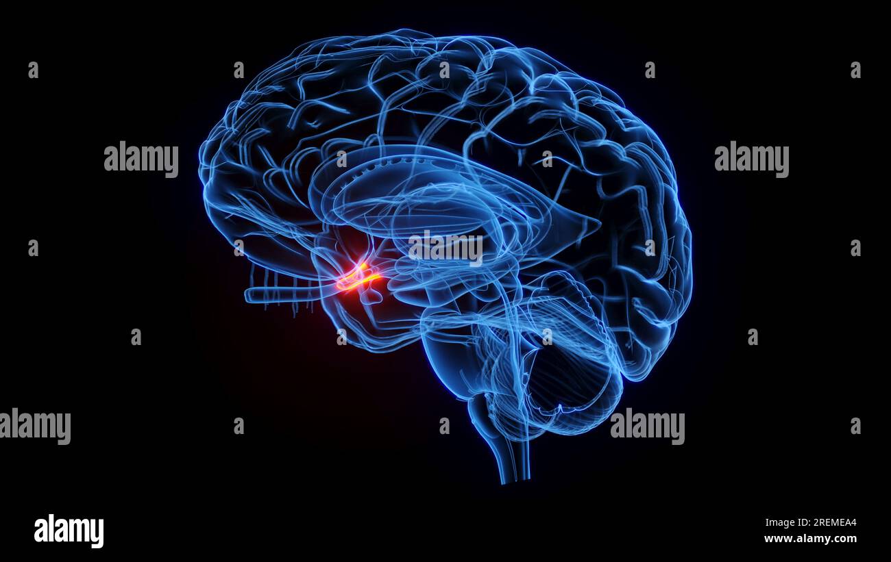 Optic chiasm, illustration. Stock Photohttps://www.alamy.com/image-license-details/?v=1https://www.alamy.com/optic-chiasm-illustration-image559787260.html
RF2REMEA4–Optic chiasm, illustration.
 . The development of the human body : a manual of human embryology. Embryology; Embryo, Non-Mammalian. THE RETINA 457 while the inner layer, that nearest the marginal velum, has larger nuclei and is presumably composed of the ganglion cells. Little is as yet known concerning the further differentiation of the nervous elements of the human retina, but the history of some of them has been traced in the cat, in which, as in other mammals, the histogenetic processes take place at a relatively later period than in man. Of the histogenesis of the inner layer the information is. Fig. 273.—Diagram sho Stock Photohttps://www.alamy.com/image-license-details/?v=1https://www.alamy.com/the-development-of-the-human-body-a-manual-of-human-embryology-embryology-embryo-non-mammalian-the-retina-457-while-the-inner-layer-that-nearest-the-marginal-velum-has-larger-nuclei-and-is-presumably-composed-of-the-ganglion-cells-little-is-as-yet-known-concerning-the-further-differentiation-of-the-nervous-elements-of-the-human-retina-but-the-history-of-some-of-them-has-been-traced-in-the-cat-in-which-as-in-other-mammals-the-histogenetic-processes-take-place-at-a-relatively-later-period-than-in-man-of-the-histogenesis-of-the-inner-layer-the-information-is-fig-273diagram-sho-image215969193.html
RMPFA6WD–. The development of the human body : a manual of human embryology. Embryology; Embryo, Non-Mammalian. THE RETINA 457 while the inner layer, that nearest the marginal velum, has larger nuclei and is presumably composed of the ganglion cells. Little is as yet known concerning the further differentiation of the nervous elements of the human retina, but the history of some of them has been traced in the cat, in which, as in other mammals, the histogenetic processes take place at a relatively later period than in man. Of the histogenesis of the inner layer the information is. Fig. 273.—Diagram sho
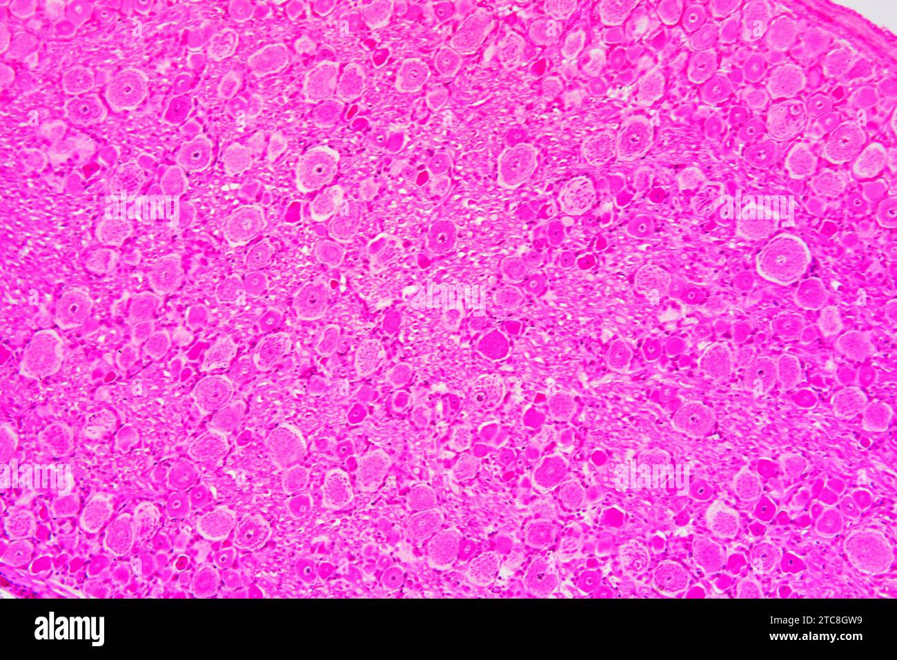 Spinal ganglion cross section. Light microscope X150 at 10 cm wide. Stock Photohttps://www.alamy.com/image-license-details/?v=1https://www.alamy.com/spinal-ganglion-cross-section-light-microscope-x150-at-10-cm-wide-image575506885.html
RF2TC8GW9–Spinal ganglion cross section. Light microscope X150 at 10 cm wide.
 Human Eye Stock Vectorhttps://www.alamy.com/image-license-details/?v=1https://www.alamy.com/human-eye-image445227496.html
RF2GT9T60–Human Eye
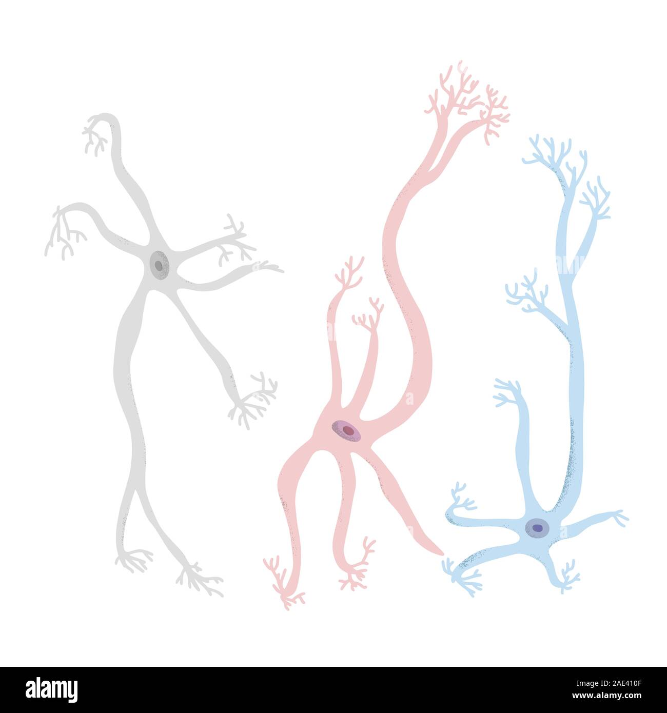 Neuron cells set. Collection of brain neurocyte. Vector illustartion. Stock Vectorhttps://www.alamy.com/image-license-details/?v=1https://www.alamy.com/neuron-cells-set-collection-of-brain-neurocyte-vector-illustartion-image335690783.html
RF2AE410F–Neuron cells set. Collection of brain neurocyte. Vector illustartion.
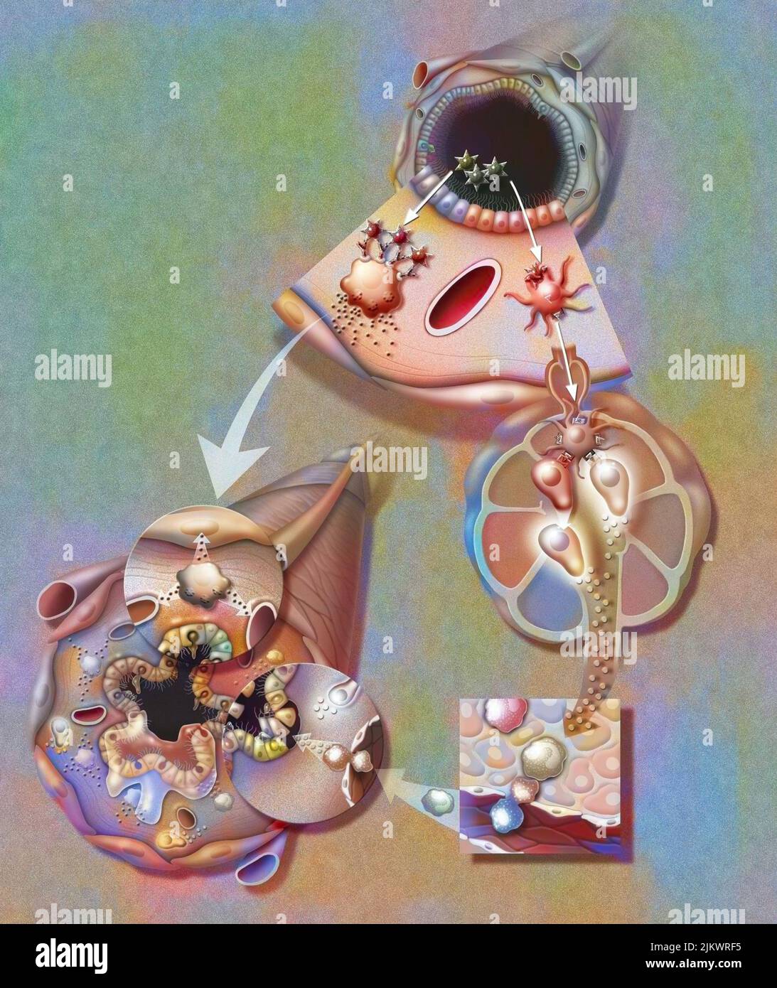 Asthma attack: Plasma cells cause the muscles of the bronchioles to contract, the vessels to dilate and the secretion of mucus. Stock Photohttps://www.alamy.com/image-license-details/?v=1https://www.alamy.com/asthma-attack-plasma-cells-cause-the-muscles-of-the-bronchioles-to-contract-the-vessels-to-dilate-and-the-secretion-of-mucus-image476925657.html
RF2JKWRF5–Asthma attack: Plasma cells cause the muscles of the bronchioles to contract, the vessels to dilate and the secretion of mucus.
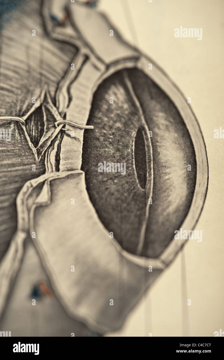 Illustration of the Human Eye copyright 1904 Stock Photohttps://www.alamy.com/image-license-details/?v=1https://www.alamy.com/stock-photo-illustration-of-the-human-eye-copyright-1904-37148632.html
RFC4C7CT–Illustration of the Human Eye copyright 1904
 . Electro-physiology . FIG. 15S.—Ganglion-cell with richly-developed nerve processes from ventral cord of cray- fish. (Methyl blue and picric acid.) (Biedermann.) ganglion-cells in the ventral cord of crustaceans and worms (Fig. 158), as well as the " collaterals " from the vertebrate spinal cord. Here the branching obviously forms connection between various Stock Photohttps://www.alamy.com/image-license-details/?v=1https://www.alamy.com/electro-physiology-fig-15sganglion-cell-with-richly-developed-nerve-processes-from-ventral-cord-of-cray-fish-methyl-blue-and-picric-acid-biedermann-ganglion-cells-in-the-ventral-cord-of-crustaceans-and-worms-fig-158-as-well-as-the-quot-collaterals-quot-from-the-vertebrate-spinal-cord-here-the-branching-obviously-forms-connection-between-various-image178411436.html
RMMA79H0–. Electro-physiology . FIG. 15S.—Ganglion-cell with richly-developed nerve processes from ventral cord of cray- fish. (Methyl blue and picric acid.) (Biedermann.) ganglion-cells in the ventral cord of crustaceans and worms (Fig. 158), as well as the " collaterals " from the vertebrate spinal cord. Here the branching obviously forms connection between various
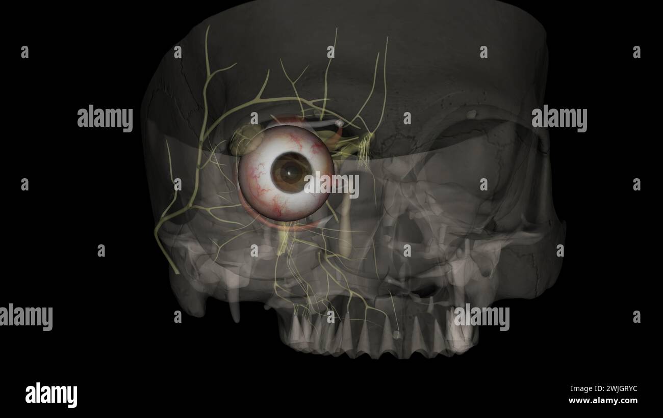 The eye light reflex, is regulated by three structures: the retina, the pretectum, and the (rods), bipolar cells, and retinal ganglion cells Stock Photohttps://www.alamy.com/image-license-details/?v=1https://www.alamy.com/the-eye-light-reflex-is-regulated-by-three-structures-the-retina-the-pretectum-and-the-rods-bipolar-cells-and-retinal-ganglion-cells-image596586352.html
RF2WJGRYC–The eye light reflex, is regulated by three structures: the retina, the pretectum, and the (rods), bipolar cells, and retinal ganglion cells
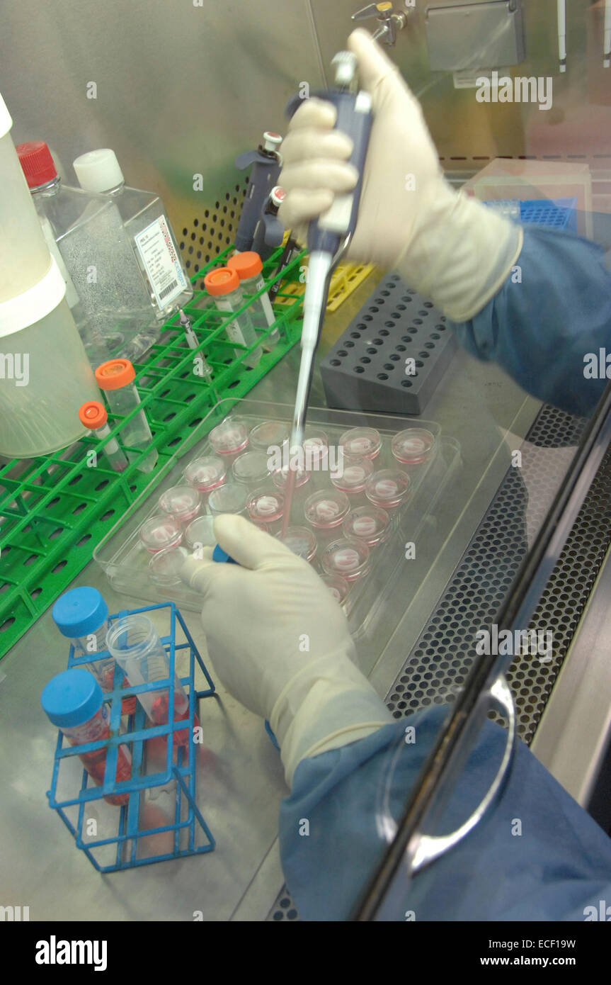 Primary cultures of superior cervical ganglia cells with pipette. Stock Photohttps://www.alamy.com/image-license-details/?v=1https://www.alamy.com/stock-photo-primary-cultures-of-superior-cervical-ganglia-cells-with-pipette-76547685.html
RFECF19W–Primary cultures of superior cervical ganglia cells with pipette.
 Elementary text-book of zoology, general part and special part: protozoa to insecta . elementarytextbo00clau Year: 1892 Ihe muscular epithelium and the jabrous layer. The ganglion cells in the upper nerve-ring are smaller, and the fibrillar given off from it pass to the tentacles. The fibrillar of the sense nerves may be derived from both rings. The marginal bodies have long been recognised as sense organs, and are either eye spots (ocelli) or auditory vesicles; hence the Hydromedusoi may be divided into two groups, the Ocellata or Vesiculata. In the Vesiculata the auditory vesicles are situa Stock Photohttps://www.alamy.com/image-license-details/?v=1https://www.alamy.com/elementary-text-book-of-zoology-general-part-and-special-part-protozoa-to-insecta-elementarytextbo00clau-year-1892-ihe-muscular-epithelium-and-the-jabrous-layer-the-ganglion-cells-in-the-upper-nerve-ring-are-smaller-and-the-fibrillar-given-off-from-it-pass-to-the-tentacles-the-fibrillar-of-the-sense-nerves-may-be-derived-from-both-rings-the-marginal-bodies-have-long-been-recognised-as-sense-organs-and-are-either-eye-spots-ocelli-or-auditory-vesicles-hence-the-hydromedusoi-may-be-divided-into-two-groups-the-ocellata-or-vesiculata-in-the-vesiculata-the-auditory-vesicles-are-situa-image241025492.html
RMT03JD8–Elementary text-book of zoology, general part and special part: protozoa to insecta . elementarytextbo00clau Year: 1892 Ihe muscular epithelium and the jabrous layer. The ganglion cells in the upper nerve-ring are smaller, and the fibrillar given off from it pass to the tentacles. The fibrillar of the sense nerves may be derived from both rings. The marginal bodies have long been recognised as sense organs, and are either eye spots (ocelli) or auditory vesicles; hence the Hydromedusoi may be divided into two groups, the Ocellata or Vesiculata. In the Vesiculata the auditory vesicles are situa
 General physiology; an outline of the science of life . aph wires is very fit-ting with respect to theprinciple of centralisationupon which the two arebased. But, as has some-times happened, such acomparison ought not to becarried too far ; for example,the nerves should not beregarded simply as conduct-ing-wires for electricity. Inreality, nerves are extensionsof ganglion-cells, and, likethese, consist of living sub-stance, i.e., they have ametabolism with which theirlife and, therefore, theirfunction are inseparablyconnected. This followsdirectly from the fact thatthe nerve invariably perishe Stock Photohttps://www.alamy.com/image-license-details/?v=1https://www.alamy.com/general-physiology-an-outline-of-the-science-of-life-aph-wires-is-very-fit-ting-with-respect-to-theprinciple-of-centralisationupon-which-the-two-arebased-but-as-has-some-times-happened-such-acomparison-ought-not-to-becarried-too-far-for-examplethe-nerves-should-not-beregarded-simply-as-conduct-ing-wires-for-electricity-inreality-nerves-are-extensionsof-ganglion-cells-and-likethese-consist-of-living-sub-stance-ie-they-have-ametabolism-with-which-theirlife-and-therefore-theirfunction-are-inseparablyconnected-this-followsdirectly-from-the-fact-thatthe-nerve-invariably-perishe-image338473828.html
RM2AJJPR0–General physiology; an outline of the science of life . aph wires is very fit-ting with respect to theprinciple of centralisationupon which the two arebased. But, as has some-times happened, such acomparison ought not to becarried too far ; for example,the nerves should not beregarded simply as conduct-ing-wires for electricity. Inreality, nerves are extensionsof ganglion-cells, and, likethese, consist of living sub-stance, i.e., they have ametabolism with which theirlife and, therefore, theirfunction are inseparablyconnected. This followsdirectly from the fact thatthe nerve invariably perishe
 Intestine epithelium, illustration. Stock Photohttps://www.alamy.com/image-license-details/?v=1https://www.alamy.com/intestine-epithelium-illustration-image626690782.html
RF2YBG6DJ–Intestine epithelium, illustration.
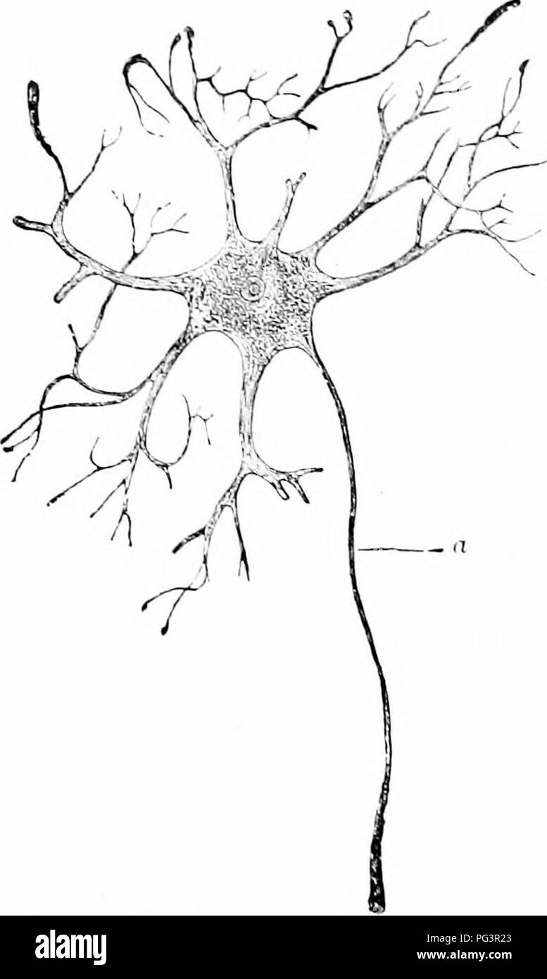 . A manual of zoology. Zoology. S4 GEXERAI, TRIXCIPLES OF ZOOLOGY. telegraph system. These are to lie eonsiilered as the specihc element of the nervous system. Ill the vertebrates the ganglion ci;lls varv greatly in size; besides small elements there are large cells, only exceeded by the eggs in size, which correspondingly hae large nuclei recalling the germinal esi- cles. Unipolar, bipolar, and multi- polar ganglion cells are recognized, the tlifterences depending upon the number of processes (nerve-tibres) which arise. In multipolar cells the number is large (fig. 52) and they are of two k Stock Photohttps://www.alamy.com/image-license-details/?v=1https://www.alamy.com/a-manual-of-zoology-zoology-s4-gexerai-trixciples-of-zoology-telegraph-system-these-are-to-lie-eonsiilered-as-the-specihc-element-of-the-nervous-system-ill-the-vertebrates-the-ganglion-cills-varv-greatly-in-size-besides-small-elements-there-are-large-cells-only-exceeded-by-the-eggs-in-size-which-correspondingly-hae-large-nuclei-recalling-the-germinal-esi-cles-unipolar-bipolar-and-multi-polar-ganglion-cells-are-recognized-the-tlifterences-depending-upon-the-number-of-processes-nerve-tibres-which-arise-in-multipolar-cells-the-number-is-large-fig-52-and-they-are-of-two-k-image216442859.html
RMPG3R23–. A manual of zoology. Zoology. S4 GEXERAI, TRIXCIPLES OF ZOOLOGY. telegraph system. These are to lie eonsiilered as the specihc element of the nervous system. Ill the vertebrates the ganglion ci;lls varv greatly in size; besides small elements there are large cells, only exceeded by the eggs in size, which correspondingly hae large nuclei recalling the germinal esi- cles. Unipolar, bipolar, and multi- polar ganglion cells are recognized, the tlifterences depending upon the number of processes (nerve-tibres) which arise. In multipolar cells the number is large (fig. 52) and they are of two k
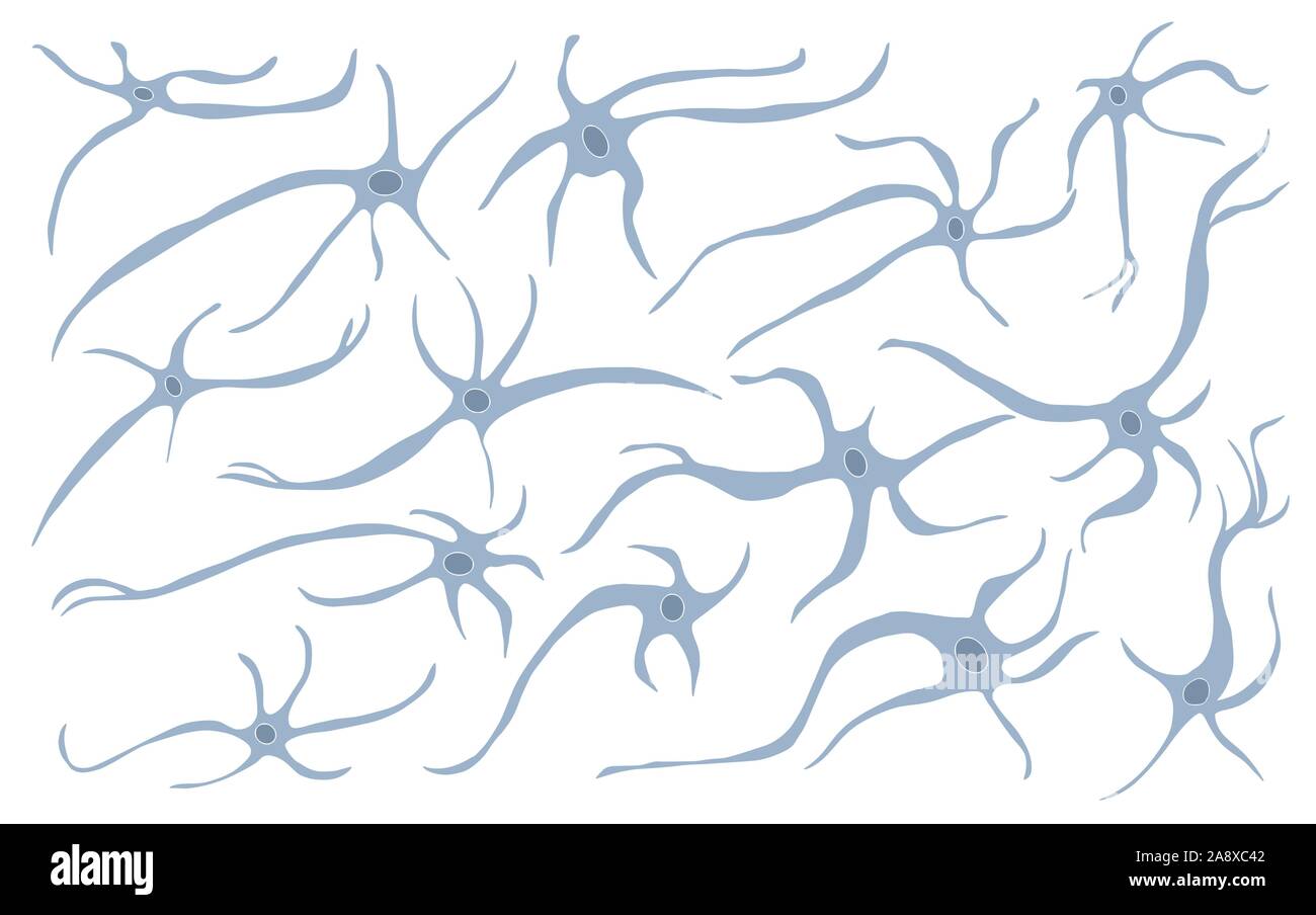 Neuron cells set. Collection of brain neurocyte. Vector illustartion. Stock Vectorhttps://www.alamy.com/image-license-details/?v=1https://www.alamy.com/neuron-cells-set-collection-of-brain-neurocyte-vector-illustartion-image332494514.html
RF2A8XC42–Neuron cells set. Collection of brain neurocyte. Vector illustartion.
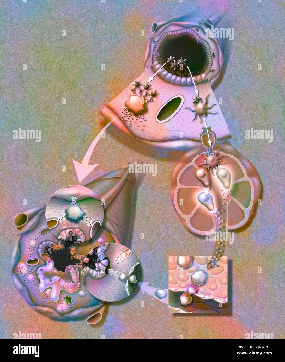 Asthma attack: Plasma cells cause the muscles of the bronchioles to contract, the vessels to dilate and the secretion of mucus. Stock Photohttps://www.alamy.com/image-license-details/?v=1https://www.alamy.com/asthma-attack-plasma-cells-cause-the-muscles-of-the-bronchioles-to-contract-the-vessels-to-dilate-and-the-secretion-of-mucus-image476925692.html
RF2JKWRGC–Asthma attack: Plasma cells cause the muscles of the bronchioles to contract, the vessels to dilate and the secretion of mucus.
 Illustration of Human Eyeball copyright 1904 Stock Photohttps://www.alamy.com/image-license-details/?v=1https://www.alamy.com/stock-photo-illustration-of-human-eyeball-copyright-1904-37148716.html
RFC4C7FT–Illustration of Human Eyeball copyright 1904
 . The elementary nervous system . FIG. 48.—Transverse section of the ventral nerve-chain of the marine worm Sigalion showing this chain as a thickened portion of the superficial ectoderm in which the sequence of tissues from the exterior inward is superficial epithelium e, ganglion cells g, and nerve fibers/. (After Hatschek, 1888.) the absence of a central station and the diffuseness of transmission, both of which are aspects of the same gen- eral condition, are the most striking characteristics of the receptor-effector system and bring this system into strong contrast with that final type of Stock Photohttps://www.alamy.com/image-license-details/?v=1https://www.alamy.com/the-elementary-nervous-system-fig-48transverse-section-of-the-ventral-nerve-chain-of-the-marine-worm-sigalion-showing-this-chain-as-a-thickened-portion-of-the-superficial-ectoderm-in-which-the-sequence-of-tissues-from-the-exterior-inward-is-superficial-epithelium-e-ganglion-cells-g-and-nerve-fibers-after-hatschek-1888-the-absence-of-a-central-station-and-the-diffuseness-of-transmission-both-of-which-are-aspects-of-the-same-gen-eral-condition-are-the-most-striking-characteristics-of-the-receptor-effector-system-and-bring-this-system-into-strong-contrast-with-that-final-type-of-image178403741.html
RMMA6YP5–. The elementary nervous system . FIG. 48.—Transverse section of the ventral nerve-chain of the marine worm Sigalion showing this chain as a thickened portion of the superficial ectoderm in which the sequence of tissues from the exterior inward is superficial epithelium e, ganglion cells g, and nerve fibers/. (After Hatschek, 1888.) the absence of a central station and the diffuseness of transmission, both of which are aspects of the same gen- eral condition, are the most striking characteristics of the receptor-effector system and bring this system into strong contrast with that final type of
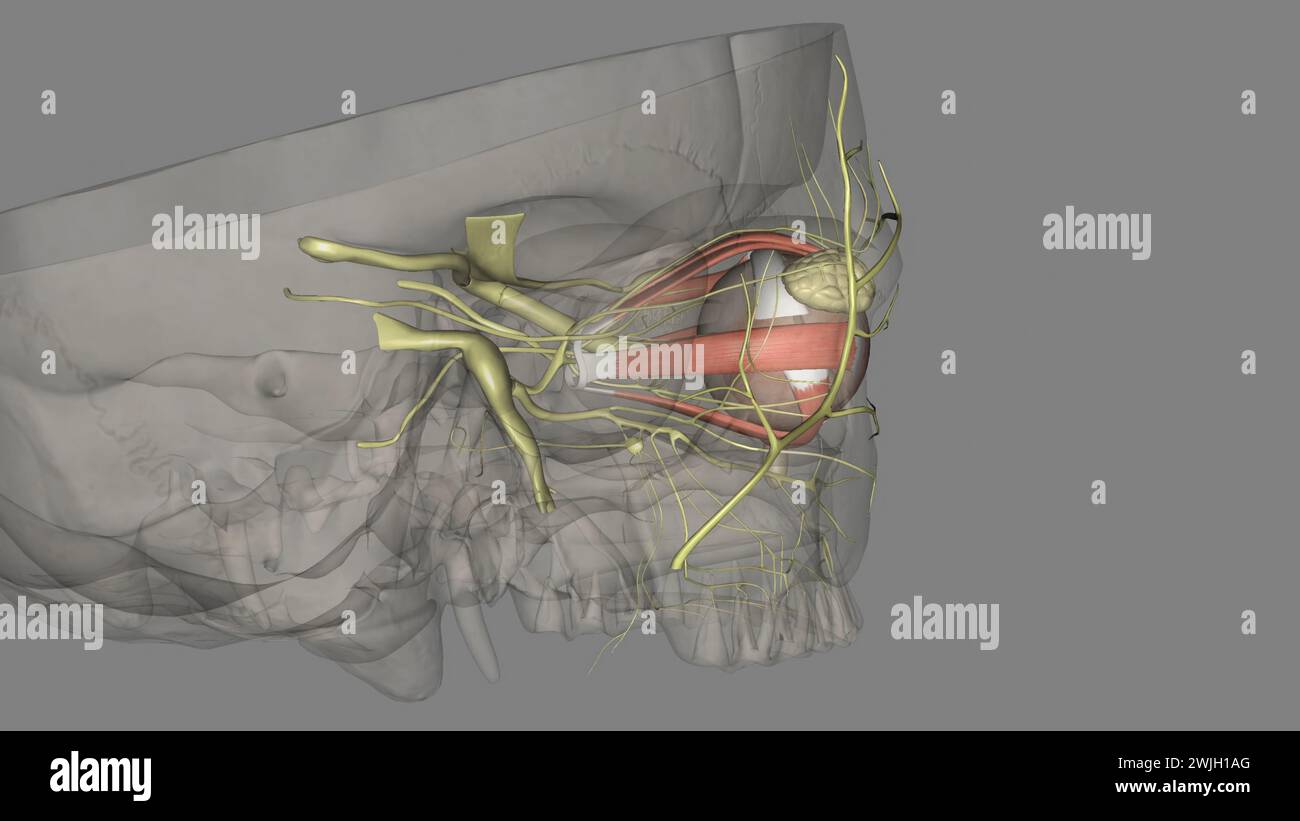 The eye light reflex, is regulated by three structures: the retina, the pretectum, and the (rods), bipolar cells, and retinal ganglion cells Stock Photohttps://www.alamy.com/image-license-details/?v=1https://www.alamy.com/the-eye-light-reflex-is-regulated-by-three-structures-the-retina-the-pretectum-and-the-rods-bipolar-cells-and-retinal-ganglion-cells-image596590584.html
RF2WJH1AG–The eye light reflex, is regulated by three structures: the retina, the pretectum, and the (rods), bipolar cells, and retinal ganglion cells
 Primary cultures of superior cervical ganglia cells with pipette. Stock Photohttps://www.alamy.com/image-license-details/?v=1https://www.alamy.com/stock-photo-primary-cultures-of-superior-cervical-ganglia-cells-with-pipette-76547689.html
RFECF1A1–Primary cultures of superior cervical ganglia cells with pipette.
 Elementary text-book of zoology (1884) Elementary text-book of zoology elementarytextbo0101clau Year: 1884 392 ANNELIDA. organs, which give rise to the a mil vesicles (fig. 315 a, AS). The rudiments both of the cerebral ganglion and of the ventral cord are derived from growths of the ectoderm,—the former from the apical plate, the latter as a paired thickening of the ventral ectoderm. The two are connected by the cesophageal ring, which is also provided with ganglion cells. In older stages, after the disappearance of the segments, the ciliary apparatus begins to degenerate and finally vanishe Stock Photohttps://www.alamy.com/image-license-details/?v=1https://www.alamy.com/elementary-text-book-of-zoology-1884-elementary-text-book-of-zoology-elementarytextbo0101clau-year-1884-392-annelida-organs-which-give-rise-to-the-a-mil-vesicles-fig-315-a-as-the-rudiments-both-of-the-cerebral-ganglion-and-of-the-ventral-cord-are-derived-from-growths-of-the-ectodermthe-former-from-the-apical-plate-the-latter-as-a-paired-thickening-of-the-ventral-ectoderm-the-two-are-connected-by-the-cesophageal-ring-which-is-also-provided-with-ganglion-cells-in-older-stages-after-the-disappearance-of-the-segments-the-ciliary-apparatus-begins-to-degenerate-and-finally-vanishe-image239661410.html
RMRWWEG2–Elementary text-book of zoology (1884) Elementary text-book of zoology elementarytextbo0101clau Year: 1884 392 ANNELIDA. organs, which give rise to the a mil vesicles (fig. 315 a, AS). The rudiments both of the cerebral ganglion and of the ventral cord are derived from growths of the ectoderm,—the former from the apical plate, the latter as a paired thickening of the ventral ectoderm. The two are connected by the cesophageal ring, which is also provided with ganglion cells. In older stages, after the disappearance of the segments, the ciliary apparatus begins to degenerate and finally vanishe
 The porifera and coelentera . anglion cells,and the transmission of stimuli is effected by simple cell contact.It must be borne in mind, however, that there is also a sub-epithelial nerve plexus with ganglion cells and nerve fibrils, thoughthe latter are not known to be connected with the aboral senseorgan. The tentacles of the Ctenophora serve for the capture of prey,and are not used in locomotion. They are most fully developedin tlie Cydippidae {Ihinniphora and Ihitrohmchia); are present,though much modified, in the Cestidae and Lobatae, but are absentin the Beroidae. In Plennihrachia and Il Stock Photohttps://www.alamy.com/image-license-details/?v=1https://www.alamy.com/the-porifera-and-coelentera-anglion-cellsand-the-transmission-of-stimuli-is-effected-by-simple-cell-contactit-must-be-borne-in-mind-however-that-there-is-also-a-sub-epithelial-nerve-plexus-with-ganglion-cells-and-nerve-fibrils-thoughthe-latter-are-not-known-to-be-connected-with-the-aboral-senseorgan-the-tentacles-of-the-ctenophora-serve-for-the-capture-of-preyand-are-not-used-in-locomotion-they-are-most-fully-developedin-tlie-cydippidae-ihinniphora-and-ihitrohmchia-are-presentthough-much-modified-in-the-cestidae-and-lobatae-but-are-absentin-the-beroidae-in-plennihrachia-and-il-image343324418.html
RM2AXFNPA–The porifera and coelentera . anglion cells,and the transmission of stimuli is effected by simple cell contact.It must be borne in mind, however, that there is also a sub-epithelial nerve plexus with ganglion cells and nerve fibrils, thoughthe latter are not known to be connected with the aboral senseorgan. The tentacles of the Ctenophora serve for the capture of prey,and are not used in locomotion. They are most fully developedin tlie Cydippidae {Ihinniphora and Ihitrohmchia); are present,though much modified, in the Cestidae and Lobatae, but are absentin the Beroidae. In Plennihrachia and Il
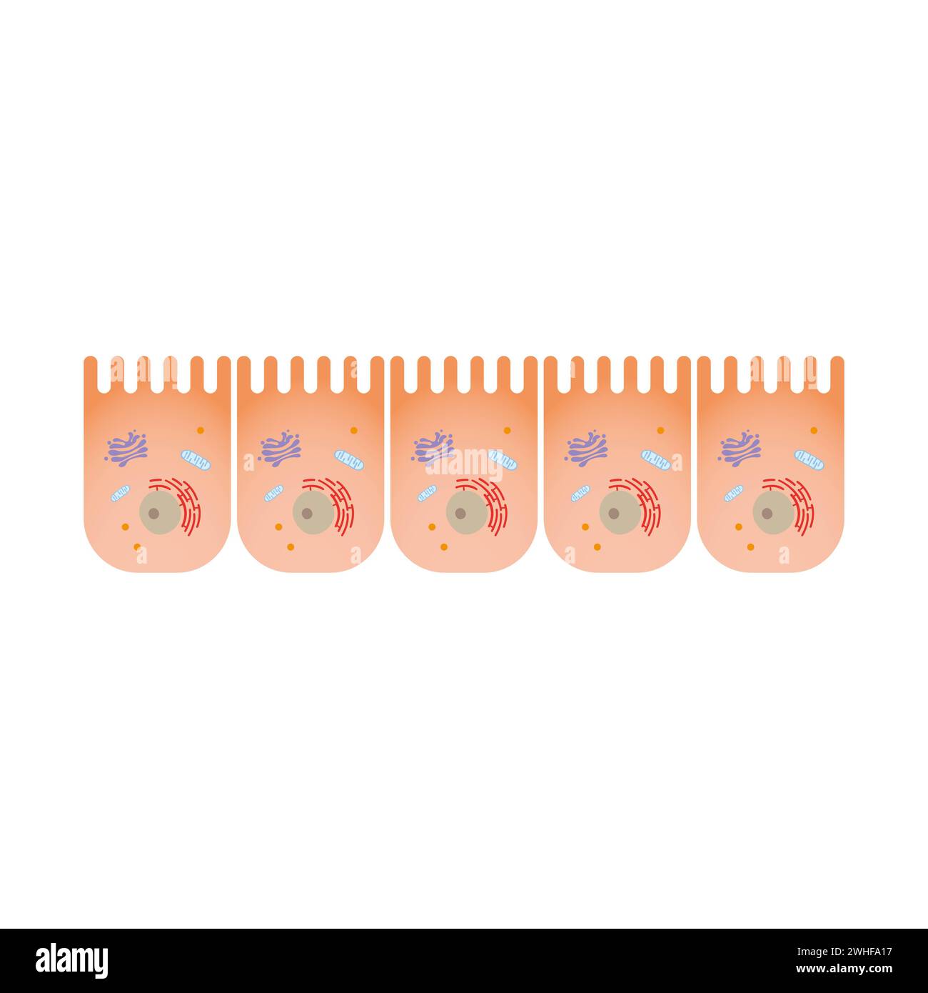 Intestine epithelium, illustration Stock Photohttps://www.alamy.com/image-license-details/?v=1https://www.alamy.com/intestine-epithelium-illustration-image595938819.html
RF2WHFA17–Intestine epithelium, illustration
 . The anatomy, physiology, morphology and development of the blow-fly (Calliphora erythrocephala.) A study in the comparative anatomy and morphology of insects; with plates and illustrations executed directly from the drawings of the author;. Blowflies. 634 THE SENSES AND SENSORY ORGANS. The setae are on an average 0"i mm. in length, and each apparently receives a process from one of the subjacent ganglion cells. A layer of small cells lies immediately beneath the cuticle, and these apparently send processes into the bases of the setae. The deeply-seated position of this organ in a small Stock Photohttps://www.alamy.com/image-license-details/?v=1https://www.alamy.com/the-anatomy-physiology-morphology-and-development-of-the-blow-fly-calliphora-erythrocephala-a-study-in-the-comparative-anatomy-and-morphology-of-insects-with-plates-and-illustrations-executed-directly-from-the-drawings-of-the-author-blowflies-634-the-senses-and-sensory-organs-the-setae-are-on-an-average-0quoti-mm-in-length-and-each-apparently-receives-a-process-from-one-of-the-subjacent-ganglion-cells-a-layer-of-small-cells-lies-immediately-beneath-the-cuticle-and-these-apparently-send-processes-into-the-bases-of-the-setae-the-deeply-seated-position-of-this-organ-in-a-small-image216287386.html
RMPFTMNE–. The anatomy, physiology, morphology and development of the blow-fly (Calliphora erythrocephala.) A study in the comparative anatomy and morphology of insects; with plates and illustrations executed directly from the drawings of the author;. Blowflies. 634 THE SENSES AND SENSORY ORGANS. The setae are on an average 0"i mm. in length, and each apparently receives a process from one of the subjacent ganglion cells. A layer of small cells lies immediately beneath the cuticle, and these apparently send processes into the bases of the setae. The deeply-seated position of this organ in a small
 Neuron cells set. Collection of brain neurocyte. Vector illustartion. Stock Vectorhttps://www.alamy.com/image-license-details/?v=1https://www.alamy.com/neuron-cells-set-collection-of-brain-neurocyte-vector-illustartion-image332494191.html
RF2A8XBMF–Neuron cells set. Collection of brain neurocyte. Vector illustartion.
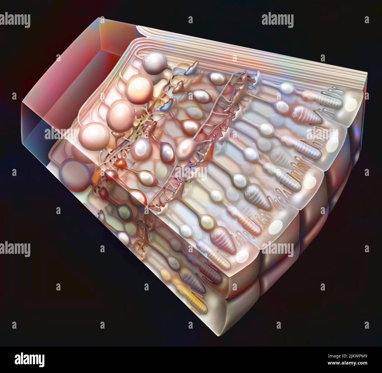 Cross-section of the retina with the photoreceptors: cones, rods, and also the optic nerve. Stock Photohttps://www.alamy.com/image-license-details/?v=1https://www.alamy.com/cross-section-of-the-retina-with-the-photoreceptors-cones-rods-and-also-the-optic-nerve-image476925017.html
RF2JKWPM9–Cross-section of the retina with the photoreceptors: cones, rods, and also the optic nerve.
 Illustration of Human Eyeball copyright 1904 Stock Photohttps://www.alamy.com/image-license-details/?v=1https://www.alamy.com/stock-photo-illustration-of-human-eyeball-copyright-1904-37148558.html
RFC4C7A6–Illustration of Human Eyeball copyright 1904
 . The elementary nervous system . FIG. 49.—Transverse section of the ventral nerve-chain of the earthworm Allolobophora showing this chain as separated from the superficial ectoderm of the worm but still retaining on its ventral or more superficial side the ganglion cells g, and on its deeper side the nerve fibers /. (After Hatschek, 1888.) ticular part of this layer and the separation of this part from the rest of the skin, together with its migration into a deeper position in the animal, are rearrangements in which are retained a good deal of the original distribu- tion of the elements as se Stock Photohttps://www.alamy.com/image-license-details/?v=1https://www.alamy.com/the-elementary-nervous-system-fig-49transverse-section-of-the-ventral-nerve-chain-of-the-earthworm-allolobophora-showing-this-chain-as-separated-from-the-superficial-ectoderm-of-the-worm-but-still-retaining-on-its-ventral-or-more-superficial-side-the-ganglion-cells-g-and-on-its-deeper-side-the-nerve-fibers-after-hatschek-1888-ticular-part-of-this-layer-and-the-separation-of-this-part-from-the-rest-of-the-skin-together-with-its-migration-into-a-deeper-position-in-the-animal-are-rearrangements-in-which-are-retained-a-good-deal-of-the-original-distribu-tion-of-the-elements-as-se-image178403759.html
RMMA6YPR–. The elementary nervous system . FIG. 49.—Transverse section of the ventral nerve-chain of the earthworm Allolobophora showing this chain as separated from the superficial ectoderm of the worm but still retaining on its ventral or more superficial side the ganglion cells g, and on its deeper side the nerve fibers /. (After Hatschek, 1888.) ticular part of this layer and the separation of this part from the rest of the skin, together with its migration into a deeper position in the animal, are rearrangements in which are retained a good deal of the original distribu- tion of the elements as se
 The eye light reflex, is regulated by three structures: the retina, the pretectum, and the (rods), bipolar cells, and retinal ganglion cells Stock Photohttps://www.alamy.com/image-license-details/?v=1https://www.alamy.com/the-eye-light-reflex-is-regulated-by-three-structures-the-retina-the-pretectum-and-the-rods-bipolar-cells-and-retinal-ganglion-cells-image596585683.html
RF2WJGR3F–The eye light reflex, is regulated by three structures: the retina, the pretectum, and the (rods), bipolar cells, and retinal ganglion cells
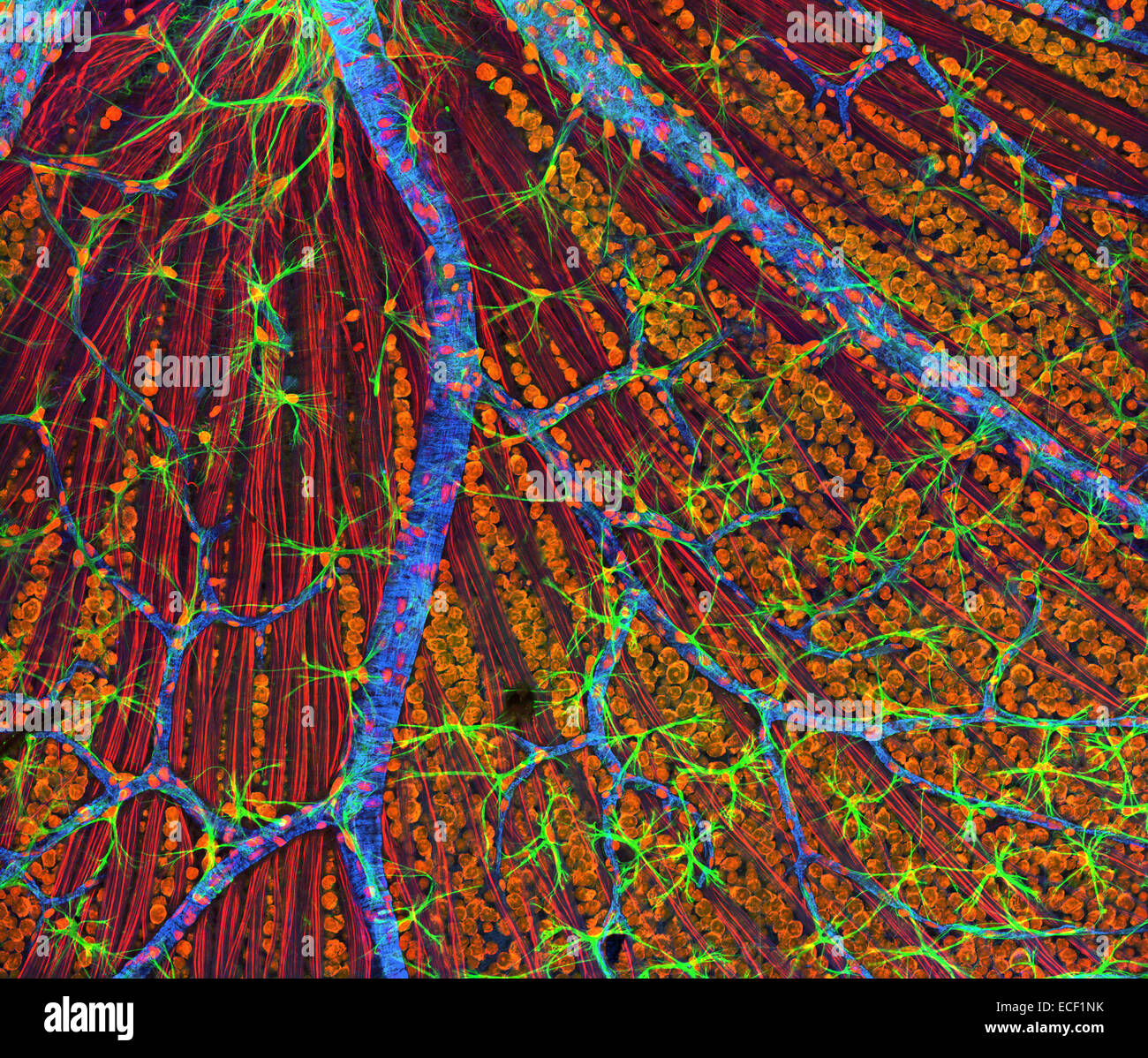 A genetic disorder of the nervous system, neurofibromatosis causes tumors to form on nerves throughout the body, including a typ Stock Photohttps://www.alamy.com/image-license-details/?v=1https://www.alamy.com/stock-photo-a-genetic-disorder-of-the-nervous-system-neurofibromatosis-causes-76548015.html
RFECF1NK–A genetic disorder of the nervous system, neurofibromatosis causes tumors to form on nerves throughout the body, including a typ
 Elementary text-book of zoology (1884) Elementary text-book of zoology elementarytextbo0101clau Year: 1884 HTDEOZOA. 239 the muscular epithelium and the fibrous layer. The ganglion cells in the upper nerve-ring are smaller, and the fibrillpe given off from it pass to the tentacles. The fibrilhf of the sense nerves may be derived from both rings. The marginal bodies have long been recognised as sense organs, and are either eye spots (ocelli) or auditory vesicles ; hence the ffydromeditsce may be divided into two groups, the Ocellata or Vesiculata. In the Vesiculata the auditory vesicles are si Stock Photohttps://www.alamy.com/image-license-details/?v=1https://www.alamy.com/elementary-text-book-of-zoology-1884-elementary-text-book-of-zoology-elementarytextbo0101clau-year-1884-htdeozoa-239-the-muscular-epithelium-and-the-fibrous-layer-the-ganglion-cells-in-the-upper-nerve-ring-are-smaller-and-the-fibrillpe-given-off-from-it-pass-to-the-tentacles-the-fibrilhf-of-the-sense-nerves-may-be-derived-from-both-rings-the-marginal-bodies-have-long-been-recognised-as-sense-organs-and-are-either-eye-spots-ocelli-or-auditory-vesicles-hence-the-ffydromeditsce-may-be-divided-into-two-groups-the-ocellata-or-vesiculata-in-the-vesiculata-the-auditory-vesicles-are-si-image239621372.html
RMRWRKE4–Elementary text-book of zoology (1884) Elementary text-book of zoology elementarytextbo0101clau Year: 1884 HTDEOZOA. 239 the muscular epithelium and the fibrous layer. The ganglion cells in the upper nerve-ring are smaller, and the fibrillpe given off from it pass to the tentacles. The fibrilhf of the sense nerves may be derived from both rings. The marginal bodies have long been recognised as sense organs, and are either eye spots (ocelli) or auditory vesicles ; hence the ffydromeditsce may be divided into two groups, the Ocellata or Vesiculata. In the Vesiculata the auditory vesicles are si
 AMAarchives of neurology & psychiatry . t was highly important to attempt to estab-lish the sequence of events with reference to the involvement of the variousstructural units including the myelin sheath of nerve fibers, the axis cylinders,the interstitial supporting glial elements, the ganglion cells of the gray matter,and also the mesodermal structures, particularly the blood vessels. GLOBUS-STRALSS—FUMCULAR MYELOPATHY 371 The Myelin Sheath : Our material showed conclusively and uniformly thatthe myelin sheath was the seat of the most intense disease process. In areasin which the softening p Stock Photohttps://www.alamy.com/image-license-details/?v=1https://www.alamy.com/amaarchives-of-neurology-psychiatry-t-was-highly-important-to-attempt-to-estab-lish-the-sequence-of-events-with-reference-to-the-involvement-of-the-variousstructural-units-including-the-myelin-sheath-of-nerve-fibers-the-axis-cylindersthe-interstitial-supporting-glial-elements-the-ganglion-cells-of-the-gray-matterand-also-the-mesodermal-structures-particularly-the-blood-vessels-globus-stralssfumcular-myelopathy-371-the-myelin-sheath-our-material-showed-conclusively-and-uniformly-thatthe-myelin-sheath-was-the-seat-of-the-most-intense-disease-process-in-areasin-which-the-softening-p-image340238051.html
RM2ANF52Y–AMAarchives of neurology & psychiatry . t was highly important to attempt to estab-lish the sequence of events with reference to the involvement of the variousstructural units including the myelin sheath of nerve fibers, the axis cylinders,the interstitial supporting glial elements, the ganglion cells of the gray matter,and also the mesodermal structures, particularly the blood vessels. GLOBUS-STRALSS—FUMCULAR MYELOPATHY 371 The Myelin Sheath : Our material showed conclusively and uniformly thatthe myelin sheath was the seat of the most intense disease process. In areasin which the softening p
 . A manual of zoology. Zoology. of nervous elements. In the medusce such a place is the rim of the bell; consequently a stronger nerve-cord much richer in ganglion cells is found here. This, as well as the nerve-ring and the five radial nerves of echinoderms, may be called a central system, thereby distinguishing the rest of the nervous network as the peripheral nervous system. Ganglionic Central Nervous System.—Numerous transitional forms lead to the ganglionic central nervous system of the worms, molluscs, and arthropods (fig. 77). The central nervous system here consists of two or more gang Stock Photohttps://www.alamy.com/image-license-details/?v=1https://www.alamy.com/a-manual-of-zoology-zoology-of-nervous-elements-in-the-medusce-such-a-place-is-the-rim-of-the-bell-consequently-a-stronger-nerve-cord-much-richer-in-ganglion-cells-is-found-here-this-as-well-as-the-nerve-ring-and-the-five-radial-nerves-of-echinoderms-may-be-called-a-central-system-thereby-distinguishing-the-rest-of-the-nervous-network-as-the-peripheral-nervous-system-ganglionic-central-nervous-systemnumerous-transitional-forms-lead-to-the-ganglionic-central-nervous-system-of-the-worms-molluscs-and-arthropods-fig-77-the-central-nervous-system-here-consists-of-two-or-more-gang-image216442731.html
RMPG3PWF–. A manual of zoology. Zoology. of nervous elements. In the medusce such a place is the rim of the bell; consequently a stronger nerve-cord much richer in ganglion cells is found here. This, as well as the nerve-ring and the five radial nerves of echinoderms, may be called a central system, thereby distinguishing the rest of the nervous network as the peripheral nervous system. Ganglionic Central Nervous System.—Numerous transitional forms lead to the ganglionic central nervous system of the worms, molluscs, and arthropods (fig. 77). The central nervous system here consists of two or more gang
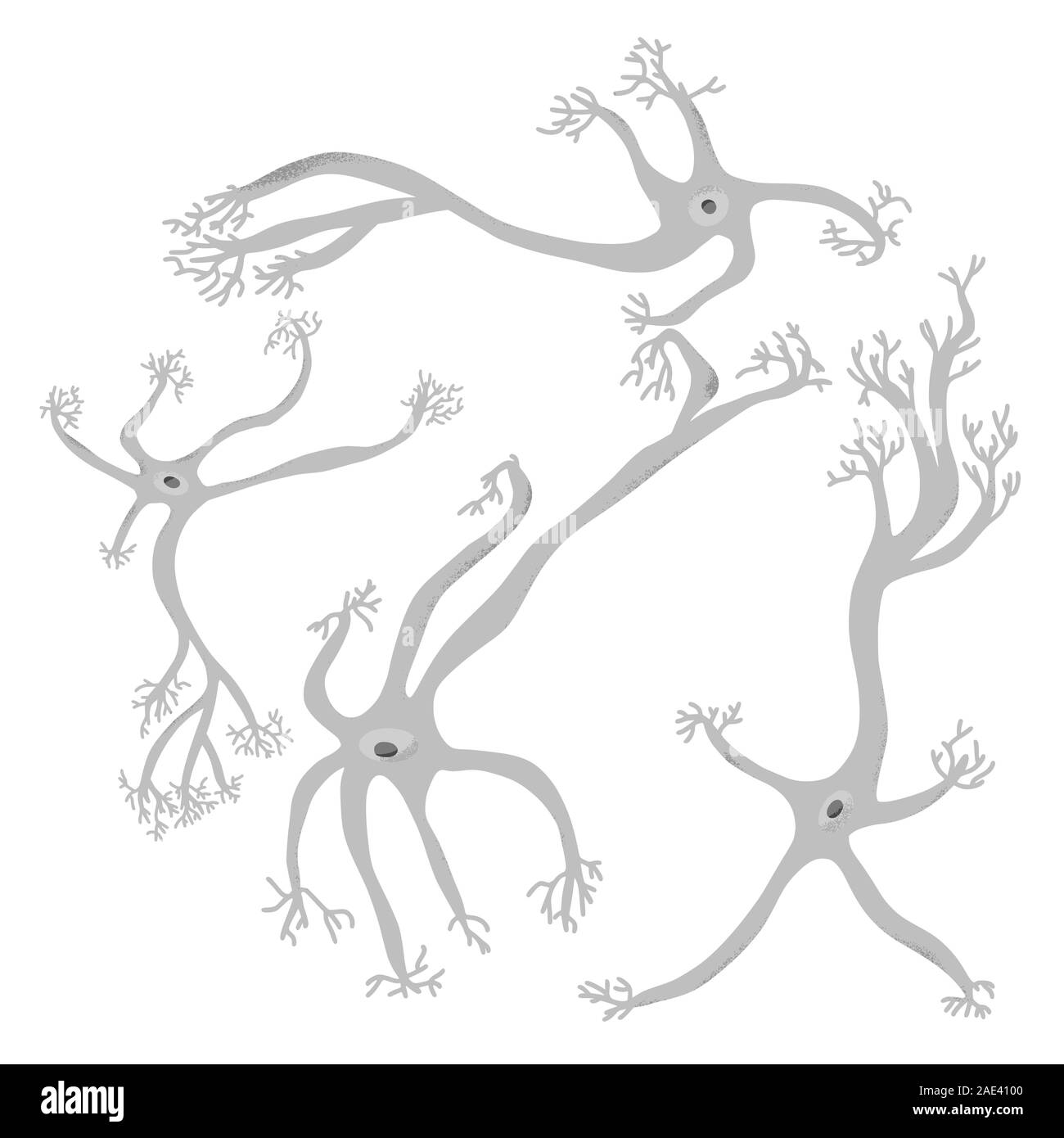 Neuron cells set. Collection of brain neurocyte. Vector illustartion. Stock Vectorhttps://www.alamy.com/image-license-details/?v=1https://www.alamy.com/neuron-cells-set-collection-of-brain-neurocyte-vector-illustartion-image335690768.html
RF2AE4100–Neuron cells set. Collection of brain neurocyte. Vector illustartion.
 Retina at the level of the bowl drawn by the fovea. Stock Photohttps://www.alamy.com/image-license-details/?v=1https://www.alamy.com/retina-at-the-level-of-the-bowl-drawn-by-the-fovea-image476925818.html
RF2JKWRMX–Retina at the level of the bowl drawn by the fovea.
 Illustration of the Human Eye copyright 1904 Stock Photohttps://www.alamy.com/image-license-details/?v=1https://www.alamy.com/stock-photo-illustration-of-the-human-eye-copyright-1904-37148649.html
RFC4C7DD–Illustration of the Human Eye copyright 1904
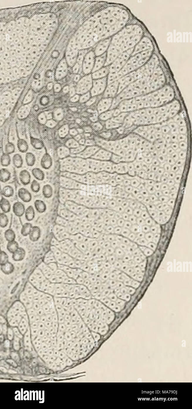 . Electro-physiology . fact that the electrical organs of Gymnotus extend to the tip of the tail, we find ganglion-cells of the electrical type as far as the end of the spinal cord, but they gradually diminish in number and size, and more nearly resemble in form the ordinary motor cells of the anterior horn. While in these cases we have in Torpedo and Gymnotus electrical organs of such high differentiation that even the most powerful effects seem tib initio to be accounted for, there are in the tail of the common Skate (Rajct), as well as in the species of Mor- myrus, organs that in struc- tur Stock Photohttps://www.alamy.com/image-license-details/?v=1https://www.alamy.com/electro-physiology-fact-that-the-electrical-organs-of-gymnotus-extend-to-the-tip-of-the-tail-we-find-ganglion-cells-of-the-electrical-type-as-far-as-the-end-of-the-spinal-cord-but-they-gradually-diminish-in-number-and-size-and-more-nearly-resemble-in-form-the-ordinary-motor-cells-of-the-anterior-horn-while-in-these-cases-we-have-in-torpedo-and-gymnotus-electrical-organs-of-such-high-differentiation-that-even-the-most-powerful-effects-seem-tib-initio-to-be-accounted-for-there-are-in-the-tail-of-the-common-skate-rajct-as-well-as-in-the-species-of-mor-myrus-organs-that-in-struc-tur-image178411342.html
RMMA79DJ–. Electro-physiology . fact that the electrical organs of Gymnotus extend to the tip of the tail, we find ganglion-cells of the electrical type as far as the end of the spinal cord, but they gradually diminish in number and size, and more nearly resemble in form the ordinary motor cells of the anterior horn. While in these cases we have in Torpedo and Gymnotus electrical organs of such high differentiation that even the most powerful effects seem tib initio to be accounted for, there are in the tail of the common Skate (Rajct), as well as in the species of Mor- myrus, organs that in struc- tur