Golgi complexes Stock Photos and Images
 . The Biological bulletin. Biology; Zoology; Biology; Marine Biology. 106. Figure 10. Schematic cross section through the basal region of a developing lime-twig gland; the cell at the right contains some early storage vacuoles. The arrows point to Golgi complexes (Scale bar: 1 ^m). lives irreversibly fixed to its substratum, and the limited number and localized arrangement of the lime twig cap- sules in a restricted body area speaks at least against their major defensive function. Most probably, the sticky threads act as lime-twigs, trapping larger planktonic food particles or sedentary organi Stock Photohttps://www.alamy.com/image-license-details/?v=1https://www.alamy.com/the-biological-bulletin-biology-zoology-biology-marine-biology-106-figure-10-schematic-cross-section-through-the-basal-region-of-a-developing-lime-twig-gland-the-cell-at-the-right-contains-some-early-storage-vacuoles-the-arrows-point-to-golgi-complexes-scale-bar-1-m-lives-irreversibly-fixed-to-its-substratum-and-the-limited-number-and-localized-arrangement-of-the-lime-twig-cap-sules-in-a-restricted-body-area-speaks-at-least-against-their-major-defensive-function-most-probably-the-sticky-threads-act-as-lime-twigs-trapping-larger-planktonic-food-particles-or-sedentary-organi-image234634594.html
RMRHMEPX–. The Biological bulletin. Biology; Zoology; Biology; Marine Biology. 106. Figure 10. Schematic cross section through the basal region of a developing lime-twig gland; the cell at the right contains some early storage vacuoles. The arrows point to Golgi complexes (Scale bar: 1 ^m). lives irreversibly fixed to its substratum, and the limited number and localized arrangement of the lime twig cap- sules in a restricted body area speaks at least against their major defensive function. Most probably, the sticky threads act as lime-twigs, trapping larger planktonic food particles or sedentary organi
 . The Biological bulletin. Biology; Zoology; Biology; Marine Biology. 268 C. REED-MILLER. FIGURE 2. Mantle epithelium, 24 hours of regeneration, showing two Golgi complexes (arrows). Note the light fibrillar material near the Golgi complexes. Bar = 1 urn. FIGURE 3. Foot epithelium, 48 hours of regeneration with several Golgi complexes (arrows) and associated vesicles containing some condensed material. Bar = 1. Please note that these images are extracted from scanned page images that may have been digitally enhanced for readability - coloration and appearance of these illustrations may not per Stock Photohttps://www.alamy.com/image-license-details/?v=1https://www.alamy.com/the-biological-bulletin-biology-zoology-biology-marine-biology-268-c-reed-miller-figure-2-mantle-epithelium-24-hours-of-regeneration-showing-two-golgi-complexes-arrows-note-the-light-fibrillar-material-near-the-golgi-complexes-bar-=-1-urn-figure-3-foot-epithelium-48-hours-of-regeneration-with-several-golgi-complexes-arrows-and-associated-vesicles-containing-some-condensed-material-bar-=-1-please-note-that-these-images-are-extracted-from-scanned-page-images-that-may-have-been-digitally-enhanced-for-readability-coloration-and-appearance-of-these-illustrations-may-not-per-image234646227.html
RMRHN1JB–. The Biological bulletin. Biology; Zoology; Biology; Marine Biology. 268 C. REED-MILLER. FIGURE 2. Mantle epithelium, 24 hours of regeneration, showing two Golgi complexes (arrows). Note the light fibrillar material near the Golgi complexes. Bar = 1 urn. FIGURE 3. Foot epithelium, 48 hours of regeneration with several Golgi complexes (arrows) and associated vesicles containing some condensed material. Bar = 1. Please note that these images are extracted from scanned page images that may have been digitally enhanced for readability - coloration and appearance of these illustrations may not per
 . The Biological bulletin. Biology; Zoology; Biology; Marine Biology. FIGURE 2. Mantle epithelium, 24 hours of regeneration, showing two Golgi complexes (arrows). Note the light fibrillar material near the Golgi complexes. Bar = 1 urn. FIGURE 3. Foot epithelium, 48 hours of regeneration with several Golgi complexes (arrows) and associated vesicles containing some condensed material. Bar = 1. FIGURE 4. Scanning electron micrograph of the shell window, 48 hours of regeneration, showing small crystals associated with an organic matrix. Bar = 10 ^m. FIGURE 5. Higher magnification scanning electron Stock Photohttps://www.alamy.com/image-license-details/?v=1https://www.alamy.com/the-biological-bulletin-biology-zoology-biology-marine-biology-figure-2-mantle-epithelium-24-hours-of-regeneration-showing-two-golgi-complexes-arrows-note-the-light-fibrillar-material-near-the-golgi-complexes-bar-=-1-urn-figure-3-foot-epithelium-48-hours-of-regeneration-with-several-golgi-complexes-arrows-and-associated-vesicles-containing-some-condensed-material-bar-=-1-figure-4-scanning-electron-micrograph-of-the-shell-window-48-hours-of-regeneration-showing-small-crystals-associated-with-an-organic-matrix-bar-=-10-m-figure-5-higher-magnification-scanning-electron-image234646200.html
RMRHN1HC–. The Biological bulletin. Biology; Zoology; Biology; Marine Biology. FIGURE 2. Mantle epithelium, 24 hours of regeneration, showing two Golgi complexes (arrows). Note the light fibrillar material near the Golgi complexes. Bar = 1 urn. FIGURE 3. Foot epithelium, 48 hours of regeneration with several Golgi complexes (arrows) and associated vesicles containing some condensed material. Bar = 1. FIGURE 4. Scanning electron micrograph of the shell window, 48 hours of regeneration, showing small crystals associated with an organic matrix. Bar = 10 ^m. FIGURE 5. Higher magnification scanning electron
 . The Biological bulletin. Biology; Zoology; Biology; Marine Biology. 766 YUDIN, DIENER, CLARK, AND CHANG. FIGURE 14. A micrograph showing a large aggregate of SER in association with Golgi complexes (G). Branches of TER can be seen along the periphery of the SER and throughout the cytoplasm. Bar — 0.9 ^m. FIGURE 15. A relatively nonactive mandihular cell with only small patches of SER. Nucleus (N). Bar ™ 0.86 /mi.. Please note that these images are extracted from scanned page images that may have been digitally enhanced for readability - coloration and appearance of these illustrations may no Stock Photohttps://www.alamy.com/image-license-details/?v=1https://www.alamy.com/the-biological-bulletin-biology-zoology-biology-marine-biology-766-yudin-diener-clark-and-chang-figure-14-a-micrograph-showing-a-large-aggregate-of-ser-in-association-with-golgi-complexes-g-branches-of-ter-can-be-seen-along-the-periphery-of-the-ser-and-throughout-the-cytoplasm-bar-09-m-figure-15-a-relatively-nonactive-mandihular-cell-with-only-small-patches-of-ser-nucleus-n-bar-086-mi-please-note-that-these-images-are-extracted-from-scanned-page-images-that-may-have-been-digitally-enhanced-for-readability-coloration-and-appearance-of-these-illustrations-may-no-image234645108.html
RMRHN06C–. The Biological bulletin. Biology; Zoology; Biology; Marine Biology. 766 YUDIN, DIENER, CLARK, AND CHANG. FIGURE 14. A micrograph showing a large aggregate of SER in association with Golgi complexes (G). Branches of TER can be seen along the periphery of the SER and throughout the cytoplasm. Bar — 0.9 ^m. FIGURE 15. A relatively nonactive mandihular cell with only small patches of SER. Nucleus (N). Bar ™ 0.86 /mi.. Please note that these images are extracted from scanned page images that may have been digitally enhanced for readability - coloration and appearance of these illustrations may no
 . The Biological bulletin. Biology; Zoology; Biology; Marine Biology. 58 P. P. JAKOBSEN AND P. SUHR-JESSEN. 12a Figures 8-10. Differently structured immature large granules from Tachyplen tridcnlatus granulo- cytes. Insert: close-up of the about 17-nm tubular structures in transverse and longitudinal section (bar equals 100 nm). Apparently, a coated pit (CP) and a coated vesicle (CV) are present. Golgi complexes (G).. Please note that these images are extracted from scanned page images that may have been digitally enhanced for readability - coloration and appearance of these illustrations may Stock Photohttps://www.alamy.com/image-license-details/?v=1https://www.alamy.com/the-biological-bulletin-biology-zoology-biology-marine-biology-58-p-p-jakobsen-and-p-suhr-jessen-12a-figures-8-10-differently-structured-immature-large-granules-from-tachyplen-tridcnlatus-granulo-cytes-insert-close-up-of-the-about-17-nm-tubular-structures-in-transverse-and-longitudinal-section-bar-equals-100-nm-apparently-a-coated-pit-cp-and-a-coated-vesicle-cv-are-present-golgi-complexes-g-please-note-that-these-images-are-extracted-from-scanned-page-images-that-may-have-been-digitally-enhanced-for-readability-coloration-and-appearance-of-these-illustrations-may-image234646835.html
RMRHN2C3–. The Biological bulletin. Biology; Zoology; Biology; Marine Biology. 58 P. P. JAKOBSEN AND P. SUHR-JESSEN. 12a Figures 8-10. Differently structured immature large granules from Tachyplen tridcnlatus granulo- cytes. Insert: close-up of the about 17-nm tubular structures in transverse and longitudinal section (bar equals 100 nm). Apparently, a coated pit (CP) and a coated vesicle (CV) are present. Golgi complexes (G).. Please note that these images are extracted from scanned page images that may have been digitally enhanced for readability - coloration and appearance of these illustrations may
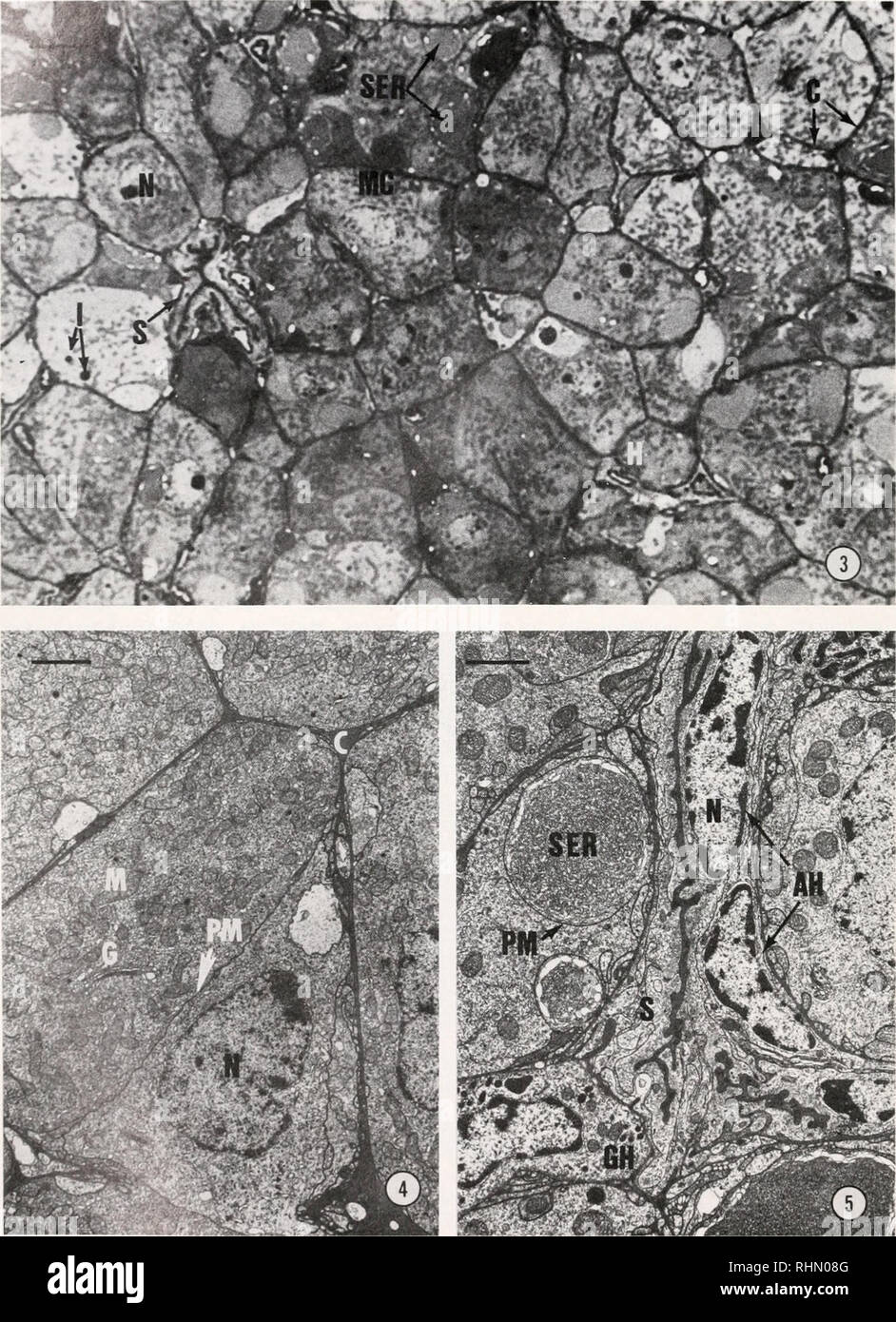
 . The Biological bulletin. Biology; Zoology; Biology; Marine Biology. 762 YUDIN, DIENER, CLARK, AND CHANG. FIGURE 3. Light micrograph revealing the cord arrangement of the mandibular cells (MC). Nucleus (N), hemocyte (H), hemolymph channel (C), smooth endoplasmic reticulum (SER), hemolymph sinus (S), lipidlike inclusions (I). Bar = 11 /j.m. FIGURE 4. Electron micrograph illustrating the gland's cellular arrangement. Nucleus (N), Golgi complexes (G), mitochondria (M), hemolymph channel (C), plasma membrane (PM). Bar := 0.73 /mi. FIGURE 5. Section through a hemolymph sinus (S). Nucleus (N), agra Stock Photohttps://www.alamy.com/image-license-details/?v=1https://www.alamy.com/the-biological-bulletin-biology-zoology-biology-marine-biology-762-yudin-diener-clark-and-chang-figure-3-light-micrograph-revealing-the-cord-arrangement-of-the-mandibular-cells-mc-nucleus-n-hemocyte-h-hemolymph-channel-c-smooth-endoplasmic-reticulum-ser-hemolymph-sinus-s-lipidlike-inclusions-i-bar-=-11-jm-figure-4-electron-micrograph-illustrating-the-glands-cellular-arrangement-nucleus-n-golgi-complexes-g-mitochondria-m-hemolymph-channel-c-plasma-membrane-pm-bar-=-073-mi-figure-5-section-through-a-hemolymph-sinus-s-nucleus-n-agra-image234645168.html
RMRHN08G–. The Biological bulletin. Biology; Zoology; Biology; Marine Biology. 762 YUDIN, DIENER, CLARK, AND CHANG. FIGURE 3. Light micrograph revealing the cord arrangement of the mandibular cells (MC). Nucleus (N), hemocyte (H), hemolymph channel (C), smooth endoplasmic reticulum (SER), hemolymph sinus (S), lipidlike inclusions (I). Bar = 11 /j.m. FIGURE 4. Electron micrograph illustrating the gland's cellular arrangement. Nucleus (N), Golgi complexes (G), mitochondria (M), hemolymph channel (C), plasma membrane (PM). Bar := 0.73 /mi. FIGURE 5. Section through a hemolymph sinus (S). Nucleus (N), agra
![. Anatomie des centres nerveux. 6 P G 3 o — •- ? a 3 - : fl> - — ^ so O u O -• rt o fc, cylindre-axe qui se divise en ramifications arboriformes, complexes et HISTOGENESE DU SYSTÈME NERVEUX. 1IÏ9 libres, à lintérieur même de la substance grise où siège la cellule. Consi-. ?s* 5 « 5 tf •S* § I OJ ???6 i a 5 3eu O ?jî> oO OI fc. dérées à tort par Golgi comme des cellules sensitives, elles répondent au 170 ANATOMIE DES CENTRES NERVEUX. Cellule île Golgi. Cellule «le Dciters. type II de cet auteur, et sont généralement désignées sous le nom de cel-lules du type I] de Golgi, de cellules de G Stock Photo . Anatomie des centres nerveux. 6 P G 3 o — •- ? a 3 - : fl> - — ^ so O u O -• rt o fc, cylindre-axe qui se divise en ramifications arboriformes, complexes et HISTOGENESE DU SYSTÈME NERVEUX. 1IÏ9 libres, à lintérieur même de la substance grise où siège la cellule. Consi-. ?s* 5 « 5 tf •S* § I OJ ???6 i a 5 3eu O ?jî> oO OI fc. dérées à tort par Golgi comme des cellules sensitives, elles répondent au 170 ANATOMIE DES CENTRES NERVEUX. Cellule île Golgi. Cellule «le Dciters. type II de cet auteur, et sont généralement désignées sous le nom de cel-lules du type I] de Golgi, de cellules de G Stock Photo](https://c8.alamy.com/comp/2CET88T/anatomie-des-centres-nerveux-6-p-g-3-o-a-3-flgt-so-o-u-o-rt-o-fc-cylindre-axe-qui-se-divise-en-ramifications-arboriformes-complexes-et-histogenese-du-systme-nerveux-1i9-libres-lintrieur-mme-de-la-substance-grise-o-sige-la-cellule-consi-s-5-5-tf-s-i-oj-6-i-a-5-3eu-o-jgt-oo-oi-fc-dres-tort-par-golgi-comme-des-cellules-sensitives-elles-rpondent-au-170-anatomie-des-centres-nerveux-cellule-le-golgi-cellule-le-dciters-type-ii-de-cet-auteur-et-sont-gnralement-dsignes-sous-le-nom-de-cel-lules-du-type-i-de-golgi-de-cellules-de-g-2CET88T.jpg) . Anatomie des centres nerveux. 6 P G 3 o — •- ? a 3 - : fl> - — ^ so O u O -• rt o fc, cylindre-axe qui se divise en ramifications arboriformes, complexes et HISTOGENESE DU SYSTÈME NERVEUX. 1IÏ9 libres, à lintérieur même de la substance grise où siège la cellule. Consi-. ?s* 5 « 5 tf •S* § I OJ ???6 i a 5 3eu O ?jî> oO OI fc. dérées à tort par Golgi comme des cellules sensitives, elles répondent au 170 ANATOMIE DES CENTRES NERVEUX. Cellule île Golgi. Cellule «le Dciters. type II de cet auteur, et sont généralement désignées sous le nom de cel-lules du type I] de Golgi, de cellules de G Stock Photohttps://www.alamy.com/image-license-details/?v=1https://www.alamy.com/anatomie-des-centres-nerveux-6-p-g-3-o-a-3-flgt-so-o-u-o-rt-o-fc-cylindre-axe-qui-se-divise-en-ramifications-arboriformes-complexes-et-histogenese-du-systme-nerveux-1i9-libres-lintrieur-mme-de-la-substance-grise-o-sige-la-cellule-consi-s-5-5-tf-s-i-oj-6-i-a-5-3eu-o-jgt-oo-oi-fc-dres-tort-par-golgi-comme-des-cellules-sensitives-elles-rpondent-au-170-anatomie-des-centres-nerveux-cellule-le-golgi-cellule-le-dciters-type-ii-de-cet-auteur-et-sont-gnralement-dsignes-sous-le-nom-de-cel-lules-du-type-i-de-golgi-de-cellules-de-g-image370556280.html
. Anatomie des centres nerveux. 6 P G 3 o — •- ? a 3 - : fl> - — ^ so O u O -• rt o fc, cylindre-axe qui se divise en ramifications arboriformes, complexes et HISTOGENESE DU SYSTÈME NERVEUX. 1IÏ9 libres, à lintérieur même de la substance grise où siège la cellule. Consi-. ?s* 5 « 5 tf •S* § I OJ ???6 i a 5 3eu O ?jî> oO OI fc. dérées à tort par Golgi comme des cellules sensitives, elles répondent au 170 ANATOMIE DES CENTRES NERVEUX. Cellule île Golgi. Cellule «le Dciters. type II de cet auteur, et sont généralement désignées sous le nom de cel-lules du type I] de Golgi, de cellules de G Stock Photohttps://www.alamy.com/image-license-details/?v=1https://www.alamy.com/anatomie-des-centres-nerveux-6-p-g-3-o-a-3-flgt-so-o-u-o-rt-o-fc-cylindre-axe-qui-se-divise-en-ramifications-arboriformes-complexes-et-histogenese-du-systme-nerveux-1i9-libres-lintrieur-mme-de-la-substance-grise-o-sige-la-cellule-consi-s-5-5-tf-s-i-oj-6-i-a-5-3eu-o-jgt-oo-oi-fc-dres-tort-par-golgi-comme-des-cellules-sensitives-elles-rpondent-au-170-anatomie-des-centres-nerveux-cellule-le-golgi-cellule-le-dciters-type-ii-de-cet-auteur-et-sont-gnralement-dsignes-sous-le-nom-de-cel-lules-du-type-i-de-golgi-de-cellules-de-g-image370556280.htmlRM2CET88T–. Anatomie des centres nerveux. 6 P G 3 o — •- ? a 3 - : fl> - — ^ so O u O -• rt o fc, cylindre-axe qui se divise en ramifications arboriformes, complexes et HISTOGENESE DU SYSTÈME NERVEUX. 1IÏ9 libres, à lintérieur même de la substance grise où siège la cellule. Consi-. ?s* 5 « 5 tf •S* § I OJ ???6 i a 5 3eu O ?jî> oO OI fc. dérées à tort par Golgi comme des cellules sensitives, elles répondent au 170 ANATOMIE DES CENTRES NERVEUX. Cellule île Golgi. Cellule «le Dciters. type II de cet auteur, et sont généralement désignées sous le nom de cel-lules du type I] de Golgi, de cellules de G
 . The Biological bulletin. Biology; Zoology; Biology; Marine Biology. ^ *.**'- ,«4 . /" •^v. "T^.fm J-'.., .*. ' •& .-.^, fc- - ?*' •a? f - If - '• -'^:s. * Kk*'. > • *• ' r* l^r «^W^HM»' 7'^ gj^ c.. FIGURE 3. (A.) An electron micrograph of a portion of a stage 2 chamber. The nucleus is from a pro-oocyte that has turned to the nurse cell developmental pathway. Note the remnant> ( r ) of old synaiJtonemal complexes. A well developed golgi apparatus (g) lies next to the nucleus; ( B.) an enlargement uf the left outlined area in A showing a 4th generation ring canal in vertic Stock Photohttps://www.alamy.com/image-license-details/?v=1https://www.alamy.com/the-biological-bulletin-biology-zoology-biology-marine-biology-4-quot-v-quottfm-j-amp-fc-a-f-if-s-kk-gt-r-lr-whm-7-gj-c-figure-3-a-an-electron-micrograph-of-a-portion-of-a-stage-2-chamber-the-nucleus-is-from-a-pro-oocyte-that-has-turned-to-the-nurse-cell-developmental-pathway-note-the-remnantgt-r-of-old-synaijtonemal-complexes-a-well-developed-golgi-apparatus-g-lies-next-to-the-nucleus-b-an-enlargement-uf-the-left-outlined-area-in-a-showing-a-4th-generation-ring-canal-in-vertic-image234650608.html
RMRHN76T–. The Biological bulletin. Biology; Zoology; Biology; Marine Biology. ^ *.**'- ,«4 . /" •^v. "T^.fm J-'.., .*. ' •& .-.^, fc- - ?*' •a? f - If - '• -'^:s. * Kk*'. > • *• ' r* l^r «^W^HM»' 7'^ gj^ c.. FIGURE 3. (A.) An electron micrograph of a portion of a stage 2 chamber. The nucleus is from a pro-oocyte that has turned to the nurse cell developmental pathway. Note the remnant> ( r ) of old synaiJtonemal complexes. A well developed golgi apparatus (g) lies next to the nucleus; ( B.) an enlargement uf the left outlined area in A showing a 4th generation ring canal in vertic