Quick filters:
Gonopore Stock Photos and Images
 scorpion Stock Photohttps://www.alamy.com/image-license-details/?v=1https://www.alamy.com/scorpion-image353304414.html
RF2BEPBAP–scorpion
 . English: Paraplanaria dactyligera musculosa (in the original paper Planaria dactyligera musculosa) quietly gliding animal; gp, gonopore; m, mouth; ph, pharynx. 24 September 2014, 20:10:10. Romank Kenk 40 Paraplanaria dactyligera musculosa Stock Photohttps://www.alamy.com/image-license-details/?v=1https://www.alamy.com/english-paraplanaria-dactyligera-musculosa-in-the-original-paper-planaria-dactyligera-musculosa-quietly-gliding-animal-gp-gonopore-m-mouth-ph-pharynx-24-september-2014-201010-romank-kenk-40-paraplanaria-dactyligera-musculosa-image208050911.html
RMP2DF1K–. English: Paraplanaria dactyligera musculosa (in the original paper Planaria dactyligera musculosa) quietly gliding animal; gp, gonopore; m, mouth; ph, pharynx. 24 September 2014, 20:10:10. Romank Kenk 40 Paraplanaria dactyligera musculosa
 Smithsonian miscellaneous collections . the endophallic musclesmust produce movements of the endophallic walls other than those con-cerned with the opening and closing of the gonopore and the com-pression of the ejaculatory sac described above. The male genitalia of the Tetrigidae, by comparison with theacridid organs, are not only very simple in structure, but, as observed 70 SMITHSONIAN MISCELLANEOUS COLLECTIONS VOL, 94 by Walker (1922), they are surprisingly unlike those of the Acridi-dae. The phallus of Tettigidea lateralis (fig. 27 D) consists of alow ovate elevation on the floor of the g Stock Photohttps://www.alamy.com/image-license-details/?v=1https://www.alamy.com/smithsonian-miscellaneous-collections-the-endophallic-musclesmust-produce-movements-of-the-endophallic-walls-other-than-those-con-cerned-with-the-opening-and-closing-of-the-gonopore-and-the-com-pression-of-the-ejaculatory-sac-described-above-the-male-genitalia-of-the-tetrigidae-by-comparison-with-theacridid-organs-are-not-only-very-simple-in-structure-but-as-observed-70-smithsonian-miscellaneous-collections-vol-94-by-walker-1922-they-are-surprisingly-unlike-those-of-the-acridi-dae-the-phallus-of-tettigidea-lateralis-fig-27-d-consists-of-alow-ovate-elevation-on-the-floor-of-the-g-image339998884.html
RM2AN4818–Smithsonian miscellaneous collections . the endophallic musclesmust produce movements of the endophallic walls other than those con-cerned with the opening and closing of the gonopore and the com-pression of the ejaculatory sac described above. The male genitalia of the Tetrigidae, by comparison with theacridid organs, are not only very simple in structure, but, as observed 70 SMITHSONIAN MISCELLANEOUS COLLECTIONS VOL, 94 by Walker (1922), they are surprisingly unlike those of the Acridi-dae. The phallus of Tettigidea lateralis (fig. 27 D) consists of alow ovate elevation on the floor of the g
 Animal testing, illustration, vector on a white background. Stock Vectorhttps://www.alamy.com/image-license-details/?v=1https://www.alamy.com/animal-testing-illustration-vector-on-a-white-background-image484924381.html
RF2K4X5YW–Animal testing, illustration, vector on a white background.
 A brown slug moving over rocks and a leaf with great detail on the tentacles and body Stock Photohttps://www.alamy.com/image-license-details/?v=1https://www.alamy.com/a-brown-slug-moving-over-rocks-and-a-leaf-with-great-detail-on-the-tentacles-and-body-image490521180.html
RF2KE14NG–A brown slug moving over rocks and a leaf with great detail on the tentacles and body
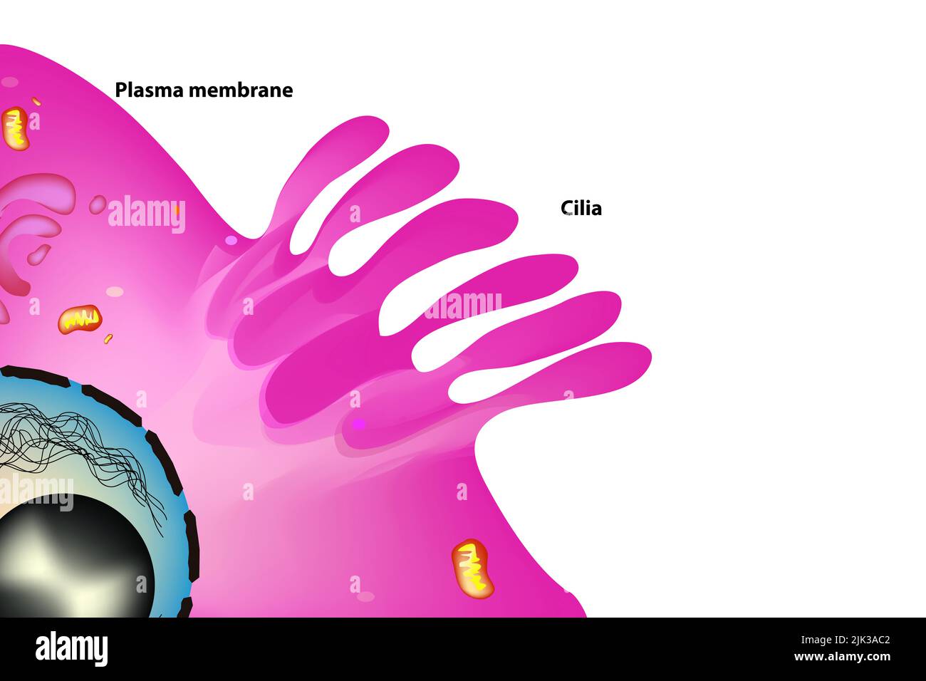 cilia anatomy in the cell Stock Photohttps://www.alamy.com/image-license-details/?v=1https://www.alamy.com/cilia-anatomy-in-the-cell-image476432434.html
RF2JK3AC2–cilia anatomy in the cell
 . A manual of zoology. iSo MANUAL OF ZOOLOGY bodies, the penial seta (p/i. s). In the female the repro- ductive aperture or gonopore is separated from the anus, and. Please note that these images are extracted from scanned page images that may have been digitally enhanced for readability - coloration and appearance of these illustrations may not perfectly resemble the original work.. Parker, T. Jeffery (Thomas Jeffery), 1850-1897; Haswell, William A. (William Aitcheson), 1854-1925. New York, The Macmillan Company; London, Macmillan & Co. ,Ltd. Stock Photohttps://www.alamy.com/image-license-details/?v=1https://www.alamy.com/a-manual-of-zoology-iso-manual-of-zoology-bodies-the-penial-seta-pi-s-in-the-female-the-repro-ductive-aperture-or-gonopore-is-separated-from-the-anus-and-please-note-that-these-images-are-extracted-from-scanned-page-images-that-may-have-been-digitally-enhanced-for-readability-coloration-and-appearance-of-these-illustrations-may-not-perfectly-resemble-the-original-work-parker-t-jeffery-thomas-jeffery-1850-1897-haswell-william-a-william-aitcheson-1854-1925-new-york-the-macmillan-company-london-macmillan-amp-co-ltd-image216447153.html
RMPG40FD–. A manual of zoology. iSo MANUAL OF ZOOLOGY bodies, the penial seta (p/i. s). In the female the repro- ductive aperture or gonopore is separated from the anus, and. Please note that these images are extracted from scanned page images that may have been digitally enhanced for readability - coloration and appearance of these illustrations may not perfectly resemble the original work.. Parker, T. Jeffery (Thomas Jeffery), 1850-1897; Haswell, William A. (William Aitcheson), 1854-1925. New York, The Macmillan Company; London, Macmillan & Co. ,Ltd.
 scorpion Stock Photohttps://www.alamy.com/image-license-details/?v=1https://www.alamy.com/scorpion-image353304424.html
RF2BEPBB4–scorpion
 . The earthworms (lumbricidae and sparganophilidae) of Ontario . girdle See clitellum. gizzard (Fr. gesier m.) (g) The muscularized portion of the digestive system, in Lumbricidae, anterior to the intestine and posterior to the crop (Fig. 2). gonopore (Fr. gonopore m.) See male pores, female pores. hearts (Fr. coeurs m.) (h) The enlarged, segmental, pulsating connectives of the blood system between the ventral and one or two other longitudinal trunks (e.g., dorsal and/or supra-oesophageal) (Fig. 2). hemerobiont A species dependent on human culture. hemerodiaphore A species indifferent to the i Stock Photohttps://www.alamy.com/image-license-details/?v=1https://www.alamy.com/the-earthworms-lumbricidae-and-sparganophilidae-of-ontario-girdle-see-clitellum-gizzard-fr-gesier-m-g-the-muscularized-portion-of-the-digestive-system-in-lumbricidae-anterior-to-the-intestine-and-posterior-to-the-crop-fig-2-gonopore-fr-gonopore-m-see-male-pores-female-pores-hearts-fr-coeurs-m-h-the-enlarged-segmental-pulsating-connectives-of-the-blood-system-between-the-ventral-and-one-or-two-other-longitudinal-trunks-eg-dorsal-andor-supra-oesophageal-fig-2-hemerobiont-a-species-dependent-on-human-culture-hemerodiaphore-a-species-indifferent-to-the-i-image178491768.html
RMMAB020–. The earthworms (lumbricidae and sparganophilidae) of Ontario . girdle See clitellum. gizzard (Fr. gesier m.) (g) The muscularized portion of the digestive system, in Lumbricidae, anterior to the intestine and posterior to the crop (Fig. 2). gonopore (Fr. gonopore m.) See male pores, female pores. hearts (Fr. coeurs m.) (h) The enlarged, segmental, pulsating connectives of the blood system between the ventral and one or two other longitudinal trunks (e.g., dorsal and/or supra-oesophageal) (Fig. 2). hemerobiont A species dependent on human culture. hemerodiaphore A species indifferent to the i
 Smithsonian miscellaneous collections . he inner ends of the ejaculatory ducts (C), but toavoid a multiplicity of names the term gonopore is applied to theopening of any genital duct, whether primary or secondary. A com-bination of ejaculatory ducts with external penes is of commonoccurrence (D). The primitive paired condition of the genital openings and associ-ated structures is retained in some members of most of the arthropodgroups, and is characteristic of Xiphosurida. Crustacea, and Diplop-oda. In other groups, and in some of the crustaceans and diplopods,an unpaired condition of the term Stock Photohttps://www.alamy.com/image-license-details/?v=1https://www.alamy.com/smithsonian-miscellaneous-collections-he-inner-ends-of-the-ejaculatory-ducts-c-but-toavoid-a-multiplicity-of-names-the-term-gonopore-is-applied-to-theopening-of-any-genital-duct-whether-primary-or-secondary-a-com-bination-of-ejaculatory-ducts-with-external-penes-is-of-commonoccurrence-d-the-primitive-paired-condition-of-the-genital-openings-and-associ-ated-structures-is-retained-in-some-members-of-most-of-the-arthropodgroups-and-is-characteristic-of-xiphosurida-crustacea-and-diplop-oda-in-other-groups-and-in-some-of-the-crustaceans-and-diplopodsan-unpaired-condition-of-the-term-image338466089.html
RM2AJJCXH–Smithsonian miscellaneous collections . he inner ends of the ejaculatory ducts (C), but toavoid a multiplicity of names the term gonopore is applied to theopening of any genital duct, whether primary or secondary. A com-bination of ejaculatory ducts with external penes is of commonoccurrence (D). The primitive paired condition of the genital openings and associ-ated structures is retained in some members of most of the arthropodgroups, and is characteristic of Xiphosurida. Crustacea, and Diplop-oda. In other groups, and in some of the crustaceans and diplopods,an unpaired condition of the term
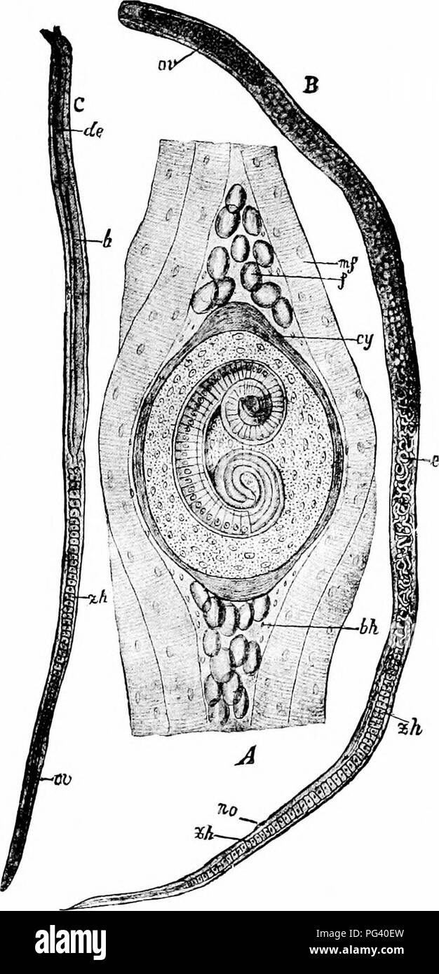 . A manual of zoology. . Fig. 85. — Trichina spiralis. A, encysted form in muscle of host; B, female; C, male, bh, connective tissue envelope; cy, cyst; de, ejaculatory duct; e, em- bryos ; f. fat globules; /:, testis; m. f, muscle fibre; oe, pharynx; or, ovary; ivo, gonopore; zh, cell masses in intestine. (From Lang's Comparative Anatomy', after Clans.) 155. Please note that these images are extracted from scanned page images that may have been digitally enhanced for readability - coloration and appearance of these illustrations may not perfectly resemble the original work.. Parker, T. Jeffer Stock Photohttps://www.alamy.com/image-license-details/?v=1https://www.alamy.com/a-manual-of-zoology-fig-85-trichina-spiralis-a-encysted-form-in-muscle-of-host-b-female-c-male-bh-connective-tissue-envelope-cy-cyst-de-ejaculatory-duct-e-em-bryos-f-fat-globules-testis-m-f-muscle-fibre-oe-pharynx-or-ovary-ivo-gonopore-zh-cell-masses-in-intestine-from-langs-comparative-anatomy-after-clans-155-please-note-that-these-images-are-extracted-from-scanned-page-images-that-may-have-been-digitally-enhanced-for-readability-coloration-and-appearance-of-these-illustrations-may-not-perfectly-resemble-the-original-work-parker-t-jeffer-image216447137.html
RMPG40EW–. A manual of zoology. . Fig. 85. — Trichina spiralis. A, encysted form in muscle of host; B, female; C, male, bh, connective tissue envelope; cy, cyst; de, ejaculatory duct; e, em- bryos ; f. fat globules; /:, testis; m. f, muscle fibre; oe, pharynx; or, ovary; ivo, gonopore; zh, cell masses in intestine. (From Lang's Comparative Anatomy', after Clans.) 155. Please note that these images are extracted from scanned page images that may have been digitally enhanced for readability - coloration and appearance of these illustrations may not perfectly resemble the original work.. Parker, T. Jeffer
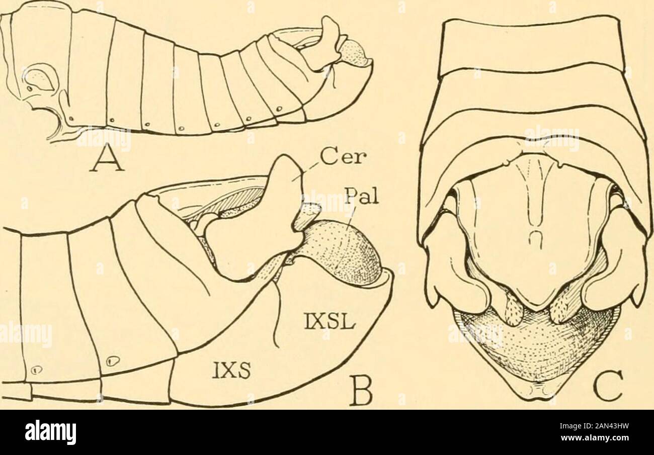 Smithsonian miscellaneous collections . edeagal apodemes (E, Apa) are short butbroad at their bases. The endophallus (F) has the usual structure,but has characteristic features. The phallotreme sclerites {o, q) are 82 SMITHSONIAN MISCELLANEOUS COLLECTIONS VOL. 94 very slender; those of the dorsal pair are united by an arched bridge(t) in the dorsal wall of the meatus; each sclerite of the ventral pairbears a large, thin, oval plate (v) in the lateral wall of the phallotremecleft. The ejaculatory sac (ejs) is relatively large and is separatedfrom the spermatophore sac (sps) by strong gonopore p Stock Photohttps://www.alamy.com/image-license-details/?v=1https://www.alamy.com/smithsonian-miscellaneous-collections-edeagal-apodemes-e-apa-are-short-butbroad-at-their-bases-the-endophallus-f-has-the-usual-structurebut-has-characteristic-features-the-phallotreme-sclerites-o-q-are-82-smithsonian-miscellaneous-collections-vol-94-very-slender-those-of-the-dorsal-pair-are-united-by-an-arched-bridget-in-the-dorsal-wall-of-the-meatus-each-sclerite-of-the-ventral-pairbears-a-large-thin-oval-plate-v-in-the-lateral-wall-of-the-phallotremecleft-the-ejaculatory-sac-ejs-is-relatively-large-and-is-separatedfrom-the-spermatophore-sac-sps-by-strong-gonopore-p-image339995429.html
RM2AN43HW–Smithsonian miscellaneous collections . edeagal apodemes (E, Apa) are short butbroad at their bases. The endophallus (F) has the usual structure,but has characteristic features. The phallotreme sclerites {o, q) are 82 SMITHSONIAN MISCELLANEOUS COLLECTIONS VOL. 94 very slender; those of the dorsal pair are united by an arched bridge(t) in the dorsal wall of the meatus; each sclerite of the ventral pairbears a large, thin, oval plate (v) in the lateral wall of the phallotremecleft. The ejaculatory sac (ejs) is relatively large and is separatedfrom the spermatophore sac (sps) by strong gonopore p
 . A manual of zoology. . Fig. 117. — Hirudo medicinalis. A, dorsal; B, ventral aspect, a, anus; <z. s, anterior sucker; e. /, first pair of eyes; e. 5, fifth pair; gp. H", male gonopore; gp. £, female gonopore; mth, mouth; np.i, first pair of nephridiopores; 11p. ly, seventeenth pair; p. s, posterior sucker; s.p, sensory papillae; I-XXVI, segments. (Partly after Whitman-) 204. Please note that these images are extracted from scanned page images that may have been digitally enhanced for readability - coloration and appearance of these illustrations may not perfectly resemble the origina Stock Photohttps://www.alamy.com/image-license-details/?v=1https://www.alamy.com/a-manual-of-zoology-fig-117-hirudo-medicinalis-a-dorsal-b-ventral-aspect-a-anus-ltz-s-anterior-sucker-e-first-pair-of-eyes-e-5-fifth-pair-gp-hquot-male-gonopore-gp-female-gonopore-mth-mouth-npi-first-pair-of-nephridiopores-11p-ly-seventeenth-pair-p-s-posterior-sucker-sp-sensory-papillae-i-xxvi-segments-partly-after-whitman-204-please-note-that-these-images-are-extracted-from-scanned-page-images-that-may-have-been-digitally-enhanced-for-readability-coloration-and-appearance-of-these-illustrations-may-not-perfectly-resemble-the-origina-image216446952.html
RMPG4088–. A manual of zoology. . Fig. 117. — Hirudo medicinalis. A, dorsal; B, ventral aspect, a, anus; <z. s, anterior sucker; e. /, first pair of eyes; e. 5, fifth pair; gp. H", male gonopore; gp. £, female gonopore; mth, mouth; np.i, first pair of nephridiopores; 11p. ly, seventeenth pair; p. s, posterior sucker; s.p, sensory papillae; I-XXVI, segments. (Partly after Whitman-) 204. Please note that these images are extracted from scanned page images that may have been digitally enhanced for readability - coloration and appearance of these illustrations may not perfectly resemble the origina
 A treatise on zoology . 4). The species referred to this genus by Angelin (1878) are all Ordovician, and agree in the following characters (Fig. XXXVIII.): A spheroid or ovoid theca, sessile on a broad base, composed of irregu-lar plates, the mesostereom of which is pierced by regularly formed 72 THE CYSTIDEA diplopores ; the anus close to the peristome, and with a valvular pyramid ;small hydropore, perhaps combined with gonopore, between mouth and anus, to the left; five oralsseparated by food - grooveswith primitive bilateralarrangement. The variousspecies may be arrangedin groups, according Stock Photohttps://www.alamy.com/image-license-details/?v=1https://www.alamy.com/a-treatise-on-zoology-4-the-species-referred-to-this-genus-by-angelin-1878-are-all-ordovician-and-agree-in-the-following-characters-fig-xxxviii-a-spheroid-or-ovoid-theca-sessile-on-a-broad-base-composed-of-irregu-lar-plates-the-mesostereom-of-which-is-pierced-by-regularly-formed-72-the-cystidea-diplopores-the-anus-close-to-the-peristome-and-with-a-valvular-pyramid-small-hydropore-perhaps-combined-with-gonopore-between-mouth-and-anus-to-the-left-five-oralsseparated-by-food-grooveswith-primitive-bilateralarrangement-the-variousspecies-may-be-arrangedin-groups-according-image340227062.html
RM2ANEK2E–A treatise on zoology . 4). The species referred to this genus by Angelin (1878) are all Ordovician, and agree in the following characters (Fig. XXXVIII.): A spheroid or ovoid theca, sessile on a broad base, composed of irregu-lar plates, the mesostereom of which is pierced by regularly formed 72 THE CYSTIDEA diplopores ; the anus close to the peristome, and with a valvular pyramid ;small hydropore, perhaps combined with gonopore, between mouth and anus, to the left; five oralsseparated by food - grooveswith primitive bilateralarrangement. The variousspecies may be arrangedin groups, according
 Smithsonian miscellaneous collections . s (fig. 25 H, o, q).The sclerites of the dorsal (anterior) pair end in the meatus, wherethey are united with each other by a transverse bridge {t) the dorsalwall of the latter; the ventral (posterior) sclerites are continuous bynarrow upcurved arms (s) with the lateral plates {n) of the en-6 8o SMITHSONIAN MISCELLANEOUS COLLECTIONS VOL. 94 dophallic bulb. The posterior angle of each endophallic plate is armedinternally by a free spinelike process (G), below which the marginof the plate extends obliquely downward and forward to the base ofthe gonopore pro Stock Photohttps://www.alamy.com/image-license-details/?v=1https://www.alamy.com/smithsonian-miscellaneous-collections-s-fig-25-h-o-qthe-sclerites-of-the-dorsal-anterior-pair-end-in-the-meatus-wherethey-are-united-with-each-other-by-a-transverse-bridge-t-the-dorsalwall-of-the-latter-the-ventral-posterior-sclerites-are-continuous-bynarrow-upcurved-arms-s-with-the-lateral-plates-n-of-the-en-6-8o-smithsonian-miscellaneous-collections-vol-94-dophallic-bulb-the-posterior-angle-of-each-endophallic-plate-is-armedinternally-by-a-free-spinelike-process-g-below-which-the-marginof-the-plate-extends-obliquely-downward-and-forward-to-the-base-ofthe-gonopore-pro-image339996027.html
RM2AN44B7–Smithsonian miscellaneous collections . s (fig. 25 H, o, q).The sclerites of the dorsal (anterior) pair end in the meatus, wherethey are united with each other by a transverse bridge {t) the dorsalwall of the latter; the ventral (posterior) sclerites are continuous bynarrow upcurved arms (s) with the lateral plates {n) of the en-6 8o SMITHSONIAN MISCELLANEOUS COLLECTIONS VOL. 94 dophallic bulb. The posterior angle of each endophallic plate is armedinternally by a free spinelike process (G), below which the marginof the plate extends obliquely downward and forward to the base ofthe gonopore pro
 Smithsonian miscellaneous collections . Fic. 6.—Oiiychophora : position of the genital openings. A, Pcripatns, gonopore {Cpr) between penultimate pair of legs. B, Peripa-loides novae-zcalandiac, gonopore between last pair of legs, but true terminalpair of legs probably here suppressed. gonopore {Od). In most species each oviduct is provided with asmall spermatheca (A, B, Spt) in the neighborhood of the ovary, andmay have also other tubular or vesicular diverticula (B, a, b). Themajority of the Onychophora are viviparous, and in such forms theembryos develop to maturity in uterine chambers of t Stock Photohttps://www.alamy.com/image-license-details/?v=1https://www.alamy.com/smithsonian-miscellaneous-collections-fic-6oiiychophora-position-of-the-genital-openings-a-pcripatns-gonopore-cpr-between-penultimate-pair-of-legs-b-peripa-loides-novae-zcalandiac-gonopore-between-last-pair-of-legs-but-true-terminalpair-of-legs-probably-here-suppressed-gonopore-od-in-most-species-each-oviduct-is-provided-with-asmall-spermatheca-a-b-spt-in-the-neighborhood-of-the-ovary-andmay-have-also-other-tubular-or-vesicular-diverticula-b-a-b-themajority-of-the-onychophora-are-viviparous-and-in-such-forms-theembryos-develop-to-maturity-in-uterine-chambers-of-t-image338464946.html
RM2AJJBDP–Smithsonian miscellaneous collections . Fic. 6.—Oiiychophora : position of the genital openings. A, Pcripatns, gonopore {Cpr) between penultimate pair of legs. B, Peripa-loides novae-zcalandiac, gonopore between last pair of legs, but true terminalpair of legs probably here suppressed. gonopore {Od). In most species each oviduct is provided with asmall spermatheca (A, B, Spt) in the neighborhood of the ovary, andmay have also other tubular or vesicular diverticula (B, a, b). Themajority of the Onychophora are viviparous, and in such forms theembryos develop to maturity in uterine chambers of t
 A treatise on zoology . CDviTiiig iiiouth ; c;/, section of food-groove runningfrom nioutli to /.;?, brachiole-facets, some of which are jiierreilby ail axial canal ; -Is, plutes clusing nvcr anus ; </, pro-inintMce with two j)orcs, wiiich liDVcn considtMcd gono-poifs; M, ridgo which Lnvcii thought might indicate amadreporlte (or (! may represent d gonopore antihydropore); diplopores surround the wliolc area. although the peristomialAUocystis, Miller (1889), THE CVS TIDE A 73 Silurian, iiulijiiui, may go here. ProteocysHs, Barrande (1887), LowerDevonian, Bolieniia (Fi^^ XLL), (lifFers from Stock Photohttps://www.alamy.com/image-license-details/?v=1https://www.alamy.com/a-treatise-on-zoology-cdvitiiig-iiiouth-c-section-of-food-groove-runningfrom-nioutli-to-brachiole-facets-some-of-which-are-jiierreilby-ail-axial-canal-is-plutes-clusing-nvcr-anus-lt-pro-inintmce-with-two-jorcs-wiiich-lidvcn-considtmcd-gono-poifs-m-ridgo-which-lnvcii-thought-might-indicate-amadreporlte-or-!-may-represent-d-gonopore-antihydropore-diplopores-surround-the-wliolc-area-although-the-peristomialauocystis-miller-1889-the-cvs-tide-a-73-silurian-iiulijiiui-may-go-here-proteocyshs-barrande-1887-lowerdevonian-bolieniia-fi-xll-liffers-from-image340226676.html
RM2ANEJGM–A treatise on zoology . CDviTiiig iiiouth ; c;/, section of food-groove runningfrom nioutli to /.;?, brachiole-facets, some of which are jiierreilby ail axial canal ; -Is, plutes clusing nvcr anus ; </, pro-inintMce with two j)orcs, wiiich liDVcn considtMcd gono-poifs; M, ridgo which Lnvcii thought might indicate amadreporlte (or (! may represent d gonopore antihydropore); diplopores surround the wliolc area. although the peristomialAUocystis, Miller (1889), THE CVS TIDE A 73 Silurian, iiulijiiui, may go here. ProteocysHs, Barrande (1887), LowerDevonian, Bolieniia (Fi^^ XLL), (lifFers from
 A treatise on zoology . XXV.Figs. XXV. & XXVI. Schizocystis armatus (fromBrit. Mus., E75S9 & E7590).X 2. XXV. from above;XXVI. from the abanal «.side. As, anus ; Jir, facets **for bracliioles ; fp, flooringplates of subvective groove ;in Fig. XXVI. the impressionsof these are seen in the distalpart of the groove ; Eh, thepectinirhonib of plates 14 &15 ; St, part of stem.. XXVI. Fio. XXVII. Analysis of Schizocystis. Plates 11 & 13are notched by the food-grooves. Plate 23 isdouble, and bears hydropore and gonopore. plates, bears about five brachioles ; a similar extension of r. post,groove to pl Stock Photohttps://www.alamy.com/image-license-details/?v=1https://www.alamy.com/a-treatise-on-zoology-xxvfigs-xxv-xxvi-schizocystis-armatus-frombrit-mus-e75s9-e7590x-2-xxv-from-abovexxvi-from-the-abanal-side-as-anus-jir-facets-for-bracliioles-fp-flooringplates-of-subvective-groove-in-fig-xxvi-the-impressionsof-these-are-seen-in-the-distalpart-of-the-groove-eh-thepectinirhonib-of-plates-14-15-st-part-of-stem-xxvi-fio-xxvii-analysis-of-schizocystis-plates-11-13are-notched-by-the-food-grooves-plate-23-isdouble-and-bears-hydropore-and-gonopore-plates-bears-about-five-brachioles-a-similar-extension-of-r-postgroove-to-pl-image340230454.html
RM2ANERBJ–A treatise on zoology . XXV.Figs. XXV. & XXVI. Schizocystis armatus (fromBrit. Mus., E75S9 & E7590).X 2. XXV. from above;XXVI. from the abanal «.side. As, anus ; Jir, facets **for bracliioles ; fp, flooringplates of subvective groove ;in Fig. XXVI. the impressionsof these are seen in the distalpart of the groove ; Eh, thepectinirhonib of plates 14 &15 ; St, part of stem.. XXVI. Fio. XXVII. Analysis of Schizocystis. Plates 11 & 13are notched by the food-grooves. Plate 23 isdouble, and bears hydropore and gonopore. plates, bears about five brachioles ; a similar extension of r. post,groove to pl
 . Bulletin of the Natural History Museum Zoology. M2 ! seminal receptacle :: copulatory duct receptacle duct. Fig. 38. Evolutionary trends in the structures of the female genital systems of the arietellid genera. A, Fusion of copulatory pores to form single pore, and anterolateral migration of both gonopores; B, Posterior migration of both gonopores and copulatory pores, and separation of.copulatory pore from gonopore; C, Anterolateral migration of gonopores, and separation of copulatory pore from gonopore and their asymmetrical arrangement and enlargement; D, Lateral migration of both gonopor Stock Photohttps://www.alamy.com/image-license-details/?v=1https://www.alamy.com/bulletin-of-the-natural-history-museum-zoology-m2-!-seminal-receptacle-copulatory-duct-receptacle-duct-fig-38-evolutionary-trends-in-the-structures-of-the-female-genital-systems-of-the-arietellid-genera-a-fusion-of-copulatory-pores-to-form-single-pore-and-anterolateral-migration-of-both-gonopores-b-posterior-migration-of-both-gonopores-and-copulatory-pores-and-separation-ofcopulatory-pore-from-gonopore-c-anterolateral-migration-of-gonopores-and-separation-of-copulatory-pore-from-gonopore-and-their-asymmetrical-arrangement-and-enlargement-d-lateral-migration-of-both-gonopor-image233849593.html
RMRGCNF5–. Bulletin of the Natural History Museum Zoology. M2 ! seminal receptacle :: copulatory duct receptacle duct. Fig. 38. Evolutionary trends in the structures of the female genital systems of the arietellid genera. A, Fusion of copulatory pores to form single pore, and anterolateral migration of both gonopores; B, Posterior migration of both gonopores and copulatory pores, and separation of.copulatory pore from gonopore; C, Anterolateral migration of gonopores, and separation of copulatory pore from gonopore and their asymmetrical arrangement and enlargement; D, Lateral migration of both gonopor
 . Bonner zoologische Beiträge : Herausgeber: Zoologisches Forschungsinstitut und Museum Alexander Koenig, Bonn. Biology; Zoology. Bonner zoologische Beiträge 55 (2006) 239 gonopore and orange or red eggs." A darkly pigmented ampulla could not be confirmed in the Mediterranean Thwidilla hopei (own unpublished data). It is possible that only Indopacifíc species have a pigmented ampulla, since Gosliner dealt with these species. Elysia viridis also lays „reddish yellow" eggs when feeding on Chaetoinorplia (Trowbridge & Todd 2001: 222). One particular clade was found in all analysis: Stock Photohttps://www.alamy.com/image-license-details/?v=1https://www.alamy.com/bonner-zoologische-beitrge-herausgeber-zoologisches-forschungsinstitut-und-museum-alexander-koenig-bonn-biology-zoology-bonner-zoologische-beitrge-55-2006-239-gonopore-and-orange-or-red-eggsquot-a-darkly-pigmented-ampulla-could-not-be-confirmed-in-the-mediterranean-thwidilla-hopei-own-unpublished-data-it-is-possible-that-only-indopacifc-species-have-a-pigmented-ampulla-since-gosliner-dealt-with-these-species-elysia-viridis-also-lays-reddish-yellowquot-eggs-when-feeding-on-chaetoinorplia-trowbridge-amp-todd-2001-222-one-particular-clade-was-found-in-all-analysis-image234488558.html
RMRHDTFA–. Bonner zoologische Beiträge : Herausgeber: Zoologisches Forschungsinstitut und Museum Alexander Koenig, Bonn. Biology; Zoology. Bonner zoologische Beiträge 55 (2006) 239 gonopore and orange or red eggs." A darkly pigmented ampulla could not be confirmed in the Mediterranean Thwidilla hopei (own unpublished data). It is possible that only Indopacifíc species have a pigmented ampulla, since Gosliner dealt with these species. Elysia viridis also lays „reddish yellow" eggs when feeding on Chaetoinorplia (Trowbridge & Todd 2001: 222). One particular clade was found in all analysis:
 . Bonner zoologische Monographien. Zoology. PANESAR, EVOLUTION IN WATER MITES Limnesiidae, especially Neomamersa, Memmecia and Neotorrenticola as apomorph. The plesiomorphic condition is expressed in Nilo- tonia species, with their acetabula lying free in the gonopore without enlarged basal sclerites. Fusion of enlarged basal-sclerites to a common strip situated on each side of the gonopore below the movable flap is an apomorphic character state found for example in Dartia hoettgeri. In Shivatonia this strip is raised and adpressed to the medial border of the genital flap, while in Neomamersa Stock Photohttps://www.alamy.com/image-license-details/?v=1https://www.alamy.com/bonner-zoologische-monographien-zoology-panesar-evolution-in-water-mites-limnesiidae-especially-neomamersa-memmecia-and-neotorrenticola-as-apomorph-the-plesiomorphic-condition-is-expressed-in-nilo-tonia-species-with-their-acetabula-lying-free-in-the-gonopore-without-enlarged-basal-sclerites-fusion-of-enlarged-basal-sclerites-to-a-common-strip-situated-on-each-side-of-the-gonopore-below-the-movable-flap-is-an-apomorphic-character-state-found-for-example-in-dartia-hoettgeri-in-shivatonia-this-strip-is-raised-and-adpressed-to-the-medial-border-of-the-genital-flap-while-in-neomamersa-image234486836.html
RMRHDP9T–. Bonner zoologische Monographien. Zoology. PANESAR, EVOLUTION IN WATER MITES Limnesiidae, especially Neomamersa, Memmecia and Neotorrenticola as apomorph. The plesiomorphic condition is expressed in Nilo- tonia species, with their acetabula lying free in the gonopore without enlarged basal sclerites. Fusion of enlarged basal-sclerites to a common strip situated on each side of the gonopore below the movable flap is an apomorphic character state found for example in Dartia hoettgeri. In Shivatonia this strip is raised and adpressed to the medial border of the genital flap, while in Neomamersa
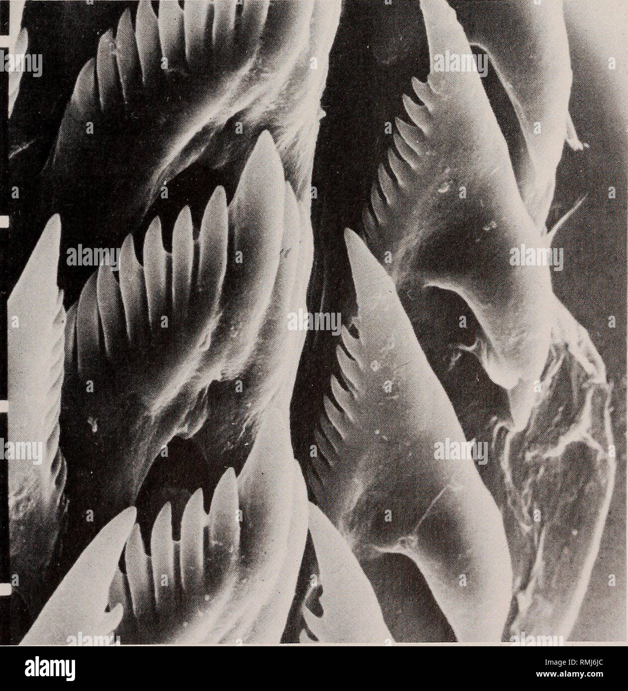 . Annals of the South African Museum = Annale van die Suid-Afrikaanse Museum. Natural history. SOME SOUTH AFRICAN AEOLIDACEAN NUDIBRANCHIA 17 are arranged in linear rows that are not clearly separated into distinct clusters (Fig. 5A). There are up to 8 ceratal rows in the right anterior digestive system, followed by as many as 17 rows per side in the posterior digestive branches. The pleuroproctic anus is ventral to the second or third ceratal row of the posterior digestive system and the nephroproct is in the interhepatic space anterior to the anus. The gonopore is ventral to the third and fo Stock Photohttps://www.alamy.com/image-license-details/?v=1https://www.alamy.com/annals-of-the-south-african-museum-=-annale-van-die-suid-afrikaanse-museum-natural-history-some-south-african-aeolidacean-nudibranchia-17-are-arranged-in-linear-rows-that-are-not-clearly-separated-into-distinct-clusters-fig-5a-there-are-up-to-8-ceratal-rows-in-the-right-anterior-digestive-system-followed-by-as-many-as-17-rows-per-side-in-the-posterior-digestive-branches-the-pleuroproctic-anus-is-ventral-to-the-second-or-third-ceratal-row-of-the-posterior-digestive-system-and-the-nephroproct-is-in-the-interhepatic-space-anterior-to-the-anus-the-gonopore-is-ventral-to-the-third-and-fo-image236428260.html
RMRMJ6JC–. Annals of the South African Museum = Annale van die Suid-Afrikaanse Museum. Natural history. SOME SOUTH AFRICAN AEOLIDACEAN NUDIBRANCHIA 17 are arranged in linear rows that are not clearly separated into distinct clusters (Fig. 5A). There are up to 8 ceratal rows in the right anterior digestive system, followed by as many as 17 rows per side in the posterior digestive branches. The pleuroproctic anus is ventral to the second or third ceratal row of the posterior digestive system and the nephroproct is in the interhepatic space anterior to the anus. The gonopore is ventral to the third and fo
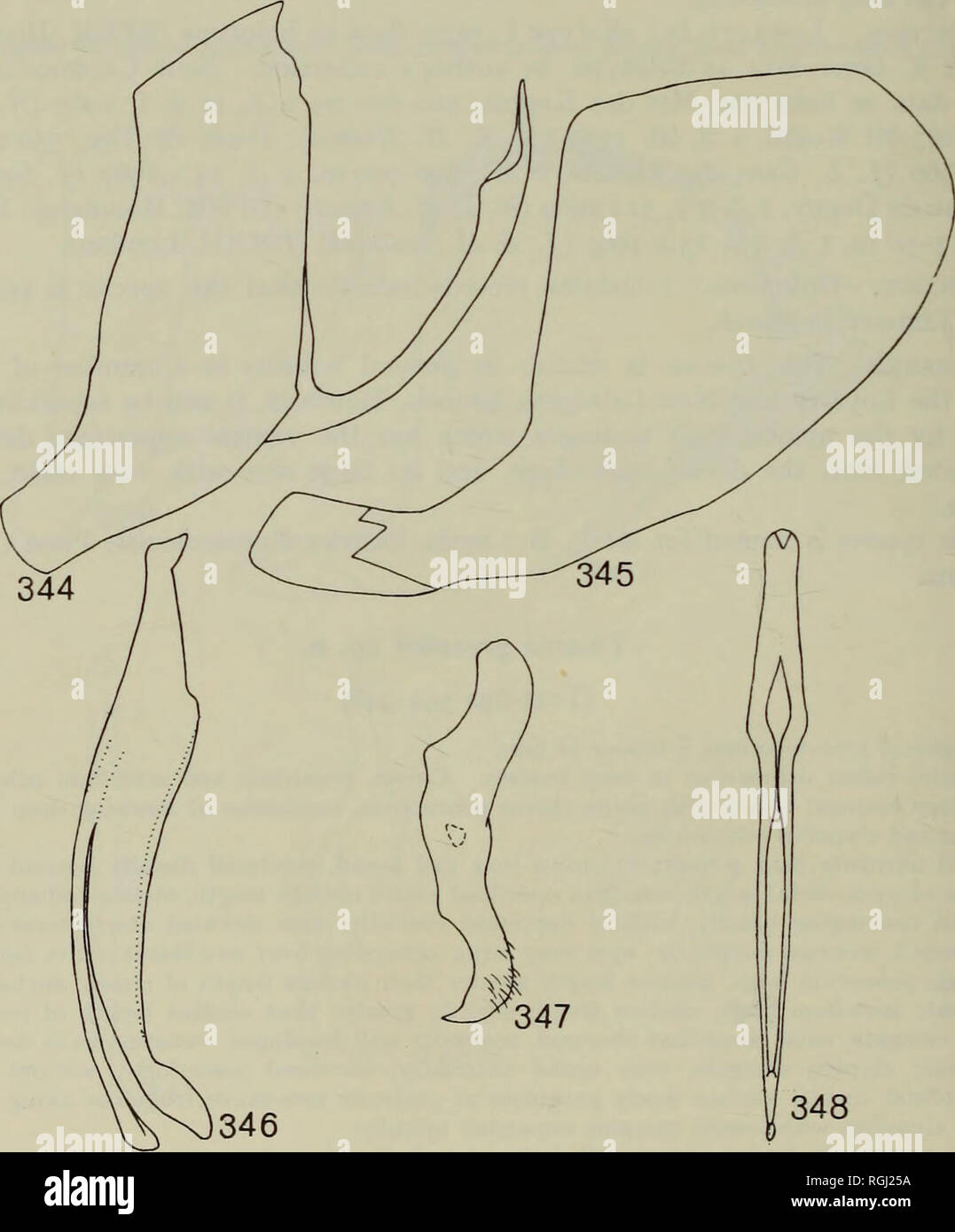 . Bulletin of the British Museum (Natural History) Entom Supp. 128 M. W. NIELSON aspect with dorsal appendage broad at basal three-fourths, attenuated at apical fourth, apex enlarged and curved caudodorsally; dorsal appendage without spines or flanges; ventral appendage very narrow, tube-like, long, expanded apically, reaching apex of dorsal appendage; gonopore apical; connective Y-shaped; style broadly clawed apically; plate with distal segment enlarged apically, subquadrate, anterior margin truncate, posterior margin broadly convex. Female seventh sternum with posterior margin nearly truncat Stock Photohttps://www.alamy.com/image-license-details/?v=1https://www.alamy.com/bulletin-of-the-british-museum-natural-history-entom-supp-128-m-w-nielson-aspect-with-dorsal-appendage-broad-at-basal-three-fourths-attenuated-at-apical-fourth-apex-enlarged-and-curved-caudodorsally-dorsal-appendage-without-spines-or-flanges-ventral-appendage-very-narrow-tube-like-long-expanded-apically-reaching-apex-of-dorsal-appendage-gonopore-apical-connective-y-shaped-style-broadly-clawed-apically-plate-with-distal-segment-enlarged-apically-subquadrate-anterior-margin-truncate-posterior-margin-broadly-convex-female-seventh-sternum-with-posterior-margin-nearly-truncat-image233966134.html
RMRGJ25A–. Bulletin of the British Museum (Natural History) Entom Supp. 128 M. W. NIELSON aspect with dorsal appendage broad at basal three-fourths, attenuated at apical fourth, apex enlarged and curved caudodorsally; dorsal appendage without spines or flanges; ventral appendage very narrow, tube-like, long, expanded apically, reaching apex of dorsal appendage; gonopore apical; connective Y-shaped; style broadly clawed apically; plate with distal segment enlarged apically, subquadrate, anterior margin truncate, posterior margin broadly convex. Female seventh sternum with posterior margin nearly truncat
 . Bonner zoologische Monographien. Zoology. FIG. 14. Morphology of nymph: ventral view of idiosoma Nilotonia emarginata (Fig. from slide IND '96/191 ny). Nymphs differ from aduks mainly in the provisory genital field (pGF). It is, in the nym- phal stage, equipped with a lesser number of aceta- bula and no gonopore is found (for abbreviations see text). the gnathosoma, have both tactile and raptorial functions. In the plesiomorphic condition, the palps have five movable segments (P1-P5), namely trochan- ter, femur, genu, tibia and tarsus, which are essentially cylindrical and articulate to allo Stock Photohttps://www.alamy.com/image-license-details/?v=1https://www.alamy.com/bonner-zoologische-monographien-zoology-fig-14-morphology-of-nymph-ventral-view-of-idiosoma-nilotonia-emarginata-fig-from-slide-ind-96191-ny-nymphs-differ-from-aduks-mainly-in-the-provisory-genital-field-pgf-it-is-in-the-nym-phal-stage-equipped-with-a-lesser-number-of-aceta-bula-and-no-gonopore-is-found-for-abbreviations-see-text-the-gnathosoma-have-both-tactile-and-raptorial-functions-in-the-plesiomorphic-condition-the-palps-have-five-movable-segments-p1-p5-namely-trochan-ter-femur-genu-tibia-and-tarsus-which-are-essentially-cylindrical-and-articulate-to-allo-image234487180.html
RMRHDPP4–. Bonner zoologische Monographien. Zoology. FIG. 14. Morphology of nymph: ventral view of idiosoma Nilotonia emarginata (Fig. from slide IND '96/191 ny). Nymphs differ from aduks mainly in the provisory genital field (pGF). It is, in the nym- phal stage, equipped with a lesser number of aceta- bula and no gonopore is found (for abbreviations see text). the gnathosoma, have both tactile and raptorial functions. In the plesiomorphic condition, the palps have five movable segments (P1-P5), namely trochan- ter, femur, genu, tibia and tarsus, which are essentially cylindrical and articulate to allo
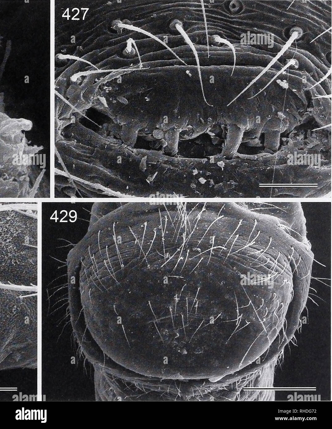 . Bonner zoologische Monographien. Zoology. FIG. 426-429. Leptopholcus tanikawai (426, 427) and Micromeryspapua (428, 429). 426. Male ALS. 427. Male gonopore. 428. Female ATS. 429. Epigynum. Scale lines: 200 pm (429), 30 pm (427), 20 pm (428), 10 pm (426). 105. Please note that these images are extracted from scanned page images that may have been digitally enhanced for readability - coloration and appearance of these illustrations may not perfectly resemble the original work.. Bonn, Zoologisches Forschungsinstitut und Museum Alexander Koenig Stock Photohttps://www.alamy.com/image-license-details/?v=1https://www.alamy.com/bonner-zoologische-monographien-zoology-fig-426-429-leptopholcus-tanikawai-426-427-and-micromeryspapua-428-429-426-male-als-427-male-gonopore-428-female-ats-429-epigynum-scale-lines-200-pm-429-30-pm-427-20-pm-428-10-pm-426-105-please-note-that-these-images-are-extracted-from-scanned-page-images-that-may-have-been-digitally-enhanced-for-readability-coloration-and-appearance-of-these-illustrations-may-not-perfectly-resemble-the-original-work-bonn-zoologisches-forschungsinstitut-und-museum-alexander-koenig-image234482054.html
RMRHDG72–. Bonner zoologische Monographien. Zoology. FIG. 426-429. Leptopholcus tanikawai (426, 427) and Micromeryspapua (428, 429). 426. Male ALS. 427. Male gonopore. 428. Female ATS. 429. Epigynum. Scale lines: 200 pm (429), 30 pm (427), 20 pm (428), 10 pm (426). 105. Please note that these images are extracted from scanned page images that may have been digitally enhanced for readability - coloration and appearance of these illustrations may not perfectly resemble the original work.. Bonn, Zoologisches Forschungsinstitut und Museum Alexander Koenig
 . The earthworms (lumbricidae and sparganophilidae) of Ontario. Lumbricidae; Worms. -GT cl-. girdle See clitellum. gizzard (Fr. gesier m.) (g) The muscularized portion of the digestive system, in Lumbricidae, anterior to the intestine and posterior to the crop (Fig. 2). gonopore (Fr. gonopore m.) See male pores, female pores. hearts (Fr. coeurs m.) (h) The enlarged, segmental, pulsating connectives of the blood system between the ventral and one or two other longitudinal trunks (e.g., dorsal and/or supra-oesophageal) (Fig. 2). hemerobiont A species dependent on human culture. hemerodiaphore A Stock Photohttps://www.alamy.com/image-license-details/?v=1https://www.alamy.com/the-earthworms-lumbricidae-and-sparganophilidae-of-ontario-lumbricidae-worms-gt-cl-girdle-see-clitellum-gizzard-fr-gesier-m-g-the-muscularized-portion-of-the-digestive-system-in-lumbricidae-anterior-to-the-intestine-and-posterior-to-the-crop-fig-2-gonopore-fr-gonopore-m-see-male-pores-female-pores-hearts-fr-coeurs-m-h-the-enlarged-segmental-pulsating-connectives-of-the-blood-system-between-the-ventral-and-one-or-two-other-longitudinal-trunks-eg-dorsal-andor-supra-oesophageal-fig-2-hemerobiont-a-species-dependent-on-human-culture-hemerodiaphore-a-image232475861.html
RMRE6599–. The earthworms (lumbricidae and sparganophilidae) of Ontario. Lumbricidae; Worms. -GT cl-. girdle See clitellum. gizzard (Fr. gesier m.) (g) The muscularized portion of the digestive system, in Lumbricidae, anterior to the intestine and posterior to the crop (Fig. 2). gonopore (Fr. gonopore m.) See male pores, female pores. hearts (Fr. coeurs m.) (h) The enlarged, segmental, pulsating connectives of the blood system between the ventral and one or two other longitudinal trunks (e.g., dorsal and/or supra-oesophageal) (Fig. 2). hemerobiont A species dependent on human culture. hemerodiaphore A
 . Annals of the South African Museum = Annale van die Suid-Afrikaanse Museum. Natural history. SOME SOUTH AFRICAN AEOLIDACEAN NUDIBRANCHIA 109 to the ampulla. Slightly more distally from the receptaculum duct, the postampull- ary duct bifurcates into a short oviduct and a longer vas deferens. The thick, prostatic vas deferens constricts sharply and terminates in a short, conical, unarmed penial papilla (Fig. 2E). The female gland mass is well developed with the mucous gland forming the largest portion. The saccate bursa copulatrix is situated near the female gonopore and appears to have a glan Stock Photohttps://www.alamy.com/image-license-details/?v=1https://www.alamy.com/annals-of-the-south-african-museum-=-annale-van-die-suid-afrikaanse-museum-natural-history-some-south-african-aeolidacean-nudibranchia-109-to-the-ampulla-slightly-more-distally-from-the-receptaculum-duct-the-postampull-ary-duct-bifurcates-into-a-short-oviduct-and-a-longer-vas-deferens-the-thick-prostatic-vas-deferens-constricts-sharply-and-terminates-in-a-short-conical-unarmed-penial-papilla-fig-2e-the-female-gland-mass-is-well-developed-with-the-mucous-gland-forming-the-largest-portion-the-saccate-bursa-copulatrix-is-situated-near-the-female-gonopore-and-appears-to-have-a-glan-image236453722.html
RMRMKB3P–. Annals of the South African Museum = Annale van die Suid-Afrikaanse Museum. Natural history. SOME SOUTH AFRICAN AEOLIDACEAN NUDIBRANCHIA 109 to the ampulla. Slightly more distally from the receptaculum duct, the postampull- ary duct bifurcates into a short oviduct and a longer vas deferens. The thick, prostatic vas deferens constricts sharply and terminates in a short, conical, unarmed penial papilla (Fig. 2E). The female gland mass is well developed with the mucous gland forming the largest portion. The saccate bursa copulatrix is situated near the female gonopore and appears to have a glan
 . The Biological bulletin. Biology; Zoology; Biology; Marine Biology. NEW SOUTH CAROLINA MARINE LEECH 473 XVIII, metameric crop and intestinal ceca, and numerous clitellar gland cells in the urosome. The mid-body segments appear 7 (14)-annulate, but this is some- what obscured. The annulation is reduced in the clitellar region, X through XII. The male gonopore is slightly elevated ventrally, but the female gonopore could not be found externally. The anus is barely visible about three annuli from the posterior sucker. The caudal sucker is somewhat longer (2.2 mm) than wide (2.0 mm), eccentrical Stock Photohttps://www.alamy.com/image-license-details/?v=1https://www.alamy.com/the-biological-bulletin-biology-zoology-biology-marine-biology-new-south-carolina-marine-leech-473-xviii-metameric-crop-and-intestinal-ceca-and-numerous-clitellar-gland-cells-in-the-urosome-the-mid-body-segments-appear-7-14-annulate-but-this-is-some-what-obscured-the-annulation-is-reduced-in-the-clitellar-region-x-through-xii-the-male-gonopore-is-slightly-elevated-ventrally-but-the-female-gonopore-could-not-be-found-externally-the-anus-is-barely-visible-about-three-annuli-from-the-posterior-sucker-the-caudal-sucker-is-somewhat-longer-22-mm-than-wide-20-mm-eccentrical-image234650433.html
RMRHN70H–. The Biological bulletin. Biology; Zoology; Biology; Marine Biology. NEW SOUTH CAROLINA MARINE LEECH 473 XVIII, metameric crop and intestinal ceca, and numerous clitellar gland cells in the urosome. The mid-body segments appear 7 (14)-annulate, but this is some- what obscured. The annulation is reduced in the clitellar region, X through XII. The male gonopore is slightly elevated ventrally, but the female gonopore could not be found externally. The anus is barely visible about three annuli from the posterior sucker. The caudal sucker is somewhat longer (2.2 mm) than wide (2.0 mm), eccentrical
 . Annals of the South African Museum = Annale van die Suid-Afrikaanse Museum. Natural history. THE ANATOMY OF THE CAPE ROCK LOBSTER 17 excretory pore antenna antennulary stennurh •p stomd exhalant aperture Sod maxilliped 1st maxilliped 3rd maxi lliped coxopodite 1st pereiopod branchiostegite coxopodite .5th pereiopod. ndopleurite endosternite position of pleurobranch orthrophragms 7th thoracic epi merdn ternal muscle chamber etiferous process cular condyle male gonopore Fig. 2. Ventral view of cephalothorax of male, showing position of right appendages and left half of endophragmaJ skeleton af Stock Photohttps://www.alamy.com/image-license-details/?v=1https://www.alamy.com/annals-of-the-south-african-museum-=-annale-van-die-suid-afrikaanse-museum-natural-history-the-anatomy-of-the-cape-rock-lobster-17-excretory-pore-antenna-antennulary-stennurh-p-stomd-exhalant-aperture-sod-maxilliped-1st-maxilliped-3rd-maxi-lliped-coxopodite-1st-pereiopod-branchiostegite-coxopodite-5th-pereiopod-ndopleurite-endosternite-position-of-pleurobranch-orthrophragms-7th-thoracic-epi-merdn-ternal-muscle-chamber-etiferous-process-cular-condyle-male-gonopore-fig-2-ventral-view-of-cephalothorax-of-male-showing-position-of-right-appendages-and-left-half-of-endophragmaj-skeleton-af-image236437278.html
RMRMJJ4E–. Annals of the South African Museum = Annale van die Suid-Afrikaanse Museum. Natural history. THE ANATOMY OF THE CAPE ROCK LOBSTER 17 excretory pore antenna antennulary stennurh •p stomd exhalant aperture Sod maxilliped 1st maxilliped 3rd maxi lliped coxopodite 1st pereiopod branchiostegite coxopodite .5th pereiopod. ndopleurite endosternite position of pleurobranch orthrophragms 7th thoracic epi merdn ternal muscle chamber etiferous process cular condyle male gonopore Fig. 2. Ventral view of cephalothorax of male, showing position of right appendages and left half of endophragmaJ skeleton af
 . Bulletin of the British Museum (Natural History) Entom Supp. n6 M. W. NIELSON margin, process with lateral margins nearly equidistant throughout, sharply pointed apically; aedeagus in lateral aspect with dorsal appendage broad along basal three-fourths, constricted subapically, slightly enlarged apically and curved dorsad; dorsal appendage with a long, lateromedial flange, and with a pair of short, subapical dorsal spines projecting caudad; ventral appendage long, narrow, tube-like, apex extended just slightly beyond apex of dorsal appendage; gonopore apical; connective Y-shaped; style clawe Stock Photohttps://www.alamy.com/image-license-details/?v=1https://www.alamy.com/bulletin-of-the-british-museum-natural-history-entom-supp-n6-m-w-nielson-margin-process-with-lateral-margins-nearly-equidistant-throughout-sharply-pointed-apically-aedeagus-in-lateral-aspect-with-dorsal-appendage-broad-along-basal-three-fourths-constricted-subapically-slightly-enlarged-apically-and-curved-dorsad-dorsal-appendage-with-a-long-lateromedial-flange-and-with-a-pair-of-short-subapical-dorsal-spines-projecting-caudad-ventral-appendage-long-narrow-tube-like-apex-extended-just-slightly-beyond-apex-of-dorsal-appendage-gonopore-apical-connective-y-shaped-style-clawe-image233966313.html
RMRGJ2BN–. Bulletin of the British Museum (Natural History) Entom Supp. n6 M. W. NIELSON margin, process with lateral margins nearly equidistant throughout, sharply pointed apically; aedeagus in lateral aspect with dorsal appendage broad along basal three-fourths, constricted subapically, slightly enlarged apically and curved dorsad; dorsal appendage with a long, lateromedial flange, and with a pair of short, subapical dorsal spines projecting caudad; ventral appendage long, narrow, tube-like, apex extended just slightly beyond apex of dorsal appendage; gonopore apical; connective Y-shaped; style clawe
 . Annals of the South African Museum = Annale van die Suid-Afrikaanse Museum. Natural history. FAMILY PLEUROBRANCHAEIDAE 21. Fig. 8. Pleurobranchaea brockii Bergh, 1897, paratype of P. gemini. A. Dorsal view of pre- served specimen. B-C. Reproductive organs. D. Gonopore with a partially everted atrium. E. Everted male and female pores.. Please note that these images are extracted from scanned page images that may have been digitally enhanced for readability - coloration and appearance of these illustrations may not perfectly resemble the original work.. South African Museum. Cape Town : The Mu Stock Photohttps://www.alamy.com/image-license-details/?v=1https://www.alamy.com/annals-of-the-south-african-museum-=-annale-van-die-suid-afrikaanse-museum-natural-history-family-pleurobranchaeidae-21-fig-8-pleurobranchaea-brockii-bergh-1897-paratype-of-p-gemini-a-dorsal-view-of-pre-served-specimen-b-c-reproductive-organs-d-gonopore-with-a-partially-everted-atrium-e-everted-male-and-female-pores-please-note-that-these-images-are-extracted-from-scanned-page-images-that-may-have-been-digitally-enhanced-for-readability-coloration-and-appearance-of-these-illustrations-may-not-perfectly-resemble-the-original-work-south-african-museum-cape-town-the-mu-image236439420.html
RMRMJMW0–. Annals of the South African Museum = Annale van die Suid-Afrikaanse Museum. Natural history. FAMILY PLEUROBRANCHAEIDAE 21. Fig. 8. Pleurobranchaea brockii Bergh, 1897, paratype of P. gemini. A. Dorsal view of pre- served specimen. B-C. Reproductive organs. D. Gonopore with a partially everted atrium. E. Everted male and female pores.. Please note that these images are extracted from scanned page images that may have been digitally enhanced for readability - coloration and appearance of these illustrations may not perfectly resemble the original work.. South African Museum. Cape Town : The Mu
 . The Canadian field-naturalist. Natural history; Sciences naturelles. Figure 2. Measured reproductive organs in Haemopis lateromaculata (A) and H. mannorata (B), viewed from the dorsum. The segments are shown in Roman numerals starting with segment X and extend to segment XVIII. Each segment has five annuh as denotated names shown on the right side of segment X. For the purposes of this paper, the first annulus is numbered 1 and is sequenced posteriorly as noted on the left side with only the first annulus of each seg- ment numbered (see Table 2). Male gonopore on IX b6 and female gonopore on Stock Photohttps://www.alamy.com/image-license-details/?v=1https://www.alamy.com/the-canadian-field-naturalist-natural-history-sciences-naturelles-figure-2-measured-reproductive-organs-in-haemopis-lateromaculata-a-and-h-mannorata-b-viewed-from-the-dorsum-the-segments-are-shown-in-roman-numerals-starting-with-segment-x-and-extend-to-segment-xviii-each-segment-has-five-annuh-as-denotated-names-shown-on-the-right-side-of-segment-x-for-the-purposes-of-this-paper-the-first-annulus-is-numbered-1-and-is-sequenced-posteriorly-as-noted-on-the-left-side-with-only-the-first-annulus-of-each-seg-ment-numbered-see-table-2-male-gonopore-on-ix-b6-and-female-gonopore-on-image233581104.html
RMRG0F28–. The Canadian field-naturalist. Natural history; Sciences naturelles. Figure 2. Measured reproductive organs in Haemopis lateromaculata (A) and H. mannorata (B), viewed from the dorsum. The segments are shown in Roman numerals starting with segment X and extend to segment XVIII. Each segment has five annuh as denotated names shown on the right side of segment X. For the purposes of this paper, the first annulus is numbered 1 and is sequenced posteriorly as noted on the left side with only the first annulus of each seg- ment numbered (see Table 2). Male gonopore on IX b6 and female gonopore on
 . Bonner zoologische Monographien. Zoology. BONNER ZOOLOGISCHE MONOGRAPHIEN Nr. 54/2007 sized, distinctly divided from trachelosome, eccentri- cally attached, eyes or eye-like spots absent, mouth- pore prior to centre (Fig. 55:2). Posterior sucker large, larger than anterior sucker, distinctly divided from urosome, eccentrically attached, surface smooth, ocelli absent (Fig. 56). Annulation. Clitellum 7-annulate, annulation weakly defined. Male gonopore located between an-. FIG. 55. Galatheabdella bruuni, external and internal structure: 1, common view; 2, anterior sucker with tra- chelosome an Stock Photohttps://www.alamy.com/image-license-details/?v=1https://www.alamy.com/bonner-zoologische-monographien-zoology-bonner-zoologische-monographien-nr-542007-sized-distinctly-divided-from-trachelosome-eccentri-cally-attached-eyes-or-eye-like-spots-absent-mouth-pore-prior-to-centre-fig-552-posterior-sucker-large-larger-than-anterior-sucker-distinctly-divided-from-urosome-eccentrically-attached-surface-smooth-ocelli-absent-fig-56-annulation-clitellum-7-annulate-annulation-weakly-defined-male-gonopore-located-between-an-fig-55-galatheabdella-bruuni-external-and-internal-structure-1-common-view-2-anterior-sucker-with-tra-chelosome-an-image234485588.html
RMRHDMN8–. Bonner zoologische Monographien. Zoology. BONNER ZOOLOGISCHE MONOGRAPHIEN Nr. 54/2007 sized, distinctly divided from trachelosome, eccentri- cally attached, eyes or eye-like spots absent, mouth- pore prior to centre (Fig. 55:2). Posterior sucker large, larger than anterior sucker, distinctly divided from urosome, eccentrically attached, surface smooth, ocelli absent (Fig. 56). Annulation. Clitellum 7-annulate, annulation weakly defined. Male gonopore located between an-. FIG. 55. Galatheabdella bruuni, external and internal structure: 1, common view; 2, anterior sucker with tra- chelosome an
 . Bonner zoologische Monographien. Zoology. BONNER ZOOLOGISCHE MONOGR.'PHIEN Nr. 52/2004. FIG. 14. Morphology of nymph: ventral view of idiosoma Nilotonia emarginata (Fig. from slide IND '96/191 ny). Nymphs differ from aduks mainly in the provisory genital field (pGF). It is, in the nym- phal stage, equipped with a lesser number of aceta- bula and no gonopore is found (for abbreviations see text). the gnathosoma, have both tactile and raptorial functions. In the plesiomorphic condition, the palps have five movable segments (P1-P5), namely trochan- ter, femur, genu, tibia and tarsus, which are Stock Photohttps://www.alamy.com/image-license-details/?v=1https://www.alamy.com/bonner-zoologische-monographien-zoology-bonner-zoologische-monogrphien-nr-522004-fig-14-morphology-of-nymph-ventral-view-of-idiosoma-nilotonia-emarginata-fig-from-slide-ind-96191-ny-nymphs-differ-from-aduks-mainly-in-the-provisory-genital-field-pgf-it-is-in-the-nym-phal-stage-equipped-with-a-lesser-number-of-aceta-bula-and-no-gonopore-is-found-for-abbreviations-see-text-the-gnathosoma-have-both-tactile-and-raptorial-functions-in-the-plesiomorphic-condition-the-palps-have-five-movable-segments-p1-p5-namely-trochan-ter-femur-genu-tibia-and-tarsus-which-are-image234487193.html
RMRHDPPH–. Bonner zoologische Monographien. Zoology. BONNER ZOOLOGISCHE MONOGR.'PHIEN Nr. 52/2004. FIG. 14. Morphology of nymph: ventral view of idiosoma Nilotonia emarginata (Fig. from slide IND '96/191 ny). Nymphs differ from aduks mainly in the provisory genital field (pGF). It is, in the nym- phal stage, equipped with a lesser number of aceta- bula and no gonopore is found (for abbreviations see text). the gnathosoma, have both tactile and raptorial functions. In the plesiomorphic condition, the palps have five movable segments (P1-P5), namely trochan- ter, femur, genu, tibia and tarsus, which are
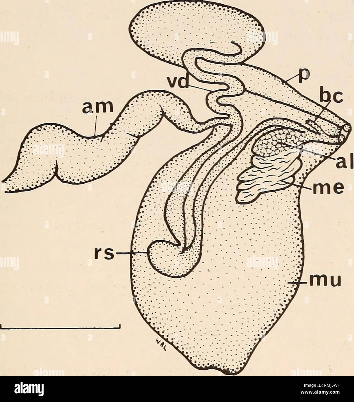 . Annals of the South African Museum = Annale van die Suid-Afrikaanse Museum. Natural history. SOUTH AFRICAN JANOLIDAE 17. Fig. 16. Janolus longidentatus sp. nov. Reproductive system. Scale 1,0 mm. between two lobes of the mucous gland and branches near the middle of its length to a short, bulbous receptaculum seminis. The major branch continues distally to its connection with the albumen gland and female gonopore. The narrow, linear bursa copulatrix joins the oviduct near the junction of the female gland mass, at the female atrium. Egg mass (Fig. 17) The egg mass is a low, flat spiral consist Stock Photohttps://www.alamy.com/image-license-details/?v=1https://www.alamy.com/annals-of-the-south-african-museum-=-annale-van-die-suid-afrikaanse-museum-natural-history-south-african-janolidae-17-fig-16-janolus-longidentatus-sp-nov-reproductive-system-scale-10-mm-between-two-lobes-of-the-mucous-gland-and-branches-near-the-middle-of-its-length-to-a-short-bulbous-receptaculum-seminis-the-major-branch-continues-distally-to-its-connection-with-the-albumen-gland-and-female-gonopore-the-narrow-linear-bursa-copulatrix-joins-the-oviduct-near-the-junction-of-the-female-gland-mass-at-the-female-atrium-egg-mass-fig-17-the-egg-mass-is-a-low-flat-spiral-consist-image236428459.html
RMRMJ6WF–. Annals of the South African Museum = Annale van die Suid-Afrikaanse Museum. Natural history. SOUTH AFRICAN JANOLIDAE 17. Fig. 16. Janolus longidentatus sp. nov. Reproductive system. Scale 1,0 mm. between two lobes of the mucous gland and branches near the middle of its length to a short, bulbous receptaculum seminis. The major branch continues distally to its connection with the albumen gland and female gonopore. The narrow, linear bursa copulatrix joins the oviduct near the junction of the female gland mass, at the female atrium. Egg mass (Fig. 17) The egg mass is a low, flat spiral consist
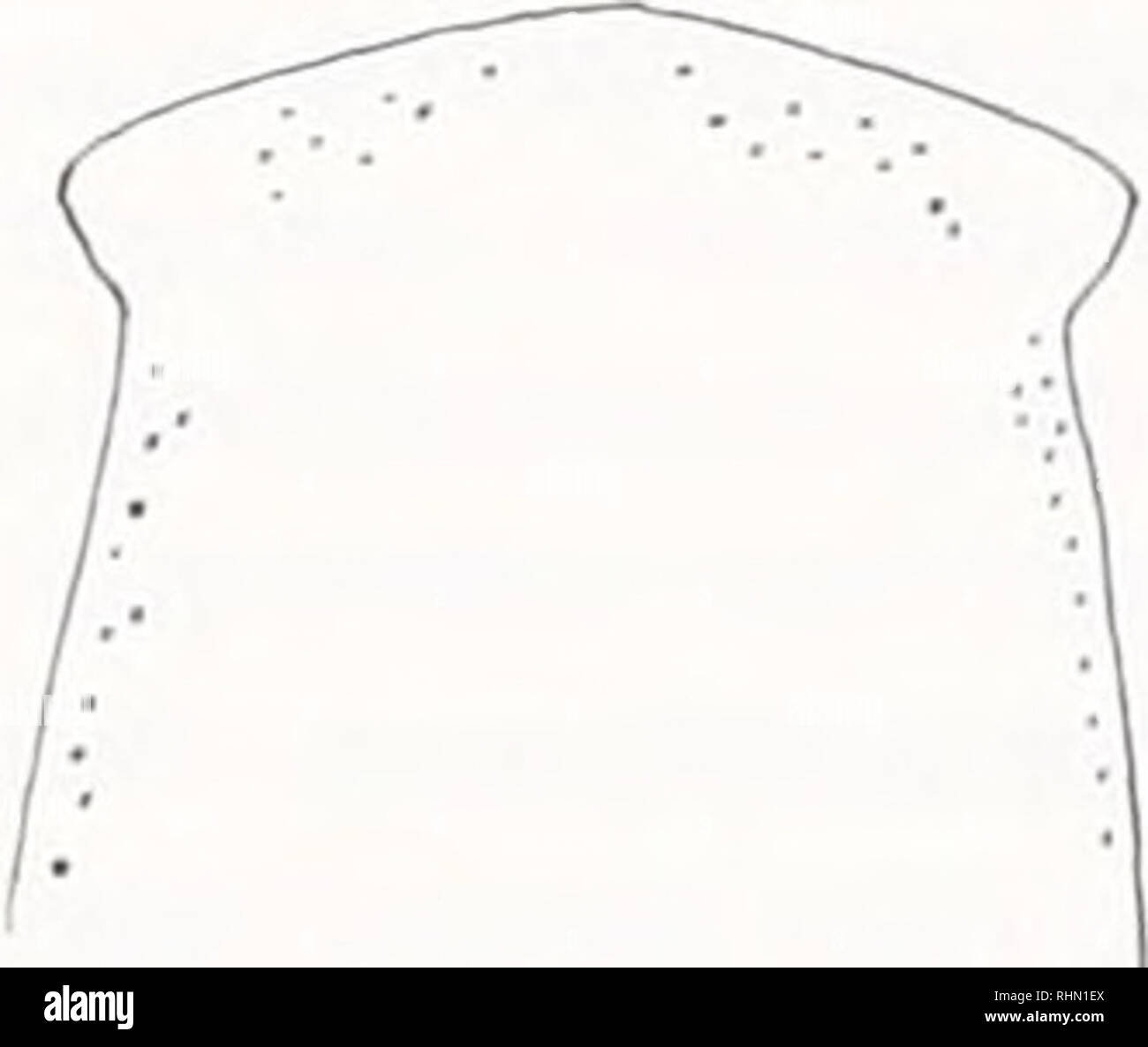 . The Biological bulletin. Biology; Zoology; Biology; Marine Biology. Figure 1. External characteristics of Polycelis remota: A, dorsal view of living animal; B. anterior end of living animal; C, anterior end of preserved animal; D, ventral view of posterior portion of preserved animal. Scale line equals 1 mm; G = gonopore, M = mouth pore, R = region of ventral gland. S = "sucker." ynx. The posterior part of the animal tapers to a rounded point. Eyes are present and numerous, and extend across the margin of the head and along the lateral margins, ex- clusive of auricles, some distanc Stock Photohttps://www.alamy.com/image-license-details/?v=1https://www.alamy.com/the-biological-bulletin-biology-zoology-biology-marine-biology-figure-1-external-characteristics-of-polycelis-remota-a-dorsal-view-of-living-animal-b-anterior-end-of-living-animal-c-anterior-end-of-preserved-animal-d-ventral-view-of-posterior-portion-of-preserved-animal-scale-line-equals-1-mm-g-=-gonopore-m-=-mouth-pore-r-=-region-of-ventral-gland-s-=-quotsuckerquot-ynx-the-posterior-part-of-the-animal-tapers-to-a-rounded-point-eyes-are-present-and-numerous-and-extend-across-the-margin-of-the-head-and-along-the-lateral-margins-ex-clusive-of-auricles-some-distanc-image234646130.html
RMRHN1EX–. The Biological bulletin. Biology; Zoology; Biology; Marine Biology. Figure 1. External characteristics of Polycelis remota: A, dorsal view of living animal; B. anterior end of living animal; C, anterior end of preserved animal; D, ventral view of posterior portion of preserved animal. Scale line equals 1 mm; G = gonopore, M = mouth pore, R = region of ventral gland. S = "sucker." ynx. The posterior part of the animal tapers to a rounded point. Eyes are present and numerous, and extend across the margin of the head and along the lateral margins, ex- clusive of auricles, some distanc
 . The Biological bulletin. Biology; Zoology; Biology; Marine Biology. REPRODUCTIVE CYCLE OF PSOLUS 1-ABR1C11 127. Figure 1. Psolusfabricii. (A) Dissected male showing the respirator, tree (R), intestine (I), body wall (W), aquapharyngeal bulb (A), cloacal muscles (C), longitudinal muscles (L). and testis (G). (B) Photograph of the mouth region showing the feeding podia, one of which is in the mouth and the others extended, and papillae surrounding the gonopore (arrow). Photographs of a testis (C) and an ovary (D) just after spawning showing the large tubules (L), small tubules (S). and tubules Stock Photohttps://www.alamy.com/image-license-details/?v=1https://www.alamy.com/the-biological-bulletin-biology-zoology-biology-marine-biology-reproductive-cycle-of-psolus-1-abr1c11-127-figure-1-psolusfabricii-a-dissected-male-showing-the-respirator-tree-r-intestine-i-body-wall-w-aquapharyngeal-bulb-a-cloacal-muscles-c-longitudinal-muscles-l-and-testis-g-b-photograph-of-the-mouth-region-showing-the-feeding-podia-one-of-which-is-in-the-mouth-and-the-others-extended-and-papillae-surrounding-the-gonopore-arrow-photographs-of-a-testis-c-and-an-ovary-d-just-after-spawning-showing-the-large-tubules-l-small-tubules-s-and-tubules-image234634963.html
RMRHMF83–. The Biological bulletin. Biology; Zoology; Biology; Marine Biology. REPRODUCTIVE CYCLE OF PSOLUS 1-ABR1C11 127. Figure 1. Psolusfabricii. (A) Dissected male showing the respirator, tree (R), intestine (I), body wall (W), aquapharyngeal bulb (A), cloacal muscles (C), longitudinal muscles (L). and testis (G). (B) Photograph of the mouth region showing the feeding podia, one of which is in the mouth and the others extended, and papillae surrounding the gonopore (arrow). Photographs of a testis (C) and an ovary (D) just after spawning showing the large tubules (L), small tubules (S). and tubules
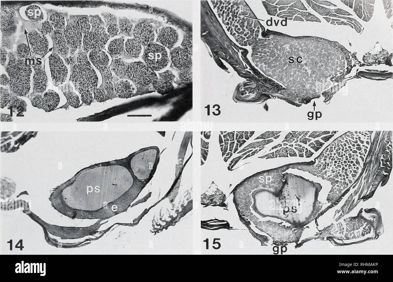 . The Biological bulletin. Biology; Zoology; Biology; Marine Biology. INSEMINATION IN TR.lCHYPENAEi'S 179. 14 Figure 12. Longitudinal section through segment of distal vas deferens. Section was stretched by heating during preparation, separating groups of sperm (spermatophores) from surrounding MVD substance, ms. MVD substance: sp. spermatophores. Figure 13. Cross section through spermatophore chamber (sc) of ejaculatory duct, left side, at level of gonopore (gp). dvd. distal vas deferens. Figure 14. Cross section through plug substance chamber, posterior end. of right ejaculatory duct, e; epi Stock Photohttps://www.alamy.com/image-license-details/?v=1https://www.alamy.com/the-biological-bulletin-biology-zoology-biology-marine-biology-insemination-in-trlchypenaeis-179-14-figure-12-longitudinal-section-through-segment-of-distal-vas-deferens-section-was-stretched-by-heating-during-preparation-separating-groups-of-sperm-spermatophores-from-surrounding-mvd-substance-ms-mvd-substance-sp-spermatophores-figure-13-cross-section-through-spermatophore-chamber-sc-of-ejaculatory-duct-left-side-at-level-of-gonopore-gp-dvd-distal-vas-deferens-figure-14-cross-section-through-plug-substance-chamber-posterior-end-of-right-ejaculatory-duct-e-epi-image234631370.html
RMRHMAKP–. The Biological bulletin. Biology; Zoology; Biology; Marine Biology. INSEMINATION IN TR.lCHYPENAEi'S 179. 14 Figure 12. Longitudinal section through segment of distal vas deferens. Section was stretched by heating during preparation, separating groups of sperm (spermatophores) from surrounding MVD substance, ms. MVD substance: sp. spermatophores. Figure 13. Cross section through spermatophore chamber (sc) of ejaculatory duct, left side, at level of gonopore (gp). dvd. distal vas deferens. Figure 14. Cross section through plug substance chamber, posterior end. of right ejaculatory duct, e; epi
 . Bulletin of the British Museum (Natural History) Entom Supp. 3« M. W. NIELSON apically, caudodorsal lobe long, broad basally, narrowed distally, caudal margin with a curved, almost sickle-shaped process; aedeagus broadly sinuate in lateral aspect, curved distally, shaft in dorsal aspect with broad, serrated lateral flange medially and a pair of retrorse, sickle-shaped dorsal processes arising distad of flange; gonopore subapical, exiting ventrally; style in dorsal aspect long, narrowed apically with small projection subapically; connective as in generic description; plate similar to carinatu Stock Photohttps://www.alamy.com/image-license-details/?v=1https://www.alamy.com/bulletin-of-the-british-museum-natural-history-entom-supp-3-m-w-nielson-apically-caudodorsal-lobe-long-broad-basally-narrowed-distally-caudal-margin-with-a-curved-almost-sickle-shaped-process-aedeagus-broadly-sinuate-in-lateral-aspect-curved-distally-shaft-in-dorsal-aspect-with-broad-serrated-lateral-flange-medially-and-a-pair-of-retrorse-sickle-shaped-dorsal-processes-arising-distad-of-flange-gonopore-subapical-exiting-ventrally-style-in-dorsal-aspect-long-narrowed-apically-with-small-projection-subapically-connective-as-in-generic-description-plate-similar-to-carinatu-image233949285.html
RMRGH8KH–. Bulletin of the British Museum (Natural History) Entom Supp. 3« M. W. NIELSON apically, caudodorsal lobe long, broad basally, narrowed distally, caudal margin with a curved, almost sickle-shaped process; aedeagus broadly sinuate in lateral aspect, curved distally, shaft in dorsal aspect with broad, serrated lateral flange medially and a pair of retrorse, sickle-shaped dorsal processes arising distad of flange; gonopore subapical, exiting ventrally; style in dorsal aspect long, narrowed apically with small projection subapically; connective as in generic description; plate similar to carinatu
 . Annals of the South African Museum = Annale van die Suid-Afrikaanse Museum. Natural history. Fig. 26 Side view of hypopygium of <$ Ectyphus pinguis v. litoralis n. showing the single aedeagus with its hood- or cowl-shaped apical part (more enlarged on left in oblique, ventral view and separ- ately also its rather large oval gonopore), and the processes of sternite 9 with the palp-like appendage (also enlarged on left). Ectyphus pinguis var. karooensis n. Two o specimens, from the late Dr. Brauns' collection in the Transvaal Museum, obviously belong to pinguis Gerst., but constitute a dist Stock Photohttps://www.alamy.com/image-license-details/?v=1https://www.alamy.com/annals-of-the-south-african-museum-=-annale-van-die-suid-afrikaanse-museum-natural-history-fig-26-side-view-of-hypopygium-of-lt-ectyphus-pinguis-v-litoralis-n-showing-the-single-aedeagus-with-its-hood-or-cowl-shaped-apical-part-more-enlarged-on-left-in-oblique-ventral-view-and-separ-ately-also-its-rather-large-oval-gonopore-and-the-processes-of-sternite-9-with-the-palp-like-appendage-also-enlarged-on-left-ectyphus-pinguis-var-karooensis-n-two-o-specimens-from-the-late-dr-brauns-collection-in-the-transvaal-museum-obviously-belong-to-pinguis-gerst-but-constitute-a-dist-image236439234.html
RMRMJMJA–. Annals of the South African Museum = Annale van die Suid-Afrikaanse Museum. Natural history. Fig. 26 Side view of hypopygium of <$ Ectyphus pinguis v. litoralis n. showing the single aedeagus with its hood- or cowl-shaped apical part (more enlarged on left in oblique, ventral view and separ- ately also its rather large oval gonopore), and the processes of sternite 9 with the palp-like appendage (also enlarged on left). Ectyphus pinguis var. karooensis n. Two o specimens, from the late Dr. Brauns' collection in the Transvaal Museum, obviously belong to pinguis Gerst., but constitute a dist
 . Bonner zoologische Monographien. Zoology. BONNER ZOOLOGISCHE MONOGRAPHIEN Nr. 58/2011 apophysis, tibia very large, procursus simple except distally (Fig. 705), with transparent prolateral pro- cess, tarsal organ capsulate (Fig. 708), bulb with large uncus, weakly sclerotized long embolus, without appendix (Fig. 706). Legs without spines and curved hairs, few vertical hairs (most hairs missing); retro- lateral trichobothrium on tibia 1 at 3%; prolateral trichobothrium absent on tibia 1, present on other tibiae. Tarsal pseudosegments barely visible in dissect- ing microscope. Gonopore with fou Stock Photohttps://www.alamy.com/image-license-details/?v=1https://www.alamy.com/bonner-zoologische-monographien-zoology-bonner-zoologische-monographien-nr-582011-apophysis-tibia-very-large-procursus-simple-except-distally-fig-705-with-transparent-prolateral-pro-cess-tarsal-organ-capsulate-fig-708-bulb-with-large-uncus-weakly-sclerotized-long-embolus-without-appendix-fig-706-legs-without-spines-and-curved-hairs-few-vertical-hairs-most-hairs-missing-retro-lateral-trichobothrium-on-tibia-1-at-3-prolateral-trichobothrium-absent-on-tibia-1-present-on-other-tibiae-tarsal-pseudosegments-barely-visible-in-dissect-ing-microscope-gonopore-with-fou-image234481477.html
RMRHDFED–. Bonner zoologische Monographien. Zoology. BONNER ZOOLOGISCHE MONOGRAPHIEN Nr. 58/2011 apophysis, tibia very large, procursus simple except distally (Fig. 705), with transparent prolateral pro- cess, tarsal organ capsulate (Fig. 708), bulb with large uncus, weakly sclerotized long embolus, without appendix (Fig. 706). Legs without spines and curved hairs, few vertical hairs (most hairs missing); retro- lateral trichobothrium on tibia 1 at 3%; prolateral trichobothrium absent on tibia 1, present on other tibiae. Tarsal pseudosegments barely visible in dissect- ing microscope. Gonopore with fou
 . The Biological bulletin. Biology; Zoology; Biology; Marine Biology. 292. Figure 1. Egg-laying in Scsanna haematoche ir and a method of measuring [he adhesiveness and plasticity of the egg envelope. (A) Morphology of the thorax (th) and abdomen (ah) of a female. Eggs just extruded from the gonopore (go) attach to many ovigerous hairs (see Fig. 6A) arranged on the ovigerous seta (os). No eggs adhere to the plumose setae (ps). m: month parts, vvl: walking leg. an: anus. (B) The incubation chamber, and an egg cluster newly extruded from the gonopore. The extruded eggs lie between thorax and abdo Stock Photohttps://www.alamy.com/image-license-details/?v=1https://www.alamy.com/the-biological-bulletin-biology-zoology-biology-marine-biology-292-figure-1-egg-laying-in-scsanna-haematoche-ir-and-a-method-of-measuring-he-adhesiveness-and-plasticity-of-the-egg-envelope-a-morphology-of-the-thorax-th-and-abdomen-ah-of-a-female-eggs-just-extruded-from-the-gonopore-go-attach-to-many-ovigerous-hairs-see-fig-6a-arranged-on-the-ovigerous-seta-os-no-eggs-adhere-to-the-plumose-setae-ps-m-month-parts-vvl-walking-leg-an-anus-b-the-incubation-chamber-and-an-egg-cluster-newly-extruded-from-the-gonopore-the-extruded-eggs-lie-between-thorax-and-abdo-image234615160.html
RMRHKJ0T–. The Biological bulletin. Biology; Zoology; Biology; Marine Biology. 292. Figure 1. Egg-laying in Scsanna haematoche ir and a method of measuring [he adhesiveness and plasticity of the egg envelope. (A) Morphology of the thorax (th) and abdomen (ah) of a female. Eggs just extruded from the gonopore (go) attach to many ovigerous hairs (see Fig. 6A) arranged on the ovigerous seta (os). No eggs adhere to the plumose setae (ps). m: month parts, vvl: walking leg. an: anus. (B) The incubation chamber, and an egg cluster newly extruded from the gonopore. The extruded eggs lie between thorax and abdo
 . Bulletin of the Natural History Museum Zoology. PHYLOGENY OF ARIETELLID COPEPODS 111. Fig. 2. Crassarietellus huysi gen. et sp. nov., female. SEM micrographs of genital double-somite of female. A, Genital double-somite, ventral view, scale bar = 200 p-m (arrows indicating positions of copulatory pores); B, Gonopore and copulatory pore (indicated by arrow) scale bar = 100 (xm; C, Right gonopore, scale bar = 30 |xm; D, Left gonopore, scale bar = 30 p.m.. Please note that these images are extracted from scanned page images that may have been digitally enhanced for readability - coloration and a Stock Photohttps://www.alamy.com/image-license-details/?v=1https://www.alamy.com/bulletin-of-the-natural-history-museum-zoology-phylogeny-of-arietellid-copepods-111-fig-2-crassarietellus-huysi-gen-et-sp-nov-female-sem-micrographs-of-genital-double-somite-of-female-a-genital-double-somite-ventral-view-scale-bar-=-200-p-m-arrows-indicating-positions-of-copulatory-pores-b-gonopore-and-copulatory-pore-indicated-by-arrow-scale-bar-=-100-xm-c-right-gonopore-scale-bar-=-30-xm-d-left-gonopore-scale-bar-=-30-pm-please-note-that-these-images-are-extracted-from-scanned-page-images-that-may-have-been-digitally-enhanced-for-readability-coloration-and-a-image233866328.html
RMRGDETT–. Bulletin of the Natural History Museum Zoology. PHYLOGENY OF ARIETELLID COPEPODS 111. Fig. 2. Crassarietellus huysi gen. et sp. nov., female. SEM micrographs of genital double-somite of female. A, Genital double-somite, ventral view, scale bar = 200 p-m (arrows indicating positions of copulatory pores); B, Gonopore and copulatory pore (indicated by arrow) scale bar = 100 (xm; C, Right gonopore, scale bar = 30 |xm; D, Left gonopore, scale bar = 30 p.m.. Please note that these images are extracted from scanned page images that may have been digitally enhanced for readability - coloration and a
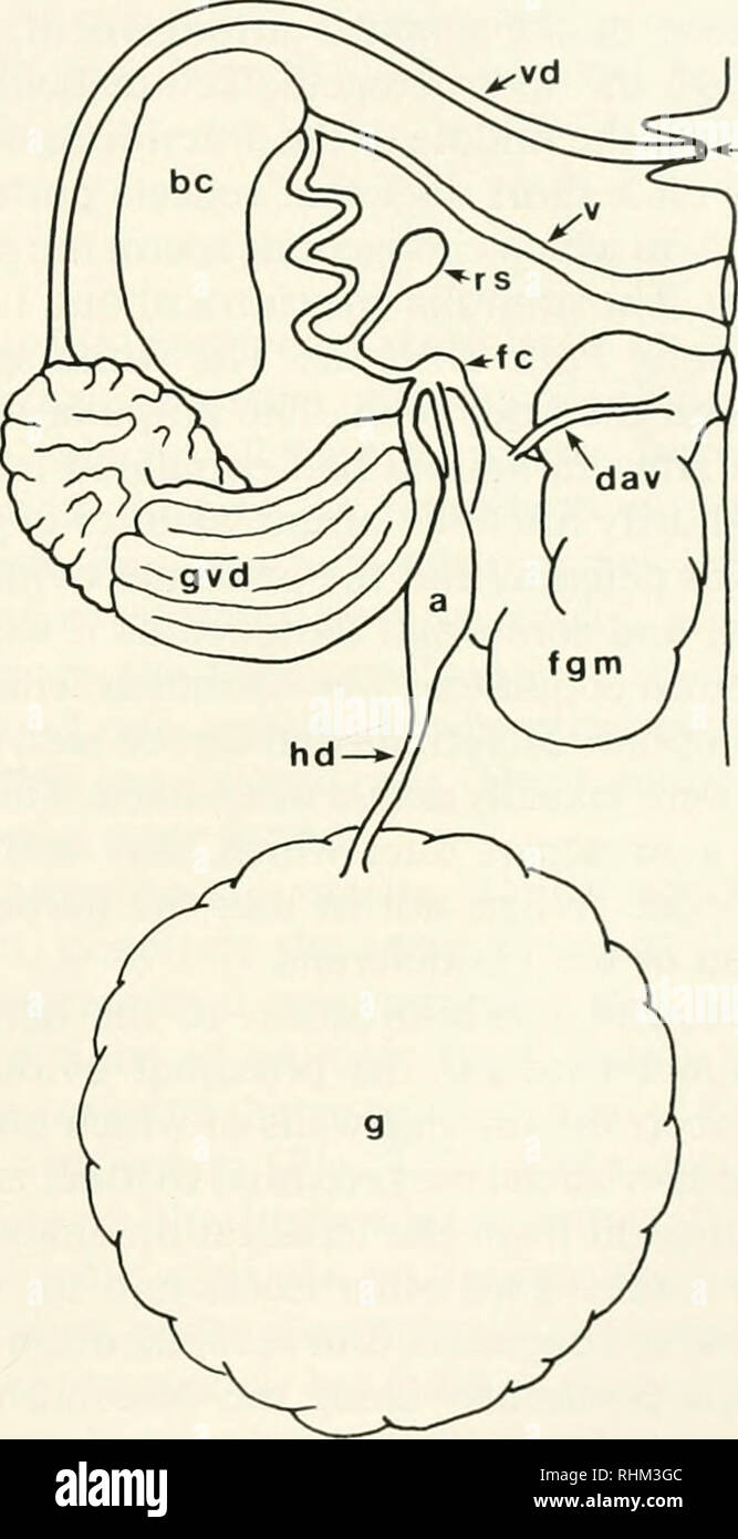 . The Biological bulletin. Biology; Zoology; Biology; Marine Biology. -^Vl. PC ? og Palio Polycera Figure 5. Schematic diagram of the reproductive systems oi Palio and Polycera. based on observations on Palio zosterae. P. duhia, Polycera quadralineata. P.faeroensis. and P. tricolor. Dorsal view. The reproductive openings would normally be housed within an external common gonopore. a—ampulla, be—bursa copulatrix, d—ductus-albumino-vestibularis, fc—fertilization chamber, fgm—female gland mass, g—visceral mass con- taining the gonad, gvd—glandular vas deferens, hd—hermaphroditic duct, og—oviducal Stock Photohttps://www.alamy.com/image-license-details/?v=1https://www.alamy.com/the-biological-bulletin-biology-zoology-biology-marine-biology-vl-pc-og-palio-polycera-figure-5-schematic-diagram-of-the-reproductive-systems-oi-palio-and-polycera-based-on-observations-on-palio-zosterae-p-duhia-polycera-quadralineata-pfaeroensis-and-p-tricolor-dorsal-view-the-reproductive-openings-would-normally-be-housed-within-an-external-common-gonopore-aampulla-bebursa-copulatrix-dductus-albumino-vestibularis-fcfertilization-chamber-fgmfemale-gland-mass-gvisceral-mass-con-taining-the-gonad-gvdglandular-vas-deferens-hdhermaphroditic-duct-ogoviducal-image234625788.html
RMRHM3GC–. The Biological bulletin. Biology; Zoology; Biology; Marine Biology. -^Vl. PC ? og Palio Polycera Figure 5. Schematic diagram of the reproductive systems oi Palio and Polycera. based on observations on Palio zosterae. P. duhia, Polycera quadralineata. P.faeroensis. and P. tricolor. Dorsal view. The reproductive openings would normally be housed within an external common gonopore. a—ampulla, be—bursa copulatrix, d—ductus-albumino-vestibularis, fc—fertilization chamber, fgm—female gland mass, g—visceral mass con- taining the gonad, gvd—glandular vas deferens, hd—hermaphroditic duct, og—oviducal
 . Bulletin of the British Museum (Natural History) Entom Supp. REVISION OF COELIDIINAE â 25 lobe; aedeagus broadly sinuate in lateral aspect, curved distally, broad basally, ventral margin distinctly serrated; gonopore subterminal, exiting ventrally; style in dorsal aspect long, curved laterally with a lateral lobe or projection subapically, rugose apically; connective as in generic description; plate broad, narrowed slightly at apex. Specimen examined. Holotype <$ (paratype J oiSandersellus carinatus DeLong), Bolivia: C. Espcranza, Bemi (Wm. M. Mana), Mulford Bio. Exped. 1921-22 (USNM, Was Stock Photohttps://www.alamy.com/image-license-details/?v=1https://www.alamy.com/bulletin-of-the-british-museum-natural-history-entom-supp-revision-of-coelidiinae-25-lobe-aedeagus-broadly-sinuate-in-lateral-aspect-curved-distally-broad-basally-ventral-margin-distinctly-serrated-gonopore-subterminal-exiting-ventrally-style-in-dorsal-aspect-long-curved-laterally-with-a-lateral-lobe-or-projection-subapically-rugose-apically-connective-as-in-generic-description-plate-broad-narrowed-slightly-at-apex-specimen-examined-holotype-lt-paratype-j-oisandersellus-carinatus-delong-bolivia-c-espcranza-bemi-wm-m-mana-mulford-bio-exped-1921-22-usnm-was-image233927250.html
RMRGG8GJ–. Bulletin of the British Museum (Natural History) Entom Supp. REVISION OF COELIDIINAE â 25 lobe; aedeagus broadly sinuate in lateral aspect, curved distally, broad basally, ventral margin distinctly serrated; gonopore subterminal, exiting ventrally; style in dorsal aspect long, curved laterally with a lateral lobe or projection subapically, rugose apically; connective as in generic description; plate broad, narrowed slightly at apex. Specimen examined. Holotype <$ (paratype J oiSandersellus carinatus DeLong), Bolivia: C. Espcranza, Bemi (Wm. M. Mana), Mulford Bio. Exped. 1921-22 (USNM, Was
 . Bonner zoologische Monographien. Zoology. BONNER ZOOLOGISCHE MONOGRAPHIEN Nr. 54/2007 sucker. Mouth-pore centrally located. Posterior sucker small, but larger than anterior one, cup-shaped, cent- rally attached, facing directly posteriorly. Annulation. Clitellum 6-annulate, including 4A3, biannulate 5 and 6, 7Ai which slightly constricted in diameter and lacking large tubercles, annulus 7Ai very small. Male gonopore between annuli 5Ai and 5A2, female gonopore between annuli 6A1 and 6A2. Copulatory area absent. Complete somite 3-annulate; annulus A2 slightly wider than Ai and A3. Annulus A2 w Stock Photohttps://www.alamy.com/image-license-details/?v=1https://www.alamy.com/bonner-zoologische-monographien-zoology-bonner-zoologische-monographien-nr-542007-sucker-mouth-pore-centrally-located-posterior-sucker-small-but-larger-than-anterior-one-cup-shaped-cent-rally-attached-facing-directly-posteriorly-annulation-clitellum-6-annulate-including-4a3-biannulate-5-and-6-7ai-which-slightly-constricted-in-diameter-and-lacking-large-tubercles-annulus-7ai-very-small-male-gonopore-between-annuli-5ai-and-5a2-female-gonopore-between-annuli-6a1-and-6a2-copulatory-area-absent-complete-somite-3-annulate-annulus-a2-slightly-wider-than-ai-and-a3-annulus-a2-w-image234486026.html
RMRHDN8X–. Bonner zoologische Monographien. Zoology. BONNER ZOOLOGISCHE MONOGRAPHIEN Nr. 54/2007 sucker. Mouth-pore centrally located. Posterior sucker small, but larger than anterior one, cup-shaped, cent- rally attached, facing directly posteriorly. Annulation. Clitellum 6-annulate, including 4A3, biannulate 5 and 6, 7Ai which slightly constricted in diameter and lacking large tubercles, annulus 7Ai very small. Male gonopore between annuli 5Ai and 5A2, female gonopore between annuli 6A1 and 6A2. Copulatory area absent. Complete somite 3-annulate; annulus A2 slightly wider than Ai and A3. Annulus A2 w
 . Bulletin of the Natural History Museum Zoology. 142 S. OHTSUKA, G.A. BOXSHALL AND H.S.J. ROE. Fig. 24. Metacalanus sp. 2, female. SEM micrographs of genital double-somite. A, Genital somite, copulatory pores indicated by arrows, scale bar = 20 u-m; B, Left gonopore and copulatory pore (indicated by an arrow), scale bar = 10 |xm; C, Right copulatory pore, scale bar = 2 u.m; D, Left copulatory pore, scale bar = 2 u.m.. Please note that these images are extracted from scanned page images that may have been digitally enhanced for readability - coloration and appearance of these illustrations may Stock Photohttps://www.alamy.com/image-license-details/?v=1https://www.alamy.com/bulletin-of-the-natural-history-museum-zoology-142-s-ohtsuka-ga-boxshall-and-hsj-roe-fig-24-metacalanus-sp-2-female-sem-micrographs-of-genital-double-somite-a-genital-somite-copulatory-pores-indicated-by-arrows-scale-bar-=-20-u-m-b-left-gonopore-and-copulatory-pore-indicated-by-an-arrow-scale-bar-=-10-xm-c-right-copulatory-pore-scale-bar-=-2-um-d-left-copulatory-pore-scale-bar-=-2-um-please-note-that-these-images-are-extracted-from-scanned-page-images-that-may-have-been-digitally-enhanced-for-readability-coloration-and-appearance-of-these-illustrations-may-image233850024.html
RMRGCP2G–. Bulletin of the Natural History Museum Zoology. 142 S. OHTSUKA, G.A. BOXSHALL AND H.S.J. ROE. Fig. 24. Metacalanus sp. 2, female. SEM micrographs of genital double-somite. A, Genital somite, copulatory pores indicated by arrows, scale bar = 20 u-m; B, Left gonopore and copulatory pore (indicated by an arrow), scale bar = 10 |xm; C, Right copulatory pore, scale bar = 2 u.m; D, Left copulatory pore, scale bar = 2 u.m.. Please note that these images are extracted from scanned page images that may have been digitally enhanced for readability - coloration and appearance of these illustrations may
 . Bonner zoologische Monographien. Zoology. UTEVSKY, ANTARCTIC PISCICOLID LEECHES. FIG. 56. Galatheabdella bruuni, common view (re- produced from Richardson & Meyer 1973). nuli 1 and 2, female gonopore between 4 and 5. Ex- ternal copulatory area absent (Fig. 55: 2). Complete somite 14-annulate (with additional annulation), an- nulation weakly defined (Fig. 55:3). Length of annuli variable. Pulsatile vesicles situated on annuli 4-6 and crossed by two interannular grooves, first pair of pulsa- tile vesicles on first annuli of urosome. Anus on an- nulus 2 from posterior sucker. Coloration. No Stock Photohttps://www.alamy.com/image-license-details/?v=1https://www.alamy.com/bonner-zoologische-monographien-zoology-utevsky-antarctic-piscicolid-leeches-fig-56-galatheabdella-bruuni-common-view-re-produced-from-richardson-amp-meyer-1973-nuli-1-and-2-female-gonopore-between-4-and-5-ex-ternal-copulatory-area-absent-fig-55-2-complete-somite-14-annulate-with-additional-annulation-an-nulation-weakly-defined-fig-553-length-of-annuli-variable-pulsatile-vesicles-situated-on-annuli-4-6-and-crossed-by-two-interannular-grooves-first-pair-of-pulsa-tile-vesicles-on-first-annuli-of-urosome-anus-on-an-nulus-2-from-posterior-sucker-coloration-no-image234485575.html
RMRHDMMR–. Bonner zoologische Monographien. Zoology. UTEVSKY, ANTARCTIC PISCICOLID LEECHES. FIG. 56. Galatheabdella bruuni, common view (re- produced from Richardson & Meyer 1973). nuli 1 and 2, female gonopore between 4 and 5. Ex- ternal copulatory area absent (Fig. 55: 2). Complete somite 14-annulate (with additional annulation), an- nulation weakly defined (Fig. 55:3). Length of annuli variable. Pulsatile vesicles situated on annuli 4-6 and crossed by two interannular grooves, first pair of pulsa- tile vesicles on first annuli of urosome. Anus on an- nulus 2 from posterior sucker. Coloration. No
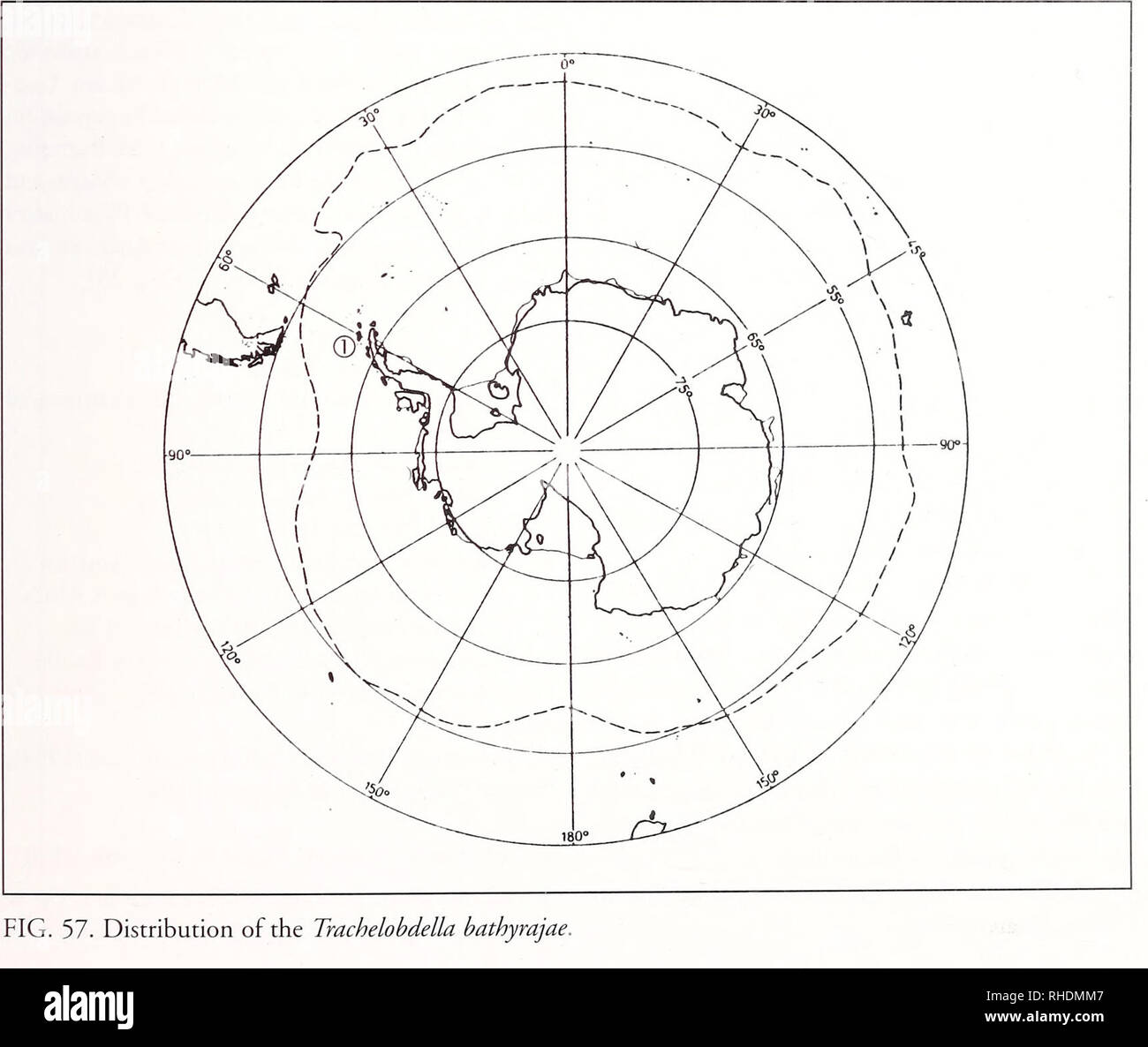 . Bonner zoologische Monographien. Zoology. FIG. 56. Galatheabdella bruuni, common view (re- produced from Richardson & Meyer 1973). nuli 1 and 2, female gonopore between 4 and 5. Ex- ternal copulatory area absent (Fig. 55: 2). Complete somite 14-annulate (with additional annulation), an- nulation weakly defined (Fig. 55:3). Length of annuli variable. Pulsatile vesicles situated on annuli 4-6 and crossed by two interannular grooves, first pair of pulsa- tile vesicles on first annuli of urosome. Anus on an- nulus 2 from posterior sucker. Coloration. No dark pigment and pattern, tes- tisacs Stock Photohttps://www.alamy.com/image-license-details/?v=1https://www.alamy.com/bonner-zoologische-monographien-zoology-fig-56-galatheabdella-bruuni-common-view-re-produced-from-richardson-amp-meyer-1973-nuli-1-and-2-female-gonopore-between-4-and-5-ex-ternal-copulatory-area-absent-fig-55-2-complete-somite-14-annulate-with-additional-annulation-an-nulation-weakly-defined-fig-553-length-of-annuli-variable-pulsatile-vesicles-situated-on-annuli-4-6-and-crossed-by-two-interannular-grooves-first-pair-of-pulsa-tile-vesicles-on-first-annuli-of-urosome-anus-on-an-nulus-2-from-posterior-sucker-coloration-no-dark-pigment-and-pattern-tes-tisacs-image234485559.html
RMRHDMM7–. Bonner zoologische Monographien. Zoology. FIG. 56. Galatheabdella bruuni, common view (re- produced from Richardson & Meyer 1973). nuli 1 and 2, female gonopore between 4 and 5. Ex- ternal copulatory area absent (Fig. 55: 2). Complete somite 14-annulate (with additional annulation), an- nulation weakly defined (Fig. 55:3). Length of annuli variable. Pulsatile vesicles situated on annuli 4-6 and crossed by two interannular grooves, first pair of pulsa- tile vesicles on first annuli of urosome. Anus on an- nulus 2 from posterior sucker. Coloration. No dark pigment and pattern, tes- tisacs
 . Bonner zoologische Monographien. Zoology. 1134. FIG. 1130-1138. Pholcus bourgini. 1130. Male prosoma, frontal view. 1131. Right bulbal processes, prolat- eral view. 1132, 1133. Tip of right procursus, distal and dorsal views. 1134. Tip of right embolus. 1135. Male ALS. 1136. Proximal male cheliceral apophyses. 1137. Distal male cheliceral apophysis. 1138. Male gonopore. Scale lines: 400 ^m (1130), 200 fim (1131), 100 fim (1132, 1133, 1136, 1138), 60 ftm (1134), 20 pm (1135, 1137). 237. Please note that these images are extracted from scanned page images that may have been digitally enhanced Stock Photohttps://www.alamy.com/image-license-details/?v=1https://www.alamy.com/bonner-zoologische-monographien-zoology-1134-fig-1130-1138-pholcus-bourgini-1130-male-prosoma-frontal-view-1131-right-bulbal-processes-prolat-eral-view-1132-1133-tip-of-right-procursus-distal-and-dorsal-views-1134-tip-of-right-embolus-1135-male-als-1136-proximal-male-cheliceral-apophyses-1137-distal-male-cheliceral-apophysis-1138-male-gonopore-scale-lines-400-m-1130-200-fim-1131-100-fim-1132-1133-1136-1138-60-ftm-1134-20-pm-1135-1137-237-please-note-that-these-images-are-extracted-from-scanned-page-images-that-may-have-been-digitally-enhanced-image234480562.html
RMRHDE9P–. Bonner zoologische Monographien. Zoology. 1134. FIG. 1130-1138. Pholcus bourgini. 1130. Male prosoma, frontal view. 1131. Right bulbal processes, prolat- eral view. 1132, 1133. Tip of right procursus, distal and dorsal views. 1134. Tip of right embolus. 1135. Male ALS. 1136. Proximal male cheliceral apophyses. 1137. Distal male cheliceral apophysis. 1138. Male gonopore. Scale lines: 400 ^m (1130), 200 fim (1131), 100 fim (1132, 1133, 1136, 1138), 60 ftm (1134), 20 pm (1135, 1137). 237. Please note that these images are extracted from scanned page images that may have been digitally enhanced
 . Bonner zoologische Monographien. Zoology. BONNER ZOOLOGISCHE MONOGRAPHIEN Nr. 58/2011. FIG. 1004-1016. Pholcus leruthi. 1004. Male ocular area, frontal view. 1005. Male distal cheliceral apoph- yses. 1006. Male palpal tarsal organ. 1007. Appendix and embolus of right genital bulb. 1008. Left procur- sus, retrolatero-dorsal view. 1009. Right procursus, prolateral view. 1010. Left procursus, retrolateral view. 1011. Left procursus tip, retrolatero-distal view. 1012. Right procursus, prolatero-distal view. 1013. Male ALS. 1014. Male gonopore. 1015. Female ALS. 1016. Epigynum. Scale lines: 300 p Stock Photohttps://www.alamy.com/image-license-details/?v=1https://www.alamy.com/bonner-zoologische-monographien-zoology-bonner-zoologische-monographien-nr-582011-fig-1004-1016-pholcus-leruthi-1004-male-ocular-area-frontal-view-1005-male-distal-cheliceral-apoph-yses-1006-male-palpal-tarsal-organ-1007-appendix-and-embolus-of-right-genital-bulb-1008-left-procur-sus-retrolatero-dorsal-view-1009-right-procursus-prolateral-view-1010-left-procursus-retrolateral-view-1011-left-procursus-tip-retrolatero-distal-view-1012-right-procursus-prolatero-distal-view-1013-male-als-1014-male-gonopore-1015-female-als-1016-epigynum-scale-lines-300-p-image234480723.html
RMRHDEFF–. Bonner zoologische Monographien. Zoology. BONNER ZOOLOGISCHE MONOGRAPHIEN Nr. 58/2011. FIG. 1004-1016. Pholcus leruthi. 1004. Male ocular area, frontal view. 1005. Male distal cheliceral apoph- yses. 1006. Male palpal tarsal organ. 1007. Appendix and embolus of right genital bulb. 1008. Left procur- sus, retrolatero-dorsal view. 1009. Right procursus, prolateral view. 1010. Left procursus, retrolateral view. 1011. Left procursus tip, retrolatero-distal view. 1012. Right procursus, prolatero-distal view. 1013. Male ALS. 1014. Male gonopore. 1015. Female ALS. 1016. Epigynum. Scale lines: 300 p
 . Bulletin of the Natural History Museum Zoology. PHYLOGENY OF ARIETELLID COPEPODS 147. Fig. 28. Paraugaptilus similis, female. SEM micrographs of genital double-somite. A, Genital double-somite, ventral view, copulatory pores indicated by arrows, scale bar = 100 xm B, Left gonopore, scale bar = 20 jj.m. and endopod fused to form flattened plate; basal setae of almost equal length; endopod represented by plumose seta; exopod completely absent. Male. Left antennule (Fig. 30C-E) 19-segmented; segments IX to XV only partly fused near posterior margin; segments XXI and XXII almost fused, but sut Stock Photohttps://www.alamy.com/image-license-details/?v=1https://www.alamy.com/bulletin-of-the-natural-history-museum-zoology-phylogeny-of-arietellid-copepods-147-fig-28-paraugaptilus-similis-female-sem-micrographs-of-genital-double-somite-a-genital-double-somite-ventral-view-copulatory-pores-indicated-by-arrows-scale-bar-=-100-xm-b-left-gonopore-scale-bar-=-20-jjm-and-endopod-fused-to-form-flattened-plate-basal-setae-of-almost-equal-length-endopod-represented-by-plumose-seta-exopod-completely-absent-male-left-antennule-fig-30c-e-19-segmented-segments-ix-to-xv-only-partly-fused-near-posterior-margin-segments-xxi-and-xxii-almost-fused-but-sut-image233849943.html
RMRGCNYK–. Bulletin of the Natural History Museum Zoology. PHYLOGENY OF ARIETELLID COPEPODS 147. Fig. 28. Paraugaptilus similis, female. SEM micrographs of genital double-somite. A, Genital double-somite, ventral view, copulatory pores indicated by arrows, scale bar = 100 xm B, Left gonopore, scale bar = 20 jj.m. and endopod fused to form flattened plate; basal setae of almost equal length; endopod represented by plumose seta; exopod completely absent. Male. Left antennule (Fig. 30C-E) 19-segmented; segments IX to XV only partly fused near posterior margin; segments XXI and XXII almost fused, but sut
![. Bonner zoologische Monographien. Zoology. HUBER, REX'ISION AND CLADISTIC ANAI VSIS Ol- /'//()/CY/.V AND CLOSELY RELATED TAXA (ARANEAE, PHOLCIDAE). FIG. 1451-1458. Pholcus quinquenotatus. 1451, 1452. Male ocular area, oblique and frontal views (arrows point at hook). 1453. Male bulbai processes. 1454, 1455. Right procursus, distal and dorsal views. 1456. Distal male cheliceral apophysis. 1457. Left procursus tip, prolateral view. 1458. Male gonopore. Scale lines: 200 pm (1451, 1452, 1454), 100 pm (1453, 1455), 80 pm (1457), 60 pm (1458), 20 pm (1456). 75°32'E], but the "impressioni torac Stock Photo . Bonner zoologische Monographien. Zoology. HUBER, REX'ISION AND CLADISTIC ANAI VSIS Ol- /'//()/CY/.V AND CLOSELY RELATED TAXA (ARANEAE, PHOLCIDAE). FIG. 1451-1458. Pholcus quinquenotatus. 1451, 1452. Male ocular area, oblique and frontal views (arrows point at hook). 1453. Male bulbai processes. 1454, 1455. Right procursus, distal and dorsal views. 1456. Distal male cheliceral apophysis. 1457. Left procursus tip, prolateral view. 1458. Male gonopore. Scale lines: 200 pm (1451, 1452, 1454), 100 pm (1453, 1455), 80 pm (1457), 60 pm (1458), 20 pm (1456). 75°32'E], but the "impressioni torac Stock Photo](https://c8.alamy.com/comp/RHDDBX/bonner-zoologische-monographien-zoology-huber-rexision-and-cladistic-anai-vsis-ol-cyv-and-closely-related-taxa-araneae-pholcidae-fig-1451-1458-pholcus-quinquenotatus-1451-1452-male-ocular-area-oblique-and-frontal-views-arrows-point-at-hook-1453-male-bulbai-processes-1454-1455-right-procursus-distal-and-dorsal-views-1456-distal-male-cheliceral-apophysis-1457-left-procursus-tip-prolateral-view-1458-male-gonopore-scale-lines-200-pm-1451-1452-1454-100-pm-1453-1455-80-pm-1457-60-pm-1458-20-pm-1456-7532e-but-the-quotimpressioni-torac-RHDDBX.jpg) . Bonner zoologische Monographien. Zoology. HUBER, REX'ISION AND CLADISTIC ANAI VSIS Ol- /'//()/CY/.V AND CLOSELY RELATED TAXA (ARANEAE, PHOLCIDAE). FIG. 1451-1458. Pholcus quinquenotatus. 1451, 1452. Male ocular area, oblique and frontal views (arrows point at hook). 1453. Male bulbai processes. 1454, 1455. Right procursus, distal and dorsal views. 1456. Distal male cheliceral apophysis. 1457. Left procursus tip, prolateral view. 1458. Male gonopore. Scale lines: 200 pm (1451, 1452, 1454), 100 pm (1453, 1455), 80 pm (1457), 60 pm (1458), 20 pm (1456). 75°32'E], but the "impressioni torac Stock Photohttps://www.alamy.com/image-license-details/?v=1https://www.alamy.com/bonner-zoologische-monographien-zoology-huber-rexision-and-cladistic-anai-vsis-ol-cyv-and-closely-related-taxa-araneae-pholcidae-fig-1451-1458-pholcus-quinquenotatus-1451-1452-male-ocular-area-oblique-and-frontal-views-arrows-point-at-hook-1453-male-bulbai-processes-1454-1455-right-procursus-distal-and-dorsal-views-1456-distal-male-cheliceral-apophysis-1457-left-procursus-tip-prolateral-view-1458-male-gonopore-scale-lines-200-pm-1451-1452-1454-100-pm-1453-1455-80-pm-1457-60-pm-1458-20-pm-1456-7532e-but-the-quotimpressioni-torac-image234479838.html
. Bonner zoologische Monographien. Zoology. HUBER, REX'ISION AND CLADISTIC ANAI VSIS Ol- /'//()/CY/.V AND CLOSELY RELATED TAXA (ARANEAE, PHOLCIDAE). FIG. 1451-1458. Pholcus quinquenotatus. 1451, 1452. Male ocular area, oblique and frontal views (arrows point at hook). 1453. Male bulbai processes. 1454, 1455. Right procursus, distal and dorsal views. 1456. Distal male cheliceral apophysis. 1457. Left procursus tip, prolateral view. 1458. Male gonopore. Scale lines: 200 pm (1451, 1452, 1454), 100 pm (1453, 1455), 80 pm (1457), 60 pm (1458), 20 pm (1456). 75°32'E], but the "impressioni torac Stock Photohttps://www.alamy.com/image-license-details/?v=1https://www.alamy.com/bonner-zoologische-monographien-zoology-huber-rexision-and-cladistic-anai-vsis-ol-cyv-and-closely-related-taxa-araneae-pholcidae-fig-1451-1458-pholcus-quinquenotatus-1451-1452-male-ocular-area-oblique-and-frontal-views-arrows-point-at-hook-1453-male-bulbai-processes-1454-1455-right-procursus-distal-and-dorsal-views-1456-distal-male-cheliceral-apophysis-1457-left-procursus-tip-prolateral-view-1458-male-gonopore-scale-lines-200-pm-1451-1452-1454-100-pm-1453-1455-80-pm-1457-60-pm-1458-20-pm-1456-7532e-but-the-quotimpressioni-torac-image234479838.htmlRMRHDDBX–. Bonner zoologische Monographien. Zoology. HUBER, REX'ISION AND CLADISTIC ANAI VSIS Ol- /'//()/CY/.V AND CLOSELY RELATED TAXA (ARANEAE, PHOLCIDAE). FIG. 1451-1458. Pholcus quinquenotatus. 1451, 1452. Male ocular area, oblique and frontal views (arrows point at hook). 1453. Male bulbai processes. 1454, 1455. Right procursus, distal and dorsal views. 1456. Distal male cheliceral apophysis. 1457. Left procursus tip, prolateral view. 1458. Male gonopore. Scale lines: 200 pm (1451, 1452, 1454), 100 pm (1453, 1455), 80 pm (1457), 60 pm (1458), 20 pm (1456). 75°32'E], but the "impressioni torac
 . Bulletin of the British Museum (Natural History) Entom Supp. 490 491 492. 493 Figs 489-493. Tharra nigroides sp. n. 489, male pygofer, lateral view; 490, plate, lateral view; 491, aedeagus, dorsal view; 492, aedeagus, lateral view; 493, style and connective, dorsolateral view. slightly beyond apex of dorsal appendage; gonopore apical; connective Y-shaped; style typically clawed apically; plate with distal segment elongate, dorsal margin expanded medially. Specimens examined. Holotype $, Solomon Is.: Bougainville, Boku, 4-6.vi. 1956 (J. L. Gressitt) (BPBM, Honolulu). Biology. Unknown. Remarks Stock Photohttps://www.alamy.com/image-license-details/?v=1https://www.alamy.com/bulletin-of-the-british-museum-natural-history-entom-supp-490-491-492-493-figs-489-493-tharra-nigroides-sp-n-489-male-pygofer-lateral-view-490-plate-lateral-view-491-aedeagus-dorsal-view-492-aedeagus-lateral-view-493-style-and-connective-dorsolateral-view-slightly-beyond-apex-of-dorsal-appendage-gonopore-apical-connective-y-shaped-style-typically-clawed-apically-plate-with-distal-segment-elongate-dorsal-margin-expanded-medially-specimens-examined-holotype-solomon-is-bougainville-boku-4-6vi-1956-j-l-gressitt-bpbm-honolulu-biology-unknown-remarks-image233948480.html
RMRGH7JT–. Bulletin of the British Museum (Natural History) Entom Supp. 490 491 492. 493 Figs 489-493. Tharra nigroides sp. n. 489, male pygofer, lateral view; 490, plate, lateral view; 491, aedeagus, dorsal view; 492, aedeagus, lateral view; 493, style and connective, dorsolateral view. slightly beyond apex of dorsal appendage; gonopore apical; connective Y-shaped; style typically clawed apically; plate with distal segment elongate, dorsal margin expanded medially. Specimens examined. Holotype $, Solomon Is.: Bougainville, Boku, 4-6.vi. 1956 (J. L. Gressitt) (BPBM, Honolulu). Biology. Unknown. Remarks
 . Bulletin of the British Museum (Natural History) Entom Supp. REVISION OF COELIDIINAE 123 gonopore apical; connective Y-shaped; style with apex hooked; plate with distal segment very broad apically, nearly subquadrate. Female seventh sternum with posterior margin nearly truncate, excised medially. Specimens examined. Holotype <$, New Caledonia: Plum, 20-60 m, 23-25.iii.1968 (T. C. Maa) (BPBM, H onolulu). Paratypes. New Caledonia: allotype $, same data as holotype (BPBM, Honolulu); 4 $, same data as holotype (BPBM, Honolulu). Biology. Unknown.. Figs 329-333. Tliavra caledoniensis sp. n. 329 Stock Photohttps://www.alamy.com/image-license-details/?v=1https://www.alamy.com/bulletin-of-the-british-museum-natural-history-entom-supp-revision-of-coelidiinae-123-gonopore-apical-connective-y-shaped-style-with-apex-hooked-plate-with-distal-segment-very-broad-apically-nearly-subquadrate-female-seventh-sternum-with-posterior-margin-nearly-truncate-excised-medially-specimens-examined-holotype-lt-new-caledonia-plum-20-60-m-23-25iii1968-t-c-maa-bpbm-h-onolulu-paratypes-new-caledonia-allotype-same-data-as-holotype-bpbm-honolulu-4-same-data-as-holotype-bpbm-honolulu-biology-unknown-figs-329-333-tliavra-caledoniensis-sp-n-329-image233966211.html
RMRGJ283–. Bulletin of the British Museum (Natural History) Entom Supp. REVISION OF COELIDIINAE 123 gonopore apical; connective Y-shaped; style with apex hooked; plate with distal segment very broad apically, nearly subquadrate. Female seventh sternum with posterior margin nearly truncate, excised medially. Specimens examined. Holotype <$, New Caledonia: Plum, 20-60 m, 23-25.iii.1968 (T. C. Maa) (BPBM, H onolulu). Paratypes. New Caledonia: allotype $, same data as holotype (BPBM, Honolulu); 4 $, same data as holotype (BPBM, Honolulu). Biology. Unknown.. Figs 329-333. Tliavra caledoniensis sp. n. 329
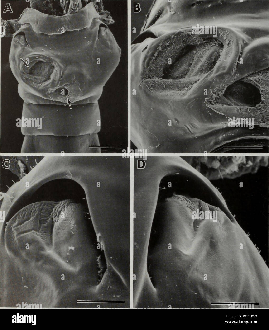 . Bulletin of the Natural History Museum Zoology. 150 S. OHTSUKA, G.A. BOXSHALL AND H.S.J. ROE. Fig. 31. Paraugaptilus buchani, female. SEM micrographs of genital double-somite. A, Genital double-somite, copulatory pores arrowed, scale bar = 100 [x.m; B, Copulatory pores, scale bar = 50 u.m; C, Right gonopore, scale bar = 20 u.m; D, Left gonopore, scale bar = 20 jim.. Please note that these images are extracted from scanned page images that may have been digitally enhanced for readability - coloration and appearance of these illustrations may not perfectly resemble the original work.. Natural Stock Photohttps://www.alamy.com/image-license-details/?v=1https://www.alamy.com/bulletin-of-the-natural-history-museum-zoology-150-s-ohtsuka-ga-boxshall-and-hsj-roe-fig-31-paraugaptilus-buchani-female-sem-micrographs-of-genital-double-somite-a-genital-double-somite-copulatory-pores-arrowed-scale-bar-=-100-xm-b-copulatory-pores-scale-bar-=-50-um-c-right-gonopore-scale-bar-=-20-um-d-left-gonopore-scale-bar-=-20-jim-please-note-that-these-images-are-extracted-from-scanned-page-images-that-may-have-been-digitally-enhanced-for-readability-coloration-and-appearance-of-these-illustrations-may-not-perfectly-resemble-the-original-work-natural-image233849871.html
RMRGCNW3–. Bulletin of the Natural History Museum Zoology. 150 S. OHTSUKA, G.A. BOXSHALL AND H.S.J. ROE. Fig. 31. Paraugaptilus buchani, female. SEM micrographs of genital double-somite. A, Genital double-somite, copulatory pores arrowed, scale bar = 100 [x.m; B, Copulatory pores, scale bar = 50 u.m; C, Right gonopore, scale bar = 20 u.m; D, Left gonopore, scale bar = 20 jim.. Please note that these images are extracted from scanned page images that may have been digitally enhanced for readability - coloration and appearance of these illustrations may not perfectly resemble the original work.. Natural
 . Bulletin of the British Museum (Natural History) Entom Supp. 54 M. W. NIELSON at apical half, small inflated flange apically between two apical short lateral projections; ventral appendage narrow, tube-like, reaching apex of dorsal appendage; gonopore apical; connective Y-shaped; style narrowed and curved apically, not clawed; plate with distal segment elongate, inflated subapically on dorsal margin. Specimens examined. Holotype $, New Guinea: Cyclops Mts, Sabron, 930 ft, v. 1936 (L. A. Cheesman) (BMNH, London). Paratypes. New Guinea: 10 km E. of Bokondini, 40 km N. of Balien Valley, 1300m, Stock Photohttps://www.alamy.com/image-license-details/?v=1https://www.alamy.com/bulletin-of-the-british-museum-natural-history-entom-supp-54-m-w-nielson-at-apical-half-small-inflated-flange-apically-between-two-apical-short-lateral-projections-ventral-appendage-narrow-tube-like-reaching-apex-of-dorsal-appendage-gonopore-apical-connective-y-shaped-style-narrowed-and-curved-apically-not-clawed-plate-with-distal-segment-elongate-inflated-subapically-on-dorsal-margin-specimens-examined-holotype-new-guinea-cyclops-mts-sabron-930-ft-v-1936-l-a-cheesman-bmnh-london-paratypes-new-guinea-10-km-e-of-bokondini-40-km-n-of-balien-valley-1300m-image233949026.html
RMRGH8AA–. Bulletin of the British Museum (Natural History) Entom Supp. 54 M. W. NIELSON at apical half, small inflated flange apically between two apical short lateral projections; ventral appendage narrow, tube-like, reaching apex of dorsal appendage; gonopore apical; connective Y-shaped; style narrowed and curved apically, not clawed; plate with distal segment elongate, inflated subapically on dorsal margin. Specimens examined. Holotype $, New Guinea: Cyclops Mts, Sabron, 930 ft, v. 1936 (L. A. Cheesman) (BMNH, London). Paratypes. New Guinea: 10 km E. of Bokondini, 40 km N. of Balien Valley, 1300m,
 . Bonner zoologische Monographien. Zoology. HUBER. REX ISION AND CLAHIS 11t: ANALYSIS Ol' P/lO/.CUS AND Cl tXSl lA Rl 1 Al l-l) TAXA (ARANHAl-, l'HOLCIDAE). FIG. 2254-2262. Pholcusphungiformes. 2254. Male ocular area, frontal view. 2255. Distal male chelicerai apophysis. 2256. Right procursus, distal view. 2257. Left procursus, prolateral view. 2258. Left uncus and embolus, prolateral view. 2259. Right bulbal processes, retrolateral view (arrow points at possible remnant of appendix). 2260. Male gonopore. 2261. Male ALS. 2262. Epigynum. Scale lines: 300 pm (2262), 200 pm (2254, 2258, 2259), 10 Stock Photohttps://www.alamy.com/image-license-details/?v=1https://www.alamy.com/bonner-zoologische-monographien-zoology-huber-rex-ision-and-clahis-11t-analysis-ol-plocus-and-cl-txsl-la-rl-1-al-l-l-taxa-aranhal-lholcidae-fig-2254-2262-pholcusphungiformes-2254-male-ocular-area-frontal-view-2255-distal-male-chelicerai-apophysis-2256-right-procursus-distal-view-2257-left-procursus-prolateral-view-2258-left-uncus-and-embolus-prolateral-view-2259-right-bulbal-processes-retrolateral-view-arrow-points-at-possible-remnant-of-appendix-2260-male-gonopore-2261-male-als-2262-epigynum-scale-lines-300-pm-2262-200-pm-2254-2258-2259-10-image234478847.html
RMRHDC4F–. Bonner zoologische Monographien. Zoology. HUBER. REX ISION AND CLAHIS 11t: ANALYSIS Ol' P/lO/.CUS AND Cl tXSl lA Rl 1 Al l-l) TAXA (ARANHAl-, l'HOLCIDAE). FIG. 2254-2262. Pholcusphungiformes. 2254. Male ocular area, frontal view. 2255. Distal male chelicerai apophysis. 2256. Right procursus, distal view. 2257. Left procursus, prolateral view. 2258. Left uncus and embolus, prolateral view. 2259. Right bulbal processes, retrolateral view (arrow points at possible remnant of appendix). 2260. Male gonopore. 2261. Male ALS. 2262. Epigynum. Scale lines: 300 pm (2262), 200 pm (2254, 2258, 2259), 10
 . Bulletin of the Natural History Museum Zoology. . Fig. 1. Crassarietellus huysi gen. et sp. nov., female (holotype: F,G; paratype: A-E). A, Habitus, dorsal view; B, Habitus, lateral view; C, Urosome, ventral view; D, Genital double-somite, ventral view; E, Genital double-somite, lateral view, cd: copulatory duct; cp: copulatory pore; g: gonopore; rd: receptacle duct; o: oviduct; s: spermatophore remnant; sr: seminal receptacle; F, Antenna, one terminal seta on second endopod segment missing; G, Terminal part of second endopod of other antenna. Scales in mm.. Please note that these images are Stock Photohttps://www.alamy.com/image-license-details/?v=1https://www.alamy.com/bulletin-of-the-natural-history-museum-zoology-fig-1-crassarietellus-huysi-gen-et-sp-nov-female-holotype-fg-paratype-a-e-a-habitus-dorsal-view-b-habitus-lateral-view-c-urosome-ventral-view-d-genital-double-somite-ventral-view-e-genital-double-somite-lateral-view-cd-copulatory-duct-cp-copulatory-pore-g-gonopore-rd-receptacle-duct-o-oviduct-s-spermatophore-remnant-sr-seminal-receptacle-f-antenna-one-terminal-seta-on-second-endopod-segment-missing-g-terminal-part-of-second-endopod-of-other-antenna-scales-in-mm-please-note-that-these-images-are-image233866333.html
RMRGDEW1–. Bulletin of the Natural History Museum Zoology. . Fig. 1. Crassarietellus huysi gen. et sp. nov., female (holotype: F,G; paratype: A-E). A, Habitus, dorsal view; B, Habitus, lateral view; C, Urosome, ventral view; D, Genital double-somite, ventral view; E, Genital double-somite, lateral view, cd: copulatory duct; cp: copulatory pore; g: gonopore; rd: receptacle duct; o: oviduct; s: spermatophore remnant; sr: seminal receptacle; F, Antenna, one terminal seta on second endopod segment missing; G, Terminal part of second endopod of other antenna. Scales in mm.. Please note that these images are
 . Bonner zoologische Monographien. Zoology. FIG. 955-963. Pholcus kribi. 955, 956. Male and female prosomata, frontal views. 957. Comb-hairs on male right tarsus 4. 958. Left procursus, retrolatero-ventral view. 959. Left bulb, retrolateral view. 960. Distal male cheliceral apophysis. 961. Male gonopore. 962. Male ALS. 963. Epigynum. Scale lines: 200 |am (955, 956), 100 pm (958, 963), 80 pm (959), 30 pm (961), 10 pm (957, 960, 962). 205. Please note that these images are extracted from scanned page images that may have been digitally enhanced for readability - coloration and appearance of thes Stock Photohttps://www.alamy.com/image-license-details/?v=1https://www.alamy.com/bonner-zoologische-monographien-zoology-fig-955-963-pholcus-kribi-955-956-male-and-female-prosomata-frontal-views-957-comb-hairs-on-male-right-tarsus-4-958-left-procursus-retrolatero-ventral-view-959-left-bulb-retrolateral-view-960-distal-male-cheliceral-apophysis-961-male-gonopore-962-male-als-963-epigynum-scale-lines-200-am-955-956-100-pm-958-963-80-pm-959-30-pm-961-10-pm-957-960-962-205-please-note-that-these-images-are-extracted-from-scanned-page-images-that-may-have-been-digitally-enhanced-for-readability-coloration-and-appearance-of-thes-image234480872.html
RMRHDEMT–. Bonner zoologische Monographien. Zoology. FIG. 955-963. Pholcus kribi. 955, 956. Male and female prosomata, frontal views. 957. Comb-hairs on male right tarsus 4. 958. Left procursus, retrolatero-ventral view. 959. Left bulb, retrolateral view. 960. Distal male cheliceral apophysis. 961. Male gonopore. 962. Male ALS. 963. Epigynum. Scale lines: 200 |am (955, 956), 100 pm (958, 963), 80 pm (959), 30 pm (961), 10 pm (957, 960, 962). 205. Please note that these images are extracted from scanned page images that may have been digitally enhanced for readability - coloration and appearance of thes
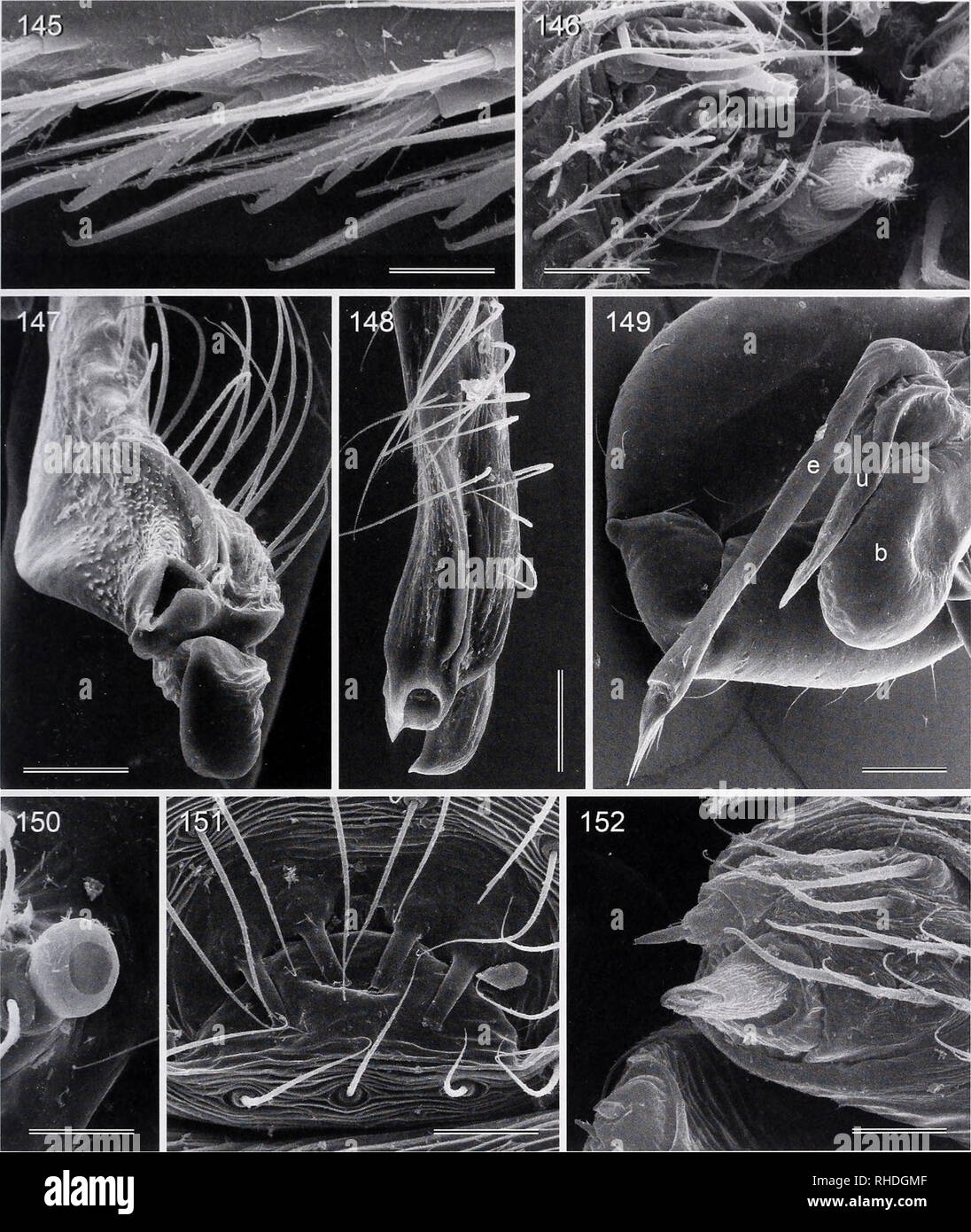 . Bonner zoologische Monographien. Zoology. FIG. 145-152. 145. Sihala ceylonica, comb-hairs on female tarsus 4. 146. Uthina luzonica, female ALS. 147- 152. Calapnita vermiformis. 147, 148. Left procursus, dorso-distal and retrolateral views. 149. Processes of left bulb. 150. Male palpal tarsal organ. 151. Male gonopore. 152. Male ALS. Scale lines: 100 jam (149), 60 pm (148), 50 pm (147), 20 ^im (145, 146, 150, 151), 10 ^im (152). 47. Please note that these images are extracted from scanned page images that may have been digitally enhanced for readability - coloration and appearance of these il Stock Photohttps://www.alamy.com/image-license-details/?v=1https://www.alamy.com/bonner-zoologische-monographien-zoology-fig-145-152-145-sihala-ceylonica-comb-hairs-on-female-tarsus-4-146-uthina-luzonica-female-als-147-152-calapnita-vermiformis-147-148-left-procursus-dorso-distal-and-retrolateral-views-149-processes-of-left-bulb-150-male-palpal-tarsal-organ-151-male-gonopore-152-male-als-scale-lines-100-jam-149-60-pm-148-50-pm-147-20-im-145-146-150-151-10-im-152-47-please-note-that-these-images-are-extracted-from-scanned-page-images-that-may-have-been-digitally-enhanced-for-readability-coloration-and-appearance-of-these-il-image234482431.html
RMRHDGMF–. Bonner zoologische Monographien. Zoology. FIG. 145-152. 145. Sihala ceylonica, comb-hairs on female tarsus 4. 146. Uthina luzonica, female ALS. 147- 152. Calapnita vermiformis. 147, 148. Left procursus, dorso-distal and retrolateral views. 149. Processes of left bulb. 150. Male palpal tarsal organ. 151. Male gonopore. 152. Male ALS. Scale lines: 100 jam (149), 60 pm (148), 50 pm (147), 20 ^im (145, 146, 150, 151), 10 ^im (152). 47. Please note that these images are extracted from scanned page images that may have been digitally enhanced for readability - coloration and appearance of these il
 . Bonner zoologische Monographien. Zoology. BONNER ZOOLOGISCHE MONOGRAPHIEN Nr. 58/20II. FIG. 1169-1176. Pholcus lamperti. 1169. Male prosoma, frontal view. 1170. Male ocular area, dorsal view. 1171. Female prosoma, frontal view. 1172, 1173. Left procursus, dorsal and prolateral views. 1174. Male gonopore. 1175, 1176. Distal male cheliceral apophyses and modified hairs. Scale lines: 500 pm (1169), 400 â pm (1171), 200 pm (1170, 1173), 100 pm (1172), 50 pm (1174), 40 pm (1175), 10 pm (1176). 244. Please note that these images are extracted from scanned page images that may have been digitally e Stock Photohttps://www.alamy.com/image-license-details/?v=1https://www.alamy.com/bonner-zoologische-monographien-zoology-bonner-zoologische-monographien-nr-5820ii-fig-1169-1176-pholcus-lamperti-1169-male-prosoma-frontal-view-1170-male-ocular-area-dorsal-view-1171-female-prosoma-frontal-view-1172-1173-left-procursus-dorsal-and-prolateral-views-1174-male-gonopore-1175-1176-distal-male-cheliceral-apophyses-and-modified-hairs-scale-lines-500-pm-1169-400-pm-1171-200-pm-1170-1173-100-pm-1172-50-pm-1174-40-pm-1175-10-pm-1176-244-please-note-that-these-images-are-extracted-from-scanned-page-images-that-may-have-been-digitally-e-image234480445.html
RMRHDE5H–. Bonner zoologische Monographien. Zoology. BONNER ZOOLOGISCHE MONOGRAPHIEN Nr. 58/20II. FIG. 1169-1176. Pholcus lamperti. 1169. Male prosoma, frontal view. 1170. Male ocular area, dorsal view. 1171. Female prosoma, frontal view. 1172, 1173. Left procursus, dorsal and prolateral views. 1174. Male gonopore. 1175, 1176. Distal male cheliceral apophyses and modified hairs. Scale lines: 500 pm (1169), 400 â pm (1171), 200 pm (1170, 1173), 100 pm (1172), 50 pm (1174), 40 pm (1175), 10 pm (1176). 244. Please note that these images are extracted from scanned page images that may have been digitally e
 . Bonner zoologische Monographien. Zoology. BONNER ZOOLOGISCHE MONOGRAPHIEN Nr. 58/2011. FIG. 1310-1321. Pholcus debilis. 1310, 1311. Male prosoma, frontal and fronto-dorsal views. 1312. Female prosoma, frontal view. 1313. Distal male cheliceral apophysis. 1314, 1315. Left and right bulbal processes, prolateral and retrolateral views. 1316, 1317. Left procursus, distal and prolateral views. 1318. Male gonopore. 1319. Male ALS and PMS. 1320. Epigynum. 1321. Comb-hairs on left female tarsus 4. Scale lines: 300 pm (1310), 200 pm (1311, 1312, 1320), 100 pm (1314, 1315, 1317), 80 pm (1316), 40 pm ( Stock Photohttps://www.alamy.com/image-license-details/?v=1https://www.alamy.com/bonner-zoologische-monographien-zoology-bonner-zoologische-monographien-nr-582011-fig-1310-1321-pholcus-debilis-1310-1311-male-prosoma-frontal-and-fronto-dorsal-views-1312-female-prosoma-frontal-view-1313-distal-male-cheliceral-apophysis-1314-1315-left-and-right-bulbal-processes-prolateral-and-retrolateral-views-1316-1317-left-procursus-distal-and-prolateral-views-1318-male-gonopore-1319-male-als-and-pms-1320-epigynum-1321-comb-hairs-on-left-female-tarsus-4-scale-lines-300-pm-1310-200-pm-1311-1312-1320-100-pm-1314-1315-1317-80-pm-1316-40-pm-image234480114.html
RMRHDDNP–. Bonner zoologische Monographien. Zoology. BONNER ZOOLOGISCHE MONOGRAPHIEN Nr. 58/2011. FIG. 1310-1321. Pholcus debilis. 1310, 1311. Male prosoma, frontal and fronto-dorsal views. 1312. Female prosoma, frontal view. 1313. Distal male cheliceral apophysis. 1314, 1315. Left and right bulbal processes, prolateral and retrolateral views. 1316, 1317. Left procursus, distal and prolateral views. 1318. Male gonopore. 1319. Male ALS and PMS. 1320. Epigynum. 1321. Comb-hairs on left female tarsus 4. Scale lines: 300 pm (1310), 200 pm (1311, 1312, 1320), 100 pm (1314, 1315, 1317), 80 pm (1316), 40 pm (
 . Bonner zoologische Monographien. Zoology. FIG. 26. Bandakiopsis phaluti s^. nov. (male). Top left: genital sclerite of male. Top right: eye plate of female. Bottom: ventral and dorsal shield female. Scale bars = 100 pm. PANESAR, EVOLUTION IN WATER MITES. FIG. 27. Bandakiopsis phaluti s^. nov. (female), Pal- pus. Top: medial view. Bottom: P4-P5 lateral view. Scale bars =100 pm. slightly enlarged, (2) gonopore relatively longer in fe- male, and (3) female in general slightly larger than male (Table 12 and Fig. 25, 27). Differential character. Bandakiopsis phaluti s'^. nov. dif- fers from Banda Stock Photohttps://www.alamy.com/image-license-details/?v=1https://www.alamy.com/bonner-zoologische-monographien-zoology-fig-26-bandakiopsis-phaluti-s-nov-male-top-left-genital-sclerite-of-male-top-right-eye-plate-of-female-bottom-ventral-and-dorsal-shield-female-scale-bars-=-100-pm-panesar-evolution-in-water-mites-fig-27-bandakiopsis-phaluti-s-nov-female-pal-pus-top-medial-view-bottom-p4-p5-lateral-view-scale-bars-=100-pm-slightly-enlarged-2-gonopore-relatively-longer-in-fe-male-and-3-female-in-general-slightly-larger-than-male-table-12-and-fig-25-27-differential-character-bandakiopsis-phaluti-s-nov-dif-fers-from-banda-image234487031.html
RMRHDPGR–. Bonner zoologische Monographien. Zoology. FIG. 26. Bandakiopsis phaluti s^. nov. (male). Top left: genital sclerite of male. Top right: eye plate of female. Bottom: ventral and dorsal shield female. Scale bars = 100 pm. PANESAR, EVOLUTION IN WATER MITES. FIG. 27. Bandakiopsis phaluti s^. nov. (female), Pal- pus. Top: medial view. Bottom: P4-P5 lateral view. Scale bars =100 pm. slightly enlarged, (2) gonopore relatively longer in fe- male, and (3) female in general slightly larger than male (Table 12 and Fig. 25, 27). Differential character. Bandakiopsis phaluti s'^. nov. dif- fers from Banda
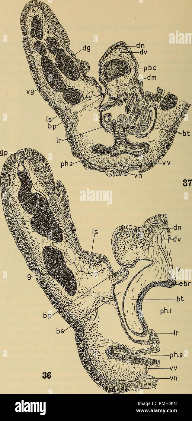 . Annals of the South African Museum = Annale van die Suid-Afrikaanse Museum. Natural history. Figs. 36, 37.—Glossobalanus alatus n. sp. 36. Cross-section of the branchial region. x52. 37. Cross-section near the posterior end of the branchial region, x 34. bp, branchial pore. 65, branchial sac. bt, branchial tongue, dg, dorsal gonad, dm, dorsal mesentery, dn, dorsal nerve cord, dv, dorsal blood- vessel, ebr, epibranchial ridge, g, gonad, gp, gonopore. Ir, limiting rldg<p Is, lateral septum, pbc, anterior blind-sac of the postbranchial canal, phi, branchial part of pharynx. ph2, digestive pa Stock Photohttps://www.alamy.com/image-license-details/?v=1https://www.alamy.com/annals-of-the-south-african-museum-=-annale-van-die-suid-afrikaanse-museum-natural-history-figs-36-37glossobalanus-alatus-n-sp-36-cross-section-of-the-branchial-region-x52-37-cross-section-near-the-posterior-end-of-the-branchial-region-x-34-bp-branchial-pore-65-branchial-sac-bt-branchial-tongue-dg-dorsal-gonad-dm-dorsal-mesentery-dn-dorsal-nerve-cord-dv-dorsal-blood-vessel-ebr-epibranchial-ridge-g-gonad-gp-gonopore-ir-limiting-rldgltp-is-lateral-septum-pbc-anterior-blind-sac-of-the-postbranchial-canal-phi-branchial-part-of-pharynx-ph2-digestive-pa-image236401641.html
RMRMH0KN–. Annals of the South African Museum = Annale van die Suid-Afrikaanse Museum. Natural history. Figs. 36, 37.—Glossobalanus alatus n. sp. 36. Cross-section of the branchial region. x52. 37. Cross-section near the posterior end of the branchial region, x 34. bp, branchial pore. 65, branchial sac. bt, branchial tongue, dg, dorsal gonad, dm, dorsal mesentery, dn, dorsal nerve cord, dv, dorsal blood- vessel, ebr, epibranchial ridge, g, gonad, gp, gonopore. Ir, limiting rldg<p Is, lateral septum, pbc, anterior blind-sac of the postbranchial canal, phi, branchial part of pharynx. ph2, digestive pa
 . Bonner zoologische Monographien. Zoology. BONNER ZOOLOGISCHE MONOGRAPHIEN Nr. 54/2007. FIG. 28. Epsteinia alba, dorsal and ventral view (Photo. Epshtein V.M.). cated (Figs. 28, 29: 2). Posterior sucker small, but larger than anterior one, excentricaly attached, sur- face smooth (Figs. 28, 29: 3). Annulation. Clitellum 8-annulate (Fig. 29: 4). Male gonopore located in depression between annuli 3 and 4, female gonopore in depression between annuli 6 and 7. 3rd annulus enlarged in central part, 4th bent posteriorly and covering annuli 5 and 6, annulus 7 bent posteriorly also. Copulatory area in Stock Photohttps://www.alamy.com/image-license-details/?v=1https://www.alamy.com/bonner-zoologische-monographien-zoology-bonner-zoologische-monographien-nr-542007-fig-28-epsteinia-alba-dorsal-and-ventral-view-photo-epshtein-vm-cated-figs-28-29-2-posterior-sucker-small-but-larger-than-anterior-one-excentricaly-attached-sur-face-smooth-figs-28-29-3-annulation-clitellum-8-annulate-fig-29-4-male-gonopore-located-in-depression-between-annuli-3-and-4-female-gonopore-in-depression-between-annuli-6-and-7-3rd-annulus-enlarged-in-central-part-4th-bent-posteriorly-and-covering-annuli-5-and-6-annulus-7-bent-posteriorly-also-copulatory-area-in-image234467129.html
RMRHCW61–. Bonner zoologische Monographien. Zoology. BONNER ZOOLOGISCHE MONOGRAPHIEN Nr. 54/2007. FIG. 28. Epsteinia alba, dorsal and ventral view (Photo. Epshtein V.M.). cated (Figs. 28, 29: 2). Posterior sucker small, but larger than anterior one, excentricaly attached, sur- face smooth (Figs. 28, 29: 3). Annulation. Clitellum 8-annulate (Fig. 29: 4). Male gonopore located in depression between annuli 3 and 4, female gonopore in depression between annuli 6 and 7. 3rd annulus enlarged in central part, 4th bent posteriorly and covering annuli 5 and 6, annulus 7 bent posteriorly also. Copulatory area in
 . Bonner zoologische Monographien. Zoology. BONNER ZOOLOGISCHE MONOGRAPHIEN Nr. 58/2011. FIG. 560-569. Pholcus kinabalu. 560, 561. Male and female prosomata, frontal views. 562. Comb-hairs on female left tarsus 4. 563, 564. Left procursus, dorsal and retrolatero-dorsal views. 565. Female ALS and PMS. 566. Male ALS. 567. Male gonopore. 568. Cleared female genitalia, dorsal view. 569. Detail of pore plate. Scale lines: 300 [am (560, 561), 100 ^im (563, 564, 568), 40 pm (567), 20 pm (562), 10 pm (565, 566, 569). many tarsal pseudosegments, only distally visible in dissecting microscope. Female. I Stock Photohttps://www.alamy.com/image-license-details/?v=1https://www.alamy.com/bonner-zoologische-monographien-zoology-bonner-zoologische-monographien-nr-582011-fig-560-569-pholcus-kinabalu-560-561-male-and-female-prosomata-frontal-views-562-comb-hairs-on-female-left-tarsus-4-563-564-left-procursus-dorsal-and-retrolatero-dorsal-views-565-female-als-and-pms-566-male-als-567-male-gonopore-568-cleared-female-genitalia-dorsal-view-569-detail-of-pore-plate-scale-lines-300-am-560-561-100-im-563-564-568-40-pm-567-20-pm-562-10-pm-565-566-569-many-tarsal-pseudosegments-only-distally-visible-in-dissecting-microscope-female-i-image234481748.html
RMRHDFT4–. Bonner zoologische Monographien. Zoology. BONNER ZOOLOGISCHE MONOGRAPHIEN Nr. 58/2011. FIG. 560-569. Pholcus kinabalu. 560, 561. Male and female prosomata, frontal views. 562. Comb-hairs on female left tarsus 4. 563, 564. Left procursus, dorsal and retrolatero-dorsal views. 565. Female ALS and PMS. 566. Male ALS. 567. Male gonopore. 568. Cleared female genitalia, dorsal view. 569. Detail of pore plate. Scale lines: 300 [am (560, 561), 100 ^im (563, 564, 568), 40 pm (567), 20 pm (562), 10 pm (565, 566, 569). many tarsal pseudosegments, only distally visible in dissecting microscope. Female. I
 . Bonner zoologische Monographien. Zoology. lll'Bl-R, RlA lSIdN AND CI APIS l lC ANAIVSIS Ol' /V/O/Cr'.V AND CI OSl'l Y Rl'I Al'l'1)'I'AXA (ARANF.Al',, I'HOI.CIDAH). FIG. 1565-1573. Pholcus bicomutus. 1565. Male ocular area. 1566, 1568. Modifications on male ocular area, frontal and dorsal views. 1567. Female prosoma, frontal view. 1569. Left male palp, dorsal view. 1570. Male gonopore. 1571. Distal male cheliceral apophysis. 1572. Male 7LS (with silk line emerging from the enlarged piriform gland spigot). 1573. Same male specimen, PMS (with silk lines emerging from median spigots). Scale lin Stock Photohttps://www.alamy.com/image-license-details/?v=1https://www.alamy.com/bonner-zoologische-monographien-zoology-lllbl-r-rla-lsidn-and-ci-apis-l-lc-anaivsis-ol-vocrv-and-ci-osll-y-rli-all1iaxa-aranfal-ihoicidah-fig-1565-1573-pholcus-bicomutus-1565-male-ocular-area-1566-1568-modifications-on-male-ocular-area-frontal-and-dorsal-views-1567-female-prosoma-frontal-view-1569-left-male-palp-dorsal-view-1570-male-gonopore-1571-distal-male-cheliceral-apophysis-1572-male-7ls-with-silk-line-emerging-from-the-enlarged-piriform-gland-spigot-1573-same-male-specimen-pms-with-silk-lines-emerging-from-median-spigots-scale-lin-image234479612.html
RMRHDD3T–. Bonner zoologische Monographien. Zoology. lll'Bl-R, RlA lSIdN AND CI APIS l lC ANAIVSIS Ol' /V/O/Cr'.V AND CI OSl'l Y Rl'I Al'l'1)'I'AXA (ARANF.Al',, I'HOI.CIDAH). FIG. 1565-1573. Pholcus bicomutus. 1565. Male ocular area. 1566, 1568. Modifications on male ocular area, frontal and dorsal views. 1567. Female prosoma, frontal view. 1569. Left male palp, dorsal view. 1570. Male gonopore. 1571. Distal male cheliceral apophysis. 1572. Male 7LS (with silk line emerging from the enlarged piriform gland spigot). 1573. Same male specimen, PMS (with silk lines emerging from median spigots). Scale lin
 . Bonner zoologische Monographien. Zoology. HL'BER, REMSION AND CL-XDISTIC ANALYSIS Ol- PHOICUS AND CLOSELY RELATED DVXA (AFIANHAE, I'HOLCIDAE). FIG. 1493-1501. Pholcus ancoralis. 1493. Male ocular area (arrows point at processes). 1494. Right bulbal processes. 1495. Right procursus tip, distal view. 1496. Male gonopore. 1497. Distal male cheliceral apophysis. 1498. Male ALS and PMS. 1499. Comb-hairs on right female tarsus 4. 1500. Female palpal tarsus tip. 1501. Epigynum. Scale lines: 200 pm (1493, 1494, 1501), 80 ^m (1495), 60 pm (1496), 50 pm (1500), 20 pm (1497, 1498, 1499). and ventral br Stock Photohttps://www.alamy.com/image-license-details/?v=1https://www.alamy.com/bonner-zoologische-monographien-zoology-hlber-remsion-and-cl-xdistic-analysis-ol-phoicus-and-closely-related-dvxa-afianhae-iholcidae-fig-1493-1501-pholcus-ancoralis-1493-male-ocular-area-arrows-point-at-processes-1494-right-bulbal-processes-1495-right-procursus-tip-distal-view-1496-male-gonopore-1497-distal-male-cheliceral-apophysis-1498-male-als-and-pms-1499-comb-hairs-on-right-female-tarsus-4-1500-female-palpal-tarsus-tip-1501-epigynum-scale-lines-200-pm-1493-1494-1501-80-m-1495-60-pm-1496-50-pm-1500-20-pm-1497-1498-1499-and-ventral-br-image234479687.html
RMRHDD6F–. Bonner zoologische Monographien. Zoology. HL'BER, REMSION AND CL-XDISTIC ANALYSIS Ol- PHOICUS AND CLOSELY RELATED DVXA (AFIANHAE, I'HOLCIDAE). FIG. 1493-1501. Pholcus ancoralis. 1493. Male ocular area (arrows point at processes). 1494. Right bulbal processes. 1495. Right procursus tip, distal view. 1496. Male gonopore. 1497. Distal male cheliceral apophysis. 1498. Male ALS and PMS. 1499. Comb-hairs on right female tarsus 4. 1500. Female palpal tarsus tip. 1501. Epigynum. Scale lines: 200 pm (1493, 1494, 1501), 80 ^m (1495), 60 pm (1496), 50 pm (1500), 20 pm (1497, 1498, 1499). and ventral br
 . Bonner zoologische Monographien. Zoology. BONNER ZOOLOGISCHE MONOGRAPHIEN Nr. 58/2011. FIG. 1986-1994. Pholcus calligaster. 1986, 1987. Male prosoma and ocular area, frontal views. 1988. Female prosoma, dorso-frontal view. 1989. Left procursus tip, prolateral view. 1990. Left bulbal processes, prolat- eral view. 1991. Distal male cheliceral apophysis. 1992. Male gonopore (two spigots damaged). 1993. Tip of female palpal tarsus. 1994. Epigynum. Scale lines: 400 pm (1986, 1988), 200 jam (1987, 1994), 100 pm (1989, 1990), 50 pm (1992), 40 pm (1993), 10 pm (1991). ened, clypeus only slightly dar Stock Photohttps://www.alamy.com/image-license-details/?v=1https://www.alamy.com/bonner-zoologische-monographien-zoology-bonner-zoologische-monographien-nr-582011-fig-1986-1994-pholcus-calligaster-1986-1987-male-prosoma-and-ocular-area-frontal-views-1988-female-prosoma-dorso-frontal-view-1989-left-procursus-tip-prolateral-view-1990-left-bulbal-processes-prolat-eral-view-1991-distal-male-cheliceral-apophysis-1992-male-gonopore-two-spigots-damaged-1993-tip-of-female-palpal-tarsus-1994-epigynum-scale-lines-400-pm-1986-1988-200-jam-1987-1994-100-pm-1989-1990-50-pm-1992-40-pm-1993-10-pm-1991-ened-clypeus-only-slightly-dar-image234479051.html
RMRHDCBR–. Bonner zoologische Monographien. Zoology. BONNER ZOOLOGISCHE MONOGRAPHIEN Nr. 58/2011. FIG. 1986-1994. Pholcus calligaster. 1986, 1987. Male prosoma and ocular area, frontal views. 1988. Female prosoma, dorso-frontal view. 1989. Left procursus tip, prolateral view. 1990. Left bulbal processes, prolat- eral view. 1991. Distal male cheliceral apophysis. 1992. Male gonopore (two spigots damaged). 1993. Tip of female palpal tarsus. 1994. Epigynum. Scale lines: 400 pm (1986, 1988), 200 jam (1987, 1994), 100 pm (1989, 1990), 50 pm (1992), 40 pm (1993), 10 pm (1991). ened, clypeus only slightly dar
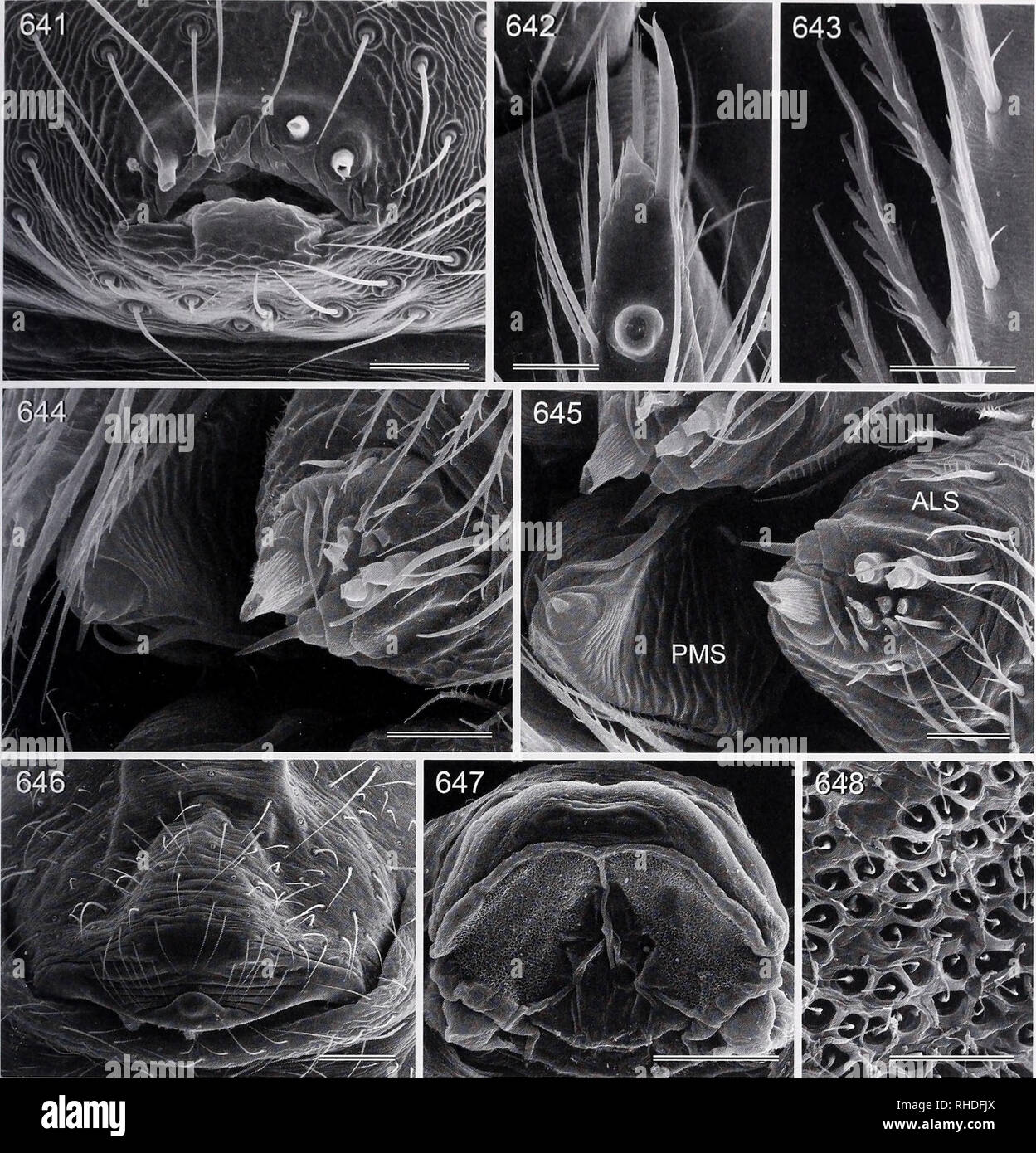 . Bonner zoologische Monographien. Zoology. HUBFR, REVISION AND CI.ADISTIC: ANALYSIS OF PHO/.CUS AUD C:i.OSFI.Y Rl'lAIFI) TAXA (ARANFAI., I'l IOI,c;il)AF). FIG. 641-648. Pholcus minang. 641. Male gonopore. 642. Tip of female palpal tarsus. 643. Comb-hairs on male tarsus 4. 644, 645. Male (644) and female (645) ALS and PMS. 646. Epigynum. 647. Cleared female genitalia, dorsal view. 648. Detail of pore plate. Scale lines: 200 pm (647), 100 pm (646), 30 pm (641, 642), 20 pm (643-645, 648). and uncus (Figs. 627, 628); from Ph. singalang also by morphology of female internal genitalia (Figs. 611, 6 Stock Photohttps://www.alamy.com/image-license-details/?v=1https://www.alamy.com/bonner-zoologische-monographien-zoology-hubfr-revision-and-ciadistic-analysis-of-phocus-aud-ciosfiy-rllaifi-taxa-aranfai-il-ioicilaf-fig-641-648-pholcus-minang-641-male-gonopore-642-tip-of-female-palpal-tarsus-643-comb-hairs-on-male-tarsus-4-644-645-male-644-and-female-645-als-and-pms-646-epigynum-647-cleared-female-genitalia-dorsal-view-648-detail-of-pore-plate-scale-lines-200-pm-647-100-pm-646-30-pm-641-642-20-pm-643-645-648-and-uncus-figs-627-628-from-ph-singalang-also-by-morphology-of-female-internal-genitalia-figs-611-6-image234481602.html
RMRHDFJX–. Bonner zoologische Monographien. Zoology. HUBFR, REVISION AND CI.ADISTIC: ANALYSIS OF PHO/.CUS AUD C:i.OSFI.Y Rl'lAIFI) TAXA (ARANFAI., I'l IOI,c;il)AF). FIG. 641-648. Pholcus minang. 641. Male gonopore. 642. Tip of female palpal tarsus. 643. Comb-hairs on male tarsus 4. 644, 645. Male (644) and female (645) ALS and PMS. 646. Epigynum. 647. Cleared female genitalia, dorsal view. 648. Detail of pore plate. Scale lines: 200 pm (647), 100 pm (646), 30 pm (641, 642), 20 pm (643-645, 648). and uncus (Figs. 627, 628); from Ph. singalang also by morphology of female internal genitalia (Figs. 611, 6
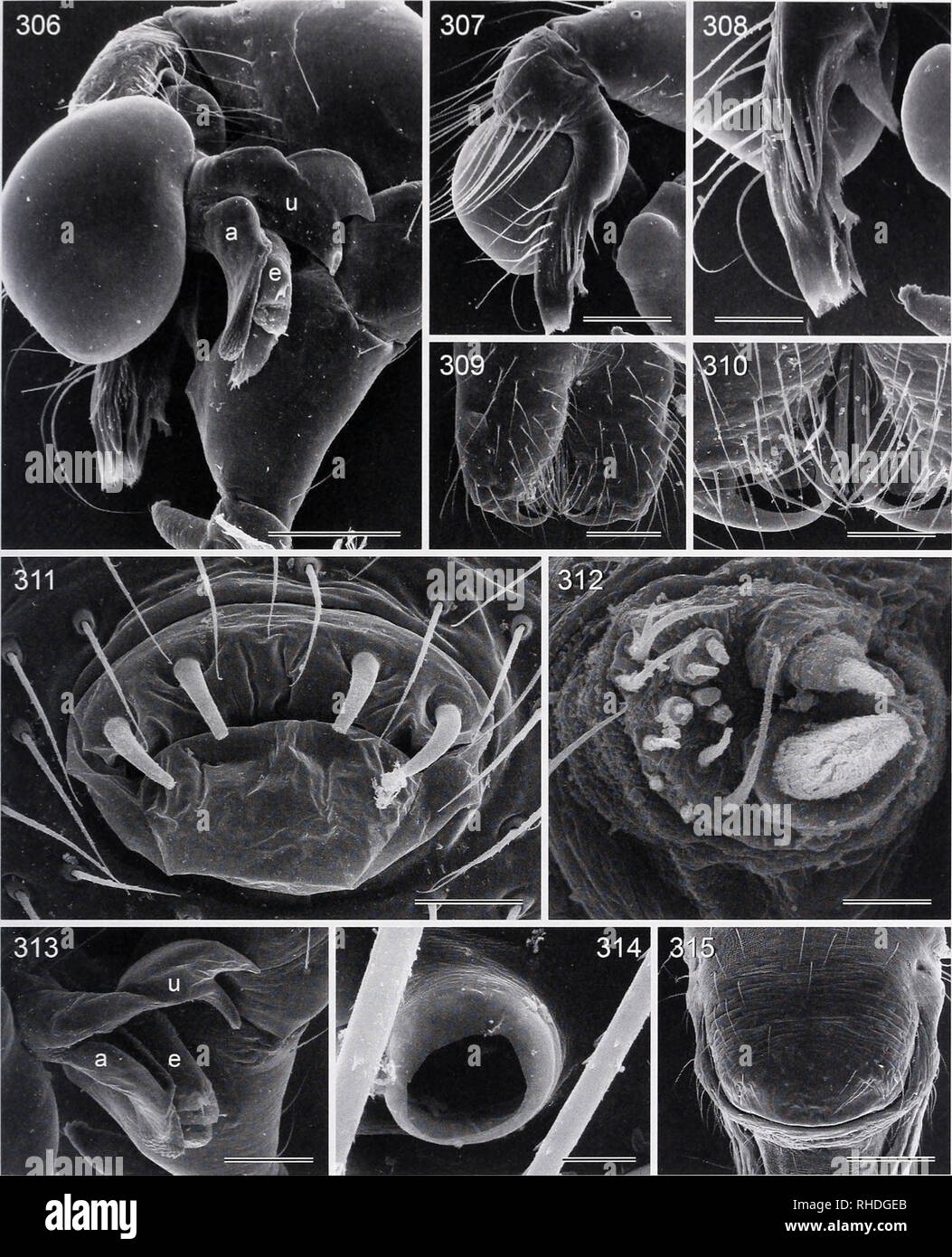 . Bonner zoologische Monographien. Zoology. HUBF.R. RHN ISION AND CI.ADIS TK: ANALYSIS OF /V/O/.Cm" AND Cl.CXSELY RHI AI HD TAXA (ARANHAH, I'l lOl.CIDAH). FIG. 306-315. Leptopholcus gracilis. South Africa. 306. Right male palp, prolateral view. 307, 308. Left procursus, retrolateral views. 309, 310. Male chelicerae. 311. Male gonopore. 312 Male ALS. 313. Processes of right bulb. 314. Male palpal tarsal organ. 315. Epigynum. Scale hnes: 200 pm (306, 307, 315), 100 pm (308, 309, 313), 50 pm (310), 30 pm (311), 10 pm (312, 314). 73. Please note that these images are extracted from scanned pa Stock Photohttps://www.alamy.com/image-license-details/?v=1https://www.alamy.com/bonner-zoologische-monographien-zoology-hubfr-rhn-ision-and-ciadis-tk-analysis-of-vocmquot-and-clcxsely-rhi-ai-hd-taxa-aranhah-il-lolcidah-fig-306-315-leptopholcus-gracilis-south-africa-306-right-male-palp-prolateral-view-307-308-left-procursus-retrolateral-views-309-310-male-chelicerae-311-male-gonopore-312-male-als-313-processes-of-right-bulb-314-male-palpal-tarsal-organ-315-epigynum-scale-hnes-200-pm-306-307-315-100-pm-308-309-313-50-pm-310-30-pm-311-10-pm-312-314-73-please-note-that-these-images-are-extracted-from-scanned-pa-image234482259.html
RMRHDGEB–. Bonner zoologische Monographien. Zoology. HUBF.R. RHN ISION AND CI.ADIS TK: ANALYSIS OF /V/O/.Cm" AND Cl.CXSELY RHI AI HD TAXA (ARANHAH, I'l lOl.CIDAH). FIG. 306-315. Leptopholcus gracilis. South Africa. 306. Right male palp, prolateral view. 307, 308. Left procursus, retrolateral views. 309, 310. Male chelicerae. 311. Male gonopore. 312 Male ALS. 313. Processes of right bulb. 314. Male palpal tarsal organ. 315. Epigynum. Scale hnes: 200 pm (306, 307, 315), 100 pm (308, 309, 313), 50 pm (310), 30 pm (311), 10 pm (312, 314). 73. Please note that these images are extracted from scanned pa
 . Bonner zoologische Monographien. Zoology. FIG. 450-458. Panjange lanthana. 450. Tip of putative embolus. 451. Palpal tarsal organ. 452. Male gonopore. 453. Male ALS and PMS. 454. Female prosoma, frontal view. 455, 458. Comb-hairs on right female tarsus 4. 456. Female v^LS and PMS. 457. Epigynum. Scale lines: 300 pm (454), 100 pm (457), 40 pm (452), 20 pm (450, 451, 453, 455, 456), 10 pm (458). Diagnosis. Easily distinguished from congeners by combination of male eye stalks (Fig. 441), male palpal morphology (Figs. 436, 437; dorsal trochanter apophysis, procursus, bulbal process extending in Stock Photohttps://www.alamy.com/image-license-details/?v=1https://www.alamy.com/bonner-zoologische-monographien-zoology-fig-450-458-panjange-lanthana-450-tip-of-putative-embolus-451-palpal-tarsal-organ-452-male-gonopore-453-male-als-and-pms-454-female-prosoma-frontal-view-455-458-comb-hairs-on-right-female-tarsus-4-456-female-vls-and-pms-457-epigynum-scale-lines-300-pm-454-100-pm-457-40-pm-452-20-pm-450-451-453-455-456-10-pm-458-diagnosis-easily-distinguished-from-congeners-by-combination-of-male-eye-stalks-fig-441-male-palpal-morphology-figs-436-437-dorsal-trochanter-apophysis-procursus-bulbal-process-extending-in-image234481990.html
RMRHDG4P–. Bonner zoologische Monographien. Zoology. FIG. 450-458. Panjange lanthana. 450. Tip of putative embolus. 451. Palpal tarsal organ. 452. Male gonopore. 453. Male ALS and PMS. 454. Female prosoma, frontal view. 455, 458. Comb-hairs on right female tarsus 4. 456. Female v^LS and PMS. 457. Epigynum. Scale lines: 300 pm (454), 100 pm (457), 40 pm (452), 20 pm (450, 451, 453, 455, 456), 10 pm (458). Diagnosis. Easily distinguished from congeners by combination of male eye stalks (Fig. 441), male palpal morphology (Figs. 436, 437; dorsal trochanter apophysis, procursus, bulbal process extending in
 . Bonner zoologische Monographien. Zoology. FIG. 405-417. Leptopholcus podophthalmus. 405. Male eye stalks, frontal view. 406, 407. Left procursus, distal and retrolateral views. 408, 409. Right procursus, prolateral and ventral views. 410. Processes of left bulb. 411. Tip of female palpal tarsus. 412. Male gonopore (one spigot damaged). 413. Male palpal tarsal organ. 414. Comb-hairs on female tarsus 4. 415. Male ALS. 416. Female ALS and PMS. 417. Epigynum. Scale lines: 200 pm (405, 409, 417), 100 pm (410), 80 pm (408), 60 pm (406, 407), 40 pm (412), 30 pm (411), 20 pm (413, 416), 10 pm (414, Stock Photohttps://www.alamy.com/image-license-details/?v=1https://www.alamy.com/bonner-zoologische-monographien-zoology-fig-405-417-leptopholcus-podophthalmus-405-male-eye-stalks-frontal-view-406-407-left-procursus-distal-and-retrolateral-views-408-409-right-procursus-prolateral-and-ventral-views-410-processes-of-left-bulb-411-tip-of-female-palpal-tarsus-412-male-gonopore-one-spigot-damaged-413-male-palpal-tarsal-organ-414-comb-hairs-on-female-tarsus-4-415-male-als-416-female-als-and-pms-417-epigynum-scale-lines-200-pm-405-409-417-100-pm-410-80-pm-408-60-pm-406-407-40-pm-412-30-pm-411-20-pm-413-416-10-pm-414-image234482099.html
RMRHDG8K–. Bonner zoologische Monographien. Zoology. FIG. 405-417. Leptopholcus podophthalmus. 405. Male eye stalks, frontal view. 406, 407. Left procursus, distal and retrolateral views. 408, 409. Right procursus, prolateral and ventral views. 410. Processes of left bulb. 411. Tip of female palpal tarsus. 412. Male gonopore (one spigot damaged). 413. Male palpal tarsal organ. 414. Comb-hairs on female tarsus 4. 415. Male ALS. 416. Female ALS and PMS. 417. Epigynum. Scale lines: 200 pm (405, 409, 417), 100 pm (410), 80 pm (408), 60 pm (406, 407), 40 pm (412), 30 pm (411), 20 pm (413, 416), 10 pm (414,
 . Bonner zoologische Monographien. Zoology. BONNER ZOOLOGISCHE MONOGRAPHIEN Nr. 58/2011. FIG. 1286-1297. Pholcus kwamgumi. 1286. Male chelicerae, frontal view. 1287. Right procursus, retrolatero- distal view. 1288. Left procursus, prolatero-distal view. 1289. Tip of left procursus, distal view. 1290, 1291. Left bulbal processes. 1292. Distal male cheliceral apophysis. 1293. Female prosoma, frontal view. 1294. Male ALS. 1295. Male gonopore. 1296. Epigynum. 1297. Female ALS and PMS. Scale lines: 200 fim (1293), 100 pm (1286-1290, 1296), 80 pm (1291), 30 pm (1295), 20 pm (1297), 10 pm (1292, 1294 Stock Photohttps://www.alamy.com/image-license-details/?v=1https://www.alamy.com/bonner-zoologische-monographien-zoology-bonner-zoologische-monographien-nr-582011-fig-1286-1297-pholcus-kwamgumi-1286-male-chelicerae-frontal-view-1287-right-procursus-retrolatero-distal-view-1288-left-procursus-prolatero-distal-view-1289-tip-of-left-procursus-distal-view-1290-1291-left-bulbal-processes-1292-distal-male-cheliceral-apophysis-1293-female-prosoma-frontal-view-1294-male-als-1295-male-gonopore-1296-epigynum-1297-female-als-and-pms-scale-lines-200-fim-1293-100-pm-1286-1290-1296-80-pm-1291-30-pm-1295-20-pm-1297-10-pm-1292-1294-image234480183.html
RMRHDDT7–. Bonner zoologische Monographien. Zoology. BONNER ZOOLOGISCHE MONOGRAPHIEN Nr. 58/2011. FIG. 1286-1297. Pholcus kwamgumi. 1286. Male chelicerae, frontal view. 1287. Right procursus, retrolatero- distal view. 1288. Left procursus, prolatero-distal view. 1289. Tip of left procursus, distal view. 1290, 1291. Left bulbal processes. 1292. Distal male cheliceral apophysis. 1293. Female prosoma, frontal view. 1294. Male ALS. 1295. Male gonopore. 1296. Epigynum. 1297. Female ALS and PMS. Scale lines: 200 fim (1293), 100 pm (1286-1290, 1296), 80 pm (1291), 30 pm (1295), 20 pm (1297), 10 pm (1292, 1294
 . Bonner zoologische Monographien. Zoology. FIG. 176-188. Calapnita phyllicola. 176, 177. Male (palps removed) and female prosomata, frontal x iews. 178. Male distal cheliceral apophyses. 179, 180. Male palpal tarsal organ. 181. Tips of appendix and embolus, dorso-distal view. 182. Uncus of left bulb, prolateral view. 183. Male gonopore. 184. Tip of right embolus (arrow points at sperm duct opening). 185. Tip of right appendix, retrolateral view. 186. Comb hairs on left female tarsus 4. 187. Female ALS and PMS. 188. Epigynum. Scale lines: 200 ftm (176, 177, 188), 100 fim (185), 80 fim (181), 6 Stock Photohttps://www.alamy.com/image-license-details/?v=1https://www.alamy.com/bonner-zoologische-monographien-zoology-fig-176-188-calapnita-phyllicola-176-177-male-palps-removed-and-female-prosomata-frontal-x-iews-178-male-distal-cheliceral-apophyses-179-180-male-palpal-tarsal-organ-181-tips-of-appendix-and-embolus-dorso-distal-view-182-uncus-of-left-bulb-prolateral-view-183-male-gonopore-184-tip-of-right-embolus-arrow-points-at-sperm-duct-opening-185-tip-of-right-appendix-retrolateral-view-186-comb-hairs-on-left-female-tarsus-4-187-female-als-and-pms-188-epigynum-scale-lines-200-ftm-176-177-188-100-fim-185-80-fim-181-6-image234482389.html
RMRHDGK1–. Bonner zoologische Monographien. Zoology. FIG. 176-188. Calapnita phyllicola. 176, 177. Male (palps removed) and female prosomata, frontal x iews. 178. Male distal cheliceral apophyses. 179, 180. Male palpal tarsal organ. 181. Tips of appendix and embolus, dorso-distal view. 182. Uncus of left bulb, prolateral view. 183. Male gonopore. 184. Tip of right embolus (arrow points at sperm duct opening). 185. Tip of right appendix, retrolateral view. 186. Comb hairs on left female tarsus 4. 187. Female ALS and PMS. 188. Epigynum. Scale lines: 200 ftm (176, 177, 188), 100 fim (185), 80 fim (181), 6
 . Bonner zoologische Monographien. Zoology. FIG. 798-804. Pholcus ethagala. 798. Male ocular area, frontal view. 799. Right male eye stalk, fronto-dorsal view. 800. Male palpal tarsal organ. 801. Left bulbal processes, prolateral view. 802. Left bulb, prolatero- ventral view. 803. Male gonopore. 804. Male v^LS. Scale lines: 200 (798), 100 fim (802), 80 jam (799), 60 pm (801), 30 pm (803), 20 pm (800), 10 pm (804). Female. In general similar to male but triads barely elevated, closer together (distance PME-PME 205 pm), without pointed process; most females with carapace ochre-yellow with light Stock Photohttps://www.alamy.com/image-license-details/?v=1https://www.alamy.com/bonner-zoologische-monographien-zoology-fig-798-804-pholcus-ethagala-798-male-ocular-area-frontal-view-799-right-male-eye-stalk-fronto-dorsal-view-800-male-palpal-tarsal-organ-801-left-bulbal-processes-prolateral-view-802-left-bulb-prolatero-ventral-view-803-male-gonopore-804-male-vls-scale-lines-200-798-100-fim-802-80-jam-799-60-pm-801-30-pm-803-20-pm-800-10-pm-804-female-in-general-similar-to-male-but-triads-barely-elevated-closer-together-distance-pme-pme-205-pm-without-pointed-process-most-females-with-carapace-ochre-yellow-with-light-image234481303.html
RMRHDF87–. Bonner zoologische Monographien. Zoology. FIG. 798-804. Pholcus ethagala. 798. Male ocular area, frontal view. 799. Right male eye stalk, fronto-dorsal view. 800. Male palpal tarsal organ. 801. Left bulbal processes, prolateral view. 802. Left bulb, prolatero- ventral view. 803. Male gonopore. 804. Male v^LS. Scale lines: 200 (798), 100 fim (802), 80 jam (799), 60 pm (801), 30 pm (803), 20 pm (800), 10 pm (804). Female. In general similar to male but triads barely elevated, closer together (distance PME-PME 205 pm), without pointed process; most females with carapace ochre-yellow with light
 . Bonner zoologische Monographien. Zoology. BONNER ZOOLOGISCHE MONOGRAPHIEN Nr. 58/2011. FIG. 1625-1635. Pholcus creticus. 1625. Right procursus, retrolatero-dorsal view. 1626. Left procursus, distal view. 1627. Female ALS. 1628. Male palpal tarsal organ. 1629, 1630. Left bulbal processes. 1631. Distal male cheliceral apophysis. 1632. Male gonopore. 1633. Epigynum. 1634. Tip of female palpal tarsus. 1635. Comb-hairs on female tarsus 4. Scale lines: 200 pm (1633), 100 pm (1630), 60 pm (1625, 1626, 1629), 40 pm (1632), 30 pm (1634), 10 pm (1627, 1628, 1631, 1635). 338. Please note that these ima Stock Photohttps://www.alamy.com/image-license-details/?v=1https://www.alamy.com/bonner-zoologische-monographien-zoology-bonner-zoologische-monographien-nr-582011-fig-1625-1635-pholcus-creticus-1625-right-procursus-retrolatero-dorsal-view-1626-left-procursus-distal-view-1627-female-als-1628-male-palpal-tarsal-organ-1629-1630-left-bulbal-processes-1631-distal-male-cheliceral-apophysis-1632-male-gonopore-1633-epigynum-1634-tip-of-female-palpal-tarsus-1635-comb-hairs-on-female-tarsus-4-scale-lines-200-pm-1633-100-pm-1630-60-pm-1625-1626-1629-40-pm-1632-30-pm-1634-10-pm-1627-1628-1631-1635-338-please-note-that-these-ima-image234479457.html
RMRHDCX9–. Bonner zoologische Monographien. Zoology. BONNER ZOOLOGISCHE MONOGRAPHIEN Nr. 58/2011. FIG. 1625-1635. Pholcus creticus. 1625. Right procursus, retrolatero-dorsal view. 1626. Left procursus, distal view. 1627. Female ALS. 1628. Male palpal tarsal organ. 1629, 1630. Left bulbal processes. 1631. Distal male cheliceral apophysis. 1632. Male gonopore. 1633. Epigynum. 1634. Tip of female palpal tarsus. 1635. Comb-hairs on female tarsus 4. Scale lines: 200 pm (1633), 100 pm (1630), 60 pm (1625, 1626, 1629), 40 pm (1632), 30 pm (1634), 10 pm (1627, 1628, 1631, 1635). 338. Please note that these ima
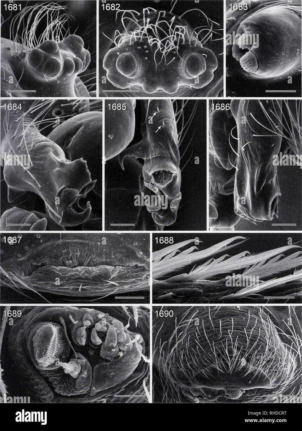 . Bonner zoologische Monographien. Zoology. HliBl R. Rl'A'ISlON AND CLADLSTIC ANAH SIS Ol- /7/0/CY/.V AND Cl.OSl.l Y Rl'I ATi:!) l AXA (ARANKAi:, PI lOl.CIDAE). FIG. 1681-1690. Pholcusponticus. 1681, 1682. Male ocular area, oblique and dorsal views. 1683. Distal male cheliceral apophysis. 1684. Right procursus, retrolatero-distal view. 1685. Left procursus, dorsal view (arrows point at spines). 1686. Right procursus, retrolatero-dorsal view. 1687. Male gonopore. 1688. Comb-hairs on female tarsus 4. 1689. Female ALS. 1690. Epigynum. Scale hnes: 200 pm (1681, 1682, 1690), 100 pm (1684-1686), 60 Stock Photohttps://www.alamy.com/image-license-details/?v=1https://www.alamy.com/bonner-zoologische-monographien-zoology-hlibl-r-rlaislon-and-cladlstic-anah-sis-ol-70cyv-and-closll-y-rli-ati!-l-axa-arankai-pi-lolcidae-fig-1681-1690-pholcusponticus-1681-1682-male-ocular-area-oblique-and-dorsal-views-1683-distal-male-cheliceral-apophysis-1684-right-procursus-retrolatero-distal-view-1685-left-procursus-dorsal-view-arrows-point-at-spines-1686-right-procursus-retrolatero-dorsal-view-1687-male-gonopore-1688-comb-hairs-on-female-tarsus-4-1689-female-als-1690-epigynum-scale-hnes-200-pm-1681-1682-1690-100-pm-1684-1686-60-image234479388.html
RMRHDCRT–. Bonner zoologische Monographien. Zoology. HliBl R. Rl'A'ISlON AND CLADLSTIC ANAH SIS Ol- /7/0/CY/.V AND Cl.OSl.l Y Rl'I ATi:!) l AXA (ARANKAi:, PI lOl.CIDAE). FIG. 1681-1690. Pholcusponticus. 1681, 1682. Male ocular area, oblique and dorsal views. 1683. Distal male cheliceral apophysis. 1684. Right procursus, retrolatero-distal view. 1685. Left procursus, dorsal view (arrows point at spines). 1686. Right procursus, retrolatero-dorsal view. 1687. Male gonopore. 1688. Comb-hairs on female tarsus 4. 1689. Female ALS. 1690. Epigynum. Scale hnes: 200 pm (1681, 1682, 1690), 100 pm (1684-1686), 60
 . Bonner zoologische Monographien. Zoology. BONNER ZOOLOGISCHE MONOGRAPHIEN Nr. 58/2011. FIG. 90-101. Micropholcus fauroti. 90. Left palp, retrolateral view. 91. Left procursus, prolateral view. 92. Pro- cesses of right bulb, prolateral view. 93, 94. Processes of left bulb, dorsal views (arrow points at sperm duct open- ing). 95. Tip of palpal trochanter apophysis. 96. Comb-hairs on right male tarsus 4. 97. Male palpal tarsal organ. 98. Modified hairs on distal male cheliceral apophysis. 99. Male gonopore. 100. Female ALS. 101. Epigynum. Scale lines: 200pm (90,101), 100pm (91), 60pm (92-94), 4 Stock Photohttps://www.alamy.com/image-license-details/?v=1https://www.alamy.com/bonner-zoologische-monographien-zoology-bonner-zoologische-monographien-nr-582011-fig-90-101-micropholcus-fauroti-90-left-palp-retrolateral-view-91-left-procursus-prolateral-view-92-pro-cesses-of-right-bulb-prolateral-view-93-94-processes-of-left-bulb-dorsal-views-arrow-points-at-sperm-duct-open-ing-95-tip-of-palpal-trochanter-apophysis-96-comb-hairs-on-right-male-tarsus-4-97-male-palpal-tarsal-organ-98-modified-hairs-on-distal-male-cheliceral-apophysis-99-male-gonopore-100-female-als-101-epigynum-scale-lines-200pm-90101-100pm-91-60pm-92-94-4-image234482611.html
RMRHDGXY–. Bonner zoologische Monographien. Zoology. BONNER ZOOLOGISCHE MONOGRAPHIEN Nr. 58/2011. FIG. 90-101. Micropholcus fauroti. 90. Left palp, retrolateral view. 91. Left procursus, prolateral view. 92. Pro- cesses of right bulb, prolateral view. 93, 94. Processes of left bulb, dorsal views (arrow points at sperm duct open- ing). 95. Tip of palpal trochanter apophysis. 96. Comb-hairs on right male tarsus 4. 97. Male palpal tarsal organ. 98. Modified hairs on distal male cheliceral apophysis. 99. Male gonopore. 100. Female ALS. 101. Epigynum. Scale lines: 200pm (90,101), 100pm (91), 60pm (92-94), 4
 . The Biological bulletin. Biology; Zoology; Biology; Marine Biology. F. FIGURE 2. A—eggs in process of being laid from gonopore; B—eggs about to be laid adjacent to gonopore; C—sperm and eggs in duct; D—eggs being laid; E—ovary showing con- traction bands after egg-laying; F—mucus-embedded eggs attached to grass blade. Figures A- C are from sections, D-F from live material. Diameter of eggs approx. 0.2 mm. e4—eggs in process of being laid; e5—attached (laid) eggs; gb—grass blade; m—muscle; mu—mucus, Other abbreviations as in Figure 1.. Please note that these images are extracted from scanned Stock Photohttps://www.alamy.com/image-license-details/?v=1https://www.alamy.com/the-biological-bulletin-biology-zoology-biology-marine-biology-f-figure-2-aeggs-in-process-of-being-laid-from-gonopore-beggs-about-to-be-laid-adjacent-to-gonopore-csperm-and-eggs-in-duct-deggs-being-laid-eovary-showing-con-traction-bands-after-egg-laying-fmucus-embedded-eggs-attached-to-grass-blade-figures-a-c-are-from-sections-d-f-from-live-material-diameter-of-eggs-approx-02-mm-e4eggs-in-process-of-being-laid-e5attached-laid-eggs-gbgrass-blade-mmuscle-mumucus-other-abbreviations-as-in-figure-1-please-note-that-these-images-are-extracted-from-scanned-image234650362.html
RMRHN6X2–. The Biological bulletin. Biology; Zoology; Biology; Marine Biology. F. FIGURE 2. A—eggs in process of being laid from gonopore; B—eggs about to be laid adjacent to gonopore; C—sperm and eggs in duct; D—eggs being laid; E—ovary showing con- traction bands after egg-laying; F—mucus-embedded eggs attached to grass blade. Figures A- C are from sections, D-F from live material. Diameter of eggs approx. 0.2 mm. e4—eggs in process of being laid; e5—attached (laid) eggs; gb—grass blade; m—muscle; mu—mucus, Other abbreviations as in Figure 1.. Please note that these images are extracted from scanned
 . Bonner zoologische Monographien. Zoology. HUBER, RF.VISION AND CLADISTIC ANALYSIS Ol' /V/C;/CY/.V AND c:i.(XSi:i,Y Kl'.LAl'ia) TAXA (ARANl'.Ai:, I'l lOLCIDAlO. FIG. 662-672. Pholcus bohorok. 662, 663. Male (662) and female (663) prosomata, oblique views. 664. Male chelicerae, oblique view. 665. Male eye stalks, frontal view. 666. Male palpal tarsal organ. 667. Male gonopore. 668. Tips of left embolus and procursus, prolateral view. 669. Left bulb and uncus, prolateral view. 670. Comb-hairs on tarsus 4. 671. Female ALS and PMS. 672. Epigynum. Scale lines: 200 pm (662, 663, 665), 100 pm (664, Stock Photohttps://www.alamy.com/image-license-details/?v=1https://www.alamy.com/bonner-zoologische-monographien-zoology-huber-rfvision-and-cladistic-analysis-ol-vccyv-and-cixsiiy-kllalia-taxa-aranlai-il-lolcidalo-fig-662-672-pholcus-bohorok-662-663-male-662-and-female-663-prosomata-oblique-views-664-male-chelicerae-oblique-view-665-male-eye-stalks-frontal-view-666-male-palpal-tarsal-organ-667-male-gonopore-668-tips-of-left-embolus-and-procursus-prolateral-view-669-left-bulb-and-uncus-prolateral-view-670-comb-hairs-on-tarsus-4-671-female-als-and-pms-672-epigynum-scale-lines-200-pm-662-663-665-100-pm-664-image234481539.html
RMRHDFGK–. Bonner zoologische Monographien. Zoology. HUBER, RF.VISION AND CLADISTIC ANALYSIS Ol' /V/C;/CY/.V AND c:i.(XSi:i,Y Kl'.LAl'ia) TAXA (ARANl'.Ai:, I'l lOLCIDAlO. FIG. 662-672. Pholcus bohorok. 662, 663. Male (662) and female (663) prosomata, oblique views. 664. Male chelicerae, oblique view. 665. Male eye stalks, frontal view. 666. Male palpal tarsal organ. 667. Male gonopore. 668. Tips of left embolus and procursus, prolateral view. 669. Left bulb and uncus, prolateral view. 670. Comb-hairs on tarsus 4. 671. Female ALS and PMS. 672. Epigynum. Scale lines: 200 pm (662, 663, 665), 100 pm (664,
![. Bonner zoologische Monographien. Zoology. HUBER, REX'ISION AND CLADLSTIC ANALYSIS OF PHO/.CUS AND CLOSELY RELATED TAXA (ARANEAE, PHOLCIDAE). FIG. 978-986. Pholcus lualaba. 978. Left genital bulb, prolateral view. 979. Left procursus, prolateral view. 980. Possible modified hairs dorsally on procursus. 981. Male palpal tarsal organ. 982. Distal male chelic- eral apophysis. 983. Male gonopore. 984. Male ALS. 985. Female ALS and PMS. 986. Epig}^num. Scale lines: 200 ^xm (978, 986), 100 pm (979), 30 pm (981, 983), 20 pm (980, 982, 984, 985). Lukusa" [Lukusa: 6°59'S, 23°28'E], 31.x. 1939 (M. Stock Photo . Bonner zoologische Monographien. Zoology. HUBER, REX'ISION AND CLADLSTIC ANALYSIS OF PHO/.CUS AND CLOSELY RELATED TAXA (ARANEAE, PHOLCIDAE). FIG. 978-986. Pholcus lualaba. 978. Left genital bulb, prolateral view. 979. Left procursus, prolateral view. 980. Possible modified hairs dorsally on procursus. 981. Male palpal tarsal organ. 982. Distal male chelic- eral apophysis. 983. Male gonopore. 984. Male ALS. 985. Female ALS and PMS. 986. Epig}^num. Scale lines: 200 ^xm (978, 986), 100 pm (979), 30 pm (981, 983), 20 pm (980, 982, 984, 985). Lukusa" [Lukusa: 6°59'S, 23°28'E], 31.x. 1939 (M. Stock Photo](https://c8.alamy.com/comp/RHDEJB/bonner-zoologische-monographien-zoology-huber-rexision-and-cladlstic-analysis-of-phocus-and-closely-related-taxa-araneae-pholcidae-fig-978-986-pholcus-lualaba-978-left-genital-bulb-prolateral-view-979-left-procursus-prolateral-view-980-possible-modified-hairs-dorsally-on-procursus-981-male-palpal-tarsal-organ-982-distal-male-chelic-eral-apophysis-983-male-gonopore-984-male-als-985-female-als-and-pms-986-epignum-scale-lines-200-xm-978-986-100-pm-979-30-pm-981-983-20-pm-980-982-984-985-lukusaquot-lukusa-659s-2328e-31x-1939-m-RHDEJB.jpg) . Bonner zoologische Monographien. Zoology. HUBER, REX'ISION AND CLADLSTIC ANALYSIS OF PHO/.CUS AND CLOSELY RELATED TAXA (ARANEAE, PHOLCIDAE). FIG. 978-986. Pholcus lualaba. 978. Left genital bulb, prolateral view. 979. Left procursus, prolateral view. 980. Possible modified hairs dorsally on procursus. 981. Male palpal tarsal organ. 982. Distal male chelic- eral apophysis. 983. Male gonopore. 984. Male ALS. 985. Female ALS and PMS. 986. Epig}^num. Scale lines: 200 ^xm (978, 986), 100 pm (979), 30 pm (981, 983), 20 pm (980, 982, 984, 985). Lukusa" [Lukusa: 6°59'S, 23°28'E], 31.x. 1939 (M. Stock Photohttps://www.alamy.com/image-license-details/?v=1https://www.alamy.com/bonner-zoologische-monographien-zoology-huber-rexision-and-cladlstic-analysis-of-phocus-and-closely-related-taxa-araneae-pholcidae-fig-978-986-pholcus-lualaba-978-left-genital-bulb-prolateral-view-979-left-procursus-prolateral-view-980-possible-modified-hairs-dorsally-on-procursus-981-male-palpal-tarsal-organ-982-distal-male-chelic-eral-apophysis-983-male-gonopore-984-male-als-985-female-als-and-pms-986-epignum-scale-lines-200-xm-978-986-100-pm-979-30-pm-981-983-20-pm-980-982-984-985-lukusaquot-lukusa-659s-2328e-31x-1939-m-image234480803.html
. Bonner zoologische Monographien. Zoology. HUBER, REX'ISION AND CLADLSTIC ANALYSIS OF PHO/.CUS AND CLOSELY RELATED TAXA (ARANEAE, PHOLCIDAE). FIG. 978-986. Pholcus lualaba. 978. Left genital bulb, prolateral view. 979. Left procursus, prolateral view. 980. Possible modified hairs dorsally on procursus. 981. Male palpal tarsal organ. 982. Distal male chelic- eral apophysis. 983. Male gonopore. 984. Male ALS. 985. Female ALS and PMS. 986. Epig}^num. Scale lines: 200 ^xm (978, 986), 100 pm (979), 30 pm (981, 983), 20 pm (980, 982, 984, 985). Lukusa" [Lukusa: 6°59'S, 23°28'E], 31.x. 1939 (M. Stock Photohttps://www.alamy.com/image-license-details/?v=1https://www.alamy.com/bonner-zoologische-monographien-zoology-huber-rexision-and-cladlstic-analysis-of-phocus-and-closely-related-taxa-araneae-pholcidae-fig-978-986-pholcus-lualaba-978-left-genital-bulb-prolateral-view-979-left-procursus-prolateral-view-980-possible-modified-hairs-dorsally-on-procursus-981-male-palpal-tarsal-organ-982-distal-male-chelic-eral-apophysis-983-male-gonopore-984-male-als-985-female-als-and-pms-986-epignum-scale-lines-200-xm-978-986-100-pm-979-30-pm-981-983-20-pm-980-982-984-985-lukusaquot-lukusa-659s-2328e-31x-1939-m-image234480803.htmlRMRHDEJB–. Bonner zoologische Monographien. Zoology. HUBER, REX'ISION AND CLADLSTIC ANALYSIS OF PHO/.CUS AND CLOSELY RELATED TAXA (ARANEAE, PHOLCIDAE). FIG. 978-986. Pholcus lualaba. 978. Left genital bulb, prolateral view. 979. Left procursus, prolateral view. 980. Possible modified hairs dorsally on procursus. 981. Male palpal tarsal organ. 982. Distal male chelic- eral apophysis. 983. Male gonopore. 984. Male ALS. 985. Female ALS and PMS. 986. Epig}^num. Scale lines: 200 ^xm (978, 986), 100 pm (979), 30 pm (981, 983), 20 pm (980, 982, 984, 985). Lukusa" [Lukusa: 6°59'S, 23°28'E], 31.x. 1939 (M.
 . Bonner zoologische Monographien. Zoology. BONNER ZOOLOGISCHE MONOGRAPHIEN Nr. 58/2011. FIG. 1900-1910. Pholcus dade. 1900, 1901. Male and female prosomata, frontal views. 1902. Left procursus, retrolateral view. 1903. Right procursus tip, distal view. 1904. Right bulbal processes, prolateral view. 1905. Male gonopore. 1906. Distal male cheliceral apophysis. 1907. Epigynum. 1908. Female ALS. 1909. Cleared female genitalia, dorsal view. 1910. Detail of pore plate. Scale lines: 400 jam (1900, 1901), 200 pm (1907), 100 fim (1902, 1904, 1909), 50 pm (1903), 40 pm (1905), 20 pm (1906, 1908), 10 pm Stock Photohttps://www.alamy.com/image-license-details/?v=1https://www.alamy.com/bonner-zoologische-monographien-zoology-bonner-zoologische-monographien-nr-582011-fig-1900-1910-pholcus-dade-1900-1901-male-and-female-prosomata-frontal-views-1902-left-procursus-retrolateral-view-1903-right-procursus-tip-distal-view-1904-right-bulbal-processes-prolateral-view-1905-male-gonopore-1906-distal-male-cheliceral-apophysis-1907-epigynum-1908-female-als-1909-cleared-female-genitalia-dorsal-view-1910-detail-of-pore-plate-scale-lines-400-jam-1900-1901-200-pm-1907-100-fim-1902-1904-1909-50-pm-1903-40-pm-1905-20-pm-1906-1908-10-pm-image234479081.html
RMRHDCCW–. Bonner zoologische Monographien. Zoology. BONNER ZOOLOGISCHE MONOGRAPHIEN Nr. 58/2011. FIG. 1900-1910. Pholcus dade. 1900, 1901. Male and female prosomata, frontal views. 1902. Left procursus, retrolateral view. 1903. Right procursus tip, distal view. 1904. Right bulbal processes, prolateral view. 1905. Male gonopore. 1906. Distal male cheliceral apophysis. 1907. Epigynum. 1908. Female ALS. 1909. Cleared female genitalia, dorsal view. 1910. Detail of pore plate. Scale lines: 400 jam (1900, 1901), 200 pm (1907), 100 fim (1902, 1904, 1909), 50 pm (1903), 40 pm (1905), 20 pm (1906, 1908), 10 pm
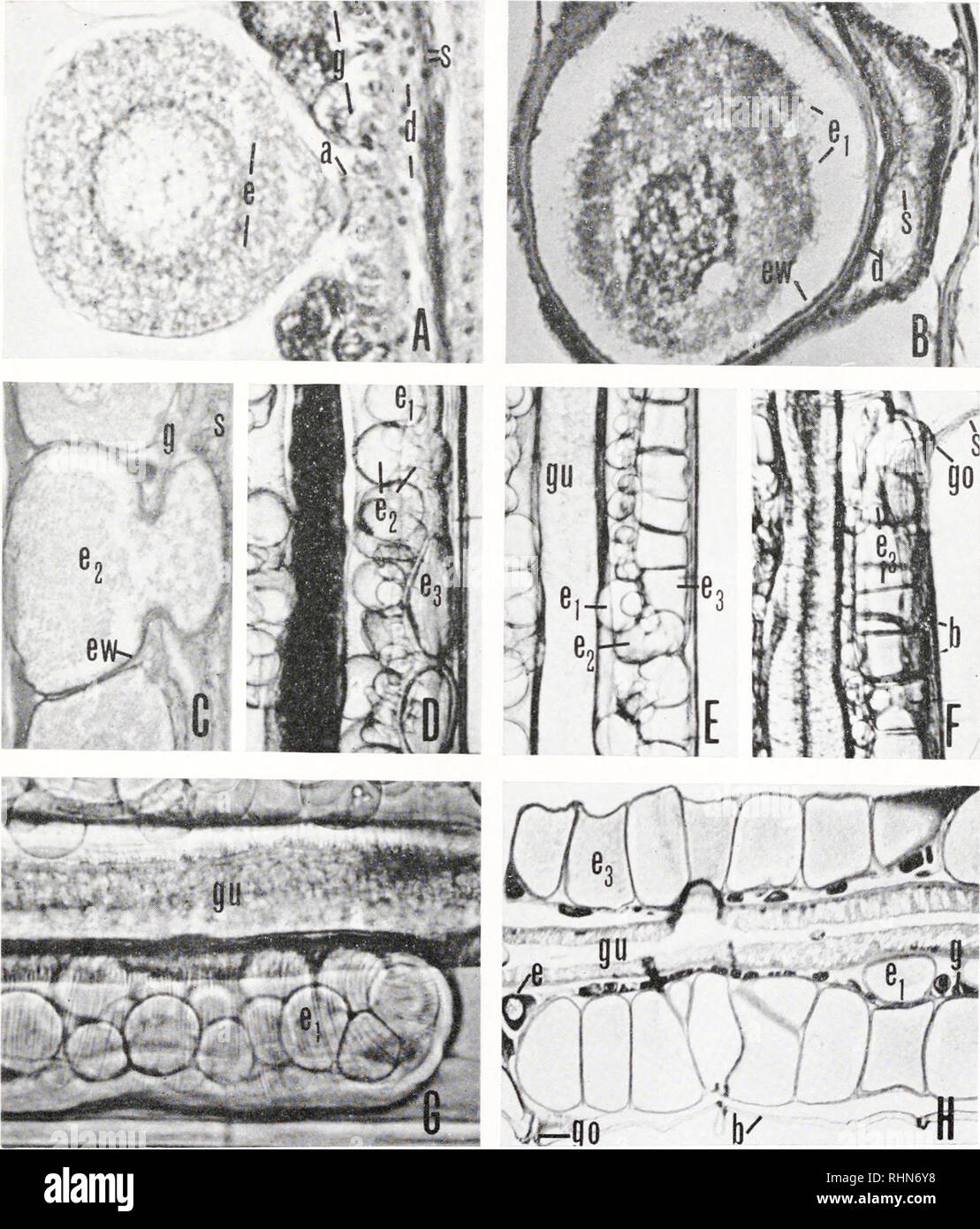 . The Biological bulletin. Biology; Zoology; Biology; Marine Biology. EGG-LAYING IN SAGITTA 249. FIGURE 1. A—maturing egg; B—mature egg; C to F—stages in migration of egg to duct; G—ovary with mature unmigrated eggs showing contractions of ovary walls; H—eggs in duct immediately prior to laying. Figures A-C and H are from sections, D-G from live material; width of body approximately 0.5 mm ; a—accessory fertilization cells; b—body wall; d—duct; e—maturing egg; ei—mature unmigrated egg; 62—migrating egg; e3—migrated egg; cw—egg cell wall; g—germinal epithelium; go—gonopore ; gu—gut; s—sperm.. P Stock Photohttps://www.alamy.com/image-license-details/?v=1https://www.alamy.com/the-biological-bulletin-biology-zoology-biology-marine-biology-egg-laying-in-sagitta-249-figure-1-amaturing-egg-bmature-egg-c-to-fstages-in-migration-of-egg-to-duct-govary-with-mature-unmigrated-eggs-showing-contractions-of-ovary-walls-heggs-in-duct-immediately-prior-to-laying-figures-a-c-and-h-are-from-sections-d-g-from-live-material-width-of-body-approximately-05-mm-aaccessory-fertilization-cells-bbody-wall-dduct-ematuring-egg-eimature-unmigrated-egg-62migrating-egg-e3migrated-egg-cwegg-cell-wall-ggerminal-epithelium-gogonopore-gugut-ssperm-p-image234650396.html
RMRHN6Y8–. The Biological bulletin. Biology; Zoology; Biology; Marine Biology. EGG-LAYING IN SAGITTA 249. FIGURE 1. A—maturing egg; B—mature egg; C to F—stages in migration of egg to duct; G—ovary with mature unmigrated eggs showing contractions of ovary walls; H—eggs in duct immediately prior to laying. Figures A-C and H are from sections, D-G from live material; width of body approximately 0.5 mm ; a—accessory fertilization cells; b—body wall; d—duct; e—maturing egg; ei—mature unmigrated egg; 62—migrating egg; e3—migrated egg; cw—egg cell wall; g—germinal epithelium; go—gonopore ; gu—gut; s—sperm.. P
 . Bonner zoologische Monographien. Zoology. HUBER, REVISION AND CU ADISTIC ANALYSIS OF PHüI.ClIS AND CI.OSHI Y RELATED i'AXA (ARANEAE, l'HOLC:iDAE). FIG. 690-695. Pholcus tahai. 690. Distal male cheliceral apophyses. 691. Male gonopore. 692, 694. Male (692) and female (694) ALS and PMS. 693. Comb-hairs on right female tarsus 4. 695. Epigynum. Scale lines: 200 pm (695), 50 pm (690), 30 pm (691), 20 pm (692, 694), 10 pm (693). tibia 2: 6.0, tibia 3: 3.3, tibia 4: 5.2; tibia 1 L/d: 111. Habitus as in Fig. 586. Carapace pale ochre-yellow with pair of light brown marks posteriorly, ocular area and Stock Photohttps://www.alamy.com/image-license-details/?v=1https://www.alamy.com/bonner-zoologische-monographien-zoology-huber-revision-and-cu-adistic-analysis-of-phiclis-and-cioshi-y-related-iaxa-araneae-lholcidae-fig-690-695-pholcus-tahai-690-distal-male-cheliceral-apophyses-691-male-gonopore-692-694-male-692-and-female-694-als-and-pms-693-comb-hairs-on-right-female-tarsus-4-695-epigynum-scale-lines-200-pm-695-50-pm-690-30-pm-691-20-pm-692-694-10-pm-693-tibia-2-60-tibia-3-33-tibia-4-52-tibia-1-ld-111-habitus-as-in-fig-586-carapace-pale-ochre-yellow-with-pair-of-light-brown-marks-posteriorly-ocular-area-and-image234481494.html
RMRHDFF2–. Bonner zoologische Monographien. Zoology. HUBER, REVISION AND CU ADISTIC ANALYSIS OF PHüI.ClIS AND CI.OSHI Y RELATED i'AXA (ARANEAE, l'HOLC:iDAE). FIG. 690-695. Pholcus tahai. 690. Distal male cheliceral apophyses. 691. Male gonopore. 692, 694. Male (692) and female (694) ALS and PMS. 693. Comb-hairs on right female tarsus 4. 695. Epigynum. Scale lines: 200 pm (695), 50 pm (690), 30 pm (691), 20 pm (692, 694), 10 pm (693). tibia 2: 6.0, tibia 3: 3.3, tibia 4: 5.2; tibia 1 L/d: 111. Habitus as in Fig. 586. Carapace pale ochre-yellow with pair of light brown marks posteriorly, ocular area and
 . Bonner zoologische Monographien. Zoology. HL'Bl R. RlA ISION AND CI AHIS TK: ANAIA SIS Ol- PJ/OICUS AhiD Cl.OSl l Y RFl Al HI) TAXA (ARANr.AF,, PHOI.CIDAE). FIG. 2001-2009. Pholcus djelalabad. 2001, 2002. Male ocular area, oblique and frontal views. 2003. Projec- tion on male ocular area. 2004. Right procursus, prolateral view. 2005, 2006. Right bulbal processes, prolat- eral and dorsal views. 2007. Right embolus. 2008. Distal male cheliceral apophysis. 2009. Male gonopore. Scale lines: 200 fim (2002), 100 pm (2001, 2005, 2006), 80 ^m (2004), 60 pm (2009), 40 pm (2007), 20 pm (2003, 2008). 4 Stock Photohttps://www.alamy.com/image-license-details/?v=1https://www.alamy.com/bonner-zoologische-monographien-zoology-hlbl-r-rla-ision-and-ci-ahis-tk-anaia-sis-ol-pjoicus-ahid-closl-l-y-rfl-al-hi-taxa-aranraf-phoicidae-fig-2001-2009-pholcus-djelalabad-2001-2002-male-ocular-area-oblique-and-frontal-views-2003-projec-tion-on-male-ocular-area-2004-right-procursus-prolateral-view-2005-2006-right-bulbal-processes-prolat-eral-and-dorsal-views-2007-right-embolus-2008-distal-male-cheliceral-apophysis-2009-male-gonopore-scale-lines-200-fim-2002-100-pm-2001-2005-2006-80-m-2004-60-pm-2009-40-pm-2007-20-pm-2003-2008-4-image234479040.html
RMRHDCBC–. Bonner zoologische Monographien. Zoology. HL'Bl R. RlA ISION AND CI AHIS TK: ANAIA SIS Ol- PJ/OICUS AhiD Cl.OSl l Y RFl Al HI) TAXA (ARANr.AF,, PHOI.CIDAE). FIG. 2001-2009. Pholcus djelalabad. 2001, 2002. Male ocular area, oblique and frontal views. 2003. Projec- tion on male ocular area. 2004. Right procursus, prolateral view. 2005, 2006. Right bulbal processes, prolat- eral and dorsal views. 2007. Right embolus. 2008. Distal male cheliceral apophysis. 2009. Male gonopore. Scale lines: 200 fim (2002), 100 pm (2001, 2005, 2006), 80 ^m (2004), 60 pm (2009), 40 pm (2007), 20 pm (2003, 2008). 4
![. Bonner zoologische Monographien. Zoology. Hl'Bl'R. RF'1SK)N ANH C'l ADIS I K' ANAH SIS CM- /V/0/.(,7',V AND (1 OSl n Rl 1 Al l- l) TAXA (ARANlvM-, l'M()l,(;inAl-). FIG. 475-482. Panjange iban. 475. Male eye stalks, frontal view. 476. Distal male cheliceral apophyses. 477. Left procursus, ventro-distal view. 478. Left procursus tip, ventro-distal view. 479. Left procursus, ventral view. 480. Right embolus tip, retrolateral view. 481. Male palpal tarsal organ. 482. Male gonopore. Scale lines: 200 ]im (475, 477), 100 pm (479), 50 pm (476, 480), 40 pm (478, 482), 10 pm (481). internal structure Stock Photo . Bonner zoologische Monographien. Zoology. Hl'Bl'R. RF'1SK)N ANH C'l ADIS I K' ANAH SIS CM- /V/0/.(,7',V AND (1 OSl n Rl 1 Al l- l) TAXA (ARANlvM-, l'M()l,(;inAl-). FIG. 475-482. Panjange iban. 475. Male eye stalks, frontal view. 476. Distal male cheliceral apophyses. 477. Left procursus, ventro-distal view. 478. Left procursus tip, ventro-distal view. 479. Left procursus, ventral view. 480. Right embolus tip, retrolateral view. 481. Male palpal tarsal organ. 482. Male gonopore. Scale lines: 200 ]im (475, 477), 100 pm (479), 50 pm (476, 480), 40 pm (478, 482), 10 pm (481). internal structure Stock Photo](https://c8.alamy.com/comp/RHDG2K/bonner-zoologische-monographien-zoology-hlblr-rf1skn-anh-cl-adis-i-k-anah-sis-cm-v07v-and-1-osl-n-rl-1-al-l-l-taxa-aranlvm-lmlinal-fig-475-482-panjange-iban-475-male-eye-stalks-frontal-view-476-distal-male-cheliceral-apophyses-477-left-procursus-ventro-distal-view-478-left-procursus-tip-ventro-distal-view-479-left-procursus-ventral-view-480-right-embolus-tip-retrolateral-view-481-male-palpal-tarsal-organ-482-male-gonopore-scale-lines-200-im-475-477-100-pm-479-50-pm-476-480-40-pm-478-482-10-pm-481-internal-structure-RHDG2K.jpg) . Bonner zoologische Monographien. Zoology. Hl'Bl'R. RF'1SK)N ANH C'l ADIS I K' ANAH SIS CM- /V/0/.(,7',V AND (1 OSl n Rl 1 Al l- l) TAXA (ARANlvM-, l'M()l,(;inAl-). FIG. 475-482. Panjange iban. 475. Male eye stalks, frontal view. 476. Distal male cheliceral apophyses. 477. Left procursus, ventro-distal view. 478. Left procursus tip, ventro-distal view. 479. Left procursus, ventral view. 480. Right embolus tip, retrolateral view. 481. Male palpal tarsal organ. 482. Male gonopore. Scale lines: 200 ]im (475, 477), 100 pm (479), 50 pm (476, 480), 40 pm (478, 482), 10 pm (481). internal structure Stock Photohttps://www.alamy.com/image-license-details/?v=1https://www.alamy.com/bonner-zoologische-monographien-zoology-hlblr-rf1skn-anh-cl-adis-i-k-anah-sis-cm-v07v-and-1-osl-n-rl-1-al-l-l-taxa-aranlvm-lmlinal-fig-475-482-panjange-iban-475-male-eye-stalks-frontal-view-476-distal-male-cheliceral-apophyses-477-left-procursus-ventro-distal-view-478-left-procursus-tip-ventro-distal-view-479-left-procursus-ventral-view-480-right-embolus-tip-retrolateral-view-481-male-palpal-tarsal-organ-482-male-gonopore-scale-lines-200-im-475-477-100-pm-479-50-pm-476-480-40-pm-478-482-10-pm-481-internal-structure-image234481931.html
. Bonner zoologische Monographien. Zoology. Hl'Bl'R. RF'1SK)N ANH C'l ADIS I K' ANAH SIS CM- /V/0/.(,7',V AND (1 OSl n Rl 1 Al l- l) TAXA (ARANlvM-, l'M()l,(;inAl-). FIG. 475-482. Panjange iban. 475. Male eye stalks, frontal view. 476. Distal male cheliceral apophyses. 477. Left procursus, ventro-distal view. 478. Left procursus tip, ventro-distal view. 479. Left procursus, ventral view. 480. Right embolus tip, retrolateral view. 481. Male palpal tarsal organ. 482. Male gonopore. Scale lines: 200 ]im (475, 477), 100 pm (479), 50 pm (476, 480), 40 pm (478, 482), 10 pm (481). internal structure Stock Photohttps://www.alamy.com/image-license-details/?v=1https://www.alamy.com/bonner-zoologische-monographien-zoology-hlblr-rf1skn-anh-cl-adis-i-k-anah-sis-cm-v07v-and-1-osl-n-rl-1-al-l-l-taxa-aranlvm-lmlinal-fig-475-482-panjange-iban-475-male-eye-stalks-frontal-view-476-distal-male-cheliceral-apophyses-477-left-procursus-ventro-distal-view-478-left-procursus-tip-ventro-distal-view-479-left-procursus-ventral-view-480-right-embolus-tip-retrolateral-view-481-male-palpal-tarsal-organ-482-male-gonopore-scale-lines-200-im-475-477-100-pm-479-50-pm-476-480-40-pm-478-482-10-pm-481-internal-structure-image234481931.htmlRMRHDG2K–. Bonner zoologische Monographien. Zoology. Hl'Bl'R. RF'1SK)N ANH C'l ADIS I K' ANAH SIS CM- /V/0/.(,7',V AND (1 OSl n Rl 1 Al l- l) TAXA (ARANlvM-, l'M()l,(;inAl-). FIG. 475-482. Panjange iban. 475. Male eye stalks, frontal view. 476. Distal male cheliceral apophyses. 477. Left procursus, ventro-distal view. 478. Left procursus tip, ventro-distal view. 479. Left procursus, ventral view. 480. Right embolus tip, retrolateral view. 481. Male palpal tarsal organ. 482. Male gonopore. Scale lines: 200 ]im (475, 477), 100 pm (479), 50 pm (476, 480), 40 pm (478, 482), 10 pm (481). internal structure
 . A manual of zoology. iSo MANUAL OF ZOOLOGY bodies, the penial seta (p/i. s). In the female the repro- ductive aperture or gonopore is separated from the anus, and. Please note that these images are extracted from scanned page images that may have been digitally enhanced for readability - coloration and appearance of these illustrations may not perfectly resemble the original work.. Parker, T. Jeffery (Thomas Jeffery), 1850-1897; Haswell, William A. (William Aitcheson), 1854-1925. New York, The Macmillan Company; London, Macmillan & Co. ,Ltd. Stock Photohttps://www.alamy.com/image-license-details/?v=1https://www.alamy.com/a-manual-of-zoology-iso-manual-of-zoology-bodies-the-penial-seta-pi-s-in-the-female-the-repro-ductive-aperture-or-gonopore-is-separated-from-the-anus-and-please-note-that-these-images-are-extracted-from-scanned-page-images-that-may-have-been-digitally-enhanced-for-readability-coloration-and-appearance-of-these-illustrations-may-not-perfectly-resemble-the-original-work-parker-t-jeffery-thomas-jeffery-1850-1897-haswell-william-a-william-aitcheson-1854-1925-new-york-the-macmillan-company-london-macmillan-amp-co-ltd-image232126252.html
RMRDJ7B8–. A manual of zoology. iSo MANUAL OF ZOOLOGY bodies, the penial seta (p/i. s). In the female the repro- ductive aperture or gonopore is separated from the anus, and. Please note that these images are extracted from scanned page images that may have been digitally enhanced for readability - coloration and appearance of these illustrations may not perfectly resemble the original work.. Parker, T. Jeffery (Thomas Jeffery), 1850-1897; Haswell, William A. (William Aitcheson), 1854-1925. New York, The Macmillan Company; London, Macmillan & Co. ,Ltd.
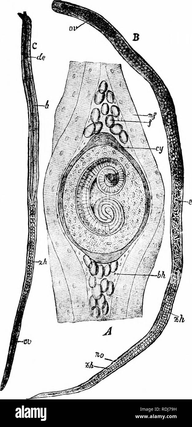 . A manual of zoology. . Fig. 85. — Trichina spiralis. A, encysted form in muscle of host; B, female; C, male, bh, connective tissue envelope; cy, cyst; de, ejaculatory duct; e, em- bryos ; f. fat globules; /:, testis; m. f, muscle fibre; oe, pharynx; or, ovary; ivo, gonopore; zh, cell masses in intestine. (From Lang's Comparative Anatomy', after Clans.) 155. Please note that these images are extracted from scanned page images that may have been digitally enhanced for readability - coloration and appearance of these illustrations may not perfectly resemble the original work.. Parker, T. Jeffer Stock Photohttps://www.alamy.com/image-license-details/?v=1https://www.alamy.com/a-manual-of-zoology-fig-85-trichina-spiralis-a-encysted-form-in-muscle-of-host-b-female-c-male-bh-connective-tissue-envelope-cy-cyst-de-ejaculatory-duct-e-em-bryos-f-fat-globules-testis-m-f-muscle-fibre-oe-pharynx-or-ovary-ivo-gonopore-zh-cell-masses-in-intestine-from-langs-comparative-anatomy-after-clans-155-please-note-that-these-images-are-extracted-from-scanned-page-images-that-may-have-been-digitally-enhanced-for-readability-coloration-and-appearance-of-these-illustrations-may-not-perfectly-resemble-the-original-work-parker-t-jeffer-image232126205.html
RMRDJ79H–. A manual of zoology. . Fig. 85. — Trichina spiralis. A, encysted form in muscle of host; B, female; C, male, bh, connective tissue envelope; cy, cyst; de, ejaculatory duct; e, em- bryos ; f. fat globules; /:, testis; m. f, muscle fibre; oe, pharynx; or, ovary; ivo, gonopore; zh, cell masses in intestine. (From Lang's Comparative Anatomy', after Clans.) 155. Please note that these images are extracted from scanned page images that may have been digitally enhanced for readability - coloration and appearance of these illustrations may not perfectly resemble the original work.. Parker, T. Jeffer
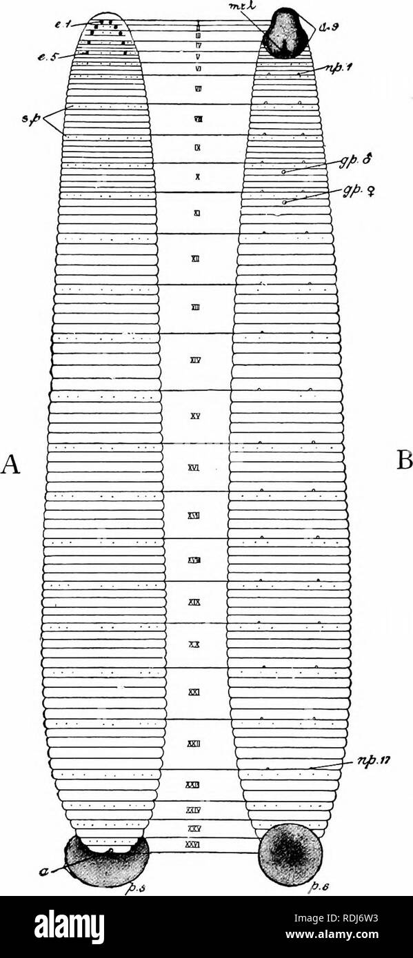 . A manual of zoology. . Fig. 117. — Hirudo medicinalis. A, dorsal; B, ventral aspect, a, anus; <z. s, anterior sucker; e. /, first pair of eyes; e. 5, fifth pair; gp. H", male gonopore; gp. £, female gonopore; mth, mouth; np.i, first pair of nephridiopores; 11p. ly, seventeenth pair; p. s, posterior sucker; s.p, sensory papillae; I-XXVI, segments. (Partly after Whitman-) 204. Please note that these images are extracted from scanned page images that may have been digitally enhanced for readability - coloration and appearance of these illustrations may not perfectly resemble the origina Stock Photohttps://www.alamy.com/image-license-details/?v=1https://www.alamy.com/a-manual-of-zoology-fig-117-hirudo-medicinalis-a-dorsal-b-ventral-aspect-a-anus-ltz-s-anterior-sucker-e-first-pair-of-eyes-e-5-fifth-pair-gp-hquot-male-gonopore-gp-female-gonopore-mth-mouth-npi-first-pair-of-nephridiopores-11p-ly-seventeenth-pair-p-s-posterior-sucker-sp-sensory-papillae-i-xxvi-segments-partly-after-whitman-204-please-note-that-these-images-are-extracted-from-scanned-page-images-that-may-have-been-digitally-enhanced-for-readability-coloration-and-appearance-of-these-illustrations-may-not-perfectly-resemble-the-origina-image232125855.html
RMRDJ6W3–. A manual of zoology. . Fig. 117. — Hirudo medicinalis. A, dorsal; B, ventral aspect, a, anus; <z. s, anterior sucker; e. /, first pair of eyes; e. 5, fifth pair; gp. H", male gonopore; gp. £, female gonopore; mth, mouth; np.i, first pair of nephridiopores; 11p. ly, seventeenth pair; p. s, posterior sucker; s.p, sensory papillae; I-XXVI, segments. (Partly after Whitman-) 204. Please note that these images are extracted from scanned page images that may have been digitally enhanced for readability - coloration and appearance of these illustrations may not perfectly resemble the origina