Grana and lamellae Stock Photos and Images
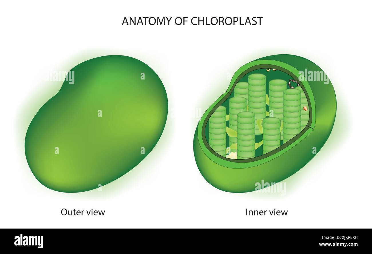 Anatomy of chloroplast plasmid Stock Photohttps://www.alamy.com/image-license-details/?v=1https://www.alamy.com/anatomy-of-chloroplast-plasmid-image476853065.html
RF2JKPEXH–Anatomy of chloroplast plasmid
![. Biological structure and function; proceedings. Biochemistry; Cytology. Fig. 2. Electron micrograph of a section of a maize chloroplast showing details of structure. The dense areas that resemble stacks of coins are the grana. The layers within each granum are called grana lamellae. The grana lamellae of different grana are inter-connected by stroma lamellae. Magnification 35 000 x (courtesy of Dr. A. E. Vatter). chloroplasts [38, 39] in the dark, it was converted to sugar phosphates. The hght and dark phases when carried out separately, yielded essentially the same final photosynthetic prod Stock Photo . Biological structure and function; proceedings. Biochemistry; Cytology. Fig. 2. Electron micrograph of a section of a maize chloroplast showing details of structure. The dense areas that resemble stacks of coins are the grana. The layers within each granum are called grana lamellae. The grana lamellae of different grana are inter-connected by stroma lamellae. Magnification 35 000 x (courtesy of Dr. A. E. Vatter). chloroplasts [38, 39] in the dark, it was converted to sugar phosphates. The hght and dark phases when carried out separately, yielded essentially the same final photosynthetic prod Stock Photo](https://c8.alamy.com/comp/RHM0M1/biological-structure-and-function-proceedings-biochemistry-cytology-fig-2-electron-micrograph-of-a-section-of-a-maize-chloroplast-showing-details-of-structure-the-dense-areas-that-resemble-stacks-of-coins-are-the-grana-the-layers-within-each-granum-are-called-grana-lamellae-the-grana-lamellae-of-different-grana-are-inter-connected-by-stroma-lamellae-magnification-35-000-x-courtesy-of-dr-a-e-vatter-chloroplasts-38-39-in-the-dark-it-was-converted-to-sugar-phosphates-the-hght-and-dark-phases-when-carried-out-separately-yielded-essentially-the-same-final-photosynthetic-prod-RHM0M1.jpg) . Biological structure and function; proceedings. Biochemistry; Cytology. Fig. 2. Electron micrograph of a section of a maize chloroplast showing details of structure. The dense areas that resemble stacks of coins are the grana. The layers within each granum are called grana lamellae. The grana lamellae of different grana are inter-connected by stroma lamellae. Magnification 35 000 x (courtesy of Dr. A. E. Vatter). chloroplasts [38, 39] in the dark, it was converted to sugar phosphates. The hght and dark phases when carried out separately, yielded essentially the same final photosynthetic prod Stock Photohttps://www.alamy.com/image-license-details/?v=1https://www.alamy.com/biological-structure-and-function-proceedings-biochemistry-cytology-fig-2-electron-micrograph-of-a-section-of-a-maize-chloroplast-showing-details-of-structure-the-dense-areas-that-resemble-stacks-of-coins-are-the-grana-the-layers-within-each-granum-are-called-grana-lamellae-the-grana-lamellae-of-different-grana-are-inter-connected-by-stroma-lamellae-magnification-35-000-x-courtesy-of-dr-a-e-vatter-chloroplasts-38-39-in-the-dark-it-was-converted-to-sugar-phosphates-the-hght-and-dark-phases-when-carried-out-separately-yielded-essentially-the-same-final-photosynthetic-prod-image234623537.html
. Biological structure and function; proceedings. Biochemistry; Cytology. Fig. 2. Electron micrograph of a section of a maize chloroplast showing details of structure. The dense areas that resemble stacks of coins are the grana. The layers within each granum are called grana lamellae. The grana lamellae of different grana are inter-connected by stroma lamellae. Magnification 35 000 x (courtesy of Dr. A. E. Vatter). chloroplasts [38, 39] in the dark, it was converted to sugar phosphates. The hght and dark phases when carried out separately, yielded essentially the same final photosynthetic prod Stock Photohttps://www.alamy.com/image-license-details/?v=1https://www.alamy.com/biological-structure-and-function-proceedings-biochemistry-cytology-fig-2-electron-micrograph-of-a-section-of-a-maize-chloroplast-showing-details-of-structure-the-dense-areas-that-resemble-stacks-of-coins-are-the-grana-the-layers-within-each-granum-are-called-grana-lamellae-the-grana-lamellae-of-different-grana-are-inter-connected-by-stroma-lamellae-magnification-35-000-x-courtesy-of-dr-a-e-vatter-chloroplasts-38-39-in-the-dark-it-was-converted-to-sugar-phosphates-the-hght-and-dark-phases-when-carried-out-separately-yielded-essentially-the-same-final-photosynthetic-prod-image234623537.htmlRMRHM0M1–. Biological structure and function; proceedings. Biochemistry; Cytology. Fig. 2. Electron micrograph of a section of a maize chloroplast showing details of structure. The dense areas that resemble stacks of coins are the grana. The layers within each granum are called grana lamellae. The grana lamellae of different grana are inter-connected by stroma lamellae. Magnification 35 000 x (courtesy of Dr. A. E. Vatter). chloroplasts [38, 39] in the dark, it was converted to sugar phosphates. The hght and dark phases when carried out separately, yielded essentially the same final photosynthetic prod
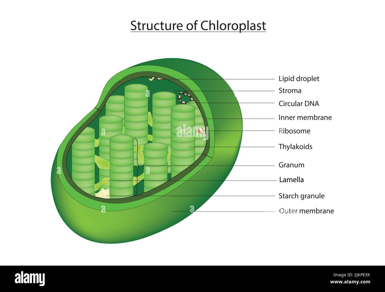 3D Chloroplast Stock Photohttps://www.alamy.com/image-license-details/?v=1https://www.alamy.com/3d-chloroplast-image476853067.html
RF2JKPEXK–3D Chloroplast
 . Cytology. Cytology. Figure 3-19. Electron Micrograph Showing Submicroscopic Structure of Plastid in a Three-Week-Old Etiolated Barley Leaf. Note concentric arrange- ment of the grana lamellae. Approximately 37,OOOx. (From von Wettstein, D., 1959. "Developmental Changes in Chloroplasts and Their Genetic Con- trol," in "Developmental Cytology," D. Rudnick (Ed.), Ronald Press, New York, N. Y., Fig. 1, p. 150. Courtesy of Dr. D. von Wettstein. Forest Re- search Institute, Stockholm, Sweden.) LYSOSOMES The term "lysosomes" was originated by deDuve and group (1955) to Stock Photohttps://www.alamy.com/image-license-details/?v=1https://www.alamy.com/cytology-cytology-figure-3-19-electron-micrograph-showing-submicroscopic-structure-of-plastid-in-a-three-week-old-etiolated-barley-leaf-note-concentric-arrange-ment-of-the-grana-lamellae-approximately-37ooox-from-von-wettstein-d-1959-quotdevelopmental-changes-in-chloroplasts-and-their-genetic-con-trolquot-in-quotdevelopmental-cytologyquot-d-rudnick-ed-ronald-press-new-york-n-y-fig-1-p-150-courtesy-of-dr-d-von-wettstein-forest-re-search-institute-stockholm-sweden-lysosomes-the-term-quotlysosomesquot-was-originated-by-deduve-and-group-1955-to-image216168851.html
RMPFK9G3–. Cytology. Cytology. Figure 3-19. Electron Micrograph Showing Submicroscopic Structure of Plastid in a Three-Week-Old Etiolated Barley Leaf. Note concentric arrange- ment of the grana lamellae. Approximately 37,OOOx. (From von Wettstein, D., 1959. "Developmental Changes in Chloroplasts and Their Genetic Con- trol," in "Developmental Cytology," D. Rudnick (Ed.), Ronald Press, New York, N. Y., Fig. 1, p. 150. Courtesy of Dr. D. von Wettstein. Forest Re- search Institute, Stockholm, Sweden.) LYSOSOMES The term "lysosomes" was originated by deDuve and group (1955) to
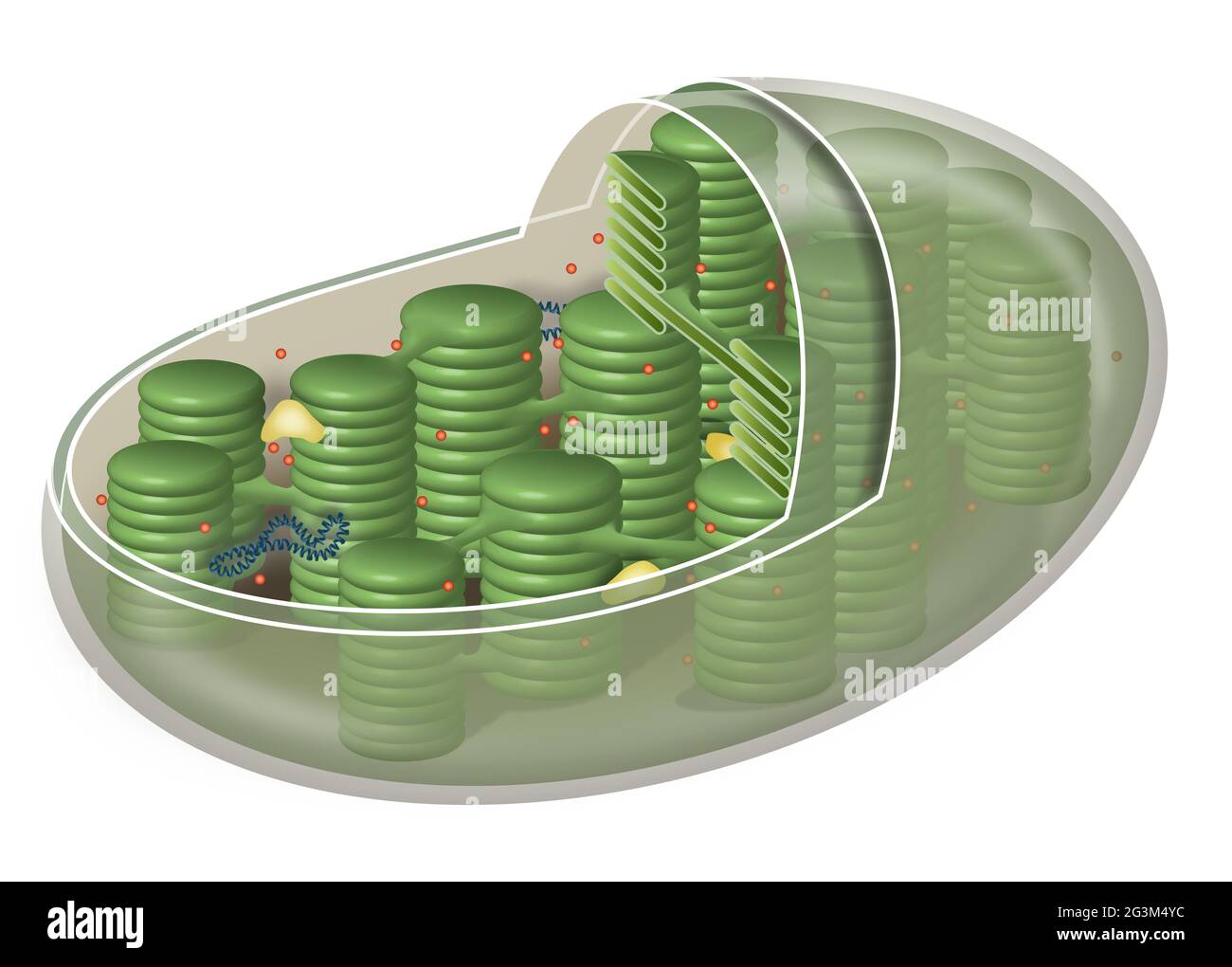 Chloroplast, plant cell organelle Stock Photohttps://www.alamy.com/image-license-details/?v=1https://www.alamy.com/chloroplast-plant-cell-organelle-image432546112.html
RF2G3M4YC–Chloroplast, plant cell organelle
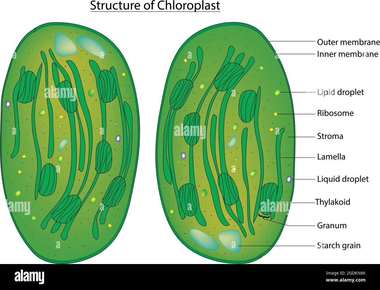 Detailed diagram of chloroplast, chloroplast structure, labeled diagram anatomy of the chloroplast with thylakoids and other elements Stock Vectorhttps://www.alamy.com/image-license-details/?v=1https://www.alamy.com/detailed-diagram-of-chloroplast-chloroplast-structure-labeled-diagram-anatomy-of-the-chloroplast-with-thylakoids-and-other-elements-image438683510.html
RF2GDKN86–Detailed diagram of chloroplast, chloroplast structure, labeled diagram anatomy of the chloroplast with thylakoids and other elements
 . Cytology. Cytology. Figure 3-19. Electron Micrograph Showing Submicroscopic Structure of Plastid in a Three-Week-Old Etiolated Barley Leaf. Note concentric arrange- ment of the grana lamellae. Approximately 37,OOOx. (From von Wettstein, D., 1959. "Developmental Changes in Chloroplasts and Their Genetic Con- trol," in "Developmental Cytology," D. Rudnick (Ed.), Ronald Press, New York, N. Y., Fig. 1, p. 150. Courtesy of Dr. D. von Wettstein. Forest Re- search Institute, Stockholm, Sweden.) LYSOSOMES The term "lysosomes" was originated by deDuve and group (1955) to Stock Photohttps://www.alamy.com/image-license-details/?v=1https://www.alamy.com/cytology-cytology-figure-3-19-electron-micrograph-showing-submicroscopic-structure-of-plastid-in-a-three-week-old-etiolated-barley-leaf-note-concentric-arrange-ment-of-the-grana-lamellae-approximately-37ooox-from-von-wettstein-d-1959-quotdevelopmental-changes-in-chloroplasts-and-their-genetic-con-trolquot-in-quotdevelopmental-cytologyquot-d-rudnick-ed-ronald-press-new-york-n-y-fig-1-p-150-courtesy-of-dr-d-von-wettstein-forest-re-search-institute-stockholm-sweden-lysosomes-the-term-quotlysosomesquot-was-originated-by-deduve-and-group-1955-to-image231804732.html
RMRD3H8C–. Cytology. Cytology. Figure 3-19. Electron Micrograph Showing Submicroscopic Structure of Plastid in a Three-Week-Old Etiolated Barley Leaf. Note concentric arrange- ment of the grana lamellae. Approximately 37,OOOx. (From von Wettstein, D., 1959. "Developmental Changes in Chloroplasts and Their Genetic Con- trol," in "Developmental Cytology," D. Rudnick (Ed.), Ronald Press, New York, N. Y., Fig. 1, p. 150. Courtesy of Dr. D. von Wettstein. Forest Re- search Institute, Stockholm, Sweden.) LYSOSOMES The term "lysosomes" was originated by deDuve and group (1955) to
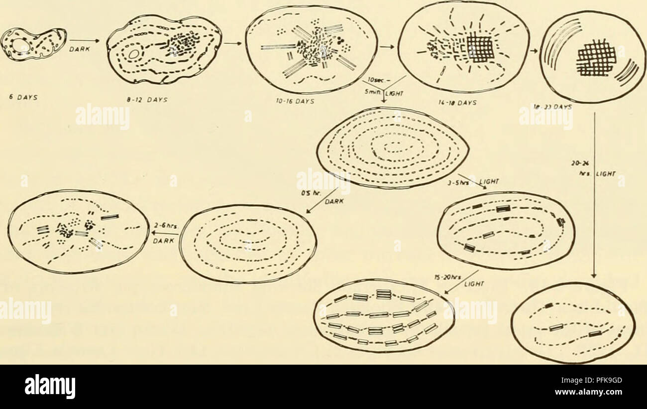 . Cytology. Cytology. plastids increase in size but do not develop grana. These plastids can synthesize chlorophyll in the presence of light but it is broken down as fast as it is formed. In the barley mutant, xantha-3, the plastids differ- entiate to the extent of developing some internal layered structures, but no true lamellae are ever found and large numbers of globuli accumulate within the plastid. Some chlorophyll and carotenoid pigments are synthe- sized and localized in the globuli, but since there is no extensive lamellar. Figure 3-18. Schematic Diagram Depicting the Sequence of Chang Stock Photohttps://www.alamy.com/image-license-details/?v=1https://www.alamy.com/cytology-cytology-plastids-increase-in-size-but-do-not-develop-grana-these-plastids-can-synthesize-chlorophyll-in-the-presence-of-light-but-it-is-broken-down-as-fast-as-it-is-formed-in-the-barley-mutant-xantha-3-the-plastids-differ-entiate-to-the-extent-of-developing-some-internal-layered-structures-but-no-true-lamellae-are-ever-found-and-large-numbers-of-globuli-accumulate-within-the-plastid-some-chlorophyll-and-carotenoid-pigments-are-synthe-sized-and-localized-in-the-globuli-but-since-there-is-no-extensive-lamellar-figure-3-18-schematic-diagram-depicting-the-sequence-of-chang-image216168861.html
RMPFK9GD–. Cytology. Cytology. plastids increase in size but do not develop grana. These plastids can synthesize chlorophyll in the presence of light but it is broken down as fast as it is formed. In the barley mutant, xantha-3, the plastids differ- entiate to the extent of developing some internal layered structures, but no true lamellae are ever found and large numbers of globuli accumulate within the plastid. Some chlorophyll and carotenoid pigments are synthe- sized and localized in the globuli, but since there is no extensive lamellar. Figure 3-18. Schematic Diagram Depicting the Sequence of Chang
 . Cytology. Cytology. plastids increase in size but do not develop grana. These plastids can synthesize chlorophyll in the presence of light but it is broken down as fast as it is formed. In the barley mutant, xantha-3, the plastids differ- entiate to the extent of developing some internal layered structures, but no true lamellae are ever found and large numbers of globuli accumulate within the plastid. Some chlorophyll and carotenoid pigments are synthe- sized and localized in the globuli, but since there is no extensive lamellar. Figure 3-18. Schematic Diagram Depicting the Sequence of Chang Stock Photohttps://www.alamy.com/image-license-details/?v=1https://www.alamy.com/cytology-cytology-plastids-increase-in-size-but-do-not-develop-grana-these-plastids-can-synthesize-chlorophyll-in-the-presence-of-light-but-it-is-broken-down-as-fast-as-it-is-formed-in-the-barley-mutant-xantha-3-the-plastids-differ-entiate-to-the-extent-of-developing-some-internal-layered-structures-but-no-true-lamellae-are-ever-found-and-large-numbers-of-globuli-accumulate-within-the-plastid-some-chlorophyll-and-carotenoid-pigments-are-synthe-sized-and-localized-in-the-globuli-but-since-there-is-no-extensive-lamellar-figure-3-18-schematic-diagram-depicting-the-sequence-of-chang-image231804748.html
RMRD3H90–. Cytology. Cytology. plastids increase in size but do not develop grana. These plastids can synthesize chlorophyll in the presence of light but it is broken down as fast as it is formed. In the barley mutant, xantha-3, the plastids differ- entiate to the extent of developing some internal layered structures, but no true lamellae are ever found and large numbers of globuli accumulate within the plastid. Some chlorophyll and carotenoid pigments are synthe- sized and localized in the globuli, but since there is no extensive lamellar. Figure 3-18. Schematic Diagram Depicting the Sequence of Chang
 . Biophysical science. Biophysics. (a) (b) Figure 2. Green grana of corn leaves, (a) A sketch of an electron micrograph of a section of granum within an intact chloroplast of Zea mais. There are about 50 such grana per chloroplast. Each granum has about 15 parallel lamellae which are about 400 A thick and about 4,000 A in diameter. (b) A sketch of an electron micrograph of a granum which is believed to be dissociated into separate disks. After E. I. Rabinowitch, Photosynthesis, II, 2 (New York: Interscience Publishers, Inc., 1956), from Vatter, unpublished, modified. Figure 2 shows the lamella Stock Photohttps://www.alamy.com/image-license-details/?v=1https://www.alamy.com/biophysical-science-biophysics-a-b-figure-2-green-grana-of-corn-leaves-a-a-sketch-of-an-electron-micrograph-of-a-section-of-granum-within-an-intact-chloroplast-of-zea-mais-there-are-about-50-such-grana-per-chloroplast-each-granum-has-about-15-parallel-lamellae-which-are-about-400-a-thick-and-about-4000-a-in-diameter-b-a-sketch-of-an-electron-micrograph-of-a-granum-which-is-believed-to-be-dissociated-into-separate-disks-after-e-i-rabinowitch-photosynthesis-ii-2-new-york-interscience-publishers-inc-1956-from-vatter-unpublished-modified-figure-2-shows-the-lamella-image234538559.html
RMRHG493–. Biophysical science. Biophysics. (a) (b) Figure 2. Green grana of corn leaves, (a) A sketch of an electron micrograph of a section of granum within an intact chloroplast of Zea mais. There are about 50 such grana per chloroplast. Each granum has about 15 parallel lamellae which are about 400 A thick and about 4,000 A in diameter. (b) A sketch of an electron micrograph of a granum which is believed to be dissociated into separate disks. After E. I. Rabinowitch, Photosynthesis, II, 2 (New York: Interscience Publishers, Inc., 1956), from Vatter, unpublished, modified. Figure 2 shows the lamella