Hemothorax Stock Photos and Images
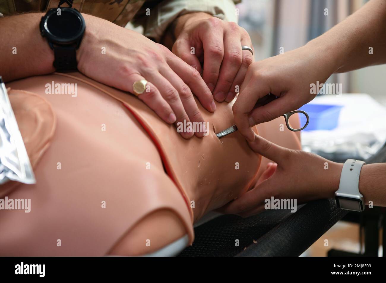 U.S. Army Sgt. 1st Class Bryan Rowland, U.S. Army Medical Department Activity Bavaria NCOIC of education and training, left, teaches A1C Justice Nerad, Health Care Operations Squadron aerospace medical technician, right, how to treat a collapsed lung on a mannequin, at Aviano Air Base, Italy, Aug. 25, 2022. Two collapsed lung scenarios simulated during the joint training were the treatment of air trapped in the chest cavity, also known as a pneumothorax, and blood trapped in the chest cavity, or hemothorax. Stock Photohttps://www.alamy.com/image-license-details/?v=1https://www.alamy.com/us-army-sgt-1st-class-bryan-rowland-us-army-medical-department-activity-bavaria-ncoic-of-education-and-training-left-teaches-a1c-justice-nerad-health-care-operations-squadron-aerospace-medical-technician-right-how-to-treat-a-collapsed-lung-on-a-mannequin-at-aviano-air-base-italy-aug-25-2022-two-collapsed-lung-scenarios-simulated-during-the-joint-training-were-the-treatment-of-air-trapped-in-the-chest-cavity-also-known-as-a-pneumothorax-and-blood-trapped-in-the-chest-cavity-or-hemothorax-image510351865.html
RM2MJ8F09–U.S. Army Sgt. 1st Class Bryan Rowland, U.S. Army Medical Department Activity Bavaria NCOIC of education and training, left, teaches A1C Justice Nerad, Health Care Operations Squadron aerospace medical technician, right, how to treat a collapsed lung on a mannequin, at Aviano Air Base, Italy, Aug. 25, 2022. Two collapsed lung scenarios simulated during the joint training were the treatment of air trapped in the chest cavity, also known as a pneumothorax, and blood trapped in the chest cavity, or hemothorax.
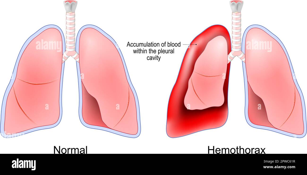 Hemothorax. Healthy human lungs and red lungs after accumulation of blood within the pleural cavity. Chest trauma. pulmonary embolism treatment Stock Vectorhttps://www.alamy.com/image-license-details/?v=1https://www.alamy.com/hemothorax-healthy-human-lungs-and-red-lungs-after-accumulation-of-blood-within-the-pleural-cavity-chest-trauma-pulmonary-embolism-treatment-image549155987.html
RF2PWC61R–Hemothorax. Healthy human lungs and red lungs after accumulation of blood within the pleural cavity. Chest trauma. pulmonary embolism treatment
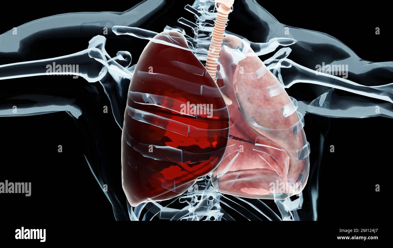 3d Illustration of Hemothorax, Normal lung versus collapsed, symptoms of Hemothorax, pleural effusion, empyema, complications after a chest injury, ai Stock Photohttps://www.alamy.com/image-license-details/?v=1https://www.alamy.com/3d-illustration-of-hemothorax-normal-lung-versus-collapsed-symptoms-of-hemothorax-pleural-effusion-empyema-complications-after-a-chest-injury-ai-image499762879.html
RM2M124J7–3d Illustration of Hemothorax, Normal lung versus collapsed, symptoms of Hemothorax, pleural effusion, empyema, complications after a chest injury, ai
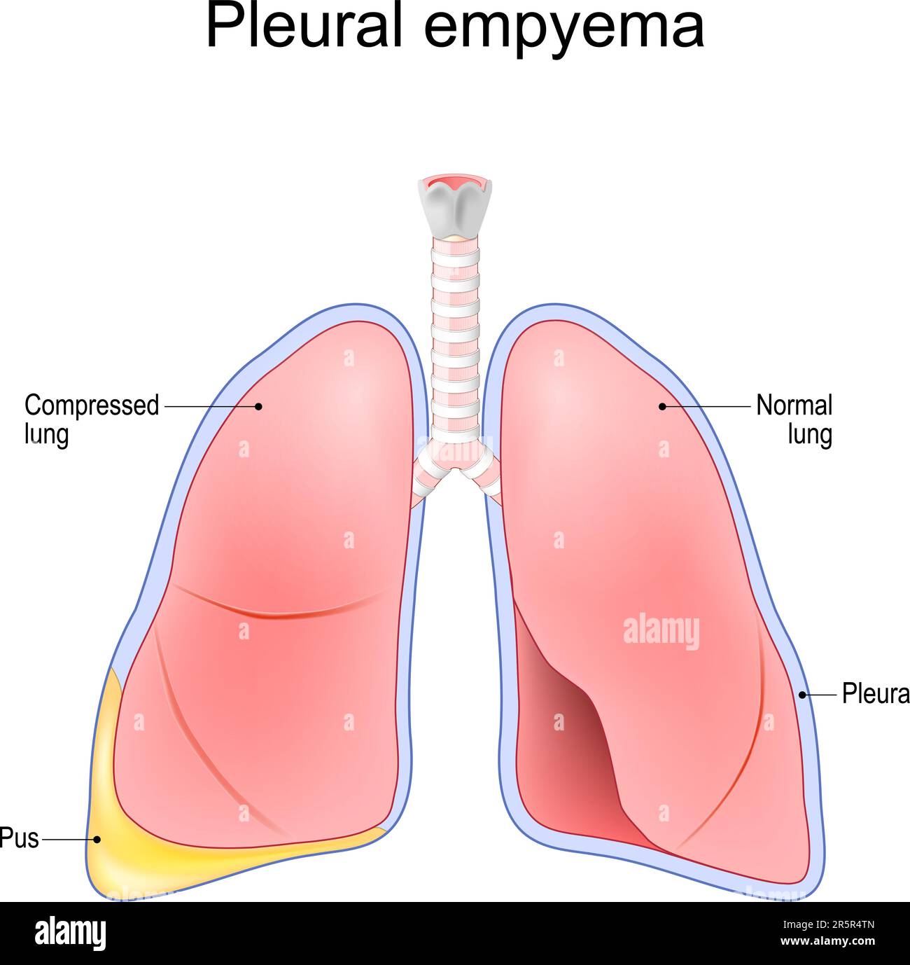 Pleural empyema. Normal lung and lungs after accumulation of pus within the pleural cavity. Vector illustration Stock Vectorhttps://www.alamy.com/image-license-details/?v=1https://www.alamy.com/pleural-empyema-normal-lung-and-lungs-after-accumulation-of-pus-within-the-pleural-cavity-vector-illustration-image554313781.html
RF2R5R4TN–Pleural empyema. Normal lung and lungs after accumulation of pus within the pleural cavity. Vector illustration
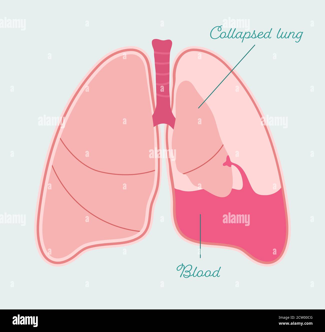 Hemothorax of lungs. Bleeding and collapsed human lung - vector anatomical scheme Stock Vectorhttps://www.alamy.com/image-license-details/?v=1https://www.alamy.com/hemothorax-of-lungs-bleeding-and-collapsed-human-lung-vector-anatomical-scheme-image376784480.html
RF2CW00CG–Hemothorax of lungs. Bleeding and collapsed human lung - vector anatomical scheme
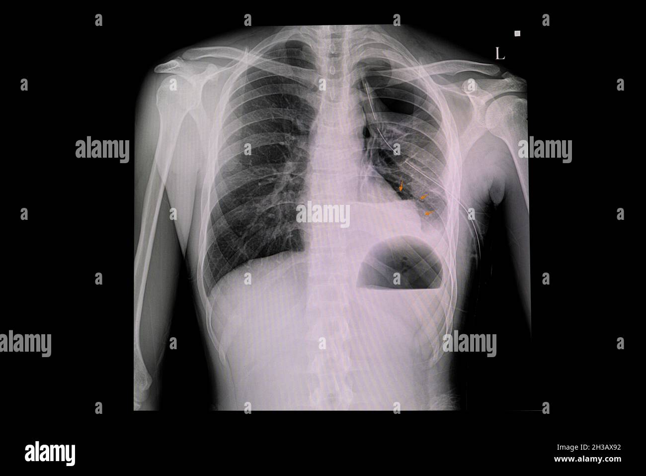 Xray chest of a patient with left ICD and moderate amount of left-hydro-pneumothorax. Stock Photohttps://www.alamy.com/image-license-details/?v=1https://www.alamy.com/xray-chest-of-a-patient-with-left-icd-and-moderate-amount-of-left-hydro-pneumothorax-image449553694.html
RF2H3AX92–Xray chest of a patient with left ICD and moderate amount of left-hydro-pneumothorax.
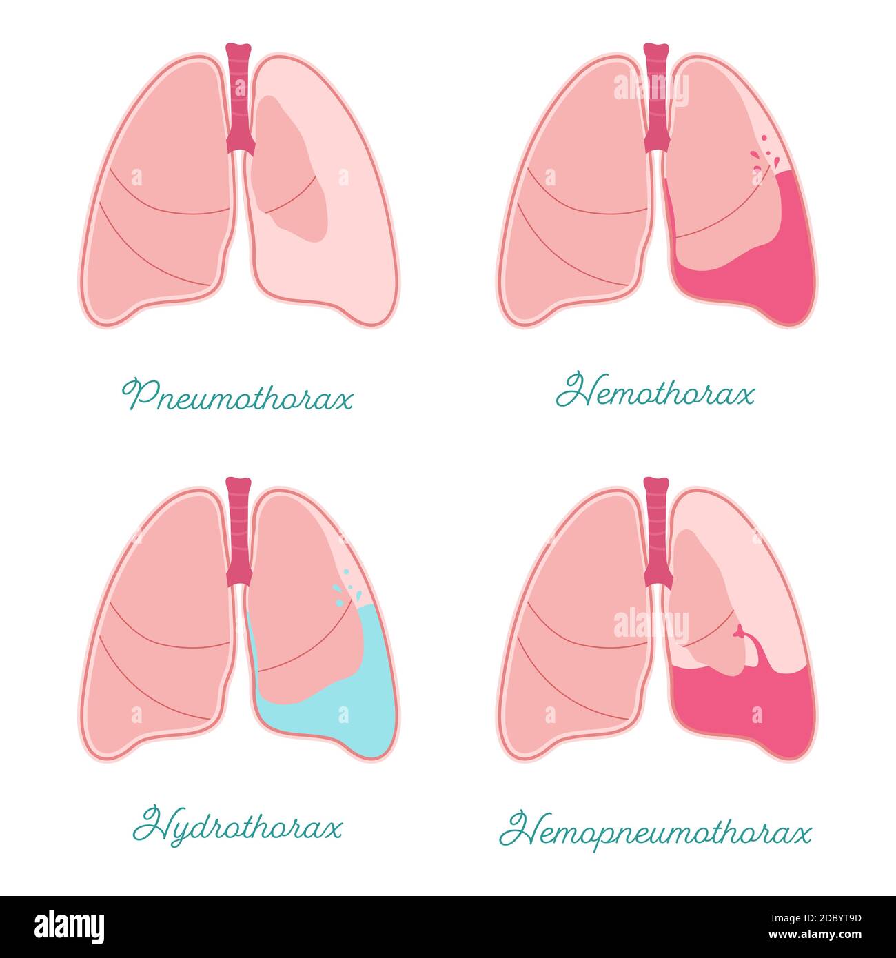 Some types of pleural effusions. Abnormal gathering fluid or air pleural space. Hemothorax, pneumothorax, hemopneumothorax and hydrothorax also. Compa Stock Vectorhttps://www.alamy.com/image-license-details/?v=1https://www.alamy.com/some-types-of-pleural-effusions-abnormal-gathering-fluid-or-air-pleural-space-hemothorax-pneumothorax-hemopneumothorax-and-hydrothorax-also-compa-image386001097.html
RF2DBYT9D–Some types of pleural effusions. Abnormal gathering fluid or air pleural space. Hemothorax, pneumothorax, hemopneumothorax and hydrothorax also. Compa
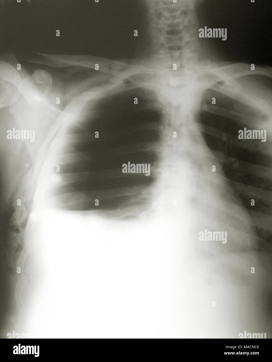 Chest x-ray - stabbing injury Stock Photohttps://www.alamy.com/image-license-details/?v=1https://www.alamy.com/chest-x-ray-stabbing-injury-image4969925.html
RMAMCNC6–Chest x-ray - stabbing injury
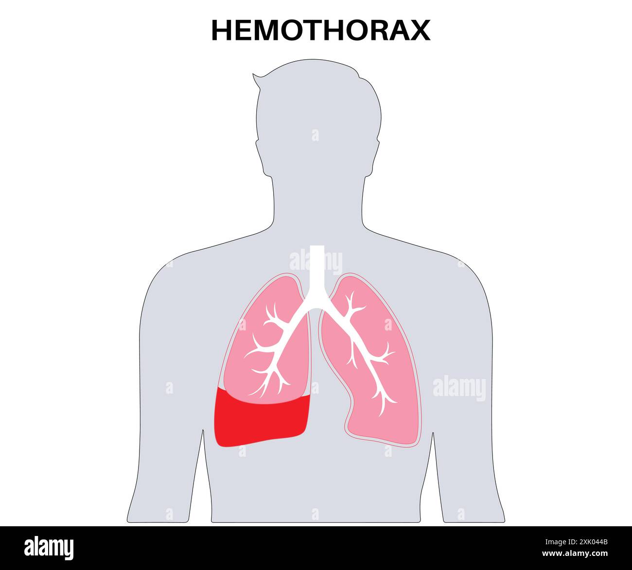 Hemothorax Medical Illustration - Blood Accumulation in the Lung, Respiratory System Disease Diagram Stock Vectorhttps://www.alamy.com/image-license-details/?v=1https://www.alamy.com/hemothorax-medical-illustration-blood-accumulation-in-the-lung-respiratory-system-disease-diagram-image614044603.html
RF2XK044B–Hemothorax Medical Illustration - Blood Accumulation in the Lung, Respiratory System Disease Diagram
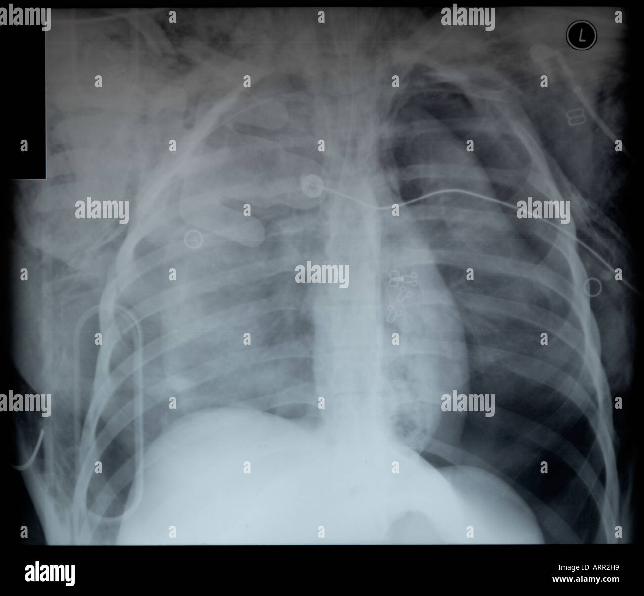 frontal x-ray of fatal blunt chest trauma Stock Photohttps://www.alamy.com/image-license-details/?v=1https://www.alamy.com/frontal-x-ray-of-fatal-blunt-chest-trauma-image9205912.html
RFARR2H9–frontal x-ray of fatal blunt chest trauma
 Eosinophils asthma cells with red blood cells Stock Photohttps://www.alamy.com/image-license-details/?v=1https://www.alamy.com/eosinophils-asthma-cells-with-red-blood-cells-image613819887.html
RF2XJHWER–Eosinophils asthma cells with red blood cells
 Pleural effusion diagram Stock Vectorhttps://www.alamy.com/image-license-details/?v=1https://www.alamy.com/pleural-effusion-diagram-image631240034.html
RF2YJYD2X–Pleural effusion diagram
 . A practical treatise on medical diagnosis for students and physicians . ntinuous with that of theheart extended to the second rib and laterally to the post-axillary line.The dulness occupied three interspaces. Additional physical signs were HEMOTHORAX. 913 immobility, prominence of interspaces, localized above the heart, absentfremitus and resonance. There were no breath-sounds, but an abundanceof rales, apparently very superficial. The rales complicated the physicalsigns. Martin removed 2 ounces of pus from a small abscess above theheart and between the lobes. In empyema a local area may be Stock Photohttps://www.alamy.com/image-license-details/?v=1https://www.alamy.com/a-practical-treatise-on-medical-diagnosis-for-students-and-physicians-ntinuous-with-that-of-theheart-extended-to-the-second-rib-and-laterally-to-the-post-axillary-linethe-dulness-occupied-three-interspaces-additional-physical-signs-were-hemothorax-913-immobility-prominence-of-interspaces-localized-above-the-heart-absentfremitus-and-resonance-there-were-no-breath-sounds-but-an-abundanceof-rales-apparently-very-superficial-the-rales-complicated-the-physicalsigns-martin-removed-2-ounces-of-pus-from-a-small-abscess-above-theheart-and-between-the-lobes-in-empyema-a-local-area-may-be-image376075666.html
RM2CRRM9P–. A practical treatise on medical diagnosis for students and physicians . ntinuous with that of theheart extended to the second rib and laterally to the post-axillary line.The dulness occupied three interspaces. Additional physical signs were HEMOTHORAX. 913 immobility, prominence of interspaces, localized above the heart, absentfremitus and resonance. There were no breath-sounds, but an abundanceof rales, apparently very superficial. The rales complicated the physicalsigns. Martin removed 2 ounces of pus from a small abscess above theheart and between the lobes. In empyema a local area may be
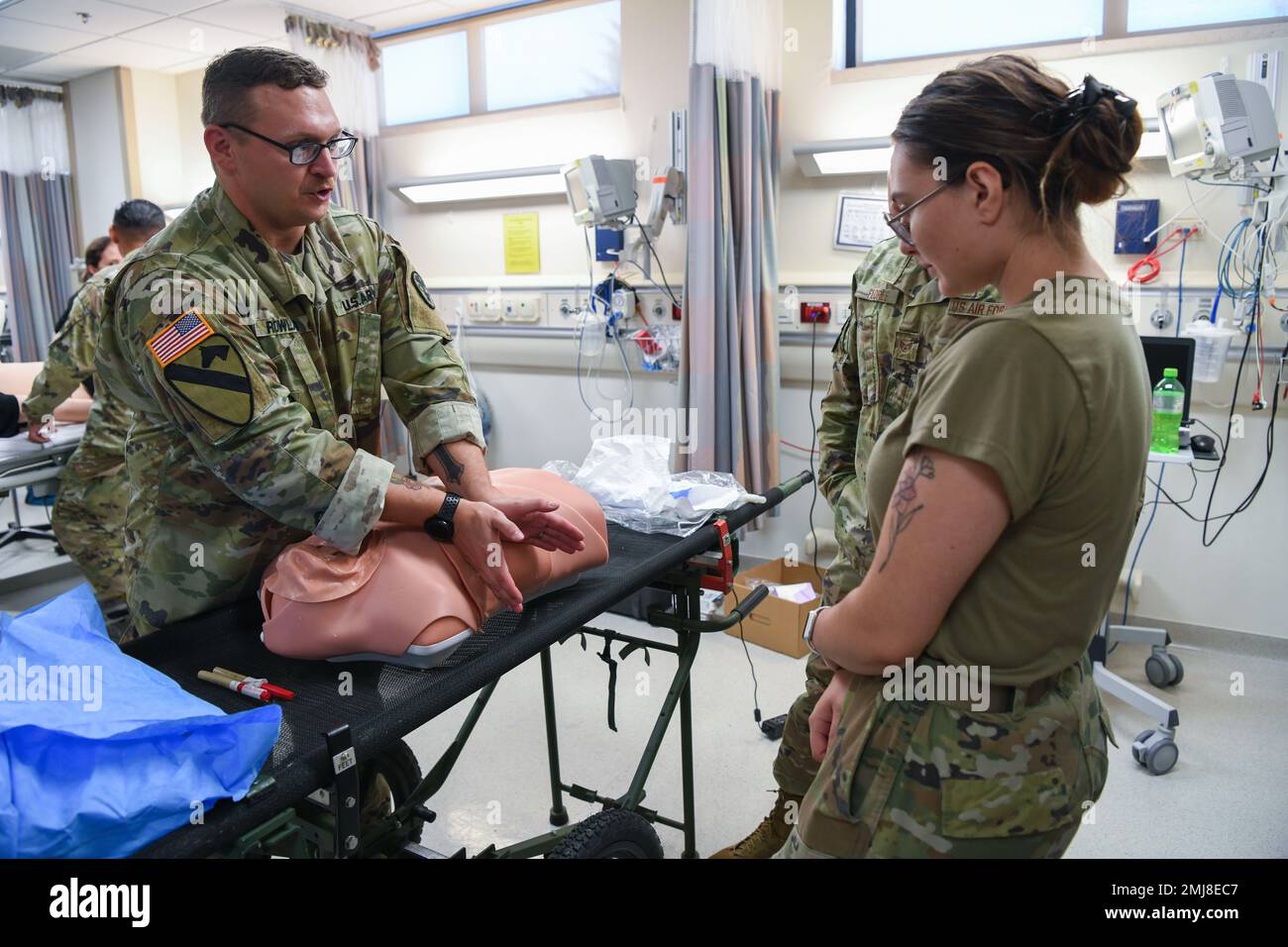 U.S. Army Sgt. 1st Class Bryan Rowland, U.S. Army Medical Department Activity Bavaria NCOIC of education and training, left, teaches A1C Justice Nerad, 31st Health Care Operations Squadron aerospace medical technician, right, how to treat a collapsed lung using a training mannequin at Aviano Air Base, Italy, Aug. 25, 2022. Two scenarios simulated during the joint training were a pneumothorax and hemothorax, which is the name of the condition when trapped air or trapped blood respectively leaks out of the lung into the chest cavity. Stock Photohttps://www.alamy.com/image-license-details/?v=1https://www.alamy.com/us-army-sgt-1st-class-bryan-rowland-us-army-medical-department-activity-bavaria-ncoic-of-education-and-training-left-teaches-a1c-justice-nerad-31st-health-care-operations-squadron-aerospace-medical-technician-right-how-to-treat-a-collapsed-lung-using-a-training-mannequin-at-aviano-air-base-italy-aug-25-2022-two-scenarios-simulated-during-the-joint-training-were-a-pneumothorax-and-hemothorax-which-is-the-name-of-the-condition-when-trapped-air-or-trapped-blood-respectively-leaks-out-of-the-lung-into-the-chest-cavity-image510351415.html
RM2MJ8EC7–U.S. Army Sgt. 1st Class Bryan Rowland, U.S. Army Medical Department Activity Bavaria NCOIC of education and training, left, teaches A1C Justice Nerad, 31st Health Care Operations Squadron aerospace medical technician, right, how to treat a collapsed lung using a training mannequin at Aviano Air Base, Italy, Aug. 25, 2022. Two scenarios simulated during the joint training were a pneumothorax and hemothorax, which is the name of the condition when trapped air or trapped blood respectively leaks out of the lung into the chest cavity.
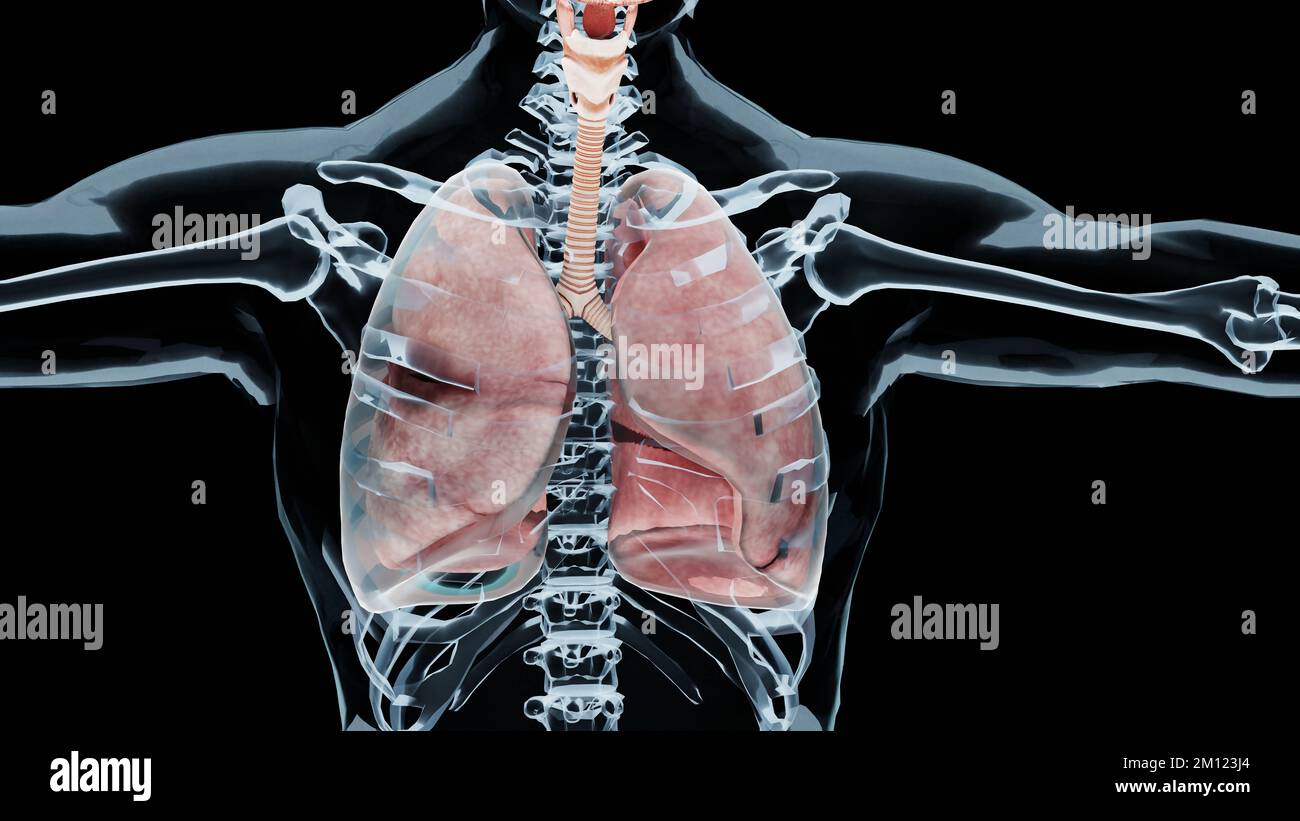 3d Illustration of Pneumothorax, Normal lung versus collapsed, symptoms of pneumothorax, pleural effusion, empyema, complications after a chest injury Stock Photohttps://www.alamy.com/image-license-details/?v=1https://www.alamy.com/3d-illustration-of-pneumothorax-normal-lung-versus-collapsed-symptoms-of-pneumothorax-pleural-effusion-empyema-complications-after-a-chest-injury-image499762092.html
RM2M123J4–3d Illustration of Pneumothorax, Normal lung versus collapsed, symptoms of pneumothorax, pleural effusion, empyema, complications after a chest injury
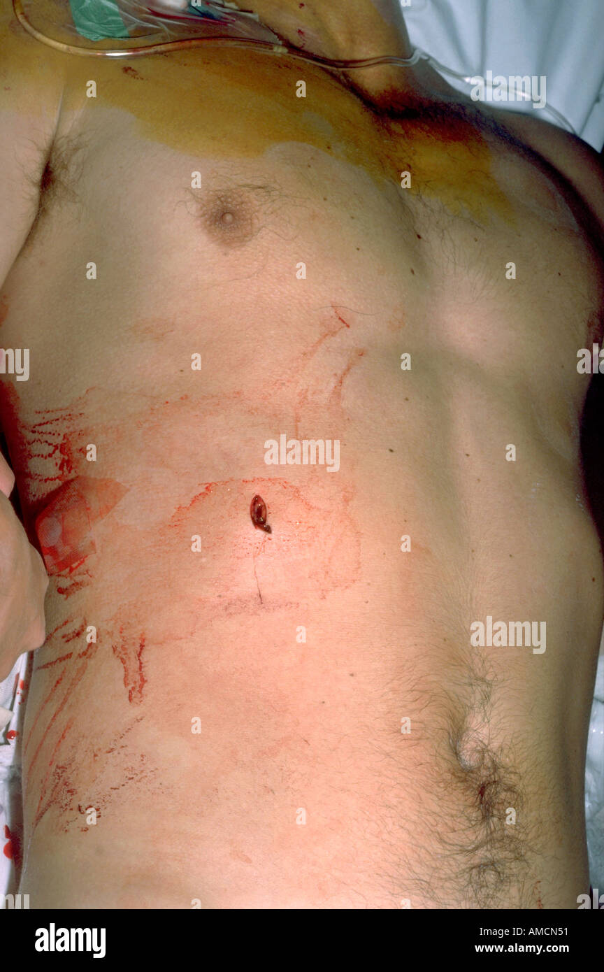 Stabbing Stock Photohttps://www.alamy.com/image-license-details/?v=1https://www.alamy.com/stabbing-image4969808.html
RMAMCN51–Stabbing
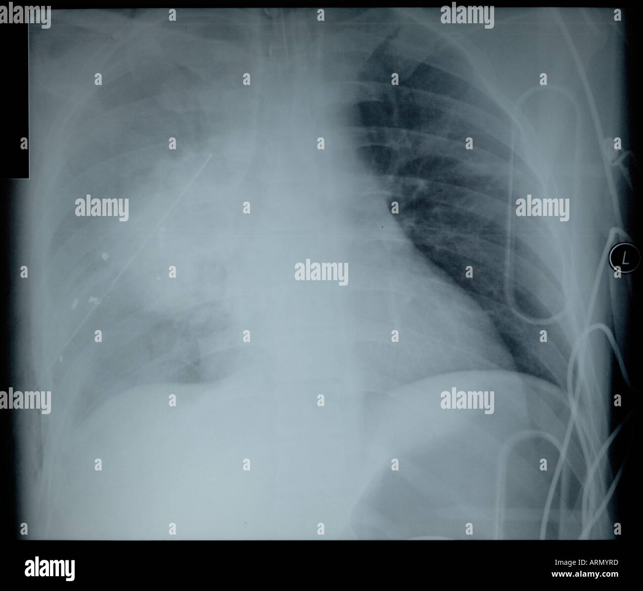 frontal x-ray of gunshot wound to right lung Stock Photohttps://www.alamy.com/image-license-details/?v=1https://www.alamy.com/frontal-x-ray-of-gunshot-wound-to-right-lung-image9194684.html
RFARMYRD–frontal x-ray of gunshot wound to right lung
 Pleural effusion represents inadequate disposal Stock Photohttps://www.alamy.com/image-license-details/?v=1https://www.alamy.com/pleural-effusion-represents-inadequate-disposal-image613819903.html
RF2XJHWFB–Pleural effusion represents inadequate disposal
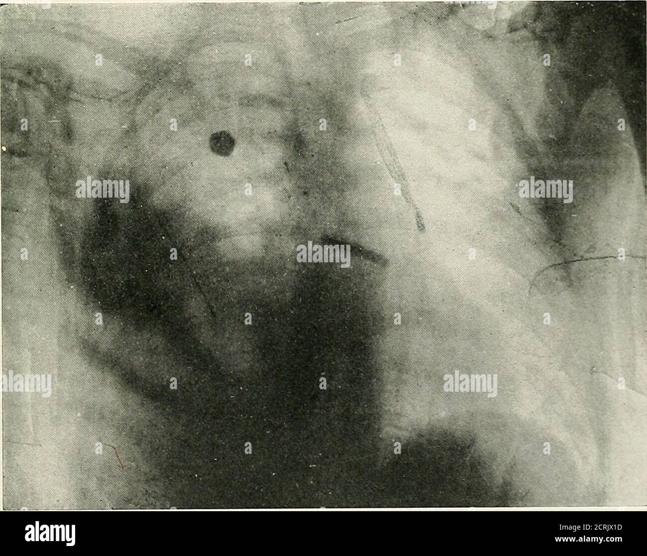 . Radio-diagnosis of pleuro-pulmonary affection . Radiograph A. TRAUMATIC ENCYSTED HEMOTHORAX IN A CASEOF OLD PLEURISY The bullet, after passing through the thorax, lodged in the wall. Pleurotomy,complete recovery, thorax became entirely clear from apex to base.. Radiograph B. PIECE OF SHRAPNEL IN THE RIGHT LUNGLong splinter of shell in the posterior wall. Right pleurisy. CLINICAL AND RADIOLOGICAL STUDY 163 having been established, it is indispensable to use this sameradioscopic examination for information as to the generalposition: whether the projectile is intra-thoracic, in thelung or in th Stock Photohttps://www.alamy.com/image-license-details/?v=1https://www.alamy.com/radio-diagnosis-of-pleuro-pulmonary-affection-radiograph-a-traumatic-encysted-hemothorax-in-a-caseof-old-pleurisy-the-bullet-after-passing-through-the-thorax-lodged-in-the-wall-pleurotomycomplete-recovery-thorax-became-entirely-clear-from-apex-to-base-radiograph-b-piece-of-shrapnel-in-the-right-lunglong-splinter-of-shell-in-the-posterior-wall-right-pleurisy-clinical-and-radiological-study-163-having-been-established-it-is-indispensable-to-use-this-sameradioscopic-examination-for-information-as-to-the-generalposition-whether-the-projectile-is-intra-thoracic-in-thelung-or-in-th-image375970377.html
RM2CRJX1D–. Radio-diagnosis of pleuro-pulmonary affection . Radiograph A. TRAUMATIC ENCYSTED HEMOTHORAX IN A CASEOF OLD PLEURISY The bullet, after passing through the thorax, lodged in the wall. Pleurotomy,complete recovery, thorax became entirely clear from apex to base.. Radiograph B. PIECE OF SHRAPNEL IN THE RIGHT LUNGLong splinter of shell in the posterior wall. Right pleurisy. CLINICAL AND RADIOLOGICAL STUDY 163 having been established, it is indispensable to use this sameradioscopic examination for information as to the generalposition: whether the projectile is intra-thoracic, in thelung or in th
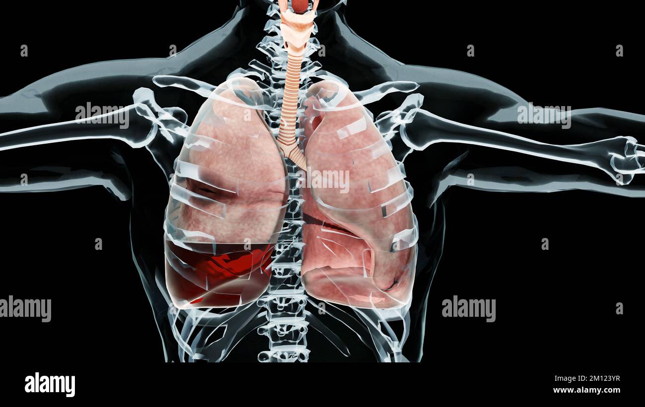 3d Illustration of Hemopneumothorax, Normal lung versus collapsed, symptoms of Hemopneumothorax, pleural effusion, empyema, complications after a ches Stock Photohttps://www.alamy.com/image-license-details/?v=1https://www.alamy.com/3d-illustration-of-hemopneumothorax-normal-lung-versus-collapsed-symptoms-of-hemopneumothorax-pleural-effusion-empyema-complications-after-a-ches-image499762363.html
RM2M123YR–3d Illustration of Hemopneumothorax, Normal lung versus collapsed, symptoms of Hemopneumothorax, pleural effusion, empyema, complications after a ches
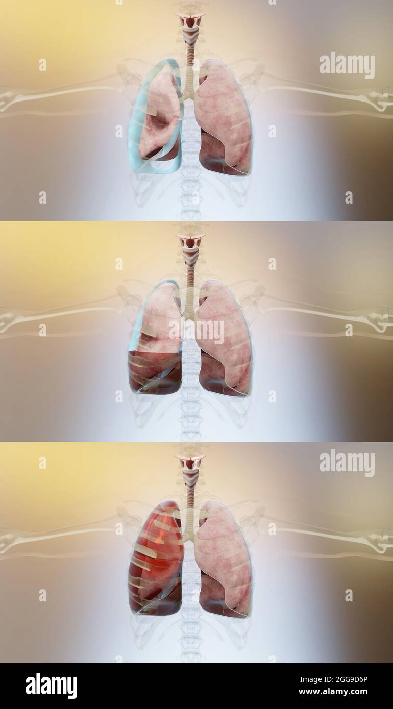 3d Illustration of Pneumothorax, Hemothorax and Hemopneumothorax, Normal lung versus collapsed, symptoms of pneumothorax, pleural effusion, empyema, 3 Stock Photohttps://www.alamy.com/image-license-details/?v=1https://www.alamy.com/3d-illustration-of-pneumothorax-hemothorax-and-hemopneumothorax-normal-lung-versus-collapsed-symptoms-of-pneumothorax-pleural-effusion-empyema-3-image440301646.html
RM2GG9D6P–3d Illustration of Pneumothorax, Hemothorax and Hemopneumothorax, Normal lung versus collapsed, symptoms of pneumothorax, pleural effusion, empyema, 3
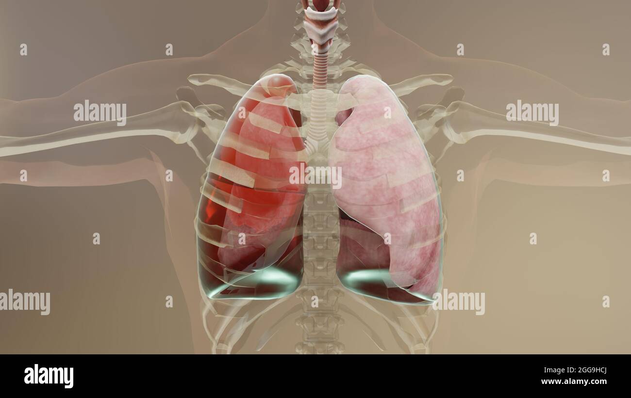 3d Illustration of Hemothorax, Normal lung versus collapsed, symptoms of Hemothorax, pleural effusion, empyema, complications after a chest injury, Stock Photohttps://www.alamy.com/image-license-details/?v=1https://www.alamy.com/3d-illustration-of-hemothorax-normal-lung-versus-collapsed-symptoms-of-hemothorax-pleural-effusion-empyema-complications-after-a-chest-injury-image440304946.html
RM2GG9HCJ–3d Illustration of Hemothorax, Normal lung versus collapsed, symptoms of Hemothorax, pleural effusion, empyema, complications after a chest injury,
 Accident and emergency - chest drain procedure Stock Photohttps://www.alamy.com/image-license-details/?v=1https://www.alamy.com/accident-and-emergency-chest-drain-procedure-image4969818.html
RMAMCN5B–Accident and emergency - chest drain procedure
 Axial CT slice of lung with knife still in wound Stock Photohttps://www.alamy.com/image-license-details/?v=1https://www.alamy.com/axial-ct-slice-of-lung-with-knife-still-in-wound-image9194616.html
RFARMYK9–Axial CT slice of lung with knife still in wound
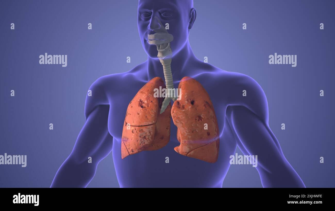 Lungs pneumothorax disease medical concept Stock Photohttps://www.alamy.com/image-license-details/?v=1https://www.alamy.com/lungs-pneumothorax-disease-medical-concept-image613819906.html
RF2XJHWFE–Lungs pneumothorax disease medical concept
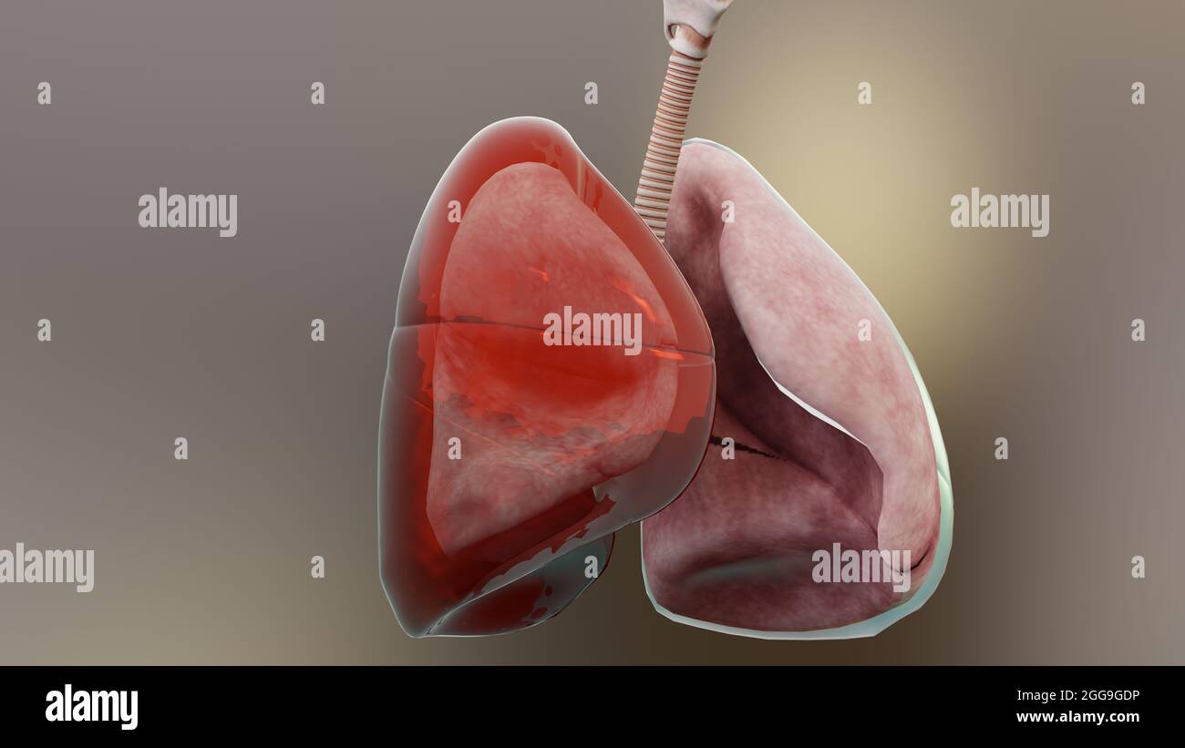 3d Illustration of Hemothorax, Normal lung versus collapsed, symptoms of Hemothorax, pleural effusion, empyema, complications after a chest injury, Stock Photohttps://www.alamy.com/image-license-details/?v=1https://www.alamy.com/3d-illustration-of-hemothorax-normal-lung-versus-collapsed-symptoms-of-hemothorax-pleural-effusion-empyema-complications-after-a-chest-injury-image440304194.html
RM2GG9GDP–3d Illustration of Hemothorax, Normal lung versus collapsed, symptoms of Hemothorax, pleural effusion, empyema, complications after a chest injury,
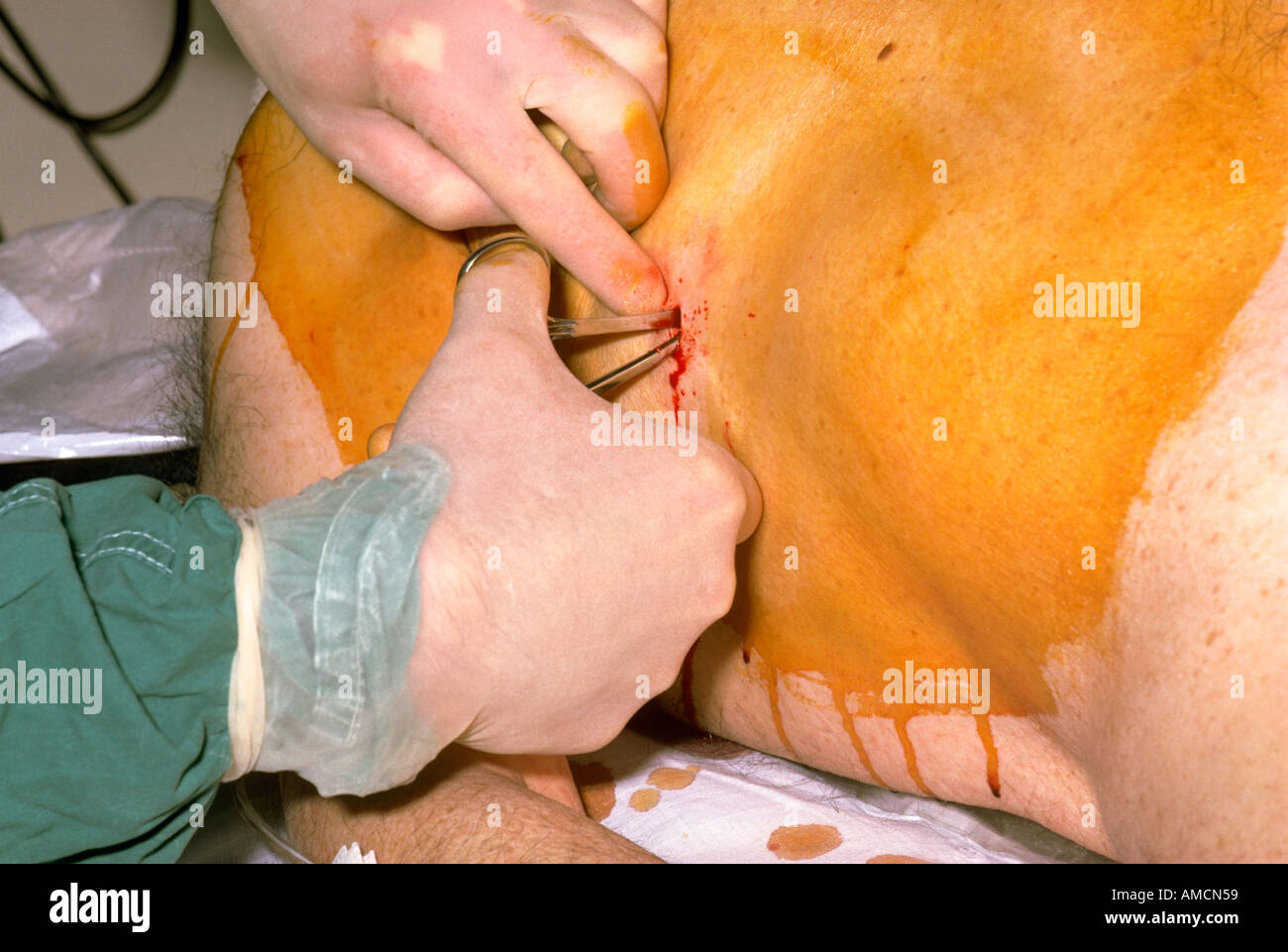 Accident and emergency - chest drain procedure Stock Photohttps://www.alamy.com/image-license-details/?v=1https://www.alamy.com/accident-and-emergency-chest-drain-procedure-image4969816.html
RMAMCN59–Accident and emergency - chest drain procedure
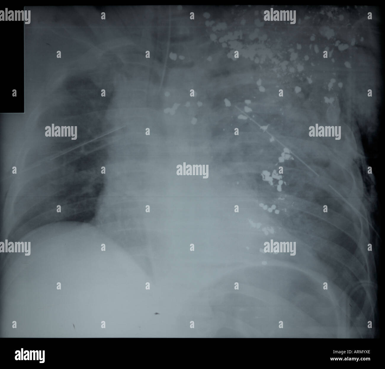 frontal x-ray of fatal shotgun wound to adult chest Stock Photohttps://www.alamy.com/image-license-details/?v=1https://www.alamy.com/frontal-x-ray-of-fatal-shotgun-wound-to-adult-chest-image9194733.html
RFARMYXE–frontal x-ray of fatal shotgun wound to adult chest
 Pleural effusion represents inadequate disposal Stock Photohttps://www.alamy.com/image-license-details/?v=1https://www.alamy.com/pleural-effusion-represents-inadequate-disposal-image613819904.html
RF2XJHWFC–Pleural effusion represents inadequate disposal
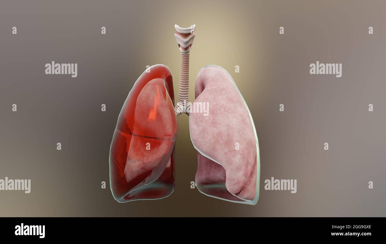 3d Illustration of Hemothorax, Normal lung versus collapsed, symptoms of Hemothorax, pleural effusion, empyema, complications after a chest injury, Stock Photohttps://www.alamy.com/image-license-details/?v=1https://www.alamy.com/3d-illustration-of-hemothorax-normal-lung-versus-collapsed-symptoms-of-hemothorax-pleural-effusion-empyema-complications-after-a-chest-injury-image440304550.html
RM2GG9GXE–3d Illustration of Hemothorax, Normal lung versus collapsed, symptoms of Hemothorax, pleural effusion, empyema, complications after a chest injury,
 Chest drain Stock Photohttps://www.alamy.com/image-license-details/?v=1https://www.alamy.com/chest-drain-image1684952.html
RMATB5D9–Chest drain
 lateral x-ray of lung with knife still in wound Stock Photohttps://www.alamy.com/image-license-details/?v=1https://www.alamy.com/lateral-x-ray-of-lung-with-knife-still-in-wound-image9194609.html
RFARMYK2–lateral x-ray of lung with knife still in wound
 Eosinophils asthma cells medical concept Stock Photohttps://www.alamy.com/image-license-details/?v=1https://www.alamy.com/eosinophils-asthma-cells-medical-concept-image613819898.html
RF2XJHWF6–Eosinophils asthma cells medical concept
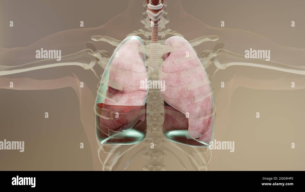 3d Illustration of Hemopneumothorax, Normal lung versus collapsed, symptoms of Hemopneumothorax, pleural effusion, empyema, complications Stock Photohttps://www.alamy.com/image-license-details/?v=1https://www.alamy.com/3d-illustration-of-hemopneumothorax-normal-lung-versus-collapsed-symptoms-of-hemopneumothorax-pleural-effusion-empyema-complications-image440305012.html
RM2GG9HF0–3d Illustration of Hemopneumothorax, Normal lung versus collapsed, symptoms of Hemopneumothorax, pleural effusion, empyema, complications
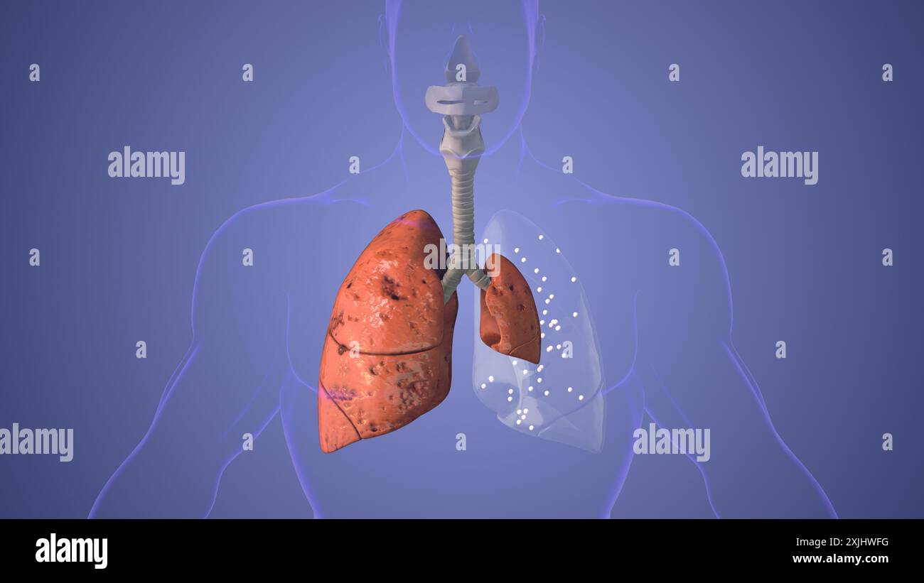 Lungs pneumothorax disease medical concept Stock Photohttps://www.alamy.com/image-license-details/?v=1https://www.alamy.com/lungs-pneumothorax-disease-medical-concept-image613819908.html
RF2XJHWFG–Lungs pneumothorax disease medical concept
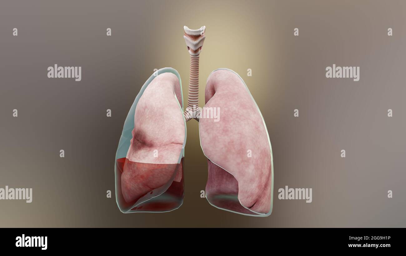 3d Illustration of Hemopneumothorax, Normal lung versus collapsed, symptoms of Hemopneumothorax, pleural effusion, empyema, complications Stock Photohttps://www.alamy.com/image-license-details/?v=1https://www.alamy.com/3d-illustration-of-hemopneumothorax-normal-lung-versus-collapsed-symptoms-of-hemopneumothorax-pleural-effusion-empyema-complications-image440304642.html
RM2GG9H1P–3d Illustration of Hemopneumothorax, Normal lung versus collapsed, symptoms of Hemopneumothorax, pleural effusion, empyema, complications
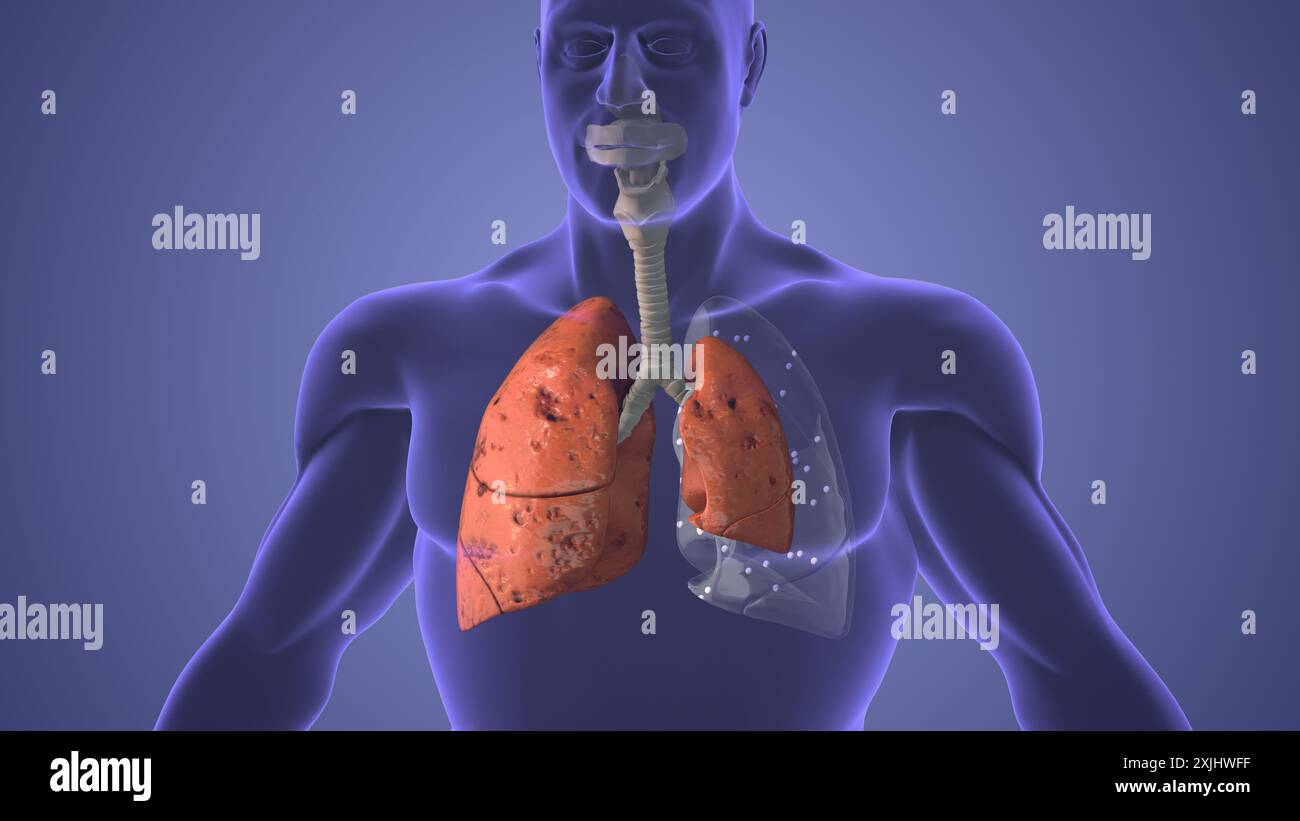 Lungs pneumothorax disease medical concept Stock Photohttps://www.alamy.com/image-license-details/?v=1https://www.alamy.com/lungs-pneumothorax-disease-medical-concept-image613819907.html
RF2XJHWFF–Lungs pneumothorax disease medical concept
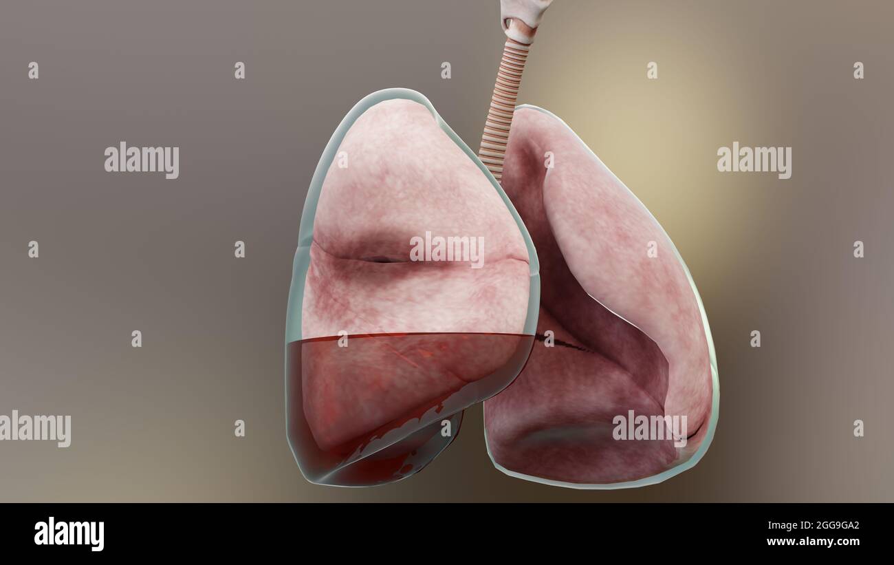 3d Illustration of Hemopneumothorax, Normal lung versus collapsed, symptoms of Hemopneumothorax, pleural effusion, empyema, complications Stock Photohttps://www.alamy.com/image-license-details/?v=1https://www.alamy.com/3d-illustration-of-hemopneumothorax-normal-lung-versus-collapsed-symptoms-of-hemopneumothorax-pleural-effusion-empyema-complications-image440304090.html
RM2GG9GA2–3d Illustration of Hemopneumothorax, Normal lung versus collapsed, symptoms of Hemopneumothorax, pleural effusion, empyema, complications
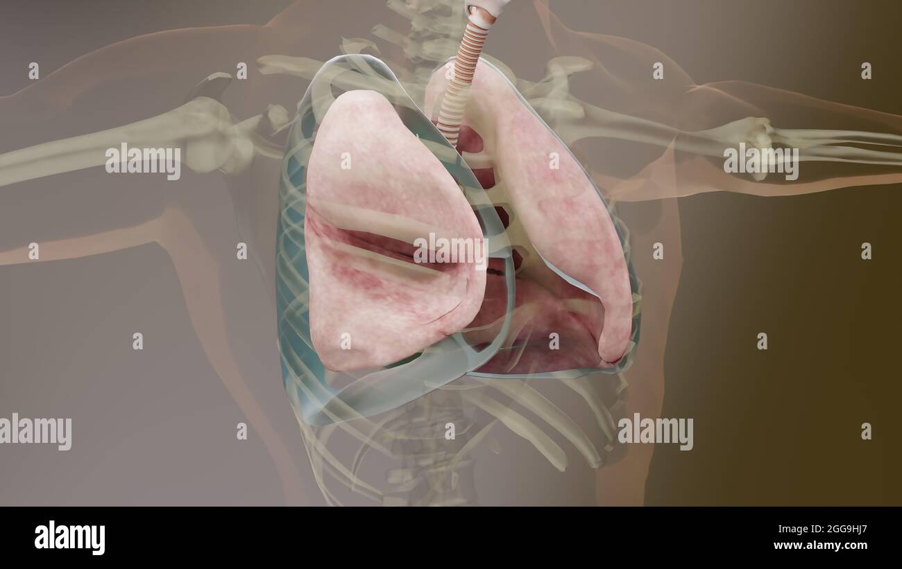 3d Illustration of Pneumothorax, Normal lung versus collapsed, symptoms of pneumothorax, pleural effusion, empyema, complications after a chest injury Stock Photohttps://www.alamy.com/image-license-details/?v=1https://www.alamy.com/3d-illustration-of-pneumothorax-normal-lung-versus-collapsed-symptoms-of-pneumothorax-pleural-effusion-empyema-complications-after-a-chest-injury-image440305103.html
RM2GG9HJ7–3d Illustration of Pneumothorax, Normal lung versus collapsed, symptoms of pneumothorax, pleural effusion, empyema, complications after a chest injury
 Lungs pneumothorax disease medical concept Stock Photohttps://www.alamy.com/image-license-details/?v=1https://www.alamy.com/lungs-pneumothorax-disease-medical-concept-image613819909.html
RF2XJHWFH–Lungs pneumothorax disease medical concept
 Eosinophils asthma cells in artery with blood cells Stock Photohttps://www.alamy.com/image-license-details/?v=1https://www.alamy.com/eosinophils-asthma-cells-in-artery-with-blood-cells-image612851401.html
RF2XH1P61–Eosinophils asthma cells in artery with blood cells
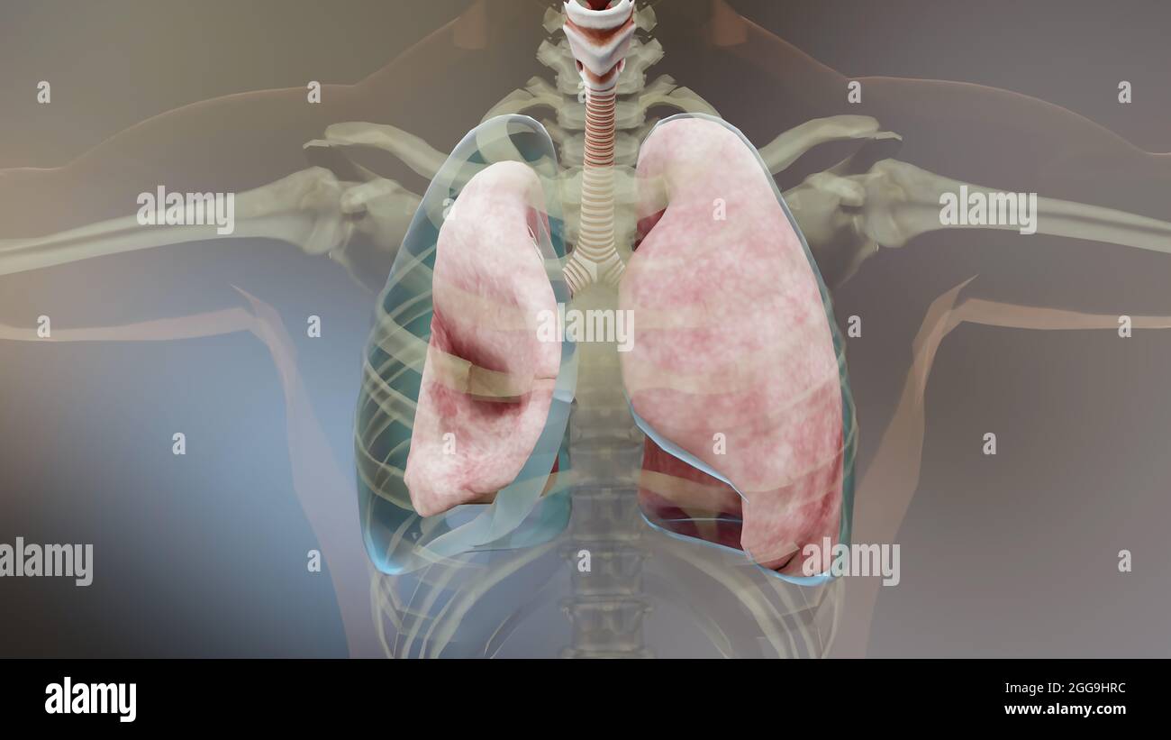 3d Illustration of Pneumothorax, Normal lung versus collapsed, symptoms of pneumothorax, pleural effusion, empyema, complications after a chest injury Stock Photohttps://www.alamy.com/image-license-details/?v=1https://www.alamy.com/3d-illustration-of-pneumothorax-normal-lung-versus-collapsed-symptoms-of-pneumothorax-pleural-effusion-empyema-complications-after-a-chest-injury-image440305248.html
RM2GG9HRC–3d Illustration of Pneumothorax, Normal lung versus collapsed, symptoms of pneumothorax, pleural effusion, empyema, complications after a chest injury
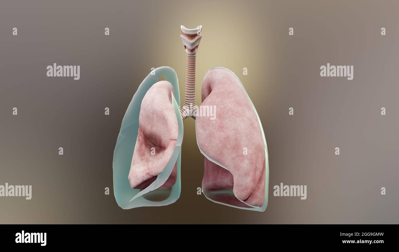 3d Illustration of Pneumothorax, Normal lung versus collapsed, symptoms of pneumothorax, pleural effusion, empyema, complications after a chest injury Stock Photohttps://www.alamy.com/image-license-details/?v=1https://www.alamy.com/3d-illustration-of-pneumothorax-normal-lung-versus-collapsed-symptoms-of-pneumothorax-pleural-effusion-empyema-complications-after-a-chest-injury-image440304393.html
RM2GG9GMW–3d Illustration of Pneumothorax, Normal lung versus collapsed, symptoms of pneumothorax, pleural effusion, empyema, complications after a chest injury
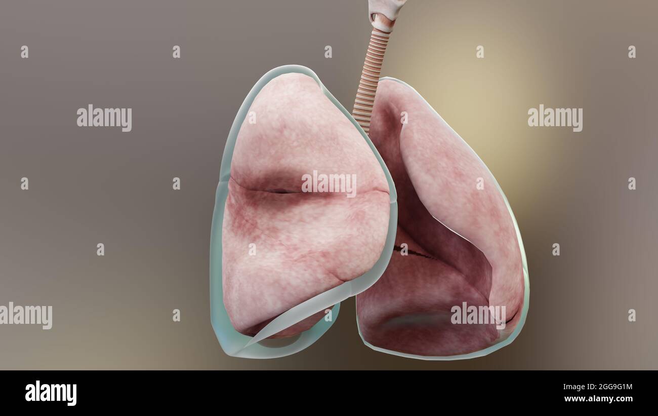 3d Illustration of Pneumothorax, Normal lung versus collapsed, symptoms of pneumothorax, pleural effusion, empyema, complications after a chest injury Stock Photohttps://www.alamy.com/image-license-details/?v=1https://www.alamy.com/3d-illustration-of-pneumothorax-normal-lung-versus-collapsed-symptoms-of-pneumothorax-pleural-effusion-empyema-complications-after-a-chest-injury-image440303856.html
RM2GG9G1M–3d Illustration of Pneumothorax, Normal lung versus collapsed, symptoms of pneumothorax, pleural effusion, empyema, complications after a chest injury
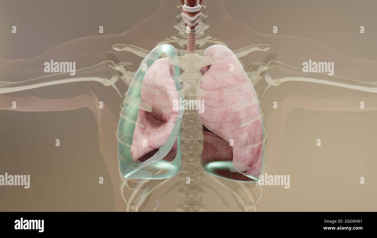 3d Illustration of Pneumothorax, Normal lung versus collapsed, symptoms of pneumothorax, pleural effusion, empyema, complications after a chest injury Stock Photohttps://www.alamy.com/image-license-details/?v=1https://www.alamy.com/3d-illustration-of-pneumothorax-normal-lung-versus-collapsed-symptoms-of-pneumothorax-pleural-effusion-empyema-complications-after-a-chest-injury-image440304761.html
RM2GG9H61–3d Illustration of Pneumothorax, Normal lung versus collapsed, symptoms of pneumothorax, pleural effusion, empyema, complications after a chest injury