Horse anatomy illustration Stock Photos and Images
(3,078)See horse anatomy illustration stock video clipsQuick filters:
Horse anatomy illustration Stock Photos and Images
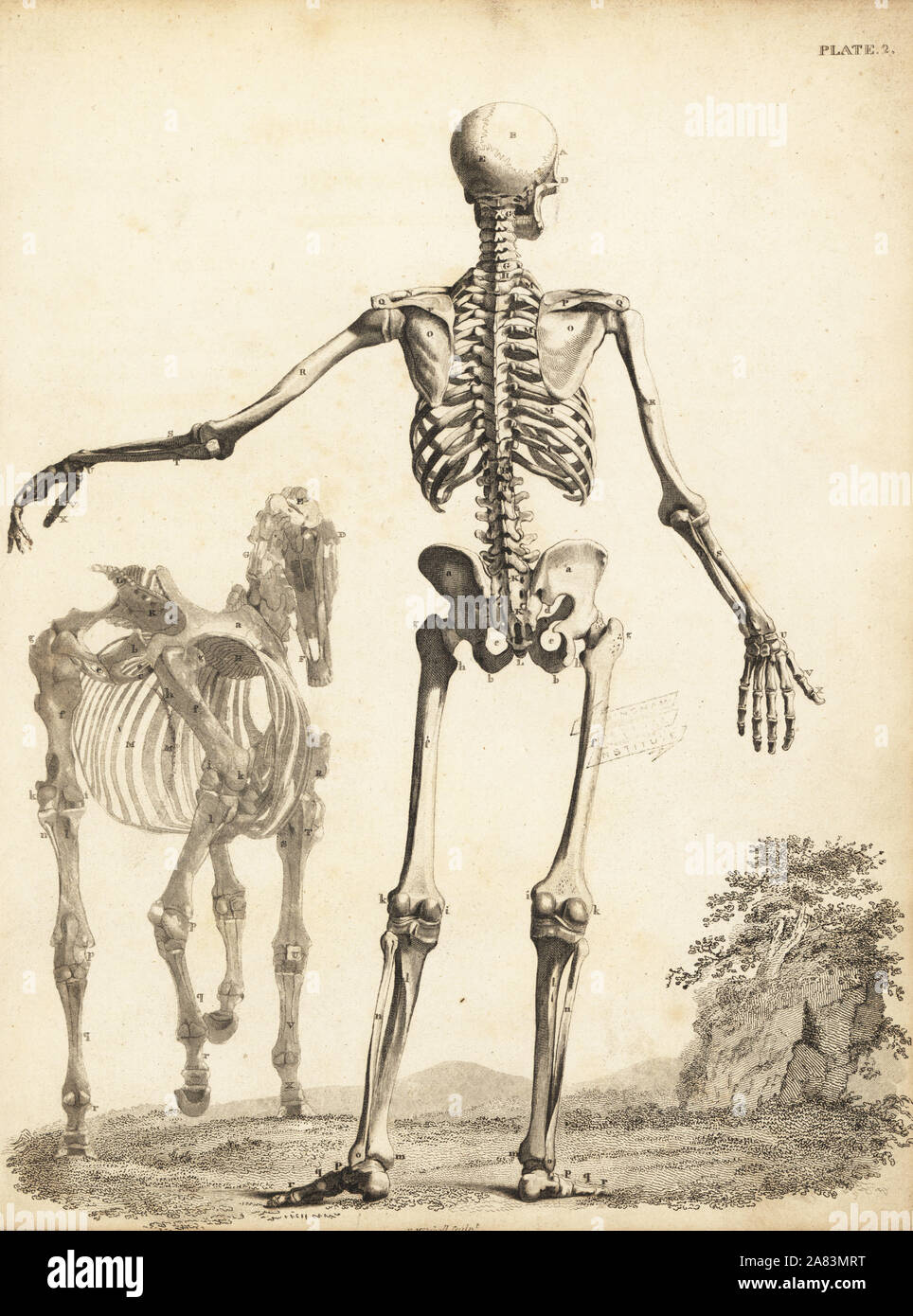 Human skeleton from the rear, with horse skeleton by George Stubbs. Copperplate engraving by Edward Mitchell after an anatomical illustration by Bernhard Siefried Albinus from John Barclay's A Series of Engravings of the Human Skeleton, MacLachlan and Stewart, Edinburgh, 1824. Stock Photohttps://www.alamy.com/image-license-details/?v=1https://www.alamy.com/human-skeleton-from-the-rear-with-horse-skeleton-by-george-stubbs-copperplate-engraving-by-edward-mitchell-after-an-anatomical-illustration-by-bernhard-siefried-albinus-from-john-barclays-a-series-of-engravings-of-the-human-skeleton-maclachlan-and-stewart-edinburgh-1824-image331996444.html
RM2A83MRT–Human skeleton from the rear, with horse skeleton by George Stubbs. Copperplate engraving by Edward Mitchell after an anatomical illustration by Bernhard Siefried Albinus from John Barclay's A Series of Engravings of the Human Skeleton, MacLachlan and Stewart, Edinburgh, 1824.
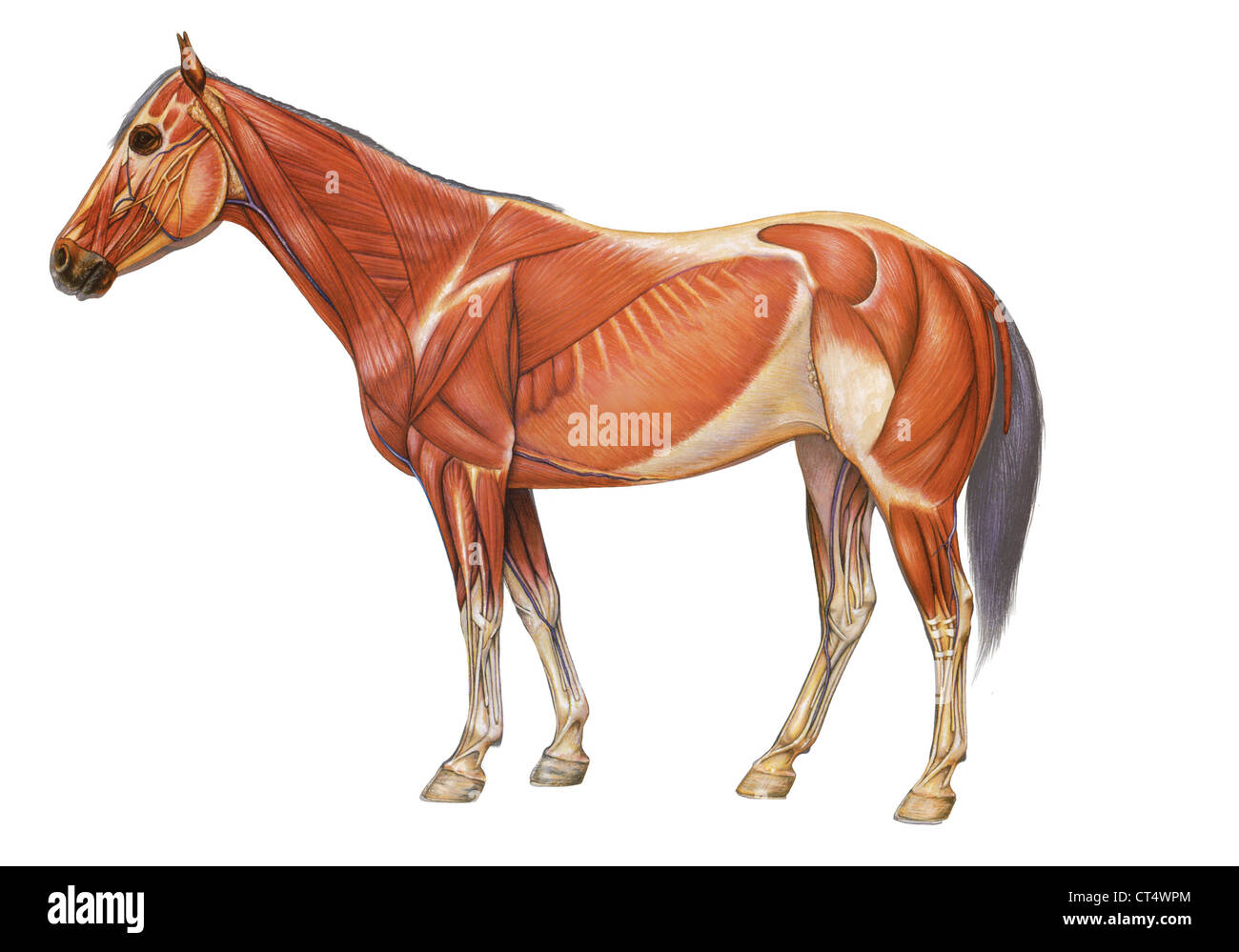 Horse anatomy, drawing Stock Photohttps://www.alamy.com/image-license-details/?v=1https://www.alamy.com/stock-photo-horse-anatomy-drawing-49280524.html
RMCT4WPM–Horse anatomy, drawing
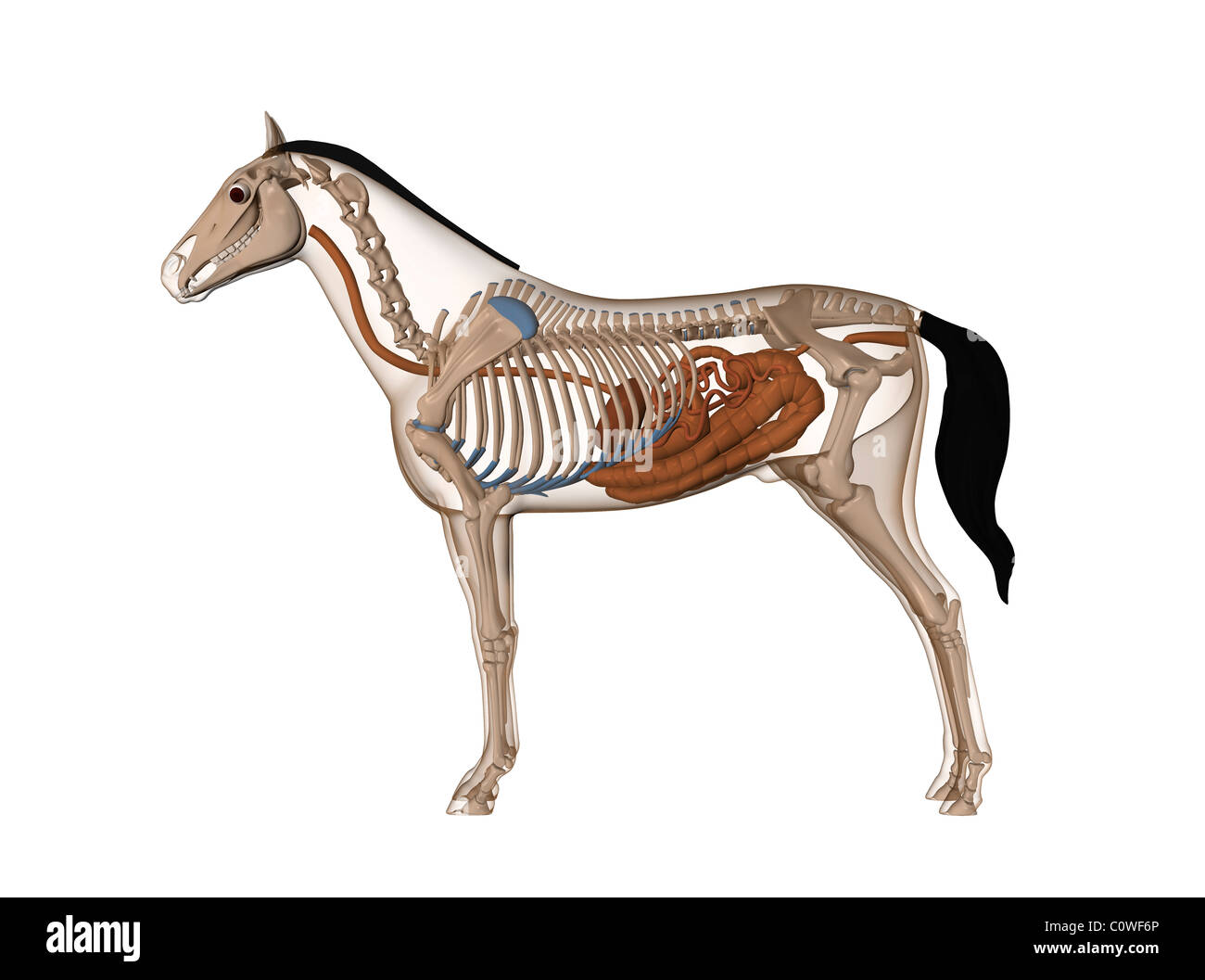 horse anatomy digestion stomach skeleton Stock Photohttps://www.alamy.com/image-license-details/?v=1https://www.alamy.com/stock-photo-horse-anatomy-digestion-stomach-skeleton-34981486.html
RMC0WF6P–horse anatomy digestion stomach skeleton
 Horse anatomy. Antique French wooden plaque detailing the anatomy of a horse c.1843 Stock Photohttps://www.alamy.com/image-license-details/?v=1https://www.alamy.com/horse-anatomy-antique-french-wooden-plaque-detailing-the-anatomy-of-image67919563.html
RMDXE02K–Horse anatomy. Antique French wooden plaque detailing the anatomy of a horse c.1843
 Musculature, Horse, Illustration, 1772 Stock Photohttps://www.alamy.com/image-license-details/?v=1https://www.alamy.com/musculature-horse-illustration-1772-image352797057.html
RM2BDY86W–Musculature, Horse, Illustration, 1772
 Horse anatomy, illustration Stock Photohttps://www.alamy.com/image-license-details/?v=1https://www.alamy.com/horse-anatomy-illustration-image267282175.html
RFWERN3Y–Horse anatomy, illustration
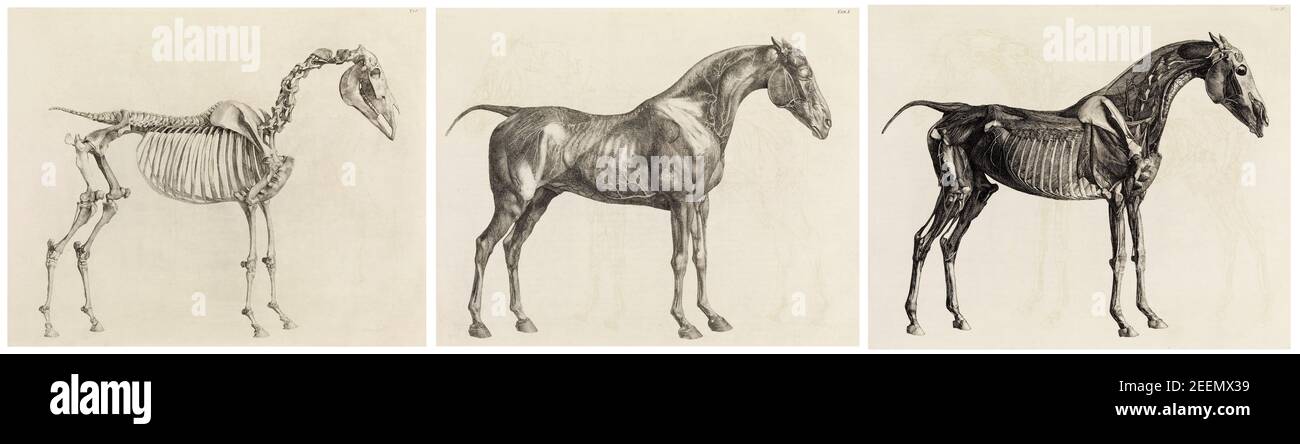 The Anatomy of the Horse, including a particular description of the bones, cartilages, muscles, fascias, ligaments, nerves, arteries, veins, and glands, anatomical drawing by George Stubbs, 1766 Stock Photohttps://www.alamy.com/image-license-details/?v=1https://www.alamy.com/the-anatomy-of-the-horse-including-a-particular-description-of-the-bones-cartilages-muscles-fascias-ligaments-nerves-arteries-veins-and-glands-anatomical-drawing-by-george-stubbs-1766-image404903165.html
RM2EEMX39–The Anatomy of the Horse, including a particular description of the bones, cartilages, muscles, fascias, ligaments, nerves, arteries, veins, and glands, anatomical drawing by George Stubbs, 1766
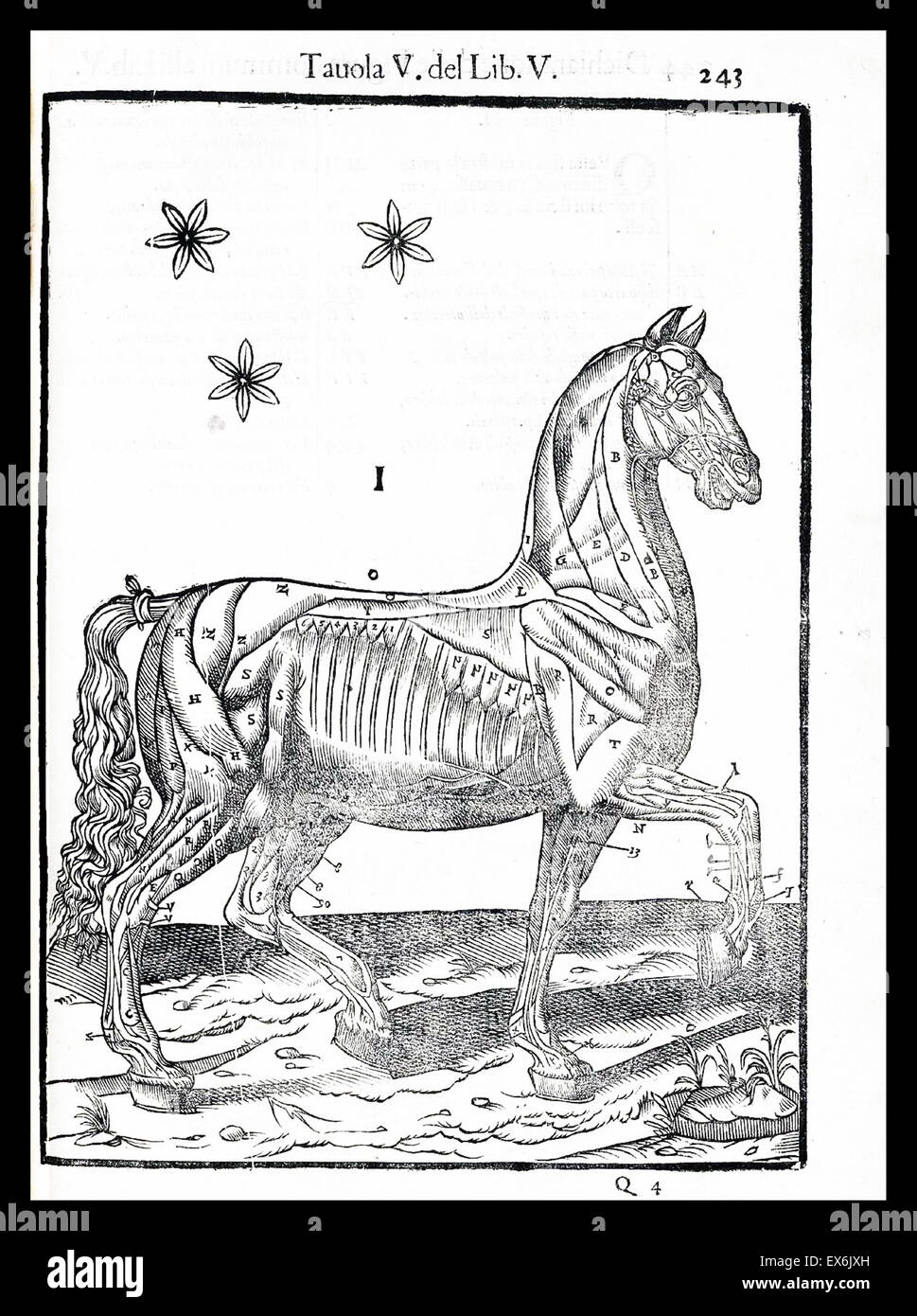 Equine anatomical Illustration from 'Anatomia del cavallo, infermità, et suoi rimedii'. Anatomy of a horse); (1618). by Carlo Ruini, (1530-1598) Stock Photohttps://www.alamy.com/image-license-details/?v=1https://www.alamy.com/stock-photo-equine-anatomical-illustration-from-anatomia-del-cavallo-infermit-84969097.html
RMEX6JXH–Equine anatomical Illustration from 'Anatomia del cavallo, infermità, et suoi rimedii'. Anatomy of a horse); (1618). by Carlo Ruini, (1530-1598)
 Muscular anatomy of a horse with black outline, top view. Stock Photohttps://www.alamy.com/image-license-details/?v=1https://www.alamy.com/muscular-anatomy-of-a-horse-with-black-outline-top-view-image350070537.html
RF2B9F2F5–Muscular anatomy of a horse with black outline, top view.
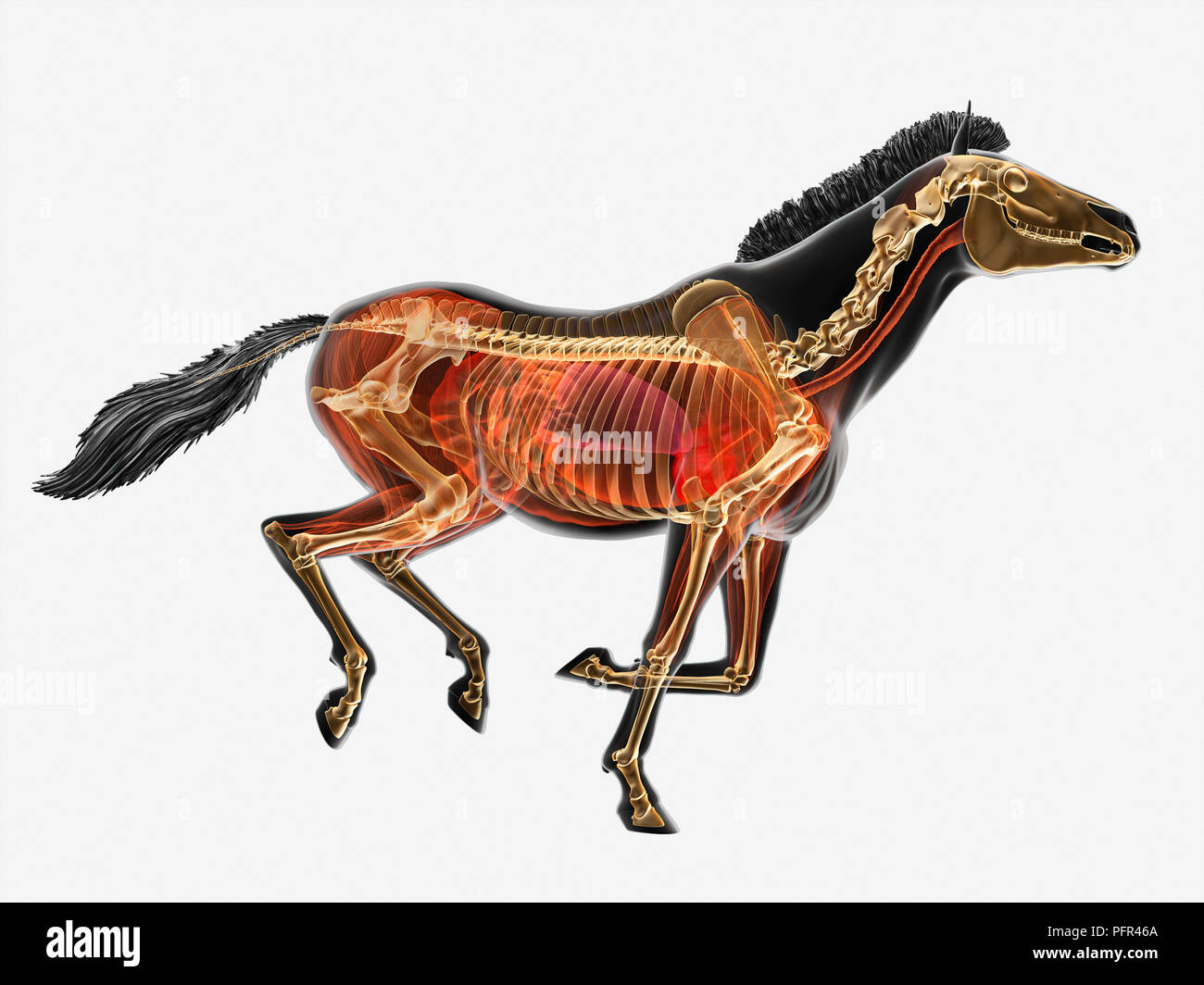 Illustration, anatomy of Przewalski's horse (Equus ferus przewalskii) Stock Photohttps://www.alamy.com/image-license-details/?v=1https://www.alamy.com/illustration-anatomy-of-przewalskis-horse-equus-ferus-przewalskii-image216252466.html
RMPFR46A–Illustration, anatomy of Przewalski's horse (Equus ferus przewalskii)
 A 19th Century depiction of the skeletons of Man and Horse: the illustration shows the equine terms in brackets. .A. Knee (Stifle); B. Ankle (Hock); C. Elbow; D. Wrist (Knee); E, Humerus; F. Femur Stock Photohttps://www.alamy.com/image-license-details/?v=1https://www.alamy.com/a-19th-century-depiction-of-the-skeletons-of-man-and-horse-the-illustration-shows-the-equine-terms-in-brackets-a-knee-stifle-b-ankle-hock-c-elbow-d-wrist-knee-e-humerus-f-femur-image383662331.html
RM2D85963–A 19th Century depiction of the skeletons of Man and Horse: the illustration shows the equine terms in brackets. .A. Knee (Stifle); B. Ankle (Hock); C. Elbow; D. Wrist (Knee); E, Humerus; F. Femur
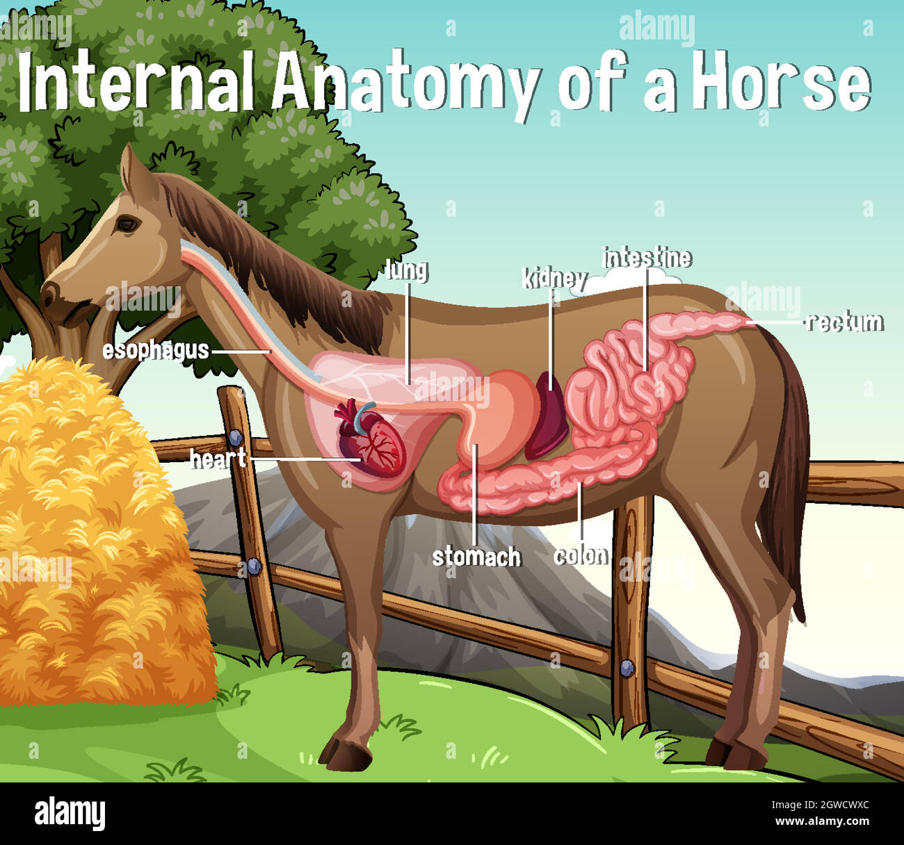 Internal Anatomy of a Horse with label Stock Vectorhttps://www.alamy.com/image-license-details/?v=1https://www.alamy.com/internal-anatomy-of-a-horse-with-label-image445909364.html
RF2GWCWXC–Internal Anatomy of a Horse with label
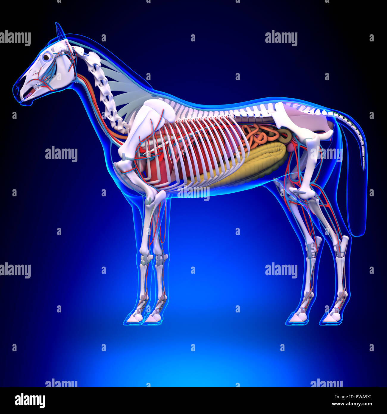 Horse Anatomy - Internal Anatomy of Horse Stock Photohttps://www.alamy.com/image-license-details/?v=1https://www.alamy.com/stock-photo-horse-anatomy-internal-anatomy-of-horse-84435177.html
RFEWA9X1–Horse Anatomy - Internal Anatomy of Horse
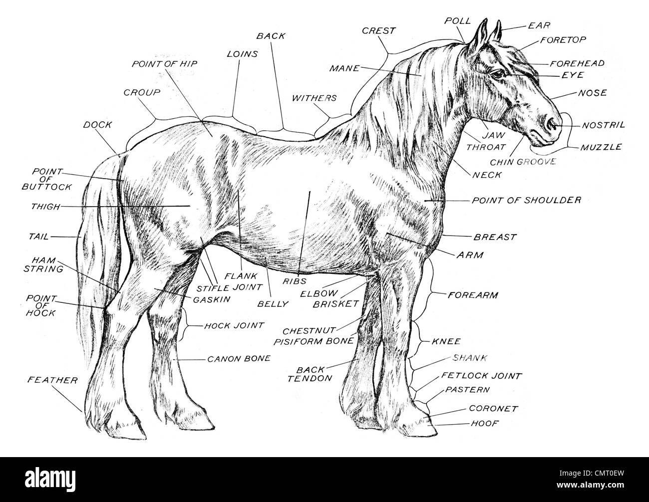 1923 Anatomy of a horse Equus ferus caballus odd-toed ungulate mammal Stock Photohttps://www.alamy.com/image-license-details/?v=1https://www.alamy.com/stock-photo-1923-anatomy-of-a-horse-equus-ferus-caballus-odd-toed-ungulate-mammal-47241121.html
RFCMT0EW–1923 Anatomy of a horse Equus ferus caballus odd-toed ungulate mammal
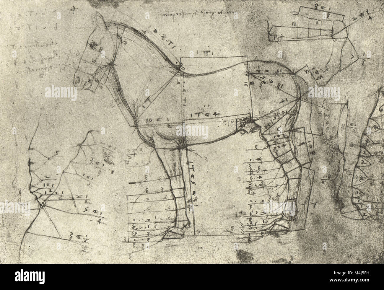 A measured Horse in profile to the left, equine Anatomical drawing, drawn by Leonardo Da Vinci, 1452-1519 Stock Photohttps://www.alamy.com/image-license-details/?v=1https://www.alamy.com/stock-photo-a-measured-horse-in-profile-to-the-left-equine-anatomical-drawing-174961797.html
RMM4J5FH–A measured Horse in profile to the left, equine Anatomical drawing, drawn by Leonardo Da Vinci, 1452-1519
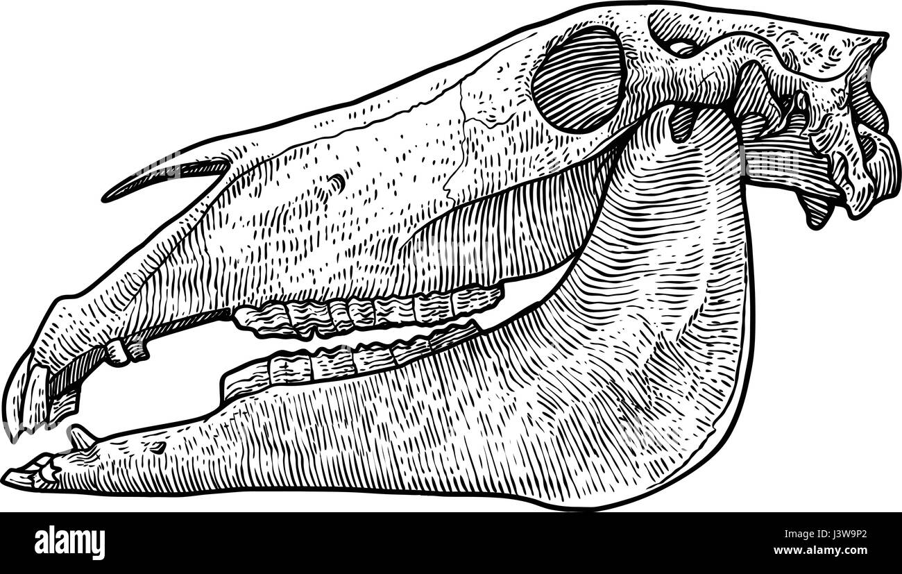 Horse skull illustration, drawing, engraving, ink, line art, vector Stock Vectorhttps://www.alamy.com/image-license-details/?v=1https://www.alamy.com/stock-photo-horse-skull-illustration-drawing-engraving-ink-line-art-vector-140083386.html
RFJ3W9P2–Horse skull illustration, drawing, engraving, ink, line art, vector
 'Anatomy' of a horse pictogram. Added four horse pictograms which can be used as a logo, illustration, graphic elements etc. Stock Vectorhttps://www.alamy.com/image-license-details/?v=1https://www.alamy.com/stock-photo-anatomy-of-a-horse-pictogram-added-four-horse-pictograms-which-can-116451536.html
RFGNCR3C–'Anatomy' of a horse pictogram. Added four horse pictograms which can be used as a logo, illustration, graphic elements etc.
 Left illustration shows a portion of the muscles of the body, neck, and limbs; the right cut shows the bones which enter into the composition of these parts. from The anatomy and physiology of the horse: with anatomical and questional illustrations. Containing, also, a series of examinations on equine anatomy and physiology, with instructions in reference to dissection and the mode of making anatomical preparations. To which is added, glossary of veterinary technicalities, toxicological chart, and dictionary of veterinary science by Dadd, George H., Publication date 1857 Stock Photohttps://www.alamy.com/image-license-details/?v=1https://www.alamy.com/left-illustration-shows-a-portion-of-the-muscles-of-the-body-neck-and-limbs-the-right-cut-shows-the-bones-which-enter-into-the-composition-of-these-parts-from-the-anatomy-and-physiology-of-the-horse-with-anatomical-and-questional-illustrations-containing-also-a-series-of-examinations-on-equine-anatomy-and-physiology-with-instructions-in-reference-to-dissection-and-the-mode-of-making-anatomical-preparations-to-which-is-added-glossary-of-veterinary-technicalities-toxicological-chart-and-dictionary-of-veterinary-science-by-dadd-george-h-publication-date-1857-image635528665.html
RM2YWXR89–Left illustration shows a portion of the muscles of the body, neck, and limbs; the right cut shows the bones which enter into the composition of these parts. from The anatomy and physiology of the horse: with anatomical and questional illustrations. Containing, also, a series of examinations on equine anatomy and physiology, with instructions in reference to dissection and the mode of making anatomical preparations. To which is added, glossary of veterinary technicalities, toxicological chart, and dictionary of veterinary science by Dadd, George H., Publication date 1857
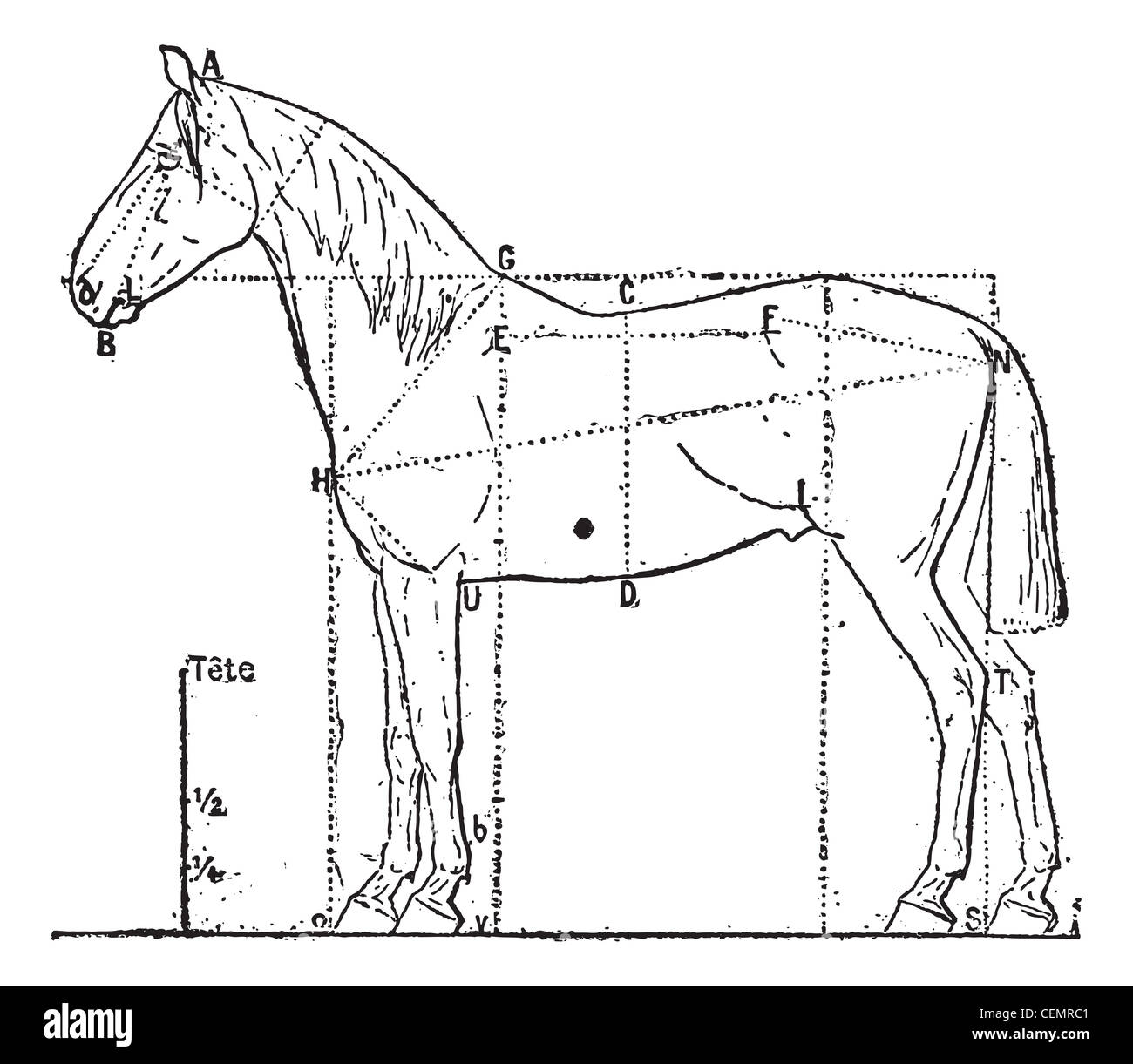 Proportions of the horse, vintage engraved illustration. Dictionary of words and things - Larive and Fleury - 1895. Stock Photohttps://www.alamy.com/image-license-details/?v=1https://www.alamy.com/stock-photo-proportions-of-the-horse-vintage-engraved-illustration-dictionary-43483329.html
RFCEMRC1–Proportions of the horse, vintage engraved illustration. Dictionary of words and things - Larive and Fleury - 1895.
 Human male skeleton from the front, with horse skeleton by George Stubbs. Copperplate engraving by Edward Mitchell after an anatomical illustration by Bernhard Siegfried Albinus from John Barclay's A Series of Engravings of the Human Skeleton, MacLachlan and Stewart, Edinburgh, 1824. Stock Photohttps://www.alamy.com/image-license-details/?v=1https://www.alamy.com/human-male-skeleton-from-the-front-with-horse-skeleton-by-george-stubbs-copperplate-engraving-by-edward-mitchell-after-an-anatomical-illustration-by-bernhard-siegfried-albinus-from-john-barclays-a-series-of-engravings-of-the-human-skeleton-maclachlan-and-stewart-edinburgh-1824-image331996441.html
RM2A83MRN–Human male skeleton from the front, with horse skeleton by George Stubbs. Copperplate engraving by Edward Mitchell after an anatomical illustration by Bernhard Siegfried Albinus from John Barclay's A Series of Engravings of the Human Skeleton, MacLachlan and Stewart, Edinburgh, 1824.
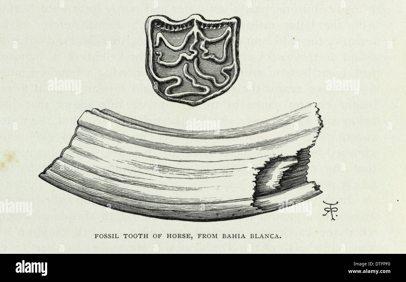 'Fossil tooth of horse, from Bahia Blanca' Stock Photohttps://www.alamy.com/image-license-details/?v=1https://www.alamy.com/fossil-tooth-of-horse-from-bahia-blanca-image66729796.html
RMDTFPF0–'Fossil tooth of horse, from Bahia Blanca'
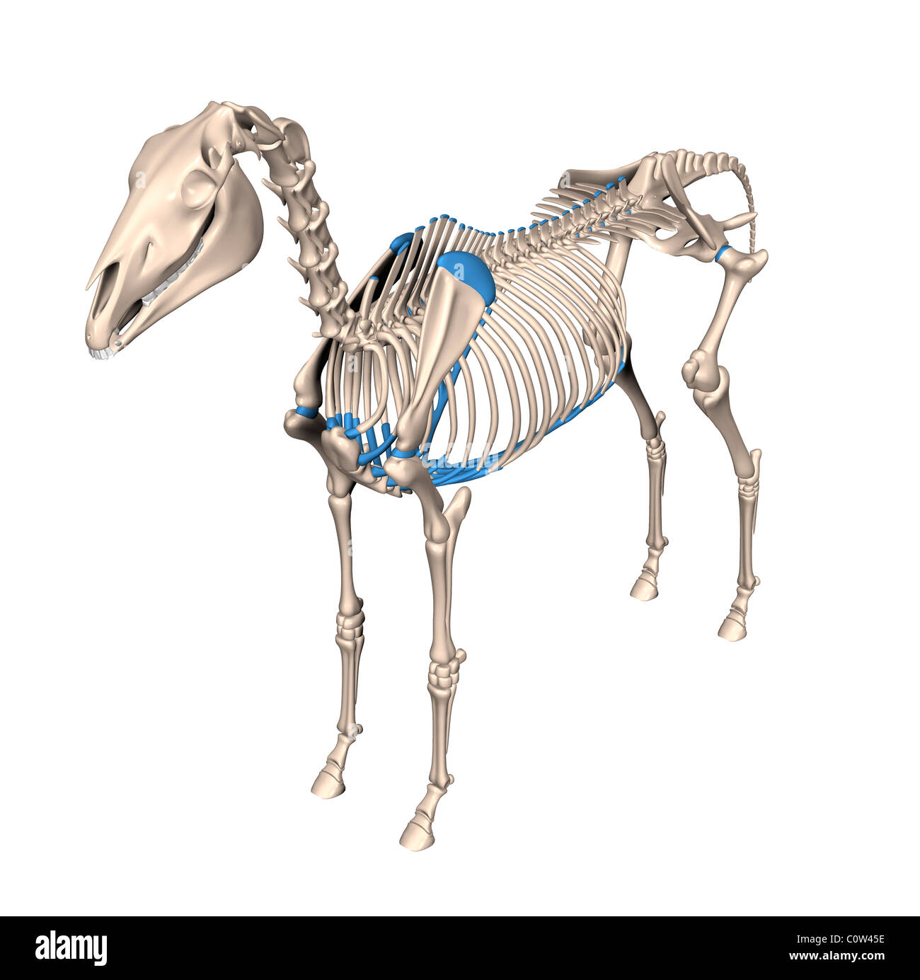 horse anatomy skeleton Stock Photohttps://www.alamy.com/image-license-details/?v=1https://www.alamy.com/stock-photo-horse-anatomy-skeleton-34972826.html
RMC0W45E–horse anatomy skeleton
 Veterinary medicine, Anatomy, Skull bone of a horse, Historical, digitally restored reproduction from a 19th century original / Tiermedizin, Anatomie, Schädelknochen eines Pferdes, Historisch, digital restaurierte Reproduktion von einer Vorlage aus dem 19. Jahrhundert Stock Photohttps://www.alamy.com/image-license-details/?v=1https://www.alamy.com/veterinary-medicine-anatomy-skull-bone-of-a-horse-historical-digitally-restored-reproduction-from-a-19th-century-original-tiermedizin-anatomie-schdelknochen-eines-pferdes-historisch-digital-restaurierte-reproduktion-von-einer-vorlage-aus-dem-19-jahrhundert-image557120147.html
RF2RAB0C3–Veterinary medicine, Anatomy, Skull bone of a horse, Historical, digitally restored reproduction from a 19th century original / Tiermedizin, Anatomie, Schädelknochen eines Pferdes, Historisch, digital restaurierte Reproduktion von einer Vorlage aus dem 19. Jahrhundert
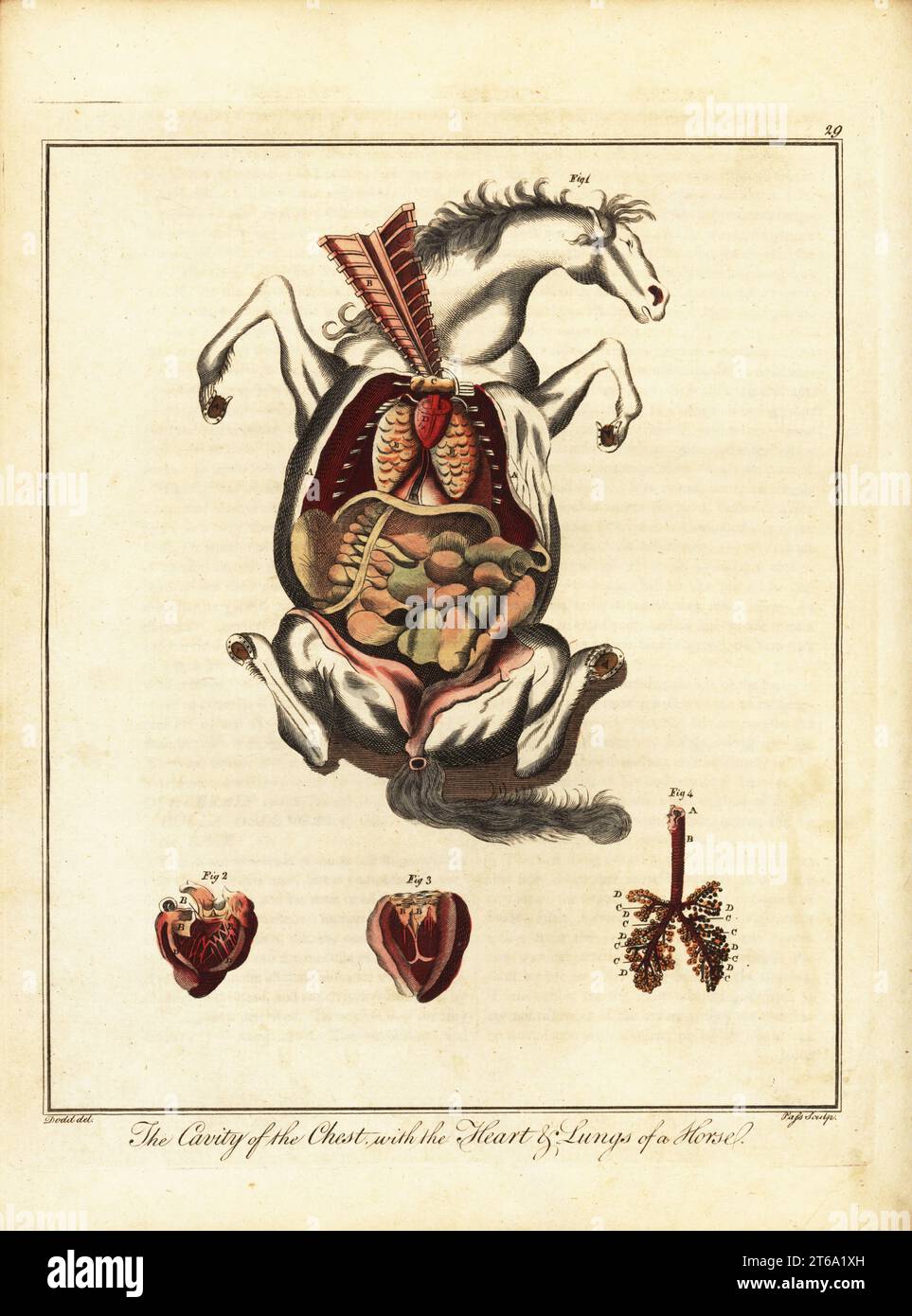 Anatomy of a horse. Chest cavity exposed 1, heart or vena cava 2, left ventricle 3, and lungs 4.. Handcoloured copperplate engraving by J. Pass after an illustration by Daniel Dodd from William Augustus Osbaldistons The British Sportsman, or Nobleman, Gentleman and Farmers Dictionary of Recreation and Amusement, J. Stead, London, 1792. Stock Photohttps://www.alamy.com/image-license-details/?v=1https://www.alamy.com/anatomy-of-a-horse-chest-cavity-exposed-1-heart-or-vena-cava-2-left-ventricle-3-and-lungs-4-handcoloured-copperplate-engraving-by-j-pass-after-an-illustration-by-daniel-dodd-from-william-augustus-osbaldistons-the-british-sportsman-or-nobleman-gentleman-and-farmers-dictionary-of-recreation-and-amusement-j-stead-london-1792-image571851129.html
RM2T6A1XH–Anatomy of a horse. Chest cavity exposed 1, heart or vena cava 2, left ventricle 3, and lungs 4.. Handcoloured copperplate engraving by J. Pass after an illustration by Daniel Dodd from William Augustus Osbaldistons The British Sportsman, or Nobleman, Gentleman and Farmers Dictionary of Recreation and Amusement, J. Stead, London, 1792.
 Horse anatomy, illustration Stock Photohttps://www.alamy.com/image-license-details/?v=1https://www.alamy.com/horse-anatomy-illustration-image267282210.html
RFWERN56–Horse anatomy, illustration
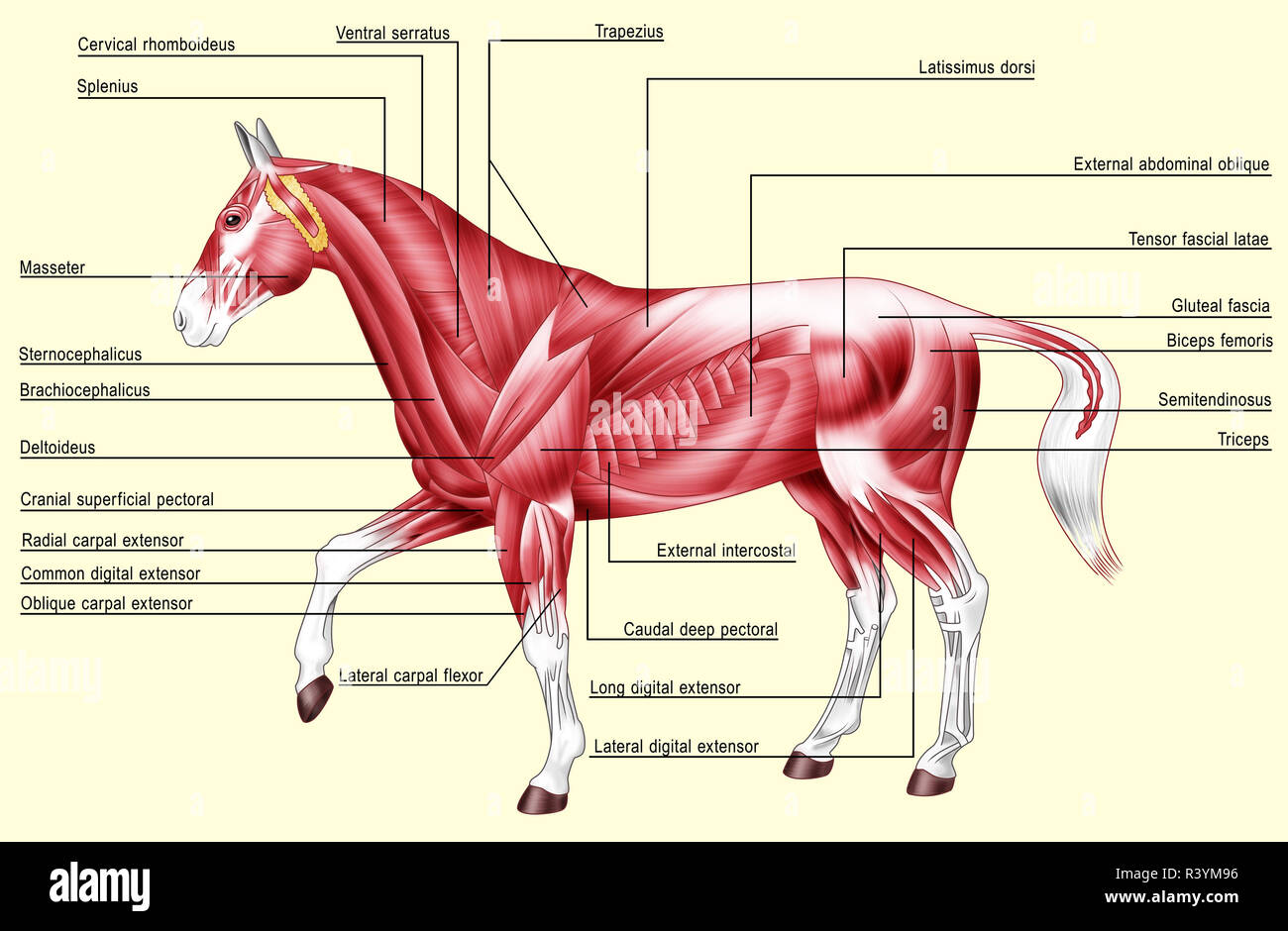 Horse anatomy - Muscles Stock Photohttps://www.alamy.com/image-license-details/?v=1https://www.alamy.com/horse-anatomy-muscles-image226187394.html
RFR3YM96–Horse anatomy - Muscles
 Equine anatomical Illustration from 'Anatomia del cavallo, infermità, et suoi rimedii'. Anatomy of a horse); (1618). by Carlo Ruini, (1530-1598) Stock Photohttps://www.alamy.com/image-license-details/?v=1https://www.alamy.com/stock-photo-equine-anatomical-illustration-from-anatomia-del-cavallo-infermit-84968781.html
RMEX6JF9–Equine anatomical Illustration from 'Anatomia del cavallo, infermità, et suoi rimedii'. Anatomy of a horse); (1618). by Carlo Ruini, (1530-1598)
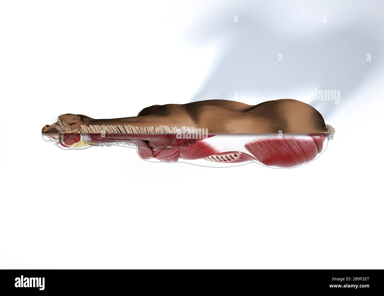 Muscular anatomy of a horse with cutaway effect, top view. Stock Photohttps://www.alamy.com/image-license-details/?v=1https://www.alamy.com/muscular-anatomy-of-a-horse-with-cutaway-effect-top-view-image350070528.html
RF2B9F2ET–Muscular anatomy of a horse with cutaway effect, top view.
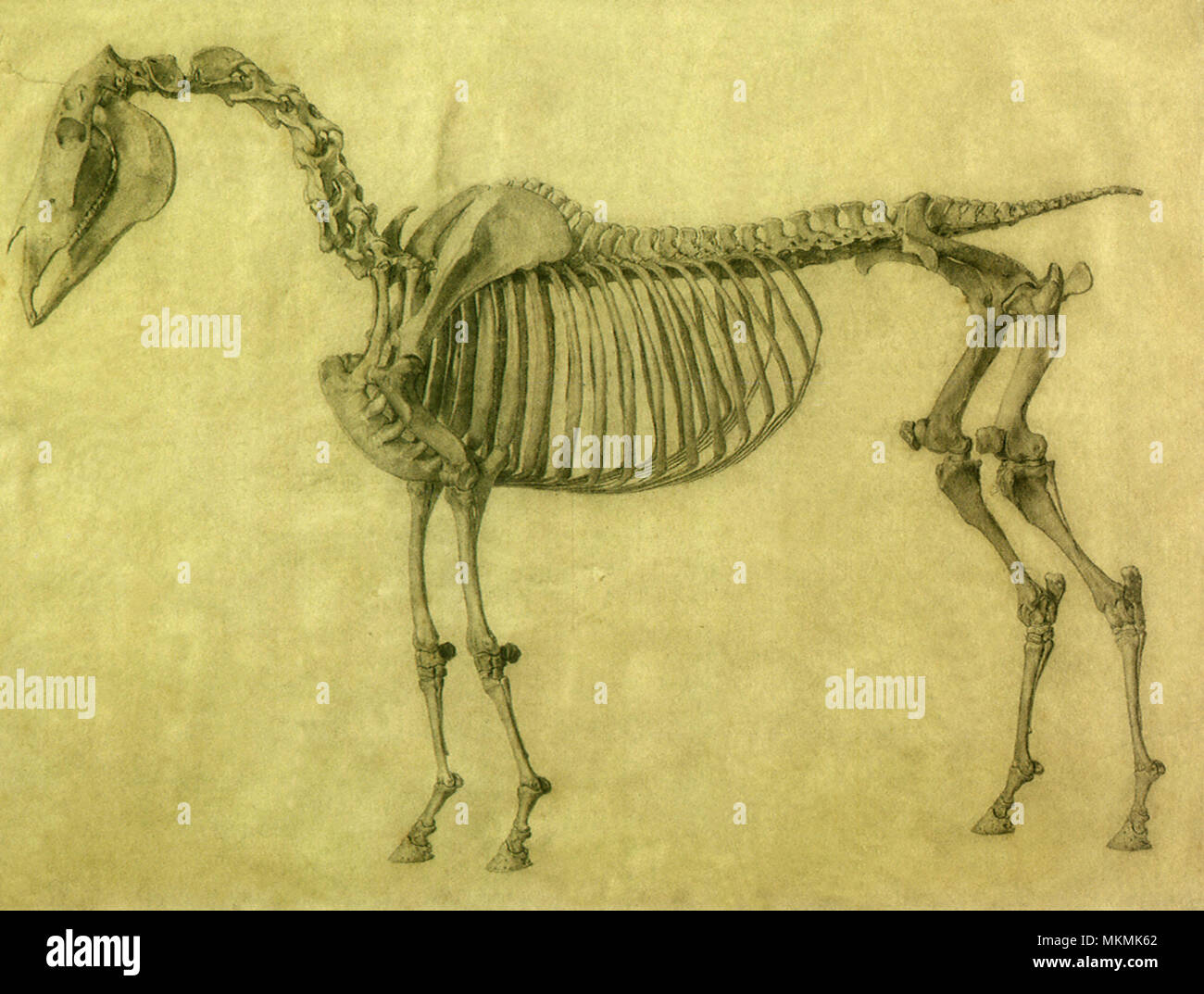 Skeleton of a Horse Stock Photohttps://www.alamy.com/image-license-details/?v=1https://www.alamy.com/skeleton-of-a-horse-image184236250.html
RMMKMK62–Skeleton of a Horse
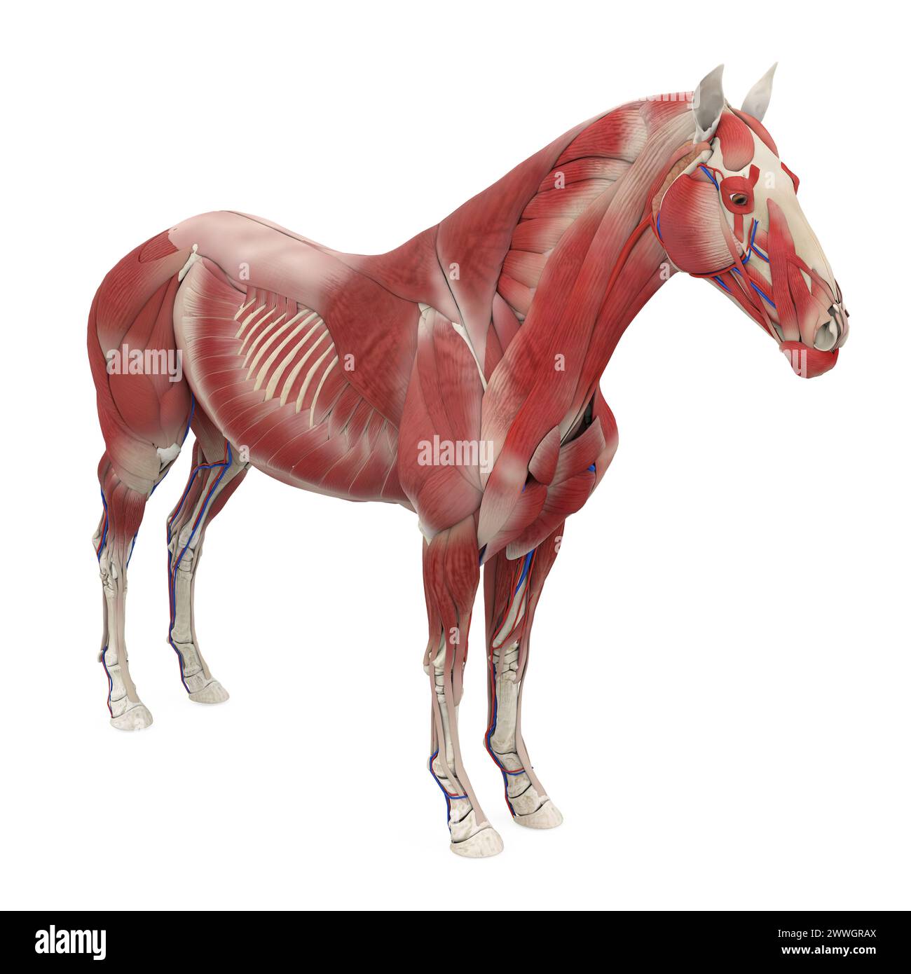 Horse Anatomy Muscular System Stock Photohttps://www.alamy.com/image-license-details/?v=1https://www.alamy.com/horse-anatomy-muscular-system-image600888482.html
RF2WWGRAX–Horse Anatomy Muscular System
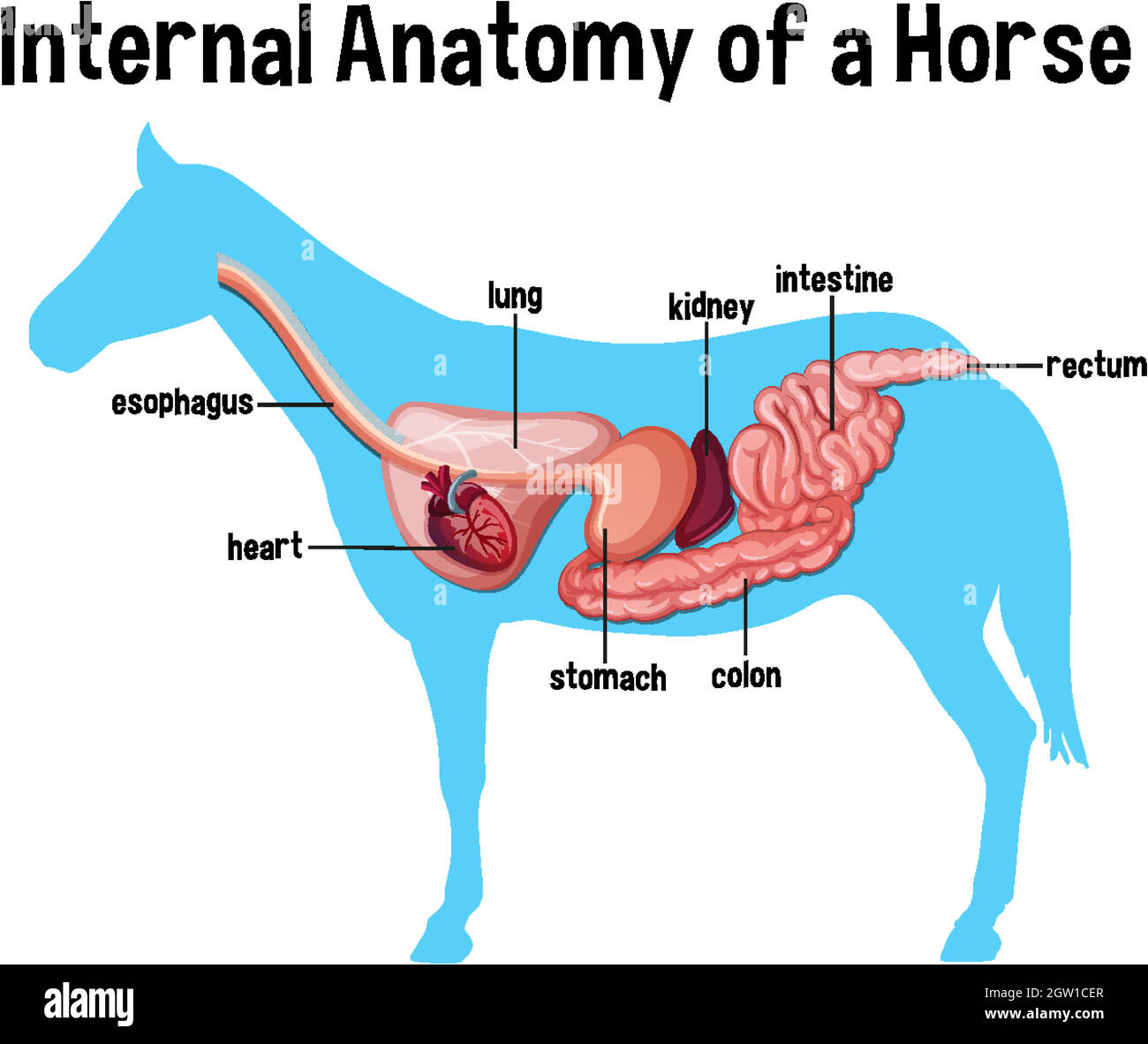 Internal Anatomy of a Horse with label Stock Vectorhttps://www.alamy.com/image-license-details/?v=1https://www.alamy.com/internal-anatomy-of-a-horse-with-label-image445657375.html
RF2GW1CER–Internal Anatomy of a Horse with label
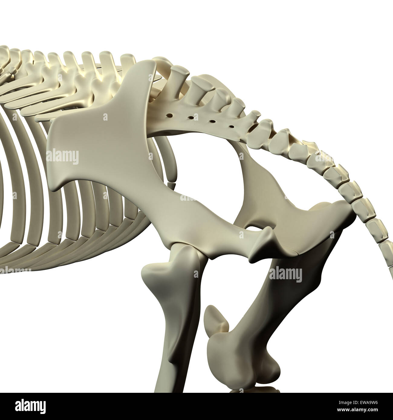 Horse Pelvis - Horse Equus Anatomy - isolated on white Stock Photohttps://www.alamy.com/image-license-details/?v=1https://www.alamy.com/stock-photo-horse-pelvis-horse-equus-anatomy-isolated-on-white-84435154.html
RFEWA9W6–Horse Pelvis - Horse Equus Anatomy - isolated on white
 Horse illustration. 19th-century plate from a book on the practical treatise about the humane management of the horse. Stock Photohttps://www.alamy.com/image-license-details/?v=1https://www.alamy.com/horse-illustration-19th-century-plate-from-a-book-on-the-practical-treatise-about-the-humane-management-of-the-horse-image626109537.html
RF2YAHN2W–Horse illustration. 19th-century plate from a book on the practical treatise about the humane management of the horse.
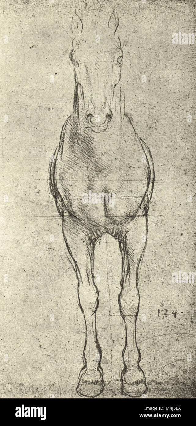 A Horse seen from the front, equine Anatomical drawing, drawn by Leonardo Da Vinci, 1452-1519 Stock Photohttps://www.alamy.com/image-license-details/?v=1https://www.alamy.com/stock-photo-a-horse-seen-from-the-front-equine-anatomical-drawing-drawn-by-leonardo-174961778.html
RMM4J5EX–A Horse seen from the front, equine Anatomical drawing, drawn by Leonardo Da Vinci, 1452-1519
 Digital illustration - a bucking Palomino Horse on a dark background. Palomino implies a gold coat and ight cream or white mane and tail. Stock Photohttps://www.alamy.com/image-license-details/?v=1https://www.alamy.com/digital-illustration-a-bucking-palomino-horse-on-a-dark-background-palomino-implies-a-gold-coat-and-ight-cream-or-white-mane-and-tail-image418215756.html
RF2F8BAD0–Digital illustration - a bucking Palomino Horse on a dark background. Palomino implies a gold coat and ight cream or white mane and tail.
 Leonardo Da Vinci, Study of the front legs of a horse, anatomical drawing, circa 1490 Stock Photohttps://www.alamy.com/image-license-details/?v=1https://www.alamy.com/leonardo-da-vinci-study-of-the-front-legs-of-a-horse-anatomical-drawing-circa-1490-image257391549.html
RMTXN5F9–Leonardo Da Vinci, Study of the front legs of a horse, anatomical drawing, circa 1490
 posterior direction Left illustration shows a portion of the muscles of the body, neck, and limbs; the right cut shows the bones which enter into the composition of these parts. from The anatomy and physiology of the horse: with anatomical and questional illustrations. Containing, also, a series of examinations on equine anatomy and physiology, with instructions in reference to dissection and the mode of making anatomical preparations. To which is added, glossary of veterinary technicalities, toxicological chart, and dictionary of veterinary science by Dadd, George H., Publication date 1857 Stock Photohttps://www.alamy.com/image-license-details/?v=1https://www.alamy.com/posterior-direction-left-illustration-shows-a-portion-of-the-muscles-of-the-body-neck-and-limbs-the-right-cut-shows-the-bones-which-enter-into-the-composition-of-these-parts-from-the-anatomy-and-physiology-of-the-horse-with-anatomical-and-questional-illustrations-containing-also-a-series-of-examinations-on-equine-anatomy-and-physiology-with-instructions-in-reference-to-dissection-and-the-mode-of-making-anatomical-preparations-to-which-is-added-glossary-of-veterinary-technicalities-toxicological-chart-and-dictionary-of-veterinary-science-by-dadd-george-h-publication-date-1857-image635528671.html
RM2YWXR8F–posterior direction Left illustration shows a portion of the muscles of the body, neck, and limbs; the right cut shows the bones which enter into the composition of these parts. from The anatomy and physiology of the horse: with anatomical and questional illustrations. Containing, also, a series of examinations on equine anatomy and physiology, with instructions in reference to dissection and the mode of making anatomical preparations. To which is added, glossary of veterinary technicalities, toxicological chart, and dictionary of veterinary science by Dadd, George H., Publication date 1857
 An illustration by Muhammad ibn Yaqub al-Khuttuli (9th century, Iraq or Syria) identifying various parts of the anatomy of a horse. Stock Photohttps://www.alamy.com/image-license-details/?v=1https://www.alamy.com/an-illustration-by-muhammad-ibn-yaqub-al-khuttuli-9th-century-iraq-or-syria-identifying-various-parts-of-the-anatomy-of-a-horse-image344277553.html
RM2B035EW–An illustration by Muhammad ibn Yaqub al-Khuttuli (9th century, Iraq or Syria) identifying various parts of the anatomy of a horse.
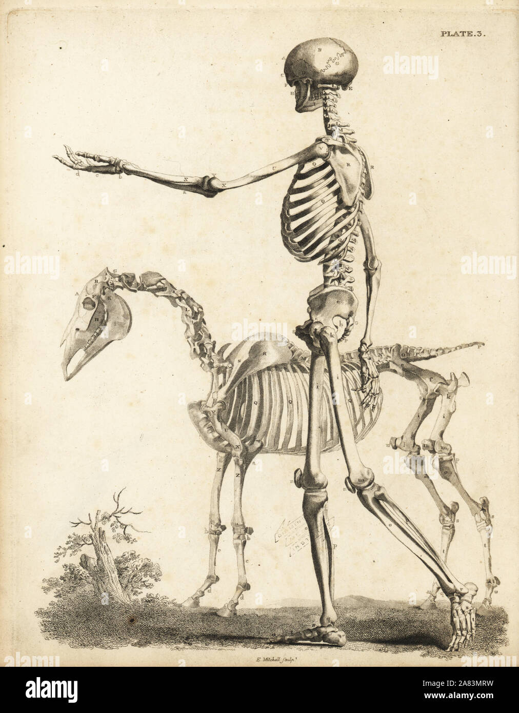 Side view of human skeleton and horse skeleton. Copperplate engraving by Edward Mitchell after anatomical illustrations by Bernhard Siegfried Albinus and George Stubbs from John Barclay's A Series of Engravings of the Human Skeleton, MacLachlan and Stewart, Edinburgh, 1824. Stock Photohttps://www.alamy.com/image-license-details/?v=1https://www.alamy.com/side-view-of-human-skeleton-and-horse-skeleton-copperplate-engraving-by-edward-mitchell-after-anatomical-illustrations-by-bernhard-siegfried-albinus-and-george-stubbs-from-john-barclays-a-series-of-engravings-of-the-human-skeleton-maclachlan-and-stewart-edinburgh-1824-image331996445.html
RM2A83MRW–Side view of human skeleton and horse skeleton. Copperplate engraving by Edward Mitchell after anatomical illustrations by Bernhard Siegfried Albinus and George Stubbs from John Barclay's A Series of Engravings of the Human Skeleton, MacLachlan and Stewart, Edinburgh, 1824.
 Color print of a horse's coeliac axis, artery, or trunk, a major visceral artery located in the abdominal cavity that is responsible for supplying the foregut, published in Sir John McFadyean's 'The Anatomy of the Horse: a dissection guide', 1922. Courtesy Internet Archive. () Stock Photohttps://www.alamy.com/image-license-details/?v=1https://www.alamy.com/color-print-of-a-horses-coeliac-axis-artery-or-trunk-a-major-visceral-artery-located-in-the-abdominal-cavity-that-is-responsible-for-supplying-the-foregut-published-in-sir-john-mcfadyeans-the-anatomy-of-the-horse-a-dissection-guide-1922-courtesy-internet-archive-image215432546.html
RMPEDPBE–Color print of a horse's coeliac axis, artery, or trunk, a major visceral artery located in the abdominal cavity that is responsible for supplying the foregut, published in Sir John McFadyean's 'The Anatomy of the Horse: a dissection guide', 1922. Courtesy Internet Archive. ()
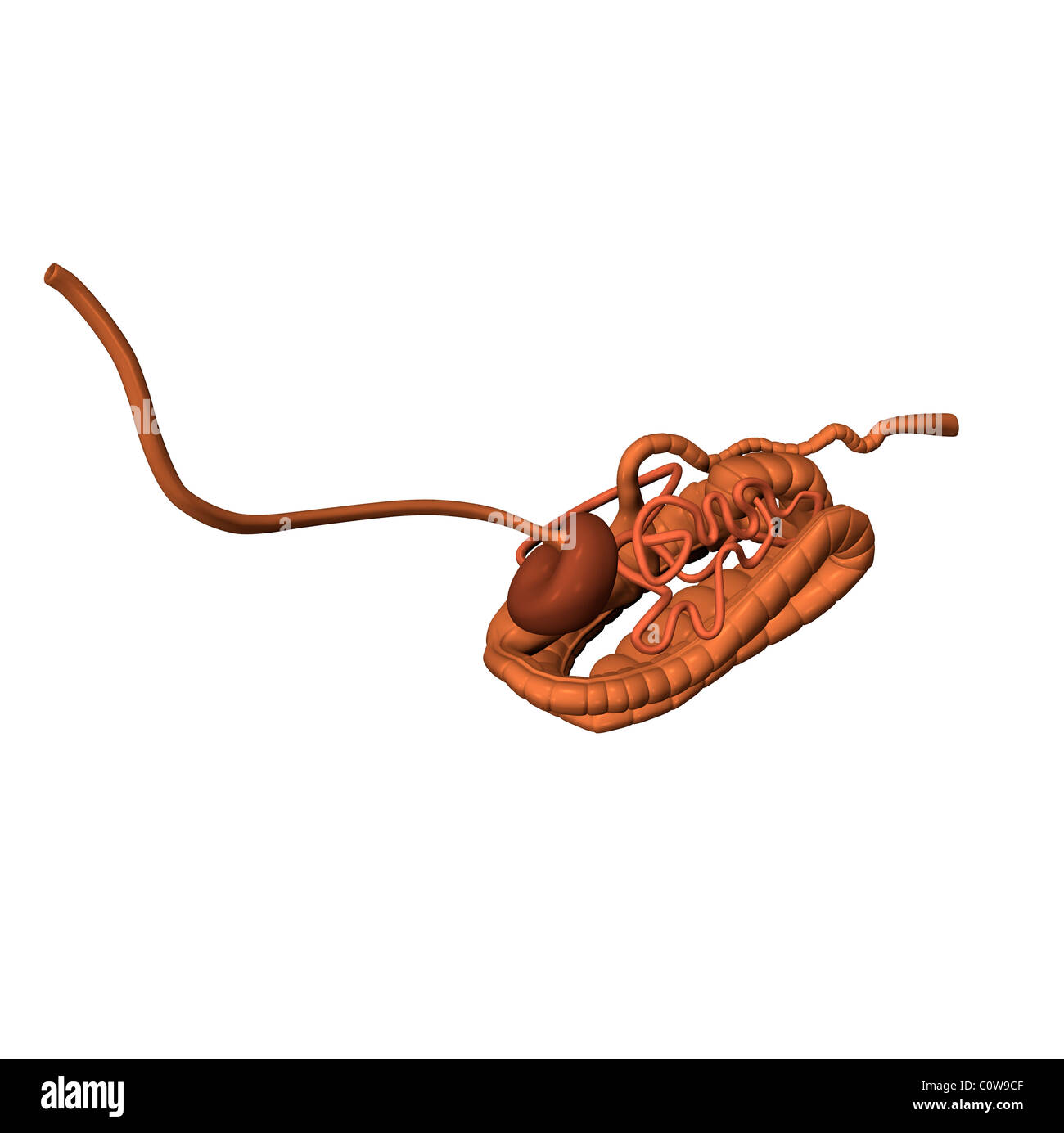 horse anatomy digestion Stock Photohttps://www.alamy.com/image-license-details/?v=1https://www.alamy.com/stock-photo-horse-anatomy-digestion-34976943.html
RMC0W9CF–horse anatomy digestion
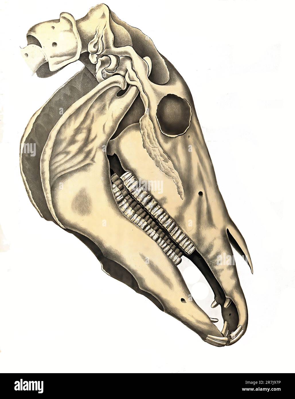 Veterinary medicine, Anatomy, Skull bone of a horse, Historical, digitally restored reproduction from a 19th century original / Tiermedizin, Anatomie, Schädelknochen eines Pferdes, Historisch, digital restaurierte Reproduktion von einer Vorlage aus dem 19. Jahrhundert Stock Photohttps://www.alamy.com/image-license-details/?v=1https://www.alamy.com/veterinary-medicine-anatomy-skull-bone-of-a-horse-historical-digitally-restored-reproduction-from-a-19th-century-original-tiermedizin-anatomie-schdelknochen-eines-pferdes-historisch-digital-restaurierte-reproduktion-von-einer-vorlage-aus-dem-19-jahrhundert-image555436778.html
RF2R7J97P–Veterinary medicine, Anatomy, Skull bone of a horse, Historical, digitally restored reproduction from a 19th century original / Tiermedizin, Anatomie, Schädelknochen eines Pferdes, Historisch, digital restaurierte Reproduktion von einer Vorlage aus dem 19. Jahrhundert
 Anatomy of a horse. The head, brains, stomach, pancreas, and generative parts. Stomach 1,2, liver 3, spleen 4, pancreas 5, kidneys 6, womb 7, ovaries 8, brain 9, nervous system 10, eye 11, ear 12. Handcoloured copperplate engraving by J. Pass after an illustration by Daniel Dodd/J. Stead from William Augustus Osbaldistons The British Sportsman, or Nobleman, Gentleman and Farmers Dictionary of Recreation and Amusement, J. Stead, London, 1792. Stock Photohttps://www.alamy.com/image-license-details/?v=1https://www.alamy.com/anatomy-of-a-horse-the-head-brains-stomach-pancreas-and-generative-parts-stomach-12-liver-3-spleen-4-pancreas-5-kidneys-6-womb-7-ovaries-8-brain-9-nervous-system-10-eye-11-ear-12-handcoloured-copperplate-engraving-by-j-pass-after-an-illustration-by-daniel-doddj-stead-from-william-augustus-osbaldistons-the-british-sportsman-or-nobleman-gentleman-and-farmers-dictionary-of-recreation-and-amusement-j-stead-london-1792-image571848706.html
RM2T69XT2–Anatomy of a horse. The head, brains, stomach, pancreas, and generative parts. Stomach 1,2, liver 3, spleen 4, pancreas 5, kidneys 6, womb 7, ovaries 8, brain 9, nervous system 10, eye 11, ear 12. Handcoloured copperplate engraving by J. Pass after an illustration by Daniel Dodd/J. Stead from William Augustus Osbaldistons The British Sportsman, or Nobleman, Gentleman and Farmers Dictionary of Recreation and Amusement, J. Stead, London, 1792.
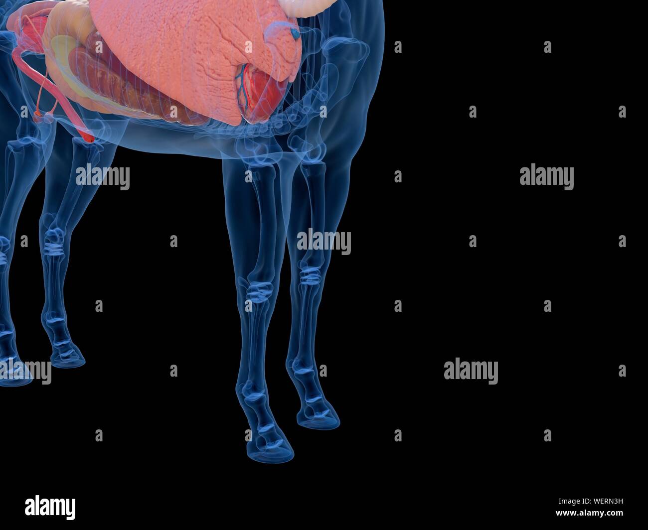 Horse anatomy, illustration Stock Photohttps://www.alamy.com/image-license-details/?v=1https://www.alamy.com/horse-anatomy-illustration-image267282165.html
RFWERN3H–Horse anatomy, illustration
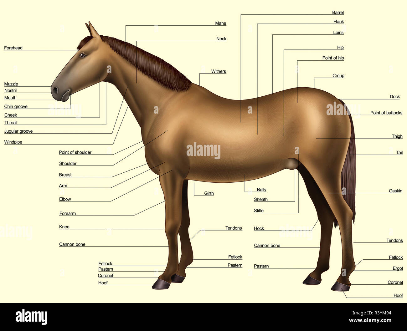 Horse anatomy - Body parts Stock Photohttps://www.alamy.com/image-license-details/?v=1https://www.alamy.com/horse-anatomy-body-parts-image226187392.html
RFR3YM94–Horse anatomy - Body parts
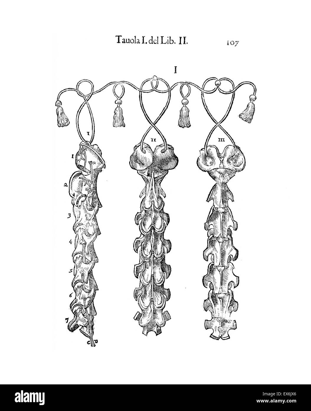 Equine anatomical Illustration from 'Anatomia del cavallo, infermità, et suoi rimedii'. Anatomy of a horse); (1618). by Carlo Ruini, (1530-1598) Stock Photohttps://www.alamy.com/image-license-details/?v=1https://www.alamy.com/stock-photo-equine-anatomical-illustration-from-anatomia-del-cavallo-infermit-84969086.html
RMEX6JX6–Equine anatomical Illustration from 'Anatomia del cavallo, infermità, et suoi rimedii'. Anatomy of a horse); (1618). by Carlo Ruini, (1530-1598)
 Muscular anatomy of a horse with ghost effect. Stock Photohttps://www.alamy.com/image-license-details/?v=1https://www.alamy.com/muscular-anatomy-of-a-horse-with-ghost-effect-image350070117.html
RF2B9F205–Muscular anatomy of a horse with ghost effect.
 Horse Muscles and Bones Stock Photohttps://www.alamy.com/image-license-details/?v=1https://www.alamy.com/horse-muscles-and-bones-image184236248.html
RMMKMK60–Horse Muscles and Bones
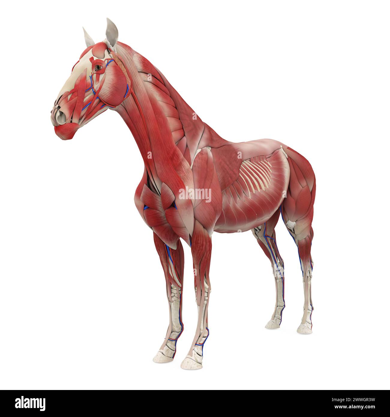 Horse Anatomy Muscular System Stock Photohttps://www.alamy.com/image-license-details/?v=1https://www.alamy.com/horse-anatomy-muscular-system-image600888285.html
RF2WWGR3W–Horse Anatomy Muscular System
 Internal Anatomy of a Horse isolated on white background Stock Vectorhttps://www.alamy.com/image-license-details/?v=1https://www.alamy.com/internal-anatomy-of-a-horse-isolated-on-white-background-image446385401.html
RF2GX6H3N–Internal Anatomy of a Horse isolated on white background
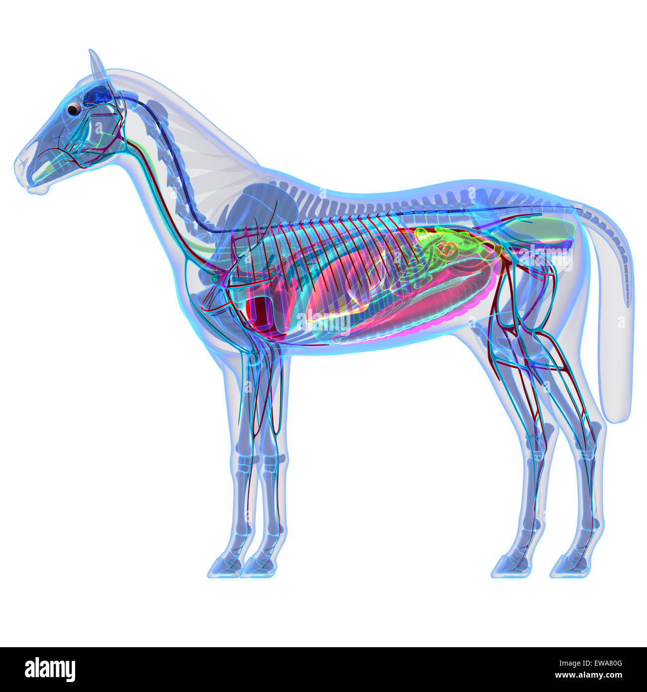 Horse Anatomy - Internal Anatomy of a Horse Stock Photohttps://www.alamy.com/image-license-details/?v=1https://www.alamy.com/stock-photo-horse-anatomy-internal-anatomy-of-a-horse-84433680.html
RFEWA80G–Horse Anatomy - Internal Anatomy of a Horse
 Horse illustration. 19th-century plate from a book on the practical treatise about the humane management of the horse. Stock Photohttps://www.alamy.com/image-license-details/?v=1https://www.alamy.com/horse-illustration-19th-century-plate-from-a-book-on-the-practical-treatise-about-the-humane-management-of-the-horse-image626109547.html
RF2YAHN37–Horse illustration. 19th-century plate from a book on the practical treatise about the humane management of the horse.
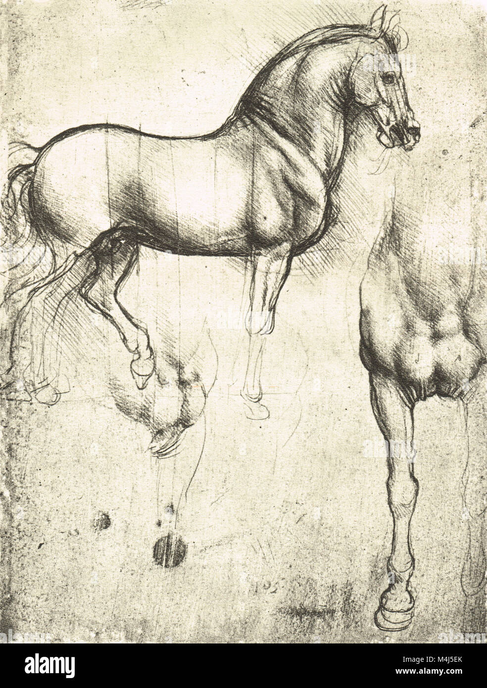 Horse in profile, to the right and its fore-legs, equine Anatomical drawing, drawn by Leonardo Da Vinci, 1452-1519 Stock Photohttps://www.alamy.com/image-license-details/?v=1https://www.alamy.com/stock-photo-horse-in-profile-to-the-right-and-its-fore-legs-equine-anatomical-174961771.html
RMM4J5EK–Horse in profile, to the right and its fore-legs, equine Anatomical drawing, drawn by Leonardo Da Vinci, 1452-1519
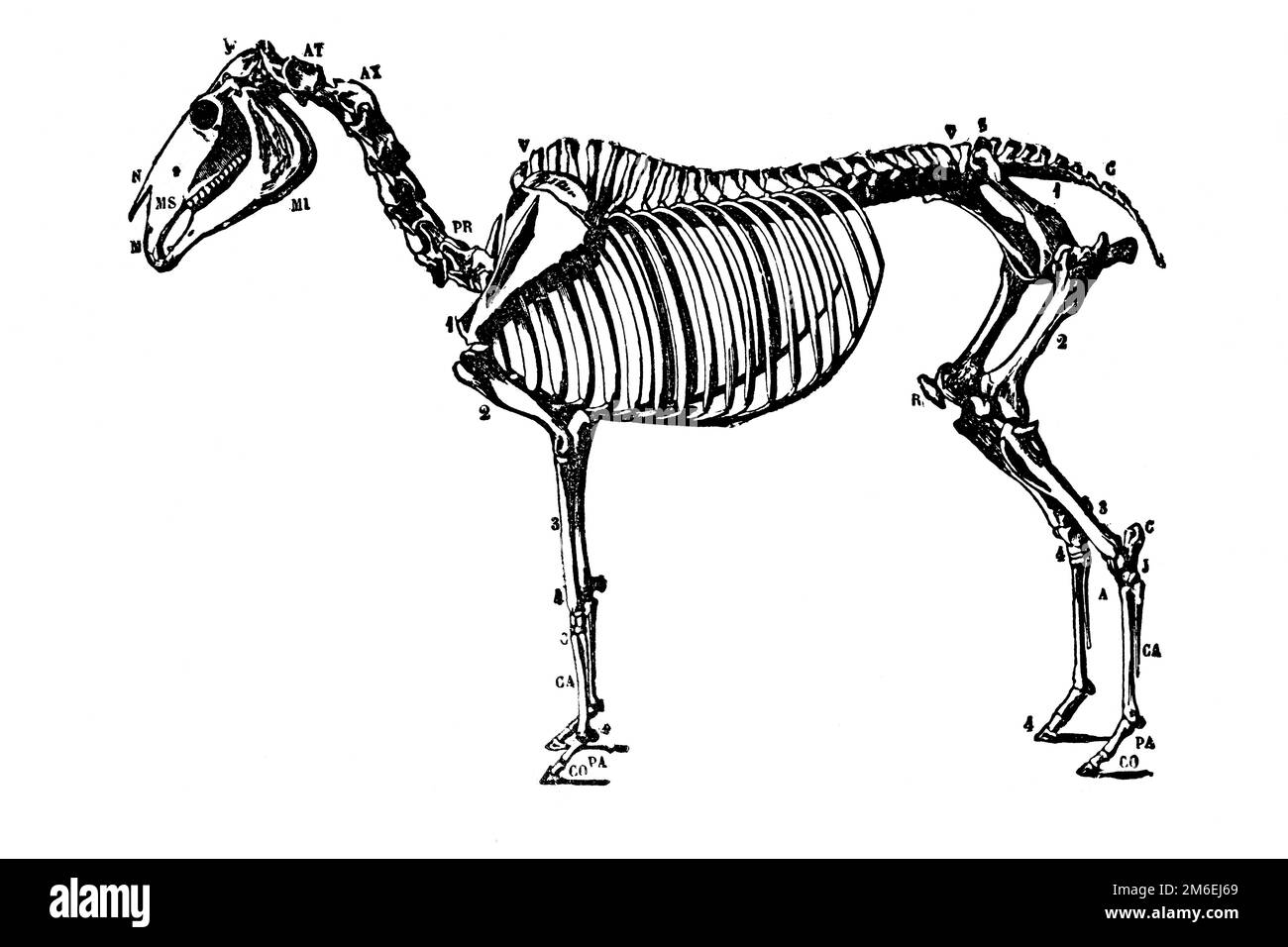 Horse skeleton. Antique illustration from a medical book, 1889. Stock Photohttps://www.alamy.com/image-license-details/?v=1https://www.alamy.com/horse-skeleton-antique-illustration-from-a-medical-book-1889-image503110225.html
RF2M6EJ69–Horse skeleton. Antique illustration from a medical book, 1889.
 Skeleton of a Horse, Stubbs, George Stock Photohttps://www.alamy.com/image-license-details/?v=1https://www.alamy.com/skeleton-of-a-horse-stubbs-george-image215545625.html
RMPEJXJ1–Skeleton of a Horse, Stubbs, George
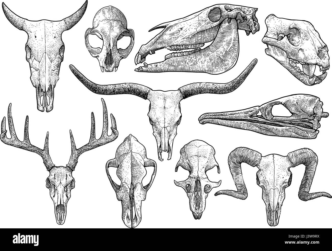 Skull collection illustration, drawing, engraving, ink, line art, vector Stock Vectorhttps://www.alamy.com/image-license-details/?v=1https://www.alamy.com/stock-photo-skull-collection-illustration-drawing-engraving-ink-line-art-vector-140083438.html
RFJ3W9RX–Skull collection illustration, drawing, engraving, ink, line art, vector
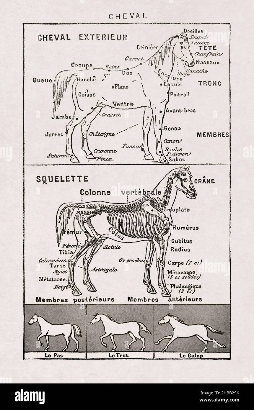 Old illustration about horses printed in the french dictionary 'Dictionnaire complet illustré' by the editor Larousse in 1899. Stock Photohttps://www.alamy.com/image-license-details/?v=1https://www.alamy.com/old-illustration-about-horses-printed-in-the-french-dictionary-dictionnaire-complet-illustre-by-the-editor-larousse-in-1899-image454474095.html
RF2HBB29K–Old illustration about horses printed in the french dictionary 'Dictionnaire complet illustré' by the editor Larousse in 1899.
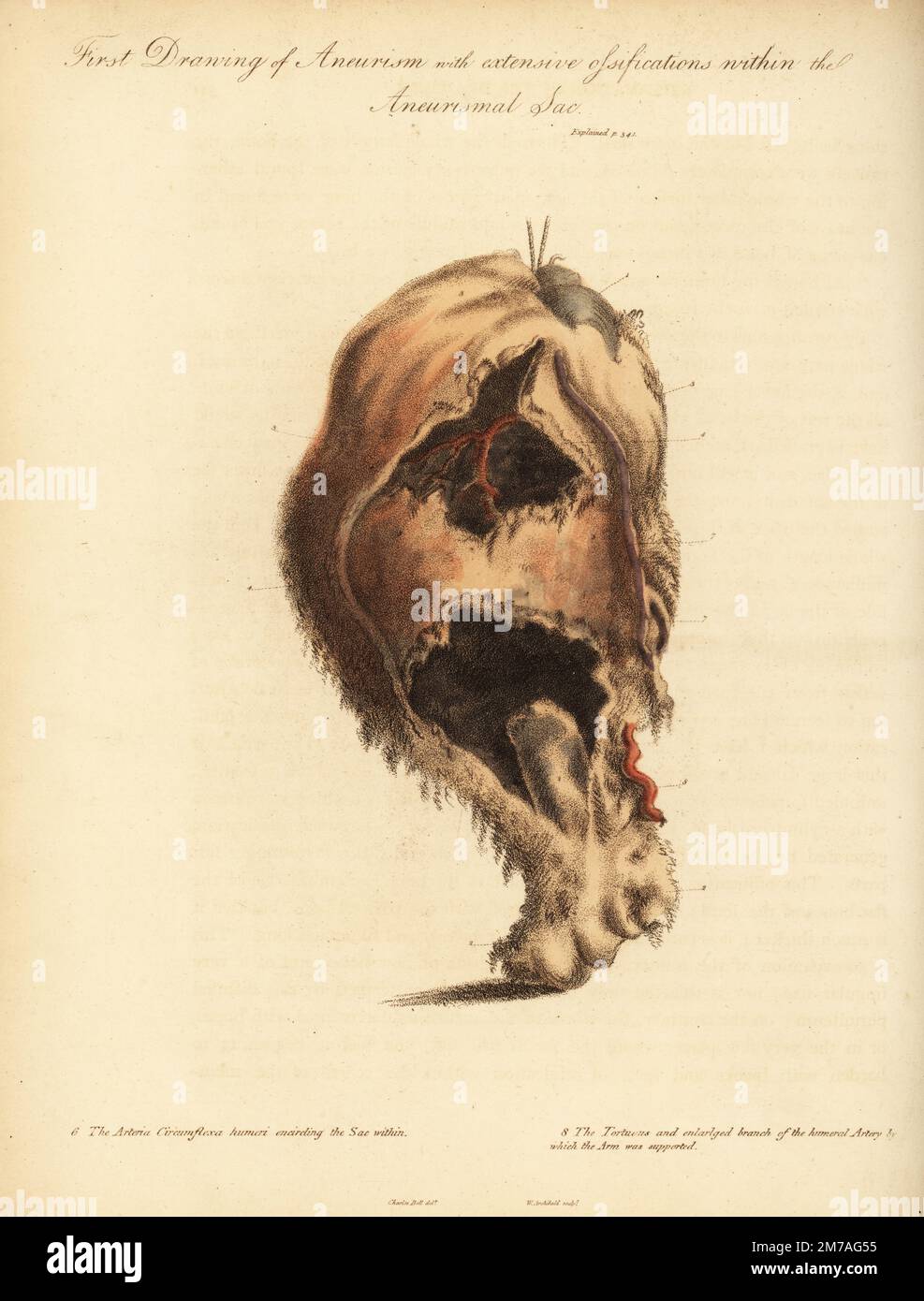 Aneurysmal tumor from the broken arm of a woman knocked down by a horse and cart, 1815. First drawing of aneurism and extensive ossifications with the aneurismal sac. Head of the humerus 1, lower part of the bone 2, arteria circumflexa humeri encircling the sac within 6, and the tortuous and enlarged branch of the humeral artery. Handcoloured copperplate engraving by William Archibald after an illustration by John Bell from his own Principles of Surgery, as they Relate to Wounds, Ulcers and Fistulas, Longman, Hurst, Rees, Orme and Brown, London, 1815. Stock Photohttps://www.alamy.com/image-license-details/?v=1https://www.alamy.com/aneurysmal-tumor-from-the-broken-arm-of-a-woman-knocked-down-by-a-horse-and-cart-1815-first-drawing-of-aneurism-and-extensive-ossifications-with-the-aneurismal-sac-head-of-the-humerus-1-lower-part-of-the-bone-2-arteria-circumflexa-humeri-encircling-the-sac-within-6-and-the-tortuous-and-enlarged-branch-of-the-humeral-artery-handcoloured-copperplate-engraving-by-william-archibald-after-an-illustration-by-john-bell-from-his-own-principles-of-surgery-as-they-relate-to-wounds-ulcers-and-fistulas-longman-hurst-rees-orme-and-brown-london-1815-image503635473.html
RM2M7AG55–Aneurysmal tumor from the broken arm of a woman knocked down by a horse and cart, 1815. First drawing of aneurism and extensive ossifications with the aneurismal sac. Head of the humerus 1, lower part of the bone 2, arteria circumflexa humeri encircling the sac within 6, and the tortuous and enlarged branch of the humeral artery. Handcoloured copperplate engraving by William Archibald after an illustration by John Bell from his own Principles of Surgery, as they Relate to Wounds, Ulcers and Fistulas, Longman, Hurst, Rees, Orme and Brown, London, 1815.
 Four horse pictograms which can be used as a logo, illustration, graphic elements etc. Stock Vectorhttps://www.alamy.com/image-license-details/?v=1https://www.alamy.com/stock-photo-four-horse-pictograms-which-can-be-used-as-a-logo-illustration-graphic-116451537.html
RFGNCR3D–Four horse pictograms which can be used as a logo, illustration, graphic elements etc.
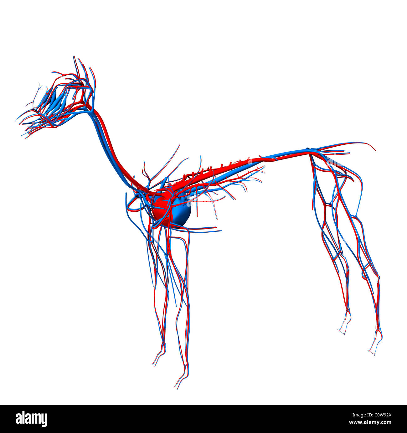 horse anatomy heart circulation Stock Photohttps://www.alamy.com/image-license-details/?v=1https://www.alamy.com/stock-photo-horse-anatomy-heart-circulation-34976674.html
RMC0W92X–horse anatomy heart circulation
 3d rendered medically accurate illustration of the horse anatomy Stock Photohttps://www.alamy.com/image-license-details/?v=1https://www.alamy.com/3d-rendered-medically-accurate-illustration-of-the-horse-anatomy-image257877560.html
RFTYF9CT–3d rendered medically accurate illustration of the horse anatomy
 the skeleton of a horse from The anatomy and physiology of the horse: with anatomical and questional illustrations. Containing, also, a series of examinations on equine anatomy and physiology, with instructions in reference to dissection and the mode of making anatomical preparations. To which is added, glossary of veterinary technicalities, toxicological chart, and dictionary of veterinary science by Dadd, George H., Publication date 1857 Stock Photohttps://www.alamy.com/image-license-details/?v=1https://www.alamy.com/the-skeleton-of-a-horse-from-the-anatomy-and-physiology-of-the-horse-with-anatomical-and-questional-illustrations-containing-also-a-series-of-examinations-on-equine-anatomy-and-physiology-with-instructions-in-reference-to-dissection-and-the-mode-of-making-anatomical-preparations-to-which-is-added-glossary-of-veterinary-technicalities-toxicological-chart-and-dictionary-of-veterinary-science-by-dadd-george-h-publication-date-1857-image635528721.html
RM2YWXRA9–the skeleton of a horse from The anatomy and physiology of the horse: with anatomical and questional illustrations. Containing, also, a series of examinations on equine anatomy and physiology, with instructions in reference to dissection and the mode of making anatomical preparations. To which is added, glossary of veterinary technicalities, toxicological chart, and dictionary of veterinary science by Dadd, George H., Publication date 1857
 Horse anatomy, illustration Stock Photohttps://www.alamy.com/image-license-details/?v=1https://www.alamy.com/horse-anatomy-illustration-image267282211.html
RFWERN57–Horse anatomy, illustration
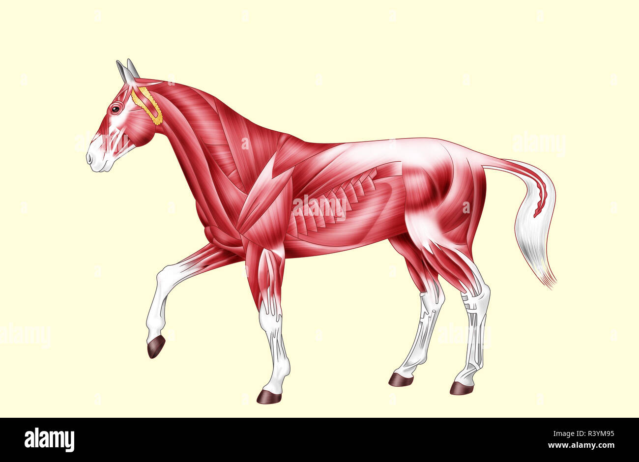 Horse anatomy - Muscles - No text Stock Photohttps://www.alamy.com/image-license-details/?v=1https://www.alamy.com/horse-anatomy-muscles-no-text-image226187393.html
RFR3YM95–Horse anatomy - Muscles - No text
 Equine anatomical Illustration from 'Anatomia del cavallo, infermità, et suoi rimedii'. Anatomy of a horse); (1618). by Carlo Ruini, (1530-1598) Stock Photohttps://www.alamy.com/image-license-details/?v=1https://www.alamy.com/stock-photo-equine-anatomical-illustration-from-anatomia-del-cavallo-infermit-84969085.html
RMEX6JX5–Equine anatomical Illustration from 'Anatomia del cavallo, infermità, et suoi rimedii'. Anatomy of a horse); (1618). by Carlo Ruini, (1530-1598)
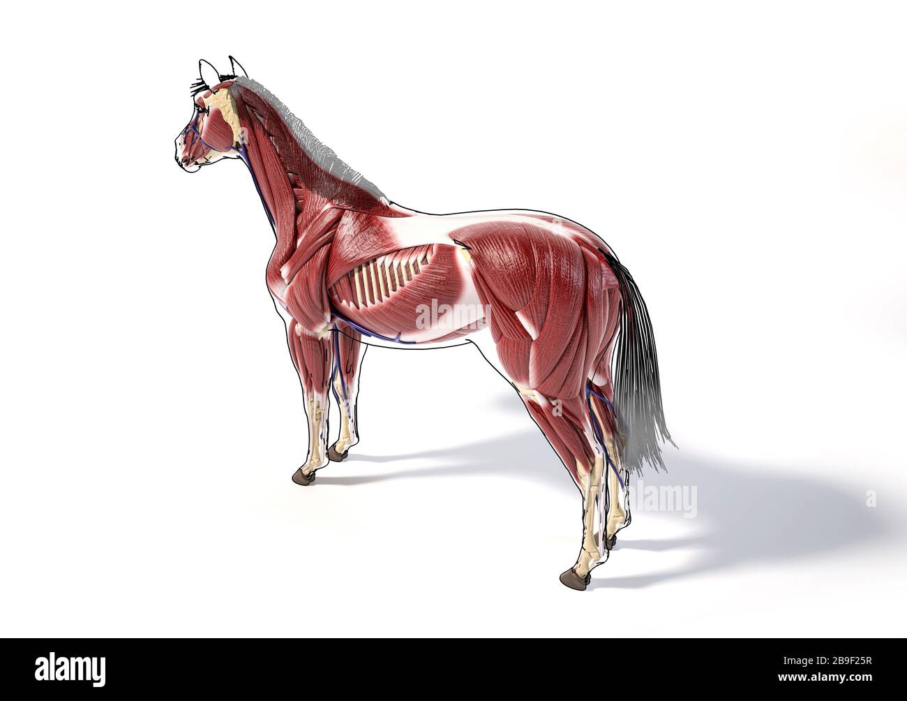 Muscular anatomy of a horse with with black outline. Stock Photohttps://www.alamy.com/image-license-details/?v=1https://www.alamy.com/muscular-anatomy-of-a-horse-with-with-black-outline-image350070275.html
RF2B9F25R–Muscular anatomy of a horse with with black outline.
 Horse Musculature, Side Stock Photohttps://www.alamy.com/image-license-details/?v=1https://www.alamy.com/horse-musculature-side-image184236249.html
RMMKMK61–Horse Musculature, Side
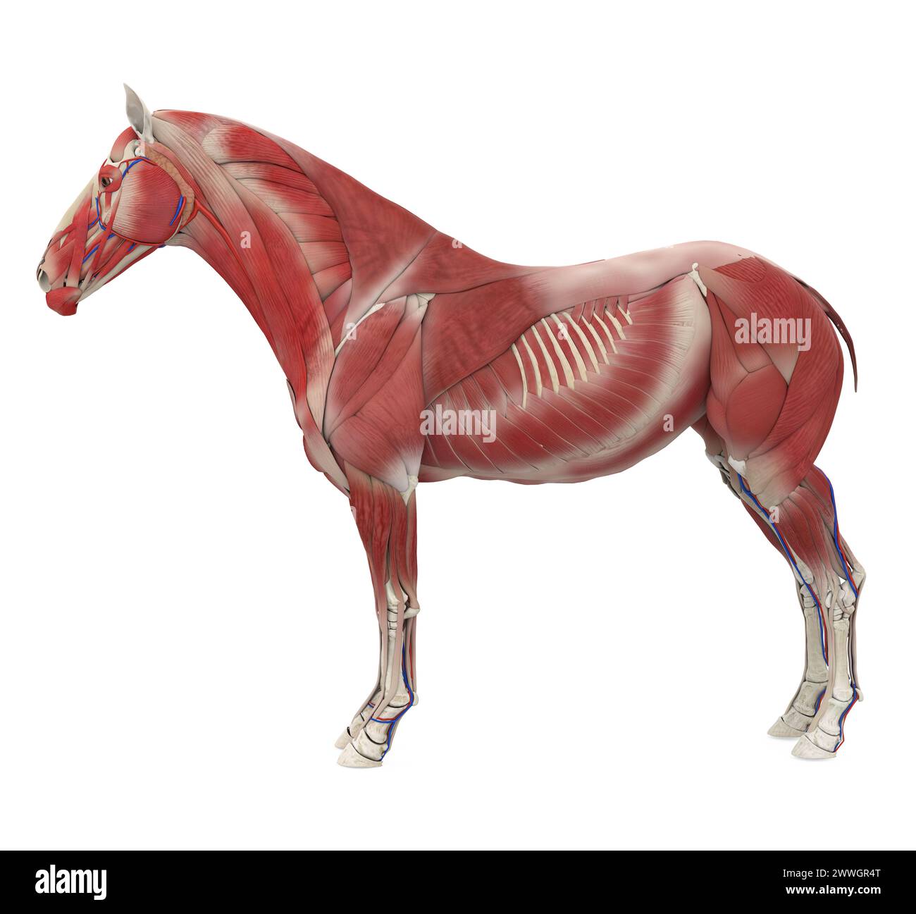 Horse Anatomy Muscular System Stock Photohttps://www.alamy.com/image-license-details/?v=1https://www.alamy.com/horse-anatomy-muscular-system-image600888312.html
RF2WWGR4T–Horse Anatomy Muscular System
 Internal Anatomy of a Horse isolated on white background Stock Vectorhttps://www.alamy.com/image-license-details/?v=1https://www.alamy.com/internal-anatomy-of-a-horse-isolated-on-white-background-image445508074.html
RF2GTPJ2J–Internal Anatomy of a Horse isolated on white background
 Horse Cecum - Horse Equus Anatomy - isolated on white Stock Photohttps://www.alamy.com/image-license-details/?v=1https://www.alamy.com/stock-photo-horse-cecum-horse-equus-anatomy-isolated-on-white-84434378.html
RFEWA8WE–Horse Cecum - Horse Equus Anatomy - isolated on white
 Horse illustration. 19th-century plate from a book on the practical treatise about the humane management of the horse. Stock Photohttps://www.alamy.com/image-license-details/?v=1https://www.alamy.com/horse-illustration-19th-century-plate-from-a-book-on-the-practical-treatise-about-the-humane-management-of-the-horse-image626109541.html
RF2YAHN31–Horse illustration. 19th-century plate from a book on the practical treatise about the humane management of the horse.
 Sesamoid bones, vintage engraved illustration. Dictionary of words and things - Larive and Fleury - 1895. Stock Photohttps://www.alamy.com/image-license-details/?v=1https://www.alamy.com/stock-photo-sesamoid-bones-vintage-engraved-illustration-dictionary-of-words-and-43485105.html
RFCEMWKD–Sesamoid bones, vintage engraved illustration. Dictionary of words and things - Larive and Fleury - 1895.
 Skull of a horse on a dark background old barn Stock Photohttps://www.alamy.com/image-license-details/?v=1https://www.alamy.com/stock-photo-skull-of-a-horse-on-a-dark-background-old-barn-176247424.html
RFM6MNAT–Skull of a horse on a dark background old barn
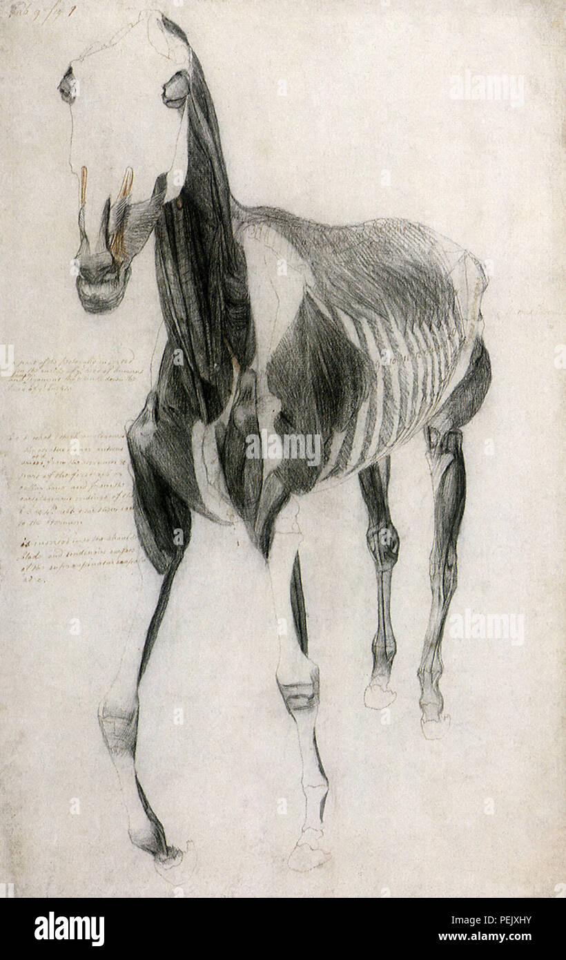 Horse Muscles and Bones, Stubbs, George Stock Photohttps://www.alamy.com/image-license-details/?v=1https://www.alamy.com/horse-muscles-and-bones-stubbs-george-image215545623.html
RMPEJXHY–Horse Muscles and Bones, Stubbs, George
 The encyclopædia of the stable: a complete manual of the horse, its breeds, anatomy, physiology, diseases, breeding, breaking, t Stock Photohttps://www.alamy.com/image-license-details/?v=1https://www.alamy.com/stock-photo-the-encyclopdia-of-the-stable-a-complete-manual-of-the-horse-its-breeds-87531930.html
RMF2BBTA–The encyclopædia of the stable: a complete manual of the horse, its breeds, anatomy, physiology, diseases, breeding, breaking, t
 Horse skull photo. Set of pictures for animals anatomy. Stock Photohttps://www.alamy.com/image-license-details/?v=1https://www.alamy.com/horse-skull-photo-set-of-pictures-for-animals-anatomy-image376638793.html
RF2CTNAHD–Horse skull photo. Set of pictures for animals anatomy.
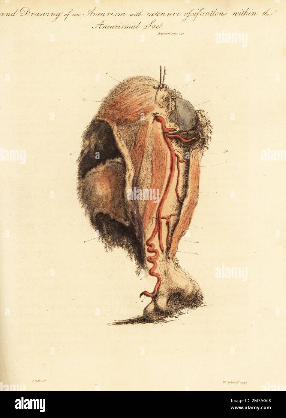 Aneurysmal tumor from the broken arm of a woman knocked down by a horse and cart, 1815. Second drawing of aneurism and extensive ossifications with the aneurismal sac. Humerus bone upper 1 and lower 2, sac 3, coraco brachialis a, biceps b, and deltoid c muscles, arteries f-m and basilic vein 7 stretched over the sac. Handcoloured copperplate engraving by William Archibald after an illustration by John Bell from his own Principles of Surgery, as they Relate to Wounds, Ulcers and Fistulas, Longman, Hurst, Rees, Orme and Brown, London, 1815. Stock Photohttps://www.alamy.com/image-license-details/?v=1https://www.alamy.com/aneurysmal-tumor-from-the-broken-arm-of-a-woman-knocked-down-by-a-horse-and-cart-1815-second-drawing-of-aneurism-and-extensive-ossifications-with-the-aneurismal-sac-humerus-bone-upper-1-and-lower-2-sac-3-coraco-brachialis-a-biceps-b-and-deltoid-c-muscles-arteries-f-m-and-basilic-vein-7-stretched-over-the-sac-handcoloured-copperplate-engraving-by-william-archibald-after-an-illustration-by-john-bell-from-his-own-principles-of-surgery-as-they-relate-to-wounds-ulcers-and-fistulas-longman-hurst-rees-orme-and-brown-london-1815-image503635504.html
RM2M7AG68–Aneurysmal tumor from the broken arm of a woman knocked down by a horse and cart, 1815. Second drawing of aneurism and extensive ossifications with the aneurismal sac. Humerus bone upper 1 and lower 2, sac 3, coraco brachialis a, biceps b, and deltoid c muscles, arteries f-m and basilic vein 7 stretched over the sac. Handcoloured copperplate engraving by William Archibald after an illustration by John Bell from his own Principles of Surgery, as they Relate to Wounds, Ulcers and Fistulas, Longman, Hurst, Rees, Orme and Brown, London, 1815.
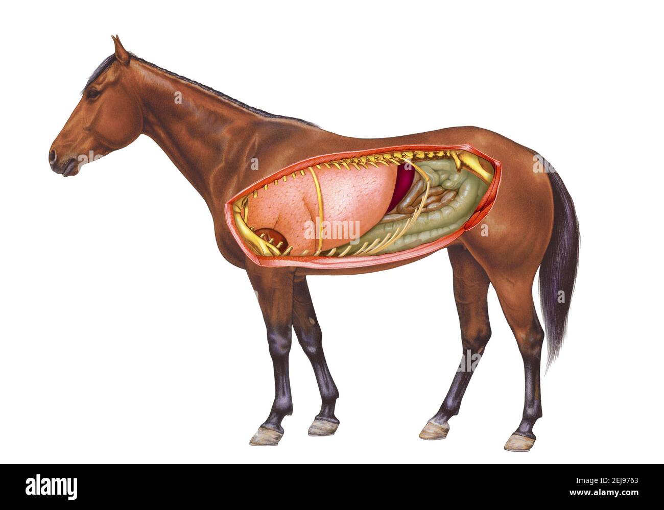 Horse anatomy, drawing Stock Photohttps://www.alamy.com/image-license-details/?v=1https://www.alamy.com/horse-anatomy-drawing-image407105499.html
RM2EJ9763–Horse anatomy, drawing
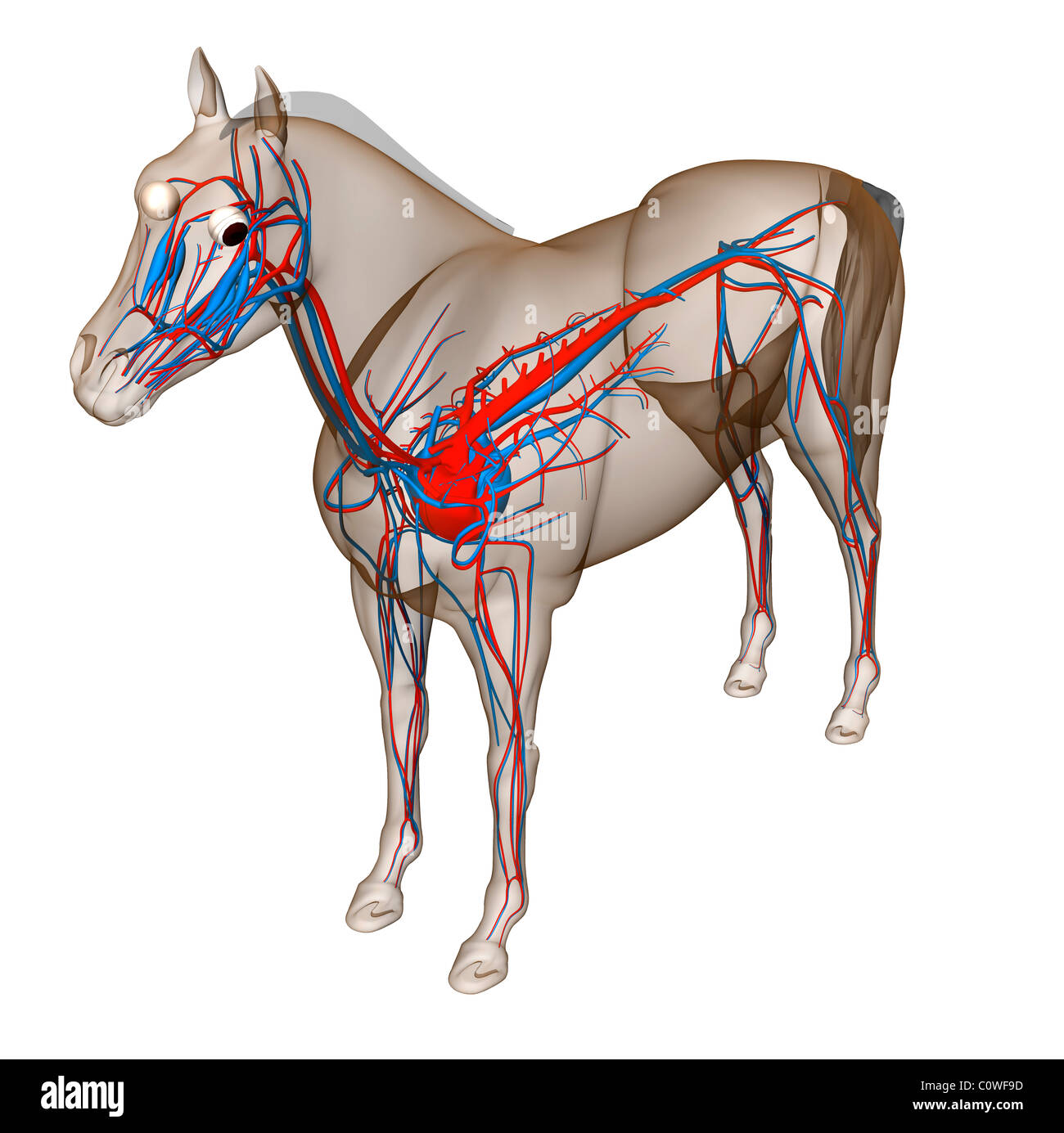 horse anatomy heart circulation Stock Photohttps://www.alamy.com/image-license-details/?v=1https://www.alamy.com/stock-photo-horse-anatomy-heart-circulation-34981561.html
RMC0WF9D–horse anatomy heart circulation
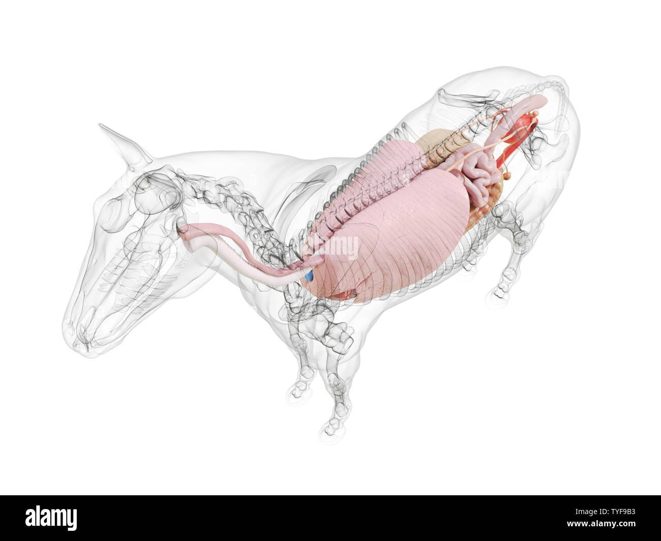 3d rendered medically accurate illustration of the horse anatomy Stock Photohttps://www.alamy.com/image-license-details/?v=1https://www.alamy.com/3d-rendered-medically-accurate-illustration-of-the-horse-anatomy-image257877511.html
RFTYF9B3–3d rendered medically accurate illustration of the horse anatomy
 a view of the muscles and tendons of the fore and hind extremities from The anatomy and physiology of the horse: with anatomical and questional illustrations. Containing, also, a series of examinations on equine anatomy and physiology, with instructions in reference to dissection and the mode of making anatomical preparations. To which is added, glossary of veterinary technicalities, toxicological chart, and dictionary of veterinary science by Dadd, George H., Publication date 1857 Stock Photohttps://www.alamy.com/image-license-details/?v=1https://www.alamy.com/a-view-of-the-muscles-and-tendons-of-the-fore-and-hind-extremities-from-the-anatomy-and-physiology-of-the-horse-with-anatomical-and-questional-illustrations-containing-also-a-series-of-examinations-on-equine-anatomy-and-physiology-with-instructions-in-reference-to-dissection-and-the-mode-of-making-anatomical-preparations-to-which-is-added-glossary-of-veterinary-technicalities-toxicological-chart-and-dictionary-of-veterinary-science-by-dadd-george-h-publication-date-1857-image635528699.html
RM2YWXR9F–a view of the muscles and tendons of the fore and hind extremities from The anatomy and physiology of the horse: with anatomical and questional illustrations. Containing, also, a series of examinations on equine anatomy and physiology, with instructions in reference to dissection and the mode of making anatomical preparations. To which is added, glossary of veterinary technicalities, toxicological chart, and dictionary of veterinary science by Dadd, George H., Publication date 1857
 Horse anatomy, illustration Stock Photohttps://www.alamy.com/image-license-details/?v=1https://www.alamy.com/horse-anatomy-illustration-image267282261.html
RFWERN71–Horse anatomy, illustration
 Horse anatomy - Body parts - No text Stock Photohttps://www.alamy.com/image-license-details/?v=1https://www.alamy.com/horse-anatomy-body-parts-no-text-image226187390.html
RFR3YM92–Horse anatomy - Body parts - No text
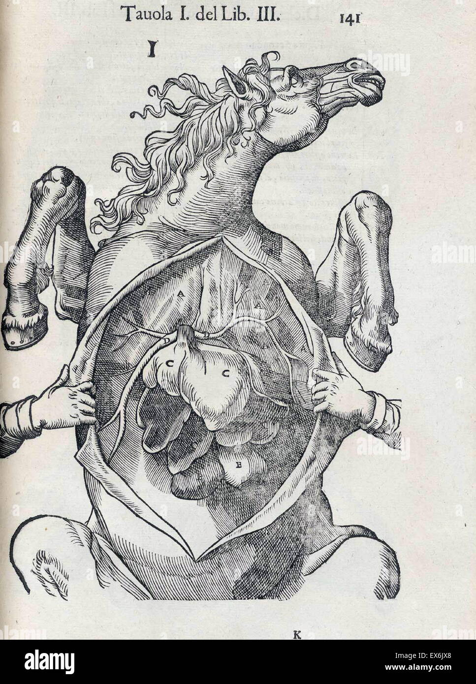 Equine anatomical Illustration from 'Anatomia del cavallo, infermità, et suoi rimedii'. Anatomy of a horse); (1618). by Carlo Ruini, (1530-1598) Stock Photohttps://www.alamy.com/image-license-details/?v=1https://www.alamy.com/stock-photo-equine-anatomical-illustration-from-anatomia-del-cavallo-infermit-84969088.html
RMEX6JX8–Equine anatomical Illustration from 'Anatomia del cavallo, infermità, et suoi rimedii'. Anatomy of a horse); (1618). by Carlo Ruini, (1530-1598)
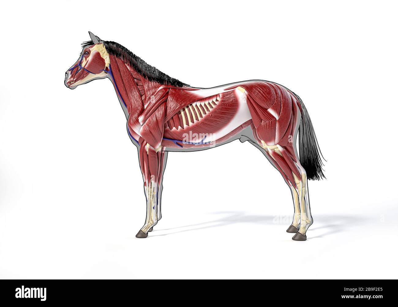 Muscular anatomy of a horse over grey silhouette, side view. Stock Photohttps://www.alamy.com/image-license-details/?v=1https://www.alamy.com/muscular-anatomy-of-a-horse-over-grey-silhouette-side-view-image350070509.html
RF2B9F2E5–Muscular anatomy of a horse over grey silhouette, side view.
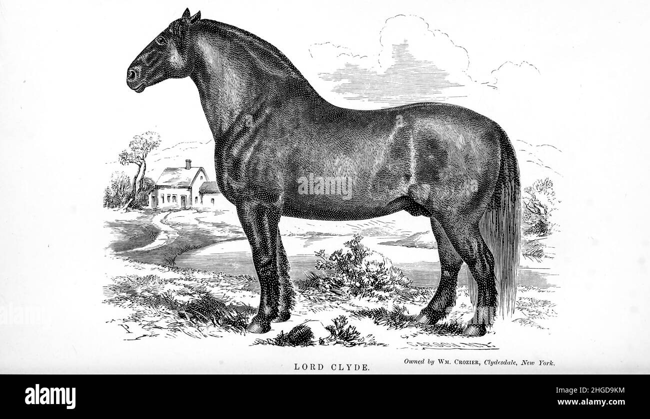 Lord Clyde from Every horse owner's cyclopedia : the anatomy and physiology of the horse; general characteristics; the points of the horse, with directions how to choose him; the principles of breeding, and the best kind to breed from; the treatment of the brood mare and foal; raising and breaking the colt; stables and stable management; riding, driving, etc., etc. Diseases, and how to cure them. The principal medicines, and the doses in which they can be safely administered; accidents, fractures, and the operations necessary in each case; shoeing, etc. Publisher: Philadelphia : Porter & Coate Stock Photohttps://www.alamy.com/image-license-details/?v=1https://www.alamy.com/lord-clyde-from-every-horse-owners-cyclopedia-the-anatomy-and-physiology-of-the-horse-general-characteristics-the-points-of-the-horse-with-directions-how-to-choose-him-the-principles-of-breeding-and-the-best-kind-to-breed-from-the-treatment-of-the-brood-mare-and-foal-raising-and-breaking-the-colt-stables-and-stable-management-riding-driving-etc-etc-diseases-and-how-to-cure-them-the-principal-medicines-and-the-doses-in-which-they-can-be-safely-administered-accidents-fractures-and-the-operations-necessary-in-each-case-shoeing-etc-publisher-philadelphia-porter-coate-image457597048.html
RF2HGD9KM–Lord Clyde from Every horse owner's cyclopedia : the anatomy and physiology of the horse; general characteristics; the points of the horse, with directions how to choose him; the principles of breeding, and the best kind to breed from; the treatment of the brood mare and foal; raising and breaking the colt; stables and stable management; riding, driving, etc., etc. Diseases, and how to cure them. The principal medicines, and the doses in which they can be safely administered; accidents, fractures, and the operations necessary in each case; shoeing, etc. Publisher: Philadelphia : Porter & Coate
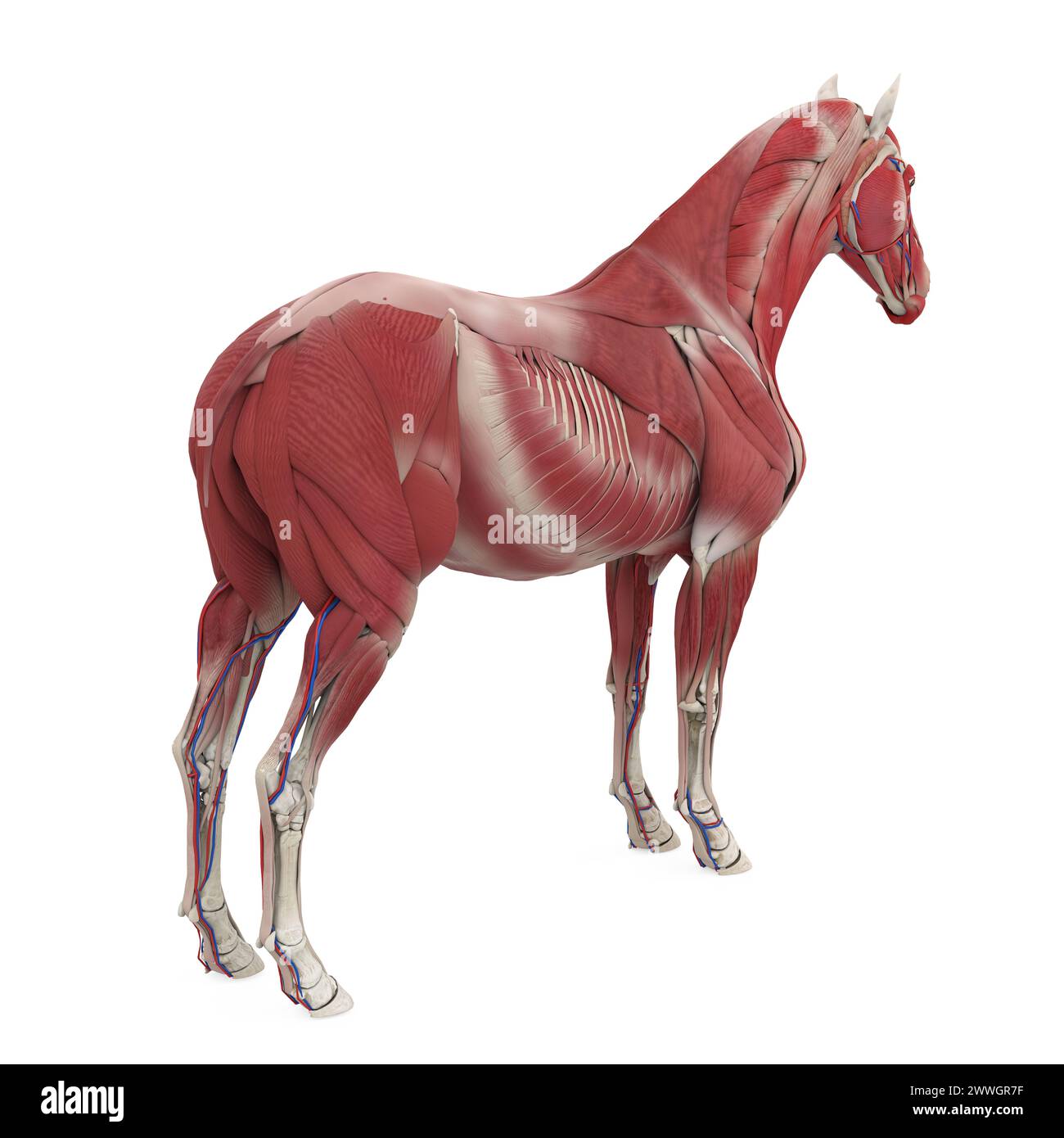 Horse Anatomy Muscular System Stock Photohttps://www.alamy.com/image-license-details/?v=1https://www.alamy.com/horse-anatomy-muscular-system-image600888387.html
RF2WWGR7F–Horse Anatomy Muscular System
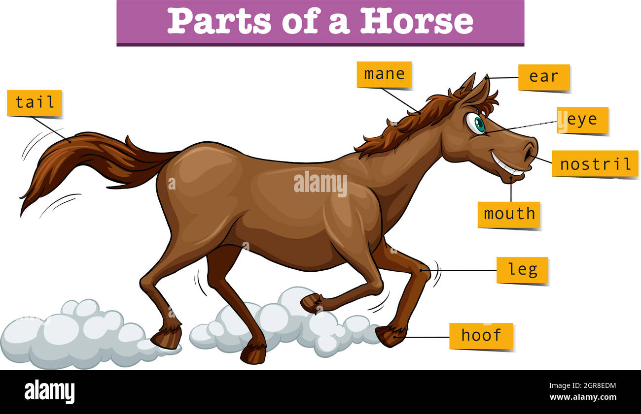 Diagram showing parts of horse Stock Vectorhttps://www.alamy.com/image-license-details/?v=1https://www.alamy.com/diagram-showing-parts-of-horse-image444583264.html
RF2GR8EDM–Diagram showing parts of horse
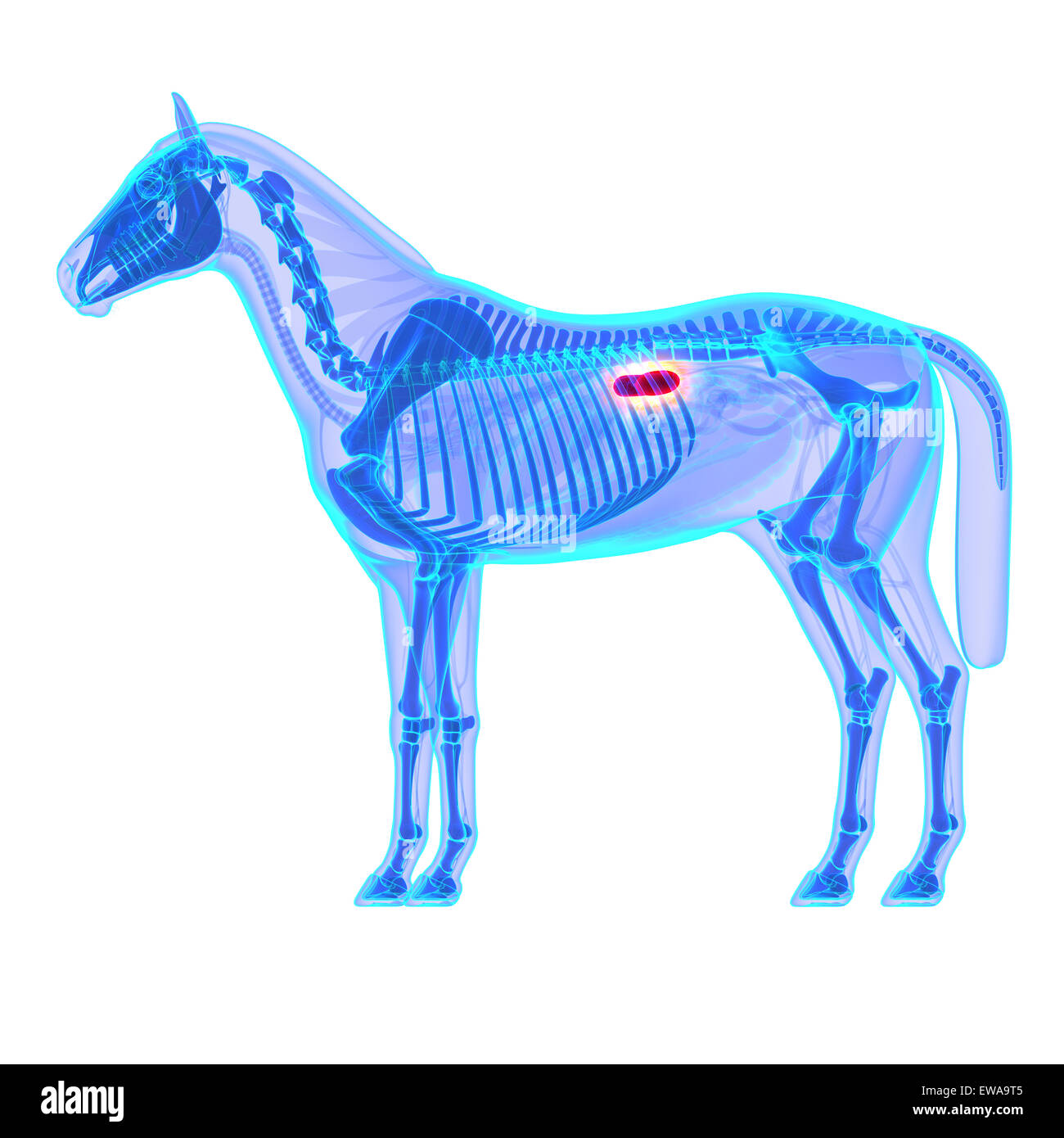 Horse Kidneys - Horse Equus Anatomy - isolated on white Stock Photohttps://www.alamy.com/image-license-details/?v=1https://www.alamy.com/stock-photo-horse-kidneys-horse-equus-anatomy-isolated-on-white-84435125.html
RFEWA9T5–Horse Kidneys - Horse Equus Anatomy - isolated on white
 Horse illustration. 19th-century plate from a book on the practical treatise about the humane management of the horse. Stock Photohttps://www.alamy.com/image-license-details/?v=1https://www.alamy.com/horse-illustration-19th-century-plate-from-a-book-on-the-practical-treatise-about-the-humane-management-of-the-horse-image626109539.html
RF2YAHN2Y–Horse illustration. 19th-century plate from a book on the practical treatise about the humane management of the horse.
 Horse's hoof, vintage engraved illustration. Dictionary of words and things - Larive and Fleury - 1895. Stock Photohttps://www.alamy.com/image-license-details/?v=1https://www.alamy.com/stock-photo-horses-hoof-vintage-engraved-illustration-dictionary-of-words-and-43484589.html
RFCEMW11–Horse's hoof, vintage engraved illustration. Dictionary of words and things - Larive and Fleury - 1895.
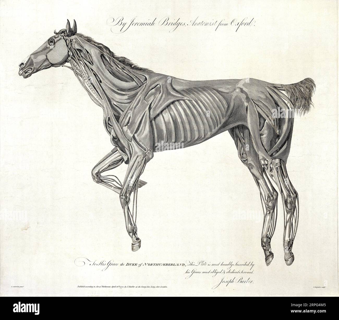 Muscles of the horse, engraving by C. Grignion after F. Sartorius for Jeremiah Bridges 1772 Stock Photohttps://www.alamy.com/image-license-details/?v=1https://www.alamy.com/muscles-of-the-horse-engraving-by-c-grignion-after-f-sartorius-for-jeremiah-bridges-1772-image564609141.html
RM2RPG4M5–Muscles of the horse, engraving by C. Grignion after F. Sartorius for Jeremiah Bridges 1772
 Horse Musculature, Side, Stubbs, George Stock Photohttps://www.alamy.com/image-license-details/?v=1https://www.alamy.com/horse-musculature-side-stubbs-george-image215545624.html
RMPEJXJ0–Horse Musculature, Side, Stubbs, George
 The encyclopædia of the stable: a complete manual of the horse, its breeds, anatomy, physiology, diseases, breeding, breaking, t Stock Photohttps://www.alamy.com/image-license-details/?v=1https://www.alamy.com/stock-photo-the-encyclopdia-of-the-stable-a-complete-manual-of-the-horse-its-breeds-87531904.html
RMF2BBRC–The encyclopædia of the stable: a complete manual of the horse, its breeds, anatomy, physiology, diseases, breeding, breaking, t
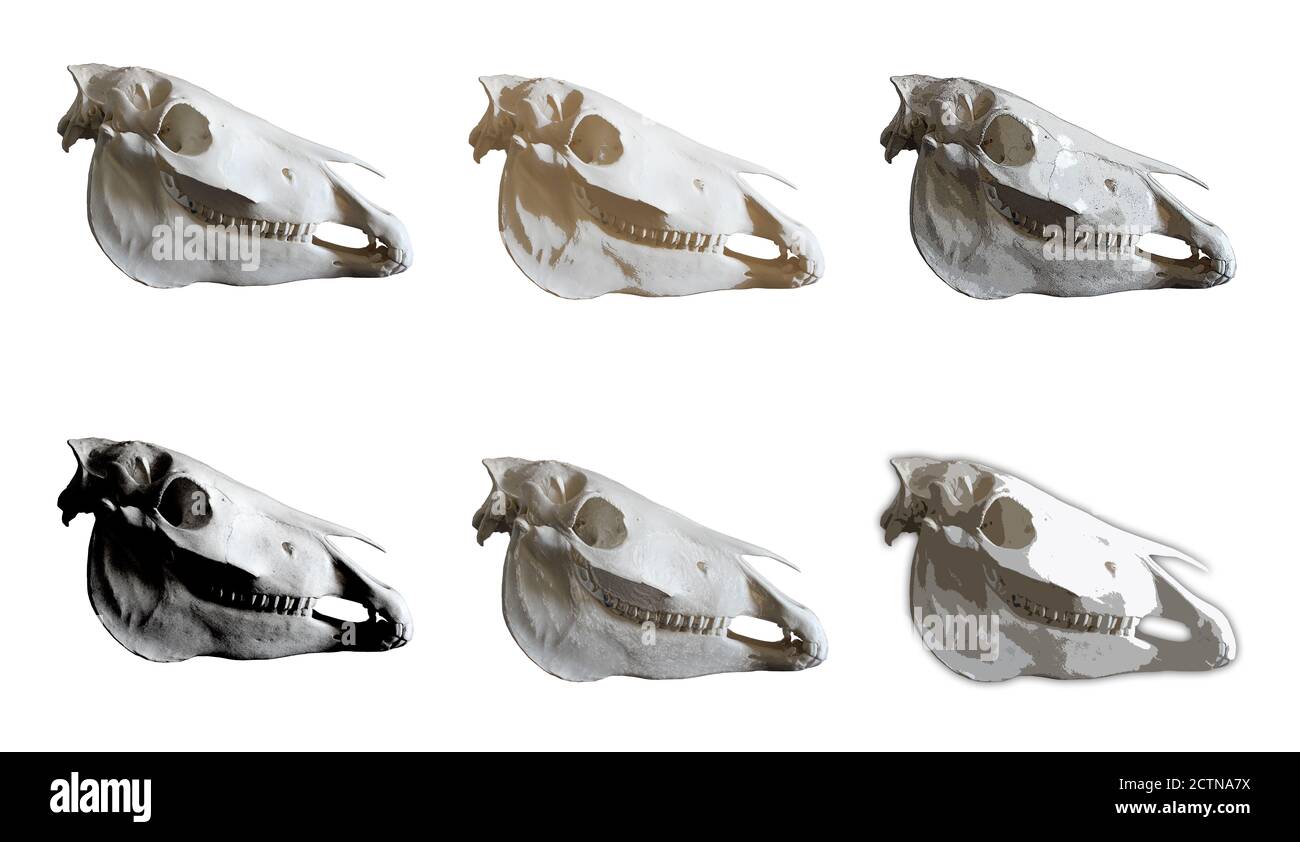 Horse skull photo. Set of pictures for animals anatomy. Stock Photohttps://www.alamy.com/image-license-details/?v=1https://www.alamy.com/horse-skull-photo-set-of-pictures-for-animals-anatomy-image376638526.html
RF2CTNA7X–Horse skull photo. Set of pictures for animals anatomy.
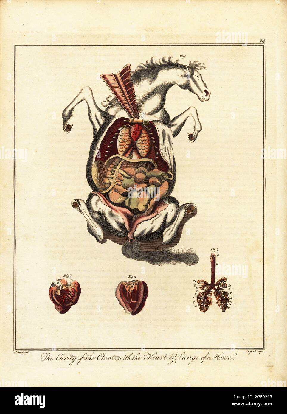 Anatomy of a horse. Chest cavity exposed 1, heart or vena cava 2, left ventricle 3, and lungs 4.. Handcoloured copperplate engraving by J. Pass after an illustration by Daniel Dodd from William Augustus Osbaldiston’s The British Sportsman, or Nobleman, Gentleman and Farmer’s Dictionary of Recreation and Amusement, J. Stead, London, 1792. Stock Photohttps://www.alamy.com/image-license-details/?v=1https://www.alamy.com/anatomy-of-a-horse-chest-cavity-exposed-1-heart-or-vena-cava-2-left-ventricle-3-and-lungs-4-handcoloured-copperplate-engraving-by-j-pass-after-an-illustration-by-daniel-dodd-from-william-augustus-osbaldistons-the-british-sportsman-or-nobleman-gentleman-and-farmers-dictionary-of-recreation-and-amusement-j-stead-london-1792-image439063693.html
RM2GE9265–Anatomy of a horse. Chest cavity exposed 1, heart or vena cava 2, left ventricle 3, and lungs 4.. Handcoloured copperplate engraving by J. Pass after an illustration by Daniel Dodd from William Augustus Osbaldiston’s The British Sportsman, or Nobleman, Gentleman and Farmer’s Dictionary of Recreation and Amusement, J. Stead, London, 1792.
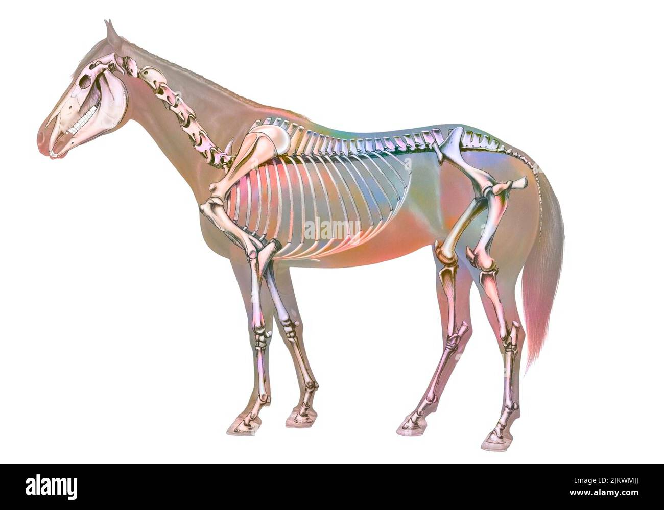 Anatomy of the horse with its bone system. Stock Photohttps://www.alamy.com/image-license-details/?v=1https://www.alamy.com/anatomy-of-the-horse-with-its-bone-system-image476923402.html
RF2JKWMJJ–Anatomy of the horse with its bone system.
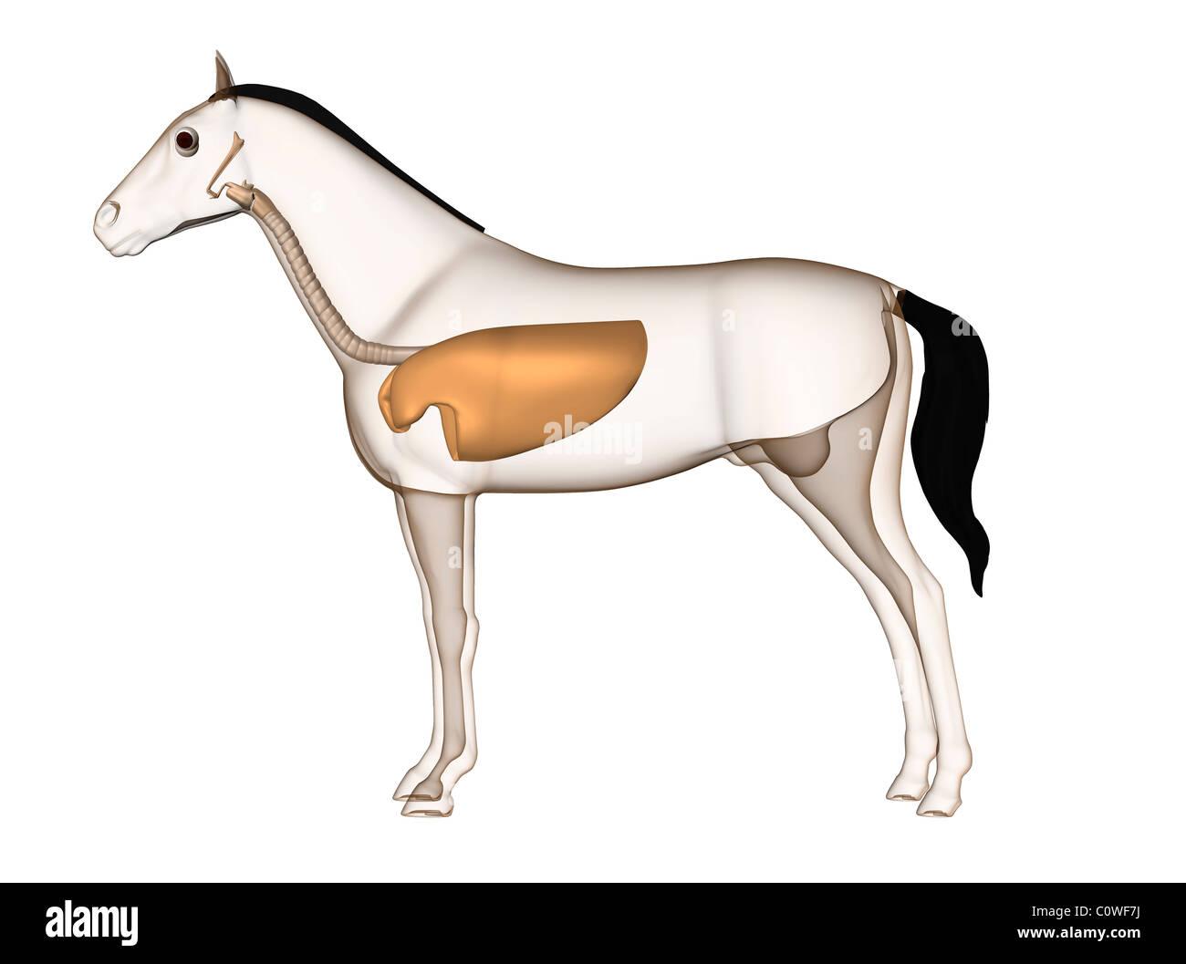 horse anatomy respiratory lungs Stock Photohttps://www.alamy.com/image-license-details/?v=1https://www.alamy.com/stock-photo-horse-anatomy-respiratory-lungs-34981510.html
RMC0WF7J–horse anatomy respiratory lungs
 3d rendered medically accurate illustration of the horse anatomy Stock Photohttps://www.alamy.com/image-license-details/?v=1https://www.alamy.com/3d-rendered-medically-accurate-illustration-of-the-horse-anatomy-image257878177.html
RFTYFA6W–3d rendered medically accurate illustration of the horse anatomy
 a representation of the superficial muscles of the body, of a part of the neck, and of the extremities from The anatomy and physiology of the horse: with anatomical and questional illustrations. Containing, also, a series of examinations on equine anatomy and physiology, with instructions in reference to dissection and the mode of making anatomical preparations. To which is added, glossary of veterinary technicalities, toxicological chart, and dictionary of veterinary science by Dadd, George H., Publication date 1857 Stock Photohttps://www.alamy.com/image-license-details/?v=1https://www.alamy.com/a-representation-of-the-superficial-muscles-of-the-body-of-a-part-of-the-neck-and-of-the-extremities-from-the-anatomy-and-physiology-of-the-horse-with-anatomical-and-questional-illustrations-containing-also-a-series-of-examinations-on-equine-anatomy-and-physiology-with-instructions-in-reference-to-dissection-and-the-mode-of-making-anatomical-preparations-to-which-is-added-glossary-of-veterinary-technicalities-toxicological-chart-and-dictionary-of-veterinary-science-by-dadd-george-h-publication-date-1857-image635528649.html
RM2YWXR7N–a representation of the superficial muscles of the body, of a part of the neck, and of the extremities from The anatomy and physiology of the horse: with anatomical and questional illustrations. Containing, also, a series of examinations on equine anatomy and physiology, with instructions in reference to dissection and the mode of making anatomical preparations. To which is added, glossary of veterinary technicalities, toxicological chart, and dictionary of veterinary science by Dadd, George H., Publication date 1857