Quick filters:
Human embryo showing Stock Photos and Images
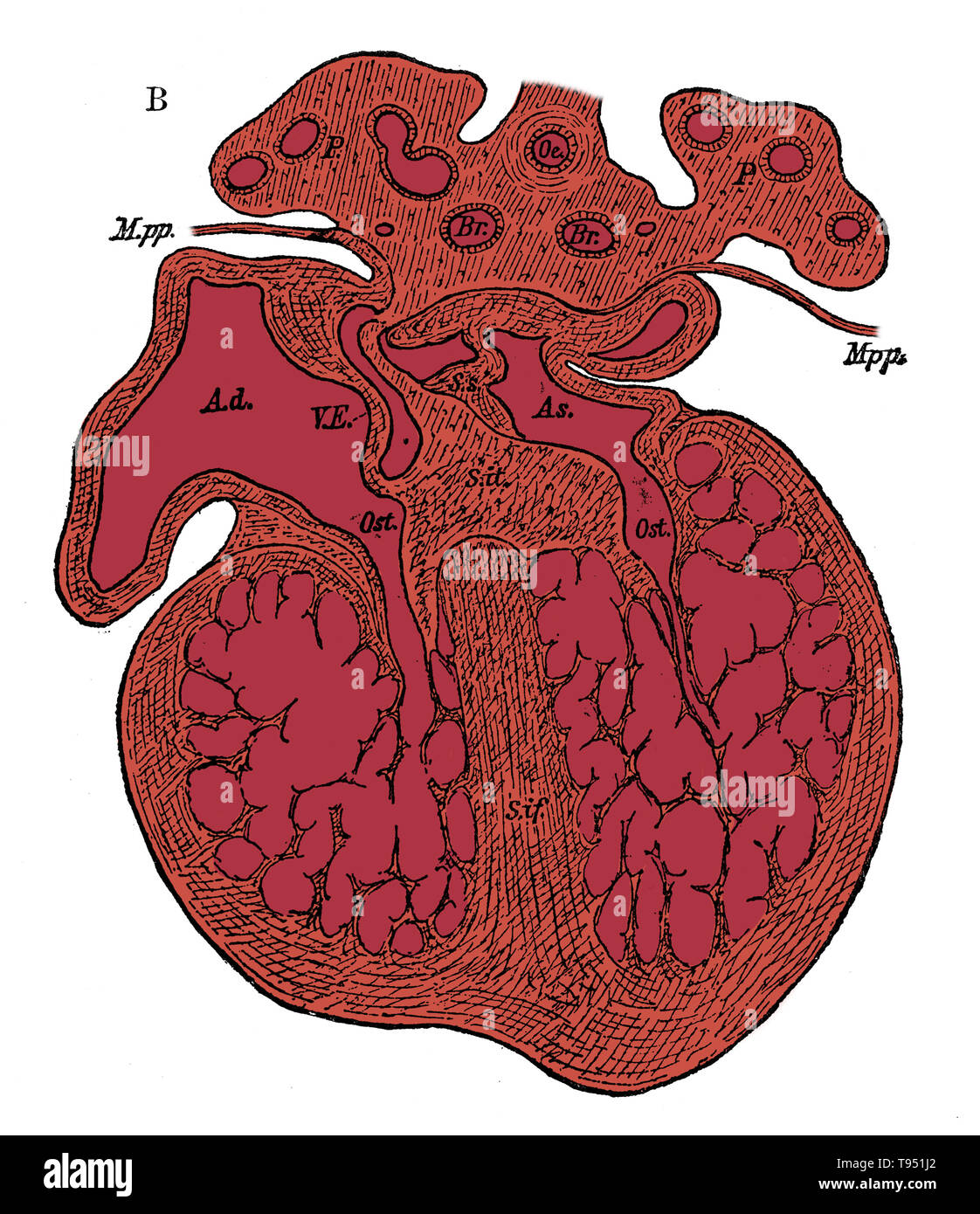 Section through the heart of human embryo showing the formation of the cardiac septa and the auriculo-ventricular valves, (see 9N3690) from a somewhat more advanced embryo. Ad, As, right and left auricle; Ost, auriculo-ventricular apertures; S.s, septum superior of auricles; S. it, endocardial cushion (septum intermedium); S. if, septum infers ventriculorum, now denser and more muscular. Stock Photohttps://www.alamy.com/image-license-details/?v=1https://www.alamy.com/section-through-the-heart-of-human-embryo-showing-the-formation-of-the-cardiac-septa-and-the-auriculo-ventricular-valves-see-9n3690-from-a-somewhat-more-advanced-embryo-ad-as-right-and-left-auricle-ost-auriculo-ventricular-apertures-ss-septum-superior-of-auricles-s-it-endocardial-cushion-septum-intermedium-s-if-septum-infers-ventriculorum-now-denser-and-more-muscular-image246588106.html
RMT951J2–Section through the heart of human embryo showing the formation of the cardiac septa and the auriculo-ventricular valves, (see 9N3690) from a somewhat more advanced embryo. Ad, As, right and left auricle; Ost, auriculo-ventricular apertures; S.s, septum superior of auricles; S. it, endocardial cushion (septum intermedium); S. if, septum infers ventriculorum, now denser and more muscular.
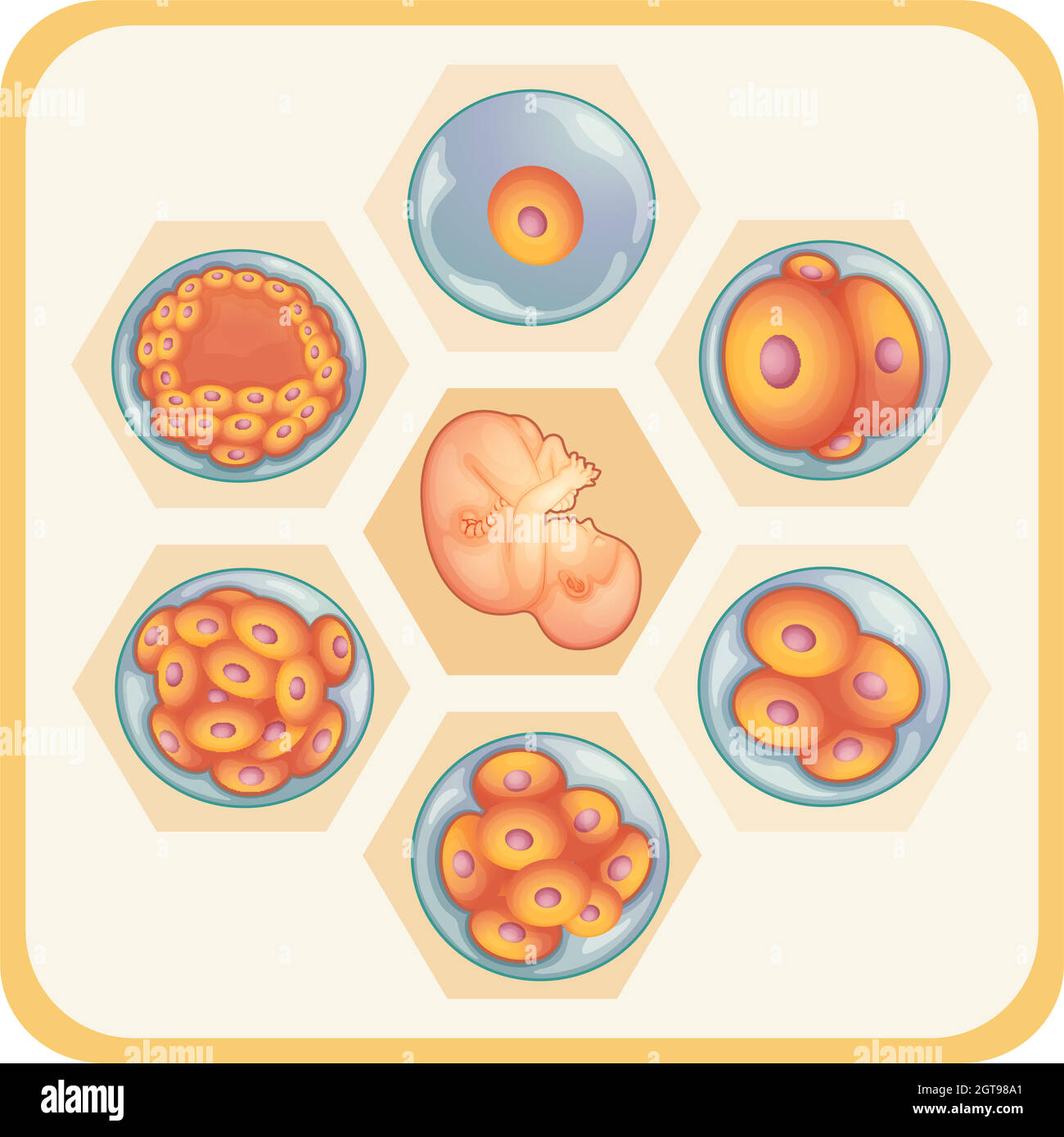 Diagram showing reproductive of human Stock Vectorhttps://www.alamy.com/image-license-details/?v=1https://www.alamy.com/diagram-showing-reproductive-of-human-image445215065.html
RF2GT98A1–Diagram showing reproductive of human
 Curious daughter with mobile phone showing ultrasound embryo of baby at home Stock Photohttps://www.alamy.com/image-license-details/?v=1https://www.alamy.com/curious-daughter-with-mobile-phone-showing-ultrasound-embryo-of-baby-at-home-image420128564.html
RF2FBEE7G–Curious daughter with mobile phone showing ultrasound embryo of baby at home
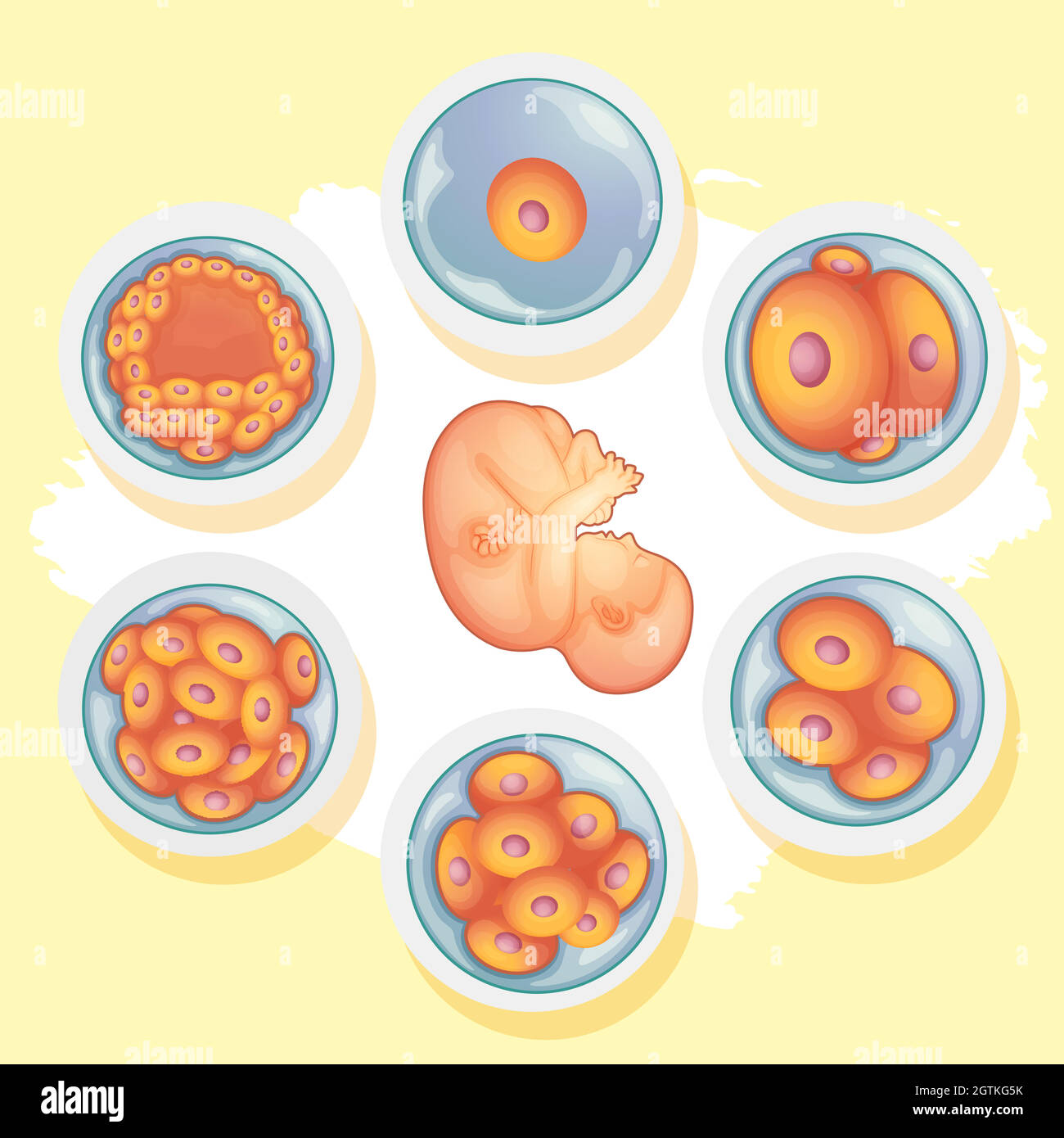 Diagram showing different stages of human baby Stock Vectorhttps://www.alamy.com/image-license-details/?v=1https://www.alamy.com/diagram-showing-different-stages-of-human-baby-image445440735.html
RF2GTKG5K–Diagram showing different stages of human baby
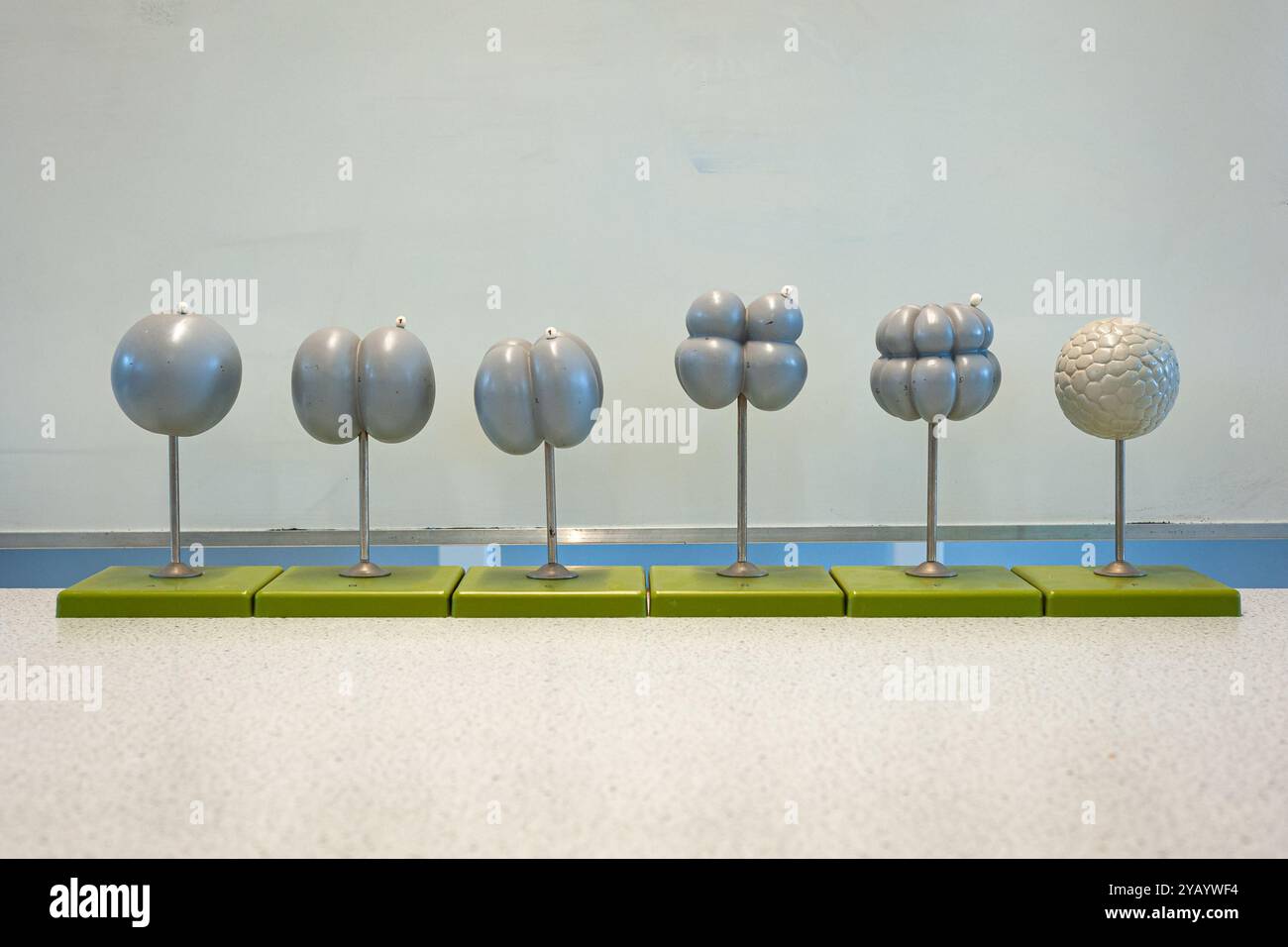 Several plastic models showing the initial stage of embryo development by cell division. Used in school biology lessons. Stock Photohttps://www.alamy.com/image-license-details/?v=1https://www.alamy.com/several-plastic-models-showing-the-initial-stage-of-embryo-development-by-cell-division-used-in-school-biology-lessons-image626332536.html
RF2YAYWF4–Several plastic models showing the initial stage of embryo development by cell division. Used in school biology lessons.
 The baby pod / 3D illustration of science fiction scene showing human child asleep inside complex futuristic incubator cloning machinery Stock Photohttps://www.alamy.com/image-license-details/?v=1https://www.alamy.com/the-baby-pod-3d-illustration-of-science-fiction-scene-showing-human-child-asleep-inside-complex-futuristic-incubator-cloning-machinery-image243445704.html
RFT41WDC–The baby pod / 3D illustration of science fiction scene showing human child asleep inside complex futuristic incubator cloning machinery
 Hands holding a digital tablet showing an ultrasound image of a fetus in surgery room Stock Photohttps://www.alamy.com/image-license-details/?v=1https://www.alamy.com/stock-photo-hands-holding-a-digital-tablet-showing-an-ultrasound-image-of-a-fetus-90287895.html
RFF6TY3K–Hands holding a digital tablet showing an ultrasound image of a fetus in surgery room
 3d illustration of pregnancy, graphic representation of the interior of the uterus. Showing the human fetus with the umbilical cord. Stock Photohttps://www.alamy.com/image-license-details/?v=1https://www.alamy.com/3d-illustration-of-pregnancy-graphic-representation-of-the-interior-of-the-uterus-showing-the-human-fetus-with-the-umbilical-cord-image336175418.html
RF2AEX34X–3d illustration of pregnancy, graphic representation of the interior of the uterus. Showing the human fetus with the umbilical cord.
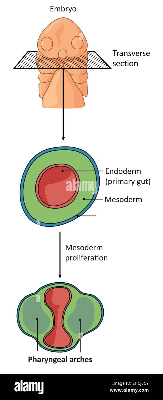 Transverse section through an embryo showing differentaition of pharyngeal arches Stock Photohttps://www.alamy.com/image-license-details/?v=1https://www.alamy.com/transverse-section-through-an-embryo-showing-differentaition-of-pharyngeal-arches-image455240939.html
RF2HCJ0CY–Transverse section through an embryo showing differentaition of pharyngeal arches
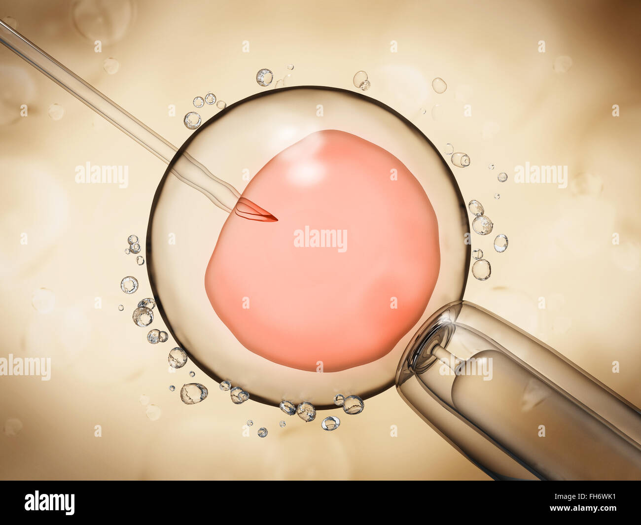 Artificial insemination illustration showing the ovule on yellow background Stock Photohttps://www.alamy.com/image-license-details/?v=1https://www.alamy.com/stock-photo-artificial-insemination-illustration-showing-the-ovule-on-yellow-background-96652837.html
RFFH6WK1–Artificial insemination illustration showing the ovule on yellow background
 Diagram showing how baby grows during pregnancy Stock Vectorhttps://www.alamy.com/image-license-details/?v=1https://www.alamy.com/diagram-showing-how-baby-grows-during-pregnancy-image445996078.html
RF2GWGTFA–Diagram showing how baby grows during pregnancy
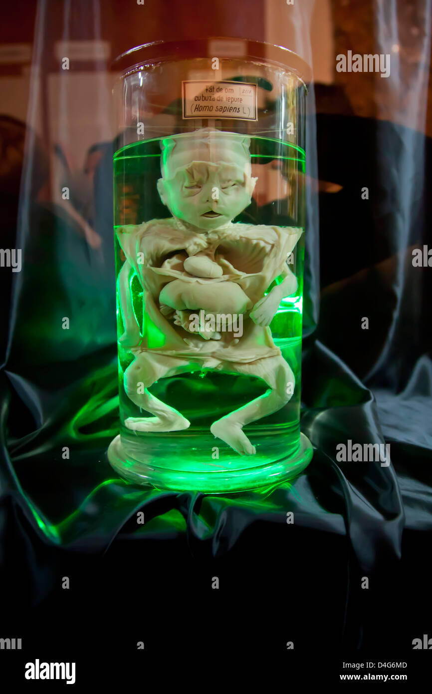 preserved baby fetus in a jar with organs showing Stock Photohttps://www.alamy.com/image-license-details/?v=1https://www.alamy.com/stock-photo-preserved-baby-fetus-in-a-jar-with-organs-showing-54446237.html
RFD4G6MD–preserved baby fetus in a jar with organs showing
 Diagram showing baby growing inside woman womb Stock Vectorhttps://www.alamy.com/image-license-details/?v=1https://www.alamy.com/diagram-showing-baby-growing-inside-woman-womb-image445130941.html
RF2GT5D1H–Diagram showing baby growing inside woman womb
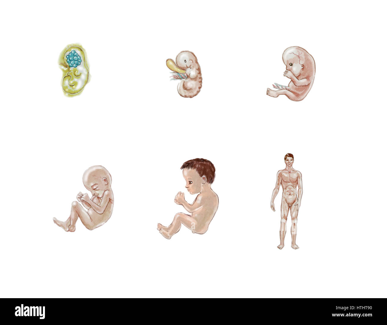 watercolor illustration showing stages in human development Stock Photohttps://www.alamy.com/image-license-details/?v=1https://www.alamy.com/stock-photo-watercolor-illustration-showing-stages-in-human-development-135616572.html
RFHTHT90–watercolor illustration showing stages in human development
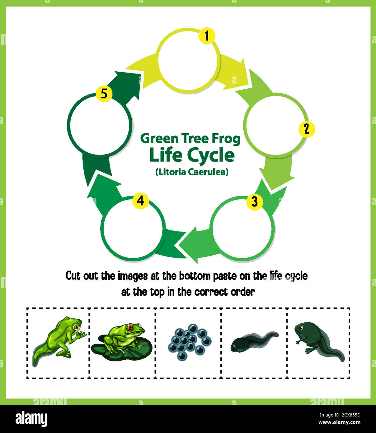 Diagram showing life cycle of Frog Stock Vectorhttps://www.alamy.com/image-license-details/?v=1https://www.alamy.com/diagram-showing-life-cycle-of-frog-image446419945.html
RF2GX855D–Diagram showing life cycle of Frog
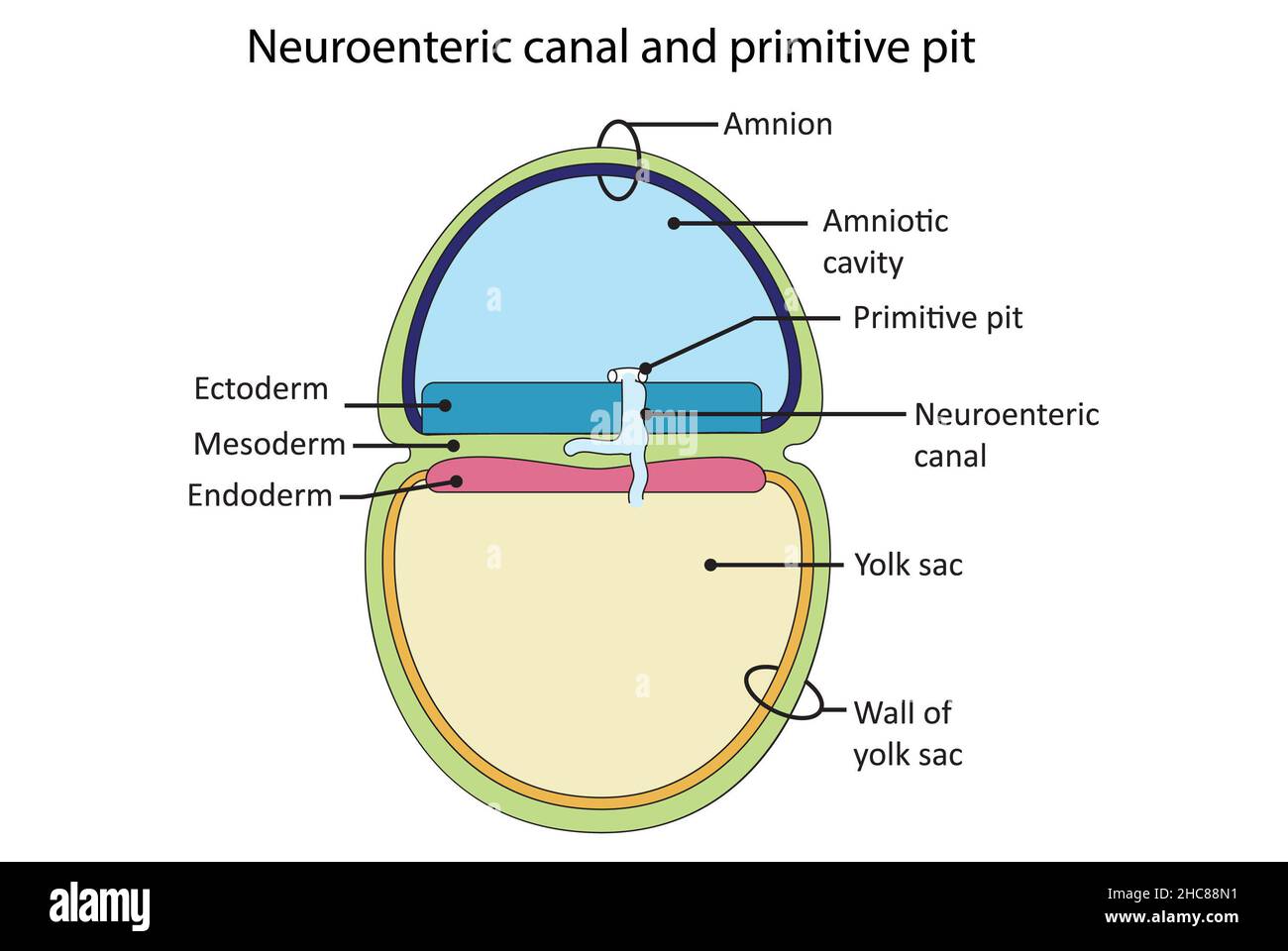 Simple diagram showing creation of the neuroenteric canal and primitive pit during the stage of gastrula. Stock Photohttps://www.alamy.com/image-license-details/?v=1https://www.alamy.com/simple-diagram-showing-creation-of-the-neuroenteric-canal-and-primitive-pit-during-the-stage-of-gastrula-image455027917.html
RF2HC88N1–Simple diagram showing creation of the neuroenteric canal and primitive pit during the stage of gastrula.
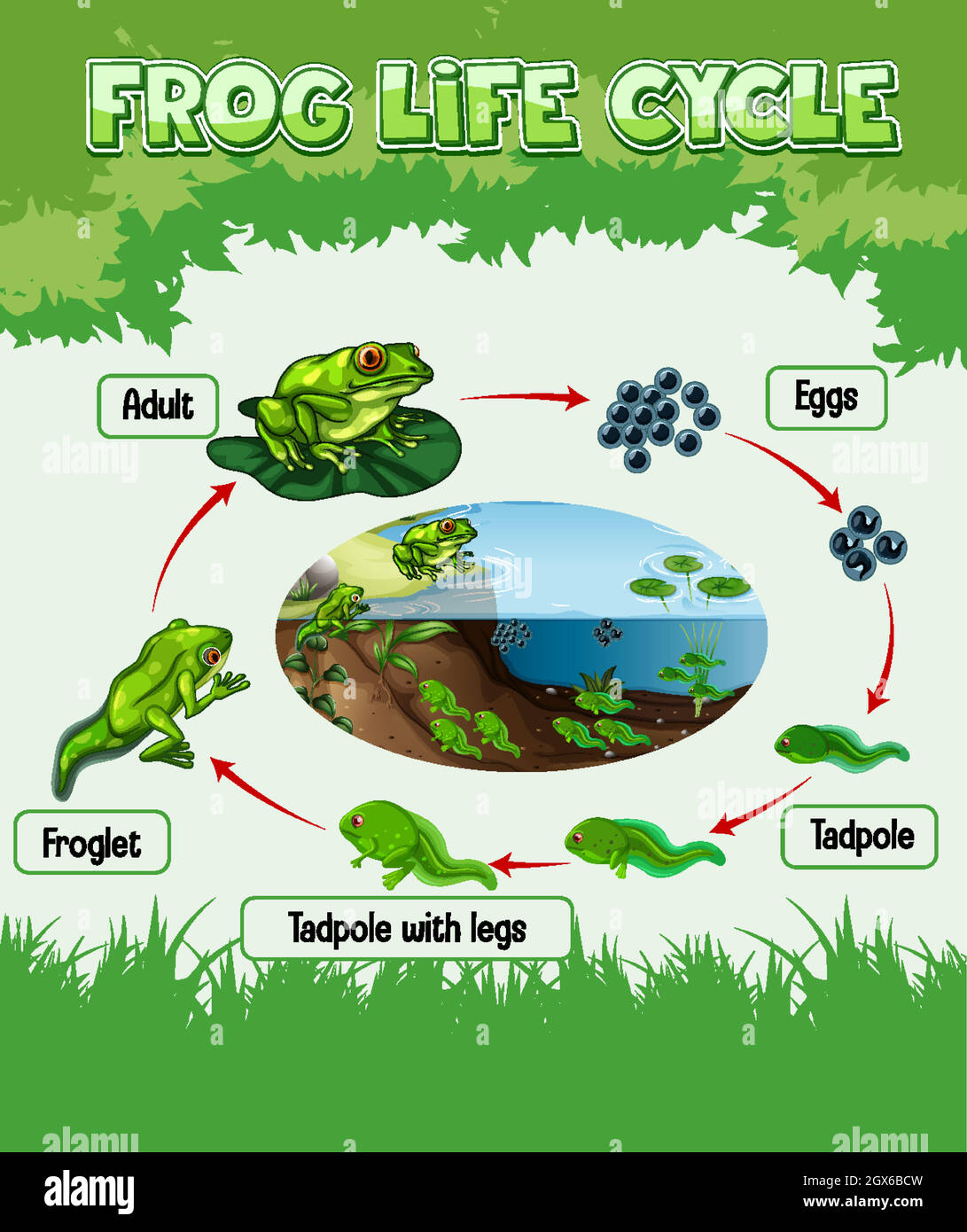 Diagram showing life cycle of Frog Stock Vectorhttps://www.alamy.com/image-license-details/?v=1https://www.alamy.com/diagram-showing-life-cycle-of-frog-image446380953.html
RF2GX6BCW–Diagram showing life cycle of Frog
 A smiling caucasian nurse showing a photo of an embryo - closeup. High quality photo Stock Photohttps://www.alamy.com/image-license-details/?v=1https://www.alamy.com/a-smiling-caucasian-nurse-showing-a-photo-of-an-embryo-closeup-high-quality-photo-image476080394.html
RF2JJF9B6–A smiling caucasian nurse showing a photo of an embryo - closeup. High quality photo
 Diagram showing life cycle of Frog Stock Vectorhttps://www.alamy.com/image-license-details/?v=1https://www.alamy.com/diagram-showing-life-cycle-of-frog-image446352136.html
RF2GX52KM–Diagram showing life cycle of Frog
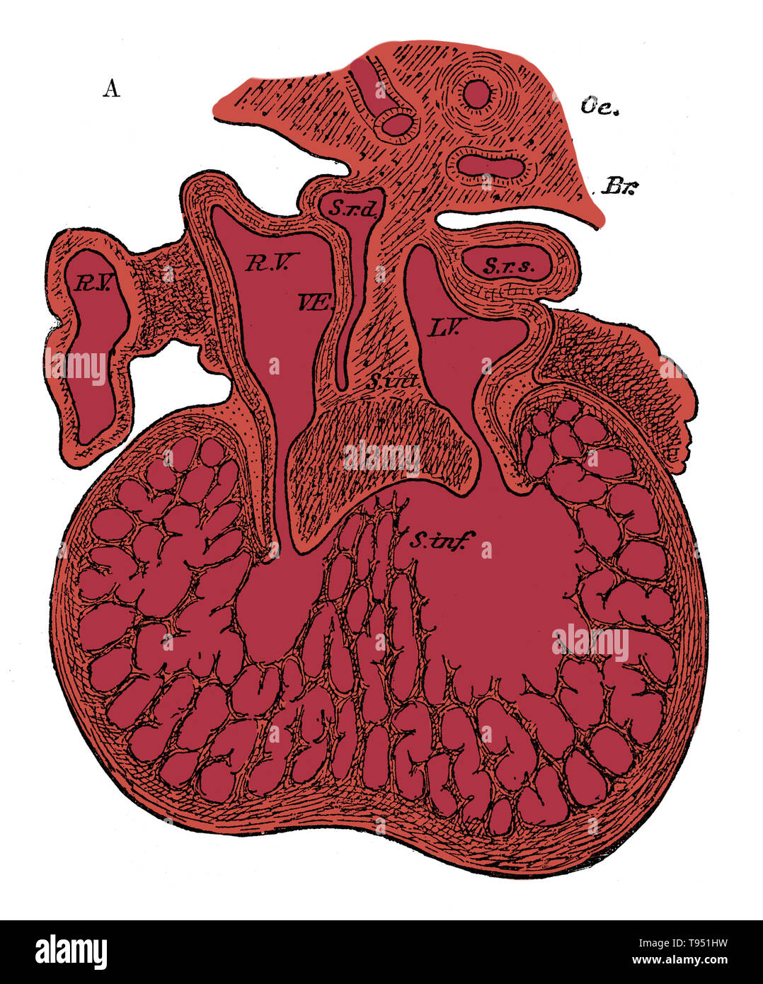 Section through the heart of human embryo showing the formation of the cardiac septa and the auriculo-ventricular valves, 5 to 6 weeks. R.V, right auricle; L.V, left auricle; S.r.d, right horn of sinus; Sr.s, left horn of sinus; s. int, septum superior and endocardial cushion (septum intermedium); s. inf, septum infers ventriculorum; This septum, as well as the bulk of the ventricle, is a muscular sponge at this stage. Oc, esophagus; Br, bronchus. Stock Photohttps://www.alamy.com/image-license-details/?v=1https://www.alamy.com/section-through-the-heart-of-human-embryo-showing-the-formation-of-the-cardiac-septa-and-the-auriculo-ventricular-valves-5-to-6-weeks-rv-right-auricle-lv-left-auricle-srd-right-horn-of-sinus-srs-left-horn-of-sinus-s-int-septum-superior-and-endocardial-cushion-septum-intermedium-s-inf-septum-infers-ventriculorum-this-septum-as-well-as-the-bulk-of-the-ventricle-is-a-muscular-sponge-at-this-stage-oc-esophagus-br-bronchus-image246588101.html
RMT951HW–Section through the heart of human embryo showing the formation of the cardiac septa and the auriculo-ventricular valves, 5 to 6 weeks. R.V, right auricle; L.V, left auricle; S.r.d, right horn of sinus; Sr.s, left horn of sinus; s. int, septum superior and endocardial cushion (septum intermedium); s. inf, septum infers ventriculorum; This septum, as well as the bulk of the ventricle, is a muscular sponge at this stage. Oc, esophagus; Br, bronchus.
 Diagram showing life cycle of Frog Stock Vectorhttps://www.alamy.com/image-license-details/?v=1https://www.alamy.com/diagram-showing-life-cycle-of-frog-image445837781.html
RF2GW9JHW–Diagram showing life cycle of Frog
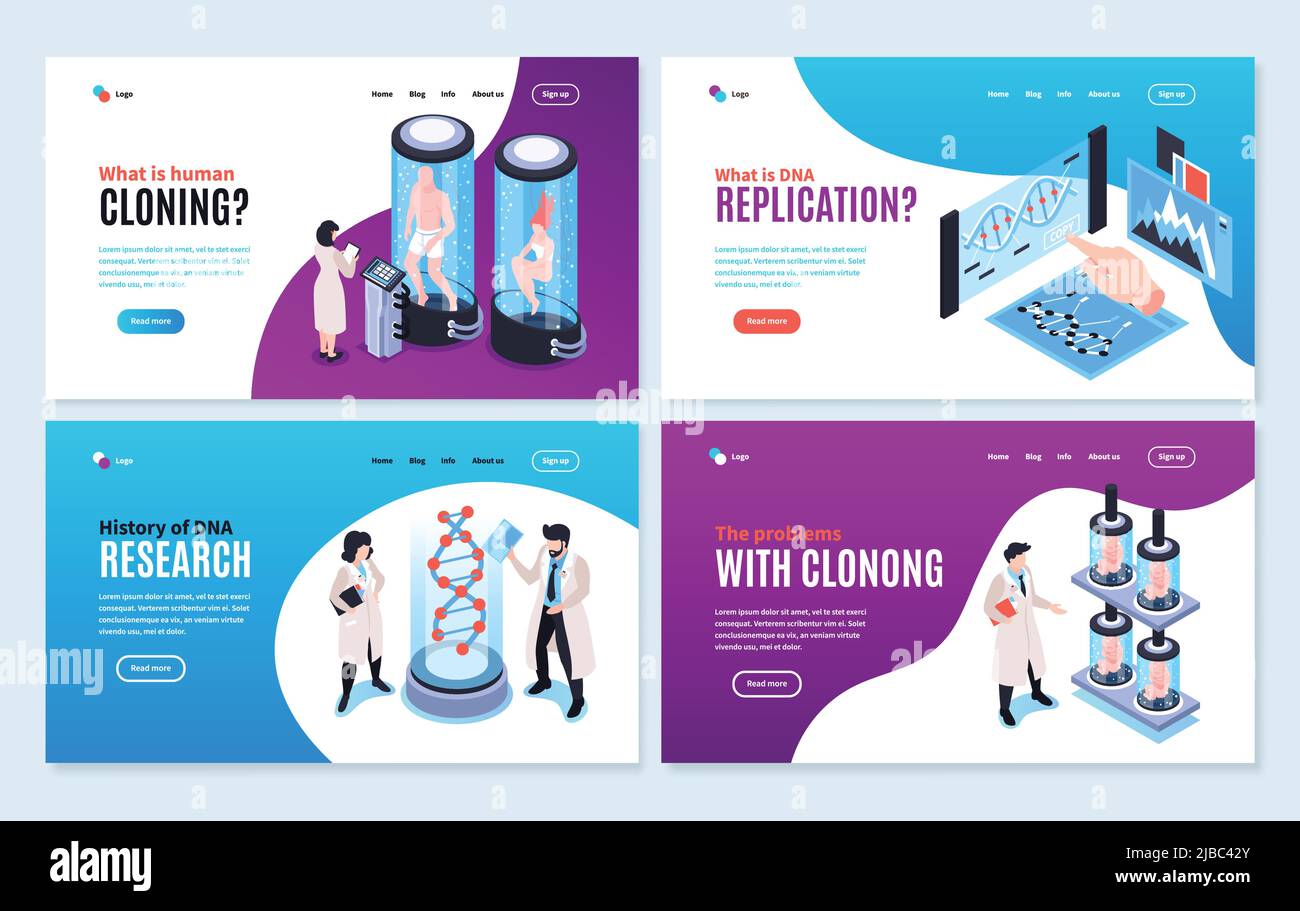 Human cloning isometric banners showing history and problems associated with human genome experiments vector illustration Stock Vectorhttps://www.alamy.com/image-license-details/?v=1https://www.alamy.com/human-cloning-isometric-banners-showing-history-and-problems-associated-with-human-genome-experiments-vector-illustration-image471707795.html
RF2JBC42Y–Human cloning isometric banners showing history and problems associated with human genome experiments vector illustration
 Diagram showing life cycle of Frog Stock Vectorhttps://www.alamy.com/image-license-details/?v=1https://www.alamy.com/diagram-showing-life-cycle-of-frog-image445242574.html
RF2GTAFCE–Diagram showing life cycle of Frog
 Baby in womb Stock Photohttps://www.alamy.com/image-license-details/?v=1https://www.alamy.com/stock-photo-baby-in-womb-125549954.html
RFH8786X–Baby in womb
 The human pod / 3D illustration of science fiction scene showing human male figure in foetal position inside complex futuristic incubator cloning mach Stock Photohttps://www.alamy.com/image-license-details/?v=1https://www.alamy.com/the-human-pod-3d-illustration-of-science-fiction-scene-showing-human-male-figure-in-foetal-position-inside-complex-futuristic-incubator-cloning-mach-image243501145.html
RFT44C5D–The human pod / 3D illustration of science fiction scene showing human male figure in foetal position inside complex futuristic incubator cloning mach
 Berlin, Germany. 27th Aug, 2019. Panda male Jiao Qing eats in his zoo enclosure. Panda female Meng-Meng is now in the zoo's indoor enclosure. The zoo today published an ultrasound of the female panda, showing an embryo with a beating heart. Credit: Paul Zinken/dpa/Alamy Live News Stock Photohttps://www.alamy.com/image-license-details/?v=1https://www.alamy.com/berlin-germany-27th-aug-2019-panda-male-jiao-qing-eats-in-his-zoo-enclosure-panda-female-meng-meng-is-now-in-the-zoos-indoor-enclosure-the-zoo-today-published-an-ultrasound-of-the-female-panda-showing-an-embryo-with-a-beating-heart-credit-paul-zinkendpaalamy-live-news-image265415092.html
RMWBPKJC–Berlin, Germany. 27th Aug, 2019. Panda male Jiao Qing eats in his zoo enclosure. Panda female Meng-Meng is now in the zoo's indoor enclosure. The zoo today published an ultrasound of the female panda, showing an embryo with a beating heart. Credit: Paul Zinken/dpa/Alamy Live News
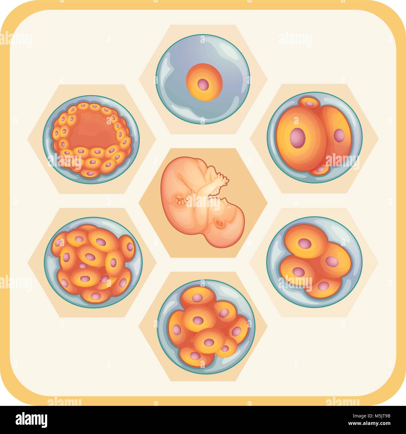 Diagram showing reproductive of human illustration Stock Vectorhttps://www.alamy.com/image-license-details/?v=1https://www.alamy.com/stock-photo-diagram-showing-reproductive-of-human-illustration-175591175.html
RFM5JT9B–Diagram showing reproductive of human illustration
 Parents showing their firstborn son pictures in family album Stock Photohttps://www.alamy.com/image-license-details/?v=1https://www.alamy.com/parents-showing-their-firstborn-son-pictures-in-family-album-image226671385.html
RFR4NNJH–Parents showing their firstborn son pictures in family album
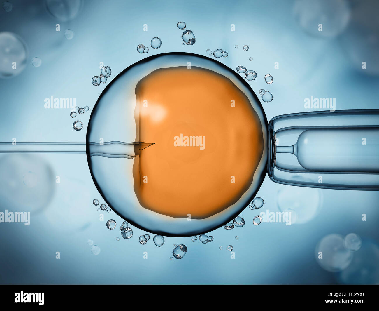 Artificial insemination illustration showing the ovule on blue background Stock Photohttps://www.alamy.com/image-license-details/?v=1https://www.alamy.com/stock-photo-artificial-insemination-illustration-showing-the-ovule-on-blue-background-96652529.html
RFFH6W81–Artificial insemination illustration showing the ovule on blue background
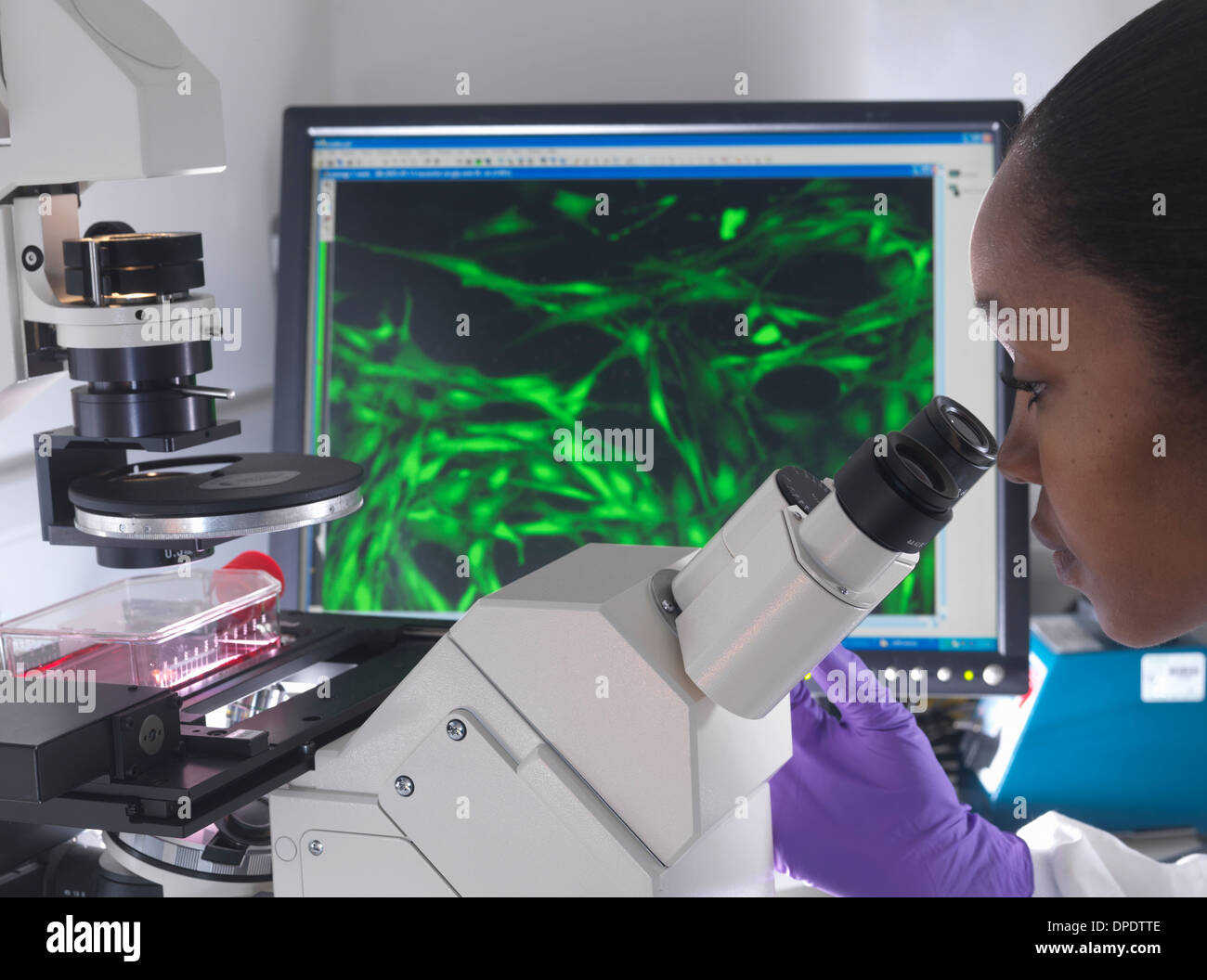 Female researcher using inverted microscope to view stem cells displayed showing fluorescent labeled cells Stock Photohttps://www.alamy.com/image-license-details/?v=1https://www.alamy.com/female-researcher-using-inverted-microscope-to-view-stem-cells-displayed-image65458414.html
RFDPDTTE–Female researcher using inverted microscope to view stem cells displayed showing fluorescent labeled cells
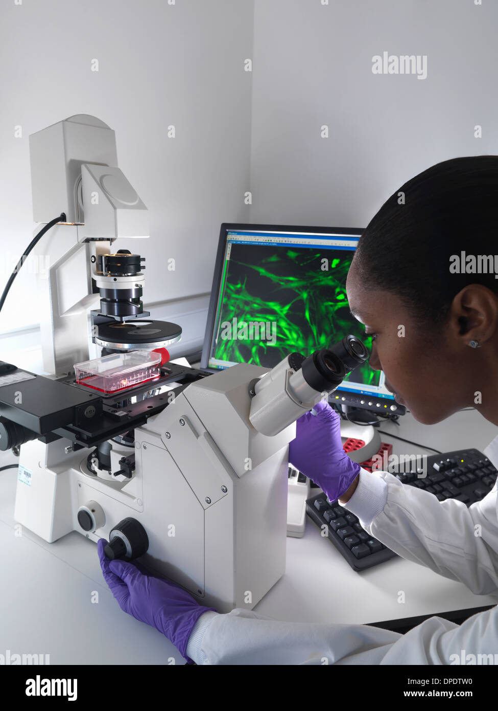 Female researcher using inverted microscope to view stem cells displayed showing fluorescent labeled cells Stock Photohttps://www.alamy.com/image-license-details/?v=1https://www.alamy.com/female-researcher-using-inverted-microscope-to-view-stem-cells-displayed-image65458428.html
RFDPDTW0–Female researcher using inverted microscope to view stem cells displayed showing fluorescent labeled cells
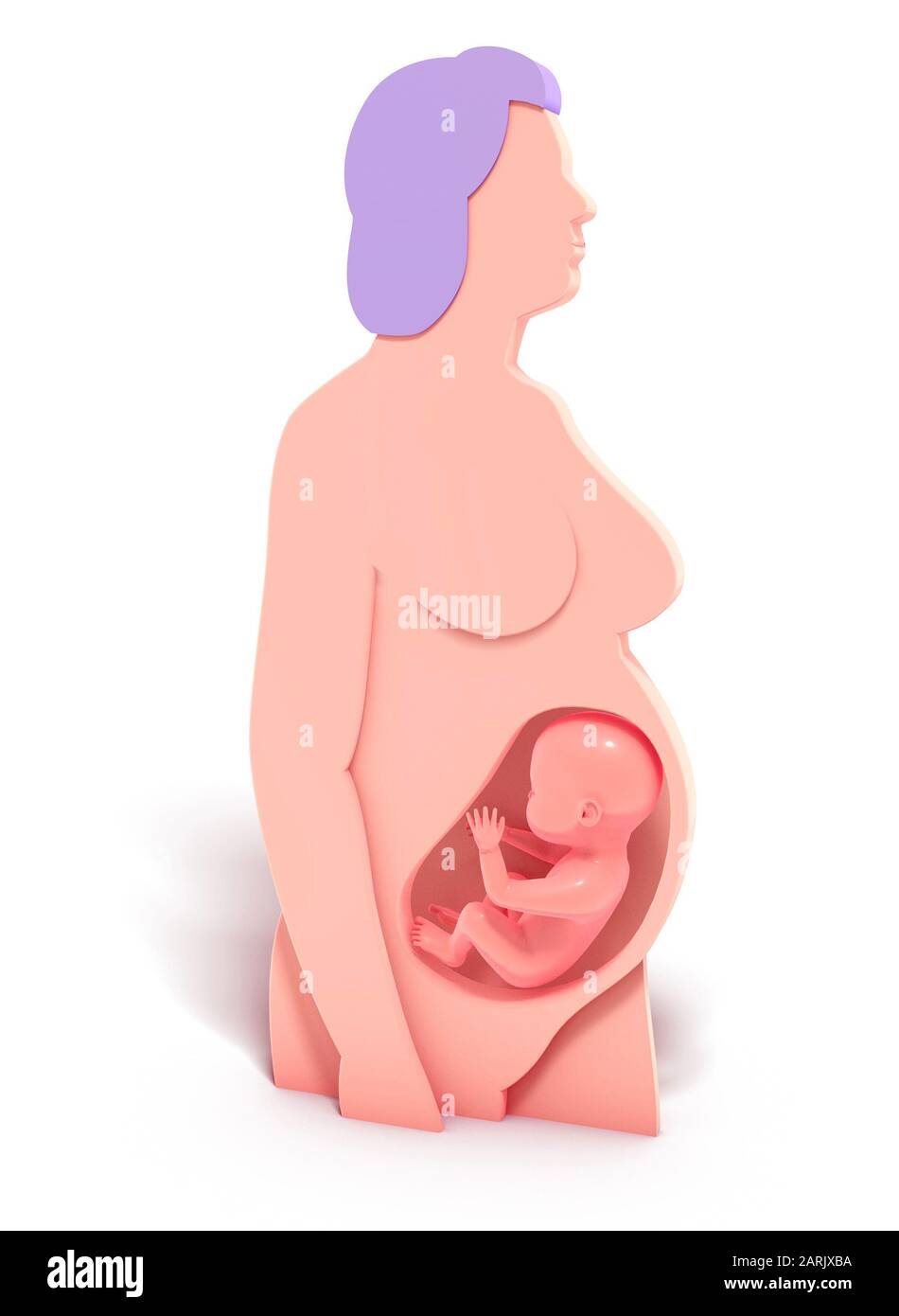 3D illustration of the graphic representation of a pregnancy. Anatomical interior of the woman in the form of emptying and the human fetus. Stock Photohttps://www.alamy.com/image-license-details/?v=1https://www.alamy.com/3d-illustration-of-the-graphic-representation-of-a-pregnancy-anatomical-interior-of-the-woman-in-the-form-of-emptying-and-the-human-fetus-image341549918.html
RF2ARJXBA–3D illustration of the graphic representation of a pregnancy. Anatomical interior of the woman in the form of emptying and the human fetus.
 Black and white photo of Pregnant woman with ultrasound scan in hands near belly. Female is holding sonogram baby embryo image over a pregnant tummy. Stock Photohttps://www.alamy.com/image-license-details/?v=1https://www.alamy.com/black-and-white-photo-of-pregnant-woman-with-ultrasound-scan-in-hands-near-belly-female-is-holding-sonogram-baby-embryo-image-over-a-pregnant-tummy-image590704057.html
RF2W90W1D–Black and white photo of Pregnant woman with ultrasound scan in hands near belly. Female is holding sonogram baby embryo image over a pregnant tummy.
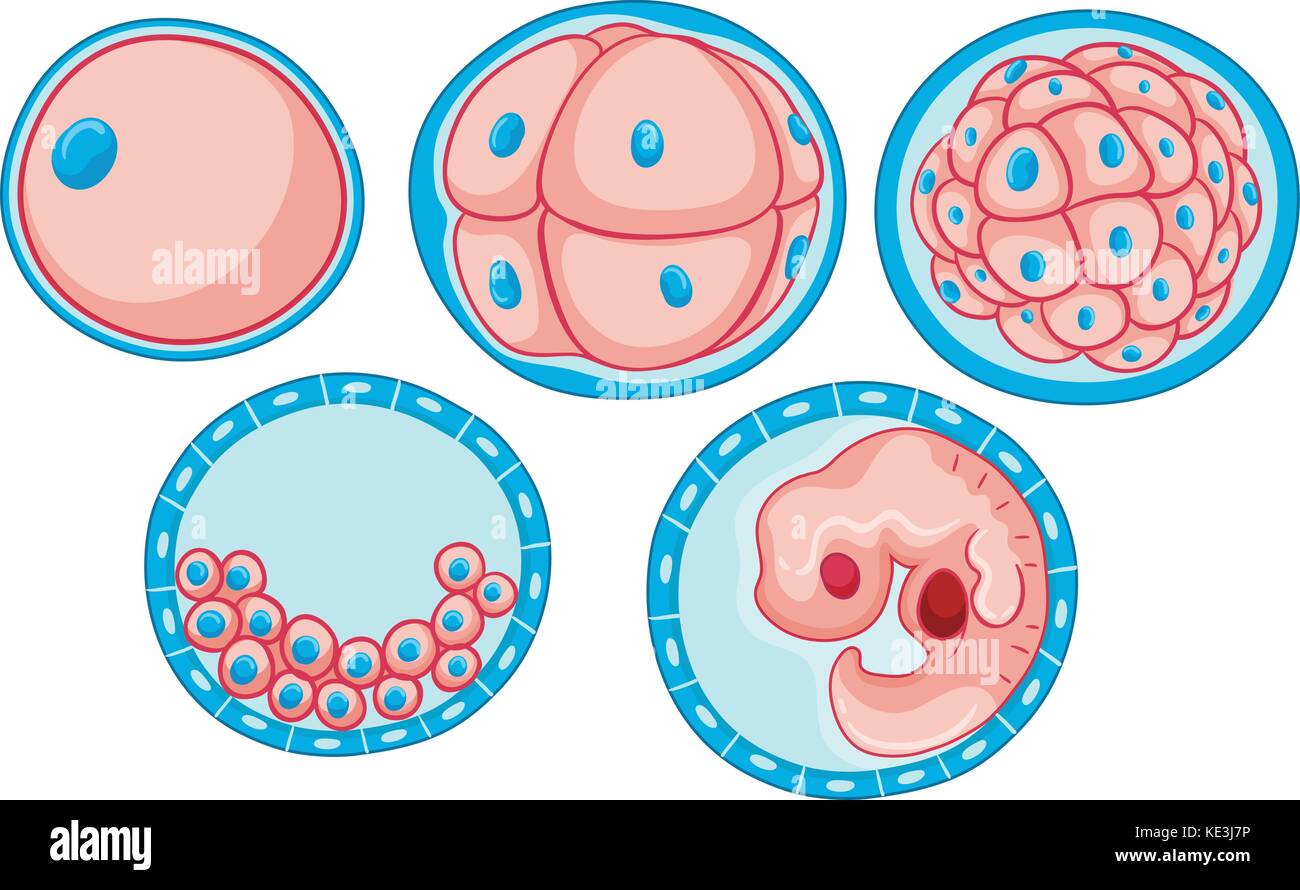 Diagram showing process of growing embryo illustration Stock Vectorhttps://www.alamy.com/image-license-details/?v=1https://www.alamy.com/stock-image-diagram-showing-process-of-growing-embryo-illustration-163578682.html
RFKE3J7P–Diagram showing process of growing embryo illustration
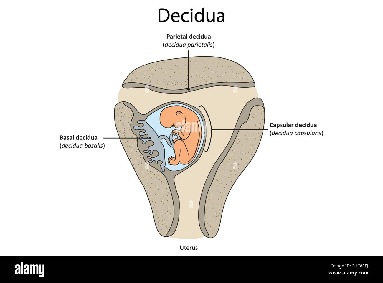 Simple illustration showing key parts of the decidua: decidua parietalis, decidua basilaris, capsular decidua. Stock Photohttps://www.alamy.com/image-license-details/?v=1https://www.alamy.com/simple-illustration-showing-key-parts-of-the-decidua-decidua-parietalis-decidua-basilaris-capsular-decidua-image455027962.html
RF2HC88PJ–Simple illustration showing key parts of the decidua: decidua parietalis, decidua basilaris, capsular decidua.
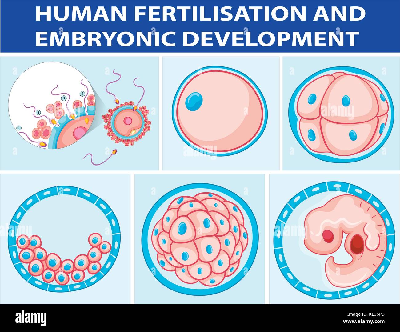 Diagram showing human fertilisation and embryonic development illustration Stock Vectorhttps://www.alamy.com/image-license-details/?v=1https://www.alamy.com/stock-image-diagram-showing-human-fertilisation-and-embryonic-development-illustration-163569685.html
RFKE36PD–Diagram showing human fertilisation and embryonic development illustration
 A smiling white-skinned nurse presenting a photo of an embryo - closeup. High quality photo Stock Photohttps://www.alamy.com/image-license-details/?v=1https://www.alamy.com/a-smiling-white-skinned-nurse-presenting-a-photo-of-an-embryo-closeup-high-quality-photo-image476348073.html
RF2JJYER5–A smiling white-skinned nurse presenting a photo of an embryo - closeup. High quality photo
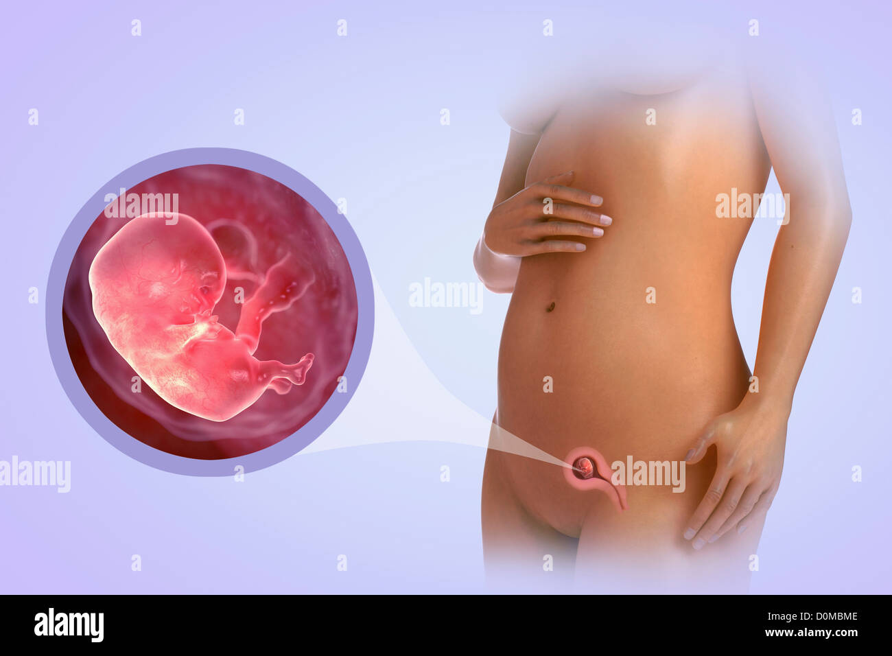 A human model showing pregnancy at week 9. Stock Photohttps://www.alamy.com/image-license-details/?v=1https://www.alamy.com/stock-photo-a-human-model-showing-pregnancy-at-week-9-52079342.html
RMD0MBME–A human model showing pregnancy at week 9.
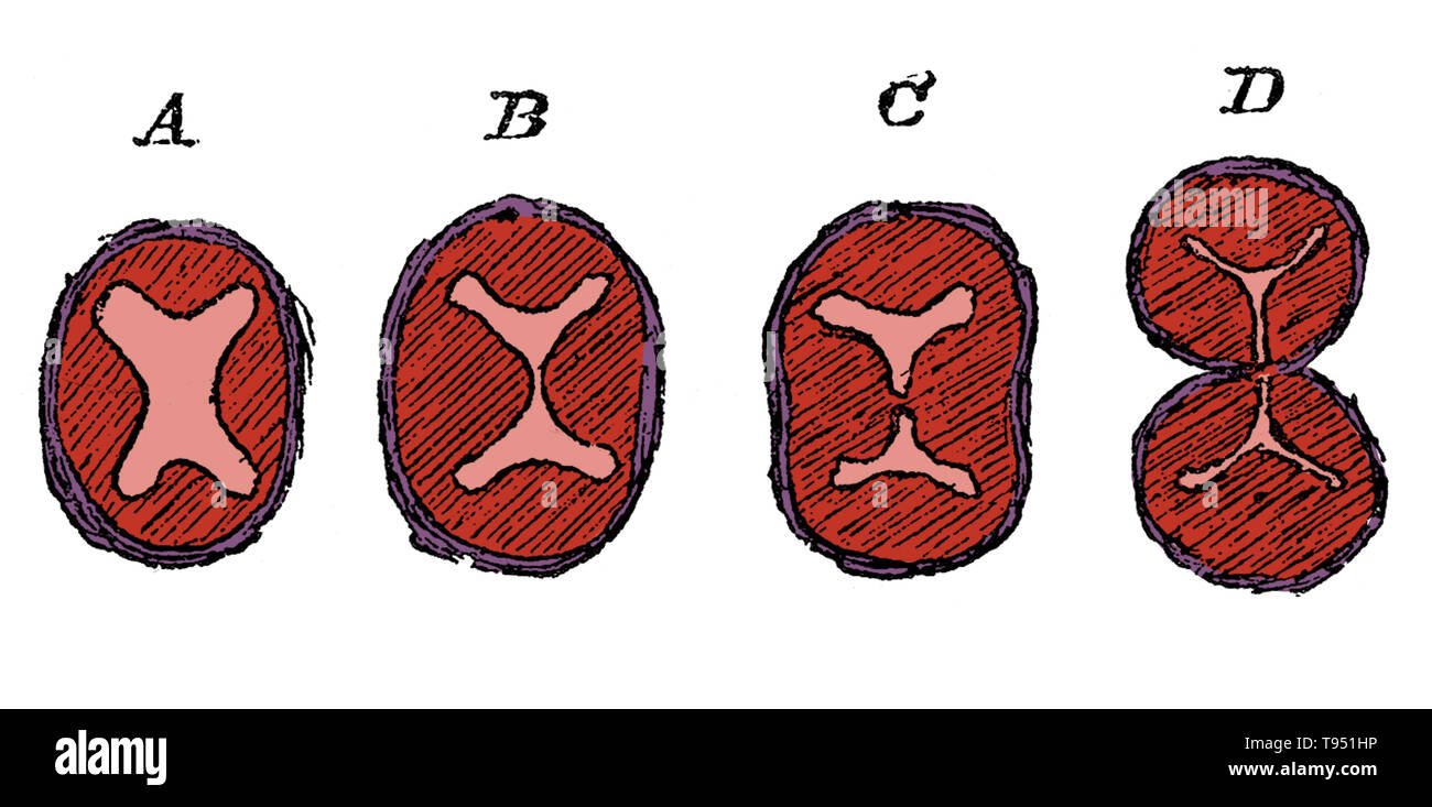 Diagram showing the division of the lower part of the bulbus aorta, aorta, and the formation of the semilunar valves. A, undivided truncus arteriosus with four endocardial cushions; B, advance of the two lateral cushions resulting in the division of the lumen; C, projection of three endocardial cushions in each part; D, the separation into aorta and pulmonary trunks completed. Stock Photohttps://www.alamy.com/image-license-details/?v=1https://www.alamy.com/diagram-showing-the-division-of-the-lower-part-of-the-bulbus-aorta-aorta-and-the-formation-of-the-semilunar-valves-a-undivided-truncus-arteriosus-with-four-endocardial-cushions-b-advance-of-the-two-lateral-cushions-resulting-in-the-division-of-the-lumen-c-projection-of-three-endocardial-cushions-in-each-part-d-the-separation-into-aorta-and-pulmonary-trunks-completed-image246588098.html
RMT951HP–Diagram showing the division of the lower part of the bulbus aorta, aorta, and the formation of the semilunar valves. A, undivided truncus arteriosus with four endocardial cushions; B, advance of the two lateral cushions resulting in the division of the lumen; C, projection of three endocardial cushions in each part; D, the separation into aorta and pulmonary trunks completed.
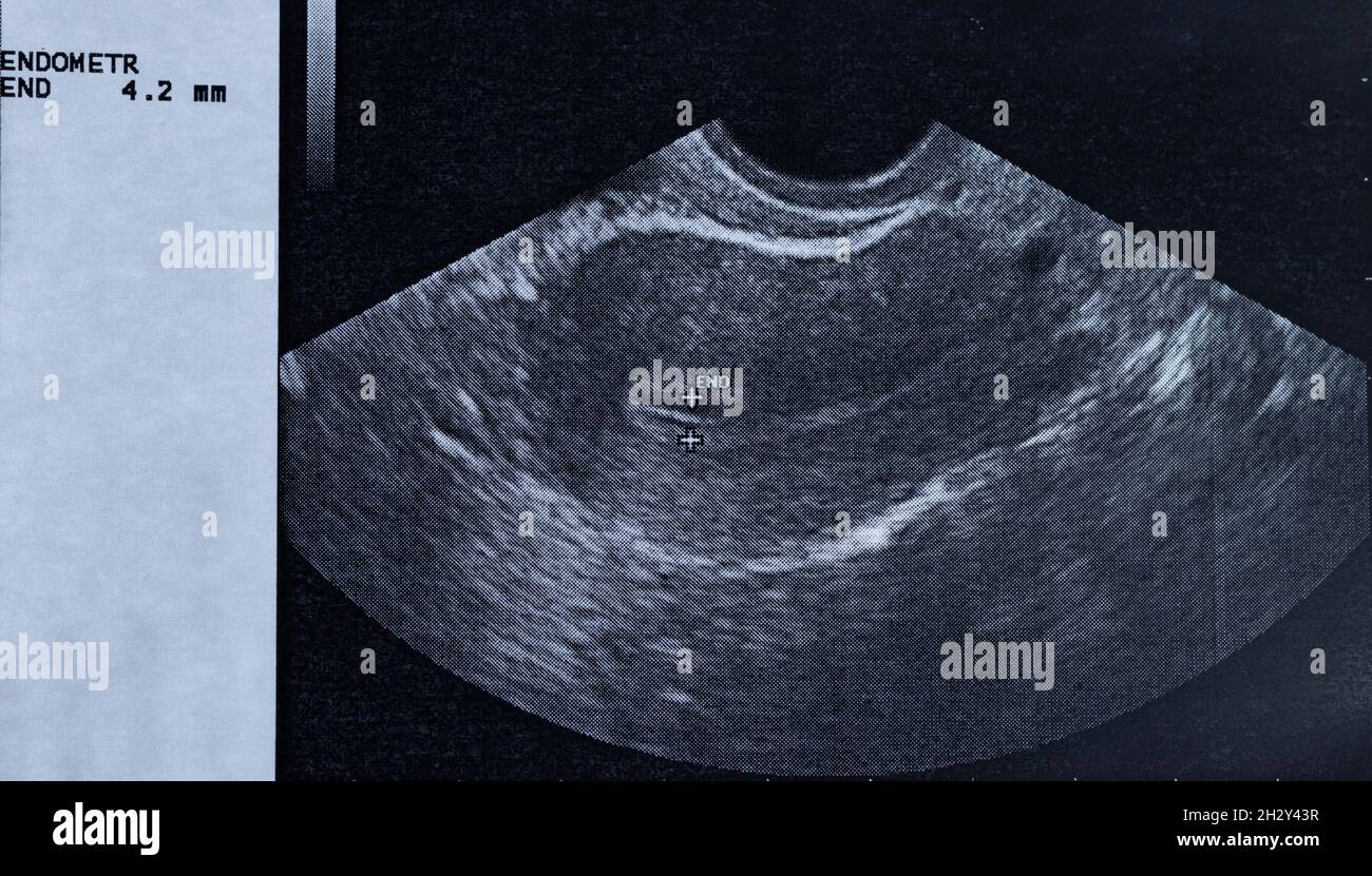 Ultrasound of a woman's uterus showing the size of the endometrium Stock Photohttps://www.alamy.com/image-license-details/?v=1https://www.alamy.com/ultrasound-of-a-womans-uterus-showing-the-size-of-the-endometrium-image449294827.html
RF2H2Y43R–Ultrasound of a woman's uterus showing the size of the endometrium
 Teenage boy laughing and showing his arms strength businessman i Stock Photohttps://www.alamy.com/image-license-details/?v=1https://www.alamy.com/stock-photo-teenage-boy-laughing-and-showing-his-arms-strength-businessman-i-90392711.html
RFF71MR3–Teenage boy laughing and showing his arms strength businessman i
RF2EDDW5E–Set of isolated decorative icons showing stages of human embryonic development with period clarification in months realistic vector illustration
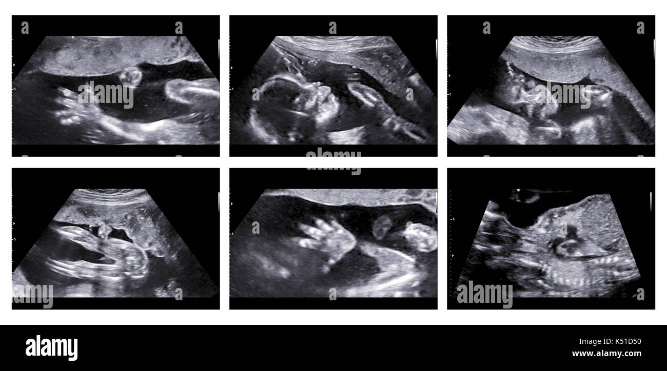 Collage of medical images of ultrasound anomaly scan on a female fetus 20 weeks into the pregnancy, showing child's hand, head, feet, legs, spine and Stock Photohttps://www.alamy.com/image-license-details/?v=1https://www.alamy.com/collage-of-medical-images-of-ultrasound-anomaly-scan-on-a-female-fetus-image157998876.html
RFK51D50–Collage of medical images of ultrasound anomaly scan on a female fetus 20 weeks into the pregnancy, showing child's hand, head, feet, legs, spine and
 The baby pod chrome / 3D illustration of science fiction scene showing human child asleep inside bright complex futuristic incubator cloning machinery Stock Photohttps://www.alamy.com/image-license-details/?v=1https://www.alamy.com/the-baby-pod-chrome-3d-illustration-of-science-fiction-scene-showing-human-child-asleep-inside-bright-complex-futuristic-incubator-cloning-machinery-image243609281.html
RFT49A3D–The baby pod chrome / 3D illustration of science fiction scene showing human child asleep inside bright complex futuristic incubator cloning machinery
 Berlin, Germany. 27th Aug, 2019. The sign is in the enclosure of Panda female Meng Meng's zoo in Berlin, indicating that Meng Meng is usually not seen at the moment, as she is preparing for her birth. The zoo today published an ultrasound of the female panda, showing an embryo with a beating heart. The announcement said that the birth could be expected in about two weeks. Credit: Paul Zinken/dpa/Alamy Live News Stock Photohttps://www.alamy.com/image-license-details/?v=1https://www.alamy.com/berlin-germany-27th-aug-2019-the-sign-is-in-the-enclosure-of-panda-female-meng-mengs-zoo-in-berlin-indicating-that-meng-meng-is-usually-not-seen-at-the-moment-as-she-is-preparing-for-her-birth-the-zoo-today-published-an-ultrasound-of-the-female-panda-showing-an-embryo-with-a-beating-heart-the-announcement-said-that-the-birth-could-be-expected-in-about-two-weeks-credit-paul-zinkendpaalamy-live-news-image265462679.html
RMWBTT9Y–Berlin, Germany. 27th Aug, 2019. The sign is in the enclosure of Panda female Meng Meng's zoo in Berlin, indicating that Meng Meng is usually not seen at the moment, as she is preparing for her birth. The zoo today published an ultrasound of the female panda, showing an embryo with a beating heart. The announcement said that the birth could be expected in about two weeks. Credit: Paul Zinken/dpa/Alamy Live News
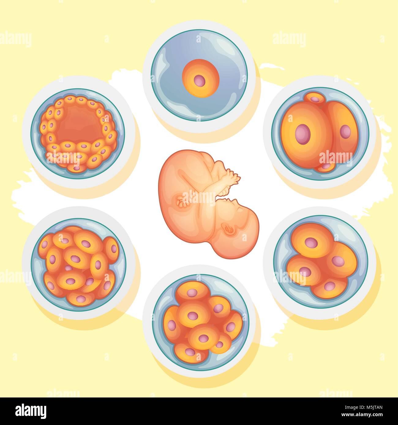 Diagram showing different stages of human baby illustration Stock Vectorhttps://www.alamy.com/image-license-details/?v=1https://www.alamy.com/stock-photo-diagram-showing-different-stages-of-human-baby-illustration-175591213.html
RFM5JTAN–Diagram showing different stages of human baby illustration
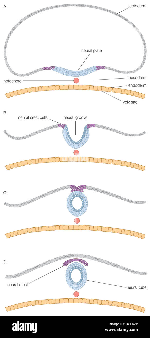 A cross section of the embryonic disk, showing formation of the neural tube. Stock Photohttps://www.alamy.com/image-license-details/?v=1https://www.alamy.com/stock-photo-a-cross-section-of-the-embryonic-disk-showing-formation-of-the-neural-24898350.html
RMBCE62P–A cross section of the embryonic disk, showing formation of the neural tube.
 Diagram showing how baby grows during pregnancy illustration Stock Vectorhttps://www.alamy.com/image-license-details/?v=1https://www.alamy.com/stock-photo-diagram-showing-how-baby-grows-during-pregnancy-illustration-139304789.html
RFJ2HTK1–Diagram showing how baby grows during pregnancy illustration
 Close up shot of woman holding up sonogram Stock Photohttps://www.alamy.com/image-license-details/?v=1https://www.alamy.com/close-up-shot-of-woman-holding-up-sonogram-image487160094.html
RF2K8G1JP–Close up shot of woman holding up sonogram
 caucasian female and male hand holding baby ultrasound photo at home inside bedroom, copy space, medicine and obstetrics concept Stock Photohttps://www.alamy.com/image-license-details/?v=1https://www.alamy.com/caucasian-female-and-male-hand-holding-baby-ultrasound-photo-at-home-inside-bedroom-copy-space-medicine-and-obstetrics-concept-image467565907.html
RF2J4KD2B–caucasian female and male hand holding baby ultrasound photo at home inside bedroom, copy space, medicine and obstetrics concept
 3D illustration of the graphic representation of a pregnancy. Anatomical interior of the woman in the form of emptying and the human fetus. Stock Photohttps://www.alamy.com/image-license-details/?v=1https://www.alamy.com/3d-illustration-of-the-graphic-representation-of-a-pregnancy-anatomical-interior-of-the-woman-in-the-form-of-emptying-and-the-human-fetus-image341549876.html
RF2ARJX9T–3D illustration of the graphic representation of a pregnancy. Anatomical interior of the woman in the form of emptying and the human fetus.
 Black and white photo of Pregnant woman with ultrasound scan in hands near belly. Female is holding sonogram baby embryo image over a pregnant tummy. Stock Photohttps://www.alamy.com/image-license-details/?v=1https://www.alamy.com/black-and-white-photo-of-pregnant-woman-with-ultrasound-scan-in-hands-near-belly-female-is-holding-sonogram-baby-embryo-image-over-a-pregnant-tummy-image590703565.html
RF2W90TBW–Black and white photo of Pregnant woman with ultrasound scan in hands near belly. Female is holding sonogram baby embryo image over a pregnant tummy.
 3D illustration of the graphic representation of a pregnancy. Anatomical interior of the woman in the form of emptying and the human fetus. Stock Photohttps://www.alamy.com/image-license-details/?v=1https://www.alamy.com/3d-illustration-of-the-graphic-representation-of-a-pregnancy-anatomical-interior-of-the-woman-in-the-form-of-emptying-and-the-human-fetus-image341549874.html
RF2ARJX9P–3D illustration of the graphic representation of a pregnancy. Anatomical interior of the woman in the form of emptying and the human fetus.
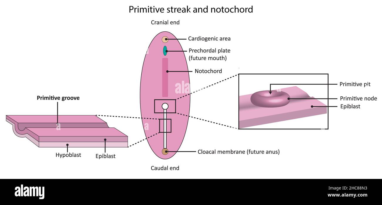 Simple diagram showing embryological developmentof primitive streak and notochord Stock Photohttps://www.alamy.com/image-license-details/?v=1https://www.alamy.com/simple-diagram-showing-embryological-developmentof-primitive-streak-and-notochord-image455027919.html
RF2HC88N3–Simple diagram showing embryological developmentof primitive streak and notochord
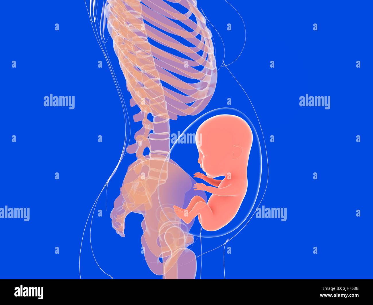 Anatomical 3d illustration of a human pregnancy. Image of the inside of a woman's body seen in profile. Stock Photohttps://www.alamy.com/image-license-details/?v=1https://www.alamy.com/anatomical-3d-illustration-of-a-human-pregnancy-image-of-the-inside-of-a-womans-body-seen-in-profile-image475462383.html
RF2JHF53B–Anatomical 3d illustration of a human pregnancy. Image of the inside of a woman's body seen in profile.
 A smiling white nurse in blue displaying a photo of an embryo - closeup. High quality photo Stock Photohttps://www.alamy.com/image-license-details/?v=1https://www.alamy.com/a-smiling-white-nurse-in-blue-displaying-a-photo-of-an-embryo-closeup-high-quality-photo-image476080404.html
RF2JJF9BG–A smiling white nurse in blue displaying a photo of an embryo - closeup. High quality photo
 A human model showing pregnancy at week 15. Stock Photohttps://www.alamy.com/image-license-details/?v=1https://www.alamy.com/stock-photo-a-human-model-showing-pregnancy-at-week-15-52079320.html
RMD0MBKM–A human model showing pregnancy at week 15.
 Pregnant woman demonstrating her ultrasound photo while sitting on sofa at home. Picture with ultrasound scan result in foreground and has focus. Stock Photohttps://www.alamy.com/image-license-details/?v=1https://www.alamy.com/pregnant-woman-demonstrating-her-ultrasound-photo-while-sitting-on-sofa-at-home-picture-with-ultrasound-scan-result-in-foreground-and-has-focus-image470017314.html
RF2J8K3TJ–Pregnant woman demonstrating her ultrasound photo while sitting on sofa at home. Picture with ultrasound scan result in foreground and has focus.
 Anti-abortion protest in a silent march, Berlin, Germany Stock Photohttps://www.alamy.com/image-license-details/?v=1https://www.alamy.com/stock-photo-anti-abortion-protest-in-a-silent-march-berlin-germany-36467289.html
RMC396B5–Anti-abortion protest in a silent march, Berlin, Germany
RFF919C2–Teen boy screaming and showing hands the power icon set Educatio
 Pregnant female is holding sonogram baby embryo image over a pregnant belly. Stock Photohttps://www.alamy.com/image-license-details/?v=1https://www.alamy.com/pregnant-female-is-holding-sonogram-baby-embryo-image-over-a-pregnant-belly-image596313459.html
RF2WJ4BW7–Pregnant female is holding sonogram baby embryo image over a pregnant belly.
 Principles and practice of operative dentistry . Incrementallines Fig. 165.—Oblique section of enamel and dentin, showing incremental (V. A. Latham.) y. Fig. 166.—Vertical Commencingformation ofthe root bylengthening ofthe pulp ;tion of face of human embryo, showing the beginning of the formation of the rootsof the teeth. (V. A. Latham.) X 7. Stock Photohttps://www.alamy.com/image-license-details/?v=1https://www.alamy.com/principles-and-practice-of-operative-dentistry-incrementallines-fig-165oblique-section-of-enamel-and-dentin-showing-incremental-v-a-latham-y-fig-166vertical-commencingformation-ofthe-root-bylengthening-ofthe-pulp-tion-of-face-of-human-embryo-showing-the-beginning-of-the-formation-of-the-rootsof-the-teeth-v-a-latham-x-7-image340255282.html
RM2ANFY2A–Principles and practice of operative dentistry . Incrementallines Fig. 165.—Oblique section of enamel and dentin, showing incremental (V. A. Latham.) y. Fig. 166.—Vertical Commencingformation ofthe root bylengthening ofthe pulp ;tion of face of human embryo, showing the beginning of the formation of the rootsof the teeth. (V. A. Latham.) X 7.
 Formation of the human foetus: five figures, showing the development from embryo at conception, to foetus of four months gestation. Colour engraving by J. Pass after D. Dodd, 1794. Stock Photohttps://www.alamy.com/image-license-details/?v=1https://www.alamy.com/formation-of-the-human-foetus-five-figures-showing-the-development-from-embryo-at-conception-to-foetus-of-four-months-gestation-colour-engraving-by-j-pass-after-d-dodd-1794-image449994849.html
RM2H4310H–Formation of the human foetus: five figures, showing the development from embryo at conception, to foetus of four months gestation. Colour engraving by J. Pass after D. Dodd, 1794.
 Berlin, Germany. 27th Aug, 2019. Panda male Jiao Qing eats in his zoo enclosure. Meanwhile panda female Meng Meng is in the inside enclosure of the zoo away from the visitors. The zoo today published an ultrasound of the female panda, showing an embryo with a beating heart. The announcement said that the birth could be expected in about two weeks. Credit: Paul Zinken/dpa/Alamy Live News Stock Photohttps://www.alamy.com/image-license-details/?v=1https://www.alamy.com/berlin-germany-27th-aug-2019-panda-male-jiao-qing-eats-in-his-zoo-enclosure-meanwhile-panda-female-meng-meng-is-in-the-inside-enclosure-of-the-zoo-away-from-the-visitors-the-zoo-today-published-an-ultrasound-of-the-female-panda-showing-an-embryo-with-a-beating-heart-the-announcement-said-that-the-birth-could-be-expected-in-about-two-weeks-credit-paul-zinkendpaalamy-live-news-image265462649.html
RMWBTT8W–Berlin, Germany. 27th Aug, 2019. Panda male Jiao Qing eats in his zoo enclosure. Meanwhile panda female Meng Meng is in the inside enclosure of the zoo away from the visitors. The zoo today published an ultrasound of the female panda, showing an embryo with a beating heart. The announcement said that the birth could be expected in about two weeks. Credit: Paul Zinken/dpa/Alamy Live News
 Young parents rejoice in pregnancy. A married couple is holding an ultrasound scan showing their child. Stock Photohttps://www.alamy.com/image-license-details/?v=1https://www.alamy.com/young-parents-rejoice-in-pregnancy-a-married-couple-is-holding-an-ultrasound-scan-showing-their-child-image382527163.html
RF2D69H8B–Young parents rejoice in pregnancy. A married couple is holding an ultrasound scan showing their child.
 3D obstetric ultrasound of a fetus, 4D baby scan showing movement, gynecology, Hospital Policlinica Gipuzkoa, San Sebastian, Donostia, Euskadi, Spain. Stock Photohttps://www.alamy.com/image-license-details/?v=1https://www.alamy.com/3d-obstetric-ultrasound-of-a-fetus-4d-baby-scan-showing-movement-gynecology-hospital-policlinica-gipuzkoa-san-sebastian-donostia-euskadi-spain-image602285825.html
RM2WYTDM1–3D obstetric ultrasound of a fetus, 4D baby scan showing movement, gynecology, Hospital Policlinica Gipuzkoa, San Sebastian, Donostia, Euskadi, Spain.
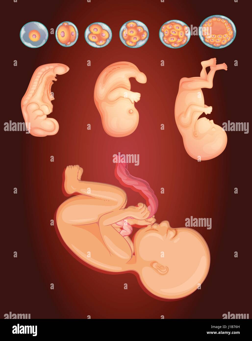 Diagram showing baby growing inside woman womb illustration Stock Vectorhttps://www.alamy.com/image-license-details/?v=1https://www.alamy.com/stock-photo-diagram-showing-baby-growing-inside-woman-womb-illustration-138544745.html
RFJ1B76H–Diagram showing baby growing inside woman womb illustration
 Chicken egg in a female hand. A female hand holds a chicken egg. Stock Photohttps://www.alamy.com/image-license-details/?v=1https://www.alamy.com/chicken-egg-in-a-female-hand-a-female-hand-holds-a-chicken-egg-image426515525.html
RF2FNWCW9–Chicken egg in a female hand. A female hand holds a chicken egg.
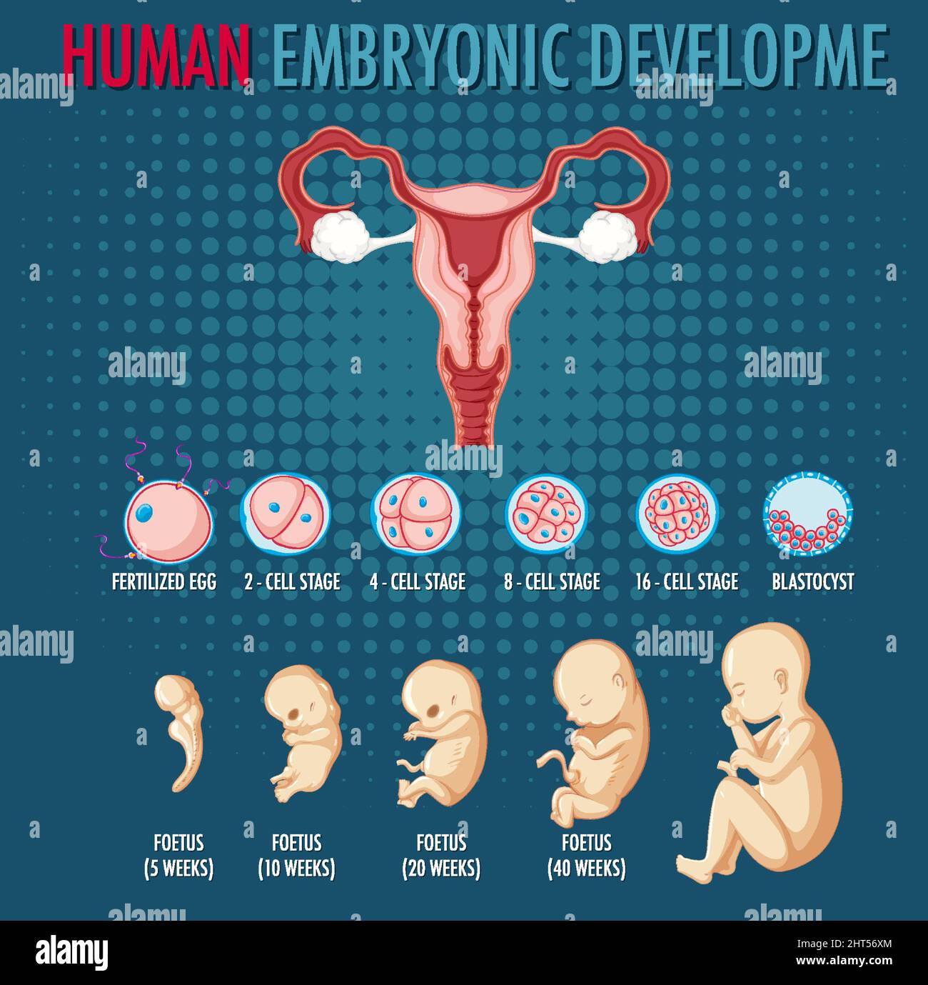 Diagram showing human embryonic development illustration Stock Vectorhttps://www.alamy.com/image-license-details/?v=1https://www.alamy.com/diagram-showing-human-embryonic-development-illustration-image462336524.html
RF2HT56XM–Diagram showing human embryonic development illustration
 Wheat. Germinating. Showing shoots and roots after a soaking in water for 48 hours. Stock Photohttps://www.alamy.com/image-license-details/?v=1https://www.alamy.com/stock-photo-wheat-germinating-showing-shoots-and-roots-after-a-soaking-in-water-72571648.html
RME61WTG–Wheat. Germinating. Showing shoots and roots after a soaking in water for 48 hours.
 Black and white photo of Pregnant woman with ultrasound scan in hands near belly. Female is holding sonogram baby embryo image over a pregnant tummy. Stock Photohttps://www.alamy.com/image-license-details/?v=1https://www.alamy.com/black-and-white-photo-of-pregnant-woman-with-ultrasound-scan-in-hands-near-belly-female-is-holding-sonogram-baby-embryo-image-over-a-pregnant-tummy-image590704028.html
RF2W90W0C–Black and white photo of Pregnant woman with ultrasound scan in hands near belly. Female is holding sonogram baby embryo image over a pregnant tummy.
 Male cartoon doctor showing student infographics with baby Stock Vectorhttps://www.alamy.com/image-license-details/?v=1https://www.alamy.com/male-cartoon-doctor-showing-student-infographics-with-baby-image554147519.html
RF2R5FGPR–Male cartoon doctor showing student infographics with baby
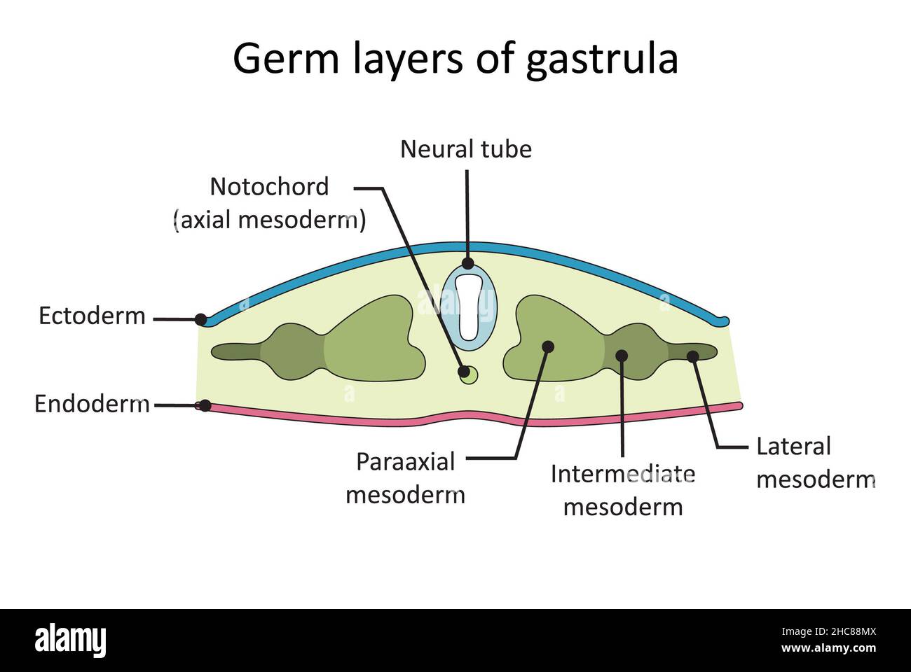 Simple diagram showing germ layers developed in the stage of gastrula Stock Photohttps://www.alamy.com/image-license-details/?v=1https://www.alamy.com/simple-diagram-showing-germ-layers-developed-in-the-stage-of-gastrula-image455027914.html
RF2HC88MX–Simple diagram showing germ layers developed in the stage of gastrula
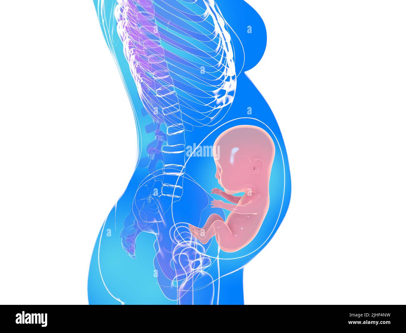 Anatomical 3d illustration of a human pregnancy in an advanced stage of gestation. Image of transparent woman's body silhouette. Stock Photohttps://www.alamy.com/image-license-details/?v=1https://www.alamy.com/anatomical-3d-illustration-of-a-human-pregnancy-in-an-advanced-stage-of-gestation-image-of-transparent-womans-body-silhouette-image475462117.html
RF2JHF4NW–Anatomical 3d illustration of a human pregnancy in an advanced stage of gestation. Image of transparent woman's body silhouette.
 In vitro fertilization. Detailed infographic showing laboratory fertilization of eggs. Stock Vectorhttps://www.alamy.com/image-license-details/?v=1https://www.alamy.com/in-vitro-fertilization-detailed-infographic-showing-laboratory-fertilization-of-eggs-image457041888.html
RF2HFG1GG–In vitro fertilization. Detailed infographic showing laboratory fertilization of eggs.
 Anatomical 3d illustration of a pregnancy. Image of the inside of a woman's body showing a fetus in an advanced stage of pregnancy. Stock Photohttps://www.alamy.com/image-license-details/?v=1https://www.alamy.com/anatomical-3d-illustration-of-a-pregnancy-image-of-the-inside-of-a-womans-body-showing-a-fetus-in-an-advanced-stage-of-pregnancy-image475462150.html
RF2JHF4R2–Anatomical 3d illustration of a pregnancy. Image of the inside of a woman's body showing a fetus in an advanced stage of pregnancy.
 A human model showing pregnancy at week 18. Stock Photohttps://www.alamy.com/image-license-details/?v=1https://www.alamy.com/stock-photo-a-human-model-showing-pregnancy-at-week-18-52087685.html
RMD0MPAD–A human model showing pregnancy at week 18.
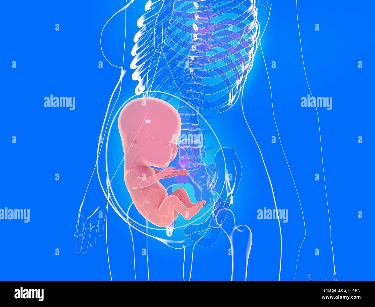 3d anatomical illustration of a pregnancy. Image of the inside of a woman's body with light showing a fetus in an advanced stage of pregnancy. Stock Photohttps://www.alamy.com/image-license-details/?v=1https://www.alamy.com/3d-anatomical-illustration-of-a-pregnancy-image-of-the-inside-of-a-womans-body-with-light-showing-a-fetus-in-an-advanced-stage-of-pregnancy-image475462169.html
RF2JHF4RN–3d anatomical illustration of a pregnancy. Image of the inside of a woman's body with light showing a fetus in an advanced stage of pregnancy.
 . The mammary apparatus of the mammalia : in the light of ontogenesis and phylogenesis . Mammals; Mammary glands. THE MILK-STREAK 107 which are not preceded by any primordia cor- responding to the milk-streak. Secondly, it follows that when we trace back the mammary apparatus of the Placentals, we have to com- pare the primary-primordia, not with the milk- lines, but with the milk-streaks. And indeed this comparison presents no difficulty.. Fig. 38.—Human Embryo, showing "Milk-Streak" AND Central Region Becoming Elevated to Form "Milk-Line." (Schmitt.) 11'5 mm. It is clear, Stock Photohttps://www.alamy.com/image-license-details/?v=1https://www.alamy.com/the-mammary-apparatus-of-the-mammalia-in-the-light-of-ontogenesis-and-phylogenesis-mammals-mammary-glands-the-milk-streak-107-which-are-not-preceded-by-any-primordia-cor-responding-to-the-milk-streak-secondly-it-follows-that-when-we-trace-back-the-mammary-apparatus-of-the-placentals-we-have-to-com-pare-the-primary-primordia-not-with-the-milk-lines-but-with-the-milk-streaks-and-indeed-this-comparison-presents-no-difficulty-fig-38human-embryo-showing-quotmilk-streakquot-and-central-region-becoming-elevated-to-form-quotmilk-linequot-schmitt-115-mm-it-is-clear-image216449389.html
RMPG43B9–. The mammary apparatus of the mammalia : in the light of ontogenesis and phylogenesis . Mammals; Mammary glands. THE MILK-STREAK 107 which are not preceded by any primordia cor- responding to the milk-streak. Secondly, it follows that when we trace back the mammary apparatus of the Placentals, we have to com- pare the primary-primordia, not with the milk- lines, but with the milk-streaks. And indeed this comparison presents no difficulty.. Fig. 38.—Human Embryo, showing "Milk-Streak" AND Central Region Becoming Elevated to Form "Milk-Line." (Schmitt.) 11'5 mm. It is clear,
 3d anatomical illustration of a pregnancy in an advanced stage of gestation. Image of the transparent woman's body with natural colors. Stock Photohttps://www.alamy.com/image-license-details/?v=1https://www.alamy.com/3d-anatomical-illustration-of-a-pregnancy-in-an-advanced-stage-of-gestation-image-of-the-transparent-womans-body-with-natural-colors-image475462139.html
RF2JHF4PK–3d anatomical illustration of a pregnancy in an advanced stage of gestation. Image of the transparent woman's body with natural colors.
 Pregnant female is holding sonogram baby embryo image over a pregnant belly. Stock Photohttps://www.alamy.com/image-license-details/?v=1https://www.alamy.com/pregnant-female-is-holding-sonogram-baby-embryo-image-over-a-pregnant-belly-image596313441.html
RF2WJ4BTH–Pregnant female is holding sonogram baby embryo image over a pregnant belly.
 Human anatomy, including structure and development and practical considerations . 7-0-. Xeurobhists Efferent axones Portion of spinal cord of human embryo, showing development ofventral root-axones as outgrowths from ventral neuroblasts. X 300.< After His.) Die Entwickfclung des menschlichen Gehirns, 1904.Amer. Journal of Anatomy, vol. ii., 1903. IOI2 HUMAN ANATOMY. Fig. 859. Stock Photohttps://www.alamy.com/image-license-details/?v=1https://www.alamy.com/human-anatomy-including-structure-and-development-and-practical-considerations-7-0-xeurobhists-efferent-axones-portion-of-spinal-cord-of-human-embryo-showing-development-ofventral-root-axones-as-outgrowths-from-ventral-neuroblasts-x-300lt-after-his-die-entwickfclung-des-menschlichen-gehirns-1904amer-journal-of-anatomy-vol-ii-1903-ioi2-human-anatomy-fig-859-image342723016.html
RM2AWGAKM–Human anatomy, including structure and development and practical considerations . 7-0-. Xeurobhists Efferent axones Portion of spinal cord of human embryo, showing development ofventral root-axones as outgrowths from ventral neuroblasts. X 300.< After His.) Die Entwickfclung des menschlichen Gehirns, 1904.Amer. Journal of Anatomy, vol. ii., 1903. IOI2 HUMAN ANATOMY. Fig. 859.
 Berlin, Germany. 27th Aug, 2019. Panda male Jiao Qing eats in his zoo enclosure. Meanwhile panda female Meng Meng is in the inside enclosure of the zoo away from the visitors. The zoo today published an ultrasound of the female panda, showing an embryo with a beating heart. The announcement said that the birth could be expected in about two weeks. Credit: Paul Zinken/dpa/Alamy Live News Stock Photohttps://www.alamy.com/image-license-details/?v=1https://www.alamy.com/berlin-germany-27th-aug-2019-panda-male-jiao-qing-eats-in-his-zoo-enclosure-meanwhile-panda-female-meng-meng-is-in-the-inside-enclosure-of-the-zoo-away-from-the-visitors-the-zoo-today-published-an-ultrasound-of-the-female-panda-showing-an-embryo-with-a-beating-heart-the-announcement-said-that-the-birth-could-be-expected-in-about-two-weeks-credit-paul-zinkendpaalamy-live-news-image265462652.html
RMWBTT90–Berlin, Germany. 27th Aug, 2019. Panda male Jiao Qing eats in his zoo enclosure. Meanwhile panda female Meng Meng is in the inside enclosure of the zoo away from the visitors. The zoo today published an ultrasound of the female panda, showing an embryo with a beating heart. The announcement said that the birth could be expected in about two weeks. Credit: Paul Zinken/dpa/Alamy Live News
 Young parents rejoice in pregnancy. A married couple is holding an ultrasound scan showing their child. Stock Photohttps://www.alamy.com/image-license-details/?v=1https://www.alamy.com/young-parents-rejoice-in-pregnancy-a-married-couple-is-holding-an-ultrasound-scan-showing-their-child-image382527309.html
RF2D69HDH–Young parents rejoice in pregnancy. A married couple is holding an ultrasound scan showing their child.
 3D obstetric ultrasound of a fetus, 4D baby scan showing movement, gynecology, Hospital Policlinica Gipuzkoa, San Sebastian, Donostia, Euskadi, Spain. Stock Photohttps://www.alamy.com/image-license-details/?v=1https://www.alamy.com/3d-obstetric-ultrasound-of-a-fetus-4d-baby-scan-showing-movement-gynecology-hospital-policlinica-gipuzkoa-san-sebastian-donostia-euskadi-spain-image602267249.html
RM2WYRJ0H–3D obstetric ultrasound of a fetus, 4D baby scan showing movement, gynecology, Hospital Policlinica Gipuzkoa, San Sebastian, Donostia, Euskadi, Spain.
 Pregnant belly with ultrasound result picture. Woman see unborned kid face Stock Photohttps://www.alamy.com/image-license-details/?v=1https://www.alamy.com/pregnant-belly-with-ultrasound-result-picture-woman-see-unborned-kid-face-image447918178.html
RF2H0MC5P–Pregnant belly with ultrasound result picture. Woman see unborned kid face
 Diagram showing life cycle of Frog illustration Stock Vectorhttps://www.alamy.com/image-license-details/?v=1https://www.alamy.com/diagram-showing-life-cycle-of-frog-illustration-image424606621.html
RF2FJPE25–Diagram showing life cycle of Frog illustration
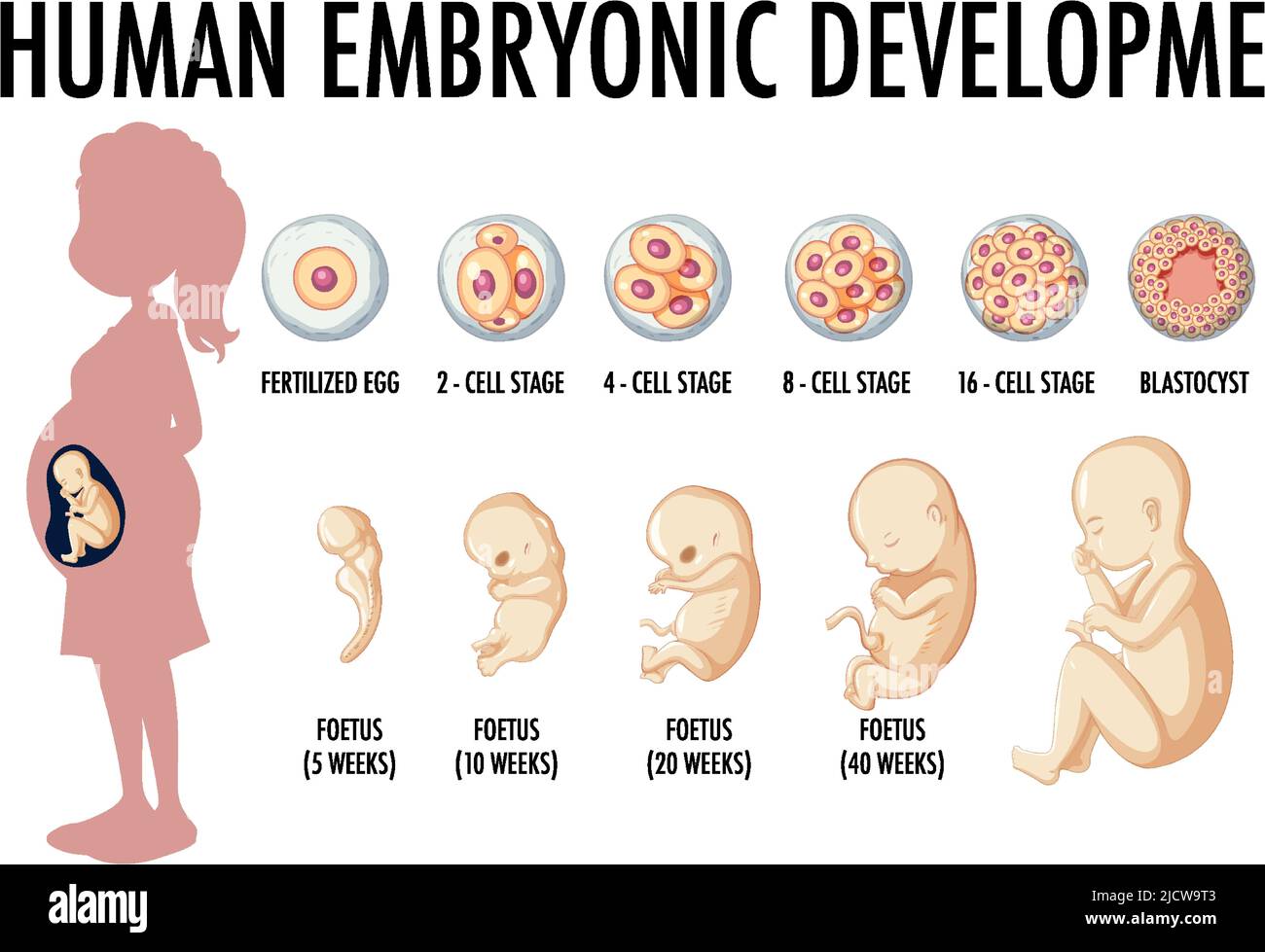 Diagram showing human embryonic development illustration Stock Vectorhttps://www.alamy.com/image-license-details/?v=1https://www.alamy.com/diagram-showing-human-embryonic-development-illustration-image472612339.html
RF2JCW9T3–Diagram showing human embryonic development illustration
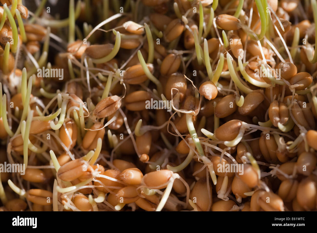 Wheat. Germinating. Showing shoots and roots after a soaking in water for 48 hours. Stock Photohttps://www.alamy.com/image-license-details/?v=1https://www.alamy.com/stock-photo-wheat-germinating-showing-shoots-and-roots-after-a-soaking-in-water-72571644.html
RME61WTC–Wheat. Germinating. Showing shoots and roots after a soaking in water for 48 hours.
 Human cloning isometric banners showing history and problems associated with human genome experiments vector illustration Stock Vectorhttps://www.alamy.com/image-license-details/?v=1https://www.alamy.com/human-cloning-isometric-banners-showing-history-and-problems-associated-with-human-genome-experiments-vector-illustration-image472061197.html
RF2JC06TD–Human cloning isometric banners showing history and problems associated with human genome experiments vector illustration
 Male cartoon doctor showing student infographics with baby Stock Vectorhttps://www.alamy.com/image-license-details/?v=1https://www.alamy.com/male-cartoon-doctor-showing-student-infographics-with-baby-image544308697.html
RF2PHFB89–Male cartoon doctor showing student infographics with baby
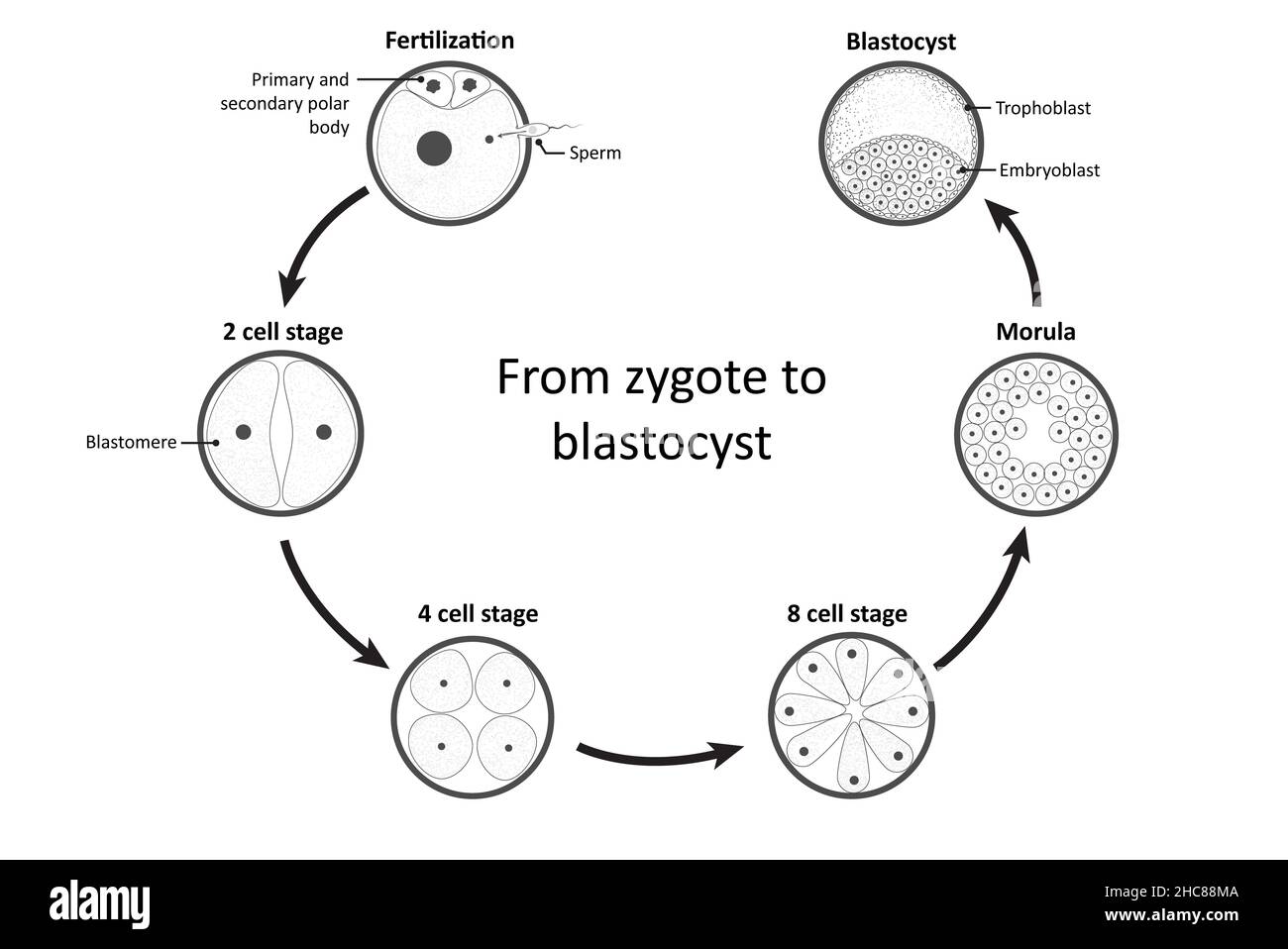 Diagram showing simplified process of fertilization and development from zygote to blastocyst (emphasis at the totipotency) Stock Photohttps://www.alamy.com/image-license-details/?v=1https://www.alamy.com/diagram-showing-simplified-process-of-fertilization-and-development-from-zygote-to-blastocyst-emphasis-at-the-totipotency-image455027898.html
RF2HC88MA–Diagram showing simplified process of fertilization and development from zygote to blastocyst (emphasis at the totipotency)
 Diagram showing life cycle of Frog illustration Stock Vectorhttps://www.alamy.com/image-license-details/?v=1https://www.alamy.com/diagram-showing-life-cycle-of-frog-illustration-image406400715.html
RF2EH5477–Diagram showing life cycle of Frog illustration
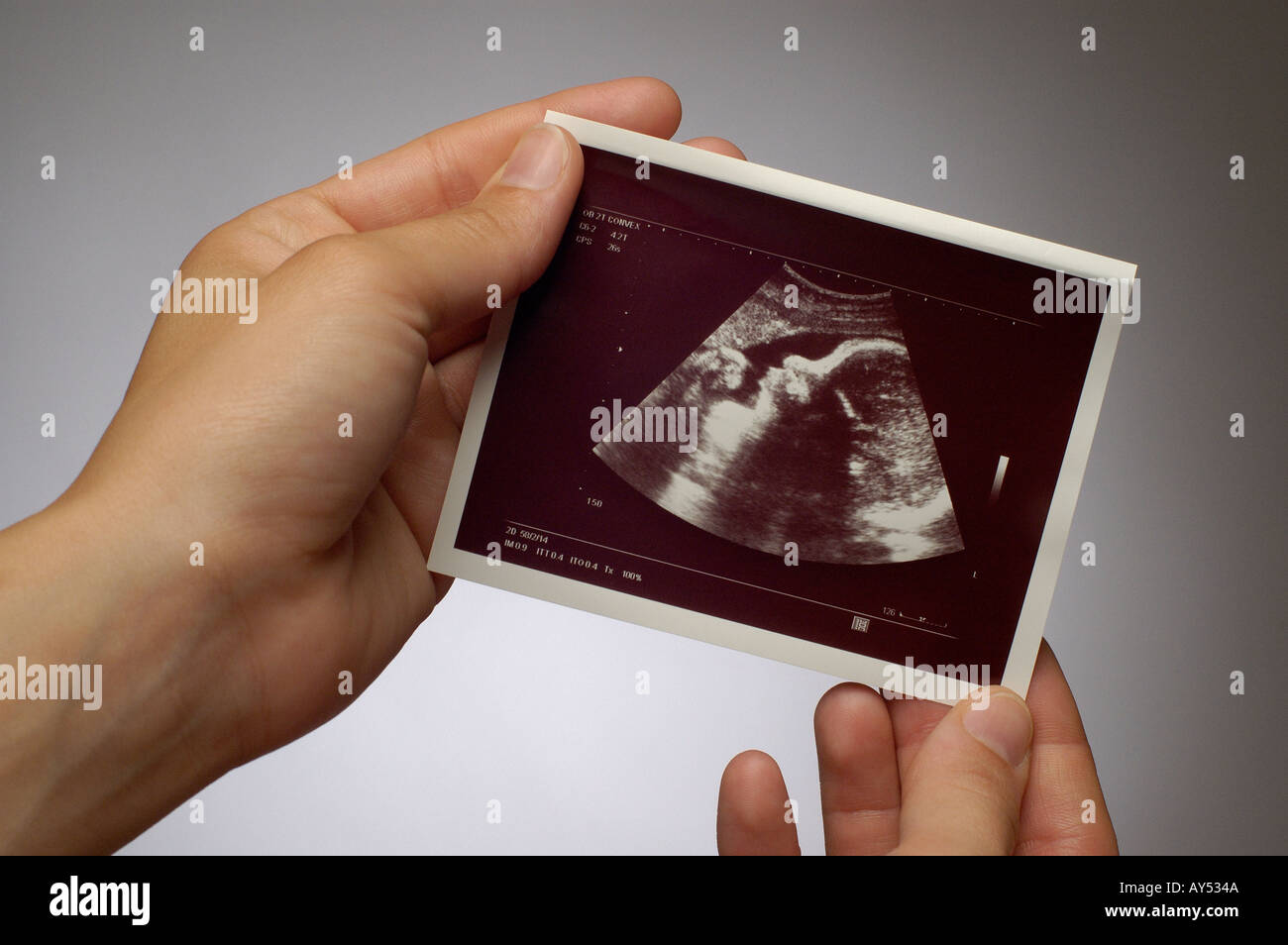 ULTRASOUND IMAGE SCAN Stock Photohttps://www.alamy.com/image-license-details/?v=1https://www.alamy.com/ultrasound-image-scan-image1790793.html
RFAY534A–ULTRASOUND IMAGE SCAN
 Embryologist is showing cryovial filled with cryopreserved biological samples Stock Photohttps://www.alamy.com/image-license-details/?v=1https://www.alamy.com/embryologist-is-showing-cryovial-filled-with-cryopreserved-biological-samples-image570430204.html
RF2T419F8–Embryologist is showing cryovial filled with cryopreserved biological samples
 A human model showing pregnancy at week 13. Stock Photohttps://www.alamy.com/image-license-details/?v=1https://www.alamy.com/stock-photo-a-human-model-showing-pregnancy-at-week-13-52079350.html
RMD0MBMP–A human model showing pregnancy at week 13.
 Text sign showing Stem. Conceptual photo Life Sciences Engineering in all aspect Continuous expanding technology. Stock Photohttps://www.alamy.com/image-license-details/?v=1https://www.alamy.com/text-sign-showing-stem-conceptual-photo-life-sciences-engineering-in-all-aspect-continuous-expanding-technology-image223530815.html
RFPYJKRB–Text sign showing Stem. Conceptual photo Life Sciences Engineering in all aspect Continuous expanding technology.
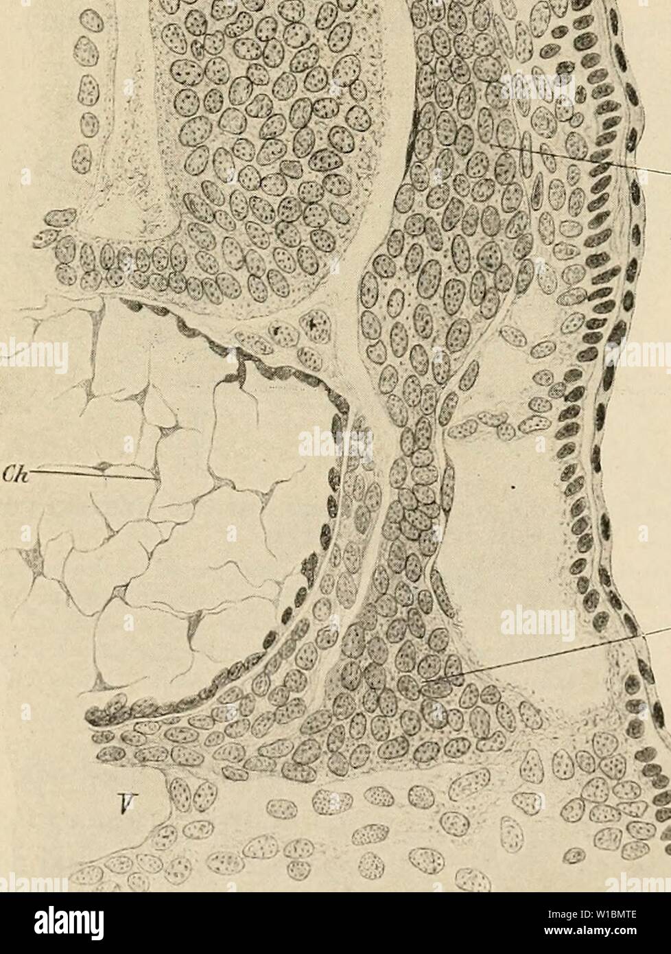 Archive image from page 430 of The development of the human. The development of the human body : a manual of human embryology . developmentofhum00mcmu Year: 1914 THE SYMPATHETIC SYSTEM 419 ''v - â Fig. 248.âTransverse Section through an Embryo Shark (Scyllium) of ii mm., SHOWING THE ORIGIN OF A SYMPATHETIC GANGLION. Ch, Notochord; E, ectoderm; G, posterior root ganglion; Gs, sympathetic ganglion; .1/, spinal cord.â(Onodi.) Stock Photohttps://www.alamy.com/image-license-details/?v=1https://www.alamy.com/archive-image-from-page-430-of-the-development-of-the-human-the-development-of-the-human-body-a-manual-of-human-embryology-developmentofhum00mcmu-year-1914-the-sympathetic-system-419-v-fig-248transverse-section-through-an-embryo-shark-scyllium-of-ii-mm-showing-the-origin-of-a-sympathetic-ganglion-ch-notochord-e-ectoderm-g-posterior-root-ganglion-gs-sympathetic-ganglion-1-spinal-cordonodi-image259028014.html
RMW1BMTE–Archive image from page 430 of The development of the human. The development of the human body : a manual of human embryology . developmentofhum00mcmu Year: 1914 THE SYMPATHETIC SYSTEM 419 ''v - â Fig. 248.âTransverse Section through an Embryo Shark (Scyllium) of ii mm., SHOWING THE ORIGIN OF A SYMPATHETIC GANGLION. Ch, Notochord; E, ectoderm; G, posterior root ganglion; Gs, sympathetic ganglion; .1/, spinal cord.â(Onodi.)
 . The development of the human body : a manual of human embryology. Embryology; Embryo, Non-Mammalian. 144 DEVELOPMENT OF THE NAILS The Development of the Nails.—The earliest indications of the development of the nails have been described by Zander in embryos of about nine weeks as slight thickenings of the epidermis. Fig. 83.—Diagram showing the Overlap of the III, IV, and V Intercostal Nerves of a Monkey.—(Sherrington.). Please note that these images are extracted from scanned page images that may have been digitally enhanced for readability - coloration and appearance of these illustrations Stock Photohttps://www.alamy.com/image-license-details/?v=1https://www.alamy.com/the-development-of-the-human-body-a-manual-of-human-embryology-embryology-embryo-non-mammalian-144-development-of-the-nails-the-development-of-the-nailsthe-earliest-indications-of-the-development-of-the-nails-have-been-described-by-zander-in-embryos-of-about-nine-weeks-as-slight-thickenings-of-the-epidermis-fig-83diagram-showing-the-overlap-of-the-iii-iv-and-v-intercostal-nerves-of-a-monkeysherrington-please-note-that-these-images-are-extracted-from-scanned-page-images-that-may-have-been-digitally-enhanced-for-readability-coloration-and-appearance-of-these-illustrations-image215969825.html
RMPFA7M1–. The development of the human body : a manual of human embryology. Embryology; Embryo, Non-Mammalian. 144 DEVELOPMENT OF THE NAILS The Development of the Nails.—The earliest indications of the development of the nails have been described by Zander in embryos of about nine weeks as slight thickenings of the epidermis. Fig. 83.—Diagram showing the Overlap of the III, IV, and V Intercostal Nerves of a Monkey.—(Sherrington.). Please note that these images are extracted from scanned page images that may have been digitally enhanced for readability - coloration and appearance of these illustrations
 Pregnant female is holding sonogram baby embryo image over a pregnant belly. Stock Photohttps://www.alamy.com/image-license-details/?v=1https://www.alamy.com/pregnant-female-is-holding-sonogram-baby-embryo-image-over-a-pregnant-belly-image596313440.html
RF2WJ4BTG–Pregnant female is holding sonogram baby embryo image over a pregnant belly.