Human lymphocyte microscope Stock Photos and Images
(398)See human lymphocyte microscope stock video clipsQuick filters:
Human lymphocyte microscope Stock Photos and Images
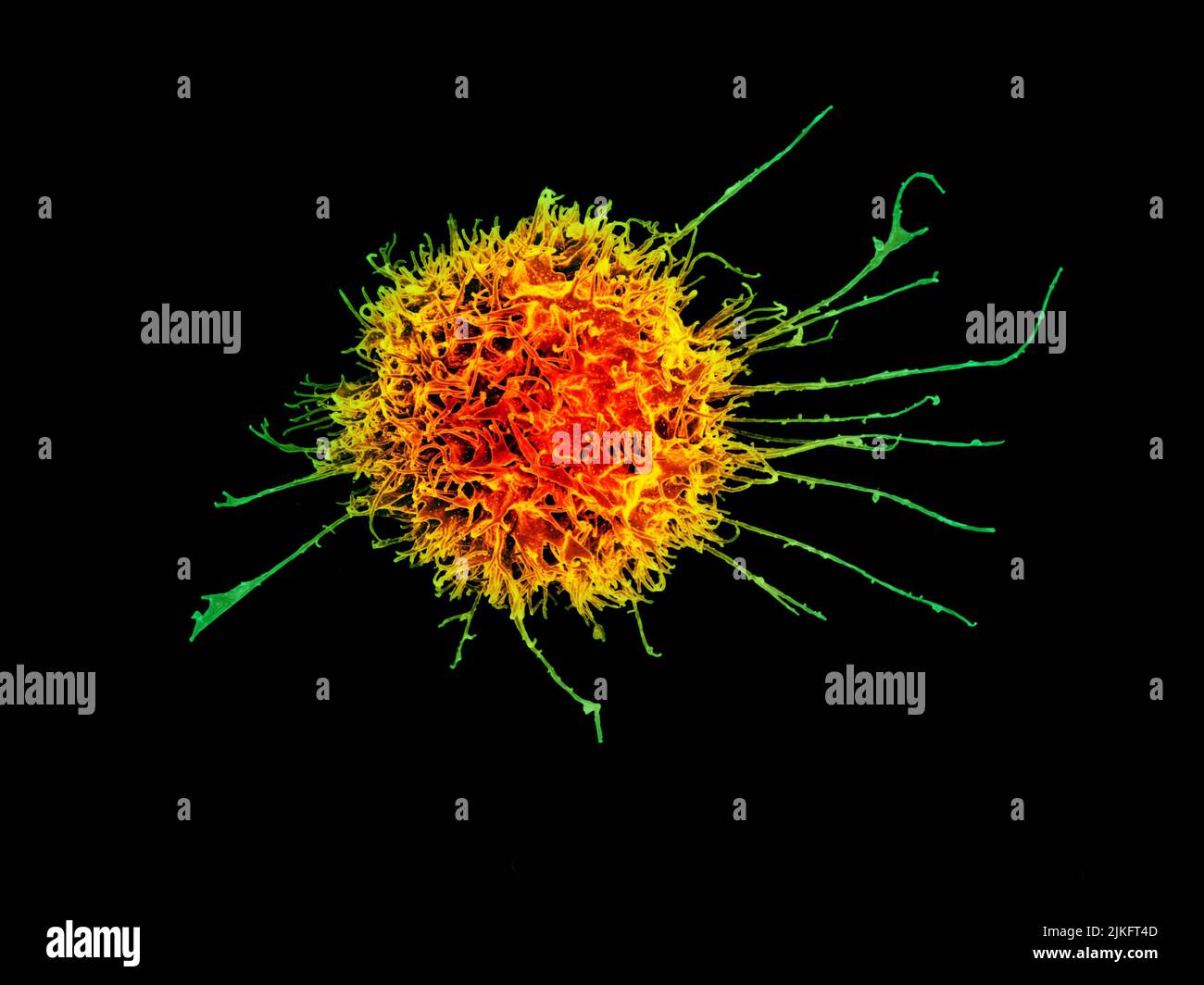 Scanning electron micrograph of a human natural killer cell. Credit: NIAID Stock Photohttps://www.alamy.com/image-license-details/?v=1https://www.alamy.com/scanning-electron-micrograph-of-a-human-natural-killer-cell-credit-niaid-image476706621.html
RM2JKFT4D–Scanning electron micrograph of a human natural killer cell. Credit: NIAID
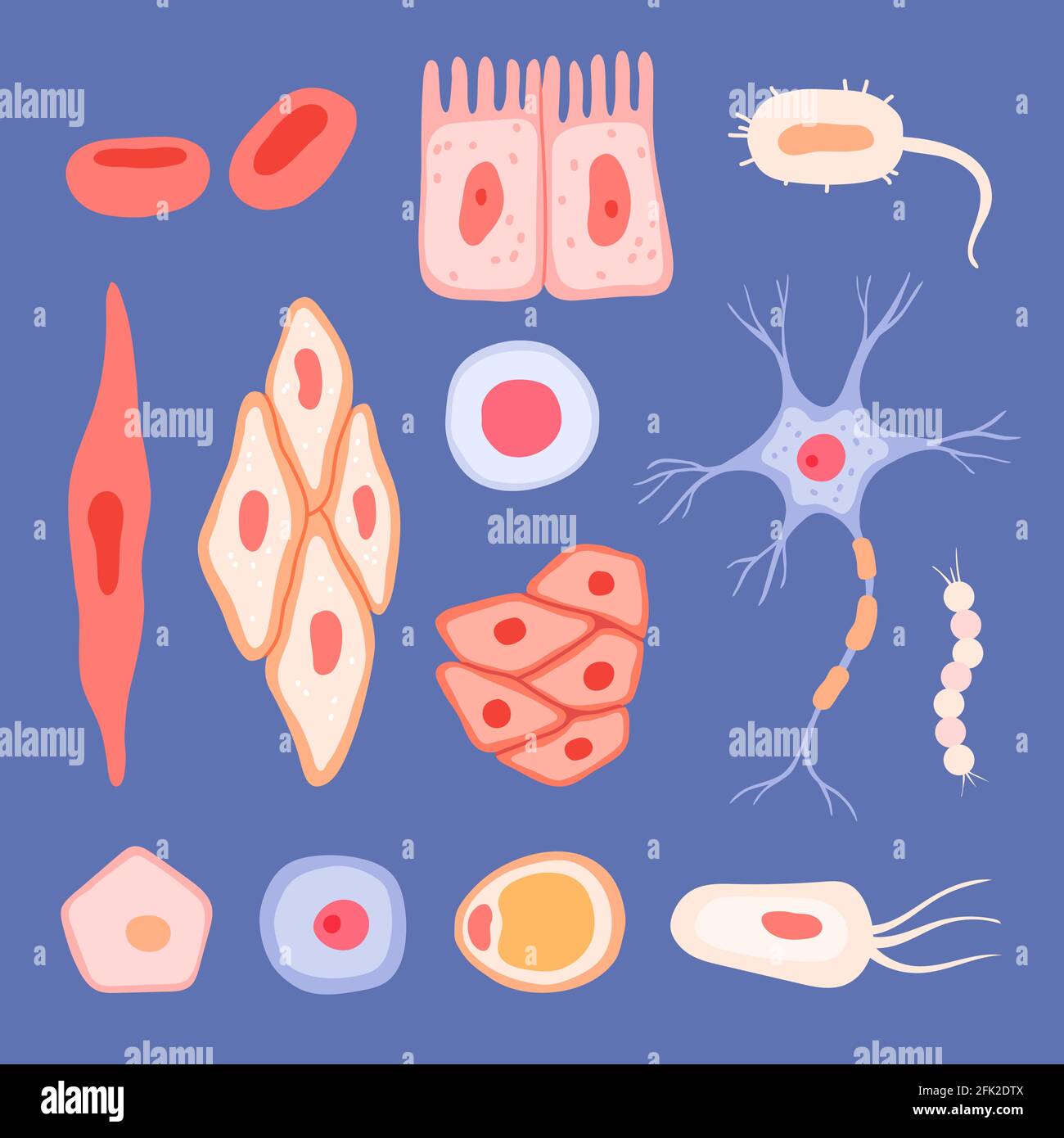 Human cells. Biological structure of blood scenes collection lymphocyte vector flat pictures of cells Stock Vectorhttps://www.alamy.com/image-license-details/?v=1https://www.alamy.com/human-cells-biological-structure-of-blood-scenes-collection-lymphocyte-vector-flat-pictures-of-cells-image424782090.html
RF2FK2DTX–Human cells. Biological structure of blood scenes collection lymphocyte vector flat pictures of cells
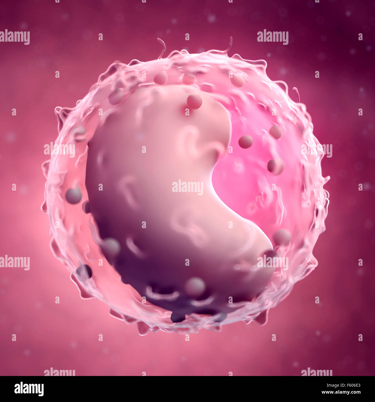 medically accurate illustration of a lymphocyte Stock Photohttps://www.alamy.com/image-license-details/?v=1https://www.alamy.com/stock-photo-medically-accurate-illustration-of-a-lymphocyte-89744875.html
RFF606E3–medically accurate illustration of a lymphocyte
 Lymphocyte . White blood cells with transparency membrane and large nucleus . Isolated white background . 3D render . Stock Photohttps://www.alamy.com/image-license-details/?v=1https://www.alamy.com/lymphocyte-white-blood-cells-with-transparency-membrane-and-large-nucleus-isolated-white-background-3d-render-image474457152.html
RF2JFWAX8–Lymphocyte . White blood cells with transparency membrane and large nucleus . Isolated white background . 3D render .
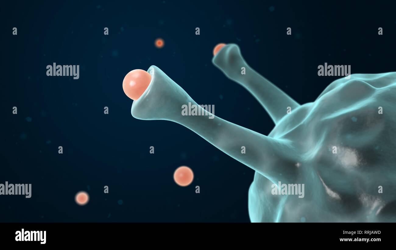 Macrophage engulfing pathogen. 3d Illustration. White blood cell. Immune system. Stock Photohttps://www.alamy.com/image-license-details/?v=1https://www.alamy.com/macrophage-engulfing-pathogen-3d-illustration-white-blood-cell-immune-system-image238275561.html
RFRRJAWD–Macrophage engulfing pathogen. 3d Illustration. White blood cell. Immune system.
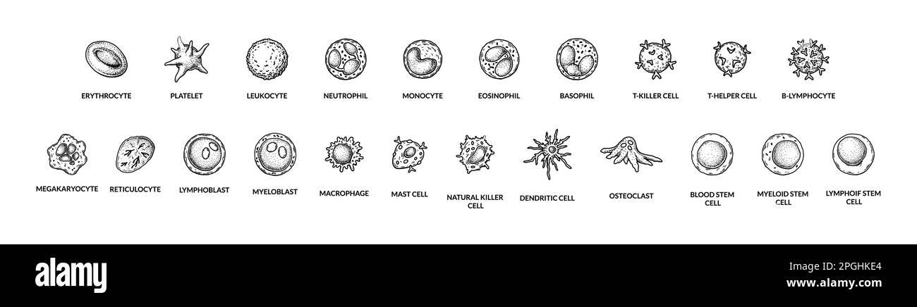 Human blood sells. Medicine scientific poster. Microbiology anatomy vector illustration in sketch style Stock Vectorhttps://www.alamy.com/image-license-details/?v=1https://www.alamy.com/human-blood-sells-medicine-scientific-poster-microbiology-anatomy-vector-illustration-in-sketch-style-image543744380.html
RF2PGHKE4–Human blood sells. Medicine scientific poster. Microbiology anatomy vector illustration in sketch style
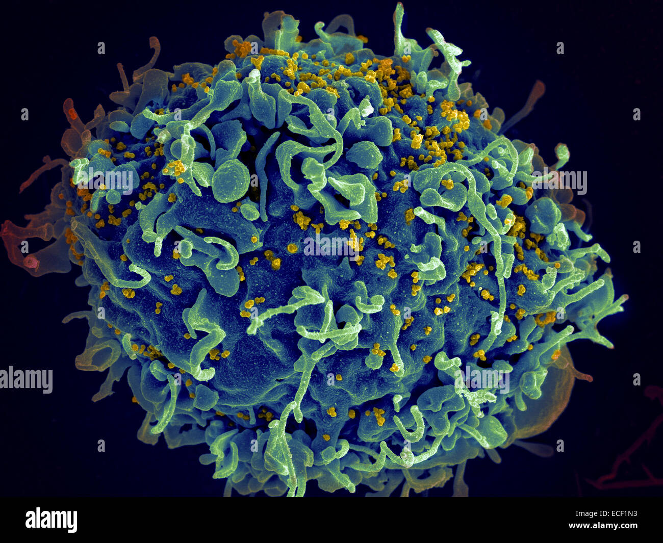 Scanning electron micrograph of HIV particles infecting a human H9 T cell. Stock Photohttps://www.alamy.com/image-license-details/?v=1https://www.alamy.com/stock-photo-scanning-electron-micrograph-of-hiv-particles-infecting-a-human-h9-76547999.html
RFECF1N3–Scanning electron micrograph of HIV particles infecting a human H9 T cell.
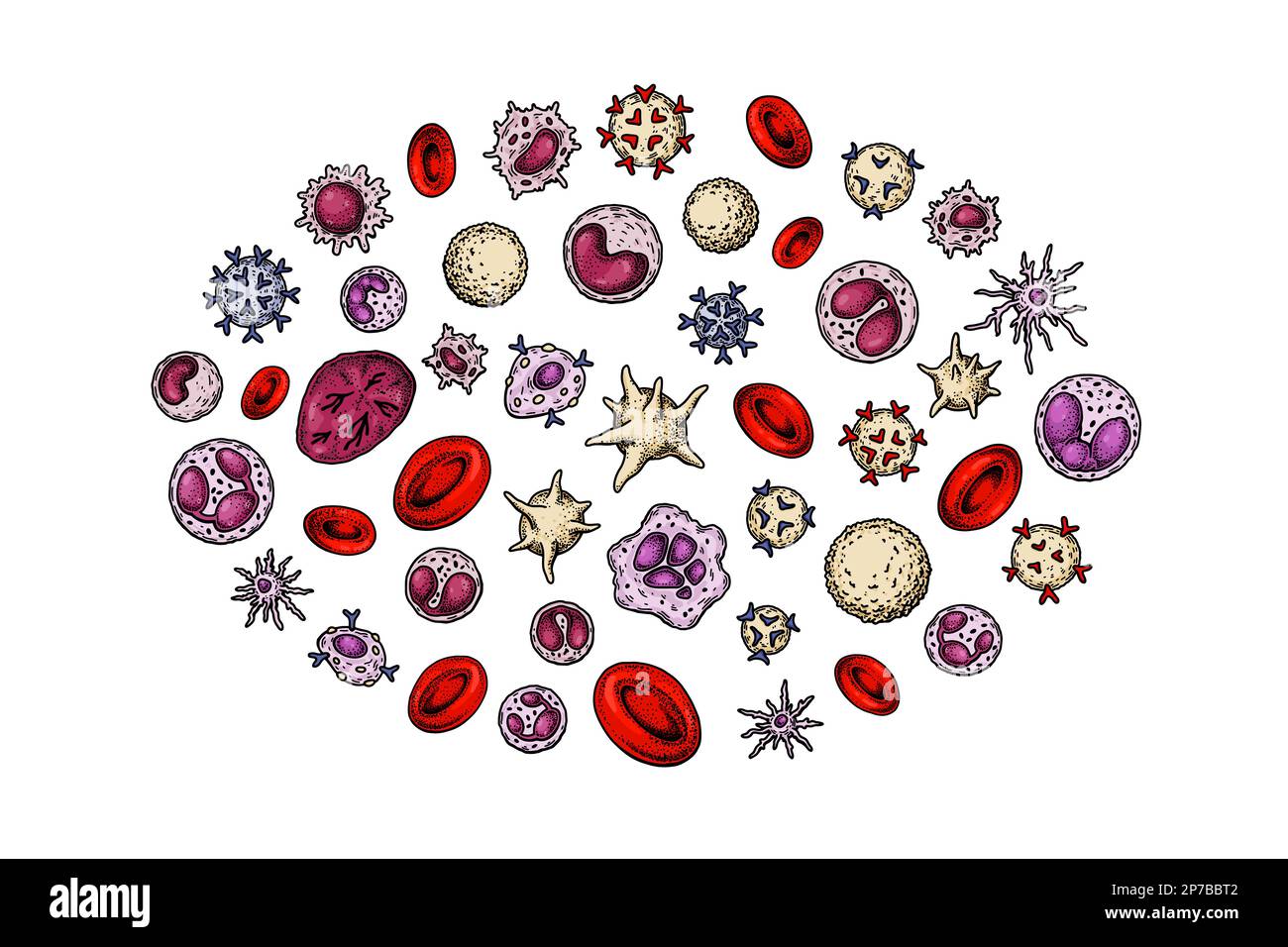 Human blood sells. Medicine scientific poster. Microbiology anatomy vector illustration in sketch style Stock Vectorhttps://www.alamy.com/image-license-details/?v=1https://www.alamy.com/human-blood-sells-medicine-scientific-poster-microbiology-anatomy-vector-illustration-in-sketch-style-image538074770.html
RF2P7BBT2–Human blood sells. Medicine scientific poster. Microbiology anatomy vector illustration in sketch style
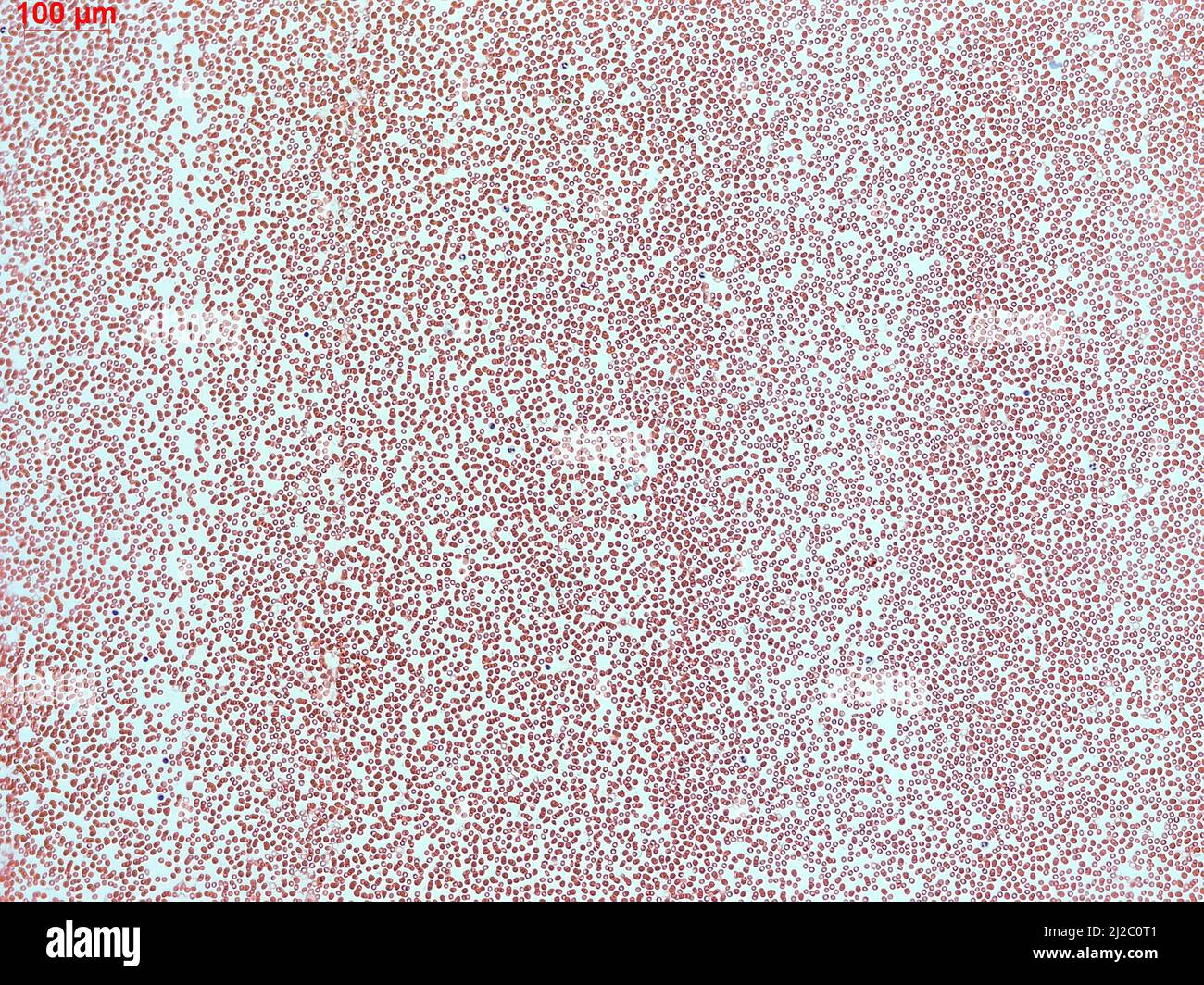 Human blood cell under microscope. Magnified smear of red blood cells in blood plasma. Stock Photohttps://www.alamy.com/image-license-details/?v=1https://www.alamy.com/human-blood-cell-under-microscope-magnified-smear-of-red-blood-cells-in-blood-plasma-image466173345.html
RF2J2C0T1–Human blood cell under microscope. Magnified smear of red blood cells in blood plasma.
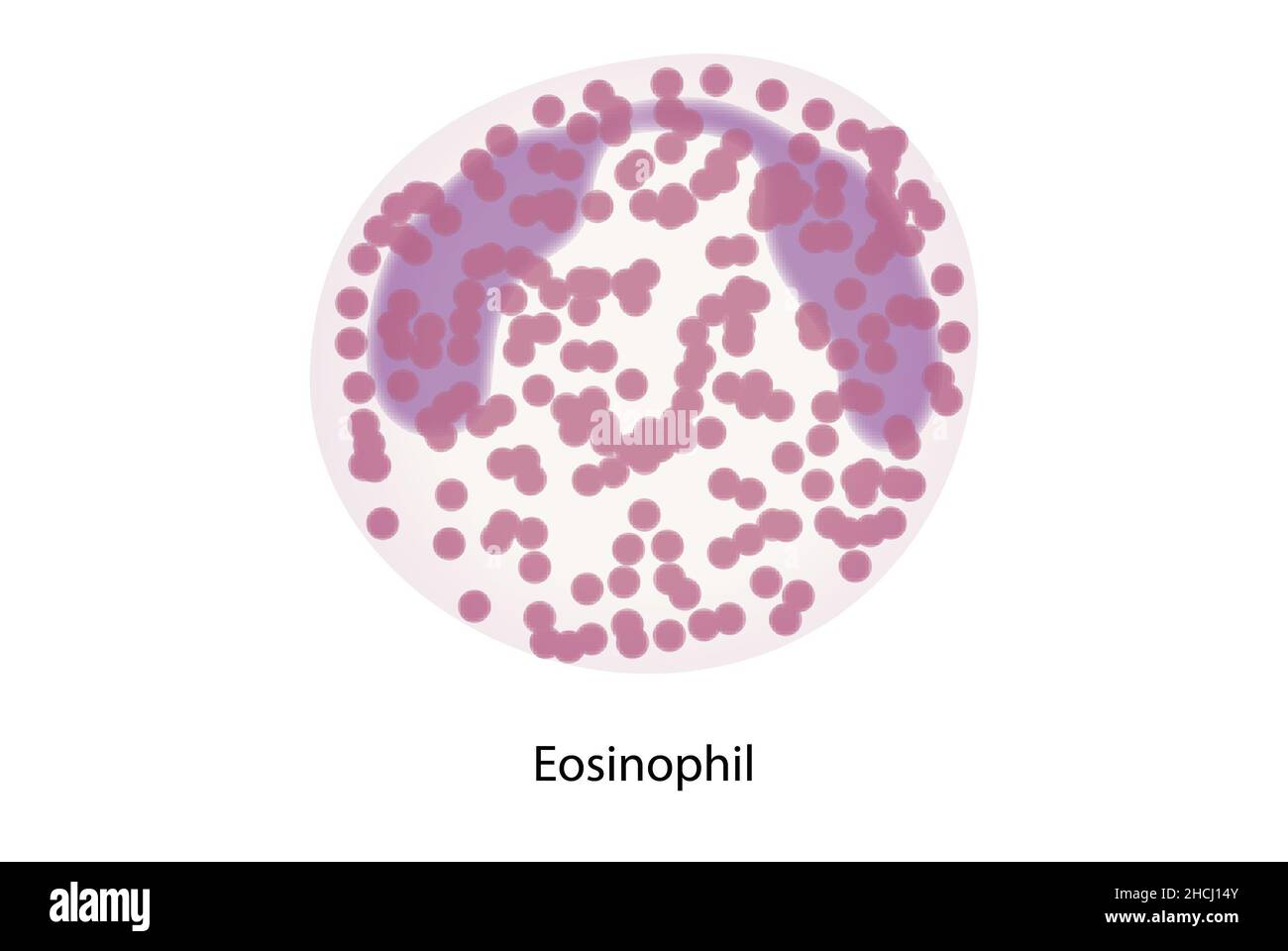 Eosinophil, white blood cells, immune system cells Stock Photohttps://www.alamy.com/image-license-details/?v=1https://www.alamy.com/eosinophil-white-blood-cells-immune-system-cells-image455241499.html
RF2HCJ14Y–Eosinophil, white blood cells, immune system cells
 illustration of bioscience of immunity white blood cell circulating the human body Stock Vectorhttps://www.alamy.com/image-license-details/?v=1https://www.alamy.com/illustration-of-bioscience-of-immunity-white-blood-cell-circulating-the-human-body-image455461043.html
RF2HD015R–illustration of bioscience of immunity white blood cell circulating the human body
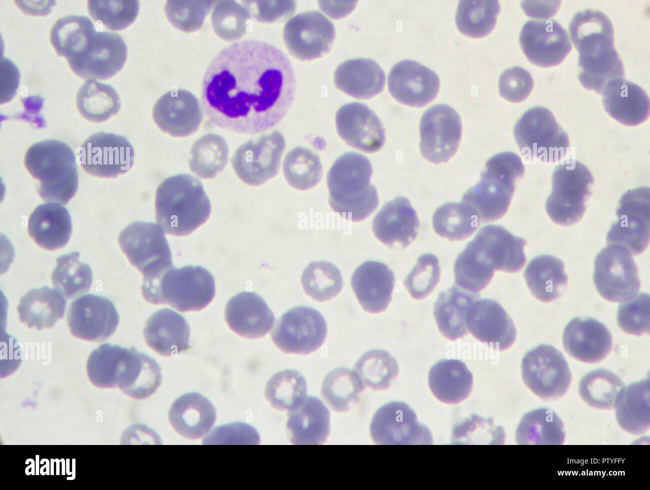 Blood under a microscope. Lymphocyte Stock Photohttps://www.alamy.com/image-license-details/?v=1https://www.alamy.com/blood-under-a-microscope-lymphocyte-image221881071.html
RFPTYFFY–Blood under a microscope. Lymphocyte
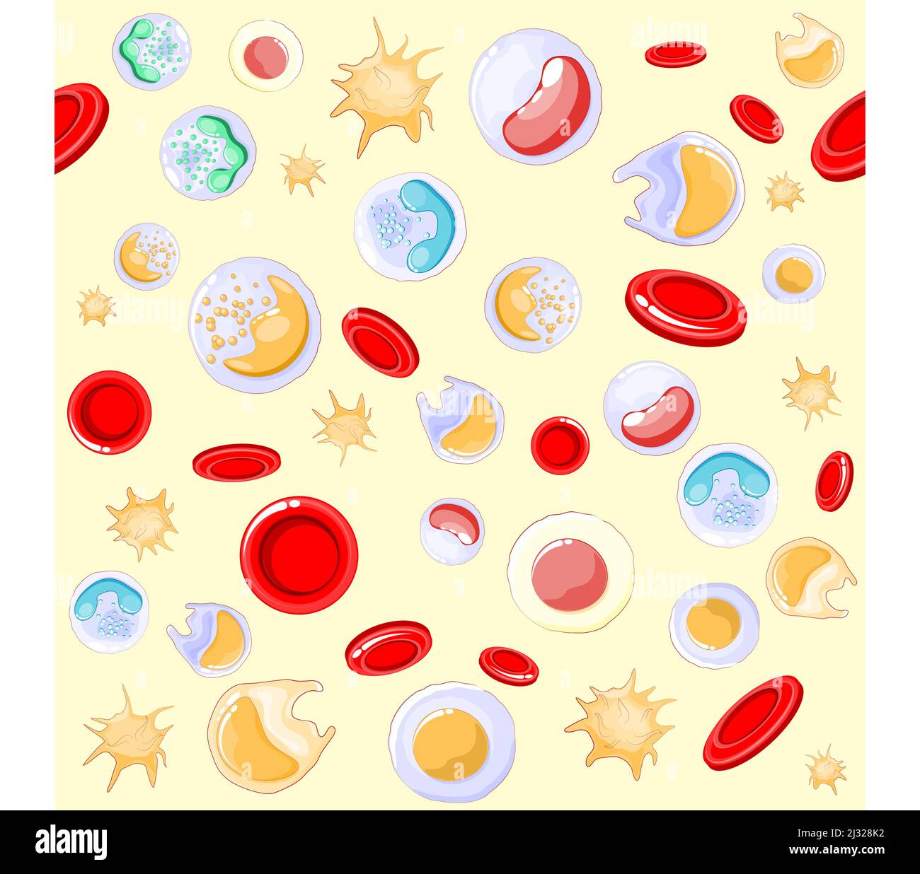 seamless pattern. red and white blood cells under microscope. Vector background Stock Vectorhttps://www.alamy.com/image-license-details/?v=1https://www.alamy.com/seamless-pattern-red-and-white-blood-cells-under-microscope-vector-background-image466574614.html
RF2J328K2–seamless pattern. red and white blood cells under microscope. Vector background
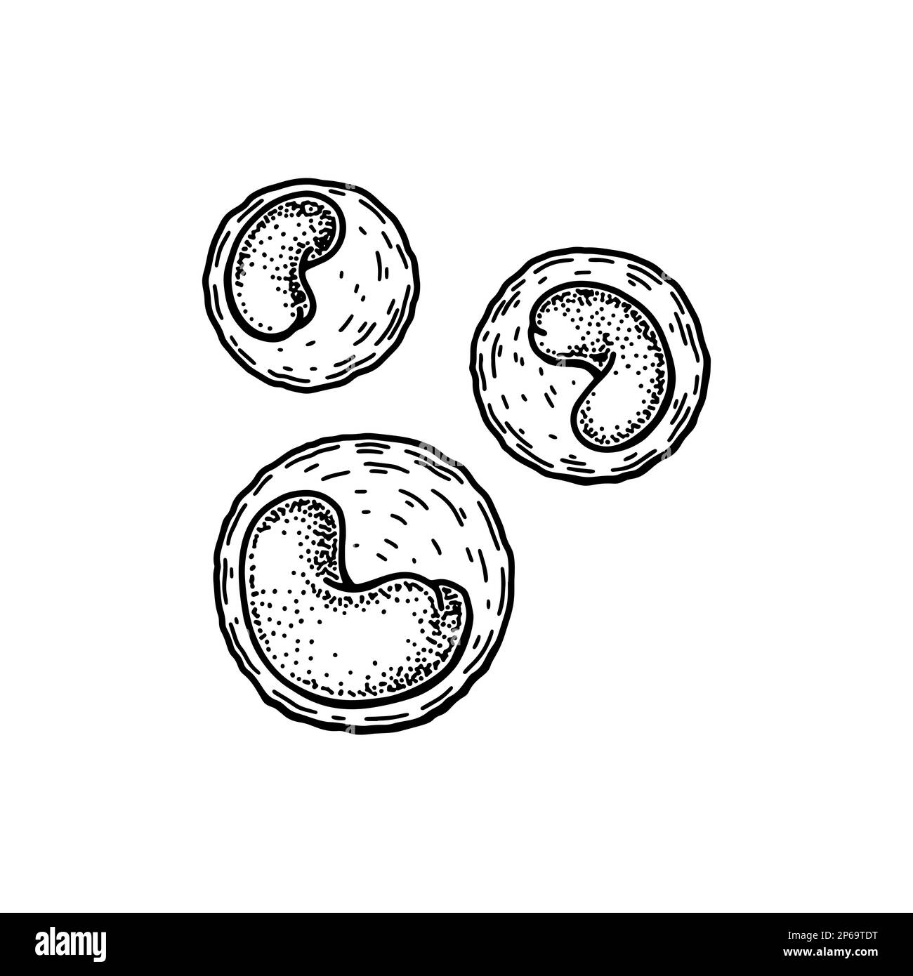 Monocyte leukocyte white blood cells isolated on white background. Hand drawn scientific microbiology vector illustration in sketch style Stock Vectorhttps://www.alamy.com/image-license-details/?v=1https://www.alamy.com/monocyte-leukocyte-white-blood-cells-isolated-on-white-background-hand-drawn-scientific-microbiology-vector-illustration-in-sketch-style-image537426116.html
RF2P69TDT–Monocyte leukocyte white blood cells isolated on white background. Hand drawn scientific microbiology vector illustration in sketch style
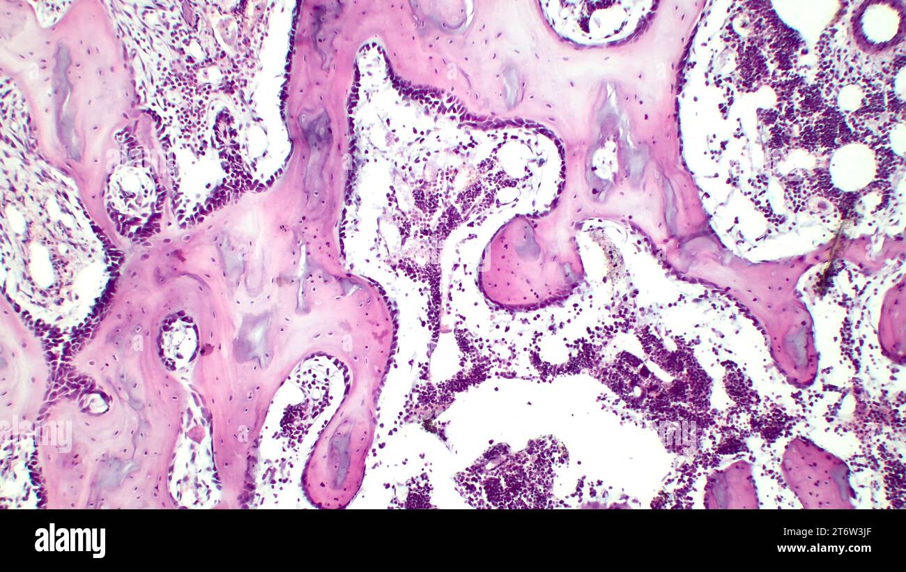 Human red bone marrow, hematopoietic press. The big tsit beetveen erythrocytes are megakaryocytes. Hematoxylin and eosin stain. Stock Photohttps://www.alamy.com/image-license-details/?v=1https://www.alamy.com/human-red-bone-marrow-hematopoietic-press-the-big-tsit-beetveen-erythrocytes-are-megakaryocytes-hematoxylin-and-eosin-stain-image572181751.html
RF2T6W3JF–Human red bone marrow, hematopoietic press. The big tsit beetveen erythrocytes are megakaryocytes. Hematoxylin and eosin stain.
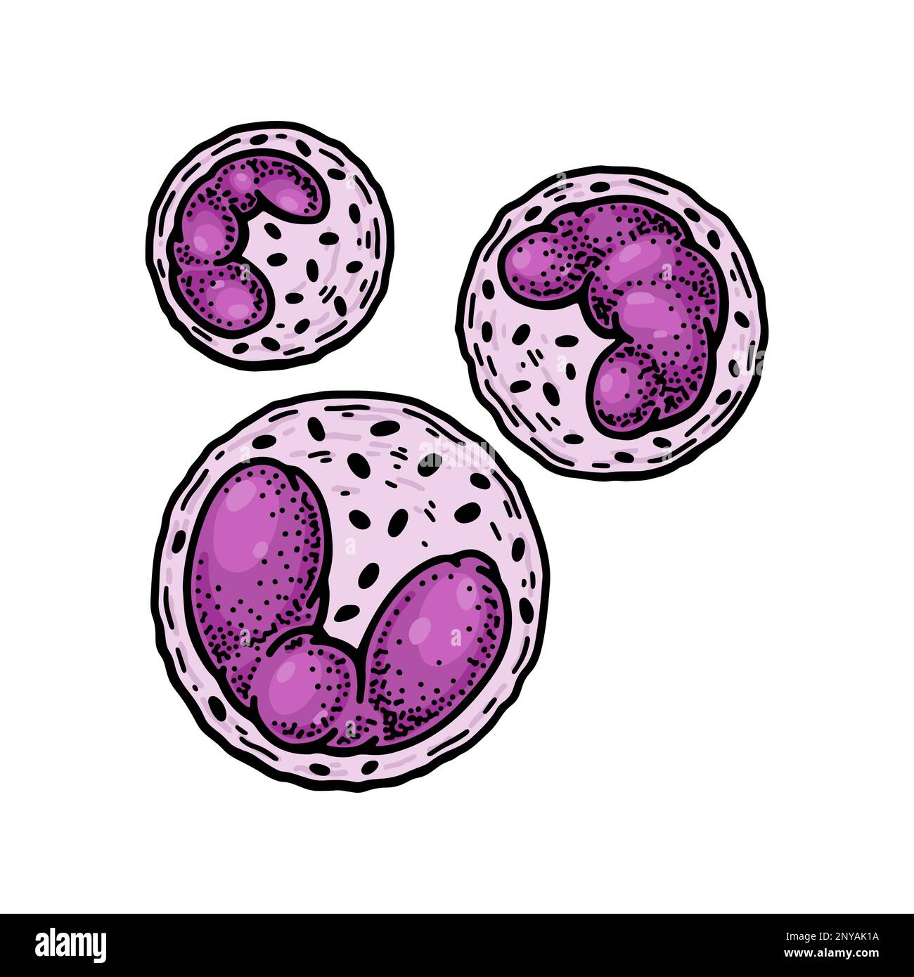 Basophil leukocyte white blood cells isolated on white background. Hand drawn scientific microbiology vector illustration in sketch style Stock Vectorhttps://www.alamy.com/image-license-details/?v=1https://www.alamy.com/basophil-leukocyte-white-blood-cells-isolated-on-white-background-hand-drawn-scientific-microbiology-vector-illustration-in-sketch-style-image533141206.html
RF2NYAK1A–Basophil leukocyte white blood cells isolated on white background. Hand drawn scientific microbiology vector illustration in sketch style
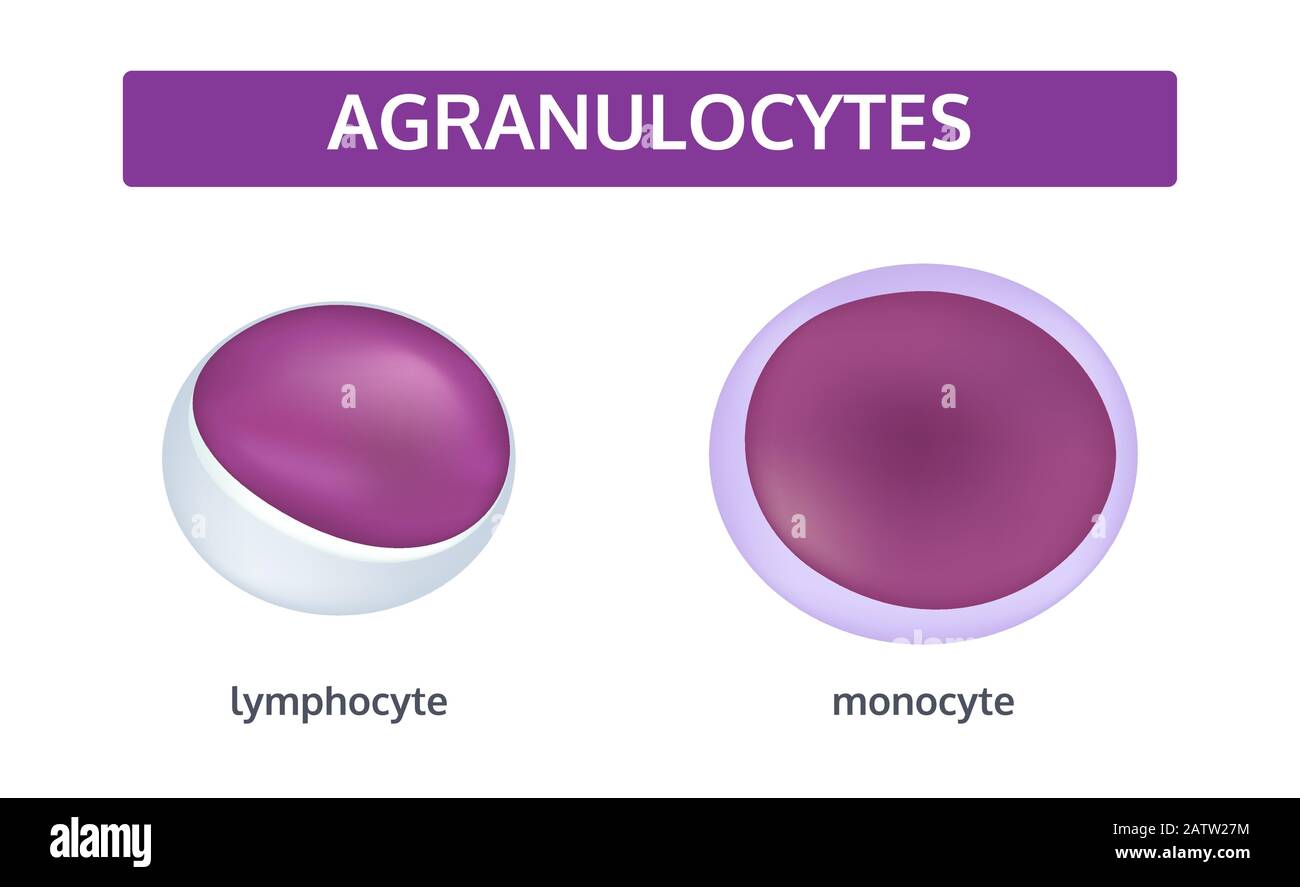 Vector set of white blood cells - agranulocytes: monocyte and lymphocyte. Medical concept. Stock Vectorhttps://www.alamy.com/image-license-details/?v=1https://www.alamy.com/vector-set-of-white-blood-cells-agranulocytes-monocyte-and-lymphocyte-medical-concept-image342299320.html
RF2ATW27M–Vector set of white blood cells - agranulocytes: monocyte and lymphocyte. Medical concept.
 Blood cells isolated on white background. Scientific microbiology vector illustration in sketch style Stock Vectorhttps://www.alamy.com/image-license-details/?v=1https://www.alamy.com/blood-cells-isolated-on-white-background-scientific-microbiology-vector-illustration-in-sketch-style-image544016277.html
RF2PH228N–Blood cells isolated on white background. Scientific microbiology vector illustration in sketch style
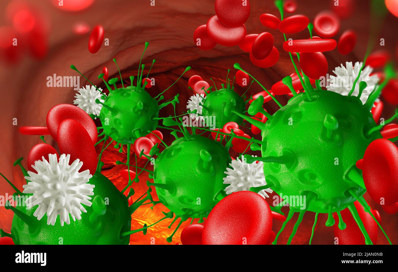 Leukocytes attack the virus in the blood. Microbes under the microscope. Disease, infection, inflammation Stock Photohttps://www.alamy.com/image-license-details/?v=1https://www.alamy.com/leukocytes-attack-the-virus-in-the-blood-microbes-under-the-microscope-disease-infection-inflammation-image471288087.html
RF2JAN0NB–Leukocytes attack the virus in the blood. Microbes under the microscope. Disease, infection, inflammation
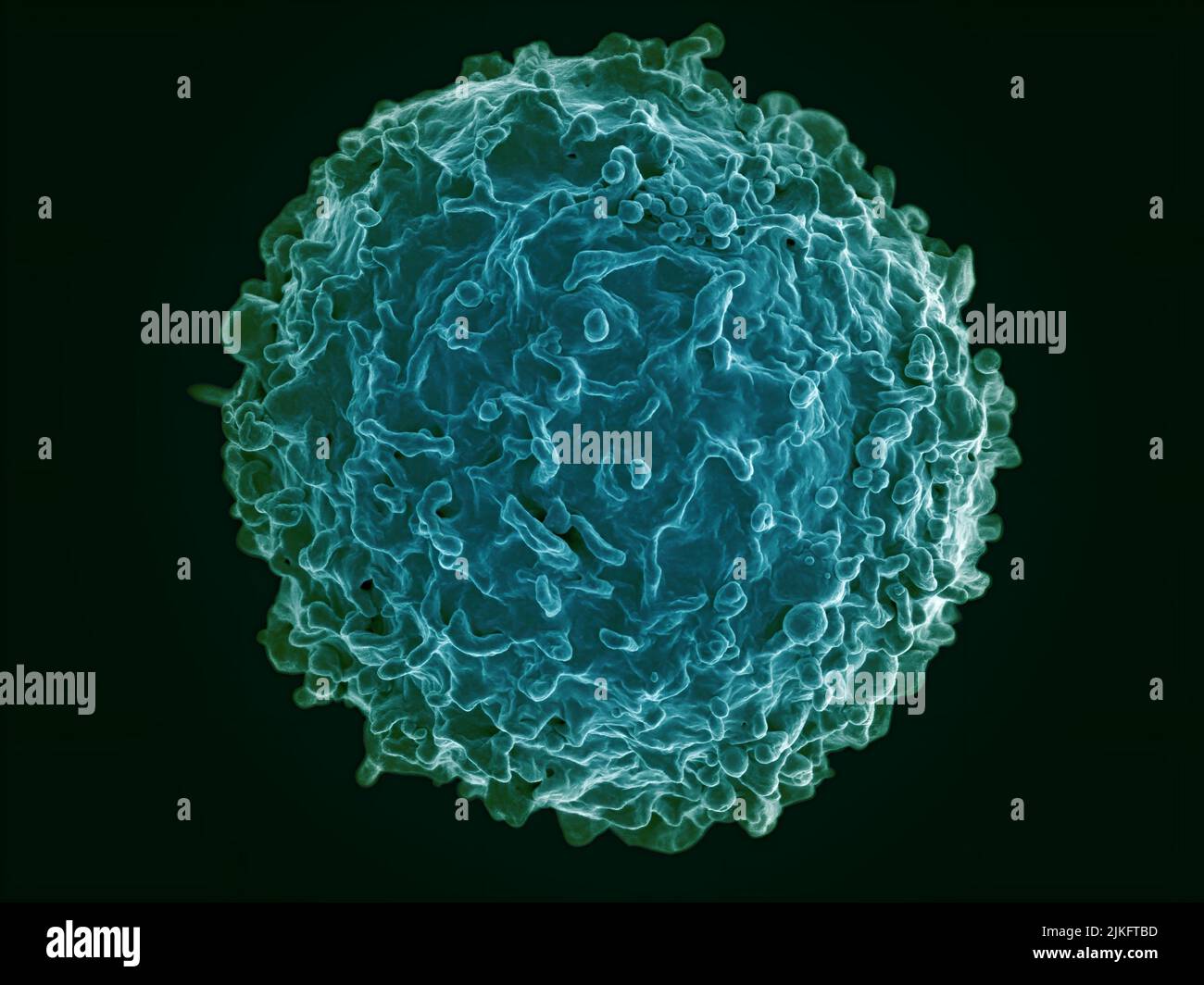 Colorized scanning electron micrograph of a B cell from a human donor. Stock Photohttps://www.alamy.com/image-license-details/?v=1https://www.alamy.com/colorized-scanning-electron-micrograph-of-a-b-cell-from-a-human-donor-image476706817.html
RM2JKFTBD–Colorized scanning electron micrograph of a B cell from a human donor.
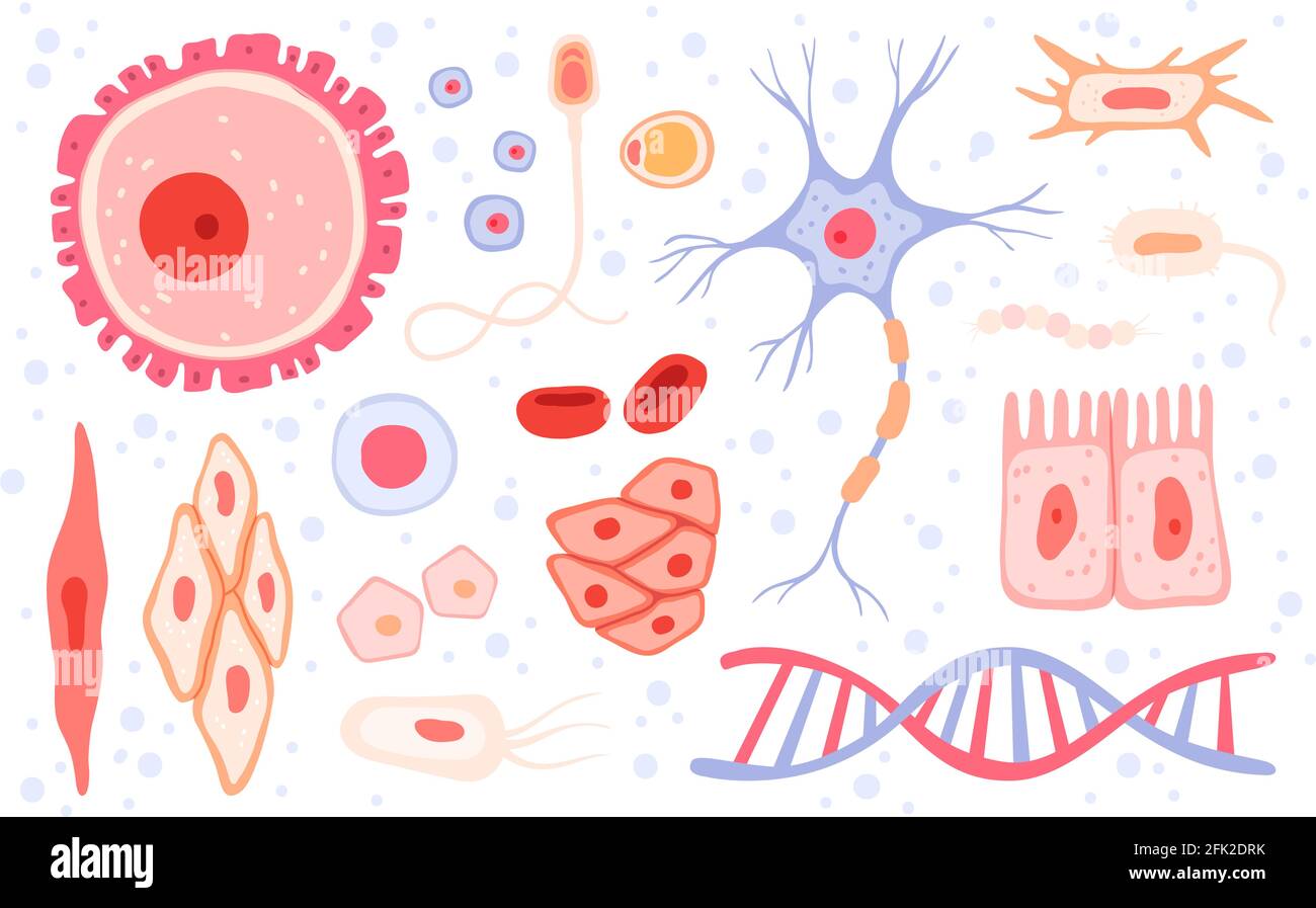 Cells collection. Human blood structure micro types of anatomy science vector collection cells set Stock Vectorhttps://www.alamy.com/image-license-details/?v=1https://www.alamy.com/cells-collection-human-blood-structure-micro-types-of-anatomy-science-vector-collection-cells-set-image424782055.html
RF2FK2DRK–Cells collection. Human blood structure micro types of anatomy science vector collection cells set
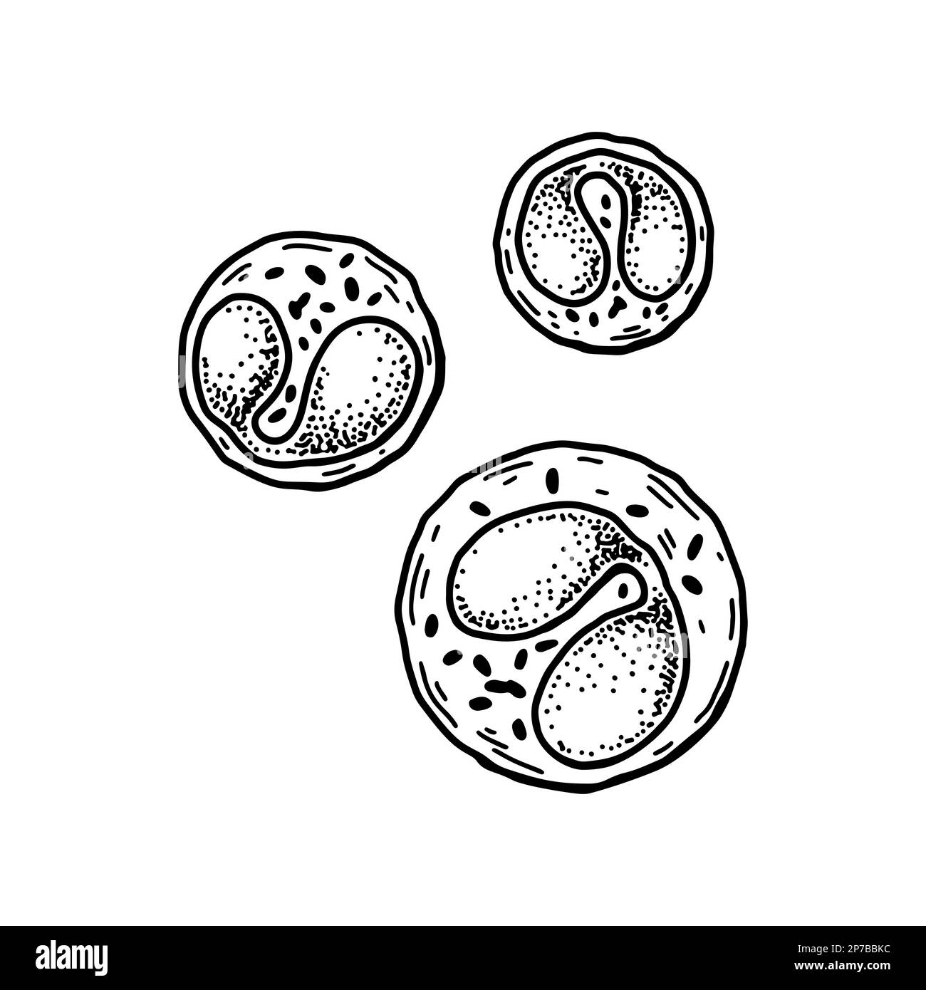 Eosinophil leukocyte white blood cells isolated on white background. Hand drawn scientific microbiology vector illustration in sketch style Stock Vectorhttps://www.alamy.com/image-license-details/?v=1https://www.alamy.com/eosinophil-leukocyte-white-blood-cells-isolated-on-white-background-hand-drawn-scientific-microbiology-vector-illustration-in-sketch-style-image538074640.html
RF2P7BBKC–Eosinophil leukocyte white blood cells isolated on white background. Hand drawn scientific microbiology vector illustration in sketch style
 Human cells death, Embryonic stem cell, metastasis Growing tumor, Leukemia spreading,malignant cancerous, viruses and bacteria, abnormal white blood c Stock Photohttps://www.alamy.com/image-license-details/?v=1https://www.alamy.com/human-cells-death-embryonic-stem-cell-metastasis-growing-tumor-leukemia-spreadingmalignant-cancerous-viruses-and-bacteria-abnormal-white-blood-c-image620981535.html
RM2Y2847Y–Human cells death, Embryonic stem cell, metastasis Growing tumor, Leukemia spreading,malignant cancerous, viruses and bacteria, abnormal white blood c
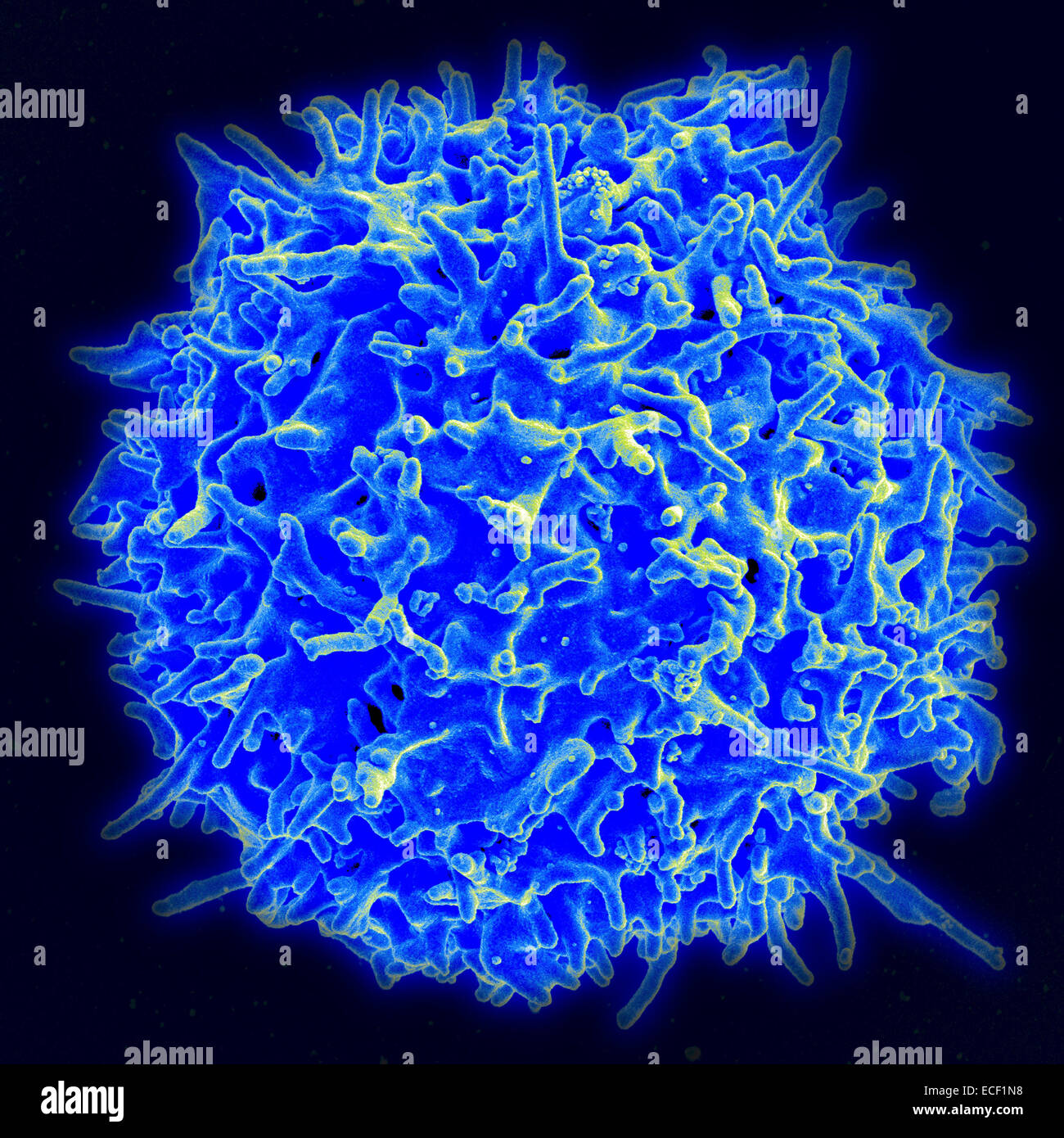 Scanning electron micrograph of a human T lymphocyte (also called a T cell) from the immune system of a healthy donor. Stock Photohttps://www.alamy.com/image-license-details/?v=1https://www.alamy.com/stock-photo-scanning-electron-micrograph-of-a-human-t-lymphocyte-also-called-a-76548004.html
RFECF1N8–Scanning electron micrograph of a human T lymphocyte (also called a T cell) from the immune system of a healthy donor.
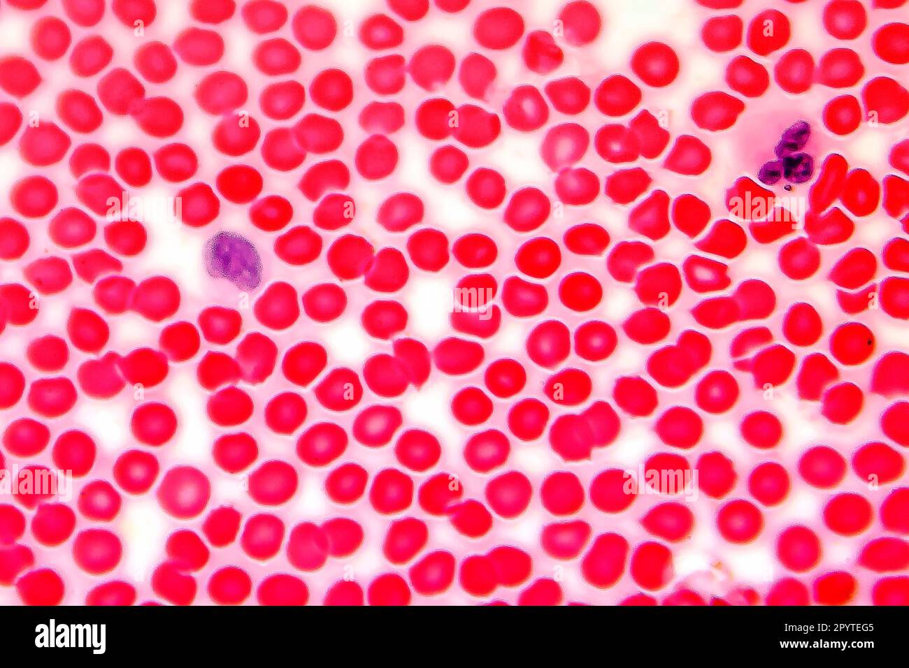 Human blood smear under microscope, light photomicrograph Stock Photohttps://www.alamy.com/image-license-details/?v=1https://www.alamy.com/human-blood-smear-under-microscope-light-photomicrograph-image550655397.html
RF2PYTEG5–Human blood smear under microscope, light photomicrograph
 Vial of medication along with a stethoscope, conceptual image Stock Photohttps://www.alamy.com/image-license-details/?v=1https://www.alamy.com/vial-of-medication-along-with-a-stethoscope-conceptual-image-image229755936.html
RFR9P814–Vial of medication along with a stethoscope, conceptual image
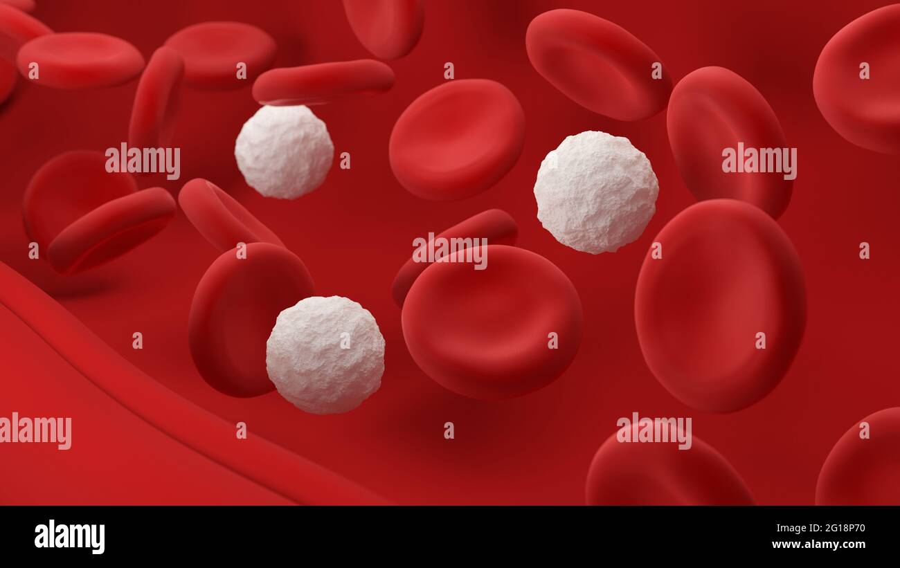 Red and white blood cells. Leukocytes and erythrocytes. 3d illustration. Stock Photohttps://www.alamy.com/image-license-details/?v=1https://www.alamy.com/red-and-white-blood-cells-leukocytes-and-erythrocytes-3d-illustration-image431066916.html
RF2G18P70–Red and white blood cells. Leukocytes and erythrocytes. 3d illustration.
 Butterfly Needle on a laboratory table next to blood samples, conceptual image Stock Photohttps://www.alamy.com/image-license-details/?v=1https://www.alamy.com/butterfly-needle-on-a-laboratory-table-next-to-blood-samples-conceptual-image-image218686467.html
RFPKP0PY–Butterfly Needle on a laboratory table next to blood samples, conceptual image
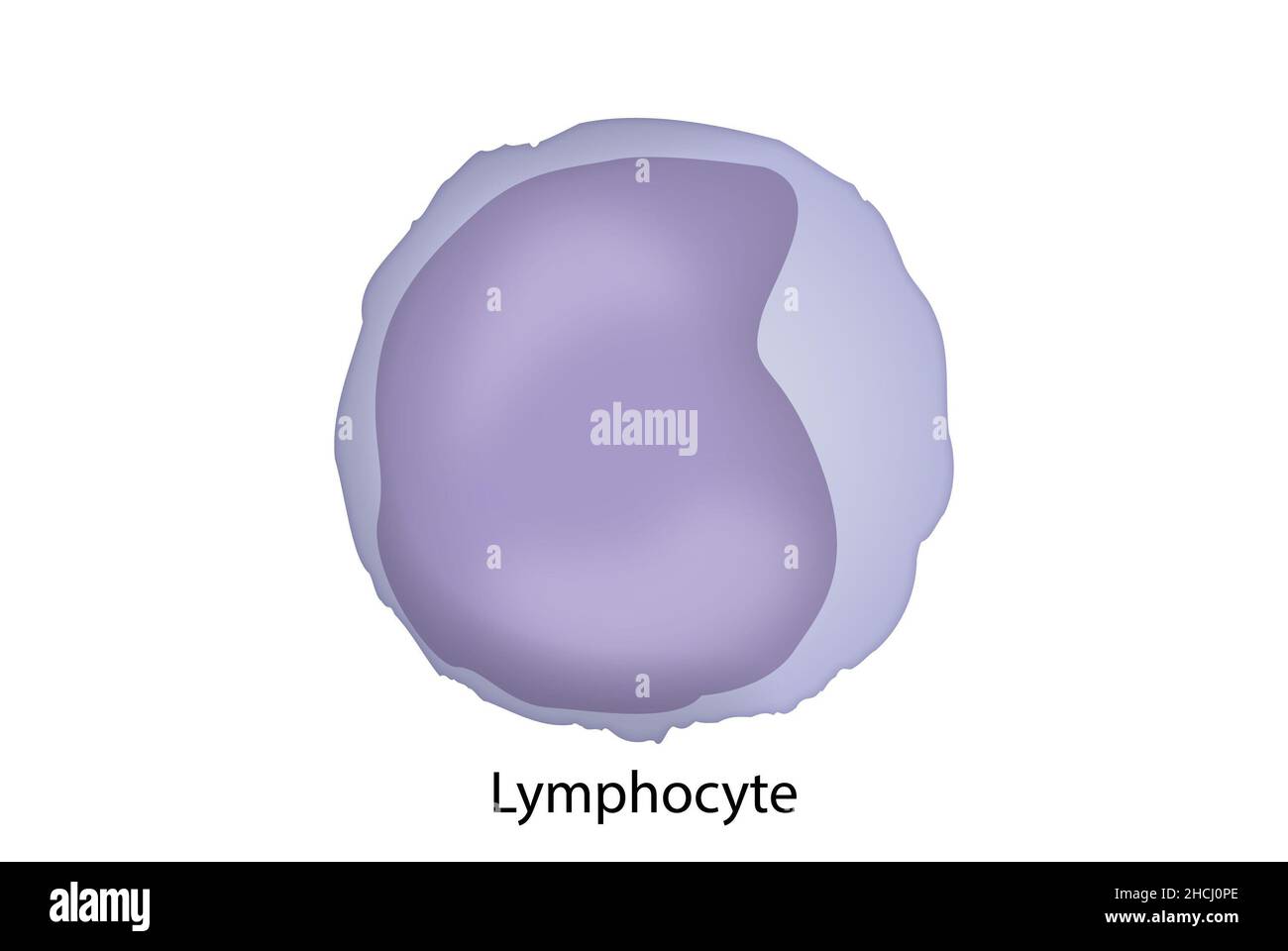 Lymphocites, blood smear, large and small lyphocyte. Stock Photohttps://www.alamy.com/image-license-details/?v=1https://www.alamy.com/lymphocites-blood-smear-large-and-small-lyphocyte-image455241206.html
RF2HCJ0PE–Lymphocites, blood smear, large and small lyphocyte.
 Lymohoma, medicines as concept of ordinary treatment, conceptual image Stock Photohttps://www.alamy.com/image-license-details/?v=1https://www.alamy.com/stock-image-lymohoma-medicines-as-concept-of-ordinary-treatment-conceptual-image-164115139.html
RFKF02EY–Lymohoma, medicines as concept of ordinary treatment, conceptual image
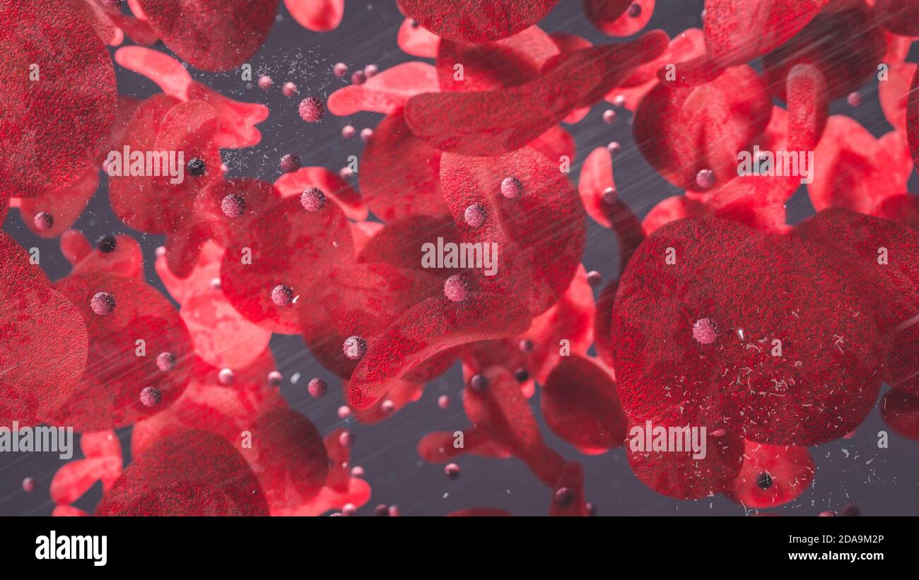 Red Blood Cells and T-Cells floating through blood vessels 3d rendering Stock Photohttps://www.alamy.com/image-license-details/?v=1https://www.alamy.com/red-blood-cells-and-t-cells-floating-through-blood-vessels-3d-rendering-image384987982.html
RF2DA9M2P–Red Blood Cells and T-Cells floating through blood vessels 3d rendering
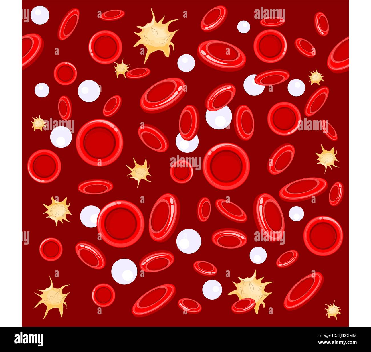 seamless pattern. red and white blood cells under microscope. Vector background Stock Vectorhttps://www.alamy.com/image-license-details/?v=1https://www.alamy.com/seamless-pattern-red-and-white-blood-cells-under-microscope-vector-background-image466580932.html
RF2J32GMM–seamless pattern. red and white blood cells under microscope. Vector background
 Diagnostic form, Vial of blood samples and Medicine in a hospital, conceptual image Stock Photohttps://www.alamy.com/image-license-details/?v=1https://www.alamy.com/diagnostic-form-vial-of-blood-samples-and-medicine-in-a-hospital-conceptual-image-image220797502.html
RFPR65D2–Diagnostic form, Vial of blood samples and Medicine in a hospital, conceptual image
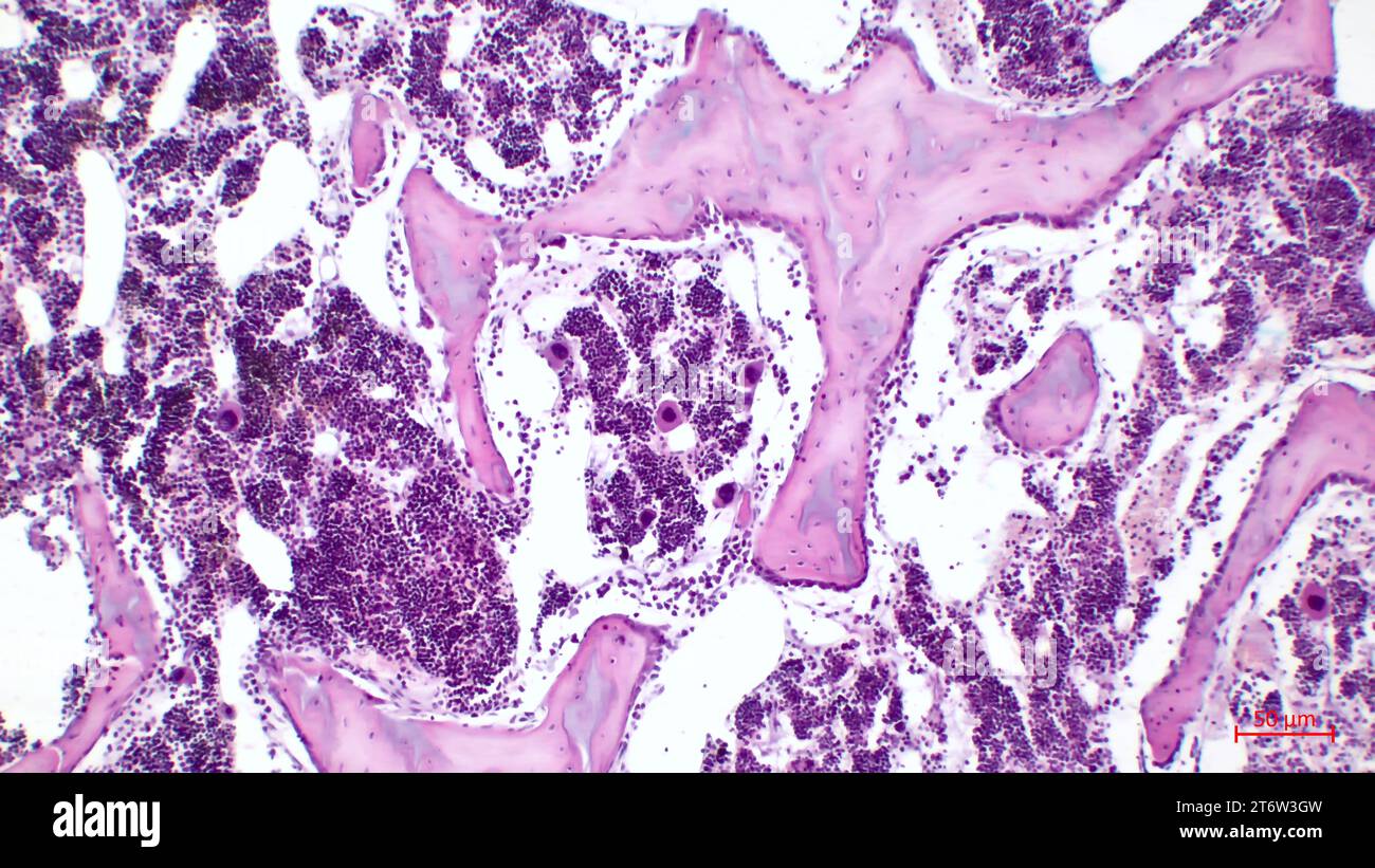 Human red bone marrow, hematopoietic press. The big tsit beetveen erythrocytes are megakaryocytes. Hematoxylin and eosin stain. Stock Photohttps://www.alamy.com/image-license-details/?v=1https://www.alamy.com/human-red-bone-marrow-hematopoietic-press-the-big-tsit-beetveen-erythrocytes-are-megakaryocytes-hematoxylin-and-eosin-stain-image572181705.html
RF2T6W3GW–Human red bone marrow, hematopoietic press. The big tsit beetveen erythrocytes are megakaryocytes. Hematoxylin and eosin stain.
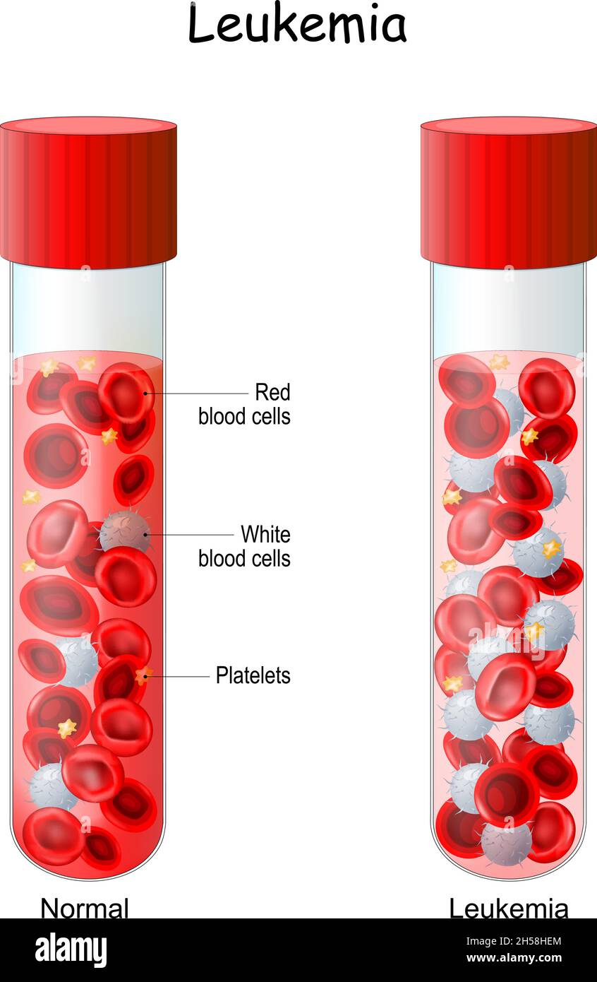 Leukemia. comparison and difference test tube with Normal blood and blood cancer. Close-up of red blood cells and lymphocytes. Vector illustration Stock Vectorhttps://www.alamy.com/image-license-details/?v=1https://www.alamy.com/leukemia-comparison-and-difference-test-tube-with-normal-blood-and-blood-cancer-close-up-of-red-blood-cells-and-lymphocytes-vector-illustration-image450732204.html
RF2H58HEM–Leukemia. comparison and difference test tube with Normal blood and blood cancer. Close-up of red blood cells and lymphocytes. Vector illustration
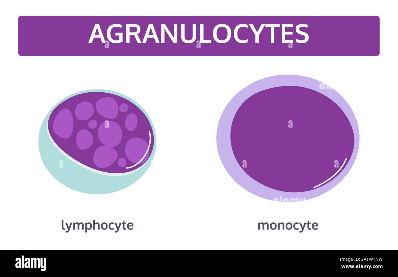 Vector set of white blood cells - agranulocytes: monocyte and lymphocyte in flat style. Medical concept.. Stock Vectorhttps://www.alamy.com/image-license-details/?v=1https://www.alamy.com/vector-set-of-white-blood-cells-agranulocytes-monocyte-and-lymphocyte-in-flat-style-medical-concept-image342298933.html
RF2ATW1NW–Vector set of white blood cells - agranulocytes: monocyte and lymphocyte in flat style. Medical concept..
 Diagnostic form, Vial of blood samples and Medicine in a hospital, conceptual image Stock Photohttps://www.alamy.com/image-license-details/?v=1https://www.alamy.com/diagnostic-form-vial-of-blood-samples-and-medicine-in-a-hospital-conceptual-image-image218091650.html
RFPJPX3E–Diagnostic form, Vial of blood samples and Medicine in a hospital, conceptual image
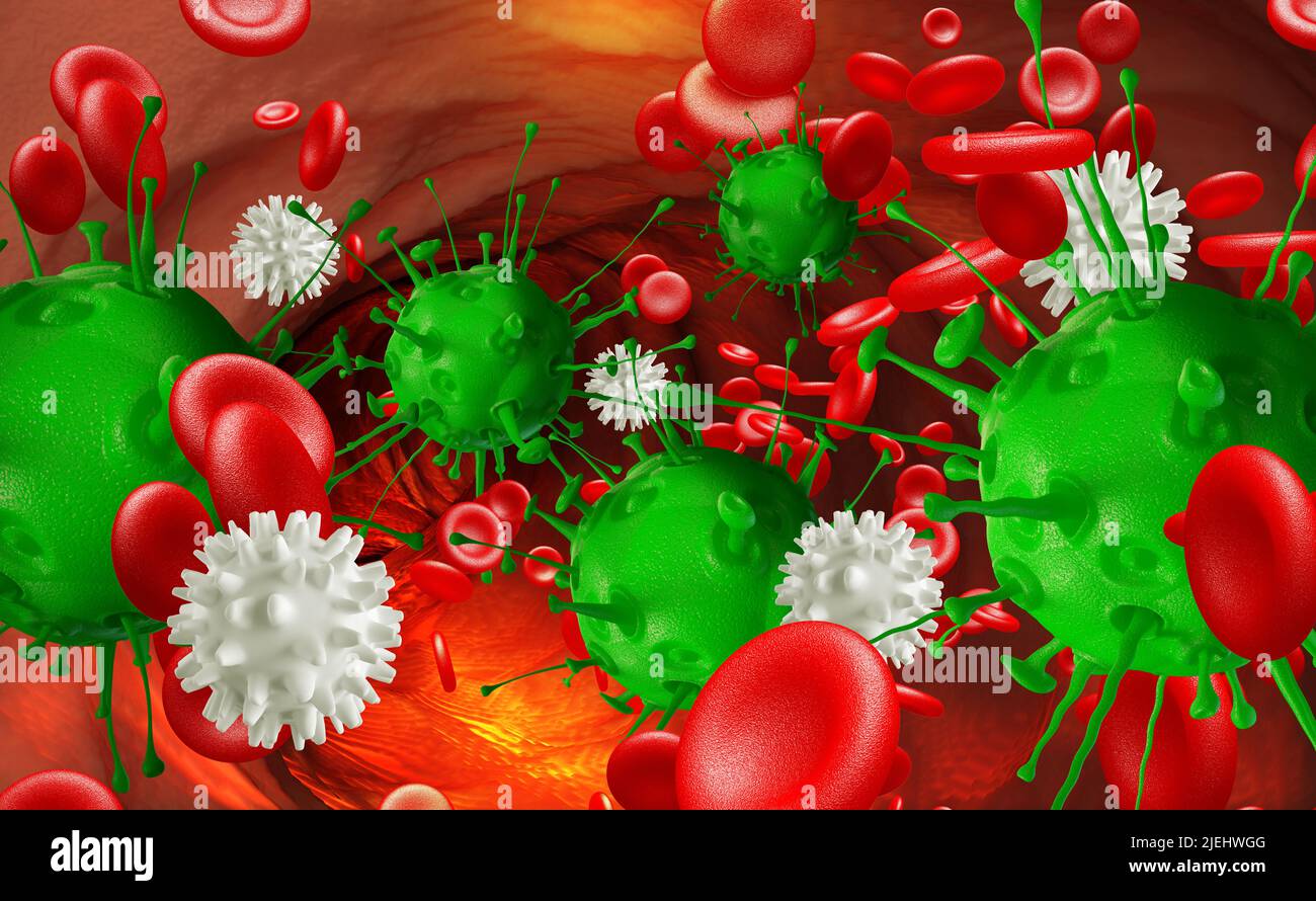 Leukocytes attack the virus in the blood. Microbes under the microscope. Disease, infection, inflammation. 3D illustration Stock Photohttps://www.alamy.com/image-license-details/?v=1https://www.alamy.com/leukocytes-attack-the-virus-in-the-blood-microbes-under-the-microscope-disease-infection-inflammation-3d-illustration-image473678368.html
RF2JEHWGG–Leukocytes attack the virus in the blood. Microbes under the microscope. Disease, infection, inflammation. 3D illustration
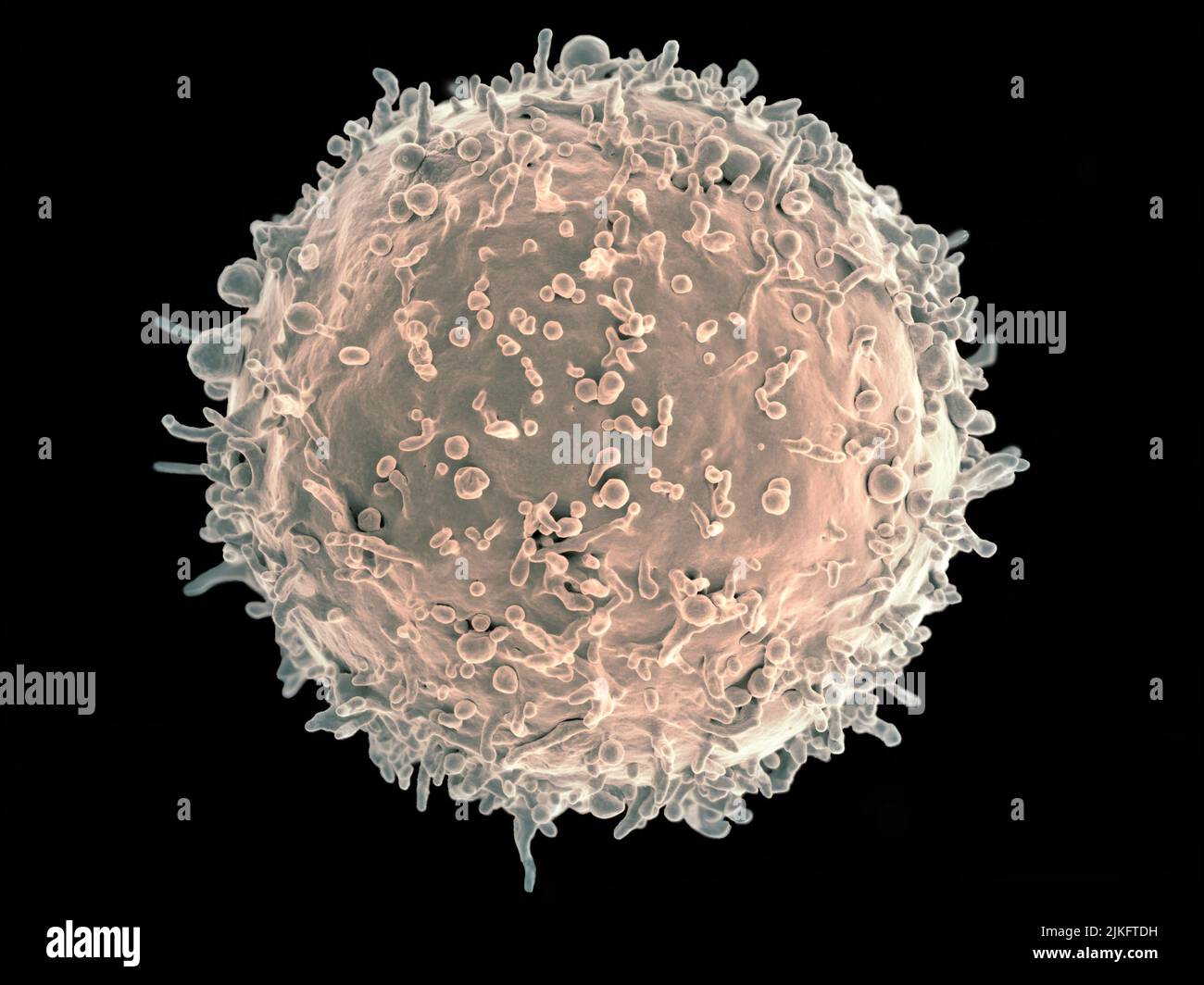 Colorized scanning electron micrograph of a B cell from a human donor. Stock Photohttps://www.alamy.com/image-license-details/?v=1https://www.alamy.com/colorized-scanning-electron-micrograph-of-a-b-cell-from-a-human-donor-image476706877.html
RM2JKFTDH–Colorized scanning electron micrograph of a B cell from a human donor.
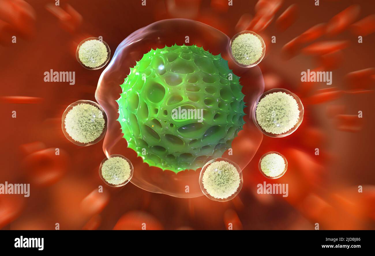 Leukocytes attack the virus. Immunity of the body. Fight against pneumonia. 3D illustration on medical research Stock Photohttps://www.alamy.com/image-license-details/?v=1https://www.alamy.com/leukocytes-attack-the-virus-immunity-of-the-body-fight-against-pneumonia-3d-illustration-on-medical-research-image472926278.html
RF2JDBJ86–Leukocytes attack the virus. Immunity of the body. Fight against pneumonia. 3D illustration on medical research
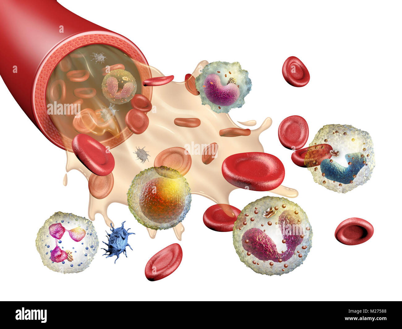 Different elements of human blood. 3d illustration. Stock Photohttps://www.alamy.com/image-license-details/?v=1https://www.alamy.com/stock-photo-different-elements-of-human-blood-3d-illustration-173490808.html
RFM27588–Different elements of human blood. 3d illustration.
 Human cells death, Embryonic stem cell, metastasis Growing tumor, Leukemia spreading,malignant cancerous, viruses and bacteria, abnormal white blood c Stock Photohttps://www.alamy.com/image-license-details/?v=1https://www.alamy.com/human-cells-death-embryonic-stem-cell-metastasis-growing-tumor-leukemia-spreadingmalignant-cancerous-viruses-and-bacteria-abnormal-white-blood-c-image620981238.html
RM2Y283WA–Human cells death, Embryonic stem cell, metastasis Growing tumor, Leukemia spreading,malignant cancerous, viruses and bacteria, abnormal white blood c
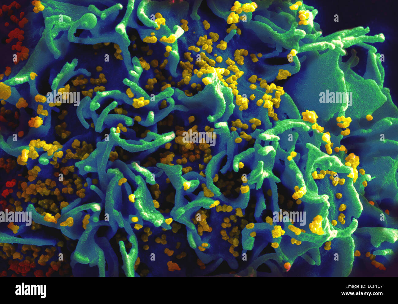 Scanning electron micrograph of HIV particles infecting a human T cell. Stock Photohttps://www.alamy.com/image-license-details/?v=1https://www.alamy.com/stock-photo-scanning-electron-micrograph-of-hiv-particles-infecting-a-human-t-76547751.html
RFECF1C7–Scanning electron micrograph of HIV particles infecting a human T cell.
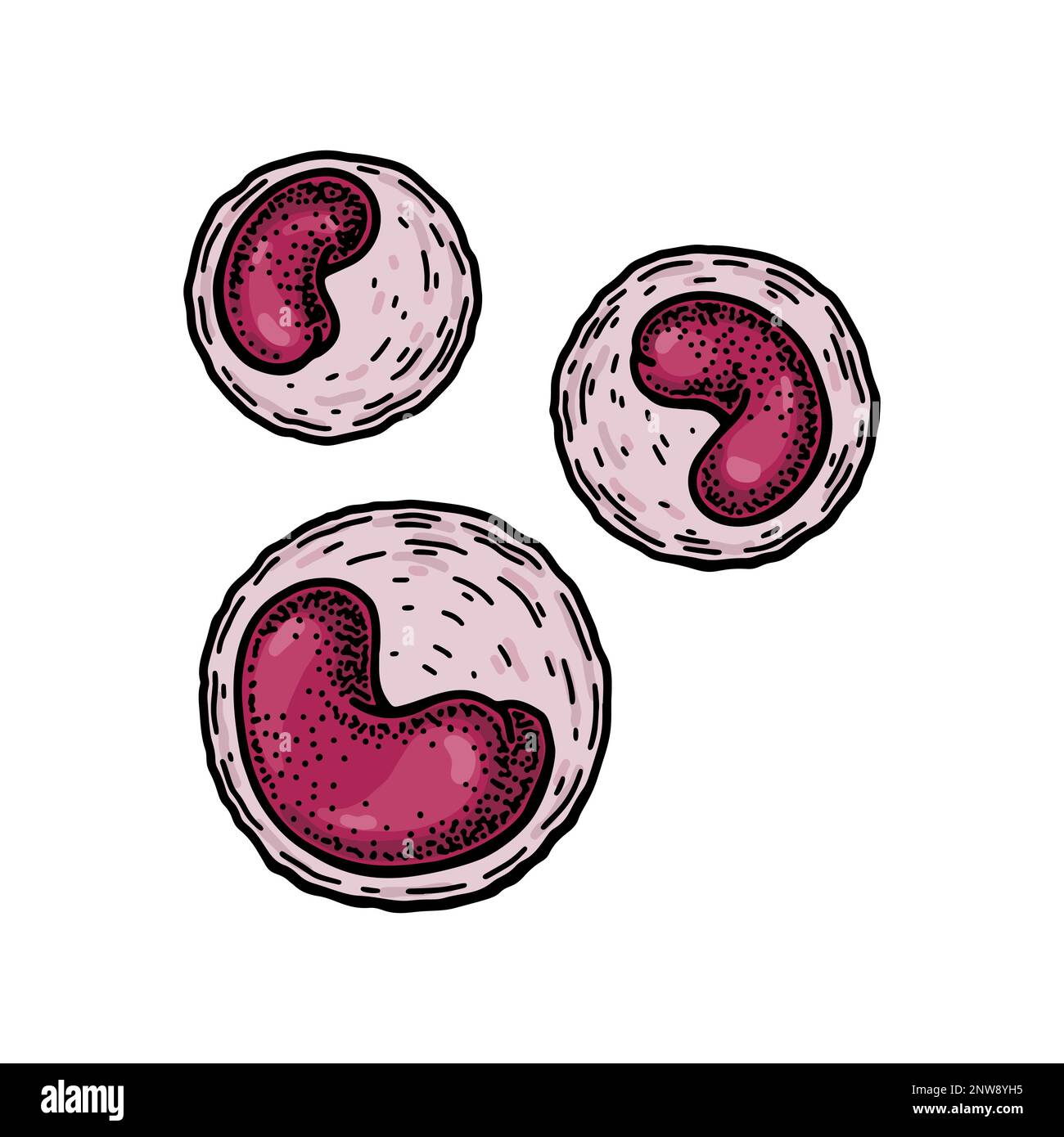 Monocyte leukocyte white blood cells isolated on white background. Hand drawn scientific microbiology vector illustration in sketch style Stock Vectorhttps://www.alamy.com/image-license-details/?v=1https://www.alamy.com/monocyte-leukocyte-white-blood-cells-isolated-on-white-background-hand-drawn-scientific-microbiology-vector-illustration-in-sketch-style-image531874705.html
RF2NW8YH5–Monocyte leukocyte white blood cells isolated on white background. Hand drawn scientific microbiology vector illustration in sketch style
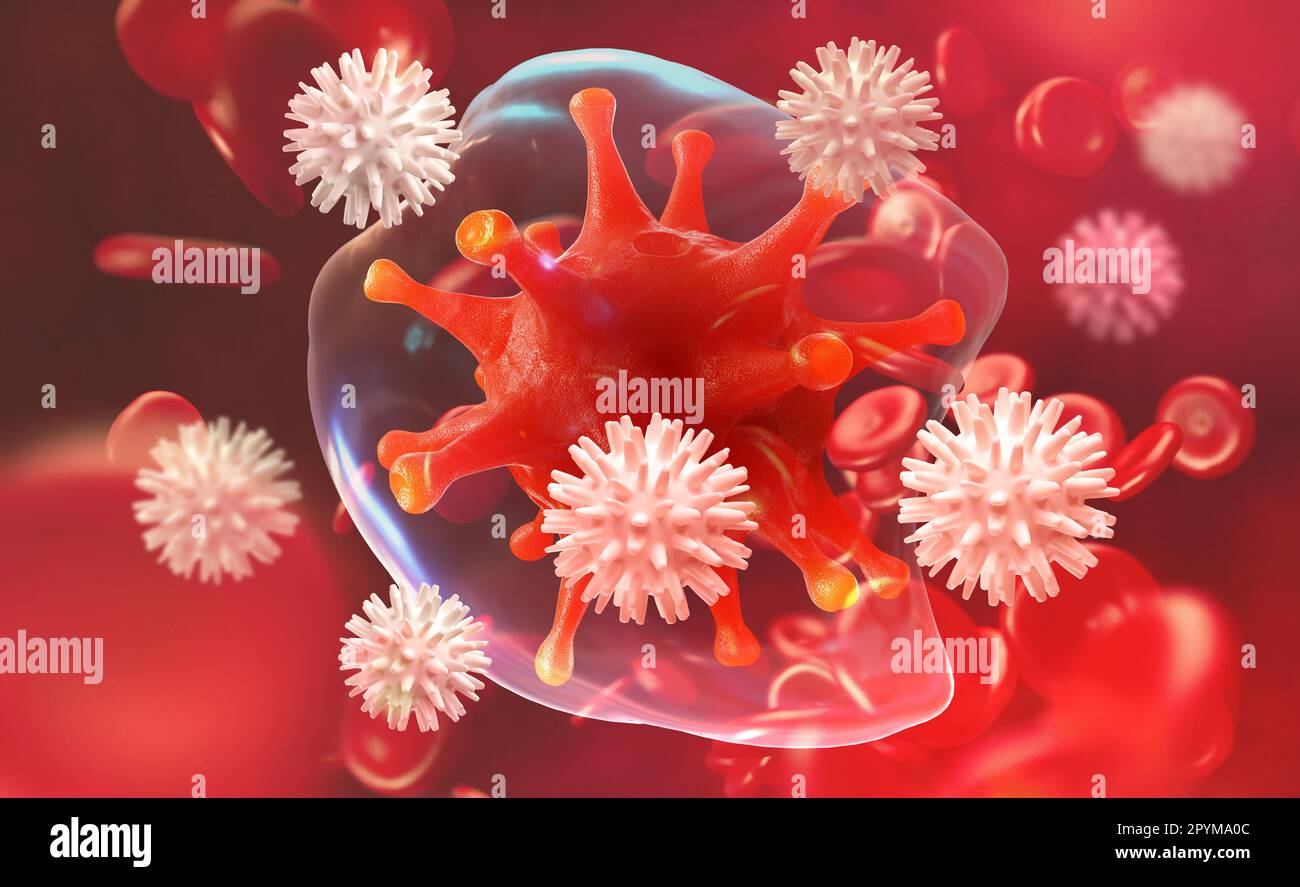 Germs in the blood. Leukocytes attack the virus. Immunity of the body. 3D illustration on medical research Stock Photohttps://www.alamy.com/image-license-details/?v=1https://www.alamy.com/germs-in-the-blood-leukocytes-attack-the-virus-immunity-of-the-body-3d-illustration-on-medical-research-image550564012.html
RF2PYMA0C–Germs in the blood. Leukocytes attack the virus. Immunity of the body. 3D illustration on medical research
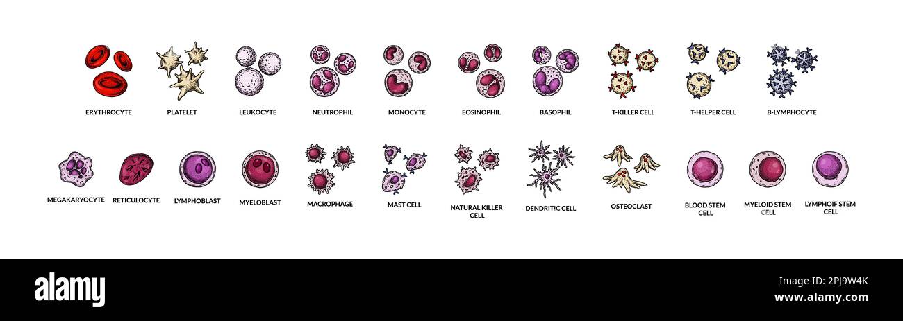 Blood cells isolated on white background. Scientific microbiology vector illustration in sketch style Stock Vectorhttps://www.alamy.com/image-license-details/?v=1https://www.alamy.com/blood-cells-isolated-on-white-background-scientific-microbiology-vector-illustration-in-sketch-style-image544802515.html
RF2PJ9W4K–Blood cells isolated on white background. Scientific microbiology vector illustration in sketch style
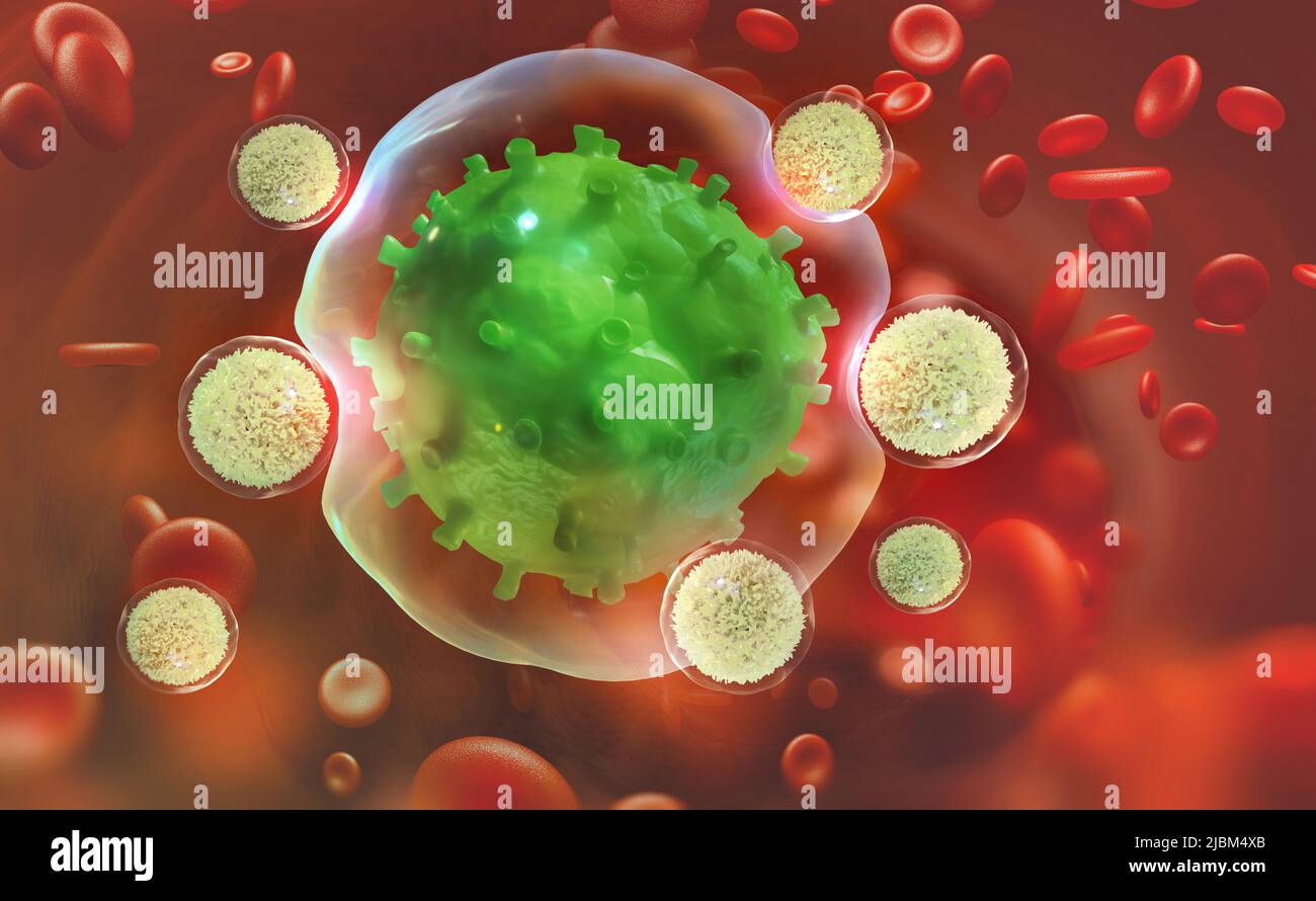 Leukocytes attack the virus. Immunity of the body. 3D illustration on medical research Stock Photohttps://www.alamy.com/image-license-details/?v=1https://www.alamy.com/leukocytes-attack-the-virus-immunity-of-the-body-3d-illustration-on-medical-research-image471884067.html
RF2JBM4XB–Leukocytes attack the virus. Immunity of the body. 3D illustration on medical research
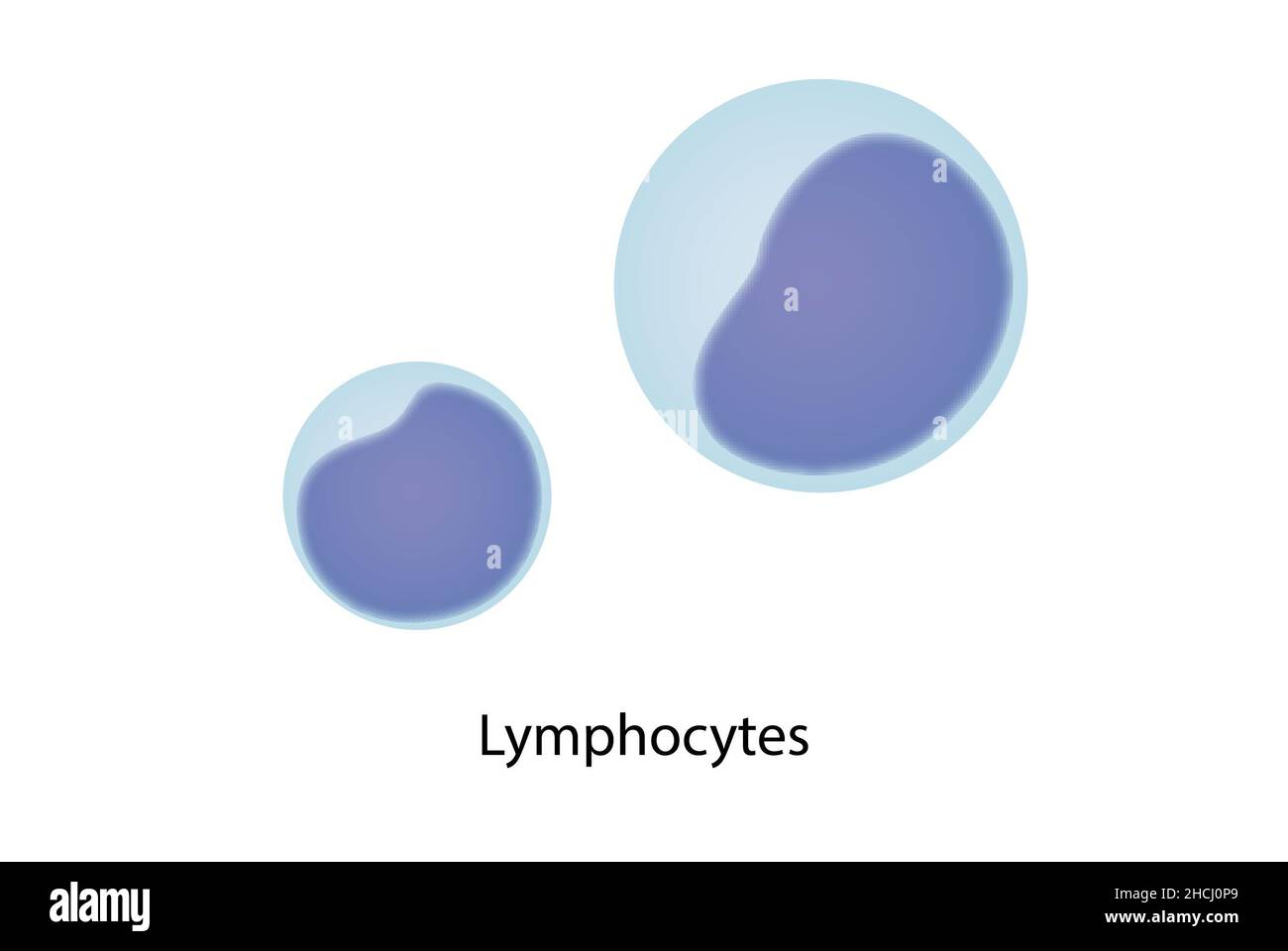 Lymphocites, blood smear, large and small lyphocyte. Stock Photohttps://www.alamy.com/image-license-details/?v=1https://www.alamy.com/lymphocites-blood-smear-large-and-small-lyphocyte-image455241201.html
RF2HCJ0P9–Lymphocites, blood smear, large and small lyphocyte.
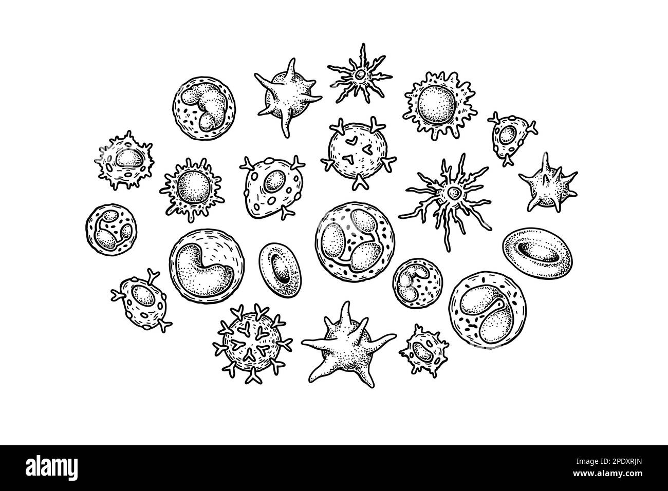 Blood cells isolated on white background. Scientific microbiology vector illustration in sketch style Stock Vectorhttps://www.alamy.com/image-license-details/?v=1https://www.alamy.com/blood-cells-isolated-on-white-background-scientific-microbiology-vector-illustration-in-sketch-style-image542101245.html
RF2PDXRJN–Blood cells isolated on white background. Scientific microbiology vector illustration in sketch style
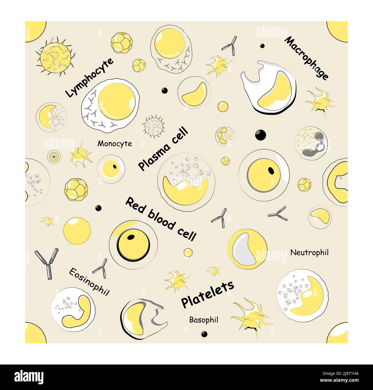 seamless pattern. red and white blood cells under microscope. Vector background Stock Vectorhttps://www.alamy.com/image-license-details/?v=1https://www.alamy.com/seamless-pattern-red-and-white-blood-cells-under-microscope-vector-background-image470366774.html
RF2J971HA–seamless pattern. red and white blood cells under microscope. Vector background
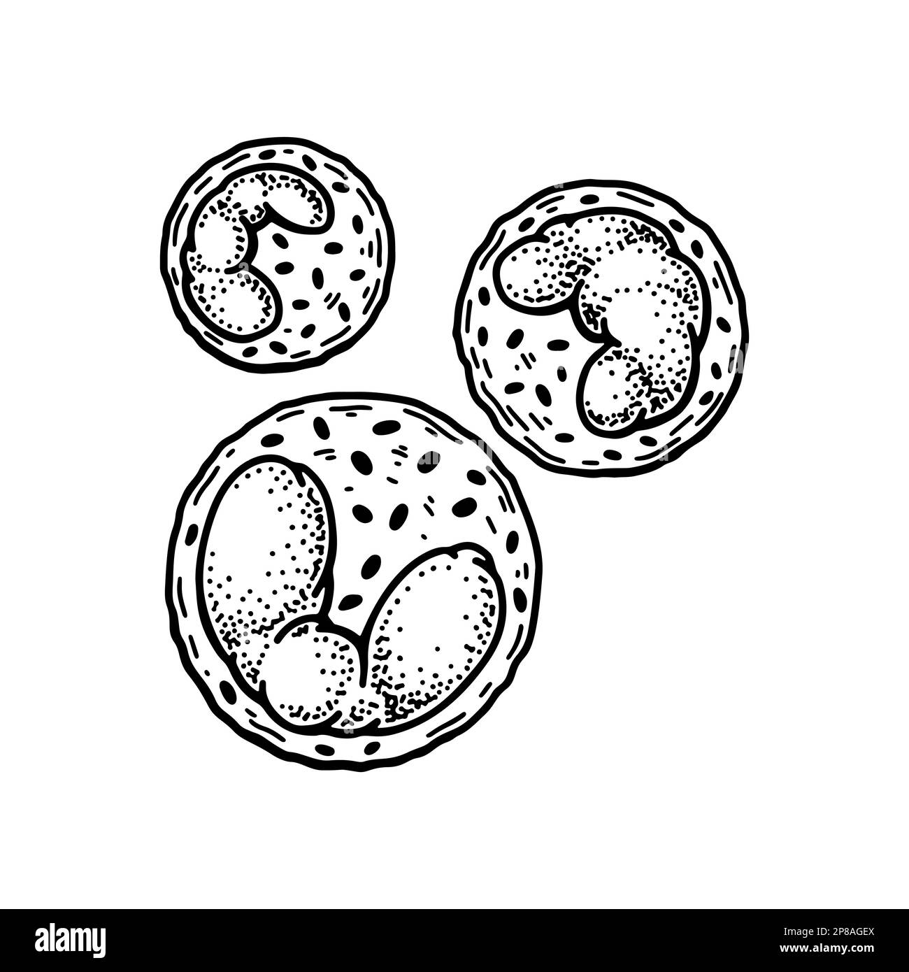 Basophil leukocyte white blood cells isolated on white background. Hand drawn scientific microbiology vector illustration in sketch style Stock Vectorhttps://www.alamy.com/image-license-details/?v=1https://www.alamy.com/basophil-leukocyte-white-blood-cells-isolated-on-white-background-hand-drawn-scientific-microbiology-vector-illustration-in-sketch-style-image538671138.html
RF2P8AGEX–Basophil leukocyte white blood cells isolated on white background. Hand drawn scientific microbiology vector illustration in sketch style
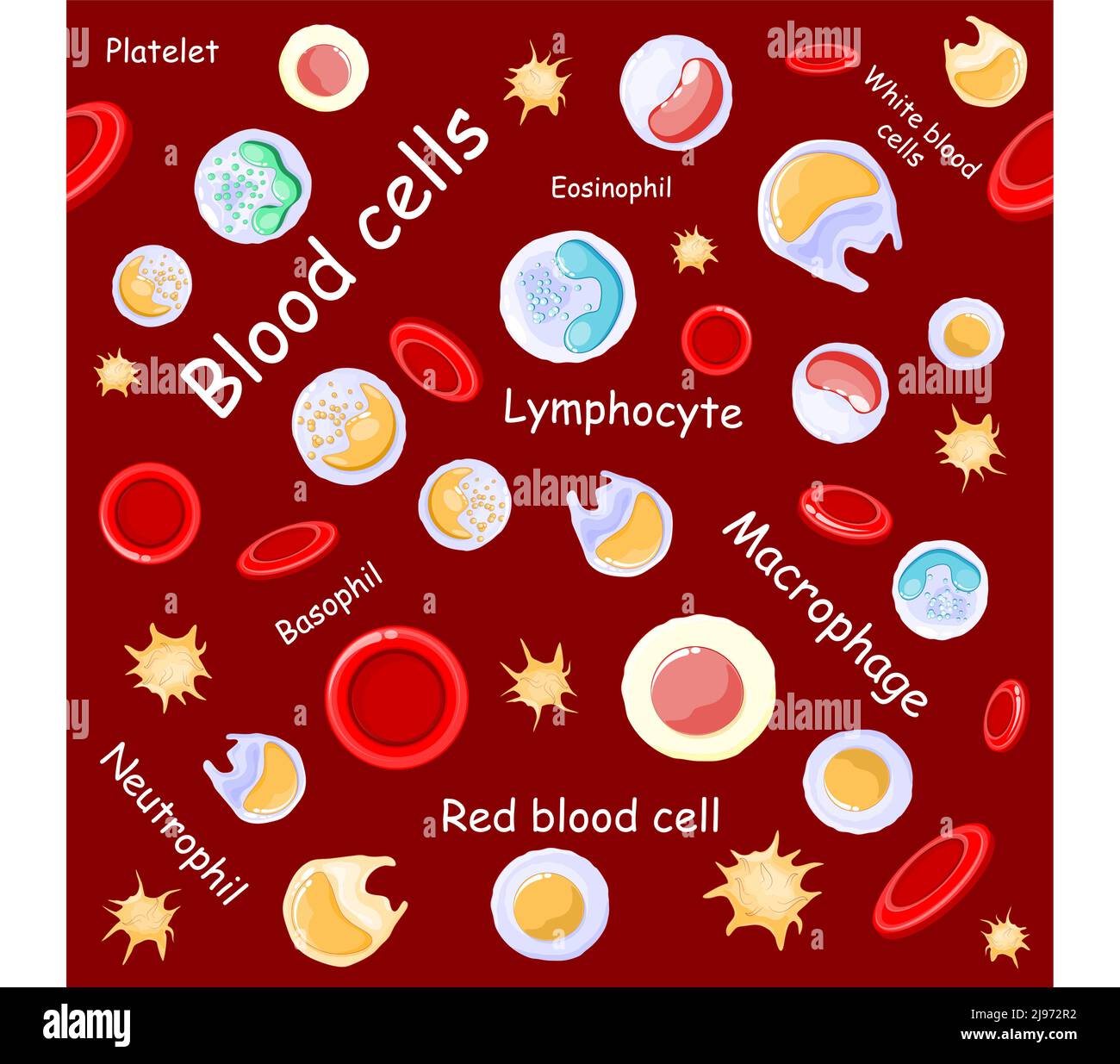 seamless pattern. red and white blood cells under microscope. Vector background Stock Vectorhttps://www.alamy.com/image-license-details/?v=1https://www.alamy.com/seamless-pattern-red-and-white-blood-cells-under-microscope-vector-background-image470367718.html
RF2J972R2–seamless pattern. red and white blood cells under microscope. Vector background
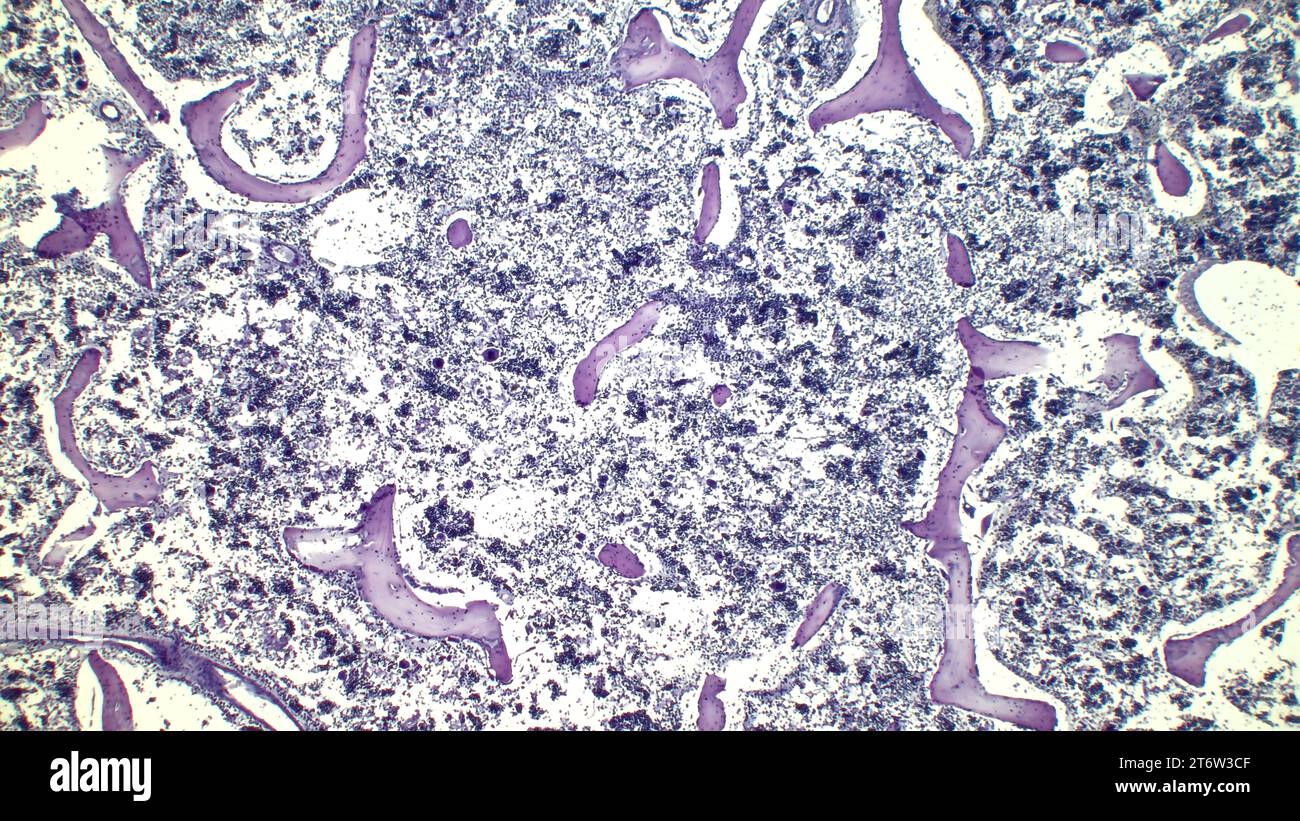 Human red bone marrow, hematopoietic press. The big tsit beetveen erythrocytes are megakaryocytes. Hematoxylin and eosin stain. Stock Photohttps://www.alamy.com/image-license-details/?v=1https://www.alamy.com/human-red-bone-marrow-hematopoietic-press-the-big-tsit-beetveen-erythrocytes-are-megakaryocytes-hematoxylin-and-eosin-stain-image572181583.html
RF2T6W3CF–Human red bone marrow, hematopoietic press. The big tsit beetveen erythrocytes are megakaryocytes. Hematoxylin and eosin stain.
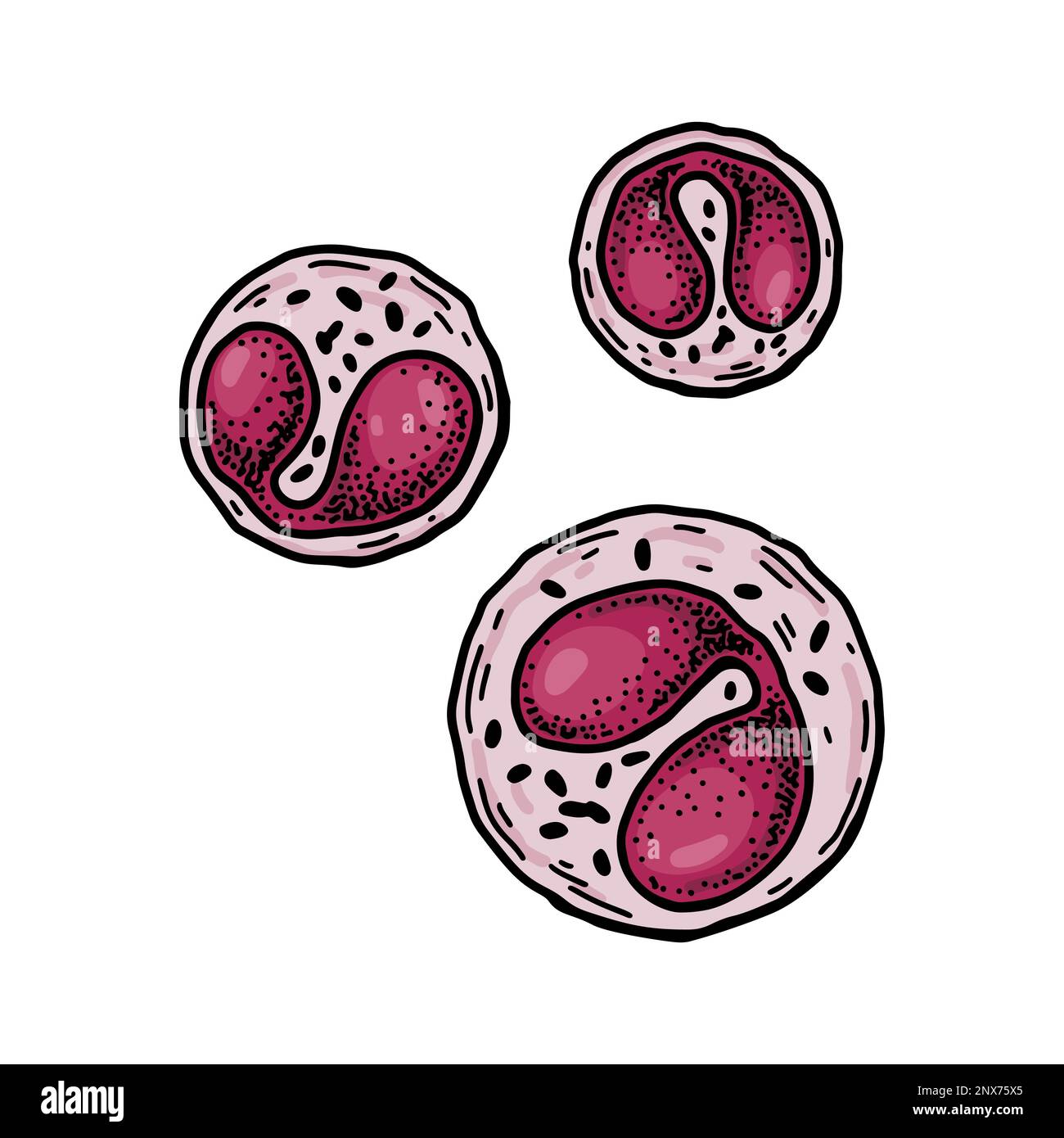 Eosinophil leukocyte white blood cells isolated on white background. Hand drawn scientific microbiology vector illustration in sketch style Stock Vectorhttps://www.alamy.com/image-license-details/?v=1https://www.alamy.com/eosinophil-leukocyte-white-blood-cells-isolated-on-white-background-hand-drawn-scientific-microbiology-vector-illustration-in-sketch-style-image532450413.html
RF2NX75X5–Eosinophil leukocyte white blood cells isolated on white background. Hand drawn scientific microbiology vector illustration in sketch style
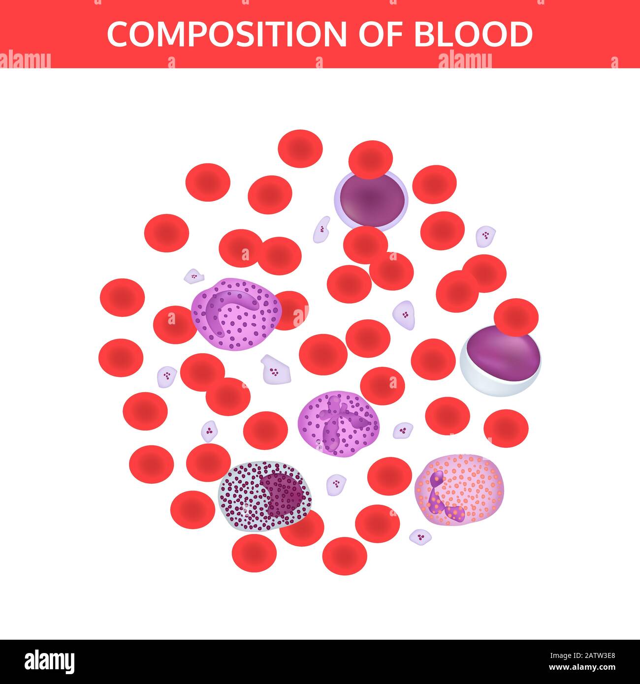 Composition of blood: red and white cells, platelets under a microscope. Medical vector concept on white background. Stock Vectorhttps://www.alamy.com/image-license-details/?v=1https://www.alamy.com/composition-of-blood-red-and-white-cells-platelets-under-a-microscope-medical-vector-concept-on-white-background-image342300288.html
RF2ATW3E8–Composition of blood: red and white cells, platelets under a microscope. Medical vector concept on white background.
 Diagnostic form, Vial of blood samples and Medicine in a hospital, conceptual image Stock Photohttps://www.alamy.com/image-license-details/?v=1https://www.alamy.com/diagnostic-form-vial-of-blood-samples-and-medicine-in-a-hospital-conceptual-image-image218091668.html
RFPJPX44–Diagnostic form, Vial of blood samples and Medicine in a hospital, conceptual image
 Resting T lymphocytes. Coloured scanning electron micrograph (SEM) of resting T lymphocytes from a human blood sample. T lymphocytes, or T cells, are a type of white blood cell and components of the body's immune system. They mature in the thymus. T lymphocytes recognise a specific site on the surface of pathogens or foreign objects (antigens), bind to it, and become activated to produce antibodies or cells to eliminate that antigen. Specimen courtesy of Professor Greg Towers, University College London, UK. Magnification: x7000 when printed at 10 centimetres wide. Stock Photohttps://www.alamy.com/image-license-details/?v=1https://www.alamy.com/resting-t-lymphocytes-coloured-scanning-electron-micrograph-sem-of-resting-t-lymphocytes-from-a-human-blood-sample-t-lymphocytes-or-t-cells-are-a-type-of-white-blood-cell-and-components-of-the-bodys-immune-system-they-mature-in-the-thymus-t-lymphocytes-recognise-a-specific-site-on-the-surface-of-pathogens-or-foreign-objects-antigens-bind-to-it-and-become-activated-to-produce-antibodies-or-cells-to-eliminate-that-antigen-specimen-courtesy-of-professor-greg-towers-university-college-london-uk-magnification-x7000-when-printed-at-10-centimetres-wide-image220702065.html
RFPR1RMH–Resting T lymphocytes. Coloured scanning electron micrograph (SEM) of resting T lymphocytes from a human blood sample. T lymphocytes, or T cells, are a type of white blood cell and components of the body's immune system. They mature in the thymus. T lymphocytes recognise a specific site on the surface of pathogens or foreign objects (antigens), bind to it, and become activated to produce antibodies or cells to eliminate that antigen. Specimen courtesy of Professor Greg Towers, University College London, UK. Magnification: x7000 when printed at 10 centimetres wide.
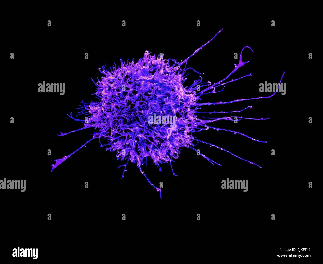 Scanning electron micrograph of a human natural killer cell. Credit: NIAID Stock Photohttps://www.alamy.com/image-license-details/?v=1https://www.alamy.com/scanning-electron-micrograph-of-a-human-natural-killer-cell-credit-niaid-image476706614.html
RM2JKFT46–Scanning electron micrograph of a human natural killer cell. Credit: NIAID
 Bacterial infection in the blood. Viruses attack the human body. Microorganisms, germs and microbes. 3D colorful illustration on microbiology Stock Photohttps://www.alamy.com/image-license-details/?v=1https://www.alamy.com/bacterial-infection-in-the-blood-viruses-attack-the-human-body-microorganisms-germs-and-microbes-3d-colorful-illustration-on-microbiology-image471884139.html
RF2JBM50Y–Bacterial infection in the blood. Viruses attack the human body. Microorganisms, germs and microbes. 3D colorful illustration on microbiology
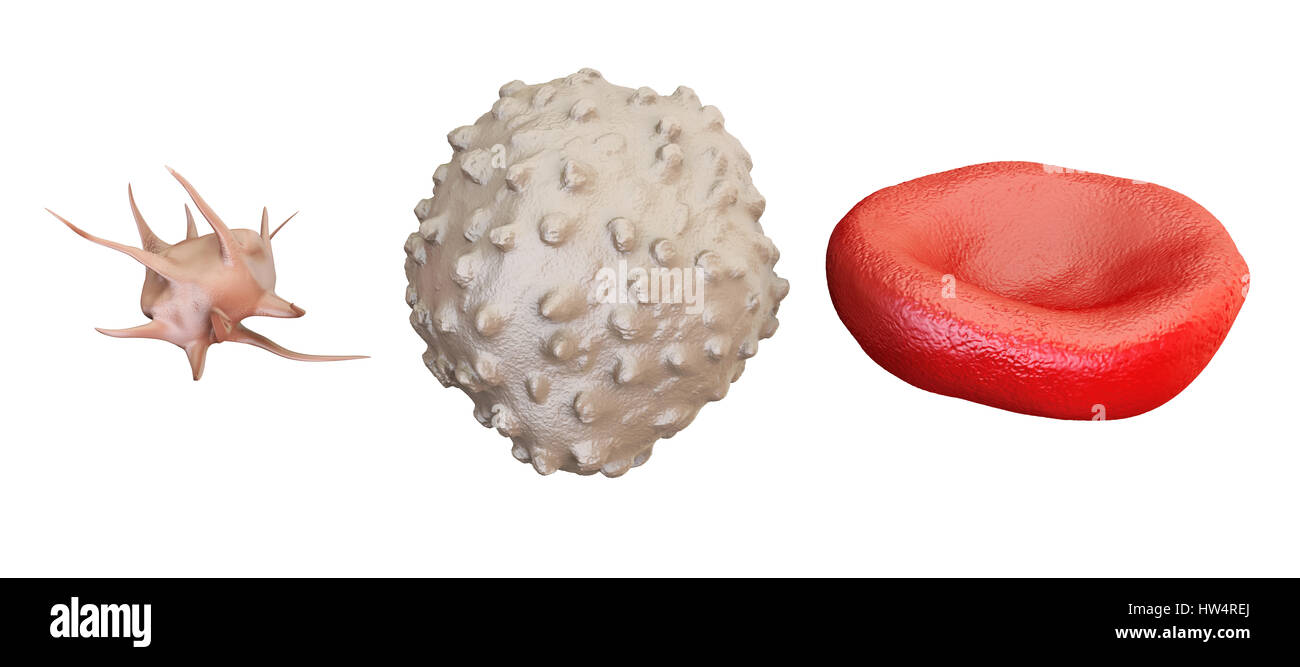 blood cells erythrocyte, lymphocyte, thrombocyte, 3D rendering isolated on white background Stock Photohttps://www.alamy.com/image-license-details/?v=1https://www.alamy.com/stock-photo-blood-cells-erythrocyte-lymphocyte-thrombocyte-3d-rendering-isolated-135945226.html
RFHW4REJ–blood cells erythrocyte, lymphocyte, thrombocyte, 3D rendering isolated on white background
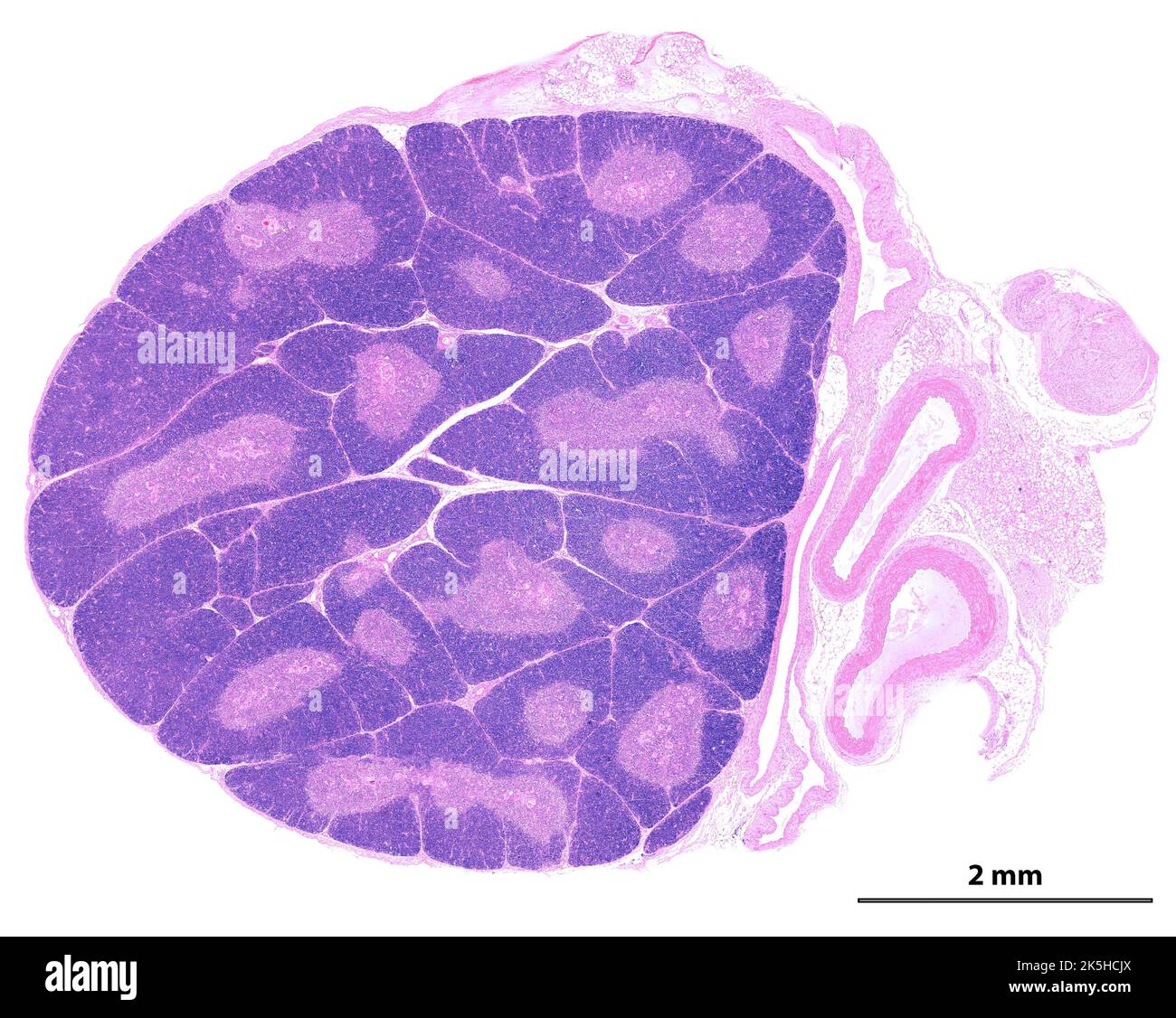 Low power light microscope micrograph showing a young thymus. The organization into lobules is clearly seen. In each lobule, the peripheral cortex app Stock Photohttps://www.alamy.com/image-license-details/?v=1https://www.alamy.com/low-power-light-microscope-micrograph-showing-a-young-thymus-the-organization-into-lobules-is-clearly-seen-in-each-lobule-the-peripheral-cortex-app-image485346706.html
RF2K5HCJX–Low power light microscope micrograph showing a young thymus. The organization into lobules is clearly seen. In each lobule, the peripheral cortex app
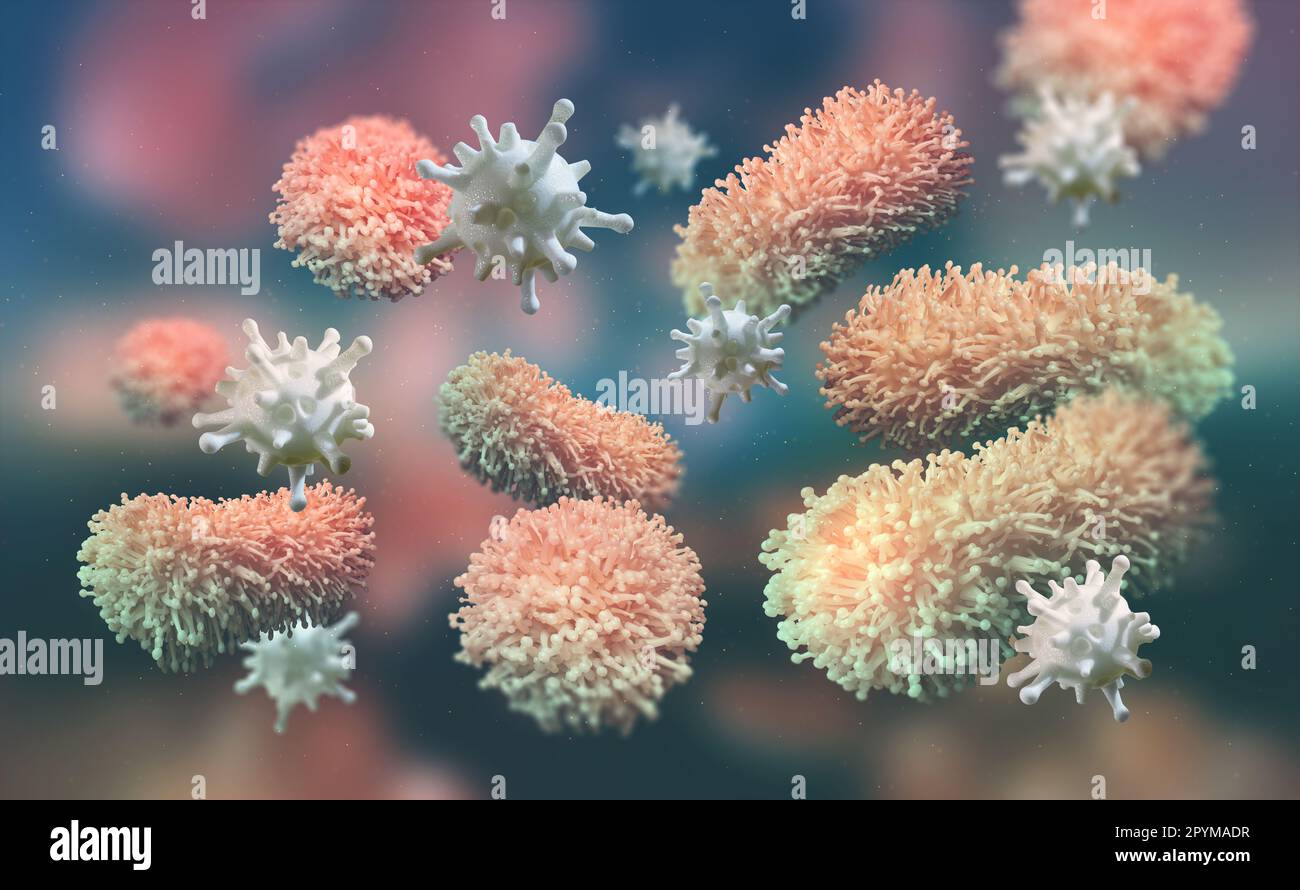 Leukocytes attack the virus. Immunity of the body. Viral infection attacks the human body. Microorganisms, germs and bacteria. 3D illustration on micr Stock Photohttps://www.alamy.com/image-license-details/?v=1https://www.alamy.com/leukocytes-attack-the-virus-immunity-of-the-body-viral-infection-attacks-the-human-body-microorganisms-germs-and-bacteria-3d-illustration-on-micr-image550564387.html
RF2PYMADR–Leukocytes attack the virus. Immunity of the body. Viral infection attacks the human body. Microorganisms, germs and bacteria. 3D illustration on micr
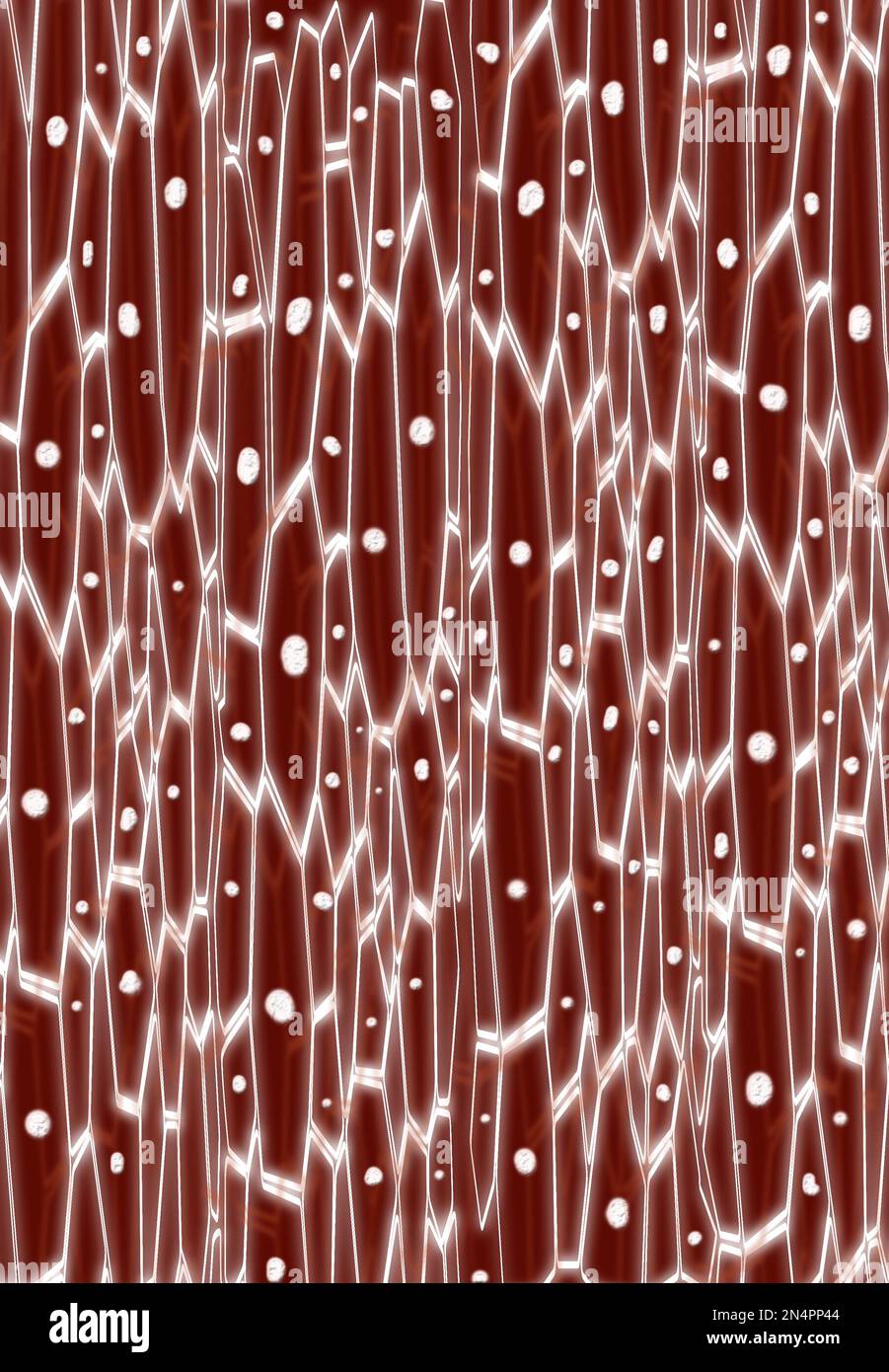 Closeup view of blood under microscope. Illustration Stock Photohttps://www.alamy.com/image-license-details/?v=1https://www.alamy.com/closeup-view-of-blood-under-microscope-illustration-image519269972.html
RF2N4PP44–Closeup view of blood under microscope. Illustration
 Lymphocyte word cloud concept Stock Photohttps://www.alamy.com/image-license-details/?v=1https://www.alamy.com/lymphocyte-word-cloud-concept-image262836301.html
RFW7H6AN–Lymphocyte word cloud concept
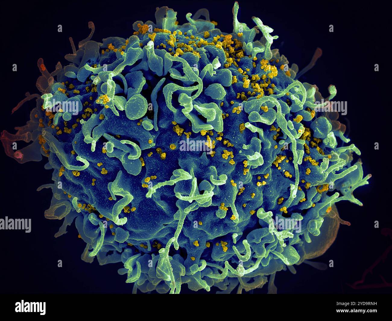 Scanning electron micrograph of a human H9 T cell blue/green infected with HIV virus particles yellow. HIV-infected H9 T Cell 016867 240 Stock Photohttps://www.alamy.com/image-license-details/?v=1https://www.alamy.com/scanning-electron-micrograph-of-a-human-h9-t-cell-bluegreen-infected-with-hiv-virus-particles-yellow-hiv-infected-h9-t-cell-016867-240-image627779981.html
RM2YD9RNH–Scanning electron micrograph of a human H9 T cell blue/green infected with HIV virus particles yellow. HIV-infected H9 T Cell 016867 240
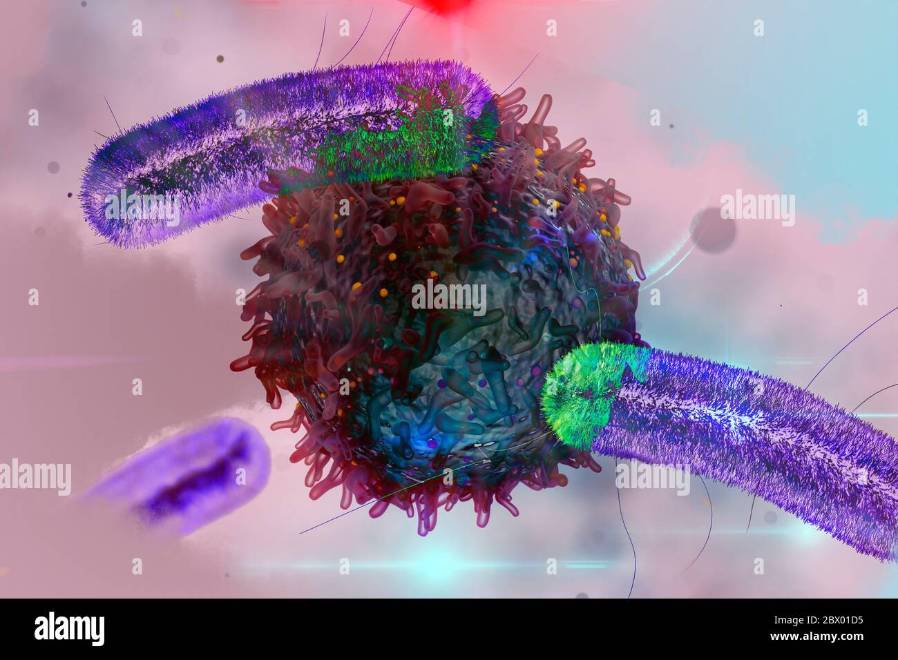 Bacteria and Lymphocyte concept blood disease bacteria treatment Stock Photohttps://www.alamy.com/image-license-details/?v=1https://www.alamy.com/bacteria-and-lymphocyte-concept-blood-disease-bacteria-treatment-image360189569.html
RF2BX01D5–Bacteria and Lymphocyte concept blood disease bacteria treatment
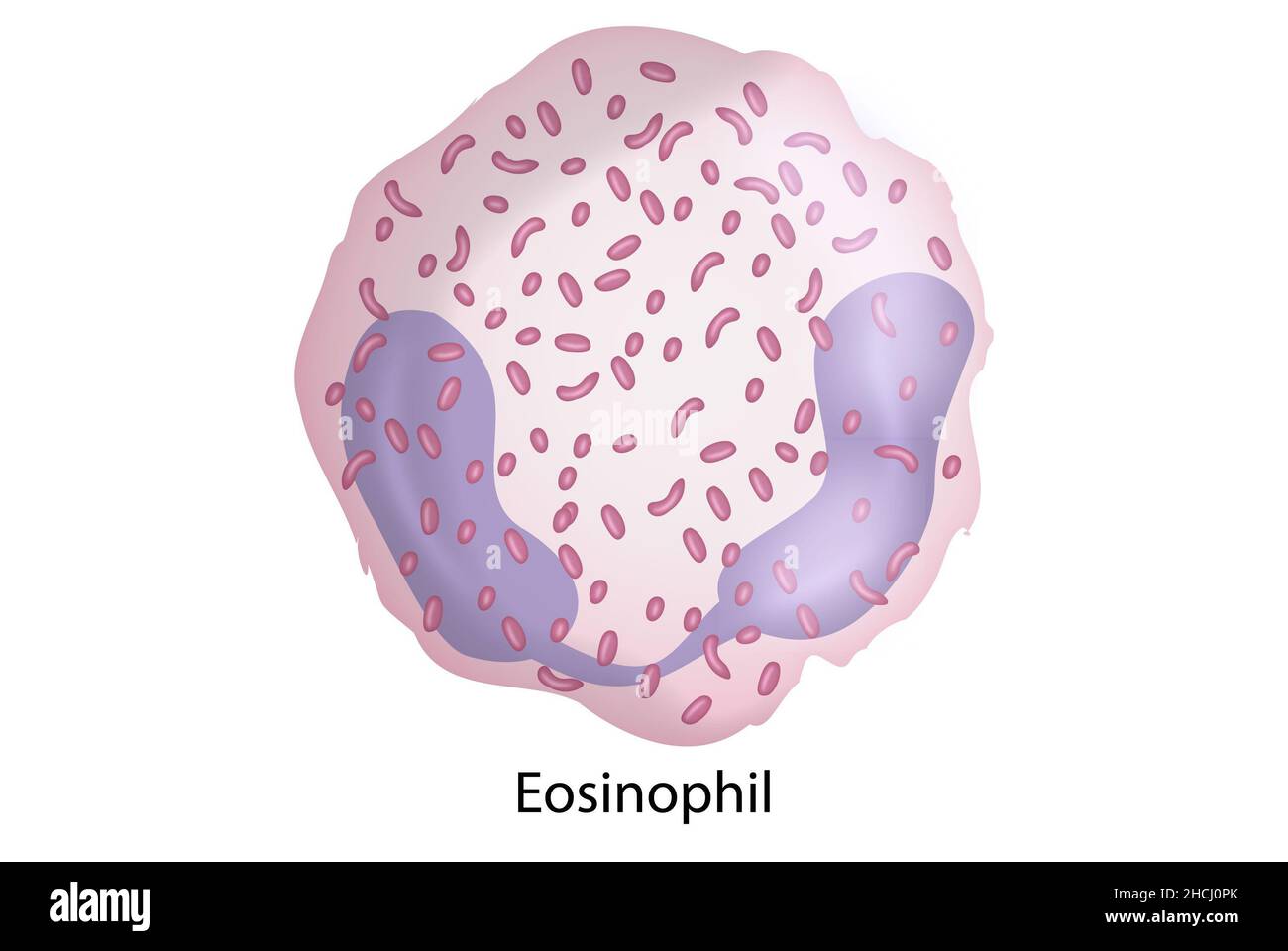 Eosinophil, white blood cells, immune system cells Stock Photohttps://www.alamy.com/image-license-details/?v=1https://www.alamy.com/eosinophil-white-blood-cells-immune-system-cells-image455241211.html
RF2HCJ0PK–Eosinophil, white blood cells, immune system cells
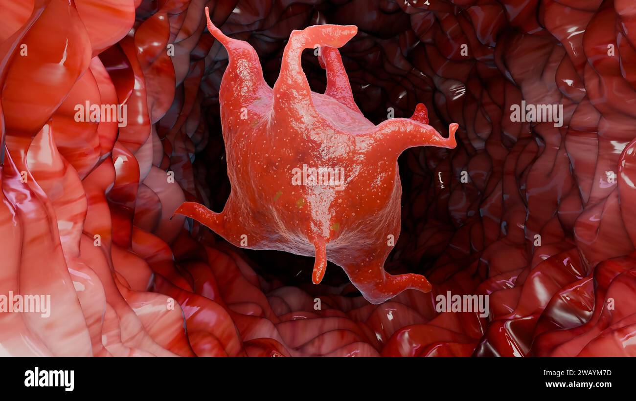 Platelets anatomy, blood cell, thrombocytes in blood vessel under microscope, healing wounds, Activated platelet, blood coagulation and fibrasion proc Stock Photohttps://www.alamy.com/image-license-details/?v=1https://www.alamy.com/platelets-anatomy-blood-cell-thrombocytes-in-blood-vessel-under-microscope-healing-wounds-activated-platelet-blood-coagulation-and-fibrasion-proc-image591907665.html
RM2WAYM7D–Platelets anatomy, blood cell, thrombocytes in blood vessel under microscope, healing wounds, Activated platelet, blood coagulation and fibrasion proc
 White blood cells, red blood cells Stock Photohttps://www.alamy.com/image-license-details/?v=1https://www.alamy.com/stock-photo-white-blood-cells-red-blood-cells-176225758.html
RFM6KNN2–White blood cells, red blood cells
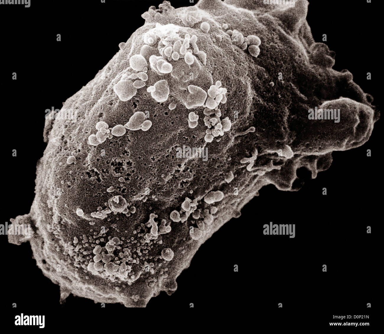 A scanning electron microscope image of a lymphocyte (white blood cell) with an HIV cluster. Stock Photohttps://www.alamy.com/image-license-details/?v=1https://www.alamy.com/stock-photo-a-scanning-electron-microscope-image-of-a-lymphocyte-white-blood-cell-52115665.html
RMD0P21N–A scanning electron microscope image of a lymphocyte (white blood cell) with an HIV cluster.
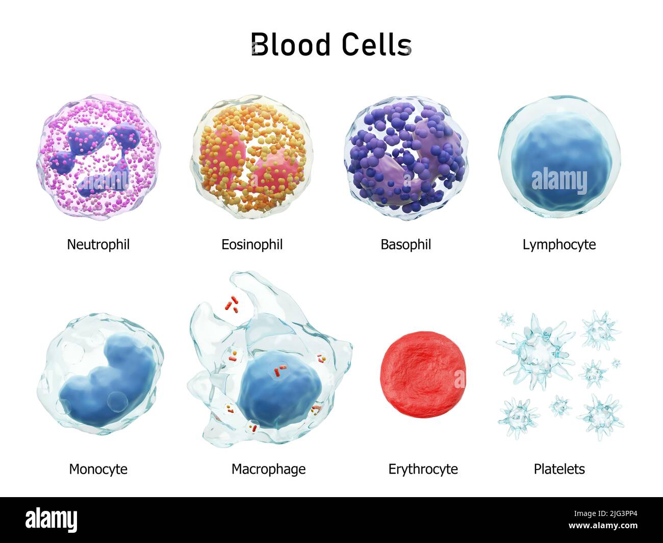 Blood cells series . Neutrophils Eosinophils Basophils Lymphocytes Monocytes Macrophages Erythrocytes and Platelets . Transparent material design . Is Stock Photohttps://www.alamy.com/image-license-details/?v=1https://www.alamy.com/blood-cells-series-neutrophils-eosinophils-basophils-lymphocytes-monocytes-macrophages-erythrocytes-and-platelets-transparent-material-design-is-image474598156.html
RF2JG3PP4–Blood cells series . Neutrophils Eosinophils Basophils Lymphocytes Monocytes Macrophages Erythrocytes and Platelets . Transparent material design . Is
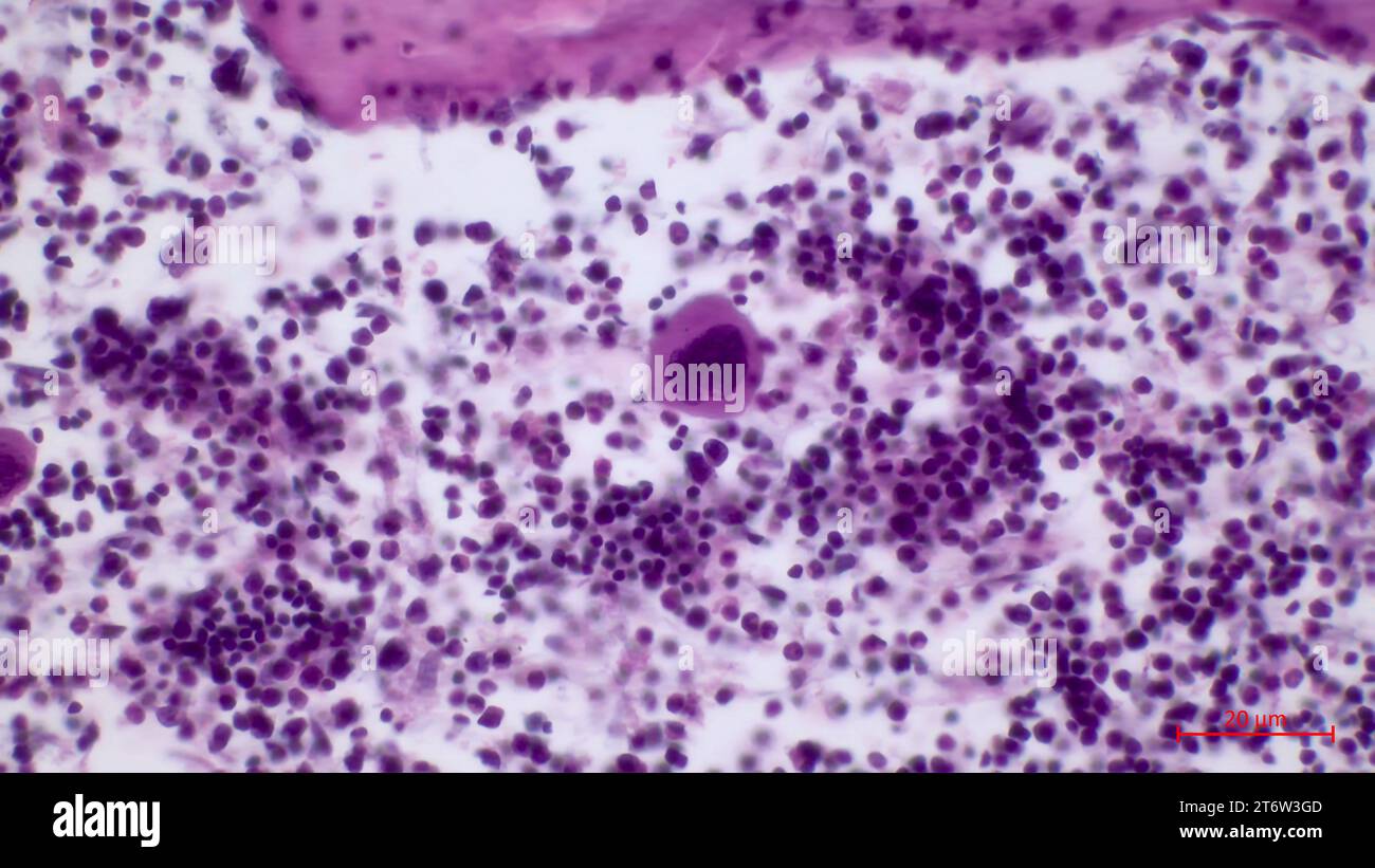 Microscopic structure of red bone marrow histology.The largest cell in the red bone marrow is the megakaryocyte (located in the center). Stock Photohttps://www.alamy.com/image-license-details/?v=1https://www.alamy.com/microscopic-structure-of-red-bone-marrow-histologythe-largest-cell-in-the-red-bone-marrow-is-the-megakaryocyte-located-in-the-center-image572181693.html
RF2T6W3GD–Microscopic structure of red bone marrow histology.The largest cell in the red bone marrow is the megakaryocyte (located in the center).
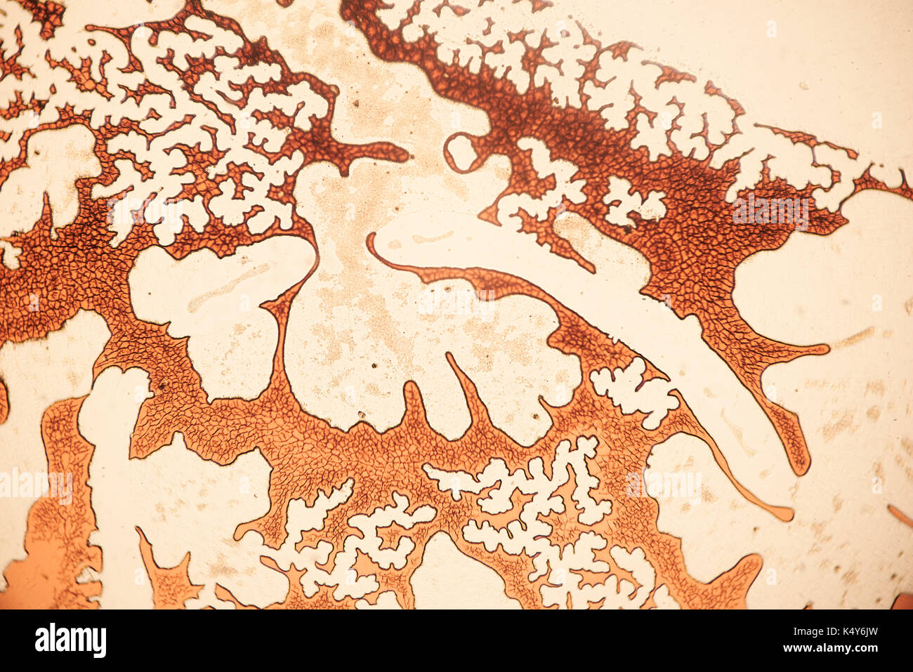 Human lung tissue 100X with blood vessels Stock Photohttps://www.alamy.com/image-license-details/?v=1https://www.alamy.com/human-lung-tissue-100x-with-blood-vessels-image157949873.html
RFK4Y6JW–Human lung tissue 100X with blood vessels
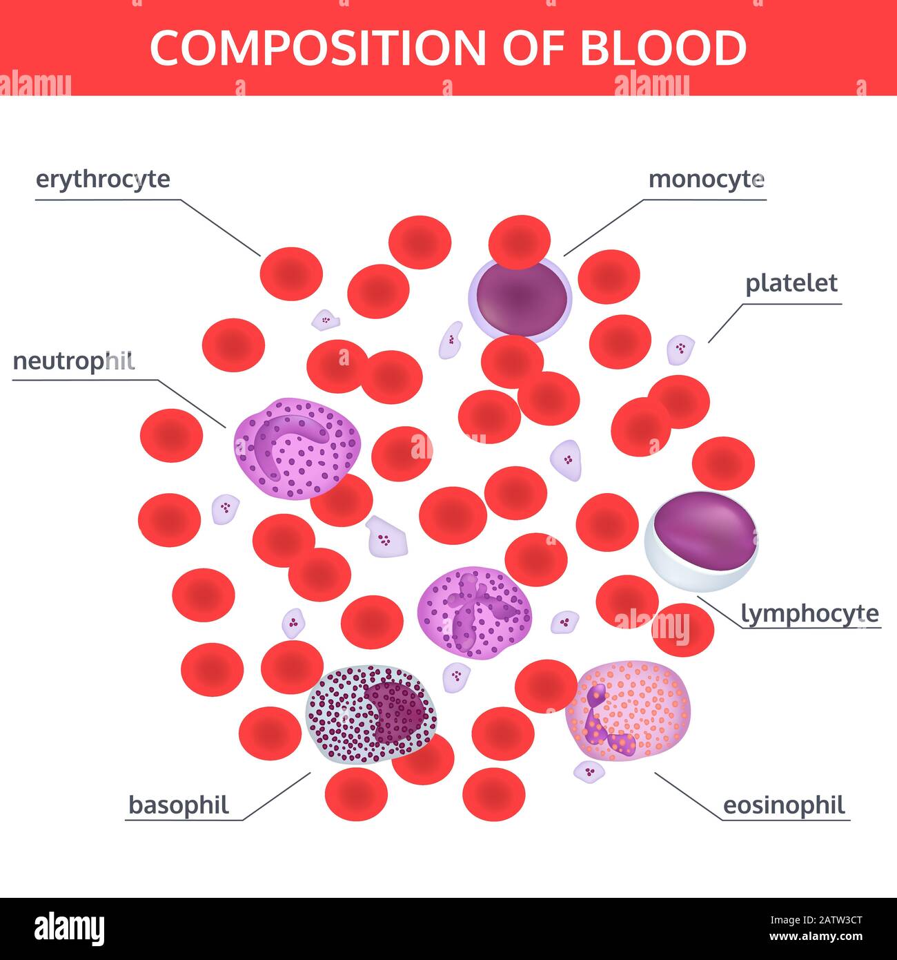 Infographics of composition of blood: red and white cells under a microscope with names on white background. Medical vector concept. Stock Vectorhttps://www.alamy.com/image-license-details/?v=1https://www.alamy.com/infographics-of-composition-of-blood-red-and-white-cells-under-a-microscope-with-names-on-white-background-medical-vector-concept-image342300248.html
RF2ATW3CT–Infographics of composition of blood: red and white cells under a microscope with names on white background. Medical vector concept.
 Diagnostic form, Vial of blood samples and Medicine in a hospital, conceptual image Stock Photohttps://www.alamy.com/image-license-details/?v=1https://www.alamy.com/diagnostic-form-vial-of-blood-samples-and-medicine-in-a-hospital-conceptual-image-image218091697.html
RFPJPX55–Diagnostic form, Vial of blood samples and Medicine in a hospital, conceptual image
 Resting T lymphocytes. Coloured scanning electron micrograph (SEM) of resting T lymphocytes from a human blood sample. T lymphocytes, or T cells, are a type of white blood cell and components of the body's immune system. They mature in the thymus. T lymphocytes recognise a specific site on the surface of pathogens or foreign objects (antigens), bind to it, and become activated to produce antibodies or cells to eliminate that antigen. Specimen courtesy of Professor Greg Towers, University College London, UK. Magnification: x7000 when printed at 10 centimetres wide. Stock Photohttps://www.alamy.com/image-license-details/?v=1https://www.alamy.com/resting-t-lymphocytes-coloured-scanning-electron-micrograph-sem-of-resting-t-lymphocytes-from-a-human-blood-sample-t-lymphocytes-or-t-cells-are-a-type-of-white-blood-cell-and-components-of-the-bodys-immune-system-they-mature-in-the-thymus-t-lymphocytes-recognise-a-specific-site-on-the-surface-of-pathogens-or-foreign-objects-antigens-bind-to-it-and-become-activated-to-produce-antibodies-or-cells-to-eliminate-that-antigen-specimen-courtesy-of-professor-greg-towers-university-college-london-uk-magnification-x7000-when-printed-at-10-centimetres-wide-image220702032.html
RFPR1RKC–Resting T lymphocytes. Coloured scanning electron micrograph (SEM) of resting T lymphocytes from a human blood sample. T lymphocytes, or T cells, are a type of white blood cell and components of the body's immune system. They mature in the thymus. T lymphocytes recognise a specific site on the surface of pathogens or foreign objects (antigens), bind to it, and become activated to produce antibodies or cells to eliminate that antigen. Specimen courtesy of Professor Greg Towers, University College London, UK. Magnification: x7000 when printed at 10 centimetres wide.
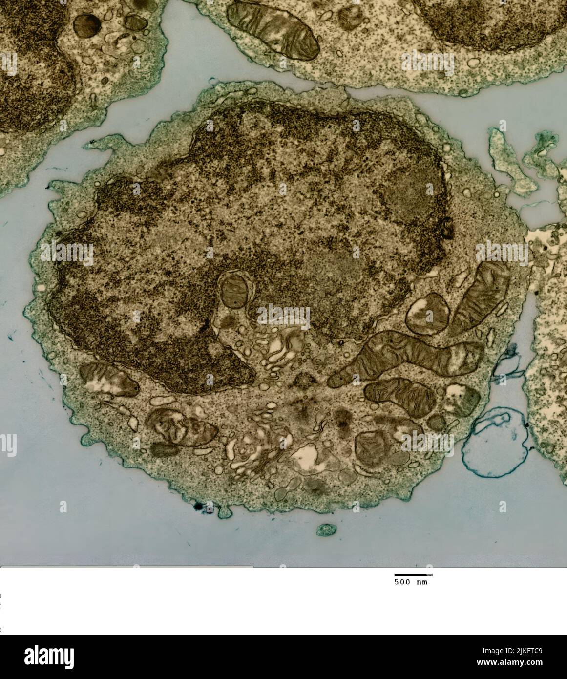 Transmission electron micrograph of a B cell from a human donor. Stock Photohttps://www.alamy.com/image-license-details/?v=1https://www.alamy.com/transmission-electron-micrograph-of-a-b-cell-from-a-human-donor-image476706841.html
RM2JKFTC9–Transmission electron micrograph of a B cell from a human donor.
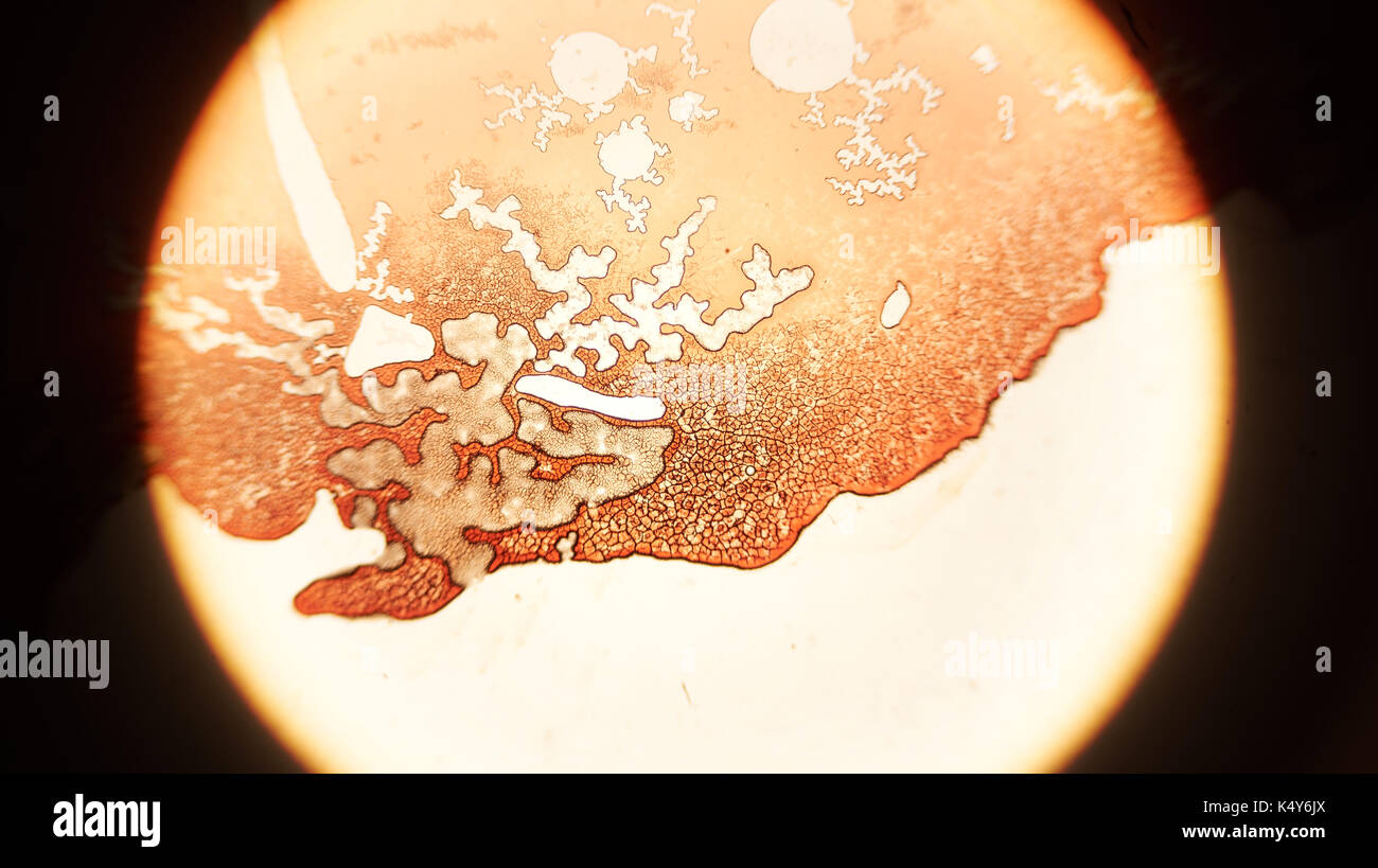 Human lung tissue 100X with blood vessels Stock Photohttps://www.alamy.com/image-license-details/?v=1https://www.alamy.com/human-lung-tissue-100x-with-blood-vessels-image157949874.html
RFK4Y6JX–Human lung tissue 100X with blood vessels
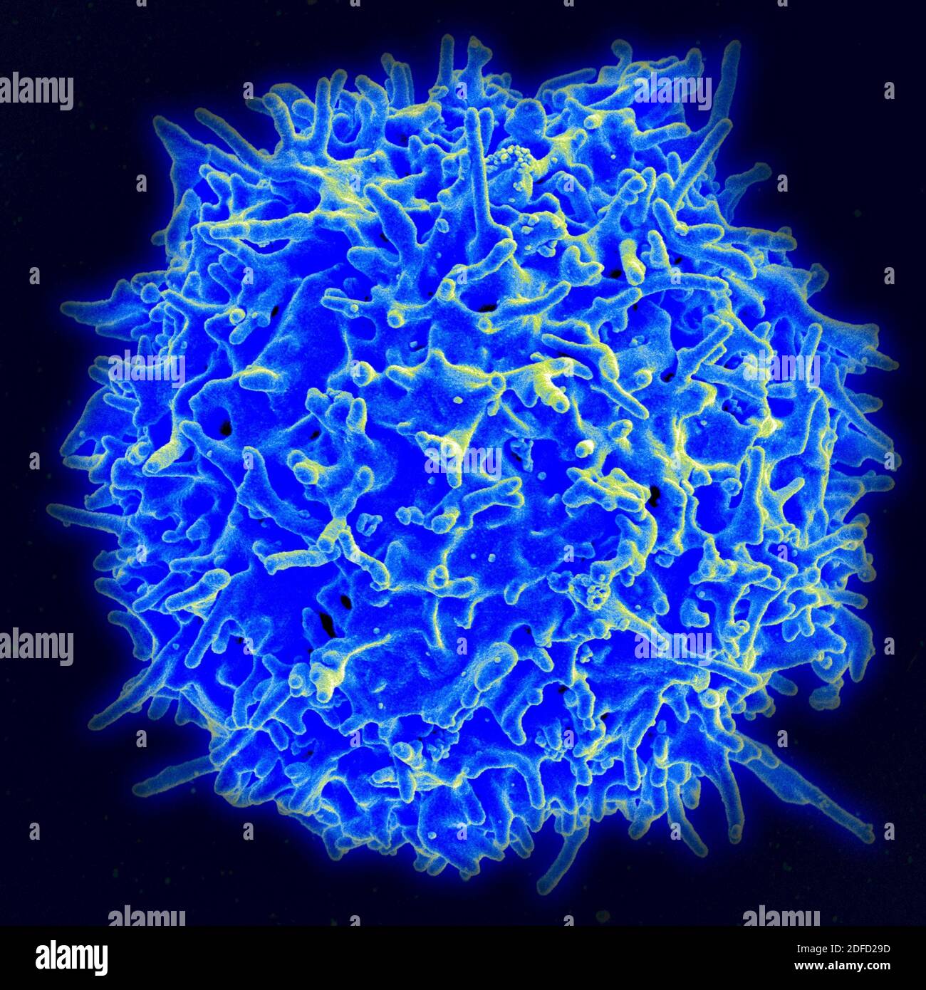 Scanning electron micrograph of a human T lymphocyte (also called a T cell) from the immune system of a healthy donor. Credit: NIAID. Stock Photohttps://www.alamy.com/image-license-details/?v=1https://www.alamy.com/scanning-electron-micrograph-of-a-human-t-lymphocyte-also-called-a-t-cell-from-the-immune-system-of-a-healthy-donor-credit-niaid-image388135145.html
RF2DFD29D–Scanning electron micrograph of a human T lymphocyte (also called a T cell) from the immune system of a healthy donor. Credit: NIAID.
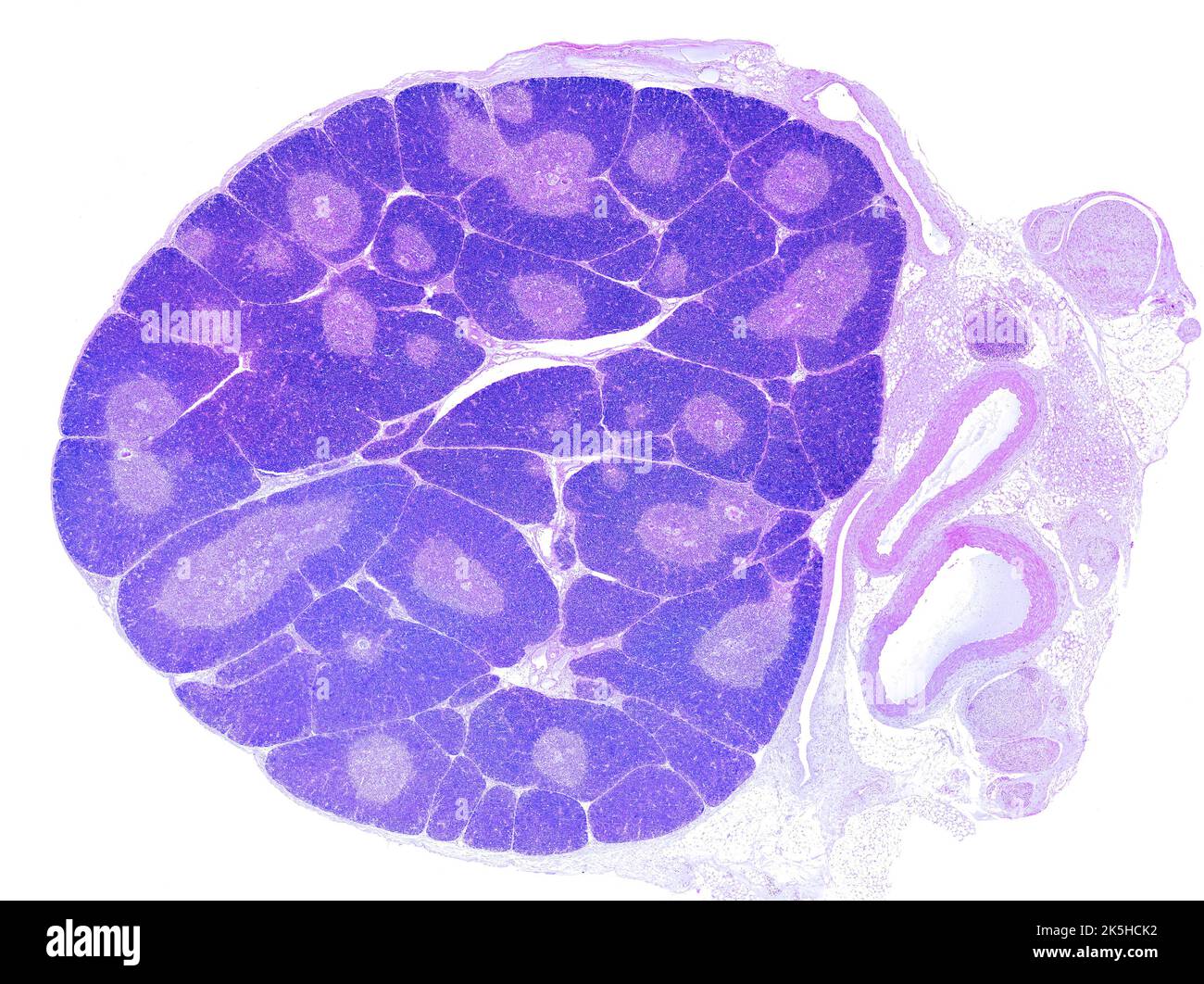 Low power light microscope micrograph showing a young thymus. The organization into lobules is clearly seen. In each lobule, the peripheral cortex app Stock Photohttps://www.alamy.com/image-license-details/?v=1https://www.alamy.com/low-power-light-microscope-micrograph-showing-a-young-thymus-the-organization-into-lobules-is-clearly-seen-in-each-lobule-the-peripheral-cortex-app-image485346710.html
RF2K5HCK2–Low power light microscope micrograph showing a young thymus. The organization into lobules is clearly seen. In each lobule, the peripheral cortex app
 Scanning electron micrograph of a human H9 T cell (blue/green) infected with HIV virus particles (yellow). Stock Photohttps://www.alamy.com/image-license-details/?v=1https://www.alamy.com/scanning-electron-micrograph-of-a-human-h9-t-cell-bluegreen-infected-with-hiv-virus-particles-yellow-image627691126.html
RM2YD5PC6–Scanning electron micrograph of a human H9 T cell (blue/green) infected with HIV virus particles (yellow).
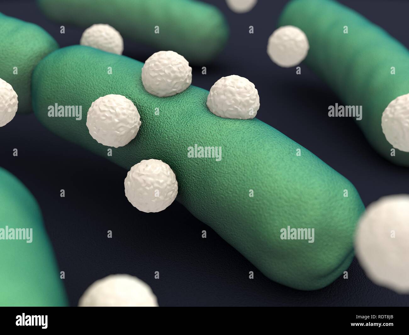 Neutrophil granulocyte Stock Photohttps://www.alamy.com/image-license-details/?v=1https://www.alamy.com/neutrophil-granulocyte-image232258947.html
RFRDT8JB–Neutrophil granulocyte
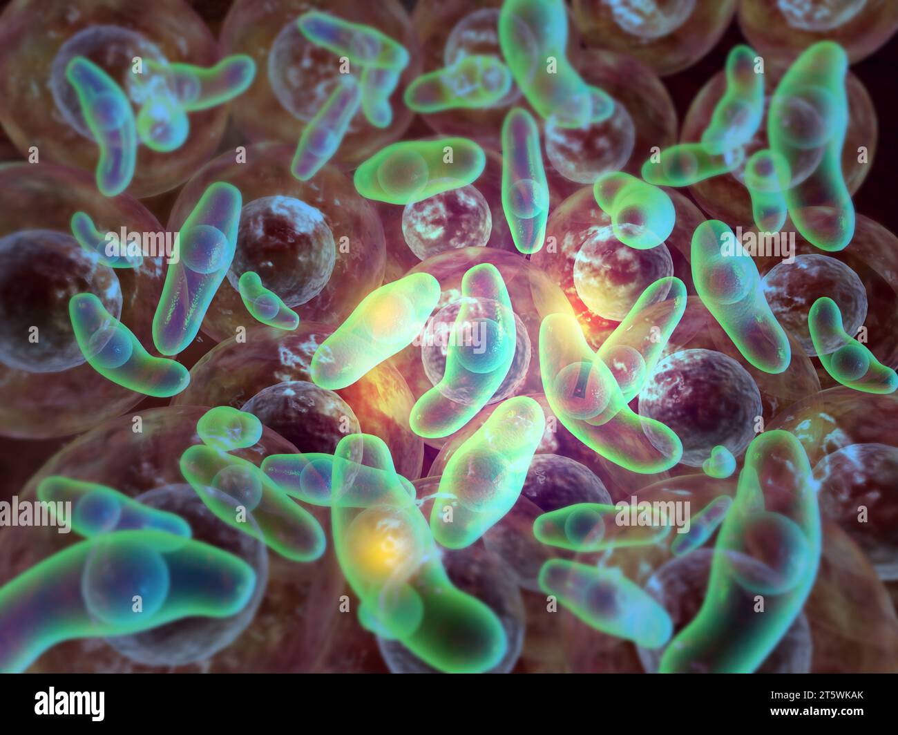 Virus attacking cells. 3d illustration Stock Photohttps://www.alamy.com/image-license-details/?v=1https://www.alamy.com/virus-attacking-cells-3d-illustration-image571579419.html
RF2T5WKAK–Virus attacking cells. 3d illustration
 Scanning electron micrograph of a human H9 T cell blue/green infected with HIV virus particles yellow. HIV-infected H9 T Cell 016867 027 Stock Photohttps://www.alamy.com/image-license-details/?v=1https://www.alamy.com/scanning-electron-micrograph-of-a-human-h9-t-cell-bluegreen-infected-with-hiv-virus-particles-yellow-hiv-infected-h9-t-cell-016867-027-image627778371.html
RM2YD9NM3–Scanning electron micrograph of a human H9 T cell blue/green infected with HIV virus particles yellow. HIV-infected H9 T Cell 016867 027
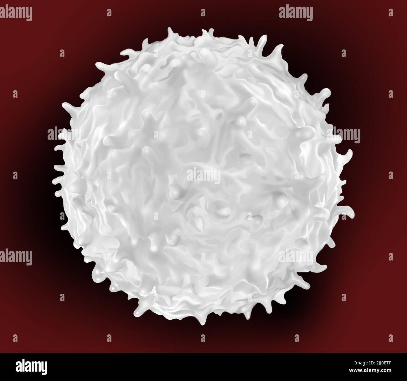 white blood cells found throughout the body in the blood and lymphatic system Stock Photohttps://www.alamy.com/image-license-details/?v=1https://www.alamy.com/white-blood-cells-found-throughout-the-body-in-the-blood-and-lymphatic-system-image475755414.html
RF2JJ0ETP–white blood cells found throughout the body in the blood and lymphatic system
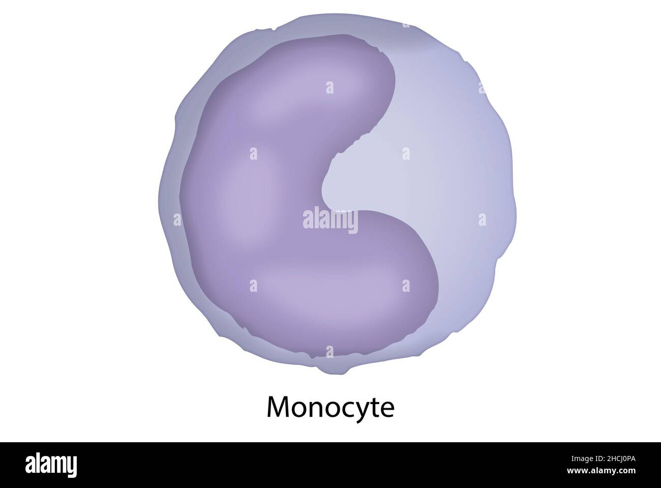 Monocyte, macrophage, cellular structure of the blood Stock Photohttps://www.alamy.com/image-license-details/?v=1https://www.alamy.com/monocyte-macrophage-cellular-structure-of-the-blood-image455241202.html
RF2HCJ0PA–Monocyte, macrophage, cellular structure of the blood
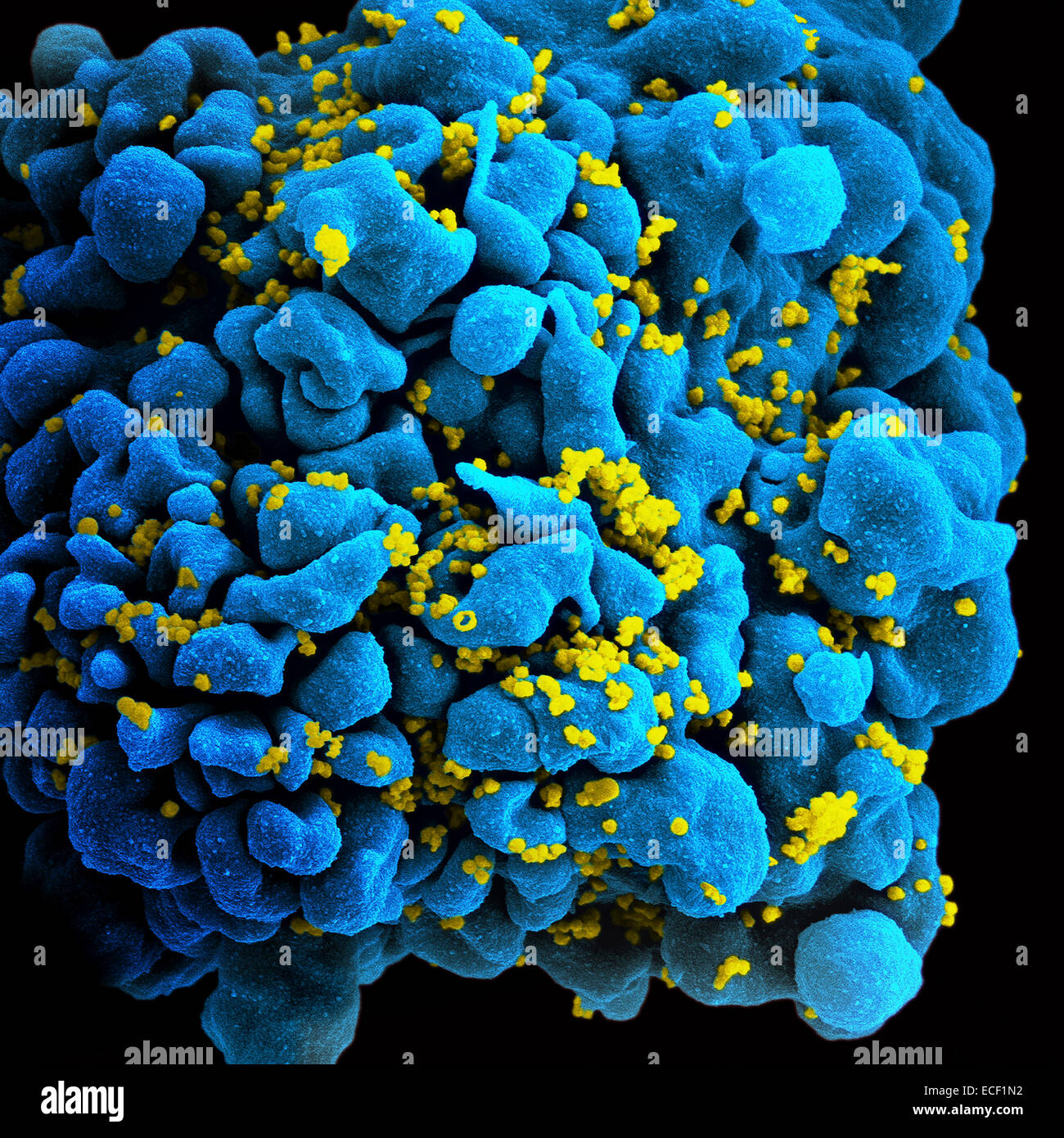 Colorized image of HIV-infected H9 T-cell. Stock Photohttps://www.alamy.com/image-license-details/?v=1https://www.alamy.com/stock-photo-colorized-image-of-hiv-infected-h9-t-cell-76547998.html
RFECF1N2–Colorized image of HIV-infected H9 T-cell.
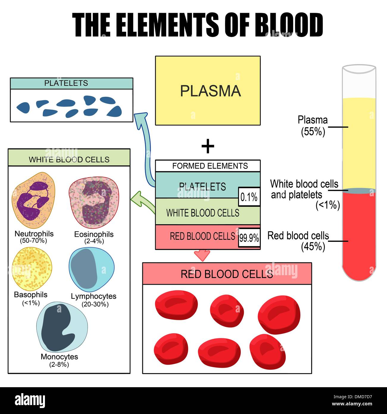 The elements of blood Stock Vectorhttps://www.alamy.com/image-license-details/?v=1https://www.alamy.com/the-elements-of-blood-image64215459.html
RFDMD7D7–The elements of blood
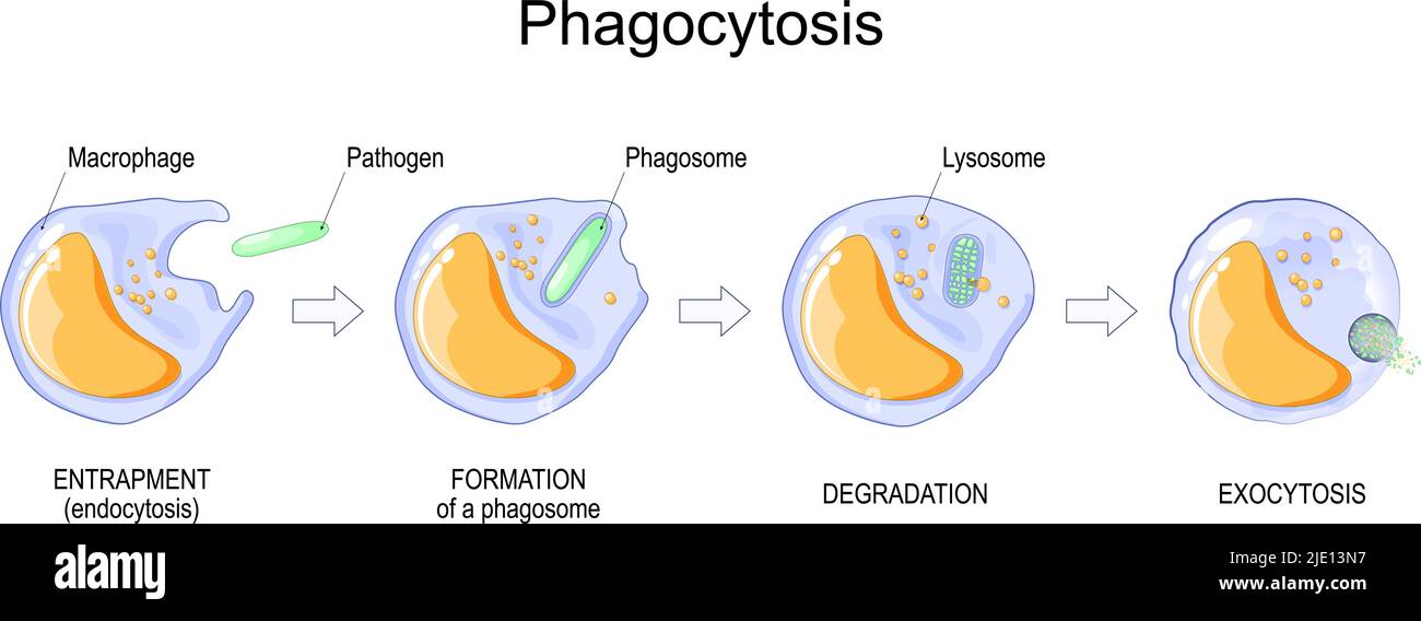 Phagocytosis. macrophage absorption of bacteria. Stages of mechanism of the immune response from entrapment or endocytosis to phagosome formation Stock Vectorhttps://www.alamy.com/image-license-details/?v=1https://www.alamy.com/phagocytosis-macrophage-absorption-of-bacteria-stages-of-mechanism-of-the-immune-response-from-entrapment-or-endocytosis-to-phagosome-formation-image473310019.html
RF2JE13N7–Phagocytosis. macrophage absorption of bacteria. Stages of mechanism of the immune response from entrapment or endocytosis to phagosome formation
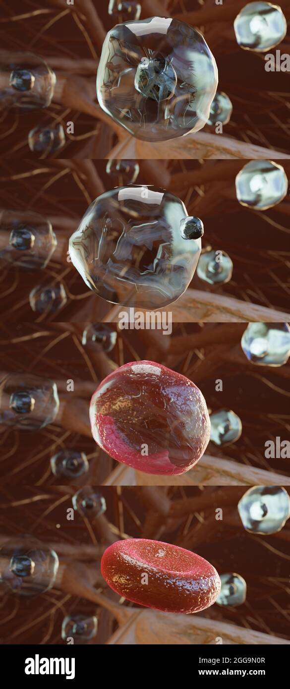 3d illustration of Hemocytoblast, producing red blood cell, blood cell formation, stem cell, erythrocytes, leukocytes, nucleus, cytoplasm, 3d render Stock Photohttps://www.alamy.com/image-license-details/?v=1https://www.alamy.com/3d-illustration-of-hemocytoblast-producing-red-blood-cell-blood-cell-formation-stem-cell-erythrocytes-leukocytes-nucleus-cytoplasm-3d-render-image440307751.html
RM2GG9N0R–3d illustration of Hemocytoblast, producing red blood cell, blood cell formation, stem cell, erythrocytes, leukocytes, nucleus, cytoplasm, 3d render
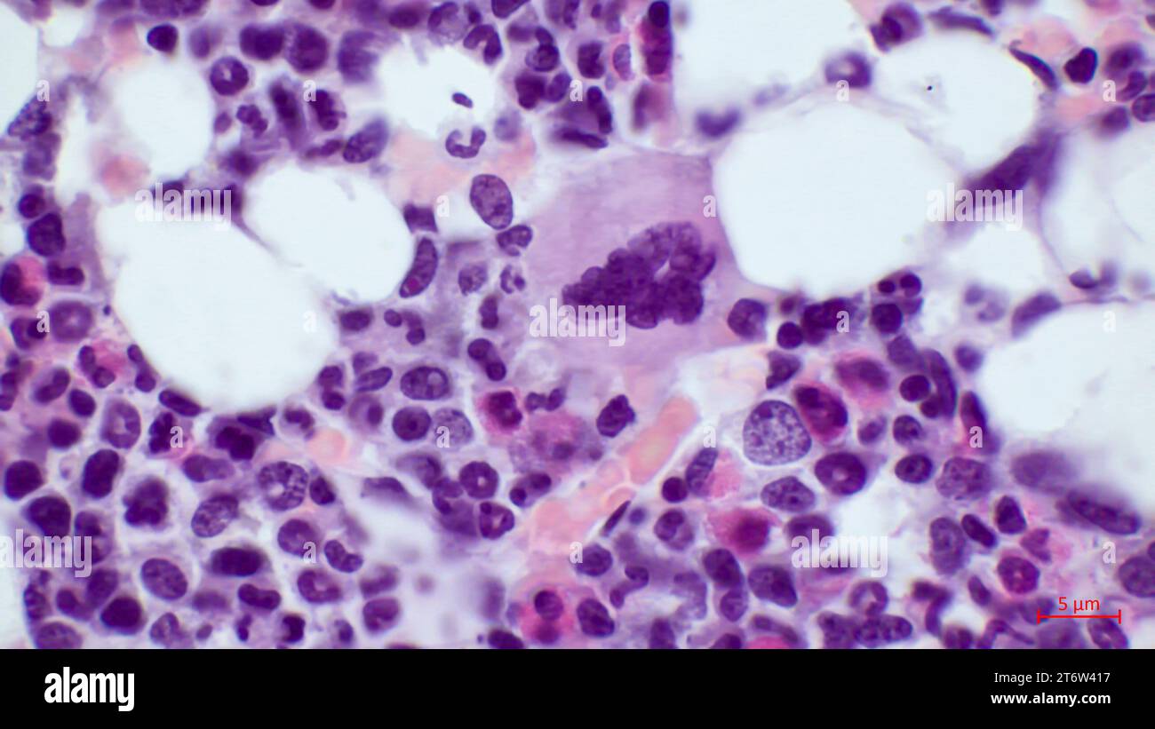 Microscopic structure of red bone marrow histology.The largest cell in the red bone marrow is the megakaryocyte (located in the center). Stock Photohttps://www.alamy.com/image-license-details/?v=1https://www.alamy.com/microscopic-structure-of-red-bone-marrow-histologythe-largest-cell-in-the-red-bone-marrow-is-the-megakaryocyte-located-in-the-center-image572182051.html
RF2T6W417–Microscopic structure of red bone marrow histology.The largest cell in the red bone marrow is the megakaryocyte (located in the center).
 3d illustration of red blood cells inside an artery, vein. The flow of blood inside a living organism. Scientific and medical microbiological concept Stock Photohttps://www.alamy.com/image-license-details/?v=1https://www.alamy.com/3d-illustration-of-red-blood-cells-inside-an-artery-vein-the-flow-of-blood-inside-a-living-organism-scientific-and-medical-microbiological-concept-image351337672.html
RF2BBGPP0–3d illustration of red blood cells inside an artery, vein. The flow of blood inside a living organism. Scientific and medical microbiological concept
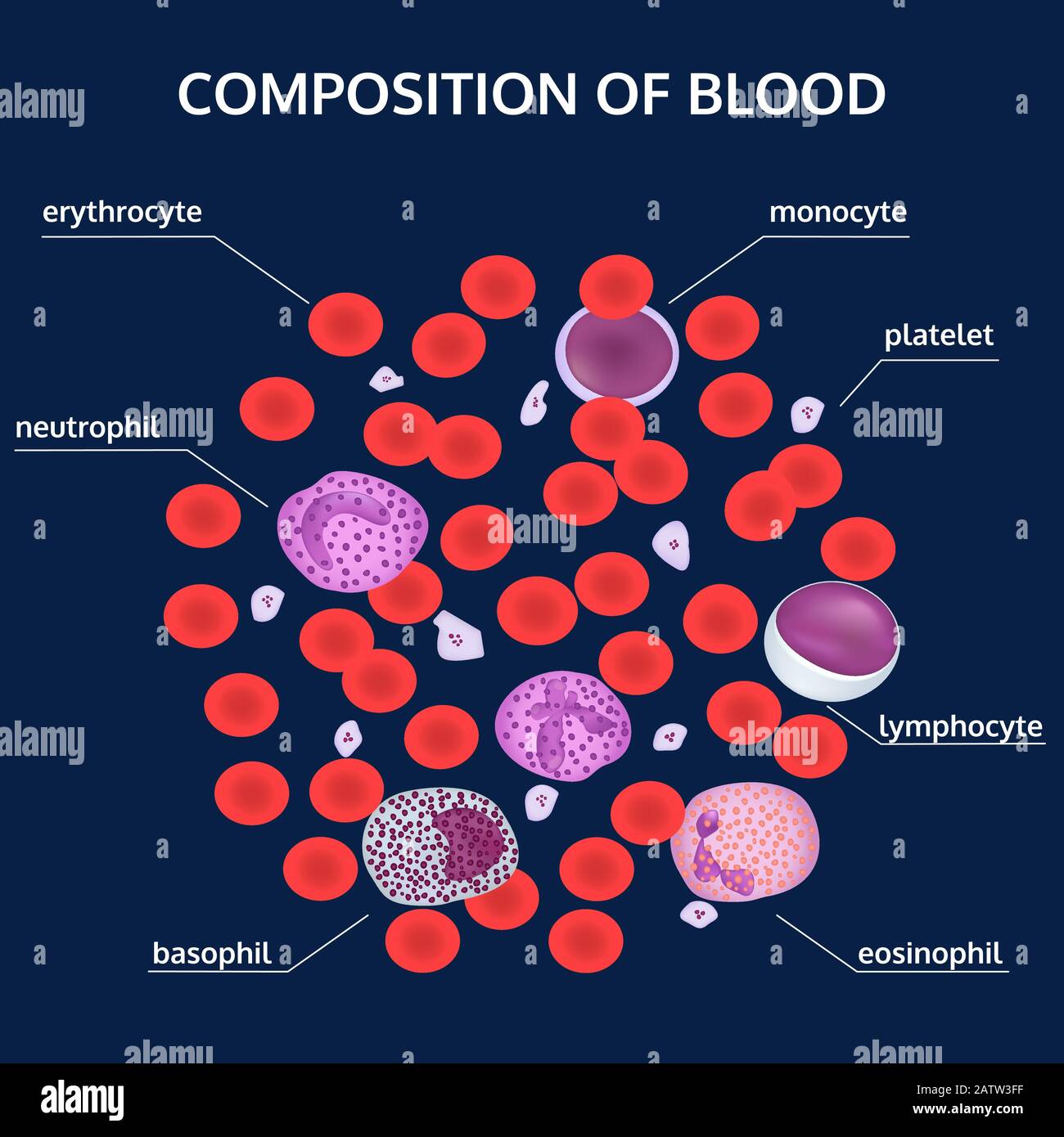 Infographics of composition of blood: red, white cells, platelets under a microscope with names on blue background. Medical vector concept. Stock Vectorhttps://www.alamy.com/image-license-details/?v=1https://www.alamy.com/infographics-of-composition-of-blood-red-white-cells-platelets-under-a-microscope-with-names-on-blue-background-medical-vector-concept-image342300323.html
RF2ATW3FF–Infographics of composition of blood: red, white cells, platelets under a microscope with names on blue background. Medical vector concept.
 Diagnostic form, syringe and Medicine in a hospital, conceptual image Stock Photohttps://www.alamy.com/image-license-details/?v=1https://www.alamy.com/diagnostic-form-syringe-and-medicine-in-a-hospital-conceptual-image-image218686471.html
RFPKP0R3–Diagnostic form, syringe and Medicine in a hospital, conceptual image
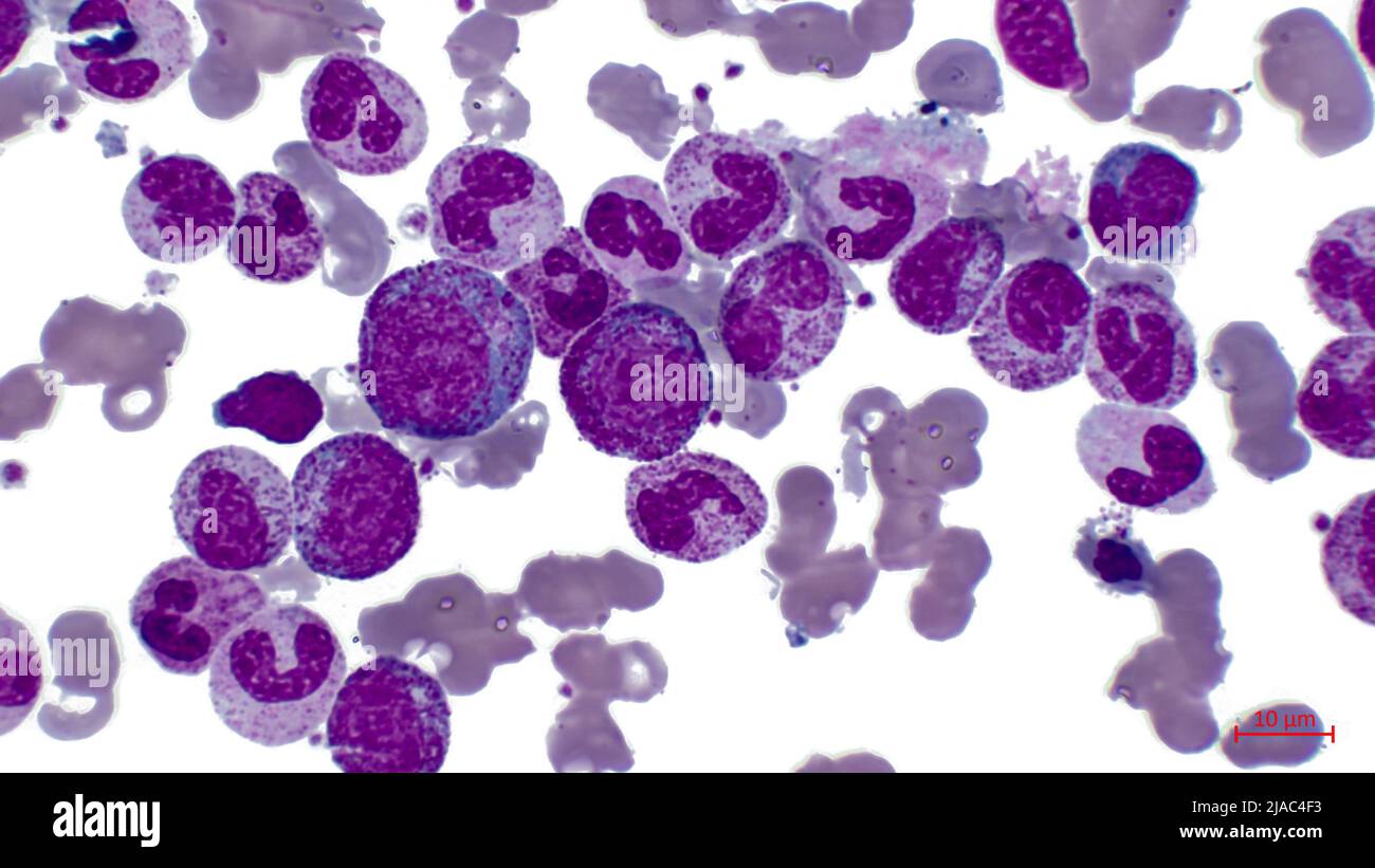 Photomicrographs of agranulocytosis: myeloblast, neutrophilic metamyelocyte, promyelocyte, neutrophilic myelocyte, neutrophilic metamyelocyte. Stock Photohttps://www.alamy.com/image-license-details/?v=1https://www.alamy.com/photomicrographs-of-agranulocytosis-myeloblast-neutrophilic-metamyelocyte-promyelocyte-neutrophilic-myelocyte-neutrophilic-metamyelocyte-image471093479.html
RF2JAC4F3–Photomicrographs of agranulocytosis: myeloblast, neutrophilic metamyelocyte, promyelocyte, neutrophilic myelocyte, neutrophilic metamyelocyte.
 Resting T lymphocytes. Coloured scanning electron micrograph (SEM) of resting T lymphocytes from a human blood sample. T lymphocytes, or T cells, are a type of white blood cell and components of the body's immune system. They mature in the thymus. T lymphocytes recognise a specific site on the surface of pathogens or foreign objects (antigens), bind to it, and become activated to produce antibodies or cells to eliminate that antigen. Specimen courtesy of Professor Greg Towers, University College London, UK. Magnification: x7000 when printed at 10 centimetres wide. Stock Photohttps://www.alamy.com/image-license-details/?v=1https://www.alamy.com/resting-t-lymphocytes-coloured-scanning-electron-micrograph-sem-of-resting-t-lymphocytes-from-a-human-blood-sample-t-lymphocytes-or-t-cells-are-a-type-of-white-blood-cell-and-components-of-the-bodys-immune-system-they-mature-in-the-thymus-t-lymphocytes-recognise-a-specific-site-on-the-surface-of-pathogens-or-foreign-objects-antigens-bind-to-it-and-become-activated-to-produce-antibodies-or-cells-to-eliminate-that-antigen-specimen-courtesy-of-professor-greg-towers-university-college-london-uk-magnification-x7000-when-printed-at-10-centimetres-wide-image220702042.html
RFPR1RKP–Resting T lymphocytes. Coloured scanning electron micrograph (SEM) of resting T lymphocytes from a human blood sample. T lymphocytes, or T cells, are a type of white blood cell and components of the body's immune system. They mature in the thymus. T lymphocytes recognise a specific site on the surface of pathogens or foreign objects (antigens), bind to it, and become activated to produce antibodies or cells to eliminate that antigen. Specimen courtesy of Professor Greg Towers, University College London, UK. Magnification: x7000 when printed at 10 centimetres wide.
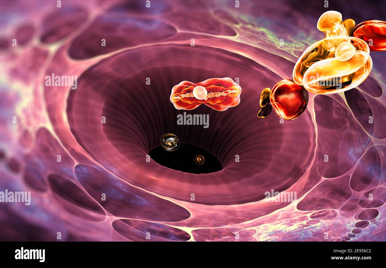 Immune system cell. White blood cell eats bacteria. 3d illustration Stock Photohttps://www.alamy.com/image-license-details/?v=1https://www.alamy.com/immune-system-cell-white-blood-cell-eats-bacteria-3d-illustration-image401485170.html
RF2E956C2–Immune system cell. White blood cell eats bacteria. 3d illustration
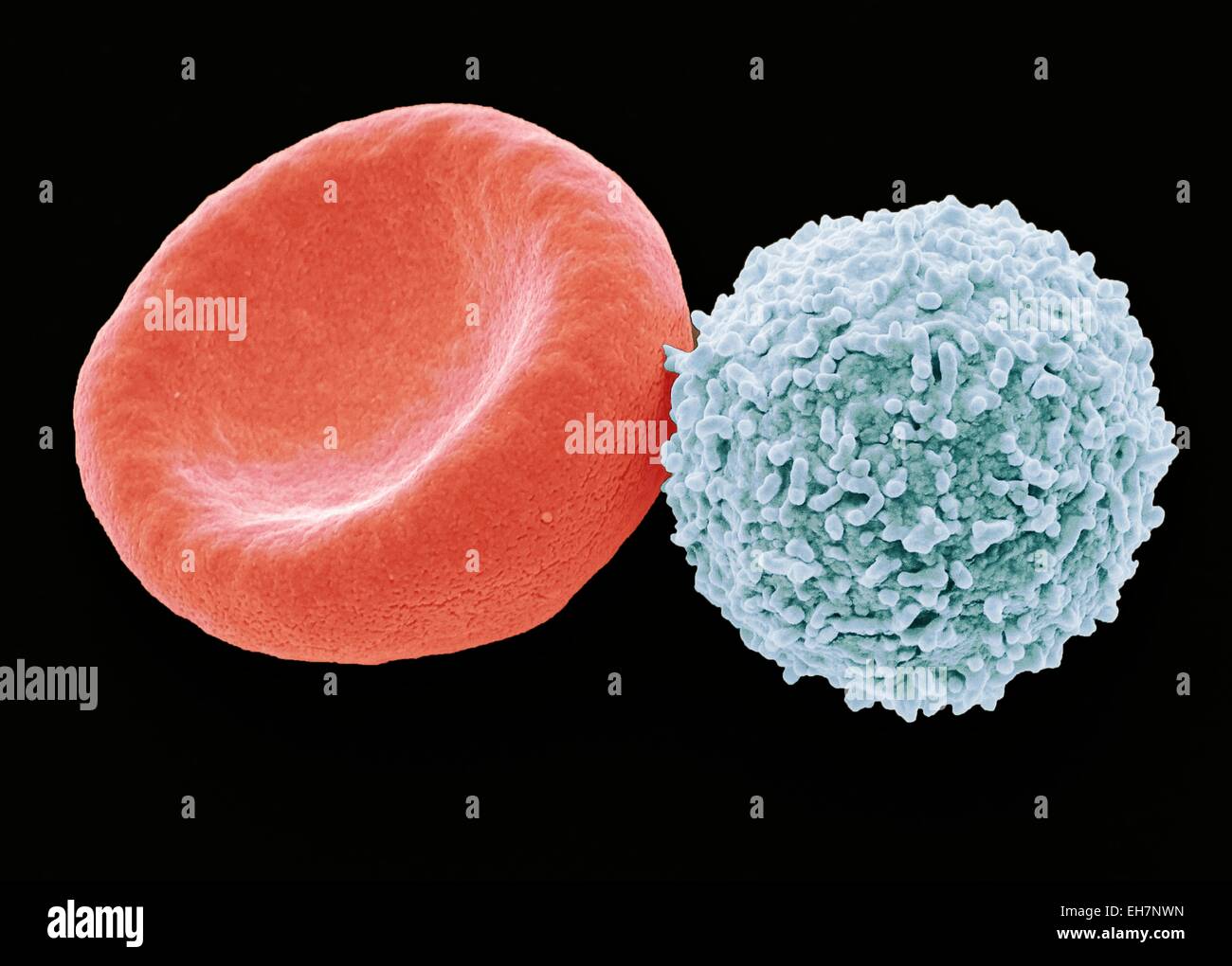 Blood cells, SEM Stock Photohttps://www.alamy.com/image-license-details/?v=1https://www.alamy.com/stock-photo-blood-cells-sem-79461473.html
RFEH7NWN–Blood cells, SEM
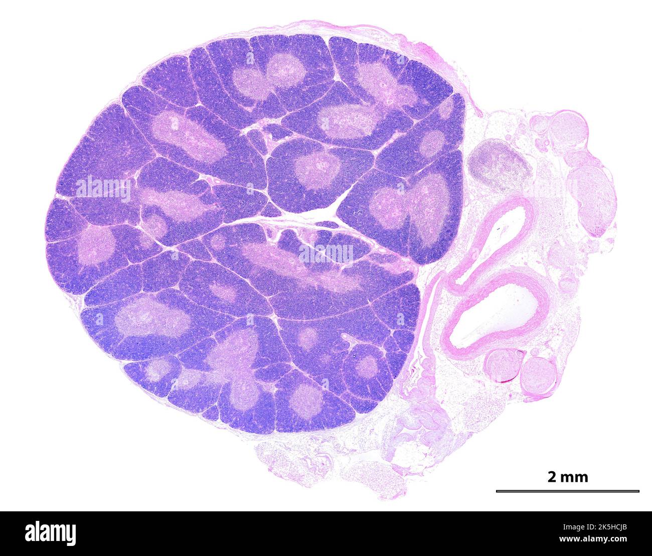 Low power light microscope micrograph showing a young thymus. The organization into lobules is clearly seen. In each lobule, the peripheral cortex app Stock Photohttps://www.alamy.com/image-license-details/?v=1https://www.alamy.com/low-power-light-microscope-micrograph-showing-a-young-thymus-the-organization-into-lobules-is-clearly-seen-in-each-lobule-the-peripheral-cortex-app-image485346691.html
RF2K5HCJB–Low power light microscope micrograph showing a young thymus. The organization into lobules is clearly seen. In each lobule, the peripheral cortex app
 Scanning electron micrograph of a human H9 T cell (blue/green) infected with HIV virus particles (yellow). Stock Photohttps://www.alamy.com/image-license-details/?v=1https://www.alamy.com/scanning-electron-micrograph-of-a-human-h9-t-cell-bluegreen-infected-with-hiv-virus-particles-yellow-image627689593.html
RM2YD5MDD–Scanning electron micrograph of a human H9 T cell (blue/green) infected with HIV virus particles (yellow).