Human rna model Cut Out Stock Images
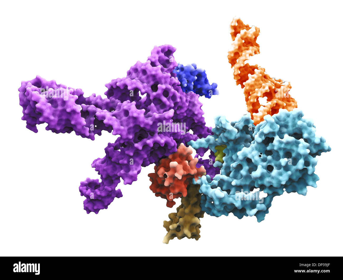 Human 80S ribosome Stock Photohttps://www.alamy.com/image-license-details/?v=1https://www.alamy.com/human-80s-ribosome-image65226967.html
RFDP39JF–Human 80S ribosome
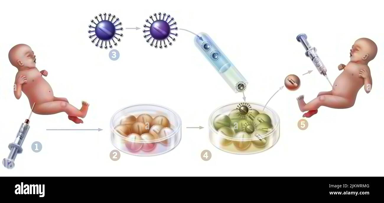 Gene therapy: introduction of retroviruses to modify mutated spinal cord cells of a newborn. Stock Photohttps://www.alamy.com/image-license-details/?v=1https://www.alamy.com/gene-therapy-introduction-of-retroviruses-to-modify-mutated-spinal-cord-cells-of-a-newborn-image476925808.html
RF2JKWRMG–Gene therapy: introduction of retroviruses to modify mutated spinal cord cells of a newborn.
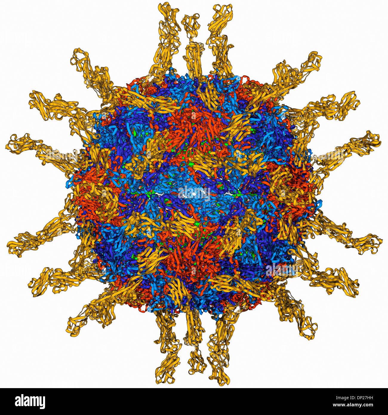 Human poliovirus, molecular model Stock Photohttps://www.alamy.com/image-license-details/?v=1https://www.alamy.com/human-poliovirus-molecular-model-image65203421.html
RFDP27HH–Human poliovirus, molecular model
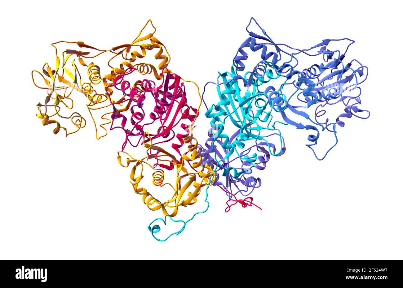 Human Cytoplasmic PheRS Stock Photohttps://www.alamy.com/image-license-details/?v=1https://www.alamy.com/human-cytoplasmic-phers-image416784515.html
RM2F624W7–Human Cytoplasmic PheRS
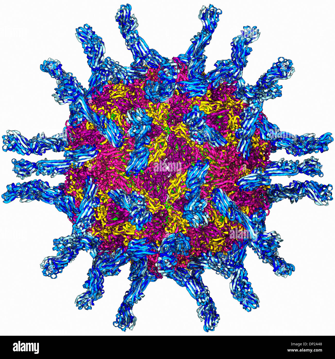 Human poliovirus, molecular model Stock Photohttps://www.alamy.com/image-license-details/?v=1https://www.alamy.com/human-poliovirus-molecular-model-image65205400.html
RFDP2A48–Human poliovirus, molecular model
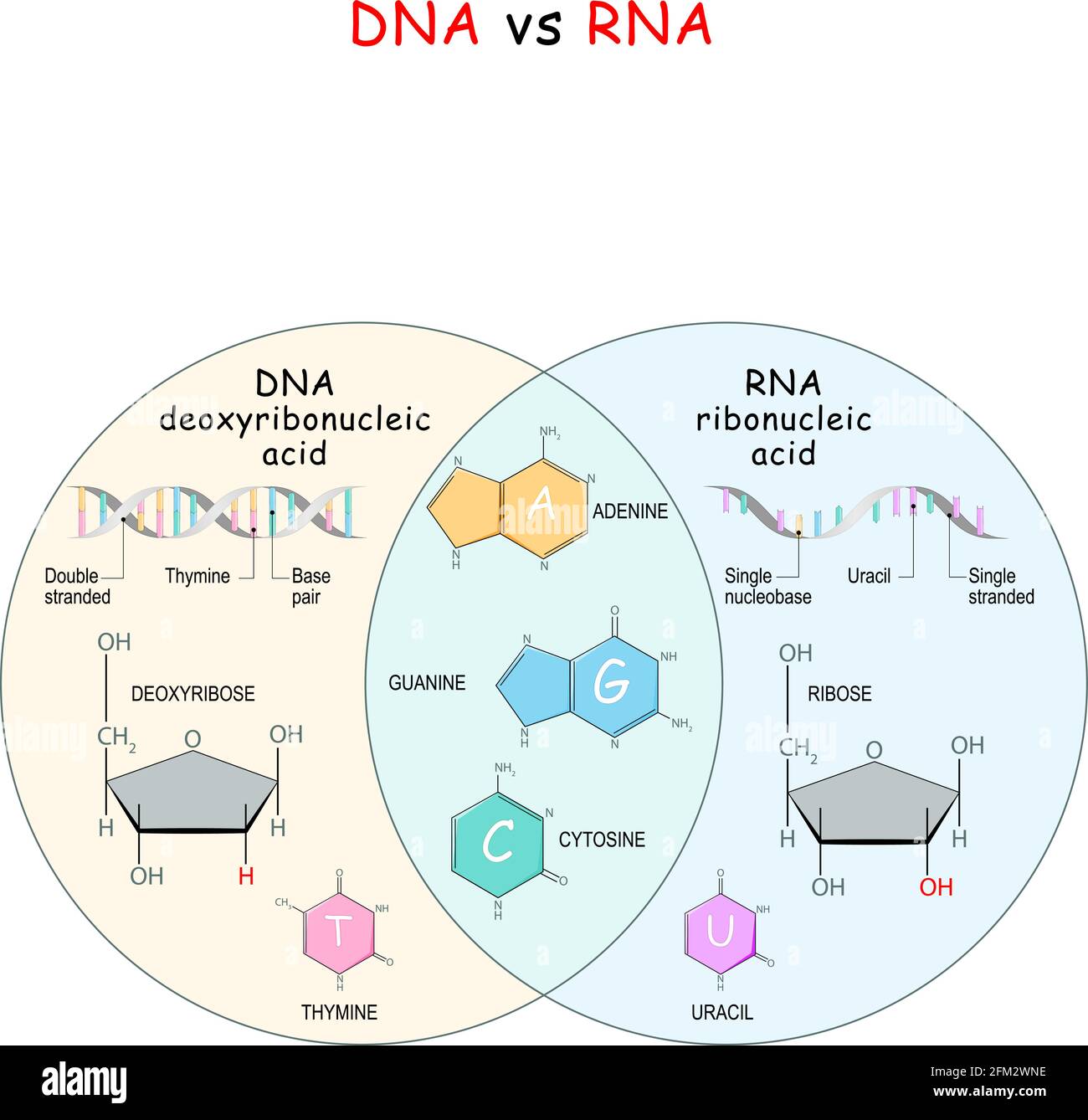 DNA and RNA. comparison and difference. Chemical structural formula and model of molecules DNA and RNA. Vector illustration Stock Vectorhttps://www.alamy.com/image-license-details/?v=1https://www.alamy.com/dna-and-rna-comparison-and-difference-chemical-structural-formula-and-model-of-molecules-dna-and-rna-vector-illustration-image425406058.html
RF2FM2WNE–DNA and RNA. comparison and difference. Chemical structural formula and model of molecules DNA and RNA. Vector illustration
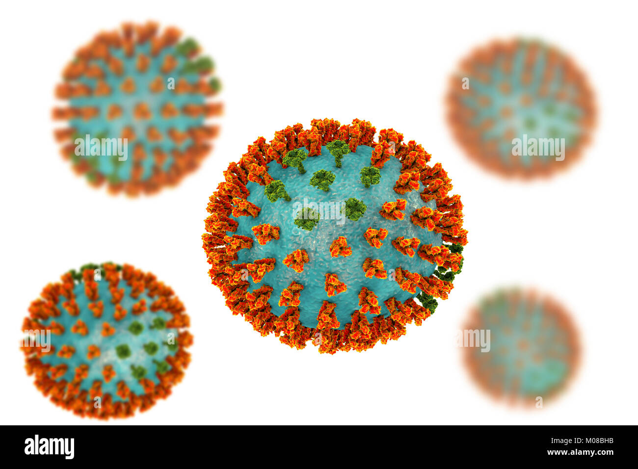 Influenza virus H3N2 strain. 3D illustration showing surface glycoprotein spikes hemagglutinin (orange) and neuraminidase (green) on an influenza (flu) virus particle. Haemagglutinin plays a role in attachment of the virus to human respiratory cells. Neuraminidase plays a role in releasing newly formed virus particles from an infected cell. H3N2 viruses are able to infect birds and mammals as well as humans. They often cause more severe infections in the young and elderly than other flu strains and can lead to increases in hospitalisations and deaths. Stock Photohttps://www.alamy.com/image-license-details/?v=1https://www.alamy.com/stock-photo-influenza-virus-h3n2-strain-3d-illustration-showing-surface-glycoprotein-172288407.html
RFM08BHB–Influenza virus H3N2 strain. 3D illustration showing surface glycoprotein spikes hemagglutinin (orange) and neuraminidase (green) on an influenza (flu) virus particle. Haemagglutinin plays a role in attachment of the virus to human respiratory cells. Neuraminidase plays a role in releasing newly formed virus particles from an infected cell. H3N2 viruses are able to infect birds and mammals as well as humans. They often cause more severe infections in the young and elderly than other flu strains and can lead to increases in hospitalisations and deaths.
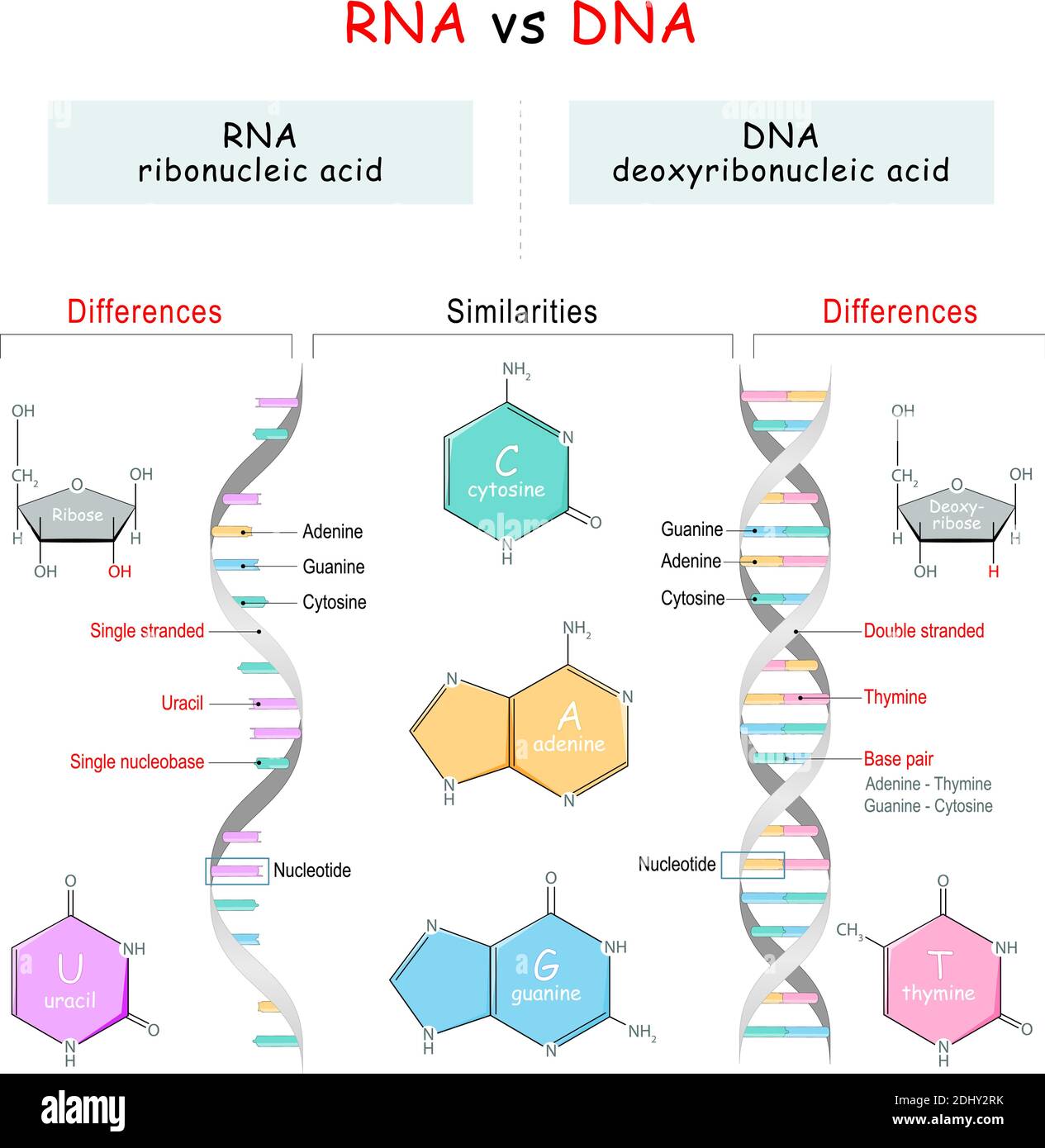 DNA vs RNA comparison. Similarities and Differences. infographic diagram. vector illustration for Educational explanation, and science use Stock Vectorhttps://www.alamy.com/image-license-details/?v=1https://www.alamy.com/dna-vs-rna-comparison-similarities-and-differences-infographic-diagram-vector-illustration-for-educational-explanation-and-science-use-image389672183.html
RF2DHY2RK–DNA vs RNA comparison. Similarities and Differences. infographic diagram. vector illustration for Educational explanation, and science use
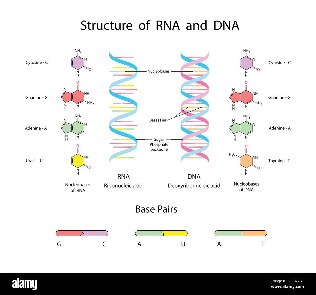 Molecular Structure Of DNA and RNA. Infographic Educational Illustration on white background Stock Photohttps://www.alamy.com/image-license-details/?v=1https://www.alamy.com/molecular-structure-of-dna-and-rna-infographic-educational-illustration-on-white-background-image401326152.html
RF2E8WYGT–Molecular Structure Of DNA and RNA. Infographic Educational Illustration on white background
RF2J65WFR–Spiral of DNA and RNA. differences in the structure of the DNA and RNA molecules. icons and Infographic Educational Vector Illustration.
 Writing displaying text Rna Virus. Conceptual photo a virus genetic information is stored in the form of RNA Stock Photohttps://www.alamy.com/image-license-details/?v=1https://www.alamy.com/writing-displaying-text-rna-virus-conceptual-photo-a-virus-genetic-information-is-stored-in-the-form-of-rna-image550256254.html
RF2PY69D2–Writing displaying text Rna Virus. Conceptual photo a virus genetic information is stored in the form of RNA
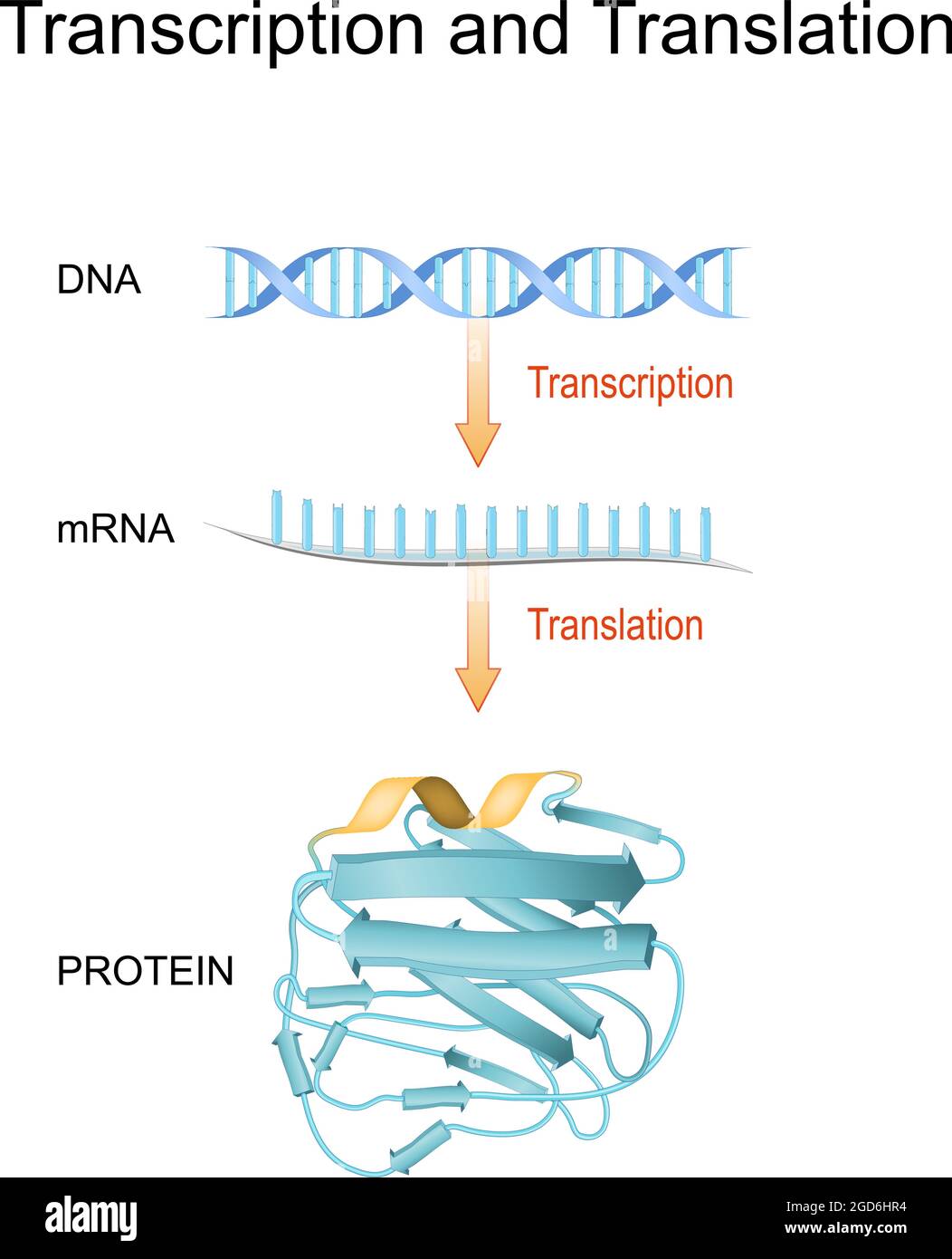 DNA, RNA, mRNA and Protein synthesis. Difference between Transcription and Translation. Biological functions of DNA. Genes and genomes. Genetic code. Stock Vectorhttps://www.alamy.com/image-license-details/?v=1https://www.alamy.com/dna-rna-mrna-and-protein-synthesis-difference-between-transcription-and-translation-biological-functions-of-dna-genes-and-genomes-genetic-code-image438395416.html
RF2GD6HR4–DNA, RNA, mRNA and Protein synthesis. Difference between Transcription and Translation. Biological functions of DNA. Genes and genomes. Genetic code.
 Coronavirus cell model illustration, Covid-19 Coronavirus pandemic, 3D Render, conceptual, close up, black background Stock Photohttps://www.alamy.com/image-license-details/?v=1https://www.alamy.com/coronavirus-cell-model-illustration-covid-19-coronavirus-pandemic-3d-render-conceptual-close-up-black-background-image352426539.html
RF2BDABJ3–Coronavirus cell model illustration, Covid-19 Coronavirus pandemic, 3D Render, conceptual, close up, black background
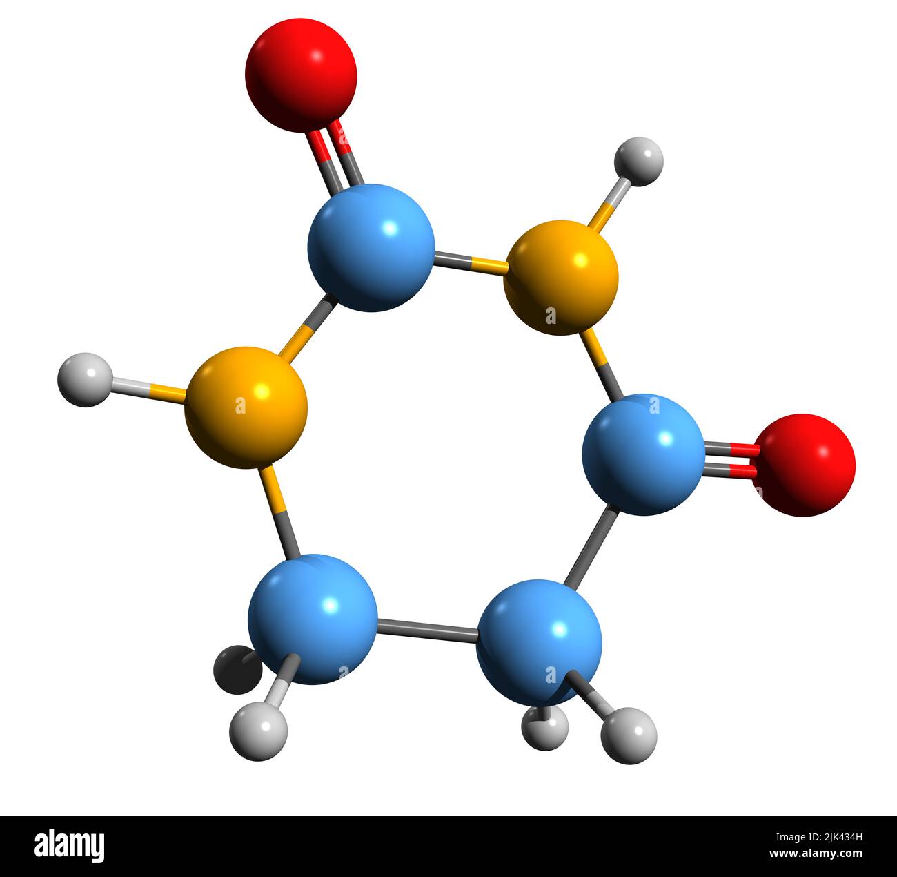 3D image of Dihydrouracil skeletal formula - molecular chemical structure of uracil intermediate isolated on white background Stock Photohttps://www.alamy.com/image-license-details/?v=1https://www.alamy.com/3d-image-of-dihydrouracil-skeletal-formula-molecular-chemical-structure-of-uracil-intermediate-isolated-on-white-background-image476448689.html
RF2JK434H–3D image of Dihydrouracil skeletal formula - molecular chemical structure of uracil intermediate isolated on white background
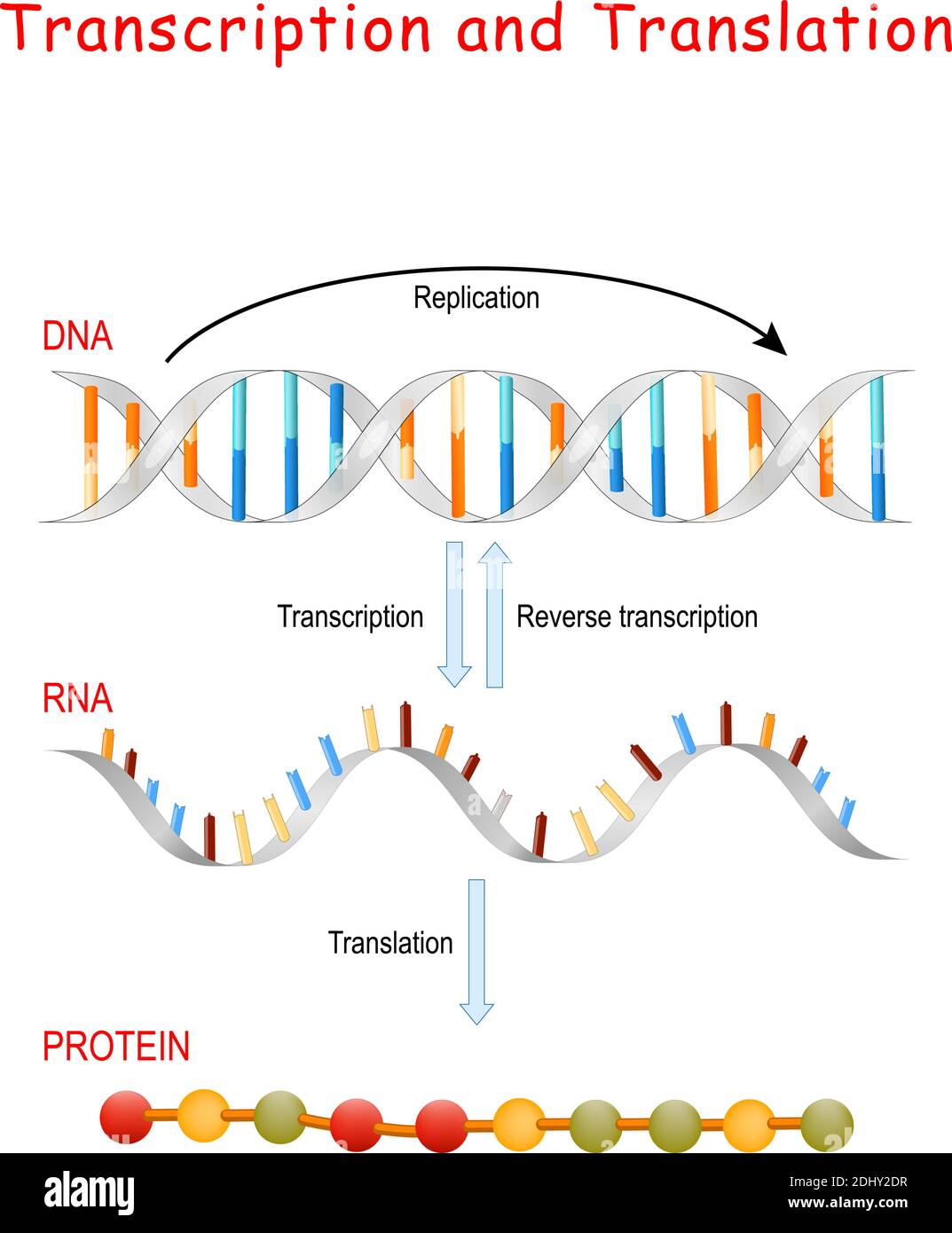 DNA Replication, Protein synthesis, Transcription and translation. Biological functions of DNA. Genes and genomes. Genetic code Stock Vectorhttps://www.alamy.com/image-license-details/?v=1https://www.alamy.com/dna-replication-protein-synthesis-transcription-and-translation-biological-functions-of-dna-genes-and-genomes-genetic-code-image389671907.html
RF2DHY2DR–DNA Replication, Protein synthesis, Transcription and translation. Biological functions of DNA. Genes and genomes. Genetic code
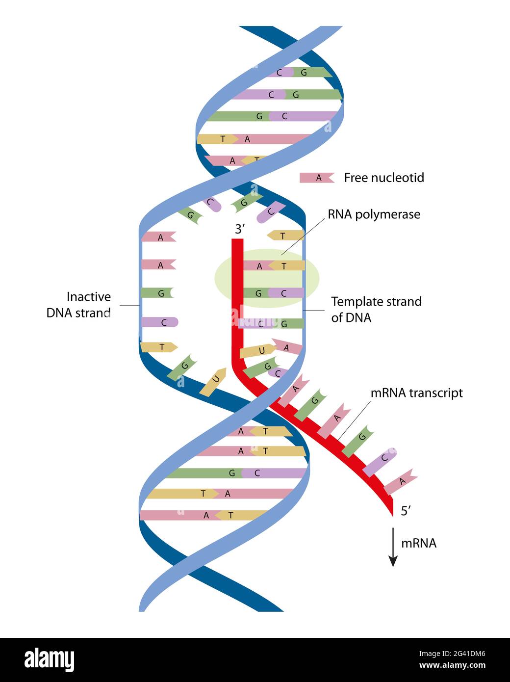 Simple diagram of transcription elongation. Transcription is the first step of gene expression Stock Photohttps://www.alamy.com/image-license-details/?v=1https://www.alamy.com/simple-diagram-of-transcription-elongation-transcription-is-the-first-step-of-gene-expression-image432750534.html
RF2G41DM6–Simple diagram of transcription elongation. Transcription is the first step of gene expression
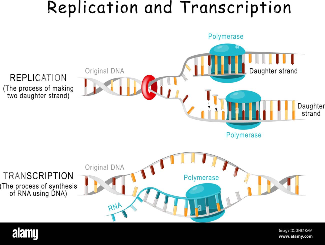 DNA Replication and Transcription. Steps. double helix is unwound. Each separated strand acts as a template for replicating a new strand. Vector Stock Vectorhttps://www.alamy.com/image-license-details/?v=1https://www.alamy.com/dna-replication-and-transcription-steps-double-helix-is-unwound-each-separated-strand-acts-as-a-template-for-replicating-a-new-strand-vector-image452423964.html
RF2H81KAM–DNA Replication and Transcription. Steps. double helix is unwound. Each separated strand acts as a template for replicating a new strand. Vector
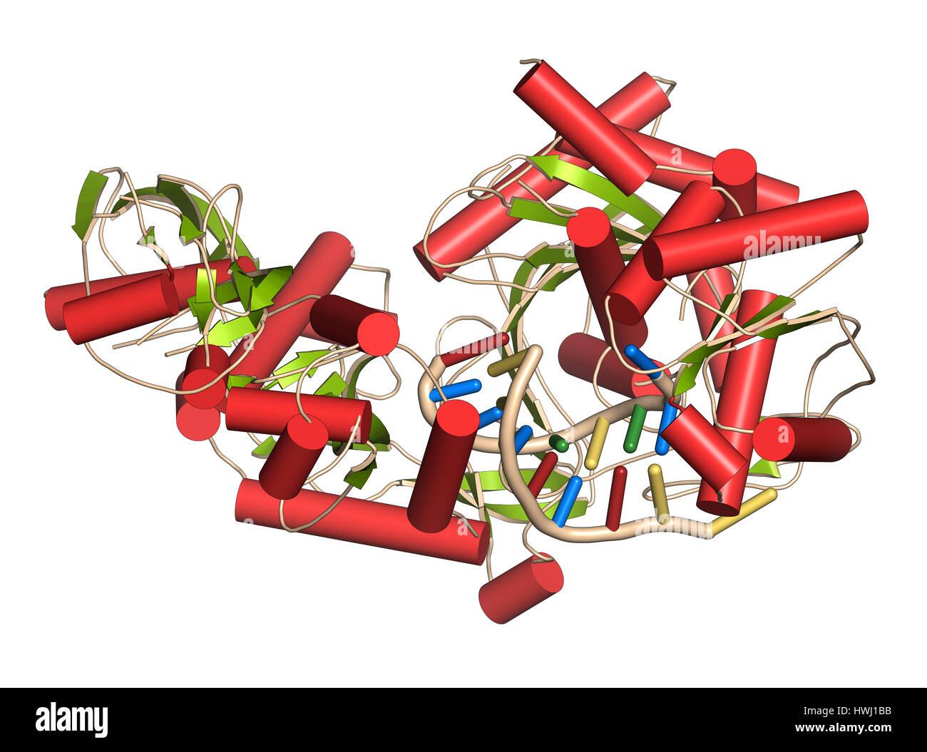 Argonaute-2 (human) enzyme. Part of the RISC complex and plays role in RNA interference (RNAi). 3D illustration. Cartoon representation with secondary Stock Photohttps://www.alamy.com/image-license-details/?v=1https://www.alamy.com/stock-photo-argonaute-2-human-enzyme-part-of-the-risc-complex-and-plays-role-in-136235215.html
RFHWJ1BB–Argonaute-2 (human) enzyme. Part of the RISC complex and plays role in RNA interference (RNAi). 3D illustration. Cartoon representation with secondary
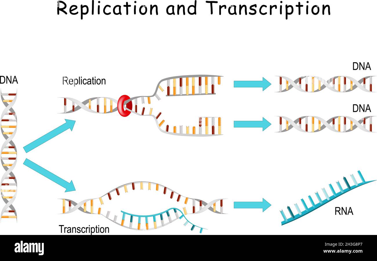 DNA Replication and Transcription. comparisons and differences. Replication - producing two identical replicas from one original DNA molecule Stock Vectorhttps://www.alamy.com/image-license-details/?v=1https://www.alamy.com/dna-replication-and-transcription-comparisons-and-differences-replication-producing-two-identical-replicas-from-one-original-dna-molecule-image449671663.html
RF2H3G8P7–DNA Replication and Transcription. comparisons and differences. Replication - producing two identical replicas from one original DNA molecule
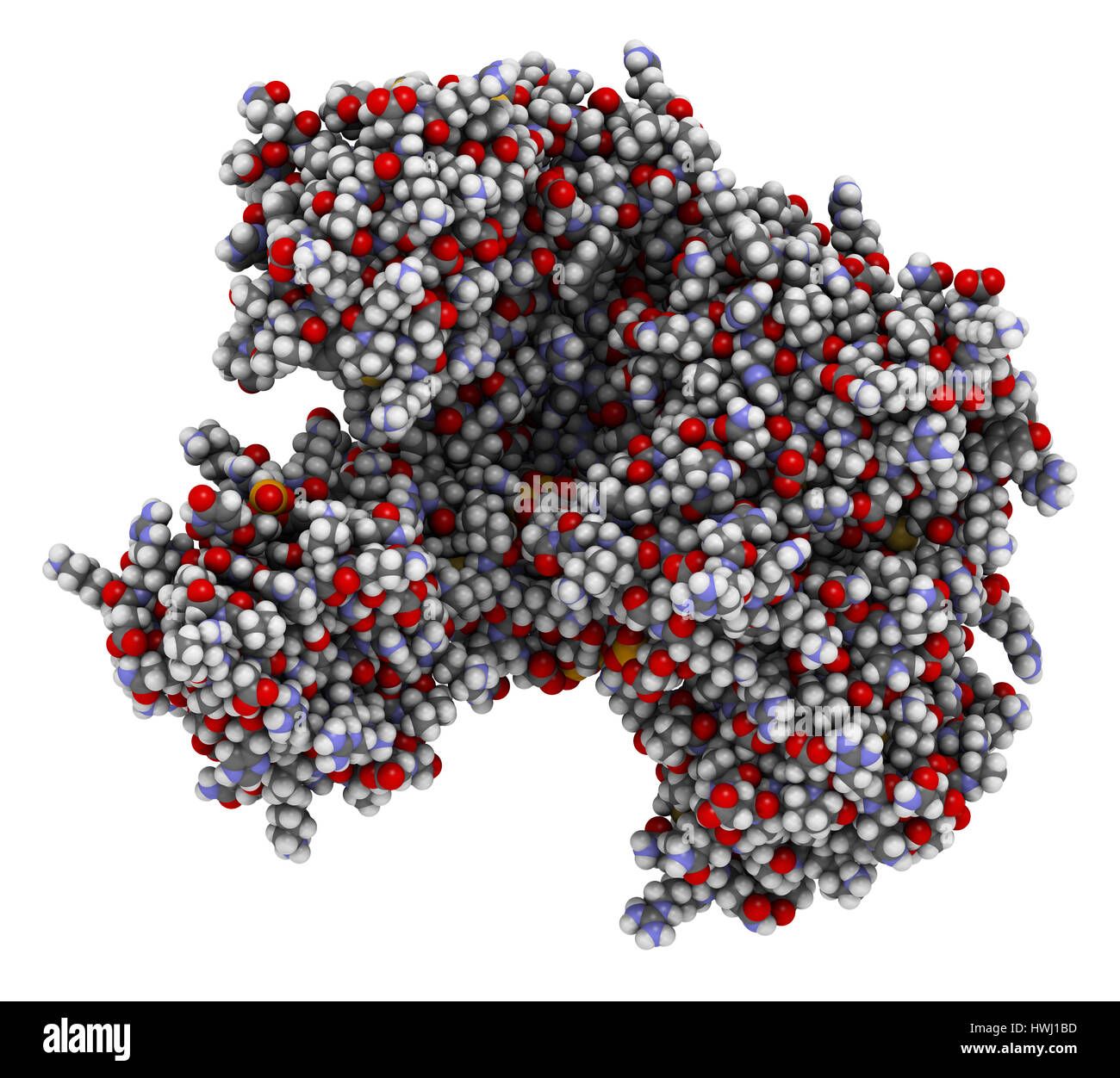 Argonaute-2 (human) enzyme. Part of the RISC complex and plays role in RNA interference (RNAi). 3D illustration. Atoms shown as spheres with conventio Stock Photohttps://www.alamy.com/image-license-details/?v=1https://www.alamy.com/stock-photo-argonaute-2-human-enzyme-part-of-the-risc-complex-and-plays-role-in-136235217.html
RFHWJ1BD–Argonaute-2 (human) enzyme. Part of the RISC complex and plays role in RNA interference (RNAi). 3D illustration. Atoms shown as spheres with conventio
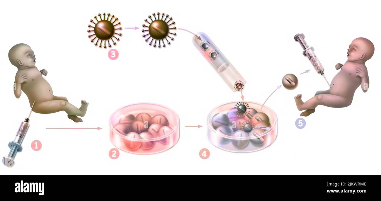 Gene therapy: introduction of retroviruses to modify mutated spinal cord cells of a newborn. Stock Photohttps://www.alamy.com/image-license-details/?v=1https://www.alamy.com/gene-therapy-introduction-of-retroviruses-to-modify-mutated-spinal-cord-cells-of-a-newborn-image476925806.html
RF2JKWRME–Gene therapy: introduction of retroviruses to modify mutated spinal cord cells of a newborn.
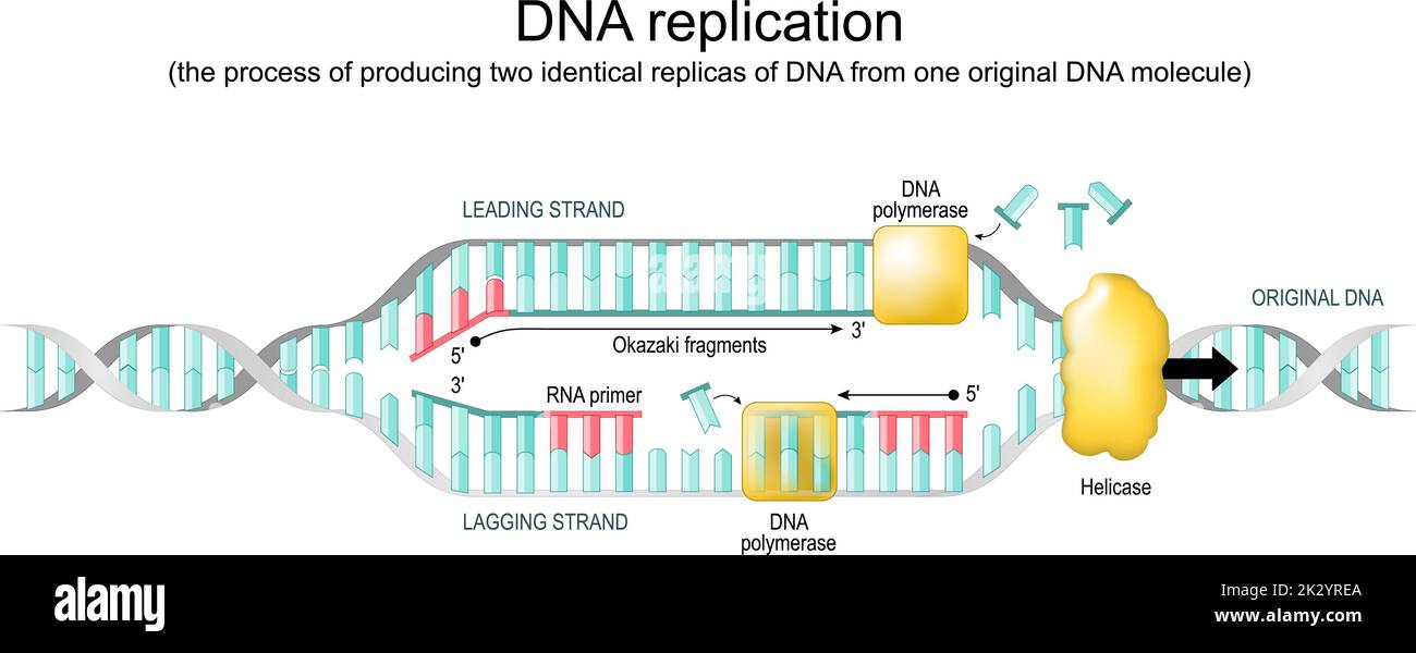 DNA replication. biological process of producing two identical replicas from one original molecule. replication fork. Vector diagram Stock Vectorhttps://www.alamy.com/image-license-details/?v=1https://www.alamy.com/dna-replication-biological-process-of-producing-two-identical-replicas-from-one-original-molecule-replication-fork-vector-diagram-image483730754.html
RF2K2YREA–DNA replication. biological process of producing two identical replicas from one original molecule. replication fork. Vector diagram
 Human Mitochondrial PheRS Stock Photohttps://www.alamy.com/image-license-details/?v=1https://www.alamy.com/human-mitochondrial-phers-image416784511.html
RM2F624W3–Human Mitochondrial PheRS
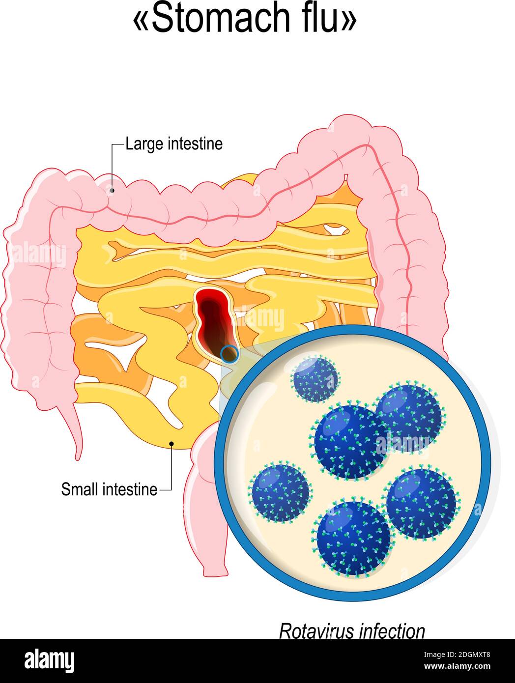 rotavirus infection or stomach flu. Small intestine, colon, and close-up of rotavirus virions. Human anatomy. Vector diagram for your design Stock Vectorhttps://www.alamy.com/image-license-details/?v=1https://www.alamy.com/rotavirus-infection-or-stomach-flu-small-intestine-colon-and-close-up-of-rotavirus-virions-human-anatomy-vector-diagram-for-your-design-image388922696.html
RF2DGMXT8–rotavirus infection or stomach flu. Small intestine, colon, and close-up of rotavirus virions. Human anatomy. Vector diagram for your design
 Illustration logo of the DNA model. Dna log design. Healix logotype Stock Vectorhttps://www.alamy.com/image-license-details/?v=1https://www.alamy.com/illustration-logo-of-the-dna-model-dna-log-design-healix-logotype-image566976386.html
RF2RXC04J–Illustration logo of the DNA model. Dna log design. Healix logotype
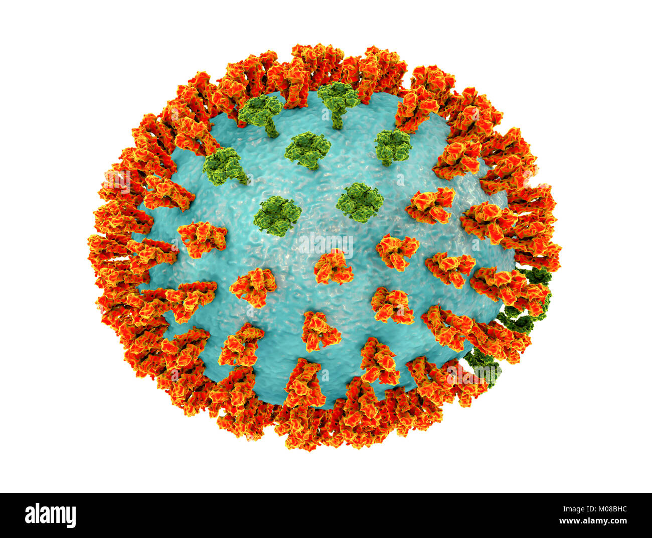 Influenza virus H3N2 strain. 3D illustration showing surface glycoprotein spikes hemagglutinin (orange) and neuraminidase (green) on an influenza (flu) virus particle. Haemagglutinin plays a role in attachment of the virus to human respiratory cells. Neuraminidase plays a role in releasing newly formed virus particles from an infected cell. H3N2 viruses are able to infect birds and mammals as well as humans. They often cause more severe infections in the young and elderly than other flu strains and can lead to increases in hospitalisations and deaths. Stock Photohttps://www.alamy.com/image-license-details/?v=1https://www.alamy.com/stock-photo-influenza-virus-h3n2-strain-3d-illustration-showing-surface-glycoprotein-172288408.html
RFM08BHC–Influenza virus H3N2 strain. 3D illustration showing surface glycoprotein spikes hemagglutinin (orange) and neuraminidase (green) on an influenza (flu) virus particle. Haemagglutinin plays a role in attachment of the virus to human respiratory cells. Neuraminidase plays a role in releasing newly formed virus particles from an infected cell. H3N2 viruses are able to infect birds and mammals as well as humans. They often cause more severe infections in the young and elderly than other flu strains and can lead to increases in hospitalisations and deaths.
 Respiratory syncytial virus. RSV structure. Close-up of a orthopneumovirus. Virion anatomy. Magnified of virus that causes infections of a human respi Stock Vectorhttps://www.alamy.com/image-license-details/?v=1https://www.alamy.com/respiratory-syncytial-virus-rsv-structure-close-up-of-a-orthopneumovirus-virion-anatomy-magnified-of-virus-that-causes-infections-of-a-human-respi-image592430586.html
RF2WBRF76–Respiratory syncytial virus. RSV structure. Close-up of a orthopneumovirus. Virion anatomy. Magnified of virus that causes infections of a human respi
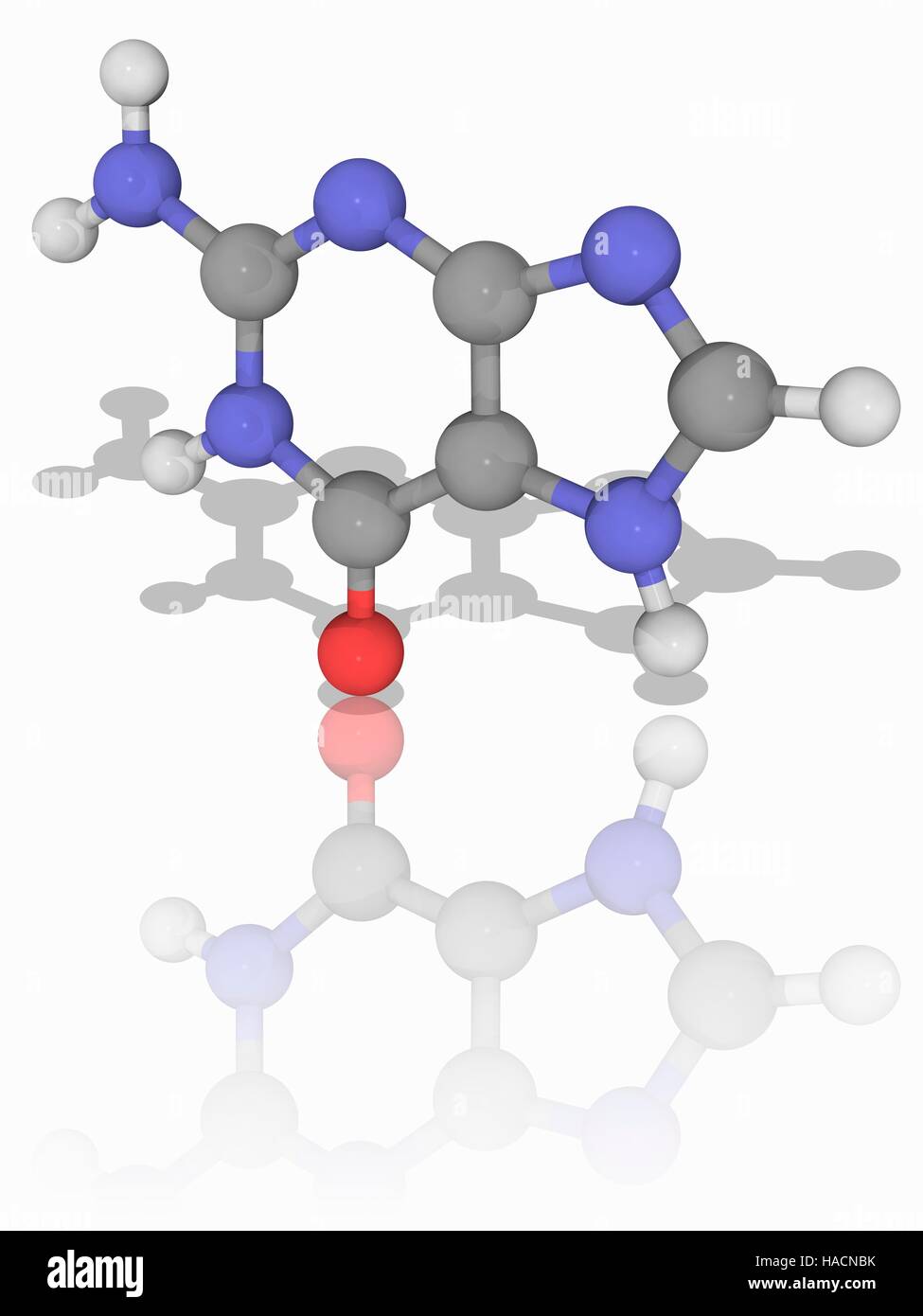 Guanine. Molecular model of the nucleobase guanine (C5.H5.N5.O). This is one of the bases found in the genetic molecules DNA (deoxyribonucleic acid) and RNA (ribonucleic acid), where it is paired with cytosine. Atoms are represented as spheres and are colour-coded: carbon (grey), hydrogen (white), nitrogen (blue) and oxygen (red). Illustration. Stock Photohttps://www.alamy.com/image-license-details/?v=1https://www.alamy.com/stock-photo-guanine-molecular-model-of-the-nucleobase-guanine-c5h5n5o-this-is-126899351.html
RFHACNBK–Guanine. Molecular model of the nucleobase guanine (C5.H5.N5.O). This is one of the bases found in the genetic molecules DNA (deoxyribonucleic acid) and RNA (ribonucleic acid), where it is paired with cytosine. Atoms are represented as spheres and are colour-coded: carbon (grey), hydrogen (white), nitrogen (blue) and oxygen (red). Illustration.
RF2EE5JHB–DNA or RNA helix vector isolated icons. Symbols of chromosome cell molecule, molecular chain of human genes or genome for genetics medical concept des
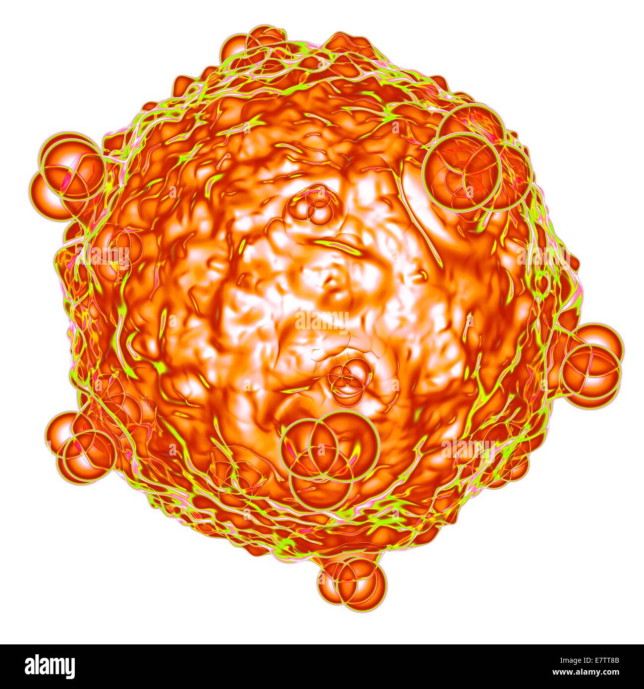 Human foamy virus (HFV), computer artwork. Stock Photohttps://www.alamy.com/image-license-details/?v=1https://www.alamy.com/stock-photo-human-foamy-virus-hfv-computer-artwork-73689963.html
RFE7TT8B–Human foamy virus (HFV), computer artwork.
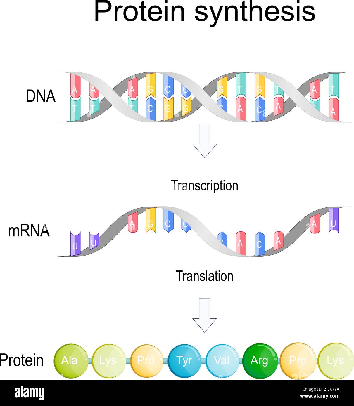 transcription and translation. Protein synthesis. During transcription a section of DNA converted into a mRNA. mRNA is read by ribosomes Stock Vectorhttps://www.alamy.com/image-license-details/?v=1https://www.alamy.com/transcription-and-translation-protein-synthesis-during-transcription-a-section-of-dna-converted-into-a-mrna-mrna-is-read-by-ribosomes-image473862126.html
RF2JEX7YA–transcription and translation. Protein synthesis. During transcription a section of DNA converted into a mRNA. mRNA is read by ribosomes
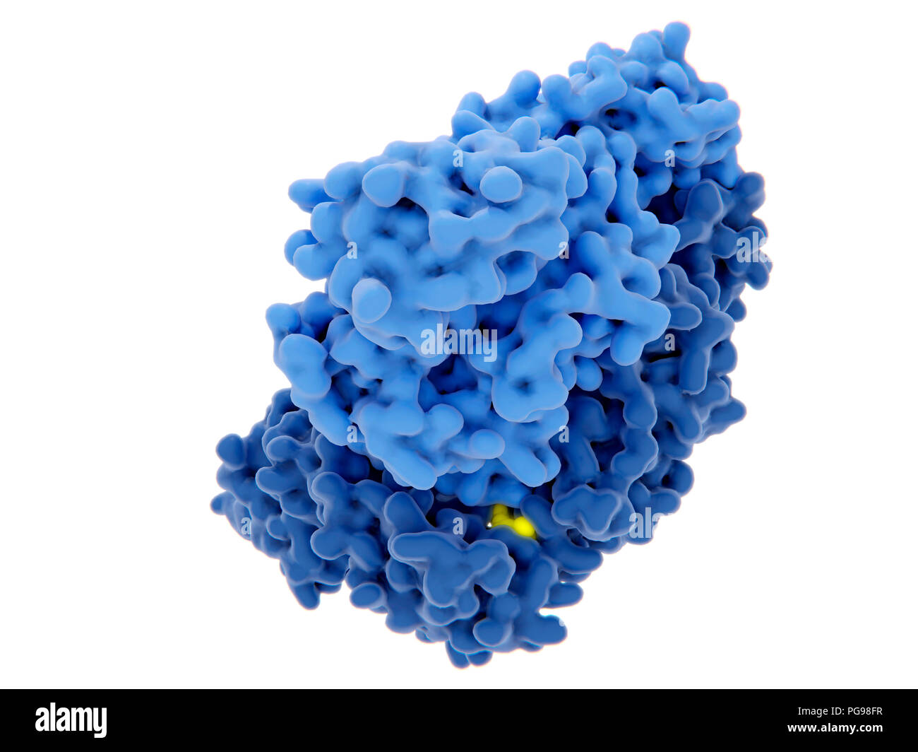 HIV-1 reverse transcriptase inhibition, illustration. The human immunodeficiency virus single-stranded RNA genome is converted into double-stranded DNA by the viral reverse transcriptase (blue) and then the DNA is integrated in the DNA of an infected human cell. The reverse transcriptase is one of the main targets to disrupt the virus multiplication through an inhibitor. There are nucleoside and nucleotide inhibitors and non-nucleoside analogue inhibitors. One of these inhibitors (yellow) is shown binding to the reverse transcriptase. Stock Photohttps://www.alamy.com/image-license-details/?v=1https://www.alamy.com/hiv-1-reverse-transcriptase-inhibition-illustration-the-human-immunodeficiency-virus-single-stranded-rna-genome-is-converted-into-double-stranded-dna-by-the-viral-reverse-transcriptase-blue-and-then-the-dna-is-integrated-in-the-dna-of-an-infected-human-cell-the-reverse-transcriptase-is-one-of-the-main-targets-to-disrupt-the-virus-multiplication-through-an-inhibitor-there-are-nucleoside-and-nucleotide-inhibitors-and-non-nucleoside-analogue-inhibitors-one-of-these-inhibitors-yellow-is-shown-binding-to-the-reverse-transcriptase-image216563195.html
RFPG98FR–HIV-1 reverse transcriptase inhibition, illustration. The human immunodeficiency virus single-stranded RNA genome is converted into double-stranded DNA by the viral reverse transcriptase (blue) and then the DNA is integrated in the DNA of an infected human cell. The reverse transcriptase is one of the main targets to disrupt the virus multiplication through an inhibitor. There are nucleoside and nucleotide inhibitors and non-nucleoside analogue inhibitors. One of these inhibitors (yellow) is shown binding to the reverse transcriptase.
 Coronavirus cell model illustration, Covid-19 Coronavirus pandemic, 3D Render, conceptual, close up, white background Stock Photohttps://www.alamy.com/image-license-details/?v=1https://www.alamy.com/coronavirus-cell-model-illustration-covid-19-coronavirus-pandemic-3d-render-conceptual-close-up-white-background-image352420621.html
RF2BDA42N–Coronavirus cell model illustration, Covid-19 Coronavirus pandemic, 3D Render, conceptual, close up, white background
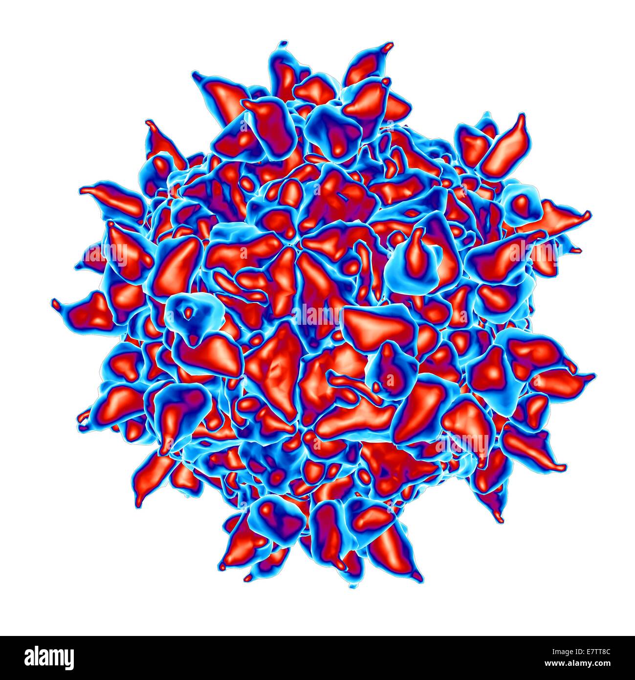 Human rhinovirus, computer artwork. Stock Photohttps://www.alamy.com/image-license-details/?v=1https://www.alamy.com/stock-photo-human-rhinovirus-computer-artwork-73689964.html
RFE7TT8C–Human rhinovirus, computer artwork.
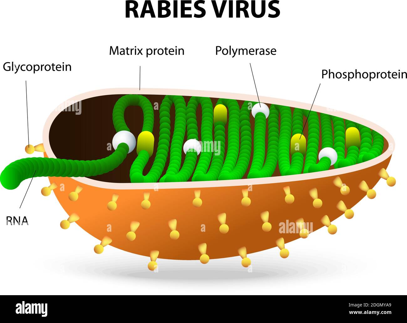 Rabies virus or Rhabdovirus. rabies virus causes rabies in humans and animals Stock Vectorhttps://www.alamy.com/image-license-details/?v=1https://www.alamy.com/rabies-virus-or-rhabdovirus-rabies-virus-causes-rabies-in-humans-and-animals-image388923089.html
RF2DGMYA9–Rabies virus or Rhabdovirus. rabies virus causes rabies in humans and animals
 Microscopic 3D Visualization Of The Covid-19 Corona Virus Stock Photohttps://www.alamy.com/image-license-details/?v=1https://www.alamy.com/microscopic-3d-visualization-of-the-covid-19-corona-virus-image364944888.html
RF2C5MJX0–Microscopic 3D Visualization Of The Covid-19 Corona Virus
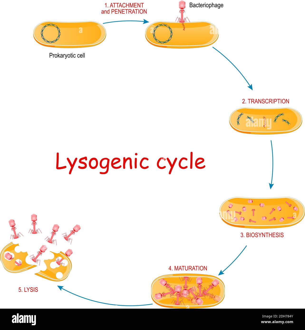
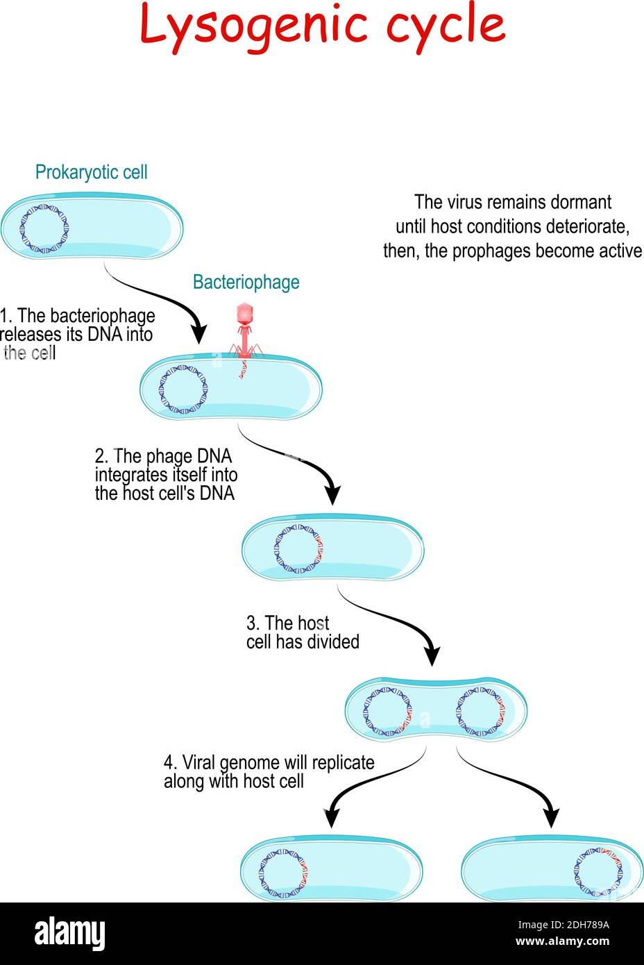 lysogenic cycle with bacteriophage. Lysogeny does not result in immediate lysing of the host cell. The virus remains dormant until host conditions Stock Vectorhttps://www.alamy.com/image-license-details/?v=1https://www.alamy.com/lysogenic-cycle-with-bacteriophage-lysogeny-does-not-result-in-immediate-lysing-of-the-host-cell-the-virus-remains-dormant-until-host-conditions-image389237446.html
RF2DH789A–lysogenic cycle with bacteriophage. Lysogeny does not result in immediate lysing of the host cell. The virus remains dormant until host conditions
RF2HK2GG6–Transcript Simple vector icon. Illustration symbol design template for web mobile UI element.
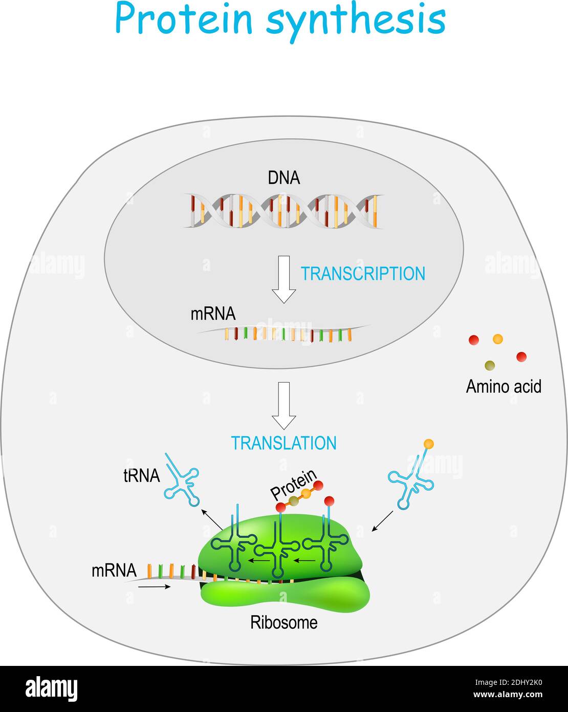 Protein synthesis in ribosome. transcription and translation. synthesis of mRNA from DNA in the nucleus. The mRNA decoding ribosomes Stock Vectorhttps://www.alamy.com/image-license-details/?v=1https://www.alamy.com/protein-synthesis-in-ribosome-transcription-and-translation-synthesis-of-mrna-from-dna-in-the-nucleus-the-mrna-decoding-ribosomes-image389672052.html
RF2DHY2K0–Protein synthesis in ribosome. transcription and translation. synthesis of mRNA from DNA in the nucleus. The mRNA decoding ribosomes
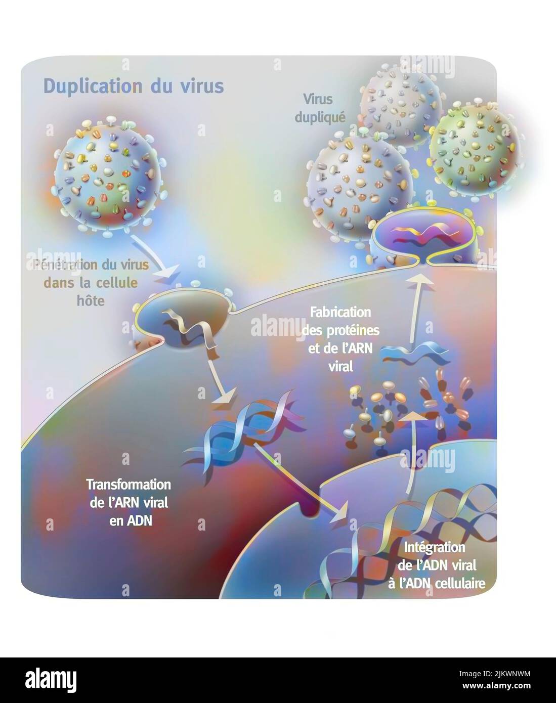 Penetration and replication of a retrovirus (example: AIDS) in a host cell. Stock Photohttps://www.alamy.com/image-license-details/?v=1https://www.alamy.com/penetration-and-replication-of-a-retrovirus-example-aids-in-a-host-cell-image476924384.html
RF2JKWNWM–Penetration and replication of a retrovirus (example: AIDS) in a host cell.
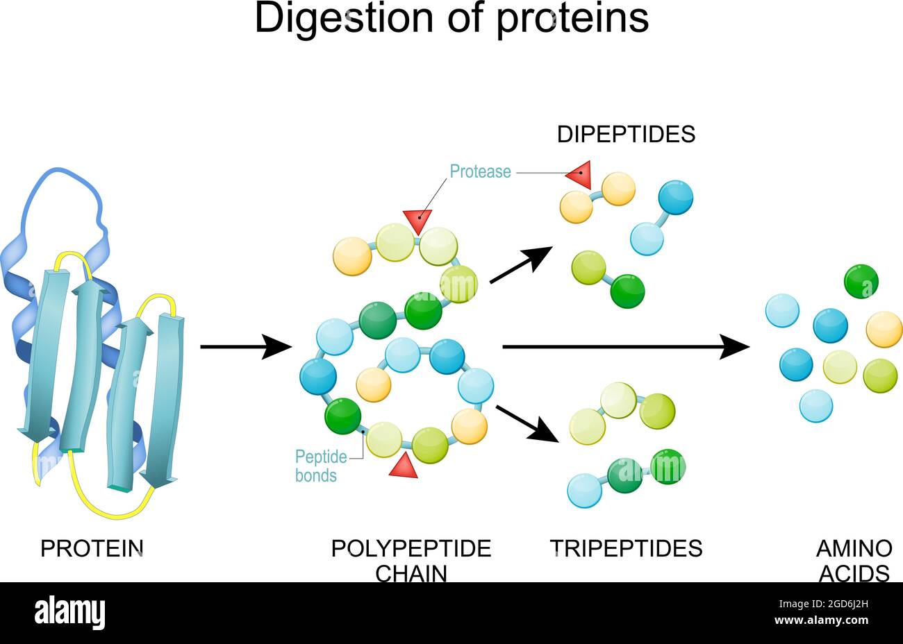 Protein Digestion. Enzymes (proteases and peptidases) are digestion breaks the protein into smaller peptide chains and into single amino acids Stock Vectorhttps://www.alamy.com/image-license-details/?v=1https://www.alamy.com/protein-digestion-enzymes-proteases-and-peptidases-are-digestion-breaks-the-protein-into-smaller-peptide-chains-and-into-single-amino-acids-image438395625.html
RF2GD6J2H–Protein Digestion. Enzymes (proteases and peptidases) are digestion breaks the protein into smaller peptide chains and into single amino acids
 Human Mitochondrial PheRS Stock Photohttps://www.alamy.com/image-license-details/?v=1https://www.alamy.com/human-mitochondrial-phers-image416784513.html
RM2F624W5–Human Mitochondrial PheRS
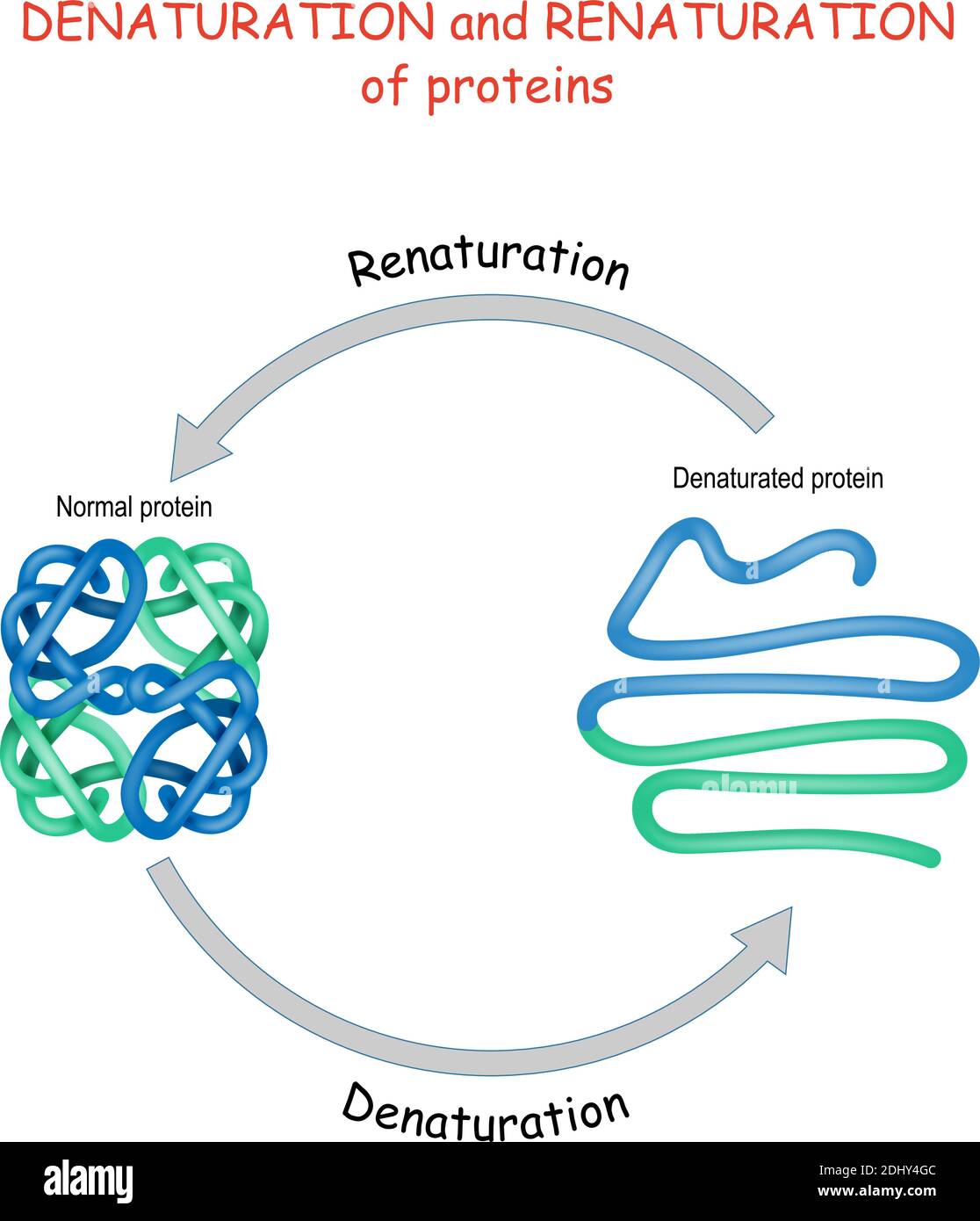 Process of Denaturation and renaturation of proteins. Vector diagram for science, education, and medical use. Stock Vectorhttps://www.alamy.com/image-license-details/?v=1https://www.alamy.com/process-of-denaturation-and-renaturation-of-proteins-vector-diagram-for-science-education-and-medical-use-image389673548.html
RF2DHY4GC–Process of Denaturation and renaturation of proteins. Vector diagram for science, education, and medical use.
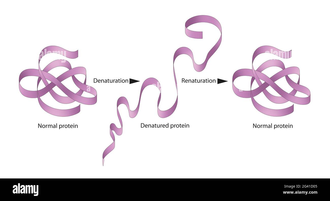 Denaturation and renaturation of Proteins Stock Photohttps://www.alamy.com/image-license-details/?v=1https://www.alamy.com/denaturation-and-renaturation-of-proteins-image432749973.html
RF2G41D05–Denaturation and renaturation of Proteins
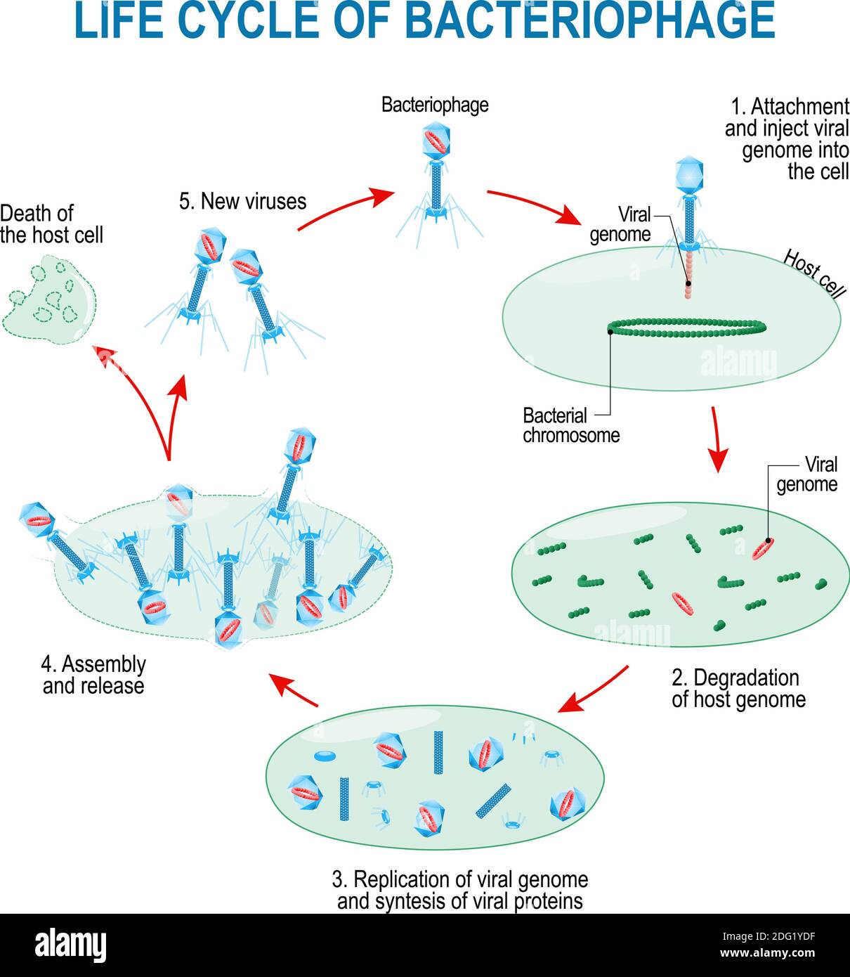 viruses life cycle for example bacteriophage and bacteria. Schematic diagram. Stock Vectorhttps://www.alamy.com/image-license-details/?v=1https://www.alamy.com/viruses-life-cycle-for-example-bacteriophage-and-bacteria-schematic-diagram-image388506091.html
RF2DG1YDF–viruses life cycle for example bacteriophage and bacteria. Schematic diagram.
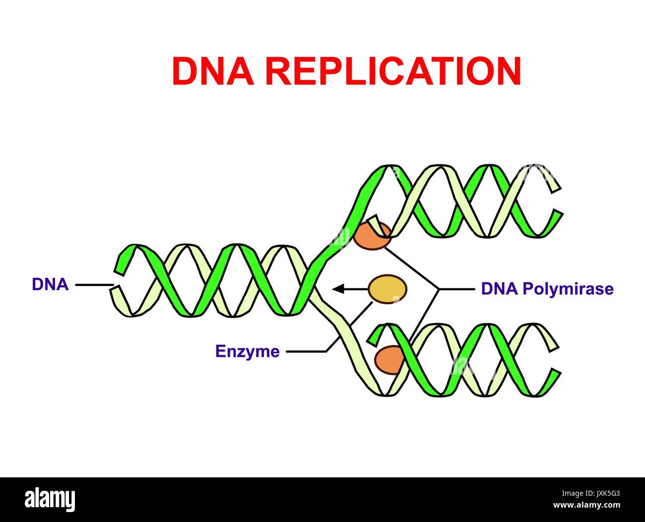 DNA replication on white. Education info graphic. Stock Vectorhttps://www.alamy.com/image-license-details/?v=1https://www.alamy.com/dna-replication-on-white-education-info-graphic-image154085459.html
RFJXK5G3–DNA replication on white. Education info graphic.
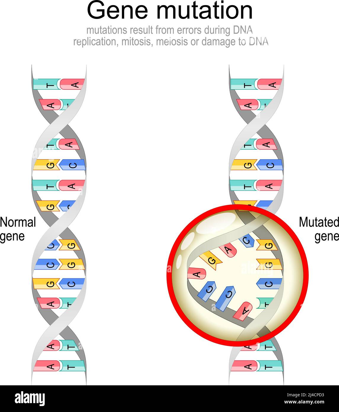 Gene mutation Normal DNA and helix with Mutated gene. Biological manipulation. Vector diagram Stock Vectorhttps://www.alamy.com/image-license-details/?v=1https://www.alamy.com/gene-mutation-normal-dna-and-helix-with-mutated-gene-biological-manipulation-vector-diagram-image467419599.html
RF2J4CPD3–Gene mutation Normal DNA and helix with Mutated gene. Biological manipulation. Vector diagram
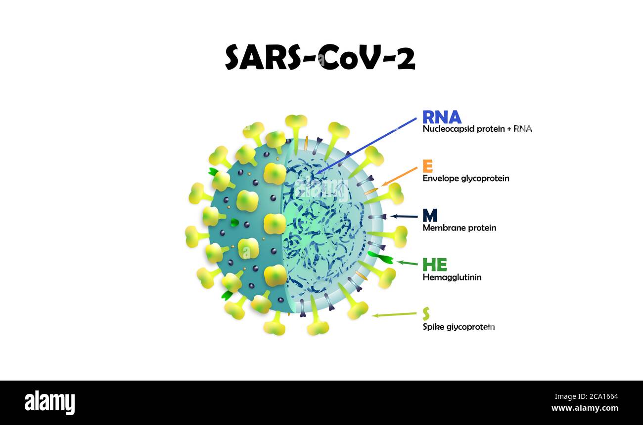 SARS-CoV2. Cross-sectional model of a coronavirus. Stock Photohttps://www.alamy.com/image-license-details/?v=1https://www.alamy.com/sars-cov2-cross-sectional-model-of-a-coronavirus-image367591116.html
RF2CA1664–SARS-CoV2. Cross-sectional model of a coronavirus.
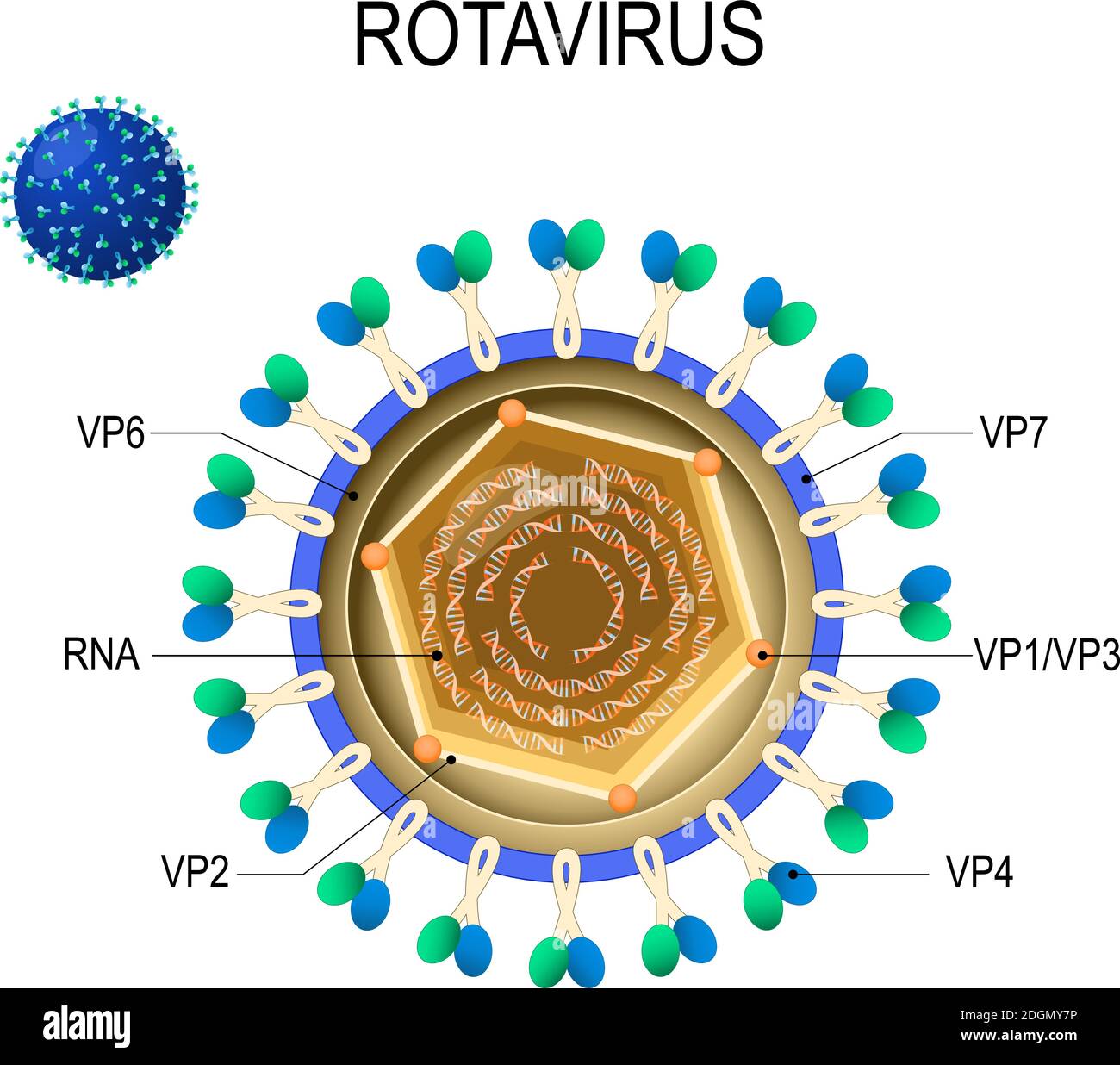 Rotavirus anatomy. structure of virion. Vector diagram of the location of rotavirus structural proteins. Rota virus causing acute gastroenteritis Stock Vectorhttps://www.alamy.com/image-license-details/?v=1https://www.alamy.com/rotavirus-anatomy-structure-of-virion-vector-diagram-of-the-location-of-rotavirus-structural-proteins-rota-virus-causing-acute-gastroenteritis-image388923018.html
RF2DGMY7P–Rotavirus anatomy. structure of virion. Vector diagram of the location of rotavirus structural proteins. Rota virus causing acute gastroenteritis
 Illustration of chrome dna model isolated on white background Stock Photohttps://www.alamy.com/image-license-details/?v=1https://www.alamy.com/stock-photo-illustration-of-chrome-dna-model-isolated-on-white-background-50054776.html
RFCWC5AG–Illustration of chrome dna model isolated on white background
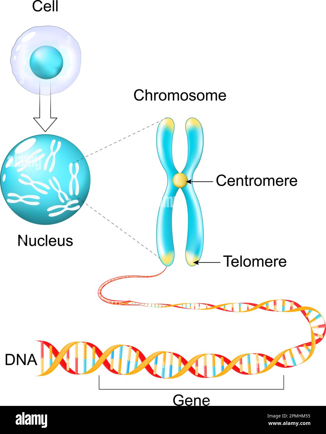 Cell structure. Genetic material from Gene to DNA and Chromosome. genome sequence. Molecular biology. Vector poster Stock Vectorhttps://www.alamy.com/image-license-details/?v=1https://www.alamy.com/cell-structure-genetic-material-from-gene-to-dna-and-chromosome-genome-sequence-molecular-biology-vector-poster-image546203537.html
RF2PMHM55–Cell structure. Genetic material from Gene to DNA and Chromosome. genome sequence. Molecular biology. Vector poster
 Coronavirus cell model illustration, Covid-19 Coronavirus pandemic, 3D Render, conceptual, close up, black background Stock Photohttps://www.alamy.com/image-license-details/?v=1https://www.alamy.com/coronavirus-cell-model-illustration-covid-19-coronavirus-pandemic-3d-render-conceptual-close-up-black-background-image352426546.html
RF2BDABJA–Coronavirus cell model illustration, Covid-19 Coronavirus pandemic, 3D Render, conceptual, close up, black background
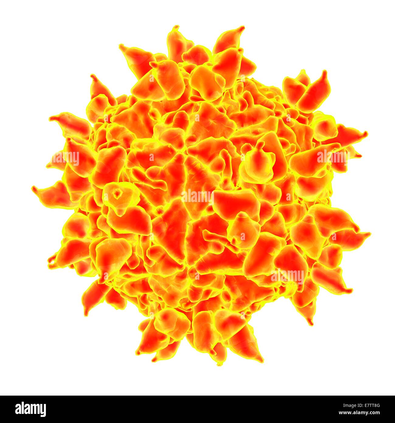 Human rhinovirus, computer artwork. Stock Photohttps://www.alamy.com/image-license-details/?v=1https://www.alamy.com/stock-photo-human-rhinovirus-computer-artwork-73689968.html
RFE7TT8G–Human rhinovirus, computer artwork.
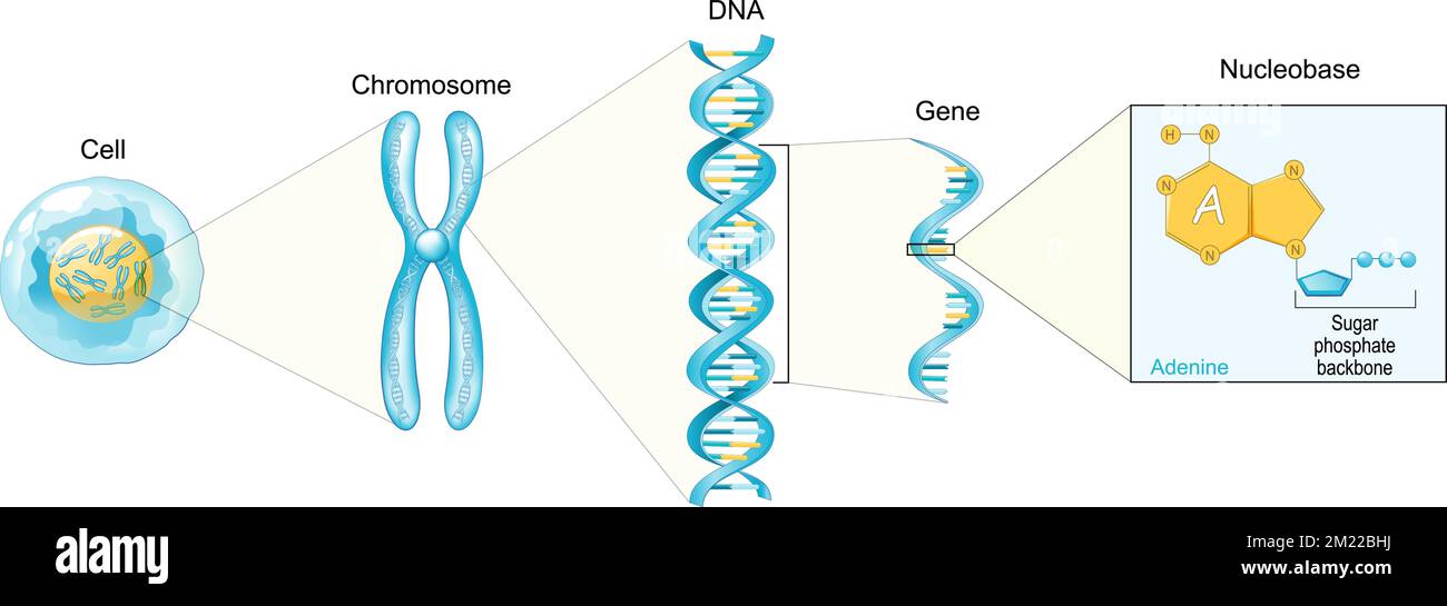 Structure of Cell. From Nucleobase like adenine to Gene, DNA and Chromosome. genome sequence. Molecular biology. Vector poster Stock Vectorhttps://www.alamy.com/image-license-details/?v=1https://www.alamy.com/structure-of-cell-from-nucleobase-like-adenine-to-gene-dna-and-chromosome-genome-sequence-molecular-biology-vector-poster-image500383006.html
RF2M22BHJ–Structure of Cell. From Nucleobase like adenine to Gene, DNA and Chromosome. genome sequence. Molecular biology. Vector poster
 Bird flu virus. 3D illustration of avian influenza H5N8 virus particles. Within the viral lipid envelope (orange) are two types of protein spike, hemagglutinin (H, blue) and neuraminidase (N, green), which determine the strain of virus. This strain of the virus has caused disease in wild birds and poultry in Europe and Asia since June 2016. Unusually, the virus causes mortality in wild birds, which are more often silent carriers. As of December 2016 no human cases of the disease have been reported, and risk of transmission to humans is thought to be low. Stock Photohttps://www.alamy.com/image-license-details/?v=1https://www.alamy.com/stock-photo-bird-flu-virus-3d-illustration-of-avian-influenza-h5n8-virus-particles-129179902.html
RFHE4J7X–Bird flu virus. 3D illustration of avian influenza H5N8 virus particles. Within the viral lipid envelope (orange) are two types of protein spike, hemagglutinin (H, blue) and neuraminidase (N, green), which determine the strain of virus. This strain of the virus has caused disease in wild birds and poultry in Europe and Asia since June 2016. Unusually, the virus causes mortality in wild birds, which are more often silent carriers. As of December 2016 no human cases of the disease have been reported, and risk of transmission to humans is thought to be low.
 Lytic cycle with bacteria and bacteriophage. Cycles of viral reproduction that results in the destruction of the infected cell. The stages of lytic Stock Vectorhttps://www.alamy.com/image-license-details/?v=1https://www.alamy.com/lytic-cycle-with-bacteria-and-bacteriophage-cycles-of-viral-reproduction-that-results-in-the-destruction-of-the-infected-cell-the-stages-of-lytic-image389237323.html
RF2DH784Y–Lytic cycle with bacteria and bacteriophage. Cycles of viral reproduction that results in the destruction of the infected cell. The stages of lytic
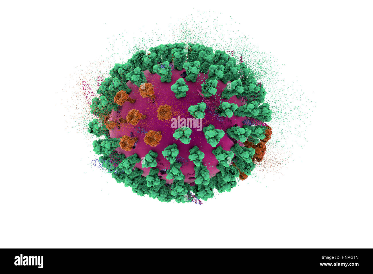 Destruction of bird flu virus, conceptual 3D illustration. This is an avian influenza H5N8 virus particle. This strain of the virus has caused disease in wild birds and poultry in Europe and Asia since June 2016. Unusually, the virus causes mortality in wild birds, which are more often silent carriers. As of February 2017 no human cases of the disease have been reported, and risk of transmission to humans is thought to be low. Stock Photohttps://www.alamy.com/image-license-details/?v=1https://www.alamy.com/stock-photo-destruction-of-bird-flu-virus-conceptual-3d-illustration-this-is-an-133613109.html
RFHNAGTN–Destruction of bird flu virus, conceptual 3D illustration. This is an avian influenza H5N8 virus particle. This strain of the virus has caused disease in wild birds and poultry in Europe and Asia since June 2016. Unusually, the virus causes mortality in wild birds, which are more often silent carriers. As of February 2017 no human cases of the disease have been reported, and risk of transmission to humans is thought to be low.
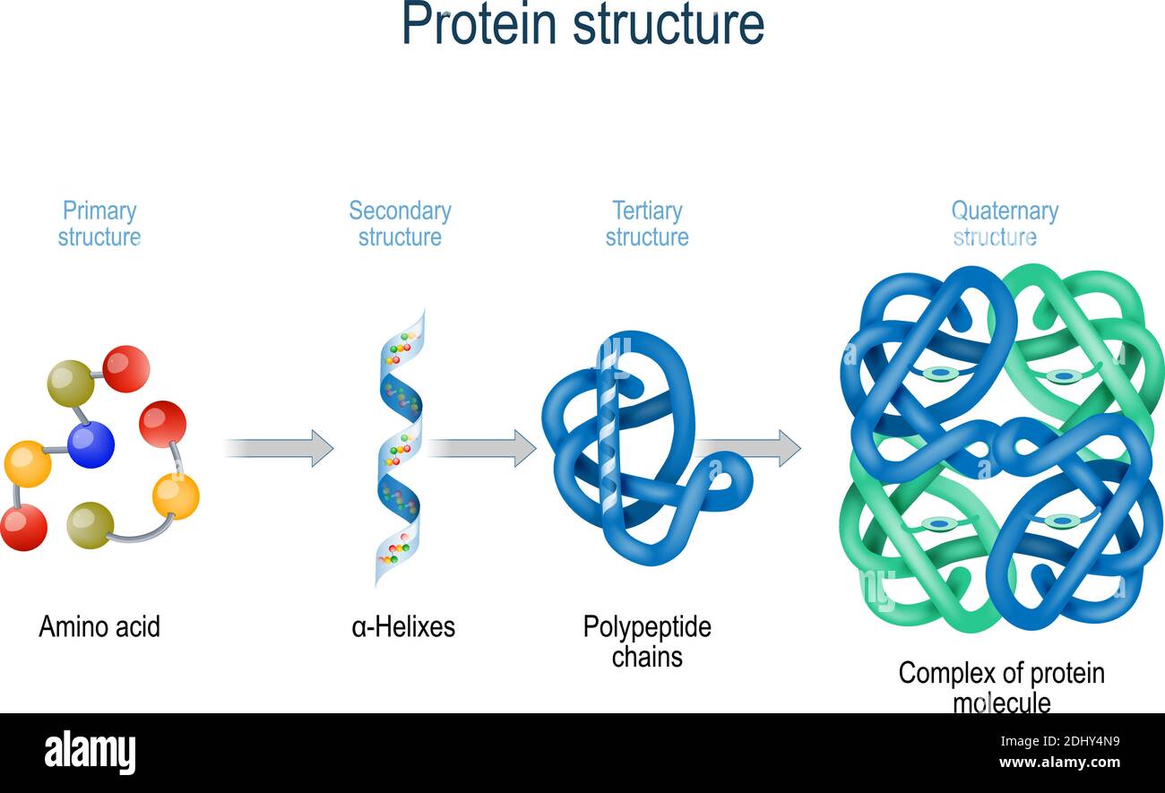 Levels of protein structure from amino acids to Complex of protein molecule. Protein is a polymer (polypeptide) Stock Vectorhttps://www.alamy.com/image-license-details/?v=1https://www.alamy.com/levels-of-protein-structure-from-amino-acids-to-complex-of-protein-molecule-protein-is-a-polymerpolypeptide-image389673685.html
RF2DHY4N9–Levels of protein structure from amino acids to Complex of protein molecule. Protein is a polymer (polypeptide)
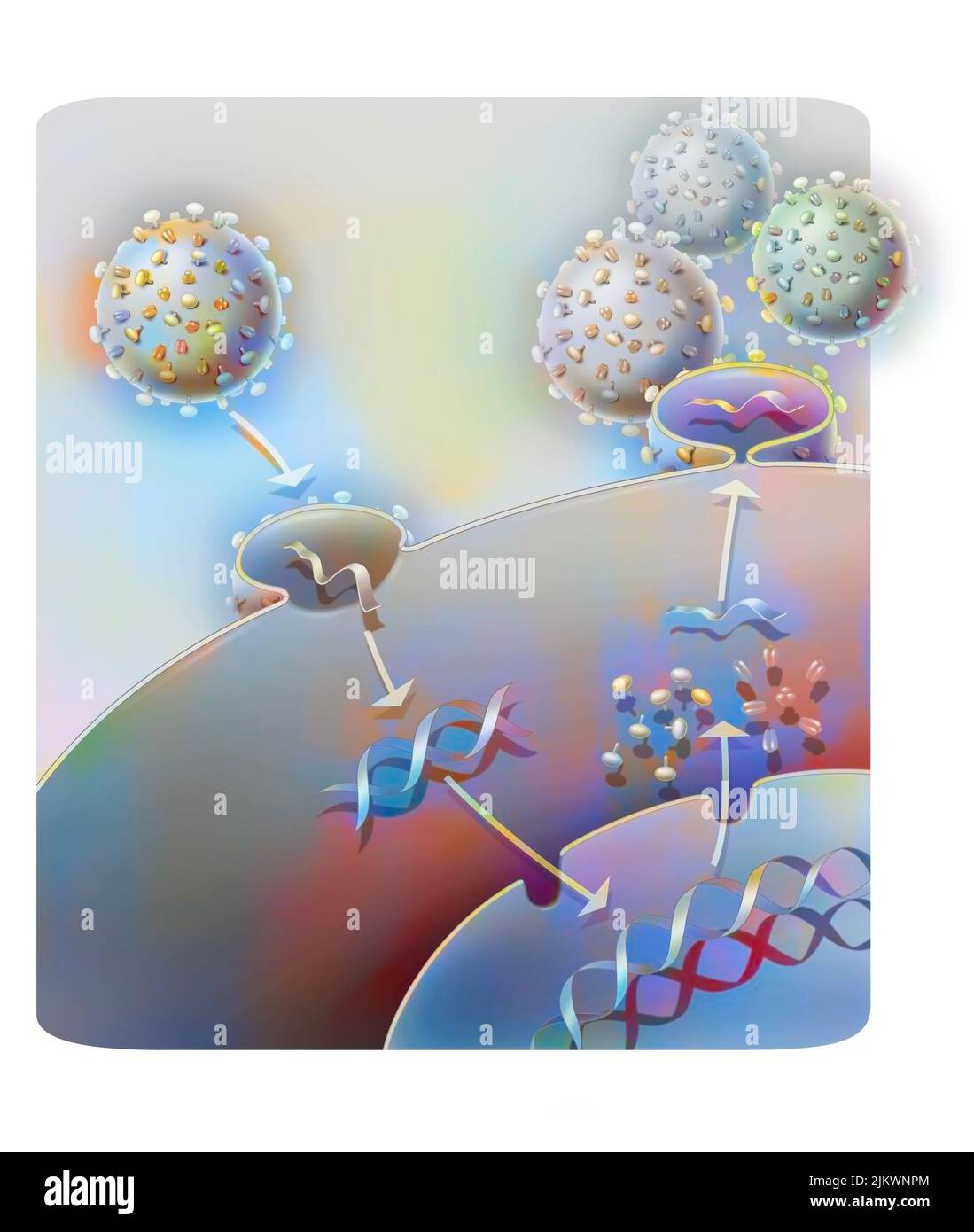 Penetration and replication of a retrovirus (example: AIDS) in a host cell. Stock Photohttps://www.alamy.com/image-license-details/?v=1https://www.alamy.com/penetration-and-replication-of-a-retrovirus-example-aids-in-a-host-cell-image476924300.html
RF2JKWNPM–Penetration and replication of a retrovirus (example: AIDS) in a host cell.
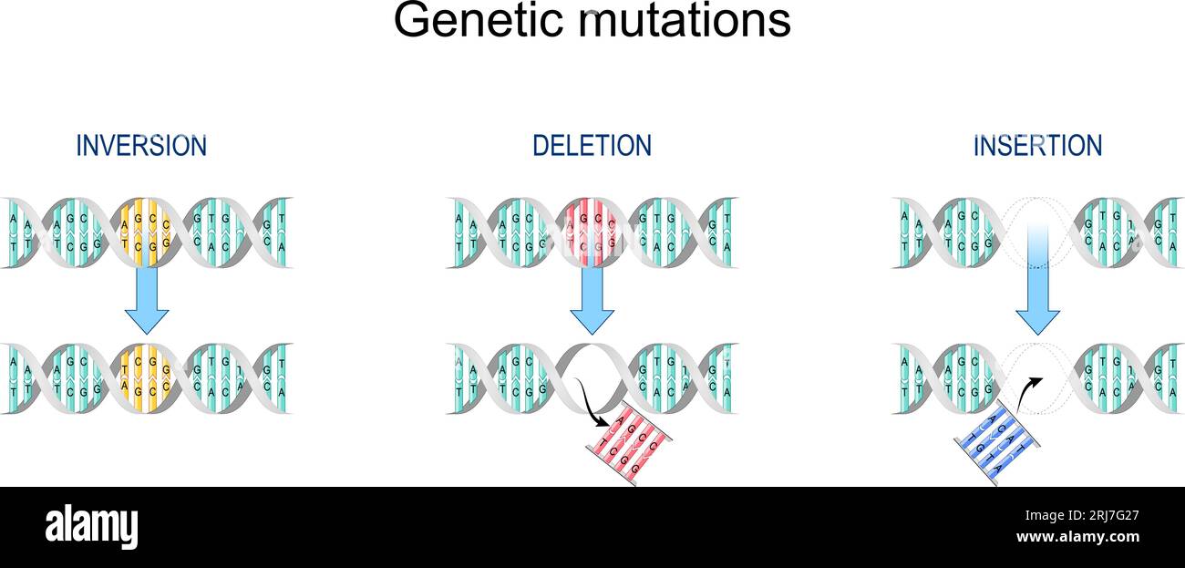 Gene mutation. Types of mutation: Insertion, Inversion, Deletion. Error during DNA replication. Normal DNA and helix with Mutated gene. Biological ma Stock Vectorhttps://www.alamy.com/image-license-details/?v=1https://www.alamy.com/gene-mutation-types-of-mutation-insertion-inversion-deletion-error-during-dna-replication-normal-dna-and-helix-with-mutated-gene-biological-ma-image561961855.html
RF2RJ7G27–Gene mutation. Types of mutation: Insertion, Inversion, Deletion. Error during DNA replication. Normal DNA and helix with Mutated gene. Biological ma
 Microscopic 3D Visualization Of The Covid-19 Corona Virus Stock Photohttps://www.alamy.com/image-license-details/?v=1https://www.alamy.com/microscopic-3d-visualization-of-the-covid-19-corona-virus-image364944897.html
RF2C5MJX9–Microscopic 3D Visualization Of The Covid-19 Corona Virus
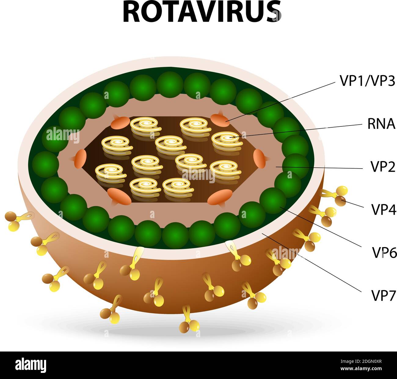 rotavirus or rota virus virion. Rota virus causing acute gastroenteritis in birds, mammals and humans. Stock Vectorhttps://www.alamy.com/image-license-details/?v=1https://www.alamy.com/rotavirus-or-rota-virus-virion-rota-virus-causing-acute-gastroenteritis-in-birds-mammals-and-humans-image388924335.html
RF2DGN0XR–rotavirus or rota virus virion. Rota virus causing acute gastroenteritis in birds, mammals and humans.
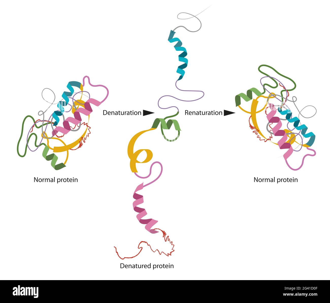 Structure of normal and disassembled protein Stock Photohttps://www.alamy.com/image-license-details/?v=1https://www.alamy.com/structure-of-normal-and-disassembled-protein-image432749983.html
RF2G41D0F–Structure of normal and disassembled protein
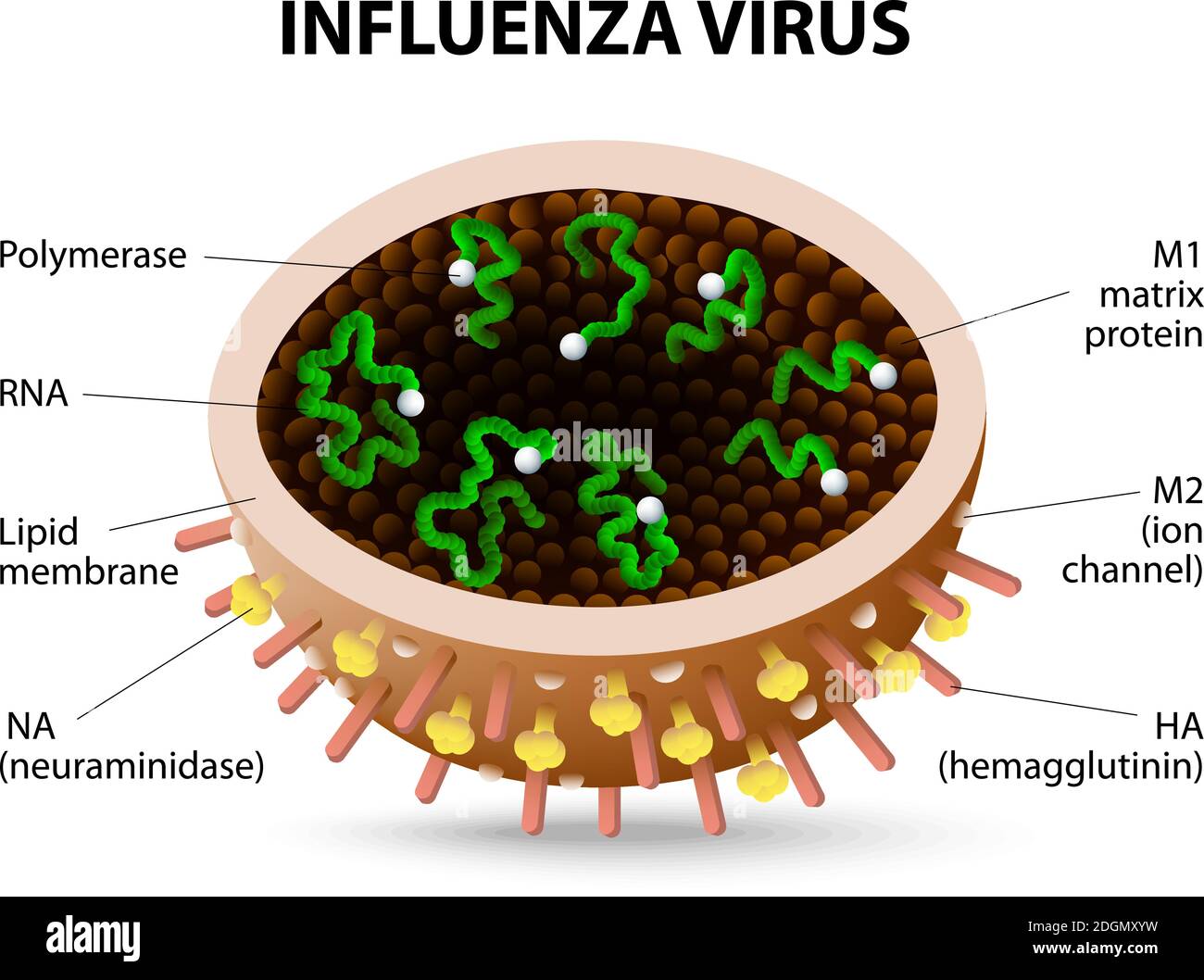 Structure of influenza virus. virion. Vector diagram Stock Vectorhttps://www.alamy.com/image-license-details/?v=1https://www.alamy.com/structure-of-influenza-virus-virion-vector-diagram-image388922797.html
RF2DGMXYW–Structure of influenza virus. virion. Vector diagram
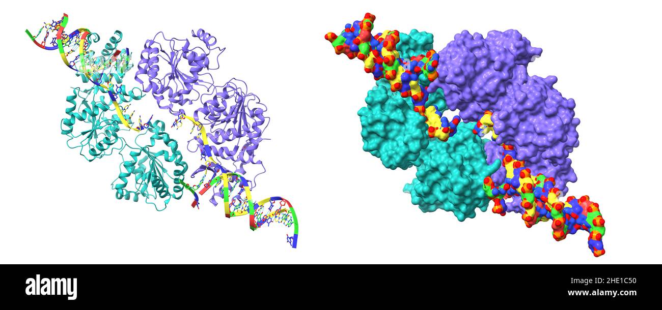 Structure of human RECQ-like helicase in complex with DNA. 3D cartoon and Gaussian surface models, PDB 2wwy, white background Stock Photohttps://www.alamy.com/image-license-details/?v=1https://www.alamy.com/structure-of-human-recq-like-helicase-in-complex-with-dna-3d-cartoon-and-gaussian-surface-models-pdb-2wwy-white-background-image456106252.html
RF2HE1C50–Structure of human RECQ-like helicase in complex with DNA. 3D cartoon and Gaussian surface models, PDB 2wwy, white background
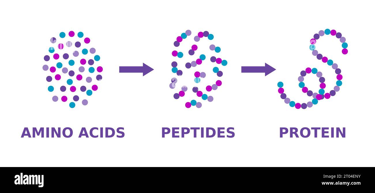 Protein structure. Amino acids, peptides, protein. Proteins formation model. Scientific diagram. Proteins molecule synthesis. Vector illustration. Stock Vectorhttps://www.alamy.com/image-license-details/?v=1https://www.alamy.com/protein-structure-amino-acids-peptides-protein-proteins-formation-model-scientific-diagram-proteins-molecule-synthesis-vector-illustration-image568041543.html
RF2T04ENY–Protein structure. Amino acids, peptides, protein. Proteins formation model. Scientific diagram. Proteins molecule synthesis. Vector illustration.
RF2J6T12R–RNA related vector glyph icon. RNA sign. Isolated on white background. Editable vector illustration
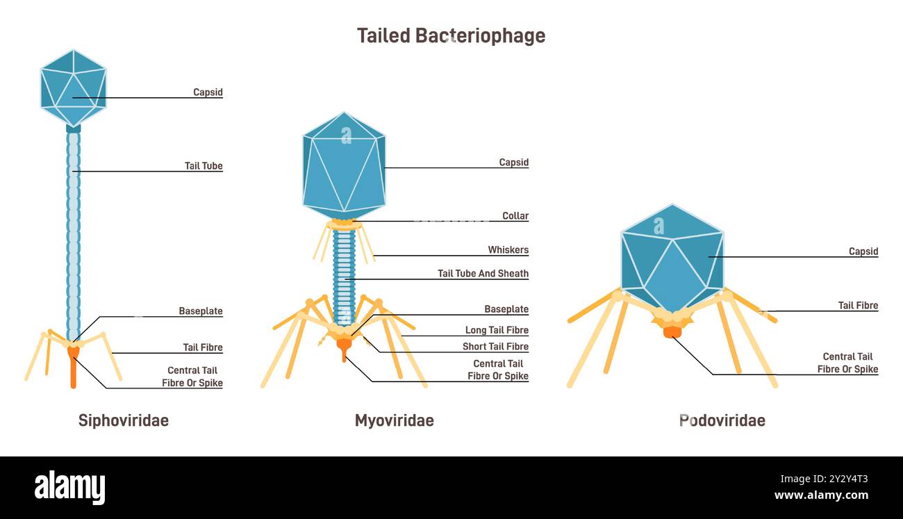 Tailed bacteriophage Myoviridae, siphoviridae and podoviriade, microscopic model of virus infecting a bacterial cells to replicate itself. Microbiology and virology studying. Flat vector illustration Stock Vectorhttps://www.alamy.com/image-license-details/?v=1https://www.alamy.com/tailed-bacteriophage-myoviridae-siphoviridae-and-podoviriade-microscopic-model-of-virus-infecting-a-bacterial-cells-to-replicate-itself-microbiology-and-virology-studying-flat-vector-illustration-image621399075.html
RF2Y2Y4T3–Tailed bacteriophage Myoviridae, siphoviridae and podoviriade, microscopic model of virus infecting a bacterial cells to replicate itself. Microbiology and virology studying. Flat vector illustration
 Illustration of gold dna model isolated on white background Stock Photohttps://www.alamy.com/image-license-details/?v=1https://www.alamy.com/stock-photo-illustration-of-gold-dna-model-isolated-on-white-background-50311463.html
RFCWRTNY–Illustration of gold dna model isolated on white background
RF2J7JM6P–RNA related vector glyph icon. RNA sign. Isolated on white background. Editable vector illustration
 Coronavirus cell model illustration, Covid-19 Coronavirus pandemic, 3D Render, conceptual, close up, black background Stock Photohttps://www.alamy.com/image-license-details/?v=1https://www.alamy.com/coronavirus-cell-model-illustration-covid-19-coronavirus-pandemic-3d-render-conceptual-close-up-black-background-image352426550.html
RF2BDABJE–Coronavirus cell model illustration, Covid-19 Coronavirus pandemic, 3D Render, conceptual, close up, black background
 DNA replication. DNA molecules, molecular biology. Vector stock illustration. Stock Vectorhttps://www.alamy.com/image-license-details/?v=1https://www.alamy.com/dna-replication-dna-molecules-molecular-biology-vector-stock-illustration-image467804601.html
RF2J529F5–DNA replication. DNA molecules, molecular biology. Vector stock illustration.
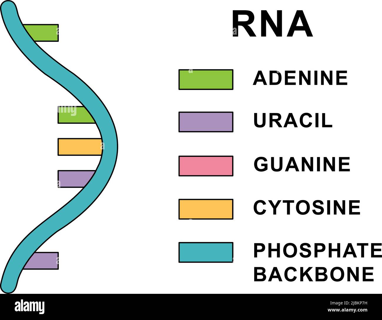 Structure of spiral Ribonucleic acid molecule. RNA molecule with nucleobases structure description - cytosine, guanine, adenine, uracil. Stock Vectorhttps://www.alamy.com/image-license-details/?v=1https://www.alamy.com/structure-of-spiral-ribonucleic-acid-molecule-rna-molecule-with-nucleobases-structure-description-cytosine-guanine-adenine-uracil-image471875701.html
RF2JBKP7H–Structure of spiral Ribonucleic acid molecule. RNA molecule with nucleobases structure description - cytosine, guanine, adenine, uracil.
RF2HH9B98–RNA related vector glyph icon
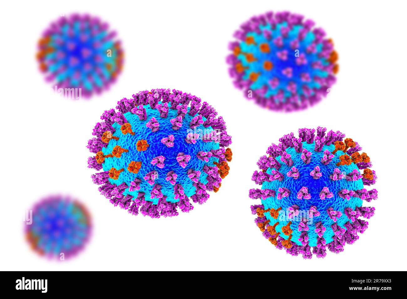 Influenza virus. 3D illustration showing surface glycoprotein spikes hemagglutinin purple and neuraminidase orange Stock Photohttps://www.alamy.com/image-license-details/?v=1https://www.alamy.com/influenza-virus-3d-illustration-showing-surface-glycoprotein-spikes-hemagglutinin-purple-and-neuraminidase-orange-image555253051.html
RF2R79XX3–Influenza virus. 3D illustration showing surface glycoprotein spikes hemagglutinin purple and neuraminidase orange
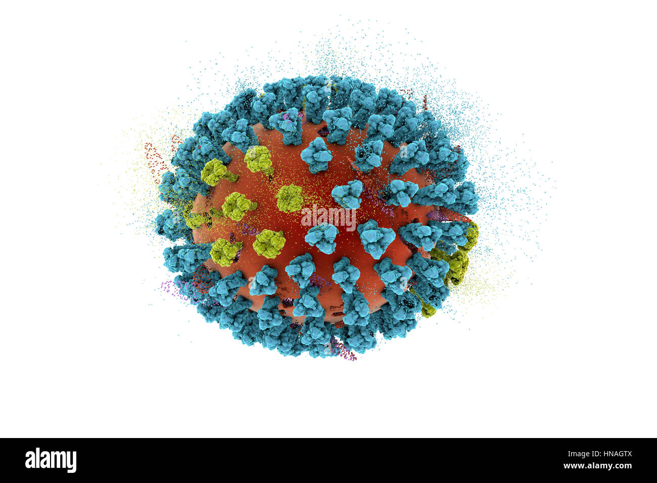 Destruction of bird flu virus, conceptual 3D illustration. This is an avian influenza H5N8 virus particle. This strain of the virus has caused disease in wild birds and poultry in Europe and Asia since June 2016. Unusually, the virus causes mortality in wild birds, which are more often silent carriers. As of February 2017 no human cases of the disease have been reported, and risk of transmission to humans is thought to be low. Stock Photohttps://www.alamy.com/image-license-details/?v=1https://www.alamy.com/stock-photo-destruction-of-bird-flu-virus-conceptual-3d-illustration-this-is-an-133613114.html
RFHNAGTX–Destruction of bird flu virus, conceptual 3D illustration. This is an avian influenza H5N8 virus particle. This strain of the virus has caused disease in wild birds and poultry in Europe and Asia since June 2016. Unusually, the virus causes mortality in wild birds, which are more often silent carriers. As of February 2017 no human cases of the disease have been reported, and risk of transmission to humans is thought to be low.
RF2WR1AH7–Vector illustration. DNA molecule icon isolated on white background.
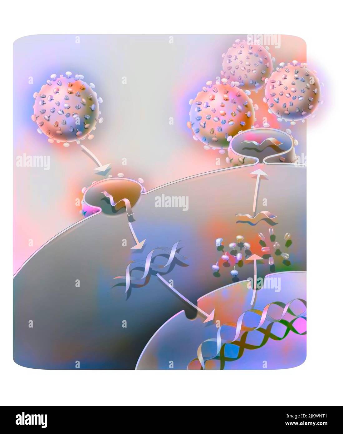 Penetration and replication of a retrovirus (example: AIDS) in a host cell. Stock Photohttps://www.alamy.com/image-license-details/?v=1https://www.alamy.com/penetration-and-replication-of-a-retrovirus-example-aids-in-a-host-cell-image476924337.html
RF2JKWNT1–Penetration and replication of a retrovirus (example: AIDS) in a host cell.
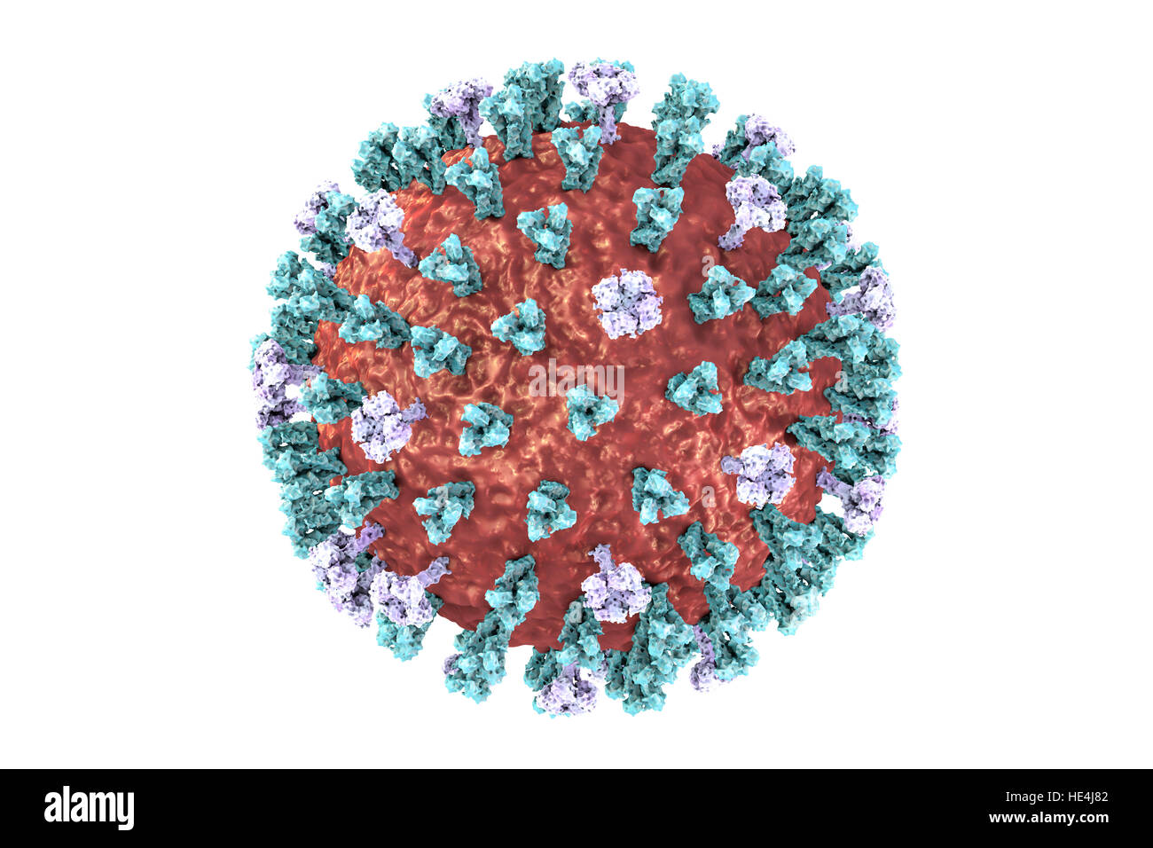 Bird flu virus. 3D illustration of an avian influenza H5N8 virus particle. Within the viral lipid envelope (red) are two types of protein spike, hemagglutinin (H, blue) and neuraminidase (N, purple), which determine the strain of virus. This strain of the virus has caused disease in wild birds and poultry in Europe and Asia since June 2016. Unusually, the virus causes mortality in wild birds, which are more often silent carriers. As of December 2016 no human cases of the disease have been reported, and risk of transmission to humans is thought to be low. Stock Photohttps://www.alamy.com/image-license-details/?v=1https://www.alamy.com/stock-photo-bird-flu-virus-3d-illustration-of-an-avian-influenza-h5n8-virus-particle-129179906.html
RFHE4J82–Bird flu virus. 3D illustration of an avian influenza H5N8 virus particle. Within the viral lipid envelope (red) are two types of protein spike, hemagglutinin (H, blue) and neuraminidase (N, purple), which determine the strain of virus. This strain of the virus has caused disease in wild birds and poultry in Europe and Asia since June 2016. Unusually, the virus causes mortality in wild birds, which are more often silent carriers. As of December 2016 no human cases of the disease have been reported, and risk of transmission to humans is thought to be low.
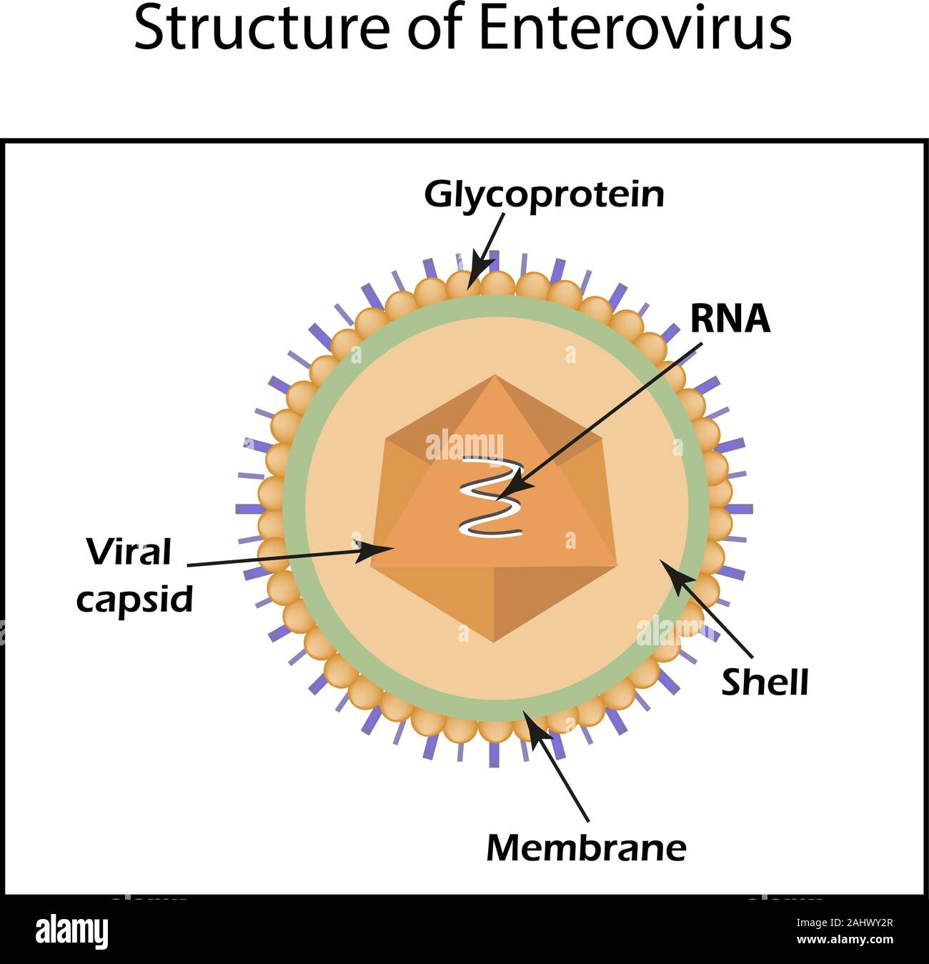 The structure of the enterovirus. Infographics. Vector illustration on isolated background Stock Vectorhttps://www.alamy.com/image-license-details/?v=1https://www.alamy.com/the-structure-of-the-enterovirus-infographics-vector-illustration-on-isolated-background-image338016191.html
RF2AHWY2R–The structure of the enterovirus. Infographics. Vector illustration on isolated background
 Writing displaying text Rna Virus. Conceptual photo a virus genetic information is stored in the form of RNA Stock Photohttps://www.alamy.com/image-license-details/?v=1https://www.alamy.com/writing-displaying-text-rna-virus-conceptual-photo-a-virus-genetic-information-is-stored-in-the-form-of-rna-image521655383.html
RF2N8KCNB–Writing displaying text Rna Virus. Conceptual photo a virus genetic information is stored in the form of RNA
 Human primase, 3D cartoon model isolated, white background Stock Photohttps://www.alamy.com/image-license-details/?v=1https://www.alamy.com/human-primase-3d-cartoon-model-isolated-white-background-image389239457.html
RF2DH7AW5–Human primase, 3D cartoon model isolated, white background
 Coronavirus cell model illustration, Covid-19 Coronavirus pandemic, 3D Render, conceptual, close up, white background Stock Photohttps://www.alamy.com/image-license-details/?v=1https://www.alamy.com/coronavirus-cell-model-illustration-covid-19-coronavirus-pandemic-3d-render-conceptual-close-up-white-background-image352420696.html
RF2BDA45C–Coronavirus cell model illustration, Covid-19 Coronavirus pandemic, 3D Render, conceptual, close up, white background
 DNA replication. DNA molecules, molecular biology. Vector stock illustration. Stock Vectorhttps://www.alamy.com/image-license-details/?v=1https://www.alamy.com/dna-replication-dna-molecules-molecular-biology-vector-stock-illustration-image467806920.html
RF2J52CE0–DNA replication. DNA molecules, molecular biology. Vector stock illustration.
 Coronavirus cell model illustration, Covid-19 Coronavirus pandemic, 3D Render, conceptual, close up, white background Stock Photohttps://www.alamy.com/image-license-details/?v=1https://www.alamy.com/coronavirus-cell-model-illustration-covid-19-coronavirus-pandemic-3d-render-conceptual-close-up-white-background-image352213647.html
RF2BD0M2R–Coronavirus cell model illustration, Covid-19 Coronavirus pandemic, 3D Render, conceptual, close up, white background
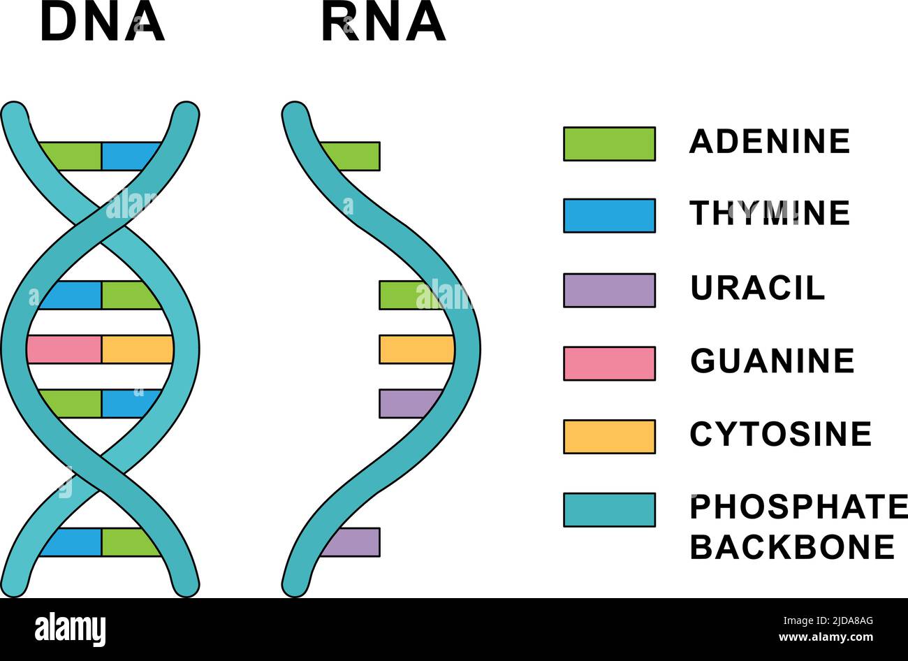 Structure of Ribonucleic acid and Deoxyribonucleic acid molecules. DNA and RNA nucleobases structure - cytosine, guanine, adenine, uracil, thymine. Stock Vectorhttps://www.alamy.com/image-license-details/?v=1https://www.alamy.com/structure-of-ribonucleic-acid-and-deoxyribonucleic-acid-molecules-dna-and-rna-nucleobases-structure-cytosine-guanine-adenine-uracil-thymine-image472896552.html
RF2JDA8AG–Structure of Ribonucleic acid and Deoxyribonucleic acid molecules. DNA and RNA nucleobases structure - cytosine, guanine, adenine, uracil, thymine.
RF2BKHYAM–RNA related vector thin line icon
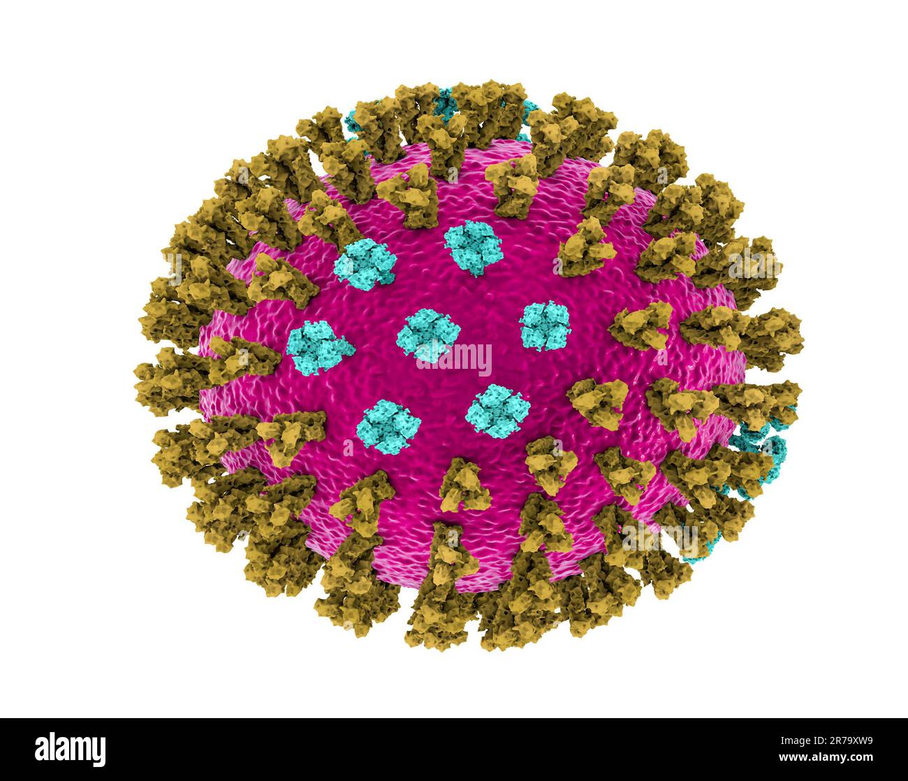 Influenza virus, Michigan strain. 3D illustration showing surface glycoprotein spikes hemagglutinin green and neuraminidase blue Stock Photohttps://www.alamy.com/image-license-details/?v=1https://www.alamy.com/influenza-virus-michigan-strain-3d-illustration-showing-surface-glycoprotein-spikes-hemagglutinin-green-and-neuraminidase-blue-image555253029.html
RF2R79XW9–Influenza virus, Michigan strain. 3D illustration showing surface glycoprotein spikes hemagglutinin green and neuraminidase blue
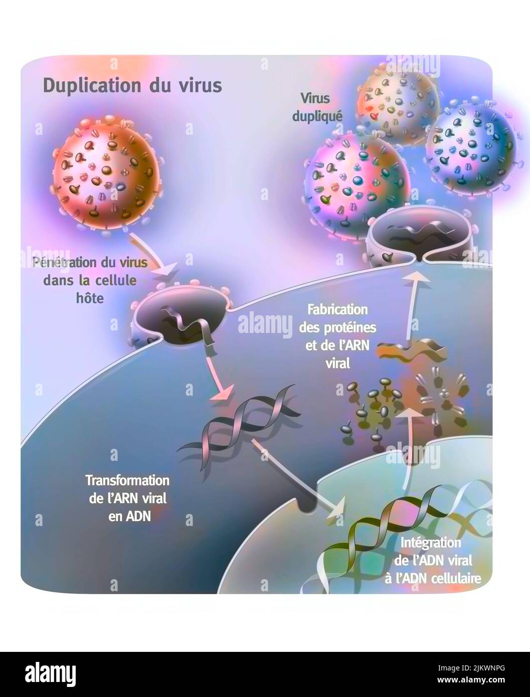 Penetration and replication of a retrovirus (example: AIDS) in a host cell. Stock Photohttps://www.alamy.com/image-license-details/?v=1https://www.alamy.com/penetration-and-replication-of-a-retrovirus-example-aids-in-a-host-cell-image476924296.html
RF2JKWNPG–Penetration and replication of a retrovirus (example: AIDS) in a host cell.
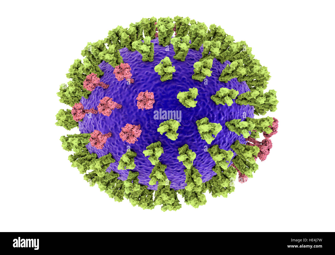 Bird flu virus. 3D illustration of an avian influenza H5N8 virus particle. Within the viral lipid envelope (purple) are two types of protein spike, hemagglutinin (H, green) and neuraminidase (N, pink), which determine the strain of virus. This strain of the virus has caused disease in wild birds and poultry in Europe and Asia since June 2016. Unusually, the virus causes mortality in wild birds, which are more often silent carriers. As of December 2016 no human cases of the disease have been reported, and risk of transmission to humans is thought to be low. Stock Photohttps://www.alamy.com/image-license-details/?v=1https://www.alamy.com/stock-photo-bird-flu-virus-3d-illustration-of-an-avian-influenza-h5n8-virus-particle-129179901.html
RFHE4J7W–Bird flu virus. 3D illustration of an avian influenza H5N8 virus particle. Within the viral lipid envelope (purple) are two types of protein spike, hemagglutinin (H, green) and neuraminidase (N, pink), which determine the strain of virus. This strain of the virus has caused disease in wild birds and poultry in Europe and Asia since June 2016. Unusually, the virus causes mortality in wild birds, which are more often silent carriers. As of December 2016 no human cases of the disease have been reported, and risk of transmission to humans is thought to be low.
 Structure of human argonaute-1 complexed with let-7 miRNA. 3D cartoon model, secondary structure color scheme, PDB 4krf, white background Stock Photohttps://www.alamy.com/image-license-details/?v=1https://www.alamy.com/structure-of-human-argonaute-1-complexed-with-let-7-mirna-3d-cartoon-model-secondary-structure-color-scheme-pdb-4krf-white-background-image456106278.html
RF2HE1C5X–Structure of human argonaute-1 complexed with let-7 miRNA. 3D cartoon model, secondary structure color scheme, PDB 4krf, white background
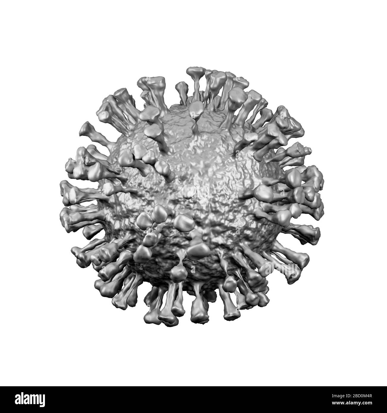 Coronavirus cell model illustration, Covid-19 Coronavirus pandemic, 3D Render, conceptual, close up, white background Stock Photohttps://www.alamy.com/image-license-details/?v=1https://www.alamy.com/coronavirus-cell-model-illustration-covid-19-coronavirus-pandemic-3d-render-conceptual-close-up-white-background-image352213703.html
RF2BD0M4R–Coronavirus cell model illustration, Covid-19 Coronavirus pandemic, 3D Render, conceptual, close up, white background
 Set of adenine, thymine, guanine, cytosine, uracil chemical formulas. Adenine, thymine, guanine, cytosine, uracil structural chemical formulas. Stock Vectorhttps://www.alamy.com/image-license-details/?v=1https://www.alamy.com/set-of-adenine-thymine-guanine-cytosine-uracil-chemical-formulas-adenine-thymine-guanine-cytosine-uracil-structural-chemical-formulas-image472010416.html
RF2JBWX2T–Set of adenine, thymine, guanine, cytosine, uracil chemical formulas. Adenine, thymine, guanine, cytosine, uracil structural chemical formulas.
RF2BKN5JW–RNA related vector thin line icon
 Influenza virus. 3D illustration showing surface glycoprotein spikes hemagglutinin purple and neuraminidase green Stock Photohttps://www.alamy.com/image-license-details/?v=1https://www.alamy.com/influenza-virus-3d-illustration-showing-surface-glycoprotein-spikes-hemagglutinin-purple-and-neuraminidase-green-image555252994.html
RF2R79XT2–Influenza virus. 3D illustration showing surface glycoprotein spikes hemagglutinin purple and neuraminidase green
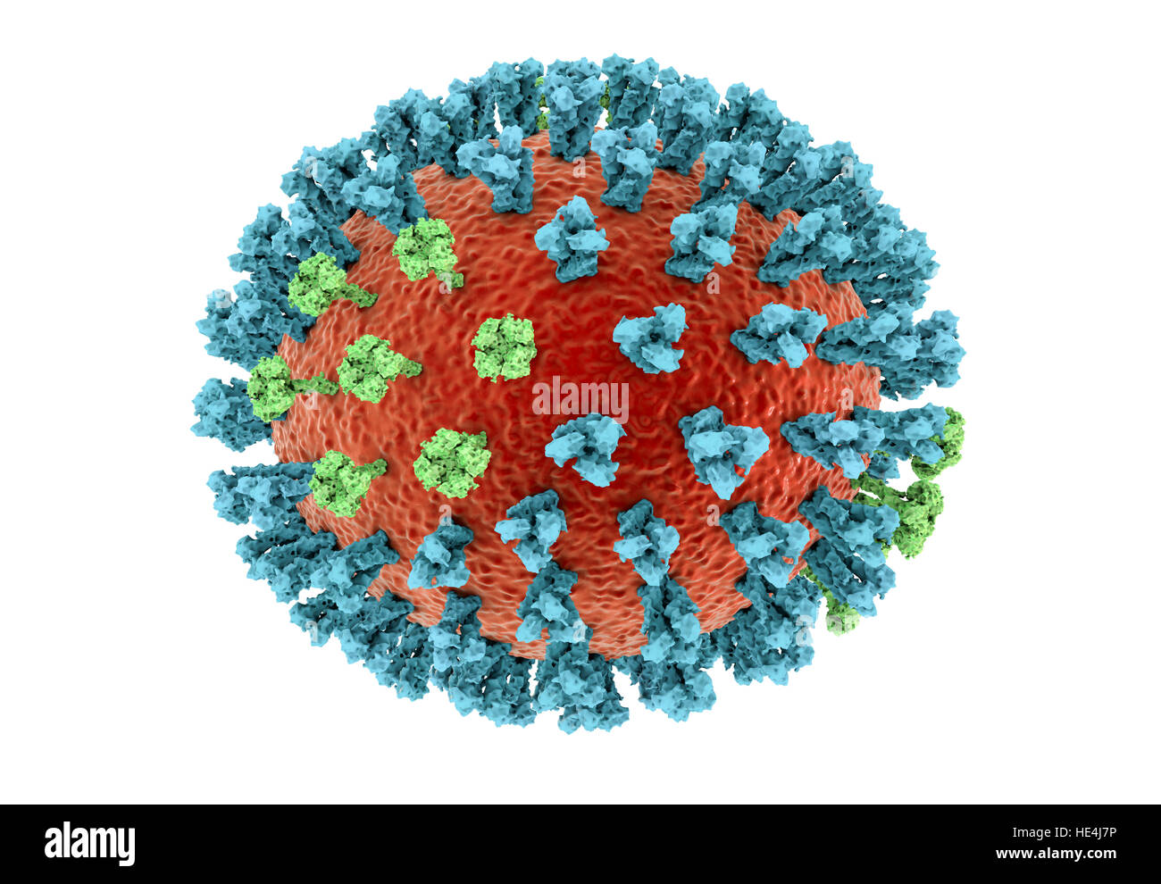 Bird flu virus. 3D illustration of an avian influenza H5N8 virus particle. Within the viral lipid envelope (orange) are two types of protein spike, hemagglutinin (H, blue) and neuraminidase (N, green), which determine the strain of virus. This strain of the virus has caused disease in wild birds and poultry in Europe and Asia since June 2016. Unusually, the virus causes mortality in wild birds, which are more often silent carriers. As of December 2016 no human cases of the disease have been reported, and risk of transmission to humans is thought to be low. Stock Photohttps://www.alamy.com/image-license-details/?v=1https://www.alamy.com/stock-photo-bird-flu-virus-3d-illustration-of-an-avian-influenza-h5n8-virus-particle-129179898.html
RFHE4J7P–Bird flu virus. 3D illustration of an avian influenza H5N8 virus particle. Within the viral lipid envelope (orange) are two types of protein spike, hemagglutinin (H, blue) and neuraminidase (N, green), which determine the strain of virus. This strain of the virus has caused disease in wild birds and poultry in Europe and Asia since June 2016. Unusually, the virus causes mortality in wild birds, which are more often silent carriers. As of December 2016 no human cases of the disease have been reported, and risk of transmission to humans is thought to be low.
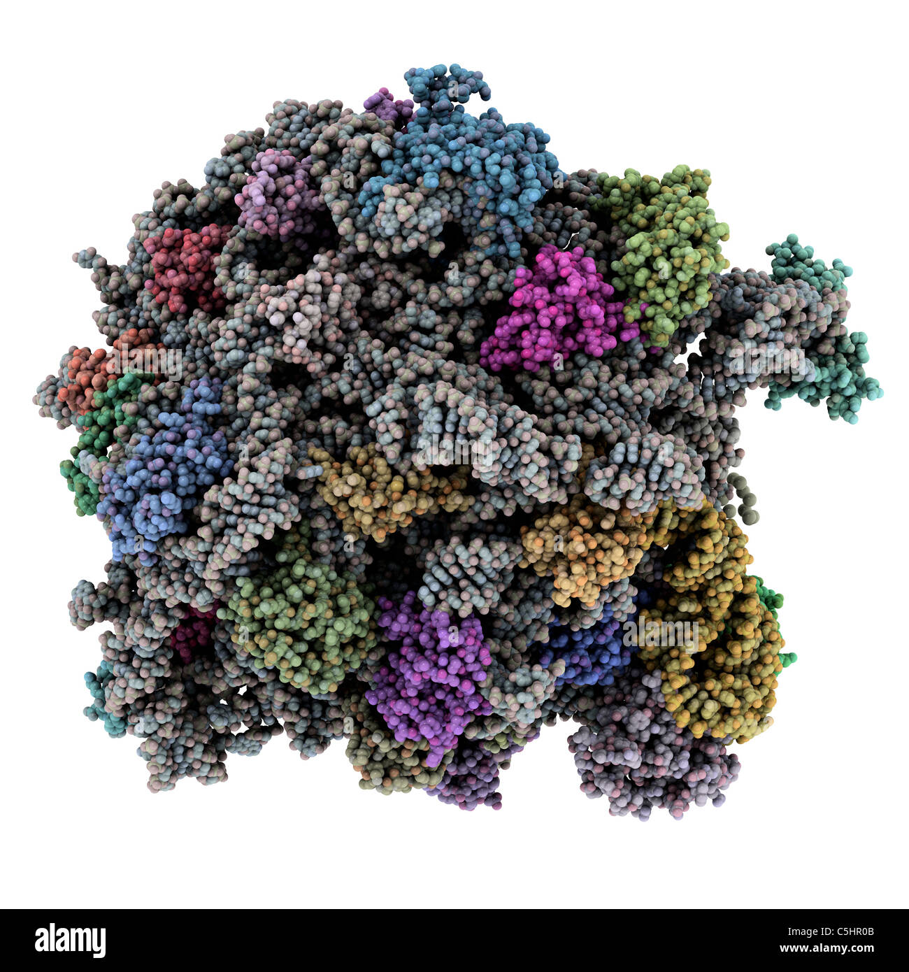 Ribosomal subunit, molecular model Stock Photohttps://www.alamy.com/image-license-details/?v=1https://www.alamy.com/stock-photo-ribosomal-subunit-molecular-model-37885243.html
RFC5HR0B–Ribosomal subunit, molecular model
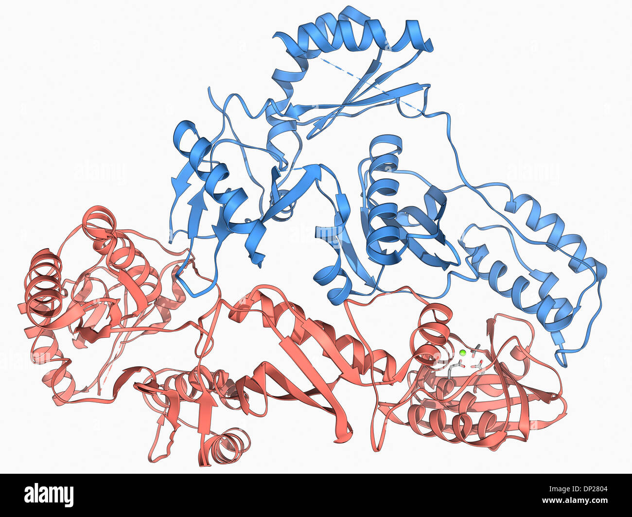 HIV reverse transcription enzyme Stock Photohttps://www.alamy.com/image-license-details/?v=1https://www.alamy.com/hiv-reverse-transcription-enzyme-image65203716.html
RFDP2804–HIV reverse transcription enzyme
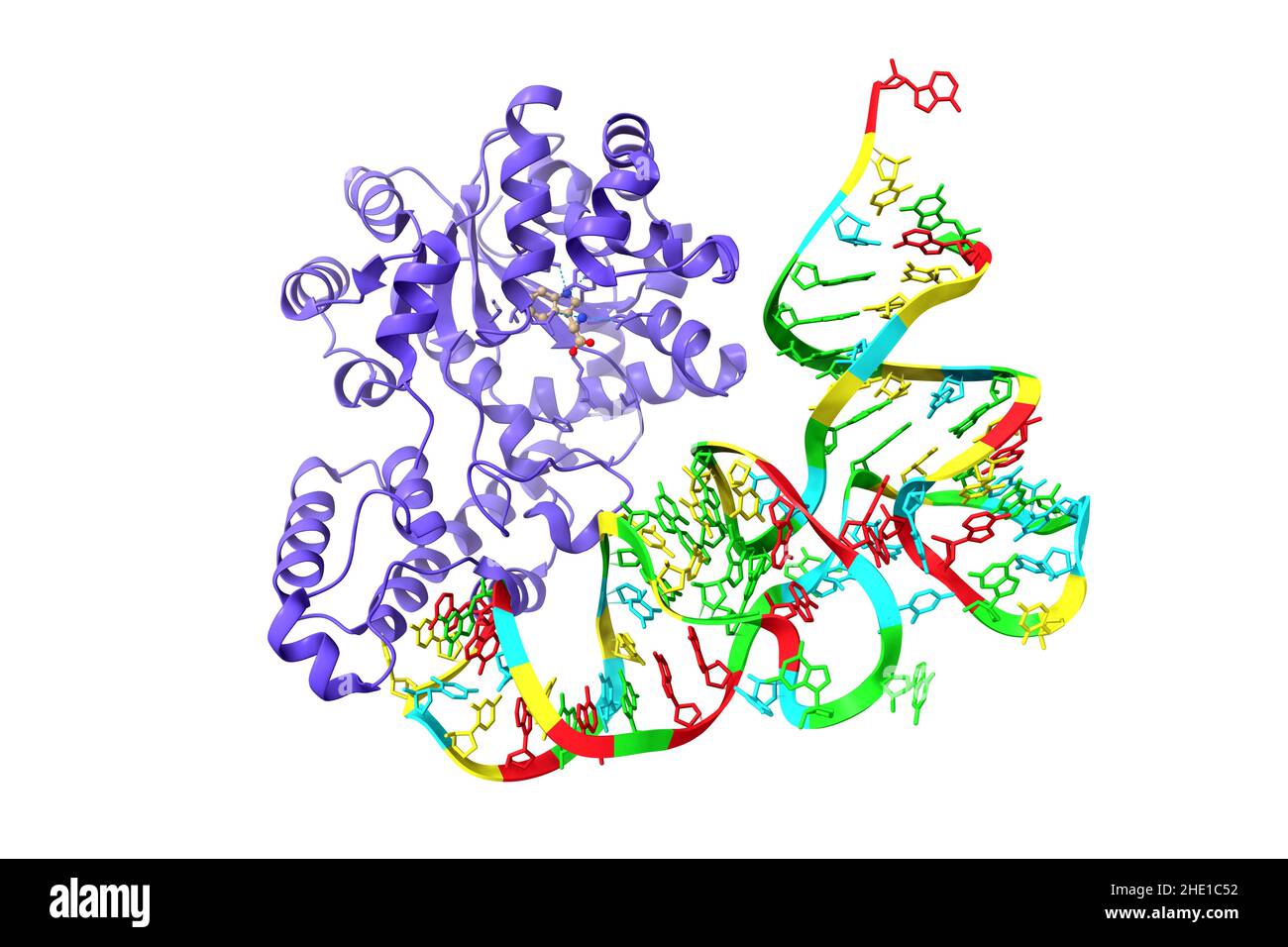 Structure of human tryptophanyl-tRNA synthetase in complex with tRNA(Trp). 3D cartoon model, PDB 2ake, white background Stock Photohttps://www.alamy.com/image-license-details/?v=1https://www.alamy.com/structure-of-human-tryptophanyl-trna-synthetase-in-complex-with-trnatrp-3d-cartoon-model-pdb-2ake-white-background-image456106254.html
RF2HE1C52–Structure of human tryptophanyl-tRNA synthetase in complex with tRNA(Trp). 3D cartoon model, PDB 2ake, white background
