Quick filters:
Hypochondrium Stock Photos and Images
 adult bald man sits on a sofa, holds his hand on the right side of the hypochondrium, a grimace of suffering on his face, the concept of liver disease Stock Photohttps://www.alamy.com/image-license-details/?v=1https://www.alamy.com/adult-bald-man-sits-on-a-sofa-holds-his-hand-on-the-right-side-of-the-hypochondrium-a-grimace-of-suffering-on-his-face-the-concept-of-liver-disease-image603584859.html
RF2X1YJJ3–adult bald man sits on a sofa, holds his hand on the right side of the hypochondrium, a grimace of suffering on his face, the concept of liver disease
 The doctor examines a girl patient with pain in the hypochondrium gall bladder. Gallbladder disease cholecystitis and biliary dyskinesia, cholecystect Stock Photohttps://www.alamy.com/image-license-details/?v=1https://www.alamy.com/the-doctor-examines-a-girl-patient-with-pain-in-the-hypochondrium-gall-bladder-gallbladder-disease-cholecystitis-and-biliary-dyskinesia-cholecystect-image268661353.html
RFWH2G89–The doctor examines a girl patient with pain in the hypochondrium gall bladder. Gallbladder disease cholecystitis and biliary dyskinesia, cholecystect
 young woman has pain in right hypochondrium, stomachache, gallbladder disease Stock Photohttps://www.alamy.com/image-license-details/?v=1https://www.alamy.com/young-woman-has-pain-in-right-hypochondrium-stomachache-gallbladder-disease-image240384245.html
RFRY2CFH–young woman has pain in right hypochondrium, stomachache, gallbladder disease
 Pain in the patient's right hypochondrium, the doctor palpates the men in the abdomen. Gallbladder disease concept, cholecitis and gallstones. Jaundic Stock Photohttps://www.alamy.com/image-license-details/?v=1https://www.alamy.com/pain-in-the-patients-right-hypochondrium-the-doctor-palpates-the-men-in-the-abdomen-gallbladder-disease-concept-cholecitis-and-gallstones-jaundic-image415458600.html
RF2F3WNK4–Pain in the patient's right hypochondrium, the doctor palpates the men in the abdomen. Gallbladder disease concept, cholecitis and gallstones. Jaundic
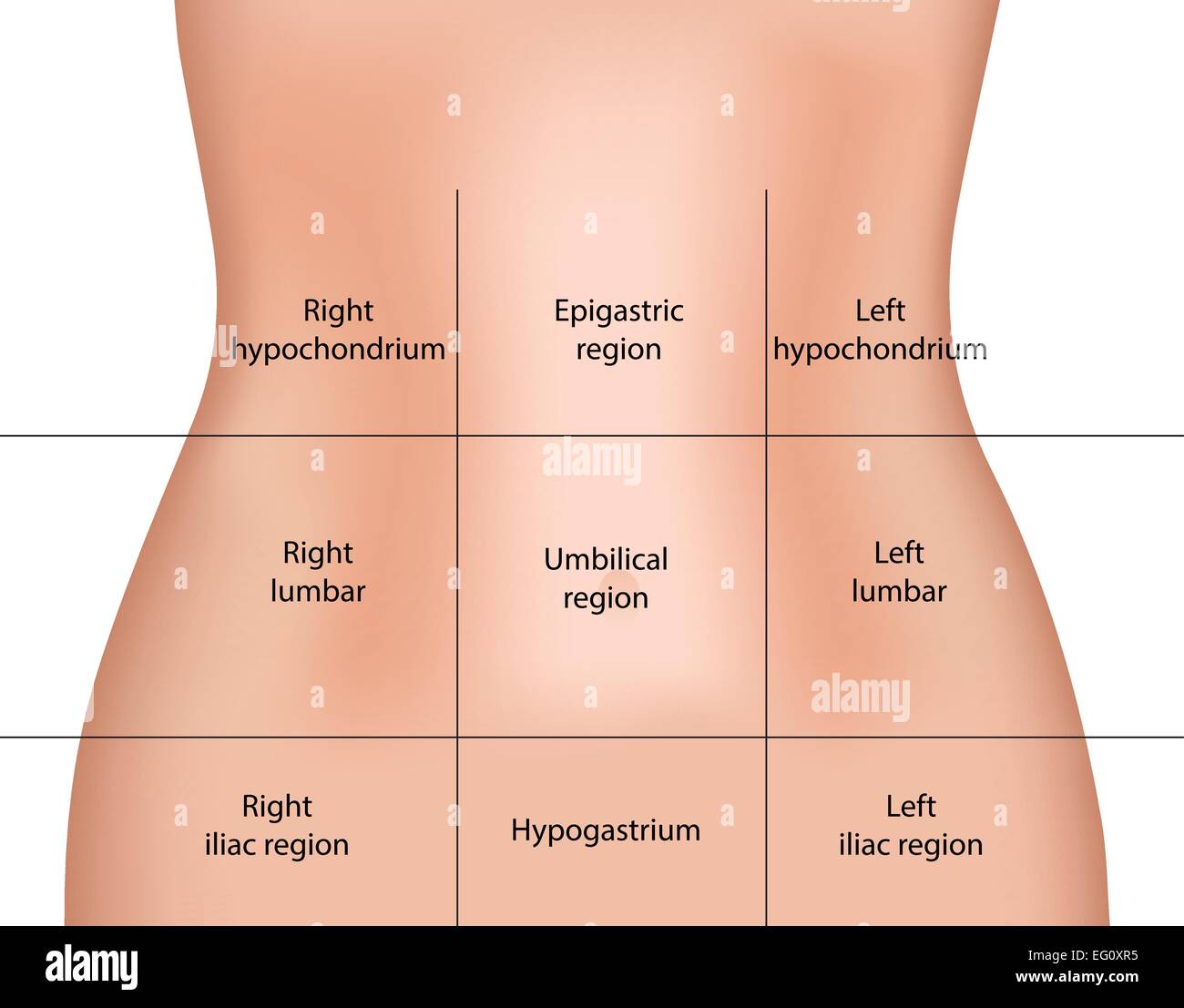 Abdominal Regions Stock Vectorhttps://www.alamy.com/image-license-details/?v=1https://www.alamy.com/stock-photo-abdominal-regions-78697001.html
RFEG0XR5–Abdominal Regions
 A little girl lies on the bed and feels unwell. Pain in the abdomen and hypochondrium. Diseases of the liver and gall bladder in children, cholecystit Stock Photohttps://www.alamy.com/image-license-details/?v=1https://www.alamy.com/a-little-girl-lies-on-the-bed-and-feels-unwell-pain-in-the-abdomen-and-hypochondrium-diseases-of-the-liver-and-gall-bladder-in-children-cholecystit-image594098755.html
RF2WEFF0K–A little girl lies on the bed and feels unwell. Pain in the abdomen and hypochondrium. Diseases of the liver and gall bladder in children, cholecystit
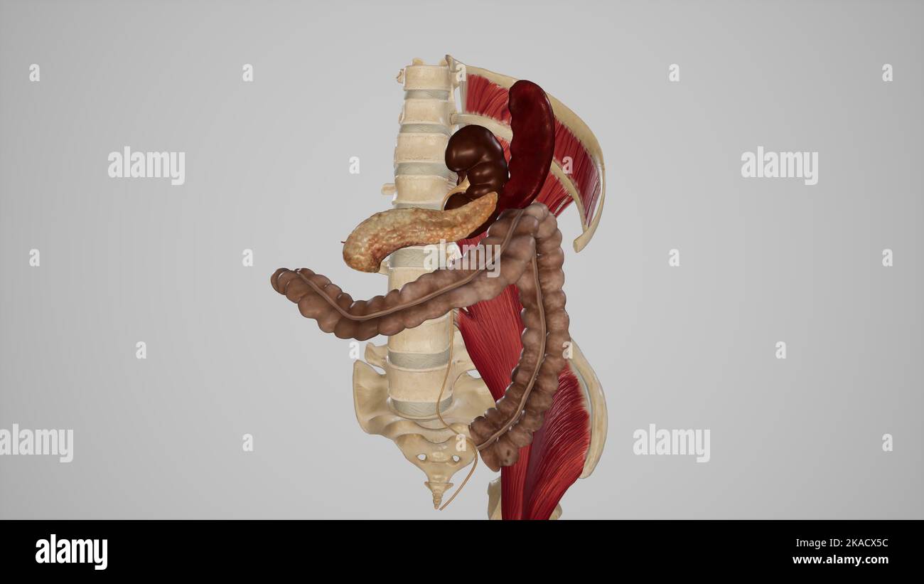 View of Relations of Left Colic Flexure (Splenic Flexure) Stock Photohttps://www.alamy.com/image-license-details/?v=1https://www.alamy.com/view-of-relations-of-left-colic-flexure-splenic-flexure-image488320824.html
RF2KACX5C–View of Relations of Left Colic Flexure (Splenic Flexure)
 Word hypochondria by wooden cubes Stock Photohttps://www.alamy.com/image-license-details/?v=1https://www.alamy.com/word-hypochondria-by-wooden-cubes-image417402106.html
RF2F728J2–Word hypochondria by wooden cubes
 Medical concept. On a human chest x-ray, a pen and strips of paper labeled - intercostal neuralgia Stock Photohttps://www.alamy.com/image-license-details/?v=1https://www.alamy.com/medical-concept-on-a-human-chest-x-ray-a-pen-and-strips-of-paper-labeled-intercostal-neuralgia-image509665525.html
RF2MH57G5–Medical concept. On a human chest x-ray, a pen and strips of paper labeled - intercostal neuralgia
 A Background filled with blisters with medicines. Stock Photohttps://www.alamy.com/image-license-details/?v=1https://www.alamy.com/a-background-filled-with-blisters-with-medicines-image593611328.html
RF2WDN98G–A Background filled with blisters with medicines.
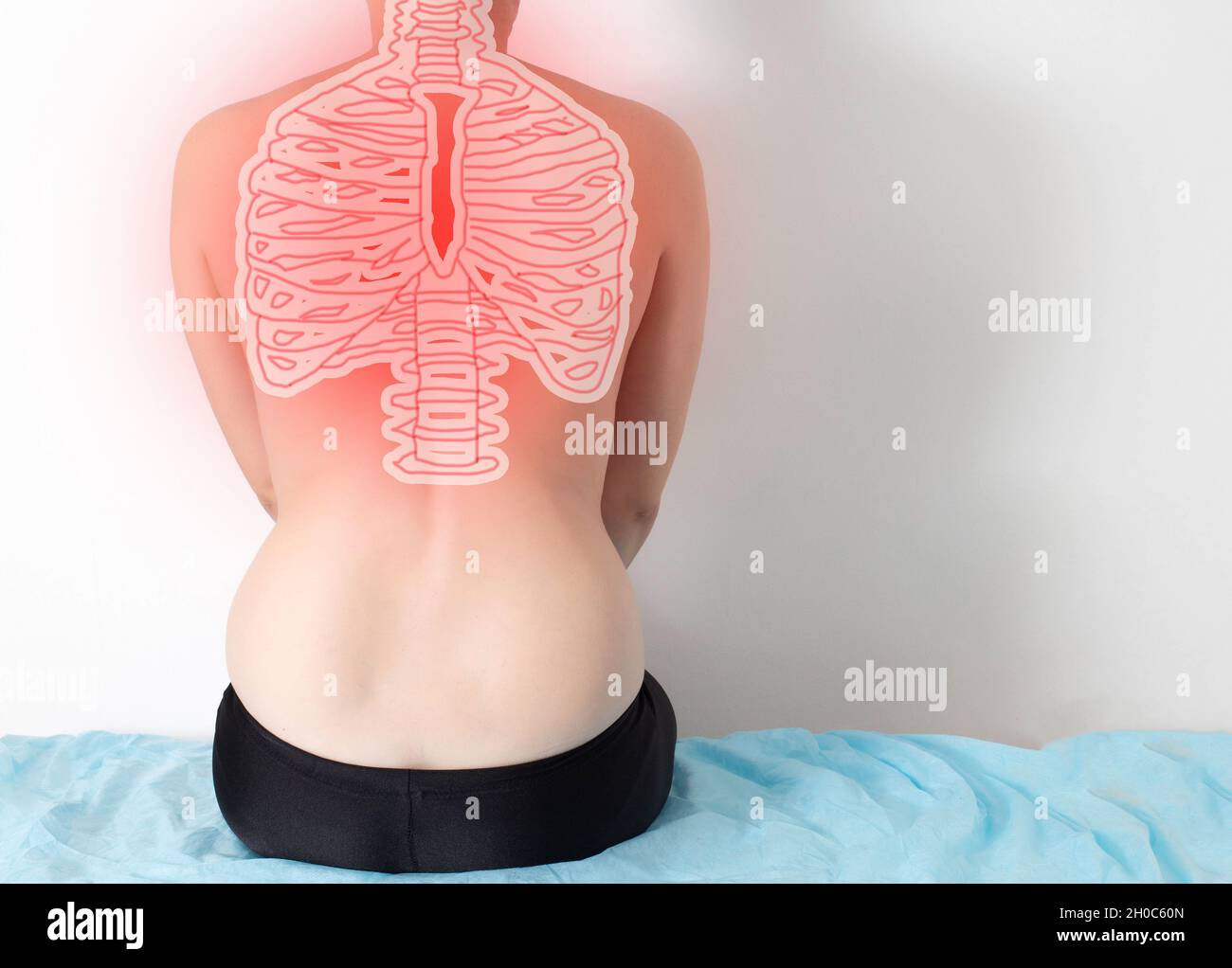 The girl sits with her back on a white background with pain in the ribs in the thoracic spine. Intercostal neuralgia, muscle and ligament diseases Stock Photohttps://www.alamy.com/image-license-details/?v=1https://www.alamy.com/the-girl-sits-with-her-back-on-a-white-background-with-pain-in-the-ribs-in-the-thoracic-spine-intercostal-neuralgia-muscle-and-ligament-diseases-image447737717.html
RF2H0C60N–The girl sits with her back on a white background with pain in the ribs in the thoracic spine. Intercostal neuralgia, muscle and ligament diseases
 A treatise on the practice of medicine, for the use of students and practitioners . are occasionally encoun-tered in which no characteristic symptoms existed ; the patient hasill-defined uneasiness in the right hypochondrium, disorders of diges-tion, and low spirits ; he emaciates progressively, is cachectic, andultimately dies. Again, cancer of the liver has a clinical history whichis merely the conclusion of a series of symptoms referable to cancer inanother organ, notably the stomach. The defined symptoms of hepaticcancer are apt to be obscured by some leading condition associatedwith it, a Stock Photohttps://www.alamy.com/image-license-details/?v=1https://www.alamy.com/a-treatise-on-the-practice-of-medicine-for-the-use-of-students-and-practitioners-are-occasionally-encoun-tered-in-which-no-characteristic-symptoms-existed-the-patient-hasill-defined-uneasiness-in-the-right-hypochondrium-disorders-of-diges-tion-and-low-spirits-he-emaciates-progressively-is-cachectic-andultimately-dies-again-cancer-of-the-liver-has-a-clinical-history-whichis-merely-the-conclusion-of-a-series-of-symptoms-referable-to-cancer-inanother-organ-notably-the-stomach-the-defined-symptoms-of-hepaticcancer-are-apt-to-be-obscured-by-some-leading-condition-associatedwith-it-a-image342645435.html
RM2AWCRMY–A treatise on the practice of medicine, for the use of students and practitioners . are occasionally encoun-tered in which no characteristic symptoms existed ; the patient hasill-defined uneasiness in the right hypochondrium, disorders of diges-tion, and low spirits ; he emaciates progressively, is cachectic, andultimately dies. Again, cancer of the liver has a clinical history whichis merely the conclusion of a series of symptoms referable to cancer inanother organ, notably the stomach. The defined symptoms of hepaticcancer are apt to be obscured by some leading condition associatedwith it, a
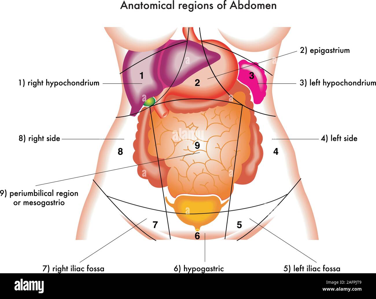 diagram of anatomical regions of abdomen Stock Vectorhttps://www.alamy.com/image-license-details/?v=1https://www.alamy.com/diagram-of-anatomical-regions-of-abdomen-image336714569.html
RF2AFPJT9–diagram of anatomical regions of abdomen
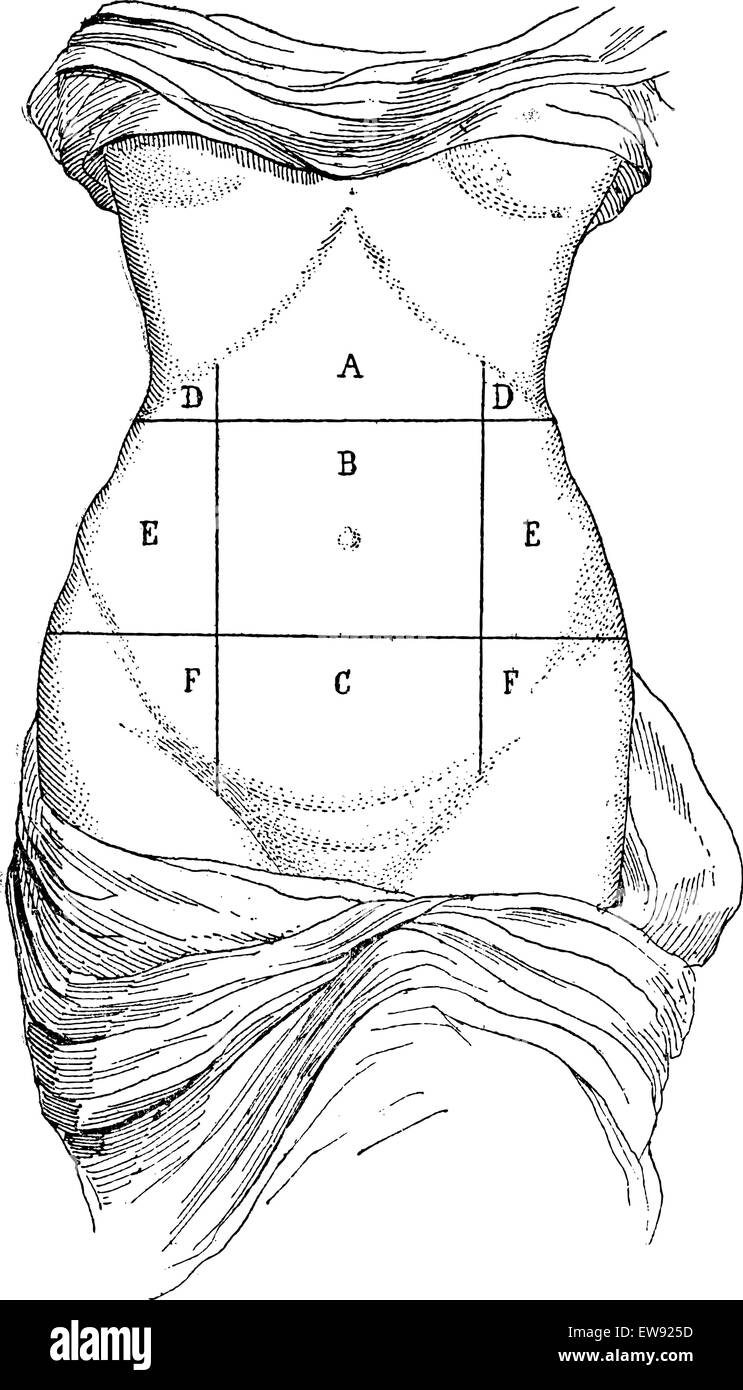 Abdomen and its subdivisions, vintage engraved illustration. Magasin Pittoresque 1875. Stock Vectorhttps://www.alamy.com/image-license-details/?v=1https://www.alamy.com/stock-photo-abdomen-and-its-subdivisions-vintage-engraved-illustration-magasin-84407161.html
RFEW925D–Abdomen and its subdivisions, vintage engraved illustration. Magasin Pittoresque 1875.
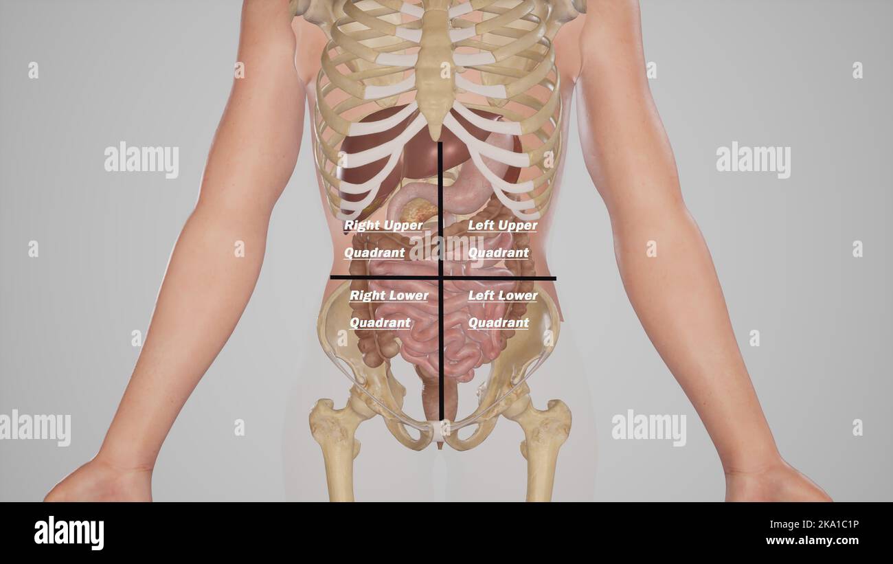
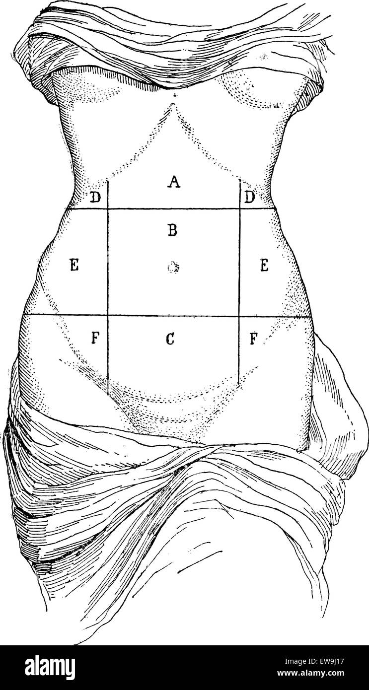
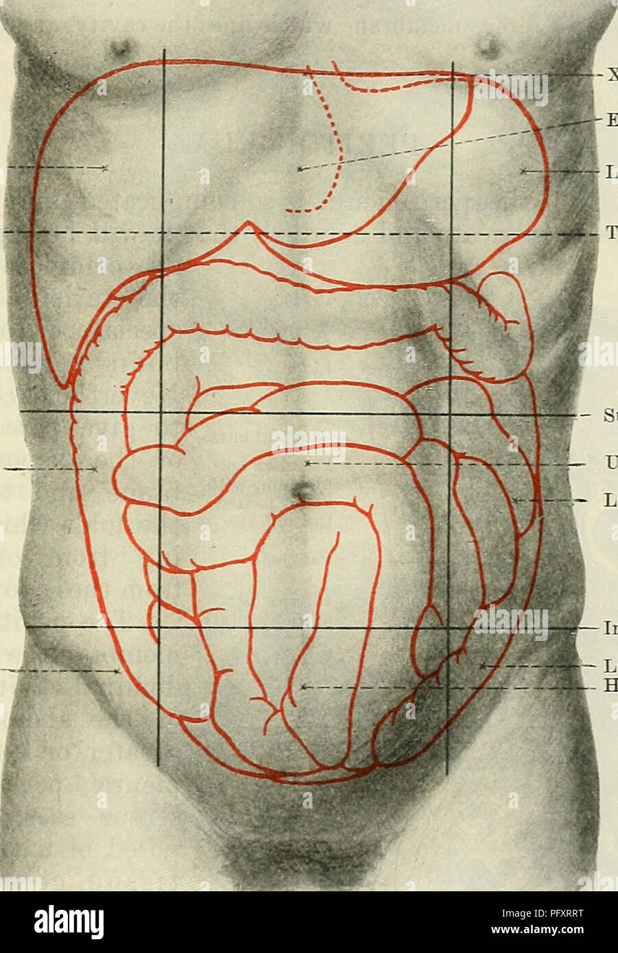 . Cunningham's Text-book of anatomy. Anatomy. THE ABDOMINAL CAVITY. 1159 perpendicular planes each of these is subdivided into three parts, a central and two lateral. Thus, in the upper zone, we get a hypochondriac region or hypochondrium on each side, and an epigastric region or epigastrium in the centre. Similarly, the umbilical zone is divided into right and left lumbar regions, with an umbilical region between. And the hypogastric zone has a hypogastric region or hypogastrium in the centre, with right and left iliac regions at the sides. In addition, the portion of the abdominal wall above Stock Photohttps://www.alamy.com/image-license-details/?v=1https://www.alamy.com/cunninghams-text-book-of-anatomy-anatomy-the-abdominal-cavity-1159-perpendicular-planes-each-of-these-is-subdivided-into-three-parts-a-central-and-two-lateral-thus-in-the-upper-zone-we-get-a-hypochondriac-region-or-hypochondrium-on-each-side-and-an-epigastric-region-or-epigastrium-in-the-centre-similarly-the-umbilical-zone-is-divided-into-right-and-left-lumbar-regions-with-an-umbilical-region-between-and-the-hypogastric-zone-has-a-hypogastric-region-or-hypogastrium-in-the-centre-with-right-and-left-iliac-regions-at-the-sides-in-addition-the-portion-of-the-abdominal-wall-above-image216333708.html
RMPFXRRT–. Cunningham's Text-book of anatomy. Anatomy. THE ABDOMINAL CAVITY. 1159 perpendicular planes each of these is subdivided into three parts, a central and two lateral. Thus, in the upper zone, we get a hypochondriac region or hypochondrium on each side, and an epigastric region or epigastrium in the centre. Similarly, the umbilical zone is divided into right and left lumbar regions, with an umbilical region between. And the hypogastric zone has a hypogastric region or hypogastrium in the centre, with right and left iliac regions at the sides. In addition, the portion of the abdominal wall above
 Diseases of cattle, sheep, goats Diseases of cattle, sheep, goats and swine diseasesofcattle00mous Year: 1920 EXUDATIVE PERICARDITIS DUE TO FOREIGN BODIES. 387 the hypochondrium becomes attached to the sternum. (3.) Point at which the external mammary vein penetrates the abdominal wall (Fig. 178). Lines uniting these three points enclose a right-angled triangle, which the operator must imagine to be bisected by a third line. The incision, which should be about 8 inches in length, follows this bisecting line at an equal distance between the white line and the circle of the hypochondrium, to a Stock Photohttps://www.alamy.com/image-license-details/?v=1https://www.alamy.com/diseases-of-cattle-sheep-goats-diseases-of-cattle-sheep-goats-and-swine-diseasesofcattle00mous-year-1920-exudative-pericarditis-due-to-foreign-bodies-387-the-hypochondrium-becomes-attached-to-the-sternum-3-point-at-which-the-external-mammary-vein-penetrates-the-abdominal-wall-fig-178-lines-uniting-these-three-points-enclose-a-right-angled-triangle-which-the-operator-must-imagine-to-be-bisected-by-a-third-line-the-incision-which-should-be-about-8-inches-in-length-follows-this-bisecting-line-at-an-equal-distance-between-the-white-line-and-the-circle-of-the-hypochondrium-to-a-image241953530.html
RMT1HX5E–Diseases of cattle, sheep, goats Diseases of cattle, sheep, goats and swine diseasesofcattle00mous Year: 1920 EXUDATIVE PERICARDITIS DUE TO FOREIGN BODIES. 387 the hypochondrium becomes attached to the sternum. (3.) Point at which the external mammary vein penetrates the abdominal wall (Fig. 178). Lines uniting these three points enclose a right-angled triangle, which the operator must imagine to be bisected by a third line. The incision, which should be about 8 inches in length, follows this bisecting line at an equal distance between the white line and the circle of the hypochondrium, to a
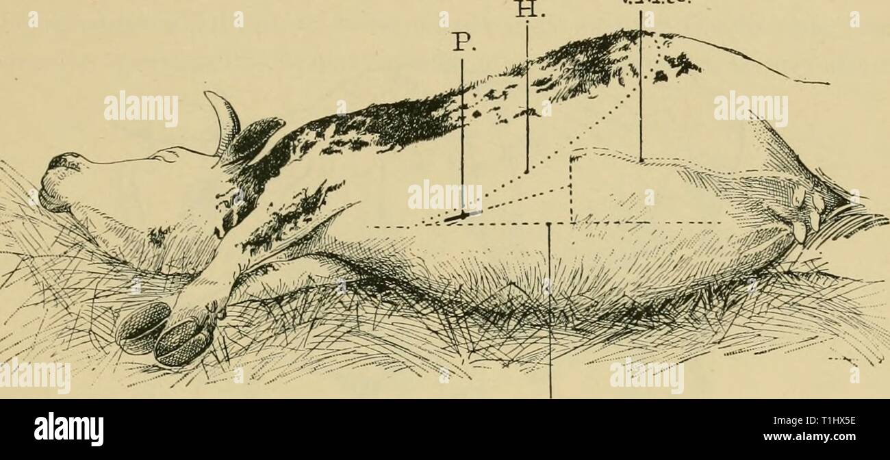
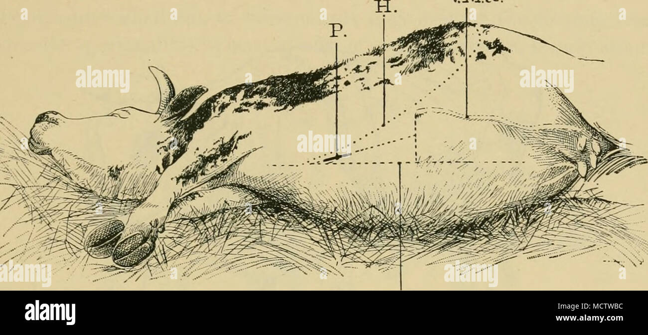 . Fig. 178.—Seat of operation for puncturing the pericardium by way of the ensiforni cartilage. L B, White line; H, hne of the hypochondrium; V. M.a., anterior mammary vein ; P, point where the pericardium is punctured through the incision. pectoral muscles attached to the neck of the ensiforni cartilage can then be divided with the bistoury. The area of operation is thus uncovered. Second stage. The second phase comprises incision of the tissues opposite the neck of the ensiform cartilage, about 8 inches in front of the base of the triangle and at equal distances from the points Nos. 1 and 2; Stock Photohttps://www.alamy.com/image-license-details/?v=1https://www.alamy.com/fig-178seat-of-operation-for-puncturing-the-pericardium-by-way-of-the-ensiforni-cartilage-l-b-white-line-h-hne-of-the-hypochondrium-v-ma-anterior-mammary-vein-p-point-where-the-pericardium-is-punctured-through-the-incision-pectoral-muscles-attached-to-the-neck-of-the-ensiforni-cartilage-can-then-be-divided-with-the-bistoury-the-area-of-operation-is-thus-uncovered-second-stage-the-second-phase-comprises-incision-of-the-tissues-opposite-the-neck-of-the-ensiform-cartilage-about-8-inches-in-front-of-the-base-of-the-triangle-and-at-equal-distances-from-the-points-nos-1-and-2-image180026320.html
RMMCTWBC–. Fig. 178.—Seat of operation for puncturing the pericardium by way of the ensiforni cartilage. L B, White line; H, hne of the hypochondrium; V. M.a., anterior mammary vein ; P, point where the pericardium is punctured through the incision. pectoral muscles attached to the neck of the ensiforni cartilage can then be divided with the bistoury. The area of operation is thus uncovered. Second stage. The second phase comprises incision of the tissues opposite the neck of the ensiform cartilage, about 8 inches in front of the base of the triangle and at equal distances from the points Nos. 1 and 2;
 young woman has pain in right hypochondrium, stomachache, gallbladder disease Stock Photohttps://www.alamy.com/image-license-details/?v=1https://www.alamy.com/young-woman-has-pain-in-right-hypochondrium-stomachache-gallbladder-disease-image240385125.html
RFRY2DK1–young woman has pain in right hypochondrium, stomachache, gallbladder disease
 Abdominal Regions Stock Photohttps://www.alamy.com/image-license-details/?v=1https://www.alamy.com/abdominal-regions-image488068274.html
RF2KA1C1P–Abdominal Regions
 A treatise on the practice of medicine, for the use of students and practitioners . are occasionally encoun-tered in which no characteristic symptoms existed ; the patient hasill-defined uneasiness in the right hypochondrium, disorders of diges-tion, and low spirits ; he emaciates progressively, is cachectic, andultimately dies. Again, cancer of the liver has a clinical history whichis merely the conclusion of a series of symptoms referable to cancer inanother organ, notably the stomach. The defined symptoms of hepaticcancer are apt to be obscured by some leading condition associatedwith it, a Stock Photohttps://www.alamy.com/image-license-details/?v=1https://www.alamy.com/a-treatise-on-the-practice-of-medicine-for-the-use-of-students-and-practitioners-are-occasionally-encoun-tered-in-which-no-characteristic-symptoms-existed-the-patient-hasill-defined-uneasiness-in-the-right-hypochondrium-disorders-of-diges-tion-and-low-spirits-he-emaciates-progressively-is-cachectic-andultimately-dies-again-cancer-of-the-liver-has-a-clinical-history-whichis-merely-the-conclusion-of-a-series-of-symptoms-referable-to-cancer-inanother-organ-notably-the-stomach-the-defined-symptoms-of-hepaticcancer-are-apt-to-be-obscured-by-some-leading-condition-associatedwith-it-a-image342646155.html
RM2AWCTJK–A treatise on the practice of medicine, for the use of students and practitioners . are occasionally encoun-tered in which no characteristic symptoms existed ; the patient hasill-defined uneasiness in the right hypochondrium, disorders of diges-tion, and low spirits ; he emaciates progressively, is cachectic, andultimately dies. Again, cancer of the liver has a clinical history whichis merely the conclusion of a series of symptoms referable to cancer inanother organ, notably the stomach. The defined symptoms of hepaticcancer are apt to be obscured by some leading condition associatedwith it, a
 Abdomen and its subdivisions, vintage engraved illustration. Magasin Pittoresque 1875. Stock Vectorhttps://www.alamy.com/image-license-details/?v=1https://www.alamy.com/stock-photo-abdomen-and-its-subdivisions-vintage-engraved-illustration-magasin-84419587.html
RFEW9J17–Abdomen and its subdivisions, vintage engraved illustration. Magasin Pittoresque 1875.
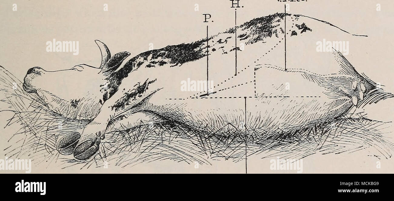 . L3. Fig. 178.—Seat of operation for puncturing the pericardium by way of tlie ensiform cartilage. L B, AVhite line; H, line of the hypochondrium; Y.M.a., anterior mammary vein; P, point where the pericardium is punctured through the incision. pectoral muscles attached to the neck of the ensiform cartilage can then be divided with the bistoury. The area of operation is thus uncovered. Second stage. The second phase comprises incision of the tissues opposite the neck of the ensiform cartilage, about 8 inches in front of the base of the triangle and at equal distances from the points Nos. 1 and Stock Photohttps://www.alamy.com/image-license-details/?v=1https://www.alamy.com/l3-fig-178seat-of-operation-for-puncturing-the-pericardium-by-way-of-tlie-ensiform-cartilage-l-b-avhite-line-h-line-of-the-hypochondrium-yma-anterior-mammary-vein-p-point-where-the-pericardium-is-punctured-through-the-incision-pectoral-muscles-attached-to-the-neck-of-the-ensiform-cartilage-can-then-be-divided-with-the-bistoury-the-area-of-operation-is-thus-uncovered-second-stage-the-second-phase-comprises-incision-of-the-tissues-opposite-the-neck-of-the-ensiform-cartilage-about-8-inches-in-front-of-the-base-of-the-triangle-and-at-equal-distances-from-the-points-nos-1-and-image179905721.html
RMMCKBG9–. L3. Fig. 178.—Seat of operation for puncturing the pericardium by way of tlie ensiform cartilage. L B, AVhite line; H, line of the hypochondrium; Y.M.a., anterior mammary vein; P, point where the pericardium is punctured through the incision. pectoral muscles attached to the neck of the ensiform cartilage can then be divided with the bistoury. The area of operation is thus uncovered. Second stage. The second phase comprises incision of the tissues opposite the neck of the ensiform cartilage, about 8 inches in front of the base of the triangle and at equal distances from the points Nos. 1 and
 young woman has pain in right hypochondrium, stomachache, gallbladder disease Stock Photohttps://www.alamy.com/image-license-details/?v=1https://www.alamy.com/young-woman-has-pain-in-right-hypochondrium-stomachache-gallbladder-disease-image240386634.html
RFRY2FGX–young woman has pain in right hypochondrium, stomachache, gallbladder disease
 A treatise on the practice of medicine, for the use of students and practitioners . right hypochondrium, and some disorders of digestion ; if it hap-pen to be near the hilus of the organ, the portal vein and the com-mon or the hepatic duct may be pressedupon, causing ascites and jaundice ; ifnear or at the upper convex surface ofthe right lobe, the diaphragm will bepushed up, and a dry cough and dysp-noea will be the result. The degreeof enlargement is necessarily various.The tumor may fill in the whole spacefrom the inferior border of the secondrib to the pelvis, displacing the tho-acic and a Stock Photohttps://www.alamy.com/image-license-details/?v=1https://www.alamy.com/a-treatise-on-the-practice-of-medicine-for-the-use-of-students-and-practitioners-right-hypochondrium-and-some-disorders-of-digestion-if-it-hap-pen-to-be-near-the-hilus-of-the-organ-the-portal-vein-and-the-com-mon-or-the-hepatic-duct-may-be-pressedupon-causing-ascites-and-jaundice-ifnear-or-at-the-upper-convex-surface-ofthe-right-lobe-the-diaphragm-will-bepushed-up-and-a-dry-cough-and-dysp-noea-will-be-the-result-the-degreeof-enlargement-is-necessarily-variousthe-tumor-may-fill-in-the-whole-spacefrom-the-inferior-border-of-the-secondrib-to-the-pelvis-displacing-the-tho-acic-and-a-image340311220.html
RM2ANJEC4–A treatise on the practice of medicine, for the use of students and practitioners . right hypochondrium, and some disorders of digestion ; if it hap-pen to be near the hilus of the organ, the portal vein and the com-mon or the hepatic duct may be pressedupon, causing ascites and jaundice ; ifnear or at the upper convex surface ofthe right lobe, the diaphragm will bepushed up, and a dry cough and dysp-noea will be the result. The degreeof enlargement is necessarily various.The tumor may fill in the whole spacefrom the inferior border of the secondrib to the pelvis, displacing the tho-acic and a
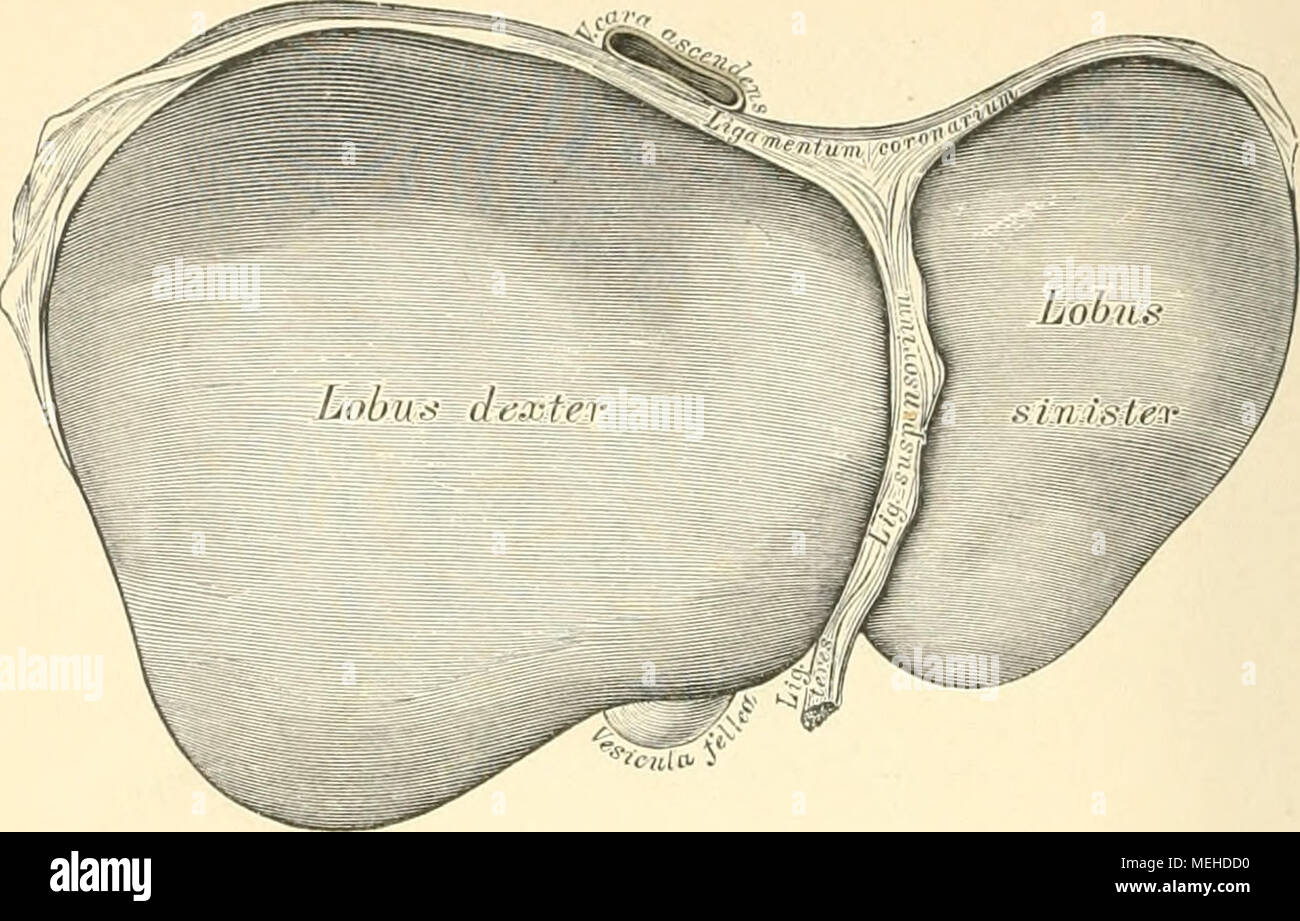 . Die descriptive und topographische Anatomie des Menschen . 401. Die Leber, Hepar. Ansicht von oben. Die Leber liegt im rechten Hypochondrium und erstreckt sich bis hinüber in das linke. Ihr vorderer scharfer Rand besitzt einen Einschnitt zur Aufnahme des Ligamentum Suspensorium; ihr hinterer, stumpfer Rand steht höher als der vordere; der rechte Rand ist gleichfalls stumpf, der linke, zuge- schärfte liegt vor der Cardia des Magens. Die obere Fläche ist entsprechend der Wölbung des Diaphragma convex und etwas nach vorne goneigt; durch das Lig. Suspensorium ist die Grenze zwischen dem grossen Stock Photohttps://www.alamy.com/image-license-details/?v=1https://www.alamy.com/die-descriptive-und-topographische-anatomie-des-menschen-401-die-leber-hepar-ansicht-von-oben-die-leber-liegt-im-rechten-hypochondrium-und-erstreckt-sich-bis-hinber-in-das-linke-ihr-vorderer-scharfer-rand-besitzt-einen-einschnitt-zur-aufnahme-des-ligamentum-suspensorium-ihr-hinterer-stumpfer-rand-steht-hher-als-der-vordere-der-rechte-rand-ist-gleichfalls-stumpf-der-linke-zuge-schrfte-liegt-vor-der-cardia-des-magens-die-obere-flche-ist-entsprechend-der-wlbung-des-diaphragma-convex-und-etwas-nach-vorne-goneigt-durch-das-lig-suspensorium-ist-die-grenze-zwischen-dem-grossen-image181092604.html
RMMEHDD0–. Die descriptive und topographische Anatomie des Menschen . 401. Die Leber, Hepar. Ansicht von oben. Die Leber liegt im rechten Hypochondrium und erstreckt sich bis hinüber in das linke. Ihr vorderer scharfer Rand besitzt einen Einschnitt zur Aufnahme des Ligamentum Suspensorium; ihr hinterer, stumpfer Rand steht höher als der vordere; der rechte Rand ist gleichfalls stumpf, der linke, zuge- schärfte liegt vor der Cardia des Magens. Die obere Fläche ist entsprechend der Wölbung des Diaphragma convex und etwas nach vorne goneigt; durch das Lig. Suspensorium ist die Grenze zwischen dem grossen
 young woman has pain in right hypochondrium, stomachache, gallbladder disease Stock Photohttps://www.alamy.com/image-license-details/?v=1https://www.alamy.com/young-woman-has-pain-in-right-hypochondrium-stomachache-gallbladder-disease-image240386563.html
RFRY2FEB–young woman has pain in right hypochondrium, stomachache, gallbladder disease
 . Medical diagnosis for the student and practitioner. left hypochondrium.* With such a wide area of distribution for cardiac pain and granting thelogical assumption that various sensations of discomfort may substitute painin cardiac overstrain, toxemia and fatigue, as in like conditions affecting anyother muscle, we see the possible relationships not alone of precordial dis-tress, but of the many axillary, neck,shoulder, thoracic and upper ab-dominal pains or uncomfortable sen-sations, such as are usually, andrightly given the more obvious in-terpretation suggested by their sur-face relation t Stock Photohttps://www.alamy.com/image-license-details/?v=1https://www.alamy.com/medical-diagnosis-for-the-student-and-practitioner-left-hypochondrium-with-such-a-wide-area-of-distribution-for-cardiac-pain-and-granting-thelogical-assumption-that-various-sensations-of-discomfort-may-substitute-painin-cardiac-overstrain-toxemia-and-fatigue-as-in-like-conditions-affecting-anyother-muscle-we-see-the-possible-relationships-not-alone-of-precordial-dis-tress-but-of-the-many-axillary-neckshoulder-thoracic-and-upper-ab-dominal-pains-or-uncomfortable-sen-sations-such-as-are-usually-andrightly-given-the-more-obvious-in-terpretation-suggested-by-their-sur-face-relation-t-image336832415.html
RM2AG0153–. Medical diagnosis for the student and practitioner. left hypochondrium.* With such a wide area of distribution for cardiac pain and granting thelogical assumption that various sensations of discomfort may substitute painin cardiac overstrain, toxemia and fatigue, as in like conditions affecting anyother muscle, we see the possible relationships not alone of precordial dis-tress, but of the many axillary, neck,shoulder, thoracic and upper ab-dominal pains or uncomfortable sen-sations, such as are usually, andrightly given the more obvious in-terpretation suggested by their sur-face relation t
 young woman has pain in right hypochondrium, stomachache, gallbladder disease Stock Photohttps://www.alamy.com/image-license-details/?v=1https://www.alamy.com/young-woman-has-pain-in-right-hypochondrium-stomachache-gallbladder-disease-image240385161.html
RFRY2DM9–young woman has pain in right hypochondrium, stomachache, gallbladder disease
 Geriatrics : the diseases of old age and their treatment, including physiological old age, home and institutional care, and medico-legal relations . er eating. This isdue to dilatation and atrophic catarrh of the stomach. The intestinal symptoms are constipation, flatulence, met-eorism, occasional watery or bloody diarrhea, beginning withoutapparent cause, lasting for a few hours there followed by con-stipation, sharp pains in the right hypochondrium, neuralgic butnot colicky, occurring spasmodically and frequently at night.The mesenteric arteries are occasionally atheromatous but theygive no Stock Photohttps://www.alamy.com/image-license-details/?v=1https://www.alamy.com/geriatrics-the-diseases-of-old-age-and-their-treatment-including-physiological-old-age-home-and-institutional-care-and-medico-legal-relations-er-eating-this-isdue-to-dilatation-and-atrophic-catarrh-of-the-stomach-the-intestinal-symptoms-are-constipation-flatulence-met-eorism-occasional-watery-or-bloody-diarrhea-beginning-withoutapparent-cause-lasting-for-a-few-hours-there-followed-by-con-stipation-sharp-pains-in-the-right-hypochondrium-neuralgic-butnot-colicky-occurring-spasmodically-and-frequently-at-nightthe-mesenteric-arteries-are-occasionally-atheromatous-but-theygive-no-image342781879.html
RM2AWK1NY–Geriatrics : the diseases of old age and their treatment, including physiological old age, home and institutional care, and medico-legal relations . er eating. This isdue to dilatation and atrophic catarrh of the stomach. The intestinal symptoms are constipation, flatulence, met-eorism, occasional watery or bloody diarrhea, beginning withoutapparent cause, lasting for a few hours there followed by con-stipation, sharp pains in the right hypochondrium, neuralgic butnot colicky, occurring spasmodically and frequently at night.The mesenteric arteries are occasionally atheromatous but theygive no
 young woman has pain in right hypochondrium, stomachache, gallbladder disease Stock Photohttps://www.alamy.com/image-license-details/?v=1https://www.alamy.com/young-woman-has-pain-in-right-hypochondrium-stomachache-gallbladder-disease-image240384230.html
RFRY2CF2–young woman has pain in right hypochondrium, stomachache, gallbladder disease
 The diseases of women : a handbook for students and practitioners . Pelvic cellulitis.Ovarian tumor.Tubal pregnancy. Fig. 94.—Diagram to indicate the positions of abdominal swellings (A. E. G.). ment of kidney from hydronephrosis or ne.w growth—mov-able kidney; enlarged spleen ; omental tumors ; malignantdisease of the intestines; ectopic gestation (Figs. 94, 95). We must begin by excluding the first two conditions:palpation and percussion will generally suffice, especiallyunder an anaesthetic. Ascites is indicated by the absence ofdefinite limits, the dulness in the flanks and hypochondrium D Stock Photohttps://www.alamy.com/image-license-details/?v=1https://www.alamy.com/the-diseases-of-women-a-handbook-for-students-and-practitioners-pelvic-cellulitisovarian-tumortubal-pregnancy-fig-94diagram-to-indicate-the-positions-of-abdominal-swellings-a-e-g-ment-of-kidney-from-hydronephrosis-or-new-growthmov-able-kidney-enlarged-spleen-omental-tumors-malignantdisease-of-the-intestines-ectopic-gestation-figs-94-95-we-must-begin-by-excluding-the-first-two-conditionspalpation-and-percussion-will-generally-suffice-especiallyunder-an-anaesthetic-ascites-is-indicated-by-the-absence-ofdefinite-limits-the-dulness-in-the-flanks-and-hypochondrium-d-image342880857.html
RM2AWRG0W–The diseases of women : a handbook for students and practitioners . Pelvic cellulitis.Ovarian tumor.Tubal pregnancy. Fig. 94.—Diagram to indicate the positions of abdominal swellings (A. E. G.). ment of kidney from hydronephrosis or ne.w growth—mov-able kidney; enlarged spleen ; omental tumors ; malignantdisease of the intestines; ectopic gestation (Figs. 94, 95). We must begin by excluding the first two conditions:palpation and percussion will generally suffice, especiallyunder an anaesthetic. Ascites is indicated by the absence ofdefinite limits, the dulness in the flanks and hypochondrium D
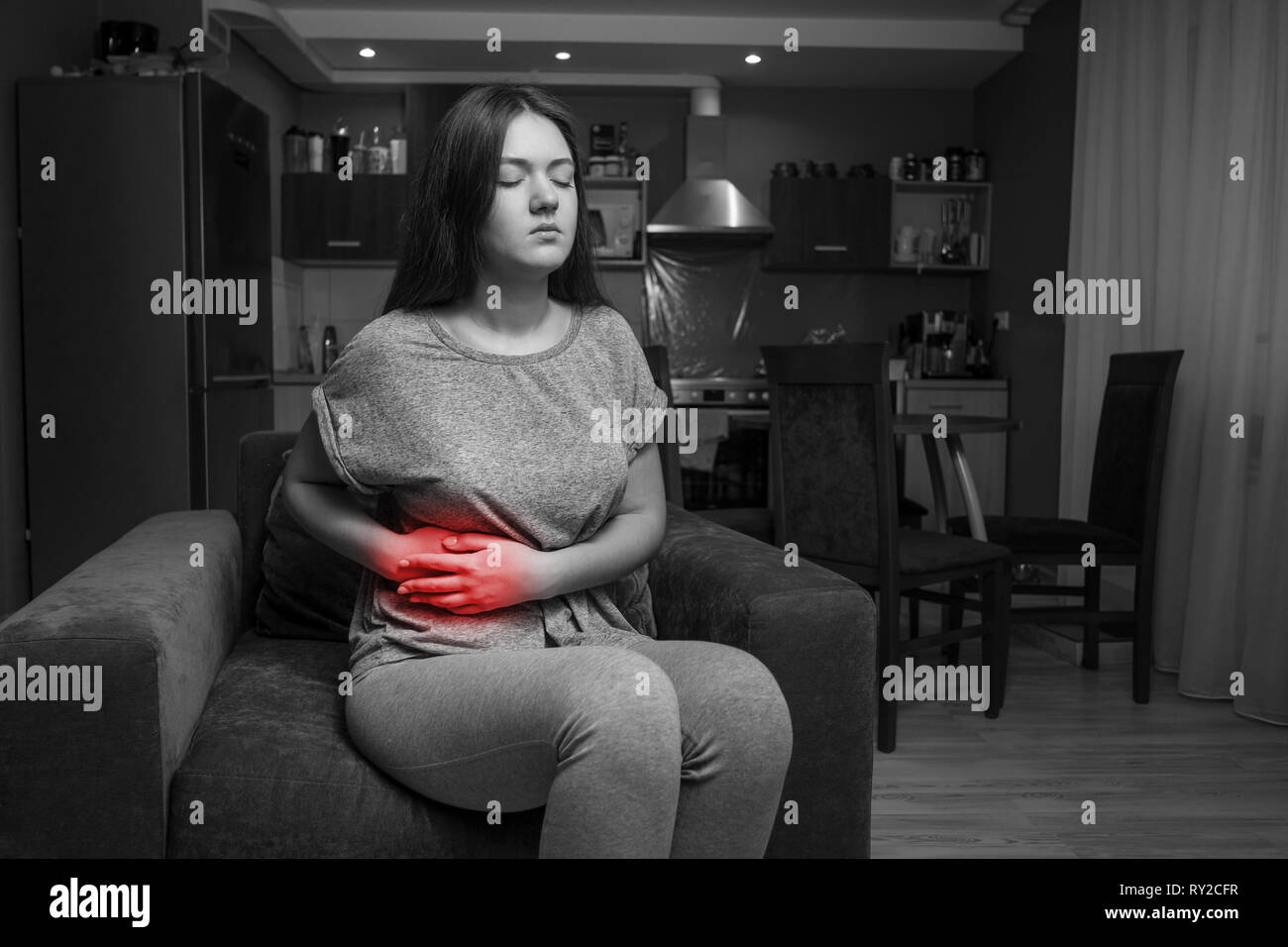 young woman has pain in right hypochondrium, stomachache, black and white photo, red accent, gallbladder disease Stock Photohttps://www.alamy.com/image-license-details/?v=1https://www.alamy.com/young-woman-has-pain-in-right-hypochondrium-stomachache-black-and-white-photo-red-accent-gallbladder-disease-image240384251.html
RFRY2CFR–young woman has pain in right hypochondrium, stomachache, black and white photo, red accent, gallbladder disease
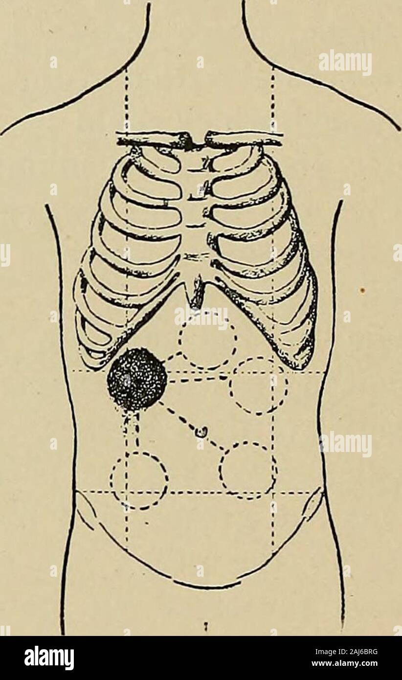 Cancer of the stomach : a clinical study . was thefollowing :— No. 107.—R. B. (hosp. no. 15,236), male; was admitted Feb. 15th,1896. Occupying left hypochondrium, and extending into left lumbar,and above into the epigastric regions and below into the left upper 64 CANCER OF THE STOMACH quadrant of the umbilical region, was a firm, solid mass, irregularon the surface. Below and to the left the outline was rounded andeasily defined; to the right the border was indistinct. Above, in theparasternal line, it passed below the costal margin. On deep inspirationthe mass descended so that the fingers c Stock Photohttps://www.alamy.com/image-license-details/?v=1https://www.alamy.com/cancer-of-the-stomach-a-clinical-study-was-thefollowing-no-107r-b-hosp-no-15236-male-was-admitted-feb-15th1896-occupying-left-hypochondrium-and-extending-into-left-lumbarand-above-into-the-epigastric-regions-and-below-into-the-left-upper-64-cancer-of-the-stomach-quadrant-of-the-umbilical-region-was-a-firm-solid-mass-irregularon-the-surface-below-and-to-the-left-the-outline-was-rounded-andeasily-defined-to-the-right-the-border-was-indistinct-above-in-theparasternal-line-it-passed-below-the-costal-margin-on-deep-inspirationthe-mass-descended-so-that-the-fingers-c-image338201796.html
RM2AJ6BRG–Cancer of the stomach : a clinical study . was thefollowing :— No. 107.—R. B. (hosp. no. 15,236), male; was admitted Feb. 15th,1896. Occupying left hypochondrium, and extending into left lumbar,and above into the epigastric regions and below into the left upper 64 CANCER OF THE STOMACH quadrant of the umbilical region, was a firm, solid mass, irregularon the surface. Below and to the left the outline was rounded andeasily defined; to the right the border was indistinct. Above, in theparasternal line, it passed below the costal margin. On deep inspirationthe mass descended so that the fingers c
 young woman has pain in right hypochondrium, stomachache, black and white photo, red accent, gallbladder disease Stock Photohttps://www.alamy.com/image-license-details/?v=1https://www.alamy.com/young-woman-has-pain-in-right-hypochondrium-stomachache-black-and-white-photo-red-accent-gallbladder-disease-image240386626.html
RFRY2FGJ–young woman has pain in right hypochondrium, stomachache, black and white photo, red accent, gallbladder disease
 A practical treatise on urinary and renal diseases : including urinary deposits . hs that she had felt, on applyingher hand there, a tumour in one (the right ?) hypochondrium, and aboutfour weeks another on the other side. The aortic impulse had beentroublesome for five weeks past. On making an examination of the abdomen (which was rather thin, andof short antero-posterior diameter, while the parietes were also flaccid), theaortic impulse was found to extend from the upper part of the epigastriumto more than an inch below the umbilicus, and it was exceedingly wellmarked and strong. The left ki Stock Photohttps://www.alamy.com/image-license-details/?v=1https://www.alamy.com/a-practical-treatise-on-urinary-and-renal-diseases-including-urinary-deposits-hs-that-she-had-felt-on-applyingher-hand-there-a-tumour-in-one-the-right-hypochondrium-and-aboutfour-weeks-another-on-the-other-side-the-aortic-impulse-had-beentroublesome-for-five-weeks-past-on-making-an-examination-of-the-abdomen-which-was-rather-thin-andof-short-antero-posterior-diameter-while-the-parietes-were-also-flaccid-theaortic-impulse-was-found-to-extend-from-the-upper-part-of-the-epigastriumto-more-than-an-inch-below-the-umbilicus-and-it-was-exceedingly-wellmarked-and-strong-the-left-ki-image340076972.html
RM2AN7RJ4–A practical treatise on urinary and renal diseases : including urinary deposits . hs that she had felt, on applyingher hand there, a tumour in one (the right ?) hypochondrium, and aboutfour weeks another on the other side. The aortic impulse had beentroublesome for five weeks past. On making an examination of the abdomen (which was rather thin, andof short antero-posterior diameter, while the parietes were also flaccid), theaortic impulse was found to extend from the upper part of the epigastriumto more than an inch below the umbilicus, and it was exceedingly wellmarked and strong. The left ki
 young woman has pain in right hypochondrium, stomachache, black and white photo, red accent, gallbladder disease Stock Photohttps://www.alamy.com/image-license-details/?v=1https://www.alamy.com/young-woman-has-pain-in-right-hypochondrium-stomachache-black-and-white-photo-red-accent-gallbladder-disease-image240385126.html
RFRY2DK2–young woman has pain in right hypochondrium, stomachache, black and white photo, red accent, gallbladder disease
 Cancer of the stomach : a clinical study . Fig. 4 (Case No. 43).—Showingthe positions into which thetumour could be placed. Fig. 5 (Case No. 81).—To illus-trate mechanical movementof the tumour. leaving the left hypochondriac and left umbilical regions free. Therewas no special change on inflation. The left kidney was distinctly felt.Feb. 25th.—The tumour to-day was in the left hypochondrium andepigastric regions, and altogether above the transverse costal line. Itcould be tilted forward with the hand in the left flank, and moveddown below the navel as before. When the patient turned on leftsi Stock Photohttps://www.alamy.com/image-license-details/?v=1https://www.alamy.com/cancer-of-the-stomach-a-clinical-study-fig-4-case-no-43showingthe-positions-into-which-thetumour-could-be-placed-fig-5-case-no-81to-illus-trate-mechanical-movementof-the-tumour-leaving-the-left-hypochondriac-and-left-umbilical-regions-free-therewas-no-special-change-on-inflation-the-left-kidney-was-distinctly-feltfeb-25ththe-tumour-to-day-was-in-the-left-hypochondrium-andepigastric-regions-and-altogether-above-the-transverse-costal-line-itcould-be-tilted-forward-with-the-hand-in-the-left-flank-and-moveddown-below-the-navel-as-before-when-the-patient-turned-on-leftsi-image338201578.html
RM2AJ6BFP–Cancer of the stomach : a clinical study . Fig. 4 (Case No. 43).—Showingthe positions into which thetumour could be placed. Fig. 5 (Case No. 81).—To illus-trate mechanical movementof the tumour. leaving the left hypochondriac and left umbilical regions free. Therewas no special change on inflation. The left kidney was distinctly felt.Feb. 25th.—The tumour to-day was in the left hypochondrium andepigastric regions, and altogether above the transverse costal line. Itcould be tilted forward with the hand in the left flank, and moveddown below the navel as before. When the patient turned on leftsi
 young woman has pain in right hypochondrium, stomachache, black and white photo, red accent, gallbladder disease Stock Photohttps://www.alamy.com/image-license-details/?v=1https://www.alamy.com/young-woman-has-pain-in-right-hypochondrium-stomachache-black-and-white-photo-red-accent-gallbladder-disease-image240385129.html
RFRY2DK5–young woman has pain in right hypochondrium, stomachache, black and white photo, red accent, gallbladder disease
 . The practice of pediatrics. bdominal wall, a lump from one to two inches in diam-eter, which forms at the left of the median line, sometimes appearingto rise from the margin of the ribs, and passes across the epigastrium(maintaining its original size in transit) to the right hypochondrium, PYLORIC STENOSIS 195 where it disappears. In a few seconds the phenomenon is repeated.Not infrequently, before the first wave disappears a second will form.I have seen cases in which the elevation and depression (see Fig. 18)were sufficient to involve the entire abdominal wall. The peristalticwave describe Stock Photohttps://www.alamy.com/image-license-details/?v=1https://www.alamy.com/the-practice-of-pediatrics-bdominal-wall-a-lump-from-one-to-two-inches-in-diam-eter-which-forms-at-the-left-of-the-median-line-sometimes-appearingto-rise-from-the-margin-of-the-ribs-and-passes-across-the-epigastriummaintaining-its-original-size-in-transit-to-the-right-hypochondrium-pyloric-stenosis-195-where-it-disappears-in-a-few-seconds-the-phenomenon-is-repeatednot-infrequently-before-the-first-wave-disappears-a-second-will-formi-have-seen-cases-in-which-the-elevation-and-depression-see-fig-18were-sufficient-to-involve-the-entire-abdominal-wall-the-peristalticwave-describe-image336936424.html
RM2AG4NRM–. The practice of pediatrics. bdominal wall, a lump from one to two inches in diam-eter, which forms at the left of the median line, sometimes appearingto rise from the margin of the ribs, and passes across the epigastrium(maintaining its original size in transit) to the right hypochondrium, PYLORIC STENOSIS 195 where it disappears. In a few seconds the phenomenon is repeated.Not infrequently, before the first wave disappears a second will form.I have seen cases in which the elevation and depression (see Fig. 18)were sufficient to involve the entire abdominal wall. The peristalticwave describe
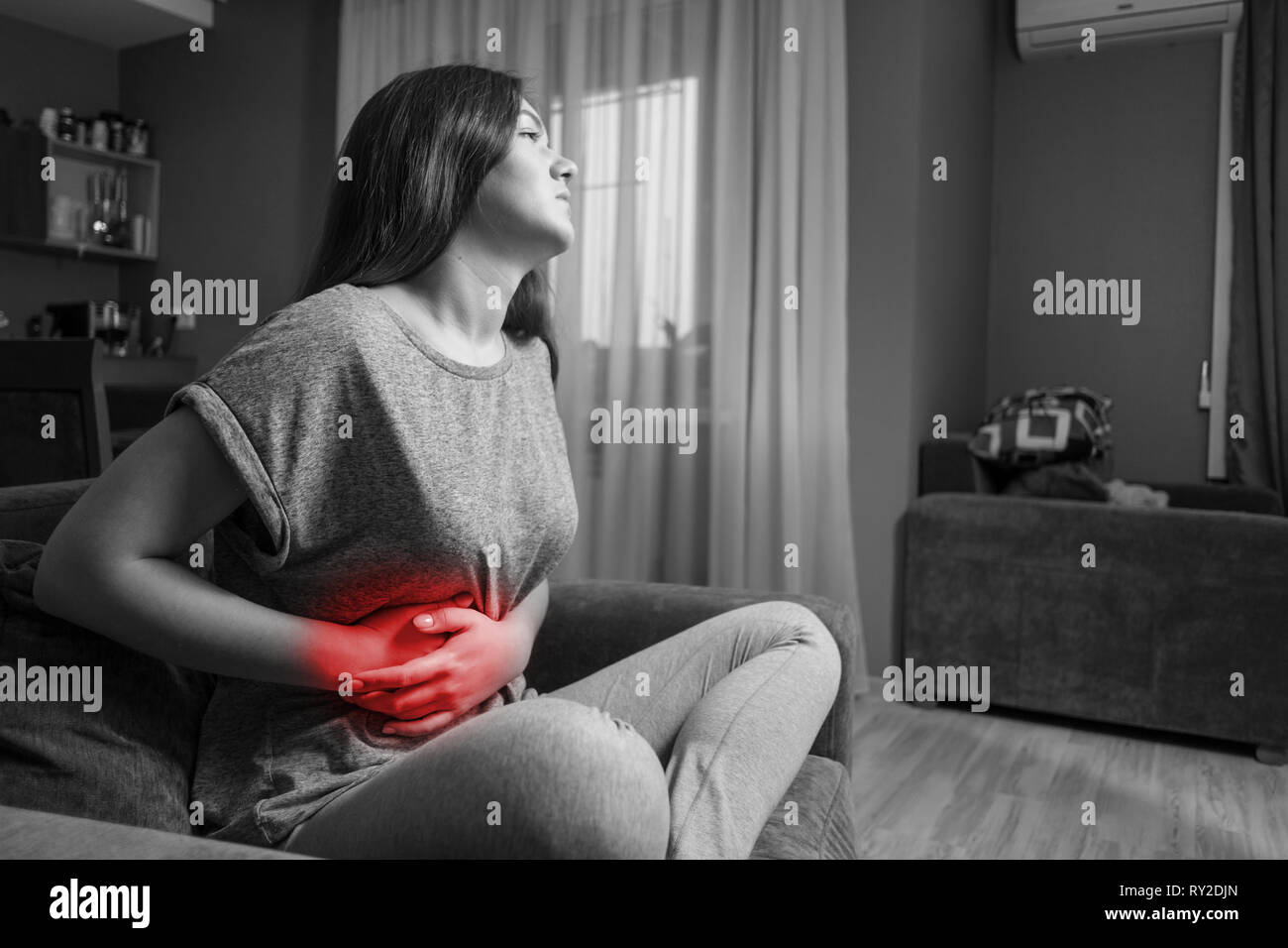 young woman has pain in right hypochondrium, stomachache, black and white photo, red accent, gallbladder disease Stock Photohttps://www.alamy.com/image-license-details/?v=1https://www.alamy.com/young-woman-has-pain-in-right-hypochondrium-stomachache-black-and-white-photo-red-accent-gallbladder-disease-image240385117.html
RFRY2DJN–young woman has pain in right hypochondrium, stomachache, black and white photo, red accent, gallbladder disease
 Die Heilgymnastik in der Gynaekologie : und die mechanische Behandlung von Erkrankungen des Uterus und seiner Adnexe nach Thure Brandt . aufgefordert, unter gleichzeitigerleichter Beckenhebung tief Atem zu holen. In diesem Augenblickdrängt die linke Hand von unten gegen das Zwerchfell zu. Man be-kommt dann, nicht allzu starke Bauchdecken vorausgesetzt, die Meredeutlich zwischen die Finger. Zur Keposition werden die beiden Hände in halber Supina-tions-Stellung, etwas weniger wie zu einer Uterus-Hebung ge-öffnet (vgl. Fig. 63) in das Hypochondrium, während einer In-spiration und Beckenhebung der Stock Photohttps://www.alamy.com/image-license-details/?v=1https://www.alamy.com/die-heilgymnastik-in-der-gynaekologie-und-die-mechanische-behandlung-von-erkrankungen-des-uterus-und-seiner-adnexe-nach-thure-brandt-aufgefordert-unter-gleichzeitigerleichter-beckenhebung-tief-atem-zu-holen-in-diesem-augenblickdrngt-die-linke-hand-von-unten-gegen-das-zwerchfell-zu-man-be-kommt-dann-nicht-allzu-starke-bauchdecken-vorausgesetzt-die-meredeutlich-zwischen-die-finger-zur-keposition-werden-die-beiden-hnde-in-halber-supina-tions-stellung-etwas-weniger-wie-zu-einer-uterus-hebung-ge-ffnet-vgl-fig-63-in-das-hypochondrium-whrend-einer-in-spiration-und-beckenhebung-der-image343049395.html
RM2AX3703–Die Heilgymnastik in der Gynaekologie : und die mechanische Behandlung von Erkrankungen des Uterus und seiner Adnexe nach Thure Brandt . aufgefordert, unter gleichzeitigerleichter Beckenhebung tief Atem zu holen. In diesem Augenblickdrängt die linke Hand von unten gegen das Zwerchfell zu. Man be-kommt dann, nicht allzu starke Bauchdecken vorausgesetzt, die Meredeutlich zwischen die Finger. Zur Keposition werden die beiden Hände in halber Supina-tions-Stellung, etwas weniger wie zu einer Uterus-Hebung ge-öffnet (vgl. Fig. 63) in das Hypochondrium, während einer In-spiration und Beckenhebung der
 young woman has pain in right hypochondrium, stomachache, black and white photo, red accent, gallbladder disease Stock Photohttps://www.alamy.com/image-license-details/?v=1https://www.alamy.com/young-woman-has-pain-in-right-hypochondrium-stomachache-black-and-white-photo-red-accent-gallbladder-disease-image240384231.html
RFRY2CF3–young woman has pain in right hypochondrium, stomachache, black and white photo, red accent, gallbladder disease
 . The spleen and anæmia; experimental and clinical studies . To control bleeding after mobilization of the spleen a large tampon of gauze, wrung out in hotwater, is introduced into the left hypochondrium. PLATE XV .. Tractional ligation after isolation of the vessels of the pedicle by blunt removal of fat and connective tissue. PLATE XVI Stock Photohttps://www.alamy.com/image-license-details/?v=1https://www.alamy.com/the-spleen-and-anmia-experimental-and-clinical-studies-to-control-bleeding-after-mobilization-of-the-spleen-a-large-tampon-of-gauze-wrung-out-in-hotwater-is-introduced-into-the-left-hypochondrium-plate-xv-tractional-ligation-after-isolation-of-the-vessels-of-the-pedicle-by-blunt-removal-of-fat-and-connective-tissue-plate-xvi-image372347585.html
RM2CHNW41–. The spleen and anæmia; experimental and clinical studies . To control bleeding after mobilization of the spleen a large tampon of gauze, wrung out in hotwater, is introduced into the left hypochondrium. PLATE XV .. Tractional ligation after isolation of the vessels of the pedicle by blunt removal of fat and connective tissue. PLATE XVI
 . The spleen and anæmia; experimental and clinical studies . First step in the mobilization of the spleen. With the right hand the operator separates theadhesions between the superior surface of the spleen and the diaphragm. PLATE XIV. To control bleeding after mobilization of the spleen a large tampon of gauze, wrung out in hotwater, is introduced into the left hypochondrium. PLATE XV . Stock Photohttps://www.alamy.com/image-license-details/?v=1https://www.alamy.com/the-spleen-and-anmia-experimental-and-clinical-studies-first-step-in-the-mobilization-of-the-spleen-with-the-right-hand-the-operator-separates-theadhesions-between-the-superior-surface-of-the-spleen-and-the-diaphragm-plate-xiv-to-control-bleeding-after-mobilization-of-the-spleen-a-large-tampon-of-gauze-wrung-out-in-hotwater-is-introduced-into-the-left-hypochondrium-plate-xv-image372348440.html
RM2CHNX6G–. The spleen and anæmia; experimental and clinical studies . First step in the mobilization of the spleen. With the right hand the operator separates theadhesions between the superior surface of the spleen and the diaphragm. PLATE XIV. To control bleeding after mobilization of the spleen a large tampon of gauze, wrung out in hotwater, is introduced into the left hypochondrium. PLATE XV .
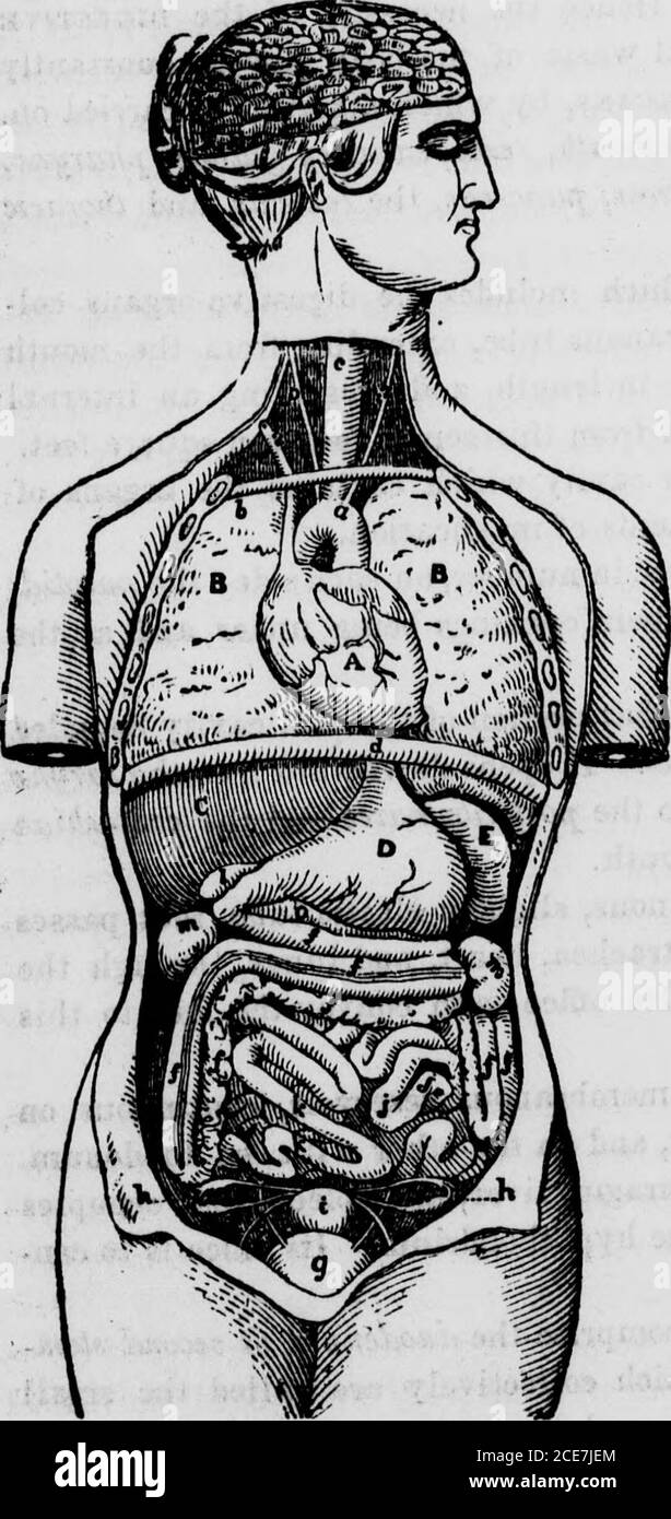 . The hydropathic family physician : a ready prescriber and hygienic adviser with reference to the nature, causes, prevention, and treatment of diseases, accidents, and casualties of every kind . gm, liver, and spleen, and occupiesthe epigastrium and a part of the hypochondrium. Its office is to con-vert the food into chyme. The intestines, or bowels, comprise the duodenum, or second stom-ach, the jejunum, and ileum, which collectively are called the smallintestine; the cacum; the colon, and the rtctum. The duodenum, or 264 Of the Digestive Organs. second stomach, leads from the pyloric orific Stock Photohttps://www.alamy.com/image-license-details/?v=1https://www.alamy.com/the-hydropathic-family-physician-a-ready-prescriber-and-hygienic-adviser-with-reference-to-the-nature-causes-prevention-and-treatment-of-diseases-accidents-and-casualties-of-every-kind-gm-liver-and-spleen-and-occupiesthe-epigastrium-and-a-part-of-the-hypochondrium-its-office-is-to-con-vert-the-food-into-chyme-the-intestines-or-bowels-comprise-the-duodenum-or-second-stom-ach-the-jejunum-and-ileum-which-collectively-are-called-the-smallintestine-the-cacum-the-colon-and-the-rtctum-the-duodenum-or-264-of-the-digestive-organs-second-stomach-leads-from-the-pyloric-orific-image370191100.html
RM2CE7JEM–. The hydropathic family physician : a ready prescriber and hygienic adviser with reference to the nature, causes, prevention, and treatment of diseases, accidents, and casualties of every kind . gm, liver, and spleen, and occupiesthe epigastrium and a part of the hypochondrium. Its office is to con-vert the food into chyme. The intestines, or bowels, comprise the duodenum, or second stom-ach, the jejunum, and ileum, which collectively are called the smallintestine; the cacum; the colon, and the rtctum. The duodenum, or 264 Of the Digestive Organs. second stomach, leads from the pyloric orific
 . A practical treatise on medical diagnosis for students and physicians . is so commonly associated with asimilar condition of the liver and kidneys, if not of other organs, thatany constitutional symptoms produced by the spleen are apt to be maskedby those produced by other organs. Hydatid Disease. Hydatid tumor of the spleen rarely causes symp-toms except when it becomes very large; then it may give rise to dis-comfort and a dragging pain in the left hypochondrium. But hydatidtumors of the spleen are only exceptionally very large; when large enoughto admit of palpation, and when the tumor is Stock Photohttps://www.alamy.com/image-license-details/?v=1https://www.alamy.com/a-practical-treatise-on-medical-diagnosis-for-students-and-physicians-is-so-commonly-associated-with-asimilar-condition-of-the-liver-and-kidneys-if-not-of-other-organs-thatany-constitutional-symptoms-produced-by-the-spleen-are-apt-to-be-maskedby-those-produced-by-other-organs-hydatid-disease-hydatid-tumor-of-the-spleen-rarely-causes-symp-toms-except-when-it-becomes-very-large-then-it-may-give-rise-to-dis-comfort-and-a-dragging-pain-in-the-left-hypochondrium-but-hydatidtumors-of-the-spleen-are-only-exceptionally-very-large-when-large-enoughto-admit-of-palpation-and-when-the-tumor-is-image375938249.html
RM2CRHD21–. A practical treatise on medical diagnosis for students and physicians . is so commonly associated with asimilar condition of the liver and kidneys, if not of other organs, thatany constitutional symptoms produced by the spleen are apt to be maskedby those produced by other organs. Hydatid Disease. Hydatid tumor of the spleen rarely causes symp-toms except when it becomes very large; then it may give rise to dis-comfort and a dragging pain in the left hypochondrium. But hydatidtumors of the spleen are only exceptionally very large; when large enoughto admit of palpation, and when the tumor is
![. On the anatomy of vertebrates [electronic resource] . Stomach of the Kangaroo. 414 ANATOMY OF VERTEBRATES, iliac regions, whence it stretched upward and to the right sideobliquely across the abdomen, to the right hypochondrium, whereit became contracted and finally bent downward and backwardto terminate in the duodenum. The whole length of the stomach,following its curvatures, was three feet six inches, equalling thatof the animal itself from the muzzle to the vent. The cavity may be regarded as consisting of a left, a middle,and a right or pyloric division. The left extremity of thestomach Stock Photo . On the anatomy of vertebrates [electronic resource] . Stomach of the Kangaroo. 414 ANATOMY OF VERTEBRATES, iliac regions, whence it stretched upward and to the right sideobliquely across the abdomen, to the right hypochondrium, whereit became contracted and finally bent downward and backwardto terminate in the duodenum. The whole length of the stomach,following its curvatures, was three feet six inches, equalling thatof the animal itself from the muzzle to the vent. The cavity may be regarded as consisting of a left, a middle,and a right or pyloric division. The left extremity of thestomach Stock Photo](https://c8.alamy.com/comp/2CP7B2Y/on-the-anatomy-of-vertebrates-electronic-resource-stomach-of-the-kangaroo-414-anatomy-of-vertebrates-iliac-regions-whence-it-stretched-upward-and-to-the-right-sideobliquely-across-the-abdomen-to-the-right-hypochondrium-whereit-became-contracted-and-finally-bent-downward-and-backwardto-terminate-in-the-duodenum-the-whole-length-of-the-stomachfollowing-its-curvatures-was-three-feet-six-inches-equalling-thatof-the-animal-itself-from-the-muzzle-to-the-vent-the-cavity-may-be-regarded-as-consisting-of-a-left-a-middleand-a-right-or-pyloric-division-the-left-extremity-of-thestomach-2CP7B2Y.jpg) . On the anatomy of vertebrates [electronic resource] . Stomach of the Kangaroo. 414 ANATOMY OF VERTEBRATES, iliac regions, whence it stretched upward and to the right sideobliquely across the abdomen, to the right hypochondrium, whereit became contracted and finally bent downward and backwardto terminate in the duodenum. The whole length of the stomach,following its curvatures, was three feet six inches, equalling thatof the animal itself from the muzzle to the vent. The cavity may be regarded as consisting of a left, a middle,and a right or pyloric division. The left extremity of thestomach Stock Photohttps://www.alamy.com/image-license-details/?v=1https://www.alamy.com/on-the-anatomy-of-vertebrates-electronic-resource-stomach-of-the-kangaroo-414-anatomy-of-vertebrates-iliac-regions-whence-it-stretched-upward-and-to-the-right-sideobliquely-across-the-abdomen-to-the-right-hypochondrium-whereit-became-contracted-and-finally-bent-downward-and-backwardto-terminate-in-the-duodenum-the-whole-length-of-the-stomachfollowing-its-curvatures-was-three-feet-six-inches-equalling-thatof-the-animal-itself-from-the-muzzle-to-the-vent-the-cavity-may-be-regarded-as-consisting-of-a-left-a-middleand-a-right-or-pyloric-division-the-left-extremity-of-thestomach-image375102531.html
. On the anatomy of vertebrates [electronic resource] . Stomach of the Kangaroo. 414 ANATOMY OF VERTEBRATES, iliac regions, whence it stretched upward and to the right sideobliquely across the abdomen, to the right hypochondrium, whereit became contracted and finally bent downward and backwardto terminate in the duodenum. The whole length of the stomach,following its curvatures, was three feet six inches, equalling thatof the animal itself from the muzzle to the vent. The cavity may be regarded as consisting of a left, a middle,and a right or pyloric division. The left extremity of thestomach Stock Photohttps://www.alamy.com/image-license-details/?v=1https://www.alamy.com/on-the-anatomy-of-vertebrates-electronic-resource-stomach-of-the-kangaroo-414-anatomy-of-vertebrates-iliac-regions-whence-it-stretched-upward-and-to-the-right-sideobliquely-across-the-abdomen-to-the-right-hypochondrium-whereit-became-contracted-and-finally-bent-downward-and-backwardto-terminate-in-the-duodenum-the-whole-length-of-the-stomachfollowing-its-curvatures-was-three-feet-six-inches-equalling-thatof-the-animal-itself-from-the-muzzle-to-the-vent-the-cavity-may-be-regarded-as-consisting-of-a-left-a-middleand-a-right-or-pyloric-division-the-left-extremity-of-thestomach-image375102531.htmlRM2CP7B2Y–. On the anatomy of vertebrates [electronic resource] . Stomach of the Kangaroo. 414 ANATOMY OF VERTEBRATES, iliac regions, whence it stretched upward and to the right sideobliquely across the abdomen, to the right hypochondrium, whereit became contracted and finally bent downward and backwardto terminate in the duodenum. The whole length of the stomach,following its curvatures, was three feet six inches, equalling thatof the animal itself from the muzzle to the vent. The cavity may be regarded as consisting of a left, a middle,and a right or pyloric division. The left extremity of thestomach
 . A practical treatise on medical diagnosis for students and physicians . t, and it is so commonly associated with asimilar condition of the liver and kidneys, if not of other organs, thatany constitutional symptoms produced by the spleen are apt to be maskedby those produced by other organs. Hydatid Disease. Hydatid tumor of the spleen rarely causes symp-toms except when it becomes very large; then it may give rise to dis-comfort and a dragging pain in the left hypochondrium. But hydatidtumors of the spleen are only exceptionally very large; when large enoughto admit of palpation, and when th Stock Photohttps://www.alamy.com/image-license-details/?v=1https://www.alamy.com/a-practical-treatise-on-medical-diagnosis-for-students-and-physicians-t-and-it-is-so-commonly-associated-with-asimilar-condition-of-the-liver-and-kidneys-if-not-of-other-organs-thatany-constitutional-symptoms-produced-by-the-spleen-are-apt-to-be-maskedby-those-produced-by-other-organs-hydatid-disease-hydatid-tumor-of-the-spleen-rarely-causes-symp-toms-except-when-it-becomes-very-large-then-it-may-give-rise-to-dis-comfort-and-a-dragging-pain-in-the-left-hypochondrium-but-hydatidtumors-of-the-spleen-are-only-exceptionally-very-large-when-large-enoughto-admit-of-palpation-and-when-th-image376073110.html
RM2CRRH2E–. A practical treatise on medical diagnosis for students and physicians . t, and it is so commonly associated with asimilar condition of the liver and kidneys, if not of other organs, thatany constitutional symptoms produced by the spleen are apt to be maskedby those produced by other organs. Hydatid Disease. Hydatid tumor of the spleen rarely causes symp-toms except when it becomes very large; then it may give rise to dis-comfort and a dragging pain in the left hypochondrium. But hydatidtumors of the spleen are only exceptionally very large; when large enoughto admit of palpation, and when th
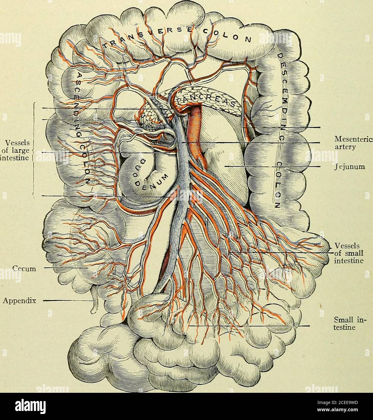 . Text-book of anatomy and physiology for nurses. leend. The location of the stomach is mostly in the epigastric region(Fig. 106). It is below the portion of the diaphragm which supportsthe heart; behind it are the largest artery and vein in the body—theaorta and the inferior vena cava. The pyloric end extends underthe liver in the right hypochondrium, while the cardiac end is in theleft hypochondrium, in contact with the spleen. Clinical notes.—When the stomach is empty it tends to a vertical position;when filled, it swings upward and forward to become again oblique. If muchdistended, as with Stock Photohttps://www.alamy.com/image-license-details/?v=1https://www.alamy.com/text-book-of-anatomy-and-physiology-for-nurses-leend-the-location-of-the-stomach-is-mostly-in-the-epigastric-regionfig-106-it-is-below-the-portion-of-the-diaphragm-which-supportsthe-heart-behind-it-are-the-largest-artery-and-vein-in-the-bodytheaorta-and-the-inferior-vena-cava-the-pyloric-end-extends-underthe-liver-in-the-right-hypochondrium-while-the-cardiac-end-is-in-theleft-hypochondrium-in-contact-with-the-spleen-clinical-noteswhen-the-stomach-is-empty-it-tends-to-a-vertical-positionwhen-filled-it-swings-upward-and-forward-to-become-again-oblique-if-muchdistended-as-with-image370338009.html
RM2CEE9WD–. Text-book of anatomy and physiology for nurses. leend. The location of the stomach is mostly in the epigastric region(Fig. 106). It is below the portion of the diaphragm which supportsthe heart; behind it are the largest artery and vein in the body—theaorta and the inferior vena cava. The pyloric end extends underthe liver in the right hypochondrium, while the cardiac end is in theleft hypochondrium, in contact with the spleen. Clinical notes.—When the stomach is empty it tends to a vertical position;when filled, it swings upward and forward to become again oblique. If muchdistended, as with
![. On the anatomy of vertebrates [electronic resource] . Stomach and intestina cana of the adult Humansubject, cxlviii. ALIMENTARY CANAL OF BIMANA. 4:3.5 stomach averages from thirteen to fifteen inches; its widestdiameter five inches; its capacity five pints. It extends almosttransversely across the upper (in Man) part of the abdomenfrom the left toward the right side, the pylorus entering theregion called ( right hypochondrium: as the stomach becomesdistended, it gently rotates the great curvature forward. Theouter or i serous coat is continued from the lesser curvatureand contributes with th Stock Photo . On the anatomy of vertebrates [electronic resource] . Stomach and intestina cana of the adult Humansubject, cxlviii. ALIMENTARY CANAL OF BIMANA. 4:3.5 stomach averages from thirteen to fifteen inches; its widestdiameter five inches; its capacity five pints. It extends almosttransversely across the upper (in Man) part of the abdomenfrom the left toward the right side, the pylorus entering theregion called ( right hypochondrium: as the stomach becomesdistended, it gently rotates the great curvature forward. Theouter or i serous coat is continued from the lesser curvatureand contributes with th Stock Photo](https://c8.alamy.com/comp/2CP77X0/on-the-anatomy-of-vertebrates-electronic-resource-stomach-and-intestina-cana-of-the-adult-humansubject-cxlviii-alimentary-canal-of-bimana-435-stomach-averages-from-thirteen-to-fifteen-inches-its-widestdiameter-five-inches-its-capacity-five-pints-it-extends-almosttransversely-across-the-upper-in-man-part-of-the-abdomenfrom-the-left-toward-the-right-side-the-pylorus-entering-theregion-called-right-hypochondrium-as-the-stomach-becomesdistended-it-gently-rotates-the-great-curvature-forward-theouter-or-i-serous-coat-is-continued-from-the-lesser-curvatureand-contributes-with-th-2CP77X0.jpg) . On the anatomy of vertebrates [electronic resource] . Stomach and intestina cana of the adult Humansubject, cxlviii. ALIMENTARY CANAL OF BIMANA. 4:3.5 stomach averages from thirteen to fifteen inches; its widestdiameter five inches; its capacity five pints. It extends almosttransversely across the upper (in Man) part of the abdomenfrom the left toward the right side, the pylorus entering theregion called ( right hypochondrium: as the stomach becomesdistended, it gently rotates the great curvature forward. Theouter or i serous coat is continued from the lesser curvatureand contributes with th Stock Photohttps://www.alamy.com/image-license-details/?v=1https://www.alamy.com/on-the-anatomy-of-vertebrates-electronic-resource-stomach-and-intestina-cana-of-the-adult-humansubject-cxlviii-alimentary-canal-of-bimana-435-stomach-averages-from-thirteen-to-fifteen-inches-its-widestdiameter-five-inches-its-capacity-five-pints-it-extends-almosttransversely-across-the-upper-in-man-part-of-the-abdomenfrom-the-left-toward-the-right-side-the-pylorus-entering-theregion-called-right-hypochondrium-as-the-stomach-becomesdistended-it-gently-rotates-the-great-curvature-forward-theouter-or-i-serous-coat-is-continued-from-the-lesser-curvatureand-contributes-with-th-image375100040.html
. On the anatomy of vertebrates [electronic resource] . Stomach and intestina cana of the adult Humansubject, cxlviii. ALIMENTARY CANAL OF BIMANA. 4:3.5 stomach averages from thirteen to fifteen inches; its widestdiameter five inches; its capacity five pints. It extends almosttransversely across the upper (in Man) part of the abdomenfrom the left toward the right side, the pylorus entering theregion called ( right hypochondrium: as the stomach becomesdistended, it gently rotates the great curvature forward. Theouter or i serous coat is continued from the lesser curvatureand contributes with th Stock Photohttps://www.alamy.com/image-license-details/?v=1https://www.alamy.com/on-the-anatomy-of-vertebrates-electronic-resource-stomach-and-intestina-cana-of-the-adult-humansubject-cxlviii-alimentary-canal-of-bimana-435-stomach-averages-from-thirteen-to-fifteen-inches-its-widestdiameter-five-inches-its-capacity-five-pints-it-extends-almosttransversely-across-the-upper-in-man-part-of-the-abdomenfrom-the-left-toward-the-right-side-the-pylorus-entering-theregion-called-right-hypochondrium-as-the-stomach-becomesdistended-it-gently-rotates-the-great-curvature-forward-theouter-or-i-serous-coat-is-continued-from-the-lesser-curvatureand-contributes-with-th-image375100040.htmlRM2CP77X0–. On the anatomy of vertebrates [electronic resource] . Stomach and intestina cana of the adult Humansubject, cxlviii. ALIMENTARY CANAL OF BIMANA. 4:3.5 stomach averages from thirteen to fifteen inches; its widestdiameter five inches; its capacity five pints. It extends almosttransversely across the upper (in Man) part of the abdomenfrom the left toward the right side, the pylorus entering theregion called ( right hypochondrium: as the stomach becomesdistended, it gently rotates the great curvature forward. Theouter or i serous coat is continued from the lesser curvatureand contributes with th
 . Annals of surgery . ai 512 RESECTIOX OF STOMACH FOR ULCER lift the duodenum over to the left hypochondrium. A perfect resultwas obtained. When the ulcer is situated at the posterior wall of the duodenum, or solow that direct anastomosis between stomach and duodenum is not safe,we can either perform a Billroth II or use Haberers •* method of gastro-duodenostomy (a modified Kocher). Since the post-operative course follow-ing a Billroth II is smootherthan that following a gastro-duodenostomy, closure ofboth ends followed by a gas-tro-enterostomy is preferable. Haberers modification•^f the Billr Stock Photohttps://www.alamy.com/image-license-details/?v=1https://www.alamy.com/annals-of-surgery-ai-512-resectiox-of-stomach-for-ulcer-lift-the-duodenum-over-to-the-left-hypochondrium-a-perfect-resultwas-obtained-when-the-ulcer-is-situated-at-the-posterior-wall-of-the-duodenum-or-solow-that-direct-anastomosis-between-stomach-and-duodenum-is-not-safewe-can-either-perform-a-billroth-ii-or-use-haberers-method-of-gastro-duodenostomy-a-modified-kocher-since-the-post-operative-course-follow-ing-a-billroth-ii-is-smootherthan-that-following-a-gastro-duodenostomy-closure-ofboth-ends-followed-by-a-gas-tro-enterostomy-is-preferable-haberers-modificationf-the-billr-image370000346.html
RM2CDXY62–. Annals of surgery . ai 512 RESECTIOX OF STOMACH FOR ULCER lift the duodenum over to the left hypochondrium. A perfect resultwas obtained. When the ulcer is situated at the posterior wall of the duodenum, or solow that direct anastomosis between stomach and duodenum is not safe,we can either perform a Billroth II or use Haberers •* method of gastro-duodenostomy (a modified Kocher). Since the post-operative course follow-ing a Billroth II is smootherthan that following a gastro-duodenostomy, closure ofboth ends followed by a gas-tro-enterostomy is preferable. Haberers modification•^f the Billr
 . Annals of surgery. bviated by carefully injectingthe fluid while the radiograph is being made. Without goingmore fully into the subject, I will show two extremes in thepelvic contour. Both were made in females with indefinitetumors in the right hypochondrium which, at operation, provedto be due to distended gall-bladder, while the kidneys werefound normal. Fig. I shows the slit-like pelvis in a normal kidney ob-tained immediately after injecting 3 c.c. of collargol, whichcaused a severe colic and which might easily be interpretedas the remnant of a pelvis encroached upon by surroundingtumor Stock Photohttps://www.alamy.com/image-license-details/?v=1https://www.alamy.com/annals-of-surgery-bviated-by-carefully-injectingthe-fluid-while-the-radiograph-is-being-made-without-goingmore-fully-into-the-subject-i-will-show-two-extremes-in-thepelvic-contour-both-were-made-in-females-with-indefinitetumors-in-the-right-hypochondrium-which-at-operation-provedto-be-due-to-distended-gall-bladder-while-the-kidneys-werefound-normal-fig-i-shows-the-slit-like-pelvis-in-a-normal-kidney-ob-tained-immediately-after-injecting-3-cc-of-collargol-whichcaused-a-severe-colic-and-which-might-easily-be-interpretedas-the-remnant-of-a-pelvis-encroached-upon-by-surroundingtumor-image370646607.html
RM2CF0BER–. Annals of surgery. bviated by carefully injectingthe fluid while the radiograph is being made. Without goingmore fully into the subject, I will show two extremes in thepelvic contour. Both were made in females with indefinitetumors in the right hypochondrium which, at operation, provedto be due to distended gall-bladder, while the kidneys werefound normal. Fig. I shows the slit-like pelvis in a normal kidney ob-tained immediately after injecting 3 c.c. of collargol, whichcaused a severe colic and which might easily be interpretedas the remnant of a pelvis encroached upon by surroundingtumor
 . Lectures on the diagnosis of abdominal tumors, delivered to the post-graduate class of Johns Hopkins university, 1893. bdomen is flaccid, symmet-rical ; a little full, perhaps, in theepigastric region. On palpation, aridge-like mass is to be felt in theleft hypochondrium, which extendsacross the middle line to the rightside as far as the parasternal line. Itdescends with deep inspiration as lowas the umbilicus. The lower edge isvery distinct and feels somewhat likea rolled omentum. It can scarcely Fig. 12.—Showing the po.sition of , i i r j.t, t ^i the tumor mass consisting of dif- ^^ Separa Stock Photohttps://www.alamy.com/image-license-details/?v=1https://www.alamy.com/lectures-on-the-diagnosis-of-abdominal-tumors-delivered-to-the-post-graduate-class-of-johns-hopkins-university-1893-bdomen-is-flaccid-symmet-rical-a-little-full-perhaps-in-theepigastric-region-on-palpation-aridge-like-mass-is-to-be-felt-in-theleft-hypochondrium-which-extendsacross-the-middle-line-to-the-rightside-as-far-as-the-parasternal-line-itdescends-with-deep-inspiration-as-lowas-the-umbilicus-the-lower-edge-isvery-distinct-and-feels-somewhat-likea-rolled-omentum-it-can-scarcely-fig-12showing-the-position-of-i-i-r-jt-t-i-the-tumor-mass-consisting-of-dif-separa-image370765821.html
RM2CF5RGD–. Lectures on the diagnosis of abdominal tumors, delivered to the post-graduate class of Johns Hopkins university, 1893. bdomen is flaccid, symmet-rical ; a little full, perhaps, in theepigastric region. On palpation, aridge-like mass is to be felt in theleft hypochondrium, which extendsacross the middle line to the rightside as far as the parasternal line. Itdescends with deep inspiration as lowas the umbilicus. The lower edge isvery distinct and feels somewhat likea rolled omentum. It can scarcely Fig. 12.—Showing the po.sition of , i i r j.t, t ^i the tumor mass consisting of dif- ^^ Separa
 . The American journal of roentgenology, radium therapy and nuclear medicine . FiG. I. Pyonephrosis with Stones. No Pre^ous A-ray Examination * Read at the Annual Meeting of the Western Section of the .American Roentgen Ray Society, Feb. 22, 1918. The A-Rays in Defining Kidney Tumors. Fig. 2. Hypernephroma amination and the result will suggest fur-ther clinical examinations, until a finalconclusion can be drawn or the diagnosisleft open, to be revealed at operation.For example: A woman had a large bean-shaped tumor easily palpable in theright hypochondrium, protruding from un-derneath the live Stock Photohttps://www.alamy.com/image-license-details/?v=1https://www.alamy.com/the-american-journal-of-roentgenology-radium-therapy-and-nuclear-medicine-fig-i-pyonephrosis-with-stones-no-preous-a-ray-examination-read-at-the-annual-meeting-of-the-western-section-of-the-american-roentgen-ray-society-feb-22-1918-the-a-rays-in-defining-kidney-tumors-fig-2-hypernephroma-amination-and-the-result-will-suggest-fur-ther-clinical-examinations-until-a-finalconclusion-can-be-drawn-or-the-diagnosisleft-open-to-be-revealed-at-operationfor-example-a-woman-had-a-large-bean-shaped-tumor-easily-palpable-in-theright-hypochondrium-protruding-from-un-derneath-the-live-image375976151.html
RM2CRK5BK–. The American journal of roentgenology, radium therapy and nuclear medicine . FiG. I. Pyonephrosis with Stones. No Pre^ous A-ray Examination * Read at the Annual Meeting of the Western Section of the .American Roentgen Ray Society, Feb. 22, 1918. The A-Rays in Defining Kidney Tumors. Fig. 2. Hypernephroma amination and the result will suggest fur-ther clinical examinations, until a finalconclusion can be drawn or the diagnosisleft open, to be revealed at operation.For example: A woman had a large bean-shaped tumor easily palpable in theright hypochondrium, protruding from un-derneath the live
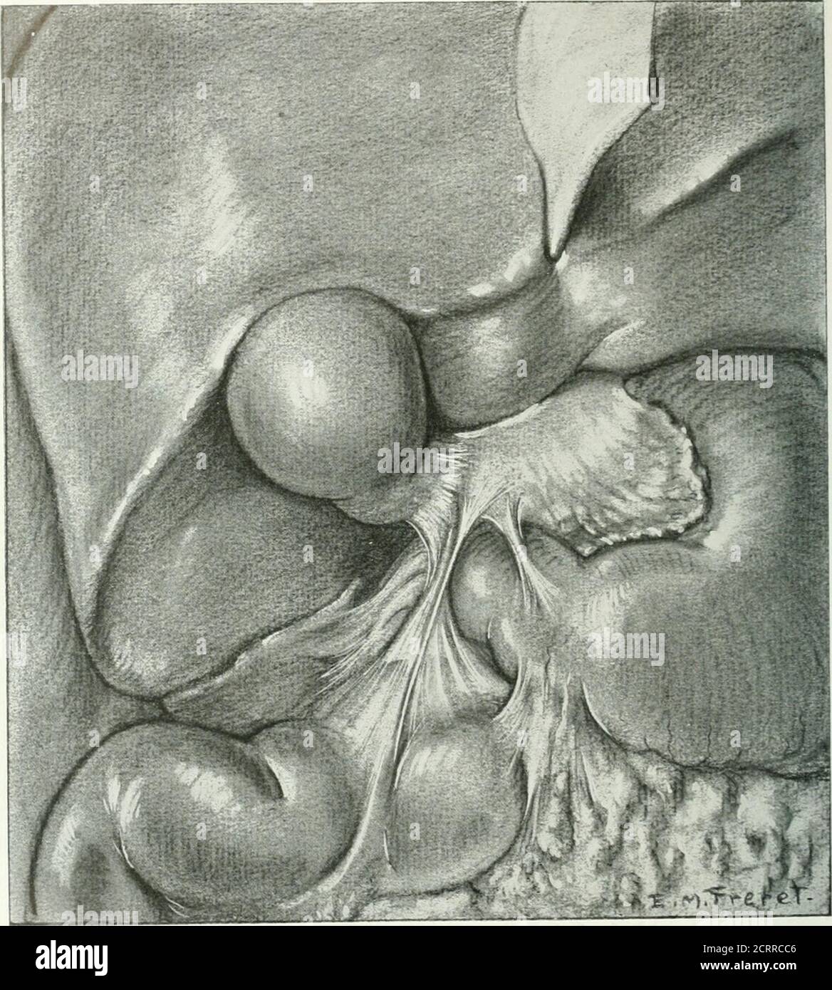 . The American journal of roentgenology, radium therapy and nuclear medicine . Fig. i. Sketch of anatomical specimen showing a veil in theright hypochondrium involving: i, the cap; 2, the neck ot thegall-bladder; 3, the descending duodenum; 4. the pyloric endof the greater curvature of the stomach; s. the hepatic flexureof the colon. , . Fig. 1. Plan dun specimen anatomique demontrant a la vue unevoile dans le hypochondre droit comprennanf, I, le bulbeduodenale; 2, le cou de la v6sicule biliaire; 3. le duodenumdescendant: 4. lextremite pylorique de la courbuntomac; s. la flexion hepatique du c Stock Photohttps://www.alamy.com/image-license-details/?v=1https://www.alamy.com/the-american-journal-of-roentgenology-radium-therapy-and-nuclear-medicine-fig-i-sketch-of-anatomical-specimen-showing-a-veil-in-theright-hypochondrium-involving-i-the-cap-2-the-neck-ot-thegall-bladder-3-the-descending-duodenum-4-the-pyloric-endof-the-greater-curvature-of-the-stomach-s-the-hepatic-flexureof-the-colon-fig-1-plan-dun-specimen-anatomique-demontrant-a-la-vue-unevoile-dans-le-hypochondre-droit-comprennanf-i-le-bulbeduodenale-2-le-cou-de-la-v6sicule-biliaire-3-le-duodenumdescendant-4-lextremite-pylorique-de-la-courbuntomac-s-la-flexion-hepatique-du-c-image376069462.html
RM2CRRCC6–. The American journal of roentgenology, radium therapy and nuclear medicine . Fig. i. Sketch of anatomical specimen showing a veil in theright hypochondrium involving: i, the cap; 2, the neck ot thegall-bladder; 3, the descending duodenum; 4. the pyloric endof the greater curvature of the stomach; s. the hepatic flexureof the colon. , . Fig. 1. Plan dun specimen anatomique demontrant a la vue unevoile dans le hypochondre droit comprennanf, I, le bulbeduodenale; 2, le cou de la v6sicule biliaire; 3. le duodenumdescendant: 4. lextremite pylorique de la courbuntomac; s. la flexion hepatique du c
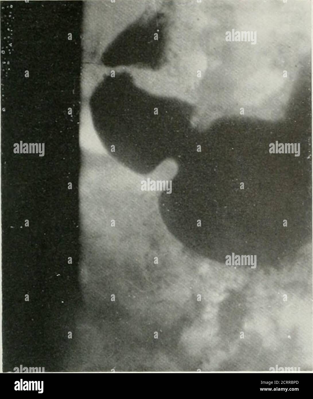 . The American journal of roentgenology, radium therapy and nuclear medicine . Fig. i and .i. Illustrates a veil thai ii < outer surface ol the i ap, thi gall I Lad lei and h< ■ re of the colon filled with g Fig. i el 4. Demontrant une voile qui comprend la surface ieureladu b ale, vesicule biliaire et la flexion tique du colon emplie pai l<Fig. j et V- Ilustrando un velo que envuelve la caxa externa delbulbo y duodenal, la vesfcula biliai y al asa hepatica del colon Uenos de gas. Veils in the Right Hypochondrium 43. Stock Photohttps://www.alamy.com/image-license-details/?v=1https://www.alamy.com/the-american-journal-of-roentgenology-radium-therapy-and-nuclear-medicine-fig-i-and-i-illustrates-a-veil-thai-ii-lt-outer-surface-ol-the-i-ap-thi-gall-i-lad-lei-and-hlt-re-of-the-colon-filled-with-g-fig-i-el-4-demontrant-une-voile-qui-comprend-la-surface-ieureladu-b-ale-vesicule-biliaire-et-la-flexion-tique-du-colon-emplie-pai-lltfig-j-et-v-ilustrando-un-velo-que-envuelve-la-caxa-externa-delbulbo-y-duodenal-la-vesfcula-biliai-y-al-asa-hepatica-del-colon-uenos-de-gas-veils-in-the-right-hypochondrium-43-image376068965.html
RM2CRRBPD–. The American journal of roentgenology, radium therapy and nuclear medicine . Fig. i and .i. Illustrates a veil thai ii < outer surface ol the i ap, thi gall I Lad lei and h< ■ re of the colon filled with g Fig. i el 4. Demontrant une voile qui comprend la surface ieureladu b ale, vesicule biliaire et la flexion tique du colon emplie pai l<Fig. j et V- Ilustrando un velo que envuelve la caxa externa delbulbo y duodenal, la vesfcula biliai y al asa hepatica del colon Uenos de gas. Veils in the Right Hypochondrium 43.
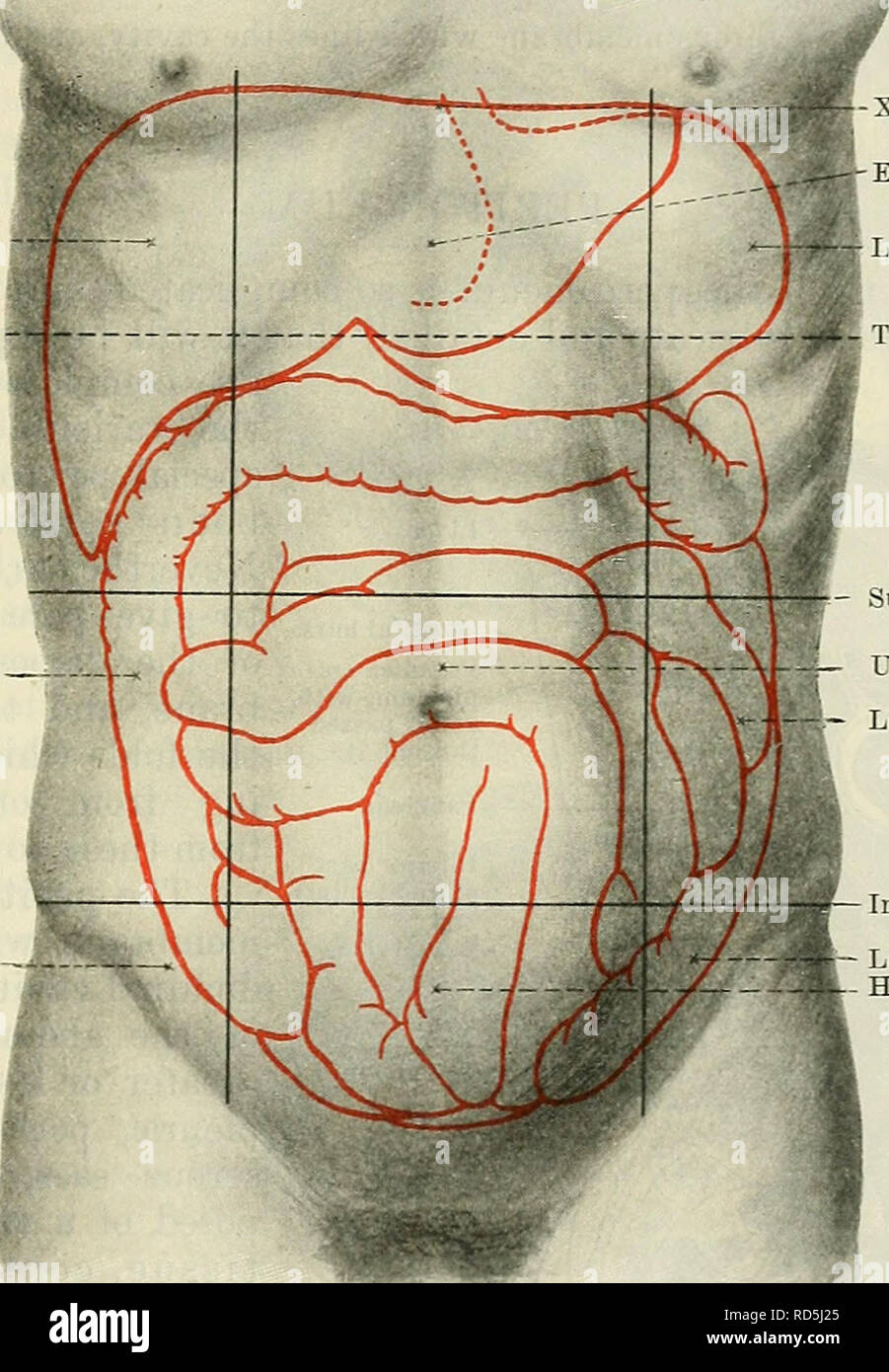 . Cunningham's Text-book of anatomy. Anatomy. THE ABDOMINAL CAVITY. 1159 perpendicular planes each of these is subdivided into three parts, a central and two lateral. Thus, in the upper zone, we get a hypochondriac region or hypochondrium on each side, and an epigastric region or epigastrium in the centre. Similarly, the umbilical zone is divided into right and left lumbar regions, with an umbilical region between. And the hypogastric zone has a hypogastric region or hypogastrium in the centre, with right and left iliac regions at the sides. In addition, the portion of the abdominal wall above Stock Photohttps://www.alamy.com/image-license-details/?v=1https://www.alamy.com/cunninghams-text-book-of-anatomy-anatomy-the-abdominal-cavity-1159-perpendicular-planes-each-of-these-is-subdivided-into-three-parts-a-central-and-two-lateral-thus-in-the-upper-zone-we-get-a-hypochondriac-region-or-hypochondrium-on-each-side-and-an-epigastric-region-or-epigastrium-in-the-centre-similarly-the-umbilical-zone-is-divided-into-right-and-left-lumbar-regions-with-an-umbilical-region-between-and-the-hypogastric-zone-has-a-hypogastric-region-or-hypogastrium-in-the-centre-with-right-and-left-iliac-regions-at-the-sides-in-addition-the-portion-of-the-abdominal-wall-above-image231849245.html
RMRD5J25–. Cunningham's Text-book of anatomy. Anatomy. THE ABDOMINAL CAVITY. 1159 perpendicular planes each of these is subdivided into three parts, a central and two lateral. Thus, in the upper zone, we get a hypochondriac region or hypochondrium on each side, and an epigastric region or epigastrium in the centre. Similarly, the umbilical zone is divided into right and left lumbar regions, with an umbilical region between. And the hypogastric zone has a hypogastric region or hypogastrium in the centre, with right and left iliac regions at the sides. In addition, the portion of the abdominal wall above
 . Diseases of cattle, sheep, goats and swine. Veterinary medicine. EXUDATIVE PERICARDITIS DUE TO FOREIGN BODIES. 387 the hypochondrium becomes attached to the sternum. (3.) Point at which the external mammary vein penetrates the abdominal wall (Fi^- 178). Lines uniting these three points enclose a right-angled triangle, which the operator must imagine to be bisected by a third line. The iucision, which should be about 8 inches in length, follows this bisecting line at an equal distance between the white line and the circle of the hypochondrium, to a point within about 8 inches of the anterior Stock Photohttps://www.alamy.com/image-license-details/?v=1https://www.alamy.com/diseases-of-cattle-sheep-goats-and-swine-veterinary-medicine-exudative-pericarditis-due-to-foreign-bodies-387-the-hypochondrium-becomes-attached-to-the-sternum-3-point-at-which-the-external-mammary-vein-penetrates-the-abdominal-wall-fi-178-lines-uniting-these-three-points-enclose-a-right-angled-triangle-which-the-operator-must-imagine-to-be-bisected-by-a-third-line-the-iucision-which-should-be-about-8-inches-in-length-follows-this-bisecting-line-at-an-equal-distance-between-the-white-line-and-the-circle-of-the-hypochondrium-to-a-point-within-about-8-inches-of-the-anterior-image232342339.html
RMRE030K–. Diseases of cattle, sheep, goats and swine. Veterinary medicine. EXUDATIVE PERICARDITIS DUE TO FOREIGN BODIES. 387 the hypochondrium becomes attached to the sternum. (3.) Point at which the external mammary vein penetrates the abdominal wall (Fi^- 178). Lines uniting these three points enclose a right-angled triangle, which the operator must imagine to be bisected by a third line. The iucision, which should be about 8 inches in length, follows this bisecting line at an equal distance between the white line and the circle of the hypochondrium, to a point within about 8 inches of the anterior
 . Diseases of cattle, sheep, goats and swine. Veterinary medicine. EXAMINATION OF THE KESPIRATORY APPARATUS. 317 approximately as far as the inferior line of insertion of the common intercostal muscle, and extends from the smnmit of the scapula in front to the hypochondrium behind. Auscultation of this region through the ileo-spinal and common intercostal muscle 'will always reveal, except in very fat animals, the vesicular murmur to a point as far back as the eleventh intercostal space. Nevertheless, this vesicular murmur is relatively feeble, and becomes imperceptible beyond the eleventh rib Stock Photohttps://www.alamy.com/image-license-details/?v=1https://www.alamy.com/diseases-of-cattle-sheep-goats-and-swine-veterinary-medicine-examination-of-the-kespiratory-apparatus-317-approximately-as-far-as-the-inferior-line-of-insertion-of-the-common-intercostal-muscle-and-extends-from-the-smnmit-of-the-scapula-in-front-to-the-hypochondrium-behind-auscultation-of-this-region-through-the-ileo-spinal-and-common-intercostal-muscle-will-always-reveal-except-in-very-fat-animals-the-vesicular-murmur-to-a-point-as-far-back-as-the-eleventh-intercostal-space-nevertheless-this-vesicular-murmur-is-relatively-feeble-and-becomes-imperceptible-beyond-the-eleventh-rib-image232342420.html
RMRE033G–. Diseases of cattle, sheep, goats and swine. Veterinary medicine. EXAMINATION OF THE KESPIRATORY APPARATUS. 317 approximately as far as the inferior line of insertion of the common intercostal muscle, and extends from the smnmit of the scapula in front to the hypochondrium behind. Auscultation of this region through the ileo-spinal and common intercostal muscle 'will always reveal, except in very fat animals, the vesicular murmur to a point as far back as the eleventh intercostal space. Nevertheless, this vesicular murmur is relatively feeble, and becomes imperceptible beyond the eleventh rib
 . Blaine's Outlines of the veterinary art, or, A treatise on the anatomy, physiology, and curative treatment of the diseases of the horse, and subordinately, of those of neat cattle and sheep ... Horses; Veterinary medicine. THE STOMACH.. 229 THE STOMACH. This important alimentary bag is remarkably small in the horse. Its situation may be described as being imme- diately behind the liver, its principal portion occupying the left hypochondrium; and a smaller part the epigastrium, with its pyloric (Fig 23 , P) orifice stretched n cross the spine to the right side. It has two surfaces; one is pos Stock Photohttps://www.alamy.com/image-license-details/?v=1https://www.alamy.com/blaines-outlines-of-the-veterinary-art-or-a-treatise-on-the-anatomy-physiology-and-curative-treatment-of-the-diseases-of-the-horse-and-subordinately-of-those-of-neat-cattle-and-sheep-horses-veterinary-medicine-the-stomach-229-the-stomach-this-important-alimentary-bag-is-remarkably-small-in-the-horse-its-situation-may-be-described-as-being-imme-diately-behind-the-liver-its-principal-portion-occupying-the-left-hypochondrium-and-a-smaller-part-the-epigastrium-with-its-pyloric-fig-23-p-orifice-stretched-n-cross-the-spine-to-the-right-side-it-has-two-surfaces-one-is-pos-image234585108.html
RMRHJ7KG–. Blaine's Outlines of the veterinary art, or, A treatise on the anatomy, physiology, and curative treatment of the diseases of the horse, and subordinately, of those of neat cattle and sheep ... Horses; Veterinary medicine. THE STOMACH.. 229 THE STOMACH. This important alimentary bag is remarkably small in the horse. Its situation may be described as being imme- diately behind the liver, its principal portion occupying the left hypochondrium; and a smaller part the epigastrium, with its pyloric (Fig 23 , P) orifice stretched n cross the spine to the right side. It has two surfaces; one is pos
 . Diseases of cattle, sheep, goats and swine. Veterinary medicine. EXAMINATION OF THE RESPIRATORY APPARATUS. 817 approximately as far as the inferior line of insertion of the common intercostal muscle, and extends from the summit of the scapula in front to the hypochondrium behind. Auscultation of this region through the ileo-spinal and common intercostal muscle will always reveal, except in very fat animals, the vesicular murmur to a point as far back as the eleventh intercostal space. Nevertheless, this vesicular murmur is relatively feeble, and becomes imperceptible beyond the eleventh rib. Stock Photohttps://www.alamy.com/image-license-details/?v=1https://www.alamy.com/diseases-of-cattle-sheep-goats-and-swine-veterinary-medicine-examination-of-the-respiratory-apparatus-817-approximately-as-far-as-the-inferior-line-of-insertion-of-the-common-intercostal-muscle-and-extends-from-the-summit-of-the-scapula-in-front-to-the-hypochondrium-behind-auscultation-of-this-region-through-the-ileo-spinal-and-common-intercostal-muscle-will-always-reveal-except-in-very-fat-animals-the-vesicular-murmur-to-a-point-as-far-back-as-the-eleventh-intercostal-space-nevertheless-this-vesicular-murmur-is-relatively-feeble-and-becomes-imperceptible-beyond-the-eleventh-rib-image231419733.html
RMRCE26D–. Diseases of cattle, sheep, goats and swine. Veterinary medicine. EXAMINATION OF THE RESPIRATORY APPARATUS. 817 approximately as far as the inferior line of insertion of the common intercostal muscle, and extends from the summit of the scapula in front to the hypochondrium behind. Auscultation of this region through the ileo-spinal and common intercostal muscle will always reveal, except in very fat animals, the vesicular murmur to a point as far back as the eleventh intercostal space. Nevertheless, this vesicular murmur is relatively feeble, and becomes imperceptible beyond the eleventh rib.
 . Diseases of cattle, sheep, goats and swine. Veterinary medicine. EXAMINATION OF THE RESPIRATORY APPARATUS. 317 approximately as far as the inferior line of insertion of the common intercostal muscle, and extends from the summit of the scapula in front to the hypochondrium behind. Auscultation of this region through the ileo-spinal and common intercostal muscle will always reveal, except in very fat animals, the vesicular murmur to a point as far back as the eleventh intercostal space. Nevertheless, this vesicular murmur is relatively feeble, and becomes imperceptible beyond the eleventh rib. Stock Photohttps://www.alamy.com/image-license-details/?v=1https://www.alamy.com/diseases-of-cattle-sheep-goats-and-swine-veterinary-medicine-examination-of-the-respiratory-apparatus-317-approximately-as-far-as-the-inferior-line-of-insertion-of-the-common-intercostal-muscle-and-extends-from-the-summit-of-the-scapula-in-front-to-the-hypochondrium-behind-auscultation-of-this-region-through-the-ileo-spinal-and-common-intercostal-muscle-will-always-reveal-except-in-very-fat-animals-the-vesicular-murmur-to-a-point-as-far-back-as-the-eleventh-intercostal-space-nevertheless-this-vesicular-murmur-is-relatively-feeble-and-becomes-imperceptible-beyond-the-eleventh-rib-image231403394.html
RMRCD9AX–. Diseases of cattle, sheep, goats and swine. Veterinary medicine. EXAMINATION OF THE RESPIRATORY APPARATUS. 317 approximately as far as the inferior line of insertion of the common intercostal muscle, and extends from the summit of the scapula in front to the hypochondrium behind. Auscultation of this region through the ileo-spinal and common intercostal muscle will always reveal, except in very fat animals, the vesicular murmur to a point as far back as the eleventh intercostal space. Nevertheless, this vesicular murmur is relatively feeble, and becomes imperceptible beyond the eleventh rib.
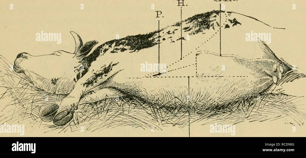 . Diseases of cattle, sheep, goats and swine. Veterinary medicine. EXUDATIVE PERICARDITIS DUE TO FOREIGN BODIES. 387 the hypochondrium becomes attached to the sternum. (3.) Point at which the external mammary vein penetrates the abdominal wall (Fig. 178). Lines uniting these three points enclose a right-angled triangle, which the operator must imagine to be bisected by a third line. The incision, which should be about 8 inches in length, follows this bisecting line at an equal distance between the white line and the circle of the hypochondrium, to a point within about 8 inches of the anterior Stock Photohttps://www.alamy.com/image-license-details/?v=1https://www.alamy.com/diseases-of-cattle-sheep-goats-and-swine-veterinary-medicine-exudative-pericarditis-due-to-foreign-bodies-387-the-hypochondrium-becomes-attached-to-the-sternum-3-point-at-which-the-external-mammary-vein-penetrates-the-abdominal-wall-fig-178-lines-uniting-these-three-points-enclose-a-right-angled-triangle-which-the-operator-must-imagine-to-be-bisected-by-a-third-line-the-incision-which-should-be-about-8-inches-in-length-follows-this-bisecting-line-at-an-equal-distance-between-the-white-line-and-the-circle-of-the-hypochondrium-to-a-point-within-about-8-inches-of-the-anterior-image231403328.html
RMRCD98G–. Diseases of cattle, sheep, goats and swine. Veterinary medicine. EXUDATIVE PERICARDITIS DUE TO FOREIGN BODIES. 387 the hypochondrium becomes attached to the sternum. (3.) Point at which the external mammary vein penetrates the abdominal wall (Fig. 178). Lines uniting these three points enclose a right-angled triangle, which the operator must imagine to be bisected by a third line. The incision, which should be about 8 inches in length, follows this bisecting line at an equal distance between the white line and the circle of the hypochondrium, to a point within about 8 inches of the anterior
 . Diseases of cattle, sheep, goats and swine. Veterinary medicine. EXUDATIVE PERICARDITIS DUE TO FOREIGN BODIES. 387 the hypochondrium becomes attached to the sternum. (3.) Point at which the external mammary vem penetrates the abdominal wall (Fig. 178). Lines uniting these three points enclose a right-angled triangle, which the operator must imagine to be bisected by a third line. The incision, which should be about 8 inches in length, follows this bisecting line at an equal distance between the white line and the circle of the hypochondrium, to a point within about 8 inches of the anterior m Stock Photohttps://www.alamy.com/image-license-details/?v=1https://www.alamy.com/diseases-of-cattle-sheep-goats-and-swine-veterinary-medicine-exudative-pericarditis-due-to-foreign-bodies-387-the-hypochondrium-becomes-attached-to-the-sternum-3-point-at-which-the-external-mammary-vem-penetrates-the-abdominal-wall-fig-178-lines-uniting-these-three-points-enclose-a-right-angled-triangle-which-the-operator-must-imagine-to-be-bisected-by-a-third-line-the-incision-which-should-be-about-8-inches-in-length-follows-this-bisecting-line-at-an-equal-distance-between-the-white-line-and-the-circle-of-the-hypochondrium-to-a-point-within-about-8-inches-of-the-anterior-m-image231379703.html
RMRCC74R–. Diseases of cattle, sheep, goats and swine. Veterinary medicine. EXUDATIVE PERICARDITIS DUE TO FOREIGN BODIES. 387 the hypochondrium becomes attached to the sternum. (3.) Point at which the external mammary vem penetrates the abdominal wall (Fig. 178). Lines uniting these three points enclose a right-angled triangle, which the operator must imagine to be bisected by a third line. The incision, which should be about 8 inches in length, follows this bisecting line at an equal distance between the white line and the circle of the hypochondrium, to a point within about 8 inches of the anterior m
 . Die descriptive und topographische Anatomie des Menschen. Anatomy. 268. 401. Die Leber, Hepar. Ansicht von oben. Die Leber liegt im rechten Hypochondrium und erstreckt sich bis hinüber in das linke. Ihr vorderer scharfer Rand besitzt einen Einschnitt zur Aufnahme des Ligamentum Suspensorium; ihr hinterer, stumpfer Rand steht höher als der vordere; der rechte Rand ist gleichfalls stumpf, der linke, zuge- schärfte liegt vor der Cardia des Magens. Die obere Fläche ist entsprechend der Wölbung des Diaphragma convex und etwas nach vorne goneigt; durch das Lig. Suspensorium ist die Grenze zwischen Stock Photohttps://www.alamy.com/image-license-details/?v=1https://www.alamy.com/die-descriptive-und-topographische-anatomie-des-menschen-anatomy-268-401-die-leber-hepar-ansicht-von-oben-die-leber-liegt-im-rechten-hypochondrium-und-erstreckt-sich-bis-hinber-in-das-linke-ihr-vorderer-scharfer-rand-besitzt-einen-einschnitt-zur-aufnahme-des-ligamentum-suspensorium-ihr-hinterer-stumpfer-rand-steht-hher-als-der-vordere-der-rechte-rand-ist-gleichfalls-stumpf-der-linke-zuge-schrfte-liegt-vor-der-cardia-des-magens-die-obere-flche-ist-entsprechend-der-wlbung-des-diaphragma-convex-und-etwas-nach-vorne-goneigt-durch-das-lig-suspensorium-ist-die-grenze-zwischen-image231585970.html
RMRCNJ7E–. Die descriptive und topographische Anatomie des Menschen. Anatomy. 268. 401. Die Leber, Hepar. Ansicht von oben. Die Leber liegt im rechten Hypochondrium und erstreckt sich bis hinüber in das linke. Ihr vorderer scharfer Rand besitzt einen Einschnitt zur Aufnahme des Ligamentum Suspensorium; ihr hinterer, stumpfer Rand steht höher als der vordere; der rechte Rand ist gleichfalls stumpf, der linke, zuge- schärfte liegt vor der Cardia des Magens. Die obere Fläche ist entsprechend der Wölbung des Diaphragma convex und etwas nach vorne goneigt; durch das Lig. Suspensorium ist die Grenze zwischen