Quick filters:
Hypoplastron Stock Photos and Images
 Sea turtle bones, Christmas (Kiritimati) Island, Kiribati Stock Photohttps://www.alamy.com/image-license-details/?v=1https://www.alamy.com/stock-photo-sea-turtle-bones-christmas-kiritimati-island-kiribati-126711274.html
RFHA45EJ–Sea turtle bones, Christmas (Kiritimati) Island, Kiribati
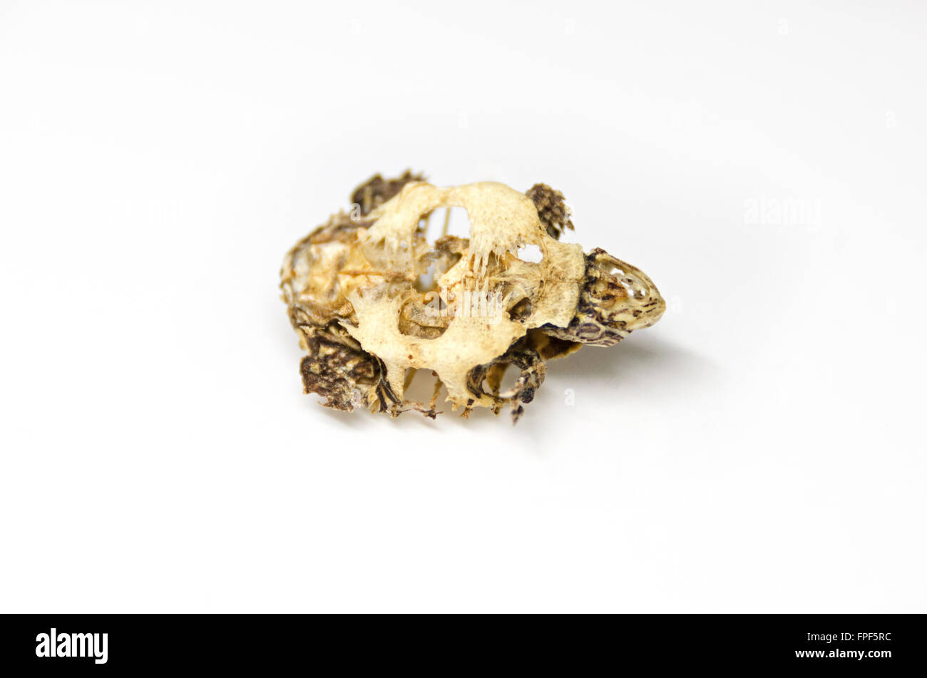 Ventral view of a baby turtle skeleton; Southbridge, Massachusetts. Stock Photohttps://www.alamy.com/image-license-details/?v=1https://www.alamy.com/stock-photo-ventral-view-of-a-baby-turtle-skeleton-southbridge-massachusetts-99908128.html
RMFPF5RC–Ventral view of a baby turtle skeleton; Southbridge, Massachusetts.
![Merveilles de la nature : l'homme et les animaux, description populaire des races humaines et des règne animal . Fig. 87. — Plastron du Ckelone midas (). Les premiers anatomistes qui ont étudié lesTorluesonl pensé que la carapace de ces ani-maux étant dure, solide, ayant tous les atlri- (] g, gulaires; //, linmérales; pc, pectorales; ab, abdo-minales; f, rémoraliS ; ax, axillaires; i, inguinales;a, anales. {) /(?/, ioterclavicalc; cl, clavicules; /////), liynplaslron;lll/p, hypoplastron; Xi, xipliiplastron (daprès Huxley). buts de los, était exclusivement formée par despièces du squelette, pri Stock Photo Merveilles de la nature : l'homme et les animaux, description populaire des races humaines et des règne animal . Fig. 87. — Plastron du Ckelone midas (). Les premiers anatomistes qui ont étudié lesTorluesonl pensé que la carapace de ces ani-maux étant dure, solide, ayant tous les atlri- (] g, gulaires; //, linmérales; pc, pectorales; ab, abdo-minales; f, rémoraliS ; ax, axillaires; i, inguinales;a, anales. {) /(?/, ioterclavicalc; cl, clavicules; /////), liynplaslron;lll/p, hypoplastron; Xi, xipliiplastron (daprès Huxley). buts de los, était exclusivement formée par despièces du squelette, pri Stock Photo](https://c8.alamy.com/comp/2ANBG49/merveilles-de-la-nature-lhomme-et-les-animaux-description-populaire-des-races-humaines-et-des-rgne-animal-fig-87-plastron-du-ckelone-midas-les-premiers-anatomistes-qui-ont-tudi-lestorluesonl-pens-que-la-carapace-de-ces-ani-maux-tant-dure-solide-ayant-tous-les-atlri-g-gulaires-linmrales-pc-pectorales-ab-abdo-minales-f-rmoralis-ax-axillaires-i-inguinalesa-anales-ioterclavicalc-cl-clavicules-liynplaslronlllp-hypoplastron-xi-xipliiplastron-daprs-huxley-buts-de-los-tait-exclusivement-forme-par-despices-du-squelette-pri-2ANBG49.jpg) Merveilles de la nature : l'homme et les animaux, description populaire des races humaines et des règne animal . Fig. 87. — Plastron du Ckelone midas (). Les premiers anatomistes qui ont étudié lesTorluesonl pensé que la carapace de ces ani-maux étant dure, solide, ayant tous les atlri- (] g, gulaires; //, linmérales; pc, pectorales; ab, abdo-minales; f, rémoraliS ; ax, axillaires; i, inguinales;a, anales. {) /(?/, ioterclavicalc; cl, clavicules; /////), liynplaslron;lll/p, hypoplastron; Xi, xipliiplastron (daprès Huxley). buts de los, était exclusivement formée par despièces du squelette, pri Stock Photohttps://www.alamy.com/image-license-details/?v=1https://www.alamy.com/merveilles-de-la-nature-lhomme-et-les-animaux-description-populaire-des-races-humaines-et-des-rgne-animal-fig-87-plastron-du-ckelone-midas-les-premiers-anatomistes-qui-ont-tudi-lestorluesonl-pens-que-la-carapace-de-ces-ani-maux-tant-dure-solide-ayant-tous-les-atlri-g-gulaires-linmrales-pc-pectorales-ab-abdo-minales-f-rmoralis-ax-axillaires-i-inguinalesa-anales-ioterclavicalc-cl-clavicules-liynplaslronlllp-hypoplastron-xi-xipliiplastron-daprs-huxley-buts-de-los-tait-exclusivement-forme-par-despices-du-squelette-pri-image340158905.html
Merveilles de la nature : l'homme et les animaux, description populaire des races humaines et des règne animal . Fig. 87. — Plastron du Ckelone midas (). Les premiers anatomistes qui ont étudié lesTorluesonl pensé que la carapace de ces ani-maux étant dure, solide, ayant tous les atlri- (] g, gulaires; //, linmérales; pc, pectorales; ab, abdo-minales; f, rémoraliS ; ax, axillaires; i, inguinales;a, anales. {) /(?/, ioterclavicalc; cl, clavicules; /////), liynplaslron;lll/p, hypoplastron; Xi, xipliiplastron (daprès Huxley). buts de los, était exclusivement formée par despièces du squelette, pri Stock Photohttps://www.alamy.com/image-license-details/?v=1https://www.alamy.com/merveilles-de-la-nature-lhomme-et-les-animaux-description-populaire-des-races-humaines-et-des-rgne-animal-fig-87-plastron-du-ckelone-midas-les-premiers-anatomistes-qui-ont-tudi-lestorluesonl-pens-que-la-carapace-de-ces-ani-maux-tant-dure-solide-ayant-tous-les-atlri-g-gulaires-linmrales-pc-pectorales-ab-abdo-minales-f-rmoralis-ax-axillaires-i-inguinalesa-anales-ioterclavicalc-cl-clavicules-liynplaslronlllp-hypoplastron-xi-xipliiplastron-daprs-huxley-buts-de-los-tait-exclusivement-forme-par-despices-du-squelette-pri-image340158905.htmlRM2ANBG49–Merveilles de la nature : l'homme et les animaux, description populaire des races humaines et des règne animal . Fig. 87. — Plastron du Ckelone midas (). Les premiers anatomistes qui ont étudié lesTorluesonl pensé que la carapace de ces ani-maux étant dure, solide, ayant tous les atlri- (] g, gulaires; //, linmérales; pc, pectorales; ab, abdo-minales; f, rémoraliS ; ax, axillaires; i, inguinales;a, anales. {) /(?/, ioterclavicalc; cl, clavicules; /////), liynplaslron;lll/p, hypoplastron; Xi, xipliiplastron (daprès Huxley). buts de los, était exclusivement formée par despièces du squelette, pri
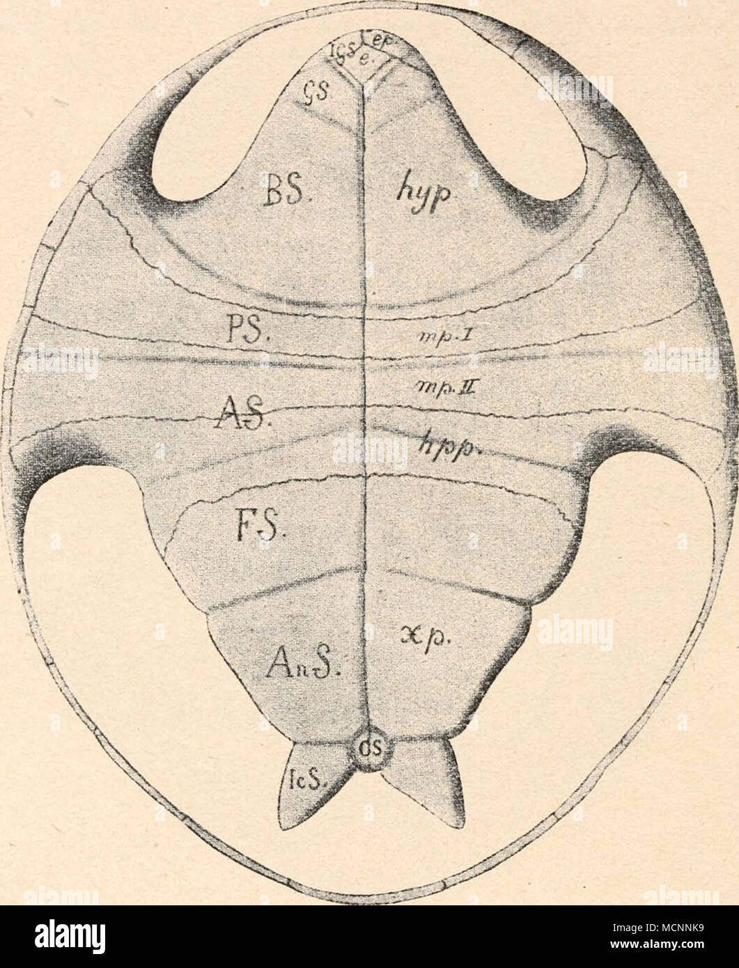 . Fig. 319. Plastron von Proterochers is (vgl. Fig. 318.) e. = Entoplastron. GS. = Gularscutum. ep. = Epiplastron. BS. = Brachialscutum. hyp. = Hyoplastron. AS. = Abdominalscutum. mp. I = vorderes Mesoplastron. FS. = Femoralscutum. mp. 11 = hinteres Mesoplastron. AnS. = Analscutnm. hpp. = Hypoplastron. IcS. = Caudalscutum. xp. = Xiphiplastron. CS. = Intercandalscutum 1GS. Intergularscutum. Arten vor, deren Zugehörigkeit zu den Pleurodiren aus der Verwachsung des Beckens mit dem Plastron hervorgeht. Diese Arten verteilen sich 1 E. Fraas, Proterochersis, eine plenrodire Schildkröte aus dem Keupe Stock Photohttps://www.alamy.com/image-license-details/?v=1https://www.alamy.com/fig-319-plastron-von-proterochers-is-vgl-fig-318-e-=-entoplastron-gs-=-gularscutum-ep-=-epiplastron-bs-=-brachialscutum-hyp-=-hyoplastron-as-=-abdominalscutum-mp-i-=-vorderes-mesoplastron-fs-=-femoralscutum-mp-11-=-hinteres-mesoplastron-ans-=-analscutnm-hpp-=-hypoplastron-ics-=-caudalscutum-xp-=-xiphiplastron-cs-=-intercandalscutum-1gs-intergularscutum-arten-vor-deren-zugehrigkeit-zu-den-pleurodiren-aus-der-verwachsung-des-beckens-mit-dem-plastron-hervorgeht-diese-arten-verteilen-sich-1-e-fraas-proterochersis-eine-plenrodire-schildkrte-aus-dem-keupe-image179957549.html
RMMCNNK9–. Fig. 319. Plastron von Proterochers is (vgl. Fig. 318.) e. = Entoplastron. GS. = Gularscutum. ep. = Epiplastron. BS. = Brachialscutum. hyp. = Hyoplastron. AS. = Abdominalscutum. mp. I = vorderes Mesoplastron. FS. = Femoralscutum. mp. 11 = hinteres Mesoplastron. AnS. = Analscutnm. hpp. = Hypoplastron. IcS. = Caudalscutum. xp. = Xiphiplastron. CS. = Intercandalscutum 1GS. Intergularscutum. Arten vor, deren Zugehörigkeit zu den Pleurodiren aus der Verwachsung des Beckens mit dem Plastron hervorgeht. Diese Arten verteilen sich 1 E. Fraas, Proterochersis, eine plenrodire Schildkröte aus dem Keupe
 Elemente der paläontologie bearbeitet (1890) Elemente der paläontologie bearbeitet elementederpal00stei Year: 1890 636 I. Thiei'i'eicli. — X. Vertebraln. Ivlasse: Sauropsida. Testudinala. platten median zusammenstossen können, von Europa. Von geringer Grösse. Jura Fig. 780. Tliulassemjjs Uugi Eüt. Oberer Jnra von Neuf- chätel. Bauchschild von aussen mit grosser Fontanelle {F) in der Mitte, es = Epiplastron; hs = Hyoplastren; lip = Hypoplastron; xs = Xiptiplastrou. ;. I-ilzingerl H. v. M. (Fig. 778). Lithograpliisclier Scliiefer von Bayern und von Cirin bei Lyon. Thalassemys Rülim.(Fig. 780 Stock Photohttps://www.alamy.com/image-license-details/?v=1https://www.alamy.com/elemente-der-palontologie-bearbeitet-1890-elemente-der-palontologie-bearbeitet-elementederpal00stei-year-1890-636-i-thieiieicli-x-vertebraln-ivlasse-sauropsida-testudinala-platten-median-zusammenstossen-knnen-von-europa-von-geringer-grsse-jura-fig-780-tliulassemjjs-uugi-et-oberer-jnra-von-neuf-chtel-bauchschild-von-aussen-mit-grosser-fontanelle-f-in-der-mitte-es-=-epiplastron-hs-=-hyoplastren-lip-=-hypoplastron-xs-=-xiptiplastrou-i-ilzingerl-h-v-m-fig-778-lithograpliisclier-scliiefer-von-bayern-und-von-cirin-bei-lyon-thalassemys-rlimfig-780-image239666128.html
RMRWWMGG–Elemente der paläontologie bearbeitet (1890) Elemente der paläontologie bearbeitet elementederpal00stei Year: 1890 636 I. Thiei'i'eicli. — X. Vertebraln. Ivlasse: Sauropsida. Testudinala. platten median zusammenstossen können, von Europa. Von geringer Grösse. Jura Fig. 780. Tliulassemjjs Uugi Eüt. Oberer Jnra von Neuf- chätel. Bauchschild von aussen mit grosser Fontanelle {F) in der Mitte, es = Epiplastron; hs = Hyoplastren; lip = Hypoplastron; xs = Xiptiplastrou. ;. I-ilzingerl H. v. M. (Fig. 778). Lithograpliisclier Scliiefer von Bayern und von Cirin bei Lyon. Thalassemys Rülim.(Fig. 780
 Sea turtle bones, Christmas (Kiritimati) Island, Kiribati Stock Photohttps://www.alamy.com/image-license-details/?v=1https://www.alamy.com/stock-photo-sea-turtle-bones-christmas-kiritimati-island-kiribati-126711276.html
RFHA45EM–Sea turtle bones, Christmas (Kiritimati) Island, Kiribati
 Ventral view of a baby turtle skeleton; Southbridge, Massachusetts. Stock Photohttps://www.alamy.com/image-license-details/?v=1https://www.alamy.com/stock-photo-ventral-view-of-a-baby-turtle-skeleton-southbridge-massachusetts-99907881.html
RMFPF5EH–Ventral view of a baby turtle skeleton; Southbridge, Massachusetts.
 Sea turtle bones, Christmas (Kiritimati) Island, Kiribati Stock Photohttps://www.alamy.com/image-license-details/?v=1https://www.alamy.com/stock-photo-sea-turtle-bones-christmas-kiritimati-island-kiribati-126711272.html
RFHA45EG–Sea turtle bones, Christmas (Kiritimati) Island, Kiribati
 . Carnegie Institution of Washington publication. i uniNYCHin.*:. 52i The xiphiplastra are missing; but the notches in the right hypoplastron show that these hones were articulated as in Platypeltis. Three cervical vertebrae were exposed in preparing the plastron, the sixth, seventh, and eighth. All of these are turned backward, as if the head had been retracted within the shell. The sixth has a length of 75 mm. The structure of these vertebrae resembles that ot P. frrox. There is present also one ilium, but there appears to be nothing distinctive about it. This species is very distinct from A Stock Photohttps://www.alamy.com/image-license-details/?v=1https://www.alamy.com/carnegie-institution-of-washington-publication-i-uninychin-52i-the-xiphiplastra-are-missing-but-the-notches-in-the-right-hypoplastron-show-that-these-hones-were-articulated-as-in-platypeltis-three-cervical-vertebrae-were-exposed-in-preparing-the-plastron-the-sixth-seventh-and-eighth-all-of-these-are-turned-backward-as-if-the-head-had-been-retracted-within-the-shell-the-sixth-has-a-length-of-75-mm-the-structure-of-these-vertebrae-resembles-that-ot-p-frrox-there-is-present-also-one-ilium-but-there-appears-to-be-nothing-distinctive-about-it-this-species-is-very-distinct-from-a-image233475313.html
RMRFRM41–. Carnegie Institution of Washington publication. i uniNYCHin.*:. 52i The xiphiplastra are missing; but the notches in the right hypoplastron show that these hones were articulated as in Platypeltis. Three cervical vertebrae were exposed in preparing the plastron, the sixth, seventh, and eighth. All of these are turned backward, as if the head had been retracted within the shell. The sixth has a length of 75 mm. The structure of these vertebrae resembles that ot P. frrox. There is present also one ilium, but there appears to be nothing distinctive about it. This species is very distinct from A
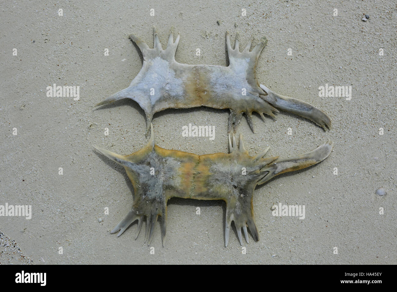 Sea turtle bones, Christmas (Kiritimati) Island, Kiribati Stock Photohttps://www.alamy.com/image-license-details/?v=1https://www.alamy.com/stock-photo-sea-turtle-bones-christmas-kiritimati-island-kiribati-126711283.html
RFHA45EY–Sea turtle bones, Christmas (Kiritimati) Island, Kiribati
 Elemente der paläontologie bearbeitet (1890) Elemente der paläontologie bearbeitet elementederpal00stei Year: 1890 632 '• Tliierreicli. — . Vertebrata. — 5. Klasse: Sauropsida. Testudinata. Zur Verbindung des Bauch- und Rückenschildes inil einander selu'ckl das Hyoplaslron und Hypoplastron Fortsätze (Flügel) aus, die entweder mit fingerförmigen Enden (Fig. 778 Iis u. i) sich nur locker an dns Hüeken- schild legen, oder kräftige senkrecht aufsteigende Axillar- und Inguinal- p feil er bilden, welche durch Ligament fester mit den Marginal- und selbst Costalplatten verbunden sind; dieursprUng Stock Photohttps://www.alamy.com/image-license-details/?v=1https://www.alamy.com/elemente-der-palontologie-bearbeitet-1890-elemente-der-palontologie-bearbeitet-elementederpal00stei-year-1890-632-tliierreicli-vertebrata-5-klasse-sauropsida-testudinata-zur-verbindung-des-bauch-und-rckenschildes-inil-einander-seluckl-das-hyoplaslron-und-hypoplastron-fortstze-flgel-aus-die-entweder-mit-fingerfrmigen-enden-fig-778-iis-u-i-sich-nur-locker-an-dns-heken-schild-legen-oder-krftige-senkrecht-aufsteigende-axillar-und-inguinal-p-feil-er-bilden-welche-durch-ligament-fester-mit-den-marginal-und-selbst-costalplatten-verbunden-sind-dieursprung-image239666070.html
RMRWWMEE–Elemente der paläontologie bearbeitet (1890) Elemente der paläontologie bearbeitet elementederpal00stei Year: 1890 632 '• Tliierreicli. — . Vertebrata. — 5. Klasse: Sauropsida. Testudinata. Zur Verbindung des Bauch- und Rückenschildes inil einander selu'ckl das Hyoplaslron und Hypoplastron Fortsätze (Flügel) aus, die entweder mit fingerförmigen Enden (Fig. 778 Iis u. i) sich nur locker an dns Hüeken- schild legen, oder kräftige senkrecht aufsteigende Axillar- und Inguinal- p feil er bilden, welche durch Ligament fester mit den Marginal- und selbst Costalplatten verbunden sind; dieursprUng
 . Einführung in die vergleichende Anatomie der Wirbeltiere, für Studierende . einem A und B Carapax und Plastron einer jungen Testudo graeca, "on von Chelone midas. C, C Costalplatten, E Entoplastron, das vielleicht ^.sternum entspricht, Ep Epiplastron, das vielleicht einer Clavicula entspricht, J?p Hypoplastron, Hy Hyoplastron, M, M Marginalplatten, N, iV Neuralplatten, iSi^p Nuehal- platte, Pij, Py Pygalplatten, R, R Rippen, A''( Xiphiplastron. (T" bedeutet vorne, If hinten.) SO aus drei Elementen zusammengesetzten Spangen durchsetzen den geraden Bauchmuskel, ohne sich jedoch mit Stock Photohttps://www.alamy.com/image-license-details/?v=1https://www.alamy.com/einfhrung-in-die-vergleichende-anatomie-der-wirbeltiere-fr-studierende-einem-a-und-b-carapax-und-plastron-einer-jungen-testudo-graeca-quoton-von-chelone-midas-c-c-costalplatten-e-entoplastron-das-vielleicht-sternum-entspricht-ep-epiplastron-das-vielleicht-einer-clavicula-entspricht-jp-hypoplastron-hy-hyoplastron-m-m-marginalplatten-n-iv-neuralplatten-isip-nuehal-platte-pij-py-pygalplatten-r-r-rippen-a-xiphiplastron-tquot-bedeutet-vorne-if-hinten-so-aus-drei-elementen-zusammengesetzten-spangen-durchsetzen-den-geraden-bauchmuskel-ohne-sich-jedoch-mit-image178415416.html
RMMA7EK4–. Einführung in die vergleichende Anatomie der Wirbeltiere, für Studierende . einem A und B Carapax und Plastron einer jungen Testudo graeca, "on von Chelone midas. C, C Costalplatten, E Entoplastron, das vielleicht ^.sternum entspricht, Ep Epiplastron, das vielleicht einer Clavicula entspricht, J?p Hypoplastron, Hy Hyoplastron, M, M Marginalplatten, N, iV Neuralplatten, iSi^p Nuehal- platte, Pij, Py Pygalplatten, R, R Rippen, A''( Xiphiplastron. (T" bedeutet vorne, If hinten.) SO aus drei Elementen zusammengesetzten Spangen durchsetzen den geraden Bauchmuskel, ohne sich jedoch mit
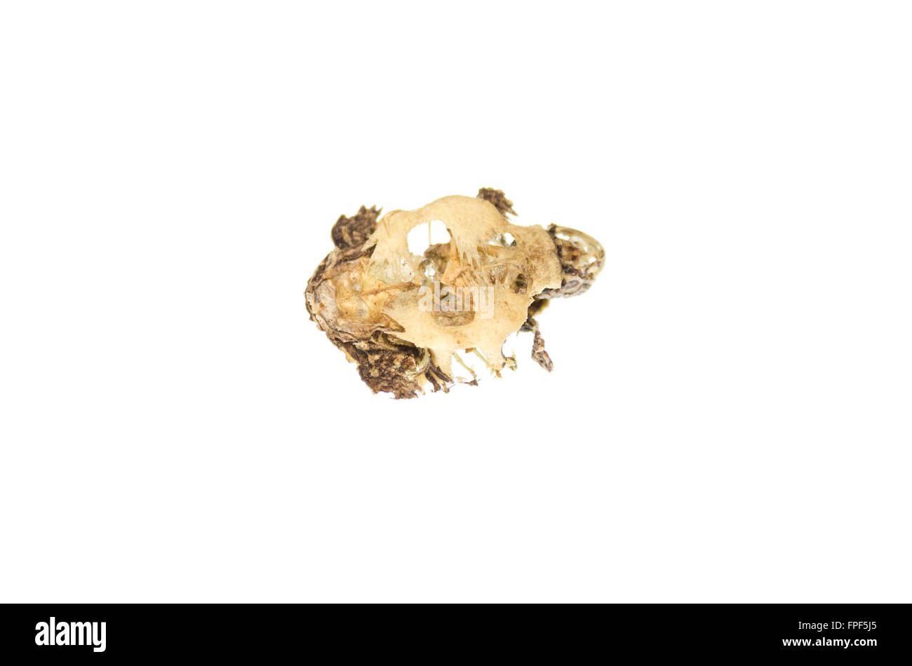 Ventral view of a baby turtle skeleton; Southbridge, Massachusetts. Stock Photohttps://www.alamy.com/image-license-details/?v=1https://www.alamy.com/stock-photo-ventral-view-of-a-baby-turtle-skeleton-southbridge-massachusetts-99907981.html
RMFPF5J5–Ventral view of a baby turtle skeleton; Southbridge, Massachusetts.
 . Anatomical and zoological researches: comprising an account of the zoological results of the two expeditions to western Yunnan in 1868 and 1875; and a monograph of the two cetacean genera, Platanista and Orcella. CHELONIA. 789 from Singapore, and it may be it is identical with species of Trionyx from Burma whicli Theobald has described as having a smooth carapace. The figure shows the remains of what appears to have been a dark reticulation of the upper surface of the head. • The plastron of T. stellatus is represented as having its hyoplastron and hypoplastron and xiphiplastron covered with Stock Photohttps://www.alamy.com/image-license-details/?v=1https://www.alamy.com/anatomical-and-zoological-researches-comprising-an-account-of-the-zoological-results-of-the-two-expeditions-to-western-yunnan-in-1868-and-1875-and-a-monograph-of-the-two-cetacean-genera-platanista-and-orcella-chelonia-789-from-singapore-and-it-may-be-it-is-identical-with-species-of-trionyx-from-burma-whicli-theobald-has-described-as-having-a-smooth-carapace-the-figure-shows-the-remains-of-what-appears-to-have-been-a-dark-reticulation-of-the-upper-surface-of-the-head-the-plastron-of-t-stellatus-is-represented-as-having-its-hyoplastron-and-hypoplastron-and-xiphiplastron-covered-with-image236859580.html
RMRN9TPM–. Anatomical and zoological researches: comprising an account of the zoological results of the two expeditions to western Yunnan in 1868 and 1875; and a monograph of the two cetacean genera, Platanista and Orcella. CHELONIA. 789 from Singapore, and it may be it is identical with species of Trionyx from Burma whicli Theobald has described as having a smooth carapace. The figure shows the remains of what appears to have been a dark reticulation of the upper surface of the head. • The plastron of T. stellatus is represented as having its hyoplastron and hypoplastron and xiphiplastron covered with
 Sea turtle bone, Christmas (Kiritimati) Island, Kiribati Stock Photohttps://www.alamy.com/image-license-details/?v=1https://www.alamy.com/stock-photo-sea-turtle-bone-christmas-kiritimati-island-kiribati-126711287.html
RFHA45F3–Sea turtle bone, Christmas (Kiritimati) Island, Kiribati
 Elemente der paläontologie bearbeitet (1890) Elemente der paläontologie bearbeitet elementederpal00stei Year: 1890 Fig. HC). Eurystefutnn ^Vagleii H. v. M. Oberer Malm von Sulnhofen. Skelet von oben, cj, ci, cs = Costalplatten; hi* = Hypoplastron (Baurhschild); hs = Hyoplastron (Baucbschild); il = Ileum; ni, («5, ins, Ulli = Margiualplatten; in, Ui, )ta = Neuralplatten; lui = Nuchalplatte ; pu = Pygal- platte. Auf ci, es, f5, cs sind Eindrücke der Horn- scbilder sichtbar. Fig. 777. FUsiocIidi/s Efalhni Pict. Oberer Malm von St. Claude (Scliweiz). Baudrschild von aussen mit kleiner mittler Stock Photohttps://www.alamy.com/image-license-details/?v=1https://www.alamy.com/elemente-der-palontologie-bearbeitet-1890-elemente-der-palontologie-bearbeitet-elementederpal00stei-year-1890-fig-hc-eurystefutnn-vagleii-h-v-m-oberer-malm-von-sulnhofen-skelet-von-oben-cj-ci-cs-=-costalplatten-hi-=-hypoplastron-baurhschild-hs-=-hyoplastron-baucbschild-il-=-ileum-ni-5-ins-ulli-=-margiualplatten-in-ui-ta-=-neuralplatten-lui-=-nuchalplatte-pu-=-pygal-platte-auf-ci-es-f5-cs-sind-eindrcke-der-horn-scbilder-sichtbar-fig-777-fusiociidis-efalhni-pict-oberer-malm-von-st-claude-scliweiz-baudrschild-von-aussen-mit-kleiner-mittler-image239666062.html
RMRWWME6–Elemente der paläontologie bearbeitet (1890) Elemente der paläontologie bearbeitet elementederpal00stei Year: 1890 Fig. HC). Eurystefutnn ^Vagleii H. v. M. Oberer Malm von Sulnhofen. Skelet von oben, cj, ci, cs = Costalplatten; hi* = Hypoplastron (Baurhschild); hs = Hyoplastron (Baucbschild); il = Ileum; ni, («5, ins, Ulli = Margiualplatten; in, Ui, )ta = Neuralplatten; lui = Nuchalplatte ; pu = Pygal- platte. Auf ci, es, f5, cs sind Eindrücke der Horn- scbilder sichtbar. Fig. 777. FUsiocIidi/s Efalhni Pict. Oberer Malm von St. Claude (Scliweiz). Baudrschild von aussen mit kleiner mittler
 . Hypo. Fig. 314. Protosphargis veronensis, Capellini, aus der oberen Kreide von Verona, in 1/16 nat. Gr. R. =< Rippen. Hyo. = Hyoplastron. M. = Marginalia. Hypo. = Hypoplastron. Epip. = Epiplastron. Xiph. = Xiphiplastron. F. Dermochelyidae. Sind auch die Gattungen, welche die Ahnenformen der Dermo- chelyiden bilden, bisher noch nicht bekannt, so ist doch der Weg voll- kommen klargelegt, den die Entwicklung dieser Familie aus den Cheloniiden genommen hat. Vielleicht wurzeln die Dermochelyiden in den älteren Thalassemydiden, so daß die weitgehenden Ähnlich- 1 R. Lydekker, Catalogue of the Fo Stock Photohttps://www.alamy.com/image-license-details/?v=1https://www.alamy.com/hypo-fig-314-protosphargis-veronensis-capellini-aus-der-oberen-kreide-von-verona-in-116-nat-gr-r-=lt-rippen-hyo-=-hyoplastron-m-=-marginalia-hypo-=-hypoplastron-epip-=-epiplastron-xiph-=-xiphiplastron-f-dermochelyidae-sind-auch-die-gattungen-welche-die-ahnenformen-der-dermo-chelyiden-bilden-bisher-noch-nicht-bekannt-so-ist-doch-der-weg-voll-kommen-klargelegt-den-die-entwicklung-dieser-familie-aus-den-cheloniiden-genommen-hat-vielleicht-wurzeln-die-dermochelyiden-in-den-lteren-thalassemydiden-so-da-die-weitgehenden-hnlich-1-r-lydekker-catalogue-of-the-fo-image179957568.html
RMMCNNM0–. Hypo. Fig. 314. Protosphargis veronensis, Capellini, aus der oberen Kreide von Verona, in 1/16 nat. Gr. R. =< Rippen. Hyo. = Hyoplastron. M. = Marginalia. Hypo. = Hypoplastron. Epip. = Epiplastron. Xiph. = Xiphiplastron. F. Dermochelyidae. Sind auch die Gattungen, welche die Ahnenformen der Dermo- chelyiden bilden, bisher noch nicht bekannt, so ist doch der Weg voll- kommen klargelegt, den die Entwicklung dieser Familie aus den Cheloniiden genommen hat. Vielleicht wurzeln die Dermochelyiden in den älteren Thalassemydiden, so daß die weitgehenden Ähnlich- 1 R. Lydekker, Catalogue of the Fo
 . Carnegie Institution of Washington publication. ism hki n did.i . '•5 The entoplastron must have been unusually large. Its anterior end approacht within }2 mm. of the anterior border. The anterior angle was slightly greater than ioo°. The sides hounding this angle were approximately 130 mm. long. The width must have been about ido nun. The interior surface of the epiplastron displays an obscure reticulation, but the hypoplastron is smooth. There are present z fragments of the right xiphiplastron. One of these is the hinder angle and bears the ischiadic articulation; the other shows the bott Stock Photohttps://www.alamy.com/image-license-details/?v=1https://www.alamy.com/carnegie-institution-of-washington-publication-ism-hki-n-didi-5-the-entoplastron-must-have-been-unusually-large-its-anterior-end-approacht-within-2-mm-of-the-anterior-border-the-anterior-angle-was-slightly-greater-than-ioo-the-sides-hounding-this-angle-were-approximately-130-mm-long-the-width-must-have-been-about-ido-nun-the-interior-surface-of-the-epiplastron-displays-an-obscure-reticulation-but-the-hypoplastron-is-smooth-there-are-present-z-fragments-of-the-right-xiphiplastron-one-of-these-is-the-hinder-angle-and-bears-the-ischiadic-articulation-the-other-shows-the-bott-image233486650.html
RMRFT6GX–. Carnegie Institution of Washington publication. ism hki n did.i . '•5 The entoplastron must have been unusually large. Its anterior end approacht within }2 mm. of the anterior border. The anterior angle was slightly greater than ioo°. The sides hounding this angle were approximately 130 mm. long. The width must have been about ido nun. The interior surface of the epiplastron displays an obscure reticulation, but the hypoplastron is smooth. There are present z fragments of the right xiphiplastron. One of these is the hinder angle and bears the ischiadic articulation; the other shows the bott
 . Carnegie Institution of Washington publication. IiOTHRKMYI)II>.-K "J mesoplastron continued to the midline. This is not probable. The mesoplastron evidently extended inward only to the change in the direction of the anterior border of the hypoplastron; that is about one-third the distance from the peripherals to the midline. The inguinal buttress is prominent, but thin. The hinder lobe narrowed rapidly, so that, while about 150 mm. wide at the inguinal notch, it was only about 120 mm. at the hypo- xiphiplastral suture. The thickness of the bone behind the inguinal notch is 4 mm. Only Stock Photohttps://www.alamy.com/image-license-details/?v=1https://www.alamy.com/carnegie-institution-of-washington-publication-iiothrkmyiiigt-k-quotj-mesoplastron-continued-to-the-midline-this-is-not-probable-the-mesoplastron-evidently-extended-inward-only-to-the-change-in-the-direction-of-the-anterior-border-of-the-hypoplastron-that-is-about-one-third-the-distance-from-the-peripherals-to-the-midline-the-inguinal-buttress-is-prominent-but-thin-the-hinder-lobe-narrowed-rapidly-so-that-while-about-150-mm-wide-at-the-inguinal-notch-it-was-only-about-120-mm-at-the-hypo-xiphiplastral-suture-the-thickness-of-the-bone-behind-the-inguinal-notch-is-4-mm-only-image233486669.html
RMRFT6HH–. Carnegie Institution of Washington publication. IiOTHRKMYI)II>.-K "J mesoplastron continued to the midline. This is not probable. The mesoplastron evidently extended inward only to the change in the direction of the anterior border of the hypoplastron; that is about one-third the distance from the peripherals to the midline. The inguinal buttress is prominent, but thin. The hinder lobe narrowed rapidly, so that, while about 150 mm. wide at the inguinal notch, it was only about 120 mm. at the hypo- xiphiplastral suture. The thickness of the bone behind the inguinal notch is 4 mm. Only
 . Carnegie Institution of Washington publication. [66 FOSSIL TURTLES OF NORTH AMERICA. hind legs was much broader than it is in the case of Chelydra; and, as in the sea-turtles, there was a fontanel between the outer end of the hyoplastron and hypoplastron. There was also an extensive umbilical fontanel. The bones of the right and left sides were joined along the midline more like those of Chelydra than like those of the Cheloniidae. The epiplastron and entoplastron are figured by Wieland (Amer. Jour. Sci., xx, p. 336, figs. 7, 8). The former are slender, the latter broad and lance-shaped. The Stock Photohttps://www.alamy.com/image-license-details/?v=1https://www.alamy.com/carnegie-institution-of-washington-publication-66-fossil-turtles-of-north-america-hind-legs-was-much-broader-than-it-is-in-the-case-of-chelydra-and-as-in-the-sea-turtles-there-was-a-fontanel-between-the-outer-end-of-the-hyoplastron-and-hypoplastron-there-was-also-an-extensive-umbilical-fontanel-the-bones-of-the-right-and-left-sides-were-joined-along-the-midline-more-like-those-of-chelydra-than-like-those-of-the-cheloniidae-the-epiplastron-and-entoplastron-are-figured-by-wieland-amer-jour-sci-xx-p-336-figs-7-8-the-former-are-slender-the-latter-broad-and-lance-shaped-the-image233486225.html
RMRFT61N–. Carnegie Institution of Washington publication. [66 FOSSIL TURTLES OF NORTH AMERICA. hind legs was much broader than it is in the case of Chelydra; and, as in the sea-turtles, there was a fontanel between the outer end of the hyoplastron and hypoplastron. There was also an extensive umbilical fontanel. The bones of the right and left sides were joined along the midline more like those of Chelydra than like those of the Cheloniidae. The epiplastron and entoplastron are figured by Wieland (Amer. Jour. Sci., xx, p. 336, figs. 7, 8). The former are slender, the latter broad and lance-shaped. The
 . Carnegie Institution of Washington publication. 256 FOSSIL TURTLES OF NORTH AMERICA. hypoplasticm a distance of 14 mm.; while the right hypoplastron comes into contact with the left xiphiplastron. The greatest thickness of the hyoplastron and hypoplastron is 18 mm. The distance between the entoplastron and the xiphiplastron is 84 mm. Half of this may be regarded as belonging to the hyoplastrals; the other half to the hypoplastron. The bridge is 100 mm. wide. The hinder lobe is much reduced. The width at the base is about 68 mm.; its length, 48 mm. The greatest thickness of the xiphiplastra i Stock Photohttps://www.alamy.com/image-license-details/?v=1https://www.alamy.com/carnegie-institution-of-washington-publication-256-fossil-turtles-of-north-america-hypoplasticm-a-distance-of-14-mm-while-the-right-hypoplastron-comes-into-contact-with-the-left-xiphiplastron-the-greatest-thickness-of-the-hyoplastron-and-hypoplastron-is-18-mm-the-distance-between-the-entoplastron-and-the-xiphiplastron-is-84-mm-half-of-this-may-be-regarded-as-belonging-to-the-hyoplastrals-the-other-half-to-the-hypoplastron-the-bridge-is-100-mm-wide-the-hinder-lobe-is-much-reduced-the-width-at-the-base-is-about-68-mm-its-length-48-mm-the-greatest-thickness-of-the-xiphiplastra-i-image233477035.html
RMRFRP9F–. Carnegie Institution of Washington publication. 256 FOSSIL TURTLES OF NORTH AMERICA. hypoplasticm a distance of 14 mm.; while the right hypoplastron comes into contact with the left xiphiplastron. The greatest thickness of the hyoplastron and hypoplastron is 18 mm. The distance between the entoplastron and the xiphiplastron is 84 mm. Half of this may be regarded as belonging to the hyoplastrals; the other half to the hypoplastron. The bridge is 100 mm. wide. The hinder lobe is much reduced. The width at the base is about 68 mm.; its length, 48 mm. The greatest thickness of the xiphiplastra i
 . Carnegie Institution of Washington publication. 5*8 FOSSIL TURTLES OF NORTH AMERICA. The plastron (plate 106, fig. i) has a breadth, from the extremity of the hyoplastral processes of one side to those of the other, ot about 310 mm. The outer fourth ot the hyoplas- tron of each side has coalesct with the corresponding hvpoplasf ion. The transverse extent of these bones, along this suture, is 133 mm. Where narrowest the bridges are 48 mm. wide. The hyoplastron and hypo- plastron have a close resemblance to those of Platypehis ferox. The hypoplastron differs from that of the species just named Stock Photohttps://www.alamy.com/image-license-details/?v=1https://www.alamy.com/carnegie-institution-of-washington-publication-58-fossil-turtles-of-north-america-the-plastron-plate-106-fig-i-has-a-breadth-from-the-extremity-of-the-hyoplastral-processes-of-one-side-to-those-of-the-other-ot-about-310-mm-the-outer-fourth-ot-the-hyoplas-tron-of-each-side-has-coalesct-with-the-corresponding-hvpoplasf-ion-the-transverse-extent-of-these-bones-along-this-suture-is-133-mm-where-narrowest-the-bridges-are-48-mm-wide-the-hyoplastron-and-hypo-plastron-have-a-close-resemblance-to-those-of-platypehis-ferox-the-hypoplastron-differs-from-that-of-the-species-just-named-image233475253.html
RMRFRM1W–. Carnegie Institution of Washington publication. 5*8 FOSSIL TURTLES OF NORTH AMERICA. The plastron (plate 106, fig. i) has a breadth, from the extremity of the hyoplastral processes of one side to those of the other, ot about 310 mm. The outer fourth ot the hyoplas- tron of each side has coalesct with the corresponding hvpoplasf ion. The transverse extent of these bones, along this suture, is 133 mm. Where narrowest the bridges are 48 mm. wide. The hyoplastron and hypo- plastron have a close resemblance to those of Platypehis ferox. The hypoplastron differs from that of the species just named
 . Carnegie Institution of Washington publication. 1)1 RMATKMYOID^E. 255 The materials possest by Leidy when he described this species consisted of the third, sixth, and the seventh left peripherals, the sixth, seventh, and eighth peripherals of the right side, a portion of the left hyoplastron, and a portion of the right hypoplastron. These had been placed in Dr. Leidy's hands by Professor George H. Cook, of the New Jersey Geological Survey. They had been found in the uppermost Cretaceous greensand, at Tinton Falls, Monmouth County, New Jersey. The sixth and seventh left peripherals and the pa Stock Photohttps://www.alamy.com/image-license-details/?v=1https://www.alamy.com/carnegie-institution-of-washington-publication-11-rmatkmyoide-255-the-materials-possest-by-leidy-when-he-described-this-species-consisted-of-the-third-sixth-and-the-seventh-left-peripherals-the-sixth-seventh-and-eighth-peripherals-of-the-right-side-a-portion-of-the-left-hyoplastron-and-a-portion-of-the-right-hypoplastron-these-had-been-placed-in-dr-leidys-hands-by-professor-george-h-cook-of-the-new-jersey-geological-survey-they-had-been-found-in-the-uppermost-cretaceous-greensand-at-tinton-falls-monmouth-county-new-jersey-the-sixth-and-seventh-left-peripherals-and-the-pa-image233477044.html
RMRFRP9T–. Carnegie Institution of Washington publication. 1)1 RMATKMYOID^E. 255 The materials possest by Leidy when he described this species consisted of the third, sixth, and the seventh left peripherals, the sixth, seventh, and eighth peripherals of the right side, a portion of the left hyoplastron, and a portion of the right hypoplastron. These had been placed in Dr. Leidy's hands by Professor George H. Cook, of the New Jersey Geological Survey. They had been found in the uppermost Cretaceous greensand, at Tinton Falls, Monmouth County, New Jersey. The sixth and seventh left peripherals and the pa
 . Comparative anatomy of vertebrates. Anatomy, Comparative; Vertebrates -- Anatomy. FIG. 34.—Plastron of Trionyx. en, ento- plastron; ep, epiplastron; hpp, hypoplastron; hyp, hyoplastron; xp, xiphiplastron. FIG. 35.—Ventral ends of ribs (r) and gastralia (g) of Sphenodon. only in the armadillos where they form a complete armor above, the plates arranged- in transverse rows, some of which are movable on each other. In the extinct glyptodons they formed an inflexible case. It is uncertain whether these are a new acquisition in the edentates or have been inherited from non-mammalian ancestors. TH Stock Photohttps://www.alamy.com/image-license-details/?v=1https://www.alamy.com/comparative-anatomy-of-vertebrates-anatomy-comparative-vertebrates-anatomy-fig-34plastron-of-trionyx-en-ento-plastron-ep-epiplastron-hpp-hypoplastron-hyp-hyoplastron-xp-xiphiplastron-fig-35ventral-ends-of-ribs-r-and-gastralia-g-of-sphenodon-only-in-the-armadillos-where-they-form-a-complete-armor-above-the-plates-arranged-in-transverse-rows-some-of-which-are-movable-on-each-other-in-the-extinct-glyptodons-they-formed-an-inflexible-case-it-is-uncertain-whether-these-are-a-new-acquisition-in-the-edentates-or-have-been-inherited-from-non-mammalian-ancestors-th-image232680600.html
RMREFEDC–. Comparative anatomy of vertebrates. Anatomy, Comparative; Vertebrates -- Anatomy. FIG. 34.—Plastron of Trionyx. en, ento- plastron; ep, epiplastron; hpp, hypoplastron; hyp, hyoplastron; xp, xiphiplastron. FIG. 35.—Ventral ends of ribs (r) and gastralia (g) of Sphenodon. only in the armadillos where they form a complete armor above, the plates arranged- in transverse rows, some of which are movable on each other. In the extinct glyptodons they formed an inflexible case. It is uncertain whether these are a new acquisition in the edentates or have been inherited from non-mammalian ancestors. TH
 . Carnegie Institution of Washington publication. TRIONYCHIDtE. 501 half. Toward the midline the hypoplastron becomes much wider than the hvoplastron. The hypoplastron sends inward a finger-like process, which no doubt joined a similar process from the bone of the opposite side. The median border of both bones was very obliquely- beveled. The greatest thickness of the hypoplastron is 12 mm. The xiphiplastron present appears to belong to the right side. It has a central, thick portion, sculptured, oval, 25 mm. wide, and about 40 mm. long. This passes by a bevel into the thinner portion on all s Stock Photohttps://www.alamy.com/image-license-details/?v=1https://www.alamy.com/carnegie-institution-of-washington-publication-trionychidte-501-half-toward-the-midline-the-hypoplastron-becomes-much-wider-than-the-hvoplastron-the-hypoplastron-sends-inward-a-finger-like-process-which-no-doubt-joined-a-similar-process-from-the-bone-of-the-opposite-side-the-median-border-of-both-bones-was-very-obliquely-beveled-the-greatest-thickness-of-the-hypoplastron-is-12-mm-the-xiphiplastron-present-appears-to-belong-to-the-right-side-it-has-a-central-thick-portion-sculptured-oval-25-mm-wide-and-about-40-mm-long-this-passes-by-a-bevel-into-the-thinner-portion-on-all-s-image233475464.html
RMRFRM9C–. Carnegie Institution of Washington publication. TRIONYCHIDtE. 501 half. Toward the midline the hypoplastron becomes much wider than the hvoplastron. The hypoplastron sends inward a finger-like process, which no doubt joined a similar process from the bone of the opposite side. The median border of both bones was very obliquely- beveled. The greatest thickness of the hypoplastron is 12 mm. The xiphiplastron present appears to belong to the right side. It has a central, thick portion, sculptured, oval, 25 mm. wide, and about 40 mm. long. This passes by a bevel into the thinner portion on all s
 . Carnegie Institution of Washington publication. 388 FOSSIL TURTLES OF NORTH AMERICA. plastra on each side of this are rounded. The bone of the plastron is relatively thick. In the case of a specimen whose plastron is 210 mm. long the thickness of the hyoplastron is 16 mm.; that of the hypoplastron half-way between the median line and the inguinal notch is 14 mm. Proceeding backward from the inguinal notch near the free border there is a low wall from the summit of which the bone slopes downward and outward. Farther backward the height of the wall diminishes rapidly and the slope is less stee Stock Photohttps://www.alamy.com/image-license-details/?v=1https://www.alamy.com/carnegie-institution-of-washington-publication-388-fossil-turtles-of-north-america-plastra-on-each-side-of-this-are-rounded-the-bone-of-the-plastron-is-relatively-thick-in-the-case-of-a-specimen-whose-plastron-is-210-mm-long-the-thickness-of-the-hyoplastron-is-16-mm-that-of-the-hypoplastron-half-way-between-the-median-line-and-the-inguinal-notch-is-14-mm-proceeding-backward-from-the-inguinal-notch-near-the-free-border-there-is-a-low-wall-from-the-summit-of-which-the-bone-slopes-downward-and-outward-farther-backward-the-height-of-the-wall-diminishes-rapidly-and-the-slope-is-less-stee-image233476124.html
RMRFRN50–. Carnegie Institution of Washington publication. 388 FOSSIL TURTLES OF NORTH AMERICA. plastra on each side of this are rounded. The bone of the plastron is relatively thick. In the case of a specimen whose plastron is 210 mm. long the thickness of the hyoplastron is 16 mm.; that of the hypoplastron half-way between the median line and the inguinal notch is 14 mm. Proceeding backward from the inguinal notch near the free border there is a low wall from the summit of which the bone slopes downward and outward. Farther backward the height of the wall diminishes rapidly and the slope is less stee
 . Carnegie Institution of Washington publication. ISOTrIKIlYI)ll>.r . S eithei the inguinal iiMjiiin iii the both measured at the midline. Neithei hypoplastron exhibit border for the mesoplastron. The hyoplastron overlapt somewhat the hypoplastron, and the latter similarly overlapt the xiphiplastron. t the antero-interior angle the hypoplastic are 10 mm. thick; at the postero-interior angle. 8 mm. The whole tree border of the hinder lobe is acute. The hinder notch is 1^5 mm. wide, and 42 mm. deep. ( )n the upper surface of the xiphiplastron (fig. 1 iS 1. FlGS. 117 119.—Taphrosphys molops. Stock Photohttps://www.alamy.com/image-license-details/?v=1https://www.alamy.com/carnegie-institution-of-washington-publication-isotrikilyillgtr-s-eithei-the-inguinal-iimjiiin-iii-the-both-measured-at-the-midline-neithei-hypoplastron-exhibit-border-for-the-mesoplastron-the-hyoplastron-overlapt-somewhat-the-hypoplastron-and-the-latter-similarly-overlapt-the-xiphiplastron-t-the-antero-interior-angle-the-hypoplastic-are-10-mm-thick-at-the-postero-interior-angle-8-mm-the-whole-tree-border-of-the-hinder-lobe-is-acute-the-hinder-notch-is-15-mm-wide-and-42-mm-deep-n-the-upper-surface-of-the-xiphiplastron-fig-1-is-1-flgs-117-119taphrosphys-molops-image233486616.html
RMRFT6FM–. Carnegie Institution of Washington publication. ISOTrIKIlYI)ll>.r . S eithei the inguinal iiMjiiin iii the both measured at the midline. Neithei hypoplastron exhibit border for the mesoplastron. The hyoplastron overlapt somewhat the hypoplastron, and the latter similarly overlapt the xiphiplastron. t the antero-interior angle the hypoplastic are 10 mm. thick; at the postero-interior angle. 8 mm. The whole tree border of the hinder lobe is acute. The hinder notch is 1^5 mm. wide, and 42 mm. deep. ( )n the upper surface of the xiphiplastron (fig. 1 iS 1. FlGS. 117 119.—Taphrosphys molops.
 . Carnegie Institution of Washington publication. . Figs. 668 and 669.—Axestemys byssina. Xj. 668. Plastron. Xiphiplastron of type; hypoplastron from No. 1034 A. M. N. H. 669. Part of nuchal and left first costal. the bone articulated with the first neural. The direction of the proximal border shows that the anterior neural was broader at the anterior end than behind. The hinder border of the costal is jagged, for sutural union with the next costal. The rib-end of the first costal plate seems to have been overlapt by the outer end of the nuchal. This species was a large one. The free borders o Stock Photohttps://www.alamy.com/image-license-details/?v=1https://www.alamy.com/carnegie-institution-of-washington-publication-figs-668-and-669axestemys-byssina-xj-668-plastron-xiphiplastron-of-type-hypoplastron-from-no-1034-a-m-n-h-669-part-of-nuchal-and-left-first-costal-the-bone-articulated-with-the-first-neural-the-direction-of-the-proximal-border-shows-that-the-anterior-neural-was-broader-at-the-anterior-end-than-behind-the-hinder-border-of-the-costal-is-jagged-for-sutural-union-with-the-next-costal-the-rib-end-of-the-first-costal-plate-seems-to-have-been-overlapt-by-the-outer-end-of-the-nuchal-this-species-was-a-large-one-the-free-borders-o-image233475366.html
RMRFRM5X–. Carnegie Institution of Washington publication. . Figs. 668 and 669.—Axestemys byssina. Xj. 668. Plastron. Xiphiplastron of type; hypoplastron from No. 1034 A. M. N. H. 669. Part of nuchal and left first costal. the bone articulated with the first neural. The direction of the proximal border shows that the anterior neural was broader at the anterior end than behind. The hinder border of the costal is jagged, for sutural union with the next costal. The rib-end of the first costal plate seems to have been overlapt by the outer end of the nuchal. This species was a large one. The free borders o
 . Carnegie Institution of Washington publication. TKSTl'DINII)^:. 393 carapace about >^ mm. Its anterior extremity is broad and rounded. The distance between the points where the pillar sulci cross the free border is l J mm. The greatest thickness ot the lobe, at the base of the lip, is 30 mm. There is a slight excavation at the base of the lip on the upper surface. The edges of the anterior lobe are subacute. The posterior lobe has a length of 120 mm. and a width of 215 mm. at the base. The thickness of the border of the hypoplastron just behind the inguinal notch is yj mm.; whereas in .S Stock Photohttps://www.alamy.com/image-license-details/?v=1https://www.alamy.com/carnegie-institution-of-washington-publication-tkstldinii-393-carapace-about-gt-mm-its-anterior-extremity-is-broad-and-rounded-the-distance-between-the-points-where-the-pillar-sulci-cross-the-free-border-is-l-j-mm-the-greatest-thickness-ot-the-lobe-at-the-base-of-the-lip-is-30-mm-there-is-a-slight-excavation-at-the-base-of-the-lip-on-the-upper-surface-the-edges-of-the-anterior-lobe-are-subacute-the-posterior-lobe-has-a-length-of-120-mm-and-a-width-of-215-mm-at-the-base-the-thickness-of-the-border-of-the-hypoplastron-just-behind-the-inguinal-notch-is-yj-mm-whereas-in-s-image233476092.html
RMRFRN3T–. Carnegie Institution of Washington publication. TKSTl'DINII)^:. 393 carapace about >^ mm. Its anterior extremity is broad and rounded. The distance between the points where the pillar sulci cross the free border is l J mm. The greatest thickness ot the lobe, at the base of the lip, is 30 mm. There is a slight excavation at the base of the lip on the upper surface. The edges of the anterior lobe are subacute. The posterior lobe has a length of 120 mm. and a width of 215 mm. at the base. The thickness of the border of the hypoplastron just behind the inguinal notch is yj mm.; whereas in .S
 . Carnegie Institution of Washington publication. TKSTUDINIDA. 4°? the right side have heen thrust forward about 10 mm. in front of those on the left. Some por- tions of neurals, costals, and peripherals are missing, especially on the right side, but all parts are represented on one side or the other. The outer ends of the hyoplastra are wanting and part of the outer end of the left hypoplastron. The species was a broad and rather deprest one, and in many respects it resembled the recent Gophcrus polyphemus. The following table presents some of the dimensions. When Cope's measurements differ t Stock Photohttps://www.alamy.com/image-license-details/?v=1https://www.alamy.com/carnegie-institution-of-washington-publication-tkstudinida-4-the-right-side-have-heen-thrust-forward-about-10-mm-in-front-of-those-on-the-left-some-por-tions-of-neurals-costals-and-peripherals-are-missing-especially-on-the-right-side-but-all-parts-are-represented-on-one-side-or-the-other-the-outer-ends-of-the-hyoplastra-are-wanting-and-part-of-the-outer-end-of-the-left-hypoplastron-the-species-was-a-broad-and-rather-deprest-one-and-in-many-respects-it-resembled-the-recent-gophcrus-polyphemus-the-following-table-presents-some-of-the-dimensions-when-copes-measurements-differ-t-image233476041.html
RMRFRN21–. Carnegie Institution of Washington publication. TKSTUDINIDA. 4°? the right side have heen thrust forward about 10 mm. in front of those on the left. Some por- tions of neurals, costals, and peripherals are missing, especially on the right side, but all parts are represented on one side or the other. The outer ends of the hyoplastra are wanting and part of the outer end of the left hypoplastron. The species was a broad and rather deprest one, and in many respects it resembled the recent Gophcrus polyphemus. The following table presents some of the dimensions. When Cope's measurements differ t
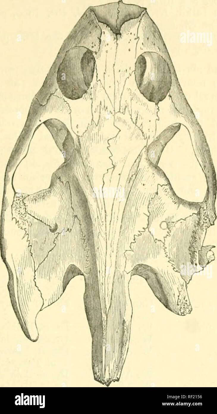
 . Catalogue of the chelonians, rhynchocephalians, and crocodiles in the British Museum (Natural History). New ed. By George Albert Boulenger. Chelonia (Genus); Crocodiles; Rhynchocephalia. 6. CYCIiANOEBIS. 271 the costals meeting on the median line and separating the neurals from each other; eighth pair of costals large. Plastron with a Fig. 73.. skull of (Jydanorbis scneyulcadit. ^^Froui Gray, P. Z. S. 18tj4.) cutaneous femoral valve, under which the hind limb may be con- cealed ; hyoplastron coossified with hypoplastron; nine or more plastral callosities in the adult, a pair being present in Stock Photohttps://www.alamy.com/image-license-details/?v=1https://www.alamy.com/catalogue-of-the-chelonians-rhynchocephalians-and-crocodiles-in-the-british-museum-natural-history-new-ed-by-george-albert-boulenger-chelonia-genus-crocodiles-rhynchocephalia-6-cyciianoebis-271-the-costals-meeting-on-the-median-line-and-separating-the-neurals-from-each-other-eighth-pair-of-costals-large-plastron-with-a-fig-73-skull-of-jydanorbis-scneyulcadit-froui-gray-p-z-s-18tj4-cutaneous-femoral-valve-under-which-the-hind-limb-may-be-con-cealed-hyoplastron-coossified-with-hypoplastron-nine-or-more-plastral-callosities-in-the-adult-a-pair-being-present-in-image233187270.html
RMRFAGMP–. Catalogue of the chelonians, rhynchocephalians, and crocodiles in the British Museum (Natural History). New ed. By George Albert Boulenger. Chelonia (Genus); Crocodiles; Rhynchocephalia. 6. CYCIiANOEBIS. 271 the costals meeting on the median line and separating the neurals from each other; eighth pair of costals large. Plastron with a Fig. 73.. skull of (Jydanorbis scneyulcadit. ^^Froui Gray, P. Z. S. 18tj4.) cutaneous femoral valve, under which the hind limb may be con- cealed ; hyoplastron coossified with hypoplastron; nine or more plastral callosities in the adult, a pair being present in
 . Catalogue of the chelonians, rhynchocephalians, and crocodiles in the British museum (Natural history). Chelonia (Genus); Rhynchocephalia; Crocodiles. 6. CYCLANOEBIS. 271 the costals meeting on the median line and separating the neurals from each other; eighth pair of costals large. Plastron with a Fig. 73.. Skull of Cyclanm'bis seneyaleusii. (^From Gray, P. Z. 8. 18(34.) cutaneous femoral valve, under which the hind limb may be con- cealed ; hyoplastron coossified with hypoplastron; nine or more plastral callosities in the adult, a pair being present in front of, and ossifying independently Stock Photohttps://www.alamy.com/image-license-details/?v=1https://www.alamy.com/catalogue-of-the-chelonians-rhynchocephalians-and-crocodiles-in-the-british-museum-natural-history-chelonia-genus-rhynchocephalia-crocodiles-6-cyclanoebis-271-the-costals-meeting-on-the-median-line-and-separating-the-neurals-from-each-other-eighth-pair-of-costals-large-plastron-with-a-fig-73-skull-of-cyclanmbis-seneyaleusii-from-gray-p-z-8-1834-cutaneous-femoral-valve-under-which-the-hind-limb-may-be-con-cealed-hyoplastron-coossified-with-hypoplastron-nine-or-more-plastral-callosities-in-the-adult-a-pair-being-present-in-front-of-and-ossifying-independently-image233187267.html
RMRFAGMK–. Catalogue of the chelonians, rhynchocephalians, and crocodiles in the British museum (Natural history). Chelonia (Genus); Rhynchocephalia; Crocodiles. 6. CYCLANOEBIS. 271 the costals meeting on the median line and separating the neurals from each other; eighth pair of costals large. Plastron with a Fig. 73.. Skull of Cyclanm'bis seneyaleusii. (^From Gray, P. Z. 8. 18(34.) cutaneous femoral valve, under which the hind limb may be con- cealed ; hyoplastron coossified with hypoplastron; nine or more plastral callosities in the adult, a pair being present in front of, and ossifying independently
 . Carnegie Institution of Washington publication. PLASTOMKNID-S. +79 The plastron (fig. 639) resembles in general that ot P. thomasi, but the bones have not joined so closely along the midline, a condition indicative ot youth. The bones average about 2 mm. in thickness. At the inguinal notch the hypoplastron is 4 mm. thick. On a line joining the axillar) and inguinal notches, the hypoplastron is nearly as wide as the hyoplas- tron; but this is quite certainly a feature due to immaturity. The same difference may be noted between the young and the adult ot Platypeltis spinifera. The plastral bon Stock Photohttps://www.alamy.com/image-license-details/?v=1https://www.alamy.com/carnegie-institution-of-washington-publication-plastomknid-s-79-the-plastron-fig-639-resembles-in-general-that-ot-p-thomasi-but-the-bones-have-not-joined-so-closely-along-the-midline-a-condition-indicative-ot-youth-the-bones-average-about-2-mm-in-thickness-at-the-inguinal-notch-the-hypoplastron-is-4-mm-thick-on-a-line-joining-the-axillar-and-inguinal-notches-the-hypoplastron-is-nearly-as-wide-as-the-hyoplas-tron-but-this-is-quite-certainly-a-feature-due-to-immaturity-the-same-difference-may-be-noted-between-the-young-and-the-adult-ot-platypeltis-spinifera-the-plastral-bon-image233475610.html
RMRFRMEJ–. Carnegie Institution of Washington publication. PLASTOMKNID-S. +79 The plastron (fig. 639) resembles in general that ot P. thomasi, but the bones have not joined so closely along the midline, a condition indicative ot youth. The bones average about 2 mm. in thickness. At the inguinal notch the hypoplastron is 4 mm. thick. On a line joining the axillar) and inguinal notches, the hypoplastron is nearly as wide as the hyoplas- tron; but this is quite certainly a feature due to immaturity. The same difference may be noted between the young and the adult ot Platypeltis spinifera. The plastral bon
 . Catalogue of the chelonians, rhynchocephalians, and crocodiles in the British museum (Natural history). Chelonia (Genus); Rhynchocephalia; Crocodiles. 6. CYCLANOEBIS. 271 the costals meeting on the median line and separating the neurals from each other; eighth pair of costals large. Plastron with a Fig. 73.. Skull of Cyclanm'bis seneyaleusii. (^From Gray, P. Z. 8. 18(34.) cutaneous femoral valve, under which the hind limb may be con- cealed ; hyoplastron coossified with hypoplastron; nine or more plastral callosities in the adult, a pair being present in front of, and ossifying independently Stock Photohttps://www.alamy.com/image-license-details/?v=1https://www.alamy.com/catalogue-of-the-chelonians-rhynchocephalians-and-crocodiles-in-the-british-museum-natural-history-chelonia-genus-rhynchocephalia-crocodiles-6-cyclanoebis-271-the-costals-meeting-on-the-median-line-and-separating-the-neurals-from-each-other-eighth-pair-of-costals-large-plastron-with-a-fig-73-skull-of-cyclanmbis-seneyaleusii-from-gray-p-z-8-1834-cutaneous-femoral-valve-under-which-the-hind-limb-may-be-con-cealed-hyoplastron-coossified-with-hypoplastron-nine-or-more-plastral-callosities-in-the-adult-a-pair-being-present-in-front-of-and-ossifying-independently-image232999455.html
RMRF2153–. Catalogue of the chelonians, rhynchocephalians, and crocodiles in the British museum (Natural history). Chelonia (Genus); Rhynchocephalia; Crocodiles. 6. CYCLANOEBIS. 271 the costals meeting on the median line and separating the neurals from each other; eighth pair of costals large. Plastron with a Fig. 73.. Skull of Cyclanm'bis seneyaleusii. (^From Gray, P. Z. 8. 18(34.) cutaneous femoral valve, under which the hind limb may be con- cealed ; hyoplastron coossified with hypoplastron; nine or more plastral callosities in the adult, a pair being present in front of, and ossifying independently
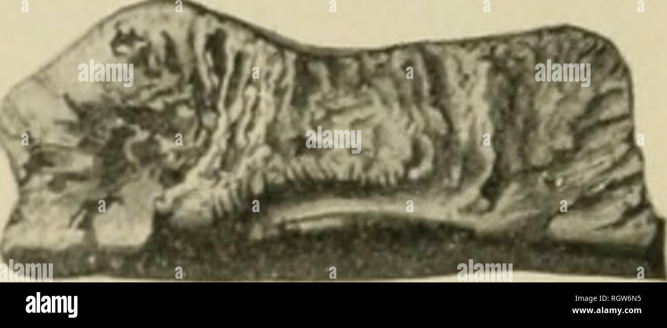 . Bulletin - American Museum of Natural History. Natural history; Science. Fig. 5.— Terrapenc putnami. x'5. Section from midline to hinge. Fig. 6.— Terrapcne putnami. %. Outline of the mesial face of the hypoplastron.. at the hinge is 15 mm. Figure 5 is a section across the bone from the midline to the hinge, while Figure 6 shows the thickness of the border which joins the bone of the opposite side, the narrow end of the figure representing the anterior hinge. Figure 7 represents the lateral hinge, the hinder end of which is broken away. At its anterior end is a deep and rough pit to receive Stock Photohttps://www.alamy.com/image-license-details/?v=1https://www.alamy.com/bulletin-american-museum-of-natural-history-natural-history-science-fig-5-terrapenc-putnami-x5-section-from-midline-to-hinge-fig-6-terrapcne-putnami-outline-of-the-mesial-face-of-the-hypoplastron-at-the-hinge-is-15-mm-figure-5-is-a-section-across-the-bone-from-the-midline-to-the-hinge-while-figure-6-shows-the-thickness-of-the-border-which-joins-the-bone-of-the-opposite-side-the-narrow-end-of-the-figure-representing-the-anterior-hinge-figure-7-represents-the-lateral-hinge-the-hinder-end-of-which-is-broken-away-at-its-anterior-end-is-a-deep-and-rough-pit-to-receive-image234123377.html
RMRGW6N5–. Bulletin - American Museum of Natural History. Natural history; Science. Fig. 5.— Terrapenc putnami. x'5. Section from midline to hinge. Fig. 6.— Terrapcne putnami. %. Outline of the mesial face of the hypoplastron.. at the hinge is 15 mm. Figure 5 is a section across the bone from the midline to the hinge, while Figure 6 shows the thickness of the border which joins the bone of the opposite side, the narrow end of the figure representing the anterior hinge. Figure 7 represents the lateral hinge, the hinder end of which is broken away. At its anterior end is a deep and rough pit to receive
 . Carnegie Institution of Washington publication. EMYDIDjE. 307 hypoplastron having a length of 73 mm. The superior beveled border is 12 mm. wide. At the inner border ot this surface the bone is 12 mm. thick. Beyond the bevel the bone thins some- what. Neat the hypoxiphiplastral suture a groove develops just inside the beveled surface. < ni tin- lower surface ot the bone is seen the broad and deeply sunken abdomino-femoral sulcus, which crosses at a distance ot 40 mm. behind the anterior bolder of the bone. The median longitudinal sulcus also has been broad and deep. There has been a large Stock Photohttps://www.alamy.com/image-license-details/?v=1https://www.alamy.com/carnegie-institution-of-washington-publication-emydidje-307-hypoplastron-having-a-length-of-73-mm-the-superior-beveled-border-is-12-mm-wide-at-the-inner-border-ot-this-surface-the-bone-is-12-mm-thick-beyond-the-bevel-the-bone-thins-some-what-neat-the-hypoxiphiplastral-suture-a-groove-develops-just-inside-the-beveled-surface-lt-ni-tin-lower-surface-ot-the-bone-is-seen-the-broad-and-deeply-sunken-abdomino-femoral-sulcus-which-crosses-at-a-distance-ot-40-mm-behind-the-anterior-bolder-of-the-bone-the-median-longitudinal-sulcus-also-has-been-broad-and-deep-there-has-been-a-large-image233476654.html
RMRFRNRX–. Carnegie Institution of Washington publication. EMYDIDjE. 307 hypoplastron having a length of 73 mm. The superior beveled border is 12 mm. wide. At the inner border ot this surface the bone is 12 mm. thick. Beyond the bevel the bone thins some- what. Neat the hypoxiphiplastral suture a groove develops just inside the beveled surface. < ni tin- lower surface ot the bone is seen the broad and deeply sunken abdomino-femoral sulcus, which crosses at a distance ot 40 mm. behind the anterior bolder of the bone. The median longitudinal sulcus also has been broad and deep. There has been a large
 . Catalogue of the chelonians, rhynchocephalians, and crocodiles in the British Museum (Natural History). New ed. By George Albert Boulenger. Chelonia (Genus); Crocodiles; Rhynchocephalia. 6. CYCIiANOEBIS. 271 the costals meeting on the median line and separating the neurals from each other; eighth pair of costals large. Plastron with a Fig. 73.. skull of (Jydanorbis scneyulcadit. ^^Froui Gray, P. Z. S. 18tj4.) cutaneous femoral valve, under which the hind limb may be con- cealed ; hyoplastron coossified with hypoplastron; nine or more plastral callosities in the adult, a pair being present in Stock Photohttps://www.alamy.com/image-license-details/?v=1https://www.alamy.com/catalogue-of-the-chelonians-rhynchocephalians-and-crocodiles-in-the-british-museum-natural-history-new-ed-by-george-albert-boulenger-chelonia-genus-crocodiles-rhynchocephalia-6-cyciianoebis-271-the-costals-meeting-on-the-median-line-and-separating-the-neurals-from-each-other-eighth-pair-of-costals-large-plastron-with-a-fig-73-skull-of-jydanorbis-scneyulcadit-froui-gray-p-z-s-18tj4-cutaneous-femoral-valve-under-which-the-hind-limb-may-be-con-cealed-hyoplastron-coossified-with-hypoplastron-nine-or-more-plastral-callosities-in-the-adult-a-pair-being-present-in-image232999458.html
RMRF2156–. Catalogue of the chelonians, rhynchocephalians, and crocodiles in the British Museum (Natural History). New ed. By George Albert Boulenger. Chelonia (Genus); Crocodiles; Rhynchocephalia. 6. CYCIiANOEBIS. 271 the costals meeting on the median line and separating the neurals from each other; eighth pair of costals large. Plastron with a Fig. 73.. skull of (Jydanorbis scneyulcadit. ^^Froui Gray, P. Z. S. 18tj4.) cutaneous femoral valve, under which the hind limb may be con- cealed ; hyoplastron coossified with hypoplastron; nine or more plastral callosities in the adult, a pair being present in
 . Carnegie Institution of Washington publication. 112 EOSSIL TURTLES OK NORTH AMERICA. costal of the left side, parts of the second and the fourth left costals, the distal end of the fifth right costal, the entoplastron, the left hypoplastron, and a portion of the right xiphiplastron. The species was one of relatively small size, with a deprest carapace (fig. 103), probably about 260 mm. long. The carapace is thin, the first costal being 6 mm. thick at the center; the fourth at its sutural border, 5 mm. thick. The first neural has a length of 35 mm. and a width of 18 mm.; the second, a length Stock Photohttps://www.alamy.com/image-license-details/?v=1https://www.alamy.com/carnegie-institution-of-washington-publication-112-eossil-turtles-ok-north-america-costal-of-the-left-side-parts-of-the-second-and-the-fourth-left-costals-the-distal-end-of-the-fifth-right-costal-the-entoplastron-the-left-hypoplastron-and-a-portion-of-the-right-xiphiplastron-the-species-was-one-of-relatively-small-size-with-a-deprest-carapace-fig-103-probably-about-260-mm-long-the-carapace-is-thin-the-first-costal-being-6-mm-thick-at-the-center-the-fourth-at-its-sutural-border-5-mm-thick-the-first-neural-has-a-length-of-35-mm-and-a-width-of-18-mm-the-second-a-length-image233486678.html
RMRFT6HX–. Carnegie Institution of Washington publication. 112 EOSSIL TURTLES OK NORTH AMERICA. costal of the left side, parts of the second and the fourth left costals, the distal end of the fifth right costal, the entoplastron, the left hypoplastron, and a portion of the right xiphiplastron. The species was one of relatively small size, with a deprest carapace (fig. 103), probably about 260 mm. long. The carapace is thin, the first costal being 6 mm. thick at the center; the fourth at its sutural border, 5 mm. thick. The first neural has a length of 35 mm. and a width of 18 mm.; the second, a length
 . Carnegie Institution of Washington publication. . 361. 563a. 363. 365. Figs. 361-365.—Clemmys hesperia. Portions of shell, xi. 361. Left hyoplastron of type. No. 2219 Univ. California. The epiplastron is restored. 362. Portion of right epiplastron. Xi. No. 2179 Univ. California. 363. First right peripheral, with section (a). Xi- No. 2179 Univ. California. 364. Probable ninth peripheral, with a section (a). No. 2179 Univ. California. 365. Section of free border of hypoplastron. No. 5527 Univ. California. joined the nuchal. The first peripheral resembles closely that of C. guttata. It will be Stock Photohttps://www.alamy.com/image-license-details/?v=1https://www.alamy.com/carnegie-institution-of-washington-publication-361-563a-363-365-figs-361-365clemmys-hesperia-portions-of-shell-xi-361-left-hyoplastron-of-type-no-2219-univ-california-the-epiplastron-is-restored-362-portion-of-right-epiplastron-xi-no-2179-univ-california-363-first-right-peripheral-with-section-a-xi-no-2179-univ-california-364-probable-ninth-peripheral-with-a-section-a-no-2179-univ-california-365-section-of-free-border-of-hypoplastron-no-5527-univ-california-joined-the-nuchal-the-first-peripheral-resembles-closely-that-of-c-guttata-it-will-be-image233476741.html
RMRFRNY1–. Carnegie Institution of Washington publication. . 361. 563a. 363. 365. Figs. 361-365.—Clemmys hesperia. Portions of shell, xi. 361. Left hyoplastron of type. No. 2219 Univ. California. The epiplastron is restored. 362. Portion of right epiplastron. Xi. No. 2179 Univ. California. 363. First right peripheral, with section (a). Xi- No. 2179 Univ. California. 364. Probable ninth peripheral, with a section (a). No. 2179 Univ. California. 365. Section of free border of hypoplastron. No. 5527 Univ. California. joined the nuchal. The first peripheral resembles closely that of C. guttata. It will be
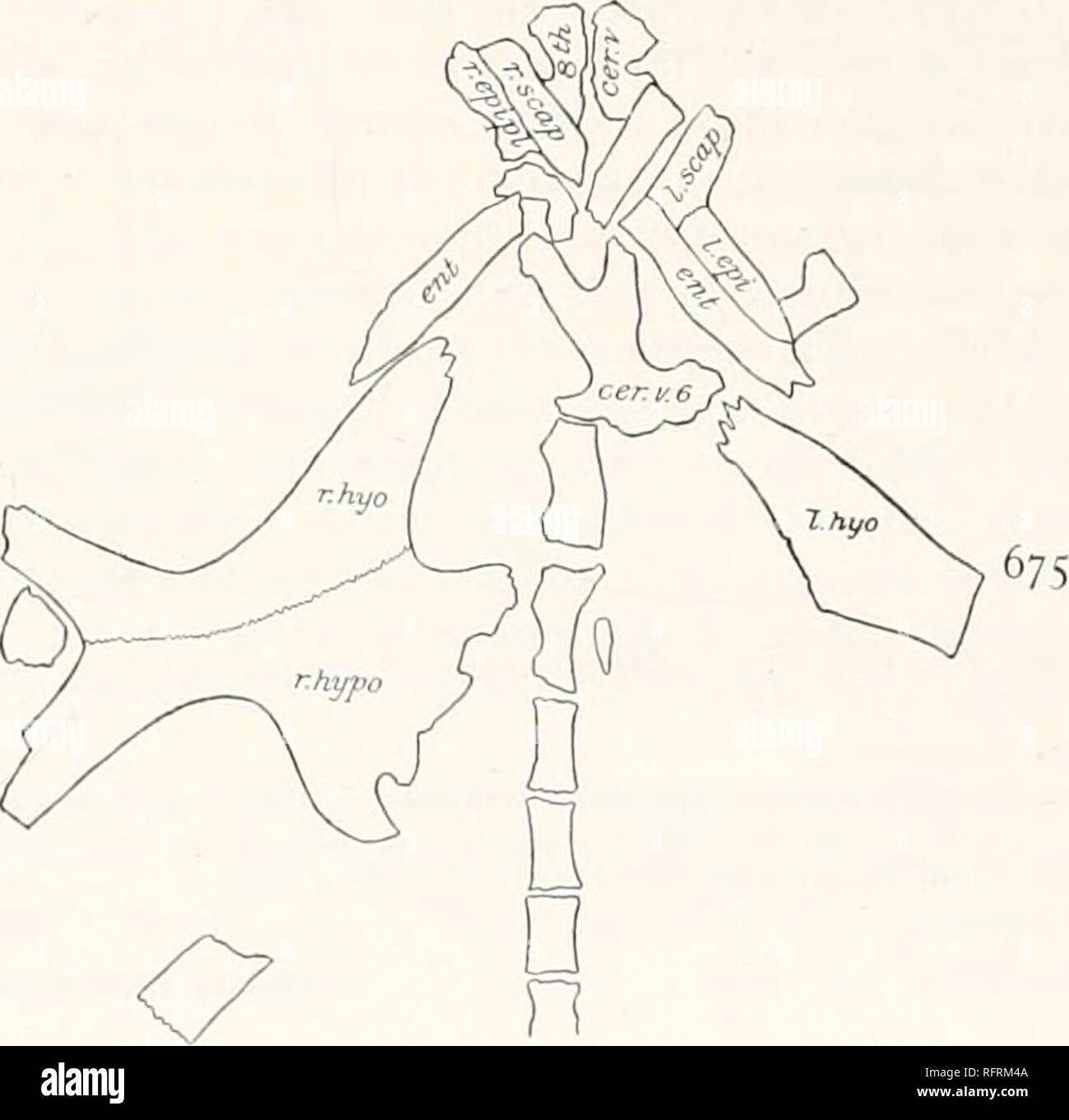 . Carnegie Institution of Washington publication. . Figs. 674 and 675.—Amyda uintaensis. Carapace and plastron of type. Xs. f>74- Carapace. 675. Plastron, cer.v. 6, cer.v. 8, sixth and eighth cervical vertebra; enl, entoplastron; /./, r.epi, left and right epiplastron; l.hyo, r.kyo, left and right hyoplastron; r.hvpo, right hypoplastron. The hyoplastron and the hypoplastron of the right side are complete, excepting that the extremities of the external processes are broken away (plate ioo, fig. I; text-fig. 675). On the lelt side there remains only a fragment of the hyoplastron. The right an Stock Photohttps://www.alamy.com/image-license-details/?v=1https://www.alamy.com/carnegie-institution-of-washington-publication-figs-674-and-675amyda-uintaensis-carapace-and-plastron-of-type-xs-fgt74-carapace-675-plastron-cerv-6-cerv-8-sixth-and-eighth-cervical-vertebra-enl-entoplastron-repi-left-and-right-epiplastron-lhyo-rkyo-left-and-right-hyoplastron-rhvpo-right-hypoplastron-the-hyoplastron-and-the-hypoplastron-of-the-right-side-are-complete-excepting-that-the-extremities-of-the-external-processes-are-broken-away-plate-ioo-fig-i-text-fig-675-on-the-lelt-side-there-remains-only-a-fragment-of-the-hyoplastron-the-right-an-image233475322.html
RMRFRM4A–. Carnegie Institution of Washington publication. . Figs. 674 and 675.—Amyda uintaensis. Carapace and plastron of type. Xs. f>74- Carapace. 675. Plastron, cer.v. 6, cer.v. 8, sixth and eighth cervical vertebra; enl, entoplastron; /./, r.epi, left and right epiplastron; l.hyo, r.kyo, left and right hyoplastron; r.hvpo, right hypoplastron. The hyoplastron and the hypoplastron of the right side are complete, excepting that the extremities of the external processes are broken away (plate ioo, fig. I; text-fig. 675). On the lelt side there remains only a fragment of the hyoplastron. The right an
![. Bulletin - American Museum of Natural History. Natural history; Science. 1906.] Hay New Genera and Species of Fossil Turtles. 31 in front of the articulation of the bone with the xiphiplastron, the hypoplastron is 22.5 mm. thiclc. In a specimen of T. Carolina whose hypoplastron is 38 mm. wide the thickness at the point named is only 4 mm. In the fossil the width becomes reduced laterally to 12 mm. two-fifths the distance toward the hinge. It then increases and. Fig. 5.— Terrapenc putnami. x'5. Section from midline to hinge. Fig. 6.— Terrapcne putnami. %. Outline of the mesial face of the h Stock Photo . Bulletin - American Museum of Natural History. Natural history; Science. 1906.] Hay New Genera and Species of Fossil Turtles. 31 in front of the articulation of the bone with the xiphiplastron, the hypoplastron is 22.5 mm. thiclc. In a specimen of T. Carolina whose hypoplastron is 38 mm. wide the thickness at the point named is only 4 mm. In the fossil the width becomes reduced laterally to 12 mm. two-fifths the distance toward the hinge. It then increases and. Fig. 5.— Terrapenc putnami. x'5. Section from midline to hinge. Fig. 6.— Terrapcne putnami. %. Outline of the mesial face of the h Stock Photo](https://c8.alamy.com/comp/RGW6NE/bulletin-american-museum-of-natural-history-natural-history-science-1906-hay-new-genera-and-species-of-fossil-turtles-31-in-front-of-the-articulation-of-the-bone-with-the-xiphiplastron-the-hypoplastron-is-225-mm-thiclc-in-a-specimen-of-t-carolina-whose-hypoplastron-is-38-mm-wide-the-thickness-at-the-point-named-is-only-4-mm-in-the-fossil-the-width-becomes-reduced-laterally-to-12-mm-two-fifths-the-distance-toward-the-hinge-it-then-increases-and-fig-5-terrapenc-putnami-x5-section-from-midline-to-hinge-fig-6-terrapcne-putnami-outline-of-the-mesial-face-of-the-h-RGW6NE.jpg) . Bulletin - American Museum of Natural History. Natural history; Science. 1906.] Hay New Genera and Species of Fossil Turtles. 31 in front of the articulation of the bone with the xiphiplastron, the hypoplastron is 22.5 mm. thiclc. In a specimen of T. Carolina whose hypoplastron is 38 mm. wide the thickness at the point named is only 4 mm. In the fossil the width becomes reduced laterally to 12 mm. two-fifths the distance toward the hinge. It then increases and. Fig. 5.— Terrapenc putnami. x'5. Section from midline to hinge. Fig. 6.— Terrapcne putnami. %. Outline of the mesial face of the h Stock Photohttps://www.alamy.com/image-license-details/?v=1https://www.alamy.com/bulletin-american-museum-of-natural-history-natural-history-science-1906-hay-new-genera-and-species-of-fossil-turtles-31-in-front-of-the-articulation-of-the-bone-with-the-xiphiplastron-the-hypoplastron-is-225-mm-thiclc-in-a-specimen-of-t-carolina-whose-hypoplastron-is-38-mm-wide-the-thickness-at-the-point-named-is-only-4-mm-in-the-fossil-the-width-becomes-reduced-laterally-to-12-mm-two-fifths-the-distance-toward-the-hinge-it-then-increases-and-fig-5-terrapenc-putnami-x5-section-from-midline-to-hinge-fig-6-terrapcne-putnami-outline-of-the-mesial-face-of-the-h-image234123386.html
. Bulletin - American Museum of Natural History. Natural history; Science. 1906.] Hay New Genera and Species of Fossil Turtles. 31 in front of the articulation of the bone with the xiphiplastron, the hypoplastron is 22.5 mm. thiclc. In a specimen of T. Carolina whose hypoplastron is 38 mm. wide the thickness at the point named is only 4 mm. In the fossil the width becomes reduced laterally to 12 mm. two-fifths the distance toward the hinge. It then increases and. Fig. 5.— Terrapenc putnami. x'5. Section from midline to hinge. Fig. 6.— Terrapcne putnami. %. Outline of the mesial face of the h Stock Photohttps://www.alamy.com/image-license-details/?v=1https://www.alamy.com/bulletin-american-museum-of-natural-history-natural-history-science-1906-hay-new-genera-and-species-of-fossil-turtles-31-in-front-of-the-articulation-of-the-bone-with-the-xiphiplastron-the-hypoplastron-is-225-mm-thiclc-in-a-specimen-of-t-carolina-whose-hypoplastron-is-38-mm-wide-the-thickness-at-the-point-named-is-only-4-mm-in-the-fossil-the-width-becomes-reduced-laterally-to-12-mm-two-fifths-the-distance-toward-the-hinge-it-then-increases-and-fig-5-terrapenc-putnami-x5-section-from-midline-to-hinge-fig-6-terrapcne-putnami-outline-of-the-mesial-face-of-the-h-image234123386.htmlRMRGW6NE–. Bulletin - American Museum of Natural History. Natural history; Science. 1906.] Hay New Genera and Species of Fossil Turtles. 31 in front of the articulation of the bone with the xiphiplastron, the hypoplastron is 22.5 mm. thiclc. In a specimen of T. Carolina whose hypoplastron is 38 mm. wide the thickness at the point named is only 4 mm. In the fossil the width becomes reduced laterally to 12 mm. two-fifths the distance toward the hinge. It then increases and. Fig. 5.— Terrapenc putnami. x'5. Section from midline to hinge. Fig. 6.— Terrapcne putnami. %. Outline of the mesial face of the h
 . Abhandlungen der Senckenbergischen Naturforschenden Gesellschaft. Natural history. Fig.5.. Fig. 1 u. 2. Chelydra sp., aus dem Meeressand bei Alzey, nat. Gr. Darmstädter Museum. Fig. 3. Trionyx gergensi (H. v. Meyer), von Weisenau b. Mainz, nat. Gr. Darmst. Museum. Fig. 4 u. 5. Hyo- und Hypoplastron von Trionyx boulengeri aus dem Meeressand bei Alzey, nat. Gr. Darmstädter Museum.. Please note that these images are extracted from scanned page images that may have been digitally enhanced for readability - coloration and appearance of these illustrations may not perfectly resemble the original w Stock Photohttps://www.alamy.com/image-license-details/?v=1https://www.alamy.com/abhandlungen-der-senckenbergischen-naturforschenden-gesellschaft-natural-history-fig5-fig-1-u-2-chelydra-sp-aus-dem-meeressand-bei-alzey-nat-gr-darmstdter-museum-fig-3-trionyx-gergensi-h-v-meyer-von-weisenau-b-mainz-nat-gr-darmst-museum-fig-4-u-5-hyo-und-hypoplastron-von-trionyx-boulengeri-aus-dem-meeressand-bei-alzey-nat-gr-darmstdter-museum-please-note-that-these-images-are-extracted-from-scanned-page-images-that-may-have-been-digitally-enhanced-for-readability-coloration-and-appearance-of-these-illustrations-may-not-perfectly-resemble-the-original-w-image238007015.html
RMRR64AF–. Abhandlungen der Senckenbergischen Naturforschenden Gesellschaft. Natural history. Fig.5.. Fig. 1 u. 2. Chelydra sp., aus dem Meeressand bei Alzey, nat. Gr. Darmstädter Museum. Fig. 3. Trionyx gergensi (H. v. Meyer), von Weisenau b. Mainz, nat. Gr. Darmst. Museum. Fig. 4 u. 5. Hyo- und Hypoplastron von Trionyx boulengeri aus dem Meeressand bei Alzey, nat. Gr. Darmstädter Museum.. Please note that these images are extracted from scanned page images that may have been digitally enhanced for readability - coloration and appearance of these illustrations may not perfectly resemble the original w
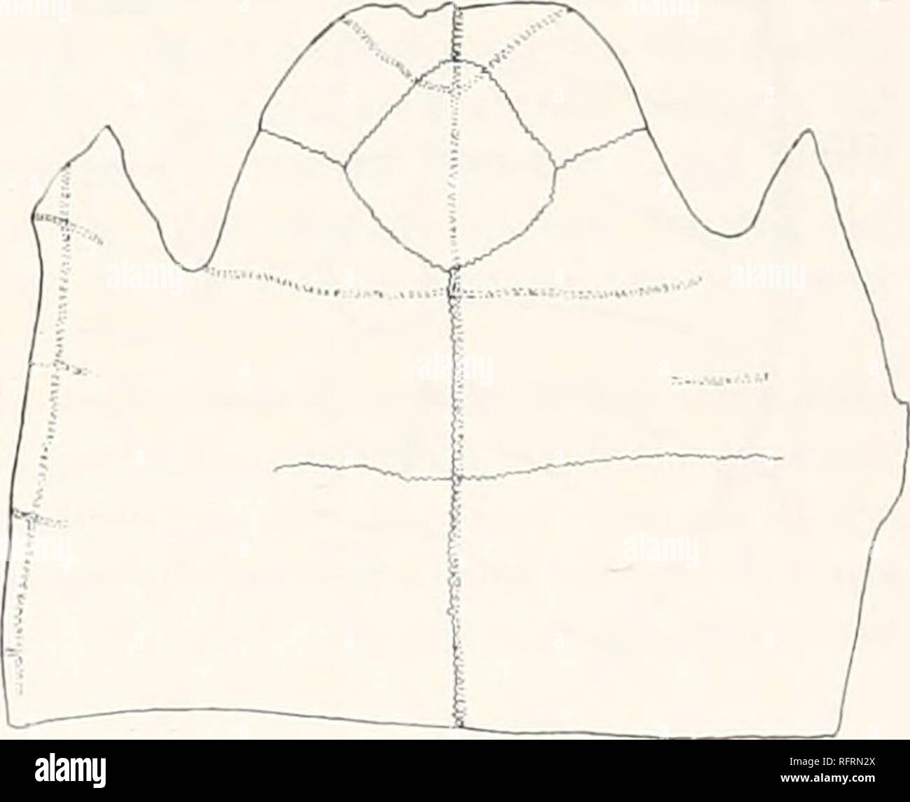 . Carnegie Institution of Washington publication. . 502. Figs. 502 and 503.—Style cal averensis. 5°3- Carapace and plastron of type. X - 502. Carapace. c,p, 1, c.p. 2, first and second costal bones; R.I,n,2, neurals; m. s. 3, m. s. 4, third and fourth marginal scutes. 507.. Portion of plastron. and third neurals, and a portion of the first. The plastron is represented from the front to near the hinder border of the hypoplastron. In the crushing that has affected the shell there has occurred a slipping of parts near the midline. The first costal of the left side overlaps a portion of the first Stock Photohttps://www.alamy.com/image-license-details/?v=1https://www.alamy.com/carnegie-institution-of-washington-publication-502-figs-502-and-503style-cal-averensis-53-carapace-and-plastron-of-type-x-502-carapace-cp-1-cp-2-first-and-second-costal-bones-rin2-neurals-m-s-3-m-s-4-third-and-fourth-marginal-scutes-507-portion-of-plastron-and-third-neurals-and-a-portion-of-the-first-the-plastron-is-represented-from-the-front-to-near-the-hinder-border-of-the-hypoplastron-in-the-crushing-that-has-affected-the-shell-there-has-occurred-a-slipping-of-parts-near-the-midline-the-first-costal-of-the-left-side-overlaps-a-portion-of-the-first-image233476066.html
RMRFRN2X–. Carnegie Institution of Washington publication. . 502. Figs. 502 and 503.—Style cal averensis. 5°3- Carapace and plastron of type. X - 502. Carapace. c,p, 1, c.p. 2, first and second costal bones; R.I,n,2, neurals; m. s. 3, m. s. 4, third and fourth marginal scutes. 507.. Portion of plastron. and third neurals, and a portion of the first. The plastron is represented from the front to near the hinder border of the hypoplastron. In the crushing that has affected the shell there has occurred a slipping of parts near the midline. The first costal of the left side overlaps a portion of the first
 . Carnegie Institution of Washington publication. 3860.. 384. Figs. 384-386.—Echmatemys testudinea. Costal and plastral bones. 384. Distal end of right first costal, inner surface. X2. No. 1181 A. M. N. H. Shows scar for axillary buttress. 385. Mesial portion of right hyoplastron. Xj. No. 1179 A.M.N.H. 386. Free border of hypoplastron, with section (a) at articulation with xiphiplastron. Xj. No. 1179 A.M.N.H. No. 1179 may or may not all belong to the same individual, and the bones are probably another portion of Cope's principal specimen. The right hyoplastron (fig. 385) measures on the midlin Stock Photohttps://www.alamy.com/image-license-details/?v=1https://www.alamy.com/carnegie-institution-of-washington-publication-3860-384-figs-384-386echmatemys-testudinea-costal-and-plastral-bones-384-distal-end-of-right-first-costal-inner-surface-x2-no-1181-a-m-n-h-shows-scar-for-axillary-buttress-385-mesial-portion-of-right-hyoplastron-xj-no-1179-amnh-386-free-border-of-hypoplastron-with-section-a-at-articulation-with-xiphiplastron-xj-no-1179-amnh-no-1179-may-or-may-not-all-belong-to-the-same-individual-and-the-bones-are-probably-another-portion-of-copes-principal-specimen-the-right-hyoplastron-fig-385-measures-on-the-midlin-image233476676.html
RMRFRNTM–. Carnegie Institution of Washington publication. 3860.. 384. Figs. 384-386.—Echmatemys testudinea. Costal and plastral bones. 384. Distal end of right first costal, inner surface. X2. No. 1181 A. M. N. H. Shows scar for axillary buttress. 385. Mesial portion of right hyoplastron. Xj. No. 1179 A.M.N.H. 386. Free border of hypoplastron, with section (a) at articulation with xiphiplastron. Xj. No. 1179 A.M.N.H. No. 1179 may or may not all belong to the same individual, and the bones are probably another portion of Cope's principal specimen. The right hyoplastron (fig. 385) measures on the midlin
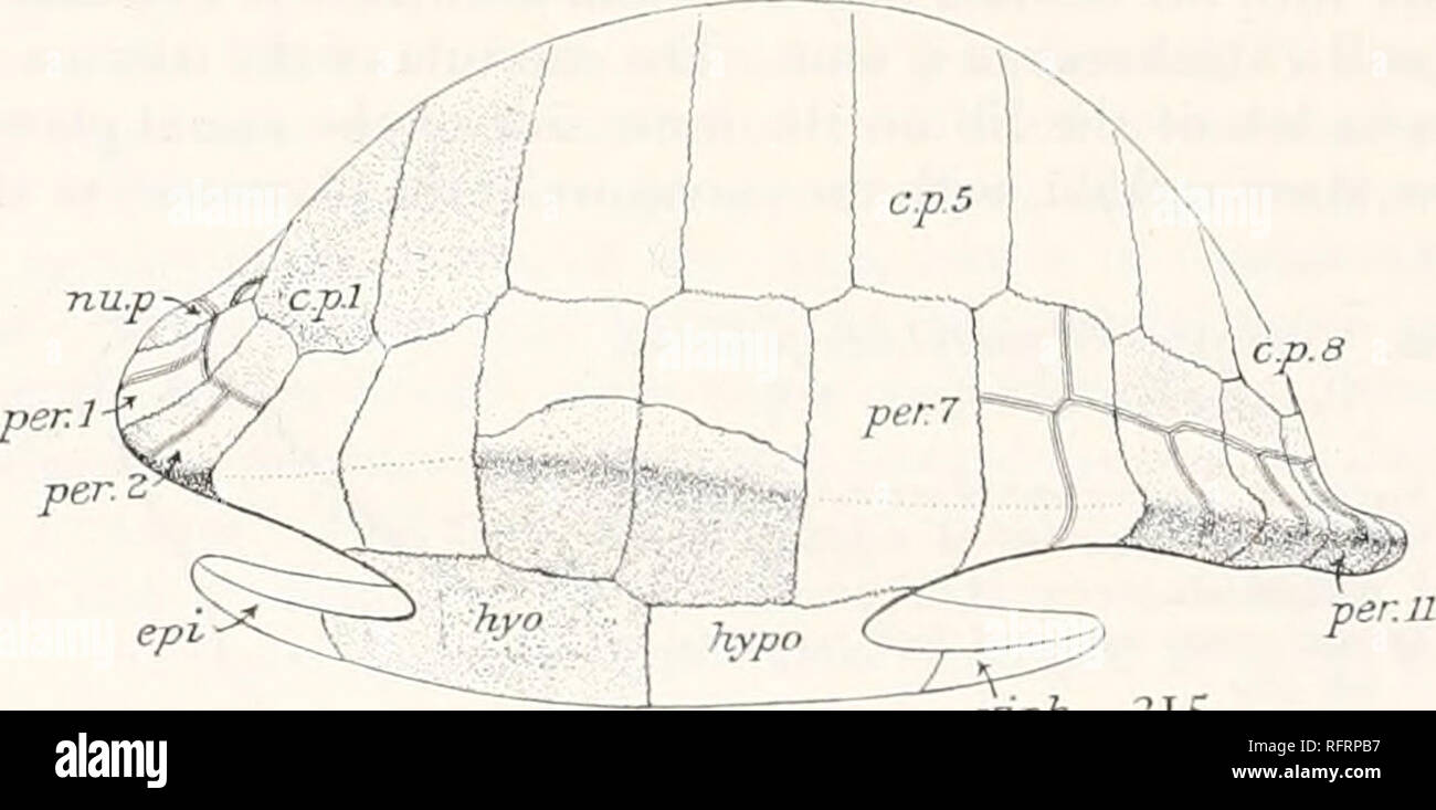 . Carnegie Institution of Washington publication. 3H-. xiph lies. 314 and 315.—Agomphus tardus. Carapace of type. 314. Sections at anterior ends of peripherals indicated by the numerals. X5. c, in first peripheral, pit for pro- cess of nuchal. s, in eighth peripheral, sutural border for union with hypoplastron; p, section of pygal. 315. Shell seen from left side. X0.23 c.p. I, etc., costal plates; epi, epiplastron; ho, hyoplastron; hypo, hypo- plastron; tin. p, nuchal plate; per. I, per, z, etc., peripheral bones; .xiph, xiphiplastron. Dr. Wieland to restore satisfactorily the form of the sh Stock Photohttps://www.alamy.com/image-license-details/?v=1https://www.alamy.com/carnegie-institution-of-washington-publication-3h-xiph-lies-314-and-315agomphus-tardus-carapace-of-type-314-sections-at-anterior-ends-of-peripherals-indicated-by-the-numerals-x5-c-in-first-peripheral-pit-for-pro-cess-of-nuchal-s-in-eighth-peripheral-sutural-border-for-union-with-hypoplastron-p-section-of-pygal-315-shell-seen-from-left-side-x023-cp-i-etc-costal-plates-epi-epiplastron-ho-hyoplastron-hypo-hypo-plastron-tin-p-nuchal-plate-per-i-per-z-etc-peripheral-bones-xiph-xiphiplastron-dr-wieland-to-restore-satisfactorily-the-form-of-the-sh-image233477083.html
RMRFRPB7–. Carnegie Institution of Washington publication. 3H-. xiph lies. 314 and 315.—Agomphus tardus. Carapace of type. 314. Sections at anterior ends of peripherals indicated by the numerals. X5. c, in first peripheral, pit for pro- cess of nuchal. s, in eighth peripheral, sutural border for union with hypoplastron; p, section of pygal. 315. Shell seen from left side. X0.23 c.p. I, etc., costal plates; epi, epiplastron; ho, hyoplastron; hypo, hypo- plastron; tin. p, nuchal plate; per. I, per, z, etc., peripheral bones; .xiph, xiphiplastron. Dr. Wieland to restore satisfactorily the form of the sh
 . Einführung in die vergleichende Anatomie der Wirbeltiere, für Studierende. . einem A und B Carapax und Plastron einer jungen Testudo graeca, "on von Chelone midas. C, C Costalplatten, E Entoplastron, das vielleicht ^.sternum entspricht, Ep Epiplastron, das vielleicht einer Clavicula entspricht, J?p Hypoplastron, Hy Hyoplastron, M, M Marginalplatten, N, iV Neuralplatten, iSi^p Nuehal- platte, Pij, Py Pygalplatten, R, R Rippen, A''( Xiphiplastron. (T" bedeutet vorne, If hinten.) SO aus drei Elementen zusammengesetzten Spangen durchsetzen den geraden Bauchmuskel, ohne sich jedoch mit Stock Photohttps://www.alamy.com/image-license-details/?v=1https://www.alamy.com/einfhrung-in-die-vergleichende-anatomie-der-wirbeltiere-fr-studierende-einem-a-und-b-carapax-und-plastron-einer-jungen-testudo-graeca-quoton-von-chelone-midas-c-c-costalplatten-e-entoplastron-das-vielleicht-sternum-entspricht-ep-epiplastron-das-vielleicht-einer-clavicula-entspricht-jp-hypoplastron-hy-hyoplastron-m-m-marginalplatten-n-iv-neuralplatten-isip-nuehal-platte-pij-py-pygalplatten-r-r-rippen-a-xiphiplastron-tquot-bedeutet-vorne-if-hinten-so-aus-drei-elementen-zusammengesetzten-spangen-durchsetzen-den-geraden-bauchmuskel-ohne-sich-jedoch-mit-image231936181.html
RMRD9GY1–. Einführung in die vergleichende Anatomie der Wirbeltiere, für Studierende. . einem A und B Carapax und Plastron einer jungen Testudo graeca, "on von Chelone midas. C, C Costalplatten, E Entoplastron, das vielleicht ^.sternum entspricht, Ep Epiplastron, das vielleicht einer Clavicula entspricht, J?p Hypoplastron, Hy Hyoplastron, M, M Marginalplatten, N, iV Neuralplatten, iSi^p Nuehal- platte, Pij, Py Pygalplatten, R, R Rippen, A''( Xiphiplastron. (T" bedeutet vorne, If hinten.) SO aus drei Elementen zusammengesetzten Spangen durchsetzen den geraden Bauchmuskel, ohne sich jedoch mit
 . Annales de la Societe Linnéenne de Lyon. Natural history. Tracé 2. — Cistudo europœa. Mouvements de l'hyo-et de I'hypoplastron. Ligne inférieure, hyoplastron ; supérieure, hypoplastron. Les ascensions de la courbe correspondent aux inspirations. r mwi ?, ? - • Tracé 3. — Cistudo europœa. Excitation du bout central du pneumogastrique provoquant un mouvement respiratoire (inspiration). On a enregistré le mou- vement du plastron, et l'ascension de la courbe correspond à l'inspiration.. Tracé 4. — Testudo grœca : excitation du bout central du pneumogastrique, provoquant une inspiration (muselièr Stock Photohttps://www.alamy.com/image-license-details/?v=1https://www.alamy.com/annales-de-la-societe-linnenne-de-lyon-natural-history-trac-2-cistudo-europa-mouvements-de-lhyo-et-de-ihypoplastron-ligne-infrieure-hyoplastron-suprieure-hypoplastron-les-ascensions-de-la-courbe-correspondent-aux-inspirations-r-mwi-trac-3-cistudo-europa-excitation-du-bout-central-du-pneumogastrique-provoquant-un-mouvement-respiratoire-inspiration-on-a-enregistr-le-mou-vement-du-plastron-et-lascension-de-la-courbe-correspond-linspiration-trac-4-testudo-grca-excitation-du-bout-central-du-pneumogastrique-provoquant-une-inspiration-muselir-image236708129.html
RMRN2YHN–. Annales de la Societe Linnéenne de Lyon. Natural history. Tracé 2. — Cistudo europœa. Mouvements de l'hyo-et de I'hypoplastron. Ligne inférieure, hyoplastron ; supérieure, hypoplastron. Les ascensions de la courbe correspondent aux inspirations. r mwi ?, ? - • Tracé 3. — Cistudo europœa. Excitation du bout central du pneumogastrique provoquant un mouvement respiratoire (inspiration). On a enregistré le mou- vement du plastron, et l'ascension de la courbe correspond à l'inspiration.. Tracé 4. — Testudo grœca : excitation du bout central du pneumogastrique, provoquant une inspiration (muselièr
 . Carnegie Institution of Washington publication. . mg.b Figs, jj and ^4. Glyptops depressus. Specimen in U. S. N. M. 33. Carapace of type. Xj. 34. Portion of plastron of type. X§. ax.h, axillary buttress; ent, entroplastron; epi, hyoplastron; hypo, hypoplastron; ing. b, inguinal buttress; mes, mesoplastron. epiplastron; hyo, Glyptops depressus sp. nov. Text-figs. 33, 34. The type of this species belongs to the United States National Museum. It appears to have been secured by one of Professor O. C. Marsh's collectors in 1889, inasmuch as it bears his packing number "1998. Box 3." W i Stock Photohttps://www.alamy.com/image-license-details/?v=1https://www.alamy.com/carnegie-institution-of-washington-publication-mgb-figs-jj-and-4-glyptops-depressus-specimen-in-u-s-n-m-33-carapace-of-type-xj-34-portion-of-plastron-of-type-x-axh-axillary-buttress-ent-entroplastron-epi-hyoplastron-hypo-hypoplastron-ing-b-inguinal-buttress-mes-mesoplastron-epiplastron-hyo-glyptops-depressus-sp-nov-text-figs-33-34-the-type-of-this-species-belongs-to-the-united-states-national-museum-it-appears-to-have-been-secured-by-one-of-professor-o-c-marshs-collectors-in-1889-inasmuch-as-it-bears-his-packing-number-quot1998-box-3quot-w-i-image233477537.html
RMRFRPYD–. Carnegie Institution of Washington publication. . mg.b Figs, jj and ^4. Glyptops depressus. Specimen in U. S. N. M. 33. Carapace of type. Xj. 34. Portion of plastron of type. X§. ax.h, axillary buttress; ent, entroplastron; epi, hyoplastron; hypo, hypoplastron; ing. b, inguinal buttress; mes, mesoplastron. epiplastron; hyo, Glyptops depressus sp. nov. Text-figs. 33, 34. The type of this species belongs to the United States National Museum. It appears to have been secured by one of Professor O. C. Marsh's collectors in 1889, inasmuch as it bears his packing number "1998. Box 3." W i
![. Allgemeine Zoologie und Abstammungslehre. Evolution; Zoology. Fig. 223. A Carapax einer jungen Teshtdo graera von oben. Die Costalia haben die Marginalia noch nicht erreicht, so daß die ßip])enenden frei hervorragen, B Plastron derselben Art, C von Chelone midas. C Costalj^latten, £'Entoplastron (Interclavicula). E)i Epiplastron (Clavicula), Hp Hypoplastron, Hy Hyoplastron, M Marginalplatten. A^ Neuralplatten, Kp Nuchalplatten, P^j Pygalplatten', 7.' Rippen, Xi Xiphiplastron, V vorn, // hinten. Aus Wiedersheim. erst auftreten, dann aber mit diesen vollständig, verwachsen. Bei CJtelouia entst Stock Photo . Allgemeine Zoologie und Abstammungslehre. Evolution; Zoology. Fig. 223. A Carapax einer jungen Teshtdo graera von oben. Die Costalia haben die Marginalia noch nicht erreicht, so daß die ßip])enenden frei hervorragen, B Plastron derselben Art, C von Chelone midas. C Costalj^latten, £'Entoplastron (Interclavicula). E)i Epiplastron (Clavicula), Hp Hypoplastron, Hy Hyoplastron, M Marginalplatten. A^ Neuralplatten, Kp Nuchalplatten, P^j Pygalplatten', 7.' Rippen, Xi Xiphiplastron, V vorn, // hinten. Aus Wiedersheim. erst auftreten, dann aber mit diesen vollständig, verwachsen. Bei CJtelouia entst Stock Photo](https://c8.alamy.com/comp/RPR80R/allgemeine-zoologie-und-abstammungslehre-evolution-zoology-fig-223-a-carapax-einer-jungen-teshtdo-graera-von-oben-die-costalia-haben-die-marginalia-noch-nicht-erreicht-so-da-die-ip-enenden-frei-hervorragen-b-plastron-derselben-art-c-von-chelone-midas-c-costaljlatten-entoplastron-interclavicula-ei-epiplastron-clavicula-hp-hypoplastron-hy-hyoplastron-m-marginalplatten-a-neuralplatten-kp-nuchalplatten-pj-pygalplatten-7-rippen-xi-xiphiplastron-v-vorn-hinten-aus-wiedersheim-erst-auftreten-dann-aber-mit-diesen-vollstndig-verwachsen-bei-cjtelouia-entst-RPR80R.jpg) . Allgemeine Zoologie und Abstammungslehre. Evolution; Zoology. Fig. 223. A Carapax einer jungen Teshtdo graera von oben. Die Costalia haben die Marginalia noch nicht erreicht, so daß die ßip])enenden frei hervorragen, B Plastron derselben Art, C von Chelone midas. C Costalj^latten, £'Entoplastron (Interclavicula). E)i Epiplastron (Clavicula), Hp Hypoplastron, Hy Hyoplastron, M Marginalplatten. A^ Neuralplatten, Kp Nuchalplatten, P^j Pygalplatten', 7.' Rippen, Xi Xiphiplastron, V vorn, // hinten. Aus Wiedersheim. erst auftreten, dann aber mit diesen vollständig, verwachsen. Bei CJtelouia entst Stock Photohttps://www.alamy.com/image-license-details/?v=1https://www.alamy.com/allgemeine-zoologie-und-abstammungslehre-evolution-zoology-fig-223-a-carapax-einer-jungen-teshtdo-graera-von-oben-die-costalia-haben-die-marginalia-noch-nicht-erreicht-so-da-die-ip-enenden-frei-hervorragen-b-plastron-derselben-art-c-von-chelone-midas-c-costaljlatten-entoplastron-interclavicula-ei-epiplastron-clavicula-hp-hypoplastron-hy-hyoplastron-m-marginalplatten-a-neuralplatten-kp-nuchalplatten-pj-pygalplatten-7-rippen-xi-xiphiplastron-v-vorn-hinten-aus-wiedersheim-erst-auftreten-dann-aber-mit-diesen-vollstndig-verwachsen-bei-cjtelouia-entst-image237768407.html
. Allgemeine Zoologie und Abstammungslehre. Evolution; Zoology. Fig. 223. A Carapax einer jungen Teshtdo graera von oben. Die Costalia haben die Marginalia noch nicht erreicht, so daß die ßip])enenden frei hervorragen, B Plastron derselben Art, C von Chelone midas. C Costalj^latten, £'Entoplastron (Interclavicula). E)i Epiplastron (Clavicula), Hp Hypoplastron, Hy Hyoplastron, M Marginalplatten. A^ Neuralplatten, Kp Nuchalplatten, P^j Pygalplatten', 7.' Rippen, Xi Xiphiplastron, V vorn, // hinten. Aus Wiedersheim. erst auftreten, dann aber mit diesen vollständig, verwachsen. Bei CJtelouia entst Stock Photohttps://www.alamy.com/image-license-details/?v=1https://www.alamy.com/allgemeine-zoologie-und-abstammungslehre-evolution-zoology-fig-223-a-carapax-einer-jungen-teshtdo-graera-von-oben-die-costalia-haben-die-marginalia-noch-nicht-erreicht-so-da-die-ip-enenden-frei-hervorragen-b-plastron-derselben-art-c-von-chelone-midas-c-costaljlatten-entoplastron-interclavicula-ei-epiplastron-clavicula-hp-hypoplastron-hy-hyoplastron-m-marginalplatten-a-neuralplatten-kp-nuchalplatten-pj-pygalplatten-7-rippen-xi-xiphiplastron-v-vorn-hinten-aus-wiedersheim-erst-auftreten-dann-aber-mit-diesen-vollstndig-verwachsen-bei-cjtelouia-entst-image237768407.htmlRMRPR80R–. Allgemeine Zoologie und Abstammungslehre. Evolution; Zoology. Fig. 223. A Carapax einer jungen Teshtdo graera von oben. Die Costalia haben die Marginalia noch nicht erreicht, so daß die ßip])enenden frei hervorragen, B Plastron derselben Art, C von Chelone midas. C Costalj^latten, £'Entoplastron (Interclavicula). E)i Epiplastron (Clavicula), Hp Hypoplastron, Hy Hyoplastron, M Marginalplatten. A^ Neuralplatten, Kp Nuchalplatten, P^j Pygalplatten', 7.' Rippen, Xi Xiphiplastron, V vorn, // hinten. Aus Wiedersheim. erst auftreten, dann aber mit diesen vollständig, verwachsen. Bei CJtelouia entst
 . Annales de la Societe Linnéenne de Lyon. Natural history. Tracé 1. — Cistudo europœa. Tracé respiratoire pris par la trachée. Inspira- tion en deux temps coupée par une pause, suivie d'une pause en expiration pleine. Les ascensions de la courbe correspondent aux inspirations.. Tracé 2. — Cistudo europœa. Mouvements de l'hyo-et de I'hypoplastron. Ligne inférieure, hyoplastron ; supérieure, hypoplastron. Les ascensions de la courbe correspondent aux inspirations. r mwi ?, ? - • Tracé 3. — Cistudo europœa. Excitation du bout central du pneumogastrique provoquant un mouvement respiratoire (inspi Stock Photohttps://www.alamy.com/image-license-details/?v=1https://www.alamy.com/annales-de-la-societe-linnenne-de-lyon-natural-history-trac-1-cistudo-europa-trac-respiratoire-pris-par-la-trache-inspira-tion-en-deux-temps-coupe-par-une-pause-suivie-dune-pause-en-expiration-pleine-les-ascensions-de-la-courbe-correspondent-aux-inspirations-trac-2-cistudo-europa-mouvements-de-lhyo-et-de-ihypoplastron-ligne-infrieure-hyoplastron-suprieure-hypoplastron-les-ascensions-de-la-courbe-correspondent-aux-inspirations-r-mwi-trac-3-cistudo-europa-excitation-du-bout-central-du-pneumogastrique-provoquant-un-mouvement-respiratoire-inspi-image236708140.html
RMRN2YJ4–. Annales de la Societe Linnéenne de Lyon. Natural history. Tracé 1. — Cistudo europœa. Tracé respiratoire pris par la trachée. Inspira- tion en deux temps coupée par une pause, suivie d'une pause en expiration pleine. Les ascensions de la courbe correspondent aux inspirations.. Tracé 2. — Cistudo europœa. Mouvements de l'hyo-et de I'hypoplastron. Ligne inférieure, hyoplastron ; supérieure, hypoplastron. Les ascensions de la courbe correspondent aux inspirations. r mwi ?, ? - • Tracé 3. — Cistudo europœa. Excitation du bout central du pneumogastrique provoquant un mouvement respiratoire (inspi
 . Bulletin de la Société des sciences naturelles de l'Ouest de la France. Natural history -- France. FlG. 2. Emys Européen. Face supérieure ventrale, ou interne, des os trouvés. âI à V, Tortue de Brain. (V.). â I, II, III, Côtes. â C, côte proprement dite; â a, extré- mité interne; â p, extrémité externe; âD., plaque dermique costale; â S, y, articulations transversales de ces plaques ; âV, articulation longitudinale interne. IV. Hyoplastron ; â «', apophyse ; ââ /. i, face interne ; â S, bord antérieur ; â i, bord inférieur. V. Ps, Hypoplastron. â Xi, Xipiioplastiujx ; â s1, s-, Stock Photohttps://www.alamy.com/image-license-details/?v=1https://www.alamy.com/bulletin-de-la-socit-des-sciences-naturelles-de-louest-de-la-france-natural-history-france-flg-2-emys-europen-face-suprieure-ventrale-ou-interne-des-os-trouvs-i-v-tortue-de-brain-v-i-ii-iii-ctes-c-cte-proprement-dite-a-extr-mit-interne-p-extrmit-externe-d-plaque-dermique-costale-s-y-articulations-transversales-de-ces-plaques-v-articulation-longitudinale-interne-iv-hyoplastron-apophyse-i-face-interne-s-bord-antrieur-i-bord-infrieur-v-ps-hypoplastron-xi-xipiioplastiujx-s1-s-image234041784.html
RMRGNEK4–. Bulletin de la Société des sciences naturelles de l'Ouest de la France. Natural history -- France. FlG. 2. Emys Européen. Face supérieure ventrale, ou interne, des os trouvés. âI à V, Tortue de Brain. (V.). â I, II, III, Côtes. â C, côte proprement dite; â a, extré- mité interne; â p, extrémité externe; âD., plaque dermique costale; â S, y, articulations transversales de ces plaques ; âV, articulation longitudinale interne. IV. Hyoplastron ; â «', apophyse ; ââ /. i, face interne ; â S, bord antérieur ; â i, bord inférieur. V. Ps, Hypoplastron. â Xi, Xipiioplastiujx ; â s1, s-,
 . Die stämme der wirbeltiere. Evolution; Paleontology; Vertebrates. 410 Die Stämme der Wirbeltiere. Proterochersis. — Obere Trias Württembergs1 (Fig. 318, 319). F. Plesiochelyidae. Aus dem oberen Jura Deutschlands und der Schweiz liegen mehrere. Fig. 319. Plastron von Proterochers is (vgl. Fig. 318.) e. = Entoplastron. GS. = Gularscutum. ep. = Epiplastron. BS. = Brachialscutum. hyp. = Hyoplastron. AS. = Abdominalscutum. mp. I = vorderes Mesoplastron. FS. = Femoralscutum. mp. 11 = hinteres Mesoplastron. AnS. = Analscutnm. hpp. = Hypoplastron. IcS. = Caudalscutum. xp. = Xiphiplastron. CS. = Inte Stock Photohttps://www.alamy.com/image-license-details/?v=1https://www.alamy.com/die-stmme-der-wirbeltiere-evolution-paleontology-vertebrates-410-die-stmme-der-wirbeltiere-proterochersis-obere-trias-wrttembergs1-fig-318-319-f-plesiochelyidae-aus-dem-oberen-jura-deutschlands-und-der-schweiz-liegen-mehrere-fig-319-plastron-von-proterochers-is-vgl-fig-318-e-=-entoplastron-gs-=-gularscutum-ep-=-epiplastron-bs-=-brachialscutum-hyp-=-hyoplastron-as-=-abdominalscutum-mp-i-=-vorderes-mesoplastron-fs-=-femoralscutum-mp-11-=-hinteres-mesoplastron-ans-=-analscutnm-hpp-=-hypoplastron-ics-=-caudalscutum-xp-=-xiphiplastron-cs-=-inte-image231455864.html
RMRCFM8T–. Die stämme der wirbeltiere. Evolution; Paleontology; Vertebrates. 410 Die Stämme der Wirbeltiere. Proterochersis. — Obere Trias Württembergs1 (Fig. 318, 319). F. Plesiochelyidae. Aus dem oberen Jura Deutschlands und der Schweiz liegen mehrere. Fig. 319. Plastron von Proterochers is (vgl. Fig. 318.) e. = Entoplastron. GS. = Gularscutum. ep. = Epiplastron. BS. = Brachialscutum. hyp. = Hyoplastron. AS. = Abdominalscutum. mp. I = vorderes Mesoplastron. FS. = Femoralscutum. mp. 11 = hinteres Mesoplastron. AnS. = Analscutnm. hpp. = Hypoplastron. IcS. = Caudalscutum. xp. = Xiphiplastron. CS. = Inte
 . Elemente der paläontologie bearbeitet. Paleontology. 632 '• Tliierreicli. — . Vertebrata. — 5. Klasse: Sauropsida. Testudinata.. Zur Verbindung des Bauch- und Rückenschildes inil einander selu'ckl das Hyoplaslron und Hypoplastron Fortsätze (Flügel) aus, die entweder mit fingerförmigen Enden (Fig. 778 Iis u. i) sich nur locker an dns Hüeken- schild legen, oder kräftige senkrecht aufsteigende Axillar- und Inguinal- p feil er bilden, welche durch Ligament fester mit den Marginal- und selbst Costalplatten verbunden sind; dieursprUnglicheFonlanellezwisehen beiden Flügeln kann verschwinden, manch Stock Photohttps://www.alamy.com/image-license-details/?v=1https://www.alamy.com/elemente-der-palontologie-bearbeitet-paleontology-632-tliierreicli-vertebrata-5-klasse-sauropsida-testudinata-zur-verbindung-des-bauch-und-rckenschildes-inil-einander-seluckl-das-hyoplaslron-und-hypoplastron-fortstze-flgel-aus-die-entweder-mit-fingerfrmigen-enden-fig-778-iis-u-i-sich-nur-locker-an-dns-heken-schild-legen-oder-krftige-senkrecht-aufsteigende-axillar-und-inguinal-p-feil-er-bilden-welche-durch-ligament-fester-mit-den-marginal-und-selbst-costalplatten-verbunden-sind-dieursprunglichefonlanellezwisehen-beiden-flgeln-kann-verschwinden-manch-image231671650.html
RMRCWFFE–. Elemente der paläontologie bearbeitet. Paleontology. 632 '• Tliierreicli. — . Vertebrata. — 5. Klasse: Sauropsida. Testudinata.. Zur Verbindung des Bauch- und Rückenschildes inil einander selu'ckl das Hyoplaslron und Hypoplastron Fortsätze (Flügel) aus, die entweder mit fingerförmigen Enden (Fig. 778 Iis u. i) sich nur locker an dns Hüeken- schild legen, oder kräftige senkrecht aufsteigende Axillar- und Inguinal- p feil er bilden, welche durch Ligament fester mit den Marginal- und selbst Costalplatten verbunden sind; dieursprUnglicheFonlanellezwisehen beiden Flügeln kann verschwinden, manch
 . Die stämme der wirbeltiere. Evolution; Paleontology; Vertebrates. 404 Die Stämme der Wirbeltiere. Argillochelys. — • Untereozän Belgiens und Englands.1 Eochelone. — Mitteleozän von Brabant (Belgien).2 Oli gochelone. — Mitteloligozän von Boom (Belgien).3 Thalassochelys. — Lebend. Chelonia. — Lebend (Fig. 296).. Hypo. Fig. 314. Protosphargis veronensis, Capellini, aus der oberen Kreide von Verona, in 1/16 nat. Gr. R. =< Rippen. Hyo. = Hyoplastron. M. = Marginalia. Hypo. = Hypoplastron. Epip. = Epiplastron. Xiph. = Xiphiplastron. F. Dermochelyidae. Sind auch die Gattungen, welche die Ahnenfo Stock Photohttps://www.alamy.com/image-license-details/?v=1https://www.alamy.com/die-stmme-der-wirbeltiere-evolution-paleontology-vertebrates-404-die-stmme-der-wirbeltiere-argillochelys-untereozn-belgiens-und-englands1-eochelone-mitteleozn-von-brabant-belgien2-oli-gochelone-mitteloligozn-von-boom-belgien3-thalassochelys-lebend-chelonia-lebend-fig-296-hypo-fig-314-protosphargis-veronensis-capellini-aus-der-oberen-kreide-von-verona-in-116-nat-gr-r-=lt-rippen-hyo-=-hyoplastron-m-=-marginalia-hypo-=-hypoplastron-epip-=-epiplastron-xiph-=-xiphiplastron-f-dermochelyidae-sind-auch-die-gattungen-welche-die-ahnenfo-image231455895.html
RMRCFM9Y–. Die stämme der wirbeltiere. Evolution; Paleontology; Vertebrates. 404 Die Stämme der Wirbeltiere. Argillochelys. — • Untereozän Belgiens und Englands.1 Eochelone. — Mitteleozän von Brabant (Belgien).2 Oli gochelone. — Mitteloligozän von Boom (Belgien).3 Thalassochelys. — Lebend. Chelonia. — Lebend (Fig. 296).. Hypo. Fig. 314. Protosphargis veronensis, Capellini, aus der oberen Kreide von Verona, in 1/16 nat. Gr. R. =< Rippen. Hyo. = Hyoplastron. M. = Marginalia. Hypo. = Hypoplastron. Epip. = Epiplastron. Xiph. = Xiphiplastron. F. Dermochelyidae. Sind auch die Gattungen, welche die Ahnenfo
 . Elemente der paläontologie bearbeitet. Paleontology. 636 I. Thiei'i'eicli. — X. Vertebraln. Ivlasse: Sauropsida. Testudinala. platten median zusammenstossen können, von Europa. Von geringer Grösse. Jura. Fig. 780. Tliulassemjjs Uugi Eüt. Oberer Jnra von Neuf- chätel. Bauchschild von aussen mit grosser Fontanelle {F) in der Mitte, es = Epiplastron; hs = Hyoplastren; lip = Hypoplastron; xs = Xiptiplastrou. ;. I-ilzingerl H. v. M. (Fig. 778). Lithograpliisclier Scliiefer von Bayern und von Cirin bei Lyon. Thalassemys Rülim.(Fig. 780). KUeiienschild sehr flach und herzförmig, an Gestalt dem der Stock Photohttps://www.alamy.com/image-license-details/?v=1https://www.alamy.com/elemente-der-palontologie-bearbeitet-paleontology-636-i-thieiieicli-x-vertebraln-ivlasse-sauropsida-testudinala-platten-median-zusammenstossen-knnen-von-europa-von-geringer-grsse-jura-fig-780-tliulassemjjs-uugi-et-oberer-jnra-von-neuf-chtel-bauchschild-von-aussen-mit-grosser-fontanelle-f-in-der-mitte-es-=-epiplastron-hs-=-hyoplastren-lip-=-hypoplastron-xs-=-xiptiplastrou-i-ilzingerl-h-v-m-fig-778-lithograpliisclier-scliiefer-von-bayern-und-von-cirin-bei-lyon-thalassemys-rlimfig-780-kueiienschild-sehr-flach-und-herzfrmig-an-gestalt-dem-der-image231671649.html
RMRCWFFD–. Elemente der paläontologie bearbeitet. Paleontology. 636 I. Thiei'i'eicli. — X. Vertebraln. Ivlasse: Sauropsida. Testudinala. platten median zusammenstossen können, von Europa. Von geringer Grösse. Jura. Fig. 780. Tliulassemjjs Uugi Eüt. Oberer Jnra von Neuf- chätel. Bauchschild von aussen mit grosser Fontanelle {F) in der Mitte, es = Epiplastron; hs = Hyoplastren; lip = Hypoplastron; xs = Xiptiplastrou. ;. I-ilzingerl H. v. M. (Fig. 778). Lithograpliisclier Scliiefer von Bayern und von Cirin bei Lyon. Thalassemys Rülim.(Fig. 780). KUeiienschild sehr flach und herzförmig, an Gestalt dem der
 . Elemente der paläontologie bearbeitet. Paleontology. Fig. HC). Eurystefutnn ^Vagleii H. v. M. Oberer Malm von Sulnhofen. Skelet von oben, cj, ci, cs = Costalplatten; hi* = Hypoplastron (Baurhschild); hs = Hyoplastron (Baucbschild); il = Ileum; ni, («5, ins, Ulli = Margiualplatten; in, Ui, )ta = Neuralplatten; lui = Nuchalplatte ; pu = Pygal- platte. Auf ci, es, f5, cs sind Eindrücke der Horn- scbilder sichtbar.. Fig. 777. FUsiocIidi/s Efalhni Pict. Oberer Malm von St. Claude (Scliweiz). Baudrschild von aussen mit kleiner mittlerer Fontanelle, em = Eutoplastron; es = Epiplastron; hs = Hyopla Stock Photohttps://www.alamy.com/image-license-details/?v=1https://www.alamy.com/elemente-der-palontologie-bearbeitet-paleontology-fig-hc-eurystefutnn-vagleii-h-v-m-oberer-malm-von-sulnhofen-skelet-von-oben-cj-ci-cs-=-costalplatten-hi-=-hypoplastron-baurhschild-hs-=-hyoplastron-baucbschild-il-=-ileum-ni-5-ins-ulli-=-margiualplatten-in-ui-ta-=-neuralplatten-lui-=-nuchalplatte-pu-=-pygal-platte-auf-ci-es-f5-cs-sind-eindrcke-der-horn-scbilder-sichtbar-fig-777-fusiociidis-efalhni-pict-oberer-malm-von-st-claude-scliweiz-baudrschild-von-aussen-mit-kleiner-mittlerer-fontanelle-em-=-eutoplastron-es-=-epiplastron-hs-=-hyopla-image231671651.html
RMRCWFFF–. Elemente der paläontologie bearbeitet. Paleontology. Fig. HC). Eurystefutnn ^Vagleii H. v. M. Oberer Malm von Sulnhofen. Skelet von oben, cj, ci, cs = Costalplatten; hi* = Hypoplastron (Baurhschild); hs = Hyoplastron (Baucbschild); il = Ileum; ni, («5, ins, Ulli = Margiualplatten; in, Ui, )ta = Neuralplatten; lui = Nuchalplatte ; pu = Pygal- platte. Auf ci, es, f5, cs sind Eindrücke der Horn- scbilder sichtbar.. Fig. 777. FUsiocIidi/s Efalhni Pict. Oberer Malm von St. Claude (Scliweiz). Baudrschild von aussen mit kleiner mittlerer Fontanelle, em = Eutoplastron; es = Epiplastron; hs = Hyopla