Quick filters:
Interpubic Stock Photos and Images
 FRACTURED PELVIS, X-RAY Stock Photohttps://www.alamy.com/image-license-details/?v=1https://www.alamy.com/stock-photo-fractured-pelvis-x-ray-49285476.html
RMCT543G–FRACTURED PELVIS, X-RAY
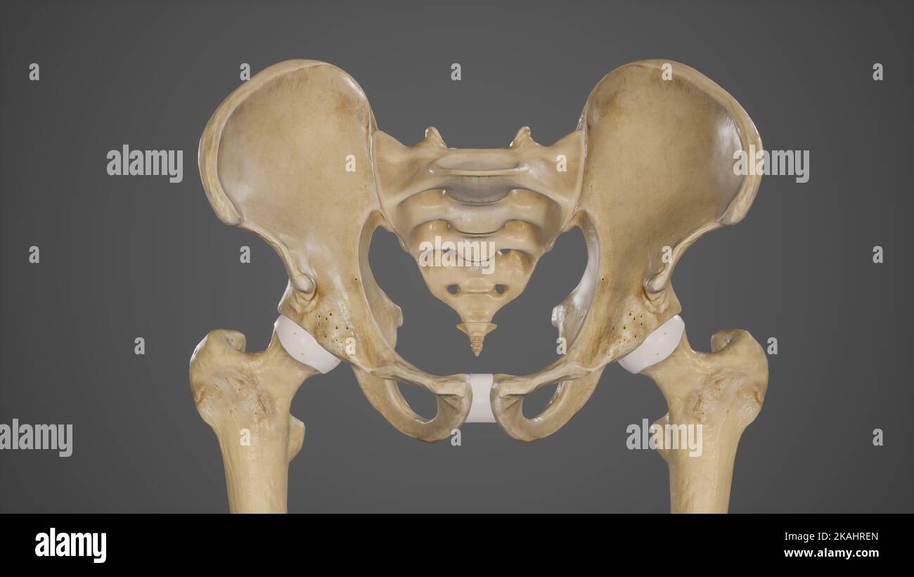 Medical Ilustration of Pelvic Bones-Hip Bone Stock Photohttps://www.alamy.com/image-license-details/?v=1https://www.alamy.com/medical-ilustration-of-pelvic-bones-hip-bone-image488428493.html
RF2KAHREN–Medical Ilustration of Pelvic Bones-Hip Bone
 The BNA arranged as an outline of regional and systematic anatomy . ac ligaments—20 :57, 58. Synovial cavity.Symphysis pubis—^20 :59. Superior pubic ligam.ents—20 :60. Arcuate ligament of pubis—:20 :61. Interpubic fibro-cartilage—20 :62. Demonstrated by removing a slice of bone from the front of thesymphysis pubis. Sacro-coccygeal symphysis—18 :48. Superficial posterior sacro-coccygeal ligament—18 :49.Deep posterior sacro-coccygeal ligament—18 :50.Anterior sacro-coccygeal ligament—18 :51.Lateral sacro-coccygeal ligament—18 :52. INFERIOR EXTREMITY Plate IX Posterior aspect of the inferior extre Stock Photohttps://www.alamy.com/image-license-details/?v=1https://www.alamy.com/the-bna-arranged-as-an-outline-of-regional-and-systematic-anatomy-ac-ligaments20-57-58-synovial-cavitysymphysis-pubis20-59-superior-pubic-ligaments20-60-arcuate-ligament-of-pubis20-61-interpubic-fibro-cartilage20-62-demonstrated-by-removing-a-slice-of-bone-from-the-front-of-thesymphysis-pubis-sacro-coccygeal-symphysis18-48-superficial-posterior-sacro-coccygeal-ligament18-49deep-posterior-sacro-coccygeal-ligament18-50anterior-sacro-coccygeal-ligament18-51lateral-sacro-coccygeal-ligament18-52-inferior-extremity-plate-ix-posterior-aspect-of-the-inferior-extre-image342993460.html
RM2AX0KJC–The BNA arranged as an outline of regional and systematic anatomy . ac ligaments—20 :57, 58. Synovial cavity.Symphysis pubis—^20 :59. Superior pubic ligam.ents—20 :60. Arcuate ligament of pubis—:20 :61. Interpubic fibro-cartilage—20 :62. Demonstrated by removing a slice of bone from the front of thesymphysis pubis. Sacro-coccygeal symphysis—18 :48. Superficial posterior sacro-coccygeal ligament—18 :49.Deep posterior sacro-coccygeal ligament—18 :50.Anterior sacro-coccygeal ligament—18 :51.Lateral sacro-coccygeal ligament—18 :52. INFERIOR EXTREMITY Plate IX Posterior aspect of the inferior extre
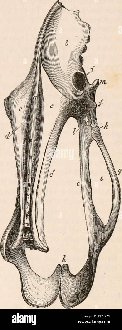 . The cyclopædia of anatomy and physiology. Anatomy; Physiology; Zoology. PELVIS. 167 marked Uio-iscJdal angle in the reverse direc- tion to that of Mammals generally, i. e. with the retiring sides anterior (see Jig. 107.). The pubes of birds are generally long, slender, rib-like, and divergent, and are com- posed of a single curved branch (jj), having no angle, and never forming a true interpubic symphysis, though, in the Ostrich and Falco Fulvus, they are closely approximated at their posterior extremity, and form a sort of sym- physis. The ilio-pubic angle is very large, from 155° to 160°, Stock Photohttps://www.alamy.com/image-license-details/?v=1https://www.alamy.com/the-cyclopdia-of-anatomy-and-physiology-anatomy-physiology-zoology-pelvis-167-marked-uio-iscjdal-angle-in-the-reverse-direc-tion-to-that-of-mammals-generally-i-e-with-the-retiring-sides-anterior-see-jig-107-the-pubes-of-birds-are-generally-long-slender-rib-like-and-divergent-and-are-com-posed-of-a-single-curved-branch-jj-having-no-angle-and-never-forming-a-true-interpubic-symphysis-though-in-the-ostrich-and-falco-fulvus-they-are-closely-approximated-at-their-posterior-extremity-and-form-a-sort-of-sym-physis-the-ilio-pubic-angle-is-very-large-from-155-to-160-image216210797.html
RMPFN725–. The cyclopædia of anatomy and physiology. Anatomy; Physiology; Zoology. PELVIS. 167 marked Uio-iscJdal angle in the reverse direc- tion to that of Mammals generally, i. e. with the retiring sides anterior (see Jig. 107.). The pubes of birds are generally long, slender, rib-like, and divergent, and are com- posed of a single curved branch (jj), having no angle, and never forming a true interpubic symphysis, though, in the Ostrich and Falco Fulvus, they are closely approximated at their posterior extremity, and form a sort of sym- physis. The ilio-pubic angle is very large, from 155° to 160°,
 FRACTURED PELVIS, X-RAY Stock Photohttps://www.alamy.com/image-license-details/?v=1https://www.alamy.com/stock-photo-fractured-pelvis-x-ray-49285486.html
RMCT543X–FRACTURED PELVIS, X-RAY
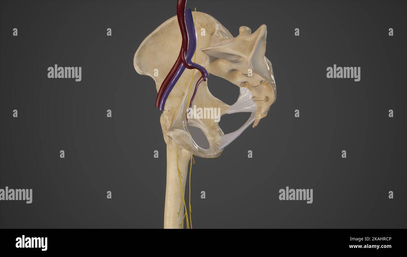 Anatomical Illustration of Obturator Canal Stock Photohttps://www.alamy.com/image-license-details/?v=1https://www.alamy.com/anatomical-illustration-of-obturator-canal-image488428438.html
RF2KAHRCP–Anatomical Illustration of Obturator Canal
 MUSCLE, DRAWING Stock Photohttps://www.alamy.com/image-license-details/?v=1https://www.alamy.com/stock-photo-muscle-drawing-49264143.html
RMCT44WK–MUSCLE, DRAWING
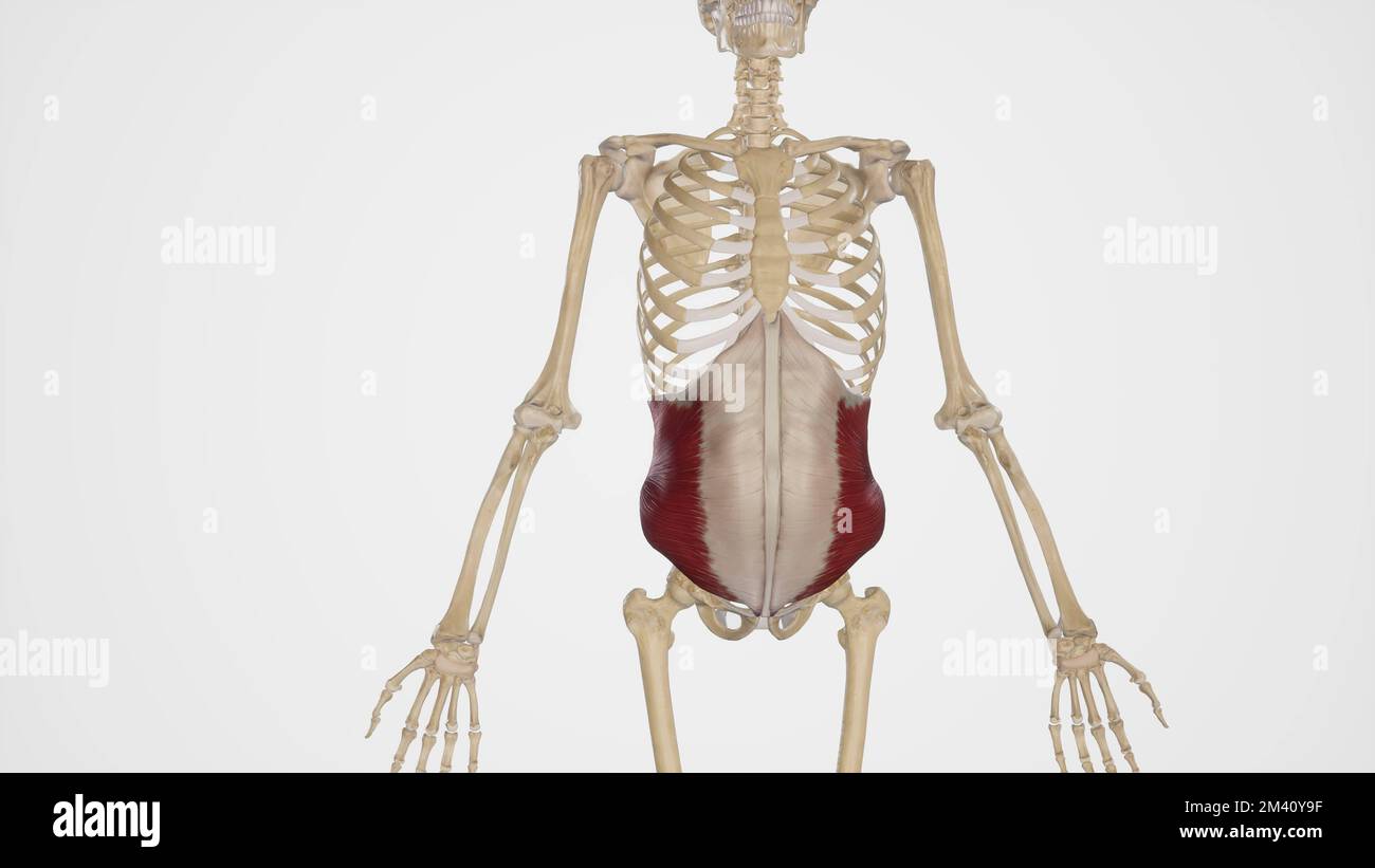 Internal Oblique Muscle Stock Photohttps://www.alamy.com/image-license-details/?v=1https://www.alamy.com/internal-oblique-muscle-image501580731.html
RF2M40Y9F–Internal Oblique Muscle
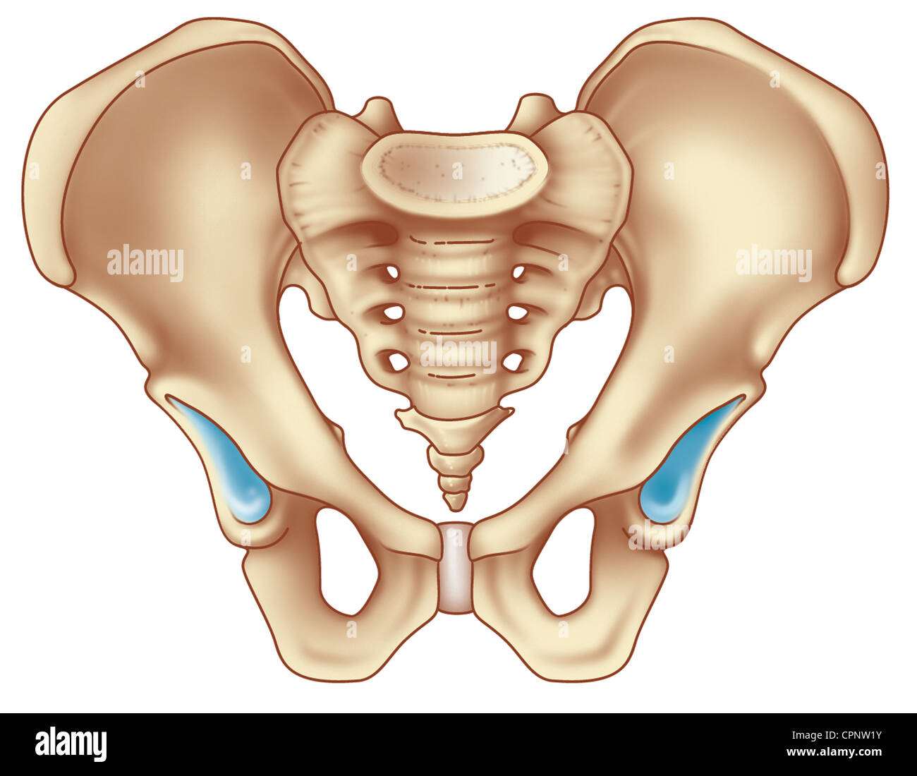 BASIN Stock Photohttps://www.alamy.com/image-license-details/?v=1https://www.alamy.com/stock-photo-basin-48423815.html
RMCPNW1Y–BASIN
 . An American text-book of obstetrics. For practitioners and students. ralstrata of interlacing fibres, the deepest of which passes directly across betweenthe bones in frout of the interpubic disk, with which they are blended; thesuperficial layers include oblique interlacing fibres continued from the tendonsof the external oblique and the recti muscles, and of the more superficialadductors of the thigh. The posterior pubic ligament consists of a few sparingly distributed fibreswhich unite the bones behind, and it is little more than the somewhat thick-ened periosteum. The superior pubic ligam Stock Photohttps://www.alamy.com/image-license-details/?v=1https://www.alamy.com/an-american-text-book-of-obstetrics-for-practitioners-and-students-ralstrata-of-interlacing-fibres-the-deepest-of-which-passes-directly-across-betweenthe-bones-in-frout-of-the-interpubic-disk-with-which-they-are-blended-thesuperficial-layers-include-oblique-interlacing-fibres-continued-from-the-tendonsof-the-external-oblique-and-the-recti-muscles-and-of-the-more-superficialadductors-of-the-thigh-the-posterior-pubic-ligament-consists-of-a-few-sparingly-distributed-fibreswhich-unite-the-bones-behind-and-it-is-little-more-than-the-somewhat-thick-ened-periosteum-the-superior-pubic-ligam-image370608337.html
RM2CEXJM1–. An American text-book of obstetrics. For practitioners and students. ralstrata of interlacing fibres, the deepest of which passes directly across betweenthe bones in frout of the interpubic disk, with which they are blended; thesuperficial layers include oblique interlacing fibres continued from the tendonsof the external oblique and the recti muscles, and of the more superficialadductors of the thigh. The posterior pubic ligament consists of a few sparingly distributed fibreswhich unite the bones behind, and it is little more than the somewhat thick-ened periosteum. The superior pubic ligam
 SKELETON, ILLUSTRATION Stock Photohttps://www.alamy.com/image-license-details/?v=1https://www.alamy.com/stock-photo-skeleton-illustration-49267046.html
RMCT48HA–SKELETON, ILLUSTRATION
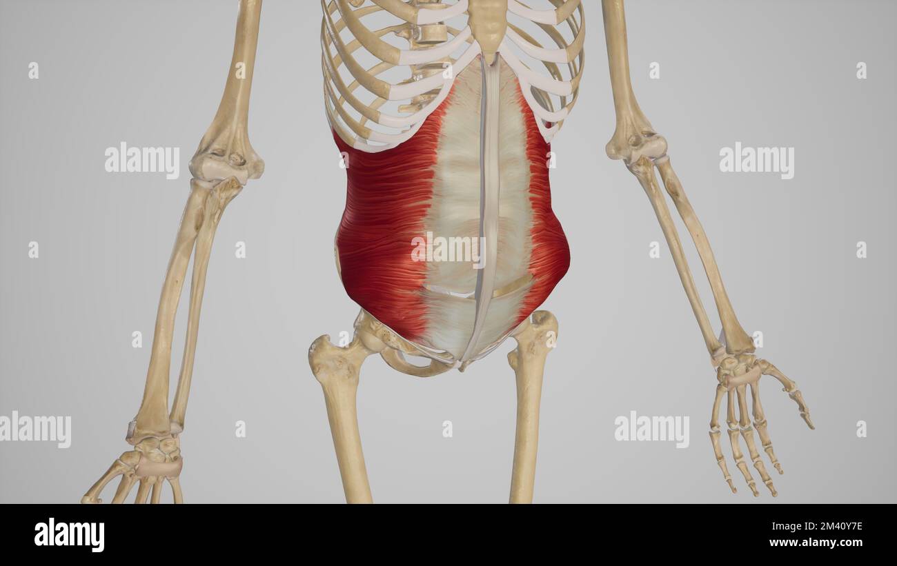 Transversus Abdominis Muscle Stock Photohttps://www.alamy.com/image-license-details/?v=1https://www.alamy.com/transversus-abdominis-muscle-image501580674.html
RF2M40Y7E–Transversus Abdominis Muscle
 . An American text-book of obstetrics. For practitioners and students. htrrpii. Fig. 5.—Section across symphysis pubis, showing interpubic disk (Lusk) tilage constituting the interpubic disk (Fig. 5). This layer, which projects ante-riorly and posteriorly beyond the adjacent bony margins, is thickest in front; thedeficiency of the intermediate tissue above and behind sometimes results in theformation of an interspace or fissure. The fis-sure within the interpubic disk extends usuallyabout half the length of the cartilage, and isproduced during life by the absorption of thefibro-cartilage: it a Stock Photohttps://www.alamy.com/image-license-details/?v=1https://www.alamy.com/an-american-text-book-of-obstetrics-for-practitioners-and-students-htrrpii-fig-5section-across-symphysis-pubis-showing-interpubic-disk-lusk-tilage-constituting-the-interpubic-disk-fig-5-this-layer-which-projects-ante-riorly-and-posteriorly-beyond-the-adjacent-bony-margins-is-thickest-in-front-thedeficiency-of-the-intermediate-tissue-above-and-behind-sometimes-results-in-theformation-of-an-interspace-or-fissure-the-fis-sure-within-the-interpubic-disk-extends-usuallyabout-half-the-length-of-the-cartilage-and-isproduced-during-life-by-the-absorption-of-thefibro-cartilage-it-a-image370608548.html
RM2CEXJYG–. An American text-book of obstetrics. For practitioners and students. htrrpii. Fig. 5.—Section across symphysis pubis, showing interpubic disk (Lusk) tilage constituting the interpubic disk (Fig. 5). This layer, which projects ante-riorly and posteriorly beyond the adjacent bony margins, is thickest in front; thedeficiency of the intermediate tissue above and behind sometimes results in theformation of an interspace or fissure. The fis-sure within the interpubic disk extends usuallyabout half the length of the cartilage, and isproduced during life by the absorption of thefibro-cartilage: it a
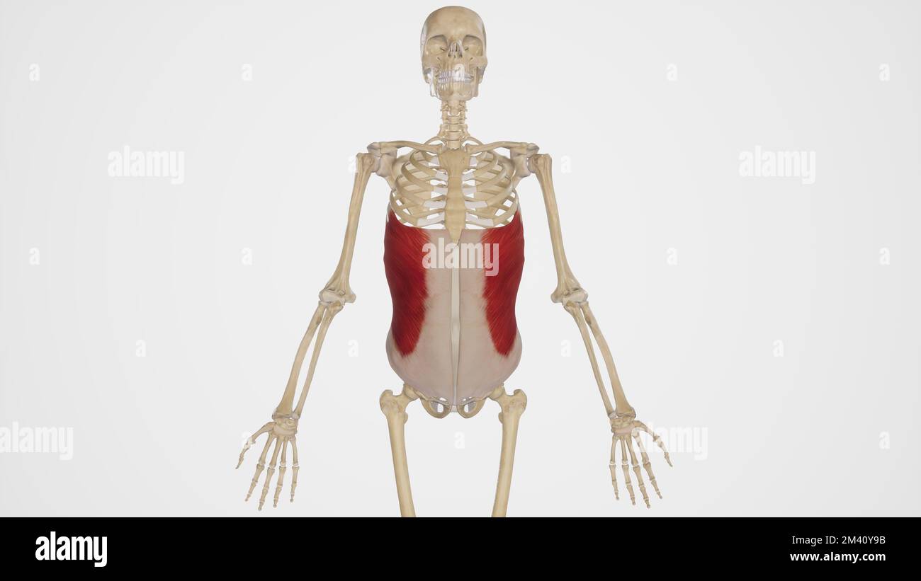 External Abdominal Oblique Muscle Stock Photohttps://www.alamy.com/image-license-details/?v=1https://www.alamy.com/external-abdominal-oblique-muscle-image501580727.html
RF2M40Y9B–External Abdominal Oblique Muscle
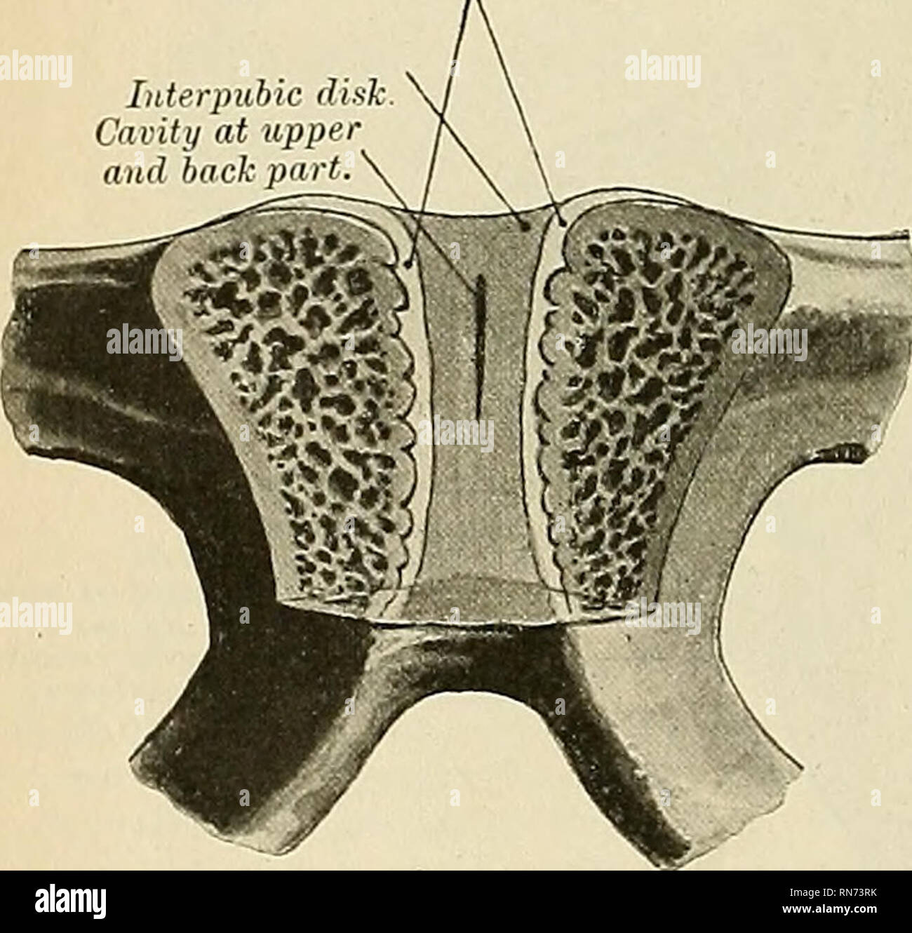 . Anatomy, descriptive and applied. Anatomy. 294 THE ARTICULATIONS, OB JOINTS Movements.—The movements which take place between the sacrum and coccyx, and between the different pieces of the latter bone, are forward and backward, and are very limited. Their extent increases during pregnancy. Interpubic dish Cavity at uppe and back pa} t 4 Articulation of the Pubic Bones (Symphysis Ossioi Pubis) (Figs. 238, 242). The articulation between the pubic bones is an amphiarthrodial joint, formed by the apposition of the two oval articular surfaces of the pubic bones. The ligaments of this articulation Stock Photohttps://www.alamy.com/image-license-details/?v=1https://www.alamy.com/anatomy-descriptive-and-applied-anatomy-294-the-articulations-ob-joints-movementsthe-movements-which-take-place-between-the-sacrum-and-coccyx-and-between-the-different-pieces-of-the-latter-bone-are-forward-and-backward-and-are-very-limited-their-extent-increases-during-pregnancy-interpubic-dish-cavity-at-uppe-and-back-pa-t-4-articulation-of-the-pubic-bones-symphysis-ossioi-pubis-figs-238-242-the-articulation-between-the-pubic-bones-is-an-amphiarthrodial-joint-formed-by-the-apposition-of-the-two-oval-articular-surfaces-of-the-pubic-bones-the-ligaments-of-this-articulation-image236799239.html
RMRN73RK–. Anatomy, descriptive and applied. Anatomy. 294 THE ARTICULATIONS, OB JOINTS Movements.—The movements which take place between the sacrum and coccyx, and between the different pieces of the latter bone, are forward and backward, and are very limited. Their extent increases during pregnancy. Interpubic dish Cavity at uppe and back pa} t 4 Articulation of the Pubic Bones (Symphysis Ossioi Pubis) (Figs. 238, 242). The articulation between the pubic bones is an amphiarthrodial joint, formed by the apposition of the two oval articular surfaces of the pubic bones. The ligaments of this articulation
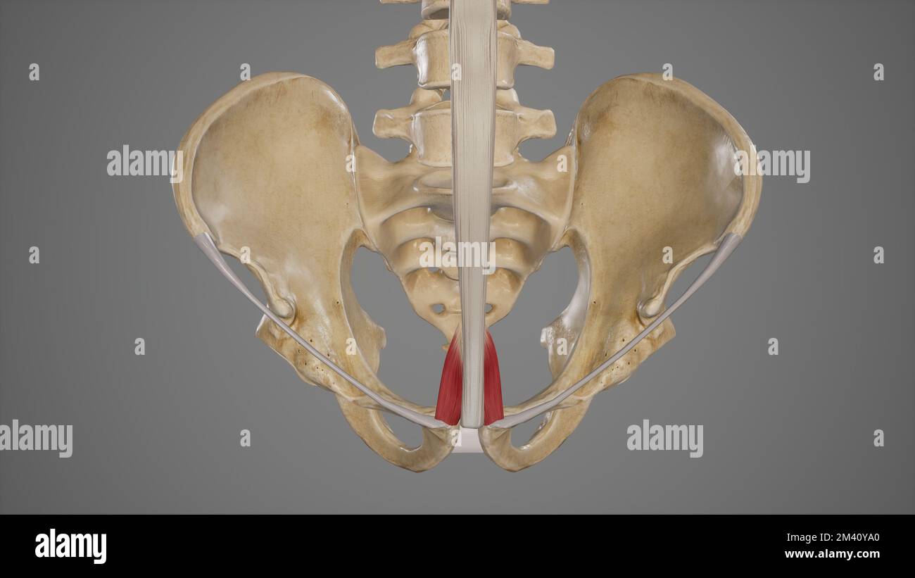 Pyramidalis Stock Photohttps://www.alamy.com/image-license-details/?v=1https://www.alamy.com/pyramidalis-image501580744.html
RF2M40YA0–Pyramidalis
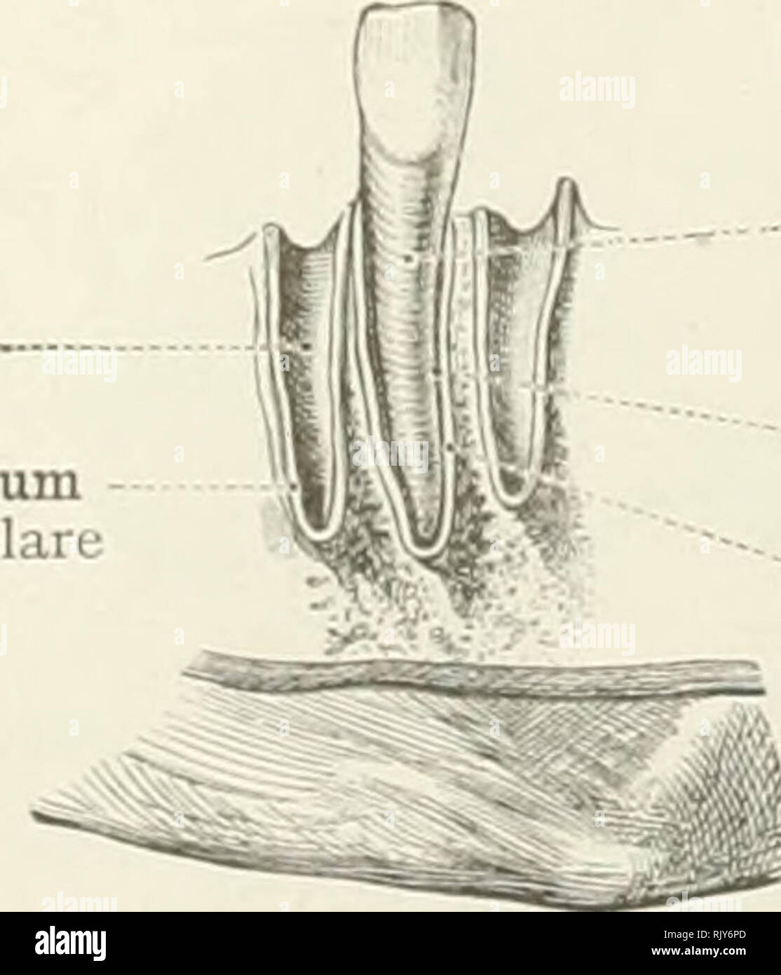 . An atlas of human anatomy for students and physicians. Anatomy. Interpubic disc •Lamina fibrocartilaginea interpubica The inferior pubic or subpubic ligament Lig. arcuatum pubis Fig. 382.—Symphysis. (The Pubic Symphysis; Frontal Section, Posterior Portion.) iUveolus or socket of the tooth Alveolus dentis Interalveolar septum Septum interalveolare. Root of the tooth Radix dentis — Dental periosteum Periosteum alveolare Interalveolar septum Septum interalveolare Fig. 383.—Gomphosis. Synarthrosis, or Continuous Articulation.. Please note that these images are extracted from scanned page images Stock Photohttps://www.alamy.com/image-license-details/?v=1https://www.alamy.com/an-atlas-of-human-anatomy-for-students-and-physicians-anatomy-interpubic-disc-lamina-fibrocartilaginea-interpubica-the-inferior-pubic-or-subpubic-ligament-lig-arcuatum-pubis-fig-382symphysis-the-pubic-symphysis-frontal-section-posterior-portion-iuveolus-or-socket-of-the-tooth-alveolus-dentis-interalveolar-septum-septum-interalveolare-root-of-the-tooth-radix-dentis-dental-periosteum-periosteum-alveolare-interalveolar-septum-septum-interalveolare-fig-383gomphosis-synarthrosis-or-continuous-articulation-please-note-that-these-images-are-extracted-from-scanned-page-images-image235396629.html
RMRJY6PD–. An atlas of human anatomy for students and physicians. Anatomy. Interpubic disc •Lamina fibrocartilaginea interpubica The inferior pubic or subpubic ligament Lig. arcuatum pubis Fig. 382.—Symphysis. (The Pubic Symphysis; Frontal Section, Posterior Portion.) iUveolus or socket of the tooth Alveolus dentis Interalveolar septum Septum interalveolare. Root of the tooth Radix dentis — Dental periosteum Periosteum alveolare Interalveolar septum Septum interalveolare Fig. 383.—Gomphosis. Synarthrosis, or Continuous Articulation.. Please note that these images are extracted from scanned page images
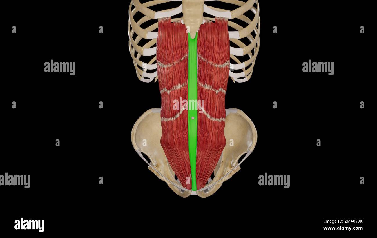 Linea Alba Stock Photohttps://www.alamy.com/image-license-details/?v=1https://www.alamy.com/linea-alba-image501580735.html
RF2M40Y9K–Linea Alba
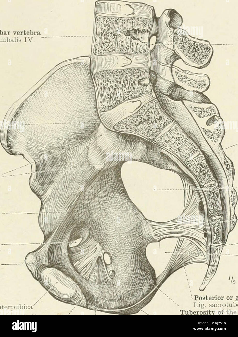 . An atlas of human anatomy for students and physicians. Anatomy. THE ARTICULATIONS OF THE LOWER LIMB 217 Fourth lumbar vertebra Vertebra lumbalis IV, Sacral promontory Promontonum Anterior sacro-iliac ligament Ligg. sacro-iliaca anteriora Brim of the pelvis Linea terminalis Obturator canal Canalis obturatorius Spine of the pubis Tuberculum pubicum Interpubic disc ( Lamina fibrocaxtilaginea interpubica' Inferior pubic or subpubic ligament /' Lig. arcuatum pubis Obturator membrane, or ligamen, Membrana obturatoria. Intervertebral foramen Foramen intervertebrale Sacral canal ,--' Canalis sacrali Stock Photohttps://www.alamy.com/image-license-details/?v=1https://www.alamy.com/an-atlas-of-human-anatomy-for-students-and-physicians-anatomy-the-articulations-of-the-lower-limb-217-fourth-lumbar-vertebra-vertebra-lumbalis-iv-sacral-promontory-promontonum-anterior-sacro-iliac-ligament-ligg-sacro-iliaca-anteriora-brim-of-the-pelvis-linea-terminalis-obturator-canal-canalis-obturatorius-spine-of-the-pubis-tuberculum-pubicum-interpubic-disc-lamina-fibrocaxtilaginea-interpubica-inferior-pubic-or-subpubic-ligament-lig-arcuatum-pubis-obturator-membrane-or-ligamen-membrana-obturatoria-intervertebral-foramen-foramen-intervertebrale-sacral-canal-canalis-sacrali-image235395252.html
RMRJY518–. An atlas of human anatomy for students and physicians. Anatomy. THE ARTICULATIONS OF THE LOWER LIMB 217 Fourth lumbar vertebra Vertebra lumbalis IV, Sacral promontory Promontonum Anterior sacro-iliac ligament Ligg. sacro-iliaca anteriora Brim of the pelvis Linea terminalis Obturator canal Canalis obturatorius Spine of the pubis Tuberculum pubicum Interpubic disc ( Lamina fibrocaxtilaginea interpubica' Inferior pubic or subpubic ligament /' Lig. arcuatum pubis Obturator membrane, or ligamen, Membrana obturatoria. Intervertebral foramen Foramen intervertebrale Sacral canal ,--' Canalis sacrali
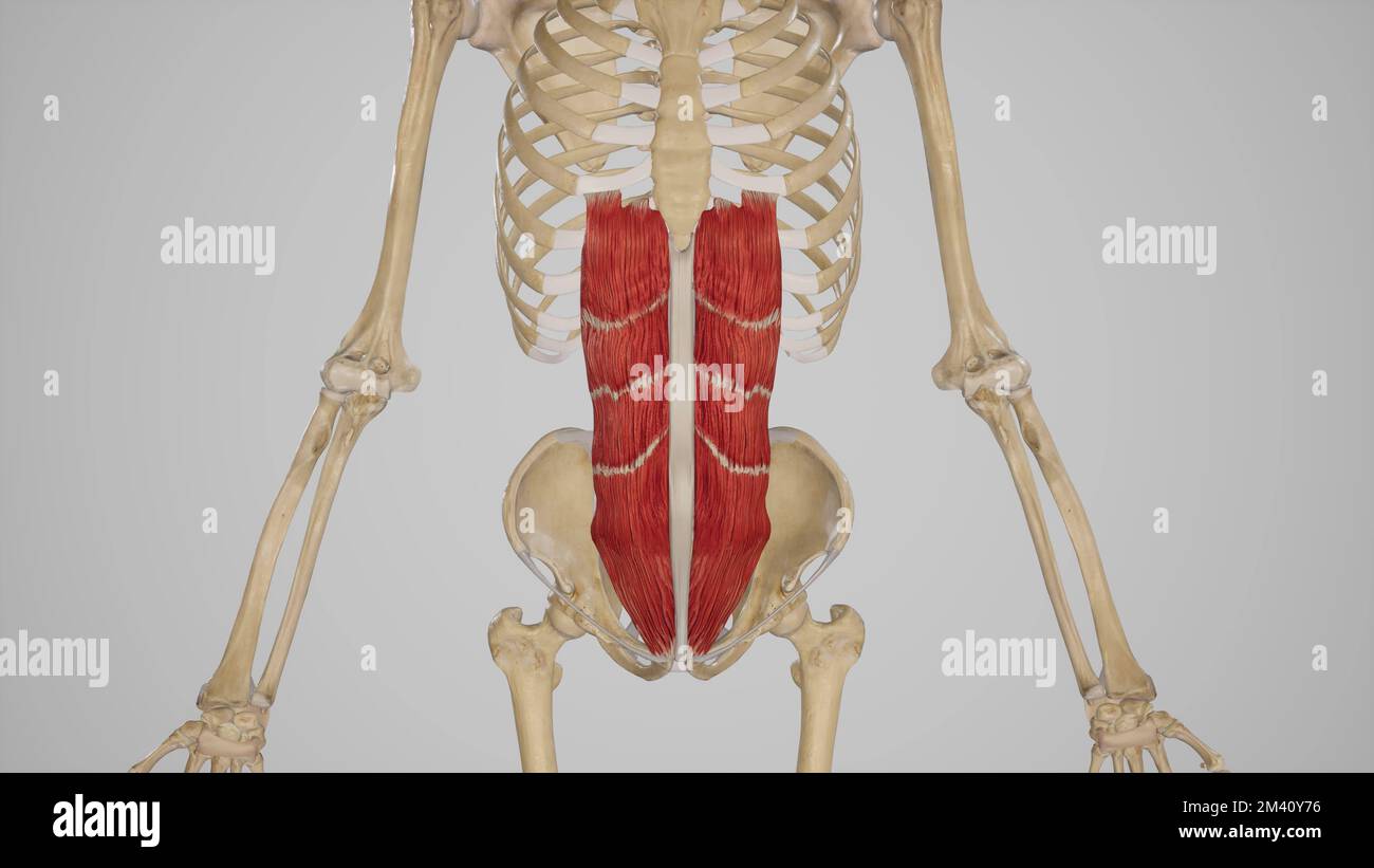 Rectus Abdominis Muscle Stock Photohttps://www.alamy.com/image-license-details/?v=1https://www.alamy.com/rectus-abdominis-muscle-image501580666.html
RF2M40Y76–Rectus Abdominis Muscle
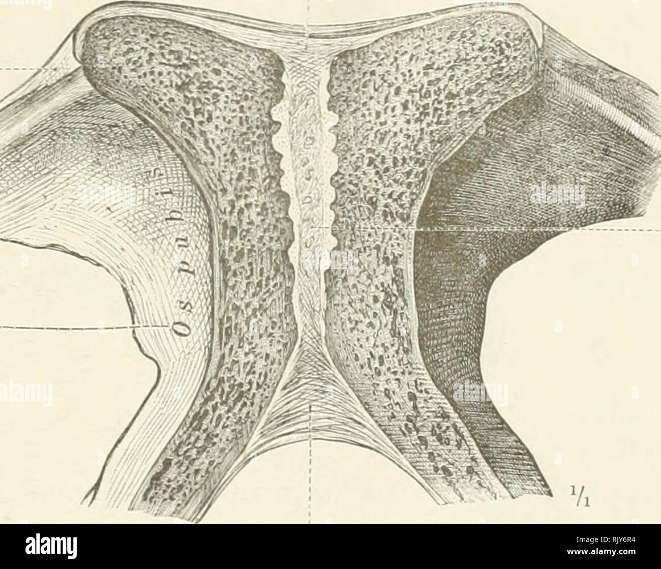
 . An atlas of human anatomy for students and physicians. Anatomy. 221 Pubic ligament of Astley Cooper, or Cooper's ligament Superior or ascending ramus of the pubis Ramus superior ossis pubis Obturator fascia Fascia obturatoria l of the interpubic disc has i name from English anatomists. A feu n, forming the posteriorpubic ligament, which is not mentioned hy Toldt.—Tr. Fig. 458.—Symphysis Ossium Pubis, Pubic Symphysis : Torus Pubicus, Posterior Prominence of the Interpubic Disc; Ligamentum Transversum Pelvis, Transverse Ligament of the Pelvis {seenote2above), with the Venous Foramina; Connexio Stock Photohttps://www.alamy.com/image-license-details/?v=1https://www.alamy.com/an-atlas-of-human-anatomy-for-students-and-physicians-anatomy-221-pubic-ligament-of-astley-cooper-or-coopers-ligament-superior-or-ascending-ramus-of-the-pubis-ramus-superior-ossis-pubis-obturator-fascia-fascia-obturatoria-l-of-the-interpubic-disc-has-i-name-from-english-anatomists-a-feu-n-forming-the-posteriorpubic-ligament-which-is-not-mentioned-hy-toldttr-fig-458symphysis-ossium-pubis-pubic-symphysis-torus-pubicus-posterior-prominence-of-the-interpubic-disc-ligamentum-transversum-pelvis-transverse-ligament-of-the-pelvis-seenote2above-with-the-venous-foramina-connexio-image235395052.html
RMRJY4P4–. An atlas of human anatomy for students and physicians. Anatomy. 221 Pubic ligament of Astley Cooper, or Cooper's ligament Superior or ascending ramus of the pubis Ramus superior ossis pubis Obturator fascia Fascia obturatoria l of the interpubic disc has i name from English anatomists. A feu n, forming the posteriorpubic ligament, which is not mentioned hy Toldt.—Tr. Fig. 458.—Symphysis Ossium Pubis, Pubic Symphysis : Torus Pubicus, Posterior Prominence of the Interpubic Disc; Ligamentum Transversum Pelvis, Transverse Ligament of the Pelvis {seenote2above), with the Venous Foramina; Connexio
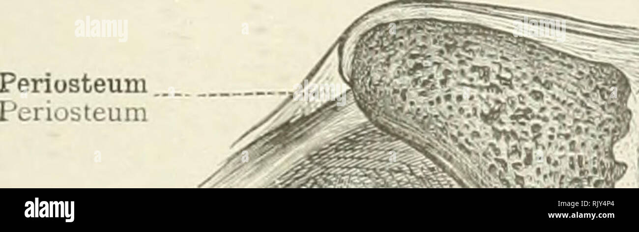
 . An atlas of human anatomy for students and physicians. Anatomy. Ala of the vomer Ala vomeris —Vertical plate of the ethmoid bone •Cartilage of the septum of the nose Septum nasi cartilagineum ..The vomer Vomer —Nasal crest of superior maxillary bone Crista nasalis Fig. 381.—Synchondrosis. (The Spheno-occipital Synchondrosis of a Girl at the Age of Two Years; Median Sagittal Section.) Periosteum - Periosteum Superior pubic ligament Lig. pubicum superius "Bie os pubis. Interpubic disc •Lamina fibrocartilaginea interpubica The inferior pubic or subpubic ligament Lig. arcuatum pubis Fig. 38 Stock Photohttps://www.alamy.com/image-license-details/?v=1https://www.alamy.com/an-atlas-of-human-anatomy-for-students-and-physicians-anatomy-ala-of-the-vomer-ala-vomeris-vertical-plate-of-the-ethmoid-bone-cartilage-of-the-septum-of-the-nose-septum-nasi-cartilagineum-the-vomer-vomer-nasal-crest-of-superior-maxillary-bone-crista-nasalis-fig-381synchondrosis-the-spheno-occipital-synchondrosis-of-a-girl-at-the-age-of-two-years-median-sagittal-section-periosteum-periosteum-superior-pubic-ligament-lig-pubicum-superius-quotbie-os-pubis-interpubic-disc-lamina-fibrocartilaginea-interpubica-the-inferior-pubic-or-subpubic-ligament-lig-arcuatum-pubis-fig-38-image235396648.html
RMRJY6R4–. An atlas of human anatomy for students and physicians. Anatomy. Ala of the vomer Ala vomeris —Vertical plate of the ethmoid bone •Cartilage of the septum of the nose Septum nasi cartilagineum ..The vomer Vomer —Nasal crest of superior maxillary bone Crista nasalis Fig. 381.—Synchondrosis. (The Spheno-occipital Synchondrosis of a Girl at the Age of Two Years; Median Sagittal Section.) Periosteum - Periosteum Superior pubic ligament Lig. pubicum superius "Bie os pubis. Interpubic disc •Lamina fibrocartilaginea interpubica The inferior pubic or subpubic ligament Lig. arcuatum pubis Fig. 38
 . The cyclopædia of anatomy and physiology. Anatomy; Physiology; Zoology. PELVIS. 167 marked Uio-iscJdal angle in the reverse direc- tion to that of Mammals generally, i. e. with the retiring sides anterior (see Jig. 107.). The pubes of birds are generally long, slender, rib-like, and divergent, and are com- posed of a single curved branch (jj), having no angle, and never forming a true interpubic symphysis, though, in the Ostrich and Falco Fulvus, they are closely approximated at their posterior extremity, and form a sort of sym- physis. The ilio-pubic angle is very large, from 155° to 160°, Stock Photohttps://www.alamy.com/image-license-details/?v=1https://www.alamy.com/the-cyclopdia-of-anatomy-and-physiology-anatomy-physiology-zoology-pelvis-167-marked-uio-iscjdal-angle-in-the-reverse-direc-tion-to-that-of-mammals-generally-i-e-with-the-retiring-sides-anterior-see-jig-107-the-pubes-of-birds-are-generally-long-slender-rib-like-and-divergent-and-are-com-posed-of-a-single-curved-branch-jj-having-no-angle-and-never-forming-a-true-interpubic-symphysis-though-in-the-ostrich-and-falco-fulvus-they-are-closely-approximated-at-their-posterior-extremity-and-form-a-sort-of-sym-physis-the-ilio-pubic-angle-is-very-large-from-155-to-160-image231851524.html
RMRD5MYG–. The cyclopædia of anatomy and physiology. Anatomy; Physiology; Zoology. PELVIS. 167 marked Uio-iscJdal angle in the reverse direc- tion to that of Mammals generally, i. e. with the retiring sides anterior (see Jig. 107.). The pubes of birds are generally long, slender, rib-like, and divergent, and are com- posed of a single curved branch (jj), having no angle, and never forming a true interpubic symphysis, though, in the Ostrich and Falco Fulvus, they are closely approximated at their posterior extremity, and form a sort of sym- physis. The ilio-pubic angle is very large, from 155° to 160°,
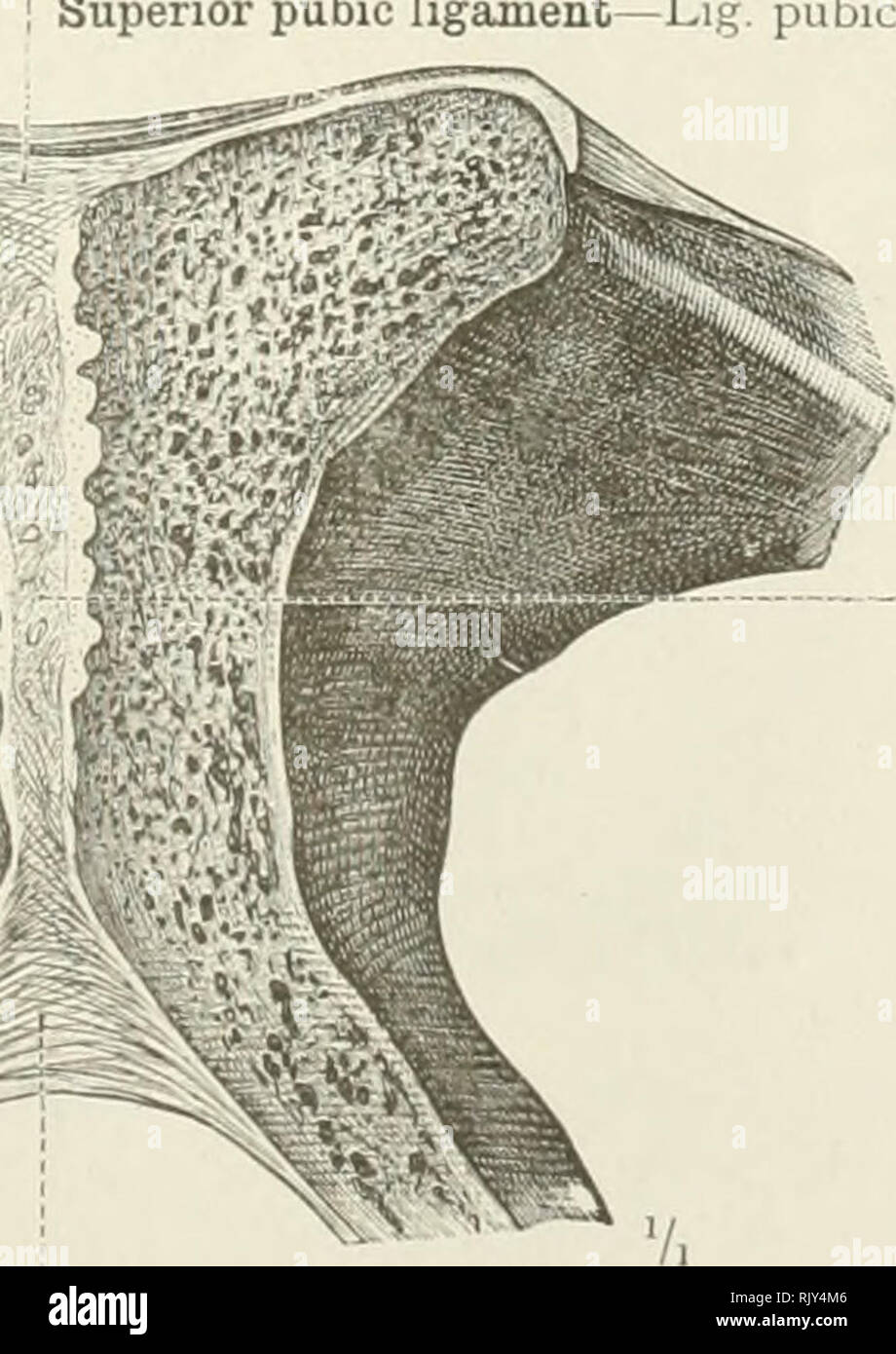 . An atlas of human anatomy for students and physicians. Anatomy. Interpubic disc Lamina fibrocartilaginea interpubica i Inferior pubic or subpubic ligament—Lig. arcuatum pubis Fig. 459.—Symphysis Ossium Pubis, Pubic Symphysis: Lamina Fibrocartilaginea Interpubica, Interpubic Disc; Ligamentum Pubicum Superius, Superior Pubic Ligament; Ligamentum Arcuatum Pubis, Inferior Pubic ok Subpubic Ligament. (Tiih Pubic Symphysis in Frontal Section-. Anterior Si ri < 1 01 Posterior Segment.) Symphysis ossium pubis—Pubic symphysis. Please note that these images are extracted from scanned page images t Stock Photohttps://www.alamy.com/image-license-details/?v=1https://www.alamy.com/an-atlas-of-human-anatomy-for-students-and-physicians-anatomy-interpubic-disc-lamina-fibrocartilaginea-interpubica-i-inferior-pubic-or-subpubic-ligamentlig-arcuatum-pubis-fig-459symphysis-ossium-pubis-pubic-symphysis-lamina-fibrocartilaginea-interpubica-interpubic-disc-ligamentum-pubicum-superius-superior-pubic-ligament-ligamentum-arcuatum-pubis-inferior-pubic-ok-subpubic-ligament-tiih-pubic-symphysis-in-frontal-section-anterior-si-ri-lt-1-01-posterior-segment-symphysis-ossium-pubispubic-symphysis-please-note-that-these-images-are-extracted-from-scanned-page-images-t-image235394998.html
RMRJY4M6–. An atlas of human anatomy for students and physicians. Anatomy. Interpubic disc Lamina fibrocartilaginea interpubica i Inferior pubic or subpubic ligament—Lig. arcuatum pubis Fig. 459.—Symphysis Ossium Pubis, Pubic Symphysis: Lamina Fibrocartilaginea Interpubica, Interpubic Disc; Ligamentum Pubicum Superius, Superior Pubic Ligament; Ligamentum Arcuatum Pubis, Inferior Pubic ok Subpubic Ligament. (Tiih Pubic Symphysis in Frontal Section-. Anterior Si ri < 1 01 Posterior Segment.) Symphysis ossium pubis—Pubic symphysis. Please note that these images are extracted from scanned page images t