Intestinal mucosa Stock Vectors & Vector Art
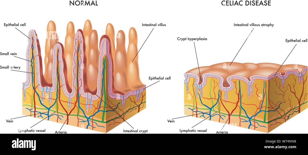 Medical illustration of the modification of the intestinal mucosa in celiac subject Stock Vectorhttps://www.alamy.com/image-license-details/?v=1https://www.alamy.com/medical-illustration-of-the-modification-of-the-intestinal-mucosa-in-celiac-subject-image262562980.html
RFW74NN8–Medical illustration of the modification of the intestinal mucosa in celiac subject
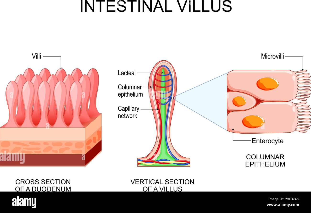 Intestinal villus. Different between Villi and microvilli. Cross section of a duodenum with submucosa, mucosa and muscularis layers. Vertical section Stock Vectorhttps://www.alamy.com/image-license-details/?v=1https://www.alamy.com/intestinal-villus-different-between-villi-and-microvilli-cross-section-of-a-duodenum-with-submucosa-mucosa-and-muscularis-layers-vertical-section-image611825888.html
RF2XFB24G–Intestinal villus. Different between Villi and microvilli. Cross section of a duodenum with submucosa, mucosa and muscularis layers. Vertical section
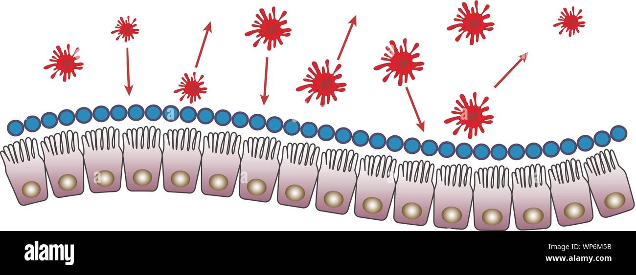 Intestinal mucosa immunity Stock Vectorhttps://www.alamy.com/image-license-details/?v=1https://www.alamy.com/intestinal-mucosa-immunity-image271825495.html
RFWP6M5B–Intestinal mucosa immunity
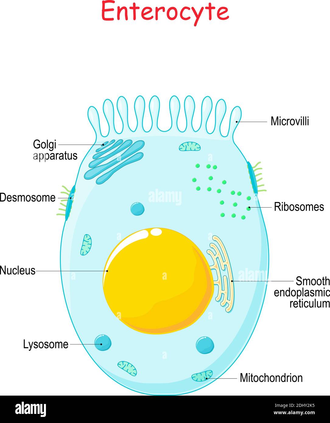 Enterocyte. intestinal absorptive epithelial cell with microvilli. Structure of the enterocyte. Infographics. Vector illustration on white background. Stock Vectorhttps://www.alamy.com/image-license-details/?v=1https://www.alamy.com/enterocyte-intestinal-absorptive-epithelial-cell-with-microvilli-structure-of-the-enterocyte-infographics-vector-illustration-on-white-background-image389672057.html
RF2DHY2K5–Enterocyte. intestinal absorptive epithelial cell with microvilli. Structure of the enterocyte. Infographics. Vector illustration on white background.
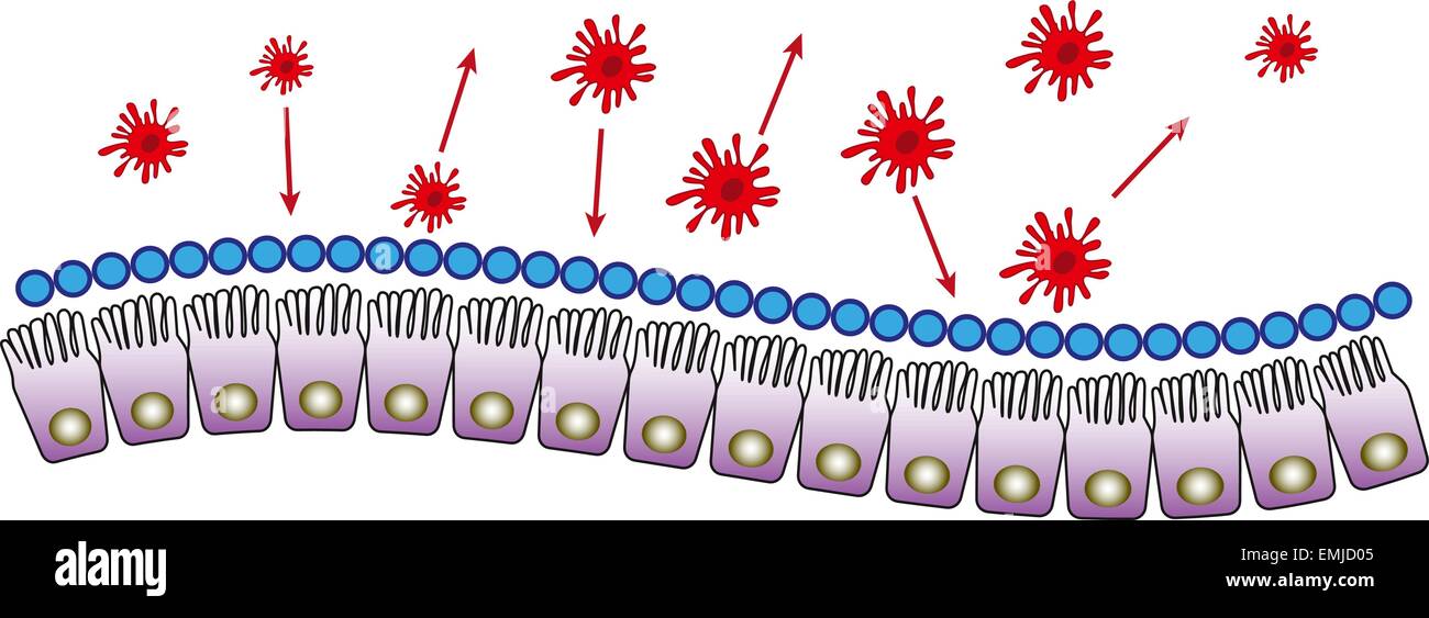 Intestinal mucosa immunity cells medical vector illustration Stock Vectorhttps://www.alamy.com/image-license-details/?v=1https://www.alamy.com/stock-photo-intestinal-mucosa-immunity-cells-medical-vector-illustration-81539925.html
RFEMJD05–Intestinal mucosa immunity cells medical vector illustration
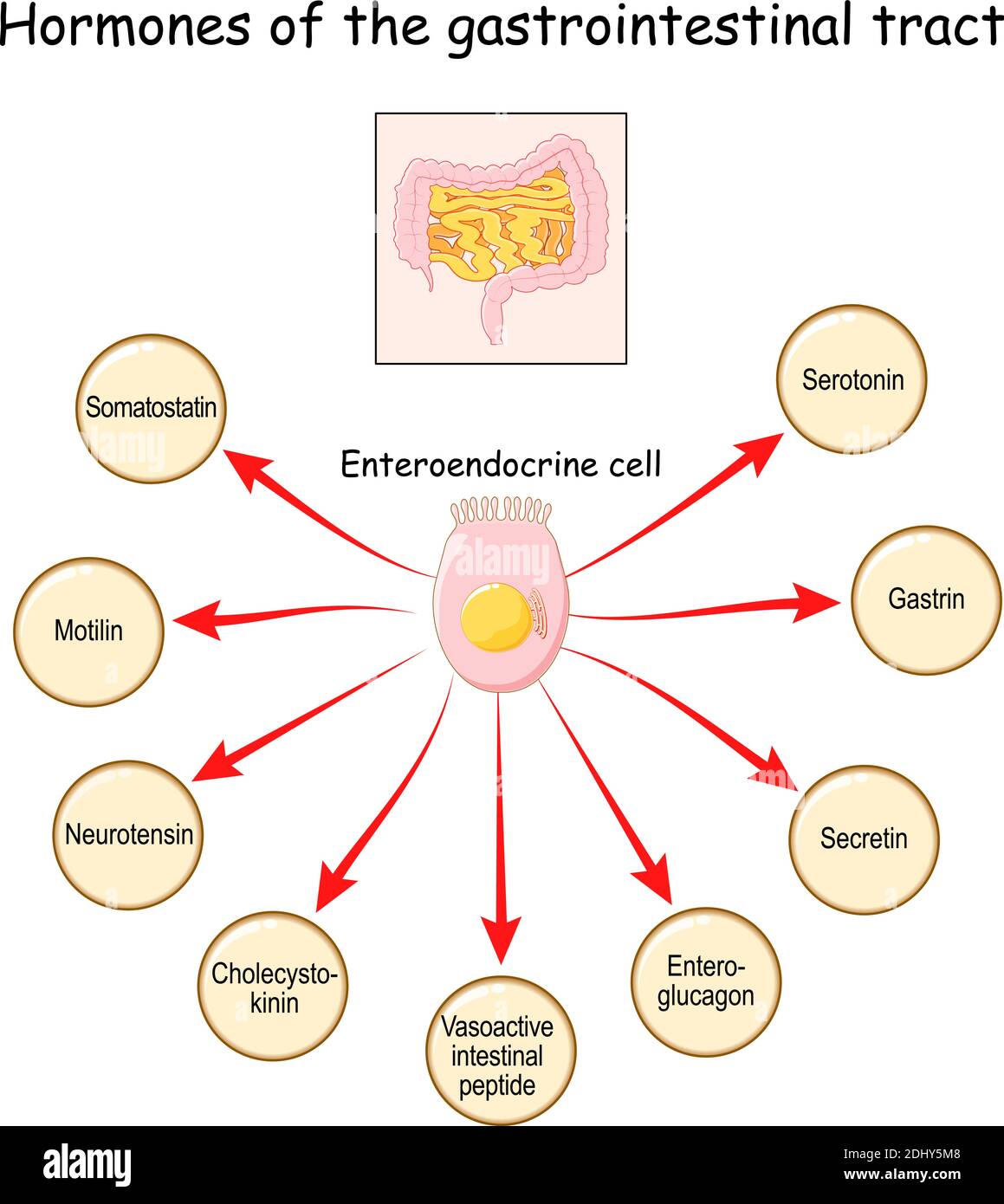 Hormones of the gastrointestinal tract and Enteroendocrine cell. Enterocyte. Human endocrine system. Vector illustration Stock Vectorhttps://www.alamy.com/image-license-details/?v=1https://www.alamy.com/hormones-of-the-gastrointestinal-tract-and-enteroendocrine-cell-enterocyte-human-endocrine-system-vector-illustration-image389674440.html
RF2DHY5M8–Hormones of the gastrointestinal tract and Enteroendocrine cell. Enterocyte. Human endocrine system. Vector illustration
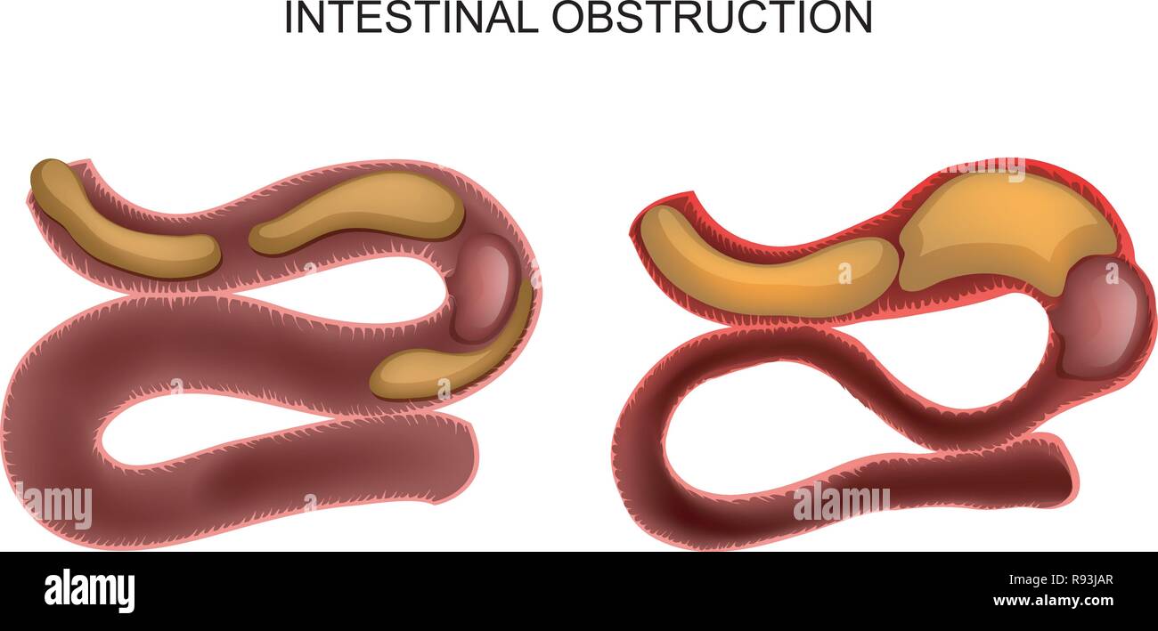 vector illustration of intestinal obstruction due to a cancerous tumor Stock Vectorhttps://www.alamy.com/image-license-details/?v=1https://www.alamy.com/vector-illustration-of-intestinal-obstruction-due-to-a-cancerous-tumor-image229346959.html
RFR93JAR–vector illustration of intestinal obstruction due to a cancerous tumor
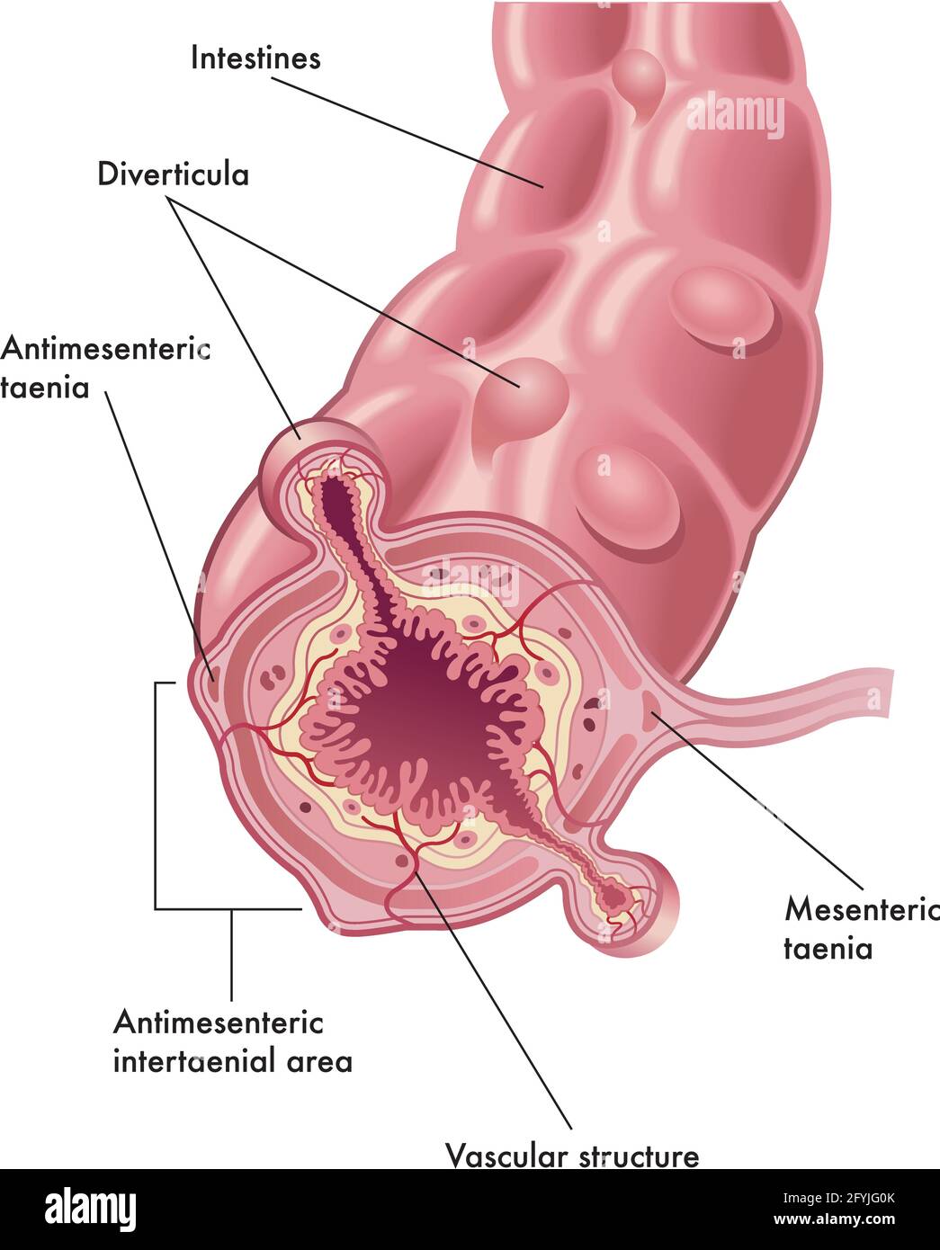 Medical illustration of the anatomy of diverticula, with annotations. Stock Vectorhttps://www.alamy.com/image-license-details/?v=1https://www.alamy.com/medical-illustration-of-the-anatomy-of-diverticula-with-annotations-image430052243.html
RF2FYJG0K–Medical illustration of the anatomy of diverticula, with annotations.
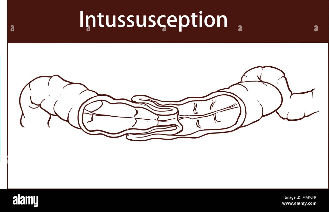 vector illustration of intussusception of intestine. obstruction. Stock Vectorhttps://www.alamy.com/image-license-details/?v=1https://www.alamy.com/vector-illustration-of-intussusception-of-intestine-obstruction-image178672475.html
RFMAK6FR–vector illustration of intussusception of intestine. obstruction.
 Small Intestine Diagram. Cross-section of a typical segment of the intestinal wall showing the layers: mucosa, submucosa, muscularis, and serosa. Stock Vectorhttps://www.alamy.com/image-license-details/?v=1https://www.alamy.com/small-intestine-diagram-cross-section-of-a-typical-segment-of-the-intestinal-wall-showing-the-layers-mucosa-submucosa-muscularis-and-serosa-image496267300.html
RF2KRAX04–Small Intestine Diagram. Cross-section of a typical segment of the intestinal wall showing the layers: mucosa, submucosa, muscularis, and serosa.
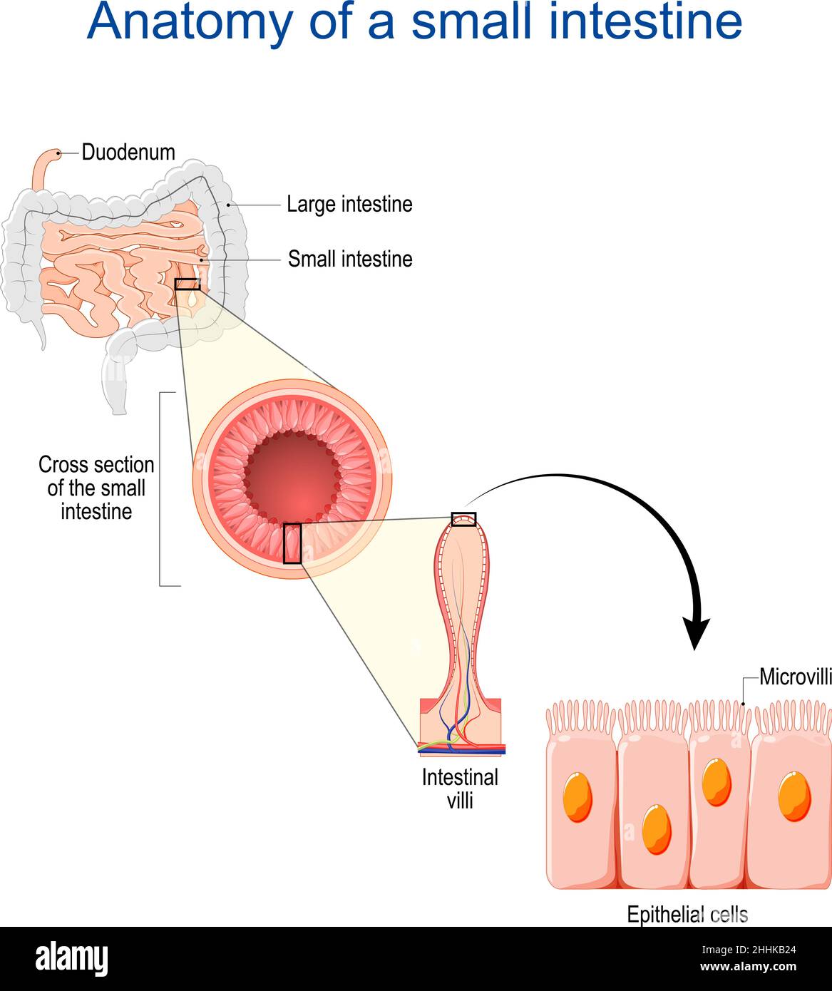 small intestine Anatomy. Cross section of a ileum with Internal villi. Close-up of Epithelial cells with Microvilli. Vector illustration Stock Vectorhttps://www.alamy.com/image-license-details/?v=1https://www.alamy.com/small-intestine-anatomy-cross-section-of-a-ileum-with-internal-villi-close-up-of-epithelial-cells-with-microvilli-vector-illustration-image458344492.html
RF2HHKB24–small intestine Anatomy. Cross section of a ileum with Internal villi. Close-up of Epithelial cells with Microvilli. Vector illustration
RF2R85JP2–Celiac disease black line icon. Autoimmune diseases. Pictogram for web page, mobile app, promo.
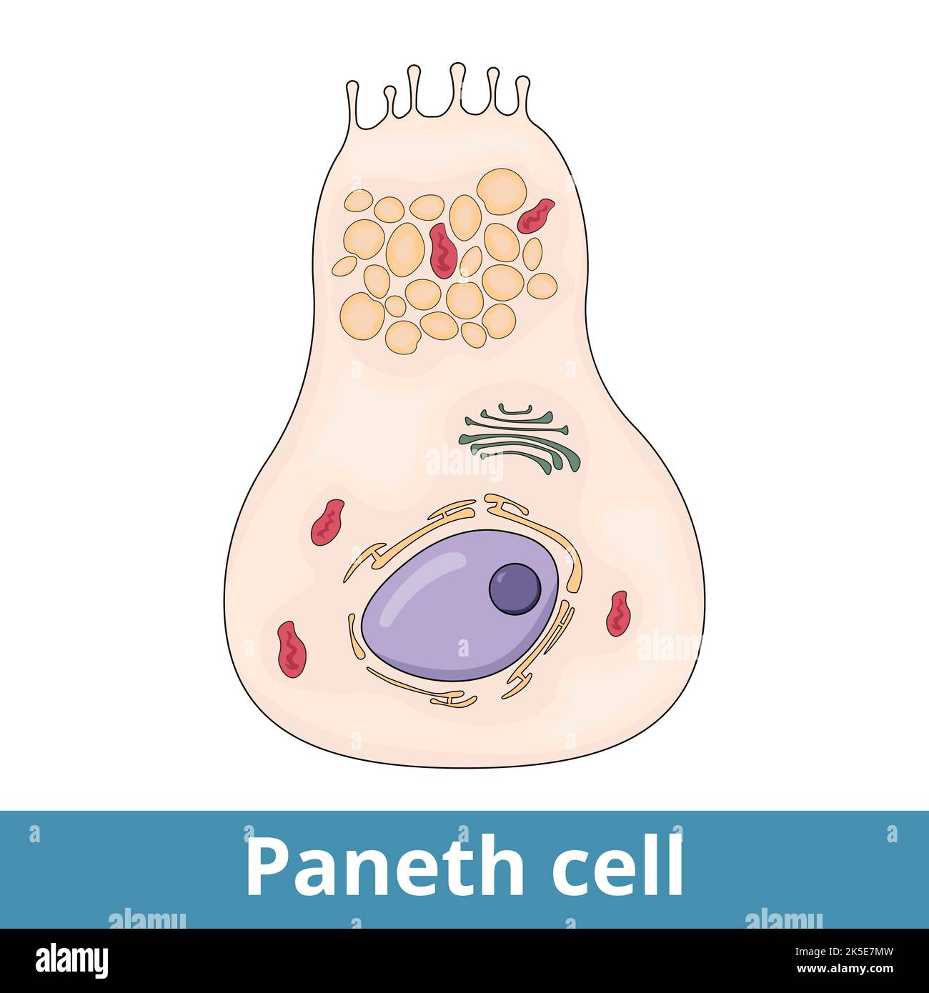 Paneth cell. Cells in the small intestine epithelium secrete compounds into the lumen of the intestinal gland Stock Vectorhttps://www.alamy.com/image-license-details/?v=1https://www.alamy.com/paneth-cell-cells-in-the-small-intestine-epithelium-secrete-compounds-into-the-lumen-of-the-intestinal-gland-image485276985.html
RF2K5E7MW–Paneth cell. Cells in the small intestine epithelium secrete compounds into the lumen of the intestinal gland
RF2R0616C–Celiac disease black line icon. Autoimmune diseases. Pictogram for web page, mobile app, promo.
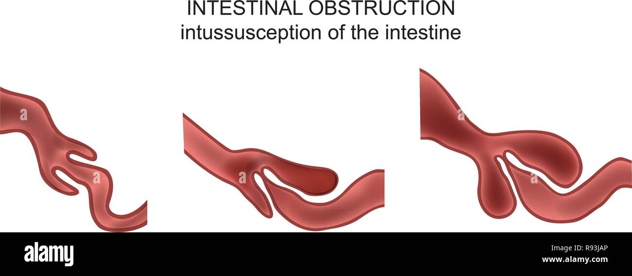 vector illustration of intussusception of intestine. obstruction. Stock Vectorhttps://www.alamy.com/image-license-details/?v=1https://www.alamy.com/vector-illustration-of-intussusception-of-intestine-obstruction-image229346958.html
RFR93JAP–vector illustration of intussusception of intestine. obstruction.
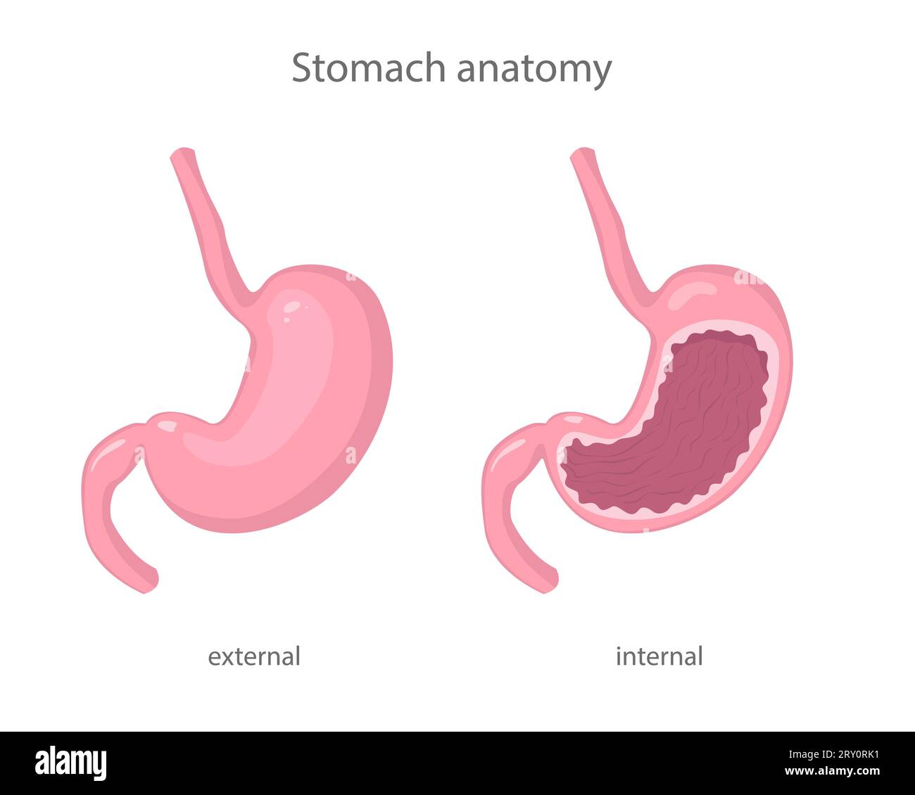 Scientific illustration of human healthy stomach external and internal view in realistic style with shadows and highlights. Stock Vectorhttps://www.alamy.com/image-license-details/?v=1https://www.alamy.com/scientific-illustration-of-human-healthy-stomach-external-and-internal-view-in-realistic-style-with-shadows-and-highlights-image567346053.html
RF2RY0RK1–Scientific illustration of human healthy stomach external and internal view in realistic style with shadows and highlights.
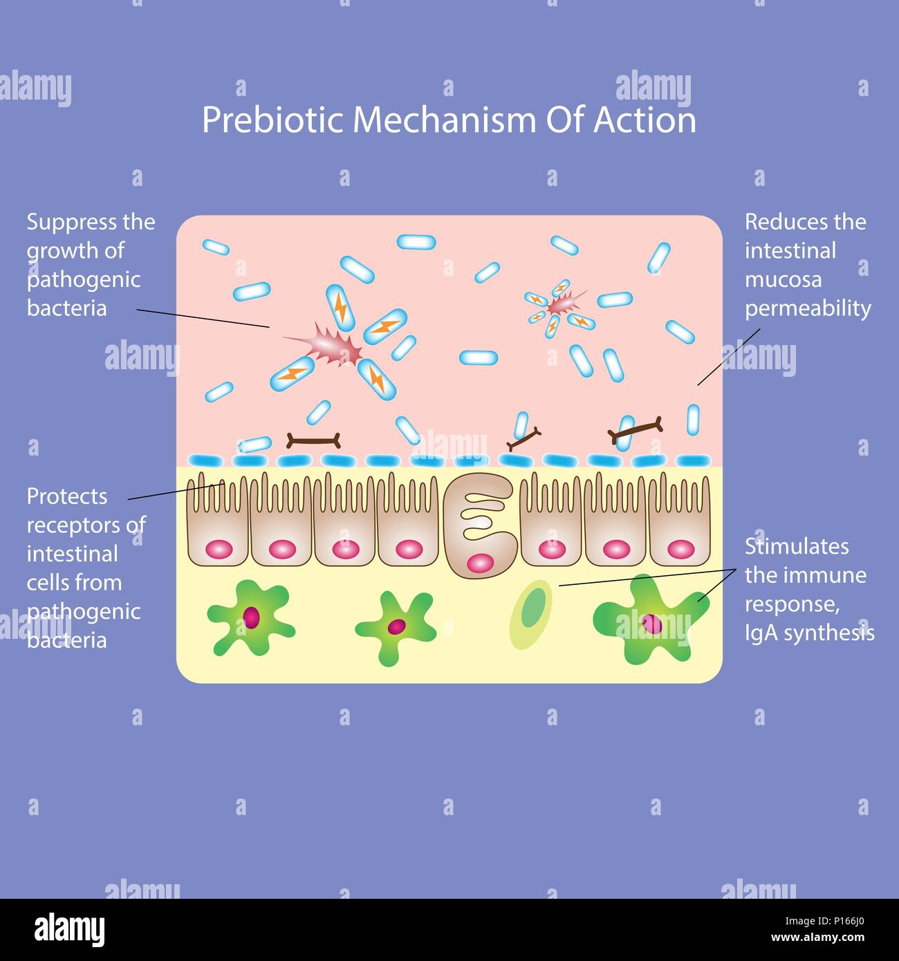 Prebiotic or probiotic mechanism of action. Medical vector illustration Stock Vectorhttps://www.alamy.com/image-license-details/?v=1https://www.alamy.com/prebiotic-or-probiotic-mechanism-of-action-medical-vector-illustration-image207275992.html
RFP166J0–Prebiotic or probiotic mechanism of action. Medical vector illustration
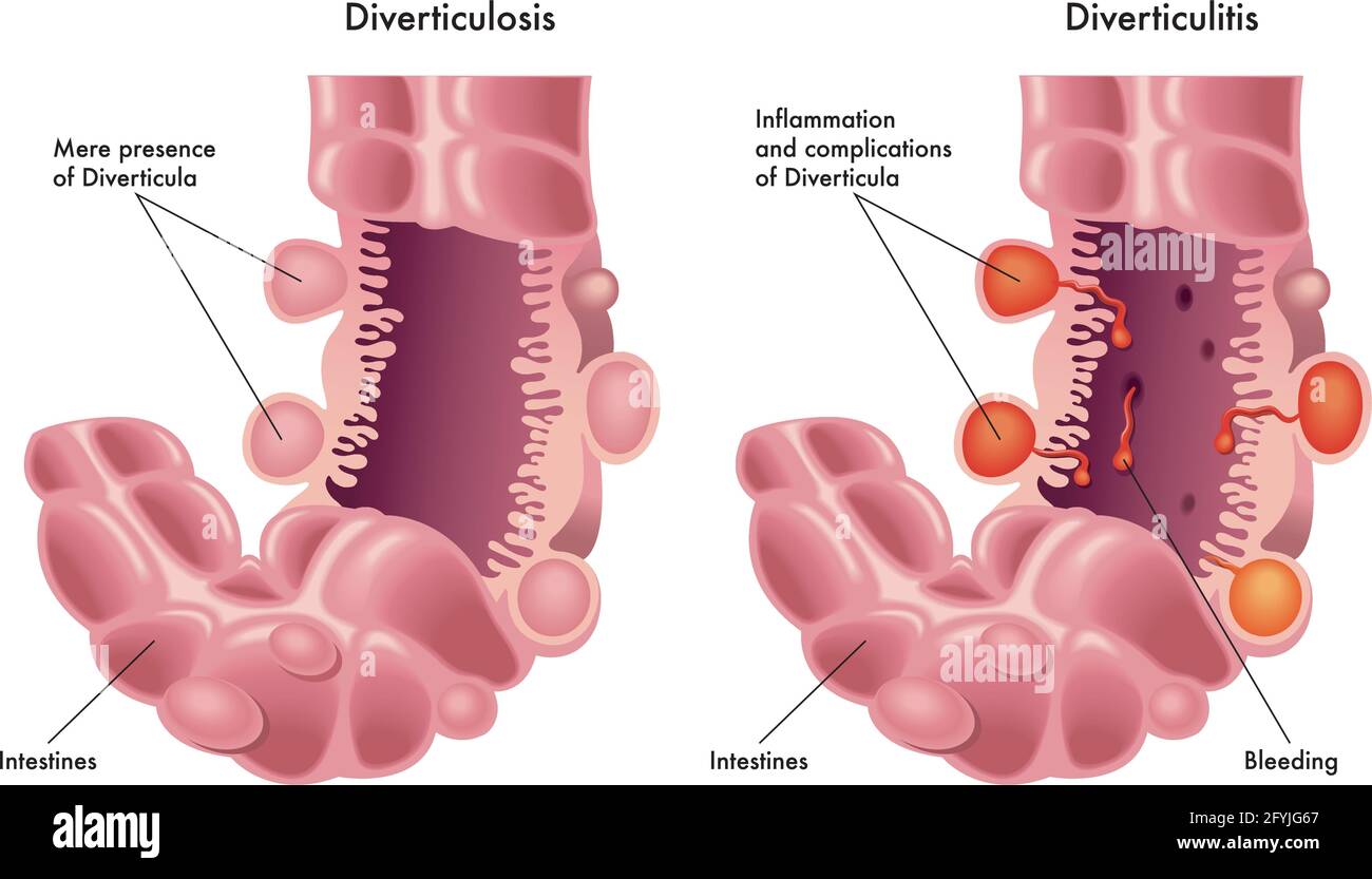 Medical illustration of the difference between diverticulosis and diverticulitis. Stock Vectorhttps://www.alamy.com/image-license-details/?v=1https://www.alamy.com/medical-illustration-of-the-difference-between-diverticulosis-and-diverticulitis-image430052399.html
RF2FYJG67–Medical illustration of the difference between diverticulosis and diverticulitis.
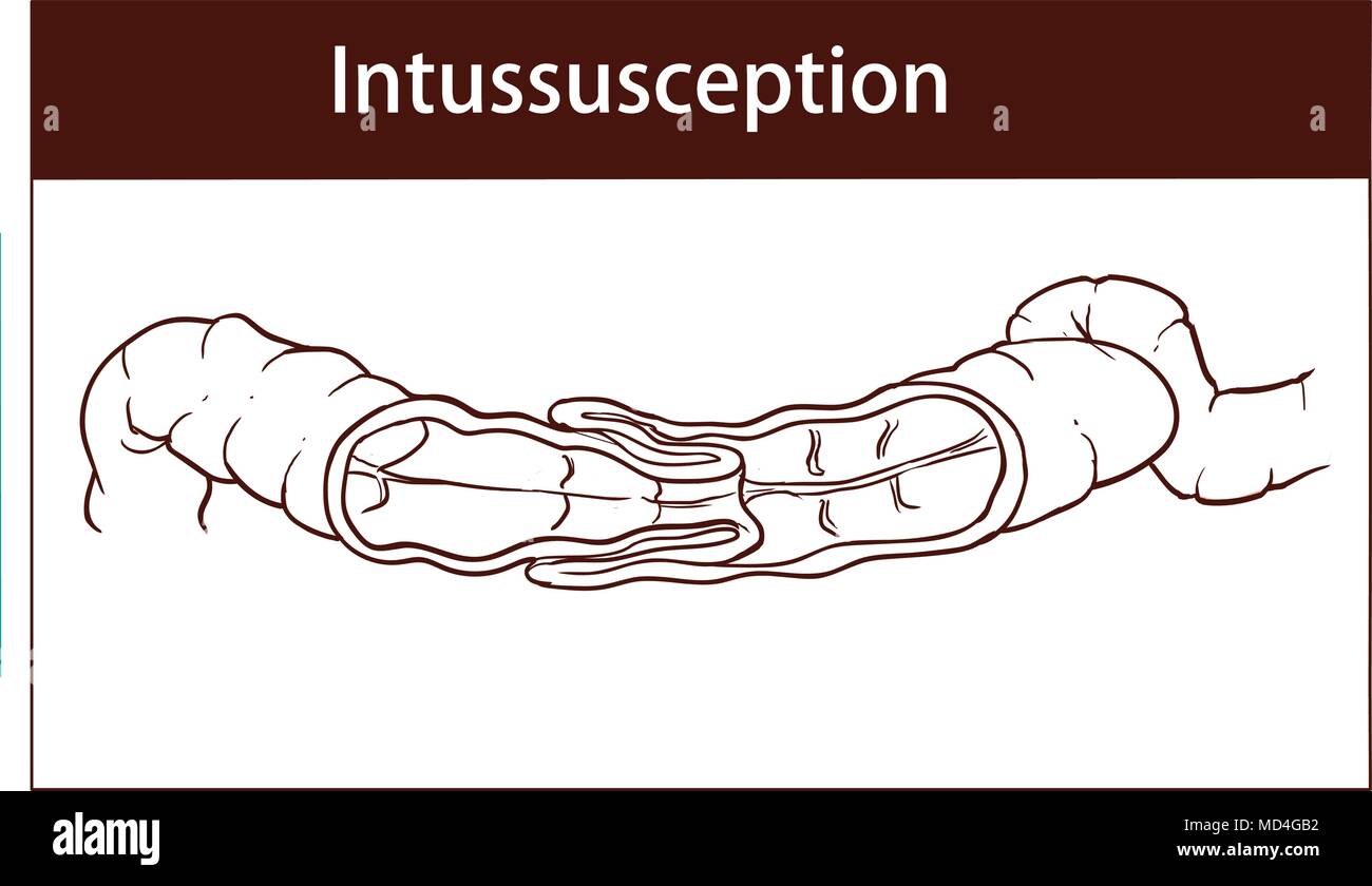 vector illustration of intussusception of intestine. obstruction. Stock Vectorhttps://www.alamy.com/image-license-details/?v=1https://www.alamy.com/vector-illustration-of-intussusception-of-intestine-obstruction-image180194870.html
RFMD4GB2–vector illustration of intussusception of intestine. obstruction.
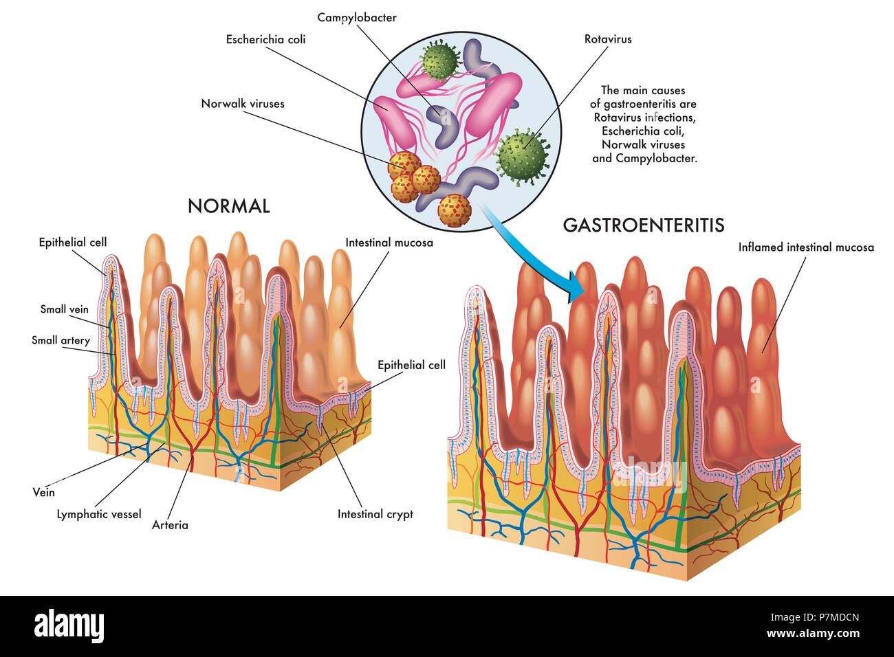 vector medical illustration of the main causes of gastroenteritis Stock Vectorhttps://www.alamy.com/image-license-details/?v=1https://www.alamy.com/vector-medical-illustration-of-the-main-causes-of-gastroenteritis-image211276597.html
RFP7MDCN–vector medical illustration of the main causes of gastroenteritis
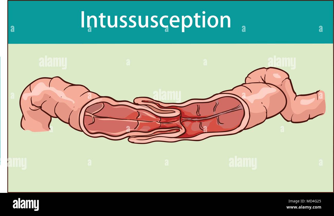 vector illustration of intussusception of intestine. obstruction. Stock Vectorhttps://www.alamy.com/image-license-details/?v=1https://www.alamy.com/vector-illustration-of-intussusception-of-intestine-obstruction-image180194621.html
RFMD4G25–vector illustration of intussusception of intestine. obstruction.
 Symbolic medical illustration of food intolerance to gluten called celiac disease Stock Vectorhttps://www.alamy.com/image-license-details/?v=1https://www.alamy.com/symbolic-medical-illustration-of-food-intolerance-to-gluten-called-celiac-disease-image262560554.html
RFW74JJJ–Symbolic medical illustration of food intolerance to gluten called celiac disease
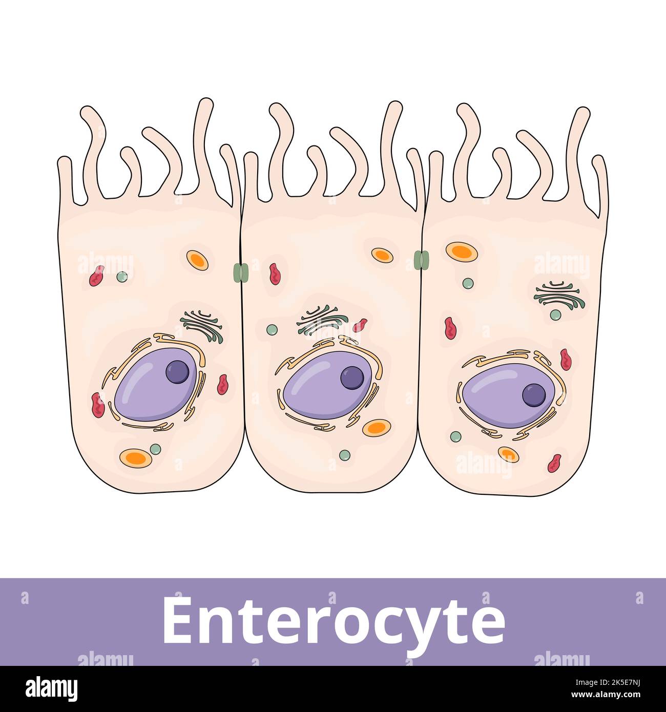 Enterocyte. Intestinal absorptive cells, are simple columnar epithelial cells which line the inner surface of the small and large intestines. Stock Vectorhttps://www.alamy.com/image-license-details/?v=1https://www.alamy.com/enterocyte-intestinal-absorptive-cells-are-simple-columnar-epithelial-cells-which-line-the-inner-surface-of-the-small-and-large-intestines-image485277006.html
RF2K5E7NJ–Enterocyte. Intestinal absorptive cells, are simple columnar epithelial cells which line the inner surface of the small and large intestines.
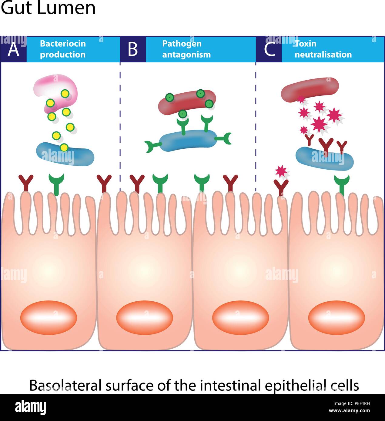 Gut lumen. Enterocytes, or intestinal absorptive cells. Small intestine. Columnar epithelial cells Stock Vectorhttps://www.alamy.com/image-license-details/?v=1https://www.alamy.com/gut-lumen-enterocytes-or-intestinal-absorptive-cells-small-intestine-columnar-epithelial-cells-image215462677.html
RFPEF4RH–Gut lumen. Enterocytes, or intestinal absorptive cells. Small intestine. Columnar epithelial cells
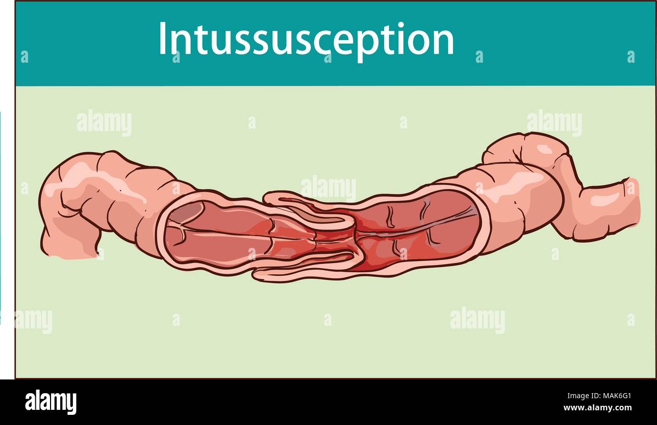 vector illustration of intussusception of intestine. obstruction. Stock Vectorhttps://www.alamy.com/image-license-details/?v=1https://www.alamy.com/vector-illustration-of-intussusception-of-intestine-obstruction-image178672481.html
RFMAK6G1–vector illustration of intussusception of intestine. obstruction.