Quick filters:
Ischial tuberosities Stock Photos and Images
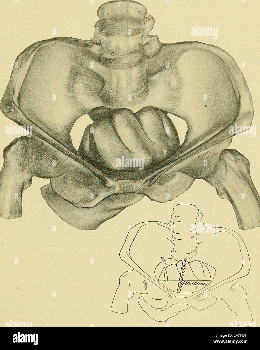 The practice of obstetrics, designed for the use of students and practitioners of medicine . he importance in doubtful cases of a thoroughexamination under full anesthesia, and by an experienced accoucheur, cannotbe overestimated. It is hardly necessary to point out all the refinements of THE EXAMINATION OF PREGNANCY. 177 diagnosis in the various forms of pelvic deformity. The educated hand willrecognize the difference in the respective lateral pelvic walls, which accom-panies the obliquely contracted pelvis (Naegele); the converging walls andapproximated ischial tuberosities, which characteri Stock Photohttps://www.alamy.com/image-license-details/?v=1https://www.alamy.com/the-practice-of-obstetrics-designed-for-the-use-of-students-and-practitioners-of-medicine-he-importance-in-doubtful-cases-of-a-thoroughexamination-under-full-anesthesia-and-by-an-experienced-accoucheur-cannotbe-overestimated-it-is-hardly-necessary-to-point-out-all-the-refinements-of-the-examination-of-pregnancy-177-diagnosis-in-the-various-forms-of-pelvic-deformity-the-educated-hand-willrecognize-the-difference-in-the-respective-lateral-pelvic-walls-which-accom-panies-the-obliquely-contracted-pelvis-naegele-the-converging-walls-andapproximated-ischial-tuberosities-which-characteri-image343321541.html
RM2AXFJ3H–The practice of obstetrics, designed for the use of students and practitioners of medicine . he importance in doubtful cases of a thoroughexamination under full anesthesia, and by an experienced accoucheur, cannotbe overestimated. It is hardly necessary to point out all the refinements of THE EXAMINATION OF PREGNANCY. 177 diagnosis in the various forms of pelvic deformity. The educated hand willrecognize the difference in the respective lateral pelvic walls, which accom-panies the obliquely contracted pelvis (Naegele); the converging walls andapproximated ischial tuberosities, which characteri
 macaque des celebes Saeugetiere Primaten Schopfmakak Macaca nigra oder Cynopithecus niger Celebes crested macaque or black macaq Stock Photohttps://www.alamy.com/image-license-details/?v=1https://www.alamy.com/stock-photo-macaque-des-celebes-saeugetiere-primaten-schopfmakak-macaca-nigra-20551325.html
RMB5C5BW–macaque des celebes Saeugetiere Primaten Schopfmakak Macaca nigra oder Cynopithecus niger Celebes crested macaque or black macaq
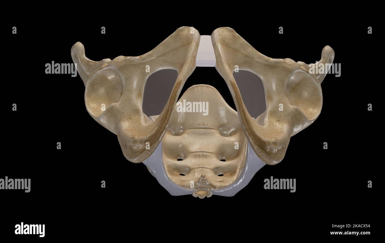 The Pelvic Girdle and Pelvic Outlet Stock Photohttps://www.alamy.com/image-license-details/?v=1https://www.alamy.com/the-pelvic-girdle-and-pelvic-outlet-image488320816.html
RF2KACX54–The Pelvic Girdle and Pelvic Outlet
 . On the anatomy of vertebrates. Vertebrates; Anatomy, Comparative; 1866. ISG ANATOMY OF VERTEBRATES. A sllri-lit modification is presented by Trionyx, in which a view of tlie pelvis is given from the dorsal aspect, the sacrum being removed, in fig. 116: here i shows the end of the ilium, 62, which was attached to that part of the vertebral column: a is the ex- panded acetabular end of these short, straight, columnar bones. no. The ischia, c, c, develope tuberosities/,/, and unite at the ischial symphysis, 64, d. The pubics h, h, articulate by a broader tube- rosity, /(, with the plastron, and Stock Photohttps://www.alamy.com/image-license-details/?v=1https://www.alamy.com/on-the-anatomy-of-vertebrates-vertebrates-anatomy-comparative-1866-isg-anatomy-of-vertebrates-a-sllri-lit-modification-is-presented-by-trionyx-in-which-a-view-of-tlie-pelvis-is-given-from-the-dorsal-aspect-the-sacrum-being-removed-in-fig-116-here-i-shows-the-end-of-the-ilium-62-which-was-attached-to-that-part-of-the-vertebral-column-a-is-the-ex-panded-acetabular-end-of-these-short-straight-columnar-bones-no-the-ischia-c-c-develope-tuberosities-and-unite-at-the-ischial-symphysis-64-d-the-pubics-h-h-articulate-by-a-broader-tube-rosity-with-the-plastron-and-image216417607.html
RMPG2JT7–. On the anatomy of vertebrates. Vertebrates; Anatomy, Comparative; 1866. ISG ANATOMY OF VERTEBRATES. A sllri-lit modification is presented by Trionyx, in which a view of tlie pelvis is given from the dorsal aspect, the sacrum being removed, in fig. 116: here i shows the end of the ilium, 62, which was attached to that part of the vertebral column: a is the ex- panded acetabular end of these short, straight, columnar bones. no. The ischia, c, c, develope tuberosities/,/, and unite at the ischial symphysis, 64, d. The pubics h, h, articulate by a broader tube- rosity, /(, with the plastron, and
 A treatise on the science and practice of midwifery . eter of thebrim is increased, while the transverse is lessened; the relative pro-portion between the two is thus reversed. While the upper propor-tion of the sacrum is displaced backwards, its lower end is projectedforward, so that the antero-posterior diameters of the cavity andoutlet are considerably diminished. The ischial tuberosities are alsonearer to each other, and the pubic arch is narrowed. Obstructionto delivery wiil be chiefly met with at the lower parts and outlet ofthe pelvic cavity; for, although the transverse diameter of the Stock Photohttps://www.alamy.com/image-license-details/?v=1https://www.alamy.com/a-treatise-on-the-science-and-practice-of-midwifery-eter-of-thebrim-is-increased-while-the-transverse-is-lessened-the-relative-pro-portion-between-the-two-is-thus-reversed-while-the-upper-propor-tion-of-the-sacrum-is-displaced-backwards-its-lower-end-is-projectedforward-so-that-the-antero-posterior-diameters-of-the-cavity-andoutlet-are-considerably-diminished-the-ischial-tuberosities-are-alsonearer-to-each-other-and-the-pubic-arch-is-narrowed-obstructionto-delivery-wiil-be-chiefly-met-with-at-the-lower-parts-and-outlet-ofthe-pelvic-cavity-for-although-the-transverse-diameter-of-the-image338401491.html
RM2AJFEFF–A treatise on the science and practice of midwifery . eter of thebrim is increased, while the transverse is lessened; the relative pro-portion between the two is thus reversed. While the upper propor-tion of the sacrum is displaced backwards, its lower end is projectedforward, so that the antero-posterior diameters of the cavity andoutlet are considerably diminished. The ischial tuberosities are alsonearer to each other, and the pubic arch is narrowed. Obstructionto delivery wiil be chiefly met with at the lower parts and outlet ofthe pelvic cavity; for, although the transverse diameter of the
 macaque des celebes Saeugetiere Primaten Schopfmakak Macaca nigra oder Cynopithecus niger Celebes crested macaque or black macaq Stock Photohttps://www.alamy.com/image-license-details/?v=1https://www.alamy.com/stock-photo-macaque-des-celebes-saeugetiere-primaten-schopfmakak-macaca-nigra-20546632.html
RMB5BYC8–macaque des celebes Saeugetiere Primaten Schopfmakak Macaca nigra oder Cynopithecus niger Celebes crested macaque or black macaq
 A manual of diseases of the nervous system . being carried so far back that avertical line from the scapula falls an inch or more behind the sacrum.It is due, not to the weakness of the trunk muscles, but to that of theextensors of the hip, in consequence of which the pelvis is inclinedforwards, carrying with it the lower lumbar vertebra ; hence the upperpart of the trunk has to be held far back to keep the centre of gravityof the body over the feet. The proof of this mechanism is that whenthe patient sits, and the pelvis is supported on the ischial tuberosities,the lordosis disappears. It is, Stock Photohttps://www.alamy.com/image-license-details/?v=1https://www.alamy.com/a-manual-of-diseases-of-the-nervous-system-being-carried-so-far-back-that-avertical-line-from-the-scapula-falls-an-inch-or-more-behind-the-sacrumit-is-due-not-to-the-weakness-of-the-trunk-muscles-but-to-that-of-theextensors-of-the-hip-in-consequence-of-which-the-pelvis-is-inclinedforwards-carrying-with-it-the-lower-lumbar-vertebra-hence-the-upperpart-of-the-trunk-has-to-be-held-far-back-to-keep-the-centre-of-gravityof-the-body-over-the-feet-the-proof-of-this-mechanism-is-that-whenthe-patient-sits-and-the-pelvis-is-supported-on-the-ischial-tuberositiesthe-lordosis-disappears-it-is-image338232721.html
RM2AJ7R81–A manual of diseases of the nervous system . being carried so far back that avertical line from the scapula falls an inch or more behind the sacrum.It is due, not to the weakness of the trunk muscles, but to that of theextensors of the hip, in consequence of which the pelvis is inclinedforwards, carrying with it the lower lumbar vertebra ; hence the upperpart of the trunk has to be held far back to keep the centre of gravityof the body over the feet. The proof of this mechanism is that whenthe patient sits, and the pelvis is supported on the ischial tuberosities,the lordosis disappears. It is,
 The practice of obstetrics, designed for the use of students and practitioners of medicine . ities and the lower border of the sacrum. Shape.—Its shape is that of a diamond or of two triangles having a common 384 PHYSIOLOGICAL LABOR. base, and varies with the mobility of the coccyx, and in labor it becomes almostcircular, thus being more changeable than the pelvic inlet. In the sittingposture the weight of the body rests entirely on the ischial tuberosities, sincethey are on a lower plane than the tip of the coccyx; and this explains whytransverse pelvic contractions are so much more frequent Stock Photohttps://www.alamy.com/image-license-details/?v=1https://www.alamy.com/the-practice-of-obstetrics-designed-for-the-use-of-students-and-practitioners-of-medicine-ities-and-the-lower-border-of-the-sacrum-shapeits-shape-is-that-of-a-diamond-or-of-two-triangles-having-a-common-384-physiological-labor-base-and-varies-with-the-mobility-of-the-coccyx-and-in-labor-it-becomes-almostcircular-thus-being-more-changeable-than-the-pelvic-inlet-in-the-sittingposture-the-weight-of-the-body-rests-entirely-on-the-ischial-tuberosities-sincethey-are-on-a-lower-plane-than-the-tip-of-the-coccyx-and-this-explains-whytransverse-pelvic-contractions-are-so-much-more-frequent-image343269899.html
RM2AXD877–The practice of obstetrics, designed for the use of students and practitioners of medicine . ities and the lower border of the sacrum. Shape.—Its shape is that of a diamond or of two triangles having a common 384 PHYSIOLOGICAL LABOR. base, and varies with the mobility of the coccyx, and in labor it becomes almostcircular, thus being more changeable than the pelvic inlet. In the sittingposture the weight of the body rests entirely on the ischial tuberosities, sincethey are on a lower plane than the tip of the coccyx; and this explains whytransverse pelvic contractions are so much more frequent
 A system of obstetrics . body resting upon the ischial tuberosities,,and by the action of muscular traction, the former are more widelyseparated, and also the pubic rami, and the pubic arch is broadened. If the child walks during the disease, the pelvic deformity is greatlyincreased, there being superadded the changes caused by pressure uponthe acetabula. The parts adjacent to the acetabula are pressed intothe basin by the resistance of the heads of the femurs, and thus thesacro-cotyloid diameters are lessened. In consequence of the approachof the ilio-pubic tubercles the ischial spines are pr Stock Photohttps://www.alamy.com/image-license-details/?v=1https://www.alamy.com/a-system-of-obstetrics-body-resting-upon-the-ischial-tuberositiesand-by-the-action-of-muscular-traction-the-former-are-more-widelyseparated-and-also-the-pubic-rami-and-the-pubic-arch-is-broadened-if-the-child-walks-during-the-disease-the-pelvic-deformity-is-greatlyincreased-there-being-superadded-the-changes-caused-by-pressure-uponthe-acetabula-the-parts-adjacent-to-the-acetabula-are-pressed-intothe-basin-by-the-resistance-of-the-heads-of-the-femurs-and-thus-thesacro-cotyloid-diameters-are-lessened-in-consequence-of-the-approachof-the-ilio-pubic-tubercles-the-ischial-spines-are-pr-image342738785.html
RM2AWH2PW–A system of obstetrics . body resting upon the ischial tuberosities,,and by the action of muscular traction, the former are more widelyseparated, and also the pubic rami, and the pubic arch is broadened. If the child walks during the disease, the pelvic deformity is greatlyincreased, there being superadded the changes caused by pressure uponthe acetabula. The parts adjacent to the acetabula are pressed intothe basin by the resistance of the heads of the femurs, and thus thesacro-cotyloid diameters are lessened. In consequence of the approachof the ilio-pubic tubercles the ischial spines are pr
 The practice of obstetrics, designed for the use of students and practitioners of medicine . Fig. 727.—Before Moulding. Fig. 728.—After Moulding.— {Authors case.). a soft, round tumor, through whichthe great trochanter will offer its re-sistance. If the tip of the coccyx befelt, the examining finger can traceback its connection with the sac-rum. The ischial tuberosities andthe external genitals also presentother important landmarks. Thetip of the coccyx always pointsaway from the back of the fetus.The heels and toes also, when thetwo feet present, will indicate theposition of the fetus. Difife Stock Photohttps://www.alamy.com/image-license-details/?v=1https://www.alamy.com/the-practice-of-obstetrics-designed-for-the-use-of-students-and-practitioners-of-medicine-fig-727before-moulding-fig-728after-moulding-authors-case-a-soft-round-tumor-through-whichthe-great-trochanter-will-offer-its-re-sistance-if-the-tip-of-the-coccyx-befelt-the-examining-finger-can-traceback-its-connection-with-the-sac-rum-the-ischial-tuberosities-andthe-external-genitals-also-presentother-important-landmarks-thetip-of-the-coccyx-always-pointsaway-from-the-back-of-the-fetusthe-heels-and-toes-also-when-thetwo-feet-present-will-indicate-theposition-of-the-fetus-difife-image343222959.html
RM2AXB4AR–The practice of obstetrics, designed for the use of students and practitioners of medicine . Fig. 727.—Before Moulding. Fig. 728.—After Moulding.— {Authors case.). a soft, round tumor, through whichthe great trochanter will offer its re-sistance. If the tip of the coccyx befelt, the examining finger can traceback its connection with the sac-rum. The ischial tuberosities andthe external genitals also presentother important landmarks. Thetip of the coccyx always pointsaway from the back of the fetus.The heels and toes also, when thetwo feet present, will indicate theposition of the fetus. Difife
 Operative midwifery : a guide to the difficulties and complications of midwifery practice . es). Pelvicdeformity should, however, always be suspected if it measures 7 inches(17*5 centimetres) or under. Lastly, the transverse diameter of theoutlet (Fig. 106)—the distance between the ischial tuberosities—shouldbe taken in all cases of kyphotic or funnel-shaped pelvis. On anaverage it measures 4| inches (11 centimetres). L80 OPERATIVE MIDWIFERY From these external measurements one can only approximatelyestimate the formation of the true pelvis. If one finds all thediameters about equally diminish Stock Photohttps://www.alamy.com/image-license-details/?v=1https://www.alamy.com/operative-midwifery-a-guide-to-the-difficulties-and-complications-of-midwifery-practice-es-pelvicdeformity-should-however-always-be-suspected-if-it-measures-7-inches175-centimetres-or-under-lastly-the-transverse-diameter-of-theoutlet-fig-106the-distance-between-the-ischial-tuberositiesshouldbe-taken-in-all-cases-of-kyphotic-or-funnel-shaped-pelvis-on-anaverage-it-measures-4-inches-11-centimetres-l80-operative-midwifery-from-these-external-measurements-one-can-only-approximatelyestimate-the-formation-of-the-true-pelvis-if-one-finds-all-thediameters-about-equally-diminish-image342837793.html
RM2AWNH2W–Operative midwifery : a guide to the difficulties and complications of midwifery practice . es). Pelvicdeformity should, however, always be suspected if it measures 7 inches(17*5 centimetres) or under. Lastly, the transverse diameter of theoutlet (Fig. 106)—the distance between the ischial tuberosities—shouldbe taken in all cases of kyphotic or funnel-shaped pelvis. On anaverage it measures 4| inches (11 centimetres). L80 OPERATIVE MIDWIFERY From these external measurements one can only approximatelyestimate the formation of the true pelvis. If one finds all thediameters about equally diminish
 Cyclopædia of obstetrics and gynecology . x<iG. 02.—Traction Forceps op Joulin.—,4, Canula. B. Screw. C, Handle of second rod. D,Extremity joining E. F, Metal fulcrum. G, Filet. H, Point of the ecraseur articulating with thecanula. th3 fenestrae. The metal disk, articnkted with the canula, is placedover the ischial tuberosities of the woman. The ends of the filets are THE FORCEPS. 63 attached to tlie dynamometer, and this is fixed to B, which moves whenthe handle, C, of the canula, is turned. The filets act doubly; theynot only pull the forceps, but they approach the blades, so that thepres Stock Photohttps://www.alamy.com/image-license-details/?v=1https://www.alamy.com/cyclopdia-of-obstetrics-and-gynecology-xltig-02traction-forceps-op-joulin4-canula-b-screw-c-handle-of-second-rod-dextremity-joining-e-f-metal-fulcrum-g-filet-h-point-of-the-ecraseur-articulating-with-thecanula-th3-fenestrae-the-metal-disk-articnkted-with-the-canula-is-placedover-the-ischial-tuberosities-of-the-woman-the-ends-of-the-filets-are-the-forceps-63-attached-to-tlie-dynamometer-and-this-is-fixed-to-b-which-moves-whenthe-handle-c-of-the-canula-is-turned-the-filets-act-doubly-theynot-only-pull-the-forceps-but-they-approach-the-blades-so-that-thepres-image338458526.html
RM2AJJ38E–Cyclopædia of obstetrics and gynecology . x<iG. 02.—Traction Forceps op Joulin.—,4, Canula. B. Screw. C, Handle of second rod. D,Extremity joining E. F, Metal fulcrum. G, Filet. H, Point of the ecraseur articulating with thecanula. th3 fenestrae. The metal disk, articnkted with the canula, is placedover the ischial tuberosities of the woman. The ends of the filets are THE FORCEPS. 63 attached to tlie dynamometer, and this is fixed to B, which moves whenthe handle, C, of the canula, is turned. The filets act doubly; theynot only pull the forceps, but they approach the blades, so that thepres
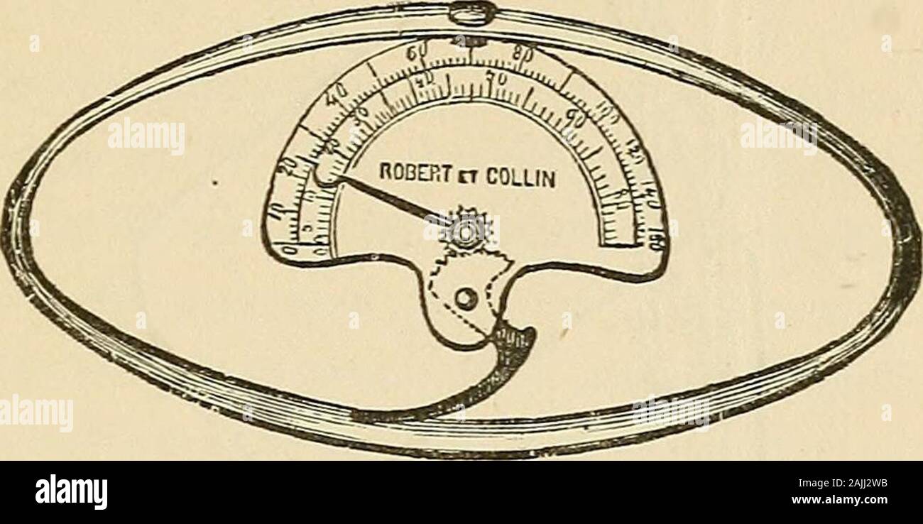 Cyclopædia of obstetrics and gynecology . x<iG. 02.—Traction Forceps op Joulin.—,4, Canula. B. Screw. C, Handle of second rod. D,Extremity joining E. F, Metal fulcrum. G, Filet. H, Point of the ecraseur articulating with thecanula. th3 fenestrae. The metal disk, articnkted with the canula, is placedover the ischial tuberosities of the woman. The ends of the filets are THE FORCEPS. 63 attached to tlie dynamometer, and this is fixed to B, which moves whenthe handle, C, of the canula, is turned. The filets act doubly; theynot only pull the forceps, but they approach the blades, so that thepres Stock Photohttps://www.alamy.com/image-license-details/?v=1https://www.alamy.com/cyclopdia-of-obstetrics-and-gynecology-xltig-02traction-forceps-op-joulin4-canula-b-screw-c-handle-of-second-rod-dextremity-joining-e-f-metal-fulcrum-g-filet-h-point-of-the-ecraseur-articulating-with-thecanula-th3-fenestrae-the-metal-disk-articnkted-with-the-canula-is-placedover-the-ischial-tuberosities-of-the-woman-the-ends-of-the-filets-are-the-forceps-63-attached-to-tlie-dynamometer-and-this-is-fixed-to-b-which-moves-whenthe-handle-c-of-the-canula-is-turned-the-filets-act-doubly-theynot-only-pull-the-forceps-but-they-approach-the-blades-so-that-thepres-image338458215.html
RM2AJJ2WB–Cyclopædia of obstetrics and gynecology . x<iG. 02.—Traction Forceps op Joulin.—,4, Canula. B. Screw. C, Handle of second rod. D,Extremity joining E. F, Metal fulcrum. G, Filet. H, Point of the ecraseur articulating with thecanula. th3 fenestrae. The metal disk, articnkted with the canula, is placedover the ischial tuberosities of the woman. The ends of the filets are THE FORCEPS. 63 attached to tlie dynamometer, and this is fixed to B, which moves whenthe handle, C, of the canula, is turned. The filets act doubly; theynot only pull the forceps, but they approach the blades, so that thepres
 The human machine, its care and repair; or, How to develop the body, preserve the health, meet emergencies, nurse the sick, and treat disease; . portion of the seatcause undue pressure upon partsin one of the most delicate re-gions of the human economy.To better place this portion ofthe subject before the reader, wehave, in connection herewith,three illustrations—Figs. I, 2and 3. Fig. 1 represents the bonypelvis of the human being. Thetwo prominences marked A arethe ischial tuberosities uponwhich the weight of the bodywhen riding a wheel should rest.B exhibits the pubic arch,which protects a p Stock Photohttps://www.alamy.com/image-license-details/?v=1https://www.alamy.com/the-human-machine-its-care-and-repair-or-how-to-develop-the-body-preserve-the-health-meet-emergencies-nurse-the-sick-and-treat-disease-portion-of-the-seatcause-undue-pressure-upon-partsin-one-of-the-most-delicate-re-gions-of-the-human-economyto-better-place-this-portion-ofthe-subject-before-the-reader-wehave-in-connection-herewiththree-illustrationsfigs-i-2and-3-fig-1-represents-the-bonypelvis-of-the-human-being-thetwo-prominences-marked-a-arethe-ischial-tuberosities-uponwhich-the-weight-of-the-bodywhen-riding-a-wheel-should-restb-exhibits-the-pubic-archwhich-protects-a-p-image343150411.html
RM2AX7RRR–The human machine, its care and repair; or, How to develop the body, preserve the health, meet emergencies, nurse the sick, and treat disease; . portion of the seatcause undue pressure upon partsin one of the most delicate re-gions of the human economy.To better place this portion ofthe subject before the reader, wehave, in connection herewith,three illustrations—Figs. I, 2and 3. Fig. 1 represents the bonypelvis of the human being. Thetwo prominences marked A arethe ischial tuberosities uponwhich the weight of the bodywhen riding a wheel should rest.B exhibits the pubic arch,which protects a p
 . An American text-book of obstetrics. For practitioners and students. ce between the iliac crests, 29 centimeters; the dis-tance between the greater trochanters, 31 centimeters; the distance betweenthe spinous process of the last lumbar vertebra and the upper margin of thepubic symphysis, or external conjugate, 204; centimeters; the distance betweenthe posterior superior spinous process and the anterior superior spinousprocess of the opposite iliac bone, or the oblique diameter, 22 centimeters;the distance between the ischial tuberosities, 11 centimeters. These externaldiameters, which are re Stock Photohttps://www.alamy.com/image-license-details/?v=1https://www.alamy.com/an-american-text-book-of-obstetrics-for-practitioners-and-students-ce-between-the-iliac-crests-29-centimeters-the-dis-tance-between-the-greater-trochanters-31-centimeters-the-distance-betweenthe-spinous-process-of-the-last-lumbar-vertebra-and-the-upper-margin-of-thepubic-symphysis-or-external-conjugate-204-centimeters-the-distance-betweenthe-posterior-superior-spinous-process-and-the-anterior-superior-spinousprocess-of-the-opposite-iliac-bone-or-the-oblique-diameter-22-centimetersthe-distance-between-the-ischial-tuberosities-11-centimeters-these-externaldiameters-which-are-re-image370609274.html
RM2CEXKWE–. An American text-book of obstetrics. For practitioners and students. ce between the iliac crests, 29 centimeters; the dis-tance between the greater trochanters, 31 centimeters; the distance betweenthe spinous process of the last lumbar vertebra and the upper margin of thepubic symphysis, or external conjugate, 204; centimeters; the distance betweenthe posterior superior spinous process and the anterior superior spinousprocess of the opposite iliac bone, or the oblique diameter, 22 centimeters;the distance between the ischial tuberosities, 11 centimeters. These externaldiameters, which are re
 . An American text-book of obstetrics. For practitioners and students. nt. This layer may be regarded as a reflection derived from both theobturator and the recto-vesical fascia, since the septum is formed by the unionof the contribution given off laterally from the obturator fascia with that sup- 34 AMERICAN TEXT-BOOK OF OBSTETRICS. plied mesially by the recto-vesical fascia: this relation is especially evident infrontal sections passing through the ischial tuberosities. The deep perineal interspace lies between the inferior and superior layers ofthe triangular ligament, and it contains withi Stock Photohttps://www.alamy.com/image-license-details/?v=1https://www.alamy.com/an-american-text-book-of-obstetrics-for-practitioners-and-students-nt-this-layer-may-be-regarded-as-a-reflection-derived-from-both-theobturator-and-the-recto-vesical-fascia-since-the-septum-is-formed-by-the-unionof-the-contribution-given-off-laterally-from-the-obturator-fascia-with-that-sup-34-american-text-book-of-obstetrics-plied-mesially-by-the-recto-vesical-fascia-this-relation-is-especially-evident-infrontal-sections-passing-through-the-ischial-tuberosities-the-deep-perineal-interspace-lies-between-the-inferior-and-superior-layers-ofthe-triangular-ligament-and-it-contains-withi-image370606951.html
RM2CEXGXF–. An American text-book of obstetrics. For practitioners and students. nt. This layer may be regarded as a reflection derived from both theobturator and the recto-vesical fascia, since the septum is formed by the unionof the contribution given off laterally from the obturator fascia with that sup- 34 AMERICAN TEXT-BOOK OF OBSTETRICS. plied mesially by the recto-vesical fascia: this relation is especially evident infrontal sections passing through the ischial tuberosities. The deep perineal interspace lies between the inferior and superior layers ofthe triangular ligament, and it contains withi
 . Obstetrics for nurses. 17.—Sagittal section through normalshowing planes and diameters. pelvis, ANATOMY OF THE FEMALE ORGANS 29 the tnio eonjiigato. In practice Iho latter moasuroment is estimatedby i^ubstraetiiii;- ^.0 ceiilinieters from llic detennined diagonal conjugate.The plane of the inferior strait or the pel vie outlet forms the lowerend of the birth eanal. It oci-upies the area between the ischial tub-erosities on the two sides, the lower margin of the symphysis anteriorlyand the tip of the coccyx posteriorly. The distance between the innersides of the ischial tuberosities is design Stock Photohttps://www.alamy.com/image-license-details/?v=1https://www.alamy.com/obstetrics-for-nurses-17sagittal-section-through-normalshowing-planes-and-diameters-pelvis-anatomy-of-the-female-organs-29-the-tnio-eonjiigato-in-practice-iho-latter-moasuroment-is-estimatedby-iubstraetiiii-0-ceiilinieters-from-llic-detennined-diagonal-conjugatethe-plane-of-the-inferior-strait-or-the-pel-vie-outlet-forms-the-lowerend-of-the-birth-eanal-it-oci-upies-the-area-between-the-ischial-tub-erosities-on-the-two-sides-the-lower-margin-of-the-symphysis-anteriorlyand-the-tip-of-the-coccyx-posteriorly-the-distance-between-the-innersides-of-the-ischial-tuberosities-is-design-image370462084.html
RM2CEM04M–. Obstetrics for nurses. 17.—Sagittal section through normalshowing planes and diameters. pelvis, ANATOMY OF THE FEMALE ORGANS 29 the tnio eonjiigato. In practice Iho latter moasuroment is estimatedby i^ubstraetiiii;- ^.0 ceiilinieters from llic detennined diagonal conjugate.The plane of the inferior strait or the pel vie outlet forms the lowerend of the birth eanal. It oci-upies the area between the ischial tub-erosities on the two sides, the lower margin of the symphysis anteriorlyand the tip of the coccyx posteriorly. The distance between the innersides of the ischial tuberosities is design
![. On the anatomy of vertebrates [electronic resource] . sacrum, a, the4 false pelvis ; that beneath,the 4 true pelvis/ Of thisthe 4 brim, or 4 superior cir-cumference e, f, b, inclosesthe 4inlet; the 6 inferiorcircumference, bounded bythe ischial tuberosities, pu-bic symphysis, and tip ofthe coccyx, incloses the4 outlet of the 4 true pelvis. The diameter from the sacral pro-montory, a, to the pubic symphysis, h, is called the 4 conjugate or4 antero-posterior one; that between the ilia taken at e, f, orhalf way between the sacro-iliac joint and the pectineal eminence,is the 4 transverse diamete Stock Photo . On the anatomy of vertebrates [electronic resource] . sacrum, a, the4 false pelvis ; that beneath,the 4 true pelvis/ Of thisthe 4 brim, or 4 superior cir-cumference e, f, b, inclosesthe 4inlet; the 6 inferiorcircumference, bounded bythe ischial tuberosities, pu-bic symphysis, and tip ofthe coccyx, incloses the4 outlet of the 4 true pelvis. The diameter from the sacral pro-montory, a, to the pubic symphysis, h, is called the 4 conjugate or4 antero-posterior one; that between the ilia taken at e, f, orhalf way between the sacro-iliac joint and the pectineal eminence,is the 4 transverse diamete Stock Photo](https://c8.alamy.com/comp/2CNGF88/on-the-anatomy-of-vertebrates-electronic-resource-sacrum-a-the4-false-pelvis-that-beneaththe-4-true-pelvis-of-thisthe-4-brim-or-4-superior-cir-cumference-e-f-b-inclosesthe-4inlet-the-6-inferiorcircumference-bounded-bythe-ischial-tuberosities-pu-bic-symphysis-and-tip-ofthe-coccyx-incloses-the4-outlet-of-the-4-true-pelvis-the-diameter-from-the-sacral-pro-montory-a-to-the-pubic-symphysis-h-is-called-the-4-conjugate-or4-antero-posterior-one-that-between-the-ilia-taken-at-e-f-orhalf-way-between-the-sacro-iliac-joint-and-the-pectineal-eminenceis-the-4-transverse-diamete-2CNGF88.jpg) . On the anatomy of vertebrates [electronic resource] . sacrum, a, the4 false pelvis ; that beneath,the 4 true pelvis/ Of thisthe 4 brim, or 4 superior cir-cumference e, f, b, inclosesthe 4inlet; the 6 inferiorcircumference, bounded bythe ischial tuberosities, pu-bic symphysis, and tip ofthe coccyx, incloses the4 outlet of the 4 true pelvis. The diameter from the sacral pro-montory, a, to the pubic symphysis, h, is called the 4 conjugate or4 antero-posterior one; that between the ilia taken at e, f, orhalf way between the sacro-iliac joint and the pectineal eminence,is the 4 transverse diamete Stock Photohttps://www.alamy.com/image-license-details/?v=1https://www.alamy.com/on-the-anatomy-of-vertebrates-electronic-resource-sacrum-a-the4-false-pelvis-that-beneaththe-4-true-pelvis-of-thisthe-4-brim-or-4-superior-cir-cumference-e-f-b-inclosesthe-4inlet-the-6-inferiorcircumference-bounded-bythe-ischial-tuberosities-pu-bic-symphysis-and-tip-ofthe-coccyx-incloses-the4-outlet-of-the-4-true-pelvis-the-diameter-from-the-sacral-pro-montory-a-to-the-pubic-symphysis-h-is-called-the-4-conjugate-or4-antero-posterior-one-that-between-the-ilia-taken-at-e-f-orhalf-way-between-the-sacro-iliac-joint-and-the-pectineal-eminenceis-the-4-transverse-diamete-image374688728.html
. On the anatomy of vertebrates [electronic resource] . sacrum, a, the4 false pelvis ; that beneath,the 4 true pelvis/ Of thisthe 4 brim, or 4 superior cir-cumference e, f, b, inclosesthe 4inlet; the 6 inferiorcircumference, bounded bythe ischial tuberosities, pu-bic symphysis, and tip ofthe coccyx, incloses the4 outlet of the 4 true pelvis. The diameter from the sacral pro-montory, a, to the pubic symphysis, h, is called the 4 conjugate or4 antero-posterior one; that between the ilia taken at e, f, orhalf way between the sacro-iliac joint and the pectineal eminence,is the 4 transverse diamete Stock Photohttps://www.alamy.com/image-license-details/?v=1https://www.alamy.com/on-the-anatomy-of-vertebrates-electronic-resource-sacrum-a-the4-false-pelvis-that-beneaththe-4-true-pelvis-of-thisthe-4-brim-or-4-superior-cir-cumference-e-f-b-inclosesthe-4inlet-the-6-inferiorcircumference-bounded-bythe-ischial-tuberosities-pu-bic-symphysis-and-tip-ofthe-coccyx-incloses-the4-outlet-of-the-4-true-pelvis-the-diameter-from-the-sacral-pro-montory-a-to-the-pubic-symphysis-h-is-called-the-4-conjugate-or4-antero-posterior-one-that-between-the-ilia-taken-at-e-f-orhalf-way-between-the-sacro-iliac-joint-and-the-pectineal-eminenceis-the-4-transverse-diamete-image374688728.htmlRM2CNGF88–. On the anatomy of vertebrates [electronic resource] . sacrum, a, the4 false pelvis ; that beneath,the 4 true pelvis/ Of thisthe 4 brim, or 4 superior cir-cumference e, f, b, inclosesthe 4inlet; the 6 inferiorcircumference, bounded bythe ischial tuberosities, pu-bic symphysis, and tip ofthe coccyx, incloses the4 outlet of the 4 true pelvis. The diameter from the sacral pro-montory, a, to the pubic symphysis, h, is called the 4 conjugate or4 antero-posterior one; that between the ilia taken at e, f, orhalf way between the sacro-iliac joint and the pectineal eminence,is the 4 transverse diamete
 . The American journal of roentgenology, radium therapy and nuclear medicine . The line drawn between the ischial tuberosities is for the measurement of theanterior and posterior sagittal diameters. (The films taken at term show hardlj- enough contrast forsatisfactory reproduction.) The following arc the measurements of this pelvis. Pelvic inlet: AnteroposteriorTransverseRight obliqueLeft oblique V V 133 V 10.9 V 108 2 + 7 2 = 12.8 cm 9.5 2 -f- 6 2 =11.2 cm.+ 1-5^ = 134 cm.+ 52 z= 12.0 cm. Plane of least dimensions: Pelvic outlet: Anteroposterior ^ 47 2 | g 2 — jo.i cm. Transverse V iiTa 2 + Stock Photohttps://www.alamy.com/image-license-details/?v=1https://www.alamy.com/the-american-journal-of-roentgenology-radium-therapy-and-nuclear-medicine-the-line-drawn-between-the-ischial-tuberosities-is-for-the-measurement-of-theanterior-and-posterior-sagittal-diameters-the-films-taken-at-term-show-hardlj-enough-contrast-forsatisfactory-reproduction-the-following-arc-the-measurements-of-this-pelvis-pelvic-inlet-anteroposteriortransverseright-obliqueleft-oblique-v-v-133-v-109-v-108-2-7-2-=-128-cm-95-2-f-6-2-=112-cm-1-5-=-134-cm-52-z=-120-cm-plane-of-least-dimensions-pelvic-outlet-anteroposterior-47-2-g-2-joi-cm-transverse-v-iita-2-image376041012.html
RM2CRP444–. The American journal of roentgenology, radium therapy and nuclear medicine . The line drawn between the ischial tuberosities is for the measurement of theanterior and posterior sagittal diameters. (The films taken at term show hardlj- enough contrast forsatisfactory reproduction.) The following arc the measurements of this pelvis. Pelvic inlet: AnteroposteriorTransverseRight obliqueLeft oblique V V 133 V 10.9 V 108 2 + 7 2 = 12.8 cm 9.5 2 -f- 6 2 =11.2 cm.+ 1-5^ = 134 cm.+ 52 z= 12.0 cm. Plane of least dimensions: Pelvic outlet: Anteroposterior ^ 47 2 | g 2 — jo.i cm. Transverse V iiTa 2 +
 . California fish and game. Fisheries -- California; Game and game-birds -- California; Fishes -- California; Animal Population Groups; Pêches; Gibier; Poissons. 20 ( VLIPORNIA K1SI1 AND Q l I A series of these measurements on black-tailed deer of both sexes is given in Table 1. Dorsal acetabulum ischial tuberosities. Please note that these images are extracted from scanned page images that may have been digitally enhanced for readability - coloration and appearance of these illustrations may not perfectly resemble the original work.. California. Dept. of Fish and Game; California. Fish and Stock Photohttps://www.alamy.com/image-license-details/?v=1https://www.alamy.com/california-fish-and-game-fisheries-california-game-and-game-birds-california-fishes-california-animal-population-groups-pches-gibier-poissons-20-vlipornia-k1si1-and-q-l-i-a-series-of-these-measurements-on-black-tailed-deer-of-both-sexes-is-given-in-table-1-dorsal-acetabulum-ischial-tuberosities-please-note-that-these-images-are-extracted-from-scanned-page-images-that-may-have-been-digitally-enhanced-for-readability-coloration-and-appearance-of-these-illustrations-may-not-perfectly-resemble-the-original-work-california-dept-of-fish-and-game-california-fish-and-image233683996.html
RMRG5690–. California fish and game. Fisheries -- California; Game and game-birds -- California; Fishes -- California; Animal Population Groups; Pêches; Gibier; Poissons. 20 ( VLIPORNIA K1SI1 AND Q l I A series of these measurements on black-tailed deer of both sexes is given in Table 1. Dorsal acetabulum ischial tuberosities. Please note that these images are extracted from scanned page images that may have been digitally enhanced for readability - coloration and appearance of these illustrations may not perfectly resemble the original work.. California. Dept. of Fish and Game; California. Fish and
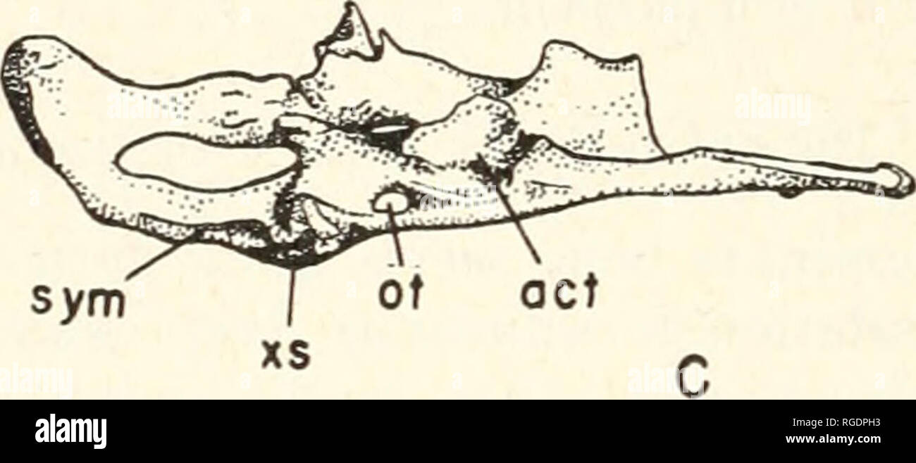 . Bulletin of the Museum of Comparative Zoology at Harvard College. Zoology. Fig. 4. More normal right innominate and entire teratological pelvis. A, medial view. B, lateral view. C, ventromedial view. Abbrevia- tions: al, regular auricular facet; a2, right auricular facet of tera- tological pelvis; act, acetabulum of teratological pelvis; ft, ischial tuberosities of teratological pelvis; Iti, left teratological ilium; oi, gap l)etween regiilMi- right innominate and teratological pelvis; ot, obturator fenestrae of teratological pelvis; sp, spongy bone mass; sitt, suture between regular right i Stock Photohttps://www.alamy.com/image-license-details/?v=1https://www.alamy.com/bulletin-of-the-museum-of-comparative-zoology-at-harvard-college-zoology-fig-4-more-normal-right-innominate-and-entire-teratological-pelvis-a-medial-view-b-lateral-view-c-ventromedial-view-abbrevia-tions-al-regular-auricular-facet-a2-right-auricular-facet-of-tera-tological-pelvis-act-acetabulum-of-teratological-pelvis-ft-ischial-tuberosities-of-teratological-pelvis-iti-left-teratological-ilium-oi-gap-letween-regiilmi-right-innominate-and-teratological-pelvis-ot-obturator-fenestrae-of-teratological-pelvis-sp-spongy-bone-mass-sitt-suture-between-regular-right-i-image233872383.html
RMRGDPH3–. Bulletin of the Museum of Comparative Zoology at Harvard College. Zoology. Fig. 4. More normal right innominate and entire teratological pelvis. A, medial view. B, lateral view. C, ventromedial view. Abbrevia- tions: al, regular auricular facet; a2, right auricular facet of tera- tological pelvis; act, acetabulum of teratological pelvis; ft, ischial tuberosities of teratological pelvis; Iti, left teratological ilium; oi, gap l)etween regiilMi- right innominate and teratological pelvis; ot, obturator fenestrae of teratological pelvis; sp, spongy bone mass; sitt, suture between regular right i
 A text-book of veterinary obstetrics : including the diseases and accidents incidental to pregnancy, parturition and early age in the domesticated animals . Fig. 1.3.Bones of Pelvis op Mare. joining the tuber ischii; while in the Horse these tuberosities are notnearly so wide apart, and the ischial arch forms a somewhat acuteangle, the margin of which is nearly straight. The obturator foramina. Fig. H.Bones of Pelvis of Horse. are also large and almost circular in the Mare, while they are small andoval in the Horse ; the ischio-pubic symphysis is farther from thecotyloid cavities in the former Stock Photohttps://www.alamy.com/image-license-details/?v=1https://www.alamy.com/a-text-book-of-veterinary-obstetrics-including-the-diseases-and-accidents-incidental-to-pregnancy-parturition-and-early-age-in-the-domesticated-animals-fig-13bones-of-pelvis-op-mare-joining-the-tuber-ischii-while-in-the-horse-these-tuberosities-are-notnearly-so-wide-apart-and-the-ischial-arch-forms-a-somewhat-acuteangle-the-margin-of-which-is-nearly-straight-the-obturator-foramina-fig-hbones-of-pelvis-of-horse-are-also-large-and-almost-circular-in-the-mare-while-they-are-small-andoval-in-the-horse-the-ischio-pubic-symphysis-is-farther-from-thecotyloid-cavities-in-the-former-image339284726.html
RM2AKYN3J–A text-book of veterinary obstetrics : including the diseases and accidents incidental to pregnancy, parturition and early age in the domesticated animals . Fig. 1.3.Bones of Pelvis op Mare. joining the tuber ischii; while in the Horse these tuberosities are notnearly so wide apart, and the ischial arch forms a somewhat acuteangle, the margin of which is nearly straight. The obturator foramina. Fig. H.Bones of Pelvis of Horse. are also large and almost circular in the Mare, while they are small andoval in the Horse ; the ischio-pubic symphysis is farther from thecotyloid cavities in the former
 . On the theory and practice of midwifery . Plane of the Inferior Strait. — The plane of the inferior strait is usually re-garded as bounded by the inner lips of the two tuberosities of the ischial bones, therami of the ischia and pubis, the ischio-sacral ligaments, and the point of the coccyx.In this way we speak of the plane of the inferior strait as one plane only; whereas,there are, in fact, two such planes, an anterior and a posterior. This figure exhibits the contour of the outlet. The line c d represents the trans-verse diameter. The letters ceaed show the anterior semi-circumference, w Stock Photohttps://www.alamy.com/image-license-details/?v=1https://www.alamy.com/on-the-theory-and-practice-of-midwifery-plane-of-the-inferior-strait-the-plane-of-the-inferior-strait-is-usually-re-garded-as-bounded-by-the-inner-lips-of-the-two-tuberosities-of-the-ischial-bones-therami-of-the-ischia-and-pubis-the-ischio-sacral-ligaments-and-the-point-of-the-coccyxin-this-way-we-speak-of-the-plane-of-the-inferior-strait-as-one-plane-only-whereasthere-are-in-fact-two-such-planes-an-anterior-and-a-posterior-this-figure-exhibits-the-contour-of-the-outlet-the-line-c-d-represents-the-trans-verse-diameter-the-letters-ceaed-show-the-anterior-semi-circumference-w-image370129977.html
RM2CE4TFN–. On the theory and practice of midwifery . Plane of the Inferior Strait. — The plane of the inferior strait is usually re-garded as bounded by the inner lips of the two tuberosities of the ischial bones, therami of the ischia and pubis, the ischio-sacral ligaments, and the point of the coccyx.In this way we speak of the plane of the inferior strait as one plane only; whereas,there are, in fact, two such planes, an anterior and a posterior. This figure exhibits the contour of the outlet. The line c d represents the trans-verse diameter. The letters ceaed show the anterior semi-circumference, w
![. California fish and game. Fisheries -- California; Game and game-birds -- California; Fishes -- California; Animal Population Groups; Pêches; Gibier; Poissons. SUSPENSORY TUBEROSITIES. a b a--ISCHIAL TUBEROSITY. Please note that these images are extracted from scanned page images that may have been digitally enhanced for readability - coloration and appearance of these illustrations may not perfectly resemble the original work.. California. Dept. of Fish and Game; California. Fish and Game Commission; California. Division of Fish and Game. [San Francisco, etc. ] State of California, Resource Stock Photo . California fish and game. Fisheries -- California; Game and game-birds -- California; Fishes -- California; Animal Population Groups; Pêches; Gibier; Poissons. SUSPENSORY TUBEROSITIES. a b a--ISCHIAL TUBEROSITY. Please note that these images are extracted from scanned page images that may have been digitally enhanced for readability - coloration and appearance of these illustrations may not perfectly resemble the original work.. California. Dept. of Fish and Game; California. Fish and Game Commission; California. Division of Fish and Game. [San Francisco, etc. ] State of California, Resource Stock Photo](https://c8.alamy.com/comp/RG56A6/california-fish-and-game-fisheries-california-game-and-game-birds-california-fishes-california-animal-population-groups-pches-gibier-poissons-suspensory-tuberosities-a-b-a-ischial-tuberosity-please-note-that-these-images-are-extracted-from-scanned-page-images-that-may-have-been-digitally-enhanced-for-readability-coloration-and-appearance-of-these-illustrations-may-not-perfectly-resemble-the-original-work-california-dept-of-fish-and-game-california-fish-and-game-commission-california-division-of-fish-and-game-san-francisco-etc-state-of-california-resource-RG56A6.jpg) . California fish and game. Fisheries -- California; Game and game-birds -- California; Fishes -- California; Animal Population Groups; Pêches; Gibier; Poissons. SUSPENSORY TUBEROSITIES. a b a--ISCHIAL TUBEROSITY. Please note that these images are extracted from scanned page images that may have been digitally enhanced for readability - coloration and appearance of these illustrations may not perfectly resemble the original work.. California. Dept. of Fish and Game; California. Fish and Game Commission; California. Division of Fish and Game. [San Francisco, etc. ] State of California, Resource Stock Photohttps://www.alamy.com/image-license-details/?v=1https://www.alamy.com/california-fish-and-game-fisheries-california-game-and-game-birds-california-fishes-california-animal-population-groups-pches-gibier-poissons-suspensory-tuberosities-a-b-a-ischial-tuberosity-please-note-that-these-images-are-extracted-from-scanned-page-images-that-may-have-been-digitally-enhanced-for-readability-coloration-and-appearance-of-these-illustrations-may-not-perfectly-resemble-the-original-work-california-dept-of-fish-and-game-california-fish-and-game-commission-california-division-of-fish-and-game-san-francisco-etc-state-of-california-resource-image233684030.html
. California fish and game. Fisheries -- California; Game and game-birds -- California; Fishes -- California; Animal Population Groups; Pêches; Gibier; Poissons. SUSPENSORY TUBEROSITIES. a b a--ISCHIAL TUBEROSITY. Please note that these images are extracted from scanned page images that may have been digitally enhanced for readability - coloration and appearance of these illustrations may not perfectly resemble the original work.. California. Dept. of Fish and Game; California. Fish and Game Commission; California. Division of Fish and Game. [San Francisco, etc. ] State of California, Resource Stock Photohttps://www.alamy.com/image-license-details/?v=1https://www.alamy.com/california-fish-and-game-fisheries-california-game-and-game-birds-california-fishes-california-animal-population-groups-pches-gibier-poissons-suspensory-tuberosities-a-b-a-ischial-tuberosity-please-note-that-these-images-are-extracted-from-scanned-page-images-that-may-have-been-digitally-enhanced-for-readability-coloration-and-appearance-of-these-illustrations-may-not-perfectly-resemble-the-original-work-california-dept-of-fish-and-game-california-fish-and-game-commission-california-division-of-fish-and-game-san-francisco-etc-state-of-california-resource-image233684030.htmlRMRG56A6–. California fish and game. Fisheries -- California; Game and game-birds -- California; Fishes -- California; Animal Population Groups; Pêches; Gibier; Poissons. SUSPENSORY TUBEROSITIES. a b a--ISCHIAL TUBEROSITY. Please note that these images are extracted from scanned page images that may have been digitally enhanced for readability - coloration and appearance of these illustrations may not perfectly resemble the original work.. California. Dept. of Fish and Game; California. Fish and Game Commission; California. Division of Fish and Game. [San Francisco, etc. ] State of California, Resource
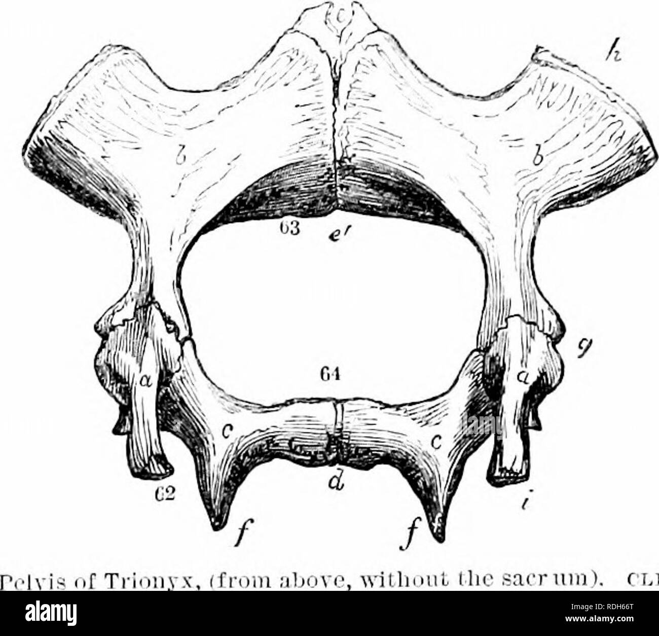 . On the anatomy of vertebrates. Vertebrates; Anatomy, Comparative; 1866. ISG ANATOMY OF VERTEBRATES. A sllri-lit modification is presented by Trionyx, in which a view of tlie pelvis is given from the dorsal aspect, the sacrum being removed, in fig. 116: here i shows the end of the ilium, 62, which was attached to that part of the vertebral column: a is the ex- panded acetabular end of these short, straight, columnar bones. no. The ischia, c, c, develope tuberosities/,/, and unite at the ischial symphysis, 64, d. The pubics h, h, articulate by a broader tube- rosity, /(, with the plastron, and Stock Photohttps://www.alamy.com/image-license-details/?v=1https://www.alamy.com/on-the-anatomy-of-vertebrates-vertebrates-anatomy-comparative-1866-isg-anatomy-of-vertebrates-a-sllri-lit-modification-is-presented-by-trionyx-in-which-a-view-of-tlie-pelvis-is-given-from-the-dorsal-aspect-the-sacrum-being-removed-in-fig-116-here-i-shows-the-end-of-the-ilium-62-which-was-attached-to-that-part-of-the-vertebral-column-a-is-the-ex-panded-acetabular-end-of-these-short-straight-columnar-bones-no-the-ischia-c-c-develope-tuberosities-and-unite-at-the-ischial-symphysis-64-d-the-pubics-h-h-articulate-by-a-broader-tube-rosity-with-the-plastron-and-image232103392.html
RMRDH66T–. On the anatomy of vertebrates. Vertebrates; Anatomy, Comparative; 1866. ISG ANATOMY OF VERTEBRATES. A sllri-lit modification is presented by Trionyx, in which a view of tlie pelvis is given from the dorsal aspect, the sacrum being removed, in fig. 116: here i shows the end of the ilium, 62, which was attached to that part of the vertebral column: a is the ex- panded acetabular end of these short, straight, columnar bones. no. The ischia, c, c, develope tuberosities/,/, and unite at the ischial symphysis, 64, d. The pubics h, h, articulate by a broader tube- rosity, /(, with the plastron, and