Quick filters:
Lactiferous sinus Stock Photos and Images
 A text-book of veterinary obstetrics : including the diseases and accidents incidental to pregnancy, parturition and early age in the domesticated animals . B . o.;v «•. Fifc-. 19. A. Lobile of the Mainniie filled with Milk ;r.. Milk Globules ; C, Colostrum : a. Cellwith a ir<ible Nucleus; /*, Cells fmrnwhich the Nucleus has disappeared. Fig. 20.Skc-iion ok thk Cows Te.t.o, ti. Principal Lactiferous Ducts ;/;. Lactiferous Sinus ; r, r. Acini;(I, Klastic or iJartoid Tissue ofthe Teat; < Orifice of the Teat. gland beuds at a right angle backwards, the branch for the anteriorquarter—the Stock Photohttps://www.alamy.com/image-license-details/?v=1https://www.alamy.com/a-text-book-of-veterinary-obstetrics-including-the-diseases-and-accidents-incidental-to-pregnancy-parturition-and-early-age-in-the-domesticated-animals-b-ov-fifc-19-a-lobile-of-the-mainniie-filled-with-milk-r-milk-globules-c-colostrum-a-cellwith-a-irltible-nucleus-cells-fmrnwhich-the-nucleus-has-disappeared-fig-20skc-iion-ok-thk-cows-teto-ti-principal-lactiferous-ducts-lactiferous-sinus-r-r-acinii-klastic-or-ijartoid-tissue-ofthe-teat-lt-orifice-of-the-teat-gland-beuds-at-a-right-angle-backwards-the-branch-for-the-anteriorquarterthe-image339283802.html
RM2AKYKXJ–A text-book of veterinary obstetrics : including the diseases and accidents incidental to pregnancy, parturition and early age in the domesticated animals . B . o.;v «•. Fifc-. 19. A. Lobile of the Mainniie filled with Milk ;r.. Milk Globules ; C, Colostrum : a. Cellwith a ir<ible Nucleus; /*, Cells fmrnwhich the Nucleus has disappeared. Fig. 20.Skc-iion ok thk Cows Te.t.o, ti. Principal Lactiferous Ducts ;/;. Lactiferous Sinus ; r, r. Acini;(I, Klastic or iJartoid Tissue ofthe Teat; < Orifice of the Teat. gland beuds at a right angle backwards, the branch for the anteriorquarter—the
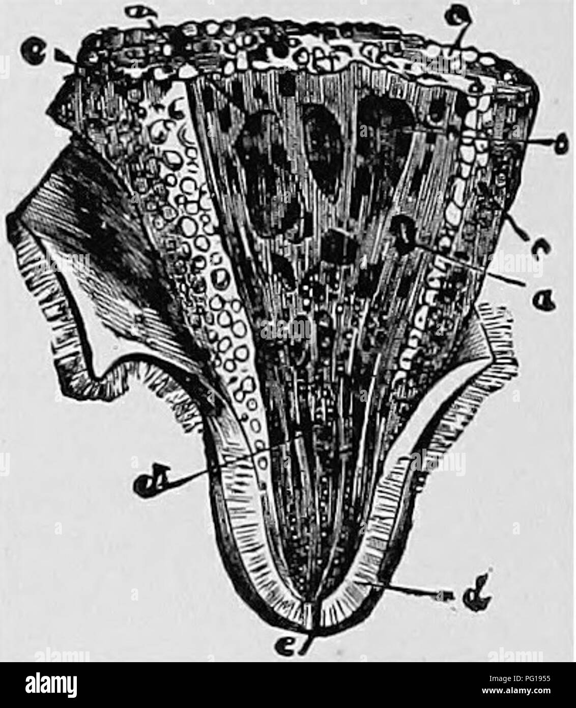 . Veterinary obstetrics; a compendium for the use of students and practitioners. Veterinary obstetrics. 26 . VETERINARY OBSTETRICS. In the small ruminants there are two mammae and two teats, constructed like those of the Cow.. Fig. 13. Section of the Cow's Teat. a, a. Principal Lactiferous Ducts; b, Lactiferous Sinus; c, c. Acini; d. Elastic or Dartoid Tissue of the Teat; e, Orifice of the Teat. In the Pig the mammae are ten or twelve in number, disposed by pairs in two parallel rows, extending from the inguinal region to beneath the thorax, and distinguished as inguinal, abdominal, and thorac Stock Photohttps://www.alamy.com/image-license-details/?v=1https://www.alamy.com/veterinary-obstetrics-a-compendium-for-the-use-of-students-and-practitioners-veterinary-obstetrics-26-veterinary-obstetrics-in-the-small-ruminants-there-are-two-mammae-and-two-teats-constructed-like-those-of-the-cow-fig-13-section-of-the-cows-teat-a-a-principal-lactiferous-ducts-b-lactiferous-sinus-c-c-acini-d-elastic-or-dartoid-tissue-of-the-teat-e-orifice-of-the-teat-in-the-pig-the-mammae-are-ten-or-twelve-in-number-disposed-by-pairs-in-two-parallel-rows-extending-from-the-inguinal-region-to-beneath-the-thorax-and-distinguished-as-inguinal-abdominal-and-thorac-image216388065.html
RMPG1955–. Veterinary obstetrics; a compendium for the use of students and practitioners. Veterinary obstetrics. 26 . VETERINARY OBSTETRICS. In the small ruminants there are two mammae and two teats, constructed like those of the Cow.. Fig. 13. Section of the Cow's Teat. a, a. Principal Lactiferous Ducts; b, Lactiferous Sinus; c, c. Acini; d. Elastic or Dartoid Tissue of the Teat; e, Orifice of the Teat. In the Pig the mammae are ten or twelve in number, disposed by pairs in two parallel rows, extending from the inguinal region to beneath the thorax, and distinguished as inguinal, abdominal, and thorac
 . The anatomy of the domestic animals. Veterinary anatomy. GENITAL ORGANS OF THE COW 6a5 consistence than the fat which is found arountl and ^?itllin the giand. It is enclosed by a fibro-elastic capsule which sends inward numerous trabeculse; these form the interstitial tissue, and divide the gland into lobes antl lobules. In the latter are the secretory tubules and alveoli, which unite to form the larger ducts. Each lobe has a duct, which opens at the base of the teat into a space called the lactiferous sinus (Sinus lactiferus), and from this two (or three) lactiferous ducts (Ductus lactifer Stock Photohttps://www.alamy.com/image-license-details/?v=1https://www.alamy.com/the-anatomy-of-the-domestic-animals-veterinary-anatomy-genital-organs-of-the-cow-6a5-consistence-than-the-fat-which-is-found-arountl-and-itllin-the-giand-it-is-enclosed-by-a-fibro-elastic-capsule-which-sends-inward-numerous-trabeculse-these-form-the-interstitial-tissue-and-divide-the-gland-into-lobes-antl-lobules-in-the-latter-are-the-secretory-tubules-and-alveoli-which-unite-to-form-the-larger-ducts-each-lobe-has-a-duct-which-opens-at-the-base-of-the-teat-into-a-space-called-the-lactiferous-sinus-sinus-lactiferus-and-from-this-two-or-three-lactiferous-ducts-ductus-lactifer-image236796631.html
RMRN70EF–. The anatomy of the domestic animals. Veterinary anatomy. GENITAL ORGANS OF THE COW 6a5 consistence than the fat which is found arountl and ^?itllin the giand. It is enclosed by a fibro-elastic capsule which sends inward numerous trabeculse; these form the interstitial tissue, and divide the gland into lobes antl lobules. In the latter are the secretory tubules and alveoli, which unite to form the larger ducts. Each lobe has a duct, which opens at the base of the teat into a space called the lactiferous sinus (Sinus lactiferus), and from this two (or three) lactiferous ducts (Ductus lactifer
 . The anatomy of the domestic animals. Veterinary anatomy. GENITAL ORGANS OF THE COW 605 consistence than the fat which is found around and within the giand. It is enclosed by a fibro-elastic capsule which sends inward numerous trabeculae; these form the interstitial tissue, and divide the gland into lobes and lobules. In the latter are the secretory tubules and alveoli, which unite to form the larger ducts. Each lobe has a duct, which opens at the base of the teat into a space called the lactiferous sinus (Sinus lactiferus), and from this two (or three) lactiferous ducts (Ductus lactiferi) pa Stock Photohttps://www.alamy.com/image-license-details/?v=1https://www.alamy.com/the-anatomy-of-the-domestic-animals-veterinary-anatomy-genital-organs-of-the-cow-605-consistence-than-the-fat-which-is-found-around-and-within-the-giand-it-is-enclosed-by-a-fibro-elastic-capsule-which-sends-inward-numerous-trabeculae-these-form-the-interstitial-tissue-and-divide-the-gland-into-lobes-and-lobules-in-the-latter-are-the-secretory-tubules-and-alveoli-which-unite-to-form-the-larger-ducts-each-lobe-has-a-duct-which-opens-at-the-base-of-the-teat-into-a-space-called-the-lactiferous-sinus-sinus-lactiferus-and-from-this-two-or-three-lactiferous-ducts-ductus-lactiferi-pa-image236796686.html
RMRN70GE–. The anatomy of the domestic animals. Veterinary anatomy. GENITAL ORGANS OF THE COW 605 consistence than the fat which is found around and within the giand. It is enclosed by a fibro-elastic capsule which sends inward numerous trabeculae; these form the interstitial tissue, and divide the gland into lobes and lobules. In the latter are the secretory tubules and alveoli, which unite to form the larger ducts. Each lobe has a duct, which opens at the base of the teat into a space called the lactiferous sinus (Sinus lactiferus), and from this two (or three) lactiferous ducts (Ductus lactiferi) pa
 . The fundamentals of live stock judging and selection . Livestock. STRUCTURAL FORM AND EXAMINATION 285 The face should be straight, except as specifically altered by breed qualifications. Quality should be apparent.. Fig. 121.—Cross-section of mammary glands of cow: u,, body of gland; ^, lactiferous sinus; c, cavity of teat; d, duct of teat; e, intermammary groove; /, septum between glands; g, supramammary fat. (Courtesy of L. W. Sisson, from Anatomy of Domestic Animals.) Digitized by Microsoft®. Please note that these images are extracted from scanned page images that may have been digitally Stock Photohttps://www.alamy.com/image-license-details/?v=1https://www.alamy.com/the-fundamentals-of-live-stock-judging-and-selection-livestock-structural-form-and-examination-285-the-face-should-be-straight-except-as-specifically-altered-by-breed-qualifications-quality-should-be-apparent-fig-121cross-section-of-mammary-glands-of-cow-u-body-of-gland-lactiferous-sinus-c-cavity-of-teat-d-duct-of-teat-e-intermammary-groove-septum-between-glands-g-supramammary-fat-courtesy-of-l-w-sisson-from-anatomy-of-domestic-animals-digitized-by-microsoft-please-note-that-these-images-are-extracted-from-scanned-page-images-that-may-have-been-digitally-image232338774.html
RMRDYXDA–. The fundamentals of live stock judging and selection . Livestock. STRUCTURAL FORM AND EXAMINATION 285 The face should be straight, except as specifically altered by breed qualifications. Quality should be apparent.. Fig. 121.—Cross-section of mammary glands of cow: u,, body of gland; ^, lactiferous sinus; c, cavity of teat; d, duct of teat; e, intermammary groove; /, septum between glands; g, supramammary fat. (Courtesy of L. W. Sisson, from Anatomy of Domestic Animals.) Digitized by Microsoft®. Please note that these images are extracted from scanned page images that may have been digitally
 . The anatomy of the domestic animals . Veterinary anatomy. GENITAL ORGANS OF THE COW 605 consistence than the fat which is found around and within the giand. It is enclosed by a fibro-elastic capsule which sends inward numerous trabecule; these form the interstitial tissue, and divide the gland into lobes and lobules. In the latter are the secretory tubules and alveoli, which unite to form the larger ducts. Each lobe has a duct, which opensat th&jJsase^pfj^yiSi^eatjnto a space called the lactiferous sinus (Sinus lactiferiisX and"from this two (or three) lactiferous ducts (Ductus lact Stock Photohttps://www.alamy.com/image-license-details/?v=1https://www.alamy.com/the-anatomy-of-the-domestic-animals-veterinary-anatomy-genital-organs-of-the-cow-605-consistence-than-the-fat-which-is-found-around-and-within-the-giand-it-is-enclosed-by-a-fibro-elastic-capsule-which-sends-inward-numerous-trabecule-these-form-the-interstitial-tissue-and-divide-the-gland-into-lobes-and-lobules-in-the-latter-are-the-secretory-tubules-and-alveoli-which-unite-to-form-the-larger-ducts-each-lobe-has-a-duct-which-opensat-thampjjsasepfjyisieatjnto-a-space-called-the-lactiferous-sinus-sinus-lactiferiisx-andquotfrom-this-two-or-three-lactiferous-ducts-ductus-lact-image232323927.html
RMRDY7F3–. The anatomy of the domestic animals . Veterinary anatomy. GENITAL ORGANS OF THE COW 605 consistence than the fat which is found around and within the giand. It is enclosed by a fibro-elastic capsule which sends inward numerous trabecule; these form the interstitial tissue, and divide the gland into lobes and lobules. In the latter are the secretory tubules and alveoli, which unite to form the larger ducts. Each lobe has a duct, which opensat th&jJsase^pfj^yiSi^eatjnto a space called the lactiferous sinus (Sinus lactiferiisX and"from this two (or three) lactiferous ducts (Ductus lact
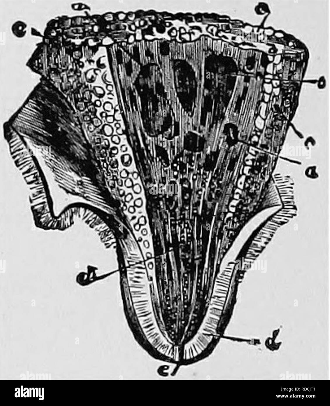 . Veterinary obstetrics; a compendium for the use of students and practitioners. Veterinary obstetrics. 26 . VETERINARY OBSTETRICS. In the small ruminants there are two mammae and two teats, constructed like those of the Cow.. Fig. 13. Section of the Cow's Teat. a, a. Principal Lactiferous Ducts; b, Lactiferous Sinus; c, c. Acini; d. Elastic or Dartoid Tissue of the Teat; e, Orifice of the Teat. In the Pig the mammae are ten or twelve in number, disposed by pairs in two parallel rows, extending from the inguinal region to beneath the thorax, and distinguished as inguinal, abdominal, and thorac Stock Photohttps://www.alamy.com/image-license-details/?v=1https://www.alamy.com/veterinary-obstetrics-a-compendium-for-the-use-of-students-and-practitioners-veterinary-obstetrics-26-veterinary-obstetrics-in-the-small-ruminants-there-are-two-mammae-and-two-teats-constructed-like-those-of-the-cow-fig-13-section-of-the-cows-teat-a-a-principal-lactiferous-ducts-b-lactiferous-sinus-c-c-acini-d-elastic-or-dartoid-tissue-of-the-teat-e-orifice-of-the-teat-in-the-pig-the-mammae-are-ten-or-twelve-in-number-disposed-by-pairs-in-two-parallel-rows-extending-from-the-inguinal-region-to-beneath-the-thorax-and-distinguished-as-inguinal-abdominal-and-thorac-image232003521.html
RMRDCJT1–. Veterinary obstetrics; a compendium for the use of students and practitioners. Veterinary obstetrics. 26 . VETERINARY OBSTETRICS. In the small ruminants there are two mammae and two teats, constructed like those of the Cow.. Fig. 13. Section of the Cow's Teat. a, a. Principal Lactiferous Ducts; b, Lactiferous Sinus; c, c. Acini; d. Elastic or Dartoid Tissue of the Teat; e, Orifice of the Teat. In the Pig the mammae are ten or twelve in number, disposed by pairs in two parallel rows, extending from the inguinal region to beneath the thorax, and distinguished as inguinal, abdominal, and thorac
 . Principles of veterinary science; a text-book for use in agricultural schools. Veterinary medicine. THE UROGENITAL SYSTEM 149 tons," to which the placenta is attached. In the non-gravid uterus they average about H inch in length, and a little less in width and thickness. During pregnancy they become greatly enlarged and pedunculated and may measure as much as 5 inches in length.. Fig. 51.—Cross-section of mammary gland of cow: u, body of gland; 6, lactiferous sinus; c, cavity of teat; d, teat canal; e, intermammary groove; /, septum between glands; g, supermammary fat. (Sisson, Anatomy Stock Photohttps://www.alamy.com/image-license-details/?v=1https://www.alamy.com/principles-of-veterinary-science-a-text-book-for-use-in-agricultural-schools-veterinary-medicine-the-urogenital-system-149-tonsquot-to-which-the-placenta-is-attached-in-the-non-gravid-uterus-they-average-about-h-inch-in-length-and-a-little-less-in-width-and-thickness-during-pregnancy-they-become-greatly-enlarged-and-pedunculated-and-may-measure-as-much-as-5-inches-in-length-fig-51cross-section-of-mammary-gland-of-cow-u-body-of-gland-6-lactiferous-sinus-c-cavity-of-teat-d-teat-canal-e-intermammary-groove-septum-between-glands-g-supermammary-fat-sisson-anatomy-image232319507.html
RMRDY1W7–. Principles of veterinary science; a text-book for use in agricultural schools. Veterinary medicine. THE UROGENITAL SYSTEM 149 tons," to which the placenta is attached. In the non-gravid uterus they average about H inch in length, and a little less in width and thickness. During pregnancy they become greatly enlarged and pedunculated and may measure as much as 5 inches in length.. Fig. 51.—Cross-section of mammary gland of cow: u, body of gland; 6, lactiferous sinus; c, cavity of teat; d, teat canal; e, intermammary groove; /, septum between glands; g, supermammary fat. (Sisson, Anatomy
 . The comparative anatomy of the domesticated animals. Veterinary anatomy. GLAND-VESICLES, WITH THEIR EXCRETORY ULTIMATE FOLLICLES, OR GLAND-VESICLES, DUCTS TERMINATING IN A DUCTUS LAC- WITH THEIR EPITHELIUM OR SECRETING TIFEROUS: FROM A MERCURIAL INJECTION CELLS, a, a, AND NUCLEI, 6, 6. (magnified FOUR TIMES). They are -^ of an inch in diameter.) The lactiferous ducts commence by blind extremities, and run into each other to constitute a certain number of principal canals ; these open into the galactophorous sinuses (each a saccidus vel sinus lactiferus). The glandular culs-de-sac are lined w Stock Photohttps://www.alamy.com/image-license-details/?v=1https://www.alamy.com/the-comparative-anatomy-of-the-domesticated-animals-veterinary-anatomy-gland-vesicles-with-their-excretory-ultimate-follicles-or-gland-vesicles-ducts-terminating-in-a-ductus-lac-with-their-epithelium-or-secreting-tiferous-from-a-mercurial-injection-cells-a-a-and-nuclei-6-6-magnified-four-times-they-are-of-an-inch-in-diameter-the-lactiferous-ducts-commence-by-blind-extremities-and-run-into-each-other-to-constitute-a-certain-number-of-principal-canals-these-open-into-the-galactophorous-sinuses-each-a-saccidus-vel-sinus-lactiferus-the-glandular-culs-de-sac-are-lined-w-image232677397.html
RMREFAB1–. The comparative anatomy of the domesticated animals. Veterinary anatomy. GLAND-VESICLES, WITH THEIR EXCRETORY ULTIMATE FOLLICLES, OR GLAND-VESICLES, DUCTS TERMINATING IN A DUCTUS LAC- WITH THEIR EPITHELIUM OR SECRETING TIFEROUS: FROM A MERCURIAL INJECTION CELLS, a, a, AND NUCLEI, 6, 6. (magnified FOUR TIMES). They are -^ of an inch in diameter.) The lactiferous ducts commence by blind extremities, and run into each other to constitute a certain number of principal canals ; these open into the galactophorous sinuses (each a saccidus vel sinus lactiferus). The glandular culs-de-sac are lined w
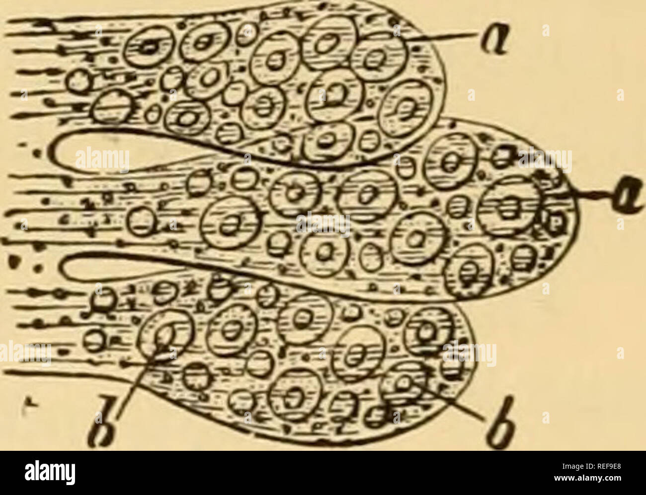 . The comparative anatomy of the domesticated animals. Horses; Veterinary anatomy. GLAND-VESICLES, WITH THEIR EXCRETORY DUCTS TERMINATING IN A DUCTUS LAC- TIFEROUS: FROM A MERCURIAL INJECTION (MAGNIFIED FOUR TIMES). ULTIMATE FOLLICLES, OR Gl AND-VESICLES, WITH THEIR EPITHELIUM OR SECRETING CELLS, a, a, AND NUCLEI, 6, b. They are ^^-^ of an inch in diameter.) The lactiferous ducts commence by blind extremities, and run into each other to constitute a certain number of principal canals ; these open into the galactophorous sinuses (each a sacculus vel sinus lactiferus). The glandular culs-de-sac Stock Photohttps://www.alamy.com/image-license-details/?v=1https://www.alamy.com/the-comparative-anatomy-of-the-domesticated-animals-horses-veterinary-anatomy-gland-vesicles-with-their-excretory-ducts-terminating-in-a-ductus-lac-tiferous-from-a-mercurial-injection-magnified-four-times-ultimate-follicles-or-gl-and-vesicles-with-their-epithelium-or-secreting-cells-a-a-and-nuclei-6-b-they-are-of-an-inch-in-diameter-the-lactiferous-ducts-commence-by-blind-extremities-and-run-into-each-other-to-constitute-a-certain-number-of-principal-canals-these-open-into-the-galactophorous-sinuses-each-a-sacculus-vel-sinus-lactiferus-the-glandular-culs-de-sac-image232676704.html
RMREF9E8–. The comparative anatomy of the domesticated animals. Horses; Veterinary anatomy. GLAND-VESICLES, WITH THEIR EXCRETORY DUCTS TERMINATING IN A DUCTUS LAC- TIFEROUS: FROM A MERCURIAL INJECTION (MAGNIFIED FOUR TIMES). ULTIMATE FOLLICLES, OR Gl AND-VESICLES, WITH THEIR EPITHELIUM OR SECRETING CELLS, a, a, AND NUCLEI, 6, b. They are ^^-^ of an inch in diameter.) The lactiferous ducts commence by blind extremities, and run into each other to constitute a certain number of principal canals ; these open into the galactophorous sinuses (each a sacculus vel sinus lactiferus). The glandular culs-de-sac