Quick filters:
Lamina terminalis Stock Photos and Images
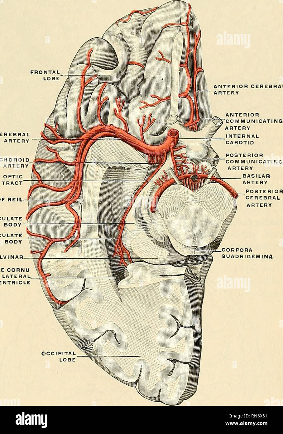 . Anatomy, descriptive and applied. Anatomy. THE INTERNAL CAROTID ARTERY 615 substance and lamina terminalis, and supply the rostrum of the corpus callosum, the septum lucidum, and the head of the caudate nucleus. The inferior internal frontal branches, two or three in number, are distributed to the orbital surface of the frontal lobe, where they supply the olfactory lobe, gyrus rectus, and internal orbital (mesorbital) convolution. The anterior internal frontal supply a part of the mesal surface of the prefrontal region, and send branches over the edge of the hemisphere to the superfrontal an Stock Photohttps://www.alamy.com/image-license-details/?v=1https://www.alamy.com/anatomy-descriptive-and-applied-anatomy-the-internal-carotid-artery-615-substance-and-lamina-terminalis-and-supply-the-rostrum-of-the-corpus-callosum-the-septum-lucidum-and-the-head-of-the-caudate-nucleus-the-inferior-internal-frontal-branches-two-or-three-in-number-are-distributed-to-the-orbital-surface-of-the-frontal-lobe-where-they-supply-the-olfactory-lobe-gyrus-rectus-and-internal-orbital-mesorbital-convolution-the-anterior-internal-frontal-supply-a-part-of-the-mesal-surface-of-the-prefrontal-region-and-send-branches-over-the-edge-of-the-hemisphere-to-the-superfrontal-an-image236794797.html
RMRN6X51–. Anatomy, descriptive and applied. Anatomy. THE INTERNAL CAROTID ARTERY 615 substance and lamina terminalis, and supply the rostrum of the corpus callosum, the septum lucidum, and the head of the caudate nucleus. The inferior internal frontal branches, two or three in number, are distributed to the orbital surface of the frontal lobe, where they supply the olfactory lobe, gyrus rectus, and internal orbital (mesorbital) convolution. The anterior internal frontal supply a part of the mesal surface of the prefrontal region, and send branches over the edge of the hemisphere to the superfrontal an
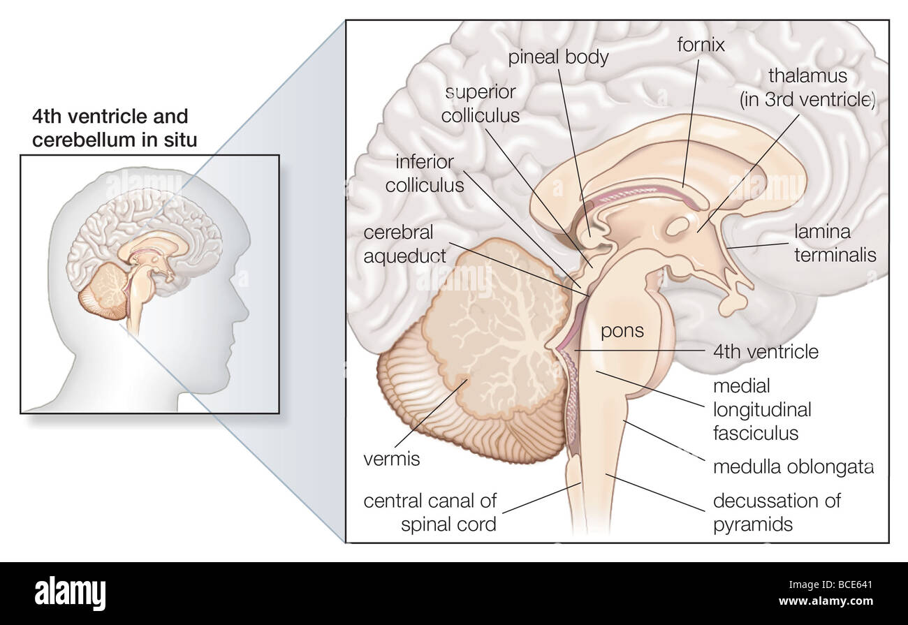 Sagittal section of the human brain, showing structures of the cerebellum, brainstem, and cerebral ventricles. Stock Photohttps://www.alamy.com/image-license-details/?v=1https://www.alamy.com/stock-photo-sagittal-section-of-the-human-brain-showing-structures-of-the-cerebellum-24898385.html
RMBCE641–Sagittal section of the human brain, showing structures of the cerebellum, brainstem, and cerebral ventricles.
 Archive image from page 171 of The development of the chick. The development of the chick : an introduction to embryology . developmentofchi02lill Year: 1936 152 THE DEVELOPMENT OF THE CHICK The median strip includes the tela choroidea, beginning at the diencephalon, and the lamina terminalis, which ends at the recessus opticus. These divisions are of great prospective signifi- cance, though at the stage of 36 s they are but slightly differen- tiated, save by their position. A slight thickening of the lamina terminalis just in front of the recessus opticus marks the site of the future anterio Stock Photohttps://www.alamy.com/image-license-details/?v=1https://www.alamy.com/archive-image-from-page-171-of-the-development-of-the-chick-the-development-of-the-chick-an-introduction-to-embryology-developmentofchi02lill-year-1936-152-the-development-of-the-chick-the-median-strip-includes-the-tela-choroidea-beginning-at-the-diencephalon-and-the-lamina-terminalis-which-ends-at-the-recessus-opticus-these-divisions-are-of-great-prospective-signifi-cance-though-at-the-stage-of-36-s-they-are-but-slightly-differen-tiated-save-by-their-position-a-slight-thickening-of-the-lamina-terminalis-just-in-front-of-the-recessus-opticus-marks-the-site-of-the-future-anterio-image258897202.html
RMW15P0J–Archive image from page 171 of The development of the chick. The development of the chick : an introduction to embryology . developmentofchi02lill Year: 1936 152 THE DEVELOPMENT OF THE CHICK The median strip includes the tela choroidea, beginning at the diencephalon, and the lamina terminalis, which ends at the recessus opticus. These divisions are of great prospective signifi- cance, though at the stage of 36 s they are but slightly differen- tiated, save by their position. A slight thickening of the lamina terminalis just in front of the recessus opticus marks the site of the future anterio
 . The development of the chick : an introduction to embryology. Embryology; Chickens -- Embryos. 152 THE DEVELOPMENT OF THE CHICK The median strip includes the tela choroidea, beginning at the diencephalon, and the lamina terminalis, which ends at the recessus opticus. These divisions are of great prospective signifi- cance, though at the stage of 36 s they are but slightly differen- tiated, save by their position. A slight thickening of the lamina terminalis just in front of the recessus opticus marks the site of the future anterior commissure (Figs. 87 and 88). Mesenc. Me/enc. /. Please note Stock Photohttps://www.alamy.com/image-license-details/?v=1https://www.alamy.com/the-development-of-the-chick-an-introduction-to-embryology-embryology-chickens-embryos-152-the-development-of-the-chick-the-median-strip-includes-the-tela-choroidea-beginning-at-the-diencephalon-and-the-lamina-terminalis-which-ends-at-the-recessus-opticus-these-divisions-are-of-great-prospective-signifi-cance-though-at-the-stage-of-36-s-they-are-but-slightly-differen-tiated-save-by-their-position-a-slight-thickening-of-the-lamina-terminalis-just-in-front-of-the-recessus-opticus-marks-the-site-of-the-future-anterior-commissure-figs-87-and-88-mesenc-meenc-please-note-image215970148.html
RMPFA83G–. The development of the chick : an introduction to embryology. Embryology; Chickens -- Embryos. 152 THE DEVELOPMENT OF THE CHICK The median strip includes the tela choroidea, beginning at the diencephalon, and the lamina terminalis, which ends at the recessus opticus. These divisions are of great prospective signifi- cance, though at the stage of 36 s they are but slightly differen- tiated, save by their position. A slight thickening of the lamina terminalis just in front of the recessus opticus marks the site of the future anterior commissure (Figs. 87 and 88). Mesenc. Me/enc. /. Please note
![. M-M-Car/ , -^ p.arasen,t 3e/ti Fig-. 50. Knorpelige Nasenkapscl eines Beutel- jungen von Halmaturus von l,.ö cm Länge; nach einem Modell; Ventralfläche. Ac vorderer geschlossener Teil der Kapsel (Annulus cartilagineus Spurgat); die Car- tilago pararaseptalis umschließt vorn röhrenförmig das Jacobsonsche Organ, ist durch einen Spalt von Sep- tum narium [Sept.] getrennt und hängt hinten mit der Schlußplatte, Lamina terminalis zusammen; Ap. 11. ext. äußere Nasenöffuung; D. n. l. Eintritt des Träuenkanals in die Nasenkapsel; Ch Choane. Nach Sevdel. Stock Photo . M-M-Car/ , -^ p.arasen,t 3e/ti Fig-. 50. Knorpelige Nasenkapscl eines Beutel- jungen von Halmaturus von l,.ö cm Länge; nach einem Modell; Ventralfläche. Ac vorderer geschlossener Teil der Kapsel (Annulus cartilagineus Spurgat); die Car- tilago pararaseptalis umschließt vorn röhrenförmig das Jacobsonsche Organ, ist durch einen Spalt von Sep- tum narium [Sept.] getrennt und hängt hinten mit der Schlußplatte, Lamina terminalis zusammen; Ap. 11. ext. äußere Nasenöffuung; D. n. l. Eintritt des Träuenkanals in die Nasenkapsel; Ch Choane. Nach Sevdel. Stock Photo](https://c8.alamy.com/comp/MCNNPN/m-m-car-parasent-3eti-fig-50-knorpelige-nasenkapscl-eines-beutel-jungen-von-halmaturus-von-l-cm-lnge-nach-einem-modell-ventralflche-ac-vorderer-geschlossener-teil-der-kapsel-annulus-cartilagineus-spurgat-die-car-tilago-pararaseptalis-umschliet-vorn-rhrenfrmig-das-jacobsonsche-organ-ist-durch-einen-spalt-von-sep-tum-narium-sept-getrennt-und-hngt-hinten-mit-der-schluplatte-lamina-terminalis-zusammen-ap-11-ext-uere-nasenffuung-d-n-l-eintritt-des-truenkanals-in-die-nasenkapsel-ch-choane-nach-sevdel-MCNNPN.jpg) . M-M-Car/ , -^ p.arasen,t 3e/ti Fig-. 50. Knorpelige Nasenkapscl eines Beutel- jungen von Halmaturus von l,.ö cm Länge; nach einem Modell; Ventralfläche. Ac vorderer geschlossener Teil der Kapsel (Annulus cartilagineus Spurgat); die Car- tilago pararaseptalis umschließt vorn röhrenförmig das Jacobsonsche Organ, ist durch einen Spalt von Sep- tum narium [Sept.] getrennt und hängt hinten mit der Schlußplatte, Lamina terminalis zusammen; Ap. 11. ext. äußere Nasenöffuung; D. n. l. Eintritt des Träuenkanals in die Nasenkapsel; Ch Choane. Nach Sevdel. Stock Photohttps://www.alamy.com/image-license-details/?v=1https://www.alamy.com/m-m-car-parasent-3eti-fig-50-knorpelige-nasenkapscl-eines-beutel-jungen-von-halmaturus-von-l-cm-lnge-nach-einem-modell-ventralflche-ac-vorderer-geschlossener-teil-der-kapsel-annulus-cartilagineus-spurgat-die-car-tilago-pararaseptalis-umschliet-vorn-rhrenfrmig-das-jacobsonsche-organ-ist-durch-einen-spalt-von-sep-tum-narium-sept-getrennt-und-hngt-hinten-mit-der-schluplatte-lamina-terminalis-zusammen-ap-11-ext-uere-nasenffuung-d-n-l-eintritt-des-truenkanals-in-die-nasenkapsel-ch-choane-nach-sevdel-image179957645.html
. M-M-Car/ , -^ p.arasen,t 3e/ti Fig-. 50. Knorpelige Nasenkapscl eines Beutel- jungen von Halmaturus von l,.ö cm Länge; nach einem Modell; Ventralfläche. Ac vorderer geschlossener Teil der Kapsel (Annulus cartilagineus Spurgat); die Car- tilago pararaseptalis umschließt vorn röhrenförmig das Jacobsonsche Organ, ist durch einen Spalt von Sep- tum narium [Sept.] getrennt und hängt hinten mit der Schlußplatte, Lamina terminalis zusammen; Ap. 11. ext. äußere Nasenöffuung; D. n. l. Eintritt des Träuenkanals in die Nasenkapsel; Ch Choane. Nach Sevdel. Stock Photohttps://www.alamy.com/image-license-details/?v=1https://www.alamy.com/m-m-car-parasent-3eti-fig-50-knorpelige-nasenkapscl-eines-beutel-jungen-von-halmaturus-von-l-cm-lnge-nach-einem-modell-ventralflche-ac-vorderer-geschlossener-teil-der-kapsel-annulus-cartilagineus-spurgat-die-car-tilago-pararaseptalis-umschliet-vorn-rhrenfrmig-das-jacobsonsche-organ-ist-durch-einen-spalt-von-sep-tum-narium-sept-getrennt-und-hngt-hinten-mit-der-schluplatte-lamina-terminalis-zusammen-ap-11-ext-uere-nasenffuung-d-n-l-eintritt-des-truenkanals-in-die-nasenkapsel-ch-choane-nach-sevdel-image179957645.htmlRMMCNNPN–. M-M-Car/ , -^ p.arasen,t 3e/ti Fig-. 50. Knorpelige Nasenkapscl eines Beutel- jungen von Halmaturus von l,.ö cm Länge; nach einem Modell; Ventralfläche. Ac vorderer geschlossener Teil der Kapsel (Annulus cartilagineus Spurgat); die Car- tilago pararaseptalis umschließt vorn röhrenförmig das Jacobsonsche Organ, ist durch einen Spalt von Sep- tum narium [Sept.] getrennt und hängt hinten mit der Schlußplatte, Lamina terminalis zusammen; Ap. 11. ext. äußere Nasenöffuung; D. n. l. Eintritt des Träuenkanals in die Nasenkapsel; Ch Choane. Nach Sevdel.
 Human brain anatomy for medical concept 3D illustration Stock Photohttps://www.alamy.com/image-license-details/?v=1https://www.alamy.com/human-brain-anatomy-for-medical-concept-3d-illustration-image504513117.html
RF2M8PFHH–Human brain anatomy for medical concept 3D illustration
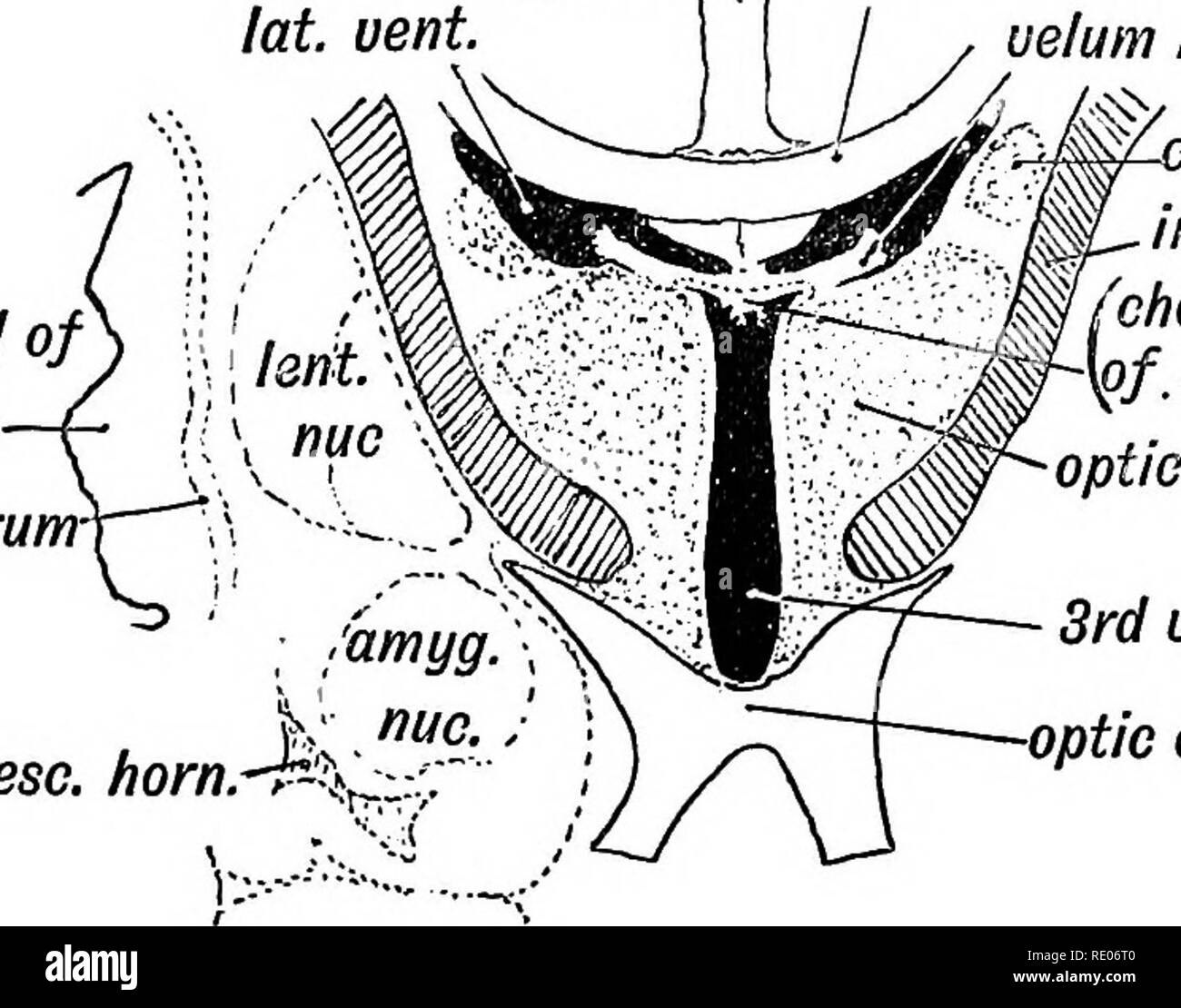 . Human embryology and morphology. Embryology, Human; Morphology. THE BEAIN AND SPINAL COKD. 205 The Lamina Cinerea or lamina terminalis (Fig. 167) represents the anterior end of the neural tube. In the adult it stretches between the optic chiasma, which is developed on the floor of the 3rd ventricle and the rostrum of the corpus callosum. Its development will be described later, but it retains with little alteration its early simple structure. The inter-peduncular space, which forms the floor of the 3rd ventricle, also retains in the adult to a considerable extent the simple embryonic form. I Stock Photohttps://www.alamy.com/image-license-details/?v=1https://www.alamy.com/human-embryology-and-morphology-embryology-human-morphology-the-beain-and-spinal-cokd-205-the-lamina-cinerea-or-lamina-terminalis-fig-167-represents-the-anterior-end-of-the-neural-tube-in-the-adult-it-stretches-between-the-optic-chiasma-which-is-developed-on-the-floor-of-the-3rd-ventricle-and-the-rostrum-of-the-corpus-callosum-its-development-will-be-described-later-but-it-retains-with-little-alteration-its-early-simple-structure-the-inter-peduncular-space-which-forms-the-floor-of-the-3rd-ventricle-also-retains-in-the-adult-to-a-considerable-extent-the-simple-embryonic-form-i-image232345344.html
RMRE06T0–. Human embryology and morphology. Embryology, Human; Morphology. THE BEAIN AND SPINAL COKD. 205 The Lamina Cinerea or lamina terminalis (Fig. 167) represents the anterior end of the neural tube. In the adult it stretches between the optic chiasma, which is developed on the floor of the 3rd ventricle and the rostrum of the corpus callosum. Its development will be described later, but it retains with little alteration its early simple structure. The inter-peduncular space, which forms the floor of the 3rd ventricle, also retains in the adult to a considerable extent the simple embryonic form. I
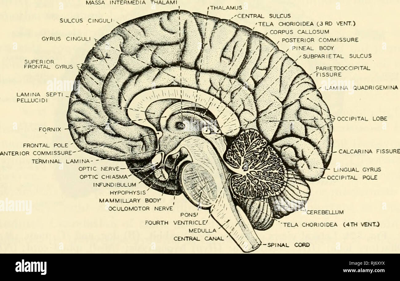
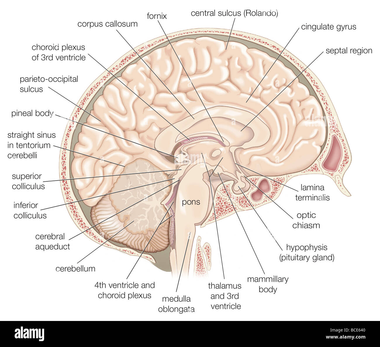 Medial view of the left hemisphere of the human brain. Stock Photohttps://www.alamy.com/image-license-details/?v=1https://www.alamy.com/stock-photo-medial-view-of-the-left-hemisphere-of-the-human-brain-24898384.html
RMBCE640–Medial view of the left hemisphere of the human brain.
 Archive image from page 171 of The development of the chick;. The development of the chick; an introduction to embryology . developmentofchi00lill Year: 1908 152 THE DEVELOPMENT OF THE CHICK The median strip includes the tela choroidea, beginning at the diencephalon, and the lamina terminalis, which ends at the recessus opticus. These divisions are of great prospective signifi- cance, though at the stage of 36 s they are but sHghtly differen- tiated, save by their position. A shght thickening of the lamina terminalis just in front of the recessus opticus marks the site of the future anterior Stock Photohttps://www.alamy.com/image-license-details/?v=1https://www.alamy.com/archive-image-from-page-171-of-the-development-of-the-chick-the-development-of-the-chick-an-introduction-to-embryology-developmentofchi00lill-year-1908-152-the-development-of-the-chick-the-median-strip-includes-the-tela-choroidea-beginning-at-the-diencephalon-and-the-lamina-terminalis-which-ends-at-the-recessus-opticus-these-divisions-are-of-great-prospective-signifi-cance-though-at-the-stage-of-36-s-they-are-but-shghtly-differen-tiated-save-by-their-position-a-shght-thickening-of-the-lamina-terminalis-just-in-front-of-the-recessus-opticus-marks-the-site-of-the-future-anterior-image258897195.html
RMW15P0B–Archive image from page 171 of The development of the chick;. The development of the chick; an introduction to embryology . developmentofchi00lill Year: 1908 152 THE DEVELOPMENT OF THE CHICK The median strip includes the tela choroidea, beginning at the diencephalon, and the lamina terminalis, which ends at the recessus opticus. These divisions are of great prospective signifi- cance, though at the stage of 36 s they are but sHghtly differen- tiated, save by their position. A shght thickening of the lamina terminalis just in front of the recessus opticus marks the site of the future anterior
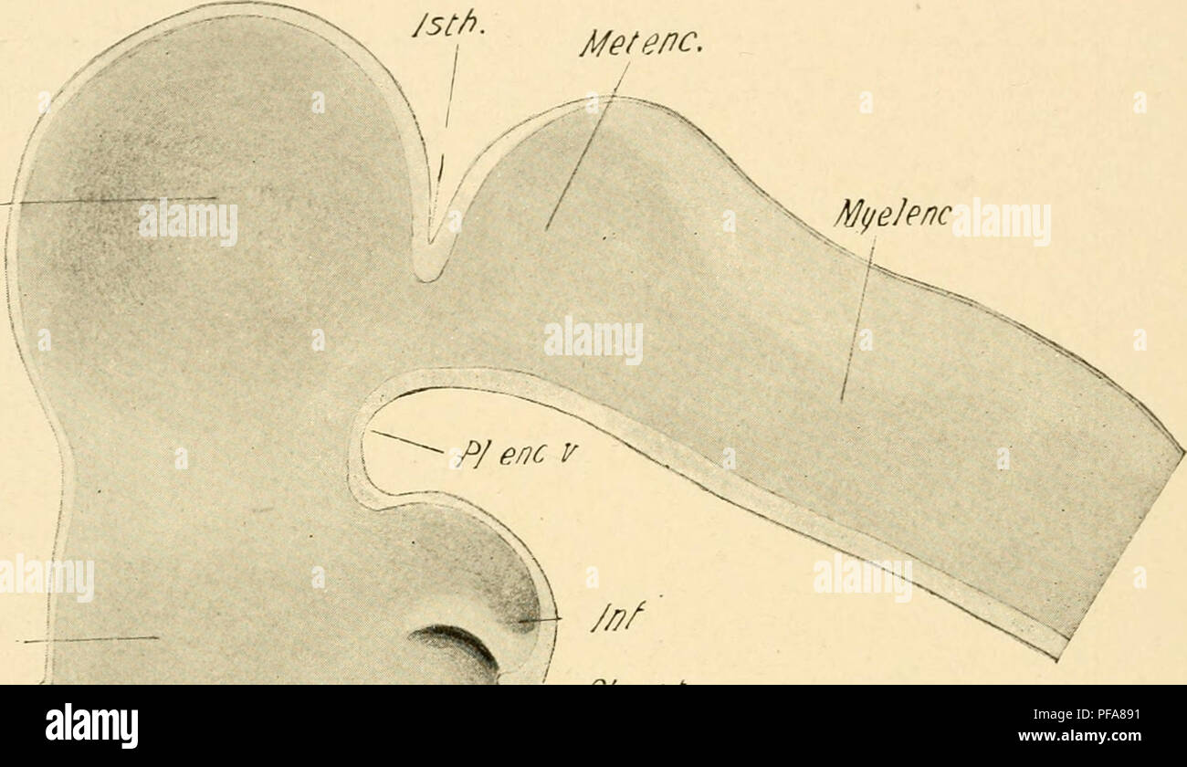 . The development of the chick; an introduction to embryology. Birds -- Embryology. 152 THE DEVELOPMENT OF THE CHICK The median strip includes the tela choroidea, beginning at the diencephalon, and the lamina terminalis, which ends at the recessus opticus. These divisions are of great prospective signifi- cance, though at the stage of 36 s they are but sHghtly differen- tiated, save by their position. A shght thickening of the lamina terminalis just in front of the recessus opticus marks the site of the future anterior commissure (Figs. 87 and 88). Metenc. Mesenc. Please note that these images Stock Photohttps://www.alamy.com/image-license-details/?v=1https://www.alamy.com/the-development-of-the-chick-an-introduction-to-embryology-birds-embryology-152-the-development-of-the-chick-the-median-strip-includes-the-tela-choroidea-beginning-at-the-diencephalon-and-the-lamina-terminalis-which-ends-at-the-recessus-opticus-these-divisions-are-of-great-prospective-signifi-cance-though-at-the-stage-of-36-s-they-are-but-shghtly-differen-tiated-save-by-their-position-a-shght-thickening-of-the-lamina-terminalis-just-in-front-of-the-recessus-opticus-marks-the-site-of-the-future-anterior-commissure-figs-87-and-88-metenc-mesenc-please-note-that-these-images-image215970301.html
RMPFA891–. The development of the chick; an introduction to embryology. Birds -- Embryology. 152 THE DEVELOPMENT OF THE CHICK The median strip includes the tela choroidea, beginning at the diencephalon, and the lamina terminalis, which ends at the recessus opticus. These divisions are of great prospective signifi- cance, though at the stage of 36 s they are but sHghtly differen- tiated, save by their position. A shght thickening of the lamina terminalis just in front of the recessus opticus marks the site of the future anterior commissure (Figs. 87 and 88). Metenc. Mesenc. Please note that these images
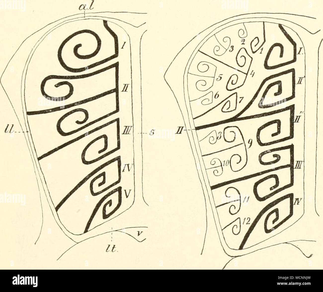 . Fig. 112. Schematisclie Querschnitte durch die linke Nasenhöhle von Säuge- tieren, dicht vor der Siebplatte und ihr parallel, nach Paulli. Links einfacher Typus ohne Ectoturbinalia; rechts mit Ectoturbinalia und zwar einer medialen und lateralen Reihe. /—F Endoturbinalia mit teils durch einfache, teils durch doppelte Einrollung entstandenen Riechwülsten. //' und //" durch Teilung der Basallamelle //entstandene Riechwülste. /, 4, 7, g, 11, 12 mediale, 2, 3, 5, 6, 8, 10 laterale Ectoturbinalia; al Ala laminae perpendicularis; II Lamina lateralis; It Lamina terminalis; i Septum; v Vomer. A Stock Photohttps://www.alamy.com/image-license-details/?v=1https://www.alamy.com/fig-112-schematisclie-querschnitte-durch-die-linke-nasenhhle-von-suge-tieren-dicht-vor-der-siebplatte-und-ihr-parallel-nach-paulli-links-einfacher-typus-ohne-ectoturbinalia-rechts-mit-ectoturbinalia-und-zwar-einer-medialen-und-lateralen-reihe-f-endoturbinalia-mit-teils-durch-einfache-teils-durch-doppelte-einrollung-entstandenen-riechwlsten-und-quot-durch-teilung-der-basallamelle-entstandene-riechwlste-4-7-g-11-12-mediale-2-3-5-6-8-10-laterale-ectoturbinalia-al-ala-laminae-perpendicularis-ii-lamina-lateralis-it-lamina-terminalis-i-septum-v-vomer-a-image179957537.html
RMMCNNJW–. Fig. 112. Schematisclie Querschnitte durch die linke Nasenhöhle von Säuge- tieren, dicht vor der Siebplatte und ihr parallel, nach Paulli. Links einfacher Typus ohne Ectoturbinalia; rechts mit Ectoturbinalia und zwar einer medialen und lateralen Reihe. /—F Endoturbinalia mit teils durch einfache, teils durch doppelte Einrollung entstandenen Riechwülsten. //' und //" durch Teilung der Basallamelle //entstandene Riechwülste. /, 4, 7, g, 11, 12 mediale, 2, 3, 5, 6, 8, 10 laterale Ectoturbinalia; al Ala laminae perpendicularis; II Lamina lateralis; It Lamina terminalis; i Septum; v Vomer. A
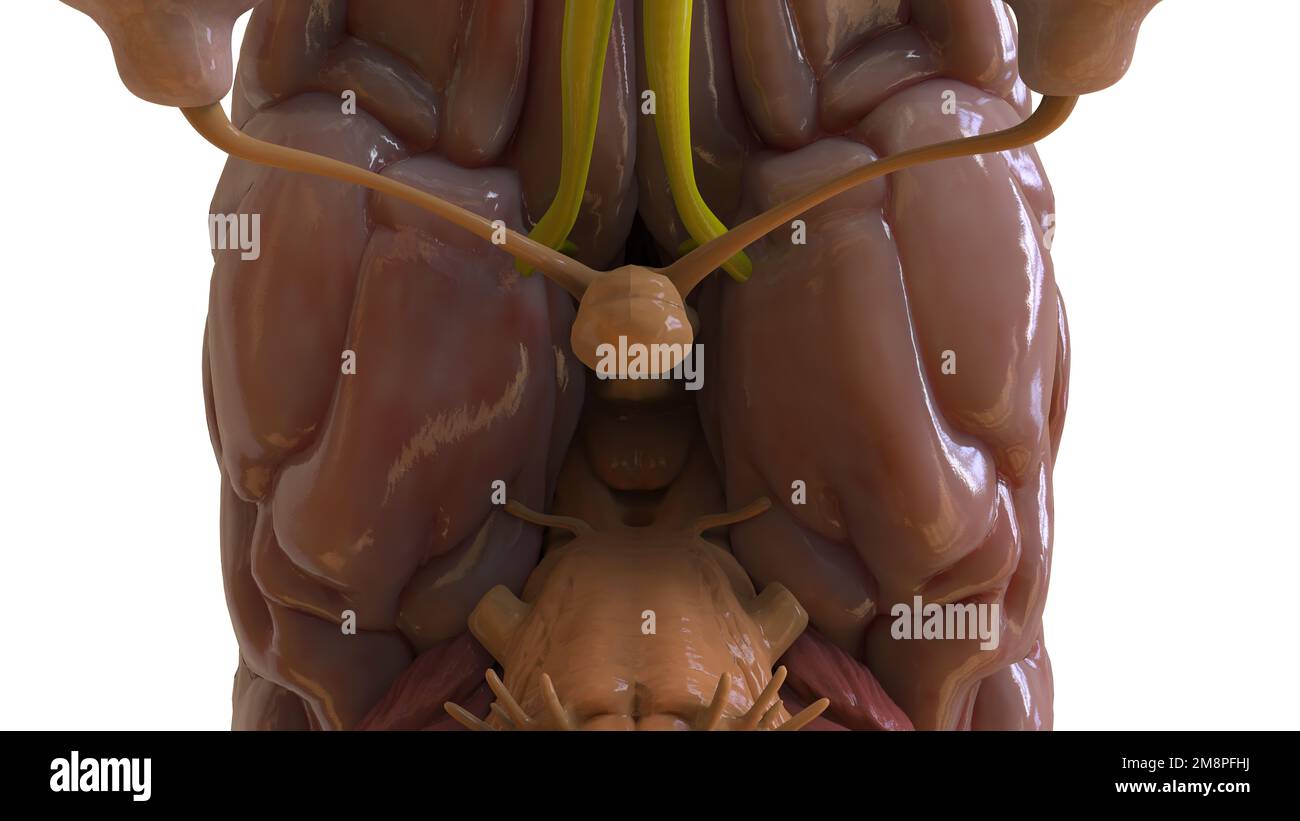 Human brain anatomy for medical concept 3D illustration Stock Photohttps://www.alamy.com/image-license-details/?v=1https://www.alamy.com/human-brain-anatomy-for-medical-concept-3d-illustration-image504513118.html
RF2M8PFHJ–Human brain anatomy for medical concept 3D illustration
 . Human embryology and morphology. Embryology, Human; Morphology. THE BRAIN AND SPINAL CORD. 209 (Fig. 171) is developed in the lamina terminalis—the primitive anterior wall of the fore-brain. The commissure passes between the temporo-sphenoidal lobes. These lobes represent the posterior ends of the cerebral vesicles. At first they are mere dilatations behind the foramen of Monro. The commissure crosses in the lamina terminalis below and rather anterior to the foramen of Monro. This is the earliest and most primitive of the cerebral (pallia!) commissures (Elliot Smith). corp. callos. lam. term Stock Photohttps://www.alamy.com/image-license-details/?v=1https://www.alamy.com/human-embryology-and-morphology-embryology-human-morphology-the-brain-and-spinal-cord-209-fig-171-is-developed-in-the-lamina-terminalisthe-primitive-anterior-wall-of-the-fore-brain-the-commissure-passes-between-the-temporo-sphenoidal-lobes-these-lobes-represent-the-posterior-ends-of-the-cerebral-vesicles-at-first-they-are-mere-dilatations-behind-the-foramen-of-monro-the-commissure-crosses-in-the-lamina-terminalis-below-and-rather-anterior-to-the-foramen-of-monro-this-is-the-earliest-and-most-primitive-of-the-cerebral-pallia!-commissures-elliot-smith-corp-callos-lam-term-image232345334.html
RMRE06RJ–. Human embryology and morphology. Embryology, Human; Morphology. THE BRAIN AND SPINAL CORD. 209 (Fig. 171) is developed in the lamina terminalis—the primitive anterior wall of the fore-brain. The commissure passes between the temporo-sphenoidal lobes. These lobes represent the posterior ends of the cerebral vesicles. At first they are mere dilatations behind the foramen of Monro. The commissure crosses in the lamina terminalis below and rather anterior to the foramen of Monro. This is the earliest and most primitive of the cerebral (pallia!) commissures (Elliot Smith). corp. callos. lam. term
![Archive image from page 91 of The development of the skull. The development of the skull of Emys Lutaria . developmentofsku00kunk Year: 1912 cptr. EXPLANATION OF ridt'ItlCS 30 Ventral view of the olfaetoi-y fajjsule of an embryo having a earapace length of 7 mm. showing the cartilago parase])talis (cp.) separated from the septum n:isi as far anterior as the lamina terminalis anterior (/./.</.) and consequently the foramen praepalatinum not enclosed posteriorly. X 20. 31 Posterior portion of the right otic capsule viewed from in front and slightly from the median line, showing especially th Stock Photo Archive image from page 91 of The development of the skull. The development of the skull of Emys Lutaria . developmentofsku00kunk Year: 1912 cptr. EXPLANATION OF ridt'ItlCS 30 Ventral view of the olfaetoi-y fajjsule of an embryo having a earapace length of 7 mm. showing the cartilago parase])talis (cp.) separated from the septum n:isi as far anterior as the lamina terminalis anterior (/./.</.) and consequently the foramen praepalatinum not enclosed posteriorly. X 20. 31 Posterior portion of the right otic capsule viewed from in front and slightly from the median line, showing especially th Stock Photo](https://c8.alamy.com/comp/W1D67P/archive-image-from-page-91-of-the-development-of-the-skull-the-development-of-the-skull-of-emys-lutaria-developmentofsku00kunk-year-1912-cptr-explanation-of-ridtitlcs-30-ventral-view-of-the-olfaetoi-y-fajjsule-of-an-embryo-having-a-earapace-length-of-7-mm-showing-the-cartilago-parase-talis-cp-separated-from-the-septum-nisi-as-far-anterior-as-the-lamina-terminalis-anterior-lt-and-consequently-the-foramen-praepalatinum-not-enclosed-posteriorly-x-20-31-posterior-portion-of-the-right-otic-capsule-viewed-from-in-front-and-slightly-from-the-median-line-showing-especially-th-W1D67P.jpg) Archive image from page 91 of The development of the skull. The development of the skull of Emys Lutaria . developmentofsku00kunk Year: 1912 cptr. EXPLANATION OF ridt'ItlCS 30 Ventral view of the olfaetoi-y fajjsule of an embryo having a earapace length of 7 mm. showing the cartilago parase])talis (cp.) separated from the septum n:isi as far anterior as the lamina terminalis anterior (/./.</.) and consequently the foramen praepalatinum not enclosed posteriorly. X 20. 31 Posterior portion of the right otic capsule viewed from in front and slightly from the median line, showing especially th Stock Photohttps://www.alamy.com/image-license-details/?v=1https://www.alamy.com/archive-image-from-page-91-of-the-development-of-the-skull-the-development-of-the-skull-of-emys-lutaria-developmentofsku00kunk-year-1912-cptr-explanation-of-ridtitlcs-30-ventral-view-of-the-olfaetoi-y-fajjsule-of-an-embryo-having-a-earapace-length-of-7-mm-showing-the-cartilago-parase-talis-cp-separated-from-the-septum-nisi-as-far-anterior-as-the-lamina-terminalis-anterior-lt-and-consequently-the-foramen-praepalatinum-not-enclosed-posteriorly-x-20-31-posterior-portion-of-the-right-otic-capsule-viewed-from-in-front-and-slightly-from-the-median-line-showing-especially-th-image259060474.html
Archive image from page 91 of The development of the skull. The development of the skull of Emys Lutaria . developmentofsku00kunk Year: 1912 cptr. EXPLANATION OF ridt'ItlCS 30 Ventral view of the olfaetoi-y fajjsule of an embryo having a earapace length of 7 mm. showing the cartilago parase])talis (cp.) separated from the septum n:isi as far anterior as the lamina terminalis anterior (/./.</.) and consequently the foramen praepalatinum not enclosed posteriorly. X 20. 31 Posterior portion of the right otic capsule viewed from in front and slightly from the median line, showing especially th Stock Photohttps://www.alamy.com/image-license-details/?v=1https://www.alamy.com/archive-image-from-page-91-of-the-development-of-the-skull-the-development-of-the-skull-of-emys-lutaria-developmentofsku00kunk-year-1912-cptr-explanation-of-ridtitlcs-30-ventral-view-of-the-olfaetoi-y-fajjsule-of-an-embryo-having-a-earapace-length-of-7-mm-showing-the-cartilago-parase-talis-cp-separated-from-the-septum-nisi-as-far-anterior-as-the-lamina-terminalis-anterior-lt-and-consequently-the-foramen-praepalatinum-not-enclosed-posteriorly-x-20-31-posterior-portion-of-the-right-otic-capsule-viewed-from-in-front-and-slightly-from-the-median-line-showing-especially-th-image259060474.htmlRMW1D67P–Archive image from page 91 of The development of the skull. The development of the skull of Emys Lutaria . developmentofsku00kunk Year: 1912 cptr. EXPLANATION OF ridt'ItlCS 30 Ventral view of the olfaetoi-y fajjsule of an embryo having a earapace length of 7 mm. showing the cartilago parase])talis (cp.) separated from the septum n:isi as far anterior as the lamina terminalis anterior (/./.</.) and consequently the foramen praepalatinum not enclosed posteriorly. X 20. 31 Posterior portion of the right otic capsule viewed from in front and slightly from the median line, showing especially th
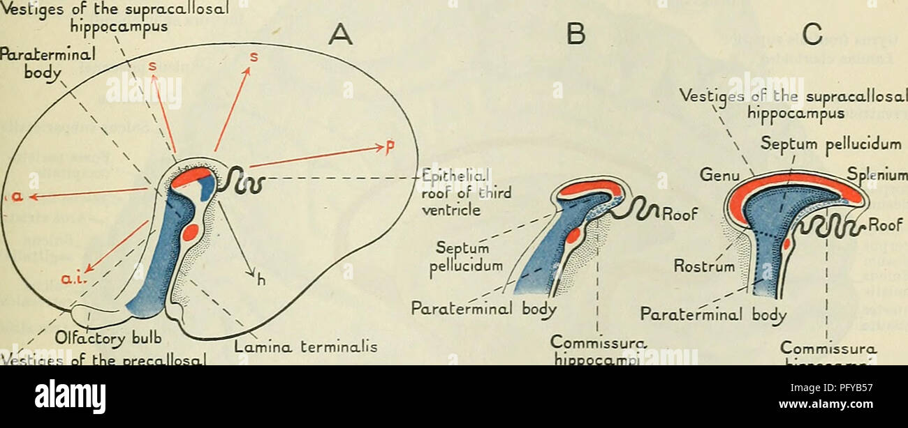 . Cunningham's Text-book of anatomy. Anatomy. THE CEEEBEAL COMMISSURES AND SEPTUM PELLUCIDUM. 629 upper end of the hippocampal formation so that it becomes removed far from the lamina terminalis. The fibres of the fimbria which are prolonged forwards under the corpus callosum and septum pellucidum to bridge this great gap form the crus fornicis on each side. As a rule in the human adult brain the crura fornicis of the two hemispheres become crowded together at the median plane so as to obscure the connecting lamella which serves as a matrix for the commissura hippocampi (Fig. 557, C); but the Stock Photohttps://www.alamy.com/image-license-details/?v=1https://www.alamy.com/cunninghams-text-book-of-anatomy-anatomy-the-ceeebeal-commissures-and-septum-pellucidum-629-upper-end-of-the-hippocampal-formation-so-that-it-becomes-removed-far-from-the-lamina-terminalis-the-fibres-of-the-fimbria-which-are-prolonged-forwards-under-the-corpus-callosum-and-septum-pellucidum-to-bridge-this-great-gap-form-the-crus-fornicis-on-each-side-as-a-rule-in-the-human-adult-brain-the-crura-fornicis-of-the-two-hemispheres-become-crowded-together-at-the-median-plane-so-as-to-obscure-the-connecting-lamella-which-serves-as-a-matrix-for-the-commissura-hippocampi-fig-557-c-but-the-image216345731.html
RMPFYB57–. Cunningham's Text-book of anatomy. Anatomy. THE CEEEBEAL COMMISSURES AND SEPTUM PELLUCIDUM. 629 upper end of the hippocampal formation so that it becomes removed far from the lamina terminalis. The fibres of the fimbria which are prolonged forwards under the corpus callosum and septum pellucidum to bridge this great gap form the crus fornicis on each side. As a rule in the human adult brain the crura fornicis of the two hemispheres become crowded together at the median plane so as to obscure the connecting lamella which serves as a matrix for the commissura hippocampi (Fig. 557, C); but the
 . Fig. 112. Schematische Querschnitte durch die linke Nasenhöhle von Säuge- tieren, dicht vor der Siebplatte und ihr parallel, nach Paulli. Links einfacher Typus ohne Ectoturbinalia; rechts mit Ectoturbinalia und zwar einer medialen und lateralen Reihe. /— V Endoturbinalia mit teils durch einfache, teils durch doppelte Einrollung entstandenen Eiechwülsten. //' und //" durch Teilung der Basallamelle // eiitstandene Eiechwülste. /, 4, 7, 9, 11, 12 mediale, 2, 3, 5, 6, 8, 10 laterale Ectoturbinalia; al Ala laminae perpendicularis; //Lamina lateralis; //" Lamina terminalis; s Septum; t/V Stock Photohttps://www.alamy.com/image-license-details/?v=1https://www.alamy.com/fig-112-schematische-querschnitte-durch-die-linke-nasenhhle-von-suge-tieren-dicht-vor-der-siebplatte-und-ihr-parallel-nach-paulli-links-einfacher-typus-ohne-ectoturbinalia-rechts-mit-ectoturbinalia-und-zwar-einer-medialen-und-lateralen-reihe-v-endoturbinalia-mit-teils-durch-einfache-teils-durch-doppelte-einrollung-entstandenen-eiechwlsten-und-quot-durch-teilung-der-basallamelle-eiitstandene-eiechwlste-4-7-9-11-12-mediale-2-3-5-6-8-10-laterale-ectoturbinalia-al-ala-laminae-perpendicularis-lamina-lateralis-quot-lamina-terminalis-s-septum-tv-image179957418.html
RMMCNNEJ–. Fig. 112. Schematische Querschnitte durch die linke Nasenhöhle von Säuge- tieren, dicht vor der Siebplatte und ihr parallel, nach Paulli. Links einfacher Typus ohne Ectoturbinalia; rechts mit Ectoturbinalia und zwar einer medialen und lateralen Reihe. /— V Endoturbinalia mit teils durch einfache, teils durch doppelte Einrollung entstandenen Eiechwülsten. //' und //" durch Teilung der Basallamelle // eiitstandene Eiechwülste. /, 4, 7, 9, 11, 12 mediale, 2, 3, 5, 6, 8, 10 laterale Ectoturbinalia; al Ala laminae perpendicularis; //Lamina lateralis; //" Lamina terminalis; s Septum; t/V
 Human brain anatomy for medical concept 3D illustration Stock Photohttps://www.alamy.com/image-license-details/?v=1https://www.alamy.com/human-brain-anatomy-for-medical-concept-3d-illustration-image504513119.html
RF2M8PFHK–Human brain anatomy for medical concept 3D illustration
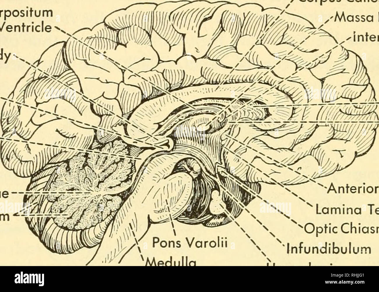 . Biology of the vertebrates : a comparative study of man and his animal allies. Vertebrates; Vertebrates -- Anatomy; Anatomy, Comparative. yo8 Biology of the Vertebrates Velum Interpositum of Third Ventricle Pineal Body Anterior Colliculus--4 Corpus Callosum ,'Massa Intermedia / ^*vinterventricular Foramen ^^^^^ ^y-I^^rA--Septum Pellucidum r-Fornix Posterior—-^-vC Colliculus Arbor Vitae Cerebellum. Pons Varolii 'Medulla Oblongata Fig. 634. Median sagittal section through the human brain. (After Toldt.) ^Anterior Commissure Lamina Terminalis N vOpticChiasma Infundibulum Hypophysis Opti Stock Photohttps://www.alamy.com/image-license-details/?v=1https://www.alamy.com/biology-of-the-vertebrates-a-comparative-study-of-man-and-his-animal-allies-vertebrates-vertebrates-anatomy-anatomy-comparative-yo8-biology-of-the-vertebrates-velum-interpositum-of-third-ventricle-pineal-body-anterior-colliculus-4-corpus-callosum-massa-intermedia-vinterventricular-foramen-y-ira-septum-pellucidum-r-fornix-posterior-vc-colliculus-arbor-vitae-cerebellum-pons-varolii-medulla-oblongata-fig-634-median-sagittal-section-through-the-human-brain-after-toldt-anterior-commissure-lamina-terminalis-n-vopticchiasma-infundibulum-hypophysis-opti-image234593633.html
RMRHJJG1–. Biology of the vertebrates : a comparative study of man and his animal allies. Vertebrates; Vertebrates -- Anatomy; Anatomy, Comparative. yo8 Biology of the Vertebrates Velum Interpositum of Third Ventricle Pineal Body Anterior Colliculus--4 Corpus Callosum ,'Massa Intermedia / ^*vinterventricular Foramen ^^^^^ ^y-I^^rA--Septum Pellucidum r-Fornix Posterior—-^-vC Colliculus Arbor Vitae Cerebellum. Pons Varolii 'Medulla Oblongata Fig. 634. Median sagittal section through the human brain. (After Toldt.) ^Anterior Commissure Lamina Terminalis N vOpticChiasma Infundibulum Hypophysis Opti
 Archive image from page 607 of Denkschriften der Medicinisch-Naturwissenschaftlichen Gesellschaft zu. Denkschriften der Medicinisch-Naturwissenschaftlichen Gesellschaft zu Jena denkschriftender62medi Year: 1879 t;78 Zur Entwicklungsgeschichte und vergleichenden Morphologie des Schädels von Echidna aculeata var. typica. g§ erfolgt. An die ganze Höhe der Caudalwand stösst von vorn das Septum nasi an, ausserdem setzt sich an ihren unteren Rand jederseits vom Septum eine knorpelige Lamina trans versalis posterior an, die in die Lamina terminalis eingelagert ist. Der mediale Rand der Lam. transv. Stock Photohttps://www.alamy.com/image-license-details/?v=1https://www.alamy.com/archive-image-from-page-607-of-denkschriften-der-medicinisch-naturwissenschaftlichen-gesellschaft-zu-denkschriften-der-medicinisch-naturwissenschaftlichen-gesellschaft-zu-jena-denkschriftender62medi-year-1879-t78-zur-entwicklungsgeschichte-und-vergleichenden-morphologie-des-schdels-von-echidna-aculeata-var-typica-g-erfolgt-an-die-ganze-hhe-der-caudalwand-stsst-von-vorn-das-septum-nasi-an-ausserdem-setzt-sich-an-ihren-unteren-rand-jederseits-vom-septum-eine-knorpelige-lamina-trans-versalis-posterior-an-die-in-die-lamina-terminalis-eingelagert-ist-der-mediale-rand-der-lam-transv-image259325538.html
RMW1W8AA–Archive image from page 607 of Denkschriften der Medicinisch-Naturwissenschaftlichen Gesellschaft zu. Denkschriften der Medicinisch-Naturwissenschaftlichen Gesellschaft zu Jena denkschriftender62medi Year: 1879 t;78 Zur Entwicklungsgeschichte und vergleichenden Morphologie des Schädels von Echidna aculeata var. typica. g§ erfolgt. An die ganze Höhe der Caudalwand stösst von vorn das Septum nasi an, ausserdem setzt sich an ihren unteren Rand jederseits vom Septum eine knorpelige Lamina trans versalis posterior an, die in die Lamina terminalis eingelagert ist. Der mediale Rand der Lam. transv.
 . Cunningham's Text-book of anatomy. Anatomy. THE CONNEXIONS OF THE OLFACTOKY NEBYES. 623 of white nerve-fibres passing to and fro between the olfactory bulb and the hemisphere ; hence it is called the tractus olfactorius. The cerebral hemisphere first appears in the form of a slight bulging upon each side of the fore-brain, but it soon assumes large dimensions. At first it grows forwards and upwards (Fig. 550), and a distinct cleft, the floor of which is the roof-plate and lamina terminalis, appears between the two hemispheres: this is known as the fissura longitudinalis cerebri. The separati Stock Photohttps://www.alamy.com/image-license-details/?v=1https://www.alamy.com/cunninghams-text-book-of-anatomy-anatomy-the-connexions-of-the-olfactoky-nebyes-623-of-white-nerve-fibres-passing-to-and-fro-between-the-olfactory-bulb-and-the-hemisphere-hence-it-is-called-the-tractus-olfactorius-the-cerebral-hemisphere-first-appears-in-the-form-of-a-slight-bulging-upon-each-side-of-the-fore-brain-but-it-soon-assumes-large-dimensions-at-first-it-grows-forwards-and-upwards-fig-550-and-a-distinct-cleft-the-floor-of-which-is-the-roof-plate-and-lamina-terminalis-appears-between-the-two-hemispheres-this-is-known-as-the-fissura-longitudinalis-cerebri-the-separati-image216345756.html
RMPFYB64–. Cunningham's Text-book of anatomy. Anatomy. THE CONNEXIONS OF THE OLFACTOKY NEBYES. 623 of white nerve-fibres passing to and fro between the olfactory bulb and the hemisphere ; hence it is called the tractus olfactorius. The cerebral hemisphere first appears in the form of a slight bulging upon each side of the fore-brain, but it soon assumes large dimensions. At first it grows forwards and upwards (Fig. 550), and a distinct cleft, the floor of which is the roof-plate and lamina terminalis, appears between the two hemispheres: this is known as the fissura longitudinalis cerebri. The separati
 . Fig. 97. Ventralfläclie des Gehirns von Ornithorhynchus x 3, nach Elliot Smith. Fig. 98. Ventralfläche des Gehirns von Orycteropus, nach Elliot Smith in */^ n. Gr. do Bulbus olfactorius; Co Cerebellum; cc Grus cerebri; Ip Locus perforatus; //>/> Lobus pyriformis posterior; mo Medulla oblongata; /* Pons Varoli; J>o Pedunculus olfactorius; to Tuberculum olfactorium; tro Tractus olfactorius; tr opt Tractus opticus. /I/ls. oculomotorius; V N. trigeminus. Als vordere Wand des 3. Ventrikels erscheint die Schlußplatte oder Lamina terminalis. Zu ihrem Verständnis, sowie des sekundären Vorde Stock Photohttps://www.alamy.com/image-license-details/?v=1https://www.alamy.com/fig-97-ventralflclie-des-gehirns-von-ornithorhynchus-x-3-nach-elliot-smith-fig-98-ventralflche-des-gehirns-von-orycteropus-nach-elliot-smith-in-n-gr-do-bulbus-olfactorius-co-cerebellum-cc-grus-cerebri-ip-locus-perforatus-gtgt-lobus-pyriformis-posterior-mo-medulla-oblongata-pons-varoli-jgto-pedunculus-olfactorius-to-tuberculum-olfactorium-tro-tractus-olfactorius-tr-opt-tractus-opticus-ils-oculomotorius-v-n-trigeminus-als-vordere-wand-des-3-ventrikels-erscheint-die-schluplatte-oder-lamina-terminalis-zu-ihrem-verstndnis-sowie-des-sekundren-vorde-image179957471.html
RMMCNNGF–. Fig. 97. Ventralfläclie des Gehirns von Ornithorhynchus x 3, nach Elliot Smith. Fig. 98. Ventralfläche des Gehirns von Orycteropus, nach Elliot Smith in */^ n. Gr. do Bulbus olfactorius; Co Cerebellum; cc Grus cerebri; Ip Locus perforatus; //>/> Lobus pyriformis posterior; mo Medulla oblongata; /* Pons Varoli; J>o Pedunculus olfactorius; to Tuberculum olfactorium; tro Tractus olfactorius; tr opt Tractus opticus. /I/ls. oculomotorius; V N. trigeminus. Als vordere Wand des 3. Ventrikels erscheint die Schlußplatte oder Lamina terminalis. Zu ihrem Verständnis, sowie des sekundären Vorde
 Human brain anatomy for medical concept 3D illustration Stock Photohttps://www.alamy.com/image-license-details/?v=1https://www.alamy.com/human-brain-anatomy-for-medical-concept-3d-illustration-image504513110.html
RF2M8PFHA–Human brain anatomy for medical concept 3D illustration
 . The chordates. Chordata. Fig. 522. Sagittal section of the brain of a calf, (a) Aqueduct; (ac) anterior commissure; (cc) corpus callosum; (/) fornix; (h) habenula; (hy) hypophysis; (i) infundibulum; {im) intermediate mass ("soft commissure"); (It) lamina terminalis; (mb) mammillary body; (ob) olfactory bulb; (oc) optic chiasma; (ol) optic lobes; (p) pinealis; (r) recessus suprapinealis; (s) septum pellucidurn; (III, IV) third and fourth ventricles. (After Butschli. Courtesy, Kingsley: "Comparative Anatomy of Vertebrates," Philadelphia, The Blakiston Company.). Fig. 523. T Stock Photohttps://www.alamy.com/image-license-details/?v=1https://www.alamy.com/the-chordates-chordata-fig-522-sagittal-section-of-the-brain-of-a-calf-a-aqueduct-ac-anterior-commissure-cc-corpus-callosum-fornix-h-habenula-hy-hypophysis-i-infundibulum-im-intermediate-mass-quotsoft-commissurequot-it-lamina-terminalis-mb-mammillary-body-ob-olfactory-bulb-oc-optic-chiasma-ol-optic-lobes-p-pinealis-r-recessus-suprapinealis-s-septum-pellucidurn-iii-iv-third-and-fourth-ventricles-after-butschli-courtesy-kingsley-quotcomparative-anatomy-of-vertebratesquot-philadelphia-the-blakiston-company-fig-523-t-image234951470.html
RMRJ6XYX–. The chordates. Chordata. Fig. 522. Sagittal section of the brain of a calf, (a) Aqueduct; (ac) anterior commissure; (cc) corpus callosum; (/) fornix; (h) habenula; (hy) hypophysis; (i) infundibulum; {im) intermediate mass ("soft commissure"); (It) lamina terminalis; (mb) mammillary body; (ob) olfactory bulb; (oc) optic chiasma; (ol) optic lobes; (p) pinealis; (r) recessus suprapinealis; (s) septum pellucidurn; (III, IV) third and fourth ventricles. (After Butschli. Courtesy, Kingsley: "Comparative Anatomy of Vertebrates," Philadelphia, The Blakiston Company.). Fig. 523. T
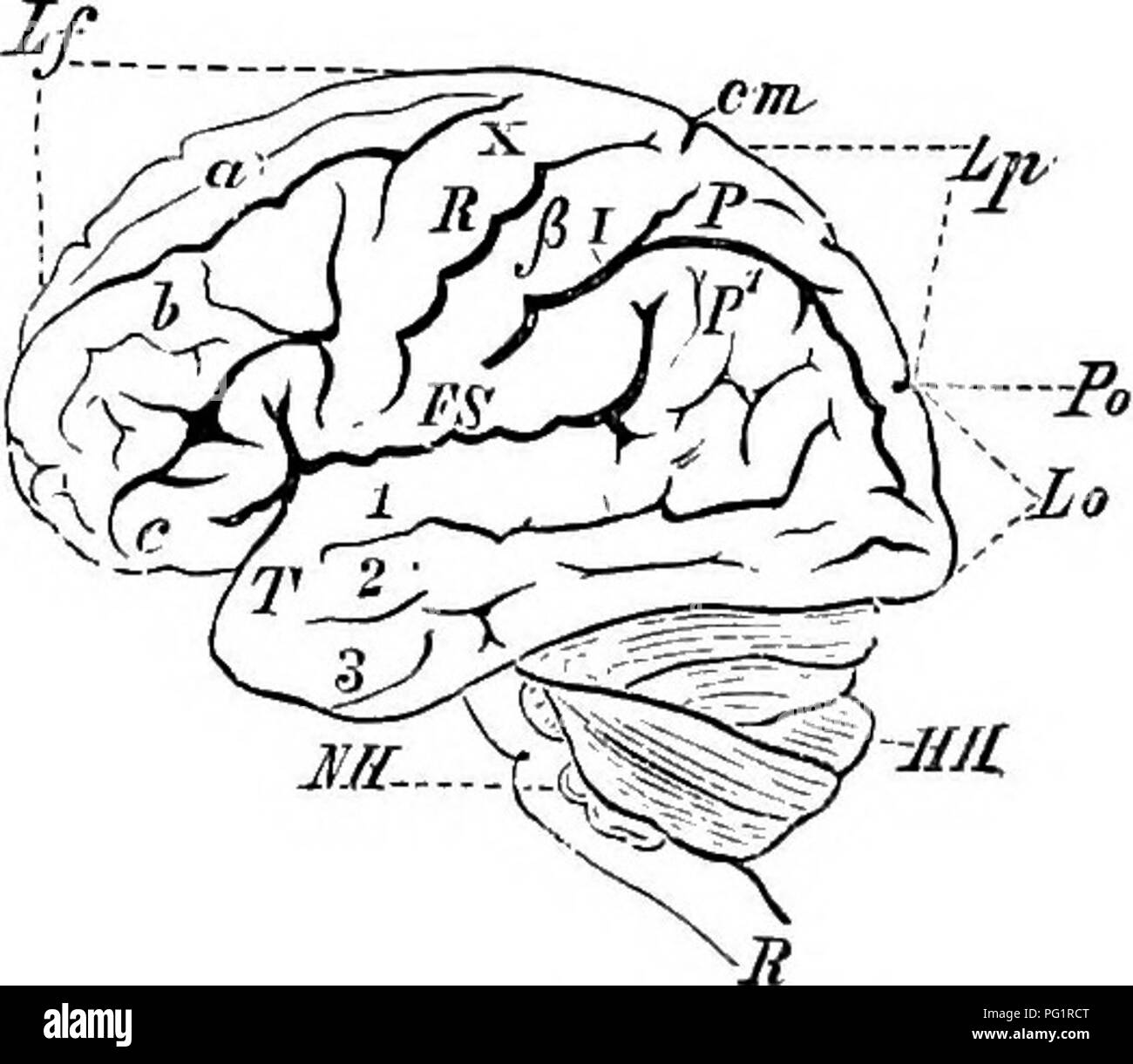 . Elements of the comparative anatomy of vertebrates. Anatomy, Comparative. -Jiff EiG. 145.—Human Ekain. (Median longitudinal vertical section.) (Mainly after Reicliert.) VH, cerebrum ; To, optic thalamus (thalamencephalon), with the middle commis- sure (Cm); Z. pineal body; T, infundibulum; H, pituitary body; ME, corpora bigemina, with the aqueduct of Sylvius (Aq), anterior to which is seen the posterior commissure (Cp); HH, cerebellum; NH, medulla oblongata, with the pons Varolii (P); i?, spinal cord ; B, corpus callosum ; G, fornix, which extends antero-ventrally to the lamina terminalis (C Stock Photohttps://www.alamy.com/image-license-details/?v=1https://www.alamy.com/elements-of-the-comparative-anatomy-of-vertebrates-anatomy-comparative-jiff-eig-145human-ekain-median-longitudinal-vertical-section-mainly-after-reicliert-vh-cerebrum-to-optic-thalamus-thalamencephalon-with-the-middle-commis-sure-cm-z-pineal-body-t-infundibulum-h-pituitary-body-me-corpora-bigemina-with-the-aqueduct-of-sylvius-aq-anterior-to-which-is-seen-the-posterior-commissure-cp-hh-cerebellum-nh-medulla-oblongata-with-the-pons-varolii-p-i-spinal-cord-b-corpus-callosum-g-fornix-which-extends-antero-ventrally-to-the-lamina-terminalis-c-image216399256.html
RMPG1RCT–. Elements of the comparative anatomy of vertebrates. Anatomy, Comparative. -Jiff EiG. 145.—Human Ekain. (Median longitudinal vertical section.) (Mainly after Reicliert.) VH, cerebrum ; To, optic thalamus (thalamencephalon), with the middle commis- sure (Cm); Z. pineal body; T, infundibulum; H, pituitary body; ME, corpora bigemina, with the aqueduct of Sylvius (Aq), anterior to which is seen the posterior commissure (Cp); HH, cerebellum; NH, medulla oblongata, with the pons Varolii (P); i?, spinal cord ; B, corpus callosum ; G, fornix, which extends antero-ventrally to the lamina terminalis (C
 . eercbtUunu Fig. 97. Ventraifläche des Gehirns von Ornithorhynchus x 3, nach Elliot Smith. Fig. 98. Ventralfläche des Gehirns von Orycteropus, nach Elliot Smith in ^jjn. Gr. bo Bulbus olfactorius; Cb Cerebellum; cc Grus cerebri; Ip Locus perforatus; Ipp Lobus pyriformis posterior; mo Medulla oblongata; P Pons Varoli; po Pedunculus olfactorius; to Tuberculum olfactorium; tro Tractus olfactorius; tr opt Tractus opticus. ///N. oculomotorius; V N. trigeminus. Als vordere Wand des 3. Ventrikels erscheint die Schlußplatte oder Lamina terminalis. Zu ihrem Verständnis, sowie des sekundären Vorder- hi Stock Photohttps://www.alamy.com/image-license-details/?v=1https://www.alamy.com/eercbtuunu-fig-97-ventraiflche-des-gehirns-von-ornithorhynchus-x-3-nach-elliot-smith-fig-98-ventralflche-des-gehirns-von-orycteropus-nach-elliot-smith-in-jjn-gr-bo-bulbus-olfactorius-cb-cerebellum-cc-grus-cerebri-ip-locus-perforatus-ipp-lobus-pyriformis-posterior-mo-medulla-oblongata-p-pons-varoli-po-pedunculus-olfactorius-to-tuberculum-olfactorium-tro-tractus-olfactorius-tr-opt-tractus-opticus-n-oculomotorius-v-n-trigeminus-als-vordere-wand-des-3-ventrikels-erscheint-die-schluplatte-oder-lamina-terminalis-zu-ihrem-verstndnis-sowie-des-sekundren-vorder-hi-image179957590.html
RMMCNNMP–. eercbtUunu Fig. 97. Ventraifläche des Gehirns von Ornithorhynchus x 3, nach Elliot Smith. Fig. 98. Ventralfläche des Gehirns von Orycteropus, nach Elliot Smith in ^jjn. Gr. bo Bulbus olfactorius; Cb Cerebellum; cc Grus cerebri; Ip Locus perforatus; Ipp Lobus pyriformis posterior; mo Medulla oblongata; P Pons Varoli; po Pedunculus olfactorius; to Tuberculum olfactorium; tro Tractus olfactorius; tr opt Tractus opticus. ///N. oculomotorius; V N. trigeminus. Als vordere Wand des 3. Ventrikels erscheint die Schlußplatte oder Lamina terminalis. Zu ihrem Verständnis, sowie des sekundären Vorder- hi
 Human brain anatomy for medical concept 3D illustration Stock Photohttps://www.alamy.com/image-license-details/?v=1https://www.alamy.com/human-brain-anatomy-for-medical-concept-3d-illustration-image504513116.html
RF2M8PFHG–Human brain anatomy for medical concept 3D illustration
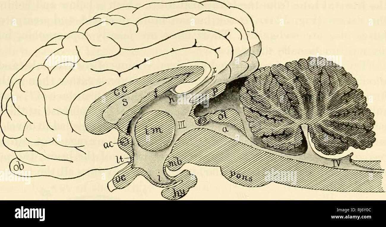 . The chordates. Chordata. Mammalia: Nervous System 705. Fig. 522. Sagittal section of the brain of a calf, (a) Aqueduct; (ac) anterior commissure; (cc) corpus callosum; (/) fornix; (h) habenula; (hy) hypophysis; (i) infundibulum; {im) intermediate mass ("soft commissure"); (It) lamina terminalis; (mb) mammillary body; (ob) olfactory bulb; (oc) optic chiasma; (ol) optic lobes; (p) pinealis; (r) recessus suprapinealis; (s) septum pellucidurn; (III, IV) third and fourth ventricles. (After Butschli. Courtesy, Kingsley: "Comparative Anatomy of Vertebrates," Philadelphia, The Bl Stock Photohttps://www.alamy.com/image-license-details/?v=1https://www.alamy.com/the-chordates-chordata-mammalia-nervous-system-705-fig-522-sagittal-section-of-the-brain-of-a-calf-a-aqueduct-ac-anterior-commissure-cc-corpus-callosum-fornix-h-habenula-hy-hypophysis-i-infundibulum-im-intermediate-mass-quotsoft-commissurequot-it-lamina-terminalis-mb-mammillary-body-ob-olfactory-bulb-oc-optic-chiasma-ol-optic-lobes-p-pinealis-r-recessus-suprapinealis-s-septum-pellucidurn-iii-iv-third-and-fourth-ventricles-after-butschli-courtesy-kingsley-quotcomparative-anatomy-of-vertebratesquot-philadelphia-the-bl-image234951484.html
RMRJ6Y0C–. The chordates. Chordata. Mammalia: Nervous System 705. Fig. 522. Sagittal section of the brain of a calf, (a) Aqueduct; (ac) anterior commissure; (cc) corpus callosum; (/) fornix; (h) habenula; (hy) hypophysis; (i) infundibulum; {im) intermediate mass ("soft commissure"); (It) lamina terminalis; (mb) mammillary body; (ob) olfactory bulb; (oc) optic chiasma; (ol) optic lobes; (p) pinealis; (r) recessus suprapinealis; (s) septum pellucidurn; (III, IV) third and fourth ventricles. (After Butschli. Courtesy, Kingsley: "Comparative Anatomy of Vertebrates," Philadelphia, The Bl
 . The elements of embryology . Embryology. 382 DEVELOPMENT OF OEGANS IN MAMMALIA. [CHAP. By the fusion of the inner walls of the hemispheres in front of the lamina terminalis a solid septum, is formed, continuous behind with the lamina terminalis,. Teansveese Section theouqh the Brain of a Sheep's Bmbeto of 2-7 CM. IN LENGTH. (From Kolliker.) The section is taken a sliort distance behind the section represented in Fig. 124, and passes through the posterior part of the hemispheres and the third ventricle. St. corpus striatum; th. optic thalamus ; to. optic tract; t. third ventricle; d. roof of Stock Photohttps://www.alamy.com/image-license-details/?v=1https://www.alamy.com/the-elements-of-embryology-embryology-382-development-of-oegans-in-mammalia-chap-by-the-fusion-of-the-inner-walls-of-the-hemispheres-in-front-of-the-lamina-terminalis-a-solid-septum-is-formed-continuous-behind-with-the-lamina-terminalis-teansveese-section-theouqh-the-brain-of-a-sheeps-bmbeto-of-2-7-cm-in-length-from-kolliker-the-section-is-taken-a-sliort-distance-behind-the-section-represented-in-fig-124-and-passes-through-the-posterior-part-of-the-hemispheres-and-the-third-ventricle-st-corpus-striatum-th-optic-thalamus-to-optic-tract-t-third-ventricle-d-roof-of-image216443863.html
RMPG3T9Y–. The elements of embryology . Embryology. 382 DEVELOPMENT OF OEGANS IN MAMMALIA. [CHAP. By the fusion of the inner walls of the hemispheres in front of the lamina terminalis a solid septum, is formed, continuous behind with the lamina terminalis,. Teansveese Section theouqh the Brain of a Sheep's Bmbeto of 2-7 CM. IN LENGTH. (From Kolliker.) The section is taken a sliort distance behind the section represented in Fig. 124, and passes through the posterior part of the hemispheres and the third ventricle. St. corpus striatum; th. optic thalamus ; to. optic tract; t. third ventricle; d. roof of
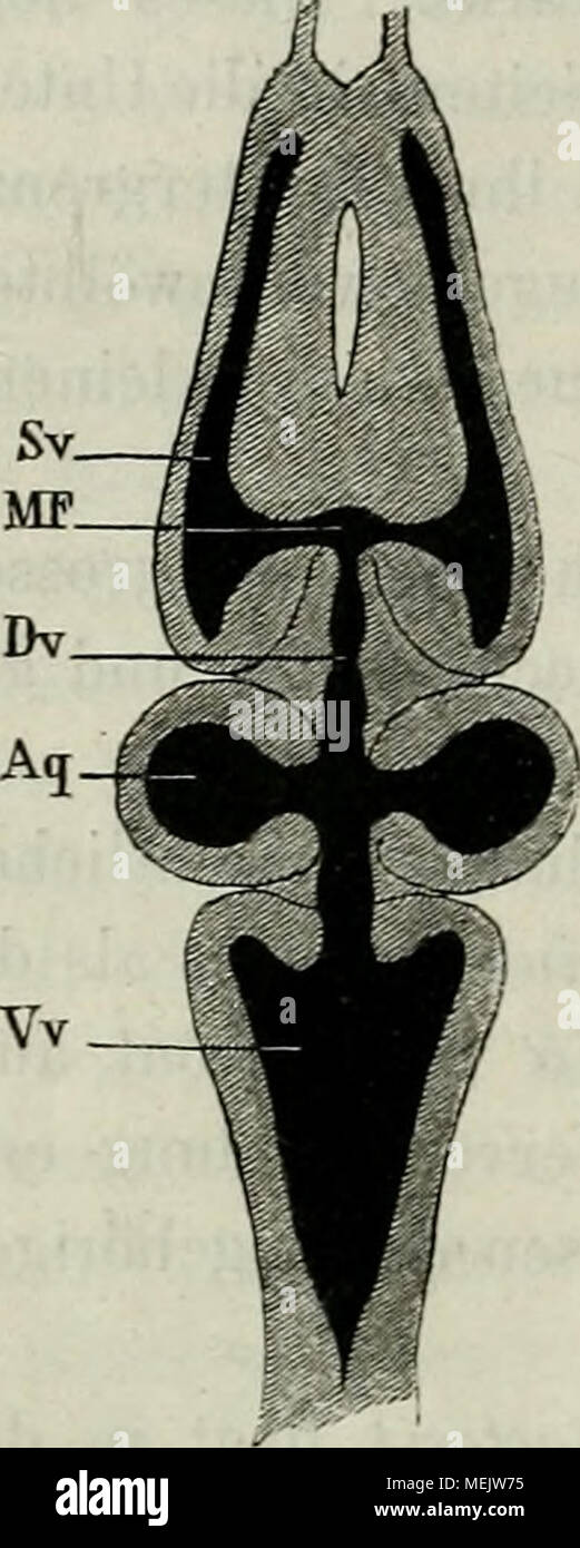 . Die Anatomie des Frosches : ein Handbuch, . Horizontaldurchschnitt des Gehirnö. Vv Vierter Ventrikel. A q Ventrikel des Mittelhirns und Aquaed. Sylvii. D V Dritter Ventrikel. MF Foramen Monroi. Sv Seiteuventrikel. opticorum., eine keilförmige Figur erscheint, die sich durch ein anderes Colorit von der übrigen Masse abhebt. Zu beiden Seiten der Lamina terminalis springen die Hemisphären ventralwärts weit her- vor (Fig. 3, Hc) und heben sich von den Lobi ölfadorii durch eine viel tiefere Furche ab, als dies auf ihrer oberen Fläche der Fall ist. Was die Höhlen des Vorderhirns anbelangt, so sind Stock Photohttps://www.alamy.com/image-license-details/?v=1https://www.alamy.com/die-anatomie-des-frosches-ein-handbuch-horizontaldurchschnitt-des-gehirn-vv-vierter-ventrikel-a-q-ventrikel-des-mittelhirns-und-aquaed-sylvii-d-v-dritter-ventrikel-mf-foramen-monroi-sv-seiteuventrikel-opticorum-eine-keilfrmige-figur-erscheint-die-sich-durch-ein-anderes-colorit-von-der-brigen-masse-abhebt-zu-beiden-seiten-der-lamina-terminalis-springen-die-hemisphren-ventralwrts-weit-her-vor-fig-3-hc-und-heben-sich-von-den-lobi-lfadorii-durch-eine-viel-tiefere-furche-ab-als-dies-auf-ihrer-oberen-flche-der-fall-ist-was-die-hhlen-des-vorderhirns-anbelangt-so-sind-image181123801.html
RMMEJW75–. Die Anatomie des Frosches : ein Handbuch, . Horizontaldurchschnitt des Gehirnö. Vv Vierter Ventrikel. A q Ventrikel des Mittelhirns und Aquaed. Sylvii. D V Dritter Ventrikel. MF Foramen Monroi. Sv Seiteuventrikel. opticorum., eine keilförmige Figur erscheint, die sich durch ein anderes Colorit von der übrigen Masse abhebt. Zu beiden Seiten der Lamina terminalis springen die Hemisphären ventralwärts weit her- vor (Fig. 3, Hc) und heben sich von den Lobi ölfadorii durch eine viel tiefere Furche ab, als dies auf ihrer oberen Fläche der Fall ist. Was die Höhlen des Vorderhirns anbelangt, so sind
 Human brain anatomy for medical concept 3D illustration Stock Photohttps://www.alamy.com/image-license-details/?v=1https://www.alamy.com/human-brain-anatomy-for-medical-concept-3d-illustration-image504513129.html
RF2M8PFJ1–Human brain anatomy for medical concept 3D illustration
 The development of the human body; a manual of human embryology . nd in the other ventricles. Owing to the very considerable size reached by thethickening of the lamina terminalis whose history has justbeen described, important changes are wrought in theadjoining portions of the mesial surface of J dg the hemispheres. Be-fore the developmentof the thickening thegyrus dentatus andthe hippocampus ex-tend forward into theanterior portion ofthe hemispheres (Fig.228), but on accountof their position theybecome encroachedupon by the enlarge-ment of the laminaterminalis, with theresult that the Stock Photohttps://www.alamy.com/image-license-details/?v=1https://www.alamy.com/the-development-of-the-human-body-a-manual-of-human-embryology-nd-in-the-other-ventricles-owing-to-the-very-considerable-size-reached-by-thethickening-of-the-lamina-terminalis-whose-history-has-justbeen-described-important-changes-are-wrought-in-theadjoining-portions-of-the-mesial-surface-of-j-dg-the-hemispheres-be-fore-the-developmentof-the-thickening-thegyrus-dentatus-andthe-hippocampus-ex-tend-forward-into-theanterior-portion-ofthe-hemispheres-fig228-but-on-accountof-their-position-theybecome-encroachedupon-by-the-enlarge-ment-of-the-laminaterminalis-with-theresult-that-the-image340309185.html
RM2ANJBRD–The development of the human body; a manual of human embryology . nd in the other ventricles. Owing to the very considerable size reached by thethickening of the lamina terminalis whose history has justbeen described, important changes are wrought in theadjoining portions of the mesial surface of J dg the hemispheres. Be-fore the developmentof the thickening thegyrus dentatus andthe hippocampus ex-tend forward into theanterior portion ofthe hemispheres (Fig.228), but on accountof their position theybecome encroachedupon by the enlarge-ment of the laminaterminalis, with theresult that the
![. The development of the skull of Emys Lutaria. Chelonia (Genus); Reptiles. cptr. EXPLANATION OF ridt'ItlCS 30 Ventral view of the olfaetoi-y fajjsule of an embryo having a earapace length of 7 mm. showing the cartilago parase])talis (cp.) separated from the septum n:isi as far anterior as the lamina terminalis anterior (/./.</.) and consequently the foramen praepalatinum not enclosed posteriorly. X 20. 31 Posterior portion of the right otic capsule viewed from in front and slightly from the median line, showing especially the groove in wliich the n. glossopharyn- geus passes through the ca Stock Photo . The development of the skull of Emys Lutaria. Chelonia (Genus); Reptiles. cptr. EXPLANATION OF ridt'ItlCS 30 Ventral view of the olfaetoi-y fajjsule of an embryo having a earapace length of 7 mm. showing the cartilago parase])talis (cp.) separated from the septum n:isi as far anterior as the lamina terminalis anterior (/./.</.) and consequently the foramen praepalatinum not enclosed posteriorly. X 20. 31 Posterior portion of the right otic capsule viewed from in front and slightly from the median line, showing especially the groove in wliich the n. glossopharyn- geus passes through the ca Stock Photo](https://c8.alamy.com/comp/PF9M67/the-development-of-the-skull-of-emys-lutaria-chelonia-genus-reptiles-cptr-explanation-of-ridtitlcs-30-ventral-view-of-the-olfaetoi-y-fajjsule-of-an-embryo-having-a-earapace-length-of-7-mm-showing-the-cartilago-parase-talis-cp-separated-from-the-septum-nisi-as-far-anterior-as-the-lamina-terminalis-anterior-lt-and-consequently-the-foramen-praepalatinum-not-enclosed-posteriorly-x-20-31-posterior-portion-of-the-right-otic-capsule-viewed-from-in-front-and-slightly-from-the-median-line-showing-especially-the-groove-in-wliich-the-n-glossopharyn-geus-passes-through-the-ca-PF9M67.jpg) . The development of the skull of Emys Lutaria. Chelonia (Genus); Reptiles. cptr. EXPLANATION OF ridt'ItlCS 30 Ventral view of the olfaetoi-y fajjsule of an embryo having a earapace length of 7 mm. showing the cartilago parase])talis (cp.) separated from the septum n:isi as far anterior as the lamina terminalis anterior (/./.</.) and consequently the foramen praepalatinum not enclosed posteriorly. X 20. 31 Posterior portion of the right otic capsule viewed from in front and slightly from the median line, showing especially the groove in wliich the n. glossopharyn- geus passes through the ca Stock Photohttps://www.alamy.com/image-license-details/?v=1https://www.alamy.com/the-development-of-the-skull-of-emys-lutaria-chelonia-genus-reptiles-cptr-explanation-of-ridtitlcs-30-ventral-view-of-the-olfaetoi-y-fajjsule-of-an-embryo-having-a-earapace-length-of-7-mm-showing-the-cartilago-parase-talis-cp-separated-from-the-septum-nisi-as-far-anterior-as-the-lamina-terminalis-anterior-lt-and-consequently-the-foramen-praepalatinum-not-enclosed-posteriorly-x-20-31-posterior-portion-of-the-right-otic-capsule-viewed-from-in-front-and-slightly-from-the-median-line-showing-especially-the-groove-in-wliich-the-n-glossopharyn-geus-passes-through-the-ca-image215957679.html
. The development of the skull of Emys Lutaria. Chelonia (Genus); Reptiles. cptr. EXPLANATION OF ridt'ItlCS 30 Ventral view of the olfaetoi-y fajjsule of an embryo having a earapace length of 7 mm. showing the cartilago parase])talis (cp.) separated from the septum n:isi as far anterior as the lamina terminalis anterior (/./.</.) and consequently the foramen praepalatinum not enclosed posteriorly. X 20. 31 Posterior portion of the right otic capsule viewed from in front and slightly from the median line, showing especially the groove in wliich the n. glossopharyn- geus passes through the ca Stock Photohttps://www.alamy.com/image-license-details/?v=1https://www.alamy.com/the-development-of-the-skull-of-emys-lutaria-chelonia-genus-reptiles-cptr-explanation-of-ridtitlcs-30-ventral-view-of-the-olfaetoi-y-fajjsule-of-an-embryo-having-a-earapace-length-of-7-mm-showing-the-cartilago-parase-talis-cp-separated-from-the-septum-nisi-as-far-anterior-as-the-lamina-terminalis-anterior-lt-and-consequently-the-foramen-praepalatinum-not-enclosed-posteriorly-x-20-31-posterior-portion-of-the-right-otic-capsule-viewed-from-in-front-and-slightly-from-the-median-line-showing-especially-the-groove-in-wliich-the-n-glossopharyn-geus-passes-through-the-ca-image215957679.htmlRMPF9M67–. The development of the skull of Emys Lutaria. Chelonia (Genus); Reptiles. cptr. EXPLANATION OF ridt'ItlCS 30 Ventral view of the olfaetoi-y fajjsule of an embryo having a earapace length of 7 mm. showing the cartilago parase])talis (cp.) separated from the septum n:isi as far anterior as the lamina terminalis anterior (/./.</.) and consequently the foramen praepalatinum not enclosed posteriorly. X 20. 31 Posterior portion of the right otic capsule viewed from in front and slightly from the median line, showing especially the groove in wliich the n. glossopharyn- geus passes through the ca
![. Die Anatomie des Frosches : ein Handbuch, . Gehirn von Bana esculenta von unten. Mo Med. oblongata. Hy Hypophysis. Tue Tub. cinereum. To Tractus opticus. Cho Chiasma n. opt. L t Lamina terminalis. Hc Grosshirnhemisphäre. L,ol Lobus olfactorius. L, 0 U Lobus olfactorius 2. I N. olfactorius Ite] /i N. olfactorius 2tej ""J^ze'- II Nervi optici. III N. oculomotorius. IV N. trochlearis. V, VII, VIII Quintus, Facialis und Acixsticus. VI N. abducens. IX, X, XI N. glossopharyngeus, N. vagus und N. accessorius. 1) Durchfärbungen des ganzen Präparates mit Beale'schem Carmin, welches die Fase Stock Photo . Die Anatomie des Frosches : ein Handbuch, . Gehirn von Bana esculenta von unten. Mo Med. oblongata. Hy Hypophysis. Tue Tub. cinereum. To Tractus opticus. Cho Chiasma n. opt. L t Lamina terminalis. Hc Grosshirnhemisphäre. L,ol Lobus olfactorius. L, 0 U Lobus olfactorius 2. I N. olfactorius Ite] /i N. olfactorius 2tej ""J^ze'- II Nervi optici. III N. oculomotorius. IV N. trochlearis. V, VII, VIII Quintus, Facialis und Acixsticus. VI N. abducens. IX, X, XI N. glossopharyngeus, N. vagus und N. accessorius. 1) Durchfärbungen des ganzen Präparates mit Beale'schem Carmin, welches die Fase Stock Photo](https://c8.alamy.com/comp/MEJW7C/die-anatomie-des-frosches-ein-handbuch-gehirn-von-bana-esculenta-von-unten-mo-med-oblongata-hy-hypophysis-tue-tub-cinereum-to-tractus-opticus-cho-chiasma-n-opt-l-t-lamina-terminalis-hc-grosshirnhemisphre-lol-lobus-olfactorius-l-0-u-lobus-olfactorius-2-i-n-olfactorius-ite-i-n-olfactorius-2tej-quotquotjze-ii-nervi-optici-iii-n-oculomotorius-iv-n-trochlearis-v-vii-viii-quintus-facialis-und-acixsticus-vi-n-abducens-ix-x-xi-n-glossopharyngeus-n-vagus-und-n-accessorius-1-durchfrbungen-des-ganzen-prparates-mit-bealeschem-carmin-welches-die-fase-MEJW7C.jpg) . Die Anatomie des Frosches : ein Handbuch, . Gehirn von Bana esculenta von unten. Mo Med. oblongata. Hy Hypophysis. Tue Tub. cinereum. To Tractus opticus. Cho Chiasma n. opt. L t Lamina terminalis. Hc Grosshirnhemisphäre. L,ol Lobus olfactorius. L, 0 U Lobus olfactorius 2. I N. olfactorius Ite] /i N. olfactorius 2tej ""J^ze'- II Nervi optici. III N. oculomotorius. IV N. trochlearis. V, VII, VIII Quintus, Facialis und Acixsticus. VI N. abducens. IX, X, XI N. glossopharyngeus, N. vagus und N. accessorius. 1) Durchfärbungen des ganzen Präparates mit Beale'schem Carmin, welches die Fase Stock Photohttps://www.alamy.com/image-license-details/?v=1https://www.alamy.com/die-anatomie-des-frosches-ein-handbuch-gehirn-von-bana-esculenta-von-unten-mo-med-oblongata-hy-hypophysis-tue-tub-cinereum-to-tractus-opticus-cho-chiasma-n-opt-l-t-lamina-terminalis-hc-grosshirnhemisphre-lol-lobus-olfactorius-l-0-u-lobus-olfactorius-2-i-n-olfactorius-ite-i-n-olfactorius-2tej-quotquotjze-ii-nervi-optici-iii-n-oculomotorius-iv-n-trochlearis-v-vii-viii-quintus-facialis-und-acixsticus-vi-n-abducens-ix-x-xi-n-glossopharyngeus-n-vagus-und-n-accessorius-1-durchfrbungen-des-ganzen-prparates-mit-bealeschem-carmin-welches-die-fase-image181123808.html
. Die Anatomie des Frosches : ein Handbuch, . Gehirn von Bana esculenta von unten. Mo Med. oblongata. Hy Hypophysis. Tue Tub. cinereum. To Tractus opticus. Cho Chiasma n. opt. L t Lamina terminalis. Hc Grosshirnhemisphäre. L,ol Lobus olfactorius. L, 0 U Lobus olfactorius 2. I N. olfactorius Ite] /i N. olfactorius 2tej ""J^ze'- II Nervi optici. III N. oculomotorius. IV N. trochlearis. V, VII, VIII Quintus, Facialis und Acixsticus. VI N. abducens. IX, X, XI N. glossopharyngeus, N. vagus und N. accessorius. 1) Durchfärbungen des ganzen Präparates mit Beale'schem Carmin, welches die Fase Stock Photohttps://www.alamy.com/image-license-details/?v=1https://www.alamy.com/die-anatomie-des-frosches-ein-handbuch-gehirn-von-bana-esculenta-von-unten-mo-med-oblongata-hy-hypophysis-tue-tub-cinereum-to-tractus-opticus-cho-chiasma-n-opt-l-t-lamina-terminalis-hc-grosshirnhemisphre-lol-lobus-olfactorius-l-0-u-lobus-olfactorius-2-i-n-olfactorius-ite-i-n-olfactorius-2tej-quotquotjze-ii-nervi-optici-iii-n-oculomotorius-iv-n-trochlearis-v-vii-viii-quintus-facialis-und-acixsticus-vi-n-abducens-ix-x-xi-n-glossopharyngeus-n-vagus-und-n-accessorius-1-durchfrbungen-des-ganzen-prparates-mit-bealeschem-carmin-welches-die-fase-image181123808.htmlRMMEJW7C–. Die Anatomie des Frosches : ein Handbuch, . Gehirn von Bana esculenta von unten. Mo Med. oblongata. Hy Hypophysis. Tue Tub. cinereum. To Tractus opticus. Cho Chiasma n. opt. L t Lamina terminalis. Hc Grosshirnhemisphäre. L,ol Lobus olfactorius. L, 0 U Lobus olfactorius 2. I N. olfactorius Ite] /i N. olfactorius 2tej ""J^ze'- II Nervi optici. III N. oculomotorius. IV N. trochlearis. V, VII, VIII Quintus, Facialis und Acixsticus. VI N. abducens. IX, X, XI N. glossopharyngeus, N. vagus und N. accessorius. 1) Durchfärbungen des ganzen Präparates mit Beale'schem Carmin, welches die Fase
 Human brain anatomy for medical concept 3D illustration Stock Photohttps://www.alamy.com/image-license-details/?v=1https://www.alamy.com/human-brain-anatomy-for-medical-concept-3d-illustration-image504513128.html
RF2M8PFJ0–Human brain anatomy for medical concept 3D illustration
 A treatise on zoology . he Teleosts, filling almostcompletely the cavity of the mid-brain (Fig. 352). Large pairedhollow optic lobes are conspicuous, except in the Chondrostei, theirroof (tectum opticum) covering the valvula. The diencephalonbecomes shortened and partially hidden above; below there is-a large infundibular outgrowth, with very well developed lobiitiferiores and saccus vasculosus (Figs. 282-3, 353). The fore-brainis remarkably undeveloped ; no cerebral hemispheres are formed,the lamina terminalis becomes almost horizontal, the basal ganglia(corpus striatum and epistriatum) are t Stock Photohttps://www.alamy.com/image-license-details/?v=1https://www.alamy.com/a-treatise-on-zoology-he-teleosts-filling-almostcompletely-the-cavity-of-the-mid-brain-fig-352-large-pairedhollow-optic-lobes-are-conspicuous-except-in-the-chondrostei-theirroof-tectum-opticum-covering-the-valvula-the-diencephalonbecomes-shortened-and-partially-hidden-above-below-there-is-a-large-infundibular-outgrowth-with-very-well-developed-lobiitiferiores-and-saccus-vasculosus-figs-282-3-353-the-fore-brainis-remarkably-undeveloped-no-cerebral-hemispheres-are-formedthe-lamina-terminalis-becomes-almost-horizontal-the-basal-gangliacorpus-striatum-and-epistriatum-are-t-image338286696.html
RM2AJA83M–A treatise on zoology . he Teleosts, filling almostcompletely the cavity of the mid-brain (Fig. 352). Large pairedhollow optic lobes are conspicuous, except in the Chondrostei, theirroof (tectum opticum) covering the valvula. The diencephalonbecomes shortened and partially hidden above; below there is-a large infundibular outgrowth, with very well developed lobiitiferiores and saccus vasculosus (Figs. 282-3, 353). The fore-brainis remarkably undeveloped ; no cerebral hemispheres are formed,the lamina terminalis becomes almost horizontal, the basal ganglia(corpus striatum and epistriatum) are t
 . Denkschriften der Medicinisch-Naturwissenschaftlichen Gesellschaft zu Jena. t;78 Zur Entwicklungsgeschichte und vergleichenden Morphologie des Schädels von Echidna aculeata var. typica. g§ erfolgt. An die ganze Höhe der Caudalwand stösst von vorn das Septum nasi an, ausserdem setzt sich an ihren unteren Rand jederseits vom Septum eine knorpelige Lamina trans versalis posterior an, die in die Lamina terminalis eingelagert ist. Der mediale Rand der Lam. transv. post. ist schon jetzt in Fig. 15.. Fen, cribrosa Lam. transv. post Fig. 17. Meat. aud. ext Anlage des Manubi Tympanicum Cart. Meckel. Stock Photohttps://www.alamy.com/image-license-details/?v=1https://www.alamy.com/denkschriften-der-medicinisch-naturwissenschaftlichen-gesellschaft-zu-jena-t78-zur-entwicklungsgeschichte-und-vergleichenden-morphologie-des-schdels-von-echidna-aculeata-var-typica-g-erfolgt-an-die-ganze-hhe-der-caudalwand-stsst-von-vorn-das-septum-nasi-an-ausserdem-setzt-sich-an-ihren-unteren-rand-jederseits-vom-septum-eine-knorpelige-lamina-trans-versalis-posterior-an-die-in-die-lamina-terminalis-eingelagert-ist-der-mediale-rand-der-lam-transv-post-ist-schon-jetzt-in-fig-15-fen-cribrosa-lam-transv-post-fig-17-meat-aud-ext-anlage-des-manubi-tympanicum-cart-meckel-image216053449.html
RMPFE2AH–. Denkschriften der Medicinisch-Naturwissenschaftlichen Gesellschaft zu Jena. t;78 Zur Entwicklungsgeschichte und vergleichenden Morphologie des Schädels von Echidna aculeata var. typica. g§ erfolgt. An die ganze Höhe der Caudalwand stösst von vorn das Septum nasi an, ausserdem setzt sich an ihren unteren Rand jederseits vom Septum eine knorpelige Lamina trans versalis posterior an, die in die Lamina terminalis eingelagert ist. Der mediale Rand der Lam. transv. post. ist schon jetzt in Fig. 15.. Fen, cribrosa Lam. transv. post Fig. 17. Meat. aud. ext Anlage des Manubi Tympanicum Cart. Meckel.
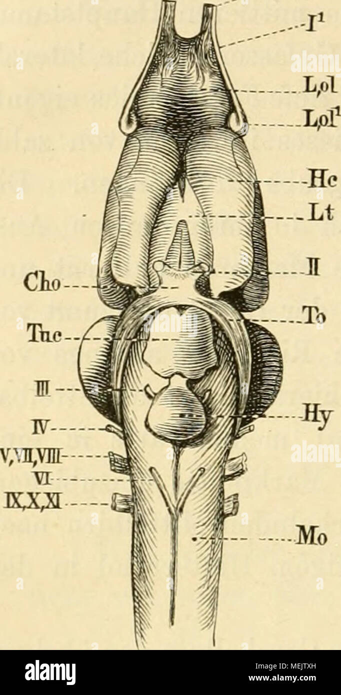 . Die anatomie des frosches. Ein handbuch für physiologen, ärzte und studirende . Gehirn von Rana esculenta von unten Mo Med. oblongata. Hy Hypophysis. Tue Tub. cinereum. To Tractus opticus. Cho Chiasma n. opt. L t Lamina terminalis. Hc Grosshirnhemisphäre. L,ol Lobus olfactorius. L, o U Lobus olfactorius 2. I N. olfactorius lte II N. olfactorius 2teJ II Nervi optici. III N. oculomotorius. IV N. trochlearis. V, YII, Till Quintus, Facialis und Acusticus. VI N. abducens. IX, X, XI KT. glossopharyngeus, N. vagus und N. accessorius. Wurzel. x) Durchfärbungen des ganzen Präparates mit Beale'sche Stock Photohttps://www.alamy.com/image-license-details/?v=1https://www.alamy.com/die-anatomie-des-frosches-ein-handbuch-fr-physiologen-rzte-und-studirende-gehirn-von-rana-esculenta-von-unten-mo-med-oblongata-hy-hypophysis-tue-tub-cinereum-to-tractus-opticus-cho-chiasma-n-opt-l-t-lamina-terminalis-hc-grosshirnhemisphre-lol-lobus-olfactorius-l-o-u-lobus-olfactorius-2-i-n-olfactorius-lte-ii-n-olfactorius-2tej-ii-nervi-optici-iii-n-oculomotorius-iv-n-trochlearis-v-yii-till-quintus-facialis-und-acusticus-vi-n-abducens-ix-x-xi-kt-glossopharyngeus-n-vagus-und-n-accessorius-wurzel-x-durchfrbungen-des-ganzen-prparates-mit-bealesche-image181123561.html
RMMEJTXH–. Die anatomie des frosches. Ein handbuch für physiologen, ärzte und studirende . Gehirn von Rana esculenta von unten Mo Med. oblongata. Hy Hypophysis. Tue Tub. cinereum. To Tractus opticus. Cho Chiasma n. opt. L t Lamina terminalis. Hc Grosshirnhemisphäre. L,ol Lobus olfactorius. L, o U Lobus olfactorius 2. I N. olfactorius lte II N. olfactorius 2teJ II Nervi optici. III N. oculomotorius. IV N. trochlearis. V, YII, Till Quintus, Facialis und Acusticus. VI N. abducens. IX, X, XI KT. glossopharyngeus, N. vagus und N. accessorius. Wurzel. x) Durchfärbungen des ganzen Präparates mit Beale'sche
 Human brain anatomy for medical concept 3D illustration Stock Photohttps://www.alamy.com/image-license-details/?v=1https://www.alamy.com/human-brain-anatomy-for-medical-concept-3d-illustration-image504513122.html
RF2M8PFHP–Human brain anatomy for medical concept 3D illustration
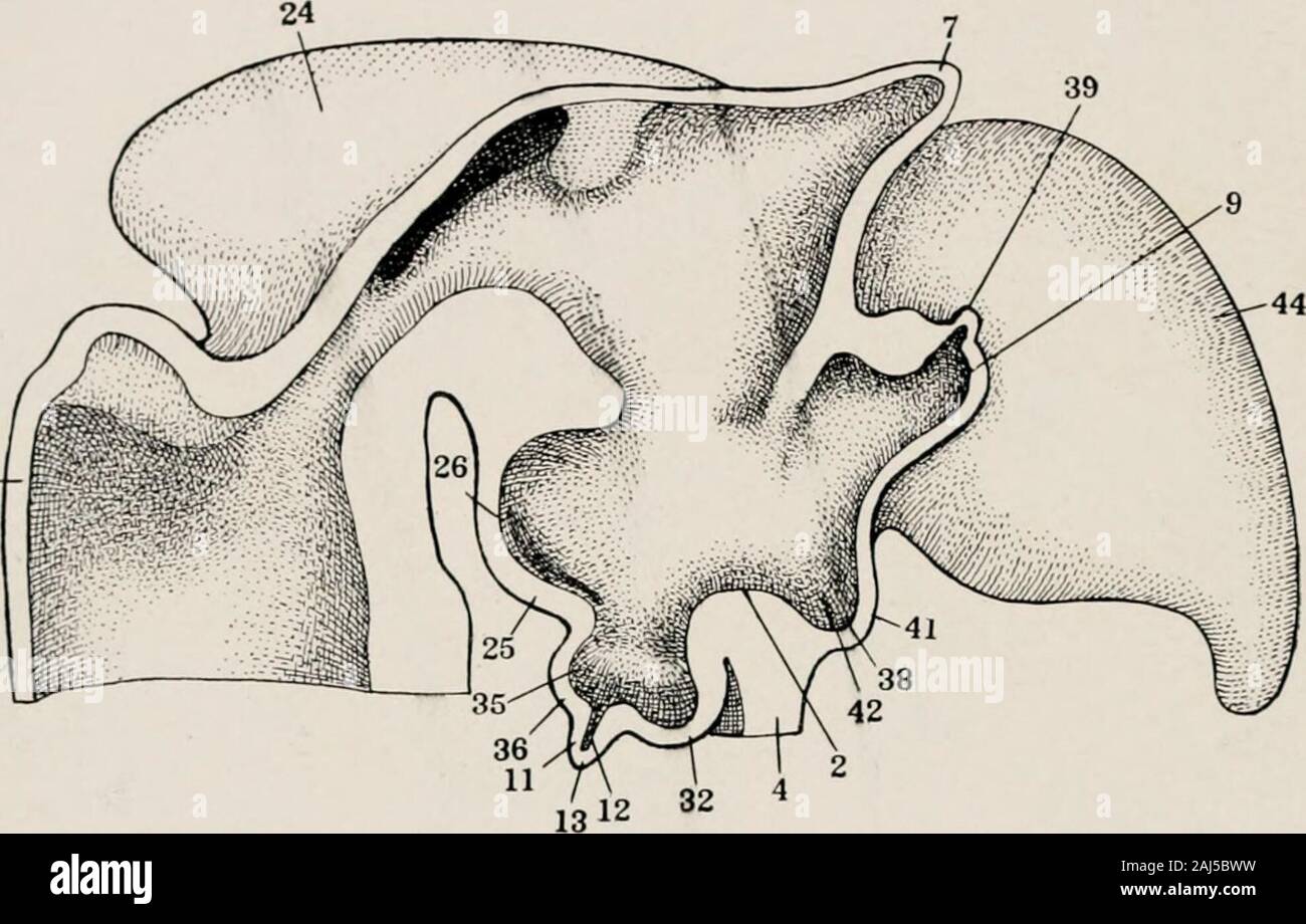 The morphology and evolutional significance of the pineal body : being part I of a contribution to the study of the epiphysis cerebri with an interpretation of the morphological, physiological and clinical evidence . Fig. 36 Mesial view of forebrain reconstruction of chick of 5 days and 20hours. X 100. The unshaded area shows the cut surfaces of the reconstruction,according to Tilney, 1915. 2, chiasmatic process; 4, chiasm; 7, epiphysis; 13, infundibular process; 20,lamina terminalis; 25, mammillary region; 32, post-chiasmatic eminence; 33, post-chiasmatic recess; 36, post-infundibular eminenc Stock Photohttps://www.alamy.com/image-license-details/?v=1https://www.alamy.com/the-morphology-and-evolutional-significance-of-the-pineal-body-being-part-i-of-a-contribution-to-the-study-of-the-epiphysis-cerebri-with-an-interpretation-of-the-morphological-physiological-and-clinical-evidence-fig-36-mesial-view-of-forebrain-reconstruction-of-chick-of-5-days-and-20hours-x-100-the-unshaded-area-shows-the-cut-surfaces-of-the-reconstructionaccording-to-tilney-1915-2-chiasmatic-process-4-chiasm-7-epiphysis-13-infundibular-process-20lamina-terminalis-25-mammillary-region-32-post-chiasmatic-eminence-33-post-chiasmatic-recess-36-post-infundibular-eminenc-image338179909.html
RM2AJ5BWW–The morphology and evolutional significance of the pineal body : being part I of a contribution to the study of the epiphysis cerebri with an interpretation of the morphological, physiological and clinical evidence . Fig. 36 Mesial view of forebrain reconstruction of chick of 5 days and 20hours. X 100. The unshaded area shows the cut surfaces of the reconstruction,according to Tilney, 1915. 2, chiasmatic process; 4, chiasm; 7, epiphysis; 13, infundibular process; 20,lamina terminalis; 25, mammillary region; 32, post-chiasmatic eminence; 33, post-chiasmatic recess; 36, post-infundibular eminenc
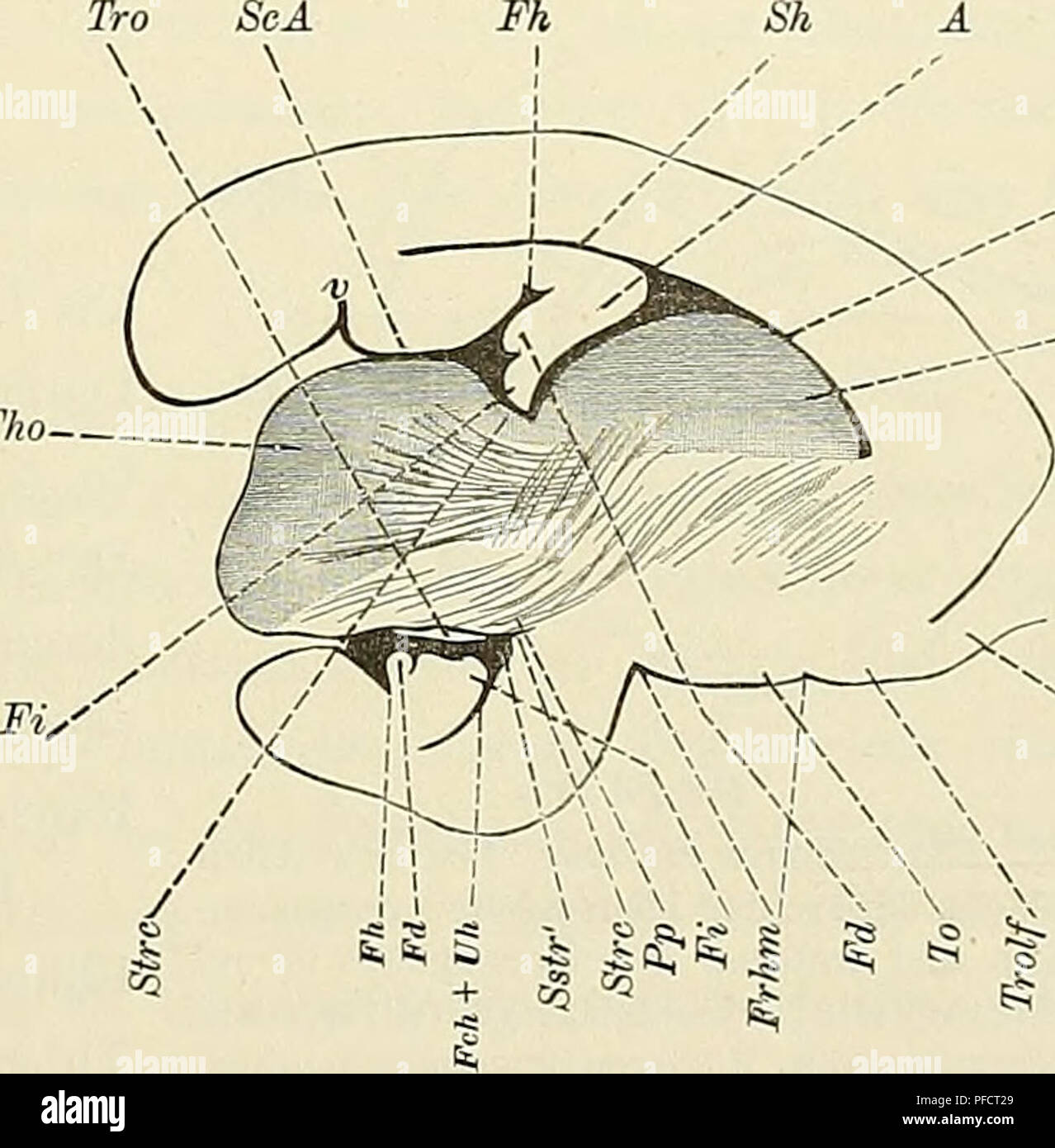 . Denkschriften der Medicinisch-Naturwissenschaftlichen Gesellschaft zu Jena. 101 Das Centralnervensystem der Monotremen und Marsupialier. IOI (i) Commissuren. Die Commissura anterior ist auf dem medianen Querschnitt fast rund. Der längere Durch- messer misst nämlich reichlich 43/4, der kürzere fast 4»/2 mm. Der absolute Flächeninhalt des Querschnitts beträgt sonach 18 qmm, der relative in dem früher definirten Sinn 1/S90. Letzterer stimmt also mit dem- jenigen von Macropus überein. Die Lamina terminalis inserirt sich an der Basalfläche der vorderen Com- missur. Die Commissura superior hat etw Stock Photohttps://www.alamy.com/image-license-details/?v=1https://www.alamy.com/denkschriften-der-medicinisch-naturwissenschaftlichen-gesellschaft-zu-jena-101-das-centralnervensystem-der-monotremen-und-marsupialier-ioi-i-commissuren-die-commissura-anterior-ist-auf-dem-medianen-querschnitt-fast-rund-der-lngere-durch-messer-misst-nmlich-reichlich-434-der-krzere-fast-42-mm-der-absolute-flcheninhalt-des-querschnitts-betrgt-sonach-18-qmm-der-relative-in-dem-frher-definirten-sinn-1s90-letzterer-stimmt-also-mit-dem-jenigen-von-macropus-berein-die-lamina-terminalis-inserirt-sich-an-der-basalflche-der-vorderen-com-missur-die-commissura-superior-hat-etw-image216026561.html
RMPFCT29–. Denkschriften der Medicinisch-Naturwissenschaftlichen Gesellschaft zu Jena. 101 Das Centralnervensystem der Monotremen und Marsupialier. IOI (i) Commissuren. Die Commissura anterior ist auf dem medianen Querschnitt fast rund. Der längere Durch- messer misst nämlich reichlich 43/4, der kürzere fast 4»/2 mm. Der absolute Flächeninhalt des Querschnitts beträgt sonach 18 qmm, der relative in dem früher definirten Sinn 1/S90. Letzterer stimmt also mit dem- jenigen von Macropus überein. Die Lamina terminalis inserirt sich an der Basalfläche der vorderen Com- missur. Die Commissura superior hat etw
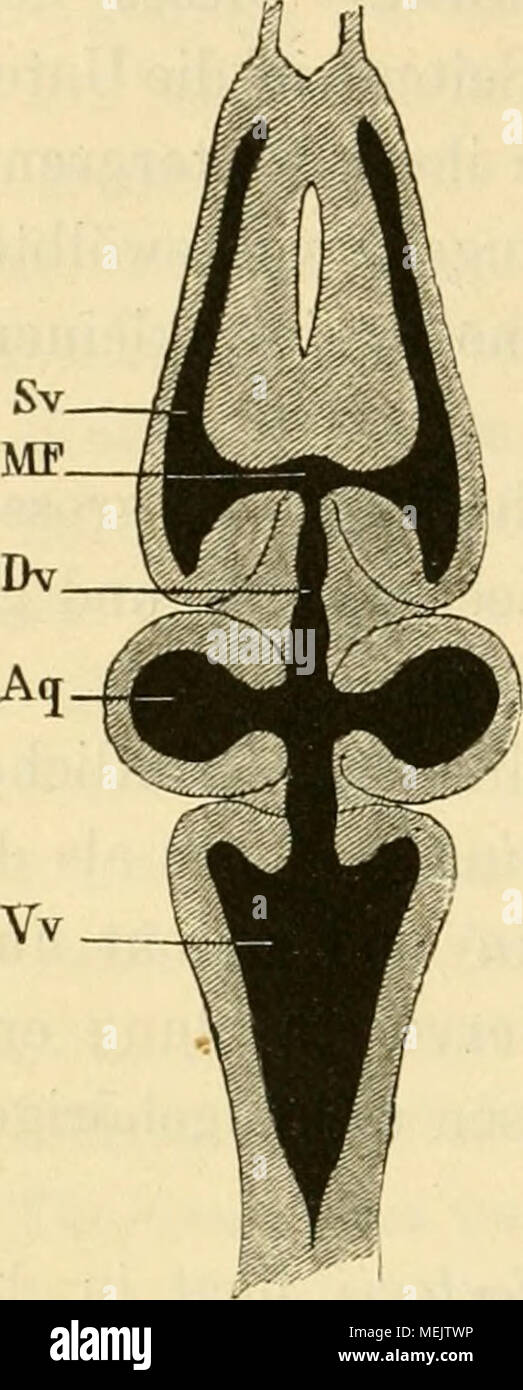 . Die anatomie des frosches. Ein handbuch für physiologen, ärzte und studirende . Horizontaldurchschnitt des Gehirns. Vv Vierter Ventrikel. A q Ventrikel des Mittelhirns und Aquaed. Sylvii. D v Dritter Ventrikel. MF Foramen Monroi. S» Seitenventrikel. opticorum, eine keilförmige Figur erscheint, die sich durch ein anderes Colorit von der übrigen Masse abhebt. Zu beiden Seiten der Lamina terminalis springen die Hemisphären ventralwärts weit her- vor (Fig. 3, Hc) und heben sich von den Lobi ölfactorii durch eine viel tiefere Furche ab, als dies auf ihrer oberen Fläche der Fall ist. Was die Höh Stock Photohttps://www.alamy.com/image-license-details/?v=1https://www.alamy.com/die-anatomie-des-frosches-ein-handbuch-fr-physiologen-rzte-und-studirende-horizontaldurchschnitt-des-gehirns-vv-vierter-ventrikel-a-q-ventrikel-des-mittelhirns-und-aquaed-sylvii-d-v-dritter-ventrikel-mf-foramen-monroi-s-seitenventrikel-opticorum-eine-keilfrmige-figur-erscheint-die-sich-durch-ein-anderes-colorit-von-der-brigen-masse-abhebt-zu-beiden-seiten-der-lamina-terminalis-springen-die-hemisphren-ventralwrts-weit-her-vor-fig-3-hc-und-heben-sich-von-den-lobi-lfactorii-durch-eine-viel-tiefere-furche-ab-als-dies-auf-ihrer-oberen-flche-der-fall-ist-was-die-hh-image181123538.html
RMMEJTWP–. Die anatomie des frosches. Ein handbuch für physiologen, ärzte und studirende . Horizontaldurchschnitt des Gehirns. Vv Vierter Ventrikel. A q Ventrikel des Mittelhirns und Aquaed. Sylvii. D v Dritter Ventrikel. MF Foramen Monroi. S» Seitenventrikel. opticorum, eine keilförmige Figur erscheint, die sich durch ein anderes Colorit von der übrigen Masse abhebt. Zu beiden Seiten der Lamina terminalis springen die Hemisphären ventralwärts weit her- vor (Fig. 3, Hc) und heben sich von den Lobi ölfactorii durch eine viel tiefere Furche ab, als dies auf ihrer oberen Fläche der Fall ist. Was die Höh
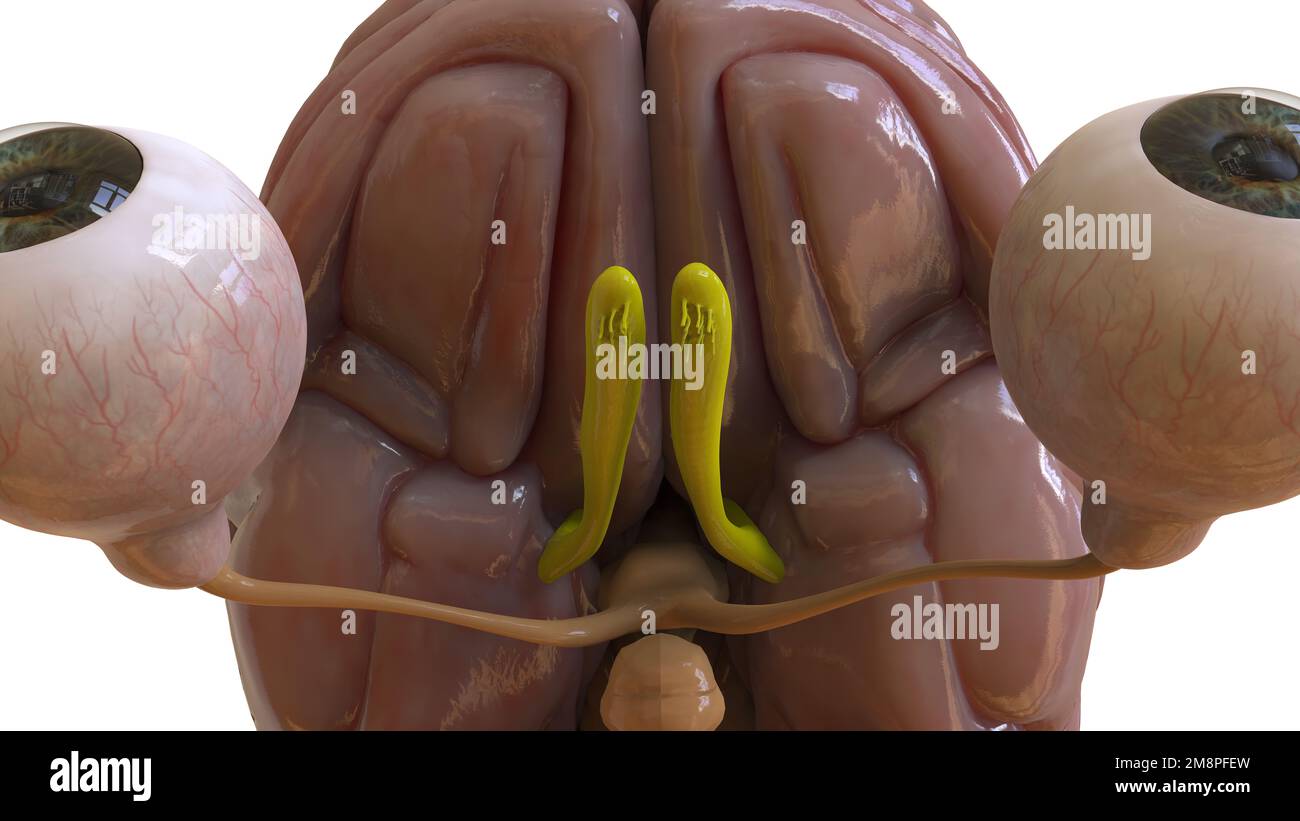 Human brain anatomy for medical concept 3D illustration Stock Photohttps://www.alamy.com/image-license-details/?v=1https://www.alamy.com/human-brain-anatomy-for-medical-concept-3d-illustration-image504513041.html
RF2M8PFEW–Human brain anatomy for medical concept 3D illustration
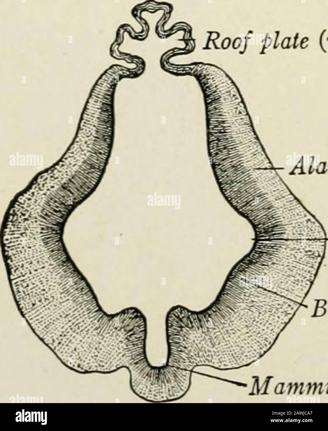 The anatomy of the nervous system, from the standpoint of development and function . Isthmus- Cerebellum MetcnccphalonRhomboid fossaMyclcncephalon . tic Hypo-/chiasma physis Medulla Lamina terminalis / ^Hyp^ihaiamus oblon^ataRhincnccphalon - Spinal cordCentral canal Fig. 17.—The brain of a fetus of the third month in median sagittal section. (His, Sobotta.) of 13.6 mm., are well defined by the third month (Fig. 17). In transversesections this division of the embryonic brain is seen to be composed of a pair ofplates on either side, which with a roof and floor form the walls of the ventricle. Ro Stock Photohttps://www.alamy.com/image-license-details/?v=1https://www.alamy.com/the-anatomy-of-the-nervous-system-from-the-standpoint-of-development-and-function-isthmus-cerebellum-metcnccphalonrhomboid-fossamyclcncephalon-tic-hypo-chiasma-physis-medulla-lamina-terminalis-hypihaiamus-oblonatarhincnccphalon-spinal-cordcentral-canal-fig-17the-brain-of-a-fetus-of-the-third-month-in-median-sagittal-section-his-sobotta-of-136-mm-are-well-defined-by-the-third-month-fig-17-in-transversesections-this-division-of-the-embryonic-brain-is-seen-to-be-composed-of-a-pair-ofplates-on-either-side-which-with-a-roof-and-floor-form-the-walls-of-the-ventricle-ro-image342768223.html
RM2AWJCA7–The anatomy of the nervous system, from the standpoint of development and function . Isthmus- Cerebellum MetcnccphalonRhomboid fossaMyclcncephalon . tic Hypo-/chiasma physis Medulla Lamina terminalis / ^Hyp^ihaiamus oblon^ataRhincnccphalon - Spinal cordCentral canal Fig. 17.—The brain of a fetus of the third month in median sagittal section. (His, Sobotta.) of 13.6 mm., are well defined by the third month (Fig. 17). In transversesections this division of the embryonic brain is seen to be composed of a pair ofplates on either side, which with a roof and floor form the walls of the ventricle. Ro
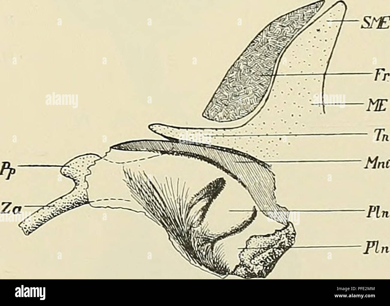 . Denkschriften der Medicinisch-Naturwissenschaftlichen Gesellschaft zu Jena. PI P". Plnl Plnl Fig. 7. Fig. 8. Fig. 7. Lateralansicht des Nasenknorpels (I. Stad.). Caudo- und oroventrales Ende fehlt. Knorpel gekörnt, "/lo "^t- ^'^â Zeichnung. Cenp Cartilago canalis nasopalatini, Cps Cartilago paraseptalis, Fb Fenestra basalis, Fji Fenestra narina, Lia Lamina terminalis anterior, PI Processus lateralis, Pn Paries nasi, Pr (fälschlich statt Pp) Processus parietalis, Sn Septum nasi, SME Spina raesethmoidalis, Tn Tectum nasi, V Vomer, Za Zona anularis. Fig. 8. Medialansicht der re Stock Photohttps://www.alamy.com/image-license-details/?v=1https://www.alamy.com/denkschriften-der-medicinisch-naturwissenschaftlichen-gesellschaft-zu-jena-pi-pquot-plnl-plnl-fig-7-fig-8-fig-7-lateralansicht-des-nasenknorpels-i-stad-caudo-und-oroventrales-ende-fehlt-knorpel-gekrnt-quotlo-quott-zeichnung-cenp-cartilago-canalis-nasopalatini-cps-cartilago-paraseptalis-fb-fenestra-basalis-fji-fenestra-narina-lia-lamina-terminalis-anterior-pi-processus-lateralis-pn-paries-nasi-pr-flschlich-statt-pp-processus-parietalis-sn-septum-nasi-sme-spina-raesethmoidalis-tn-tectum-nasi-v-vomer-za-zona-anularis-fig-8-medialansicht-der-re-image216053732.html
RMPFE2MM–. Denkschriften der Medicinisch-Naturwissenschaftlichen Gesellschaft zu Jena. PI P". Plnl Plnl Fig. 7. Fig. 8. Fig. 7. Lateralansicht des Nasenknorpels (I. Stad.). Caudo- und oroventrales Ende fehlt. Knorpel gekörnt, "/lo "^t- ^'^â Zeichnung. Cenp Cartilago canalis nasopalatini, Cps Cartilago paraseptalis, Fb Fenestra basalis, Fji Fenestra narina, Lia Lamina terminalis anterior, PI Processus lateralis, Pn Paries nasi, Pr (fälschlich statt Pp) Processus parietalis, Sn Septum nasi, SME Spina raesethmoidalis, Tn Tectum nasi, V Vomer, Za Zona anularis. Fig. 8. Medialansicht der re
 . Die Leitungsbahnen im Nervensystem der wirbellosen Tiere . Schematischer Längsschnitt des lobus opticus und des zusammengesetzten Auges der Stomatopoden und Decapoden. chiasma externa chiasma interna fibrae postretinales lamina ganglionaris medulla externa meduUa interna medulla terminalis ommatidium p. l. o. = pedunculus lobi optici r. = retinula eh. e. eh. i. p. r. i. g. m. e. m. i. m. t. o. Man wird bemerkt haben, daß bis jetzt die meisten Mitteilungen über die Hodologie der Crustaceen nebeneinander st nden, sodaß man ohne eigene Untersuchungen keine Kontrolle ihrer Zuverlässigkeit hat. B Stock Photohttps://www.alamy.com/image-license-details/?v=1https://www.alamy.com/die-leitungsbahnen-im-nervensystem-der-wirbellosen-tiere-schematischer-lngsschnitt-des-lobus-opticus-und-des-zusammengesetzten-auges-der-stomatopoden-und-decapoden-chiasma-externa-chiasma-interna-fibrae-postretinales-lamina-ganglionaris-medulla-externa-meduua-interna-medulla-terminalis-ommatidium-p-l-o-=-pedunculus-lobi-optici-r-=-retinula-eh-e-eh-i-p-r-i-g-m-e-m-i-m-t-o-man-wird-bemerkt-haben-da-bis-jetzt-die-meisten-mitteilungen-ber-die-hodologie-der-crustaceen-nebeneinander-st-nden-soda-man-ohne-eigene-untersuchungen-keine-kontrolle-ihrer-zuverlssigkeit-hat-b-image180885399.html
RMME814R–. Die Leitungsbahnen im Nervensystem der wirbellosen Tiere . Schematischer Längsschnitt des lobus opticus und des zusammengesetzten Auges der Stomatopoden und Decapoden. chiasma externa chiasma interna fibrae postretinales lamina ganglionaris medulla externa meduUa interna medulla terminalis ommatidium p. l. o. = pedunculus lobi optici r. = retinula eh. e. eh. i. p. r. i. g. m. e. m. i. m. t. o. Man wird bemerkt haben, daß bis jetzt die meisten Mitteilungen über die Hodologie der Crustaceen nebeneinander st nden, sodaß man ohne eigene Untersuchungen keine Kontrolle ihrer Zuverlässigkeit hat. B
 Human brain anatomy for medical concept 3D illustration Stock Photohttps://www.alamy.com/image-license-details/?v=1https://www.alamy.com/human-brain-anatomy-for-medical-concept-3d-illustration-image504513045.html
RF2M8PFF1–Human brain anatomy for medical concept 3D illustration
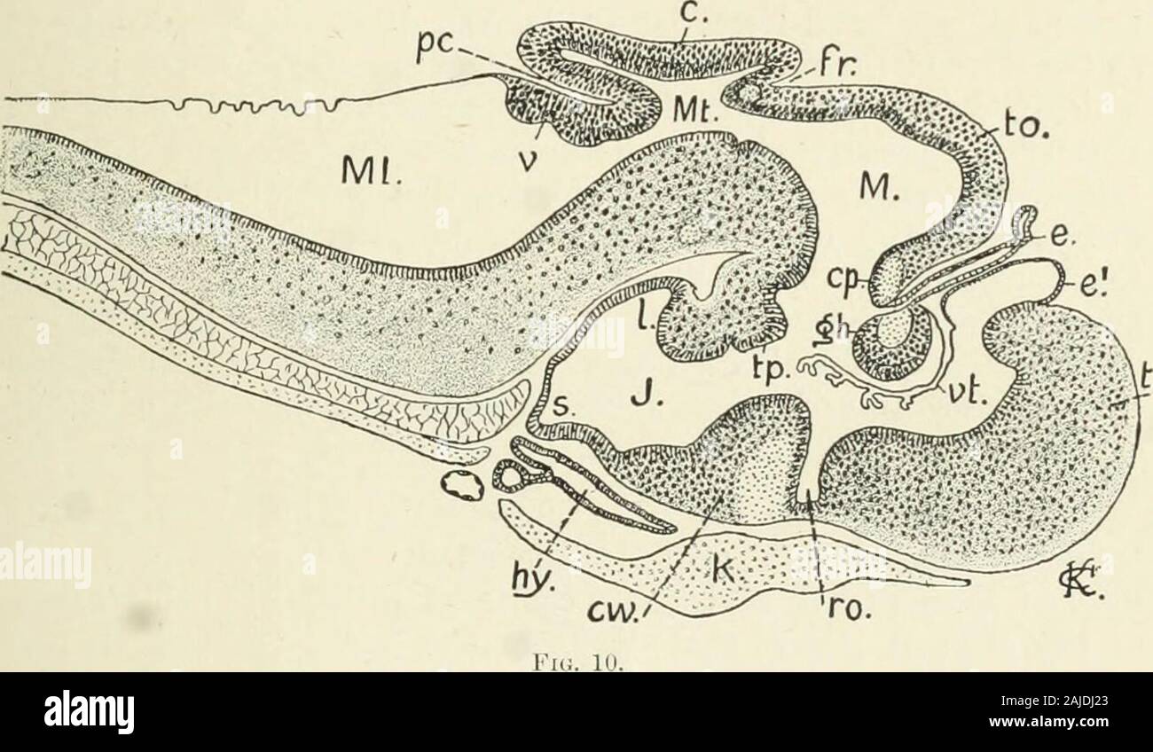 A treatise on zoology . w outgrowths becomevery distinctly paired cerebral hemispheres passing far in front ofthe lamina terminalis. The corpus striatum is a thickening ontheir outer ventral wall. The communication of their cavities oneither side with the median 3rd ventricle (prosocoele) narrows into BRAIN 17 the foijimcii of Monro. Tlie loof of the prosencephala becomesthe palliiuu which acqnires snch an enormous development in thecerebral hemispheres of the highest ^^ertel)rates. From the olfactorylolies issue the nerves to the olfactory epithelium. The greatmodifications in the shape and r Stock Photohttps://www.alamy.com/image-license-details/?v=1https://www.alamy.com/a-treatise-on-zoology-w-outgrowths-becomevery-distinctly-paired-cerebral-hemispheres-passing-far-in-front-ofthe-lamina-terminalis-the-corpus-striatum-is-a-thickening-ontheir-outer-ventral-wall-the-communication-of-their-cavities-oneither-side-with-the-median-3rd-ventricle-prosocoele-narrows-into-brain-17-the-foijimcii-of-monro-tlie-loof-of-the-prosencephala-becomesthe-palliiuu-which-acqnires-snch-an-enormous-development-in-thecerebral-hemispheres-of-the-highest-ertelrates-from-the-olfactorylolies-issue-the-nerves-to-the-olfactory-epithelium-the-greatmodifications-in-the-shape-and-r-image338360347.html
RM2AJDJ23–A treatise on zoology . w outgrowths becomevery distinctly paired cerebral hemispheres passing far in front ofthe lamina terminalis. The corpus striatum is a thickening ontheir outer ventral wall. The communication of their cavities oneither side with the median 3rd ventricle (prosocoele) narrows into BRAIN 17 the foijimcii of Monro. Tlie loof of the prosencephala becomesthe palliiuu which acqnires snch an enormous development in thecerebral hemispheres of the highest ^^ertel)rates. From the olfactorylolies issue the nerves to the olfactory epithelium. The greatmodifications in the shape and r
 . Denkschriften der Medicinisch-Naturwissenschaftlichen Gesellschaft zu Jena. . Fig. 44. Fig. 44. Oralansicht der Nasengegend (5. Stad.). '°/ioo nat Gr. Zeichnung. ßT Ethmotiurbinaüa, Fo Foramen opticum, Fos Fissura orbitalis superior. Fr Frontale, Le Lamina cribrosa, Lpp Lamina papy- racea, ME Mesethmoid, Mx Maxillare, OSp Orbitosphenoid, Stn Sinus terminalis nasi, Su Sutura, V Vomer. ^' ^•^' Fig. 45. Dorsalansicht der oralen Nasengegend (5. Stad.). Caudaler TheU abgeschnitten. Knorpel gekörnt ""/loo'nat Gr. Zeichnung. On Cavum nasi, One Cavum nasi, pars externa, Plni Püca nasalis b Stock Photohttps://www.alamy.com/image-license-details/?v=1https://www.alamy.com/denkschriften-der-medicinisch-naturwissenschaftlichen-gesellschaft-zu-jena-fig-44-fig-44-oralansicht-der-nasengegend-5-stad-ioo-nat-gr-zeichnung-t-ethmotiurbinaa-fo-foramen-opticum-fos-fissura-orbitalis-superior-fr-frontale-le-lamina-cribrosa-lpp-lamina-papy-racea-me-mesethmoid-mx-maxillare-osp-orbitosphenoid-stn-sinus-terminalis-nasi-su-sutura-v-vomer-fig-45-dorsalansicht-der-oralen-nasengegend-5-stad-caudaler-theu-abgeschnitten-knorpel-gekrnt-quotquotloonat-gr-zeichnung-on-cavum-nasi-one-cavum-nasi-pars-externa-plni-pca-nasalis-b-image216053691.html
RMPFE2K7–. Denkschriften der Medicinisch-Naturwissenschaftlichen Gesellschaft zu Jena. . Fig. 44. Fig. 44. Oralansicht der Nasengegend (5. Stad.). '°/ioo nat Gr. Zeichnung. ßT Ethmotiurbinaüa, Fo Foramen opticum, Fos Fissura orbitalis superior. Fr Frontale, Le Lamina cribrosa, Lpp Lamina papy- racea, ME Mesethmoid, Mx Maxillare, OSp Orbitosphenoid, Stn Sinus terminalis nasi, Su Sutura, V Vomer. ^' ^•^' Fig. 45. Dorsalansicht der oralen Nasengegend (5. Stad.). Caudaler TheU abgeschnitten. Knorpel gekörnt ""/loo'nat Gr. Zeichnung. On Cavum nasi, One Cavum nasi, pars externa, Plni Püca nasalis b
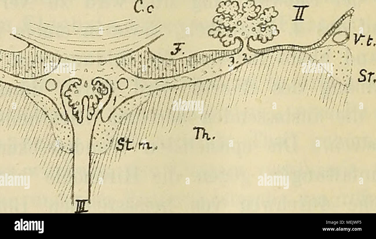 . Die anatomische Nomenclatur. Nomina anatomica, Verzeichniss der von der anatomischen Gesellschaft auf ihrer IX. Versammlung in Basel angenommenen Namen . Fig. 21. Querschnitt durch die Tela chorioidea ventriculi tertii und deren Umgebung. II Seitenventrikel. St. t. Stria terminalis. III 3. Ventrikel. F. L Vena terminalis. Co. Corpus callosum. L. Lamina affixa. JF. Fornix. 1 Taenia thalami. TL Thalamus. 2 Taenia chorioidea. St. m. Stria medullaris. 3 Taenia fornicis Die Figur zeigt den Uebergang der Taenieu in das Epithelblatt der Plexus chorioidei. Epithelplatte fort, welche den Plexus chori Stock Photohttps://www.alamy.com/image-license-details/?v=1https://www.alamy.com/die-anatomische-nomenclatur-nomina-anatomica-verzeichniss-der-von-der-anatomischen-gesellschaft-auf-ihrer-ix-versammlung-in-basel-angenommenen-namen-fig-21-querschnitt-durch-die-tela-chorioidea-ventriculi-tertii-und-deren-umgebung-ii-seitenventrikel-st-t-stria-terminalis-iii-3-ventrikel-f-l-vena-terminalis-co-corpus-callosum-l-lamina-affixa-jf-fornix-1-taenia-thalami-tl-thalamus-2-taenia-chorioidea-st-m-stria-medullaris-3-taenia-fornicis-die-figur-zeigt-den-uebergang-der-taenieu-in-das-epithelblatt-der-plexus-chorioidei-epithelplatte-fort-welche-den-plexus-chori-image181124025.html
RMMEJWF5–. Die anatomische Nomenclatur. Nomina anatomica, Verzeichniss der von der anatomischen Gesellschaft auf ihrer IX. Versammlung in Basel angenommenen Namen . Fig. 21. Querschnitt durch die Tela chorioidea ventriculi tertii und deren Umgebung. II Seitenventrikel. St. t. Stria terminalis. III 3. Ventrikel. F. L Vena terminalis. Co. Corpus callosum. L. Lamina affixa. JF. Fornix. 1 Taenia thalami. TL Thalamus. 2 Taenia chorioidea. St. m. Stria medullaris. 3 Taenia fornicis Die Figur zeigt den Uebergang der Taenieu in das Epithelblatt der Plexus chorioidei. Epithelplatte fort, welche den Plexus chori
 Human brain anatomy for medical concept 3D illustration Stock Photohttps://www.alamy.com/image-license-details/?v=1https://www.alamy.com/human-brain-anatomy-for-medical-concept-3d-illustration-image504513127.html
RF2M8PFHY–Human brain anatomy for medical concept 3D illustration
 Journal of comparative neurology . sus preopticus.There is, however, a short distance anterior to the pit a shallownotch on the ventricular surface of the lamina terminalis (fig.25, r.n.f). The tela chorioidea diencephali shows only slight indicationsof longitudinal folding. The outwardly curved and very promi-nent thalamic lip (fig. 12, t.l.) is not in contact with the lateralwall of the thalamus. At the anterior end, the tela chorioideadiencephali is very broad, and a pouch (fig. 25, a.p.) arises whichextends forward over the velum transversum. The whole telaresembles very closely the same s Stock Photohttps://www.alamy.com/image-license-details/?v=1https://www.alamy.com/journal-of-comparative-neurology-sus-preopticusthere-is-however-a-short-distance-anterior-to-the-pit-a-shallownotch-on-the-ventricular-surface-of-the-lamina-terminalis-fig25-rnf-the-tela-chorioidea-diencephali-shows-only-slight-indicationsof-longitudinal-folding-the-outwardly-curved-and-very-promi-nent-thalamic-lip-fig-12-tl-is-not-in-contact-with-the-lateralwall-of-the-thalamus-at-the-anterior-end-the-tela-chorioideadiencephali-is-very-broad-and-a-pouch-fig-25-ap-arises-whichextends-forward-over-the-velum-transversum-the-whole-telaresembles-very-closely-the-same-s-image338080750.html
RM2AJ0WCE–Journal of comparative neurology . sus preopticus.There is, however, a short distance anterior to the pit a shallownotch on the ventricular surface of the lamina terminalis (fig.25, r.n.f). The tela chorioidea diencephali shows only slight indicationsof longitudinal folding. The outwardly curved and very promi-nent thalamic lip (fig. 12, t.l.) is not in contact with the lateralwall of the thalamus. At the anterior end, the tela chorioideadiencephali is very broad, and a pouch (fig. 25, a.p.) arises whichextends forward over the velum transversum. The whole telaresembles very closely the same s
 . Denkschriften der Medicinisch-Naturwissenschaftlichen Gesellschaft zu Jena. Lc ME Fr Lpp ET Crg Stil OSp OSp ASp Eo. Fos Fig- 38. Fig. 39. Fig. 38. Oralansicht der Nasalgegend (4. Stad.). 7io 'lät. Gr. Zeichnung. ET Ethmoturbinalia, Fo Foramen opticum, Fos Fissura orbitalis superior, Fr Frontale, Lc (FE) Lamina cribrosa (Exethmoid), Lpp Lamina papyracea, ME Mesethmoid, Mx Maxillare, OSp Orbitosphenoid, Stn Sinus terminalis nasi, Su Sutura, V Vomer. Fig. 39- Caudalansicht der Ethmoidalgegend (4. Stad.). Ansicht durch das Foramen occipitale magnum. Nat. Gr. Zeichnung. ASp Alisphenoid, 0>-g Stock Photohttps://www.alamy.com/image-license-details/?v=1https://www.alamy.com/denkschriften-der-medicinisch-naturwissenschaftlichen-gesellschaft-zu-jena-lc-me-fr-lpp-et-crg-stil-osp-osp-asp-eo-fos-fig-38-fig-39-fig-38-oralansicht-der-nasalgegend-4-stad-7io-lt-gr-zeichnung-et-ethmoturbinalia-fo-foramen-opticum-fos-fissura-orbitalis-superior-fr-frontale-lc-fe-lamina-cribrosa-exethmoid-lpp-lamina-papyracea-me-mesethmoid-mx-maxillare-osp-orbitosphenoid-stn-sinus-terminalis-nasi-su-sutura-v-vomer-fig-39-caudalansicht-der-ethmoidalgegend-4-stad-ansicht-durch-das-foramen-occipitale-magnum-nat-gr-zeichnung-asp-alisphenoid-0gt-g-image216053700.html
RMPFE2KG–. Denkschriften der Medicinisch-Naturwissenschaftlichen Gesellschaft zu Jena. Lc ME Fr Lpp ET Crg Stil OSp OSp ASp Eo. Fos Fig- 38. Fig. 39. Fig. 38. Oralansicht der Nasalgegend (4. Stad.). 7io 'lät. Gr. Zeichnung. ET Ethmoturbinalia, Fo Foramen opticum, Fos Fissura orbitalis superior, Fr Frontale, Lc (FE) Lamina cribrosa (Exethmoid), Lpp Lamina papyracea, ME Mesethmoid, Mx Maxillare, OSp Orbitosphenoid, Stn Sinus terminalis nasi, Su Sutura, V Vomer. Fig. 39- Caudalansicht der Ethmoidalgegend (4. Stad.). Ansicht durch das Foramen occipitale magnum. Nat. Gr. Zeichnung. ASp Alisphenoid, 0>-g
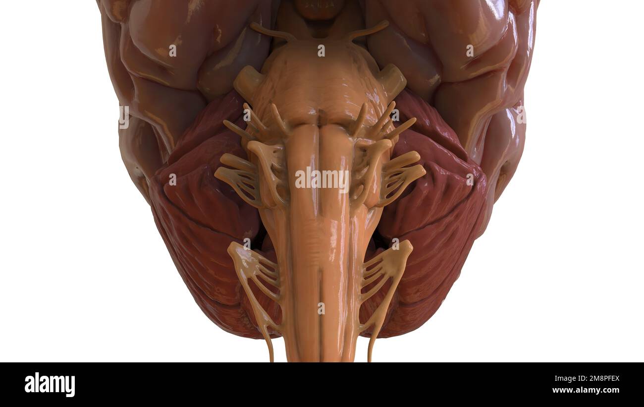 Human brain anatomy for medical concept 3D illustration Stock Photohttps://www.alamy.com/image-license-details/?v=1https://www.alamy.com/human-brain-anatomy-for-medical-concept-3d-illustration-image504513042.html
RF2M8PFEX–Human brain anatomy for medical concept 3D illustration
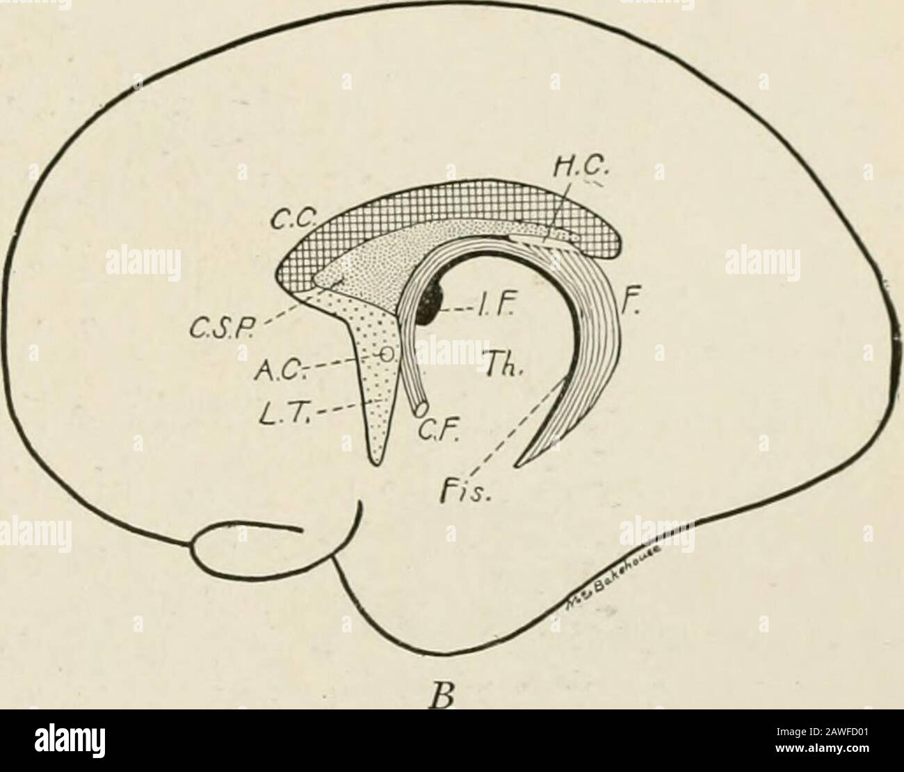 The anatomy of the nervous system, from the standpoint of development and function . Fig. 165.—Schematic representation of the development of the septum pellucidum andtelencephalic commissures: A. C, Anterior commissure; C. C, corpus callosum; C. F., columnafornicus; C. S. P., cavum septi pellucidi; F., fornix; H. C, hippocampal commissure; /. F.,interventricular foramen; Fis., chorioid fissure; L. T., lamina terminalis. (Based on drawings ofmodels of the telencephalon of a four months fetus (.4) and of a five months fetus (B) by Streeter.) the anterior commissure, the hippocampal commissure, Stock Photohttps://www.alamy.com/image-license-details/?v=1https://www.alamy.com/the-anatomy-of-the-nervous-system-from-the-standpoint-of-development-and-function-fig-165schematic-representation-of-the-development-of-the-septum-pellucidum-andtelencephalic-commissures-a-c-anterior-commissure-c-c-corpus-callosum-c-f-columnafornicus-c-s-p-cavum-septi-pellucidi-f-fornix-h-c-hippocampal-commissure-finterventricular-foramen-fis-chorioid-fissure-l-t-lamina-terminalis-based-on-drawings-ofmodels-of-the-telencephalon-of-a-four-months-fetus-4-and-of-a-five-months-fetus-b-by-streeter-the-anterior-commissure-the-hippocampal-commissure-image342702865.html
RM2AWFD01–The anatomy of the nervous system, from the standpoint of development and function . Fig. 165.—Schematic representation of the development of the septum pellucidum andtelencephalic commissures: A. C, Anterior commissure; C. C, corpus callosum; C. F., columnafornicus; C. S. P., cavum septi pellucidi; F., fornix; H. C, hippocampal commissure; /. F.,interventricular foramen; Fis., chorioid fissure; L. T., lamina terminalis. (Based on drawings ofmodels of the telencephalon of a four months fetus (.4) and of a five months fetus (B) by Streeter.) the anterior commissure, the hippocampal commissure,
 Human brain anatomy for medical concept 3D illustration Stock Photohttps://www.alamy.com/image-license-details/?v=1https://www.alamy.com/human-brain-anatomy-for-medical-concept-3d-illustration-image504513039.html
RF2M8PFER–Human brain anatomy for medical concept 3D illustration
 A reference handbook of the medical sciences, embracing the entire range of scientific and practical medicine and allied science . Fig. 956.—Transverse Section through the Cerebral Hemis-pheres in Front of the Lamina Terminalis of Human Fetus ofabout Three Months (5.6 centimeters long), h.o., Bulbus olfae-torius: C.S., corpus striatum: /. arc. ace, fissura arcuata accessoria;/.p., fissure prima; Tz., trapezfeld or precommissural body.(.fter His.) hne of attachment of the chorioid ple.xus to thelamina afExa is termed the tsnia chorioidea, whilethe line of attachment of this membrane on the oth Stock Photohttps://www.alamy.com/image-license-details/?v=1https://www.alamy.com/a-reference-handbook-of-the-medical-sciences-embracing-the-entire-range-of-scientific-and-practical-medicine-and-allied-science-fig-956transverse-section-through-the-cerebral-hemis-pheres-in-front-of-the-lamina-terminalis-of-human-fetus-ofabout-three-months-56-centimeters-long-ho-bulbus-olfae-torius-cs-corpus-striatum-arc-ace-fissura-arcuata-accessoriap-fissure-prima-tz-trapezfeld-or-precommissural-bodyfter-his-hne-of-attachment-of-the-chorioid-plexus-to-thelamina-afexa-is-termed-the-tsnia-chorioidea-whilethe-line-of-attachment-of-this-membrane-on-the-oth-image338910350.html
RM2AKAKH2–A reference handbook of the medical sciences, embracing the entire range of scientific and practical medicine and allied science . Fig. 956.—Transverse Section through the Cerebral Hemis-pheres in Front of the Lamina Terminalis of Human Fetus ofabout Three Months (5.6 centimeters long), h.o., Bulbus olfae-torius: C.S., corpus striatum: /. arc. ace, fissura arcuata accessoria;/.p., fissure prima; Tz., trapezfeld or precommissural body.(.fter His.) hne of attachment of the chorioid ple.xus to thelamina afExa is termed the tsnia chorioidea, whilethe line of attachment of this membrane on the oth
 Human brain anatomy for medical concept 3D illustration Stock Photohttps://www.alamy.com/image-license-details/?v=1https://www.alamy.com/human-brain-anatomy-for-medical-concept-3d-illustration-image504513115.html
RF2M8PFHF–Human brain anatomy for medical concept 3D illustration
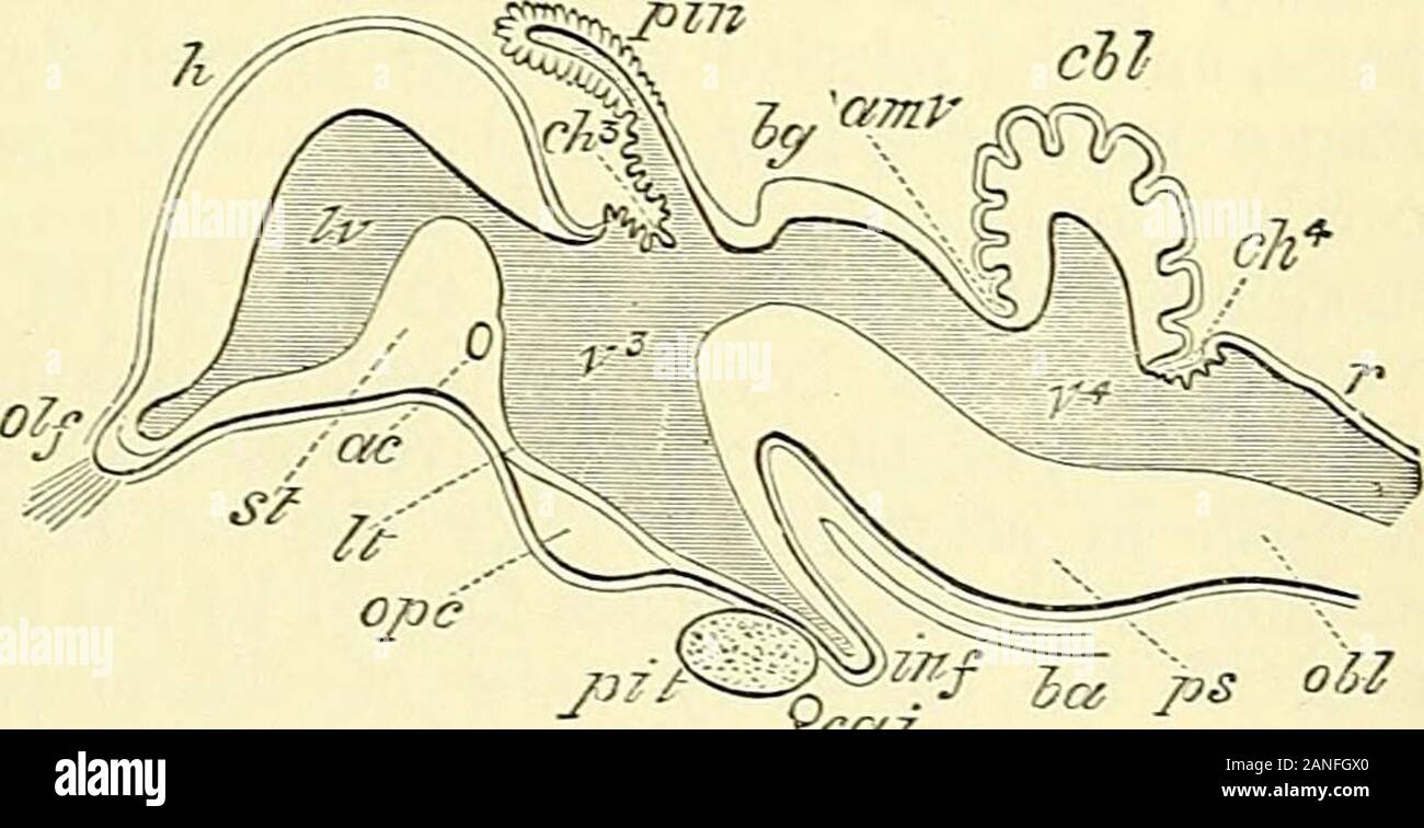 Quain's elements of anatomy . ring Fig. 729.—Outline op a Fig. 729. LONGITUDINAL SECTION THROUGH THE BRAIN OFA CHICK OF TEN DATS. (After Mihalkovics.) h, cerebral hemisphere ; olf,oKactory lobe and nerve ; st,corpus striatum ; Iv, lateralventricle; ac, anterior com-missure ; It, lamina terminalis;ope, optic commissure ; pit,pituitary gland ; inf, inf undi-bulum; cai, internal carotidartery ; v^, third ventricle ;ch?, choroid plexus of third ventricle ; pin, pineal gland; Ijg, corpora bigemina; amv, anterior medullary velum ;below -n-hich two last references are the aqueduct of Sylvius and crur Stock Photohttps://www.alamy.com/image-license-details/?v=1https://www.alamy.com/quains-elements-of-anatomy-ring-fig-729outline-op-a-fig-729-longitudinal-section-through-the-brain-ofa-chick-of-ten-dats-after-mihalkovics-h-cerebral-hemisphere-olfokactory-lobe-and-nerve-stcorpus-striatum-iv-lateralventricle-ac-anterior-com-missure-it-lamina-terminalisope-optic-commissure-pitpituitary-gland-inf-inf-undi-bulum-cai-internal-carotidartery-v-third-ventricle-ch-choroid-plexus-of-third-ventricle-pin-pineal-gland-ijg-corpora-bigemina-amv-anterior-medullary-velum-below-n-hich-two-last-references-are-the-aqueduct-of-sylvius-and-crur-image340247320.html
RM2ANFGX0–Quain's elements of anatomy . ring Fig. 729.—Outline op a Fig. 729. LONGITUDINAL SECTION THROUGH THE BRAIN OFA CHICK OF TEN DATS. (After Mihalkovics.) h, cerebral hemisphere ; olf,oKactory lobe and nerve ; st,corpus striatum ; Iv, lateralventricle; ac, anterior com-missure ; It, lamina terminalis;ope, optic commissure ; pit,pituitary gland ; inf, inf undi-bulum; cai, internal carotidartery ; v^, third ventricle ;ch?, choroid plexus of third ventricle ; pin, pineal gland; Ijg, corpora bigemina; amv, anterior medullary velum ;below -n-hich two last references are the aqueduct of Sylvius and crur
 Human brain anatomy for medical concept 3D illustration Stock Photohttps://www.alamy.com/image-license-details/?v=1https://www.alamy.com/human-brain-anatomy-for-medical-concept-3d-illustration-image504513120.html
RF2M8PFHM–Human brain anatomy for medical concept 3D illustration
![. Journal of morphology. EXPLANATION OF FKHIIiKS 30 Ventral view of the olfactory capsule of an embryo liaviuf; a ca.ra.i)ac(> leiijitliof 7 mm. showing the cartilage parascptalis {c.p.) sei)arate(l from the sei)tum nasias far anterior as the lamina terminalis anterior [l.t.a.) and consequently theforamen praepalatinum not enclosed posteriorly. X 20. 31 Post(M-i()r portion of the right otic cai)sule viewed from in front and slightlyfrom (he median line, showing cs])ecially the groove in which the n. glossopharyn-geus passes through the capsule between the foramen glossopharyngci internum(f. Stock Photo . Journal of morphology. EXPLANATION OF FKHIIiKS 30 Ventral view of the olfactory capsule of an embryo liaviuf; a ca.ra.i)ac(> leiijitliof 7 mm. showing the cartilage parascptalis {c.p.) sei)arate(l from the sei)tum nasias far anterior as the lamina terminalis anterior [l.t.a.) and consequently theforamen praepalatinum not enclosed posteriorly. X 20. 31 Post(M-i()r portion of the right otic cai)sule viewed from in front and slightlyfrom (he median line, showing cs])ecially the groove in which the n. glossopharyn-geus passes through the capsule between the foramen glossopharyngci internum(f. Stock Photo](https://c8.alamy.com/comp/2AFTRNA/journal-of-morphology-explanation-of-fkhiiiks-30-ventral-view-of-the-olfactory-capsule-of-an-embryo-liaviuf-a-caraiacgt-leiijitliof-7-mm-showing-the-cartilage-parascptalis-cp-seiaratel-from-the-seitum-nasias-far-anterior-as-the-lamina-terminalis-anterior-lta-and-consequently-theforamen-praepalatinum-not-enclosed-posteriorly-x-20-31-postm-ir-portion-of-the-right-otic-caisule-viewed-from-in-front-and-slightlyfrom-he-median-line-showing-cs-ecially-the-groove-in-which-the-n-glossopharyn-geus-passes-through-the-capsule-between-the-foramen-glossopharyngci-internumf-2AFTRNA.jpg) . Journal of morphology. EXPLANATION OF FKHIIiKS 30 Ventral view of the olfactory capsule of an embryo liaviuf; a ca.ra.i)ac(> leiijitliof 7 mm. showing the cartilage parascptalis {c.p.) sei)arate(l from the sei)tum nasias far anterior as the lamina terminalis anterior [l.t.a.) and consequently theforamen praepalatinum not enclosed posteriorly. X 20. 31 Post(M-i()r portion of the right otic cai)sule viewed from in front and slightlyfrom (he median line, showing cs])ecially the groove in which the n. glossopharyn-geus passes through the capsule between the foramen glossopharyngci internum(f. Stock Photohttps://www.alamy.com/image-license-details/?v=1https://www.alamy.com/journal-of-morphology-explanation-of-fkhiiiks-30-ventral-view-of-the-olfactory-capsule-of-an-embryo-liaviuf-a-caraiacgt-leiijitliof-7-mm-showing-the-cartilage-parascptalis-cp-seiaratel-from-the-seitum-nasias-far-anterior-as-the-lamina-terminalis-anterior-lta-and-consequently-theforamen-praepalatinum-not-enclosed-posteriorly-x-20-31-postm-ir-portion-of-the-right-otic-caisule-viewed-from-in-front-and-slightlyfrom-he-median-line-showing-cs-ecially-the-groove-in-which-the-n-glossopharyn-geus-passes-through-the-capsule-between-the-foramen-glossopharyngci-internumf-image336762310.html
. Journal of morphology. EXPLANATION OF FKHIIiKS 30 Ventral view of the olfactory capsule of an embryo liaviuf; a ca.ra.i)ac(> leiijitliof 7 mm. showing the cartilage parascptalis {c.p.) sei)arate(l from the sei)tum nasias far anterior as the lamina terminalis anterior [l.t.a.) and consequently theforamen praepalatinum not enclosed posteriorly. X 20. 31 Post(M-i()r portion of the right otic cai)sule viewed from in front and slightlyfrom (he median line, showing cs])ecially the groove in which the n. glossopharyn-geus passes through the capsule between the foramen glossopharyngci internum(f. Stock Photohttps://www.alamy.com/image-license-details/?v=1https://www.alamy.com/journal-of-morphology-explanation-of-fkhiiiks-30-ventral-view-of-the-olfactory-capsule-of-an-embryo-liaviuf-a-caraiacgt-leiijitliof-7-mm-showing-the-cartilage-parascptalis-cp-seiaratel-from-the-seitum-nasias-far-anterior-as-the-lamina-terminalis-anterior-lta-and-consequently-theforamen-praepalatinum-not-enclosed-posteriorly-x-20-31-postm-ir-portion-of-the-right-otic-caisule-viewed-from-in-front-and-slightlyfrom-he-median-line-showing-cs-ecially-the-groove-in-which-the-n-glossopharyn-geus-passes-through-the-capsule-between-the-foramen-glossopharyngci-internumf-image336762310.htmlRM2AFTRNA–. Journal of morphology. EXPLANATION OF FKHIIiKS 30 Ventral view of the olfactory capsule of an embryo liaviuf; a ca.ra.i)ac(> leiijitliof 7 mm. showing the cartilage parascptalis {c.p.) sei)arate(l from the sei)tum nasias far anterior as the lamina terminalis anterior [l.t.a.) and consequently theforamen praepalatinum not enclosed posteriorly. X 20. 31 Post(M-i()r portion of the right otic cai)sule viewed from in front and slightlyfrom (he median line, showing cs])ecially the groove in which the n. glossopharyn-geus passes through the capsule between the foramen glossopharyngci internum(f.
 Human brain anatomy for medical concept 3D illustration Stock Photohttps://www.alamy.com/image-license-details/?v=1https://www.alamy.com/human-brain-anatomy-for-medical-concept-3d-illustration-image504513044.html
RF2M8PFF0–Human brain anatomy for medical concept 3D illustration
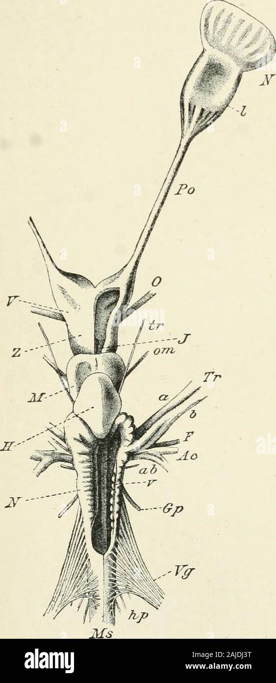 A treatise on zoology . dibulum ; l.r, lateral cavity; Li, lobus inferior; l.t, lamina terminalis; m, medullaoblongata ; o.l, olfactory lobe ; os, olfactory tract; op.l, ojitic lobe ; jyr, ijrofundus nerve ; psji,prespiracular branches of facial; s.op, superior ophthalmic branches of facial and trigeminal;sp.c, spinal cord ; s.v, saccus vasculosus ; th, diencephalon (thalamencejihalon) ; t.v, third ven-tricle ; r, vagus nerve ; v.r, ventral root of spinal nere. large size and complicated structure in the higher Pisces and higherTetrapoda. The mid-brain remains undivided. The cavity itencloses Stock Photohttps://www.alamy.com/image-license-details/?v=1https://www.alamy.com/a-treatise-on-zoology-dibulum-lr-lateral-cavity-li-lobus-inferior-lt-lamina-terminalis-m-medullaoblongata-ol-olfactory-lobe-os-olfactory-tract-opl-ojitic-lobe-jyr-ijrofundus-nerve-psjiprespiracular-branches-of-facial-sop-superior-ophthalmic-branches-of-facial-and-trigeminalspc-spinal-cord-sv-saccus-vasculosus-th-diencephalon-thalamencejihalon-tv-third-ven-tricle-r-vagus-nerve-vr-ventral-root-of-spinal-nere-large-size-and-complicated-structure-in-the-higher-pisces-and-highertetrapoda-the-mid-brain-remains-undivided-the-cavity-itencloses-image338360396.html
RM2AJDJ3T–A treatise on zoology . dibulum ; l.r, lateral cavity; Li, lobus inferior; l.t, lamina terminalis; m, medullaoblongata ; o.l, olfactory lobe ; os, olfactory tract; op.l, ojitic lobe ; jyr, ijrofundus nerve ; psji,prespiracular branches of facial; s.op, superior ophthalmic branches of facial and trigeminal;sp.c, spinal cord ; s.v, saccus vasculosus ; th, diencephalon (thalamencejihalon) ; t.v, third ven-tricle ; r, vagus nerve ; v.r, ventral root of spinal nere. large size and complicated structure in the higher Pisces and higherTetrapoda. The mid-brain remains undivided. The cavity itencloses
 Human brain anatomy for medical concept 3D illustration Stock Photohttps://www.alamy.com/image-license-details/?v=1https://www.alamy.com/human-brain-anatomy-for-medical-concept-3d-illustration-image504513047.html
RF2M8PFF3–Human brain anatomy for medical concept 3D illustration
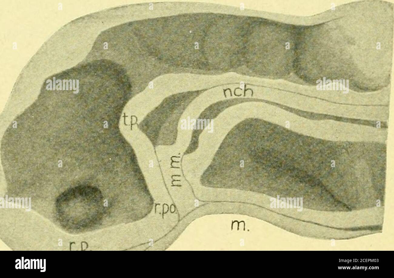 . The Journal of comparative neurology and psychology. of the lips of the neuropore from below upward and thereforethe last point of the neuropore to remain open is a point in thedorsal seam of the neural tube some distance removed from theanterior border of the neural plate. This part of the seam of closurewhich represents the length of the neuropore is what is called the 480 Journal of Comparative Neurology and Psychology. lamina terminalis. This is not very long in the embryo of 17somites, but grows distinctly in length as the forebrain expands.The apparent great thickness of the lamina ter Stock Photohttps://www.alamy.com/image-license-details/?v=1https://www.alamy.com/the-journal-of-comparative-neurology-and-psychology-of-the-lips-of-the-neuropore-from-below-upward-and-thereforethe-last-point-of-the-neuropore-to-remain-open-is-a-point-in-thedorsal-seam-of-the-neural-tube-some-distance-removed-from-theanterior-border-of-the-neural-plate-this-part-of-the-seam-of-closurewhich-represents-the-length-of-the-neuropore-is-what-is-called-the-480-journal-of-comparative-neurology-and-psychology-lamina-terminalis-this-is-not-very-long-in-the-embryo-of-17somites-but-grows-distinctly-in-length-as-the-forebrain-expandsthe-apparent-great-thickness-of-the-lamina-ter-image370521539.html
RM2CEPM03–. The Journal of comparative neurology and psychology. of the lips of the neuropore from below upward and thereforethe last point of the neuropore to remain open is a point in thedorsal seam of the neural tube some distance removed from theanterior border of the neural plate. This part of the seam of closurewhich represents the length of the neuropore is what is called the 480 Journal of Comparative Neurology and Psychology. lamina terminalis. This is not very long in the embryo of 17somites, but grows distinctly in length as the forebrain expands.The apparent great thickness of the lamina ter
 Human brain anatomy for medical concept 3D illustration Stock Photohttps://www.alamy.com/image-license-details/?v=1https://www.alamy.com/human-brain-anatomy-for-medical-concept-3d-illustration-image504513109.html
RF2M8PFH9–Human brain anatomy for medical concept 3D illustration
 . The Journal of comparative neurology and psychology. Fig. 10. Squalus ac, 2G somites, frontal section, inf., the so-called infun-(libulum. pressed down to meet the ectoderm. In Fig. 8 the origin of thisfrom the neuropore is strongly suggested, especially as the spotmarked neurop. is at the upper border of the lamina terminalis.some distance dorsal to the extreme point to which the preoral ento-derm or premandibular mesoderm ever reaches. In Dr. Nealspreparations which I have studied the entoderm has a differenttone from the other tissues and there is a decided difference in theform of the ce Stock Photohttps://www.alamy.com/image-license-details/?v=1https://www.alamy.com/the-journal-of-comparative-neurology-and-psychology-fig-10-squalus-ac-2g-somites-frontal-section-inf-the-so-called-infun-libulum-pressed-down-to-meet-the-ectoderm-in-fig-8-the-origin-of-thisfrom-the-neuropore-is-strongly-suggested-especially-as-the-spotmarked-neurop-is-at-the-upper-border-of-the-lamina-terminalissome-distance-dorsal-to-the-extreme-point-to-which-the-preoral-ento-derm-or-premandibular-mesoderm-ever-reaches-in-dr-nealspreparations-which-i-have-studied-the-entoderm-has-a-differenttone-from-the-other-tissues-and-there-is-a-decided-difference-in-theform-of-the-ce-image370521747.html
RM2CEPM7F–. The Journal of comparative neurology and psychology. Fig. 10. Squalus ac, 2G somites, frontal section, inf., the so-called infun-(libulum. pressed down to meet the ectoderm. In Fig. 8 the origin of thisfrom the neuropore is strongly suggested, especially as the spotmarked neurop. is at the upper border of the lamina terminalis.some distance dorsal to the extreme point to which the preoral ento-derm or premandibular mesoderm ever reaches. In Dr. Nealspreparations which I have studied the entoderm has a differenttone from the other tissues and there is a decided difference in theform of the ce
 Human brain anatomy for medical concept 3D illustration Stock Photohttps://www.alamy.com/image-license-details/?v=1https://www.alamy.com/human-brain-anatomy-for-medical-concept-3d-illustration-image504513113.html
RF2M8PFHD–Human brain anatomy for medical concept 3D illustration
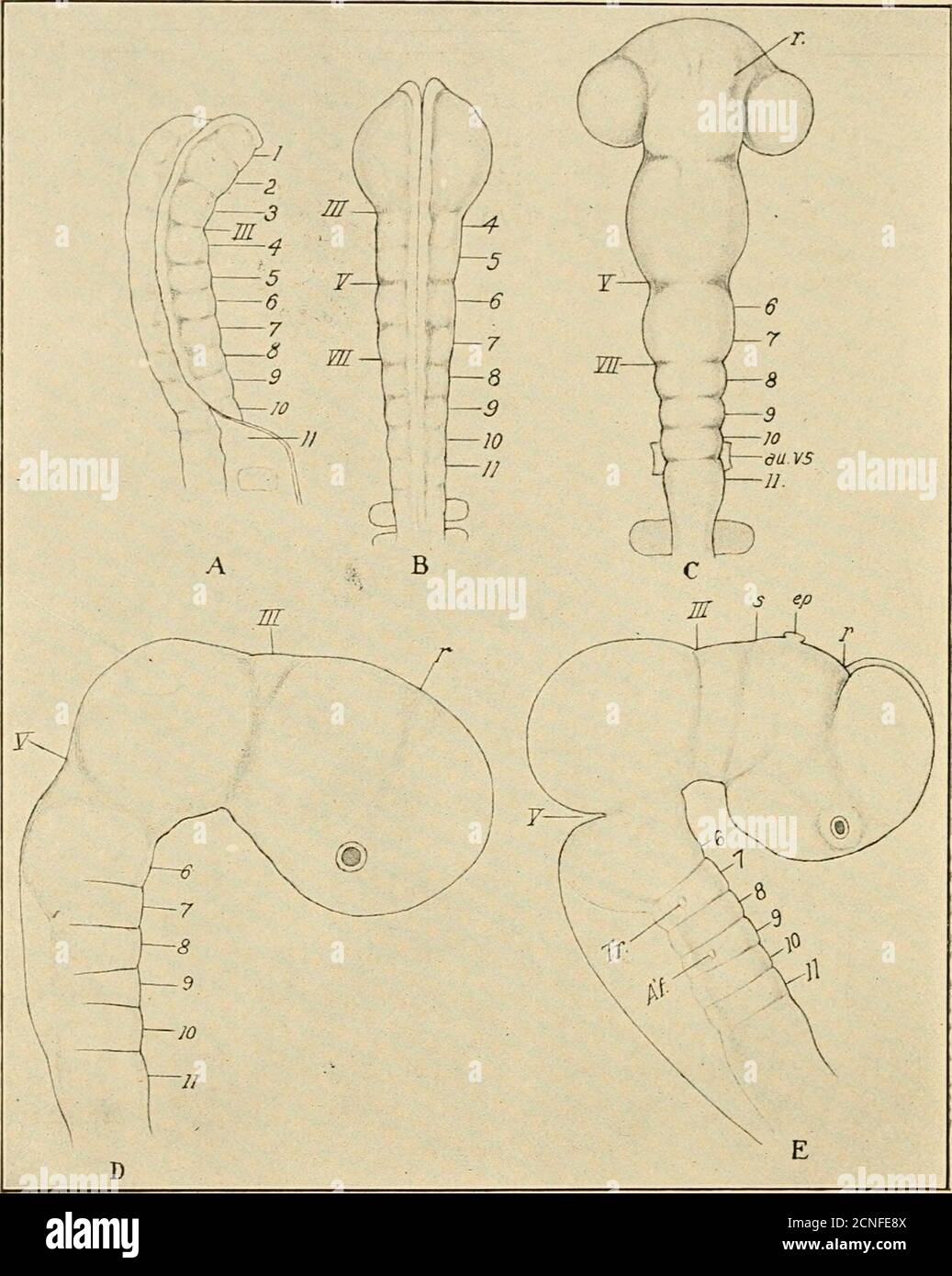 . The development of the chick; an introduction to embryology . erebral fissure a connection remains at its dorsal end betweenthe ectoderm and the neural tube. To this we may apply thename neuropore, though no actual opening is found here at thistime. The median stretch of tissue between the recessus opticusand the neuropore constitutes the lamina terminalis which remainsas the permanent anterior wall of the neural tube. It must notbe forgotten that the original anterior end of the medullary platelies at the ventral end of the lamina terminalis, i.e., in the re-cessus opticus. A third landmark Stock Photohttps://www.alamy.com/image-license-details/?v=1https://www.alamy.com/the-development-of-the-chick-an-introduction-to-embryology-erebral-fissure-a-connection-remains-at-its-dorsal-end-betweenthe-ectoderm-and-the-neural-tube-to-this-we-may-apply-thename-neuropore-though-no-actual-opening-is-found-here-at-thistime-the-median-stretch-of-tissue-between-the-recessus-opticusand-the-neuropore-constitutes-the-lamina-terminalis-which-remainsas-the-permanent-anterior-wall-of-the-neural-tube-it-must-notbe-forgotten-that-the-original-anterior-end-of-the-medullary-platelies-at-the-ventral-end-of-the-lamina-terminalis-ie-in-the-re-cessus-opticus-a-third-landmark-image374666010.html
RM2CNFE8X–. The development of the chick; an introduction to embryology . erebral fissure a connection remains at its dorsal end betweenthe ectoderm and the neural tube. To this we may apply thename neuropore, though no actual opening is found here at thistime. The median stretch of tissue between the recessus opticusand the neuropore constitutes the lamina terminalis which remainsas the permanent anterior wall of the neural tube. It must notbe forgotten that the original anterior end of the medullary platelies at the ventral end of the lamina terminalis, i.e., in the re-cessus opticus. A third landmark
 Human brain anatomy for medical concept 3D illustration Stock Photohttps://www.alamy.com/image-license-details/?v=1https://www.alamy.com/human-brain-anatomy-for-medical-concept-3d-illustration-image504513107.html
RF2M8PFH7–Human brain anatomy for medical concept 3D illustration
 . The development of the chick; an introduction to embryology . al fissure a connection remains at its dorsal end betweenthe ectoderm and the neural tube. To this we may apply thename nevropore, though no actual opening is found here at thistime. The median stretch of tissue between the recessus opticusand the neuropore constitutes the lamina terminalis which remainsas the permanent anterior wall of the neural tube. It must notbe forgotten that the original anterior end of the medullary platelies at the ventral end of the lamina terminalis, i.e., in the re-cessus opticus. A third landmark of f Stock Photohttps://www.alamy.com/image-license-details/?v=1https://www.alamy.com/the-development-of-the-chick-an-introduction-to-embryology-al-fissure-a-connection-remains-at-its-dorsal-end-betweenthe-ectoderm-and-the-neural-tube-to-this-we-may-apply-thename-nevropore-though-no-actual-opening-is-found-here-at-thistime-the-median-stretch-of-tissue-between-the-recessus-opticusand-the-neuropore-constitutes-the-lamina-terminalis-which-remainsas-the-permanent-anterior-wall-of-the-neural-tube-it-must-notbe-forgotten-that-the-original-anterior-end-of-the-medullary-platelies-at-the-ventral-end-of-the-lamina-terminalis-ie-in-the-re-cessus-opticus-a-third-landmark-of-f-image375412072.html
RM2CPNDX0–. The development of the chick; an introduction to embryology . al fissure a connection remains at its dorsal end betweenthe ectoderm and the neural tube. To this we may apply thename nevropore, though no actual opening is found here at thistime. The median stretch of tissue between the recessus opticusand the neuropore constitutes the lamina terminalis which remainsas the permanent anterior wall of the neural tube. It must notbe forgotten that the original anterior end of the medullary platelies at the ventral end of the lamina terminalis, i.e., in the re-cessus opticus. A third landmark of f
 Human brain anatomy for medical concept 3D illustration Stock Photohttps://www.alamy.com/image-license-details/?v=1https://www.alamy.com/human-brain-anatomy-for-medical-concept-3d-illustration-image504513105.html
RF2M8PFH5–Human brain anatomy for medical concept 3D illustration
![. The Journal of comparative neurology and psychology. .-^conimiss] /hippocampi. id t. fore bra in bundle Fig. 7. Section iiniuecliately rostrnl to the (lecnssation of the nervnsterminalis in the Inminn teminalis. X 30. The decussation of the medial forehrain hundle {mcd. f. 7>. 6.) occupies theventral part of the lamina terminalis. The commissura hippocampi (dorsalcommissure) is approaching the lamina terminalis from the dorsal side. Theother elements of the anterior commissure complex lie farther caudad. l88 yoiirtjal of Comparative Isleiirology and Psychology. Fig. S. Composite drawing o Stock Photo . The Journal of comparative neurology and psychology. .-^conimiss] /hippocampi. id t. fore bra in bundle Fig. 7. Section iiniuecliately rostrnl to the (lecnssation of the nervnsterminalis in the Inminn teminalis. X 30. The decussation of the medial forehrain hundle {mcd. f. 7>. 6.) occupies theventral part of the lamina terminalis. The commissura hippocampi (dorsalcommissure) is approaching the lamina terminalis from the dorsal side. Theother elements of the anterior commissure complex lie farther caudad. l88 yoiirtjal of Comparative Isleiirology and Psychology. Fig. S. Composite drawing o Stock Photo](https://c8.alamy.com/comp/2CEPR1R/the-journal-of-comparative-neurology-and-psychology-conimiss-hippocampi-id-t-fore-bra-in-bundle-fig-7-section-iiniuecliately-rostrnl-to-the-lecnssation-of-the-nervnsterminalis-in-the-inminn-teminalis-x-30-the-decussation-of-the-medial-forehrain-hundle-mcd-f-7gt-6-occupies-theventral-part-of-the-lamina-terminalis-the-commissura-hippocampi-dorsalcommissure-is-approaching-the-lamina-terminalis-from-the-dorsal-side-theother-elements-of-the-anterior-commissure-complex-lie-farther-caudad-l88-yoiirtjal-of-comparative-isleiirology-and-psychology-fig-s-composite-drawing-o-2CEPR1R.jpg) . The Journal of comparative neurology and psychology. .-^conimiss] /hippocampi. id t. fore bra in bundle Fig. 7. Section iiniuecliately rostrnl to the (lecnssation of the nervnsterminalis in the Inminn teminalis. X 30. The decussation of the medial forehrain hundle {mcd. f. 7>. 6.) occupies theventral part of the lamina terminalis. The commissura hippocampi (dorsalcommissure) is approaching the lamina terminalis from the dorsal side. Theother elements of the anterior commissure complex lie farther caudad. l88 yoiirtjal of Comparative Isleiirology and Psychology. Fig. S. Composite drawing o Stock Photohttps://www.alamy.com/image-license-details/?v=1https://www.alamy.com/the-journal-of-comparative-neurology-and-psychology-conimiss-hippocampi-id-t-fore-bra-in-bundle-fig-7-section-iiniuecliately-rostrnl-to-the-lecnssation-of-the-nervnsterminalis-in-the-inminn-teminalis-x-30-the-decussation-of-the-medial-forehrain-hundle-mcd-f-7gt-6-occupies-theventral-part-of-the-lamina-terminalis-the-commissura-hippocampi-dorsalcommissure-is-approaching-the-lamina-terminalis-from-the-dorsal-side-theother-elements-of-the-anterior-commissure-complex-lie-farther-caudad-l88-yoiirtjal-of-comparative-isleiirology-and-psychology-fig-s-composite-drawing-o-image370523939.html
. The Journal of comparative neurology and psychology. .-^conimiss] /hippocampi. id t. fore bra in bundle Fig. 7. Section iiniuecliately rostrnl to the (lecnssation of the nervnsterminalis in the Inminn teminalis. X 30. The decussation of the medial forehrain hundle {mcd. f. 7>. 6.) occupies theventral part of the lamina terminalis. The commissura hippocampi (dorsalcommissure) is approaching the lamina terminalis from the dorsal side. Theother elements of the anterior commissure complex lie farther caudad. l88 yoiirtjal of Comparative Isleiirology and Psychology. Fig. S. Composite drawing o Stock Photohttps://www.alamy.com/image-license-details/?v=1https://www.alamy.com/the-journal-of-comparative-neurology-and-psychology-conimiss-hippocampi-id-t-fore-bra-in-bundle-fig-7-section-iiniuecliately-rostrnl-to-the-lecnssation-of-the-nervnsterminalis-in-the-inminn-teminalis-x-30-the-decussation-of-the-medial-forehrain-hundle-mcd-f-7gt-6-occupies-theventral-part-of-the-lamina-terminalis-the-commissura-hippocampi-dorsalcommissure-is-approaching-the-lamina-terminalis-from-the-dorsal-side-theother-elements-of-the-anterior-commissure-complex-lie-farther-caudad-l88-yoiirtjal-of-comparative-isleiirology-and-psychology-fig-s-composite-drawing-o-image370523939.htmlRM2CEPR1R–. The Journal of comparative neurology and psychology. .-^conimiss] /hippocampi. id t. fore bra in bundle Fig. 7. Section iiniuecliately rostrnl to the (lecnssation of the nervnsterminalis in the Inminn teminalis. X 30. The decussation of the medial forehrain hundle {mcd. f. 7>. 6.) occupies theventral part of the lamina terminalis. The commissura hippocampi (dorsalcommissure) is approaching the lamina terminalis from the dorsal side. Theother elements of the anterior commissure complex lie farther caudad. l88 yoiirtjal of Comparative Isleiirology and Psychology. Fig. S. Composite drawing o
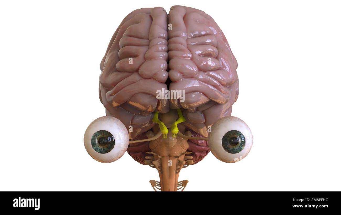 Human brain anatomy for medical concept 3D illustration Stock Photohttps://www.alamy.com/image-license-details/?v=1https://www.alamy.com/human-brain-anatomy-for-medical-concept-3d-illustration-image504513112.html
RF2M8PFHC–Human brain anatomy for medical concept 3D illustration
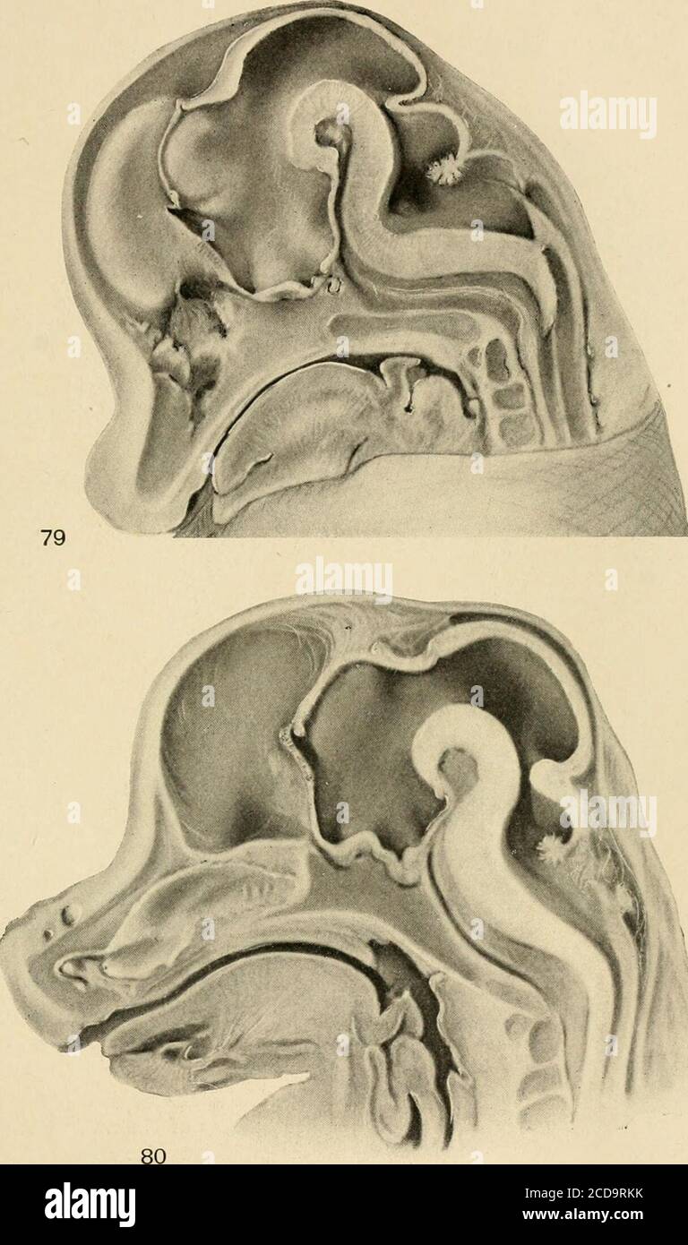 . Journal of comparative neurology . Fig. 77 Bear, section through the genu showing that the indusium as itcurves beneath the genu develops a complete, though small, hippocampal for-mation with all the typical parts present. Compare figures 29, 43 and 58. Fig. 78 Fig embryo, 23 mm. Medial surface of the right half of the head.The lamina terminalis contains the anterior commissure. The lamina supra-neuroporica is only slightly thickened. Between it and the velum transversumis the paraphysal arch, the angulus terminalis of His. 470. Fig. 79 Pig embryo, 28 mm. The lamina supraneuroporica is raise Stock Photohttps://www.alamy.com/image-license-details/?v=1https://www.alamy.com/journal-of-comparative-neurology-fig-77-bear-section-through-the-genu-showing-that-the-indusium-as-itcurves-beneath-the-genu-develops-a-complete-though-small-hippocampal-for-mation-with-all-the-typical-parts-present-compare-figures-29-43-and-58-fig-78-fig-embryo-23-mm-medial-surface-of-the-right-half-of-the-headthe-lamina-terminalis-contains-the-anterior-commissure-the-lamina-supra-neuroporica-is-only-slightly-thickened-between-it-and-the-velum-transversumis-the-paraphysal-arch-the-angulus-terminalis-of-his-470-fig-79-pig-embryo-28-mm-the-lamina-supraneuroporica-is-raise-image369624407.html
RM2CD9RKK–. Journal of comparative neurology . Fig. 77 Bear, section through the genu showing that the indusium as itcurves beneath the genu develops a complete, though small, hippocampal for-mation with all the typical parts present. Compare figures 29, 43 and 58. Fig. 78 Fig embryo, 23 mm. Medial surface of the right half of the head.The lamina terminalis contains the anterior commissure. The lamina supra-neuroporica is only slightly thickened. Between it and the velum transversumis the paraphysal arch, the angulus terminalis of His. 470. Fig. 79 Pig embryo, 28 mm. The lamina supraneuroporica is raise
 Human brain anatomy for medical concept 3D illustration Stock Photohttps://www.alamy.com/image-license-details/?v=1https://www.alamy.com/human-brain-anatomy-for-medical-concept-3d-illustration-image504513108.html
RF2M8PFH8–Human brain anatomy for medical concept 3D illustration
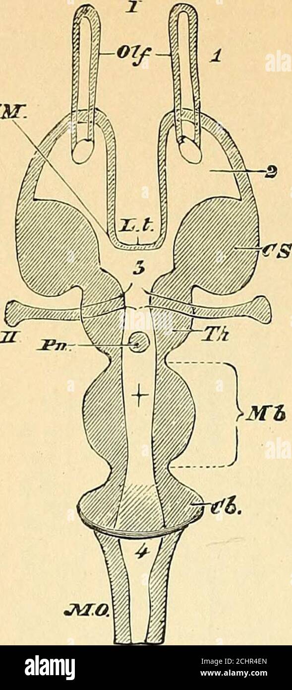 . A text-book of comparative physiology for students and practitioners of comparative (veterinary) medicine . 494 COMPARATIVE PHYSIOLOGY. mr. J. Fig. 354.—Diagrammatic horizontal section of a vertebrate brain (Huxley). The follow-ing letters serve for both this figure and the one following. Mb, mid-brain. Whatlies in front of this is the fore-brain, and what lies behind, the hind-brain. L. t,the lamina terminalis; Olf, olfactory lobes; limp, hemispheres; T/i. E, thala-mencephalon; Pn, pineal gland; Py, pituitary body; FM, foramen of Munro; CS,corpus striatum; Th, optic thalamus; CQ, corpora qu Stock Photohttps://www.alamy.com/image-license-details/?v=1https://www.alamy.com/a-text-book-of-comparative-physiology-for-students-and-practitioners-of-comparative-veterinary-medicine-494-comparative-physiology-mr-j-fig-354diagrammatic-horizontal-section-of-a-vertebrate-brain-huxley-the-follow-ing-letters-serve-for-both-this-figure-and-the-one-following-mb-mid-brain-whatlies-in-front-of-this-is-the-fore-brain-and-what-lies-behind-the-hind-brain-l-tthe-lamina-terminalis-olf-olfactory-lobes-limp-hemispheres-ti-e-thala-mencephalon-pn-pineal-gland-py-pituitary-body-fm-foramen-of-munro-cscorpus-striatum-th-optic-thalamus-cq-corpora-qu-image372375325.html
RM2CHR4EN–. A text-book of comparative physiology for students and practitioners of comparative (veterinary) medicine . 494 COMPARATIVE PHYSIOLOGY. mr. J. Fig. 354.—Diagrammatic horizontal section of a vertebrate brain (Huxley). The follow-ing letters serve for both this figure and the one following. Mb, mid-brain. Whatlies in front of this is the fore-brain, and what lies behind, the hind-brain. L. t,the lamina terminalis; Olf, olfactory lobes; limp, hemispheres; T/i. E, thala-mencephalon; Pn, pineal gland; Py, pituitary body; FM, foramen of Munro; CS,corpus striatum; Th, optic thalamus; CQ, corpora qu
 Human brain anatomy for medical concept 3D illustration Stock Photohttps://www.alamy.com/image-license-details/?v=1https://www.alamy.com/human-brain-anatomy-for-medical-concept-3d-illustration-image504513037.html
RF2M8PFEN–Human brain anatomy for medical concept 3D illustration
 . A text-book of comparative physiology for students and practitioners of comparative (veterinary) medicine . Fig. 354.—Diagrammatic horizontal section of a vertebrate brain (Huxley). The follow-ing letters serve for both this figure and the one following. Mb, mid-brain. Whatlies in front of this is the fore-brain, and what lies behind, the hind-brain. L. t,the lamina terminalis; Olf, olfactory lobes; limp, hemispheres; T/i. E, thala-mencephalon; Pn, pineal gland; Py, pituitary body; FM, foramen of Munro; CS,corpus striatum; Th, optic thalamus; CQ, corpora quadrigemina; CC, crura cere-bri; Cb, Stock Photohttps://www.alamy.com/image-license-details/?v=1https://www.alamy.com/a-text-book-of-comparative-physiology-for-students-and-practitioners-of-comparative-veterinary-medicine-fig-354diagrammatic-horizontal-section-of-a-vertebrate-brain-huxley-the-follow-ing-letters-serve-for-both-this-figure-and-the-one-following-mb-mid-brain-whatlies-in-front-of-this-is-the-fore-brain-and-what-lies-behind-the-hind-brain-l-tthe-lamina-terminalis-olf-olfactory-lobes-limp-hemispheres-ti-e-thala-mencephalon-pn-pineal-gland-py-pituitary-body-fm-foramen-of-munro-cscorpus-striatum-th-optic-thalamus-cq-corpora-quadrigemina-cc-crura-cere-bri-cb-image372374375.html
RM2CHR38R–. A text-book of comparative physiology for students and practitioners of comparative (veterinary) medicine . Fig. 354.—Diagrammatic horizontal section of a vertebrate brain (Huxley). The follow-ing letters serve for both this figure and the one following. Mb, mid-brain. Whatlies in front of this is the fore-brain, and what lies behind, the hind-brain. L. t,the lamina terminalis; Olf, olfactory lobes; limp, hemispheres; T/i. E, thala-mencephalon; Pn, pineal gland; Py, pituitary body; FM, foramen of Munro; CS,corpus striatum; Th, optic thalamus; CQ, corpora quadrigemina; CC, crura cere-bri; Cb,
 Human brain anatomy for medical concept 3D illustration Stock Photohttps://www.alamy.com/image-license-details/?v=1https://www.alamy.com/human-brain-anatomy-for-medical-concept-3d-illustration-image504513124.html
RF2M8PFHT–Human brain anatomy for medical concept 3D illustration
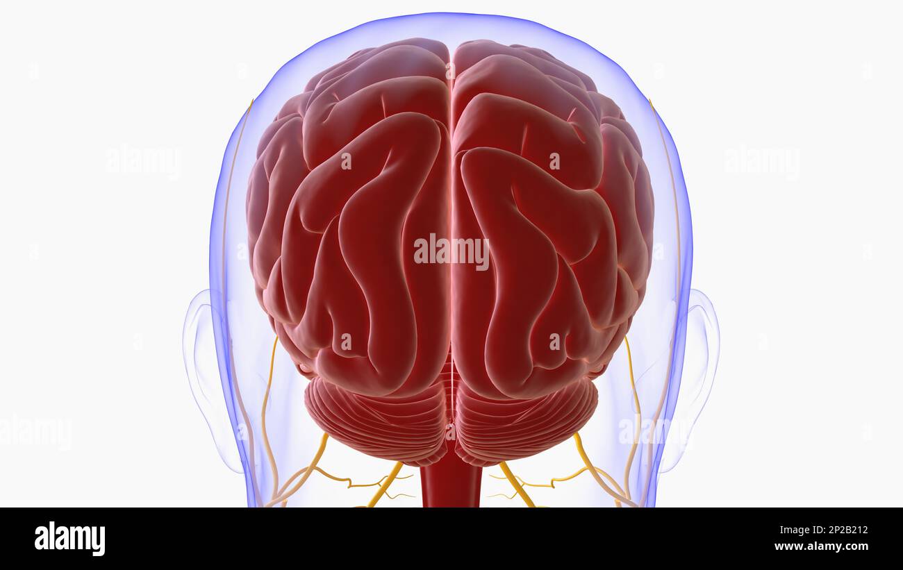 Human brain anatomy for medical concept 3D illustration Stock Photohttps://www.alamy.com/image-license-details/?v=1https://www.alamy.com/human-brain-anatomy-for-medical-concept-3d-illustration-image534993790.html
RF2P2B212–Human brain anatomy for medical concept 3D illustration
 . Quain's elements of anatomy . enient to bring Fig. 729.—Outline of a LONGITUDINAL SECTION THROUGH THE BRAIN OFA CHICK OF TEN DATS. (After Mihalkovics.) h, cerebral hemisphere ; olf,olfactory lobe and nerve ; st,corpus striatum ; Iv, lateralventricle; ac, anterior com-missure; It, lamina terminalis;ope, optic commissure ; piit,pituitary gland ; inf, infundi-bulum ; cai, internal carotidartery ; ?••*, third ventricle ;ch?, choroid plexus of third ventricle ; pin, pineal gland ; hg, corpora bigemina ; amt anterior medullarj velum ;below which two last references are the aqueduct of Sylvius and Stock Photohttps://www.alamy.com/image-license-details/?v=1https://www.alamy.com/quains-elements-of-anatomy-enient-to-bring-fig-729outline-of-a-longitudinal-section-through-the-brain-ofa-chick-of-ten-dats-after-mihalkovics-h-cerebral-hemisphere-olfolfactory-lobe-and-nerve-stcorpus-striatum-iv-lateralventricle-ac-anterior-com-missure-it-lamina-terminalisope-optic-commissure-piitpituitary-gland-inf-infundi-bulum-cai-internal-carotidartery-third-ventricle-ch-choroid-plexus-of-third-ventricle-pin-pineal-gland-hg-corpora-bigemina-amt-anterior-medullarj-velum-below-which-two-last-references-are-the-aqueduct-of-sylvius-and-image371873648.html
RM2CH08HM–. Quain's elements of anatomy . enient to bring Fig. 729.—Outline of a LONGITUDINAL SECTION THROUGH THE BRAIN OFA CHICK OF TEN DATS. (After Mihalkovics.) h, cerebral hemisphere ; olf,olfactory lobe and nerve ; st,corpus striatum ; Iv, lateralventricle; ac, anterior com-missure; It, lamina terminalis;ope, optic commissure ; piit,pituitary gland ; inf, infundi-bulum ; cai, internal carotidartery ; ?••*, third ventricle ;ch?, choroid plexus of third ventricle ; pin, pineal gland ; hg, corpora bigemina ; amt anterior medullarj velum ;below which two last references are the aqueduct of Sylvius and
 Human brain anatomy for medical concept 3D illustration Stock Photohttps://www.alamy.com/image-license-details/?v=1https://www.alamy.com/human-brain-anatomy-for-medical-concept-3d-illustration-image534993797.html
RF2P2B219–Human brain anatomy for medical concept 3D illustration
 . The development of the chick; an introduction to embryology . striatum.The lateral walls of the telencephalon medium are formed bythe posterior ends of the corpora striata and are therefore verythick. The lamina terminalis passes obliquely upwards and forwards cavity. Oes., Oesophagus, p. A., Pulmonary arch, par., Paraphysis. P. C,Pericardial cavity. Rec. op., Recessus opticus. R., Rectum. S. Inf., Saccusinfundibuli. T., Tongue. Tel., Med. Telencephalon medium. Tr., Trachea.V. 1, 10, 20, 30, First, tenth, twentieth and thirtieth vertebral centra, r. A..right auricle. Vel. tr., Velum transver Stock Photohttps://www.alamy.com/image-license-details/?v=1https://www.alamy.com/the-development-of-the-chick-an-introduction-to-embryology-striatumthe-lateral-walls-of-the-telencephalon-medium-are-formed-bythe-posterior-ends-of-the-corpora-striata-and-are-therefore-verythick-the-lamina-terminalis-passes-obliquely-upwards-and-forwards-cavity-oes-oesophagus-p-a-pulmonary-arch-par-paraphysis-p-cpericardial-cavity-rec-op-recessus-opticus-r-rectum-s-inf-saccusinfundibuli-t-tongue-tel-med-telencephalon-medium-tr-tracheav-1-10-20-30-first-tenth-twentieth-and-thirtieth-vertebral-centra-r-aright-auricle-vel-tr-velum-transver-image374660183.html
RM2CNF6TR–. The development of the chick; an introduction to embryology . striatum.The lateral walls of the telencephalon medium are formed bythe posterior ends of the corpora striata and are therefore verythick. The lamina terminalis passes obliquely upwards and forwards cavity. Oes., Oesophagus, p. A., Pulmonary arch, par., Paraphysis. P. C,Pericardial cavity. Rec. op., Recessus opticus. R., Rectum. S. Inf., Saccusinfundibuli. T., Tongue. Tel., Med. Telencephalon medium. Tr., Trachea.V. 1, 10, 20, 30, First, tenth, twentieth and thirtieth vertebral centra, r. A..right auricle. Vel. tr., Velum transver
 Human brain anatomy for medical concept 3D illustration Stock Photohttps://www.alamy.com/image-license-details/?v=1https://www.alamy.com/human-brain-anatomy-for-medical-concept-3d-illustration-image534993827.html
RF2P2B22B–Human brain anatomy for medical concept 3D illustration
 . Kirkes' handbook of physiology . Fig. 371.—Longitudinal and Vertical Diagrammatic Section of a Vertebrate Brain. Lettersas before. Lamina terminalis is represented by the strong black line joining Pn and Py. (Huxley.). Fig. 372.—Base of the Brain, r, Superior longitudinal fissure; 2, 2, 2, anterior cerebrallobe; 3, fissure of Sylvius, between anterior and 4. 4. 4. middle cerebral lobe; 5, 5, posteriorlobe; 6. medulla oblongata. The figure is in the right anterior pyramid; 7, 8, 9, 10, the cerebellum;+1 the inferior vermiform process. The figures from /. to IX, are placed against the correspo Stock Photohttps://www.alamy.com/image-license-details/?v=1https://www.alamy.com/kirkes-handbook-of-physiology-fig-371longitudinal-and-vertical-diagrammatic-section-of-a-vertebrate-brain-lettersas-before-lamina-terminalis-is-represented-by-the-strong-black-line-joining-pn-and-py-huxley-fig-372base-of-the-brain-r-superior-longitudinal-fissure-2-2-2-anterior-cerebrallobe-3-fissure-of-sylvius-between-anterior-and-4-4-4-middle-cerebral-lobe-5-5-posteriorlobe-6-medulla-oblongata-the-figure-is-in-the-right-anterior-pyramid-7-8-9-10-the-cerebellum1-the-inferior-vermiform-process-the-figures-from-to-ix-are-placed-against-the-correspo-image369986921.html
RM2CDXA2H–. Kirkes' handbook of physiology . Fig. 371.—Longitudinal and Vertical Diagrammatic Section of a Vertebrate Brain. Lettersas before. Lamina terminalis is represented by the strong black line joining Pn and Py. (Huxley.). Fig. 372.—Base of the Brain, r, Superior longitudinal fissure; 2, 2, 2, anterior cerebrallobe; 3, fissure of Sylvius, between anterior and 4. 4. 4. middle cerebral lobe; 5, 5, posteriorlobe; 6. medulla oblongata. The figure is in the right anterior pyramid; 7, 8, 9, 10, the cerebellum;+1 the inferior vermiform process. The figures from /. to IX, are placed against the correspo
 Human brain anatomy for medical concept 3D illustration Stock Photohttps://www.alamy.com/image-license-details/?v=1https://www.alamy.com/human-brain-anatomy-for-medical-concept-3d-illustration-image534993772.html
RF2P2B20C–Human brain anatomy for medical concept 3D illustration
 . Journal of comparative neurology . am Fig. 1 Chelydra serpentina, 2 mm. embryo, median sagittal section of theanterior portion to show the neuropore and adjacent structures. Magn. 52 diam. Fig. 2 Chelydra serpentina, 4.5 mm. embryo, sagittal section of head to showneuroporic recess, lamina terminalis and lamina supraneuroporica. Magn. 52diam. The section is median in the anterior region.. Fig. 3 Chelydra serpentina, 6 mm. embryo, median sagittal section. Magn.52 diam. Fig. 4 Chelydra serpentina, 9 mm. embryo, median sagittal section. Magn.52 diam. 428 SEPTUM, HIPPOCAMPUS, PALLIAL COMMISSURES Stock Photohttps://www.alamy.com/image-license-details/?v=1https://www.alamy.com/journal-of-comparative-neurology-am-fig-1-chelydra-serpentina-2-mm-embryo-median-sagittal-section-of-theanterior-portion-to-show-the-neuropore-and-adjacent-structures-magn-52-diam-fig-2-chelydra-serpentina-45-mm-embryo-sagittal-section-of-head-to-showneuroporic-recess-lamina-terminalis-and-lamina-supraneuroporica-magn-52diam-the-section-is-median-in-the-anterior-region-fig-3-chelydra-serpentina-6-mm-embryo-median-sagittal-section-magn52-diam-fig-4-chelydra-serpentina-9-mm-embryo-median-sagittal-section-magn52-diam-428-septum-hippocampus-pallial-commissures-image369635937.html
RM2CDAABD–. Journal of comparative neurology . am Fig. 1 Chelydra serpentina, 2 mm. embryo, median sagittal section of theanterior portion to show the neuropore and adjacent structures. Magn. 52 diam. Fig. 2 Chelydra serpentina, 4.5 mm. embryo, sagittal section of head to showneuroporic recess, lamina terminalis and lamina supraneuroporica. Magn. 52diam. The section is median in the anterior region.. Fig. 3 Chelydra serpentina, 6 mm. embryo, median sagittal section. Magn.52 diam. Fig. 4 Chelydra serpentina, 9 mm. embryo, median sagittal section. Magn.52 diam. 428 SEPTUM, HIPPOCAMPUS, PALLIAL COMMISSURES
 Human brain anatomy for medical concept 3D illustration Stock Photohttps://www.alamy.com/image-license-details/?v=1https://www.alamy.com/human-brain-anatomy-for-medical-concept-3d-illustration-image534993783.html
RF2P2B20R–Human brain anatomy for medical concept 3D illustration
 . The development of the chick; an introduction to embryology . ear the dorsal and ventral boun-daries of the foramen of Monro,ch. PI., Choroid plexus. Com. ant., Anterior commissure. Com. post.,Posterior commissure. C. str.. Corpus striatum. Ep., Epiphysis. H.,Hemisphere. Hyp., Hypojjhysis. L. t., Lamina terminalis. Myel., Myel-encephalon. olf., Olfactory nerve, op. N., Optic chiasma. op. L., Opticlobe. Par., Parapliysis. Paren., Parencephalon. pi. ene. v., Plica en-cephali ventralis. pont. Fl., Pontine flexure. Rec. op., Recessus opticus.S. Inf., Saccus infundibuli. Tel. med.. Telencephalon Stock Photohttps://www.alamy.com/image-license-details/?v=1https://www.alamy.com/the-development-of-the-chick-an-introduction-to-embryology-ear-the-dorsal-and-ventral-boun-daries-of-the-foramen-of-monroch-pi-choroid-plexus-com-ant-anterior-commissure-com-postposterior-commissure-c-str-corpus-striatum-ep-epiphysis-hhemisphere-hyp-hypojjhysis-l-t-lamina-terminalis-myel-myel-encephalon-olf-olfactory-nerve-op-n-optic-chiasma-op-l-opticlobe-par-parapliysis-paren-parencephalon-pi-ene-v-plica-en-cephali-ventralis-pont-fl-pontine-flexure-rec-op-recessus-opticuss-inf-saccus-infundibuli-tel-med-telencephalon-image375408721.html
RM2CPN9J9–. The development of the chick; an introduction to embryology . ear the dorsal and ventral boun-daries of the foramen of Monro,ch. PI., Choroid plexus. Com. ant., Anterior commissure. Com. post.,Posterior commissure. C. str.. Corpus striatum. Ep., Epiphysis. H.,Hemisphere. Hyp., Hypojjhysis. L. t., Lamina terminalis. Myel., Myel-encephalon. olf., Olfactory nerve, op. N., Optic chiasma. op. L., Opticlobe. Par., Parapliysis. Paren., Parencephalon. pi. ene. v., Plica en-cephali ventralis. pont. Fl., Pontine flexure. Rec. op., Recessus opticus.S. Inf., Saccus infundibuli. Tel. med.. Telencephalon
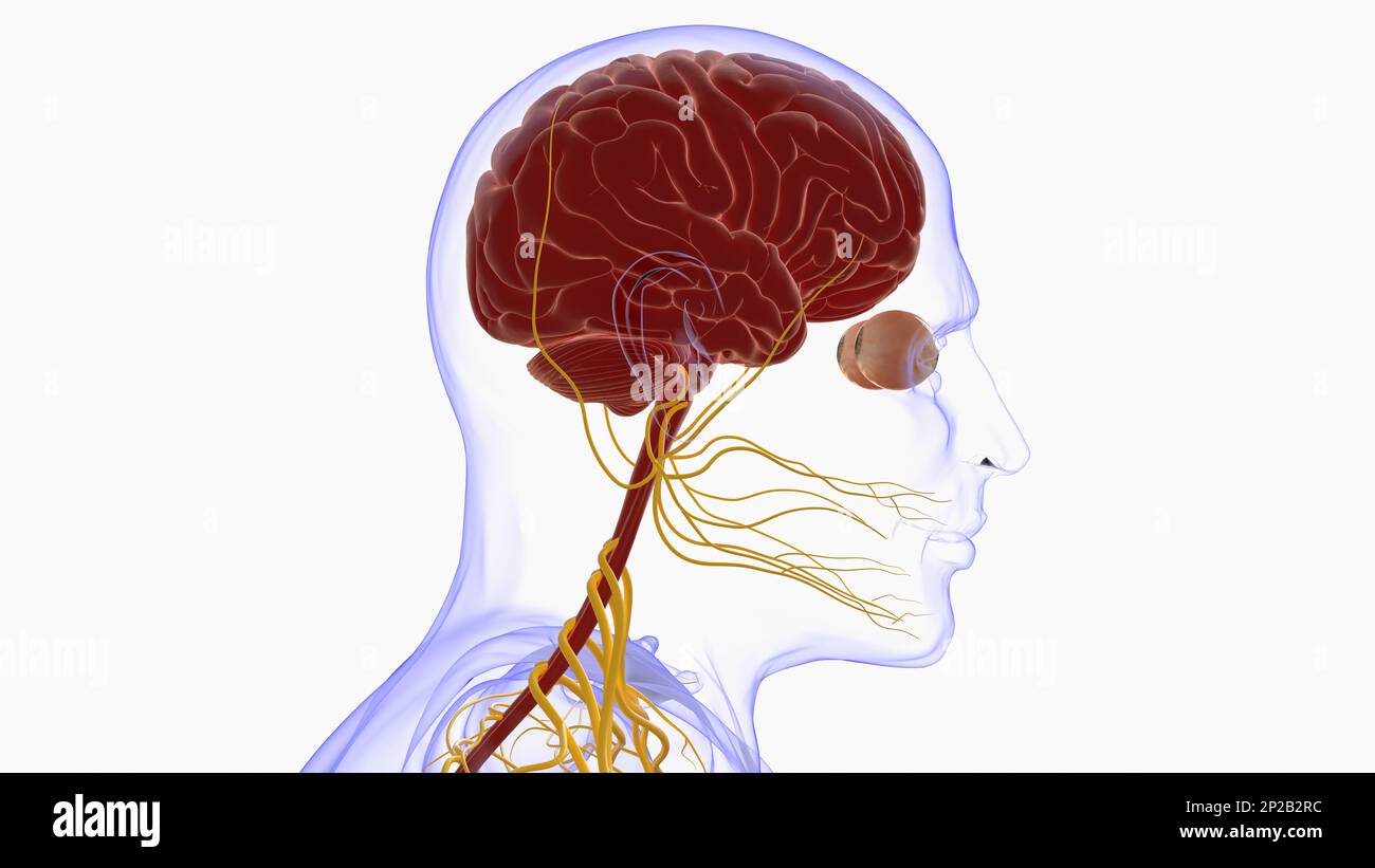 Human brain anatomy for medical concept 3D illustration Stock Photohttps://www.alamy.com/image-license-details/?v=1https://www.alamy.com/human-brain-anatomy-for-medical-concept-3d-illustration-image534994416.html
RF2P2B2RC–Human brain anatomy for medical concept 3D illustration
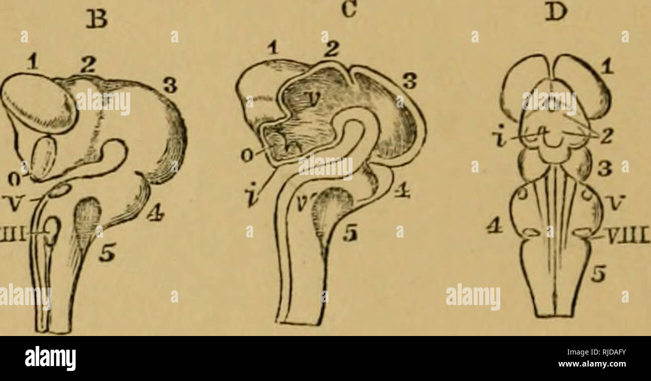 . The cat; an introduction to the study of backboned animals, especially mammals. Cats; Anatomy, Comparative. 358 THE CAT. [chap. X. vesicles, namely, those of the mid-hrain and the hmd-hrain. The fore-brain, called also the deutenccphalon, contains the anterior termination of the primitive medullary canal, and this becomes the third ventricle ; the pre-axial wall of the first vesicle becoming the lamina terminalis of the adult. The optic thalami, optic nerves, pineal gland and infundibulum, are formed from this vesicle. The mid-brain, called also the meHcncophalon, contains that part of the p Stock Photohttps://www.alamy.com/image-license-details/?v=1https://www.alamy.com/the-cat-an-introduction-to-the-study-of-backboned-animals-especially-mammals-cats-anatomy-comparative-358-the-cat-chap-x-vesicles-namely-those-of-the-mid-hrain-and-the-hmd-hrain-the-fore-brain-called-also-the-deutenccphalon-contains-the-anterior-termination-of-the-primitive-medullary-canal-and-this-becomes-the-third-ventricle-the-pre-axial-wall-of-the-first-vesicle-becoming-the-lamina-terminalis-of-the-adult-the-optic-thalami-optic-nerves-pineal-gland-and-infundibulum-are-formed-from-this-vesicle-the-mid-brain-called-also-the-mehcncophalon-contains-that-part-of-the-p-image235092255.html
RMRJDAFY–. The cat; an introduction to the study of backboned animals, especially mammals. Cats; Anatomy, Comparative. 358 THE CAT. [chap. X. vesicles, namely, those of the mid-hrain and the hmd-hrain. The fore-brain, called also the deutenccphalon, contains the anterior termination of the primitive medullary canal, and this becomes the third ventricle ; the pre-axial wall of the first vesicle becoming the lamina terminalis of the adult. The optic thalami, optic nerves, pineal gland and infundibulum, are formed from this vesicle. The mid-brain, called also the meHcncophalon, contains that part of the p
 Human brain anatomy for medical concept 3D illustration Stock Photohttps://www.alamy.com/image-license-details/?v=1https://www.alamy.com/human-brain-anatomy-for-medical-concept-3d-illustration-image534994484.html
RF2P2B2WT–Human brain anatomy for medical concept 3D illustration
 . The cat; an introduction to the study of backboned animals, especially mammals. Cats; Anatomy, Comparative. 358 THE CAT. [chap. X. vesicles, namely, those of the mid-hrain and the hmd-hrain. The fore-brain, called also the deutenccphalon, contains the anterior termination of the primitive medullary canal, and this becomes the third ventricle ; the pre-axial wall of the first vesicle becoming the lamina terminalis of the adult. The optic thalami, optic nerves, pineal gland and infundibulum, are formed from this vesicle. The mid-brain, called also the meHcncophalon, contains that part of the p Stock Photohttps://www.alamy.com/image-license-details/?v=1https://www.alamy.com/the-cat-an-introduction-to-the-study-of-backboned-animals-especially-mammals-cats-anatomy-comparative-358-the-cat-chap-x-vesicles-namely-those-of-the-mid-hrain-and-the-hmd-hrain-the-fore-brain-called-also-the-deutenccphalon-contains-the-anterior-termination-of-the-primitive-medullary-canal-and-this-becomes-the-third-ventricle-the-pre-axial-wall-of-the-first-vesicle-becoming-the-lamina-terminalis-of-the-adult-the-optic-thalami-optic-nerves-pineal-gland-and-infundibulum-are-formed-from-this-vesicle-the-mid-brain-called-also-the-mehcncophalon-contains-that-part-of-the-p-image235092410.html
RMRJDANE–. The cat; an introduction to the study of backboned animals, especially mammals. Cats; Anatomy, Comparative. 358 THE CAT. [chap. X. vesicles, namely, those of the mid-hrain and the hmd-hrain. The fore-brain, called also the deutenccphalon, contains the anterior termination of the primitive medullary canal, and this becomes the third ventricle ; the pre-axial wall of the first vesicle becoming the lamina terminalis of the adult. The optic thalami, optic nerves, pineal gland and infundibulum, are formed from this vesicle. The mid-brain, called also the meHcncophalon, contains that part of the p
 Human brain anatomy for medical concept 3D illustration Stock Photohttps://www.alamy.com/image-license-details/?v=1https://www.alamy.com/human-brain-anatomy-for-medical-concept-3d-illustration-image534993778.html
RF2P2B20J–Human brain anatomy for medical concept 3D illustration
 . The cat : an introduction to the study of backboned animals, especially mammals. Cats; Anatomy, Comparative. 358 THE GAT. [CHAP, X. vesicles, namely, those of the mid-brain and the hind-brain. The fore-brain, called also the deutencephalon, contains the anterior termination of the primitive medullary canal, and this becomes the third ventricle ; the pre-axial wall of the first vesicle becoming the lamina terminalis of the adult. The optic thalami, optic nerves, pineal gland and infundibulum, are formed from this vesicle. The mid-brain, called also the mesencephalon, contains that part of the Stock Photohttps://www.alamy.com/image-license-details/?v=1https://www.alamy.com/the-cat-an-introduction-to-the-study-of-backboned-animals-especially-mammals-cats-anatomy-comparative-358-the-gat-chap-x-vesicles-namely-those-of-the-mid-brain-and-the-hind-brain-the-fore-brain-called-also-the-deutencephalon-contains-the-anterior-termination-of-the-primitive-medullary-canal-and-this-becomes-the-third-ventricle-the-pre-axial-wall-of-the-first-vesicle-becoming-the-lamina-terminalis-of-the-adult-the-optic-thalami-optic-nerves-pineal-gland-and-infundibulum-are-formed-from-this-vesicle-the-mid-brain-called-also-the-mesencephalon-contains-that-part-of-the-image235095827.html
RMRJDF3F–. The cat : an introduction to the study of backboned animals, especially mammals. Cats; Anatomy, Comparative. 358 THE GAT. [CHAP, X. vesicles, namely, those of the mid-brain and the hind-brain. The fore-brain, called also the deutencephalon, contains the anterior termination of the primitive medullary canal, and this becomes the third ventricle ; the pre-axial wall of the first vesicle becoming the lamina terminalis of the adult. The optic thalami, optic nerves, pineal gland and infundibulum, are formed from this vesicle. The mid-brain, called also the mesencephalon, contains that part of the
 Human brain anatomy for medical concept 3D illustration Stock Photohttps://www.alamy.com/image-license-details/?v=1https://www.alamy.com/human-brain-anatomy-for-medical-concept-3d-illustration-image534993832.html
RF2P2B22G–Human brain anatomy for medical concept 3D illustration
 . The anatomy of the central nervous system of man and of vertebrates in general. Neuroanatomy; Central Nervous System. 160 ANATOMY OF THE CENTRAL NERVOUS SYSTEM. which join the hemispheres to the striatum traverse the Lamina terminalis (see Fig. 101). In mammals for the first time there arises late in the em- bryonic period, dorsal and anterior to the Lamina terminalis, a new system of transverse fibers destined to connect cortical regions of one hemisphere with those of the other: Corpus callosum. The mantle of higher vertebrates is differentiated from those of teleosts and ganoids through a Stock Photohttps://www.alamy.com/image-license-details/?v=1https://www.alamy.com/the-anatomy-of-the-central-nervous-system-of-man-and-of-vertebrates-in-general-neuroanatomy-central-nervous-system-160-anatomy-of-the-central-nervous-system-which-join-the-hemispheres-to-the-striatum-traverse-the-lamina-terminalis-see-fig-101-in-mammals-for-the-first-time-there-arises-late-in-the-em-bryonic-period-dorsal-and-anterior-to-the-lamina-terminalis-a-new-system-of-transverse-fibers-destined-to-connect-cortical-regions-of-one-hemisphere-with-those-of-the-other-corpus-callosum-the-mantle-of-higher-vertebrates-is-differentiated-from-those-of-teleosts-and-ganoids-through-a-image236814932.html
RMRN7RT4–. The anatomy of the central nervous system of man and of vertebrates in general. Neuroanatomy; Central Nervous System. 160 ANATOMY OF THE CENTRAL NERVOUS SYSTEM. which join the hemispheres to the striatum traverse the Lamina terminalis (see Fig. 101). In mammals for the first time there arises late in the em- bryonic period, dorsal and anterior to the Lamina terminalis, a new system of transverse fibers destined to connect cortical regions of one hemisphere with those of the other: Corpus callosum. The mantle of higher vertebrates is differentiated from those of teleosts and ganoids through a
 Human brain anatomy for medical concept 3D illustration Stock Photohttps://www.alamy.com/image-license-details/?v=1https://www.alamy.com/human-brain-anatomy-for-medical-concept-3d-illustration-image534994383.html
RF2P2B2P7–Human brain anatomy for medical concept 3D illustration
 . The anatomy of the central nervous system of man and of vertebrates in general. Neuroanatomy; Central Nervous System. THE INTERBEAIN OR THALAMENCEPHALON. 127 The lamina terminalis of the brain, before turning backward to form the roof of the interbrain, passes first a short distance dorsally,—Lamina supranetiroporica,—and then falls in the nsnally sail-like, pendulous Tela chorioidea, from which, through anteriorly-directed evaginations, the Plexus chorioidei of the ventricle is formed. In several amphibians (Fig. 55, B) and in the Dipnoi, whose brain can scarcely be differentiated from the Stock Photohttps://www.alamy.com/image-license-details/?v=1https://www.alamy.com/the-anatomy-of-the-central-nervous-system-of-man-and-of-vertebrates-in-general-neuroanatomy-central-nervous-system-the-interbeain-or-thalamencephalon-127-the-lamina-terminalis-of-the-brain-before-turning-backward-to-form-the-roof-of-the-interbrain-passes-first-a-short-distance-dorsallylamina-supranetiroporicaand-then-falls-in-the-nsnally-sail-like-pendulous-tela-chorioidea-from-which-through-anteriorly-directed-evaginations-the-plexus-chorioidei-of-the-ventricle-is-formed-in-several-amphibians-fig-55-b-and-in-the-dipnoi-whose-brain-can-scarcely-be-differentiated-from-the-image236815557.html
RMRN7TJD–. The anatomy of the central nervous system of man and of vertebrates in general. Neuroanatomy; Central Nervous System. THE INTERBEAIN OR THALAMENCEPHALON. 127 The lamina terminalis of the brain, before turning backward to form the roof of the interbrain, passes first a short distance dorsally,—Lamina supranetiroporica,—and then falls in the nsnally sail-like, pendulous Tela chorioidea, from which, through anteriorly-directed evaginations, the Plexus chorioidei of the ventricle is formed. In several amphibians (Fig. 55, B) and in the Dipnoi, whose brain can scarcely be differentiated from the
