Quick filters:
Lateral fenestra Stock Photos and Images
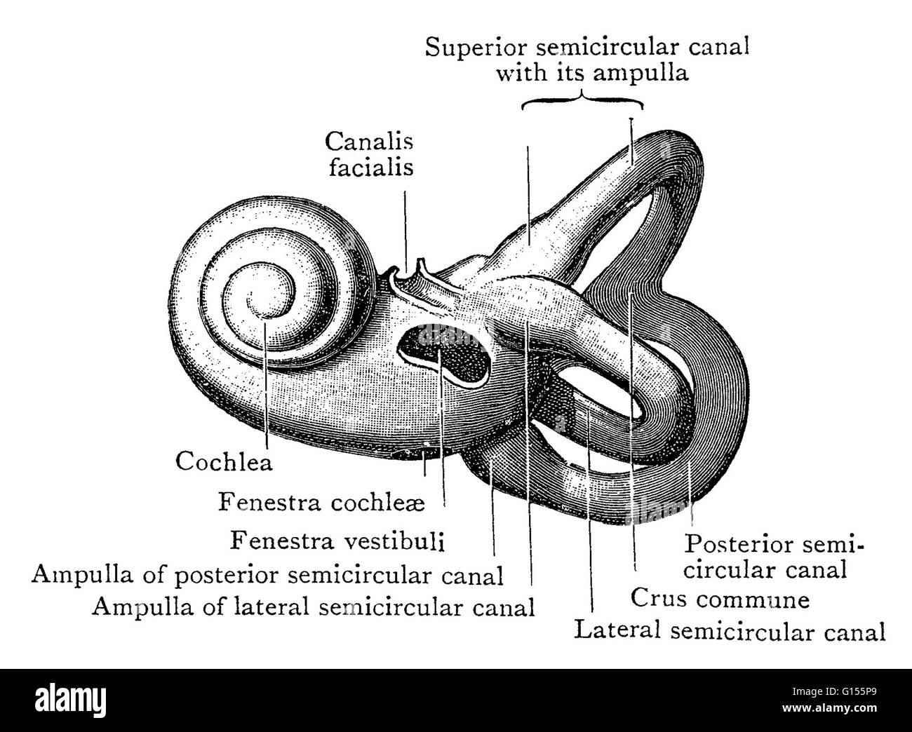 Illustration of the left bony labyrinth of the inner ear from the lateral side. The bony labyrinth or osseous labyrinth is comprised of the vestibule, the three semicircular canals and the cochlea. The membranous labyrinth is contained within it. Stock Photohttps://www.alamy.com/image-license-details/?v=1https://www.alamy.com/stock-photo-illustration-of-the-left-bony-labyrinth-of-the-inner-ear-from-the-103991169.html
RMG155P9–Illustration of the left bony labyrinth of the inner ear from the lateral side. The bony labyrinth or osseous labyrinth is comprised of the vestibule, the three semicircular canals and the cochlea. The membranous labyrinth is contained within it.
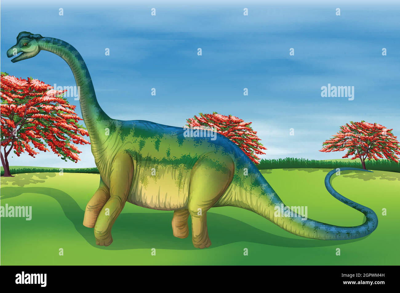 Brachiosaurus Stock Vectorhttps://www.alamy.com/image-license-details/?v=1https://www.alamy.com/brachiosaurus-image444346241.html
RF2GPWM4H–Brachiosaurus
![. The Biological bulletin. Biology; Zoology; Biology; Marine Biology. i86 S. W. WILLISTON. there is perhaps good reason for the belief that the original lateral fenestra in the icthyosaurs has become closed up by the posterior obtrusion of the orbit, as was suggested by McGregor. That this was really the case, however, has by no means been proven. We can conceive of an early diapsid stem without lateral fenestration (as is indeed the case, in Procolophoii], from which the ichthyosaurs might have been derived. Certainly the Ichthyosauria are the most primitive of reptiles, save the coty- losaur Stock Photo . The Biological bulletin. Biology; Zoology; Biology; Marine Biology. i86 S. W. WILLISTON. there is perhaps good reason for the belief that the original lateral fenestra in the icthyosaurs has become closed up by the posterior obtrusion of the orbit, as was suggested by McGregor. That this was really the case, however, has by no means been proven. We can conceive of an early diapsid stem without lateral fenestration (as is indeed the case, in Procolophoii], from which the ichthyosaurs might have been derived. Certainly the Ichthyosauria are the most primitive of reptiles, save the coty- losaur Stock Photo](https://c8.alamy.com/comp/RHRMC7/the-biological-bulletin-biology-zoology-biology-marine-biology-i86-s-w-williston-there-is-perhaps-good-reason-for-the-belief-that-the-original-lateral-fenestra-in-the-icthyosaurs-has-become-closed-up-by-the-posterior-obtrusion-of-the-orbit-as-was-suggested-by-mcgregor-that-this-was-really-the-case-however-has-by-no-means-been-proven-we-can-conceive-of-an-early-diapsid-stem-without-lateral-fenestration-as-is-indeed-the-case-in-procolophoii-from-which-the-ichthyosaurs-might-have-been-derived-certainly-the-ichthyosauria-are-the-most-primitive-of-reptiles-save-the-coty-losaur-RHRMC7.jpg) . The Biological bulletin. Biology; Zoology; Biology; Marine Biology. i86 S. W. WILLISTON. there is perhaps good reason for the belief that the original lateral fenestra in the icthyosaurs has become closed up by the posterior obtrusion of the orbit, as was suggested by McGregor. That this was really the case, however, has by no means been proven. We can conceive of an early diapsid stem without lateral fenestration (as is indeed the case, in Procolophoii], from which the ichthyosaurs might have been derived. Certainly the Ichthyosauria are the most primitive of reptiles, save the coty- losaur Stock Photohttps://www.alamy.com/image-license-details/?v=1https://www.alamy.com/the-biological-bulletin-biology-zoology-biology-marine-biology-i86-s-w-williston-there-is-perhaps-good-reason-for-the-belief-that-the-original-lateral-fenestra-in-the-icthyosaurs-has-become-closed-up-by-the-posterior-obtrusion-of-the-orbit-as-was-suggested-by-mcgregor-that-this-was-really-the-case-however-has-by-no-means-been-proven-we-can-conceive-of-an-early-diapsid-stem-without-lateral-fenestration-as-is-indeed-the-case-in-procolophoii-from-which-the-ichthyosaurs-might-have-been-derived-certainly-the-ichthyosauria-are-the-most-primitive-of-reptiles-save-the-coty-losaur-image234704855.html
. The Biological bulletin. Biology; Zoology; Biology; Marine Biology. i86 S. W. WILLISTON. there is perhaps good reason for the belief that the original lateral fenestra in the icthyosaurs has become closed up by the posterior obtrusion of the orbit, as was suggested by McGregor. That this was really the case, however, has by no means been proven. We can conceive of an early diapsid stem without lateral fenestration (as is indeed the case, in Procolophoii], from which the ichthyosaurs might have been derived. Certainly the Ichthyosauria are the most primitive of reptiles, save the coty- losaur Stock Photohttps://www.alamy.com/image-license-details/?v=1https://www.alamy.com/the-biological-bulletin-biology-zoology-biology-marine-biology-i86-s-w-williston-there-is-perhaps-good-reason-for-the-belief-that-the-original-lateral-fenestra-in-the-icthyosaurs-has-become-closed-up-by-the-posterior-obtrusion-of-the-orbit-as-was-suggested-by-mcgregor-that-this-was-really-the-case-however-has-by-no-means-been-proven-we-can-conceive-of-an-early-diapsid-stem-without-lateral-fenestration-as-is-indeed-the-case-in-procolophoii-from-which-the-ichthyosaurs-might-have-been-derived-certainly-the-ichthyosauria-are-the-most-primitive-of-reptiles-save-the-coty-losaur-image234704855.htmlRMRHRMC7–. The Biological bulletin. Biology; Zoology; Biology; Marine Biology. i86 S. W. WILLISTON. there is perhaps good reason for the belief that the original lateral fenestra in the icthyosaurs has become closed up by the posterior obtrusion of the orbit, as was suggested by McGregor. That this was really the case, however, has by no means been proven. We can conceive of an early diapsid stem without lateral fenestration (as is indeed the case, in Procolophoii], from which the ichthyosaurs might have been derived. Certainly the Ichthyosauria are the most primitive of reptiles, save the coty- losaur
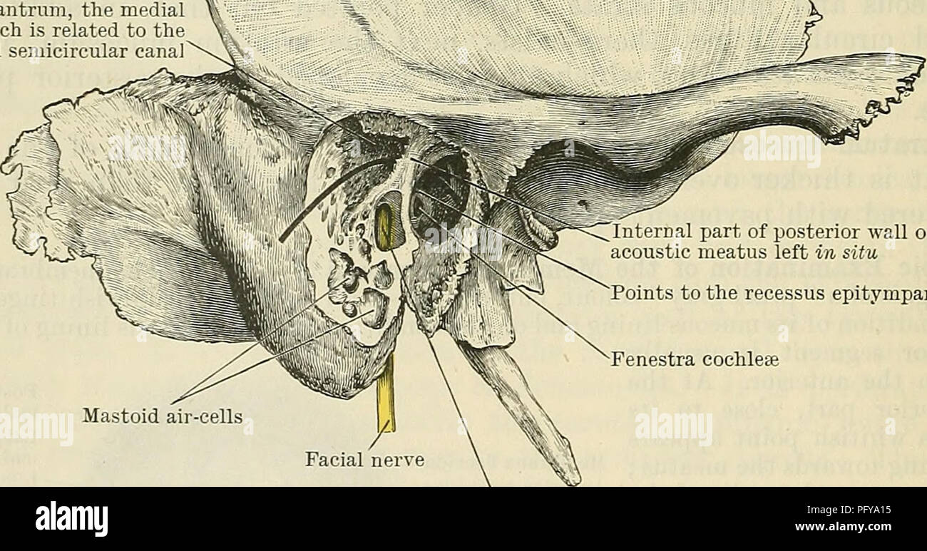 . Cunningham's Text-book of anatomy. Anatomy. Tympanic antrum, the medial : wall of which is related to the 5 lateral semicircular canal. Internal part of posterior wall of external acoustic meatus left in situ Points to the recessus epitympanicus Fenestra cochleae Mastoid air-cells Facial canal laid open, displaying the facial nerve within Fig. 712. Preparation to display the position and relations of the tympanic antrum. The greater part of the posterior wall of the external acoustic meatus has been removed, leaving only a bridge of bone at its inner ex- tremity ; under this a bristle is Stock Photohttps://www.alamy.com/image-license-details/?v=1https://www.alamy.com/cunninghams-text-book-of-anatomy-anatomy-tympanic-antrum-the-medial-wall-of-which-is-related-to-the-5-lateral-semicircular-canal-internal-part-of-posterior-wall-of-external-acoustic-meatus-left-in-situ-points-to-the-recessus-epitympanicus-fenestra-cochleae-mastoid-air-cells-facial-canal-laid-open-displaying-the-facial-nerve-within-fig-712-preparation-to-display-the-position-and-relations-of-the-tympanic-antrum-the-greater-part-of-the-posterior-wall-of-the-external-acoustic-meatus-has-been-removed-leaving-only-a-bridge-of-bone-at-its-inner-ex-tremity-under-this-a-bristle-is-image216344833.html
RMPFYA15–. Cunningham's Text-book of anatomy. Anatomy. Tympanic antrum, the medial : wall of which is related to the 5 lateral semicircular canal. Internal part of posterior wall of external acoustic meatus left in situ Points to the recessus epitympanicus Fenestra cochleae Mastoid air-cells Facial canal laid open, displaying the facial nerve within Fig. 712. Preparation to display the position and relations of the tympanic antrum. The greater part of the posterior wall of the external acoustic meatus has been removed, leaving only a bridge of bone at its inner ex- tremity ; under this a bristle is
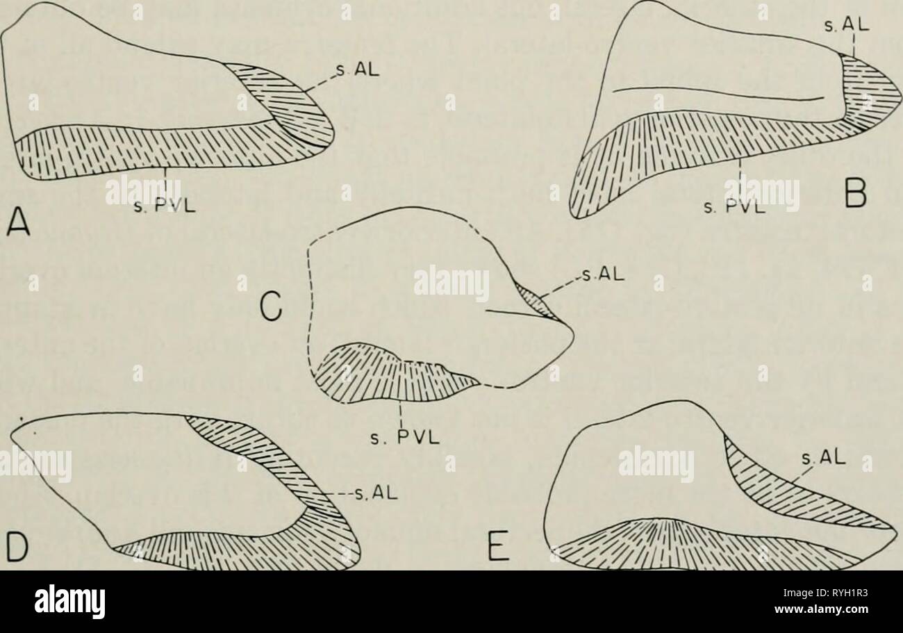 Early Devonian fishes from Utah : Arthrodira earlydevonianfis119deni Year: 1953 DENISON: EARLY DEVONIAN FISHES 527 fenestra, at least in the form upon which this part of the restoration was based. Williamsaspis has a very large posterior lateral, relatively short and very high, peculiarly shaped, laterally ridged, and with a rather large edge for the pectoral fenestra; presumably this is a specialized condition, since nothing approaching it is found in other euarthrodires. In the Brachythoraci the posterior lateral is almost -^-^W s.PVL s.PVL Fig. 111. Right posterior lateral plates of A Stock Photohttps://www.alamy.com/image-license-details/?v=1https://www.alamy.com/early-devonian-fishes-from-utah-arthrodira-earlydevonianfis119deni-year-1953-denison-early-devonian-fishes-527-fenestra-at-least-in-the-form-upon-which-this-part-of-the-restoration-was-based-williamsaspis-has-a-very-large-posterior-lateral-relatively-short-and-very-high-peculiarly-shaped-laterally-ridged-and-with-a-rather-large-edge-for-the-pectoral-fenestra-presumably-this-is-a-specialized-condition-since-nothing-approaching-it-is-found-in-other-euarthrodires-in-the-brachythoraci-the-posterior-lateral-is-almost-w-spvl-spvl-fig-111-right-posterior-lateral-plates-of-a-image240705111.html
RMRYH1R3–Early Devonian fishes from Utah : Arthrodira earlydevonianfis119deni Year: 1953 DENISON: EARLY DEVONIAN FISHES 527 fenestra, at least in the form upon which this part of the restoration was based. Williamsaspis has a very large posterior lateral, relatively short and very high, peculiarly shaped, laterally ridged, and with a rather large edge for the pectoral fenestra; presumably this is a specialized condition, since nothing approaching it is found in other euarthrodires. In the Brachythoraci the posterior lateral is almost -^-^W s.PVL s.PVL Fig. 111. Right posterior lateral plates of A
 . Early Devonian fishes from Utah : Arthrodira . Fig. 115. Arcfolepis sp. A, right half of trunk shield in dorsal view (X 3/2) showing position of section, x-y, and extent of pectoral fenestra, pf; B, section through lateral part of trunk shield (X 3), demonstrating the presence of a pectoral fenestra, pf; bones stippled, presumed cartilage cross-hatched; endoskeletal peri- chondral bone (end) black. ADL, anterior dorso-lateral; AL, anterior lateral; AVL, anterior ventro- lateral; IL, intero-lateral; MD, median dorsal; PDL, posterior dorso-lateral; PVL, posterior ventro-lateral; SP, spinal. in Stock Photohttps://www.alamy.com/image-license-details/?v=1https://www.alamy.com/early-devonian-fishes-from-utah-arthrodira-fig-115-arcfolepis-sp-a-right-half-of-trunk-shield-in-dorsal-view-x-32-showing-position-of-section-x-y-and-extent-of-pectoral-fenestra-pf-b-section-through-lateral-part-of-trunk-shield-x-3-demonstrating-the-presence-of-a-pectoral-fenestra-pf-bones-stippled-presumed-cartilage-cross-hatched-endoskeletal-peri-chondral-bone-end-black-adl-anterior-dorso-lateral-al-anterior-lateral-avl-anterior-ventro-lateral-il-intero-lateral-md-median-dorsal-pdl-posterior-dorso-lateral-pvl-posterior-ventro-lateral-sp-spinal-in-image178489749.html
RMMAAWDW–. Early Devonian fishes from Utah : Arthrodira . Fig. 115. Arcfolepis sp. A, right half of trunk shield in dorsal view (X 3/2) showing position of section, x-y, and extent of pectoral fenestra, pf; B, section through lateral part of trunk shield (X 3), demonstrating the presence of a pectoral fenestra, pf; bones stippled, presumed cartilage cross-hatched; endoskeletal peri- chondral bone (end) black. ADL, anterior dorso-lateral; AL, anterior lateral; AVL, anterior ventro- lateral; IL, intero-lateral; MD, median dorsal; PDL, posterior dorso-lateral; PVL, posterior ventro-lateral; SP, spinal. in
 . Bacteriology and surgical technics for nurses. Surgical nursing; Operations, Surgical; Bacteriology. 206 SURGICAL TECHNIC with a sheet, sitting in a chair. He should be advised that the procedure will be unpleasant but is not danger- ous, and that he must not under any circumstances pull your hand away or bite the tube, but at all times must swallow. Have the patient extend the head and open the mouth, then introduce the cold stomach- tube into the throat through the mouth, at the same. Fig. 166.—Stomach-tube and funnel for expressing the stomach con- tents: a, Showing the lateral fenestra; Stock Photohttps://www.alamy.com/image-license-details/?v=1https://www.alamy.com/bacteriology-and-surgical-technics-for-nurses-surgical-nursing-operations-surgical-bacteriology-206-surgical-technic-with-a-sheet-sitting-in-a-chair-he-should-be-advised-that-the-procedure-will-be-unpleasant-but-is-not-danger-ous-and-that-he-must-not-under-any-circumstances-pull-your-hand-away-or-bite-the-tube-but-at-all-times-must-swallow-have-the-patient-extend-the-head-and-open-the-mouth-then-introduce-the-cold-stomach-tube-into-the-throat-through-the-mouth-at-the-same-fig-166stomach-tube-and-funnel-for-expressing-the-stomach-con-tents-a-showing-the-lateral-fenestra-image235267569.html
RMRJNA55–. Bacteriology and surgical technics for nurses. Surgical nursing; Operations, Surgical; Bacteriology. 206 SURGICAL TECHNIC with a sheet, sitting in a chair. He should be advised that the procedure will be unpleasant but is not danger- ous, and that he must not under any circumstances pull your hand away or bite the tube, but at all times must swallow. Have the patient extend the head and open the mouth, then introduce the cold stomach- tube into the throat through the mouth, at the same. Fig. 166.—Stomach-tube and funnel for expressing the stomach con- tents: a, Showing the lateral fenestra;
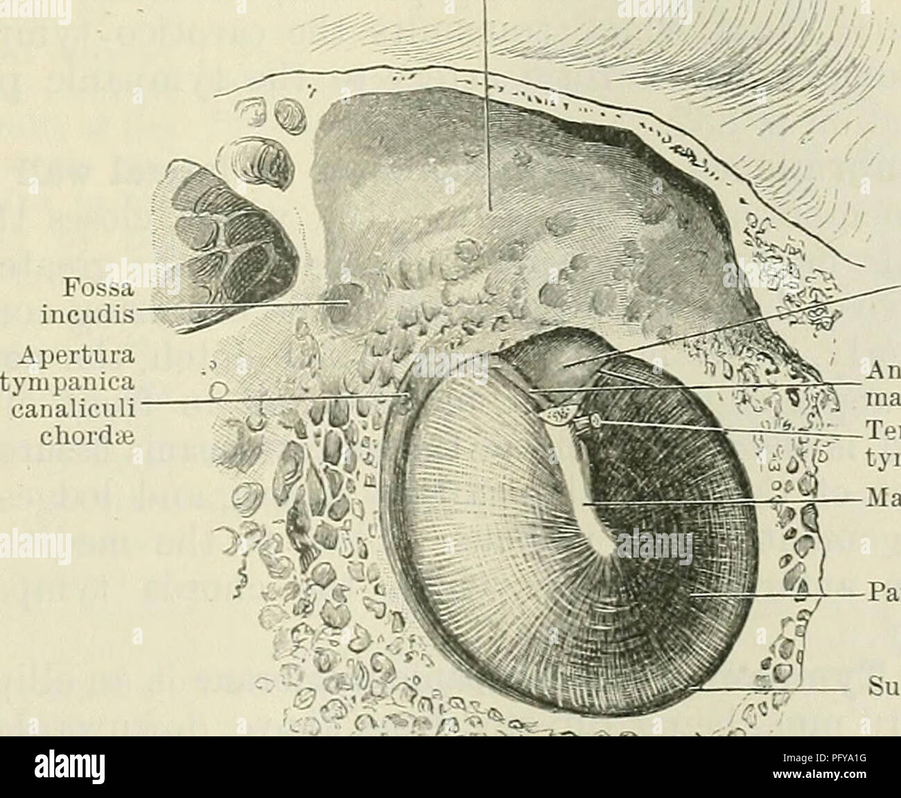 . Cunningham's Text-book of anatomy. Anatomy. Sinus tympani Mastoid air-cells Recessus epityrnpanicus Fenestra cochlea; Course of canalis facialis Fig. 709.—Section through Left Temporal Bone, showing labyriuthic wall of tympanic cavity, etc. is continued backwards and downwards behind the tympanic cavity, to end at the stylo-mastoid foramen. (4) The septum canalis musculotubarii (O.T. processus cochleariformis), which extends backwards, above the anterior end of the fenestra vestibuli, where it makes a sharp lateral curve, and forms a pulley over which the tendon of the tensor tympani muscle Stock Photohttps://www.alamy.com/image-license-details/?v=1https://www.alamy.com/cunninghams-text-book-of-anatomy-anatomy-sinus-tympani-mastoid-air-cells-recessus-epityrnpanicus-fenestra-cochlea-course-of-canalis-facialis-fig-709section-through-left-temporal-bone-showing-labyriuthic-wall-of-tympanic-cavity-etc-is-continued-backwards-and-downwards-behind-the-tympanic-cavity-to-end-at-the-stylo-mastoid-foramen-4-the-septum-canalis-musculotubarii-ot-processus-cochleariformis-which-extends-backwards-above-the-anterior-end-of-the-fenestra-vestibuli-where-it-makes-a-sharp-lateral-curve-and-forms-a-pulley-over-which-the-tendon-of-the-tensor-tympani-muscle-image216344844.html
RMPFYA1G–. Cunningham's Text-book of anatomy. Anatomy. Sinus tympani Mastoid air-cells Recessus epityrnpanicus Fenestra cochlea; Course of canalis facialis Fig. 709.—Section through Left Temporal Bone, showing labyriuthic wall of tympanic cavity, etc. is continued backwards and downwards behind the tympanic cavity, to end at the stylo-mastoid foramen. (4) The septum canalis musculotubarii (O.T. processus cochleariformis), which extends backwards, above the anterior end of the fenestra vestibuli, where it makes a sharp lateral curve, and forms a pulley over which the tendon of the tensor tympani muscle
 . Die Anatomie des Kaninschens in topographischer und operativer Rücksicht . Pars petromastoidea und tympa- nica des linken Os temporum, von der lateralen Seite. Me Meatus auditorius extemus. Pm Proces- sus mastoideus. Fs Poramen sty- lomastoideum. Bt Bulla tympani. F g Fissura Glaseri. Pars petromastoidea des linken Ostemporum, von unten. Cm Cel- lulae mastoideae. Fm Fossa mus- kularis major. Fr Promontorium. Fo Fenestra ovalis. Fr Fenestra rotunda. Pm Processus mastoi- deus, in optischer Verkürzung. Die mediale Wand zeigt das stark vorspringende Promontorium (Fig. 70 Pr), über dessen lateral Stock Photohttps://www.alamy.com/image-license-details/?v=1https://www.alamy.com/die-anatomie-des-kaninschens-in-topographischer-und-operativer-rcksicht-pars-petromastoidea-und-tympa-nica-des-linken-os-temporum-von-der-lateralen-seite-me-meatus-auditorius-extemus-pm-proces-sus-mastoideus-fs-poramen-sty-lomastoideum-bt-bulla-tympani-f-g-fissura-glaseri-pars-petromastoidea-des-linken-ostemporum-von-unten-cm-cel-lulae-mastoideae-fm-fossa-mus-kularis-major-fr-promontorium-fo-fenestra-ovalis-fr-fenestra-rotunda-pm-processus-mastoi-deus-in-optischer-verkrzung-die-mediale-wand-zeigt-das-stark-vorspringende-promontorium-fig-70-pr-ber-dessen-lateral-image181123678.html
RMMEJW2P–. Die Anatomie des Kaninschens in topographischer und operativer Rücksicht . Pars petromastoidea und tympa- nica des linken Os temporum, von der lateralen Seite. Me Meatus auditorius extemus. Pm Proces- sus mastoideus. Fs Poramen sty- lomastoideum. Bt Bulla tympani. F g Fissura Glaseri. Pars petromastoidea des linken Ostemporum, von unten. Cm Cel- lulae mastoideae. Fm Fossa mus- kularis major. Fr Promontorium. Fo Fenestra ovalis. Fr Fenestra rotunda. Pm Processus mastoi- deus, in optischer Verkürzung. Die mediale Wand zeigt das stark vorspringende Promontorium (Fig. 70 Pr), über dessen lateral
 . Annals of the New York Academy of Sciences. Science. PlGURE 5 Skull of a pseudosuchian, Euparkeria capensis. After Broom The borders of the large preorbital fenestra may serve for the attachment of the anterior part of the pterygoideus anterior muscle. Around the bony margin of the supratemporal fenestra arose the capiti-mandibularis. Both the supra- and the lateral temporal fenestra gave room for the expansion of the capiti-mandibularis, while the lateral fenestra of the mandible served a like function for the lower end of the same muscle. gaurus. The monimostylic type is quite stable in it Stock Photohttps://www.alamy.com/image-license-details/?v=1https://www.alamy.com/annals-of-the-new-york-academy-of-sciences-science-plgure-5-skull-of-a-pseudosuchian-euparkeria-capensis-after-broom-the-borders-of-the-large-preorbital-fenestra-may-serve-for-the-attachment-of-the-anterior-part-of-the-pterygoideus-anterior-muscle-around-the-bony-margin-of-the-supratemporal-fenestra-arose-the-capiti-mandibularis-both-the-supra-and-the-lateral-temporal-fenestra-gave-room-for-the-expansion-of-the-capiti-mandibularis-while-the-lateral-fenestra-of-the-mandible-served-a-like-function-for-the-lower-end-of-the-same-muscle-gaurus-the-monimostylic-type-is-quite-stable-in-it-image236515566.html
RMRMP60E–. Annals of the New York Academy of Sciences. Science. PlGURE 5 Skull of a pseudosuchian, Euparkeria capensis. After Broom The borders of the large preorbital fenestra may serve for the attachment of the anterior part of the pterygoideus anterior muscle. Around the bony margin of the supratemporal fenestra arose the capiti-mandibularis. Both the supra- and the lateral temporal fenestra gave room for the expansion of the capiti-mandibularis, while the lateral fenestra of the mandible served a like function for the lower end of the same muscle. gaurus. The monimostylic type is quite stable in it
 . The elements of embryology . Embryology. VIII.J THE SENSE CAPSULES. 241 iutemasal septum, continuous behind with the inter- orbital septum, grows up (Fig. 78); while the lateral Fig. 78. tlln. 7n^ Side view of the cartilaginous cranium of a Fowl on the SEVENTH DAT OF INCUBATION. (After Parker.) pn. prenasal cartilage, aln. alinasal cartilage, ale. aliethmoid; immediately below this is the ahseptal cartilage, eth. eth- moid, pp. para plana, ps. presphenoid or inter-orbital. pa. palatine, pg. pterygoid, z. optic nerve, as. aHsphenoid. q. quadrate, st. stapes, fr. fenestra rotunda, hso. horizon Stock Photohttps://www.alamy.com/image-license-details/?v=1https://www.alamy.com/the-elements-of-embryology-embryology-viiij-the-sense-capsules-241-iutemasal-septum-continuous-behind-with-the-inter-orbital-septum-grows-up-fig-78-while-the-lateral-fig-78-tlln-7n-side-view-of-the-cartilaginous-cranium-of-a-fowl-on-the-seventh-dat-of-incubation-after-parker-pn-prenasal-cartilage-aln-alinasal-cartilage-ale-aliethmoid-immediately-below-this-is-the-ahseptal-cartilage-eth-eth-moid-pp-para-plana-ps-presphenoid-or-inter-orbital-pa-palatine-pg-pterygoid-z-optic-nerve-as-ahsphenoid-q-quadrate-st-stapes-fr-fenestra-rotunda-hso-horizon-image216444003.html
RMPG3TEY–. The elements of embryology . Embryology. VIII.J THE SENSE CAPSULES. 241 iutemasal septum, continuous behind with the inter- orbital septum, grows up (Fig. 78); while the lateral Fig. 78. tlln. 7n^ Side view of the cartilaginous cranium of a Fowl on the SEVENTH DAT OF INCUBATION. (After Parker.) pn. prenasal cartilage, aln. alinasal cartilage, ale. aliethmoid; immediately below this is the ahseptal cartilage, eth. eth- moid, pp. para plana, ps. presphenoid or inter-orbital. pa. palatine, pg. pterygoid, z. optic nerve, as. aHsphenoid. q. quadrate, st. stapes, fr. fenestra rotunda, hso. horizon
 . Bulletin. Natural history; Natuurlijke historie. OSTEOLOGY OF DEINONYCHUS ANTIRRHOPUS 23 medial to the quadrate cotylus the fifth process extends medially and forward to contact the parietal. Without the adjacent bones, it is impossible to record the exact size and shape of the temporal fenestrae. However, the shape of the squamosal indicates a supe- rior temporal fenestra of at least moderate size. The lateral fenestra was quite deep dorsoventrally and may have been slightly restricted from behind at about mid- height, as it is in nearly all "carnosaur" skulls. Without knowledge o Stock Photohttps://www.alamy.com/image-license-details/?v=1https://www.alamy.com/bulletin-natural-history-natuurlijke-historie-osteology-of-deinonychus-antirrhopus-23-medial-to-the-quadrate-cotylus-the-fifth-process-extends-medially-and-forward-to-contact-the-parietal-without-the-adjacent-bones-it-is-impossible-to-record-the-exact-size-and-shape-of-the-temporal-fenestrae-however-the-shape-of-the-squamosal-indicates-a-supe-rior-temporal-fenestra-of-at-least-moderate-size-the-lateral-fenestra-was-quite-deep-dorsoventrally-and-may-have-been-slightly-restricted-from-behind-at-about-mid-height-as-it-is-in-nearly-all-quotcarnosaurquot-skulls-without-knowledge-o-image234211519.html
RMRH1753–. Bulletin. Natural history; Natuurlijke historie. OSTEOLOGY OF DEINONYCHUS ANTIRRHOPUS 23 medial to the quadrate cotylus the fifth process extends medially and forward to contact the parietal. Without the adjacent bones, it is impossible to record the exact size and shape of the temporal fenestrae. However, the shape of the squamosal indicates a supe- rior temporal fenestra of at least moderate size. The lateral fenestra was quite deep dorsoventrally and may have been slightly restricted from behind at about mid- height, as it is in nearly all "carnosaur" skulls. Without knowledge o
 . Cunningham's Text-book of anatomy. Anatomy. Kecessus ellipticus Crista vestibuli Recessus spha?ricus Cochlea Fenestra cochlea; Fenestra vestibuli Ampulla of posterior semi circular canal | Ampulla of lateral semi- circular canal Lateral semicircular canal Posterior semi- circular canal Crus commune. Scala tympani Lamina spiralis ossea Scala vestibuli Opening of aquseductus cochlea; Fenestra cochleae Opening of crus commune Opening of aqusductus vestibuli Fig. 716.—Left Bony Labyrinth (viewed from the lateral aspect). Recessus cochlearis Fig. 717.—Interior of Left Bony Labyrinth (viewed from Stock Photohttps://www.alamy.com/image-license-details/?v=1https://www.alamy.com/cunninghams-text-book-of-anatomy-anatomy-kecessus-ellipticus-crista-vestibuli-recessus-spharicus-cochlea-fenestra-cochlea-fenestra-vestibuli-ampulla-of-posterior-semi-circular-canal-ampulla-of-lateral-semi-circular-canal-lateral-semicircular-canal-posterior-semi-circular-canal-crus-commune-scala-tympani-lamina-spiralis-ossea-scala-vestibuli-opening-of-aquseductus-cochlea-fenestra-cochleae-opening-of-crus-commune-opening-of-aqusductus-vestibuli-fig-716left-bony-labyrinth-viewed-from-the-lateral-aspect-recessus-cochlearis-fig-717interior-of-left-bony-labyrinth-viewed-from-image216344806.html
RMPFYA06–. Cunningham's Text-book of anatomy. Anatomy. Kecessus ellipticus Crista vestibuli Recessus spha?ricus Cochlea Fenestra cochlea; Fenestra vestibuli Ampulla of posterior semi circular canal | Ampulla of lateral semi- circular canal Lateral semicircular canal Posterior semi- circular canal Crus commune. Scala tympani Lamina spiralis ossea Scala vestibuli Opening of aquseductus cochlea; Fenestra cochleae Opening of crus commune Opening of aqusductus vestibuli Fig. 716.—Left Bony Labyrinth (viewed from the lateral aspect). Recessus cochlearis Fig. 717.—Interior of Left Bony Labyrinth (viewed from
 . Breviora. BREVIORA No. 373 down this bar, between postorbital and jugal. A fraction of the latter bone is present, defining the lower margin of the orbit, a section of the cheek rim, and a small area of the anteroventral margin of the lateral temporal fenestra. Above this fenestra a stout bar of bone is present, presumably formed anteriorly by the postorbital, posteriorly by the squamosal (the suture between the two is not clear). An incomplete flange of the latter bone extends directly downward as part of the posterior border of the lateral fenestra. The squamosal extended backward beyond t Stock Photohttps://www.alamy.com/image-license-details/?v=1https://www.alamy.com/breviora-breviora-no-373-down-this-bar-between-postorbital-and-jugal-a-fraction-of-the-latter-bone-is-present-defining-the-lower-margin-of-the-orbit-a-section-of-the-cheek-rim-and-a-small-area-of-the-anteroventral-margin-of-the-lateral-temporal-fenestra-above-this-fenestra-a-stout-bar-of-bone-is-present-presumably-formed-anteriorly-by-the-postorbital-posteriorly-by-the-squamosal-the-suture-between-the-two-is-not-clear-an-incomplete-flange-of-the-latter-bone-extends-directly-downward-as-part-of-the-posterior-border-of-the-lateral-fenestra-the-squamosal-extended-backward-beyond-t-image234276936.html
RMRH46HC–. Breviora. BREVIORA No. 373 down this bar, between postorbital and jugal. A fraction of the latter bone is present, defining the lower margin of the orbit, a section of the cheek rim, and a small area of the anteroventral margin of the lateral temporal fenestra. Above this fenestra a stout bar of bone is present, presumably formed anteriorly by the postorbital, posteriorly by the squamosal (the suture between the two is not clear). An incomplete flange of the latter bone extends directly downward as part of the posterior border of the lateral fenestra. The squamosal extended backward beyond t
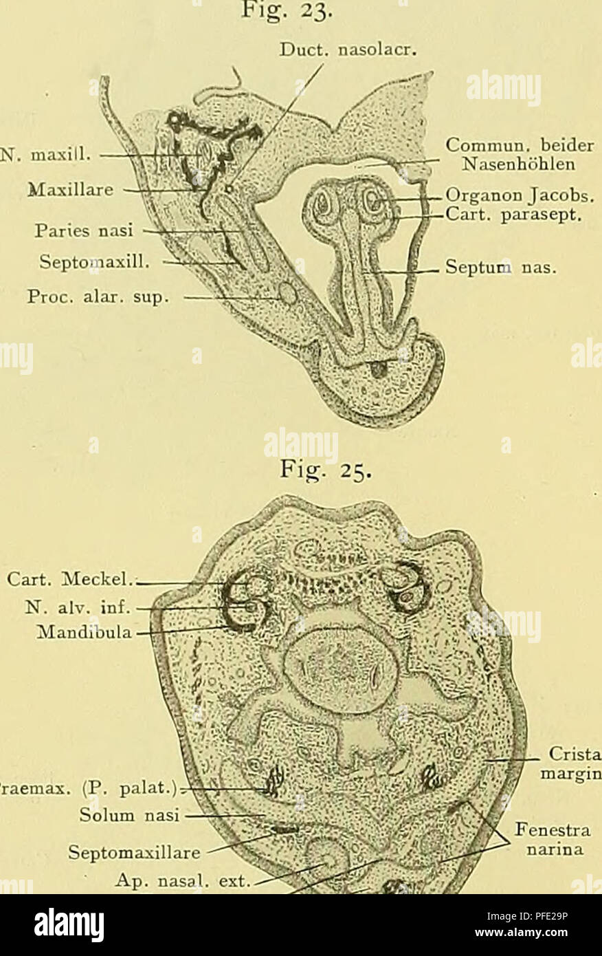 . Denkschriften der Medicinisch-Naturwissenschaftlichen Gesellschaft zu Jena. 58o Zur Entwicklungsgeschichte und vergleichenden Morphologie des Schädels von Echidna aculeata var. typica. Abschnitt abgetrennt wird. Die vordere Umgrenzung der Fenestra narina bildet der Kuppelknorpel (Cartilago cupularis), durch den das Dach in die Decke übergeht (Textfig. 23, 24). Am Ventral- umfang der Nasenhöhle findet sich, eingelagert in den „primären Boden", die Lamina trans- versalis anterior, medial in Homocontinuität mit dem ventralen Septumrand, lateral weit unter der Fenestra narina als Crista mar Stock Photohttps://www.alamy.com/image-license-details/?v=1https://www.alamy.com/denkschriften-der-medicinisch-naturwissenschaftlichen-gesellschaft-zu-jena-58o-zur-entwicklungsgeschichte-und-vergleichenden-morphologie-des-schdels-von-echidna-aculeata-var-typica-abschnitt-abgetrennt-wird-die-vordere-umgrenzung-der-fenestra-narina-bildet-der-kuppelknorpel-cartilago-cupularis-durch-den-das-dach-in-die-decke-bergeht-textfig-23-24-am-ventral-umfang-der-nasenhhle-findet-sich-eingelagert-in-den-primren-bodenquot-die-lamina-trans-versalis-anterior-medial-in-homocontinuitt-mit-dem-ventralen-septumrand-lateral-weit-unter-der-fenestra-narina-als-crista-mar-image216053426.html
RMPFE29P–. Denkschriften der Medicinisch-Naturwissenschaftlichen Gesellschaft zu Jena. 58o Zur Entwicklungsgeschichte und vergleichenden Morphologie des Schädels von Echidna aculeata var. typica. Abschnitt abgetrennt wird. Die vordere Umgrenzung der Fenestra narina bildet der Kuppelknorpel (Cartilago cupularis), durch den das Dach in die Decke übergeht (Textfig. 23, 24). Am Ventral- umfang der Nasenhöhle findet sich, eingelagert in den „primären Boden", die Lamina trans- versalis anterior, medial in Homocontinuität mit dem ventralen Septumrand, lateral weit unter der Fenestra narina als Crista mar
 . Bulletin. Natural history; Natuurlijke historie. 24 PEABODY MUSEUM BULLETIN 30 of bone almost triradiate in shape. Anteriorly a thin but deep process meets the posterior ramus of the maxilla in a weak overlapping contact. Posteriorly, a thin, tapered, two-pronged process contacted the quadratojugal in what appears to have been a weak tongue-and-groove articulation. Dorsally, a somewhat more ro- bust, grooved process met the postorbital. The latter separated the orbit and lat- eral temporal fenestra. The posterior process marks the ventral limit of the lateral fenestra. The sweeping curve of Stock Photohttps://www.alamy.com/image-license-details/?v=1https://www.alamy.com/bulletin-natural-history-natuurlijke-historie-24-peabody-museum-bulletin-30-of-bone-almost-triradiate-in-shape-anteriorly-a-thin-but-deep-process-meets-the-posterior-ramus-of-the-maxilla-in-a-weak-overlapping-contact-posteriorly-a-thin-tapered-two-pronged-process-contacted-the-quadratojugal-in-what-appears-to-have-been-a-weak-tongue-and-groove-articulation-dorsally-a-somewhat-more-ro-bust-grooved-process-met-the-postorbital-the-latter-separated-the-orbit-and-lat-eral-temporal-fenestra-the-posterior-process-marks-the-ventral-limit-of-the-lateral-fenestra-the-sweeping-curve-of-image234211502.html
RMRH174E–. Bulletin. Natural history; Natuurlijke historie. 24 PEABODY MUSEUM BULLETIN 30 of bone almost triradiate in shape. Anteriorly a thin but deep process meets the posterior ramus of the maxilla in a weak overlapping contact. Posteriorly, a thin, tapered, two-pronged process contacted the quadratojugal in what appears to have been a weak tongue-and-groove articulation. Dorsally, a somewhat more ro- bust, grooved process met the postorbital. The latter separated the orbit and lat- eral temporal fenestra. The posterior process marks the ventral limit of the lateral fenestra. The sweeping curve of
 . Denkschriften der Medicinisch-Naturwissenschaftlichen Gesellschaft zu Jena. qq Zur Entwickelungsgeschichte und vergleichenden Morphologie des Schädels von Echidna aculeata var. typica. 579 durchsetztes und im Uebrigen von Bindegewebe erfülltes Fenster, das ich als Fenestra er ib rosa bezeichnen will, weil es später von der Lamina cribrosa eingenommen wird; im präcerebralen Gebiet hat sich dagegen (aus den Lateralplatten) ein continuirliches Dach gebildet, das median mit dem Septum, lateral mit der Seitenwand zusammenhängt. Auch die letztere ist sehr vollständig, biegt hinten in die caudale W Stock Photohttps://www.alamy.com/image-license-details/?v=1https://www.alamy.com/denkschriften-der-medicinisch-naturwissenschaftlichen-gesellschaft-zu-jena-qq-zur-entwickelungsgeschichte-und-vergleichenden-morphologie-des-schdels-von-echidna-aculeata-var-typica-579-durchsetztes-und-im-uebrigen-von-bindegewebe-erflltes-fenster-das-ich-als-fenestra-er-ib-rosa-bezeichnen-will-weil-es-spter-von-der-lamina-cribrosa-eingenommen-wird-im-prcerebralen-gebiet-hat-sich-dagegen-aus-den-lateralplatten-ein-continuirliches-dach-gebildet-das-median-mit-dem-septum-lateral-mit-der-seitenwand-zusammenhngt-auch-die-letztere-ist-sehr-vollstndig-biegt-hinten-in-die-caudale-w-image216053438.html
RMPFE2A6–. Denkschriften der Medicinisch-Naturwissenschaftlichen Gesellschaft zu Jena. qq Zur Entwickelungsgeschichte und vergleichenden Morphologie des Schädels von Echidna aculeata var. typica. 579 durchsetztes und im Uebrigen von Bindegewebe erfülltes Fenster, das ich als Fenestra er ib rosa bezeichnen will, weil es später von der Lamina cribrosa eingenommen wird; im präcerebralen Gebiet hat sich dagegen (aus den Lateralplatten) ein continuirliches Dach gebildet, das median mit dem Septum, lateral mit der Seitenwand zusammenhängt. Auch die letztere ist sehr vollständig, biegt hinten in die caudale W
 Annals of the South African MuseumAnnale van die Suid-Afrikaanse Museum . osteriorly where much of the boneforming the posterior margin of the lateral temporal fenestra, the posteriorroot of the zygomatic arch, and the occiput has flaked from the matrix. Never-theless, the bone which is present, and impressions of bone in the matrix,clearly indicate that the outlines of the lateral temporal fenestra, temporalfossa, zygomatic arch, and the position of the quadrate correspond to thatshown in Figures 1A and 1C. Matrix has not been cleared from the temporalfossae or the orbits. For this reason it Stock Photohttps://www.alamy.com/image-license-details/?v=1https://www.alamy.com/annals-of-the-south-african-museumannale-van-die-suid-afrikaanse-museum-osteriorly-where-much-of-the-boneforming-the-posterior-margin-of-the-lateral-temporal-fenestra-the-posteriorroot-of-the-zygomatic-arch-and-the-occiput-has-flaked-from-the-matrix-never-theless-the-bone-which-is-present-and-impressions-of-bone-in-the-matrixclearly-indicate-that-the-outlines-of-the-lateral-temporal-fenestra-temporalfossa-zygomatic-arch-and-the-position-of-the-quadrate-correspond-to-thatshown-in-figures-1a-and-1c-matrix-has-not-been-cleared-from-the-temporalfossae-or-the-orbits-for-this-reason-it-image343268273.html
RM2AXD655–Annals of the South African MuseumAnnale van die Suid-Afrikaanse Museum . osteriorly where much of the boneforming the posterior margin of the lateral temporal fenestra, the posteriorroot of the zygomatic arch, and the occiput has flaked from the matrix. Never-theless, the bone which is present, and impressions of bone in the matrix,clearly indicate that the outlines of the lateral temporal fenestra, temporalfossa, zygomatic arch, and the position of the quadrate correspond to thatshown in Figures 1A and 1C. Matrix has not been cleared from the temporalfossae or the orbits. For this reason it
 A text-book of the diseases of the ear for students and practitioners . surface of the capitulumof the stapes, and is to be regarded as a true joint provided with a cavity(Fig. 32). The articular surfaces are covered with a thin layer of hyalinecartilage, and are united by a capsule containing many elastic fibres, whichpermits a considerable lateral mobility. According to Eiidinger, this joint isalso provided with a meniscus of fibro-cartilage. 3. Articulation of the Stapes and Fenestra Oval is (syndesmosistympano-stapedia).—The tissue connecting the margin of the fenestra ovaliswith that of t Stock Photohttps://www.alamy.com/image-license-details/?v=1https://www.alamy.com/a-text-book-of-the-diseases-of-the-ear-for-students-and-practitioners-surface-of-the-capitulumof-the-stapes-and-is-to-be-regarded-as-a-true-joint-provided-with-a-cavityfig-32-the-articular-surfaces-are-covered-with-a-thin-layer-of-hyalinecartilage-and-are-united-by-a-capsule-containing-many-elastic-fibres-whichpermits-a-considerable-lateral-mobility-according-to-eiidinger-this-joint-isalso-provided-with-a-meniscus-of-fibro-cartilage-3-articulation-of-the-stapes-and-fenestra-oval-is-syndesmosistympano-stapediathe-tissue-connecting-the-margin-of-the-fenestra-ovaliswith-that-of-t-image338478778.html
RM2AJK13P–A text-book of the diseases of the ear for students and practitioners . surface of the capitulumof the stapes, and is to be regarded as a true joint provided with a cavity(Fig. 32). The articular surfaces are covered with a thin layer of hyalinecartilage, and are united by a capsule containing many elastic fibres, whichpermits a considerable lateral mobility. According to Eiidinger, this joint isalso provided with a meniscus of fibro-cartilage. 3. Articulation of the Stapes and Fenestra Oval is (syndesmosistympano-stapedia).—The tissue connecting the margin of the fenestra ovaliswith that of t
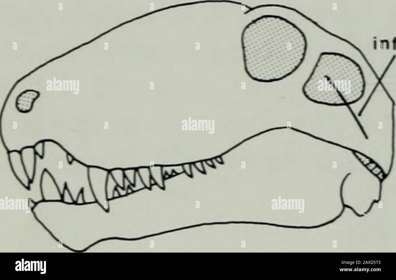 Annals of the South African MuseumAnnale van die Suid-Afrikaanse Museum . -prepared material examined while the author visited the South AfricanMuseum, Cape Town. This material included a skull and lower jaw ofL. declivis (Nat. Mus. C 403). ADDUCTOR JAW MUSCULATURE M. ADDUCTOR MANDIBULAE EXTERNUS The temporal region of Venjukoxia exhibits the posterodorsal enlargementof both the lateral temporal fenestra and temporal fossa which is characteristicof most therapsids as opposed to sphenacodontid pelycosaurs (cf. Figs 2A, C).In dorsal view (Fig. 1A) the temporal fossa is broadly exposed due to the Stock Photohttps://www.alamy.com/image-license-details/?v=1https://www.alamy.com/annals-of-the-south-african-museumannale-van-die-suid-afrikaanse-museum-prepared-material-examined-while-the-author-visited-the-south-africanmuseum-cape-town-this-material-included-a-skull-and-lower-jaw-ofl-declivis-nat-mus-c-403-adductor-jaw-musculature-m-adductor-mandibulae-externus-the-temporal-region-of-venjukoxia-exhibits-the-posterodorsal-enlargementof-both-the-lateral-temporal-fenestra-and-temporal-fossa-which-is-characteristicof-most-therapsids-as-opposed-to-sphenacodontid-pelycosaurs-cf-figs-2a-cin-dorsal-view-fig-1a-the-temporal-fossa-is-broadly-exposed-due-to-the-image343268019.html
RM2AXD5T3–Annals of the South African MuseumAnnale van die Suid-Afrikaanse Museum . -prepared material examined while the author visited the South AfricanMuseum, Cape Town. This material included a skull and lower jaw ofL. declivis (Nat. Mus. C 403). ADDUCTOR JAW MUSCULATURE M. ADDUCTOR MANDIBULAE EXTERNUS The temporal region of Venjukoxia exhibits the posterodorsal enlargementof both the lateral temporal fenestra and temporal fossa which is characteristicof most therapsids as opposed to sphenacodontid pelycosaurs (cf. Figs 2A, C).In dorsal view (Fig. 1A) the temporal fossa is broadly exposed due to the
 A text-book of the diseases of the ear and adjacent organs . d, d, Sections ofthe superior semicircular canal ; e, Internal auditory meatus ; /, Stapes ; g, Antrummastoideum. merge into each other without any well-defined demarcation. Its lateral wall,chiefly directed downwards, is formed in great part by the fenestra ovalis THE OSSEOUS LABYRINTH. 585 closed by the footplate of the stapes, which is 3 mm. long, and If mm. wide. Onthe median and inferior walls are placed two depressions separated by the cristavestibuli, and destined for the reception of the two saccules of the vestibule ;the ant Stock Photohttps://www.alamy.com/image-license-details/?v=1https://www.alamy.com/a-text-book-of-the-diseases-of-the-ear-and-adjacent-organs-d-d-sections-ofthe-superior-semicircular-canal-e-internal-auditory-meatus-stapes-g-antrummastoideum-merge-into-each-other-without-any-well-defined-demarcation-its-lateral-wallchiefly-directed-downwards-is-formed-in-great-part-by-the-fenestra-ovalis-the-osseous-labyrinth-585-closed-by-the-footplate-of-the-stapes-which-is-3-mm-long-and-if-mm-wide-onthe-median-and-inferior-walls-are-placed-two-depressions-separated-by-the-cristavestibuli-and-destined-for-the-reception-of-the-two-saccules-of-the-vestibule-the-ant-image339968815.html
RM2AN2WKB–A text-book of the diseases of the ear and adjacent organs . d, d, Sections ofthe superior semicircular canal ; e, Internal auditory meatus ; /, Stapes ; g, Antrummastoideum. merge into each other without any well-defined demarcation. Its lateral wall,chiefly directed downwards, is formed in great part by the fenestra ovalis THE OSSEOUS LABYRINTH. 585 closed by the footplate of the stapes, which is 3 mm. long, and If mm. wide. Onthe median and inferior walls are placed two depressions separated by the cristavestibuli, and destined for the reception of the two saccules of the vestibule ;the ant
 . Journal of morphology. 146 147 Figs. 146 and 147 A living egg of Cryptobranchus allegheniensis in an earlyneural groove stage, viewed so far as possible by transmitted light. Figure 146shows the upper hemisphere, figure 147 a postero-dorsal view. X 7. lateral bands are obscured by the thickening of the neural plate;in the central portion of the neural groove there usually appeara series of translucent pits arranged at regular intervals. Thepit marking the site of the closing fenestra has disappeared, butthere usually remains a translucent area indicating a vestige ofthe blastocoele; this are Stock Photohttps://www.alamy.com/image-license-details/?v=1https://www.alamy.com/journal-of-morphology-146-147-figs-146-and-147-a-living-egg-of-cryptobranchus-allegheniensis-in-an-earlyneural-groove-stage-viewed-so-far-as-possible-by-transmitted-light-figure-146shows-the-upper-hemisphere-figure-147-a-postero-dorsal-view-x-7-lateral-bands-are-obscured-by-the-thickening-of-the-neural-platein-the-central-portion-of-the-neural-groove-there-usually-appeara-series-of-translucent-pits-arranged-at-regular-intervals-thepit-marking-the-site-of-the-closing-fenestra-has-disappeared-butthere-usually-remains-a-translucent-area-indicating-a-vestige-ofthe-blastocoele-this-are-image336782873.html
RM2AFWNYN–. Journal of morphology. 146 147 Figs. 146 and 147 A living egg of Cryptobranchus allegheniensis in an earlyneural groove stage, viewed so far as possible by transmitted light. Figure 146shows the upper hemisphere, figure 147 a postero-dorsal view. X 7. lateral bands are obscured by the thickening of the neural plate;in the central portion of the neural groove there usually appeara series of translucent pits arranged at regular intervals. Thepit marking the site of the closing fenestra has disappeared, butthere usually remains a translucent area indicating a vestige ofthe blastocoele; this are
 . Outlines of zoology. FiG. 278. —Restored skeletonof PalcBospondylus gunni.—After Traquair. d.c. Cirri of dorsal margin ; l.c.^long lateral cirri; v.c, cirri ofventral margin ; n., nasal ring ;t.f.i anterior ^trabeculo-palatinepart of cranium; h.^ anteriordepression or fenestra; c, pos-terior depression or fenestra;a., lobe divided off from anteriorpart; p.a., posterior or para-chordal part of cranium; jr.,post-occipital plates. 528 HYPOSTOMATA. bral centra with neural arches. , Towards the tail the arches areproduced into slender neural spines, opposite which are shorterhaemal ones. Class HY Stock Photohttps://www.alamy.com/image-license-details/?v=1https://www.alamy.com/outlines-of-zoology-fig-278-restored-skeletonof-palcbospondylus-gunniafter-traquair-dc-cirri-of-dorsal-margin-lclong-lateral-cirri-vc-cirri-ofventral-margin-n-nasal-ring-tfi-anterior-trabeculo-palatinepart-of-cranium-h-anteriordepression-or-fenestra-c-pos-terior-depression-or-fenestraa-lobe-divided-off-from-anteriorpart-pa-posterior-or-para-chordal-part-of-cranium-jrpost-occipital-plates-528-hypostomata-bral-centra-with-neural-arches-towards-the-tail-the-arches-areproduced-into-slender-neural-spines-opposite-which-are-shorterhaemal-ones-class-hy-image337130019.html
RM2AGDGNR–. Outlines of zoology. FiG. 278. —Restored skeletonof PalcBospondylus gunni.—After Traquair. d.c. Cirri of dorsal margin ; l.c.^long lateral cirri; v.c, cirri ofventral margin ; n., nasal ring ;t.f.i anterior ^trabeculo-palatinepart of cranium; h.^ anteriordepression or fenestra; c, pos-terior depression or fenestra;a., lobe divided off from anteriorpart; p.a., posterior or para-chordal part of cranium; jr.,post-occipital plates. 528 HYPOSTOMATA. bral centra with neural arches. , Towards the tail the arches areproduced into slender neural spines, opposite which are shorterhaemal ones. Class HY
 . Journal of morphology . lateral andslightly dorsal to it. It is bounded mesially by the nasal bone andlatero-ventrally by the premaxillary and maxillary. The posterior-portion of the gland, however, becomes partially enclosed betweenthe maxillary bone and the cartilaginous nasal capsule.The Journal of Morphology—Vol. XX, No. 1. 144 Inez Whipple Wilder. From near the anterior end of the gland, in all of the specimenswhich I have examined, a single duct was found to extend over theposterior margin of the fenestra rostralis (Bruners nomenclature)of the cartilaginous capsule, from whence it pass Stock Photohttps://www.alamy.com/image-license-details/?v=1https://www.alamy.com/journal-of-morphology-lateral-andslightly-dorsal-to-it-it-is-bounded-mesially-by-the-nasal-bone-andlatero-ventrally-by-the-premaxillary-and-maxillary-the-posterior-portion-of-the-gland-however-becomes-partially-enclosed-betweenthe-maxillary-bone-and-the-cartilaginous-nasal-capsulethe-journal-of-morphologyvol-xx-no-1-144-inez-whipple-wilder-from-near-the-anterior-end-of-the-gland-in-all-of-the-specimenswhich-i-have-examined-a-single-duct-was-found-to-extend-over-theposterior-margin-of-the-fenestra-rostralis-bruners-nomenclatureof-the-cartilaginous-capsule-from-whence-it-pass-image369768754.html
RM2CDGBPX–. Journal of morphology . lateral andslightly dorsal to it. It is bounded mesially by the nasal bone andlatero-ventrally by the premaxillary and maxillary. The posterior-portion of the gland, however, becomes partially enclosed betweenthe maxillary bone and the cartilaginous nasal capsule.The Journal of Morphology—Vol. XX, No. 1. 144 Inez Whipple Wilder. From near the anterior end of the gland, in all of the specimenswhich I have examined, a single duct was found to extend over theposterior margin of the fenestra rostralis (Bruners nomenclature)of the cartilaginous capsule, from whence it pass
 . Birds: the elements of ornithology . one with two lateral notches on either side ofit, as in the Common Fowl; or its posterior margin may betransversely continuous, while a little in front of it there maybe two vacuities side by side, or five in a transverse series.Each such vacuity is called a fenestra or a fontanelle. Asternum which has neither notches nor fenestrse is calledentire, and, as just said, it may be single-notched or doiible-notclied, or it may be unifenestrate or bifenestrate. In the overwhelming majority of birds there is a keel, whence 178 ELEMENTS OF OENITHOLOGT. they are c Stock Photohttps://www.alamy.com/image-license-details/?v=1https://www.alamy.com/birds-the-elements-of-ornithology-one-with-two-lateral-notches-on-either-side-ofit-as-in-the-common-fowl-or-its-posterior-margin-may-betransversely-continuous-while-a-little-in-front-of-it-there-maybe-two-vacuities-side-by-side-or-five-in-a-transverse-serieseach-such-vacuity-is-called-a-fenestra-or-a-fontanelle-asternum-which-has-neither-notches-nor-fenestrse-is-calledentire-and-as-just-said-it-may-be-single-notched-or-doiible-notclied-or-it-may-be-unifenestrate-or-bifenestrate-in-the-overwhelming-majority-of-birds-there-is-a-keel-whence-178-elements-of-oenithologt-they-are-c-image375303545.html
RM2CPGFE1–. Birds: the elements of ornithology . one with two lateral notches on either side ofit, as in the Common Fowl; or its posterior margin may betransversely continuous, while a little in front of it there maybe two vacuities side by side, or five in a transverse series.Each such vacuity is called a fenestra or a fontanelle. Asternum which has neither notches nor fenestrse is calledentire, and, as just said, it may be single-notched or doiible-notclied, or it may be unifenestrate or bifenestrate. In the overwhelming majority of birds there is a keel, whence 178 ELEMENTS OF OENITHOLOGT. they are c
 . Journal of morphology . ducibranch skulls just described, e. g. WotopJi-tJialmus (PI. XVII) and Cj/nops (PI. XVIII), the vestibule is in the form ofa smooth bulla, with the stapes set on behind; and that plate is either quitesoft or very slightly ossified; it is also relatively small. In Spelerpes (PI. XXI, Figs. 2, 3, and PI. XVIII, Fig. 10) the vestibuleis but little protuberant; its fenestra is lateral, and corresponding with thestapes, very large. The Columella Auris in Amphibia. 579 Despite the depression for the M. opercularis the ear capsule hasin this region a bulging appearance due Stock Photohttps://www.alamy.com/image-license-details/?v=1https://www.alamy.com/journal-of-morphology-ducibranch-skulls-just-described-e-g-wotopji-tjialmus-pi-xvii-and-cjnops-pi-xviii-the-vestibule-is-in-the-form-ofa-smooth-bulla-with-the-stapes-set-on-behind-and-that-plate-is-either-quitesoft-or-very-slightly-ossified-it-is-also-relatively-small-in-spelerpes-pi-xxi-figs-2-3-and-pi-xviii-fig-10-the-vestibuleis-but-little-protuberant-its-fenestra-is-lateral-and-corresponding-with-thestapes-very-large-the-columella-auris-in-amphibia-579-despite-the-depression-for-the-m-opercularis-the-ear-capsule-hasin-this-region-a-bulging-appearance-due-image369751887.html
RM2CDFJ8F–. Journal of morphology . ducibranch skulls just described, e. g. WotopJi-tJialmus (PI. XVII) and Cj/nops (PI. XVIII), the vestibule is in the form ofa smooth bulla, with the stapes set on behind; and that plate is either quitesoft or very slightly ossified; it is also relatively small. In Spelerpes (PI. XXI, Figs. 2, 3, and PI. XVIII, Fig. 10) the vestibuleis but little protuberant; its fenestra is lateral, and corresponding with thestapes, very large. The Columella Auris in Amphibia. 579 Despite the depression for the M. opercularis the ear capsule hasin this region a bulging appearance due
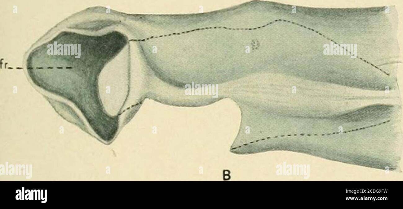 . Journal of morphology . gland,dl, the dilatator muscle. dlo, the origin of the dilatator muscle cut from the inner surface of thenasal capsule. fr, fenestra rostralis. g, lateral gland. in, introductory nasal passage. M, maxillary bone. N, nasal bone. na, nasal capsule. ne, nasal epithelium. EXPLANATION OF,PLATE I. Fig. a. Dissection of snout of adult Amphiuma showing the relation ofthe laternal or external nasal gland to siurounding parts. Fig. B. Drawing, based on a millimeter paper reconstruction, showinga lateral view of the anterior portion of the left cartilaginous nasal capsuleof an a Stock Photohttps://www.alamy.com/image-license-details/?v=1https://www.alamy.com/journal-of-morphology-glanddl-the-dilatator-muscle-dlo-the-origin-of-the-dilatator-muscle-cut-from-the-inner-surface-of-thenasal-capsule-fr-fenestra-rostralis-g-lateral-gland-in-introductory-nasal-passage-m-maxillary-bone-n-nasal-bone-na-nasal-capsule-ne-nasal-epithelium-explanation-ofplate-i-fig-a-dissection-of-snout-of-adult-amphiuma-showing-the-relation-ofthe-laternal-or-external-nasal-gland-to-siurounding-parts-fig-b-drawing-based-on-a-millimeter-paper-reconstruction-showinga-lateral-view-of-the-anterior-portion-of-the-left-cartilaginous-nasal-capsuleof-an-a-image369766989.html
RM2CDG9FW–. Journal of morphology . gland,dl, the dilatator muscle. dlo, the origin of the dilatator muscle cut from the inner surface of thenasal capsule. fr, fenestra rostralis. g, lateral gland. in, introductory nasal passage. M, maxillary bone. N, nasal bone. na, nasal capsule. ne, nasal epithelium. EXPLANATION OF,PLATE I. Fig. a. Dissection of snout of adult Amphiuma showing the relation ofthe laternal or external nasal gland to siurounding parts. Fig. B. Drawing, based on a millimeter paper reconstruction, showinga lateral view of the anterior portion of the left cartilaginous nasal capsuleof an a
 . Odontornithes: a monograph on the extinct toothed birds of North America; with thirty-four plates and forty woodcuts . Lachrymal bone.of âIuter-orbital fenestra.qj âQuadrato-jugal bone.q âQuadrate bone.bo âBasi-occipital. Fig. 2.âLeft lower jaw ; lateral view of external surface, .... 11 t âTeeth still in place. Fig. 3.âLeft lower jaw ; seen from above, -.. : .. 11 a âGroove for the teeth, with indications of sockets. b âGroove for the reception of the upper teeth when the jaws were closed c âAnglo of mandible. Fig. 4.âRight lower jaw ; inner side, 11 d âSymphysial surface, showing that the Stock Photohttps://www.alamy.com/image-license-details/?v=1https://www.alamy.com/odontornithes-a-monograph-on-the-extinct-toothed-birds-of-north-america-with-thirty-four-plates-and-forty-woodcuts-lachrymal-boneof-iuter-orbital-fenestraqj-quadrato-jugal-boneq-quadrate-bonebo-basi-occipital-fig-2left-lower-jaw-lateral-view-of-external-surface-11-t-teeth-still-in-place-fig-3left-lower-jaw-seen-from-above-11-a-groove-for-the-teeth-with-indications-of-sockets-b-groove-for-the-reception-of-the-upper-teeth-when-the-jaws-were-closed-c-anglo-of-mandible-fig-4right-lower-jaw-inner-side-11-d-symphysial-surface-showing-that-the-image375343921.html
RM2CPJB01–. Odontornithes: a monograph on the extinct toothed birds of North America; with thirty-four plates and forty woodcuts . Lachrymal bone.of âIuter-orbital fenestra.qj âQuadrato-jugal bone.q âQuadrate bone.bo âBasi-occipital. Fig. 2.âLeft lower jaw ; lateral view of external surface, .... 11 t âTeeth still in place. Fig. 3.âLeft lower jaw ; seen from above, -.. : .. 11 a âGroove for the teeth, with indications of sockets. b âGroove for the reception of the upper teeth when the jaws were closed c âAnglo of mandible. Fig. 4.âRight lower jaw ; inner side, 11 d âSymphysial surface, showing that the
 . Chordate morphology. Morphology (Animals); Chordata. preorticular coronoids articular angular etroarticula dentosplenlal supraangulor. articular retroarticulor angular sphenotic posterior orbitosphenoid arrow through lateral canal epiotic anterior orbitosphenoid nasal capsule tooth fenestra. Please note that these images are extracted from scanned page images that may have been digitally enhanced for readability - coloration and appearance of these illustrations may not perfectly resemble the original work.. Jollie, Malcolm. New York, Reinhold Stock Photohttps://www.alamy.com/image-license-details/?v=1https://www.alamy.com/chordate-morphology-morphology-animals-chordata-preorticular-coronoids-articular-angular-etroarticula-dentosplenlal-supraangulor-articular-retroarticulor-angular-sphenotic-posterior-orbitosphenoid-arrow-through-lateral-canal-epiotic-anterior-orbitosphenoid-nasal-capsule-tooth-fenestra-please-note-that-these-images-are-extracted-from-scanned-page-images-that-may-have-been-digitally-enhanced-for-readability-coloration-and-appearance-of-these-illustrations-may-not-perfectly-resemble-the-original-work-jollie-malcolm-new-york-reinhold-image234908820.html
RMRJ50GM–. Chordate morphology. Morphology (Animals); Chordata. preorticular coronoids articular angular etroarticula dentosplenlal supraangulor. articular retroarticulor angular sphenotic posterior orbitosphenoid arrow through lateral canal epiotic anterior orbitosphenoid nasal capsule tooth fenestra. Please note that these images are extracted from scanned page images that may have been digitally enhanced for readability - coloration and appearance of these illustrations may not perfectly resemble the original work.. Jollie, Malcolm. New York, Reinhold
 . Comparative anatomy of vertebrates. Anatomy, Comparative; Vertebrates. 106 COMPARATIVE ANATOMY On the dorsal side of the chondrocraniutn are a large median anterior fontanelle or fenestra, and a smaller paired posterior fontanelle, and the cartilage becomes replaced by bones in the occipital, auditory, and ethmoid regions (Figs. 77 and 78). There are paired exoccipital and prootics and a sphenethmoid, the lateral parts of which correspond in position to the orbitosphenoids of Urodeles, and which encircles the whole skull; it also forms a front wall to the cranial capsule, perforated by the o Stock Photohttps://www.alamy.com/image-license-details/?v=1https://www.alamy.com/comparative-anatomy-of-vertebrates-anatomy-comparative-vertebrates-106-comparative-anatomy-on-the-dorsal-side-of-the-chondrocraniutn-are-a-large-median-anterior-fontanelle-or-fenestra-and-a-smaller-paired-posterior-fontanelle-and-the-cartilage-becomes-replaced-by-bones-in-the-occipital-auditory-and-ethmoid-regions-figs-77-and-78-there-are-paired-exoccipital-and-prootics-and-a-sphenethmoid-the-lateral-parts-of-which-correspond-in-position-to-the-orbitosphenoids-of-urodeles-and-which-encircles-the-whole-skull-it-also-forms-a-front-wall-to-the-cranial-capsule-perforated-by-the-o-image232667661.html
RMREEWY9–. Comparative anatomy of vertebrates. Anatomy, Comparative; Vertebrates. 106 COMPARATIVE ANATOMY On the dorsal side of the chondrocraniutn are a large median anterior fontanelle or fenestra, and a smaller paired posterior fontanelle, and the cartilage becomes replaced by bones in the occipital, auditory, and ethmoid regions (Figs. 77 and 78). There are paired exoccipital and prootics and a sphenethmoid, the lateral parts of which correspond in position to the orbitosphenoids of Urodeles, and which encircles the whole skull; it also forms a front wall to the cranial capsule, perforated by the o
 . Bulletin. Natural history; Natuurlijke historie. 36 REVISION OF THE GYMNARTHRIDAE through the base of the deep pocket into which the hypoglossal foramina open (but not through these foramina). Its lateral edge bounds the fenestra ovalis and is in contact with the stapes throughout. Ventrally it bounds the posterior extension of the fenestra ovalis mentioned above.. Please note that these images are extracted from scanned page images that may have been digitally enhanced for readability - coloration and appearance of these illustrations may not perfectly resemble the original work.. Peabody M Stock Photohttps://www.alamy.com/image-license-details/?v=1https://www.alamy.com/bulletin-natural-history-natuurlijke-historie-36-revision-of-the-gymnarthridae-through-the-base-of-the-deep-pocket-into-which-the-hypoglossal-foramina-open-but-not-through-these-foramina-its-lateral-edge-bounds-the-fenestra-ovalis-and-is-in-contact-with-the-stapes-throughout-ventrally-it-bounds-the-posterior-extension-of-the-fenestra-ovalis-mentioned-above-please-note-that-these-images-are-extracted-from-scanned-page-images-that-may-have-been-digitally-enhanced-for-readability-coloration-and-appearance-of-these-illustrations-may-not-perfectly-resemble-the-original-work-peabody-m-image234225852.html
RMRH1WD0–. Bulletin. Natural history; Natuurlijke historie. 36 REVISION OF THE GYMNARTHRIDAE through the base of the deep pocket into which the hypoglossal foramina open (but not through these foramina). Its lateral edge bounds the fenestra ovalis and is in contact with the stapes throughout. Ventrally it bounds the posterior extension of the fenestra ovalis mentioned above.. Please note that these images are extracted from scanned page images that may have been digitally enhanced for readability - coloration and appearance of these illustrations may not perfectly resemble the original work.. Peabody M
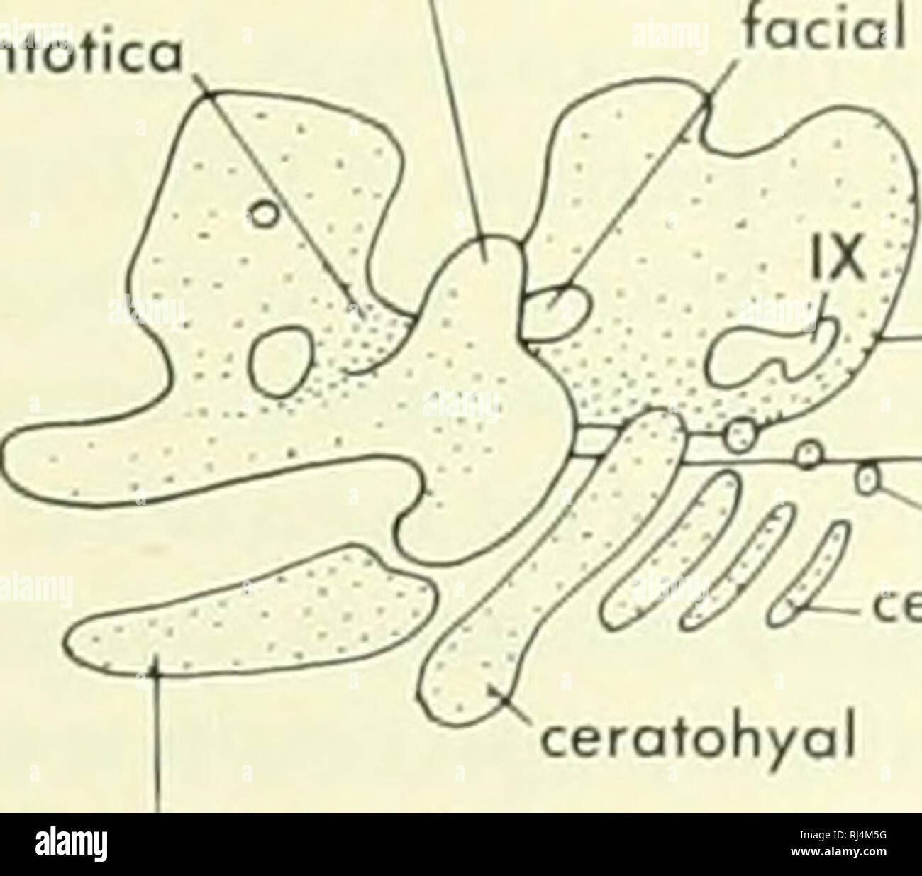 . Chordate morphology. Morphology (Animals); Chordata. trabecule irbilol cartilage otic process of quadrate cartilage pila ontotica B. quadrate cartilage bosicopsular fenestra lateral woll of otic capsule/' notochord quadrate corttlagt facial foramen otic capsuli vestibular fontonelle D epibranchials cerotobronchiols l-lll. Please note that these images are extracted from scanned page images that may have been digitally enhanced for readability - coloration and appearance of these illustrations may not perfectly resemble the original work.. Jollie, Malcolm. New York, Reinhold Stock Photohttps://www.alamy.com/image-license-details/?v=1https://www.alamy.com/chordate-morphology-morphology-animals-chordata-trabecule-irbilol-cartilage-otic-process-of-quadrate-cartilage-pila-ontotica-b-quadrate-cartilage-bosicopsular-fenestra-lateral-woll-of-otic-capsule-notochord-quadrate-corttlagt-facial-foramen-otic-capsuli-vestibular-fontonelle-d-epibranchials-cerotobronchiols-l-lll-please-note-that-these-images-are-extracted-from-scanned-page-images-that-may-have-been-digitally-enhanced-for-readability-coloration-and-appearance-of-these-illustrations-may-not-perfectly-resemble-the-original-work-jollie-malcolm-new-york-reinhold-image234902236.html
RMRJ4M5G–. Chordate morphology. Morphology (Animals); Chordata. trabecule irbilol cartilage otic process of quadrate cartilage pila ontotica B. quadrate cartilage bosicopsular fenestra lateral woll of otic capsule/' notochord quadrate corttlagt facial foramen otic capsuli vestibular fontonelle D epibranchials cerotobronchiols l-lll. Please note that these images are extracted from scanned page images that may have been digitally enhanced for readability - coloration and appearance of these illustrations may not perfectly resemble the original work.. Jollie, Malcolm. New York, Reinhold
 . Bulletin of the Museum of Comparative Zoology at Harvard College. Zoology. 46 bulletin: museum of comparative zoology In many cases, and perhaps generally, in the more primitive genera, the upturned postero-lateral corner of the parasphenoid forms part of the margin of the fenestra ovalis, or at least closely approaches the. Please note that these images are extracted from scanned page images that may have been digitally enhanced for readability - coloration and appearance of these illustrations may not perfectly resemble the original work.. Harvard University. Museum of Comparative Zoology. Stock Photohttps://www.alamy.com/image-license-details/?v=1https://www.alamy.com/bulletin-of-the-museum-of-comparative-zoology-at-harvard-college-zoology-46-bulletin-museum-of-comparative-zoology-in-many-cases-and-perhaps-generally-in-the-more-primitive-genera-the-upturned-postero-lateral-corner-of-the-parasphenoid-forms-part-of-the-margin-of-the-fenestra-ovalis-or-at-least-closely-approaches-the-please-note-that-these-images-are-extracted-from-scanned-page-images-that-may-have-been-digitally-enhanced-for-readability-coloration-and-appearance-of-these-illustrations-may-not-perfectly-resemble-the-original-work-harvard-university-museum-of-comparative-zoology-image233904289.html
RMRGF78H–. Bulletin of the Museum of Comparative Zoology at Harvard College. Zoology. 46 bulletin: museum of comparative zoology In many cases, and perhaps generally, in the more primitive genera, the upturned postero-lateral corner of the parasphenoid forms part of the margin of the fenestra ovalis, or at least closely approaches the. Please note that these images are extracted from scanned page images that may have been digitally enhanced for readability - coloration and appearance of these illustrations may not perfectly resemble the original work.. Harvard University. Museum of Comparative Zoology.
 . Annals of the South African Museum = Annale van die Suid-Afrikaanse Museum. Natural history. BRAINCASE, BASICRANIAL AXIS, MEDIAN SEPTUM IN THE DINOCEPHALIA 265 The anterior part of the prootic is pierced by two small foramina for the Vlth and Vllth cranial nerves. The fenestra ovalis is bounded by the opisthotic, basioccipital, basisphenoid and prootic and is situated quite high up in the skull. The epipterygoid has a long footplate and a fairly broad ascending columella. It lies lateral to the sella turcica and to the anterior edges of the prootic and supraoccipital and meets the parietal.. Stock Photohttps://www.alamy.com/image-license-details/?v=1https://www.alamy.com/annals-of-the-south-african-museum-=-annale-van-die-suid-afrikaanse-museum-natural-history-braincase-basicranial-axis-median-septum-in-the-dinocephalia-265-the-anterior-part-of-the-prootic-is-pierced-by-two-small-foramina-for-the-vlth-and-vllth-cranial-nerves-the-fenestra-ovalis-is-bounded-by-the-opisthotic-basioccipital-basisphenoid-and-prootic-and-is-situated-quite-high-up-in-the-skull-the-epipterygoid-has-a-long-footplate-and-a-fairly-broad-ascending-columella-it-lies-lateral-to-the-sella-turcica-and-to-the-anterior-edges-of-the-prootic-and-supraoccipital-and-meets-the-parietal-image236466440.html
RMRMKYA0–. Annals of the South African Museum = Annale van die Suid-Afrikaanse Museum. Natural history. BRAINCASE, BASICRANIAL AXIS, MEDIAN SEPTUM IN THE DINOCEPHALIA 265 The anterior part of the prootic is pierced by two small foramina for the Vlth and Vllth cranial nerves. The fenestra ovalis is bounded by the opisthotic, basioccipital, basisphenoid and prootic and is situated quite high up in the skull. The epipterygoid has a long footplate and a fairly broad ascending columella. It lies lateral to the sella turcica and to the anterior edges of the prootic and supraoccipital and meets the parietal..
 . Chordate morphology. Morphology (Animals); Chordata. extrascapular series postparietal postspiracular supratemporal parietal sclerotic ring prefrontal^ nasal series septomaxilla internosals intertemporal premaxilla lateral rostra anterior and posterior splenial A marginal gulars postcleithrum. clavicle pectoral fin anterior palatine fenestra premaxilla. palatine /o". ectopterygoid. tip of notochord. parosphenoid tooth plates B. Please note that these images are extracted from scanned page images that may have been digitally enhanced for readability - coloration and appearance of these i Stock Photohttps://www.alamy.com/image-license-details/?v=1https://www.alamy.com/chordate-morphology-morphology-animals-chordata-extrascapular-series-postparietal-postspiracular-supratemporal-parietal-sclerotic-ring-prefrontal-nasal-series-septomaxilla-internosals-intertemporal-premaxilla-lateral-rostra-anterior-and-posterior-splenial-a-marginal-gulars-postcleithrum-clavicle-pectoral-fin-anterior-palatine-fenestra-premaxilla-palatine-oquot-ectopterygoid-tip-of-notochord-parosphenoid-tooth-plates-b-please-note-that-these-images-are-extracted-from-scanned-page-images-that-may-have-been-digitally-enhanced-for-readability-coloration-and-appearance-of-these-i-image234902595.html
RMRJ4MJB–. Chordate morphology. Morphology (Animals); Chordata. extrascapular series postparietal postspiracular supratemporal parietal sclerotic ring prefrontal^ nasal series septomaxilla internosals intertemporal premaxilla lateral rostra anterior and posterior splenial A marginal gulars postcleithrum. clavicle pectoral fin anterior palatine fenestra premaxilla. palatine /o". ectopterygoid. tip of notochord. parosphenoid tooth plates B. Please note that these images are extracted from scanned page images that may have been digitally enhanced for readability - coloration and appearance of these i
 . Chordate morphology. Morphology (Animals); Chordata. articular retroarticulor angular sphenotic posterior orbitosphenoid arrow through lateral canal epiotic anterior orbitosphenoid nasal capsule tooth fenestra. posterior orbitosphenoid anterior orbitosphenoid lateral ethmoid 1 basioccipitol premaxilla fused to mesethmoid porasphenoid prootic IX bosipterygoid process. Please note that these images are extracted from scanned page images that may have been digitally enhanced for readability - coloration and appearance of these illustrations may not perfectly resemble the original work.. Jolli Stock Photohttps://www.alamy.com/image-license-details/?v=1https://www.alamy.com/chordate-morphology-morphology-animals-chordata-articular-retroarticulor-angular-sphenotic-posterior-orbitosphenoid-arrow-through-lateral-canal-epiotic-anterior-orbitosphenoid-nasal-capsule-tooth-fenestra-posterior-orbitosphenoid-anterior-orbitosphenoid-lateral-ethmoid-1-basioccipitol-premaxilla-fused-to-mesethmoid-porasphenoid-prootic-ix-bosipterygoid-process-please-note-that-these-images-are-extracted-from-scanned-page-images-that-may-have-been-digitally-enhanced-for-readability-coloration-and-appearance-of-these-illustrations-may-not-perfectly-resemble-the-original-work-jolli-image234908814.html
RMRJ50GE–. Chordate morphology. Morphology (Animals); Chordata. articular retroarticulor angular sphenotic posterior orbitosphenoid arrow through lateral canal epiotic anterior orbitosphenoid nasal capsule tooth fenestra. posterior orbitosphenoid anterior orbitosphenoid lateral ethmoid 1 basioccipitol premaxilla fused to mesethmoid porasphenoid prootic IX bosipterygoid process. Please note that these images are extracted from scanned page images that may have been digitally enhanced for readability - coloration and appearance of these illustrations may not perfectly resemble the original work.. Jolli
 . Chordate morphology. Morphology (Animals); Chordata. clavicle pectoral fin anterior palatine fenestra premaxilla. palatine /o". ectopterygoid. tip of notochord. parosphenoid tooth plates B. anterior medial gular^ internal nasal opening [A^interpterygoid fenestra ^V^rostal part of parosphenoid II hypophyseal fenestra — pterygoid marginal gulars quodratojugal quadrate lateral canal vestibular fontanelle posterior splenial supraongulor. Please note that these images are extracted from scanned page images that may have been digitally enhanced for readability - coloration and appearance of Stock Photohttps://www.alamy.com/image-license-details/?v=1https://www.alamy.com/chordate-morphology-morphology-animals-chordata-clavicle-pectoral-fin-anterior-palatine-fenestra-premaxilla-palatine-oquot-ectopterygoid-tip-of-notochord-parosphenoid-tooth-plates-b-anterior-medial-gular-internal-nasal-opening-ainterpterygoid-fenestra-vrostal-part-of-parosphenoid-ii-hypophyseal-fenestra-pterygoid-marginal-gulars-quodratojugal-quadrate-lateral-canal-vestibular-fontanelle-posterior-splenial-supraongulor-please-note-that-these-images-are-extracted-from-scanned-page-images-that-may-have-been-digitally-enhanced-for-readability-coloration-and-appearance-of-image234902573.html
RMRJ4MHH–. Chordate morphology. Morphology (Animals); Chordata. clavicle pectoral fin anterior palatine fenestra premaxilla. palatine /o". ectopterygoid. tip of notochord. parosphenoid tooth plates B. anterior medial gular^ internal nasal opening [A^interpterygoid fenestra ^V^rostal part of parosphenoid II hypophyseal fenestra — pterygoid marginal gulars quodratojugal quadrate lateral canal vestibular fontanelle posterior splenial supraongulor. Please note that these images are extracted from scanned page images that may have been digitally enhanced for readability - coloration and appearance of
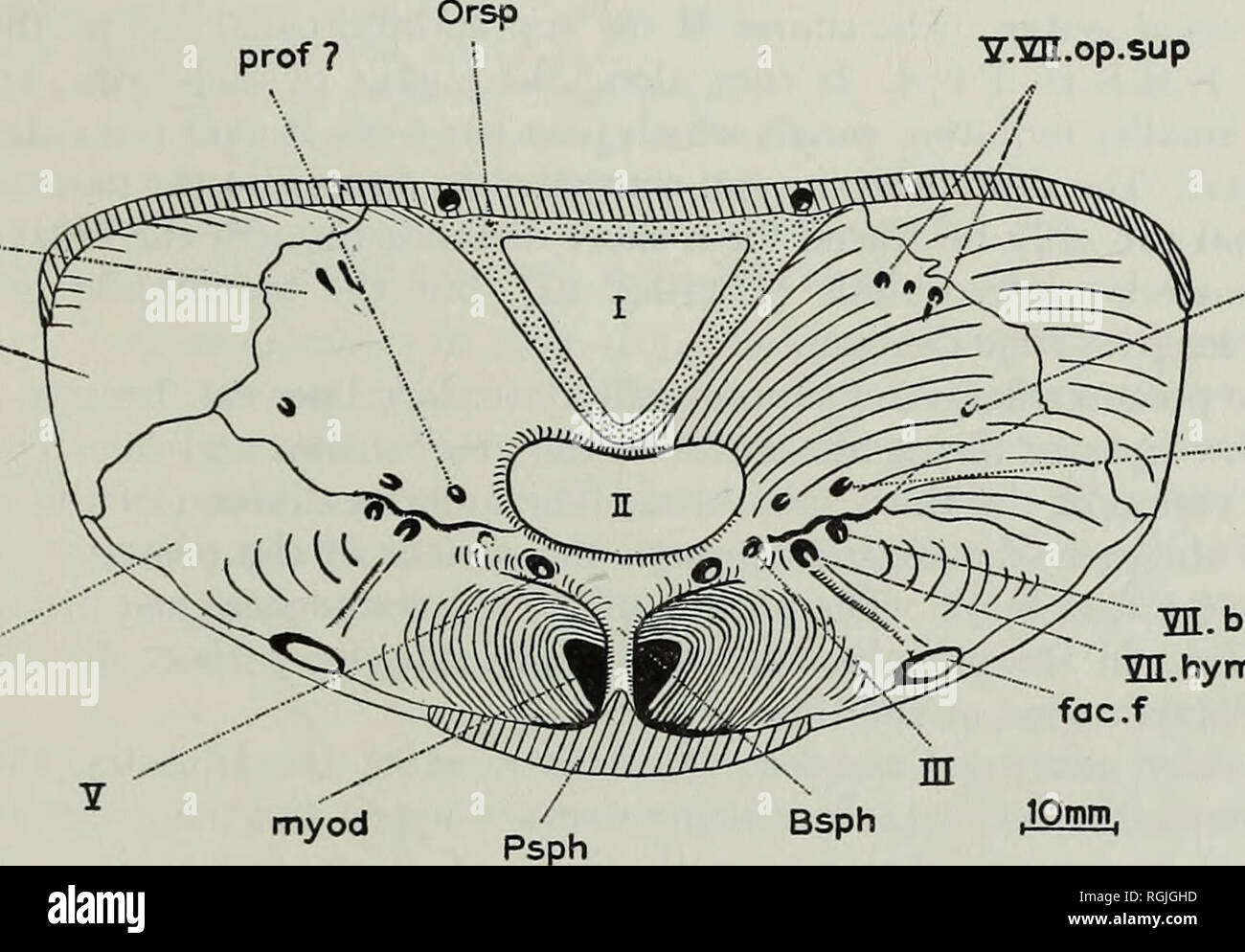 . Bulletin of the British Museum (Natural History), Geology. REVISION OF ACTINOPTERYGIAN AND COELACANTH FISHES 287 is found in the prootic, the so-called facial foramen. This foramen transmitted the hyomandibular branch of the facial nerve. The orbital surface is relatively easy to interpret (Text-fig. 27). The autosphenotic forms a large portion of the dorso-lateral region. The extent of the other orbital bones, pterosphenoid, prootic, basisphenoid and orbitosphenoid, can all be seen from Text-fig. 27. The optic foramen is large and confluent with the infra-orbital fenestra. There is a comple Stock Photohttps://www.alamy.com/image-license-details/?v=1https://www.alamy.com/bulletin-of-the-british-museum-natural-history-geology-revision-of-actinopterygian-and-coelacanth-fishes-287-is-found-in-the-prootic-the-so-called-facial-foramen-this-foramen-transmitted-the-hyomandibular-branch-of-the-facial-nerve-the-orbital-surface-is-relatively-easy-to-interpret-text-fig-27-the-autosphenotic-forms-a-large-portion-of-the-dorso-lateral-region-the-extent-of-the-other-orbital-bones-pterosphenoid-prootic-basisphenoid-and-orbitosphenoid-can-all-be-seen-from-text-fig-27-the-optic-foramen-is-large-and-confluent-with-the-infra-orbital-fenestra-there-is-a-comple-image233977449.html
RMRGJGHD–. Bulletin of the British Museum (Natural History), Geology. REVISION OF ACTINOPTERYGIAN AND COELACANTH FISHES 287 is found in the prootic, the so-called facial foramen. This foramen transmitted the hyomandibular branch of the facial nerve. The orbital surface is relatively easy to interpret (Text-fig. 27). The autosphenotic forms a large portion of the dorso-lateral region. The extent of the other orbital bones, pterosphenoid, prootic, basisphenoid and orbitosphenoid, can all be seen from Text-fig. 27. The optic foramen is large and confluent with the infra-orbital fenestra. There is a comple
 . Bulletin of the Museum of Comparative Zoology at Harvard College. Zoology. Borborophagus Koskinonodon Fig. 42. American metoposaurs. After Branson and Mehl. and this material may be an over-development of the somewhat similar, if smaller, squamosal buttress seen, for example, in Edops. Along the lateral part of the suspensorium, the skull roof is separated by a long oval fenestra from the palato-quadrate complex below it.. Please note that these images are extracted from scanned page images that may have been digitally enhanced for readability - coloration and appearance of these illustratio Stock Photohttps://www.alamy.com/image-license-details/?v=1https://www.alamy.com/bulletin-of-the-museum-of-comparative-zoology-at-harvard-college-zoology-borborophagus-koskinonodon-fig-42-american-metoposaurs-after-branson-and-mehl-and-this-material-may-be-an-over-development-of-the-somewhat-similar-if-smaller-squamosal-buttress-seen-for-example-in-edops-along-the-lateral-part-of-the-suspensorium-the-skull-roof-is-separated-by-a-long-oval-fenestra-from-the-palato-quadrate-complex-below-it-please-note-that-these-images-are-extracted-from-scanned-page-images-that-may-have-been-digitally-enhanced-for-readability-coloration-and-appearance-of-these-illustratio-image233876396.html
RMRGDYMC–. Bulletin of the Museum of Comparative Zoology at Harvard College. Zoology. Borborophagus Koskinonodon Fig. 42. American metoposaurs. After Branson and Mehl. and this material may be an over-development of the somewhat similar, if smaller, squamosal buttress seen, for example, in Edops. Along the lateral part of the suspensorium, the skull roof is separated by a long oval fenestra from the palato-quadrate complex below it.. Please note that these images are extracted from scanned page images that may have been digitally enhanced for readability - coloration and appearance of these illustratio
 . Chordate morphology. Morphology (Animals); Chordata. parasphenoid supraoccipitol prootic prootic supratrigeminal process facial toromen poroccipital process internal carotid ntercalore quadrate parasphenoid squamosal pterygoid VII / / ^—" basipterygoid process jugal quadratojugal ascending process temporal fenestra ^supraoccipitol. quadratojugal basisphenoid Figure 4-8. Skull of Sphenodon. A, rear view; B, lateral view of cranium with temporal and labial arches removed; C, medial view of right half of cranium. from the lateral nasal wall into the nasal passage. The basic peculiarities o Stock Photohttps://www.alamy.com/image-license-details/?v=1https://www.alamy.com/chordate-morphology-morphology-animals-chordata-parasphenoid-supraoccipitol-prootic-prootic-supratrigeminal-process-facial-toromen-poroccipital-process-internal-carotid-ntercalore-quadrate-parasphenoid-squamosal-pterygoid-vii-quot-basipterygoid-process-jugal-quadratojugal-ascending-process-temporal-fenestra-supraoccipitol-quadratojugal-basisphenoid-figure-4-8-skull-of-sphenodon-a-rear-view-b-lateral-view-of-cranium-with-temporal-and-labial-arches-removed-c-medial-view-of-right-half-of-cranium-from-the-lateral-nasal-wall-into-the-nasal-passage-the-basic-peculiarities-o-image234924229.html
RMRJ5M71–. Chordate morphology. Morphology (Animals); Chordata. parasphenoid supraoccipitol prootic prootic supratrigeminal process facial toromen poroccipital process internal carotid ntercalore quadrate parasphenoid squamosal pterygoid VII / / ^—" basipterygoid process jugal quadratojugal ascending process temporal fenestra ^supraoccipitol. quadratojugal basisphenoid Figure 4-8. Skull of Sphenodon. A, rear view; B, lateral view of cranium with temporal and labial arches removed; C, medial view of right half of cranium. from the lateral nasal wall into the nasal passage. The basic peculiarities o
 . The anatomical record. Anatomy; Anatomy. 278 B. W. KUNKEL concentrated tissue occupying the ventro-lateral angle of the planum basale and stretching caudally. In an embryo having a carapace-length of 8 mm. the stapes inferior is still separate from both the planum basale and the otic capsule. The fenestral plate of the columella is well sepa- rated from the margins of the fenestra especially along its ventral margin, but even along the upper margin where the continuity between the fenestral plate and the capsule is more apparent, the differentiation is appreciable (figs. 14-21). On the ventr Stock Photohttps://www.alamy.com/image-license-details/?v=1https://www.alamy.com/the-anatomical-record-anatomy-anatomy-278-b-w-kunkel-concentrated-tissue-occupying-the-ventro-lateral-angle-of-the-planum-basale-and-stretching-caudally-in-an-embryo-having-a-carapace-length-of-8-mm-the-stapes-inferior-is-still-separate-from-both-the-planum-basale-and-the-otic-capsule-the-fenestral-plate-of-the-columella-is-well-sepa-rated-from-the-margins-of-the-fenestra-especially-along-its-ventral-margin-but-even-along-the-upper-margin-where-the-continuity-between-the-fenestral-plate-and-the-capsule-is-more-apparent-the-differentiation-is-appreciable-figs-14-21-on-the-ventr-image236874392.html
RMRNAFKM–. The anatomical record. Anatomy; Anatomy. 278 B. W. KUNKEL concentrated tissue occupying the ventro-lateral angle of the planum basale and stretching caudally. In an embryo having a carapace-length of 8 mm. the stapes inferior is still separate from both the planum basale and the otic capsule. The fenestral plate of the columella is well sepa- rated from the margins of the fenestra especially along its ventral margin, but even along the upper margin where the continuity between the fenestral plate and the capsule is more apparent, the differentiation is appreciable (figs. 14-21). On the ventr
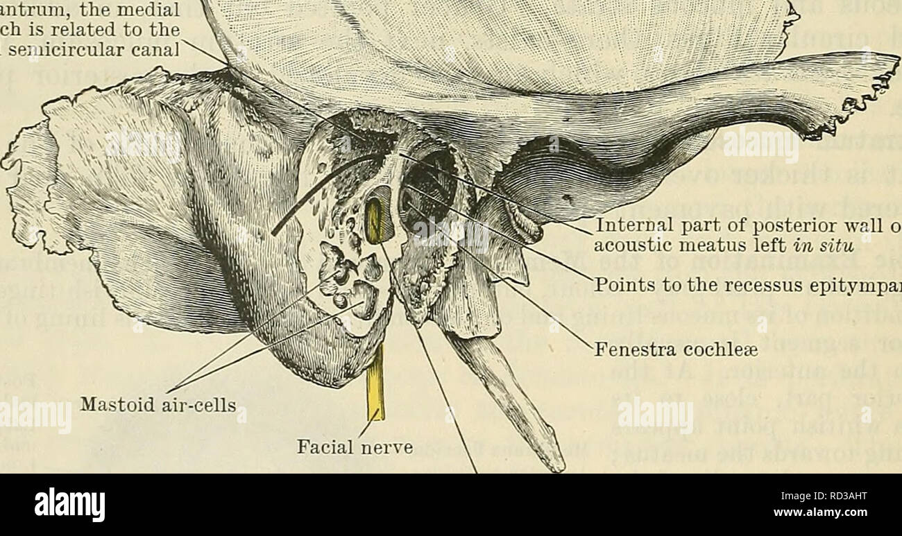 . Cunningham's Text-book of anatomy. Anatomy. Tympanic antrum, the medial : wall of which is related to the 5 lateral semicircular canal. Internal part of posterior wall of external acoustic meatus left in situ Points to the recessus epitympanicus Fenestra cochleae Mastoid air-cells Facial canal laid open, displaying the facial nerve within Fig. 712. Preparation to display the position and relations of the tympanic antrum. The greater part of the posterior wall of the external acoustic meatus has been removed, leaving only a bridge of bone at its inner ex- tremity ; under this a bristle is Stock Photohttps://www.alamy.com/image-license-details/?v=1https://www.alamy.com/cunninghams-text-book-of-anatomy-anatomy-tympanic-antrum-the-medial-wall-of-which-is-related-to-the-5-lateral-semicircular-canal-internal-part-of-posterior-wall-of-external-acoustic-meatus-left-in-situ-points-to-the-recessus-epitympanicus-fenestra-cochleae-mastoid-air-cells-facial-canal-laid-open-displaying-the-facial-nerve-within-fig-712-preparation-to-display-the-position-and-relations-of-the-tympanic-antrum-the-greater-part-of-the-posterior-wall-of-the-external-acoustic-meatus-has-been-removed-leaving-only-a-bridge-of-bone-at-its-inner-ex-tremity-under-this-a-bristle-is-image231799508.html
RMRD3AHT–. Cunningham's Text-book of anatomy. Anatomy. Tympanic antrum, the medial : wall of which is related to the 5 lateral semicircular canal. Internal part of posterior wall of external acoustic meatus left in situ Points to the recessus epitympanicus Fenestra cochleae Mastoid air-cells Facial canal laid open, displaying the facial nerve within Fig. 712. Preparation to display the position and relations of the tympanic antrum. The greater part of the posterior wall of the external acoustic meatus has been removed, leaving only a bridge of bone at its inner ex- tremity ; under this a bristle is
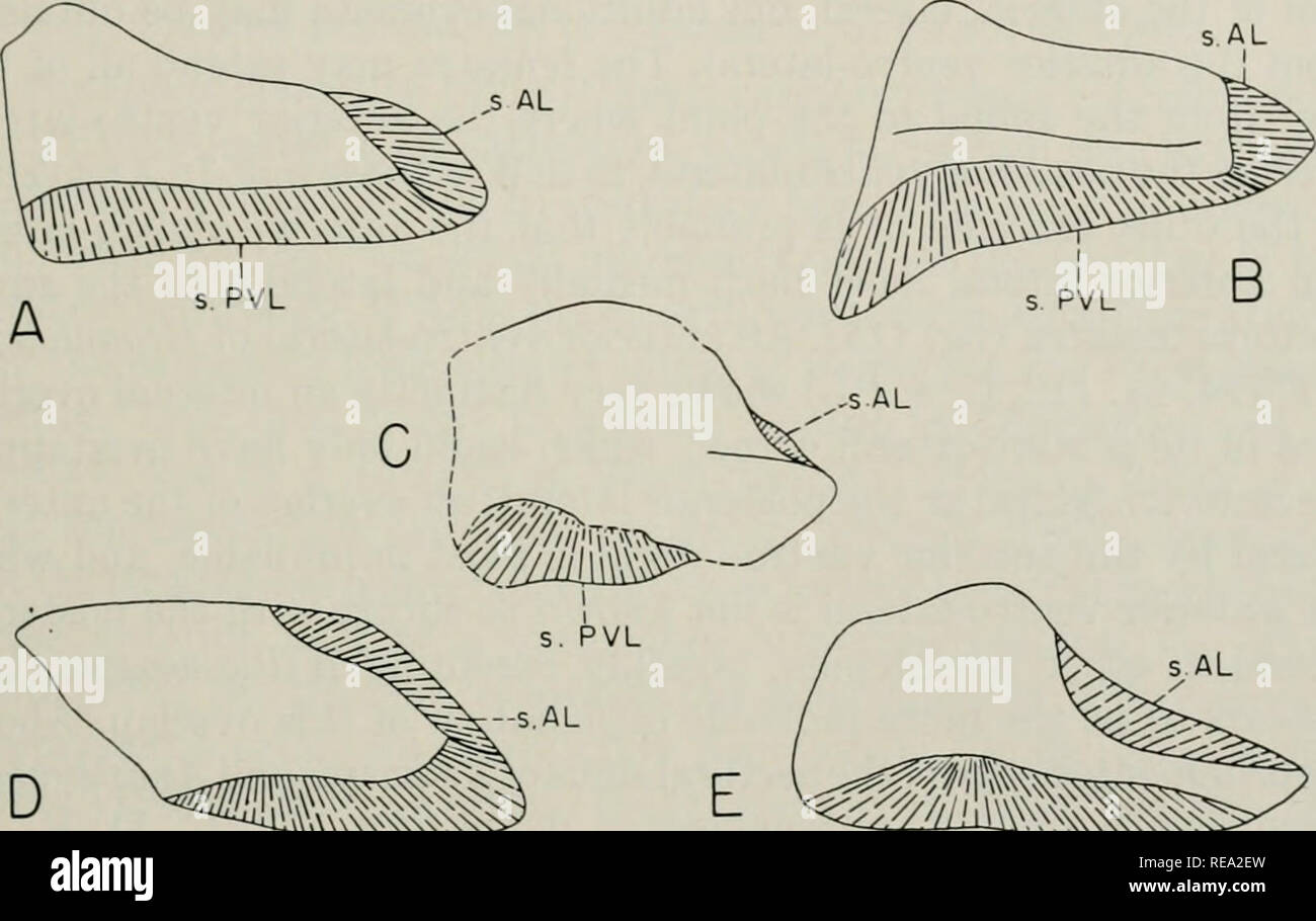 . Early Devonian fishes from Utah : Arthrodira. Fishes, Fossil; Paleontology -- Devonian; Paleontology -- Utah. DENISON: EARLY DEVONIAN FISHES 527 fenestra, at least in the form upon which this part of the restoration was based. Williamsaspis has a very large posterior lateral, relatively short and very high, peculiarly shaped, laterally ridged, and with a rather large edge for the pectoral fenestra; presumably this is a specialized condition, since nothing approaching it is found in other euarthrodires. In the Brachythoraci the posterior lateral is almost. -^-^W s.PVL s.PVL Fig. 111. Right p Stock Photohttps://www.alamy.com/image-license-details/?v=1https://www.alamy.com/early-devonian-fishes-from-utah-arthrodira-fishes-fossil-paleontology-devonian-paleontology-utah-denison-early-devonian-fishes-527-fenestra-at-least-in-the-form-upon-which-this-part-of-the-restoration-was-based-williamsaspis-has-a-very-large-posterior-lateral-relatively-short-and-very-high-peculiarly-shaped-laterally-ridged-and-with-a-rather-large-edge-for-the-pectoral-fenestra-presumably-this-is-a-specialized-condition-since-nothing-approaching-it-is-found-in-other-euarthrodires-in-the-brachythoraci-the-posterior-lateral-is-almost-w-spvl-spvl-fig-111-right-p-image232561473.html
RMREA2EW–. Early Devonian fishes from Utah : Arthrodira. Fishes, Fossil; Paleontology -- Devonian; Paleontology -- Utah. DENISON: EARLY DEVONIAN FISHES 527 fenestra, at least in the form upon which this part of the restoration was based. Williamsaspis has a very large posterior lateral, relatively short and very high, peculiarly shaped, laterally ridged, and with a rather large edge for the pectoral fenestra; presumably this is a specialized condition, since nothing approaching it is found in other euarthrodires. In the Brachythoraci the posterior lateral is almost. -^-^W s.PVL s.PVL Fig. 111. Right p
 . Breviora. 6 BREVIORA No. 379 fenestra. The posterior end of the skull roof is narrow; on either side, however, each parietal sends, posterolaterally, a long process to meet and overlap the medial surface of the squamosal behind the superior temporal fenestra. Narrow dorsally but broadening below, the medial surface of this process forms the upper lateral boundary of the occipital plate and laterally forms the medial boundary of the superior temporal fenestra. This boundary is sharply marked off dorsally; more ventrally, how- ever, the parietal slants outward to meet, obviously, the prootic a Stock Photohttps://www.alamy.com/image-license-details/?v=1https://www.alamy.com/breviora-6-breviora-no-379-fenestra-the-posterior-end-of-the-skull-roof-is-narrow-on-either-side-however-each-parietal-sends-posterolaterally-a-long-process-to-meet-and-overlap-the-medial-surface-of-the-squamosal-behind-the-superior-temporal-fenestra-narrow-dorsally-but-broadening-below-the-medial-surface-of-this-process-forms-the-upper-lateral-boundary-of-the-occipital-plate-and-laterally-forms-the-medial-boundary-of-the-superior-temporal-fenestra-this-boundary-is-sharply-marked-off-dorsally-more-ventrally-how-ever-the-parietal-slants-outward-to-meet-obviously-the-prootic-a-image234271908.html
RMRH405T–. Breviora. 6 BREVIORA No. 379 fenestra. The posterior end of the skull roof is narrow; on either side, however, each parietal sends, posterolaterally, a long process to meet and overlap the medial surface of the squamosal behind the superior temporal fenestra. Narrow dorsally but broadening below, the medial surface of this process forms the upper lateral boundary of the occipital plate and laterally forms the medial boundary of the superior temporal fenestra. This boundary is sharply marked off dorsally; more ventrally, how- ever, the parietal slants outward to meet, obviously, the prootic a
 . Breviora. 12 BREVIORA No. 379 upper margin of the ramus, whence a dorsally facing area of the surangular extends inward above the lateral border of the man- dibular fossa. The angular extends backward below the lateral mandibular fenestra, the two elements meeting at the posterior end of the fenestra, whence a ridge, with which the suture be- tween the two elements is associated, runs posteriorly. The suture is indistinct posteriorly but the conjoined angular and surangular extend backward nearly to the posterior end of the jaw, sheathing the articular laterally. On the inner surface the spl Stock Photohttps://www.alamy.com/image-license-details/?v=1https://www.alamy.com/breviora-12-breviora-no-379-upper-margin-of-the-ramus-whence-a-dorsally-facing-area-of-the-surangular-extends-inward-above-the-lateral-border-of-the-man-dibular-fossa-the-angular-extends-backward-below-the-lateral-mandibular-fenestra-the-two-elements-meeting-at-the-posterior-end-of-the-fenestra-whence-a-ridge-with-which-the-suture-be-tween-the-two-elements-is-associated-runs-posteriorly-the-suture-is-indistinct-posteriorly-but-the-conjoined-angular-and-surangular-extend-backward-nearly-to-the-posterior-end-of-the-jaw-sheathing-the-articular-laterally-on-the-inner-surface-the-spl-image234271878.html
RMRH404P–. Breviora. 12 BREVIORA No. 379 upper margin of the ramus, whence a dorsally facing area of the surangular extends inward above the lateral border of the man- dibular fossa. The angular extends backward below the lateral mandibular fenestra, the two elements meeting at the posterior end of the fenestra, whence a ridge, with which the suture be- tween the two elements is associated, runs posteriorly. The suture is indistinct posteriorly but the conjoined angular and surangular extend backward nearly to the posterior end of the jaw, sheathing the articular laterally. On the inner surface the spl
 . Annals of the South African Museum = Annale van die Suid-Afrikaanse Museum. Natural history. 252 ANNALS OF THE SOUTH AFRICAN MUSEUM destroyed in transit. More recently, information has also been gained from acid-prepared material examined while the author visited the South African Museum, Cape Town. This material included a skull and lower jaw of L. declivis (Nat. Mus. C 403). ADDUCTOR JAW MUSCULATURE M. ADDUCTOR MANDIBULAE EXTERNUS The temporal region of Venjukovia exhibits the posterodorsal enlargement of both the lateral temporal fenestra and temporal fossa which is characteristic of most Stock Photohttps://www.alamy.com/image-license-details/?v=1https://www.alamy.com/annals-of-the-south-african-museum-=-annale-van-die-suid-afrikaanse-museum-natural-history-252-annals-of-the-south-african-museum-destroyed-in-transit-more-recently-information-has-also-been-gained-from-acid-prepared-material-examined-while-the-author-visited-the-south-african-museum-cape-town-this-material-included-a-skull-and-lower-jaw-of-l-declivis-nat-mus-c-403-adductor-jaw-musculature-m-adductor-mandibulae-externus-the-temporal-region-of-venjukovia-exhibits-the-posterodorsal-enlargement-of-both-the-lateral-temporal-fenestra-and-temporal-fossa-which-is-characteristic-of-most-image236496245.html
RMRMN9AD–. Annals of the South African Museum = Annale van die Suid-Afrikaanse Museum. Natural history. 252 ANNALS OF THE SOUTH AFRICAN MUSEUM destroyed in transit. More recently, information has also been gained from acid-prepared material examined while the author visited the South African Museum, Cape Town. This material included a skull and lower jaw of L. declivis (Nat. Mus. C 403). ADDUCTOR JAW MUSCULATURE M. ADDUCTOR MANDIBULAE EXTERNUS The temporal region of Venjukovia exhibits the posterodorsal enlargement of both the lateral temporal fenestra and temporal fossa which is characteristic of most
 . Annals of the South African Museum = Annale van die Suid-Afrikaanse Museum. Natural history. 394 ANNALS OF THE SOUTH AFRICAN MUSEUM Soc. Pro Fig. 14. Moschorhinus kitchingi. Lateral view. These two elements form an anterodorsally curving bar that forms the anterior border of the pterygo-paroccipital foramen. The dorsal crest-like border of this bar is a continuation of the central ridge of the prootic. The anteroventral border of this bar forms a sharp concave crest running from the dorsal lip of the fenestra ovalis medially to the posteromedial corner of the posteroventral process of the ep Stock Photohttps://www.alamy.com/image-license-details/?v=1https://www.alamy.com/annals-of-the-south-african-museum-=-annale-van-die-suid-afrikaanse-museum-natural-history-394-annals-of-the-south-african-museum-soc-pro-fig-14-moschorhinus-kitchingi-lateral-view-these-two-elements-form-an-anterodorsally-curving-bar-that-forms-the-anterior-border-of-the-pterygo-paroccipital-foramen-the-dorsal-crest-like-border-of-this-bar-is-a-continuation-of-the-central-ridge-of-the-prootic-the-anteroventral-border-of-this-bar-forms-a-sharp-concave-crest-running-from-the-dorsal-lip-of-the-fenestra-ovalis-medially-to-the-posteromedial-corner-of-the-posteroventral-process-of-the-ep-image236407039.html
RMRMH7GF–. Annals of the South African Museum = Annale van die Suid-Afrikaanse Museum. Natural history. 394 ANNALS OF THE SOUTH AFRICAN MUSEUM Soc. Pro Fig. 14. Moschorhinus kitchingi. Lateral view. These two elements form an anterodorsally curving bar that forms the anterior border of the pterygo-paroccipital foramen. The dorsal crest-like border of this bar is a continuation of the central ridge of the prootic. The anteroventral border of this bar forms a sharp concave crest running from the dorsal lip of the fenestra ovalis medially to the posteromedial corner of the posteroventral process of the ep
 . Annals of the South African Museum = Annale van die Suid-Afrikaanse Museum. Natural history. 226 ANNALS OF THE SOUTH AFRICAN MUSEUM points will be more fully discussed at a later stage. Together with these changes in the Lystrosaurus palate, the parasphenoidal rostrum has lifted above the dorsal opening of the interpterygoidal fossa to extend dorsally as a pair of thin vertical sheets of bone closely surrounding the presphenoid. In lateral view a fenestra is visible between the dorsal border of the pterygoid and the ventral edge of the parasphenoid. In spite of the deepening of the parasphen Stock Photohttps://www.alamy.com/image-license-details/?v=1https://www.alamy.com/annals-of-the-south-african-museum-=-annale-van-die-suid-afrikaanse-museum-natural-history-226-annals-of-the-south-african-museum-points-will-be-more-fully-discussed-at-a-later-stage-together-with-these-changes-in-the-lystrosaurus-palate-the-parasphenoidal-rostrum-has-lifted-above-the-dorsal-opening-of-the-interpterygoidal-fossa-to-extend-dorsally-as-a-pair-of-thin-vertical-sheets-of-bone-closely-surrounding-the-presphenoid-in-lateral-view-a-fenestra-is-visible-between-the-dorsal-border-of-the-pterygoid-and-the-ventral-edge-of-the-parasphenoid-in-spite-of-the-deepening-of-the-parasphen-image236458381.html
RMRMKH25–. Annals of the South African Museum = Annale van die Suid-Afrikaanse Museum. Natural history. 226 ANNALS OF THE SOUTH AFRICAN MUSEUM points will be more fully discussed at a later stage. Together with these changes in the Lystrosaurus palate, the parasphenoidal rostrum has lifted above the dorsal opening of the interpterygoidal fossa to extend dorsally as a pair of thin vertical sheets of bone closely surrounding the presphenoid. In lateral view a fenestra is visible between the dorsal border of the pterygoid and the ventral edge of the parasphenoid. In spite of the deepening of the parasphen
 . Bulletin of the Museum of Comparative Zoology at Harvard College. Zoology. ROMER AND PRICE: STAHLECKERIA LENZII 467 An anterior view of the temporal region (Fig. 1) shows the con- siderable extent to which the lateral portion of the occipital and otic regions are overlapped anteriorly by the squamosal. Above the post- temporal fenestra the squamosal appears to be closely attached to the braincase, although an irregular sutural line marks the most antero-. Fig. 1. Stahleckeria lenzii, anterior view of right temporal region, x 1/4. medial extension of that bone. The squamosal curves away from Stock Photohttps://www.alamy.com/image-license-details/?v=1https://www.alamy.com/bulletin-of-the-museum-of-comparative-zoology-at-harvard-college-zoology-romer-and-price-stahleckeria-lenzii-467-an-anterior-view-of-the-temporal-region-fig-1-shows-the-con-siderable-extent-to-which-the-lateral-portion-of-the-occipital-and-otic-regions-are-overlapped-anteriorly-by-the-squamosal-above-the-post-temporal-fenestra-the-squamosal-appears-to-be-closely-attached-to-the-braincase-although-an-irregular-sutural-line-marks-the-most-antero-fig-1-stahleckeria-lenzii-anterior-view-of-right-temporal-region-x-14-medial-extension-of-that-bone-the-squamosal-curves-away-from-image233914477.html
RMRGFM8D–. Bulletin of the Museum of Comparative Zoology at Harvard College. Zoology. ROMER AND PRICE: STAHLECKERIA LENZII 467 An anterior view of the temporal region (Fig. 1) shows the con- siderable extent to which the lateral portion of the occipital and otic regions are overlapped anteriorly by the squamosal. Above the post- temporal fenestra the squamosal appears to be closely attached to the braincase, although an irregular sutural line marks the most antero-. Fig. 1. Stahleckeria lenzii, anterior view of right temporal region, x 1/4. medial extension of that bone. The squamosal curves away from
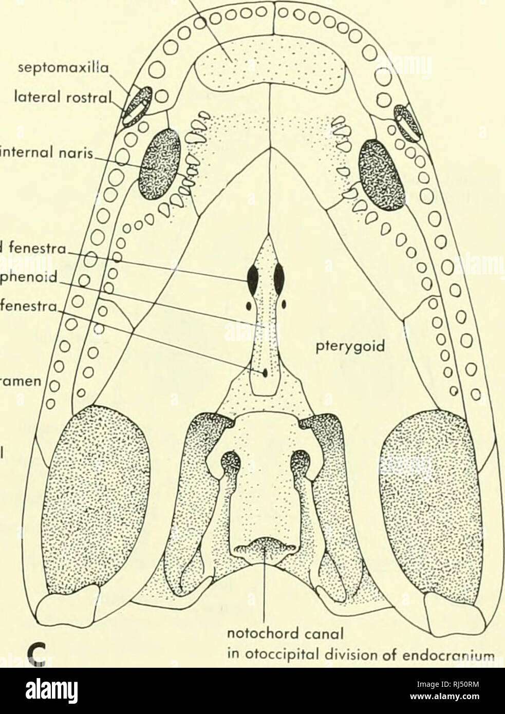 . Chordate morphology. Morphology (Animals); Chordata. anterior palatine fenestra nterpterygoid fenestra parasphenoid hypophyseal fenestrajO postorbital parietal foramen/q supra- temporal B postparietals tabular. notochord canal in otoccipital division of endocronium Figure 4-25. Skull and mandible of khthyoslega. A, lateral view; B, dorsal viev* of skull; C, palatal view of skull. (After Jarvik, 1952, Sdve-Soderbergh, 1932) The eosuchians are known from the Upper Permian. The most primitive members belong to the Family Millerettidae, which has been raised to a separate order by some. In the m Stock Photohttps://www.alamy.com/image-license-details/?v=1https://www.alamy.com/chordate-morphology-morphology-animals-chordata-anterior-palatine-fenestra-nterpterygoid-fenestra-parasphenoid-hypophyseal-fenestrajo-postorbital-parietal-foramenq-supra-temporal-b-postparietals-tabular-notochord-canal-in-otoccipital-division-of-endocronium-figure-4-25-skull-and-mandible-of-khthyoslega-a-lateral-view-b-dorsal-viev-of-skull-c-palatal-view-of-skull-after-jarvik-1952-sdve-soderbergh-1932-the-eosuchians-are-known-from-the-upper-permian-the-most-primitive-members-belong-to-the-family-millerettidae-which-has-been-raised-to-a-separate-order-by-some-in-the-m-image234909016.html
RMRJ50RM–. Chordate morphology. Morphology (Animals); Chordata. anterior palatine fenestra nterpterygoid fenestra parasphenoid hypophyseal fenestrajO postorbital parietal foramen/q supra- temporal B postparietals tabular. notochord canal in otoccipital division of endocronium Figure 4-25. Skull and mandible of khthyoslega. A, lateral view; B, dorsal viev* of skull; C, palatal view of skull. (After Jarvik, 1952, Sdve-Soderbergh, 1932) The eosuchians are known from the Upper Permian. The most primitive members belong to the Family Millerettidae, which has been raised to a separate order by some. In the m
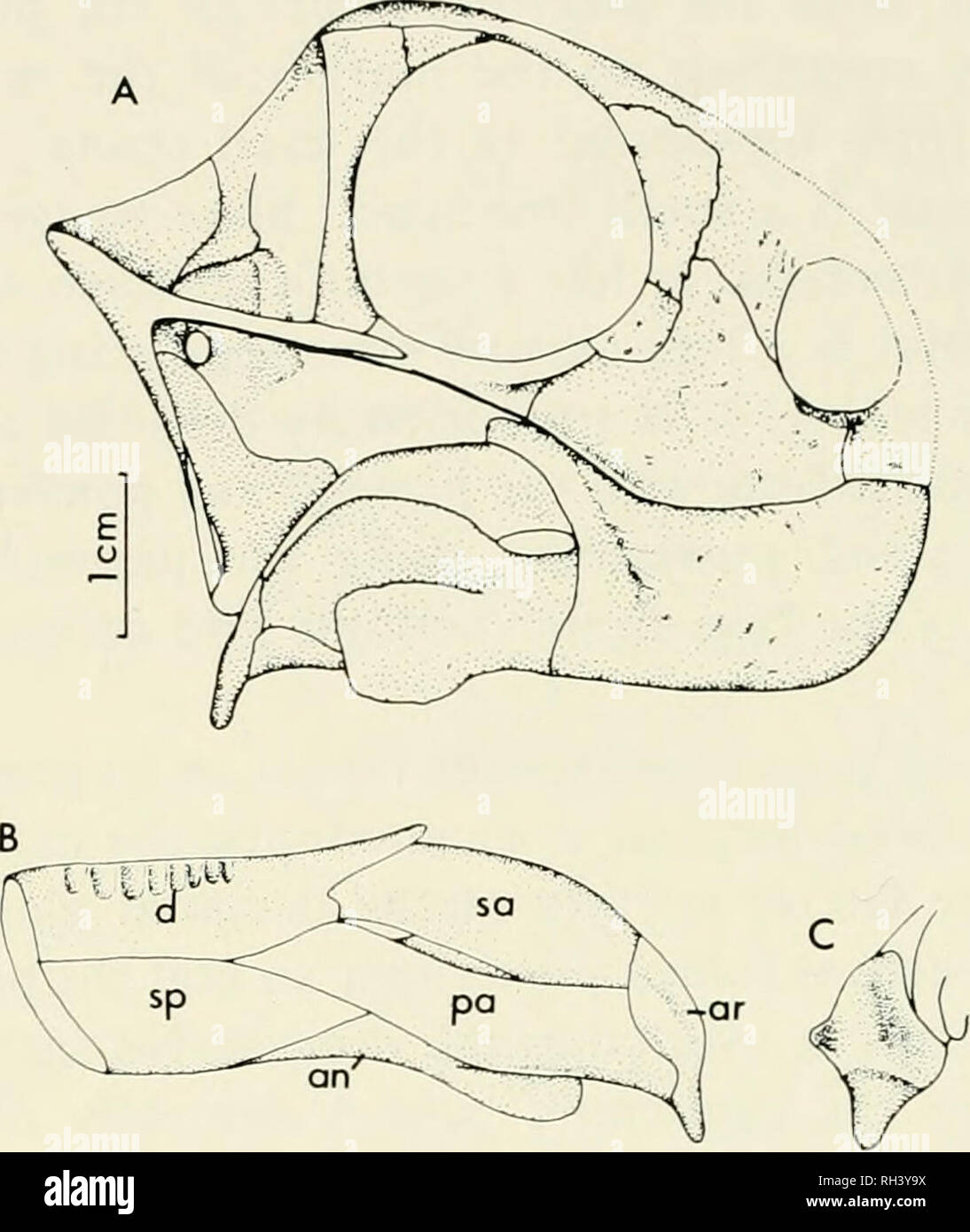 . Breviora. BREVIORA No. 465. Figure 3. Galeops whaitsi. A) reconstruction of skull and lower jaw; B) reconstruction of lower jaw, medial view; C) reconstruction of articular, dorsoposte- rior view. Abbreviations: see Figure 2. parietals extend lateral to this ridge as a ventrally facing shelf that would have provided an area for muscle attachment. This shelf forms a portion of the lateral edge of the temporal fenestra. Anteriorly, a cup-shaped depression is present just anterior and lateral to the area of attachment of the braincase. The quadratojugal is a small splintlike bone resting on the Stock Photohttps://www.alamy.com/image-license-details/?v=1https://www.alamy.com/breviora-breviora-no-465-figure-3-galeops-whaitsi-a-reconstruction-of-skull-and-lower-jaw-b-reconstruction-of-lower-jaw-medial-view-c-reconstruction-of-articular-dorsoposte-rior-view-abbreviations-see-figure-2-parietals-extend-lateral-to-this-ridge-as-a-ventrally-facing-shelf-that-would-have-provided-an-area-for-muscle-attachment-this-shelf-forms-a-portion-of-the-lateral-edge-of-the-temporal-fenestra-anteriorly-a-cup-shaped-depression-is-present-just-anterior-and-lateral-to-the-area-of-attachment-of-the-braincase-the-quadratojugal-is-a-small-splintlike-bone-resting-on-the-image234271238.html
RMRH3Y9X–. Breviora. BREVIORA No. 465. Figure 3. Galeops whaitsi. A) reconstruction of skull and lower jaw; B) reconstruction of lower jaw, medial view; C) reconstruction of articular, dorsoposte- rior view. Abbreviations: see Figure 2. parietals extend lateral to this ridge as a ventrally facing shelf that would have provided an area for muscle attachment. This shelf forms a portion of the lateral edge of the temporal fenestra. Anteriorly, a cup-shaped depression is present just anterior and lateral to the area of attachment of the braincase. The quadratojugal is a small splintlike bone resting on the
 . Elementary text-book of zoology. Fig. 284.—Anterior View of a Cervical Vertebra OF Rabbit. (Ad nat.) Pre- 'zygapophysis. Cervical Rib,. Vertebrarterial Canal, Lastly, in the middle ear is a chain of three ear-ossicles, the malleus, incus and stapes. The malleus is attached to the inner surface of the tympanum and the stapes to the fenestra ovalis of the inner ear. The vertebral column consists of cervical, thoracic, lumbar, sacral and caudal vertebrae. There are seven cervicals, as in nearly all mammals. The first is the atlas with two lateral wing-like cervical ribs, a. Please note that the Stock Photohttps://www.alamy.com/image-license-details/?v=1https://www.alamy.com/elementary-text-book-of-zoology-fig-284anterior-view-of-a-cervical-vertebra-of-rabbit-ad-nat-pre-zygapophysis-cervical-rib-vertebrarterial-canal-lastly-in-the-middle-ear-is-a-chain-of-three-ear-ossicles-the-malleus-incus-and-stapes-the-malleus-is-attached-to-the-inner-surface-of-the-tympanum-and-the-stapes-to-the-fenestra-ovalis-of-the-inner-ear-the-vertebral-column-consists-of-cervical-thoracic-lumbar-sacral-and-caudal-vertebrae-there-are-seven-cervicals-as-in-nearly-all-mammals-the-first-is-the-atlas-with-two-lateral-wing-like-cervical-ribs-a-please-note-that-the-image232088446.html
RMRDGF52–. Elementary text-book of zoology. Fig. 284.—Anterior View of a Cervical Vertebra OF Rabbit. (Ad nat.) Pre- 'zygapophysis. Cervical Rib,. Vertebrarterial Canal, Lastly, in the middle ear is a chain of three ear-ossicles, the malleus, incus and stapes. The malleus is attached to the inner surface of the tympanum and the stapes to the fenestra ovalis of the inner ear. The vertebral column consists of cervical, thoracic, lumbar, sacral and caudal vertebrae. There are seven cervicals, as in nearly all mammals. The first is the atlas with two lateral wing-like cervical ribs, a. Please note that the
 . Bulletin of the Natural Histort Museum. Geology series. nu.for Fig. 14 Baryonyx walkeh. holotype, BMNH R9951; part of right dentary, in dorsal (occlusal) view, x 0.5. the splenial is preserved complete atid forms a smooth concave curve, extending posteroventrally and then posteriorly; this, pre- sumably, formed the anteroventral border of the internal mandibular fenestra. The posterior end of the splenial which articulated with the angular is missing. The anterior tip of the splenial is a flattened tongue that bears strong longitudinal striations on its lateral face. The ventral margin of th Stock Photohttps://www.alamy.com/image-license-details/?v=1https://www.alamy.com/bulletin-of-the-natural-histort-museum-geology-series-nufor-fig-14-baryonyx-walkeh-holotype-bmnh-r9951-part-of-right-dentary-in-dorsal-occlusal-view-x-05-the-splenial-is-preserved-complete-atid-forms-a-smooth-concave-curve-extending-posteroventrally-and-then-posteriorly-this-pre-sumably-formed-the-anteroventral-border-of-the-internal-mandibular-fenestra-the-posterior-end-of-the-splenial-which-articulated-with-the-angular-is-missing-the-anterior-tip-of-the-splenial-is-a-flattened-tongue-that-bears-strong-longitudinal-striations-on-its-lateral-face-the-ventral-margin-of-th-image233869220.html
RMRGDJG4–. Bulletin of the Natural Histort Museum. Geology series. nu.for Fig. 14 Baryonyx walkeh. holotype, BMNH R9951; part of right dentary, in dorsal (occlusal) view, x 0.5. the splenial is preserved complete atid forms a smooth concave curve, extending posteroventrally and then posteriorly; this, pre- sumably, formed the anteroventral border of the internal mandibular fenestra. The posterior end of the splenial which articulated with the angular is missing. The anterior tip of the splenial is a flattened tongue that bears strong longitudinal striations on its lateral face. The ventral margin of th
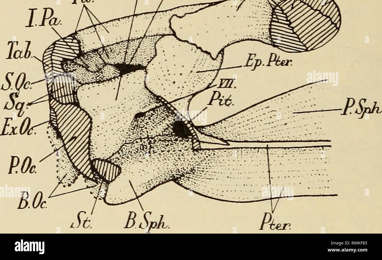 . Annals of the South African Museum = Annale van die Suid-Afrikaanse Museum. Natural history. 230 Annals of the South African Museum. The pro-otic is a large bone, whose anterior portion is flanked by the epipterygoid; posteriorly, it is applied to the anterior surface of the paroccipital, and in part to the squamosal; antero-dorsal to the fenestra ovalis lies a small foramen for the seventh cranial nerve, and under this there is a small depression for the geniculate ganglion; as, in lateral view, the anterior part of the pro-otic is overlain by the epipterygoid, forming a cavum epiptericum, Stock Photohttps://www.alamy.com/image-license-details/?v=1https://www.alamy.com/annals-of-the-south-african-museum-=-annale-van-die-suid-afrikaanse-museum-natural-history-230-annals-of-the-south-african-museum-the-pro-otic-is-a-large-bone-whose-anterior-portion-is-flanked-by-the-epipterygoid-posteriorly-it-is-applied-to-the-anterior-surface-of-the-paroccipital-and-in-part-to-the-squamosal-antero-dorsal-to-the-fenestra-ovalis-lies-a-small-foramen-for-the-seventh-cranial-nerve-and-under-this-there-is-a-small-depression-for-the-geniculate-ganglion-as-in-lateral-view-the-anterior-part-of-the-pro-otic-is-overlain-by-the-epipterygoid-forming-a-cavum-epiptericum-image236456981.html
RMRMKF85–. Annals of the South African Museum = Annale van die Suid-Afrikaanse Museum. Natural history. 230 Annals of the South African Museum. The pro-otic is a large bone, whose anterior portion is flanked by the epipterygoid; posteriorly, it is applied to the anterior surface of the paroccipital, and in part to the squamosal; antero-dorsal to the fenestra ovalis lies a small foramen for the seventh cranial nerve, and under this there is a small depression for the geniculate ganglion; as, in lateral view, the anterior part of the pro-otic is overlain by the epipterygoid, forming a cavum epiptericum,
 . Chordate morphology. Morphology (Animals); Chordata. quadrate cartilage bosicopsular fenestra lateral woll of otic capsule/' notochord quadrate corttlagt facial foramen otic capsuli vestibular fontonelle D epibranchials cerotobronchiols l-lll. superficial ophthalmic VII trabeculo communis ascending process pineal body, orbital cartilage. trabecule. Please note that these images are extracted from scanned page images that may have been digitally enhanced for readability - coloration and appearance of these illustrations may not perfectly resemble the original work.. Jollie, Malcolm. New York, Stock Photohttps://www.alamy.com/image-license-details/?v=1https://www.alamy.com/chordate-morphology-morphology-animals-chordata-quadrate-cartilage-bosicopsular-fenestra-lateral-woll-of-otic-capsule-notochord-quadrate-corttlagt-facial-foramen-otic-capsuli-vestibular-fontonelle-d-epibranchials-cerotobronchiols-l-lll-superficial-ophthalmic-vii-trabeculo-communis-ascending-process-pineal-body-orbital-cartilage-trabecule-please-note-that-these-images-are-extracted-from-scanned-page-images-that-may-have-been-digitally-enhanced-for-readability-coloration-and-appearance-of-these-illustrations-may-not-perfectly-resemble-the-original-work-jollie-malcolm-new-york-image234902222.html
RMRJ4M52–. Chordate morphology. Morphology (Animals); Chordata. quadrate cartilage bosicopsular fenestra lateral woll of otic capsule/' notochord quadrate corttlagt facial foramen otic capsuli vestibular fontonelle D epibranchials cerotobronchiols l-lll. superficial ophthalmic VII trabeculo communis ascending process pineal body, orbital cartilage. trabecule. Please note that these images are extracted from scanned page images that may have been digitally enhanced for readability - coloration and appearance of these illustrations may not perfectly resemble the original work.. Jollie, Malcolm. New York,
 . Annals of the South African Museum = Annale van die Suid-Afrikaanse Museum. Natural history. 204 ANNALS OF THE SOUTH AFRICAN MUSEUM posttemporal fenestra. Ventrally this flange buttresses the quadrate from behind and, above the quadrate, forms the area of origin of a lateral division of the adductor muscle mass (see p. 207). The anterior, zygomatic ramus forms the lateral border of the temporal fossa and terminates below the orbit between the jugal and maxilla. The dorsomedial ramus constitutes the posterior boun- dary of the temporal fossa and provides attachment for the inner portion of th Stock Photohttps://www.alamy.com/image-license-details/?v=1https://www.alamy.com/annals-of-the-south-african-museum-=-annale-van-die-suid-afrikaanse-museum-natural-history-204-annals-of-the-south-african-museum-posttemporal-fenestra-ventrally-this-flange-buttresses-the-quadrate-from-behind-and-above-the-quadrate-forms-the-area-of-origin-of-a-lateral-division-of-the-adductor-muscle-mass-see-p-207-the-anterior-zygomatic-ramus-forms-the-lateral-border-of-the-temporal-fossa-and-terminates-below-the-orbit-between-the-jugal-and-maxilla-the-dorsomedial-ramus-constitutes-the-posterior-boun-dary-of-the-temporal-fossa-and-provides-attachment-for-the-inner-portion-of-th-image236458998.html
RMRMKHT6–. Annals of the South African Museum = Annale van die Suid-Afrikaanse Museum. Natural history. 204 ANNALS OF THE SOUTH AFRICAN MUSEUM posttemporal fenestra. Ventrally this flange buttresses the quadrate from behind and, above the quadrate, forms the area of origin of a lateral division of the adductor muscle mass (see p. 207). The anterior, zygomatic ramus forms the lateral border of the temporal fossa and terminates below the orbit between the jugal and maxilla. The dorsomedial ramus constitutes the posterior boun- dary of the temporal fossa and provides attachment for the inner portion of th
 . Chordate morphology. Morphology (Animals); Chordata. basipterygoid process fenestra vestibuli paroccipital process of opisthotic sella turcica ^exoccipital basioccipital H prootic opisthotic. ^basioccipital parasphenoid fused to basisphenoid carotid canal (dashed lines) Figure 4-26. Skull and mandible of Seymouna. A, lateral view of skull and mandible; B, dorsal view; C, palatal view; D, reor view of skull; E, medial view of mandible; F, cross section of orbit'osp'henoids and parasphenoid; G, lateral view of endocranium; H, medial view of posterior part of right half of the endocranium, (Aft Stock Photohttps://www.alamy.com/image-license-details/?v=1https://www.alamy.com/chordate-morphology-morphology-animals-chordata-basipterygoid-process-fenestra-vestibuli-paroccipital-process-of-opisthotic-sella-turcica-exoccipital-basioccipital-h-prootic-opisthotic-basioccipital-parasphenoid-fused-to-basisphenoid-carotid-canal-dashed-lines-figure-4-26-skull-and-mandible-of-seymouna-a-lateral-view-of-skull-and-mandible-b-dorsal-view-c-palatal-view-d-reor-view-of-skull-e-medial-view-of-mandible-f-cross-section-of-orbitosphenoids-and-parasphenoid-g-lateral-view-of-endocranium-h-medial-view-of-posterior-part-of-right-half-of-the-endocranium-aft-image234902715.html
RMRJ4MPK–. Chordate morphology. Morphology (Animals); Chordata. basipterygoid process fenestra vestibuli paroccipital process of opisthotic sella turcica ^exoccipital basioccipital H prootic opisthotic. ^basioccipital parasphenoid fused to basisphenoid carotid canal (dashed lines) Figure 4-26. Skull and mandible of Seymouna. A, lateral view of skull and mandible; B, dorsal view; C, palatal view; D, reor view of skull; E, medial view of mandible; F, cross section of orbit'osp'henoids and parasphenoid; G, lateral view of endocranium; H, medial view of posterior part of right half of the endocranium, (Aft
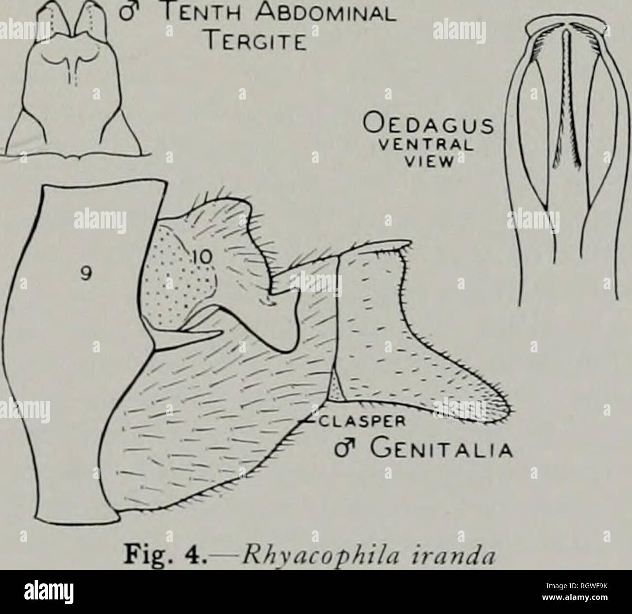 . Bulletin. Natural history; Natural history. Fig. 3.âRhyacophila fenestra, larva ment; the apical segment incised for one-third its lateral and one-tourth its mesal length; both lobes straight and rounded, the dorsal one small and the ventral one large. At the base of the segment there is a mesal incurving lobe; most of the apical segment and this lobe are covered with short, dark setae. Tenth tergite narrow, the dorsal lobe cleft down the meson for more than one- half its length; the lateral lobes so pro- duced have convex dorsal margins with a rather short, sharp apical point; below these t Stock Photohttps://www.alamy.com/image-license-details/?v=1https://www.alamy.com/bulletin-natural-history-natural-history-fig-3rhyacophila-fenestra-larva-ment-the-apical-segment-incised-for-one-third-its-lateral-and-one-tourth-its-mesal-length-both-lobes-straight-and-rounded-the-dorsal-one-small-and-the-ventral-one-large-at-the-base-of-the-segment-there-is-a-mesal-incurving-lobe-most-of-the-apical-segment-and-this-lobe-are-covered-with-short-dark-setae-tenth-tergite-narrow-the-dorsal-lobe-cleft-down-the-meson-for-more-than-one-half-its-length-the-lateral-lobes-so-pro-duced-have-convex-dorsal-margins-with-a-rather-short-sharp-apical-point-below-these-t-image234130111.html
RMRGWF9K–. Bulletin. Natural history; Natural history. Fig. 3.âRhyacophila fenestra, larva ment; the apical segment incised for one-third its lateral and one-tourth its mesal length; both lobes straight and rounded, the dorsal one small and the ventral one large. At the base of the segment there is a mesal incurving lobe; most of the apical segment and this lobe are covered with short, dark setae. Tenth tergite narrow, the dorsal lobe cleft down the meson for more than one- half its length; the lateral lobes so pro- duced have convex dorsal margins with a rather short, sharp apical point; below these t
 . Annals of the South African Museum = Annale van die Suid-Afrikaanse Museum. Natural history. PALATE AND MANDIBLE IN SOME SPECIMENS OF DICYNODON TESTUDIROSTRIS 145 dentary is produced dorsally as a blunt process. Immediately behind the s^Tnphyseal region the dorsal edge of the dentary is rounded, but further back there is a deeply incised trench lateral to the tooth row. The lateral surface of the dentary is drawn out to form a wide ledge above the mandibular fenestra. Pristerodon (Information taken from Barry, 1967). Canine tusks are present, as well as three postcanine teeth. The palatal ri Stock Photohttps://www.alamy.com/image-license-details/?v=1https://www.alamy.com/annals-of-the-south-african-museum-=-annale-van-die-suid-afrikaanse-museum-natural-history-palate-and-mandible-in-some-specimens-of-dicynodon-testudirostris-145-dentary-is-produced-dorsally-as-a-blunt-process-immediately-behind-the-stnphyseal-region-the-dorsal-edge-of-the-dentary-is-rounded-but-further-back-there-is-a-deeply-incised-trench-lateral-to-the-tooth-row-the-lateral-surface-of-the-dentary-is-drawn-out-to-form-a-wide-ledge-above-the-mandibular-fenestra-pristerodon-information-taken-from-barry-1967-canine-tusks-are-present-as-well-as-three-postcanine-teeth-the-palatal-ri-image236461844.html
RMRMKNDT–. Annals of the South African Museum = Annale van die Suid-Afrikaanse Museum. Natural history. PALATE AND MANDIBLE IN SOME SPECIMENS OF DICYNODON TESTUDIROSTRIS 145 dentary is produced dorsally as a blunt process. Immediately behind the s^Tnphyseal region the dorsal edge of the dentary is rounded, but further back there is a deeply incised trench lateral to the tooth row. The lateral surface of the dentary is drawn out to form a wide ledge above the mandibular fenestra. Pristerodon (Information taken from Barry, 1967). Canine tusks are present, as well as three postcanine teeth. The palatal ri
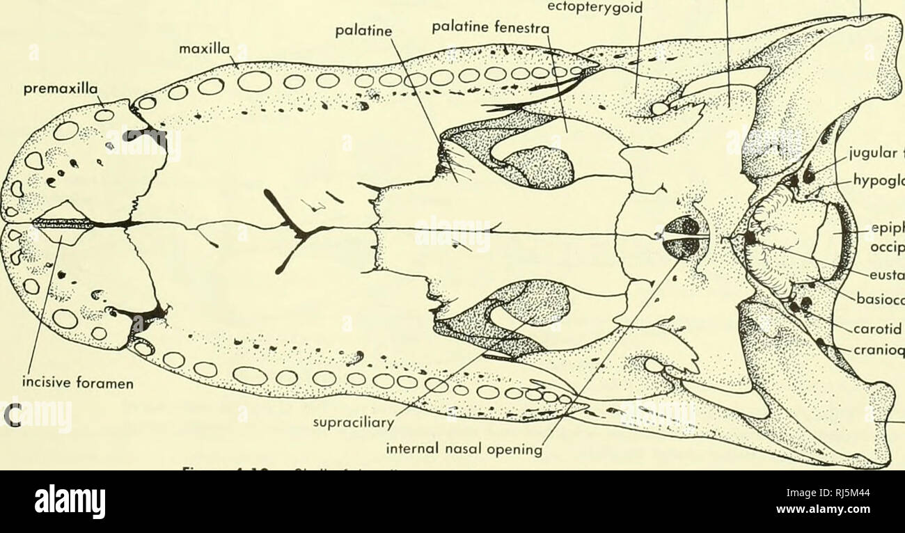 . Chordate morphology. Morphology (Animals); Chordata. quadrate ,quadratojugai quadrate squamosal fused parietals suprooccrpital exoccipital premaxilla D maxilla prefrontal lacrimal / , r ^ , > fused frontals ectopterygoid fused pterygoids quadratojugal ectopterygoid palatine palatine fenestra premaxilla. incisive foramen jugular foramen hypoglossal canals epiphysis on occipital condyle eustachian canal basioccipital carotid canal cranioquadrate fissure supraciliary internal nasal opening Figure 4-10. Skull of the alligator. A, lateral view; B, dorsal view; C, ventral view. quadrate OTHER T Stock Photohttps://www.alamy.com/image-license-details/?v=1https://www.alamy.com/chordate-morphology-morphology-animals-chordata-quadrate-quadratojugai-quadrate-squamosal-fused-parietals-suprooccrpital-exoccipital-premaxilla-d-maxilla-prefrontal-lacrimal-r-gt-fused-frontals-ectopterygoid-fused-pterygoids-quadratojugal-ectopterygoid-palatine-palatine-fenestra-premaxilla-incisive-foramen-jugular-foramen-hypoglossal-canals-epiphysis-on-occipital-condyle-eustachian-canal-basioccipital-carotid-canal-cranioquadrate-fissure-supraciliary-internal-nasal-opening-figure-4-10-skull-of-the-alligator-a-lateral-view-b-dorsal-view-c-ventral-view-quadrate-other-t-image234924148.html
RMRJ5M44–. Chordate morphology. Morphology (Animals); Chordata. quadrate ,quadratojugai quadrate squamosal fused parietals suprooccrpital exoccipital premaxilla D maxilla prefrontal lacrimal / , r ^ , > fused frontals ectopterygoid fused pterygoids quadratojugal ectopterygoid palatine palatine fenestra premaxilla. incisive foramen jugular foramen hypoglossal canals epiphysis on occipital condyle eustachian canal basioccipital carotid canal cranioquadrate fissure supraciliary internal nasal opening Figure 4-10. Skull of the alligator. A, lateral view; B, dorsal view; C, ventral view. quadrate OTHER T
 . Annals of the South African Museum = Annale van die Suid-Afrikaanse Museum. Natural history. B.P.fe^ B.Spk. FuZ.m.Fen.Ov. Pig. 7.—Leptotrachelus eupachygnathus. Type. Lateral view of the left side of the brain-case, modified after Watson. x f. P.T.F. = posttemporal fenestra.. Please note that these images are extracted from scanned page images that may have been digitally enhanced for readability - coloration and appearance of these illustrations may not perfectly resemble the original work.. South African Museum. Cape Town : The Museum Stock Photohttps://www.alamy.com/image-license-details/?v=1https://www.alamy.com/annals-of-the-south-african-museum-=-annale-van-die-suid-afrikaanse-museum-natural-history-bpfe-bspk-fuzmfenov-pig-7leptotrachelus-eupachygnathus-type-lateral-view-of-the-left-side-of-the-brain-case-modified-after-watson-x-f-ptf-=-posttemporal-fenestra-please-note-that-these-images-are-extracted-from-scanned-page-images-that-may-have-been-digitally-enhanced-for-readability-coloration-and-appearance-of-these-illustrations-may-not-perfectly-resemble-the-original-work-south-african-museum-cape-town-the-museum-image236457563.html
RMRMKG0Y–. Annals of the South African Museum = Annale van die Suid-Afrikaanse Museum. Natural history. B.P.fe^ B.Spk. FuZ.m.Fen.Ov. Pig. 7.—Leptotrachelus eupachygnathus. Type. Lateral view of the left side of the brain-case, modified after Watson. x f. P.T.F. = posttemporal fenestra.. Please note that these images are extracted from scanned page images that may have been digitally enhanced for readability - coloration and appearance of these illustrations may not perfectly resemble the original work.. South African Museum. Cape Town : The Museum
 . Bulletin of the Museum of Comparative Zoology at Harvard College. Zoology; Zoology. Anatomy of Eocaecilia micropodia • Jenkins, Walsh, and Carroll 309. Figure 15. The os basale of Eocaecilia micropodia (MNA V8063) in (A) dorsal and (B) ventral views. For stereophotographs of this specimen in ventral view, see Figure 16. (C) An otic capsule (MCZ 9169) in lateral view exhibiting the fenestra ovalis and associated operculum. (D) A partial braincase and the otic capsules in ventral view (MCZ 9167).. Please note that these images are extracted from scanned page images that may have been digitally Stock Photohttps://www.alamy.com/image-license-details/?v=1https://www.alamy.com/bulletin-of-the-museum-of-comparative-zoology-at-harvard-college-zoology-zoology-anatomy-of-eocaecilia-micropodia-jenkins-walsh-and-carroll-309-figure-15-the-os-basale-of-eocaecilia-micropodia-mna-v8063-in-a-dorsal-and-b-ventral-views-for-stereophotographs-of-this-specimen-in-ventral-view-see-figure-16-c-an-otic-capsule-mcz-9169-in-lateral-view-exhibiting-the-fenestra-ovalis-and-associated-operculum-d-a-partial-braincase-and-the-otic-capsules-in-ventral-view-mcz-9167-please-note-that-these-images-are-extracted-from-scanned-page-images-that-may-have-been-digitally-image233894277.html
RMRGEPF1–. Bulletin of the Museum of Comparative Zoology at Harvard College. Zoology; Zoology. Anatomy of Eocaecilia micropodia • Jenkins, Walsh, and Carroll 309. Figure 15. The os basale of Eocaecilia micropodia (MNA V8063) in (A) dorsal and (B) ventral views. For stereophotographs of this specimen in ventral view, see Figure 16. (C) An otic capsule (MCZ 9169) in lateral view exhibiting the fenestra ovalis and associated operculum. (D) A partial braincase and the otic capsules in ventral view (MCZ 9167).. Please note that these images are extracted from scanned page images that may have been digitally
 . Bulletin of the British Museum (Natural History), Geology. 18 ICHTHYOKENTEMA PURBECKENSIS forwards almost as far as the hind edge of the lateral ethmoid. The olfactory nerves emerged through a large foramen (I) above the base of this process, and ran forwards along the dorsal edge of the process. In the centre of the lateral face of the orbito- sphenoid there is a large oval fenestra (Text-figs. 2, 3 ; P. 44948). (c) Cheek and Upper Jaw In most of the more or less complete specimens the bones of this region are badly crushed. The fragmentary material from Lulworth, on the other hand, contain Stock Photohttps://www.alamy.com/image-license-details/?v=1https://www.alamy.com/bulletin-of-the-british-museum-natural-history-geology-18-ichthyokentema-purbeckensis-forwards-almost-as-far-as-the-hind-edge-of-the-lateral-ethmoid-the-olfactory-nerves-emerged-through-a-large-foramen-i-above-the-base-of-this-process-and-ran-forwards-along-the-dorsal-edge-of-the-process-in-the-centre-of-the-lateral-face-of-the-orbito-sphenoid-there-is-a-large-oval-fenestra-text-figs-2-3-p-44948-c-cheek-and-upper-jaw-in-most-of-the-more-or-less-complete-specimens-the-bones-of-this-region-are-badly-crushed-the-fragmentary-material-from-lulworth-on-the-other-hand-contain-image233987273.html
RMRGK149–. Bulletin of the British Museum (Natural History), Geology. 18 ICHTHYOKENTEMA PURBECKENSIS forwards almost as far as the hind edge of the lateral ethmoid. The olfactory nerves emerged through a large foramen (I) above the base of this process, and ran forwards along the dorsal edge of the process. In the centre of the lateral face of the orbito- sphenoid there is a large oval fenestra (Text-figs. 2, 3 ; P. 44948). (c) Cheek and Upper Jaw In most of the more or less complete specimens the bones of this region are badly crushed. The fragmentary material from Lulworth, on the other hand, contain
 . Chordate morphology. Morphology (Animals); Chordata. dentary retroarticular process ong' ular supraangular ^^^jibular fenestra and fossa prearticular / articular. Meckelion fenestra Figure 4-11. Skull and mandible of alligator. A, medial view of right half of skull; B rear view of skull C loterol view of cron.um with labial arch and quodrotoiugol removed; D. lateral v,ew of mandible; E, medial view of mandible. 74 . HEAD SKELETON OF OTHER TETRAPODS AND CHOANATES. Please note that these images are extracted from scanned page images that may have been digitally enhanced for readability - col Stock Photohttps://www.alamy.com/image-license-details/?v=1https://www.alamy.com/chordate-morphology-morphology-animals-chordata-dentary-retroarticular-process-ong-ular-supraangular-jibular-fenestra-and-fossa-prearticular-articular-meckelion-fenestra-figure-4-11-skull-and-mandible-of-alligator-a-medial-view-of-right-half-of-skull-b-rear-view-of-skull-c-loterol-view-of-cronum-with-labial-arch-and-quodrotoiugol-removed-d-lateral-vew-of-mandible-e-medial-view-of-mandible-74-head-skeleton-of-other-tetrapods-and-choanates-please-note-that-these-images-are-extracted-from-scanned-page-images-that-may-have-been-digitally-enhanced-for-readability-col-image234924078.html
RMRJ5M1J–. Chordate morphology. Morphology (Animals); Chordata. dentary retroarticular process ong' ular supraangular ^^^jibular fenestra and fossa prearticular / articular. Meckelion fenestra Figure 4-11. Skull and mandible of alligator. A, medial view of right half of skull; B rear view of skull C loterol view of cron.um with labial arch and quodrotoiugol removed; D. lateral v,ew of mandible; E, medial view of mandible. 74 . HEAD SKELETON OF OTHER TETRAPODS AND CHOANATES. Please note that these images are extracted from scanned page images that may have been digitally enhanced for readability - col
 . Cunningham's Text-book of anatomy. Anatomy. Sinus tympani Mastoid air-cells Recessus epityrnpanicus Fenestra cochlea; Course of canalis facialis Fig. 709.—Section through Left Temporal Bone, showing labyriuthic wall of tympanic cavity, etc. is continued backwards and downwards behind the tympanic cavity, to end at the stylo-mastoid foramen. (4) The septum canalis musculotubarii (O.T. processus cochleariformis), which extends backwards, above the anterior end of the fenestra vestibuli, where it makes a sharp lateral curve, and forms a pulley over which the tendon of the tensor tympani muscle Stock Photohttps://www.alamy.com/image-license-details/?v=1https://www.alamy.com/cunninghams-text-book-of-anatomy-anatomy-sinus-tympani-mastoid-air-cells-recessus-epityrnpanicus-fenestra-cochlea-course-of-canalis-facialis-fig-709section-through-left-temporal-bone-showing-labyriuthic-wall-of-tympanic-cavity-etc-is-continued-backwards-and-downwards-behind-the-tympanic-cavity-to-end-at-the-stylo-mastoid-foramen-4-the-septum-canalis-musculotubarii-ot-processus-cochleariformis-which-extends-backwards-above-the-anterior-end-of-the-fenestra-vestibuli-where-it-makes-a-sharp-lateral-curve-and-forms-a-pulley-over-which-the-tendon-of-the-tensor-tympani-muscle-image231799518.html
RMRD3AJ6–. Cunningham's Text-book of anatomy. Anatomy. Sinus tympani Mastoid air-cells Recessus epityrnpanicus Fenestra cochlea; Course of canalis facialis Fig. 709.—Section through Left Temporal Bone, showing labyriuthic wall of tympanic cavity, etc. is continued backwards and downwards behind the tympanic cavity, to end at the stylo-mastoid foramen. (4) The septum canalis musculotubarii (O.T. processus cochleariformis), which extends backwards, above the anterior end of the fenestra vestibuli, where it makes a sharp lateral curve, and forms a pulley over which the tendon of the tensor tympani muscle
 . Breviora. 1962 NEW SPATHICEPIIALUS ferentiates the orbit proper from its anterior extension, the lacri- mal fenestra, and frives the loxommid eye-socket its characteristic keyhole shape. Most of the lateral edge of the frontal forms the thick orbital rim. At its waist the frontal is 4.3 mm. wide, mak- ing- the interorbital distance a mere 8.6 mm. — extraordinarily narrow for a skull with an estimated width of 185 mm. The short parietal extends laterally into a square lappet; there is no indication that this lappet represents a former intertem-. Fig. 1. Skull of Spaihicepltalus pereger, n. sp Stock Photohttps://www.alamy.com/image-license-details/?v=1https://www.alamy.com/breviora-1962-new-spathicepiialus-ferentiates-the-orbit-proper-from-its-anterior-extension-the-lacri-mal-fenestra-and-frives-the-loxommid-eye-socket-its-characteristic-keyhole-shape-most-of-the-lateral-edge-of-the-frontal-forms-the-thick-orbital-rim-at-its-waist-the-frontal-is-43-mm-wide-mak-ing-the-interorbital-distance-a-mere-86-mm-extraordinarily-narrow-for-a-skull-with-an-estimated-width-of-185-mm-the-short-parietal-extends-laterally-into-a-square-lappet-there-is-no-indication-that-this-lappet-represents-a-former-intertem-fig-1-skull-of-spaihicepltalus-pereger-n-sp-image234272706.html
RMRH416A–. Breviora. 1962 NEW SPATHICEPIIALUS ferentiates the orbit proper from its anterior extension, the lacri- mal fenestra, and frives the loxommid eye-socket its characteristic keyhole shape. Most of the lateral edge of the frontal forms the thick orbital rim. At its waist the frontal is 4.3 mm. wide, mak- ing- the interorbital distance a mere 8.6 mm. — extraordinarily narrow for a skull with an estimated width of 185 mm. The short parietal extends laterally into a square lappet; there is no indication that this lappet represents a former intertem-. Fig. 1. Skull of Spaihicepltalus pereger, n. sp
 . Breviora. BREVIORA No. 379 narial margin and the secondary palate leads into a short naso- palatine duct. The antorbital fenestra is a small triangular opening, the apex of the triangle lying anteriorly, at about one-third the distance from orbit to snout tip, the posterior base separated from the orbit by a narrow bony bar. The orbits are large (as, presumably were the eyes) and are subcircular in shape; on the lateral sur- face they occupy nearly the whole height of the skull, leaving but a narrow bar of bone between them and the lower skull margin. Dorsally their semicircular margins cut Stock Photohttps://www.alamy.com/image-license-details/?v=1https://www.alamy.com/breviora-breviora-no-379-narial-margin-and-the-secondary-palate-leads-into-a-short-naso-palatine-duct-the-antorbital-fenestra-is-a-small-triangular-opening-the-apex-of-the-triangle-lying-anteriorly-at-about-one-third-the-distance-from-orbit-to-snout-tip-the-posterior-base-separated-from-the-orbit-by-a-narrow-bony-bar-the-orbits-are-large-as-presumably-were-the-eyes-and-are-subcircular-in-shape-on-the-lateral-sur-face-they-occupy-nearly-the-whole-height-of-the-skull-leaving-but-a-narrow-bar-of-bone-between-them-and-the-lower-skull-margin-dorsally-their-semicircular-margins-cut-image234271920.html
RMRH4068–. Breviora. BREVIORA No. 379 narial margin and the secondary palate leads into a short naso- palatine duct. The antorbital fenestra is a small triangular opening, the apex of the triangle lying anteriorly, at about one-third the distance from orbit to snout tip, the posterior base separated from the orbit by a narrow bony bar. The orbits are large (as, presumably were the eyes) and are subcircular in shape; on the lateral sur- face they occupy nearly the whole height of the skull, leaving but a narrow bar of bone between them and the lower skull margin. Dorsally their semicircular margins cut
 . Chordate morphology. Morphology (Animals); Chordata. parasphenoid frontoparietal orbifosphenoid orbitonosol foramen prefrontal vomer septomaxillo Jc * vU .i^. ^.^^W / frontoparietal ^' ^'^" prootic orbifosphenoid Jv--A---+-4^ ,endolymphotic duct exoccipital metotic Fissure IX-X-XI perilymphatic fenestra basioccipital articular. Figure 4-21. Skull and mandible of tfie Bullfrog. A, lateral view of skull and mandible; B, dorsal view of skull; C, ventral view of skull; D, mediol view of right half of endocranium; E, medial view of right half of mandible. The pterygoid sutures along much Stock Photohttps://www.alamy.com/image-license-details/?v=1https://www.alamy.com/chordate-morphology-morphology-animals-chordata-parasphenoid-frontoparietal-orbifosphenoid-orbitonosol-foramen-prefrontal-vomer-septomaxillo-jc-vu-i-w-frontoparietal-quot-prootic-orbifosphenoid-jv-a-4-endolymphotic-duct-exoccipital-metotic-fissure-ix-x-xi-perilymphatic-fenestra-basioccipital-articular-figure-4-21-skull-and-mandible-of-tfie-bullfrog-a-lateral-view-of-skull-and-mandible-b-dorsal-view-of-skull-c-ventral-view-of-skull-d-mediol-view-of-right-half-of-endocranium-e-medial-view-of-right-half-of-mandible-the-pterygoid-sutures-along-much-image234909190.html
RMRJ511X–. Chordate morphology. Morphology (Animals); Chordata. parasphenoid frontoparietal orbifosphenoid orbitonosol foramen prefrontal vomer septomaxillo Jc * vU .i^. ^.^^W / frontoparietal ^' ^'^" prootic orbifosphenoid Jv--A---+-4^ ,endolymphotic duct exoccipital metotic Fissure IX-X-XI perilymphatic fenestra basioccipital articular. Figure 4-21. Skull and mandible of tfie Bullfrog. A, lateral view of skull and mandible; B, dorsal view of skull; C, ventral view of skull; D, mediol view of right half of endocranium; E, medial view of right half of mandible. The pterygoid sutures along much
 . Annals of the South African Museum = Annale van die Suid-Afrikaanse Museum. Natural history. «&*& OP 250 A Fig. 4a 260 A Fig. 4a. See legend opposite page. roughly circular cavity, the perilymphatic cistern, applied to the lateral opening that is the fenestra ovalis,4 and therefore against the stapes. From this cistern 4 Or foramen vestibuli, called thus because it is situated at the level of the vestibule of the ear. This is not true, however, in certain living reptiles, nor in the mammal-like reptiles, where it opens at the level of the lagena.. Please note that these images are ex Stock Photohttps://www.alamy.com/image-license-details/?v=1https://www.alamy.com/annals-of-the-south-african-museum-=-annale-van-die-suid-afrikaanse-museum-natural-history-ampamp-op-250-a-fig-4a-260-a-fig-4a-see-legend-opposite-page-roughly-circular-cavity-the-perilymphatic-cistern-applied-to-the-lateral-opening-that-is-the-fenestra-ovalis4-and-therefore-against-the-stapes-from-this-cistern-4-or-foramen-vestibuli-called-thus-because-it-is-situated-at-the-level-of-the-vestibule-of-the-ear-this-is-not-true-however-in-certain-living-reptiles-nor-in-the-mammal-like-reptiles-where-it-opens-at-the-level-of-the-lagena-please-note-that-these-images-are-ex-image236431561.html
RMRMJAT9–. Annals of the South African Museum = Annale van die Suid-Afrikaanse Museum. Natural history. «&*& OP 250 A Fig. 4a 260 A Fig. 4a. See legend opposite page. roughly circular cavity, the perilymphatic cistern, applied to the lateral opening that is the fenestra ovalis,4 and therefore against the stapes. From this cistern 4 Or foramen vestibuli, called thus because it is situated at the level of the vestibule of the ear. This is not true, however, in certain living reptiles, nor in the mammal-like reptiles, where it opens at the level of the lagena.. Please note that these images are ex
 . Bulletin of the Museum of Comparative Zoology at Harvard College. Zoology. 42 Bulletin Museum of Comparative Zoology, Vol. 140, No. 2. Figure 6. Dinilysia pafagomca; reconsfruction of occiput. Abbreviations on p. 62. X 3.5. Dotted line = conjectural; struc- tures missing on one sick restored from the other; broken exoccipital-basioccipital hatched and not restored, in order to show fenestra rotunda. Diagrammatic. each prefrontal descends and then flattens horizontally where it rests upon the upper surface of the lateral palatine process (see below). Here the prefrontal is perforated by the c Stock Photohttps://www.alamy.com/image-license-details/?v=1https://www.alamy.com/bulletin-of-the-museum-of-comparative-zoology-at-harvard-college-zoology-42-bulletin-museum-of-comparative-zoology-vol-140-no-2-figure-6-dinilysia-pafagomca-reconsfruction-of-occiput-abbreviations-on-p-62-x-35-dotted-line-=-conjectural-struc-tures-missing-on-one-sick-restored-from-the-other-broken-exoccipital-basioccipital-hatched-and-not-restored-in-order-to-show-fenestra-rotunda-diagrammatic-each-prefrontal-descends-and-then-flattens-horizontally-where-it-rests-upon-the-upper-surface-of-the-lateral-palatine-process-see-below-here-the-prefrontal-is-perforated-by-the-c-image233900544.html
RMRGF2ET–. Bulletin of the Museum of Comparative Zoology at Harvard College. Zoology. 42 Bulletin Museum of Comparative Zoology, Vol. 140, No. 2. Figure 6. Dinilysia pafagomca; reconsfruction of occiput. Abbreviations on p. 62. X 3.5. Dotted line = conjectural; struc- tures missing on one sick restored from the other; broken exoccipital-basioccipital hatched and not restored, in order to show fenestra rotunda. Diagrammatic. each prefrontal descends and then flattens horizontally where it rests upon the upper surface of the lateral palatine process (see below). Here the prefrontal is perforated by the c
 . Chordate morphology. Morphology (Animals); Chordata. parietal orbitosphenoid frontal supraoccipital dermal petrosal squamosal exoccipitol premaxilla basioccipitai mastoid process. occipital condyle foramen magnum dermal petrosal malleus incus stylomastoid foramen orbital fissure- orbitonasal foramen ethmoid foramen palatine foramina' foramen ovale petrosal occipital condyle mastoid process tympanic cavity exoccipitol |ugular foramen temporal canal fenestra vestibuli venous-facial canal Figure 3-9. Skull of Echidna (rachyglossus). A, lateral view; B, dorsal view; C, ventral view. choanal pass Stock Photohttps://www.alamy.com/image-license-details/?v=1https://www.alamy.com/chordate-morphology-morphology-animals-chordata-parietal-orbitosphenoid-frontal-supraoccipital-dermal-petrosal-squamosal-exoccipitol-premaxilla-basioccipitai-mastoid-process-occipital-condyle-foramen-magnum-dermal-petrosal-malleus-incus-stylomastoid-foramen-orbital-fissure-orbitonasal-foramen-ethmoid-foramen-palatine-foramina-foramen-ovale-petrosal-occipital-condyle-mastoid-process-tympanic-cavity-exoccipitol-ugular-foramen-temporal-canal-fenestra-vestibuli-venous-facial-canal-figure-3-9-skull-of-echidna-rachyglossus-a-lateral-view-b-dorsal-view-c-ventral-view-choanal-pass-image234909725.html
RMRJ51N1–. Chordate morphology. Morphology (Animals); Chordata. parietal orbitosphenoid frontal supraoccipital dermal petrosal squamosal exoccipitol premaxilla basioccipitai mastoid process. occipital condyle foramen magnum dermal petrosal malleus incus stylomastoid foramen orbital fissure- orbitonasal foramen ethmoid foramen palatine foramina' foramen ovale petrosal occipital condyle mastoid process tympanic cavity exoccipitol |ugular foramen temporal canal fenestra vestibuli venous-facial canal Figure 3-9. Skull of Echidna (rachyglossus). A, lateral view; B, dorsal view; C, ventral view. choanal pass
 . Bulletin of the Natural Histort Museum. Geology series. BARYONYX WALKERI 27 alv.s. nu.for Fig. 14 Baryonyx walkeh. holotype, BMNH R9951; part of right dentary, in dorsal (occlusal) view, x 0.5. the splenial is preserved complete atid forms a smooth concave curve, extending posteroventrally and then posteriorly; this, pre- sumably, formed the anteroventral border of the internal mandibular fenestra. The posterior end of the splenial which articulated with the angular is missing. The anterior tip of the splenial is a flattened tongue that bears strong longitudinal striations on its lateral fac Stock Photohttps://www.alamy.com/image-license-details/?v=1https://www.alamy.com/bulletin-of-the-natural-histort-museum-geology-series-baryonyx-walkeri-27-alvs-nufor-fig-14-baryonyx-walkeh-holotype-bmnh-r9951-part-of-right-dentary-in-dorsal-occlusal-view-x-05-the-splenial-is-preserved-complete-atid-forms-a-smooth-concave-curve-extending-posteroventrally-and-then-posteriorly-this-pre-sumably-formed-the-anteroventral-border-of-the-internal-mandibular-fenestra-the-posterior-end-of-the-splenial-which-articulated-with-the-angular-is-missing-the-anterior-tip-of-the-splenial-is-a-flattened-tongue-that-bears-strong-longitudinal-striations-on-its-lateral-fac-image233869242.html
RMRGDJGX–. Bulletin of the Natural Histort Museum. Geology series. BARYONYX WALKERI 27 alv.s. nu.for Fig. 14 Baryonyx walkeh. holotype, BMNH R9951; part of right dentary, in dorsal (occlusal) view, x 0.5. the splenial is preserved complete atid forms a smooth concave curve, extending posteroventrally and then posteriorly; this, pre- sumably, formed the anteroventral border of the internal mandibular fenestra. The posterior end of the splenial which articulated with the angular is missing. The anterior tip of the splenial is a flattened tongue that bears strong longitudinal striations on its lateral fac
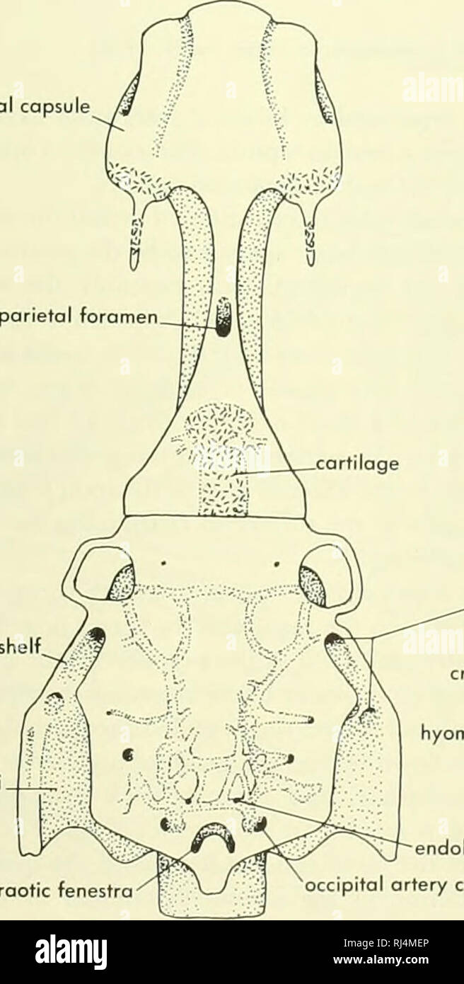 . Chordate morphology. Morphology (Animals); Chordata. suspensorial crest carotid canal pituitary vein foramen bosipterygoid process palatine branch VII fossa Bridget cartilage suprabranchial I articulation occipitospinal nerve X foramina IX lateral commissure canal articulations of pharyngobranchials I, I estibular fontonelle nasal capsule otic shelf. fossa Bridget. supraotic fenestra .otic branches of VII croniol cavity (seen through basicranial fontonelle hyomandibula articulation areas oteral commissure canal vestibular fontonelle. endolymphatic foramen occipital ortery canal B. Please not Stock Photohttps://www.alamy.com/image-license-details/?v=1https://www.alamy.com/chordate-morphology-morphology-animals-chordata-suspensorial-crest-carotid-canal-pituitary-vein-foramen-bosipterygoid-process-palatine-branch-vii-fossa-bridget-cartilage-suprabranchial-i-articulation-occipitospinal-nerve-x-foramina-ix-lateral-commissure-canal-articulations-of-pharyngobranchials-i-i-estibular-fontonelle-nasal-capsule-otic-shelf-fossa-bridget-supraotic-fenestra-otic-branches-of-vii-croniol-cavity-seen-through-basicranial-fontonelle-hyomandibula-articulation-areas-oteral-commissure-canal-vestibular-fontonelle-endolymphatic-foramen-occipital-ortery-canal-b-please-not-image234902494.html
RMRJ4MEP–. Chordate morphology. Morphology (Animals); Chordata. suspensorial crest carotid canal pituitary vein foramen bosipterygoid process palatine branch VII fossa Bridget cartilage suprabranchial I articulation occipitospinal nerve X foramina IX lateral commissure canal articulations of pharyngobranchials I, I estibular fontonelle nasal capsule otic shelf. fossa Bridget. supraotic fenestra .otic branches of VII croniol cavity (seen through basicranial fontonelle hyomandibula articulation areas oteral commissure canal vestibular fontonelle. endolymphatic foramen occipital ortery canal B. Please not
 . Anatomischer Anzeiger. Anatomy, Comparative; Anatomy, Comparative. 213 PrO with the corresponding process of the opisthotic forms the fenestra ovale. The Exoccipital-epiotic is a large plate of bone whose lower border has a notch forming the top of the foramen magnum and brain case. It articulates below with the exoccipital-opisthotic and pro-otic, its lower lateral border being wedged in between the lateral expansions of these bones. In Endothiodon it is pierced by a rather large ductus endo-lymphaticus, but in the other types this issues over a notch in the very large internal auditory mea Stock Photohttps://www.alamy.com/image-license-details/?v=1https://www.alamy.com/anatomischer-anzeiger-anatomy-comparative-anatomy-comparative-213-pro-with-the-corresponding-process-of-the-opisthotic-forms-the-fenestra-ovale-the-exoccipital-epiotic-is-a-large-plate-of-bone-whose-lower-border-has-a-notch-forming-the-top-of-the-foramen-magnum-and-brain-case-it-articulates-below-with-the-exoccipital-opisthotic-and-pro-otic-its-lower-lateral-border-being-wedged-in-between-the-lateral-expansions-of-these-bones-in-endothiodon-it-is-pierced-by-a-rather-large-ductus-endo-lymphaticus-but-in-the-other-types-this-issues-over-a-notch-in-the-very-large-internal-auditory-mea-image236806851.html
RMRN7DFF–. Anatomischer Anzeiger. Anatomy, Comparative; Anatomy, Comparative. 213 PrO with the corresponding process of the opisthotic forms the fenestra ovale. The Exoccipital-epiotic is a large plate of bone whose lower border has a notch forming the top of the foramen magnum and brain case. It articulates below with the exoccipital-opisthotic and pro-otic, its lower lateral border being wedged in between the lateral expansions of these bones. In Endothiodon it is pierced by a rather large ductus endo-lymphaticus, but in the other types this issues over a notch in the very large internal auditory mea
 . Annals of the South African Museum = Annale van die Suid-Afrikaanse Museum. Natural history. 250 ANNALS OF THE SOUTH AFRICAN MUSEUM The postorbitals lie along the side of the intertemporal bar, forming the medial borders of the temporal fenestra. The palate is obscured by the lower jaw but a single tooth is visible close to the alveolar border behind the caniniform process. The lower jaw has a prominent lateral dentary shelf and a sharp shovel- shaped symphysis. At least four teeth are present on the dorsomedial edge of the dentary. One of the teeth shows a distinct row of five posterior ser Stock Photohttps://www.alamy.com/image-license-details/?v=1https://www.alamy.com/annals-of-the-south-african-museum-=-annale-van-die-suid-afrikaanse-museum-natural-history-250-annals-of-the-south-african-museum-the-postorbitals-lie-along-the-side-of-the-intertemporal-bar-forming-the-medial-borders-of-the-temporal-fenestra-the-palate-is-obscured-by-the-lower-jaw-but-a-single-tooth-is-visible-close-to-the-alveolar-border-behind-the-caniniform-process-the-lower-jaw-has-a-prominent-lateral-dentary-shelf-and-a-sharp-shovel-shaped-symphysis-at-least-four-teeth-are-present-on-the-dorsomedial-edge-of-the-dentary-one-of-the-teeth-shows-a-distinct-row-of-five-posterior-ser-image236439671.html
RMRMJN5Y–. Annals of the South African Museum = Annale van die Suid-Afrikaanse Museum. Natural history. 250 ANNALS OF THE SOUTH AFRICAN MUSEUM The postorbitals lie along the side of the intertemporal bar, forming the medial borders of the temporal fenestra. The palate is obscured by the lower jaw but a single tooth is visible close to the alveolar border behind the caniniform process. The lower jaw has a prominent lateral dentary shelf and a sharp shovel- shaped symphysis. At least four teeth are present on the dorsomedial edge of the dentary. One of the teeth shows a distinct row of five posterior ser
 . Bulletin of the British Museum (Natural History), Geology. ON THE SKULL OF OLIGOKYPHUS 73 these fragments does not show two right-angled bends in the base of the skull ; a sagittal section of the ventral surface of the basioccipital and the basisphenoid would form an S-shaped curve, the ventral surface of the basisphenoid being lower than that of the basioccipital. Both R.7110 and R.7113 show a duct (v.c.) through the lateral wall of the brain-case just above the paroccipital process (Text-figs. 2, 6), its external opening lying close to the anterior opening of the post-temporal fenestra (pt Stock Photohttps://www.alamy.com/image-license-details/?v=1https://www.alamy.com/bulletin-of-the-british-museum-natural-history-geology-on-the-skull-of-oligokyphus-73-these-fragments-does-not-show-two-right-angled-bends-in-the-base-of-the-skull-a-sagittal-section-of-the-ventral-surface-of-the-basioccipital-and-the-basisphenoid-would-form-an-s-shaped-curve-the-ventral-surface-of-the-basisphenoid-being-lower-than-that-of-the-basioccipital-both-r7110-and-r7113-show-a-duct-vc-through-the-lateral-wall-of-the-brain-case-just-above-the-paroccipital-process-text-figs-2-6-its-external-opening-lying-close-to-the-anterior-opening-of-the-post-temporal-fenestra-pt-image233986979.html
RMRGK0NR–. Bulletin of the British Museum (Natural History), Geology. ON THE SKULL OF OLIGOKYPHUS 73 these fragments does not show two right-angled bends in the base of the skull ; a sagittal section of the ventral surface of the basioccipital and the basisphenoid would form an S-shaped curve, the ventral surface of the basisphenoid being lower than that of the basioccipital. Both R.7110 and R.7113 show a duct (v.c.) through the lateral wall of the brain-case just above the paroccipital process (Text-figs. 2, 6), its external opening lying close to the anterior opening of the post-temporal fenestra (pt
 . Annals of the South African Museum = Annale van die Suid-Afrikaanse Museum. Natural history. 174 ANNALS OF THE SOUTH AFRICAN MUSEUM PRO VliREC^. Fig. 15. Lystrosaurus declivis. Nat. Mus. No. C.403. Anterolateral view of braincase, showing stapes and lateral head vein channel. temporal fenestra probably indicates a connecting sinus between the v. capitis dorsalis and the v. capitis lateralis, providing an alternative route for venous blood from the occiput. In Lystrosaurus the gutter in each dorsolateral quarter of the occiput, between the supraoccipital and tabular and leading into the bac Stock Photohttps://www.alamy.com/image-license-details/?v=1https://www.alamy.com/annals-of-the-south-african-museum-=-annale-van-die-suid-afrikaanse-museum-natural-history-174-annals-of-the-south-african-museum-pro-vlirec-fig-15-lystrosaurus-declivis-nat-mus-no-c403-anterolateral-view-of-braincase-showing-stapes-and-lateral-head-vein-channel-temporal-fenestra-probably-indicates-a-connecting-sinus-between-the-v-capitis-dorsalis-and-the-v-capitis-lateralis-providing-an-alternative-route-for-venous-blood-from-the-occiput-in-lystrosaurus-the-gutter-in-each-dorsolateral-quarter-of-the-occiput-between-the-supraoccipital-and-tabular-and-leading-into-the-bac-image236463909.html
RMRMKT3H–. Annals of the South African Museum = Annale van die Suid-Afrikaanse Museum. Natural history. 174 ANNALS OF THE SOUTH AFRICAN MUSEUM PRO VliREC^. Fig. 15. Lystrosaurus declivis. Nat. Mus. No. C.403. Anterolateral view of braincase, showing stapes and lateral head vein channel. temporal fenestra probably indicates a connecting sinus between the v. capitis dorsalis and the v. capitis lateralis, providing an alternative route for venous blood from the occiput. In Lystrosaurus the gutter in each dorsolateral quarter of the occiput, between the supraoccipital and tabular and leading into the bac
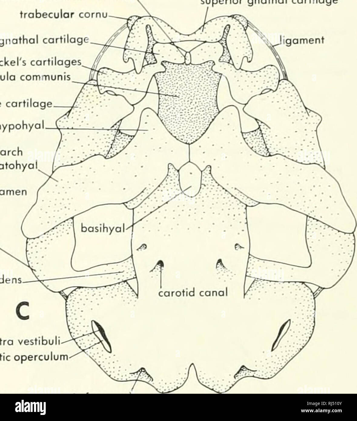 . Chordate morphology. Morphology (Animals); Chordata. intermondibular cartilage trabecular cornu inferior gnathal cartilage Meckel's cartilages. trabeculo communis prootic fenestra otic capsule quadrate cartilage hypohyal occipital arch ceratohyol. superior gnathal cartilage igoment metotic foramen superior gnathal cartilage A carotid conalX o^ic operculum fenestra vestibull otic process of quadrate cartilage processus oscendens. C. fenestra vestibuli otic operculum jugular foramen (metotic foramen) Figure 4-22. Cartilaginous jaws and chondrocranium of tadpole. A, lateral view of entire chond Stock Photohttps://www.alamy.com/image-license-details/?v=1https://www.alamy.com/chordate-morphology-morphology-animals-chordata-intermondibular-cartilage-trabecular-cornu-inferior-gnathal-cartilage-meckels-cartilages-trabeculo-communis-prootic-fenestra-otic-capsule-quadrate-cartilage-hypohyal-occipital-arch-ceratohyol-superior-gnathal-cartilage-igoment-metotic-foramen-superior-gnathal-cartilage-a-carotid-conalx-oic-operculum-fenestra-vestibull-otic-process-of-quadrate-cartilage-processus-oscendens-c-fenestra-vestibuli-otic-operculum-jugular-foramen-metotic-foramen-figure-4-22-cartilaginous-jaws-and-chondrocranium-of-tadpole-a-lateral-view-of-entire-chond-image234909163.html
RMRJ510Y–. Chordate morphology. Morphology (Animals); Chordata. intermondibular cartilage trabecular cornu inferior gnathal cartilage Meckel's cartilages. trabeculo communis prootic fenestra otic capsule quadrate cartilage hypohyal occipital arch ceratohyol. superior gnathal cartilage igoment metotic foramen superior gnathal cartilage A carotid conalX o^ic operculum fenestra vestibull otic process of quadrate cartilage processus oscendens. C. fenestra vestibuli otic operculum jugular foramen (metotic foramen) Figure 4-22. Cartilaginous jaws and chondrocranium of tadpole. A, lateral view of entire chond
 . Bulletin of the Museum of Comparative Zoology at Harvard College. Zoology. THOMSON : HIIIPIDISTIAN SNOUT 319 The roof and very thin side walls of the nasal capsule are, for the most part, fused with the overlying dermal bones. On the medial face of the lateral Avail of the capsule, immediately pos- terior to the fenestra eiidoiiarina, there is a small elliptically- shaped ridge of endocranial bone projecting into the nasal cavity. This ridge, which I have termed the crista lateralis (Figs. 5, 9, c.L), bears on its median face a groove (Figs. 5, 9, gr.c.l.) which, from its relation to the ext Stock Photohttps://www.alamy.com/image-license-details/?v=1https://www.alamy.com/bulletin-of-the-museum-of-comparative-zoology-at-harvard-college-zoology-thomson-hiiipidistian-snout-319-the-roof-and-very-thin-side-walls-of-the-nasal-capsule-are-for-the-most-part-fused-with-the-overlying-dermal-bones-on-the-medial-face-of-the-lateral-avail-of-the-capsule-immediately-pos-terior-to-the-fenestra-eiidoiiarina-there-is-a-small-elliptically-shaped-ridge-of-endocranial-bone-projecting-into-the-nasal-cavity-this-ridge-which-i-have-termed-the-crista-lateralis-figs-5-9-cl-bears-on-its-median-face-a-groove-figs-5-9-grcl-which-from-its-relation-to-the-ext-image233893163.html
RMRGEN37–. Bulletin of the Museum of Comparative Zoology at Harvard College. Zoology. THOMSON : HIIIPIDISTIAN SNOUT 319 The roof and very thin side walls of the nasal capsule are, for the most part, fused with the overlying dermal bones. On the medial face of the lateral Avail of the capsule, immediately pos- terior to the fenestra eiidoiiarina, there is a small elliptically- shaped ridge of endocranial bone projecting into the nasal cavity. This ridge, which I have termed the crista lateralis (Figs. 5, 9, c.L), bears on its median face a groove (Figs. 5, 9, gr.c.l.) which, from its relation to the ext
 . Chordate morphology. Morphology (Animals); Chordata. supraotic fenestra .otic branches of VII croniol cavity (seen through basicranial fontonelle hyomandibula articulation areas oteral commissure canal vestibular fontonelle. endolymphatic foramen occipital ortery canal B. suspensorial crest prespiracular process hypophyseal fontonelle bosipterygoid process notochord socket palatine branch VII aortic groove- notochord canal articulation of pharyngobronchiol I Figure 4-30. Endocranium of Eusfhenopferon. A, lateral view; B, dorsal view; C, ventral view. (After Jorvik, 1954) CHOANATE FISHES 95. Stock Photohttps://www.alamy.com/image-license-details/?v=1https://www.alamy.com/chordate-morphology-morphology-animals-chordata-supraotic-fenestra-otic-branches-of-vii-croniol-cavity-seen-through-basicranial-fontonelle-hyomandibula-articulation-areas-oteral-commissure-canal-vestibular-fontonelle-endolymphatic-foramen-occipital-ortery-canal-b-suspensorial-crest-prespiracular-process-hypophyseal-fontonelle-bosipterygoid-process-notochord-socket-palatine-branch-vii-aortic-groove-notochord-canal-articulation-of-pharyngobronchiol-i-figure-4-30-endocranium-of-eusfhenopferon-a-lateral-view-b-dorsal-view-c-ventral-view-after-jorvik-1954-choanate-fishes-95-image234902480.html
RMRJ4ME8–. Chordate morphology. Morphology (Animals); Chordata. supraotic fenestra .otic branches of VII croniol cavity (seen through basicranial fontonelle hyomandibula articulation areas oteral commissure canal vestibular fontonelle. endolymphatic foramen occipital ortery canal B. suspensorial crest prespiracular process hypophyseal fontonelle bosipterygoid process notochord socket palatine branch VII aortic groove- notochord canal articulation of pharyngobronchiol I Figure 4-30. Endocranium of Eusfhenopferon. A, lateral view; B, dorsal view; C, ventral view. (After Jorvik, 1954) CHOANATE FISHES 95.
 . Breviora. 6 BREVIORA No. 264 ventrally so that the connection between the portion of the bone forming the lateral wing of the occipital region and that associated with the zygoma is only by a narrow neck ventrally; in posterior view, the dorsal margins of the two portions form a V, widely open above. The pterygoid-epipterygoid bar reaches broadly back to the quadrate region above a pterygo-paroccipital fenestra. The squamosal forms the boundaries, medial and lateral, of a distinct. Figure 3. Posterior view of the skull of Massetognuthus pascuali. Natu- ral size. external auditory meatus. The Stock Photohttps://www.alamy.com/image-license-details/?v=1https://www.alamy.com/breviora-6-breviora-no-264-ventrally-so-that-the-connection-between-the-portion-of-the-bone-forming-the-lateral-wing-of-the-occipital-region-and-that-associated-with-the-zygoma-is-only-by-a-narrow-neck-ventrally-in-posterior-view-the-dorsal-margins-of-the-two-portions-form-a-v-widely-open-above-the-pterygoid-epipterygoid-bar-reaches-broadly-back-to-the-quadrate-region-above-a-pterygo-paroccipital-fenestra-the-squamosal-forms-the-boundaries-medial-and-lateral-of-a-distinct-figure-3-posterior-view-of-the-skull-of-massetognuthus-pascuali-natu-ral-size-external-auditory-meatus-the-image234298342.html
RMRH55WX–. Breviora. 6 BREVIORA No. 264 ventrally so that the connection between the portion of the bone forming the lateral wing of the occipital region and that associated with the zygoma is only by a narrow neck ventrally; in posterior view, the dorsal margins of the two portions form a V, widely open above. The pterygoid-epipterygoid bar reaches broadly back to the quadrate region above a pterygo-paroccipital fenestra. The squamosal forms the boundaries, medial and lateral, of a distinct. Figure 3. Posterior view of the skull of Massetognuthus pascuali. Natu- ral size. external auditory meatus. The
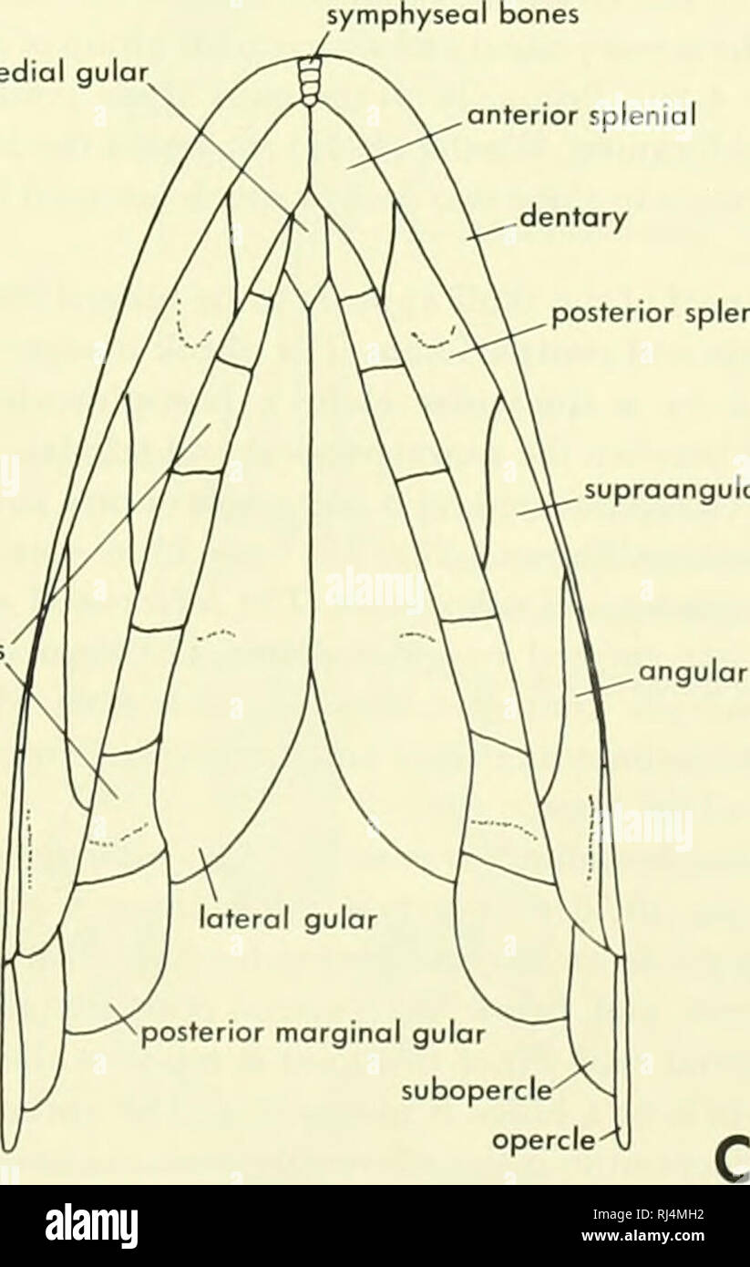 . Chordate morphology. Morphology (Animals); Chordata. anterior medial gular^ internal nasal opening [A^interpterygoid fenestra ^V^rostal part of parosphenoid II hypophyseal fenestra — pterygoid marginal gulars quodratojugal quadrate lateral canal vestibular fontanelle posterior splenial supraongulor. notochord canal Figure 4-28. Dermal head skeleton of Eusthenopferon. A, lateral view of head and pectoral girdle; B, palatal view of skull; C, ventral view of head showing gulor area. (After Jarvik, 1944) CHOANATE FISHES • 93. Please note that these images are extracted from scanned page images Stock Photohttps://www.alamy.com/image-license-details/?v=1https://www.alamy.com/chordate-morphology-morphology-animals-chordata-anterior-medial-gular-internal-nasal-opening-ainterpterygoid-fenestra-vrostal-part-of-parosphenoid-ii-hypophyseal-fenestra-pterygoid-marginal-gulars-quodratojugal-quadrate-lateral-canal-vestibular-fontanelle-posterior-splenial-supraongulor-notochord-canal-figure-4-28-dermal-head-skeleton-of-eusthenopferon-a-lateral-view-of-head-and-pectoral-girdle-b-palatal-view-of-skull-c-ventral-view-of-head-showing-gulor-area-after-jarvik-1944-choanate-fishes-93-please-note-that-these-images-are-extracted-from-scanned-page-images-image234902558.html
RMRJ4MH2–. Chordate morphology. Morphology (Animals); Chordata. anterior medial gular^ internal nasal opening [A^interpterygoid fenestra ^V^rostal part of parosphenoid II hypophyseal fenestra — pterygoid marginal gulars quodratojugal quadrate lateral canal vestibular fontanelle posterior splenial supraongulor. notochord canal Figure 4-28. Dermal head skeleton of Eusthenopferon. A, lateral view of head and pectoral girdle; B, palatal view of skull; C, ventral view of head showing gulor area. (After Jarvik, 1944) CHOANATE FISHES • 93. Please note that these images are extracted from scanned page images
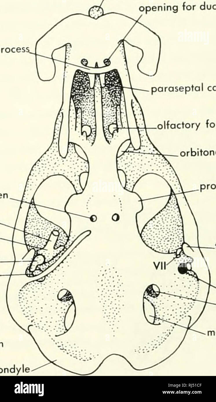 . Chordate morphology. Morphology (Animals); Chordata. palatine proces; crista galli jomina orbitonasolis orbital cartilage OS carunculae opening for duct of Jacobson's organ paraseptol cartilage olfactory foramen orbitonasal fissure carotid foramen Meckel's cartilage, malleus. incus stapes fenestra ocustica endolymphatic foramen occipital condyle. processus alaris tegmen tympani fenestra vestibuli perilymphatic foramen â metotic fissure Figure 3-15. Chondrocranium of 1 22-mm embryo of Plotypus. A, lateral view; B ventral v^w of tip of Meckel's cartilage; C, dorsal view with port of cron.al ro Stock Photohttps://www.alamy.com/image-license-details/?v=1https://www.alamy.com/chordate-morphology-morphology-animals-chordata-palatine-proces-crista-galli-jomina-orbitonasolis-orbital-cartilage-os-carunculae-opening-for-duct-of-jacobsons-organ-paraseptol-cartilage-olfactory-foramen-orbitonasal-fissure-carotid-foramen-meckels-cartilage-malleus-incus-stapes-fenestra-ocustica-endolymphatic-foramen-occipital-condyle-processus-alaris-tegmen-tympani-fenestra-vestibuli-perilymphatic-foramen-metotic-fissure-figure-3-15-chondrocranium-of-1-22-mm-embryo-of-plotypus-a-lateral-view-b-ventral-vw-of-tip-of-meckels-cartilage-c-dorsal-view-with-port-of-cronal-ro-image234909487.html
RMRJ51CF–. Chordate morphology. Morphology (Animals); Chordata. palatine proces; crista galli jomina orbitonasolis orbital cartilage OS carunculae opening for duct of Jacobson's organ paraseptol cartilage olfactory foramen orbitonasal fissure carotid foramen Meckel's cartilage, malleus. incus stapes fenestra ocustica endolymphatic foramen occipital condyle. processus alaris tegmen tympani fenestra vestibuli perilymphatic foramen â metotic fissure Figure 3-15. Chondrocranium of 1 22-mm embryo of Plotypus. A, lateral view; B ventral v^w of tip of Meckel's cartilage; C, dorsal view with port of cron.al ro
 . Chordate morphology. Morphology (Animals); Chordata. lateral ethmoii bosisphenoid A myodome' ^^ basipterygoid process hyoid efferent carotid canal fossa Bridgei lateral commissure canal occipital division of endocronium efferent pseudobranchiol artery ^spinoccipital foramina palatine branch VII- hyomondibular articulation hyomandibular branch VII efferent I- lateral ethmoid hypophyseal fenestra basipterygoid process :i- 4J, t . j-TI . iw'^^^hyoid efferent , f/ lateral canal s. /? r <^vestibular fontanelle phoryngobranc hial. B (V^suprabranchial I epibranchial I efferent I â ^spinoccipita Stock Photohttps://www.alamy.com/image-license-details/?v=1https://www.alamy.com/chordate-morphology-morphology-animals-chordata-lateral-ethmoii-bosisphenoid-a-myodome-basipterygoid-process-hyoid-efferent-carotid-canal-fossa-bridgei-lateral-commissure-canal-occipital-division-of-endocronium-efferent-pseudobranchiol-artery-spinoccipital-foramina-palatine-branch-vii-hyomondibular-articulation-hyomandibular-branch-vii-efferent-i-lateral-ethmoid-hypophyseal-fenestra-basipterygoid-process-i-4j-t-j-ti-iwhyoid-efferent-f-lateral-canal-s-r-ltvestibular-fontanelle-phoryngobranc-hial-b-vsuprabranchial-i-epibranchial-i-efferent-i-spinoccipita-image234901716.html
RMRJ4KF0–. Chordate morphology. Morphology (Animals); Chordata. lateral ethmoii bosisphenoid A myodome' ^^ basipterygoid process hyoid efferent carotid canal fossa Bridgei lateral commissure canal occipital division of endocronium efferent pseudobranchiol artery ^spinoccipital foramina palatine branch VII- hyomondibular articulation hyomandibular branch VII efferent I- lateral ethmoid hypophyseal fenestra basipterygoid process :i- 4J, t . j-TI . iw'^^^hyoid efferent , f/ lateral canal s. /? r <^vestibular fontanelle phoryngobranc hial. B (V^suprabranchial I epibranchial I efferent I â ^spinoccipita
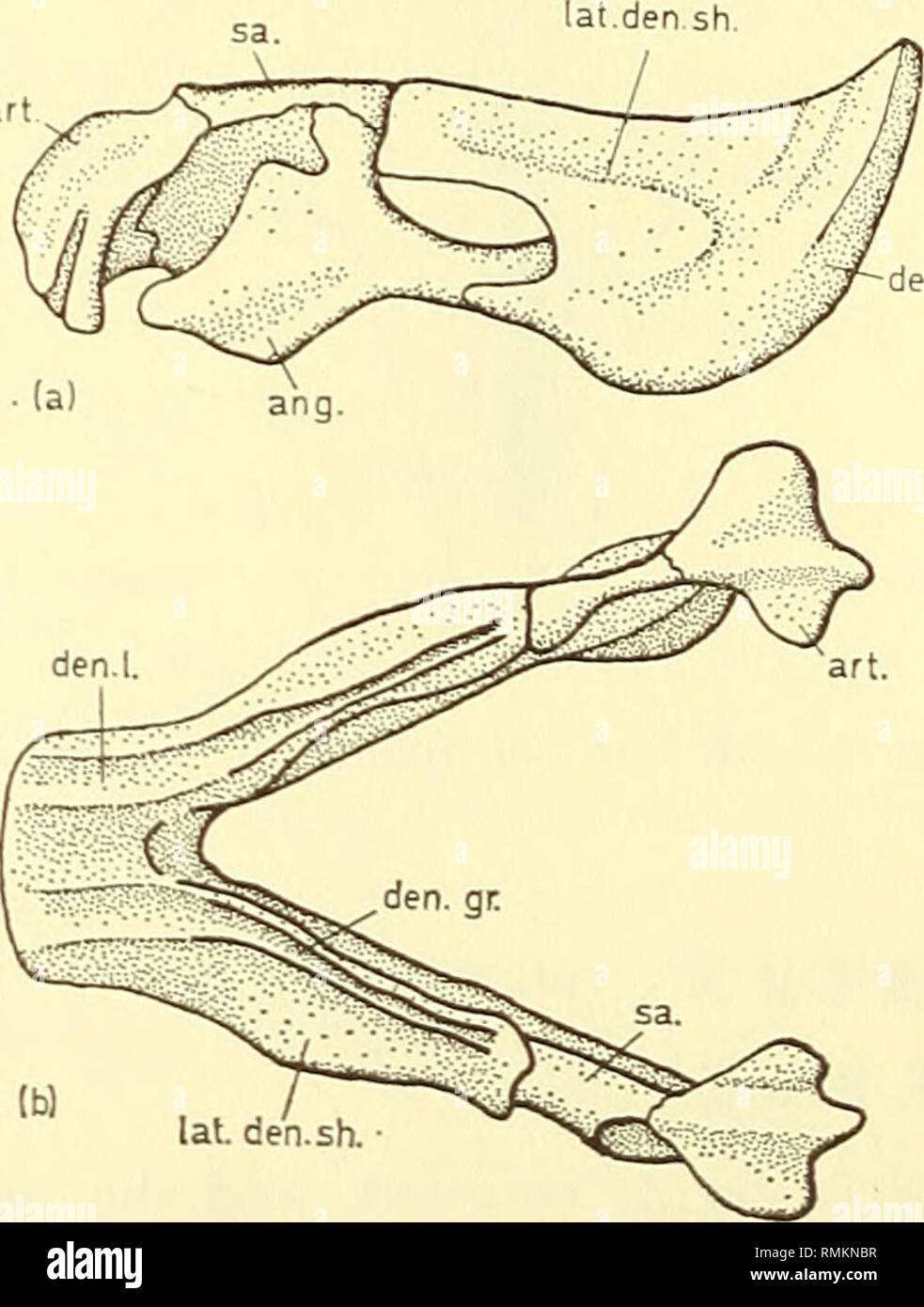 . Annals of the South African Museum = Annale van die Suid-Afrikaanse Museum. Natural history. 148 ANNALS OF THE SOUTH AFRICAN MUSEUM The jaw in several Dicynodon specimens possesses a dorsal dentary groove and built-up dentary tables, but no prominent lateral dentary ledge is found. Muscle scars in the region above the mandibular fenestra are present in some cases. Daptocephalus sp. S.A.M. Cat. No. 8784. Locality: Doornplaats, Graaff-Reinet. Tusks are present and the palatal rim is strongly developed. Anterior palatal ridges as well as the posterior one are present. Ewer (1961) has described Stock Photohttps://www.alamy.com/image-license-details/?v=1https://www.alamy.com/annals-of-the-south-african-museum-=-annale-van-die-suid-afrikaanse-museum-natural-history-148-annals-of-the-south-african-museum-the-jaw-in-several-dicynodon-specimens-possesses-a-dorsal-dentary-groove-and-built-up-dentary-tables-but-no-prominent-lateral-dentary-ledge-is-found-muscle-scars-in-the-region-above-the-mandibular-fenestra-are-present-in-some-cases-daptocephalus-sp-sam-cat-no-8784-locality-doornplaats-graaff-reinet-tusks-are-present-and-the-palatal-rim-is-strongly-developed-anterior-palatal-ridges-as-well-as-the-posterior-one-are-present-ewer-1961-has-described-image236461787.html
RMRMKNBR–. Annals of the South African Museum = Annale van die Suid-Afrikaanse Museum. Natural history. 148 ANNALS OF THE SOUTH AFRICAN MUSEUM The jaw in several Dicynodon specimens possesses a dorsal dentary groove and built-up dentary tables, but no prominent lateral dentary ledge is found. Muscle scars in the region above the mandibular fenestra are present in some cases. Daptocephalus sp. S.A.M. Cat. No. 8784. Locality: Doornplaats, Graaff-Reinet. Tusks are present and the palatal rim is strongly developed. Anterior palatal ridges as well as the posterior one are present. Ewer (1961) has described
 . Annals of the South African Museum = Annale van die Suid-Afrikaanse Museum. Natural history. ANNALS OF THE SOUTH AFRICAN MUSEUM. Fig. 2. Dicynodont. Pelvic Girdle x i. Lateral view. ap—anterior process of the iliac blade IS —ischium b—supra-acetabular buttress P—pubis f—pubo-ischiadic fenestra pt—everted pubic tuber IL—ilium meet at an acute angle, but there is no symphysis and no median keel is developed. The ischium is long; the pair lie at an acute angle, with each other and no median keel is developed. The ischiadic tubera are moderately well-developed. Anteriorly the ischium is pierced Stock Photohttps://www.alamy.com/image-license-details/?v=1https://www.alamy.com/annals-of-the-south-african-museum-=-annale-van-die-suid-afrikaanse-museum-natural-history-annals-of-the-south-african-museum-fig-2-dicynodont-pelvic-girdle-x-i-lateral-view-apanterior-process-of-the-iliac-blade-is-ischium-bsupra-acetabular-buttress-ppubis-fpubo-ischiadic-fenestra-pteverted-pubic-tuber-ililium-meet-at-an-acute-angle-but-there-is-no-symphysis-and-no-median-keel-is-developed-the-ischium-is-long-the-pair-lie-at-an-acute-angle-with-each-other-and-no-median-keel-is-developed-the-ischiadic-tubera-are-moderately-well-developed-anteriorly-the-ischium-is-pierced-image236503416.html
RMRMNJEG–. Annals of the South African Museum = Annale van die Suid-Afrikaanse Museum. Natural history. ANNALS OF THE SOUTH AFRICAN MUSEUM. Fig. 2. Dicynodont. Pelvic Girdle x i. Lateral view. ap—anterior process of the iliac blade IS —ischium b—supra-acetabular buttress P—pubis f—pubo-ischiadic fenestra pt—everted pubic tuber IL—ilium meet at an acute angle, but there is no symphysis and no median keel is developed. The ischium is long; the pair lie at an acute angle, with each other and no median keel is developed. The ischiadic tubera are moderately well-developed. Anteriorly the ischium is pierced
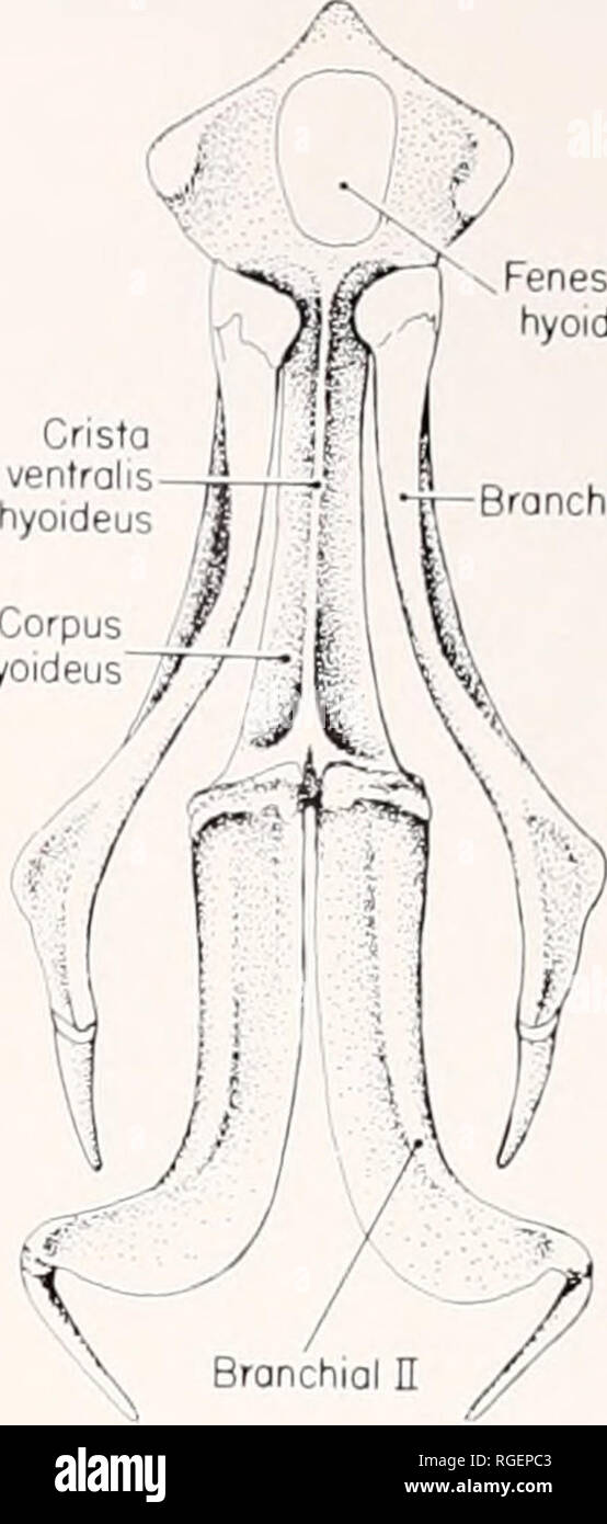 . Bulletin of the Museum of Comparative Zoology at Harvard College. Zoology. Cristo jugalis Cavum tympani Post-otic antrum Precolumellar fossa Fenestra hyoideus iall Figure 6. Lateral view of the skull of C. siebenrocki (AMS 40696). The labelled terms include new terms introduced in this paper and defined in Appendix I. midline in very large females. Surface of carapace weakly rugose with reticulate pattern of numerous, very shallow furrows. Furrows on vertebrals oriented longitudi- nally along midline; furrows on pleurals not oriented in any particular way. Length of dorsal surface of nuchal Stock Photohttps://www.alamy.com/image-license-details/?v=1https://www.alamy.com/bulletin-of-the-museum-of-comparative-zoology-at-harvard-college-zoology-cristo-jugalis-cavum-tympani-post-otic-antrum-precolumellar-fossa-fenestra-hyoideus-iall-figure-6-lateral-view-of-the-skull-of-c-siebenrocki-ams-40696-the-labelled-terms-include-new-terms-introduced-in-this-paper-and-defined-in-appendix-i-midline-in-very-large-females-surface-of-carapace-weakly-rugose-with-reticulate-pattern-of-numerous-very-shallow-furrows-furrows-on-vertebrals-oriented-longitudi-nally-along-midline-furrows-on-pleurals-not-oriented-in-any-particular-way-length-of-dorsal-surface-of-nuchal-image233894195.html
RMRGEPC3–. Bulletin of the Museum of Comparative Zoology at Harvard College. Zoology. Cristo jugalis Cavum tympani Post-otic antrum Precolumellar fossa Fenestra hyoideus iall Figure 6. Lateral view of the skull of C. siebenrocki (AMS 40696). The labelled terms include new terms introduced in this paper and defined in Appendix I. midline in very large females. Surface of carapace weakly rugose with reticulate pattern of numerous, very shallow furrows. Furrows on vertebrals oriented longitudi- nally along midline; furrows on pleurals not oriented in any particular way. Length of dorsal surface of nuchal
 . Chordate morphology. Morphology (Animals); Chordata. V2 3 prootic i——y^——,^, ^opisthotic fenestra vestJbuli XII exoccipital ix-x-xi basioccipital parasphenoid "supraoccipital" postparietal posttemporal fenestra^ ^,,,,-^——,—/—^.-^ ^tabular parasphenoid jugal cronioquadrate fissure fenestra vestibul quadratojugal. quadrate Figure 4-24. Skull of Po/oeogyr/nus, on early Pennsylvonian amphibian from Scotland. A, dorsal view with sensory-line system indicated on right half by dashed lines; B, palatal view,- C, lateral view; D, loteral view with bones of cheek removed; E, lateral view Stock Photohttps://www.alamy.com/image-license-details/?v=1https://www.alamy.com/chordate-morphology-morphology-animals-chordata-v2-3-prootic-iy-opisthotic-fenestra-vestjbuli-xii-exoccipital-ix-x-xi-basioccipital-parasphenoid-quotsupraoccipitalquot-postparietal-posttemporal-fenestra-tabular-parasphenoid-jugal-cronioquadrate-fissure-fenestra-vestibul-quadratojugal-quadrate-figure-4-24-skull-of-pooeogyrnus-on-early-pennsylvonian-amphibian-from-scotland-a-dorsal-view-with-sensory-line-system-indicated-on-right-half-by-dashed-lines-b-palatal-view-c-lateral-view-d-loteral-view-with-bones-of-cheek-removed-e-lateral-view-image234909044.html
RMRJ50TM–. Chordate morphology. Morphology (Animals); Chordata. V2 3 prootic i——y^——,^, ^opisthotic fenestra vestJbuli XII exoccipital ix-x-xi basioccipital parasphenoid "supraoccipital" postparietal posttemporal fenestra^ ^,,,,-^——,—/—^.-^ ^tabular parasphenoid jugal cronioquadrate fissure fenestra vestibul quadratojugal. quadrate Figure 4-24. Skull of Po/oeogyr/nus, on early Pennsylvonian amphibian from Scotland. A, dorsal view with sensory-line system indicated on right half by dashed lines; B, palatal view,- C, lateral view; D, loteral view with bones of cheek removed; E, lateral view
 . Annals of the South African Museum = Annale van die Suid-Afrikaanse Museum. Natural history. sa.. 3 cm Fig. 25. Dicynodon sp. SAM-B88. Skull and lower jaw in lateral view. dentary tables. Dorsal edge of dentary carries deep sulcus behind dentary tables. Rear of dentary extended dorsally to form weak posterodorsally directed process. Mandibular fenestra large, bounded dorsally by lateral den- tary shelf. Occipital surface of opisthotic carries depression above paroccipital process.. Please note that these images are extracted from scanned page images that may have been digitally enhanced for Stock Photohttps://www.alamy.com/image-license-details/?v=1https://www.alamy.com/annals-of-the-south-african-museum-=-annale-van-die-suid-afrikaanse-museum-natural-history-sa-3-cm-fig-25-dicynodon-sp-sam-b88-skull-and-lower-jaw-in-lateral-view-dentary-tables-dorsal-edge-of-dentary-carries-deep-sulcus-behind-dentary-tables-rear-of-dentary-extended-dorsally-to-form-weak-posterodorsally-directed-process-mandibular-fenestra-large-bounded-dorsally-by-lateral-den-tary-shelf-occipital-surface-of-opisthotic-carries-depression-above-paroccipital-process-please-note-that-these-images-are-extracted-from-scanned-page-images-that-may-have-been-digitally-enhanced-for-image236439951.html
RMRMJNFY–. Annals of the South African Museum = Annale van die Suid-Afrikaanse Museum. Natural history. sa.. 3 cm Fig. 25. Dicynodon sp. SAM-B88. Skull and lower jaw in lateral view. dentary tables. Dorsal edge of dentary carries deep sulcus behind dentary tables. Rear of dentary extended dorsally to form weak posterodorsally directed process. Mandibular fenestra large, bounded dorsally by lateral den- tary shelf. Occipital surface of opisthotic carries depression above paroccipital process.. Please note that these images are extracted from scanned page images that may have been digitally enhanced for
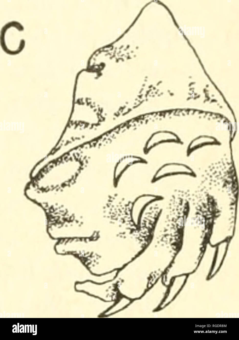 . Bulletin of the Museum of Comparative Zoology at Harvard College. Zoology. Fig. 51. Forefoot scalation in certain trionychida. A, Trionyx trmnguL% (M.C.Z. 16509); B, Cyclanorhis elegans (B.M.) ; C, Cyclanorbis senegalensis (Z.M.U.). (P. Washer del.) Key to the Trionychidae of Africa 1. Femoral flaps absent; nuchal somewhat overlapping second rib; hTo- and hypoplastra separate; lateral prong of posteromedial process of hypo- plastron gripped by xiphiplastron; pterygoids not joining opisthotic; fenestra postotica not or not much restricted; head spotted. Eange: Egypt and Eritrea south to Laie Stock Photohttps://www.alamy.com/image-license-details/?v=1https://www.alamy.com/bulletin-of-the-museum-of-comparative-zoology-at-harvard-college-zoology-fig-51-forefoot-scalation-in-certain-trionychida-a-trionyx-trmngul-mcz-16509-b-cyclanorhis-elegans-bm-c-cyclanorbis-senegalensis-zmu-p-washer-del-key-to-the-trionychidae-of-africa-1-femoral-flaps-absent-nuchal-somewhat-overlapping-second-rib-hto-and-hypoplastra-separate-lateral-prong-of-posteromedial-process-of-hypo-plastron-gripped-by-xiphiplastron-pterygoids-not-joining-opisthotic-fenestra-postotica-not-or-not-much-restricted-head-spotted-eange-egypt-and-eritrea-south-to-laie-image233872932.html
RMRGDR8M–. Bulletin of the Museum of Comparative Zoology at Harvard College. Zoology. Fig. 51. Forefoot scalation in certain trionychida. A, Trionyx trmnguL% (M.C.Z. 16509); B, Cyclanorhis elegans (B.M.) ; C, Cyclanorbis senegalensis (Z.M.U.). (P. Washer del.) Key to the Trionychidae of Africa 1. Femoral flaps absent; nuchal somewhat overlapping second rib; hTo- and hypoplastra separate; lateral prong of posteromedial process of hypo- plastron gripped by xiphiplastron; pterygoids not joining opisthotic; fenestra postotica not or not much restricted; head spotted. Eange: Egypt and Eritrea south to Laie
 . Atlas of applied (topographical) human anatomy for students and practitioners. Anatomy. massetcr Muscle External Pterygoid Muscle Par<.tid Gland M Articular Cartilage Temporal Artery Ili-ad of Mandible F.xlcrnal Auditory Meatus ^^ Facial KcTvc :rastoid Cells Lateral Sii Cerebellum Fig. 21. Horizontal Section of the Head. Organ of Healing and suiTounding Parts. Left side, viewed frcuii below. — iat. Size. ^^fMM Internal Carotid Artery Tympanic Cavity Superior Petrosal Sinus Facial Nerve Auditory Nerve Tympanic Nerve^. Promontory Umbo. Malleus Fenestra Ovalis Stapes Pyramid Chorda Tjinpan Stock Photohttps://www.alamy.com/image-license-details/?v=1https://www.alamy.com/atlas-of-applied-topographical-human-anatomy-for-students-and-practitioners-anatomy-massetcr-muscle-external-pterygoid-muscle-parlttid-gland-m-articular-cartilage-temporal-artery-ili-ad-of-mandible-fxlcrnal-auditory-meatus-facial-kctvc-rastoid-cells-lateral-sii-cerebellum-fig-21-horizontal-section-of-the-head-organ-of-healing-and-suitounding-parts-left-side-viewed-frcuii-below-iat-size-fmm-internal-carotid-artery-tympanic-cavity-superior-petrosal-sinus-facial-nerve-auditory-nerve-tympanic-nerve-promontory-umbo-malleus-fenestra-ovalis-stapes-pyramid-chorda-tjinpan-image235402591.html
RMRJYEBB–. Atlas of applied (topographical) human anatomy for students and practitioners. Anatomy. massetcr Muscle External Pterygoid Muscle Par<.tid Gland M Articular Cartilage Temporal Artery Ili-ad of Mandible F.xlcrnal Auditory Meatus ^^ Facial KcTvc :rastoid Cells Lateral Sii Cerebellum Fig. 21. Horizontal Section of the Head. Organ of Healing and suiTounding Parts. Left side, viewed frcuii below. — iat. Size. ^^fMM Internal Carotid Artery Tympanic Cavity Superior Petrosal Sinus Facial Nerve Auditory Nerve Tympanic Nerve^. Promontory Umbo. Malleus Fenestra Ovalis Stapes Pyramid Chorda Tjinpan
 . Chordate morphology. Morphology (Animals); Chordata. hyomandibula articulation area otic branch of VII. lateral ethmoii bosisphenoid A myodome' ^^ basipterygoid process hyoid efferent carotid canal fossa Bridgei lateral commissure canal occipital division of endocronium efferent pseudobranchiol artery ^spinoccipital foramina palatine branch VII- hyomondibular articulation hyomandibular branch VII efferent I- lateral ethmoid hypophyseal fenestra basipterygoid process :i- 4J, t . j-TI . iw'^^^hyoid efferent , f/ lateral canal s. /? r <^vestibular fontanelle phoryngobranc hial. Please note Stock Photohttps://www.alamy.com/image-license-details/?v=1https://www.alamy.com/chordate-morphology-morphology-animals-chordata-hyomandibula-articulation-area-otic-branch-of-vii-lateral-ethmoii-bosisphenoid-a-myodome-basipterygoid-process-hyoid-efferent-carotid-canal-fossa-bridgei-lateral-commissure-canal-occipital-division-of-endocronium-efferent-pseudobranchiol-artery-spinoccipital-foramina-palatine-branch-vii-hyomondibular-articulation-hyomandibular-branch-vii-efferent-i-lateral-ethmoid-hypophyseal-fenestra-basipterygoid-process-i-4j-t-j-ti-iwhyoid-efferent-f-lateral-canal-s-r-ltvestibular-fontanelle-phoryngobranc-hial-please-note-image234901735.html
RMRJ4KFK–. Chordate morphology. Morphology (Animals); Chordata. hyomandibula articulation area otic branch of VII. lateral ethmoii bosisphenoid A myodome' ^^ basipterygoid process hyoid efferent carotid canal fossa Bridgei lateral commissure canal occipital division of endocronium efferent pseudobranchiol artery ^spinoccipital foramina palatine branch VII- hyomondibular articulation hyomandibular branch VII efferent I- lateral ethmoid hypophyseal fenestra basipterygoid process :i- 4J, t . j-TI . iw'^^^hyoid efferent , f/ lateral canal s. /? r <^vestibular fontanelle phoryngobranc hial. Please note
 . Bulletin. Natural history; Natuurlijke historie. 5 cr pec r. Fig. 14. Reconstructed pectrum of Corosaurus alcovensis (interclavicle hypothetical). A, left lateral aspect; B, anterior aspect, cl = clavicle; cor = coracoid; gl = glenoid; icl = interclavicle; pec f = pectoral fenestra; pec r = pectoral rib; sc = scapula; sc b = scapular blade; thor c = thoracic cavity. the pectrum, met in a strong symphysis to counteract the thrust from the forehmbs. As in all sauropterygians, there was little dorsal development of the pectrum. The scapular blades probably held the large ventral basket only loo Stock Photohttps://www.alamy.com/image-license-details/?v=1https://www.alamy.com/bulletin-natural-history-natuurlijke-historie-5-cr-pec-r-fig-14-reconstructed-pectrum-of-corosaurus-alcovensis-interclavicle-hypothetical-a-left-lateral-aspect-b-anterior-aspect-cl-=-clavicle-cor-=-coracoid-gl-=-glenoid-icl-=-interclavicle-pec-f-=-pectoral-fenestra-pec-r-=-pectoral-rib-sc-=-scapula-sc-b-=-scapular-blade-thor-c-=-thoracic-cavity-the-pectrum-met-in-a-strong-symphysis-to-counteract-the-thrust-from-the-forehmbs-as-in-all-sauropterygians-there-was-little-dorsal-development-of-the-pectrum-the-scapular-blades-probably-held-the-large-ventral-basket-only-loo-image234208000.html
RMRH12KC–. Bulletin. Natural history; Natuurlijke historie. 5 cr pec r. Fig. 14. Reconstructed pectrum of Corosaurus alcovensis (interclavicle hypothetical). A, left lateral aspect; B, anterior aspect, cl = clavicle; cor = coracoid; gl = glenoid; icl = interclavicle; pec f = pectoral fenestra; pec r = pectoral rib; sc = scapula; sc b = scapular blade; thor c = thoracic cavity. the pectrum, met in a strong symphysis to counteract the thrust from the forehmbs. As in all sauropterygians, there was little dorsal development of the pectrum. The scapular blades probably held the large ventral basket only loo
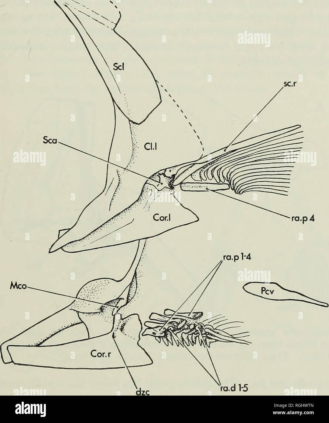 . Bulletin of the British Museum (Natural History), Geology. FISHES FROM THE LEBANON 225 the tip of the cleithrum, leaving a triangular fenestra between this contact and the upper attachment of the coracoid to the cleithrum. A short antero-dorsal process of the coracoid joins with a similar process of the scapula in receiving the ventral end of the mesocoracoid. Just below this process on the medial face of the coracoid. ra.d 1^ 2 mm Fig. 10. Gaudryella gaudryi (Pictet & Humbert). Pectoral girdle as preserved in P.9991, Hajula, Lebanon. Above is the left girdle in lateral view, below the r Stock Photohttps://www.alamy.com/image-license-details/?v=1https://www.alamy.com/bulletin-of-the-british-museum-natural-history-geology-fishes-from-the-lebanon-225-the-tip-of-the-cleithrum-leaving-a-triangular-fenestra-between-this-contact-and-the-upper-attachment-of-the-coracoid-to-the-cleithrum-a-short-antero-dorsal-process-of-the-coracoid-joins-with-a-similar-process-of-the-scapula-in-receiving-the-ventral-end-of-the-mesocoracoid-just-below-this-process-on-the-medial-face-of-the-coracoid-rad-1-2-mm-fig-10-gaudryella-gaudryi-pictet-amp-humbert-pectoral-girdle-as-preserved-in-p9991-hajula-lebanon-above-is-the-left-girdle-in-lateral-view-below-the-r-image233962757.html
RMRGHWTN–. Bulletin of the British Museum (Natural History), Geology. FISHES FROM THE LEBANON 225 the tip of the cleithrum, leaving a triangular fenestra between this contact and the upper attachment of the coracoid to the cleithrum. A short antero-dorsal process of the coracoid joins with a similar process of the scapula in receiving the ventral end of the mesocoracoid. Just below this process on the medial face of the coracoid. ra.d 1^ 2 mm Fig. 10. Gaudryella gaudryi (Pictet & Humbert). Pectoral girdle as preserved in P.9991, Hajula, Lebanon. Above is the left girdle in lateral view, below the r
 . Bulletin. Natural history; Natuurlijke historie. OSTEOLOGY OF DEINONYCHUS ANTIRRHOPUS 17 MAXILLA The right maxilla of YPM 5232 is nearly complete and only slightly crushed (Fig. 6). In lateral view, it is triangular in shape, with the narrow apex directed. FIG. 6. Snout of Deinonychus antirrhopus, skull YPM 5232, right side viewed in reverse. Notice the subsidiary antorbital fenestrae. Abbreviations: aof—antorbital fenestra; aof' and aof"—sub- sidiary antorbital fenestrae; en—external naris; ma—maxilla; na—nasal; pm—premaxilla. forward and the rear margin deeply emarginated by the anter Stock Photohttps://www.alamy.com/image-license-details/?v=1https://www.alamy.com/bulletin-natural-history-natuurlijke-historie-osteology-of-deinonychus-antirrhopus-17-maxilla-the-right-maxilla-of-ypm-5232-is-nearly-complete-and-only-slightly-crushed-fig-6-in-lateral-view-it-is-triangular-in-shape-with-the-narrow-apex-directed-fig-6-snout-of-deinonychus-antirrhopus-skull-ypm-5232-right-side-viewed-in-reverse-notice-the-subsidiary-antorbital-fenestrae-abbreviations-aofantorbital-fenestra-aof-and-aofquotsub-sidiary-antorbital-fenestrae-enexternal-naris-mamaxilla-nanasal-pmpremaxilla-forward-and-the-rear-margin-deeply-emarginated-by-the-anter-image234211592.html
RMRH177M–. Bulletin. Natural history; Natuurlijke historie. OSTEOLOGY OF DEINONYCHUS ANTIRRHOPUS 17 MAXILLA The right maxilla of YPM 5232 is nearly complete and only slightly crushed (Fig. 6). In lateral view, it is triangular in shape, with the narrow apex directed. FIG. 6. Snout of Deinonychus antirrhopus, skull YPM 5232, right side viewed in reverse. Notice the subsidiary antorbital fenestrae. Abbreviations: aof—antorbital fenestra; aof' and aof"—sub- sidiary antorbital fenestrae; en—external naris; ma—maxilla; na—nasal; pm—premaxilla. forward and the rear margin deeply emarginated by the anter
 . The anatomy of the domestic animals. Veterinary anatomy. Fig. 707. —Left Membran. (En- /, Cochlea; 2, fenestra vestibuli; 3, fenestra coch- leae; 4, ductus endolymphaticus; o, dorsal, 6", lateral, 7, ventral, duct. (After Ellenberger, in Leisering's At- las.) Fig. liyX—Schematic Sectioxal Vi /, 2, 5, Doreal, lateral, and ventral due utricle; 5, sacci:le; 6, cochlea; 7, acoustic (After Ellenberger, in Leisering's Atlas.) to the outer wall of the cochlea. The tympanic wall or floor intervenes between the cochlear duct and the scala tympani; it is formed by the periosteum of the mar- ginal Stock Photohttps://www.alamy.com/image-license-details/?v=1https://www.alamy.com/the-anatomy-of-the-domestic-animals-veterinary-anatomy-fig-707-left-membran-en-cochlea-2-fenestra-vestibuli-3-fenestra-coch-leae-4-ductus-endolymphaticus-o-dorsal-6quot-lateral-7-ventral-duct-after-ellenberger-in-leiserings-at-las-fig-liyxschematic-sectioxal-vi-2-5-doreal-lateral-and-ventral-due-utricle-5-saccile-6-cochlea-7-acoustic-after-ellenberger-in-leiserings-atlas-to-the-outer-wall-of-the-cochlea-the-tympanic-wall-or-floor-intervenes-between-the-cochlear-duct-and-the-scala-tympani-it-is-formed-by-the-periosteum-of-the-mar-ginal-image236772903.html
RMRN5X73–. The anatomy of the domestic animals. Veterinary anatomy. Fig. 707. —Left Membran. (En- /, Cochlea; 2, fenestra vestibuli; 3, fenestra coch- leae; 4, ductus endolymphaticus; o, dorsal, 6", lateral, 7, ventral, duct. (After Ellenberger, in Leisering's At- las.) Fig. liyX—Schematic Sectioxal Vi /, 2, 5, Doreal, lateral, and ventral due utricle; 5, sacci:le; 6, cochlea; 7, acoustic (After Ellenberger, in Leisering's Atlas.) to the outer wall of the cochlea. The tympanic wall or floor intervenes between the cochlear duct and the scala tympani; it is formed by the periosteum of the mar- ginal
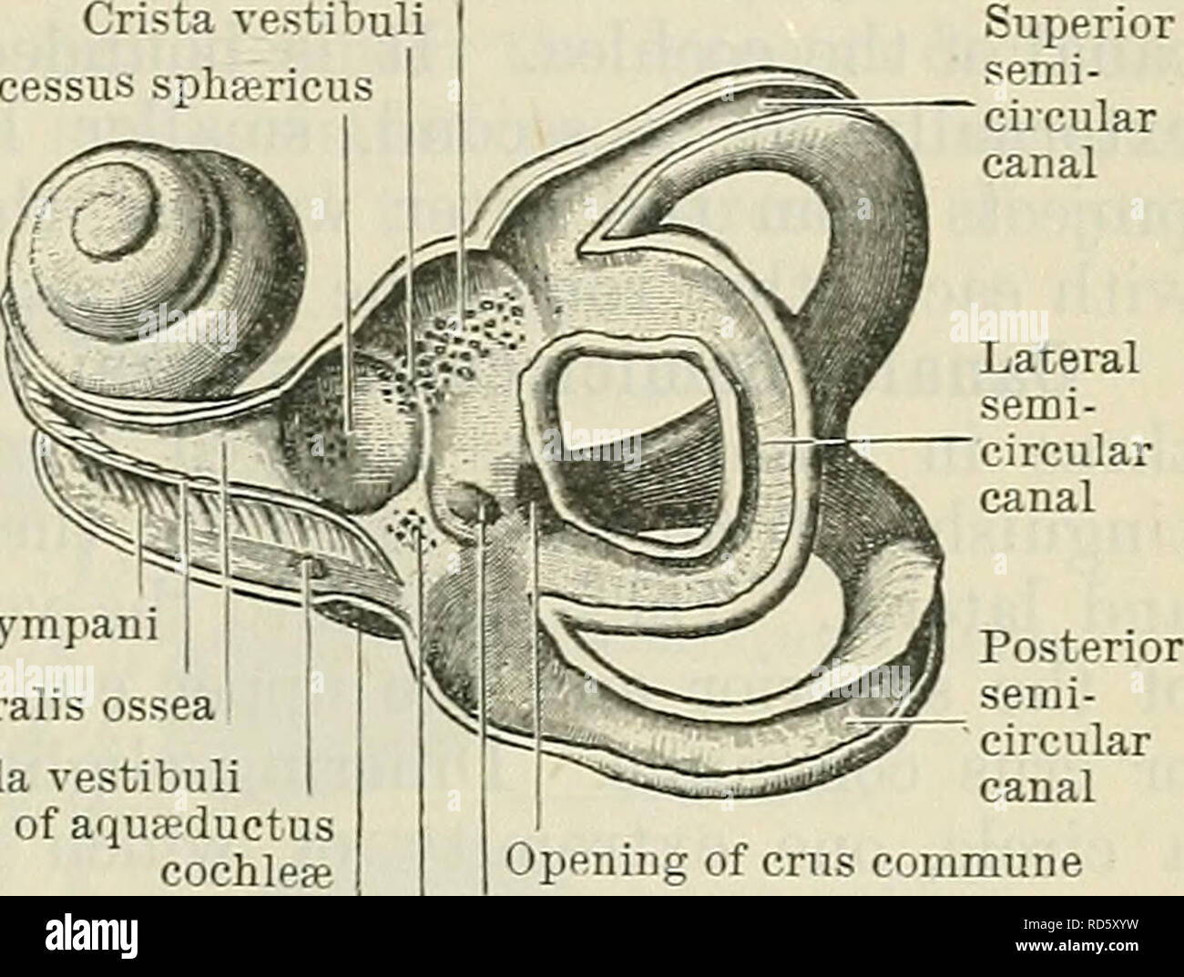 . Cunningham's Text-book of anatomy. Anatomy. Kecessus ellipticus Crista vestibuli Recessus spha?ricus Cochlea Fenestra cochlea; Fenestra vestibuli Ampulla of posterior semi circular canal | Ampulla of lateral semi- circular canal Lateral semicircular canal Posterior semi- circular canal Crus commune. Scala tympani Lamina spiralis ossea Scala vestibuli Opening of aquseductus cochlea; Fenestra cochleae Opening of crus commune Opening of aqusductus vestibuli Fig. 716.—Left Bony Labyrinth (viewed from the lateral aspect). Recessus cochlearis Fig. 717.—Interior of Left Bony Labyrinth (viewed from Stock Photohttps://www.alamy.com/image-license-details/?v=1https://www.alamy.com/cunninghams-text-book-of-anatomy-anatomy-kecessus-ellipticus-crista-vestibuli-recessus-spharicus-cochlea-fenestra-cochlea-fenestra-vestibuli-ampulla-of-posterior-semi-circular-canal-ampulla-of-lateral-semi-circular-canal-lateral-semicircular-canal-posterior-semi-circular-canal-crus-commune-scala-tympani-lamina-spiralis-ossea-scala-vestibuli-opening-of-aquseductus-cochlea-fenestra-cochleae-opening-of-crus-commune-opening-of-aqusductus-vestibuli-fig-716left-bony-labyrinth-viewed-from-the-lateral-aspect-recessus-cochlearis-fig-717interior-of-left-bony-labyrinth-viewed-from-image231856237.html
RMRD5XYW–. Cunningham's Text-book of anatomy. Anatomy. Kecessus ellipticus Crista vestibuli Recessus spha?ricus Cochlea Fenestra cochlea; Fenestra vestibuli Ampulla of posterior semi circular canal | Ampulla of lateral semi- circular canal Lateral semicircular canal Posterior semi- circular canal Crus commune. Scala tympani Lamina spiralis ossea Scala vestibuli Opening of aquseductus cochlea; Fenestra cochleae Opening of crus commune Opening of aqusductus vestibuli Fig. 716.—Left Bony Labyrinth (viewed from the lateral aspect). Recessus cochlearis Fig. 717.—Interior of Left Bony Labyrinth (viewed from
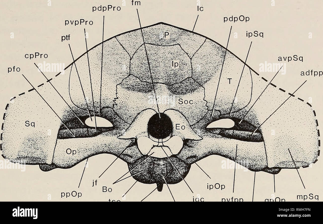 . Annals of the South African Museum = Annale van die Suid-Afrikaanse Museum. Natural history. REVISED DESCRIPTION OF MOSCHORHINUS 395 pdpPro pdpOp ipSq cpPro. kPs Bs 3cm pvfpp | qpOp mpOp mpSq Fig. 15. Moschorhinus kitchingi. Occipital view. (Fig. 16). The posterodorsal process forms most of the anterodorsal rim of the post-temporal fenestra. This process tapers off from a relatively broad base medially to a point jutting laterally, terminating in the lateral part of the roof of the post-temporal fenestra. This process protrudes from under the intermediate process of the squamosal anteromedia Stock Photohttps://www.alamy.com/image-license-details/?v=1https://www.alamy.com/annals-of-the-south-african-museum-=-annale-van-die-suid-afrikaanse-museum-natural-history-revised-description-of-moschorhinus-395-pdppro-pdpop-ipsq-cppro-kps-bs-3cm-pvfpp-qpop-mpop-mpsq-fig-15-moschorhinus-kitchingi-occipital-view-fig-16-the-posterodorsal-process-forms-most-of-the-anterodorsal-rim-of-the-post-temporal-fenestra-this-process-tapers-off-from-a-relatively-broad-base-medially-to-a-point-jutting-laterally-terminating-in-the-lateral-part-of-the-roof-of-the-post-temporal-fenestra-this-process-protrudes-from-under-the-intermediate-process-of-the-squamosal-anteromedia-image236407017.html
RMRMH7FN–. Annals of the South African Museum = Annale van die Suid-Afrikaanse Museum. Natural history. REVISED DESCRIPTION OF MOSCHORHINUS 395 pdpPro pdpOp ipSq cpPro. kPs Bs 3cm pvfpp | qpOp mpOp mpSq Fig. 15. Moschorhinus kitchingi. Occipital view. (Fig. 16). The posterodorsal process forms most of the anterodorsal rim of the post-temporal fenestra. This process tapers off from a relatively broad base medially to a point jutting laterally, terminating in the lateral part of the roof of the post-temporal fenestra. This process protrudes from under the intermediate process of the squamosal anteromedia
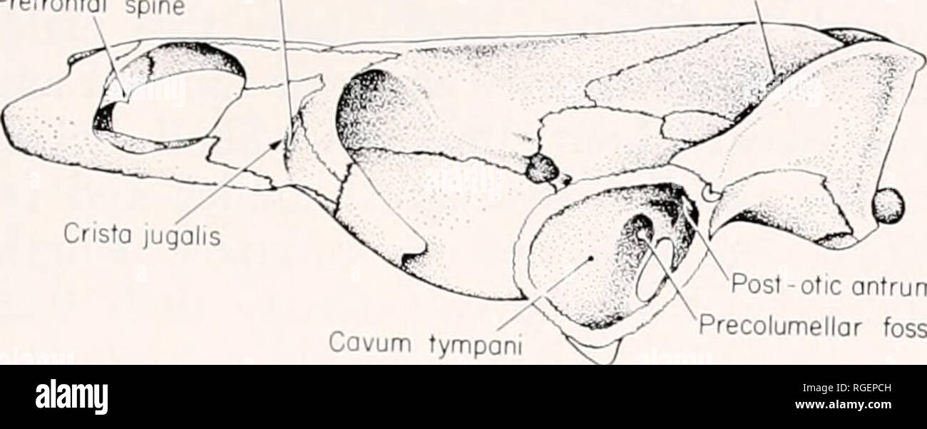 . Bulletin of the Museum of Comparative Zoology at Harvard College. Zoology. Chelodina rarkeri • Rhodin and Mittermeier 469 Prefrontal spine Sulcus jugalis Fossa temporalis superior. Cristo jugalis Cavum tympani Post-otic antrum Precolumellar fossa Fenestra hyoideus iall Figure 6. Lateral view of the skull of C. siebenrocki (AMS 40696). The labelled terms include new terms introduced in this paper and defined in Appendix I. midline in very large females. Surface of carapace weakly rugose with reticulate pattern of numerous, very shallow furrows. Furrows on vertebrals oriented longitudi- nally Stock Photohttps://www.alamy.com/image-license-details/?v=1https://www.alamy.com/bulletin-of-the-museum-of-comparative-zoology-at-harvard-college-zoology-chelodina-rarkeri-rhodin-and-mittermeier-469-prefrontal-spine-sulcus-jugalis-fossa-temporalis-superior-cristo-jugalis-cavum-tympani-post-otic-antrum-precolumellar-fossa-fenestra-hyoideus-iall-figure-6-lateral-view-of-the-skull-of-c-siebenrocki-ams-40696-the-labelled-terms-include-new-terms-introduced-in-this-paper-and-defined-in-appendix-i-midline-in-very-large-females-surface-of-carapace-weakly-rugose-with-reticulate-pattern-of-numerous-very-shallow-furrows-furrows-on-vertebrals-oriented-longitudi-nally-image233894209.html
RMRGEPCH–. Bulletin of the Museum of Comparative Zoology at Harvard College. Zoology. Chelodina rarkeri • Rhodin and Mittermeier 469 Prefrontal spine Sulcus jugalis Fossa temporalis superior. Cristo jugalis Cavum tympani Post-otic antrum Precolumellar fossa Fenestra hyoideus iall Figure 6. Lateral view of the skull of C. siebenrocki (AMS 40696). The labelled terms include new terms introduced in this paper and defined in Appendix I. midline in very large females. Surface of carapace weakly rugose with reticulate pattern of numerous, very shallow furrows. Furrows on vertebrals oriented longitudi- nally
 . Bulletin. Natural history; Natural history. March 1938 ROSS: NEARCTIC CADDIS FLIES 103 >^;i^s5^. Fig. 3.âRhyacophila fenestra, larva ment; the apical segment incised for one-third its lateral and one-tourth its mesal length; both lobes straight and rounded, the dorsal one small and the ventral one large. At the base of the segment there is a mesal incurving lobe; most of the apical segment and this lobe are covered with short, dark setae. Tenth tergite narrow, the dorsal lobe cleft down the meson for more than one- half its length; the lateral lobes so pro- duced have convex dorsal margin Stock Photohttps://www.alamy.com/image-license-details/?v=1https://www.alamy.com/bulletin-natural-history-natural-history-march-1938-ross-nearctic-caddis-flies-103-gtis5-fig-3rhyacophila-fenestra-larva-ment-the-apical-segment-incised-for-one-third-its-lateral-and-one-tourth-its-mesal-length-both-lobes-straight-and-rounded-the-dorsal-one-small-and-the-ventral-one-large-at-the-base-of-the-segment-there-is-a-mesal-incurving-lobe-most-of-the-apical-segment-and-this-lobe-are-covered-with-short-dark-setae-tenth-tergite-narrow-the-dorsal-lobe-cleft-down-the-meson-for-more-than-one-half-its-length-the-lateral-lobes-so-pro-duced-have-convex-dorsal-margin-image234130141.html
RMRGWFAN–. Bulletin. Natural history; Natural history. March 1938 ROSS: NEARCTIC CADDIS FLIES 103 >^;i^s5^. Fig. 3.âRhyacophila fenestra, larva ment; the apical segment incised for one-third its lateral and one-tourth its mesal length; both lobes straight and rounded, the dorsal one small and the ventral one large. At the base of the segment there is a mesal incurving lobe; most of the apical segment and this lobe are covered with short, dark setae. Tenth tergite narrow, the dorsal lobe cleft down the meson for more than one- half its length; the lateral lobes so pro- duced have convex dorsal margin