Lateral ventricle of the brain Stock Photos and Images
(608)See lateral ventricle of the brain stock video clipsQuick filters:
Lateral ventricle of the brain Stock Photos and Images
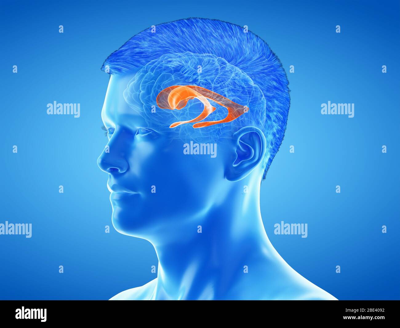 Lateral ventricle of the brain, illustration. Stock Photohttps://www.alamy.com/image-license-details/?v=1https://www.alamy.com/lateral-ventricle-of-the-brain-illustration-image352900606.html
RF2BE4092–Lateral ventricle of the brain, illustration.
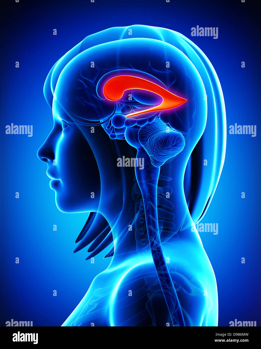 Anatomy of brain of lateral ventricle in blue Stock Photohttps://www.alamy.com/image-license-details/?v=1https://www.alamy.com/stock-photo-anatomy-of-brain-of-lateral-ventricle-in-blue-57409769.html
RFD9B6MW–Anatomy of brain of lateral ventricle in blue
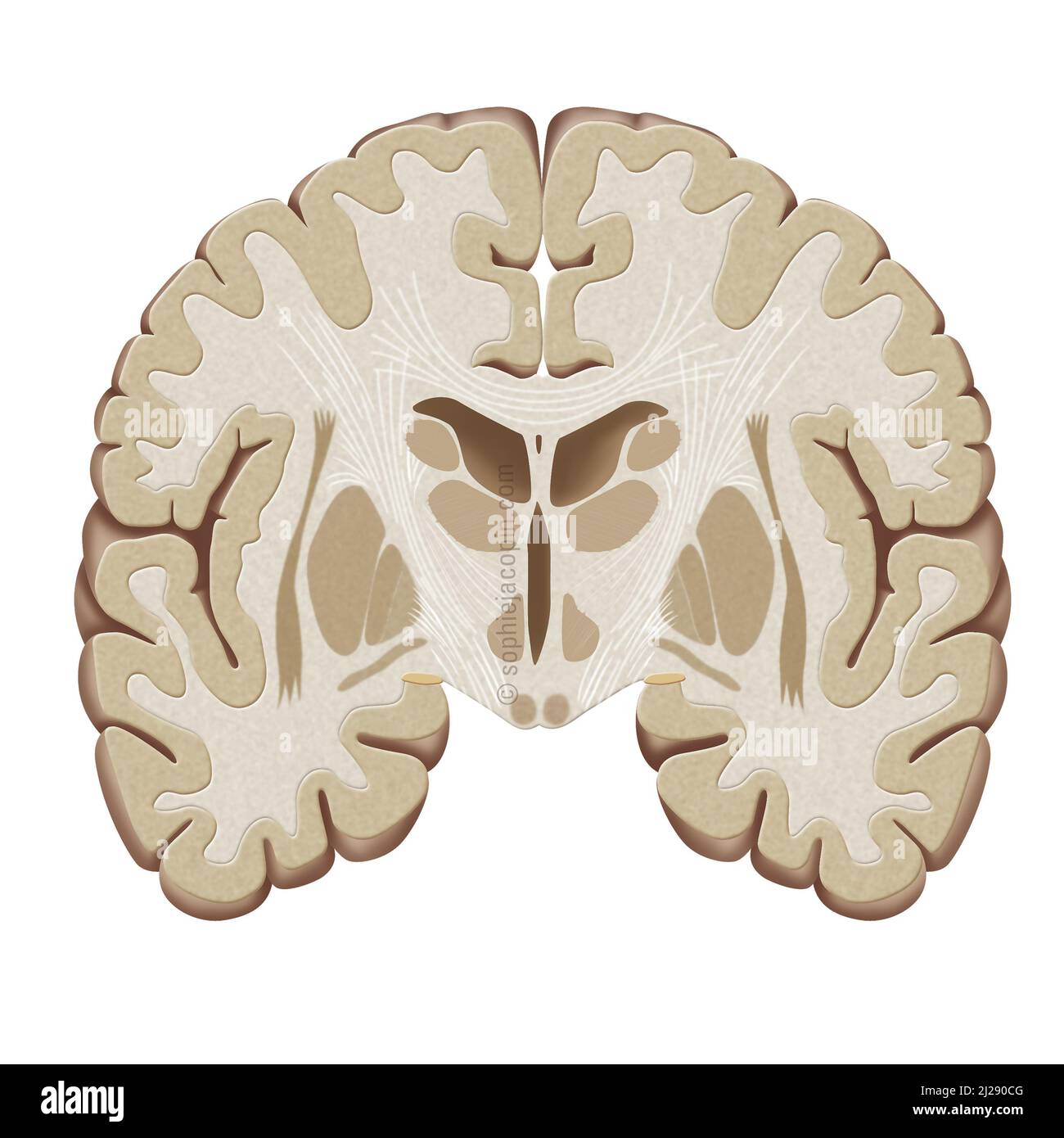 Frontal brain-cut Stock Photohttps://www.alamy.com/image-license-details/?v=1https://www.alamy.com/frontal-brain-cut-image466107168.html
RM2J290CG–Frontal brain-cut
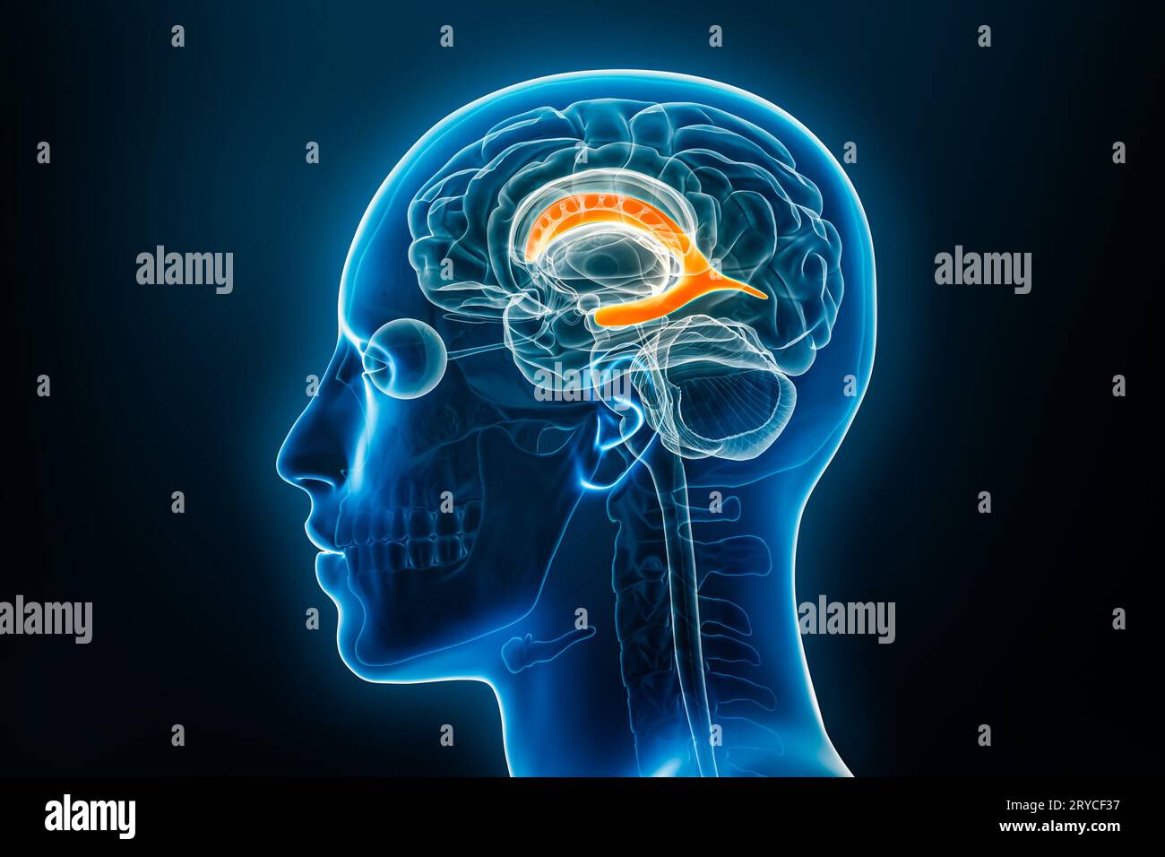 Xray profile view of the brain lateral ventricle 3D rendering illustration with male body contours. Human anatomy, medical, biology, science, neurosci Stock Photohttps://www.alamy.com/image-license-details/?v=1https://www.alamy.com/xray-profile-view-of-the-brain-lateral-ventricle-3d-rendering-illustration-with-male-body-contours-human-anatomy-medical-biology-science-neurosci-image567602763.html
RF2RYCF37–Xray profile view of the brain lateral ventricle 3D rendering illustration with male body contours. Human anatomy, medical, biology, science, neurosci
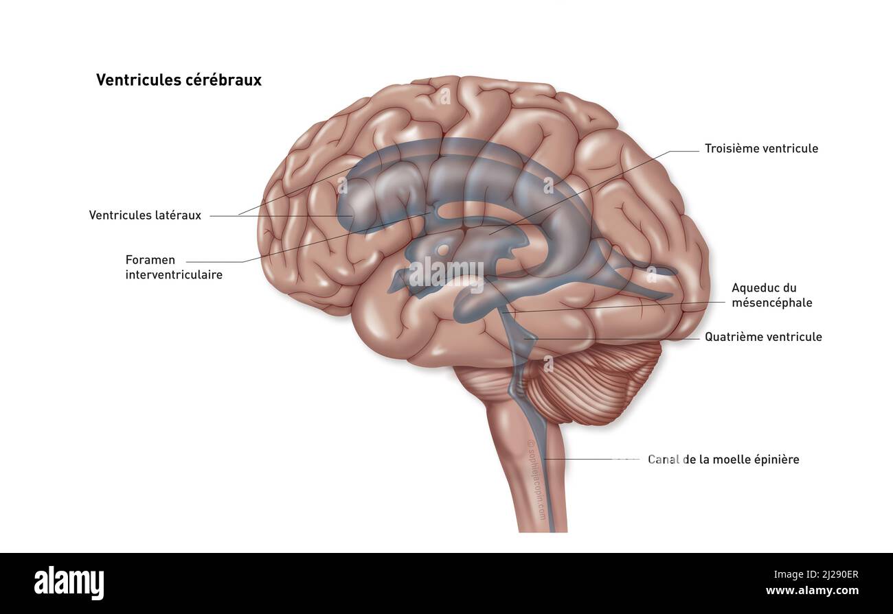 Brain ventricles Stock Photohttps://www.alamy.com/image-license-details/?v=1https://www.alamy.com/brain-ventricles-image466107231.html
RM2J290ER–Brain ventricles
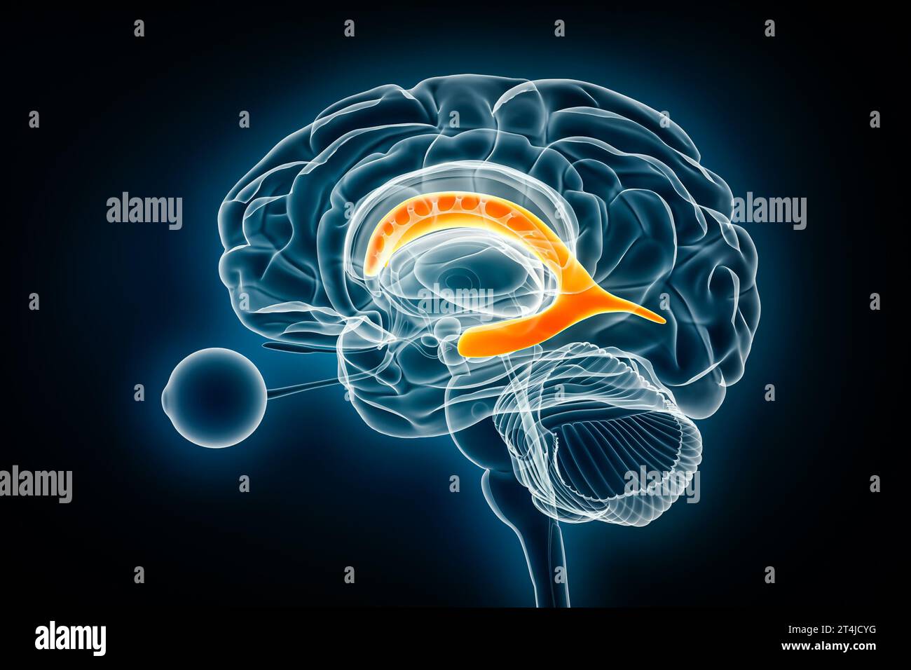 Lateral ventricle profile x-ray view 3D rendering illustration. Human brain and cerebral ventricular system anatomy, medical, healthcare, biology, sci Stock Photohttps://www.alamy.com/image-license-details/?v=1https://www.alamy.com/lateral-ventricle-profile-x-ray-view-3d-rendering-illustration-human-brain-and-cerebral-ventricular-system-anatomy-medical-healthcare-biology-sci-image570806084.html
RF2T4JCYG–Lateral ventricle profile x-ray view 3D rendering illustration. Human brain and cerebral ventricular system anatomy, medical, healthcare, biology, sci
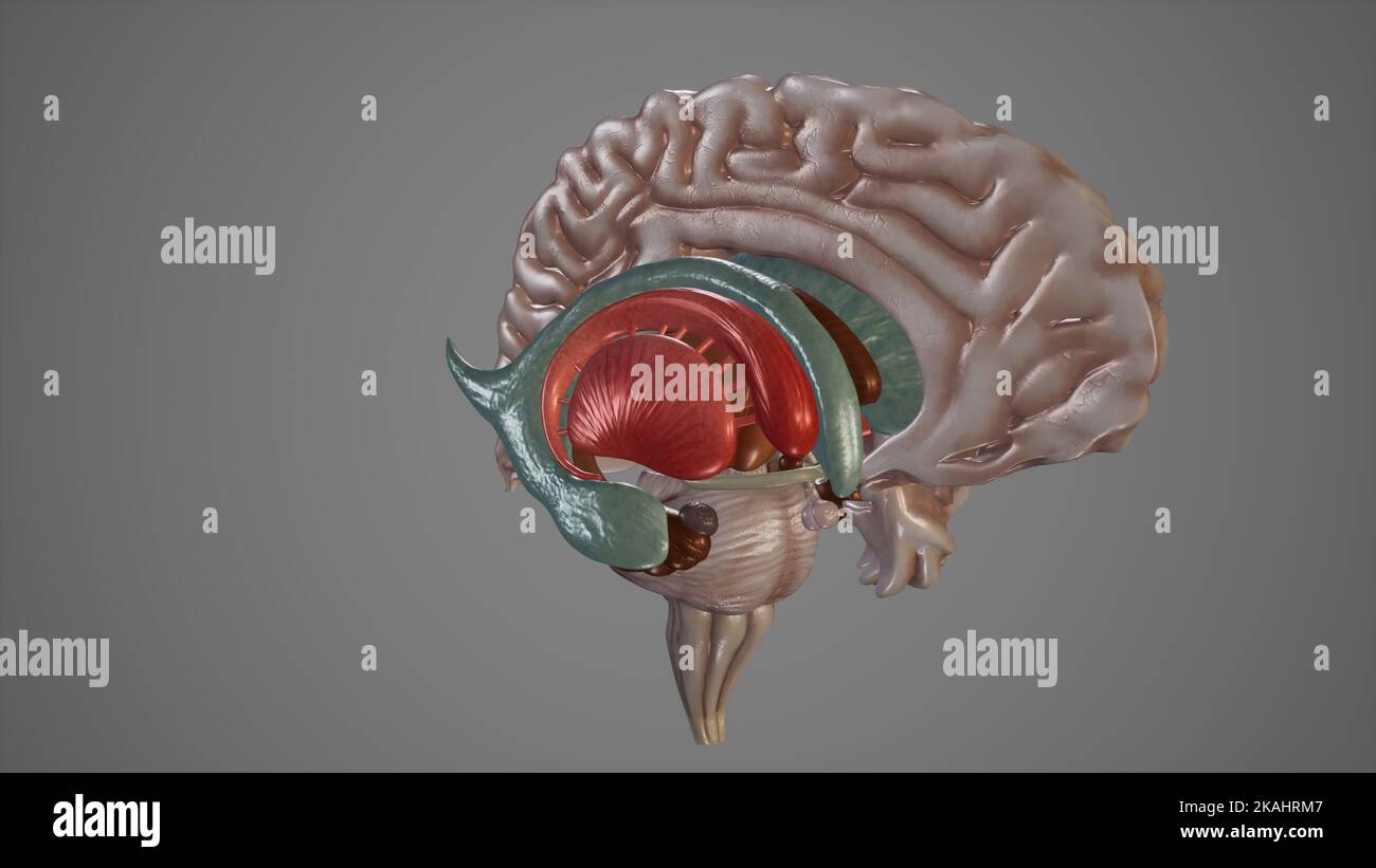 Medical Illustration of Deep Structures of Brain Stock Photohttps://www.alamy.com/image-license-details/?v=1https://www.alamy.com/medical-illustration-of-deep-structures-of-brain-image488428647.html
RF2KAHRM7–Medical Illustration of Deep Structures of Brain
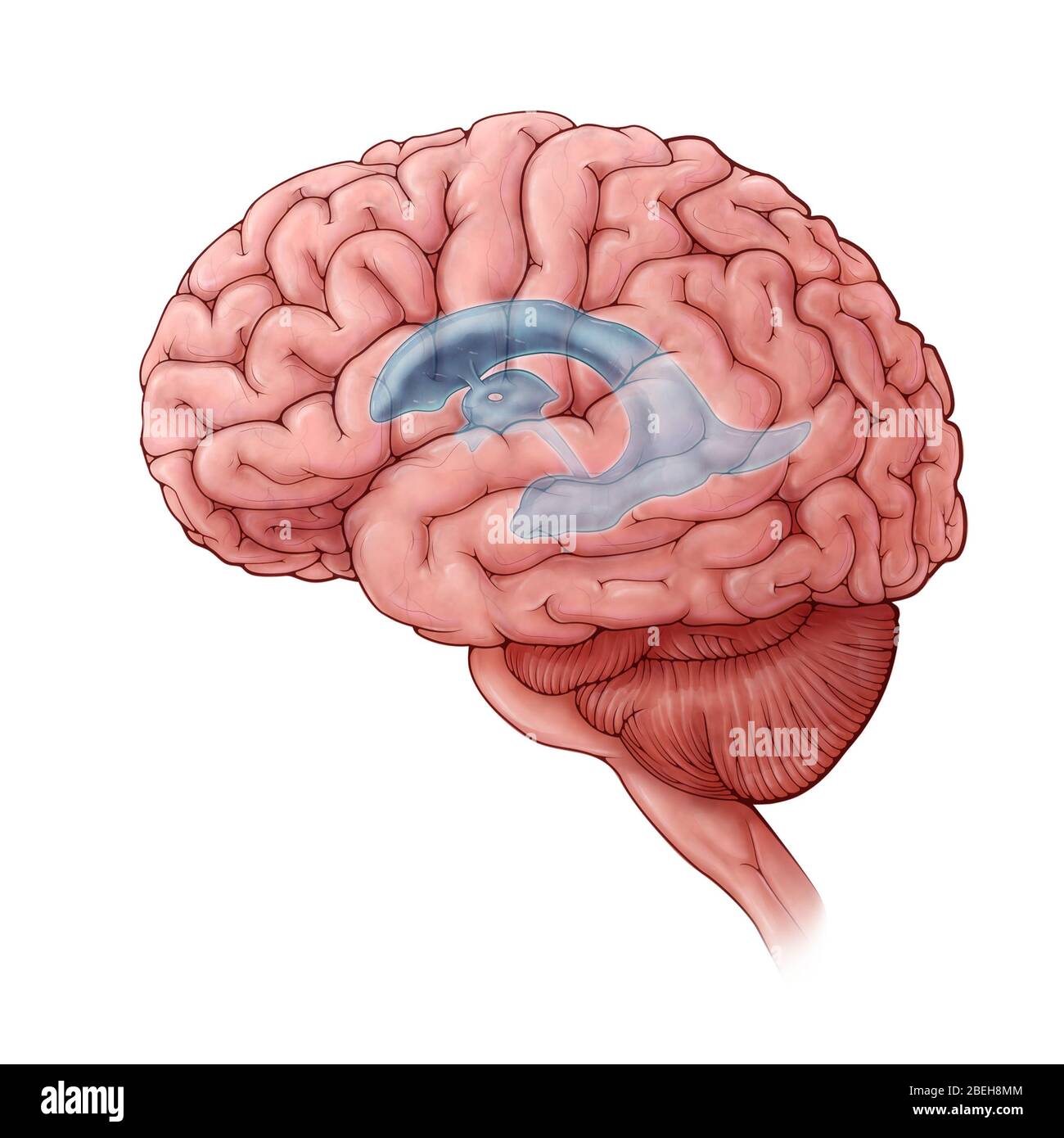 Lateral Ventricles, Illustration Stock Photohttps://www.alamy.com/image-license-details/?v=1https://www.alamy.com/lateral-ventricles-illustration-image353192580.html
RM2BEH8MM–Lateral Ventricles, Illustration
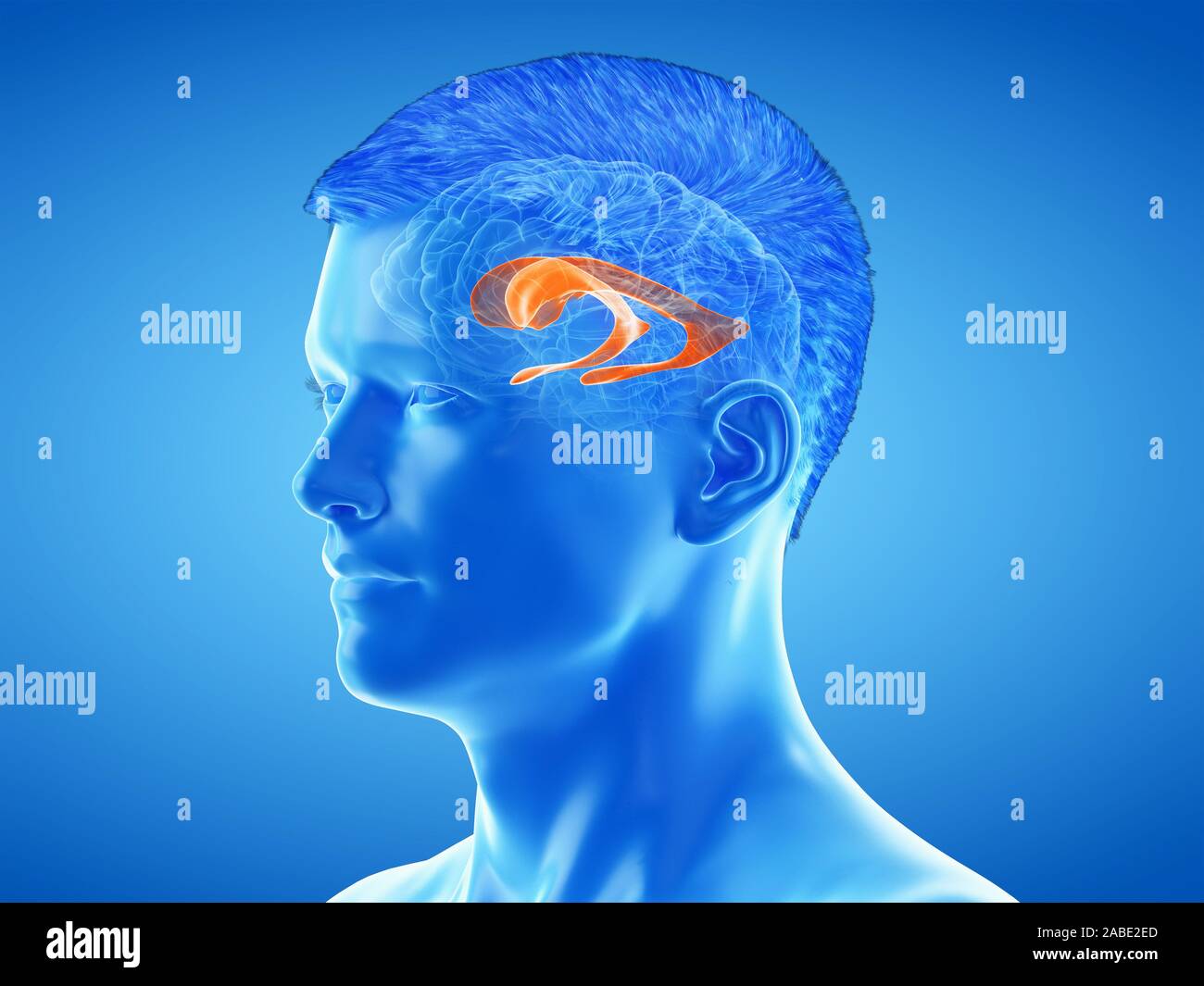 3d rendered medically accurate illustration of the brain anatomy - the lateral ventricle Stock Photohttps://www.alamy.com/image-license-details/?v=1https://www.alamy.com/3d-rendered-medically-accurate-illustration-of-the-brain-anatomy-the-lateral-ventricle-image334067509.html
RF2ABE2ED–3d rendered medically accurate illustration of the brain anatomy - the lateral ventricle
 Magnetic resonance imaging of a patient with a temporal horn cyst of the left lateral ventricle of the brain. Atrophic changes in the temporal lobe. A Stock Photohttps://www.alamy.com/image-license-details/?v=1https://www.alamy.com/magnetic-resonance-imaging-of-a-patient-with-a-temporal-horn-cyst-of-the-left-lateral-ventricle-of-the-brain-atrophic-changes-in-the-temporal-lobe-a-image415458418.html
RF2F3WNCJ–Magnetic resonance imaging of a patient with a temporal horn cyst of the left lateral ventricle of the brain. Atrophic changes in the temporal lobe. A
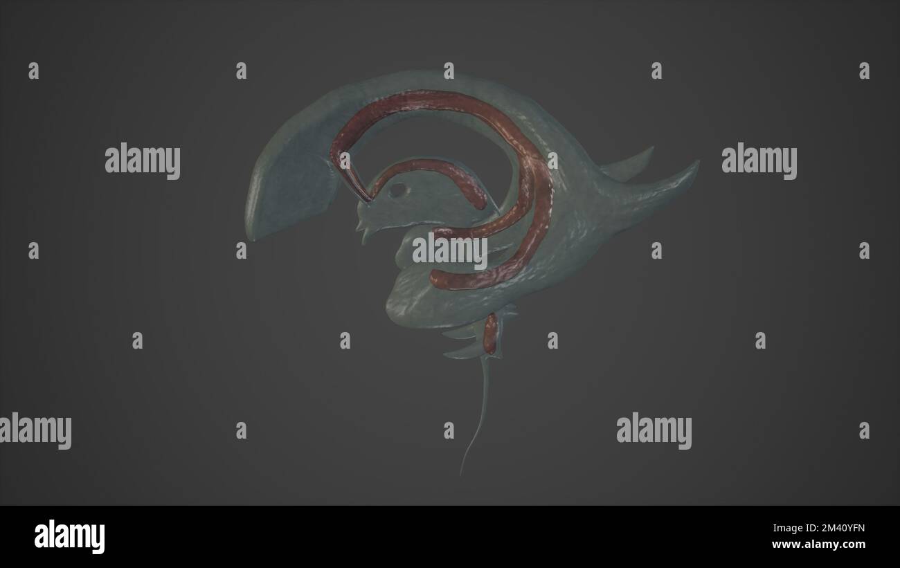 Anatomical Illustration of Choroid Plexuses.3d rendering Stock Photohttps://www.alamy.com/image-license-details/?v=1https://www.alamy.com/anatomical-illustration-of-choroid-plexuses3d-rendering-image501580905.html
RF2M40YFN–Anatomical Illustration of Choroid Plexuses.3d rendering
RF2XJ0865–Ventricular system. Cerebrospinal fluid. Black icon. Simple outline style. Stylized pictogram for web design, or mobile app. Vector illustration. flat
RF2E1PT29–A CT scan of a stroke patient showing intracerebral hemorrhage in the Cd clots at right thalamus, basal gangliconic and lateral ventricle with brain
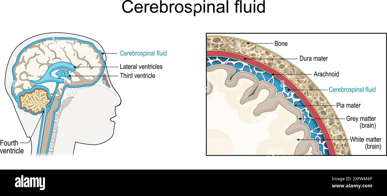 Cerebrospinal fluid. Cross section of a human brain with Ventricular system. Close-up of the meninges. CSF and Membranes that envelop the brain Stock Vectorhttps://www.alamy.com/image-license-details/?v=1https://www.alamy.com/cerebrospinal-fluid-cross-section-of-a-human-brain-with-ventricular-system-close-up-of-the-meninges-csf-and-membranes-that-envelop-the-brain-image612147334.html
RF2XFWM4P–Cerebrospinal fluid. Cross section of a human brain with Ventricular system. Close-up of the meninges. CSF and Membranes that envelop the brain
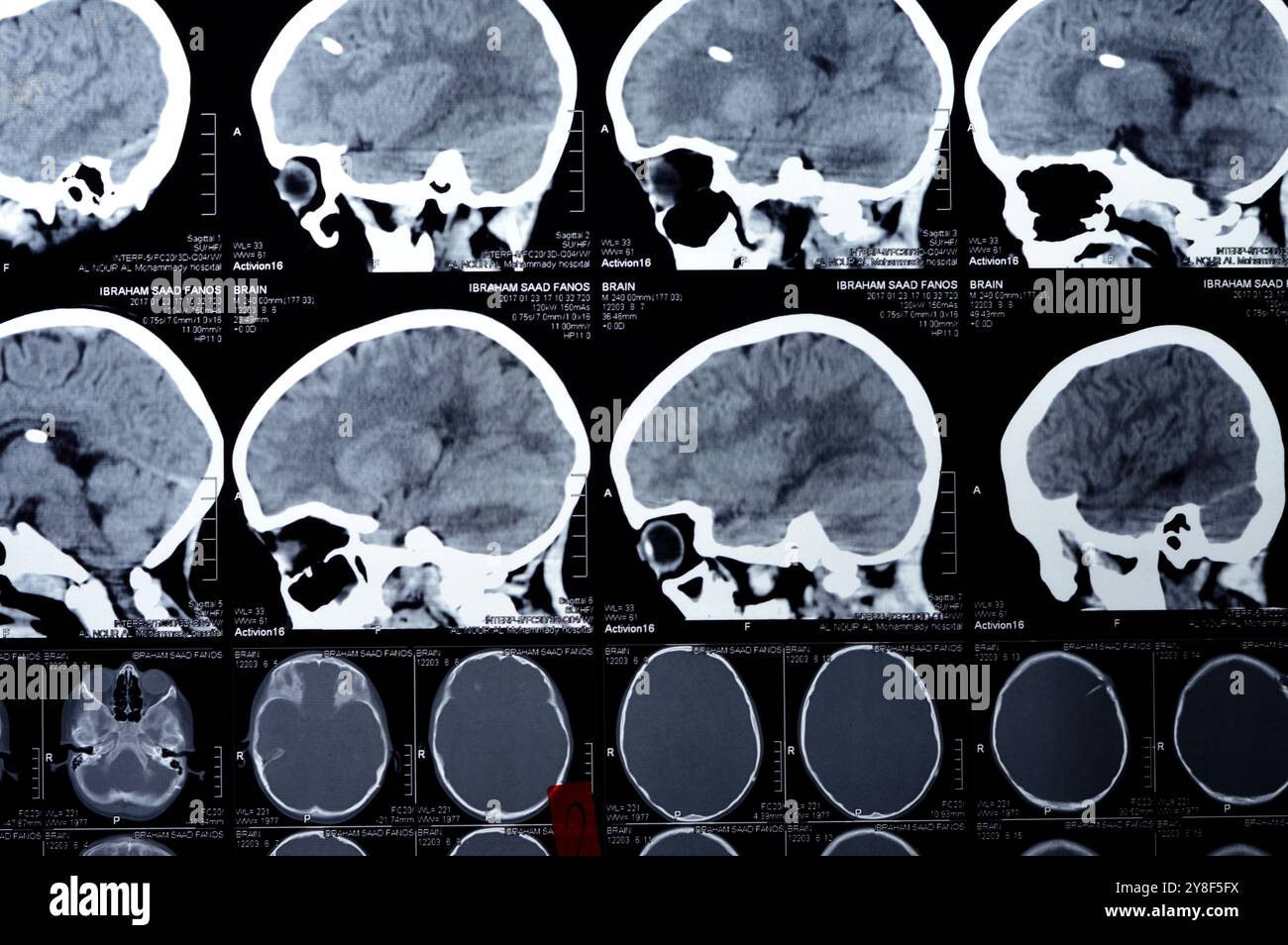 Cairo, Egypt, September 17 2024: Brain CT scan reveals left shunt tube terminating in right lateral ventricle, with no residual ventricular dilatation Stock Photohttps://www.alamy.com/image-license-details/?v=1https://www.alamy.com/cairo-egypt-september-17-2024-brain-ct-scan-reveals-left-shunt-tube-terminating-in-right-lateral-ventricle-with-no-residual-ventricular-dilatation-image624824142.html
RF2Y8F5FX–Cairo, Egypt, September 17 2024: Brain CT scan reveals left shunt tube terminating in right lateral ventricle, with no residual ventricular dilatation
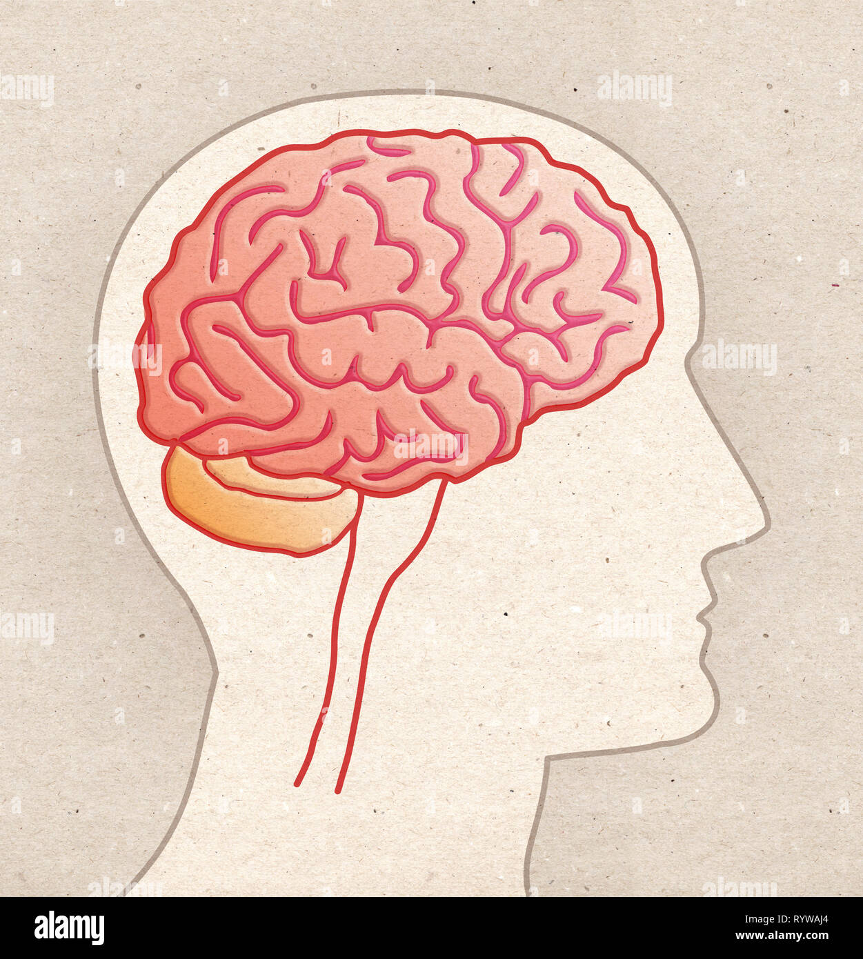 Human Anatomy drawing - Profile Head with BRAIN side view Stock Photohttps://www.alamy.com/image-license-details/?v=1https://www.alamy.com/human-anatomy-drawing-profile-head-with-brain-side-view-image240887644.html
RFRYWAJ4–Human Anatomy drawing - Profile Head with BRAIN side view
 Cross-section of a cow brain showing the convoluted tissue with a probe pointing to the frontal lobe Stock Photohttps://www.alamy.com/image-license-details/?v=1https://www.alamy.com/cross-section-of-a-cow-brain-showing-the-convoluted-tissue-with-a-image69690568.html
RFE1AK0T–Cross-section of a cow brain showing the convoluted tissue with a probe pointing to the frontal lobe
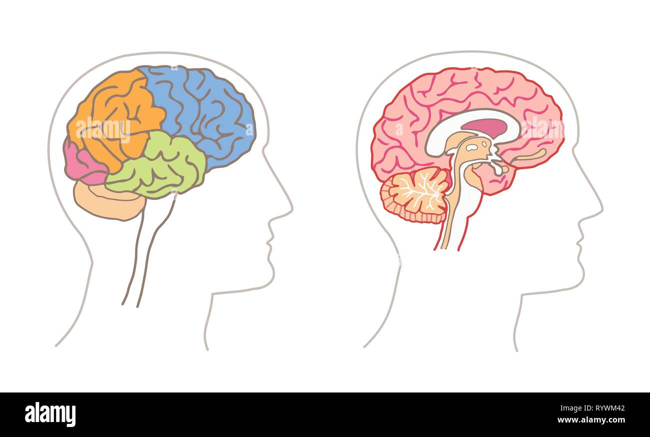 Human Anatomy drawings - BRAIN Lobes and Sagittal section Stock Vectorhttps://www.alamy.com/image-license-details/?v=1https://www.alamy.com/human-anatomy-drawings-brain-lobes-and-sagittal-section-image240895090.html
RFRYWM42–Human Anatomy drawings - BRAIN Lobes and Sagittal section
 Cerebrospinal fluid (CSF) It protects the brain and spinal cord from impact, eliminates waste from the brain and spinal cord, and helps toxins in the Stock Vectorhttps://www.alamy.com/image-license-details/?v=1https://www.alamy.com/cerebrospinal-fluid-csf-it-protects-the-brain-and-spinal-cord-from-impact-eliminates-waste-from-the-brain-and-spinal-cord-and-helps-toxins-in-the-image222730447.html
RFPXA6XR–Cerebrospinal fluid (CSF) It protects the brain and spinal cord from impact, eliminates waste from the brain and spinal cord, and helps toxins in the
 Lateral ventricle of the brain, illustration. Stock Photohttps://www.alamy.com/image-license-details/?v=1https://www.alamy.com/lateral-ventricle-of-the-brain-illustration-image352900665.html
RF2BE40B5–Lateral ventricle of the brain, illustration.
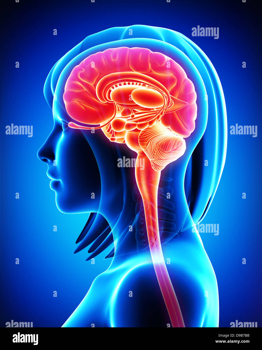 Anatomy of female brain cross section in blue Stock Photohttps://www.alamy.com/image-license-details/?v=1https://www.alamy.com/stock-photo-anatomy-of-female-brain-cross-section-in-blue-57410287.html
RFD9B7BB–Anatomy of female brain cross section in blue
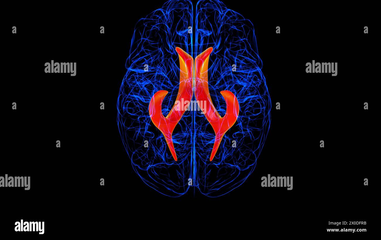 Brain lateral ventricle Anatomy For Medical Concept 3D illustration Stock Photohttps://www.alamy.com/image-license-details/?v=1https://www.alamy.com/brain-lateral-ventricle-anatomy-for-medical-concept-3d-illustration-image602660671.html
RF2X0DFRB–Brain lateral ventricle Anatomy For Medical Concept 3D illustration
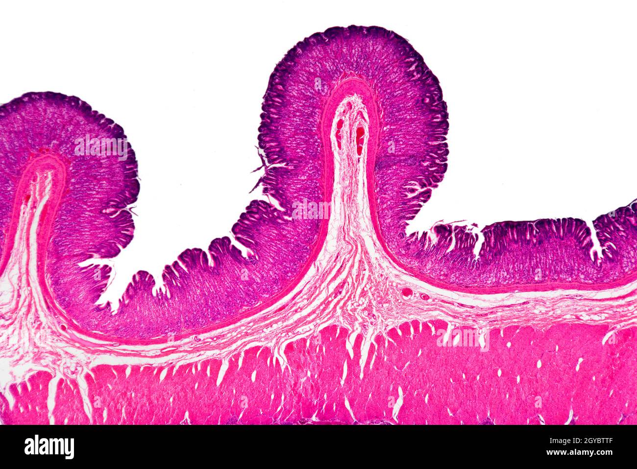 Corpus ventriculi slide section, brightfield photomicrograph, stained section Stock Photohttps://www.alamy.com/image-license-details/?v=1https://www.alamy.com/corpus-ventriculi-slide-section-brightfield-photomicrograph-stained-section-image447115887.html
RM2GYBTTF–Corpus ventriculi slide section, brightfield photomicrograph, stained section
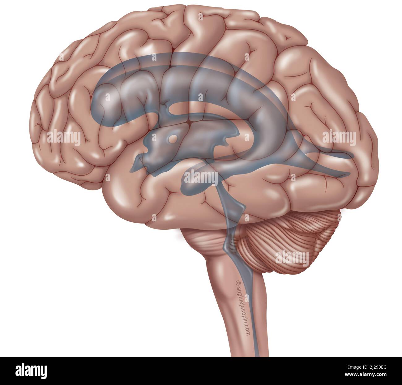 Encephal ventricular system Stock Photohttps://www.alamy.com/image-license-details/?v=1https://www.alamy.com/encephal-ventricular-system-image466107224.html
RM2J290EG–Encephal ventricular system
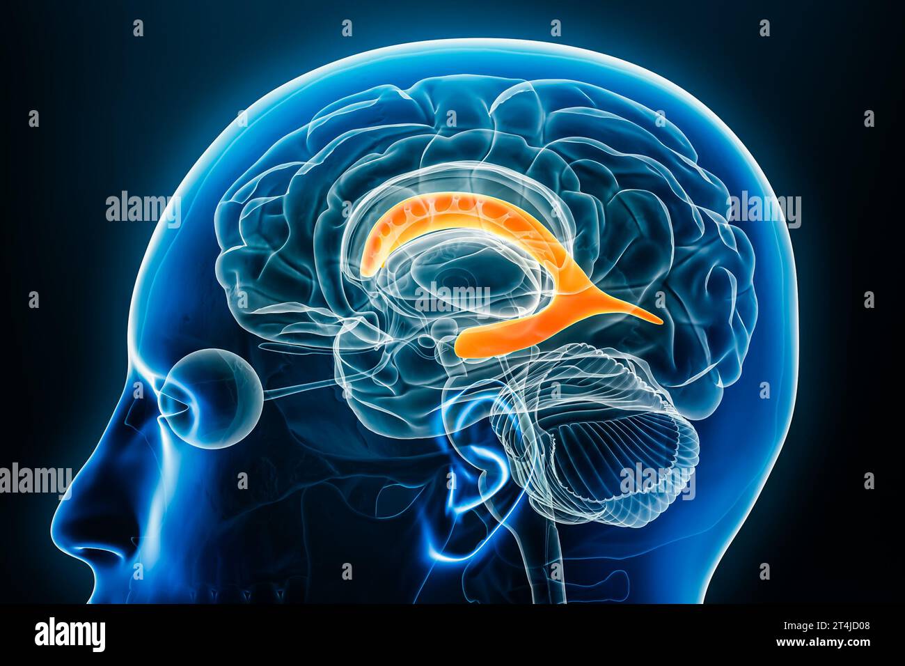 Lateral ventricle x-ray profile close-up view 3D rendering illustration with body contours. Human brain and ventricular system anatomy, medical, biolo Stock Photohttps://www.alamy.com/image-license-details/?v=1https://www.alamy.com/lateral-ventricle-x-ray-profile-close-up-view-3d-rendering-illustration-with-body-contours-human-brain-and-ventricular-system-anatomy-medical-biolo-image570806104.html
RF2T4JD08–Lateral ventricle x-ray profile close-up view 3D rendering illustration with body contours. Human brain and ventricular system anatomy, medical, biolo
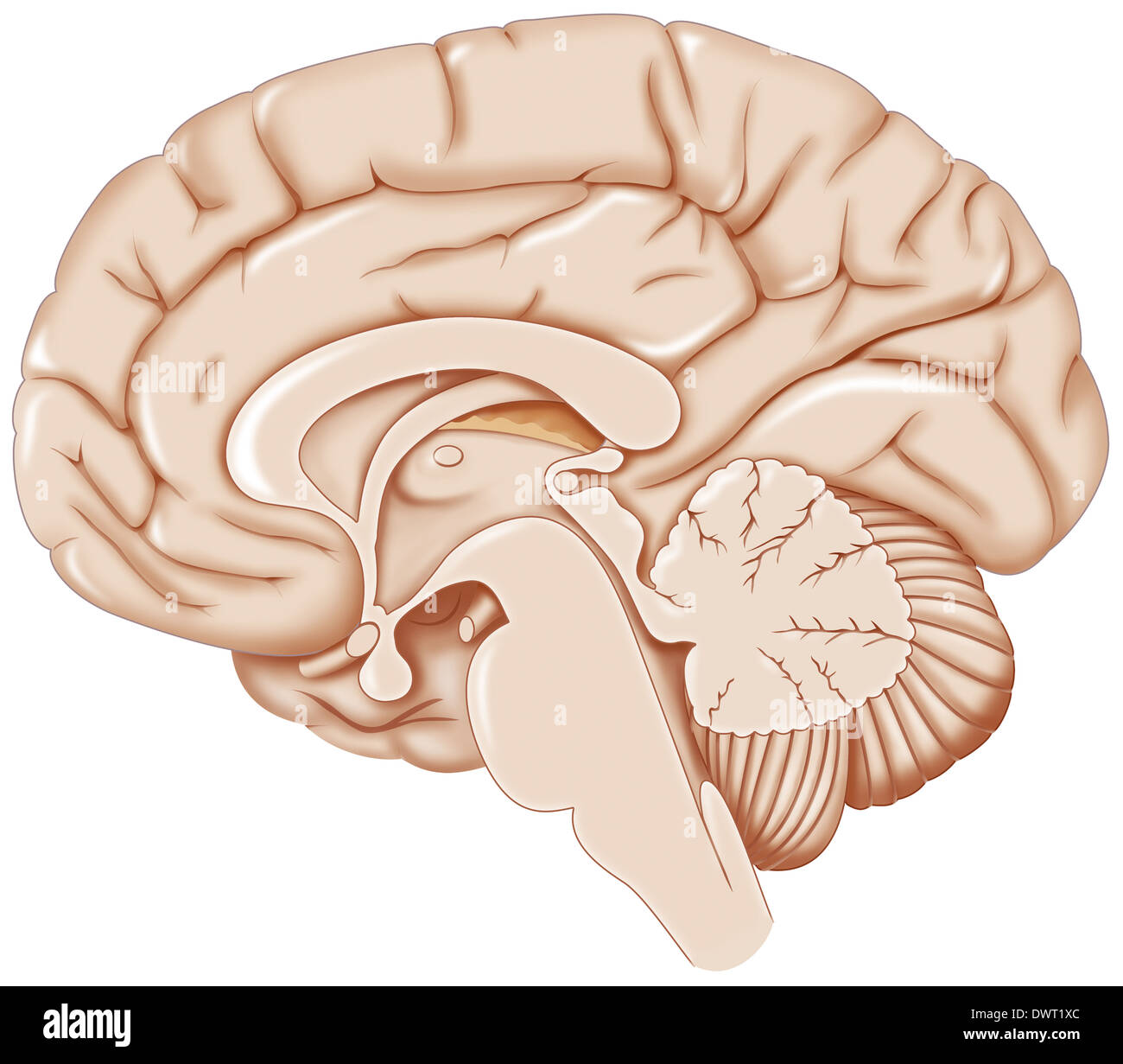 Brain, drawing Stock Photohttps://www.alamy.com/image-license-details/?v=1https://www.alamy.com/brain-drawing-image67525876.html
RMDWT1XC–Brain, drawing
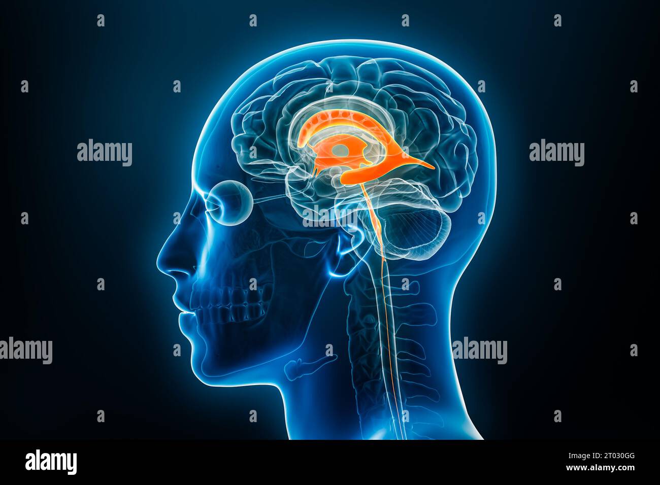 Xray lateral view of the brain ventricles 3D rendering illustration with body contours. Human and ventricular system anatomy, medical, biology, scienc Stock Photohttps://www.alamy.com/image-license-details/?v=1https://www.alamy.com/xray-lateral-view-of-the-brain-ventricles-3d-rendering-illustration-with-body-contours-human-and-ventricular-system-anatomy-medical-biology-scienc-image568008464.html
RF2T030GG–Xray lateral view of the brain ventricles 3D rendering illustration with body contours. Human and ventricular system anatomy, medical, biology, scienc
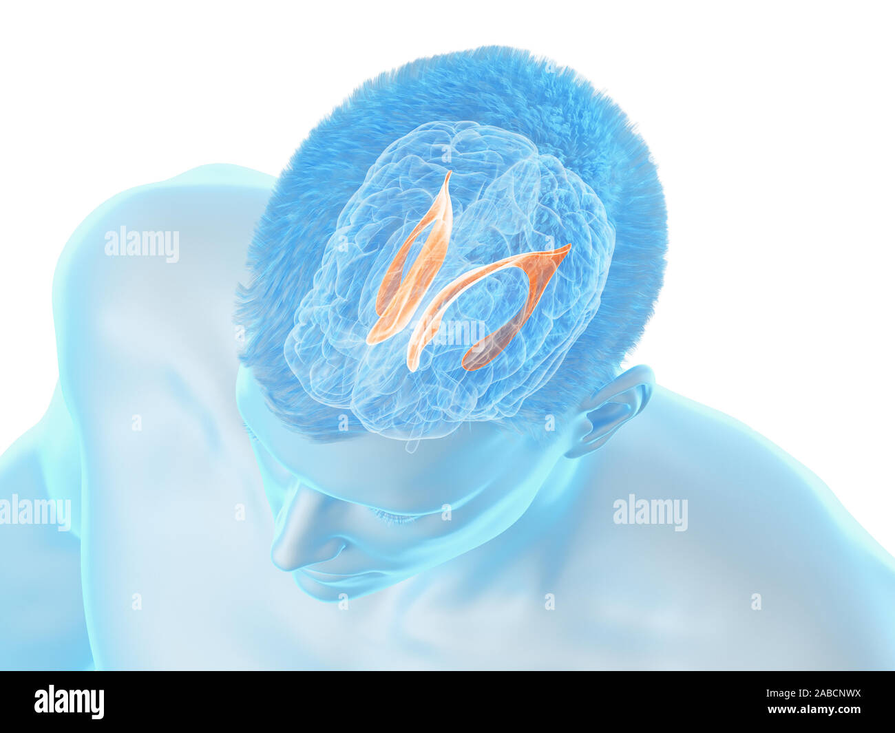 3d rendered medically accurate illustration of the brain anatomy - the lateral ventricle Stock Photohttps://www.alamy.com/image-license-details/?v=1https://www.alamy.com/3d-rendered-medically-accurate-illustration-of-the-brain-anatomy-the-lateral-ventricle-image334038822.html
RF2ABCNWX–3d rendered medically accurate illustration of the brain anatomy - the lateral ventricle
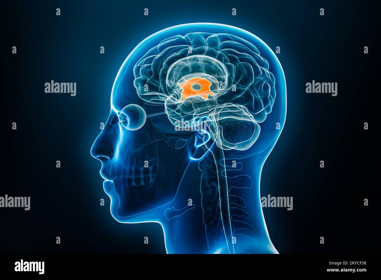 Xray lateral or profile view of the brain third ventricle 3D rendering illustration with male body contours. Human anatomy, medical, biology, science, Stock Photohttps://www.alamy.com/image-license-details/?v=1https://www.alamy.com/xray-lateral-or-profile-view-of-the-brain-third-ventricle-3d-rendering-illustration-with-male-body-contours-human-anatomy-medical-biology-science-image567602764.html
RF2RYCF38–Xray lateral or profile view of the brain third ventricle 3D rendering illustration with male body contours. Human anatomy, medical, biology, science,
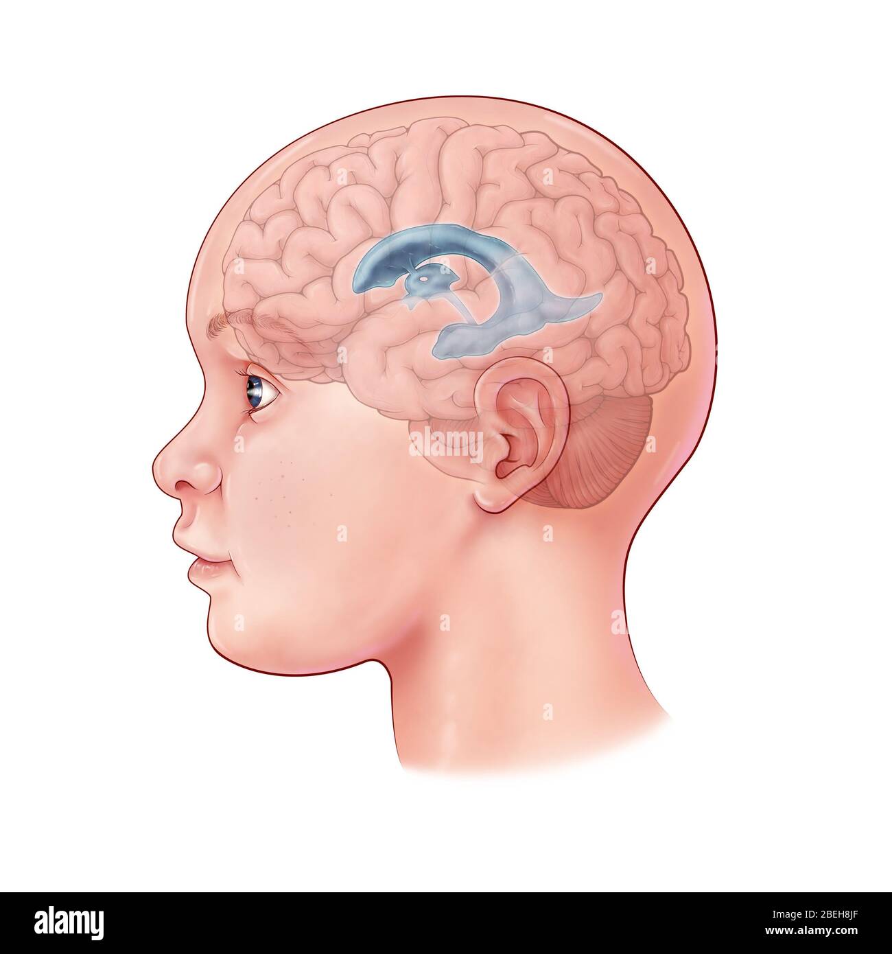 Child's Brain, Illustration Stock Photohttps://www.alamy.com/image-license-details/?v=1https://www.alamy.com/childs-brain-illustration-image353192519.html
RM2BEH8JF–Child's Brain, Illustration
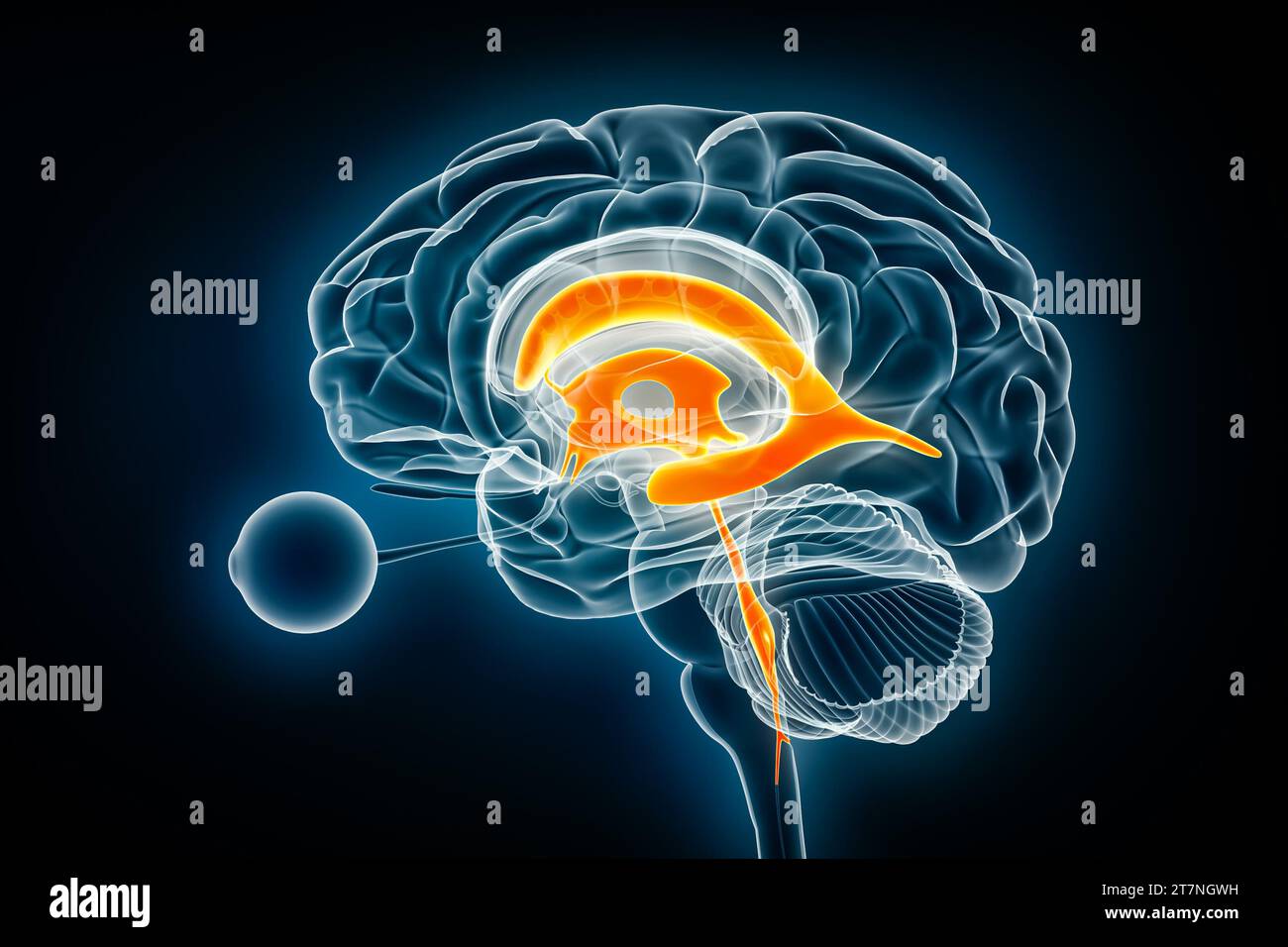 Ventricles and cerebral aqueduct lateral x-ray view 3D rendering illustration. Human brain and ventricular system anatomy, medical, healthcare, scienc Stock Photohttps://www.alamy.com/image-license-details/?v=1https://www.alamy.com/ventricles-and-cerebral-aqueduct-lateral-x-ray-view-3d-rendering-illustration-human-brain-and-ventricular-system-anatomy-medical-healthcare-scienc-image572718989.html
RF2T7NGWH–Ventricles and cerebral aqueduct lateral x-ray view 3D rendering illustration. Human brain and ventricular system anatomy, medical, healthcare, scienc
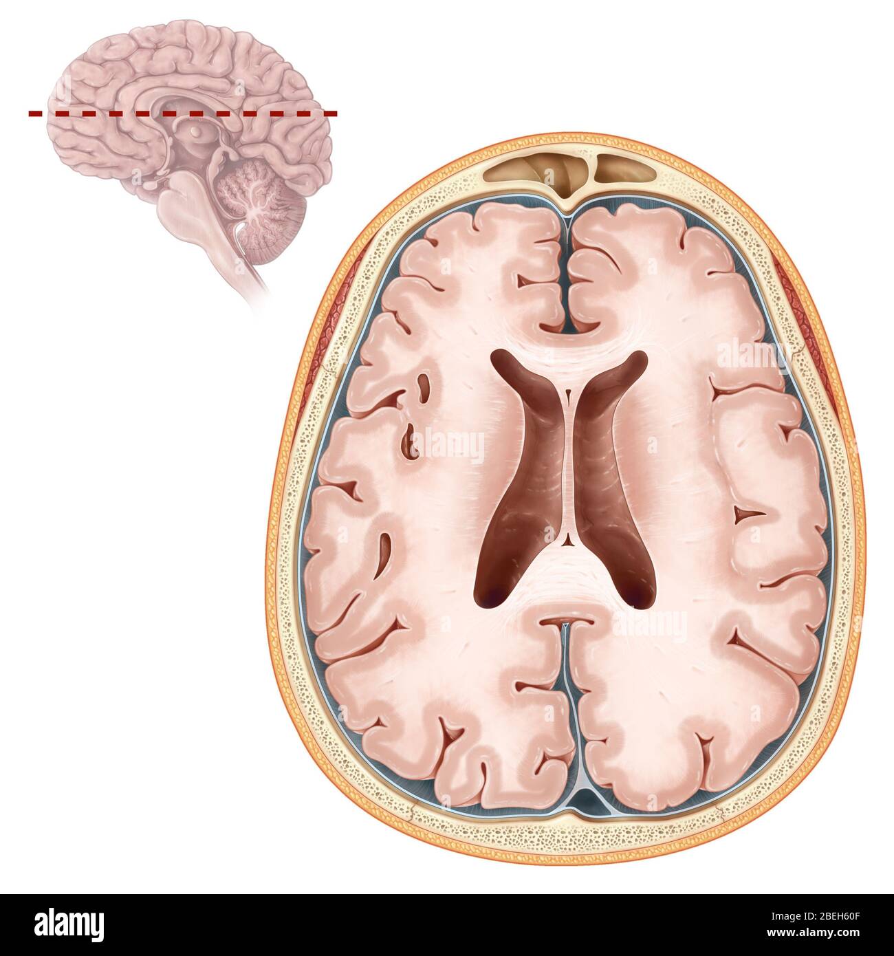 Brain, Transverse Section Stock Photohttps://www.alamy.com/image-license-details/?v=1https://www.alamy.com/brain-transverse-section-image353190447.html
RM2BEH60F–Brain, Transverse Section
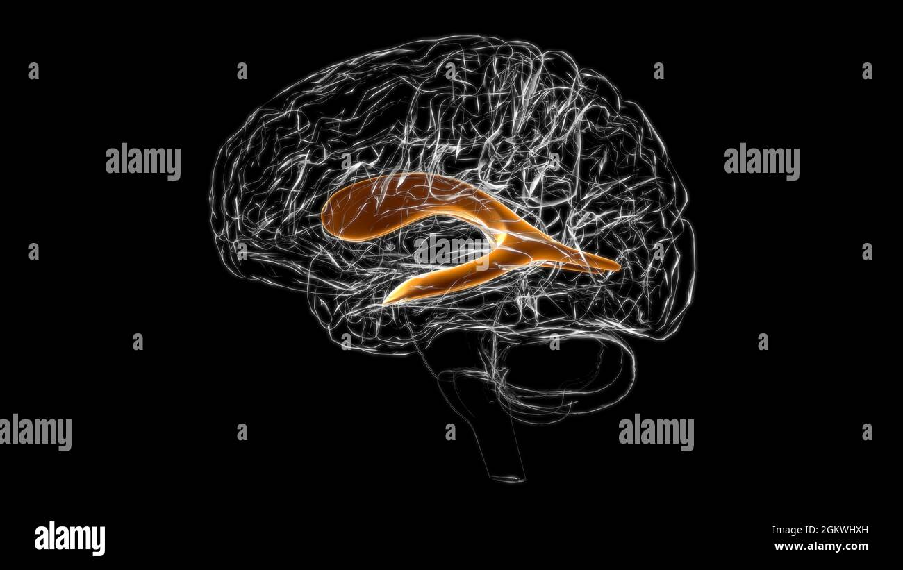 Brain lateral ventricle Anatomy For Medical Concept 3D Illustration Stock Photohttps://www.alamy.com/image-license-details/?v=1https://www.alamy.com/brain-lateral-ventricle-anatomy-for-medical-concept-3d-illustration-image442500537.html
RF2GKWHXH–Brain lateral ventricle Anatomy For Medical Concept 3D Illustration
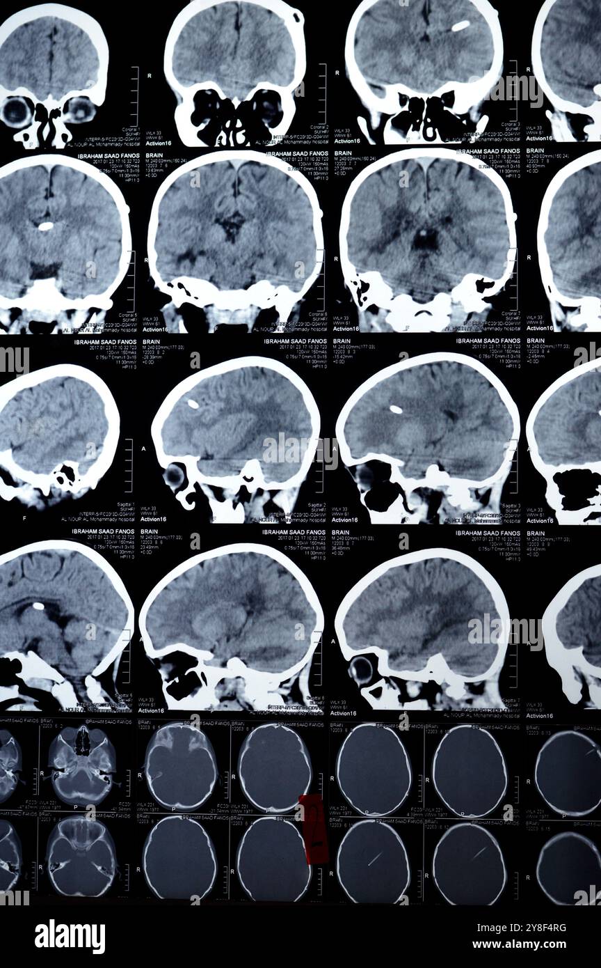 Cairo, Egypt, September 17 2024: Brain CT scan reveals left shunt tube terminating in right lateral ventricle, with no residual ventricular dilatation Stock Photohttps://www.alamy.com/image-license-details/?v=1https://www.alamy.com/cairo-egypt-september-17-2024-brain-ct-scan-reveals-left-shunt-tube-terminating-in-right-lateral-ventricle-with-no-residual-ventricular-dilatation-image624823572.html
RF2Y8F4RG–Cairo, Egypt, September 17 2024: Brain CT scan reveals left shunt tube terminating in right lateral ventricle, with no residual ventricular dilatation
 Brain lateral ventricle Anatomy For Medical Concept 3D Illustration Stock Photohttps://www.alamy.com/image-license-details/?v=1https://www.alamy.com/brain-lateral-ventricle-anatomy-for-medical-concept-3d-illustration-image429421725.html
RF2FXHRP5–Brain lateral ventricle Anatomy For Medical Concept 3D Illustration
 Female anatomy student, medical technologist or pathologist with a dissected cow brain slice through the mid section to show the Stock Photohttps://www.alamy.com/image-license-details/?v=1https://www.alamy.com/female-anatomy-student-medical-technologist-or-pathologist-with-a-image69690553.html
RFE1AK09–Female anatomy student, medical technologist or pathologist with a dissected cow brain slice through the mid section to show the
 Brain insula Anatomy For Medical Concept 3D illustration Stock Photohttps://www.alamy.com/image-license-details/?v=1https://www.alamy.com/brain-insula-anatomy-for-medical-concept-3d-illustration-image602660564.html
RF2X0DFKG–Brain insula Anatomy For Medical Concept 3D illustration
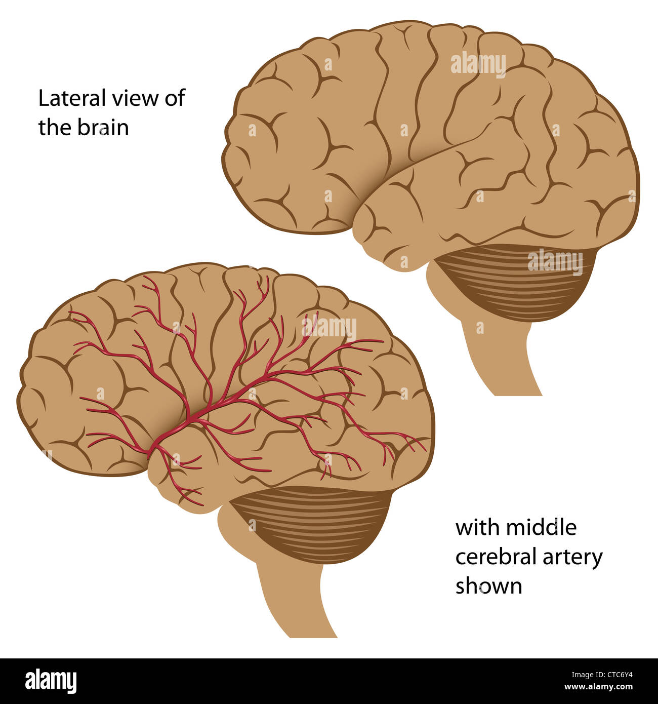 Lateral view of the brain with arteries Stock Photohttps://www.alamy.com/image-license-details/?v=1https://www.alamy.com/stock-photo-lateral-view-of-the-brain-with-arteries-49441368.html
RFCTC6Y4–Lateral view of the brain with arteries
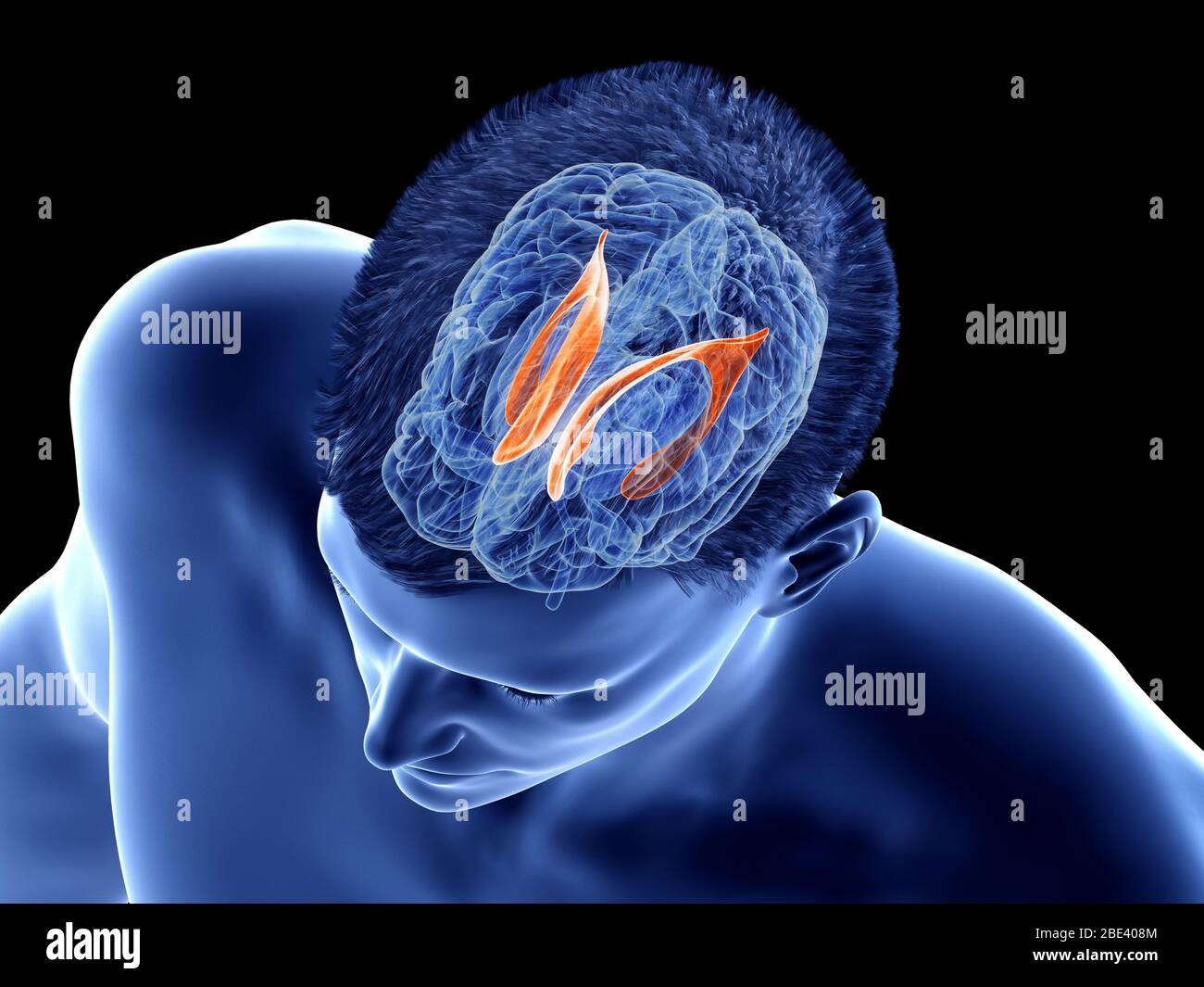 Lateral ventricle of the brain, illustration. Stock Photohttps://www.alamy.com/image-license-details/?v=1https://www.alamy.com/lateral-ventricle-of-the-brain-illustration-image352900596.html
RF2BE408M–Lateral ventricle of the brain, illustration.
 Anatomy of female brain cross section from outside Stock Photohttps://www.alamy.com/image-license-details/?v=1https://www.alamy.com/stock-photo-anatomy-of-female-brain-cross-section-from-outside-57410293.html
RFD9B7BH–Anatomy of female brain cross section from outside
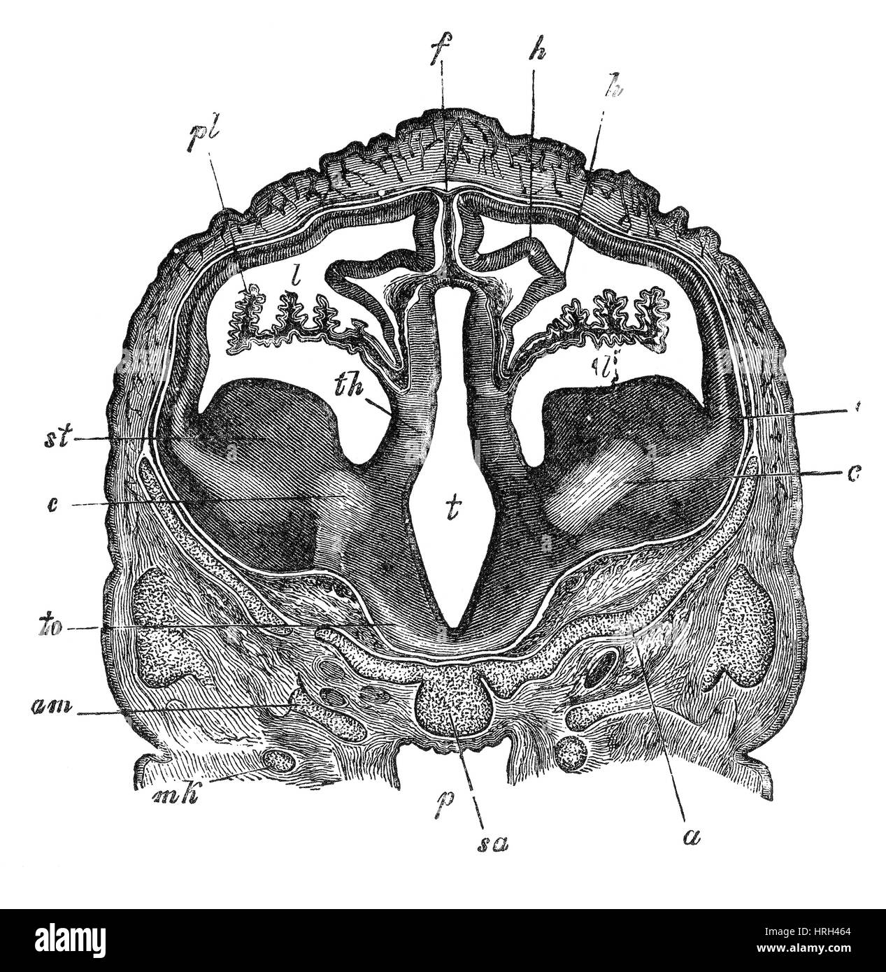
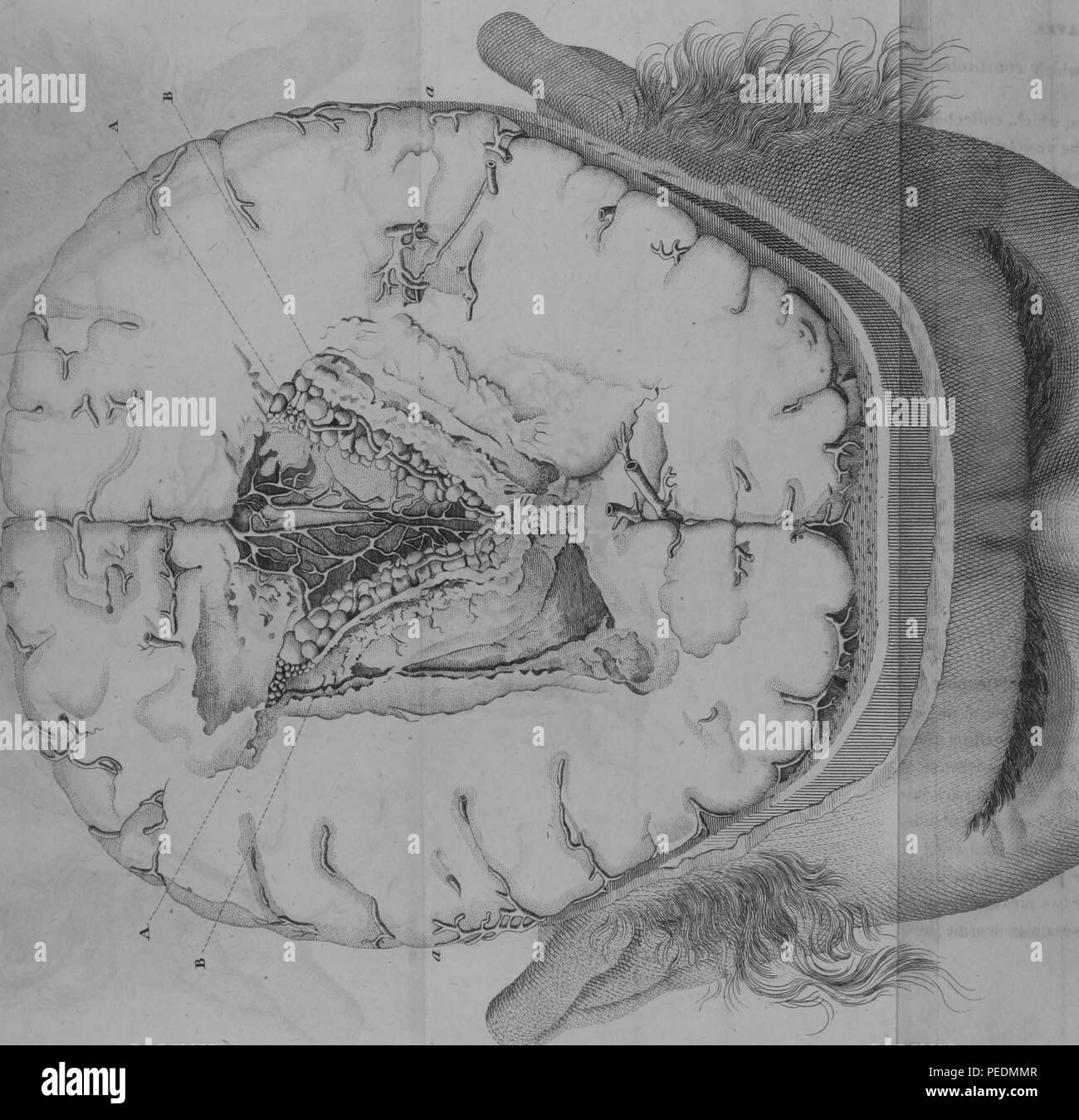 Black and white print illustrating a cross-section of the lateral ventricles of a human brain, with alphabetized figures indicating the sites of ventricular worm infestations, 1825. Courtesy Internet Archive. () Stock Photohttps://www.alamy.com/image-license-details/?v=1https://www.alamy.com/black-and-white-print-illustrating-a-cross-section-of-the-lateral-ventricles-of-a-human-brain-with-alphabetized-figures-indicating-the-sites-of-ventricular-worm-infestations-1825-courtesy-internet-archive-image215431239.html
RMPEDMMR–Black and white print illustrating a cross-section of the lateral ventricles of a human brain, with alphabetized figures indicating the sites of ventricular worm infestations, 1825. Courtesy Internet Archive. ()
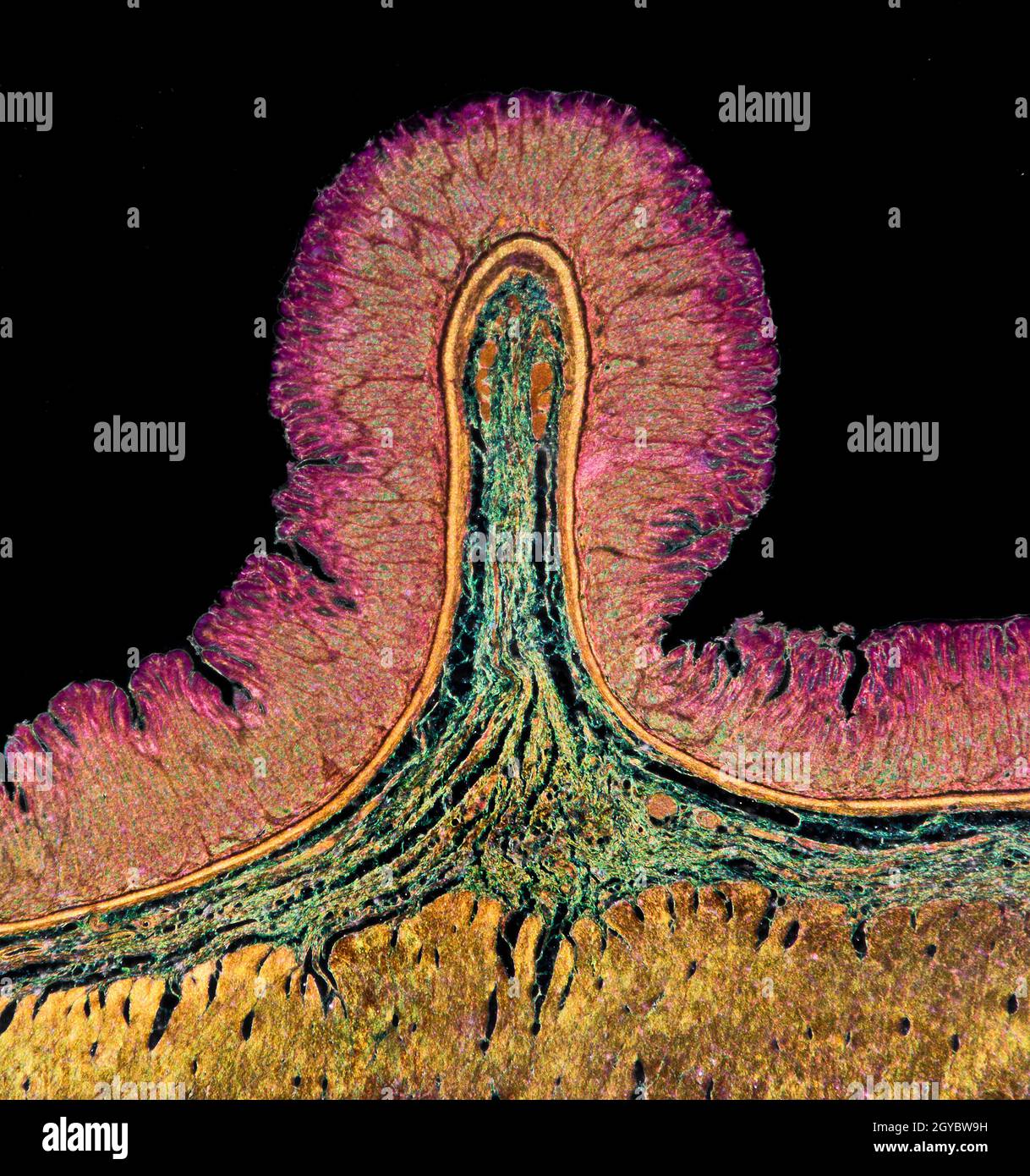 Corpus ventriculi slide section Stock Photohttps://www.alamy.com/image-license-details/?v=1https://www.alamy.com/corpus-ventriculi-slide-section-image447116253.html
RM2GYBW9H–Corpus ventriculi slide section
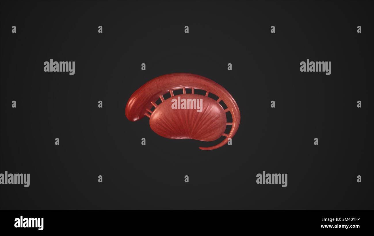 Anatomical Illustration of Corpus Striatum.3d rendering Stock Photohttps://www.alamy.com/image-license-details/?v=1https://www.alamy.com/anatomical-illustration-of-corpus-striatum3d-rendering-image501580906.html
RF2M40YFP–Anatomical Illustration of Corpus Striatum.3d rendering
 Occipital bone word cloud concept Stock Photohttps://www.alamy.com/image-license-details/?v=1https://www.alamy.com/occipital-bone-word-cloud-concept-image262838295.html
RFW7H8WY–Occipital bone word cloud concept
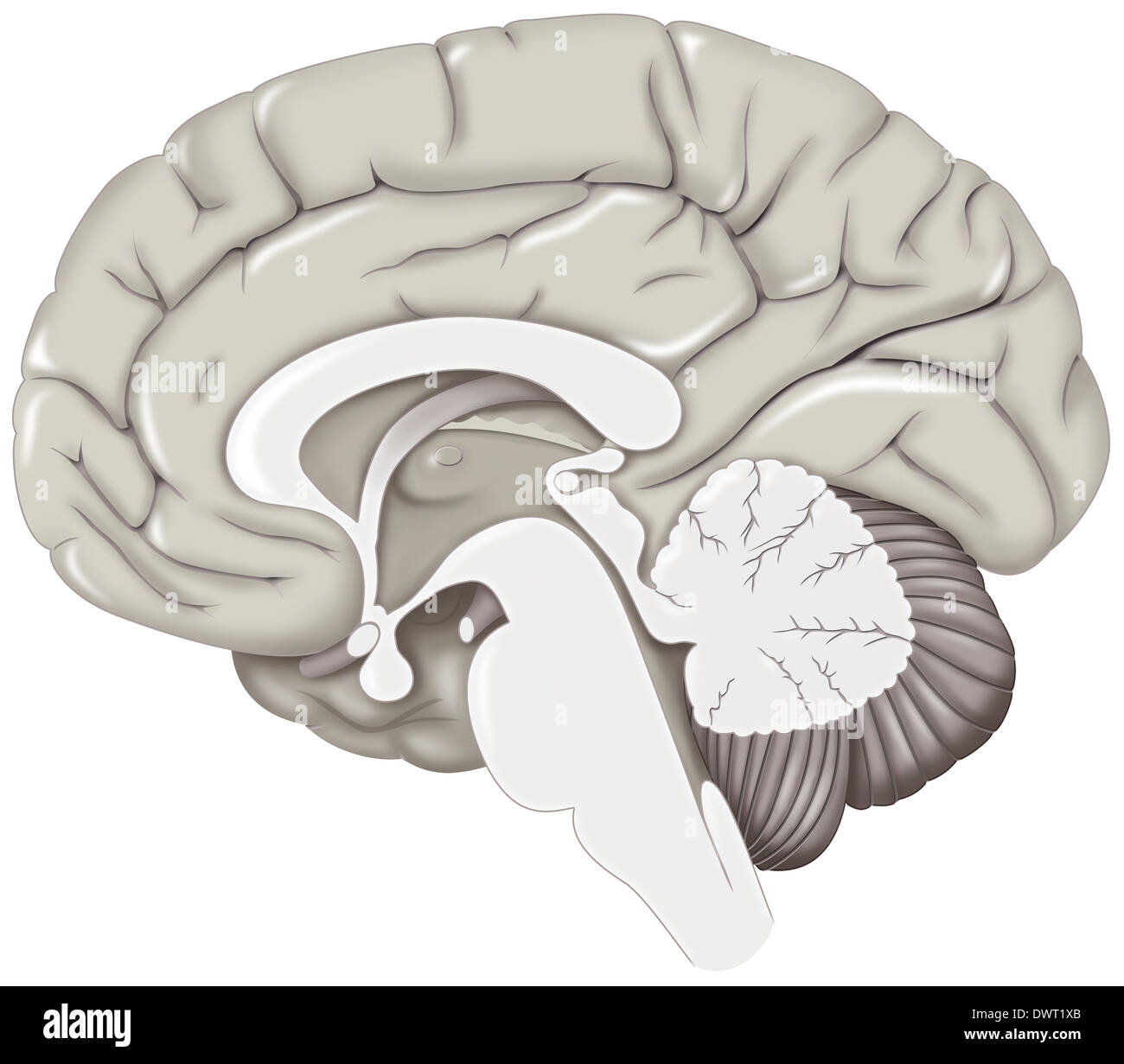 Brain, drawing Stock Photohttps://www.alamy.com/image-license-details/?v=1https://www.alamy.com/brain-drawing-image67525875.html
RMDWT1XB–Brain, drawing
 The brain in the transparent head on the light-blue background Stock Photohttps://www.alamy.com/image-license-details/?v=1https://www.alamy.com/stock-photo-the-brain-in-the-transparent-head-on-the-light-blue-background-82655635.html
RFEPD82Y–The brain in the transparent head on the light-blue background
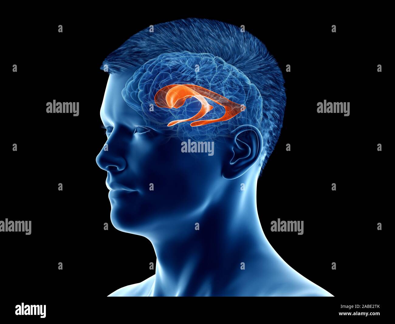 3d rendered medically accurate illustration of the brain anatomy - the lateral ventricle Stock Photohttps://www.alamy.com/image-license-details/?v=1https://www.alamy.com/3d-rendered-medically-accurate-illustration-of-the-brain-anatomy-the-lateral-ventricle-image334067795.html
RF2ABE2TK–3d rendered medically accurate illustration of the brain anatomy - the lateral ventricle
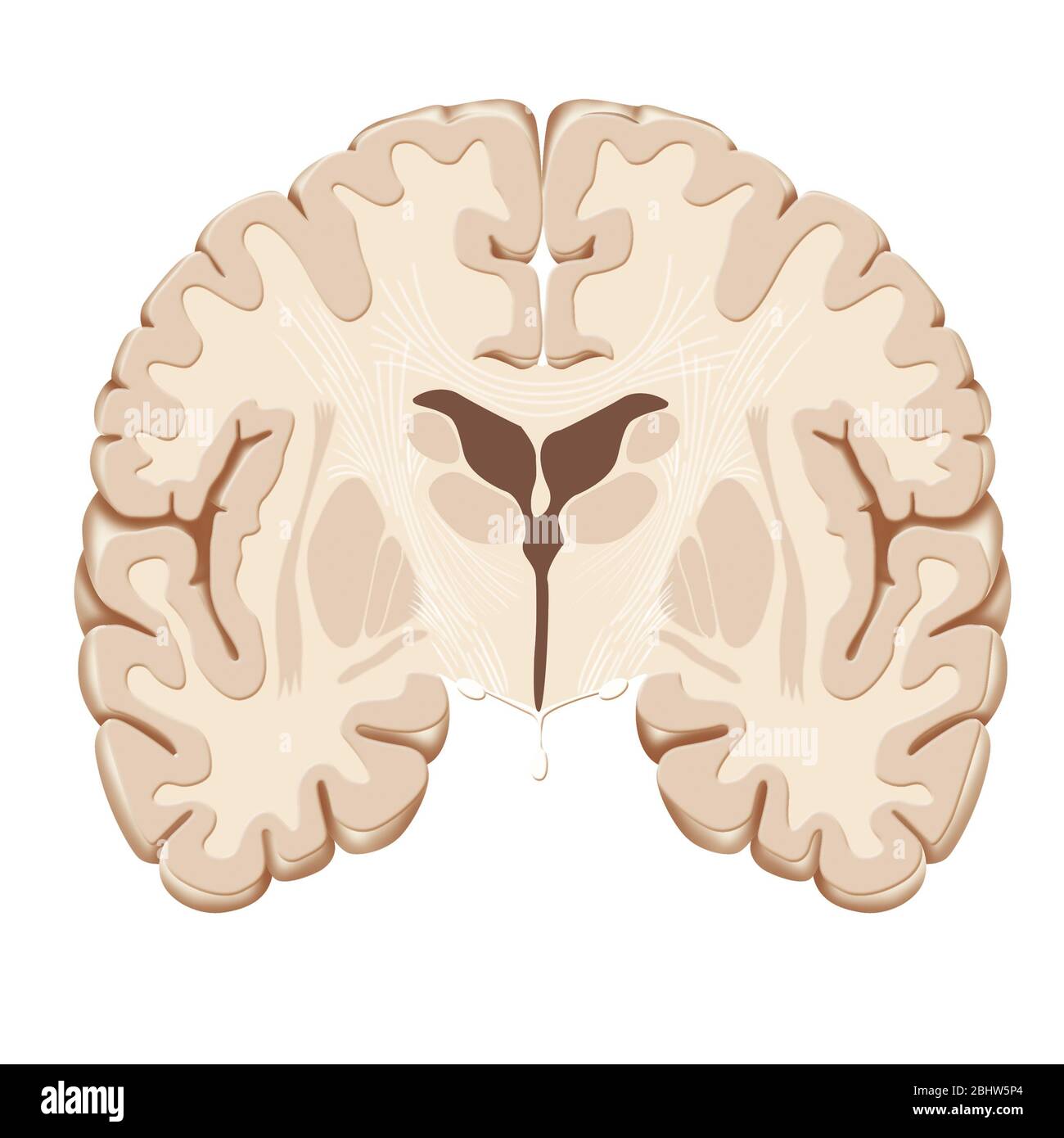 Anatomy of the brain in frontal section, distinguishing the hypophysis, the optic tracts, the thalamus and the caudate nuclei, the lateral ventricles. Stock Photohttps://www.alamy.com/image-license-details/?v=1https://www.alamy.com/anatomy-of-the-brain-in-frontal-section-distinguishing-the-hypophysis-the-optic-tracts-the-thalamus-and-the-caudate-nuclei-the-lateral-ventricles-image355209852.html
RM2BHW5P4–Anatomy of the brain in frontal section, distinguishing the hypophysis, the optic tracts, the thalamus and the caudate nuclei, the lateral ventricles.
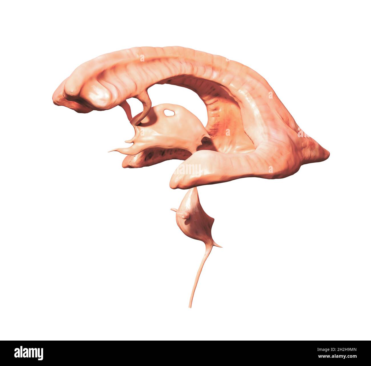 Ventricular system. Human brain with ventricles and Cerebrospinal fluid, 3d render illustration Stock Photohttps://www.alamy.com/image-license-details/?v=1https://www.alamy.com/ventricular-system-human-brain-with-ventricles-and-cerebrospinal-fluid-3d-render-illustration-image449079701.html
RF2H2H9MN–Ventricular system. Human brain with ventricles and Cerebrospinal fluid, 3d render illustration
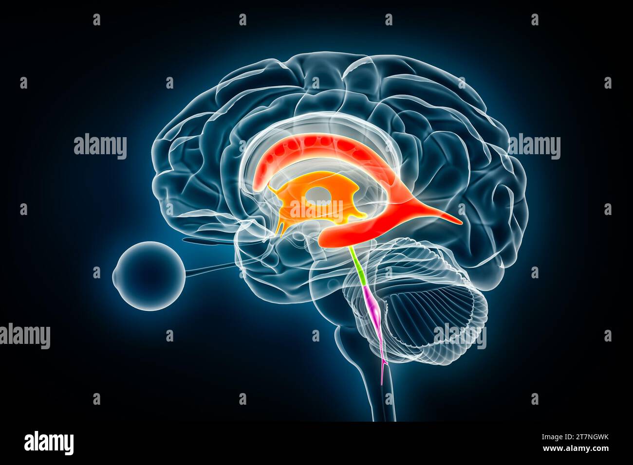 Ventricles and cerebral aqueduct lateral in colors x-ray view 3D rendering illustration. Human brain and ventricular system anatomy, medical, healthca Stock Photohttps://www.alamy.com/image-license-details/?v=1https://www.alamy.com/ventricles-and-cerebral-aqueduct-lateral-in-colors-x-ray-view-3d-rendering-illustration-human-brain-and-ventricular-system-anatomy-medical-healthca-image572718991.html
RF2T7NGWK–Ventricles and cerebral aqueduct lateral in colors x-ray view 3D rendering illustration. Human brain and ventricular system anatomy, medical, healthca
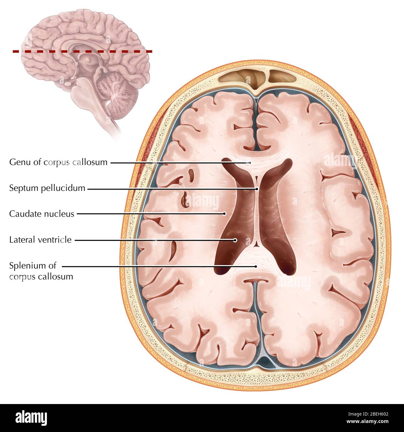 Brain, Transverse Section Stock Photohttps://www.alamy.com/image-license-details/?v=1https://www.alamy.com/brain-transverse-section-image353190434.html
RM2BEH602–Brain, Transverse Section
 This transaxial section of a human brain, passed through both cerebral hemispheres, revealing the presence of a large, left cerebral abscess. Stock Photohttps://www.alamy.com/image-license-details/?v=1https://www.alamy.com/this-transaxial-section-of-a-human-brain-passed-through-both-cerebral-hemispheres-revealing-the-presence-of-a-large-left-cerebral-abscess-image600769449.html
RM2WWBBFN–This transaxial section of a human brain, passed through both cerebral hemispheres, revealing the presence of a large, left cerebral abscess.
 Cairo, Egypt, September 17 2024: Brain CT scan reveals left shunt tube terminating in right lateral ventricle, with no residual ventricular dilatation Stock Photohttps://www.alamy.com/image-license-details/?v=1https://www.alamy.com/cairo-egypt-september-17-2024-brain-ct-scan-reveals-left-shunt-tube-terminating-in-right-lateral-ventricle-with-no-residual-ventricular-dilatation-image624823856.html
RF2Y8F55M–Cairo, Egypt, September 17 2024: Brain CT scan reveals left shunt tube terminating in right lateral ventricle, with no residual ventricular dilatation
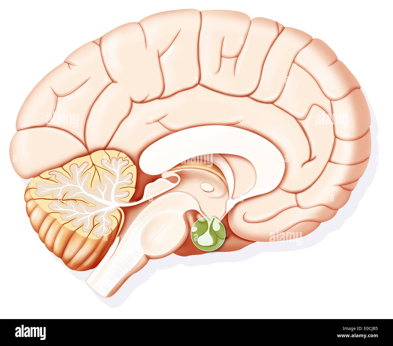 Hypophysis, drawing Stock Photohttps://www.alamy.com/image-license-details/?v=1https://www.alamy.com/hypophysis-drawing-image69119321.html
RME0CJB5–Hypophysis, drawing
 Upsy daisy, student accidentally cuts the optic nerves of a cow Brain probably cow will go blind. Stock Photohttps://www.alamy.com/image-license-details/?v=1https://www.alamy.com/upsy-daisy-student-accidentally-cuts-the-optic-nerves-of-a-cow-brain-image69690438.html
RFE1AJT6–Upsy daisy, student accidentally cuts the optic nerves of a cow Brain probably cow will go blind.
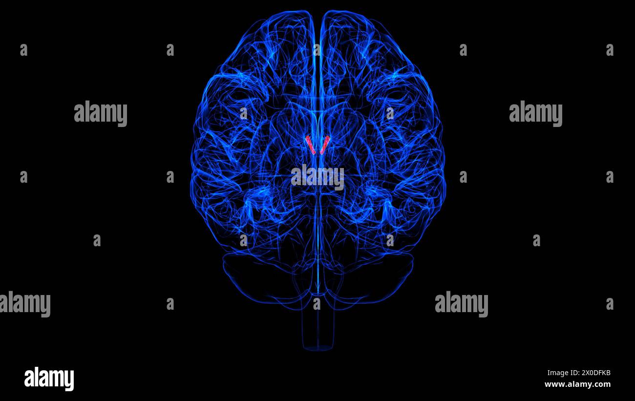 Brain Interventricular foramen Anatomy For Medical Concept 3D illustration Stock Photohttps://www.alamy.com/image-license-details/?v=1https://www.alamy.com/brain-interventricular-foramen-anatomy-for-medical-concept-3d-illustration-image602660559.html
RF2X0DFKB–Brain Interventricular foramen Anatomy For Medical Concept 3D illustration
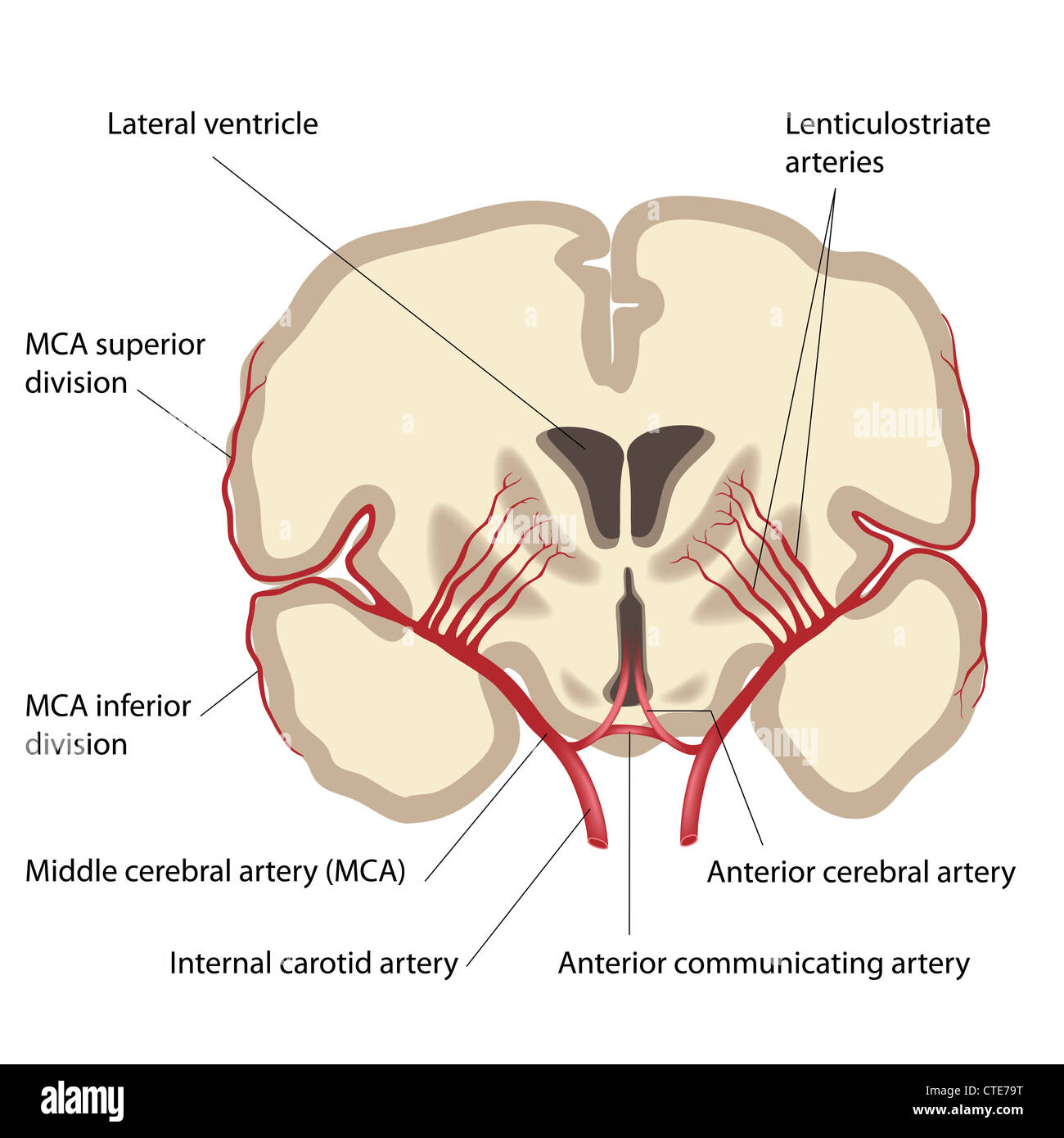 Middle cerebral artery and branches Stock Photohttps://www.alamy.com/image-license-details/?v=1https://www.alamy.com/stock-photo-middle-cerebral-artery-and-branches-49485572.html
RFCTE79T–Middle cerebral artery and branches
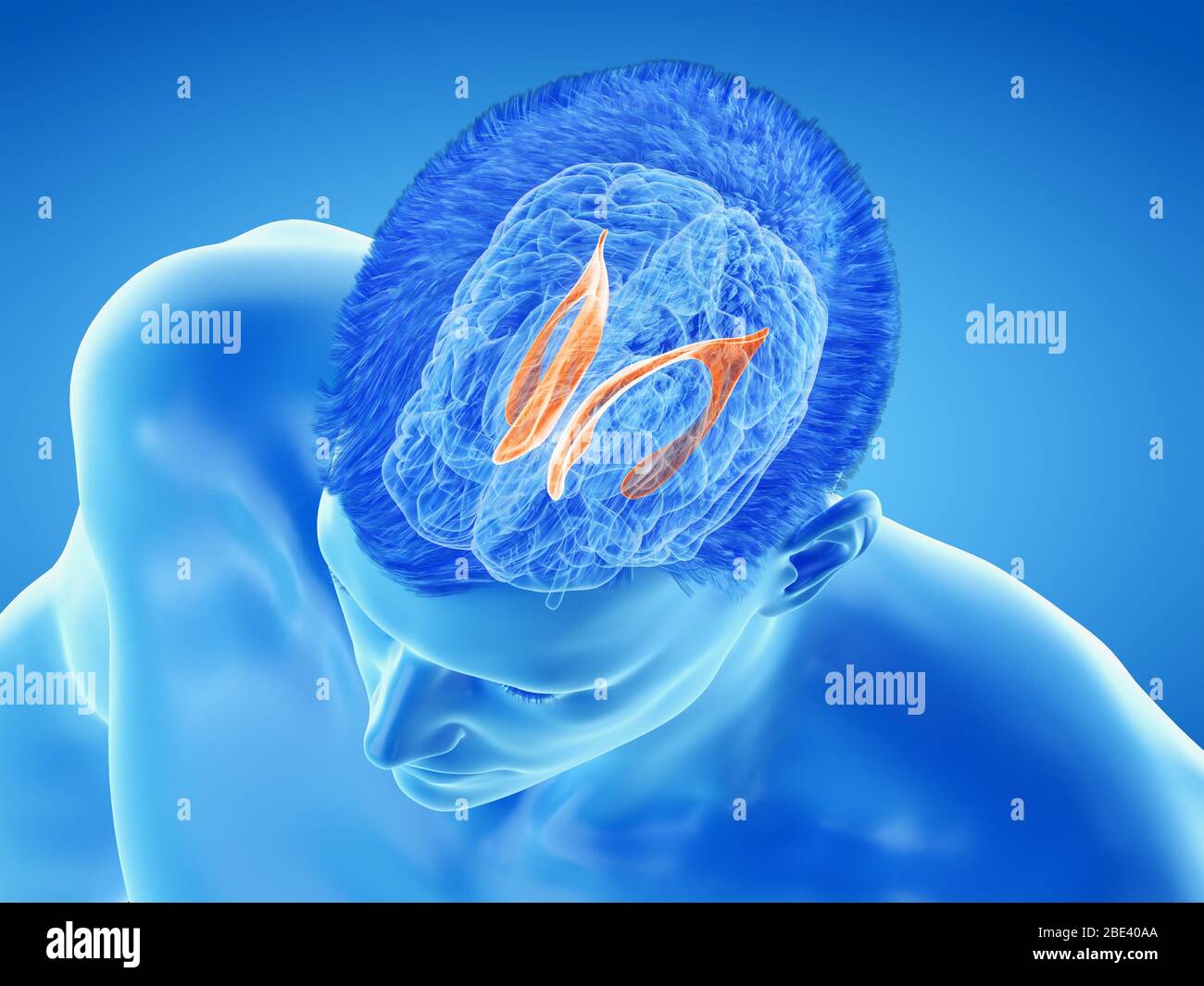 Lateral ventricle of the brain, illustration. Stock Photohttps://www.alamy.com/image-license-details/?v=1https://www.alamy.com/lateral-ventricle-of-the-brain-illustration-image352900642.html
RF2BE40AA–Lateral ventricle of the brain, illustration.
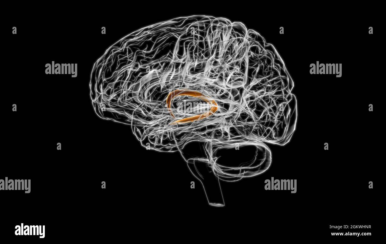 Brain fornix of forebrain Anatomy For Medical Concept 3D Illustration Stock Photohttps://www.alamy.com/image-license-details/?v=1https://www.alamy.com/brain-fornix-of-forebrain-anatomy-for-medical-concept-3d-illustration-image442500403.html
RF2GKWHNR–Brain fornix of forebrain Anatomy For Medical Concept 3D Illustration
 The brain: two figures of a section through a head and brain, showing the lateral ventricles and third ventricle. Lithograph by N.H Jacob, 1831/1854. Stock Photohttps://www.alamy.com/image-license-details/?v=1https://www.alamy.com/the-brain-two-figures-of-a-section-through-a-head-and-brain-showing-the-lateral-ventricles-and-third-ventricle-lithograph-by-nh-jacob-18311854-image450076985.html
RM2H46NP1–The brain: two figures of a section through a head and brain, showing the lateral ventricles and third ventricle. Lithograph by N.H Jacob, 1831/1854.
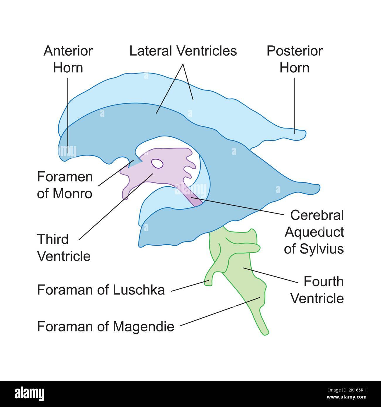 Scientific Designing of Ventricular System of Brain. Colorful Symbols. Vector Illustration. Stock Vectorhttps://www.alamy.com/image-license-details/?v=1https://www.alamy.com/scientific-designing-of-ventricular-system-of-brain-colorful-symbols-vector-illustration-image482641253.html
RF2K165RH–Scientific Designing of Ventricular System of Brain. Colorful Symbols. Vector Illustration.
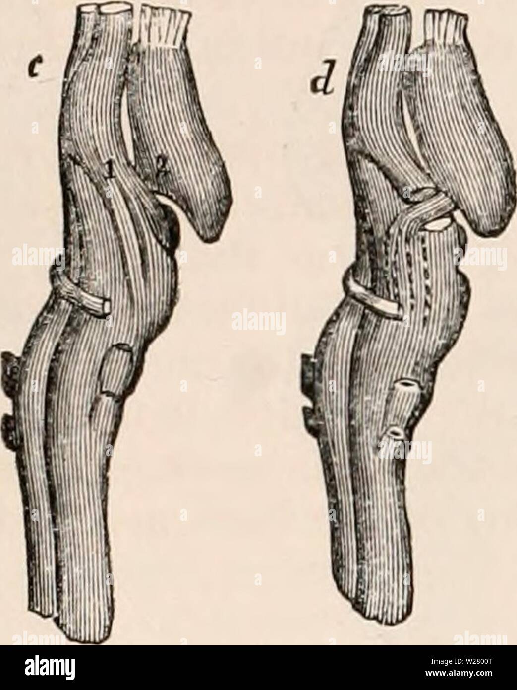 Archive image from page 331 of The cyclopædia of anatomy and. The cyclopædia of anatomy and physiology cyclopdiaofana0401todd Year: 1847 310 REPTILIA. brain occupy their usual position, but there is no commissura mollis. The cerebellum is generally extremely small, and in some cases is even reduced to a simple transverse la- mella, which does not entirely cover the fourth ventricle, formed as usual by the separation of the posterior columns of the Fig. 224. c, lateral view of the brain of a turtle; 1, optic tract; 2, cms cerebri. d, the same: a. portion of the optic nerve has been removed Stock Photohttps://www.alamy.com/image-license-details/?v=1https://www.alamy.com/archive-image-from-page-331-of-the-cyclopdia-of-anatomy-and-the-cyclopdia-of-anatomy-and-physiology-cyclopdiaofana0401todd-year-1847-310-reptilia-brain-occupy-their-usual-position-but-there-is-no-commissura-mollis-the-cerebellum-is-generally-extremely-small-and-in-some-cases-is-even-reduced-to-a-simple-transverse-la-mella-which-does-not-entirely-cover-the-fourth-ventricle-formed-as-usual-by-the-separation-of-the-posterior-columns-of-the-fig-224-c-lateral-view-of-the-brain-of-a-turtle-1-optic-tract-2-cms-cerebri-d-the-same-a-portion-of-the-optic-nerve-has-been-removed-image259560472.html
RMW2800T–Archive image from page 331 of The cyclopædia of anatomy and. The cyclopædia of anatomy and physiology cyclopdiaofana0401todd Year: 1847 310 REPTILIA. brain occupy their usual position, but there is no commissura mollis. The cerebellum is generally extremely small, and in some cases is even reduced to a simple transverse la- mella, which does not entirely cover the fourth ventricle, formed as usual by the separation of the posterior columns of the Fig. 224. c, lateral view of the brain of a turtle; 1, optic tract; 2, cms cerebri. d, the same: a. portion of the optic nerve has been removed
 . The cyclopædia of anatomy and physiology. Anatomy; Physiology; Zoology. Side view of villi of the choroid plexus of the lateral ventricle in the brain of a Goose, to show the disposition of the bloodvessels. Not to obscure the mew of t/te bloodvessels, the edge of the epithelium only has been shown. a, epithelium ; b, bloodvessels. (After Valentin.) The epithelium may be best seen by examin- ing the edge of a fold. It becomes very distinct when acted upon by acetic acid. As its particles are very delicate and consist only of a single layer, they are easily detached. The cells of epithelium a Stock Photohttps://www.alamy.com/image-license-details/?v=1https://www.alamy.com/the-cyclopdia-of-anatomy-and-physiology-anatomy-physiology-zoology-side-view-of-villi-of-the-choroid-plexus-of-the-lateral-ventricle-in-the-brain-of-a-goose-to-show-the-disposition-of-the-bloodvessels-not-to-obscure-the-mew-of-tte-bloodvessels-the-edge-of-the-epithelium-only-has-been-shown-a-epithelium-b-bloodvessels-after-valentin-the-epithelium-may-be-best-seen-by-examin-ing-the-edge-of-a-fold-it-becomes-very-distinct-when-acted-upon-by-acetic-acid-as-its-particles-are-very-delicate-and-consist-only-of-a-single-layer-they-are-easily-detached-the-cells-of-epithelium-a-image231840018.html
RMRD568J–. The cyclopædia of anatomy and physiology. Anatomy; Physiology; Zoology. Side view of villi of the choroid plexus of the lateral ventricle in the brain of a Goose, to show the disposition of the bloodvessels. Not to obscure the mew of t/te bloodvessels, the edge of the epithelium only has been shown. a, epithelium ; b, bloodvessels. (After Valentin.) The epithelium may be best seen by examin- ing the edge of a fold. It becomes very distinct when acted upon by acetic acid. As its particles are very delicate and consist only of a single layer, they are easily detached. The cells of epithelium a
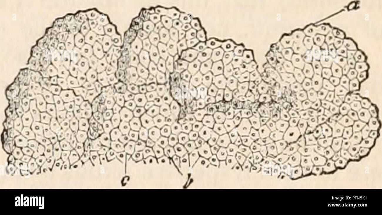 . The cyclopædia of anatomy and physiology. Anatomy; Physiology; Zoology. Side view of villi of the choroid plexus of the lateral ventricle in the brain of a Goose, to show the disposition of the bloodvessels. Not to obscure the mew of t/te bloodvessels, the edge of the epithelium only has been shown. a, epithelium ; b, bloodvessels. (After Valentin.) The epithelium may be best seen by examin- ing the edge of a fold. It becomes very distinct when acted upon by acetic acid. As its particles are very delicate and consist only of a single layer, they are easily detached. The cells of epithelium a Stock Photohttps://www.alamy.com/image-license-details/?v=1https://www.alamy.com/the-cyclopdia-of-anatomy-and-physiology-anatomy-physiology-zoology-side-view-of-villi-of-the-choroid-plexus-of-the-lateral-ventricle-in-the-brain-of-a-goose-to-show-the-disposition-of-the-bloodvessels-not-to-obscure-the-mew-of-tte-bloodvessels-the-edge-of-the-epithelium-only-has-been-shown-a-epithelium-b-bloodvessels-after-valentin-the-epithelium-may-be-best-seen-by-examin-ing-the-edge-of-a-fold-it-becomes-very-distinct-when-acted-upon-by-acetic-acid-as-its-particles-are-very-delicate-and-consist-only-of-a-single-layer-they-are-easily-detached-the-cells-of-epithelium-a-image216209701.html
RMPFN5K1–. The cyclopædia of anatomy and physiology. Anatomy; Physiology; Zoology. Side view of villi of the choroid plexus of the lateral ventricle in the brain of a Goose, to show the disposition of the bloodvessels. Not to obscure the mew of t/te bloodvessels, the edge of the epithelium only has been shown. a, epithelium ; b, bloodvessels. (After Valentin.) The epithelium may be best seen by examin- ing the edge of a fold. It becomes very distinct when acted upon by acetic acid. As its particles are very delicate and consist only of a single layer, they are easily detached. The cells of epithelium a
 The brain in the transparent head on the light-blue background Stock Photohttps://www.alamy.com/image-license-details/?v=1https://www.alamy.com/stock-photo-the-brain-in-the-transparent-head-on-the-light-blue-background-83614782.html
RFET0YE6–The brain in the transparent head on the light-blue background
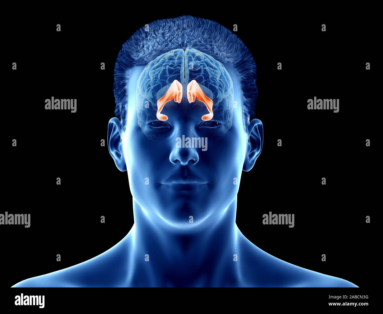 3d rendered medically accurate illustration of the brain anatomy - the lateral ventricle Stock Photohttps://www.alamy.com/image-license-details/?v=1https://www.alamy.com/3d-rendered-medically-accurate-illustration-of-the-brain-anatomy-the-lateral-ventricle-image334038196.html
RF2ABCN3G–3d rendered medically accurate illustration of the brain anatomy - the lateral ventricle
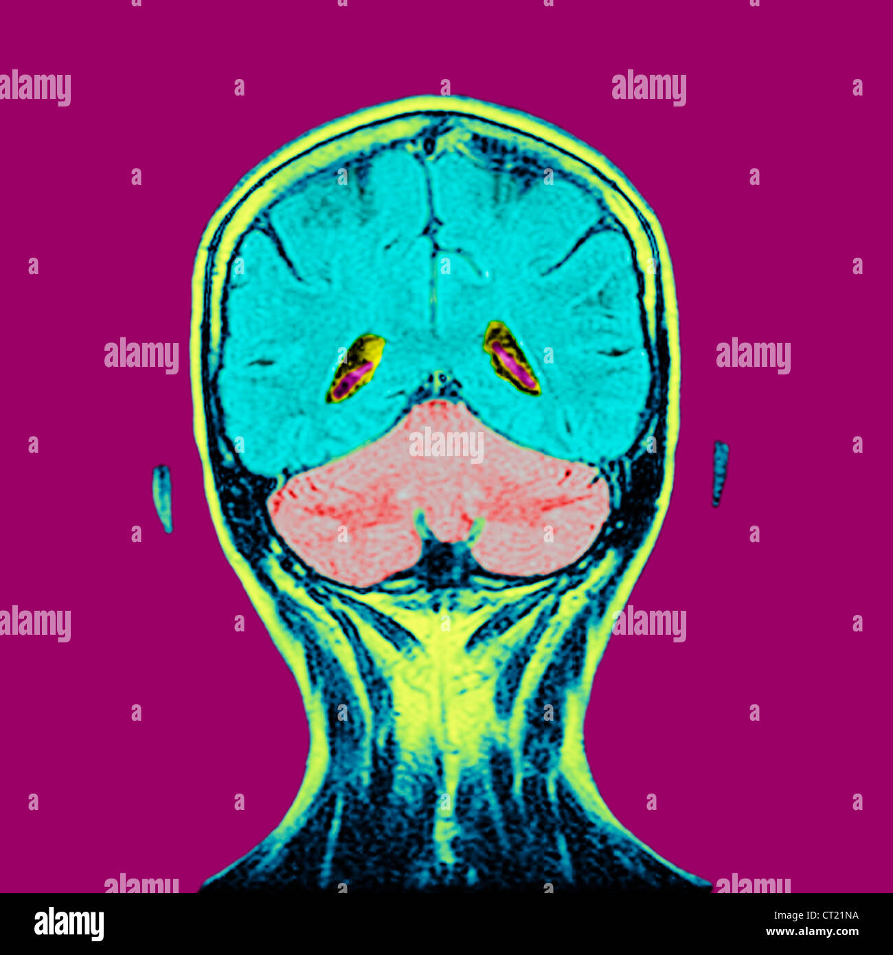 HEAD, MRI Stock Photohttps://www.alamy.com/image-license-details/?v=1https://www.alamy.com/stock-photo-head-mri-49217766.html
RMCT21NA–HEAD, MRI
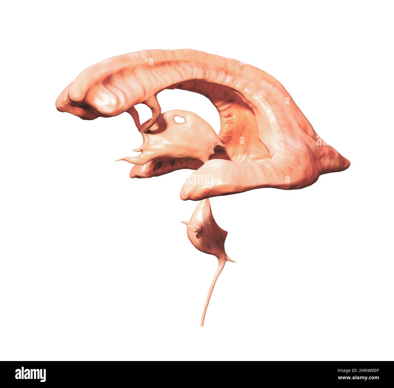 Ventricular system. Human brain with ventricles and Cerebrospinal fluid, 3d render illustration Stock Photohttps://www.alamy.com/image-license-details/?v=1https://www.alamy.com/ventricular-system-human-brain-with-ventricles-and-cerebrospinal-fluid-3d-render-illustration-image460926558.html
RF2HNW0EP–Ventricular system. Human brain with ventricles and Cerebrospinal fluid, 3d render illustration
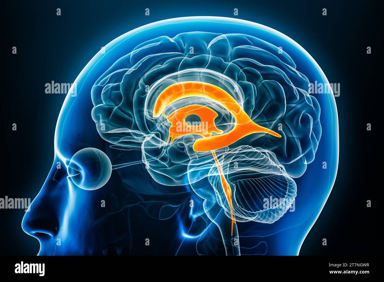 Ventricles and cerebral aqueduct x-ray profile close-up view 3D rendering illustration with body contours. Human brain and ventricular system anatomy, Stock Photohttps://www.alamy.com/image-license-details/?v=1https://www.alamy.com/ventricles-and-cerebral-aqueduct-x-ray-profile-close-up-view-3d-rendering-illustration-with-body-contours-human-brain-and-ventricular-system-anatomy-image572718995.html
RF2T7NGWR–Ventricles and cerebral aqueduct x-ray profile close-up view 3D rendering illustration with body contours. Human brain and ventricular system anatomy,
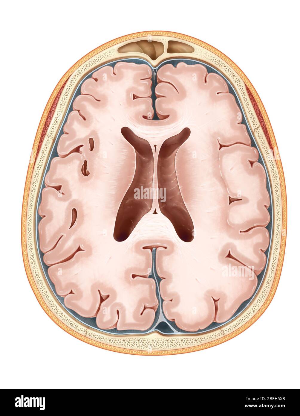 Brain, Transverse Section Stock Photohttps://www.alamy.com/image-license-details/?v=1https://www.alamy.com/brain-transverse-section-image353190387.html
RM2BEH5XB–Brain, Transverse Section
 Occipital bone word cloud concept Stock Photohttps://www.alamy.com/image-license-details/?v=1https://www.alamy.com/occipital-bone-word-cloud-concept-image268020935.html
RFWG1BC7–Occipital bone word cloud concept
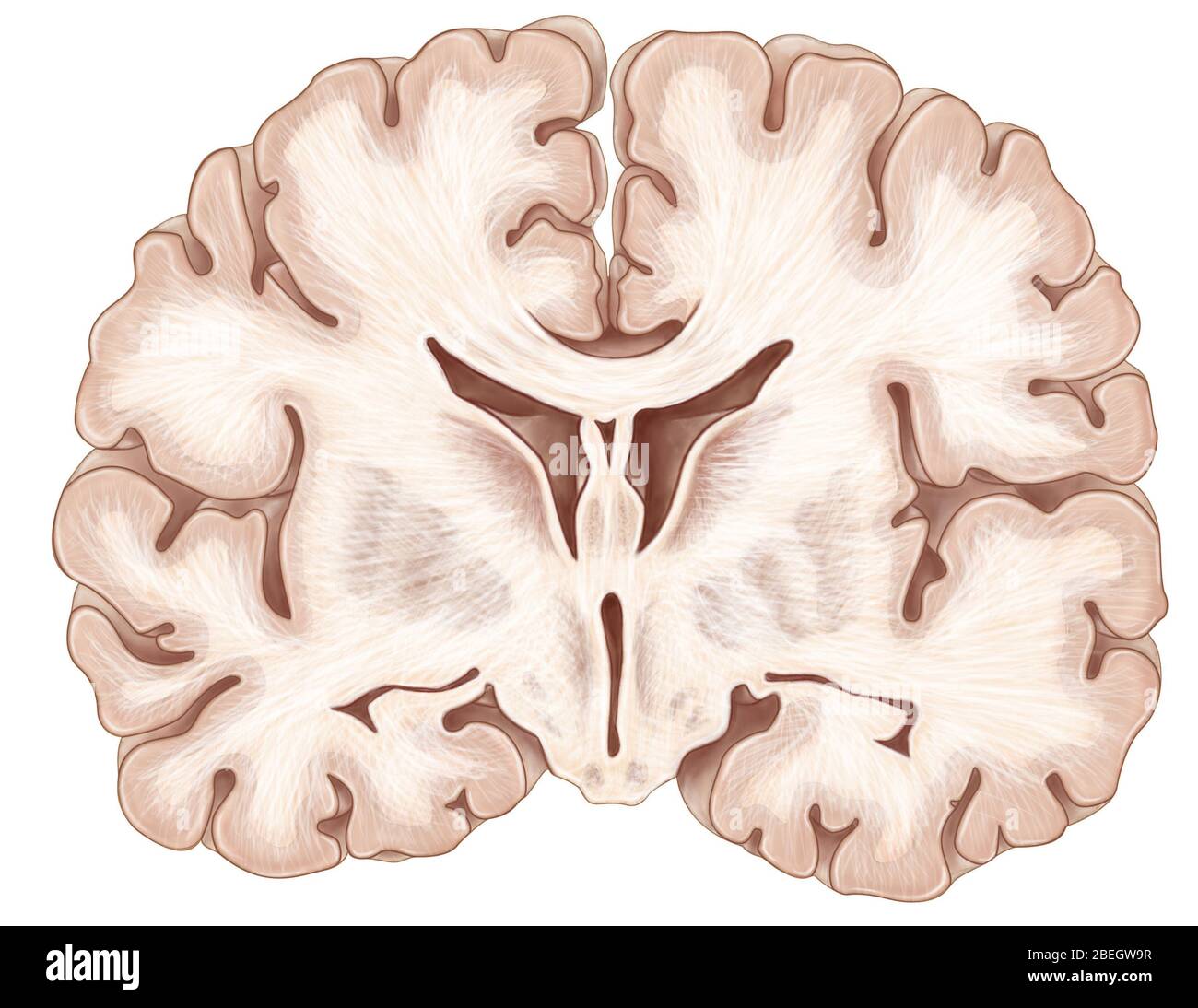 Brain, Coronal Section Stock Photohttps://www.alamy.com/image-license-details/?v=1https://www.alamy.com/brain-coronal-section-image353183651.html
RM2BEGW9R–Brain, Coronal Section
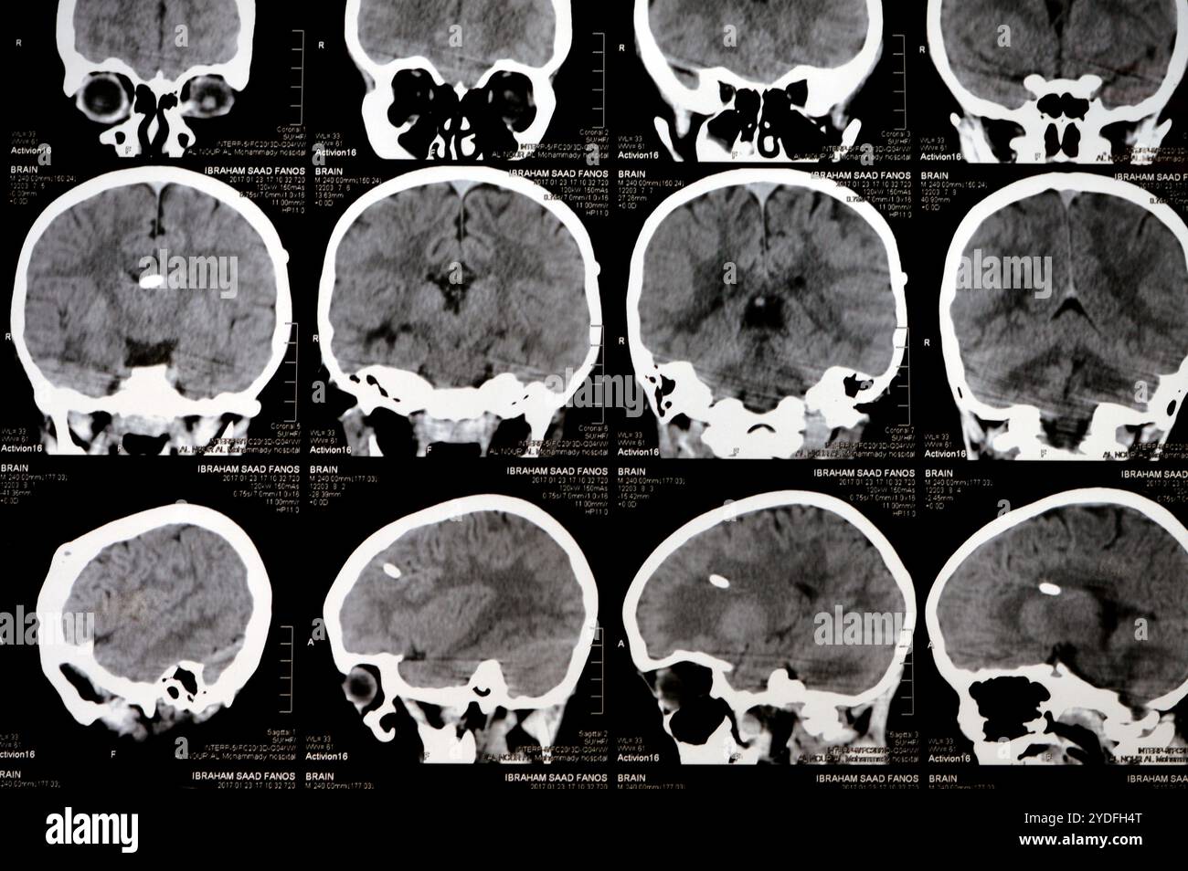 Cairo, Egypt, September 17 2024: Brain CT scan reveals left shunt tube terminating in right lateral ventricle, with no residual ventricular dilatation Stock Photohttps://www.alamy.com/image-license-details/?v=1https://www.alamy.com/cairo-egypt-september-17-2024-brain-ct-scan-reveals-left-shunt-tube-terminating-in-right-lateral-ventricle-with-no-residual-ventricular-dilatation-image627906520.html
RF2YDFH4T–Cairo, Egypt, September 17 2024: Brain CT scan reveals left shunt tube terminating in right lateral ventricle, with no residual ventricular dilatation
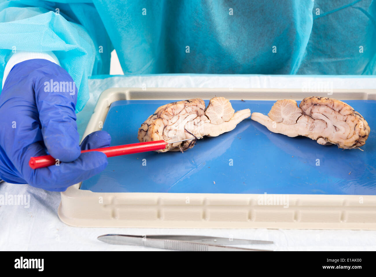 Cross-section of a cow brain in a laboratory with an anatomy student pointing with a probe to the area of the pituitary gland an Stock Photohttps://www.alamy.com/image-license-details/?v=1https://www.alamy.com/cross-section-of-a-cow-brain-in-a-laboratory-with-an-anatomy-student-image69690544.html
RFE1AK00–Cross-section of a cow brain in a laboratory with an anatomy student pointing with a probe to the area of the pituitary gland an
 Lateral ventricle of the brain, illustration. Stock Photohttps://www.alamy.com/image-license-details/?v=1https://www.alamy.com/lateral-ventricle-of-the-brain-illustration-image352900643.html
RF2BE40AB–Lateral ventricle of the brain, illustration.
 Brain globus pallidus Anatomy For Medical Concept 3D Illustration Stock Photohttps://www.alamy.com/image-license-details/?v=1https://www.alamy.com/brain-globus-pallidus-anatomy-for-medical-concept-3d-illustration-image442500322.html
RF2GKWHJX–Brain globus pallidus Anatomy For Medical Concept 3D Illustration
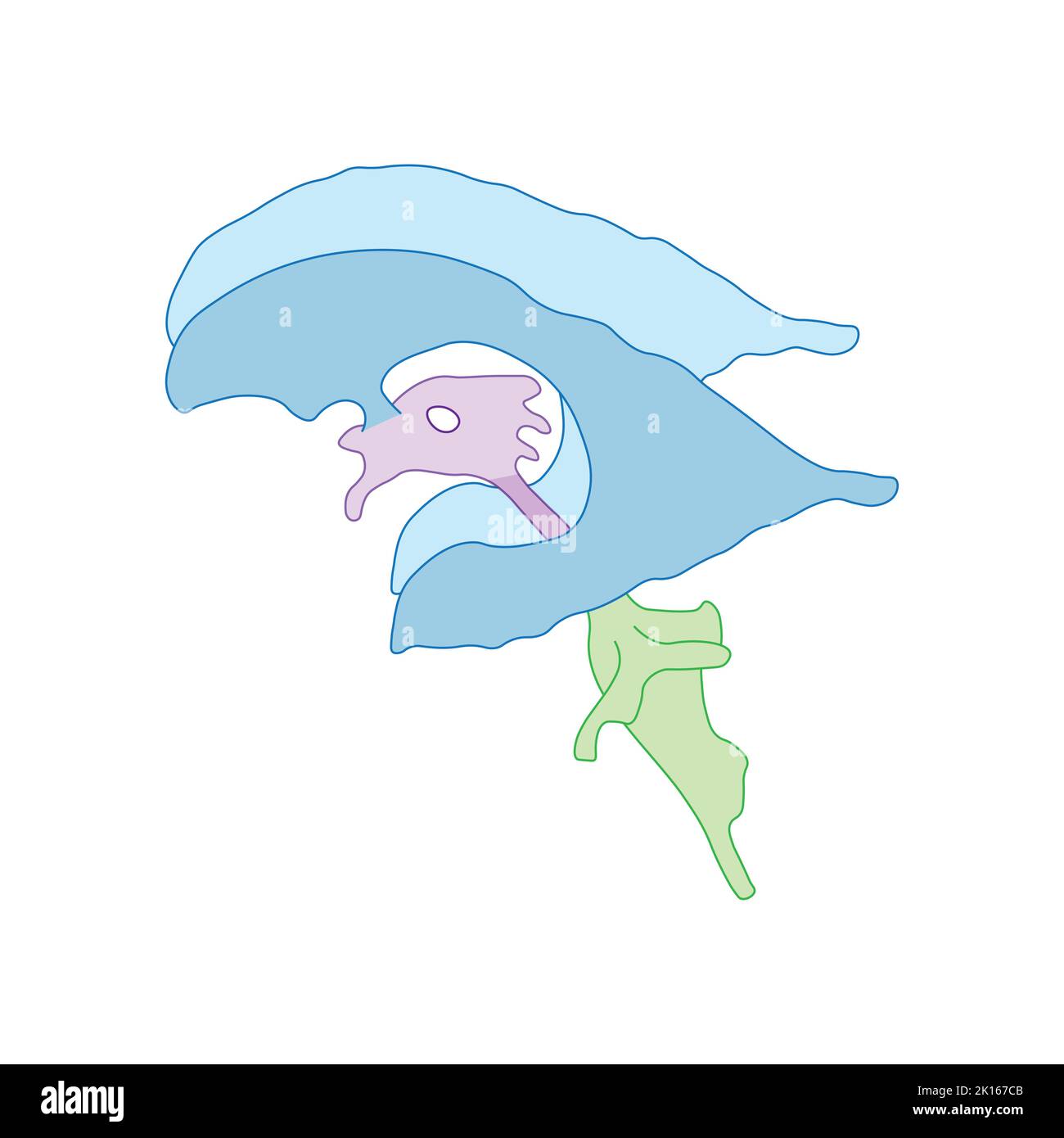 Scientific Designing of Ventricular System of Brain. Colorful Symbols. Vector Illustration. Stock Vectorhttps://www.alamy.com/image-license-details/?v=1https://www.alamy.com/scientific-designing-of-ventricular-system-of-brain-colorful-symbols-vector-illustration-image482642507.html
RF2K167CB–Scientific Designing of Ventricular System of Brain. Colorful Symbols. Vector Illustration.
 Archive image from page 1395 of Cunningham's Text-book of anatomy (1914). Cunningham's Text-book of anatomy cunninghamstextb00cunn Year: 1914 ( 1362 SUEFACE AND SUEGICAL ANATOMY. horizontal ramus of the lateral cerebral fissure, occupies a level midway between it and the temporal crest. The anterior horn of the ventricle is opposite the lower part of the coronal suture while the posterior horn is opposite the posterior part of the temporal crest. The inferior horn corresponds to the second temporal convolution. The lateral ventricle may be tapped or drained from above, by traversing brain tis Stock Photohttps://www.alamy.com/image-license-details/?v=1https://www.alamy.com/archive-image-from-page-1395-of-cunninghams-text-book-of-anatomy-1914-cunninghams-text-book-of-anatomy-cunninghamstextb00cunn-year-1914-1362-sueface-and-suegical-anatomy-horizontal-ramus-of-the-lateral-cerebral-fissure-occupies-a-level-midway-between-it-and-the-temporal-crest-the-anterior-horn-of-the-ventricle-is-opposite-the-lower-part-of-the-coronal-suture-while-the-posterior-horn-is-opposite-the-posterior-part-of-the-temporal-crest-the-inferior-horn-corresponds-to-the-second-temporal-convolution-the-lateral-ventricle-may-be-tapped-or-drained-from-above-by-traversing-brain-tis-image264069261.html
RMW9HB11–Archive image from page 1395 of Cunningham's Text-book of anatomy (1914). Cunningham's Text-book of anatomy cunninghamstextb00cunn Year: 1914 ( 1362 SUEFACE AND SUEGICAL ANATOMY. horizontal ramus of the lateral cerebral fissure, occupies a level midway between it and the temporal crest. The anterior horn of the ventricle is opposite the lower part of the coronal suture while the posterior horn is opposite the posterior part of the temporal crest. The inferior horn corresponds to the second temporal convolution. The lateral ventricle may be tapped or drained from above, by traversing brain tis
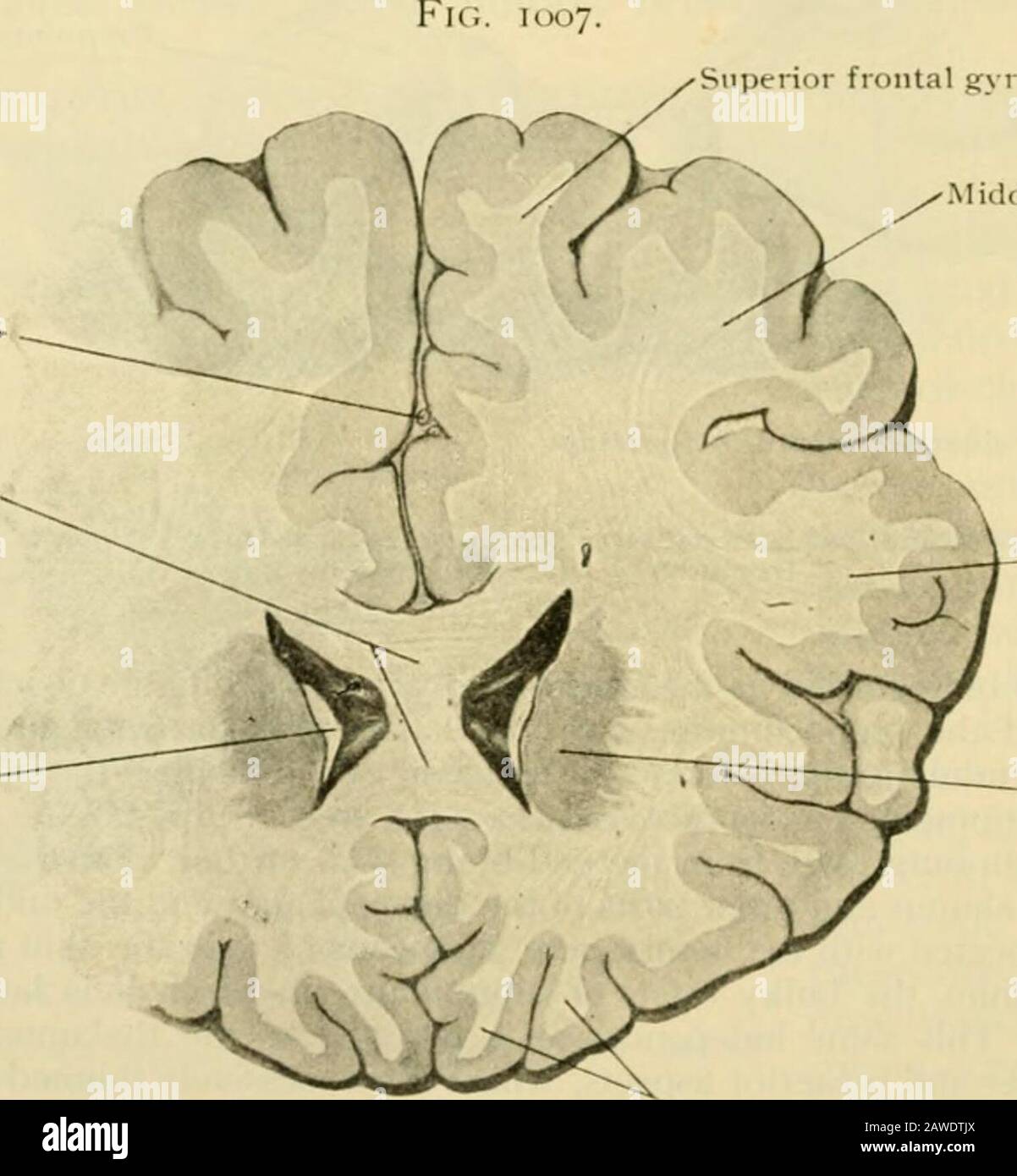 Human anatomy, including structure and development and practical considerations . f its cortical area, the hippcjcampus, and is C(jnse(iuentlycarried with the latter into the descending horn of the lateral ventricle, in this manner partswhich at (irst lay in proximity and were connected by short paths, become widely separated,with corresponding lengthening of the fibre-tracts uniting them, as illustrated in the longcourse of the fornix in the adult brain. Iurther, since the path of migration of the fornixand associated structures of the inferior horn of the lateral ventricle describes a curve, Stock Photohttps://www.alamy.com/image-license-details/?v=1https://www.alamy.com/human-anatomy-including-structure-and-development-and-practical-considerations-f-its-cortical-area-the-hippcjcampus-and-is-cjnseiuentlycarried-with-the-latter-into-the-descending-horn-of-the-lateral-ventricle-in-this-manner-partswhich-at-irst-lay-in-proximity-and-were-connected-by-short-paths-become-widely-separatedwith-corresponding-lengthening-of-the-fibre-tracts-uniting-them-as-illustrated-in-the-longcourse-of-the-fornix-in-the-adult-brain-iurther-since-the-path-of-migration-of-the-fornixand-associated-structures-of-the-inferior-horn-of-the-lateral-ventricle-describes-a-curve-image342668114.html
RM2AWDTJX–Human anatomy, including structure and development and practical considerations . f its cortical area, the hippcjcampus, and is C(jnse(iuentlycarried with the latter into the descending horn of the lateral ventricle, in this manner partswhich at (irst lay in proximity and were connected by short paths, become widely separated,with corresponding lengthening of the fibre-tracts uniting them, as illustrated in the longcourse of the fornix in the adult brain. Iurther, since the path of migration of the fornixand associated structures of the inferior horn of the lateral ventricle describes a curve,
 . The development of the human body : a manual of human embryology. Embryology; Embryo, Non-Mammalian. 388 THE BRAIN The cavity of the third vesicle persists in the adult as the fourth ventricle, traversing all the subdivisions of the vesicle; that of the second, increasing but little in height and breadth, constitutes the aquaductus cerebri; while that of the first vesicle is continued into the cerebral hemispheres to form the lateral ventricles, the remainder of it constituting the third ventricle, which includes the cavity of the median portion of the telencephalon as well as the entire cav Stock Photohttps://www.alamy.com/image-license-details/?v=1https://www.alamy.com/the-development-of-the-human-body-a-manual-of-human-embryology-embryology-embryo-non-mammalian-388-the-brain-the-cavity-of-the-third-vesicle-persists-in-the-adult-as-the-fourth-ventricle-traversing-all-the-subdivisions-of-the-vesicle-that-of-the-second-increasing-but-little-in-height-and-breadth-constitutes-the-aquaductus-cerebri-while-that-of-the-first-vesicle-is-continued-into-the-cerebral-hemispheres-to-form-the-lateral-ventricles-the-remainder-of-it-constituting-the-third-ventricle-which-includes-the-cavity-of-the-median-portion-of-the-telencephalon-as-well-as-the-entire-cav-image215969375.html
RMPFA73Y–. The development of the human body : a manual of human embryology. Embryology; Embryo, Non-Mammalian. 388 THE BRAIN The cavity of the third vesicle persists in the adult as the fourth ventricle, traversing all the subdivisions of the vesicle; that of the second, increasing but little in height and breadth, constitutes the aquaductus cerebri; while that of the first vesicle is continued into the cerebral hemispheres to form the lateral ventricles, the remainder of it constituting the third ventricle, which includes the cavity of the median portion of the telencephalon as well as the entire cav
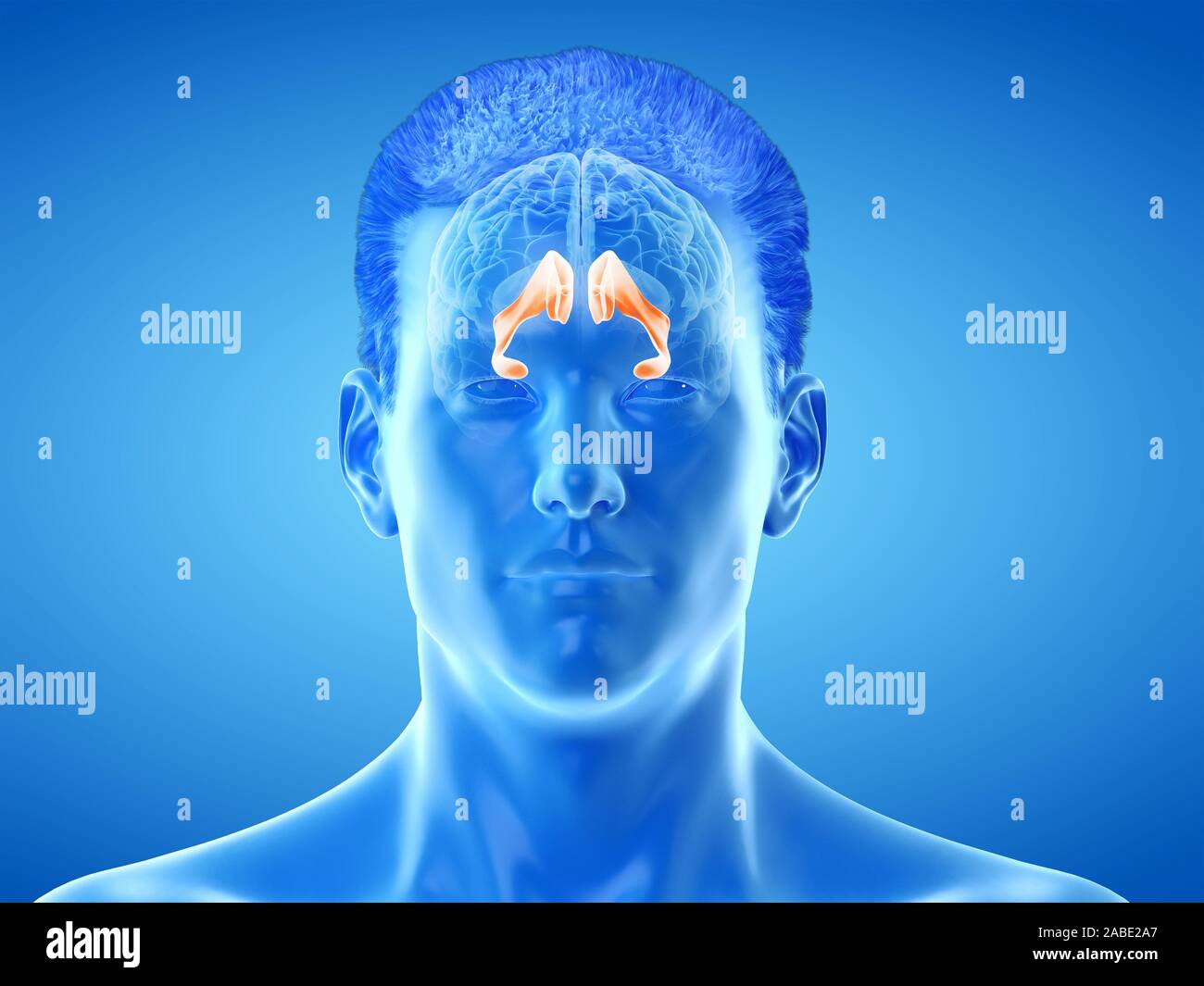 3d rendered medically accurate illustration of the brain anatomy - the lateral ventricle Stock Photohttps://www.alamy.com/image-license-details/?v=1https://www.alamy.com/3d-rendered-medically-accurate-illustration-of-the-brain-anatomy-the-lateral-ventricle-image334067391.html
RF2ABE2A7–3d rendered medically accurate illustration of the brain anatomy - the lateral ventricle
 HEAD, MRI Stock Photohttps://www.alamy.com/image-license-details/?v=1https://www.alamy.com/stock-photo-head-mri-49217765.html
RMCT21N9–HEAD, MRI
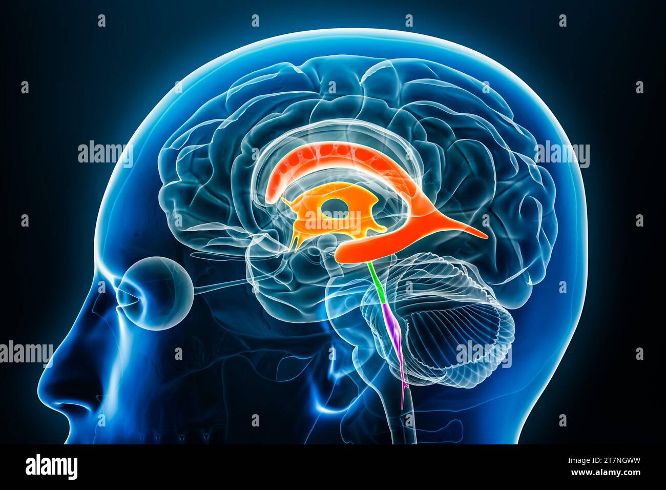 Ventricles and cerebral aqueduct in colors x-ray profile close-up view 3D rendering illustration. Human brain and ventricular system anatomy, medical, Stock Photohttps://www.alamy.com/image-license-details/?v=1https://www.alamy.com/ventricles-and-cerebral-aqueduct-in-colors-x-ray-profile-close-up-view-3d-rendering-illustration-human-brain-and-ventricular-system-anatomy-medical-image572718997.html
RF2T7NGWW–Ventricles and cerebral aqueduct in colors x-ray profile close-up view 3D rendering illustration. Human brain and ventricular system anatomy, medical,
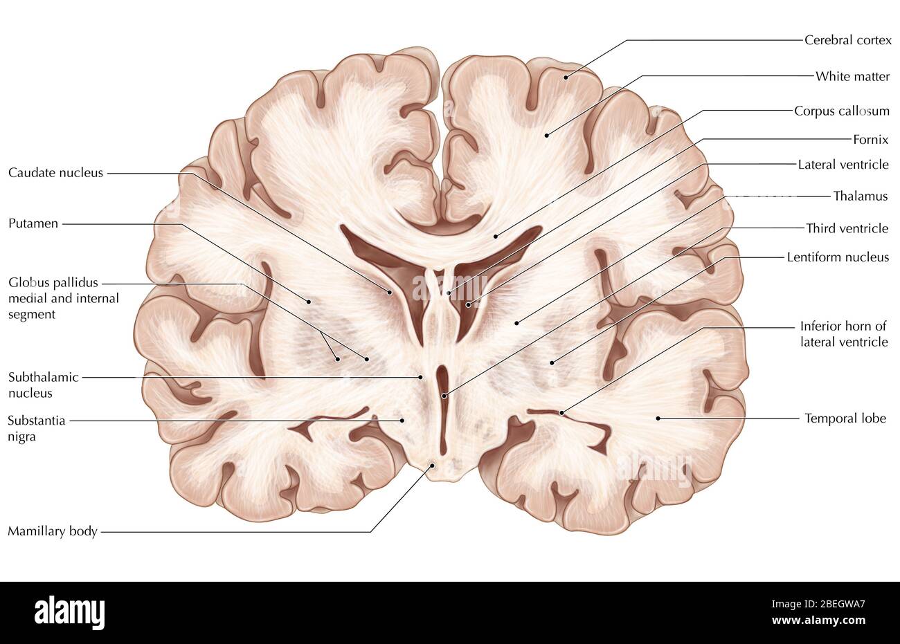 Brain, Coronal Section Stock Photohttps://www.alamy.com/image-license-details/?v=1https://www.alamy.com/brain-coronal-section-image353183663.html
RM2BEGWA7–Brain, Coronal Section
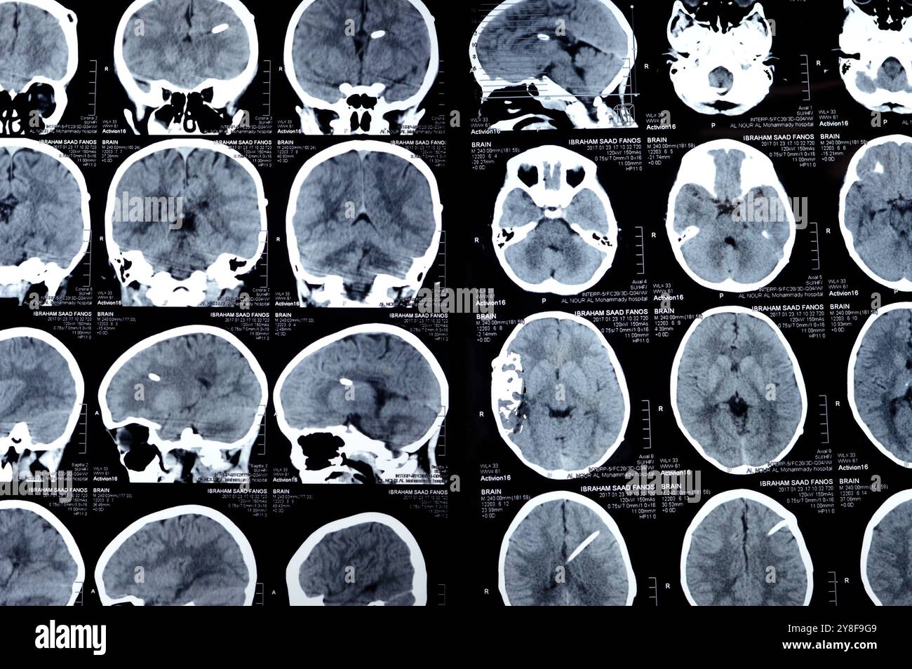 Cairo, Egypt, September 17 2024: Brain CT scan reveals left shunt tube terminating in right lateral ventricle, with no residual ventricular dilatation Stock Photohttps://www.alamy.com/image-license-details/?v=1https://www.alamy.com/cairo-egypt-september-17-2024-brain-ct-scan-reveals-left-shunt-tube-terminating-in-right-lateral-ventricle-with-no-residual-ventricular-dilatation-image624827289.html
RF2Y8F9G9–Cairo, Egypt, September 17 2024: Brain CT scan reveals left shunt tube terminating in right lateral ventricle, with no residual ventricular dilatation
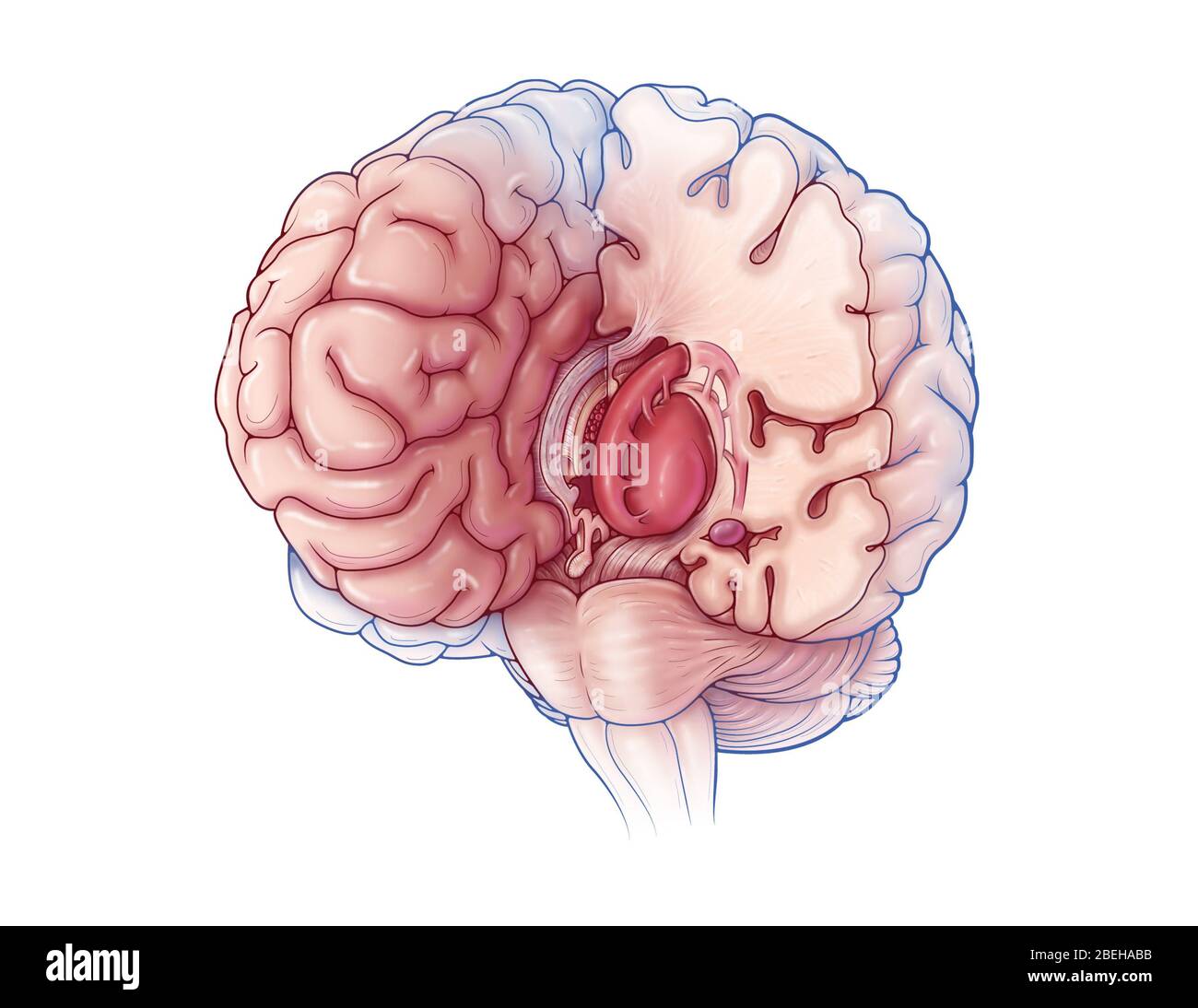 Limbic System, Illustration Stock Photohttps://www.alamy.com/image-license-details/?v=1https://www.alamy.com/limbic-system-illustration-image353193887.html
RM2BEHABB–Limbic System, Illustration
 Anatomy student dissecting a cow brain slicing through the two hemispheres and the lobes with a blade to obtain a cross-section Stock Photohttps://www.alamy.com/image-license-details/?v=1https://www.alamy.com/anatomy-student-dissecting-a-cow-brain-slicing-through-the-two-hemispheres-image69690501.html
RFE1AJXD–Anatomy student dissecting a cow brain slicing through the two hemispheres and the lobes with a blade to obtain a cross-section
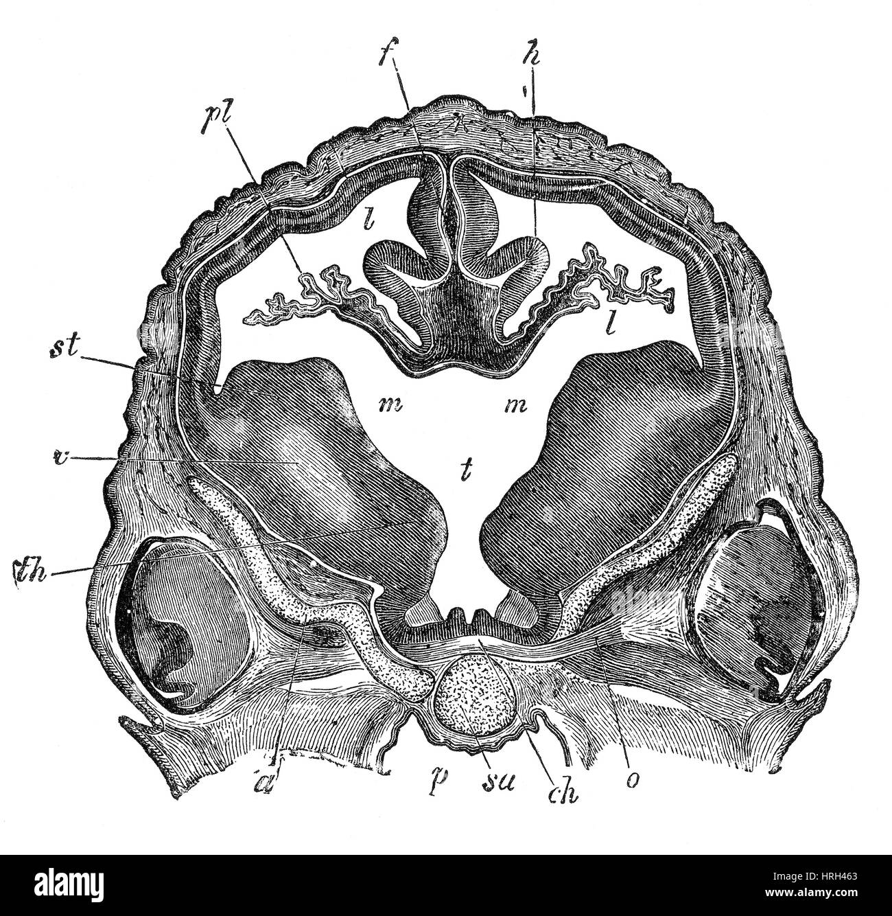 Brain of Sheep Embryo, Transverse Section Stock Photohttps://www.alamy.com/image-license-details/?v=1https://www.alamy.com/stock-photo-brain-of-sheep-embryo-transverse-section-134986155.html
RMHRH463–Brain of Sheep Embryo, Transverse Section
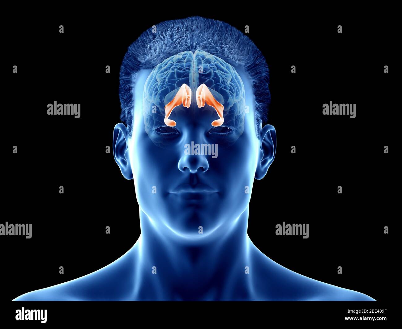 Lateral ventricle of the brain, illustration. Stock Photohttps://www.alamy.com/image-license-details/?v=1https://www.alamy.com/lateral-ventricle-of-the-brain-illustration-image352900619.html
RF2BE409F–Lateral ventricle of the brain, illustration.
 Brain Third ventricle Anatomy For Medical Concept 3D Illustration Stock Photohttps://www.alamy.com/image-license-details/?v=1https://www.alamy.com/brain-third-ventricle-anatomy-for-medical-concept-3d-illustration-image442500381.html
RF2GKWHN1–Brain Third ventricle Anatomy For Medical Concept 3D Illustration
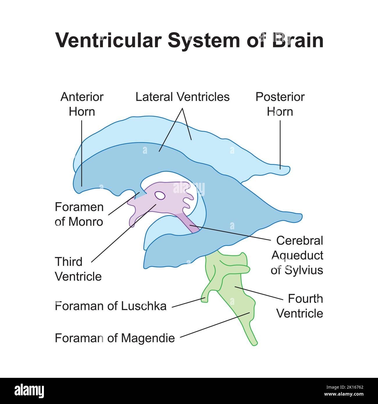 Scientific Designing of Ventricular System of Brain. Colorful Symbols. Vector Illustration. Stock Vectorhttps://www.alamy.com/image-license-details/?v=1https://www.alamy.com/scientific-designing-of-ventricular-system-of-brain-colorful-symbols-vector-illustration-image482642330.html
RF2K16762–Scientific Designing of Ventricular System of Brain. Colorful Symbols. Vector Illustration.
 Archive image from page 330 of The cyclopædia of anatomy and. The cyclopædia of anatomy and physiology cyclopdiaofana0401todd Year: 1847 Anatomy of the Brain of Turtle. {After Swan.) a. 1, corpus striatum and a lesser oblong eminence seen on opening the lateral ventricle; on the left side the choroid plexus is seen passing through an opening in the septum, to communicate with that of the right side ; part of the striated body has been removed on the right side; 2, thalamus of the optic nerve ; 3, optic lobe and ventricle continued forward under the thalami, forming a resemblance of the third Stock Photohttps://www.alamy.com/image-license-details/?v=1https://www.alamy.com/archive-image-from-page-330-of-the-cyclopdia-of-anatomy-and-the-cyclopdia-of-anatomy-and-physiology-cyclopdiaofana0401todd-year-1847-anatomy-of-the-brain-of-turtle-after-swan-a-1-corpus-striatum-and-a-lesser-oblong-eminence-seen-on-opening-the-lateral-ventricle-on-the-left-side-the-choroid-plexus-is-seen-passing-through-an-opening-in-the-septum-to-communicate-with-that-of-the-right-side-part-of-the-striated-body-has-been-removed-on-the-right-side-2-thalamus-of-the-optic-nerve-3-optic-lobe-and-ventricle-continued-forward-under-the-thalami-forming-a-resemblance-of-the-third-image259560303.html
RMW27YPR–Archive image from page 330 of The cyclopædia of anatomy and. The cyclopædia of anatomy and physiology cyclopdiaofana0401todd Year: 1847 Anatomy of the Brain of Turtle. {After Swan.) a. 1, corpus striatum and a lesser oblong eminence seen on opening the lateral ventricle; on the left side the choroid plexus is seen passing through an opening in the septum, to communicate with that of the right side ; part of the striated body has been removed on the right side; 2, thalamus of the optic nerve ; 3, optic lobe and ventricle continued forward under the thalami, forming a resemblance of the third
 Human anatomy, including structure and development and practical considerations . d in a mesial sagittal section (Fig. 910), each of these divisions is seen tobe related to some part of the system of communicating spaces that, as the lateraland third ventricles, the aqueduct of Sylvius and the fourth ventricle, extend fromthe cerebral hemispheres above, through the brain-stem and beneath the cerebellum.to the central canal of the spinal cord below. Since the lateral ventricles are two innumber, in correspondence with the cerebral hemispheres in which they lie, theirposition is lateral to the m Stock Photohttps://www.alamy.com/image-license-details/?v=1https://www.alamy.com/human-anatomy-including-structure-and-development-and-practical-considerations-d-in-a-mesial-sagittal-section-fig-910-each-of-these-divisions-is-seen-tobe-related-to-some-part-of-the-system-of-communicating-spaces-that-as-the-lateraland-third-ventricles-the-aqueduct-of-sylvius-and-the-fourth-ventricle-extend-fromthe-cerebral-hemispheres-above-through-the-brain-stem-and-beneath-the-cerebellumto-the-central-canal-of-the-spinal-cord-below-since-the-lateral-ventricles-are-two-innumber-in-correspondence-with-the-cerebral-hemispheres-in-which-they-lie-theirposition-is-lateral-to-the-m-image342709733.html
RM2AWFNN9–Human anatomy, including structure and development and practical considerations . d in a mesial sagittal section (Fig. 910), each of these divisions is seen tobe related to some part of the system of communicating spaces that, as the lateraland third ventricles, the aqueduct of Sylvius and the fourth ventricle, extend fromthe cerebral hemispheres above, through the brain-stem and beneath the cerebellum.to the central canal of the spinal cord below. Since the lateral ventricles are two innumber, in correspondence with the cerebral hemispheres in which they lie, theirposition is lateral to the m
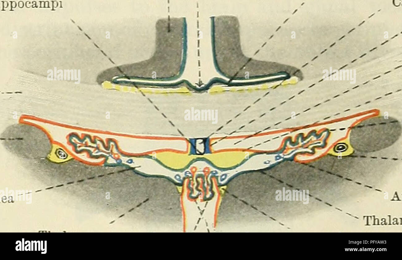 . Cunningham's Text-book of anatomy. Anatomy. THE PIA MATER. 675 lateral part is the chorioidal fissure. This is continuous with the intermediate part, and has already been described in connexion with the inferior horn of the lateral ventricle (p. 636). Pia Mater Spinalis.âThe pia mater of the spinal medulla is thicker and denser than that of the brain. This is largely due to the addition of an outside fibrous layer, in which the fibres run chiefly in the longitudinal direction. The pia mater is very firmly adherent to the surface of the spinal medulla, and in front it sends a fold into the an Stock Photohttps://www.alamy.com/image-license-details/?v=1https://www.alamy.com/cunninghams-text-book-of-anatomy-anatomy-the-pia-mater-675-lateral-part-is-the-chorioidal-fissure-this-is-continuous-with-the-intermediate-part-and-has-already-been-described-in-connexion-with-the-inferior-horn-of-the-lateral-ventricle-p-636-pia-mater-spinalisthe-pia-mater-of-the-spinal-medulla-is-thicker-and-denser-than-that-of-the-brain-this-is-largely-due-to-the-addition-of-an-outside-fibrous-layer-in-which-the-fibres-run-chiefly-in-the-longitudinal-direction-the-pia-mater-is-very-firmly-adherent-to-the-surface-of-the-spinal-medulla-and-in-front-it-sends-a-fold-into-the-an-image216345503.html
RMPFYAW3–. Cunningham's Text-book of anatomy. Anatomy. THE PIA MATER. 675 lateral part is the chorioidal fissure. This is continuous with the intermediate part, and has already been described in connexion with the inferior horn of the lateral ventricle (p. 636). Pia Mater Spinalis.âThe pia mater of the spinal medulla is thicker and denser than that of the brain. This is largely due to the addition of an outside fibrous layer, in which the fibres run chiefly in the longitudinal direction. The pia mater is very firmly adherent to the surface of the spinal medulla, and in front it sends a fold into the an
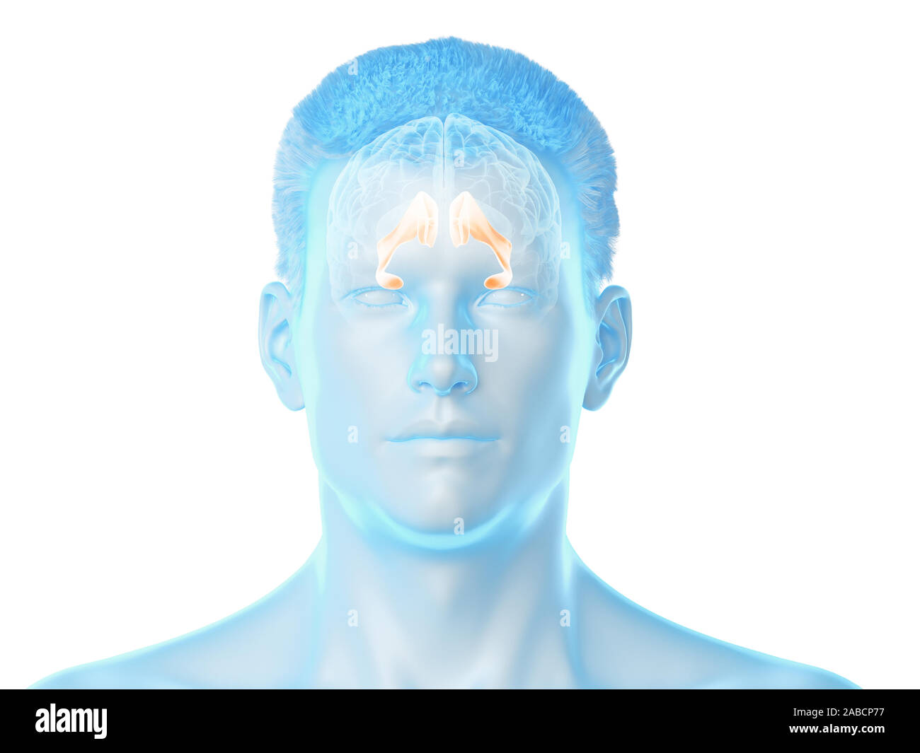 3d rendered medically accurate illustration of the brain anatomy - the lateral ventricle Stock Photohttps://www.alamy.com/image-license-details/?v=1https://www.alamy.com/3d-rendered-medically-accurate-illustration-of-the-brain-anatomy-the-lateral-ventricle-image334039083.html
RF2ABCP77–3d rendered medically accurate illustration of the brain anatomy - the lateral ventricle
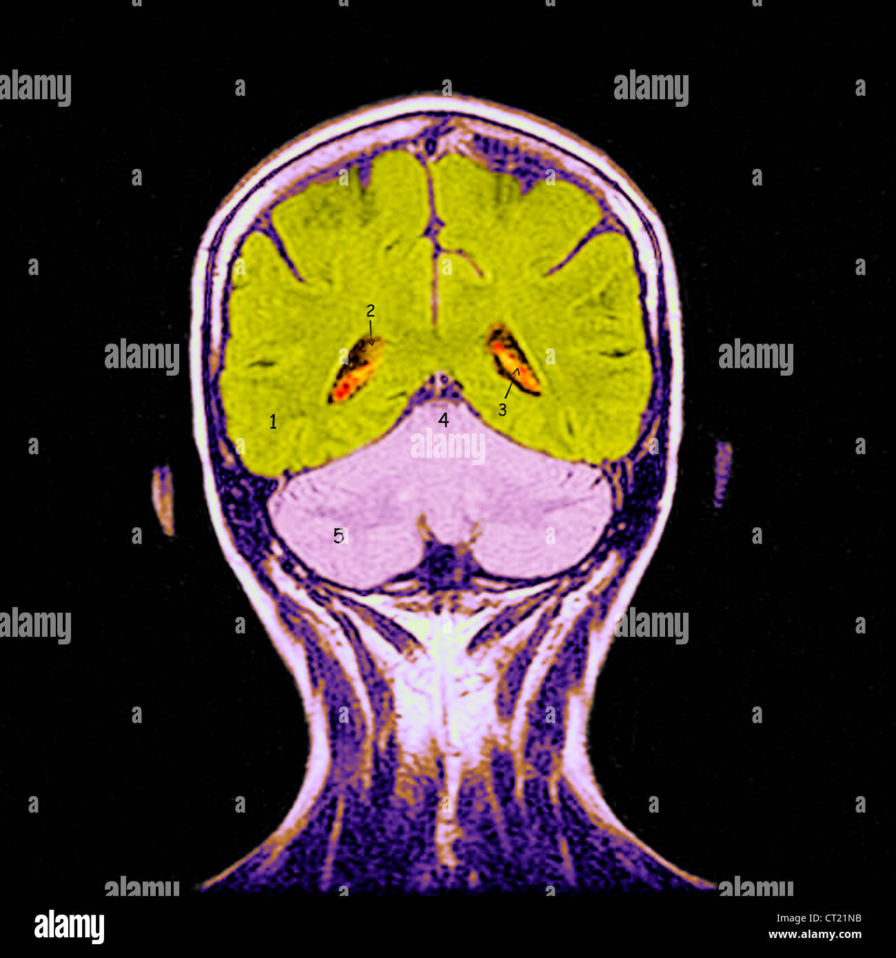 HEAD, MRI Stock Photohttps://www.alamy.com/image-license-details/?v=1https://www.alamy.com/stock-photo-head-mri-49217767.html
RMCT21NB–HEAD, MRI
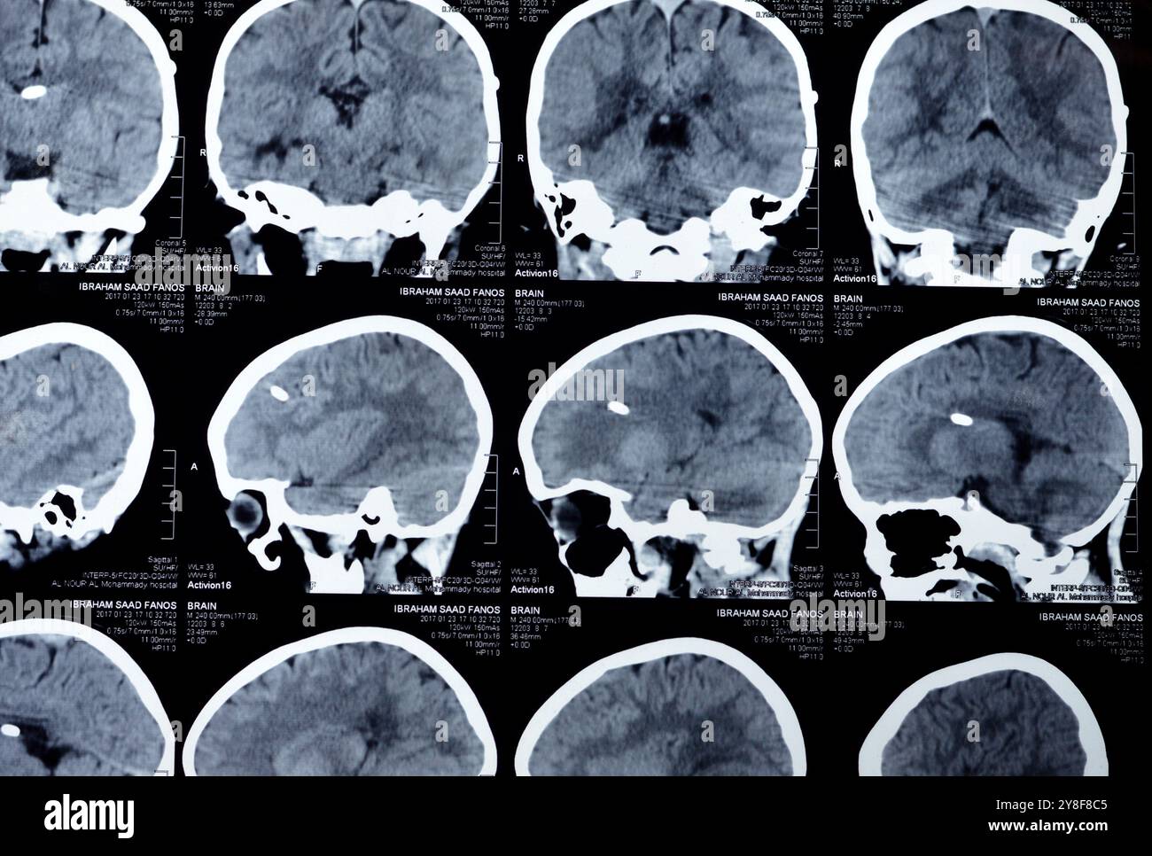 Cairo, Egypt, September 17 2024: Brain CT scan reveals left shunt tube terminating in right lateral ventricle, with no residual ventricular dilatation Stock Photohttps://www.alamy.com/image-license-details/?v=1https://www.alamy.com/cairo-egypt-september-17-2024-brain-ct-scan-reveals-left-shunt-tube-terminating-in-right-lateral-ventricle-with-no-residual-ventricular-dilatation-image624826389.html
RF2Y8F8C5–Cairo, Egypt, September 17 2024: Brain CT scan reveals left shunt tube terminating in right lateral ventricle, with no residual ventricular dilatation
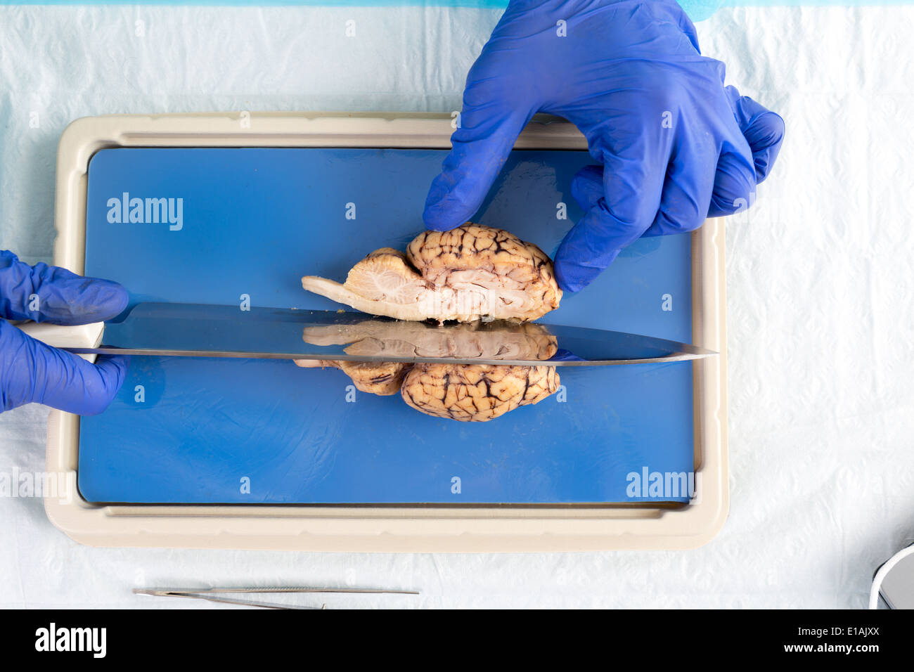 Student in a laboratory or pathologist slicing through the mid section a cow brain dissecting the brainstem and hemispheres of t Stock Photohttps://www.alamy.com/image-license-details/?v=1https://www.alamy.com/student-in-a-laboratory-or-pathologist-slicing-through-the-mid-section-image69690514.html
RFE1AJXX–Student in a laboratory or pathologist slicing through the mid section a cow brain dissecting the brainstem and hemispheres of t
