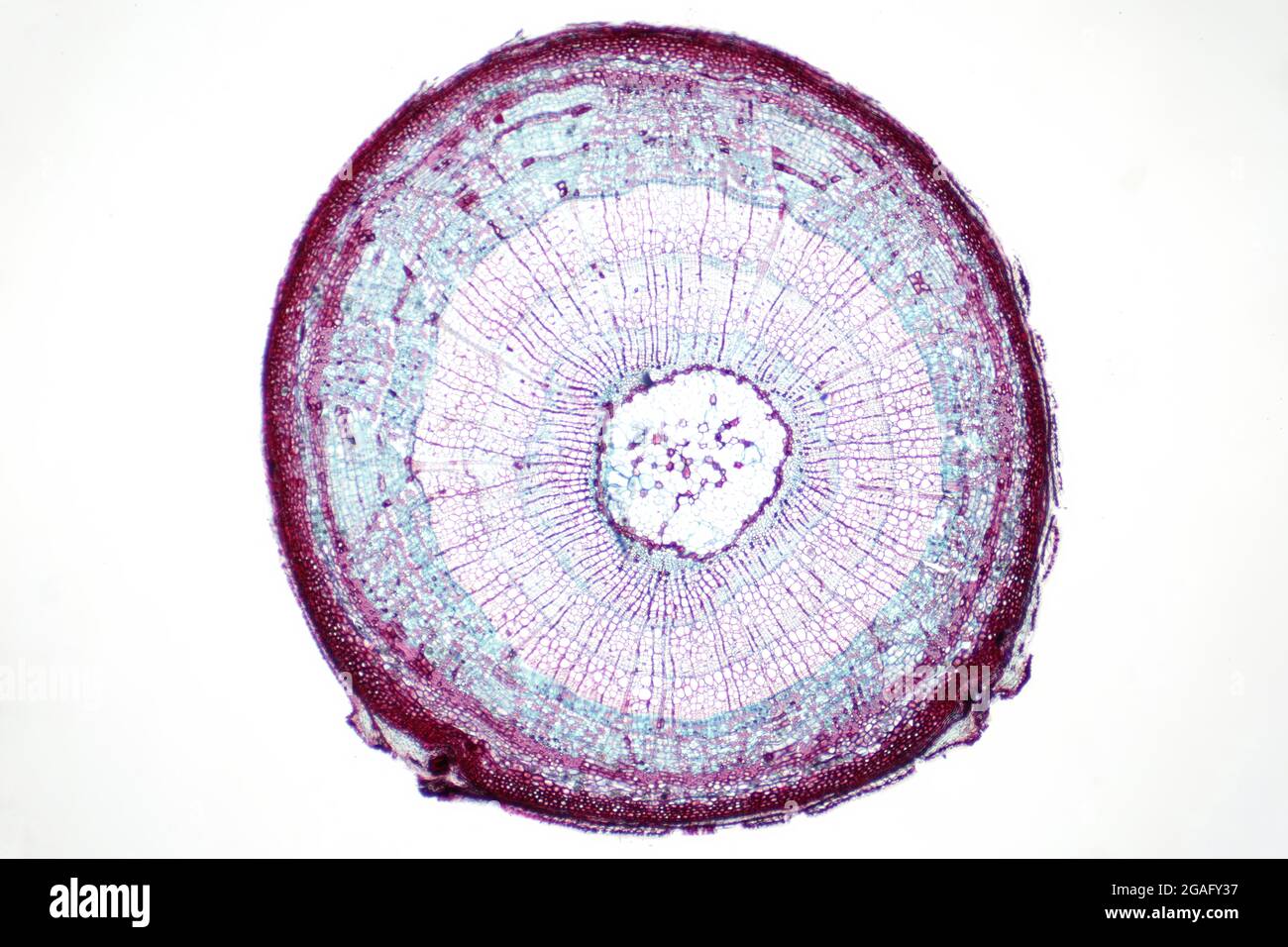
Quick filters:
Leaf micrograph section Stock Photos and Images
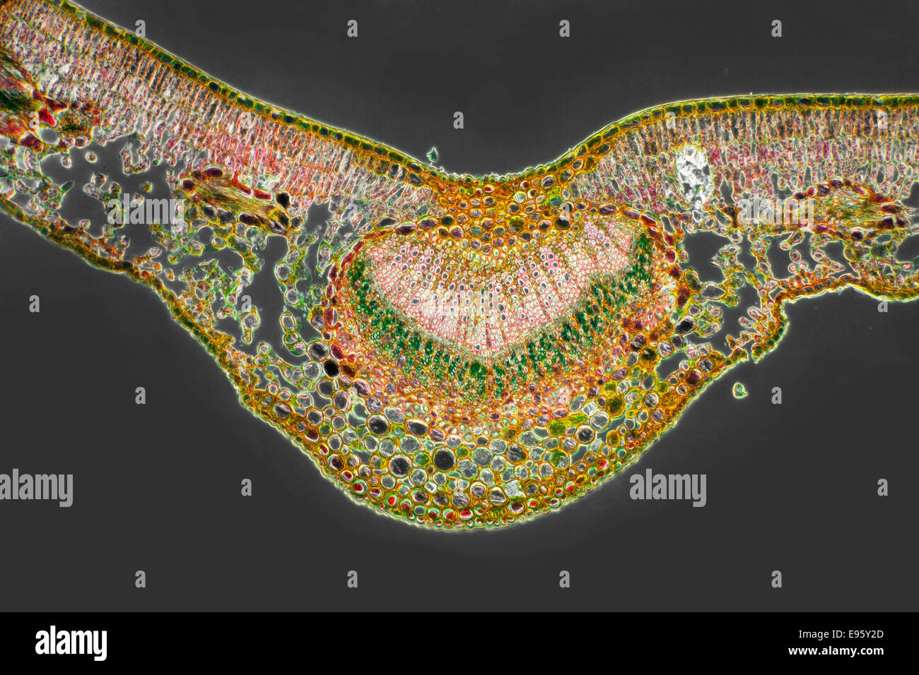 Berberis vulgaris, Barberry leaf TS stained section central vein, darkfield photomicrograph Stock Photohttps://www.alamy.com/image-license-details/?v=1https://www.alamy.com/stock-photo-berberis-vulgaris-barberry-leaf-ts-stained-section-central-vein-darkfield-74504373.html
RME95Y2D–Berberis vulgaris, Barberry leaf TS stained section central vein, darkfield photomicrograph
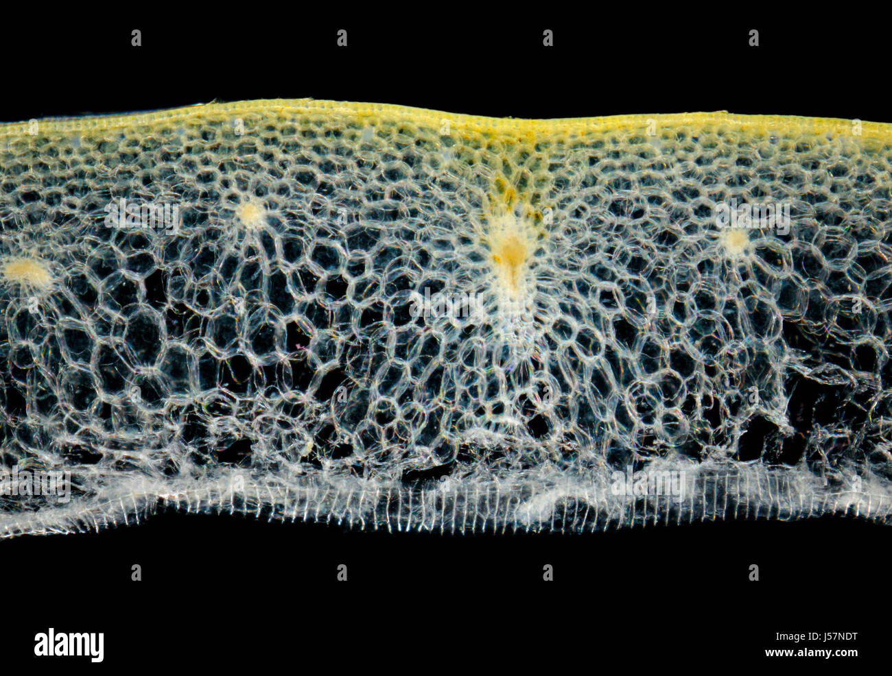 Microscopic view of a Onion (Allium cepa) bulb leaf cross-section. Darkfield illumination. Stock Photohttps://www.alamy.com/image-license-details/?v=1https://www.alamy.com/stock-photo-microscopic-view-of-a-onion-allium-cepa-bulb-leaf-cross-section-darkfield-140926740.html
RFJ57NDT–Microscopic view of a Onion (Allium cepa) bulb leaf cross-section. Darkfield illumination.
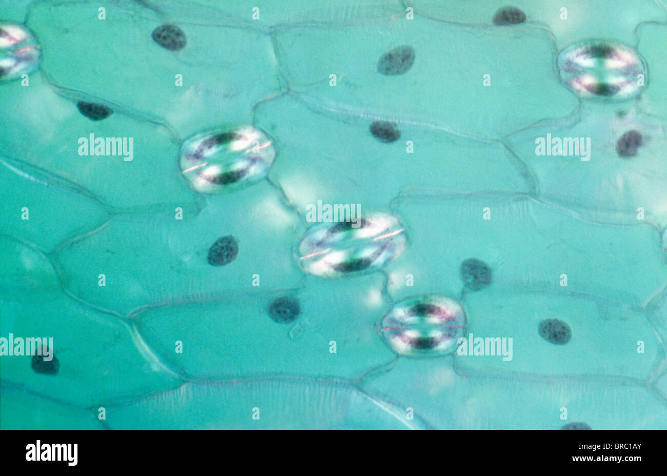 Light Micrograph (LM) of a transverse section of a leaf of a Tulip (Tulipa sp.) showing stomata, magnification x1200 Stock Photohttps://www.alamy.com/image-license-details/?v=1https://www.alamy.com/stock-photo-light-micrograph-lm-of-a-transverse-section-of-a-leaf-of-a-tulip-tulipa-31611971.html
RMBRC1AY–Light Micrograph (LM) of a transverse section of a leaf of a Tulip (Tulipa sp.) showing stomata, magnification x1200
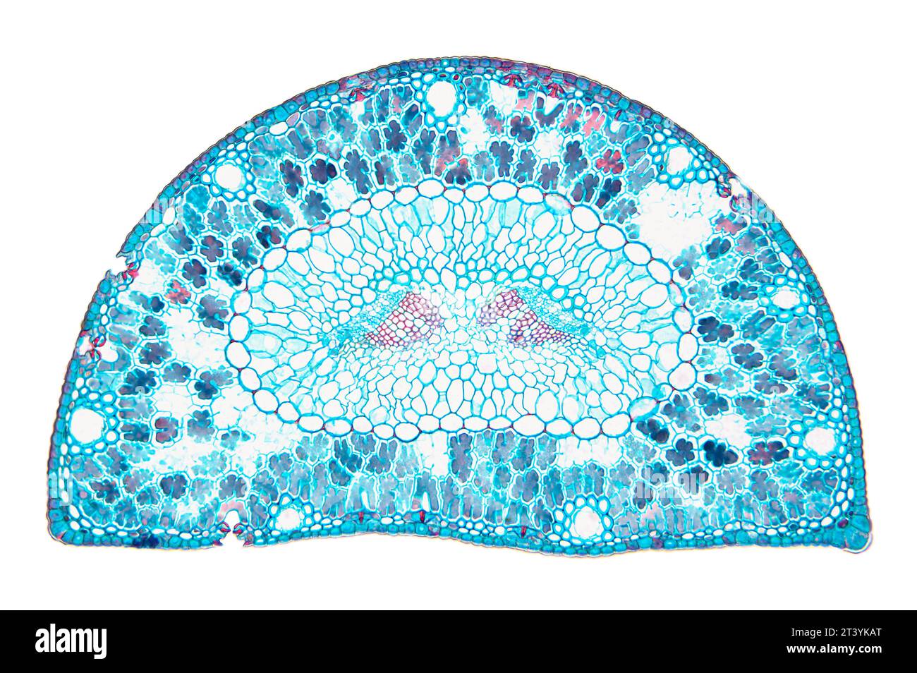 Pine leaf, cross section, 20X light micrograph. Pine tree needle under the microscope. Epidermis cells, cuticle, mesophyll, resin canals, etc. Stock Photohttps://www.alamy.com/image-license-details/?v=1https://www.alamy.com/pine-leaf-cross-section-20x-light-micrograph-pine-tree-needle-under-the-microscope-epidermis-cells-cuticle-mesophyll-resin-canals-etc-image570394016.html
RF2T3YKAT–Pine leaf, cross section, 20X light micrograph. Pine tree needle under the microscope. Epidermis cells, cuticle, mesophyll, resin canals, etc.
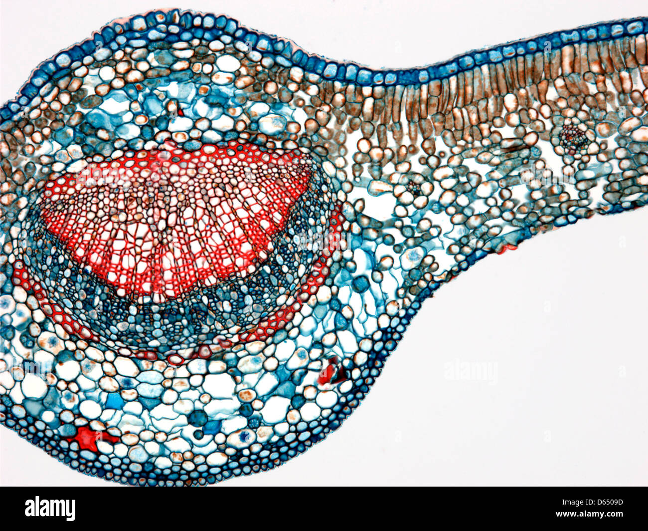 Tea leaf, light micrograph Stock Photohttps://www.alamy.com/image-license-details/?v=1https://www.alamy.com/stock-photo-tea-leaf-light-micrograph-55429065.html
RFD6509D–Tea leaf, light micrograph
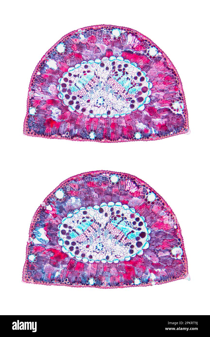 Pine tree leaf, cross section, 20X light micrograph. Needle of Pinus, under microscope. Stock Photohttps://www.alamy.com/image-license-details/?v=1https://www.alamy.com/pine-tree-leaf-cross-section-20x-light-micrograph-needle-of-pinus-under-microscope-image545723854.html
RF2PKRT9J–Pine tree leaf, cross section, 20X light micrograph. Needle of Pinus, under microscope.
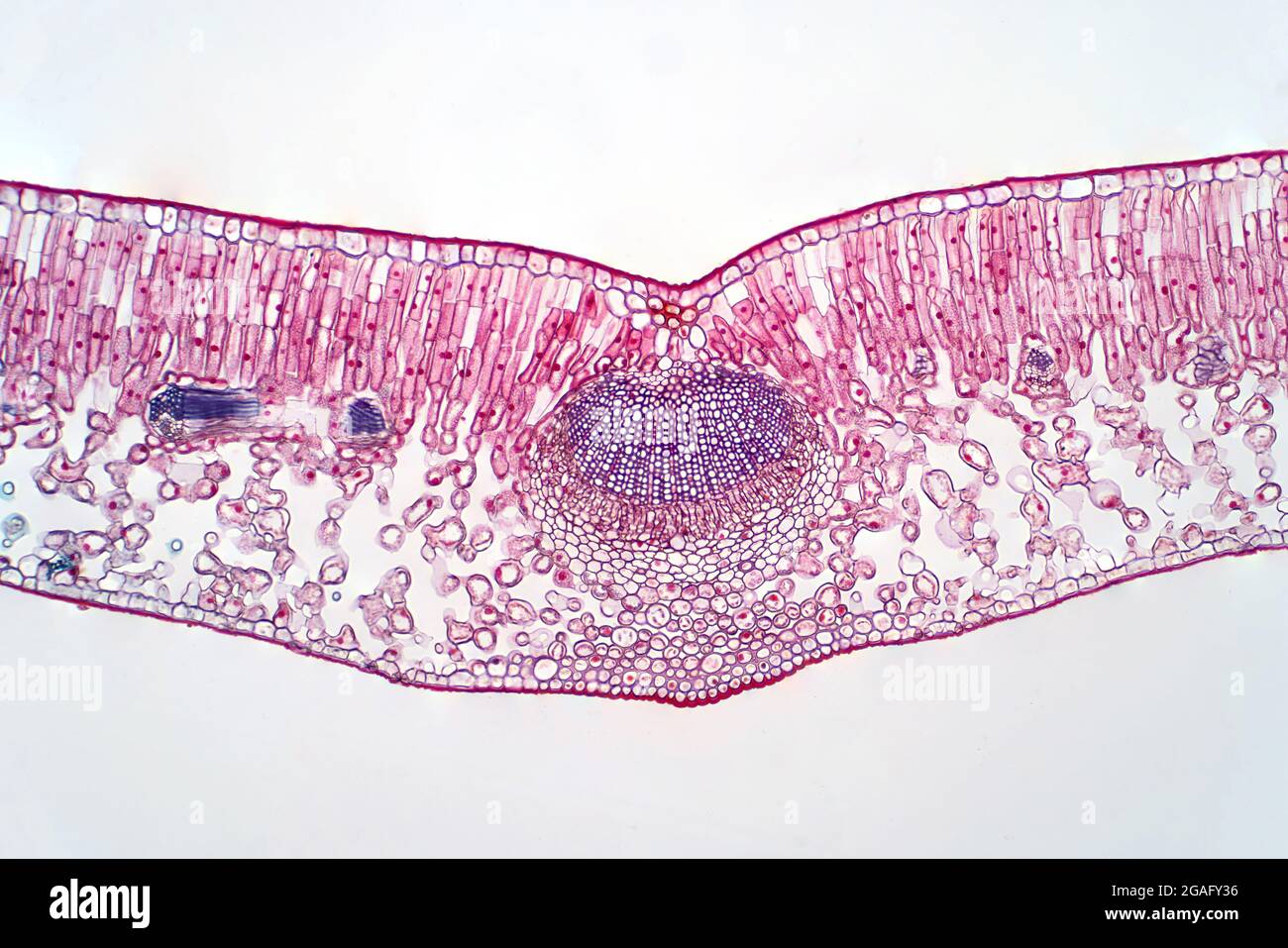 Cross section of a leaf, light micrograph Stock Photohttps://www.alamy.com/image-license-details/?v=1https://www.alamy.com/cross-section-of-a-leaf-light-micrograph-image436756298.html
RF2GAFY36–Cross section of a leaf, light micrograph
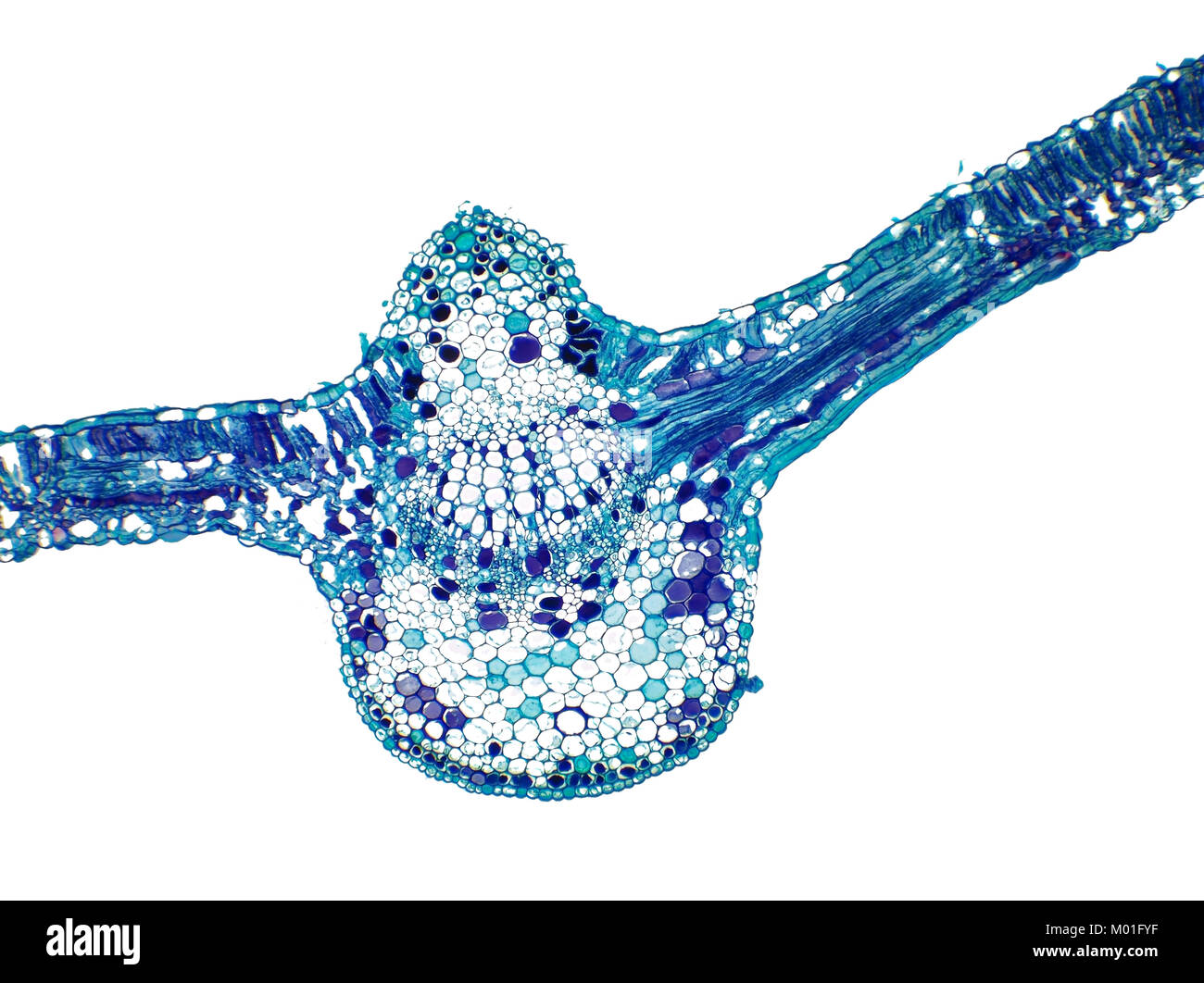 Bright field light micrograph of a cotton plant leaf, pictured area is 1.7mm wide Stock Photohttps://www.alamy.com/image-license-details/?v=1https://www.alamy.com/stock-photo-bright-field-light-micrograph-of-a-cotton-plant-leaf-pictured-area-172138163.html
RMM01FYF–Bright field light micrograph of a cotton plant leaf, pictured area is 1.7mm wide
 Microscopic view of Mallow (Malva sp.) leaf cross section. Brightfield illumination. Stock Photohttps://www.alamy.com/image-license-details/?v=1https://www.alamy.com/microscopic-view-of-mallow-malva-sp-leaf-cross-section-brightfield-illumination-image210691107.html
RFP6NPJB–Microscopic view of Mallow (Malva sp.) leaf cross section. Brightfield illumination.
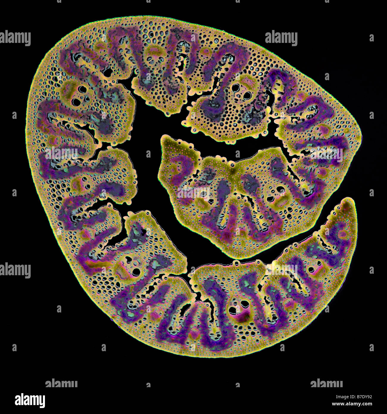 BEACH GRASS LEAF AMMOPHILA Stock Photohttps://www.alamy.com/image-license-details/?v=1https://www.alamy.com/stock-photo-beach-grass-leaf-ammophila-21819758.html
RMB7DY92–BEACH GRASS LEAF AMMOPHILA
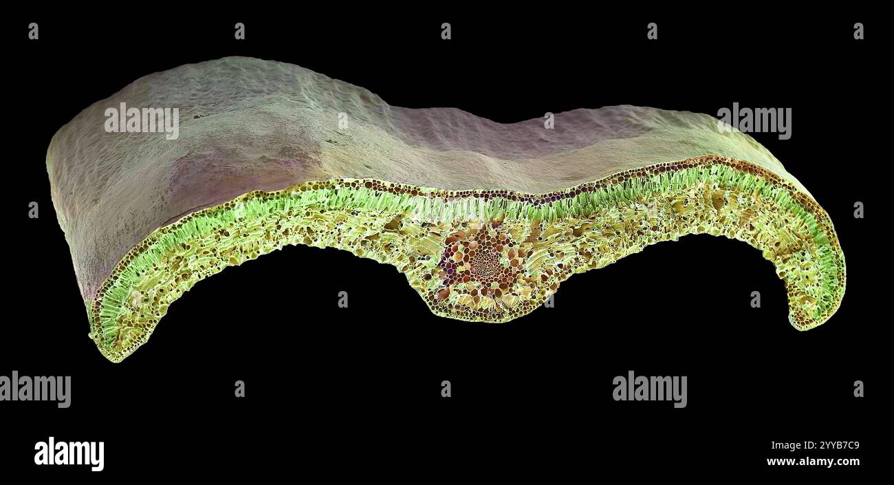 Box leaf. Coloured scanning electron micrograph (SEM) of a section through a leaf from the Common Box (Buxus sempervirens). The midrib (midvein) is the continuation of a leaf's stem along the centre of the leaf. At centre is a vascular bundle, which consists of xylem and phloem tissues. Xylem transports water and mineral nutrients from the roots throughout the plant and phloem transports carbohydrate and hormones around the plant. The surface (epidermis) of the leaf is covered in a waxy cuticle (top) that helps to prevent water loss. Magnification: x200 when printed 10 centimetres wide. Stock Photohttps://www.alamy.com/image-license-details/?v=1https://www.alamy.com/box-leaf-coloured-scanning-electron-micrograph-sem-of-a-section-through-a-leaf-from-the-common-box-buxus-sempervirens-the-midrib-midvein-is-the-continuation-of-a-leafs-stem-along-the-centre-of-the-leaf-at-centre-is-a-vascular-bundle-which-consists-of-xylem-and-phloem-tissues-xylem-transports-water-and-mineral-nutrients-from-the-roots-throughout-the-plant-and-phloem-transports-carbohydrate-and-hormones-around-the-plant-the-surface-epidermis-of-the-leaf-is-covered-in-a-waxy-cuticle-top-that-helps-to-prevent-water-loss-magnification-x200-when-printed-10-centimetres-wide-image636416265.html
RF2YYB7C9–Box leaf. Coloured scanning electron micrograph (SEM) of a section through a leaf from the Common Box (Buxus sempervirens). The midrib (midvein) is the continuation of a leaf's stem along the centre of the leaf. At centre is a vascular bundle, which consists of xylem and phloem tissues. Xylem transports water and mineral nutrients from the roots throughout the plant and phloem transports carbohydrate and hormones around the plant. The surface (epidermis) of the leaf is covered in a waxy cuticle (top) that helps to prevent water loss. Magnification: x200 when printed 10 centimetres wide.
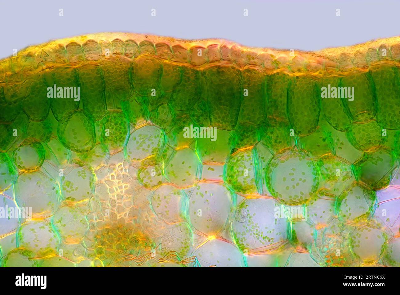 The image presents palisade mesophyll in hyacinthus leaf (transversal cross-section) photographed through the microscope in polarized light at a magn Stock Photohttps://www.alamy.com/image-license-details/?v=1https://www.alamy.com/the-image-presents-palisade-mesophyll-in-hyacinthus-leaf-transversal-cross-section-photographed-through-the-microscope-in-polarized-light-at-a-magn-image565954114.html
RM2RTNC6X–The image presents palisade mesophyll in hyacinthus leaf (transversal cross-section) photographed through the microscope in polarized light at a magn
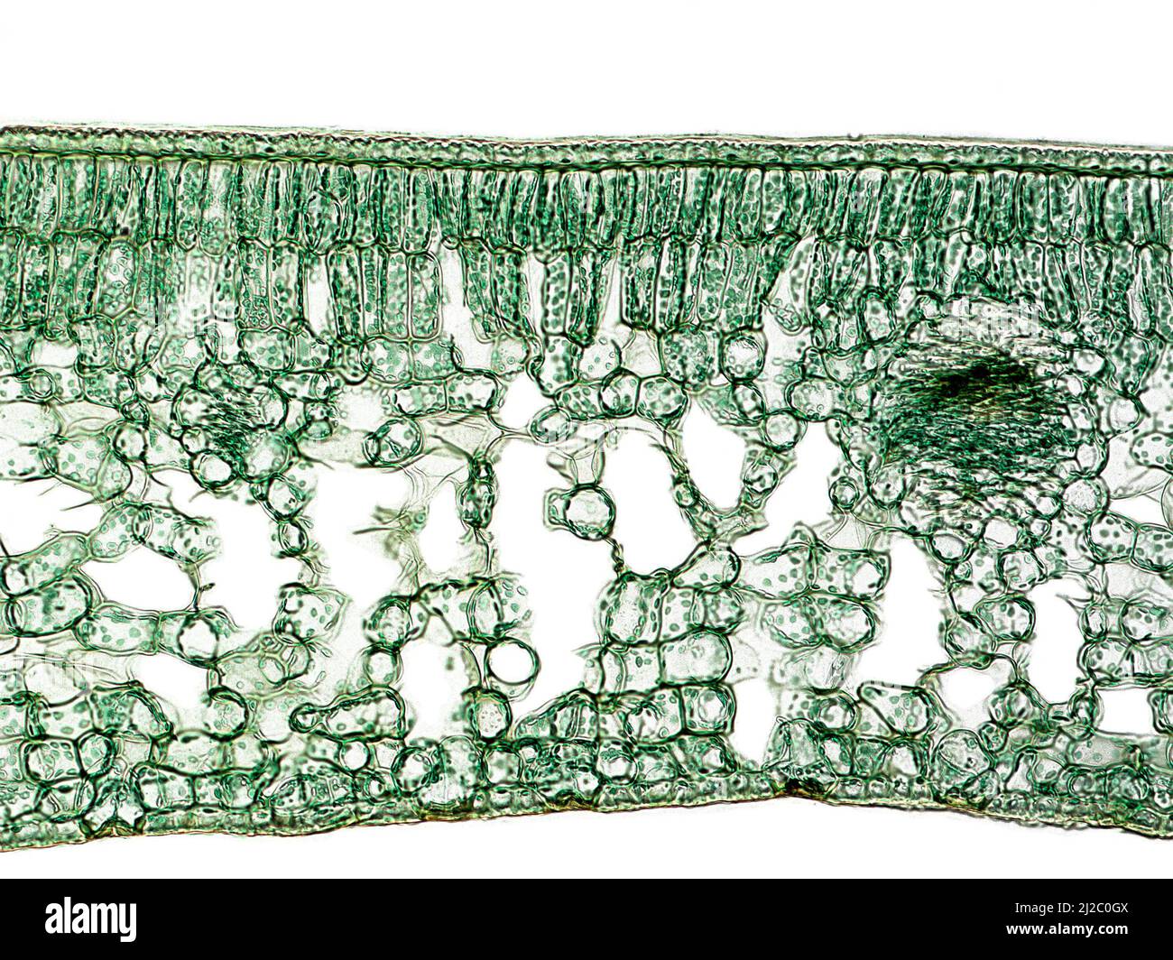 Cross section of a Camellia leaf, that show their general internal structure (cuticle, palisade parenchyma, spongy parenchyma, vascular bundles). Stock Photohttps://www.alamy.com/image-license-details/?v=1https://www.alamy.com/cross-section-of-a-camellia-leaf-that-show-their-general-internal-structure-cuticle-palisade-parenchyma-spongy-parenchyma-vascular-bundles-image466173146.html
RM2J2C0GX–Cross section of a Camellia leaf, that show their general internal structure (cuticle, palisade parenchyma, spongy parenchyma, vascular bundles).
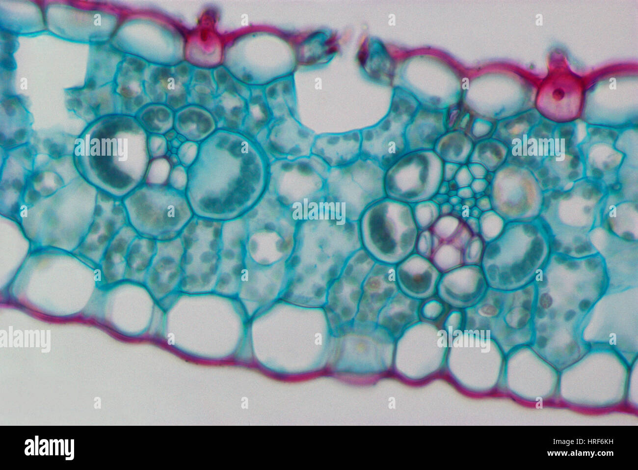 Corn Leaf Vascular Bundles and Sheaths Stock Photohttps://www.alamy.com/image-license-details/?v=1https://www.alamy.com/stock-photo-corn-leaf-vascular-bundles-and-sheaths-134944197.html
RMHRF6KH–Corn Leaf Vascular Bundles and Sheaths
 The image presents read leaf in transversal cross-section, photographed through the microscope in polarized light at a magnification of 100X Stock Photohttps://www.alamy.com/image-license-details/?v=1https://www.alamy.com/the-image-presents-read-leaf-in-transversal-cross-section-photographed-through-the-microscope-in-polarized-light-at-a-magnification-of-100x-image564575503.html
RM2RPEHPR–The image presents read leaf in transversal cross-section, photographed through the microscope in polarized light at a magnification of 100X
 Leaf section micrograph Stock Photohttps://www.alamy.com/image-license-details/?v=1https://www.alamy.com/leaf-section-micrograph-image362245008.html
RM2C19K5M–Leaf section micrograph
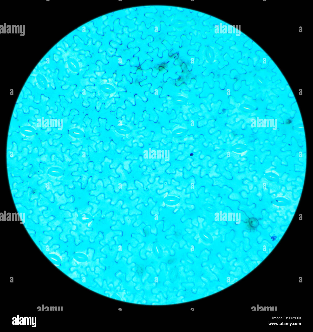 Vicia Dicot leaf under a microscope (Vicia Dicot leaf W.M.), 40x Stock Photohttps://www.alamy.com/image-license-details/?v=1https://www.alamy.com/stock-photo-vicia-dicot-leaf-under-a-microscope-vicia-dicot-leaf-wm-40x-81124355.html
RFEKYEXB–Vicia Dicot leaf under a microscope (Vicia Dicot leaf W.M.), 40x
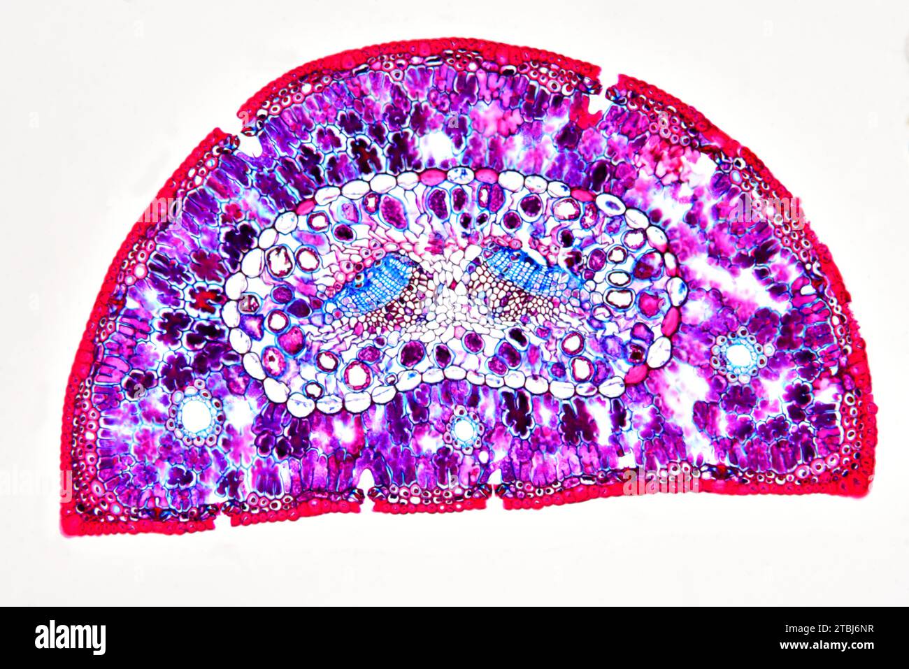 Leaf cross section of pine (Pinus sp.) showing endodermis, epidermis, hypodermis, mesophyll, resin canals, stomata, vascular bundle, phloem and xylem. Stock Photohttps://www.alamy.com/image-license-details/?v=1https://www.alamy.com/leaf-cross-section-of-pine-pinus-sp-showing-endodermis-epidermis-hypodermis-mesophyll-resin-canals-stomata-vascular-bundle-phloem-and-xylem-image575103811.html
RF2TBJ6NR–Leaf cross section of pine (Pinus sp.) showing endodermis, epidermis, hypodermis, mesophyll, resin canals, stomata, vascular bundle, phloem and xylem.
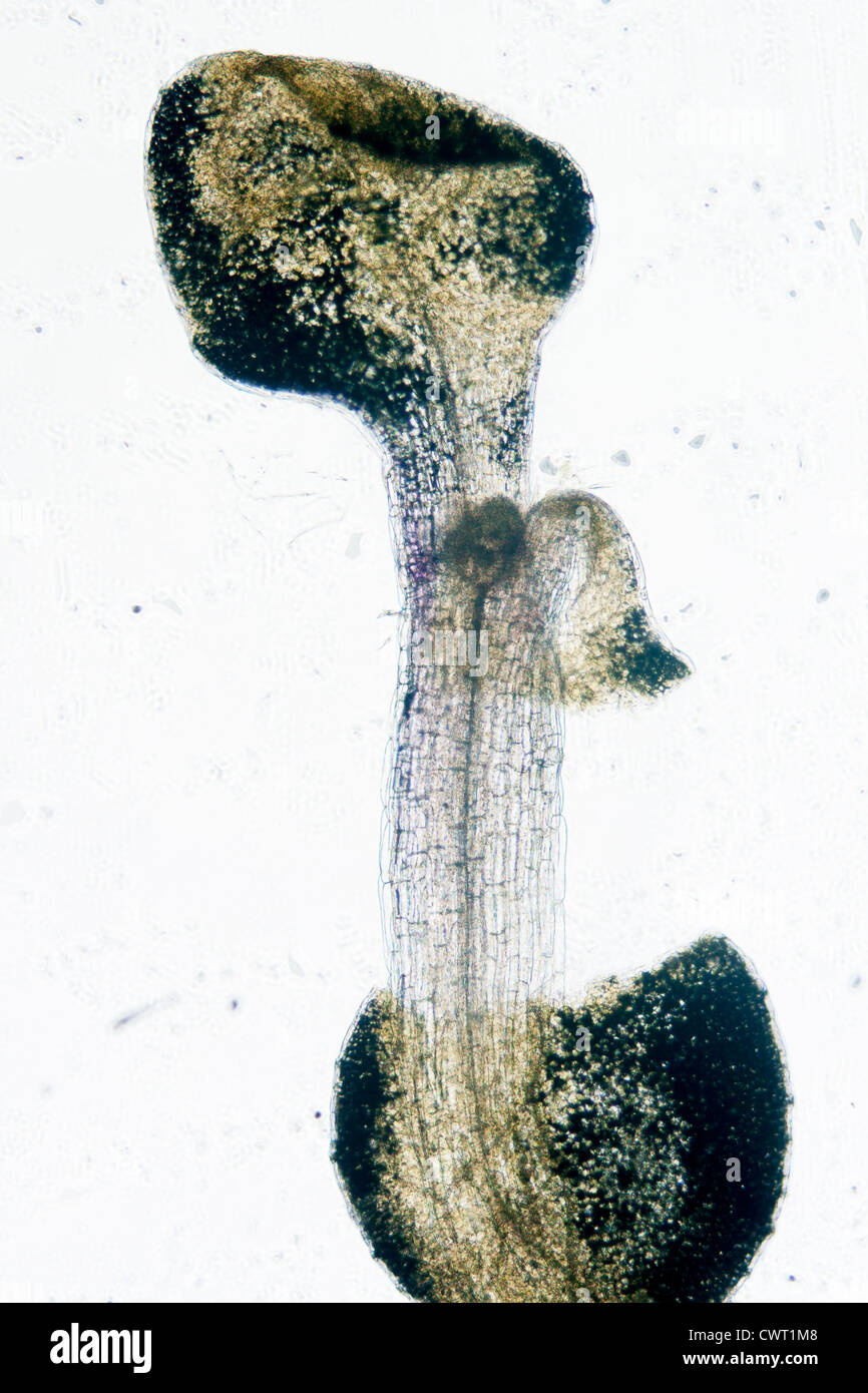 science botany micrograph plant arabidopsis thaliana root tissue micro Stock Photohttps://www.alamy.com/image-license-details/?v=1https://www.alamy.com/stock-photo-science-botany-micrograph-plant-arabidopsis-thaliana-root-tissue-micro-50315336.html
RFCWT1M8–science botany micrograph plant arabidopsis thaliana root tissue micro
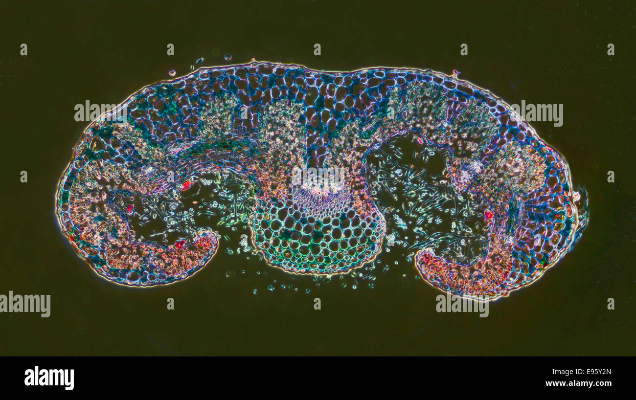 Rosemary, Rosmarinus officinalis, leaf TS, darkfield photomicrograph, stained section. Stock Photohttps://www.alamy.com/image-license-details/?v=1https://www.alamy.com/stock-photo-rosemary-rosmarinus-officinalis-leaf-ts-darkfield-photomicrograph-74504381.html
RME95Y2N–Rosemary, Rosmarinus officinalis, leaf TS, darkfield photomicrograph, stained section.
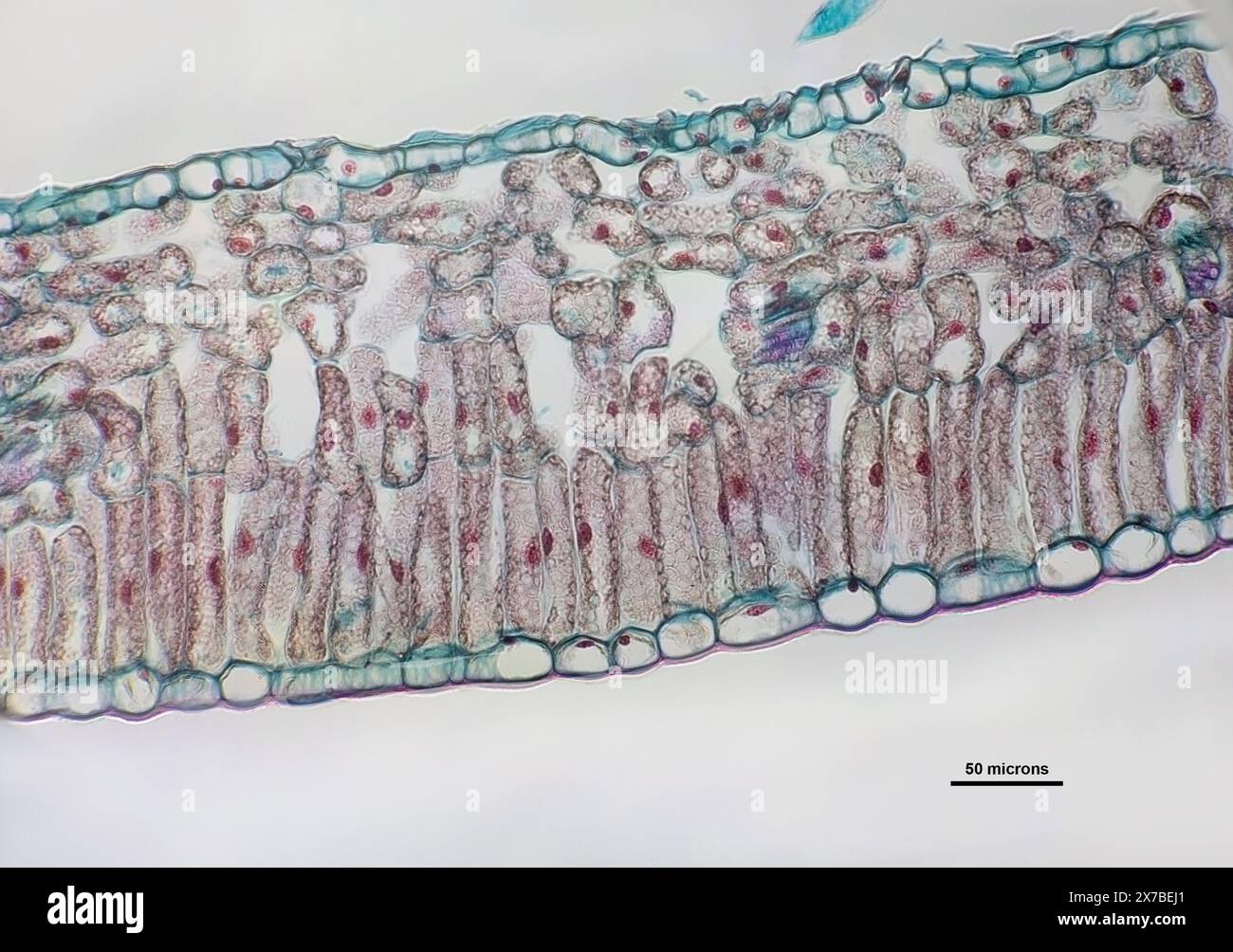 Privet leaf, transverse section under a microscope at 40 times magnification Stock Photohttps://www.alamy.com/image-license-details/?v=1https://www.alamy.com/privet-leaf-transverse-section-under-a-microscope-at-40-times-magnification-image606918425.html
RF2X7BEJ1–Privet leaf, transverse section under a microscope at 40 times magnification
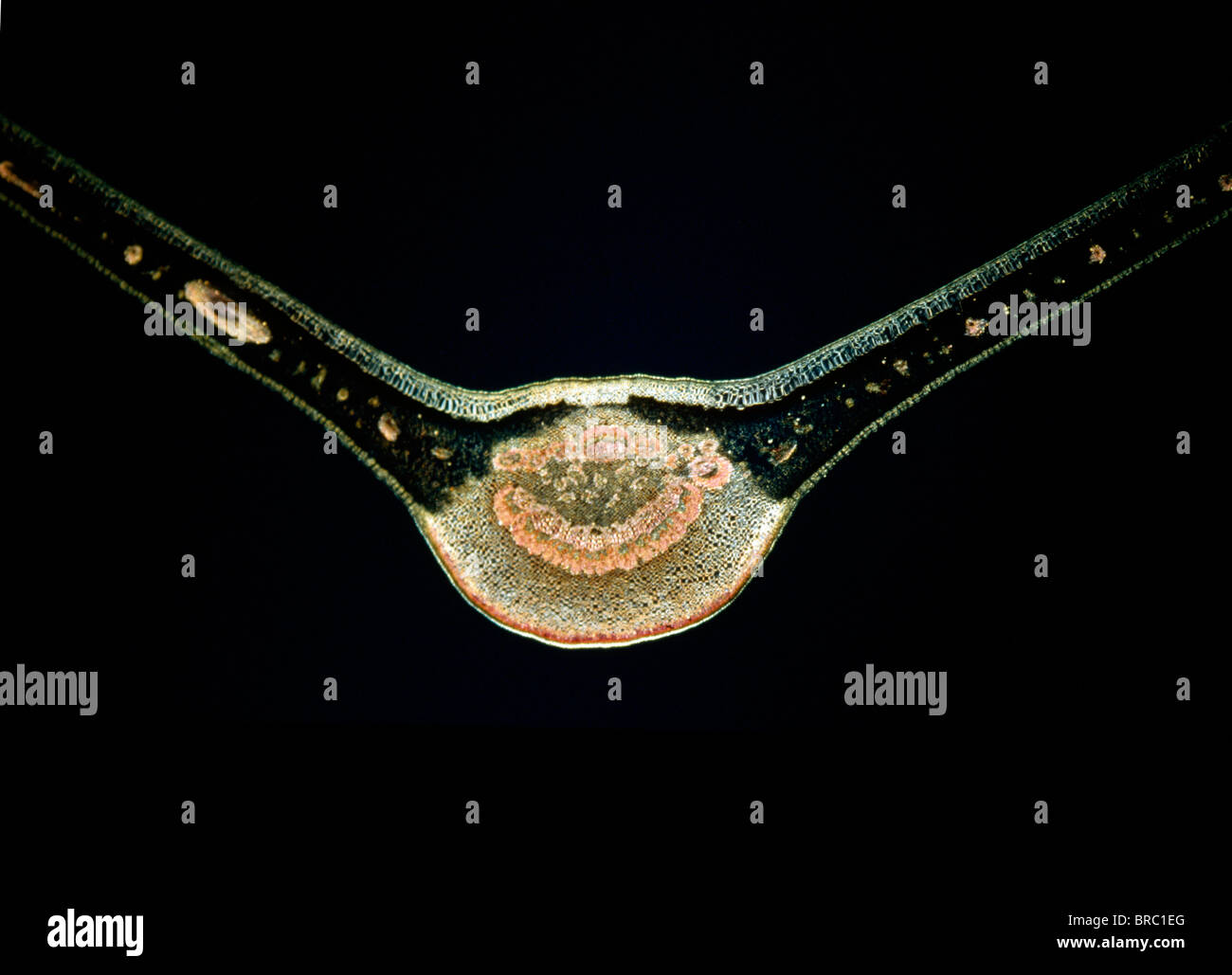 Light Micrograph (LM) of a transverse section of a fig leaf, magnification x 15 Stock Photohttps://www.alamy.com/image-license-details/?v=1https://www.alamy.com/stock-photo-light-micrograph-lm-of-a-transverse-section-of-a-fig-leaf-magnification-31612072.html
RMBRC1EG–Light Micrograph (LM) of a transverse section of a fig leaf, magnification x 15
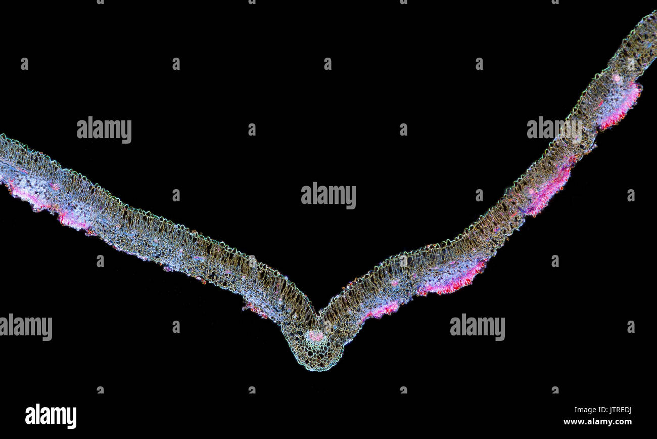 Snapdragon rust, Puccinia antirrhini fungus on antirrhinum leaf, darkfield photomicrograph, TS Stock Photohttps://www.alamy.com/image-license-details/?v=1https://www.alamy.com/snapdragon-rust-puccinia-antirrhini-fungus-on-antirrhinum-leaf-darkfield-image152950942.html
RMJTREDJ–Snapdragon rust, Puccinia antirrhini fungus on antirrhinum leaf, darkfield photomicrograph, TS
 Leaf micrograph Stock Photohttps://www.alamy.com/image-license-details/?v=1https://www.alamy.com/stock-photo-leaf-micrograph-80171364.html
RFEJC3B0–Leaf micrograph
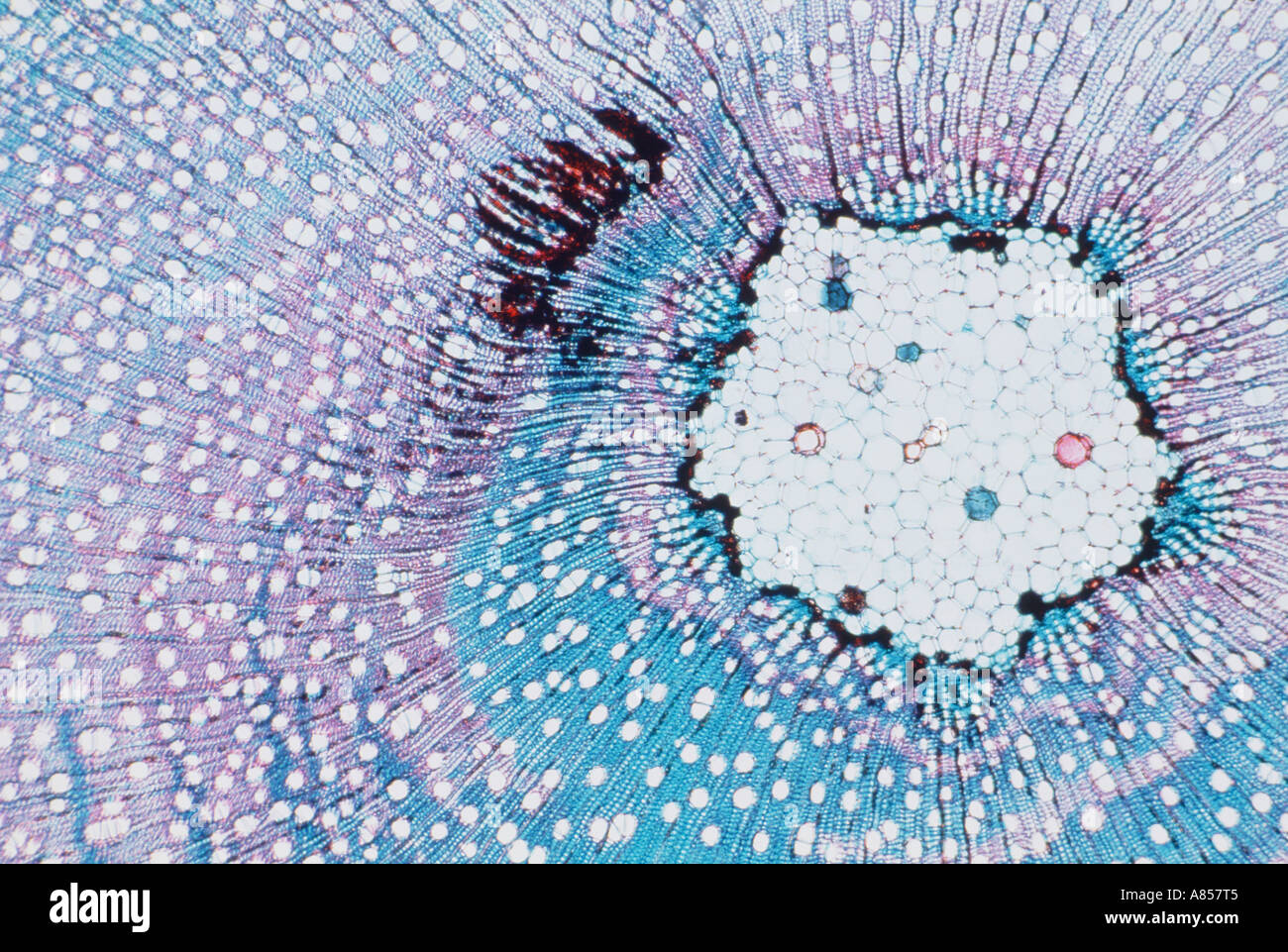 Scanning Electron Micrograph (SEM) of willow tree leaf stem cross-section. Stock Photohttps://www.alamy.com/image-license-details/?v=1https://www.alamy.com/scanning-electron-micrograph-sem-of-willow-tree-leaf-stem-cross-section-image6852292.html
RMA857T5–Scanning Electron Micrograph (SEM) of willow tree leaf stem cross-section.
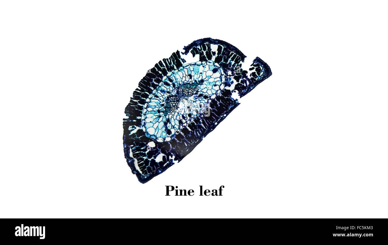 Pine leaf micrograph Stock Photohttps://www.alamy.com/image-license-details/?v=1https://www.alamy.com/stock-photo-pine-leaf-micrograph-93552931.html
RFFC5KM3–Pine leaf micrograph
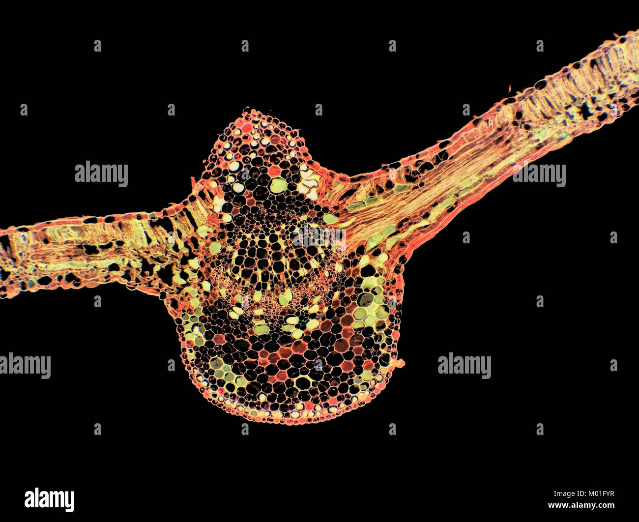 Inverted bright field light micrograph of a cotton plant leaf, pictured area is 1.7mm wide Stock Photohttps://www.alamy.com/image-license-details/?v=1https://www.alamy.com/stock-photo-inverted-bright-field-light-micrograph-of-a-cotton-plant-leaf-pictured-172138171.html
RMM01FYR–Inverted bright field light micrograph of a cotton plant leaf, pictured area is 1.7mm wide
 Microscopic view of a Showy stonecrop (Hylotelephium spectabile) leaf cross section. Polarized light, crossed polarizers. Stock Photohttps://www.alamy.com/image-license-details/?v=1https://www.alamy.com/microscopic-view-of-a-showy-stonecrop-hylotelephium-spectabile-leaf-cross-section-polarized-light-crossed-polarizers-image210691117.html
RFP6NPJN–Microscopic view of a Showy stonecrop (Hylotelephium spectabile) leaf cross section. Polarized light, crossed polarizers.
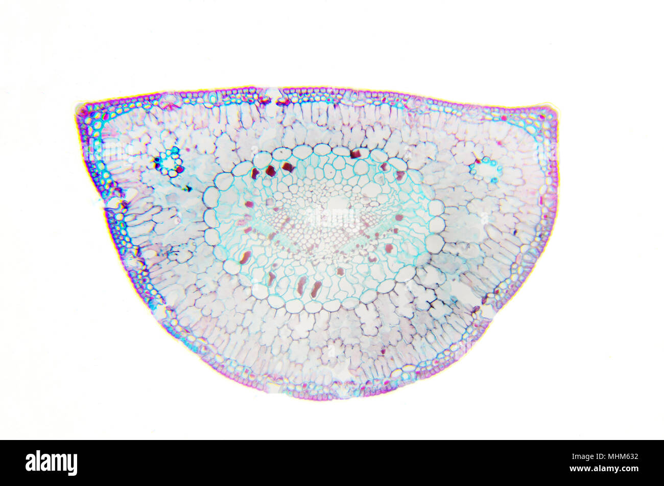 Microscopy Photography. Leaf of Pinus. Transversal Section. Stock Photohttps://www.alamy.com/image-license-details/?v=1https://www.alamy.com/microscopy-photography-leaf-of-pinus-transversal-section-image182996662.html
RFMHM632–Microscopy Photography. Leaf of Pinus. Transversal Section.
 Box leaf. Coloured scanning electron micrograph (SEM) of a section through a leaf from the Common Box (Buxus sempervirens). The midrib (midvein) is the continuation of a leaf's stem along the centre of the leaf. At centre is a vascular bundle, which consists of xylem and phloem tissues. Xylem transports water and mineral nutrients from the roots throughout the plant and phloem transports carbohydrate and hormones around the plant. The surface (epidermis) of the leaf is covered in a waxy cuticle (top) that helps to prevent water loss. Magnification: x200 when printed 10 centimetres wide. Stock Photohttps://www.alamy.com/image-license-details/?v=1https://www.alamy.com/box-leaf-coloured-scanning-electron-micrograph-sem-of-a-section-through-a-leaf-from-the-common-box-buxus-sempervirens-the-midrib-midvein-is-the-continuation-of-a-leafs-stem-along-the-centre-of-the-leaf-at-centre-is-a-vascular-bundle-which-consists-of-xylem-and-phloem-tissues-xylem-transports-water-and-mineral-nutrients-from-the-roots-throughout-the-plant-and-phloem-transports-carbohydrate-and-hormones-around-the-plant-the-surface-epidermis-of-the-leaf-is-covered-in-a-waxy-cuticle-top-that-helps-to-prevent-water-loss-magnification-x200-when-printed-10-centimetres-wide-image636416299.html
RF2YYB7DF–Box leaf. Coloured scanning electron micrograph (SEM) of a section through a leaf from the Common Box (Buxus sempervirens). The midrib (midvein) is the continuation of a leaf's stem along the centre of the leaf. At centre is a vascular bundle, which consists of xylem and phloem tissues. Xylem transports water and mineral nutrients from the roots throughout the plant and phloem transports carbohydrate and hormones around the plant. The surface (epidermis) of the leaf is covered in a waxy cuticle (top) that helps to prevent water loss. Magnification: x200 when printed 10 centimetres wide.
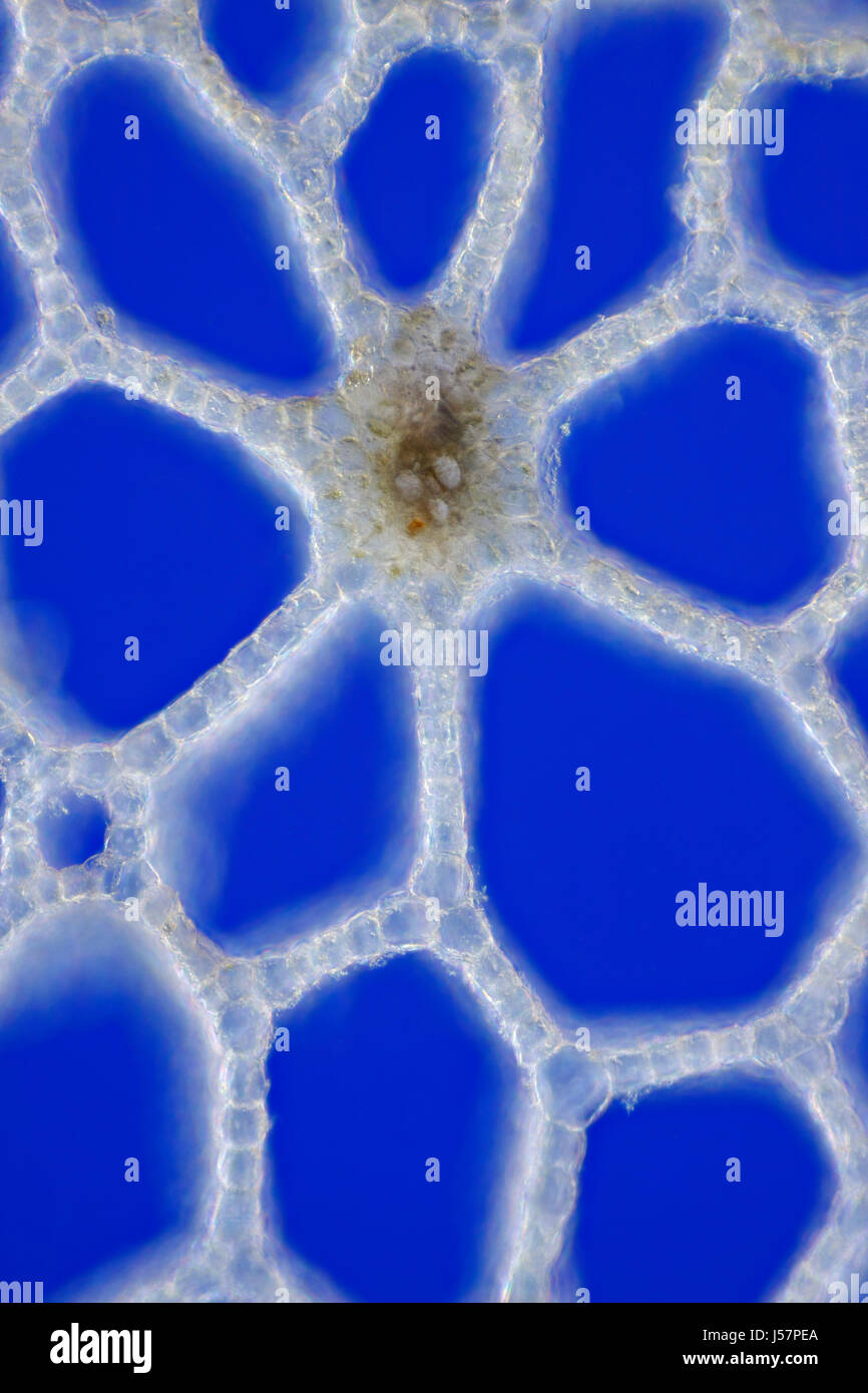 Marsh Calla (Calla palustris) leaf stem cross section. Rheinberg illumination. Stock Photohttps://www.alamy.com/image-license-details/?v=1https://www.alamy.com/stock-photo-marsh-calla-calla-palustris-leaf-stem-cross-section-rheinberg-illumination-140927538.html
RFJ57PEA–Marsh Calla (Calla palustris) leaf stem cross section. Rheinberg illumination.
 biological cell isolated on whithe background microscope 3D Illustration Stock Photohttps://www.alamy.com/image-license-details/?v=1https://www.alamy.com/biological-cell-isolated-on-whithe-background-microscope-3d-illustration-image208953290.html
RFP3XJ1E–biological cell isolated on whithe background microscope 3D Illustration
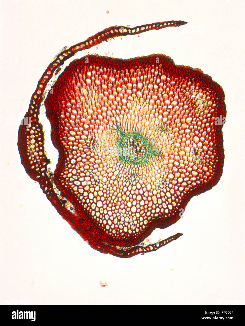 Micrograph of stem and leaf of Polytrichum commune (Common moss), cross-section Stock Photohttps://www.alamy.com/image-license-details/?v=1https://www.alamy.com/micrograph-of-stem-and-leaf-of-polytrichum-commune-common-moss-cross-section-image216105751.html
RMPFGD2F–Micrograph of stem and leaf of Polytrichum commune (Common moss), cross-section
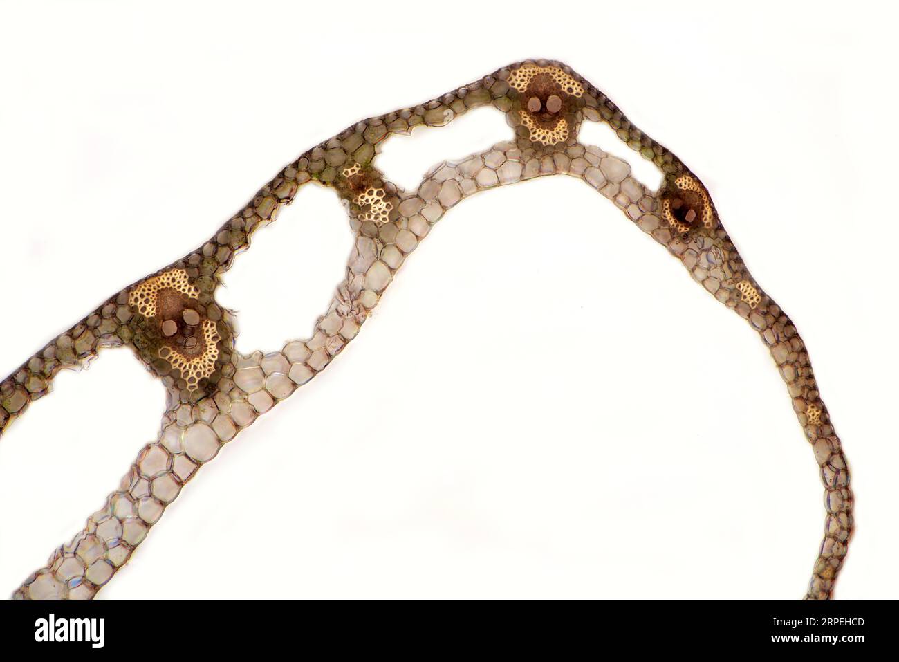 The image presents carex sp. leaf in transversal cross-section, photographed through the microscope in polarized light at a magnification of 100X Stock Photohttps://www.alamy.com/image-license-details/?v=1https://www.alamy.com/the-image-presents-carex-sp-leaf-in-transversal-cross-section-photographed-through-the-microscope-in-polarized-light-at-a-magnification-of-100x-image564575213.html
RM2RPEHCD–The image presents carex sp. leaf in transversal cross-section, photographed through the microscope in polarized light at a magnification of 100X
 Leaf micrograph Stock Photohttps://www.alamy.com/image-license-details/?v=1https://www.alamy.com/stock-photo-leaf-micrograph-103147644.html
RMFYPNTC–Leaf micrograph
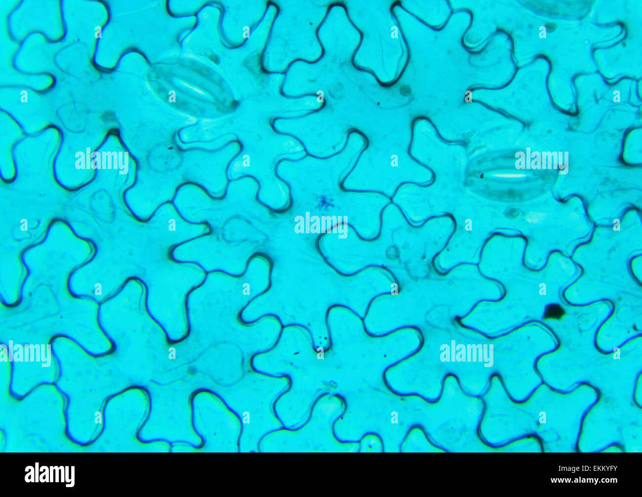 Vicia Dicot leaf under a microscope (Vicia Dicot leaf W.M.), 400x Stock Photohttps://www.alamy.com/image-license-details/?v=1https://www.alamy.com/stock-photo-vicia-dicot-leaf-under-a-microscope-vicia-dicot-leaf-wm-400x-80958639.html
RFEKKYFY–Vicia Dicot leaf under a microscope (Vicia Dicot leaf W.M.), 400x
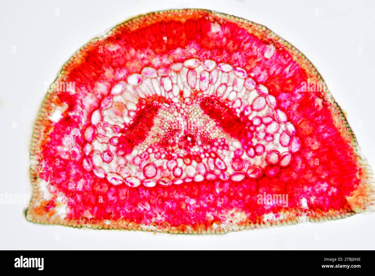 Leaf cross section of pine (Pinus sp.) showing endodermis, epidermis, hypodermis, mesophyll, resin canals, stomata, vascular bundle, phloem and xylem. Stock Photohttps://www.alamy.com/image-license-details/?v=1https://www.alamy.com/leaf-cross-section-of-pine-pinus-sp-showing-endodermis-epidermis-hypodermis-mesophyll-resin-canals-stomata-vascular-bundle-phloem-and-xylem-image575103802.html
RF2TBJ6NE–Leaf cross section of pine (Pinus sp.) showing endodermis, epidermis, hypodermis, mesophyll, resin canals, stomata, vascular bundle, phloem and xylem.
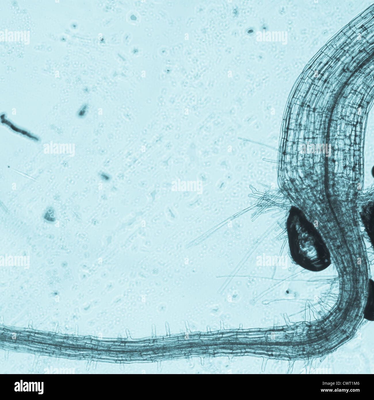 science botany micrograph plant arabidopsis thaliana root tissue micro Stock Photohttps://www.alamy.com/image-license-details/?v=1https://www.alamy.com/stock-photo-science-botany-micrograph-plant-arabidopsis-thaliana-root-tissue-micro-50315334.html
RFCWT1M6–science botany micrograph plant arabidopsis thaliana root tissue micro
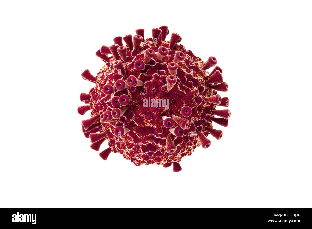 biological cell isolated on whithe background microscope 3D Illustration Stock Photohttps://www.alamy.com/image-license-details/?v=1https://www.alamy.com/biological-cell-isolated-on-whithe-background-microscope-3d-illustration-image208953338.html
RFP3XJ36–biological cell isolated on whithe background microscope 3D Illustration
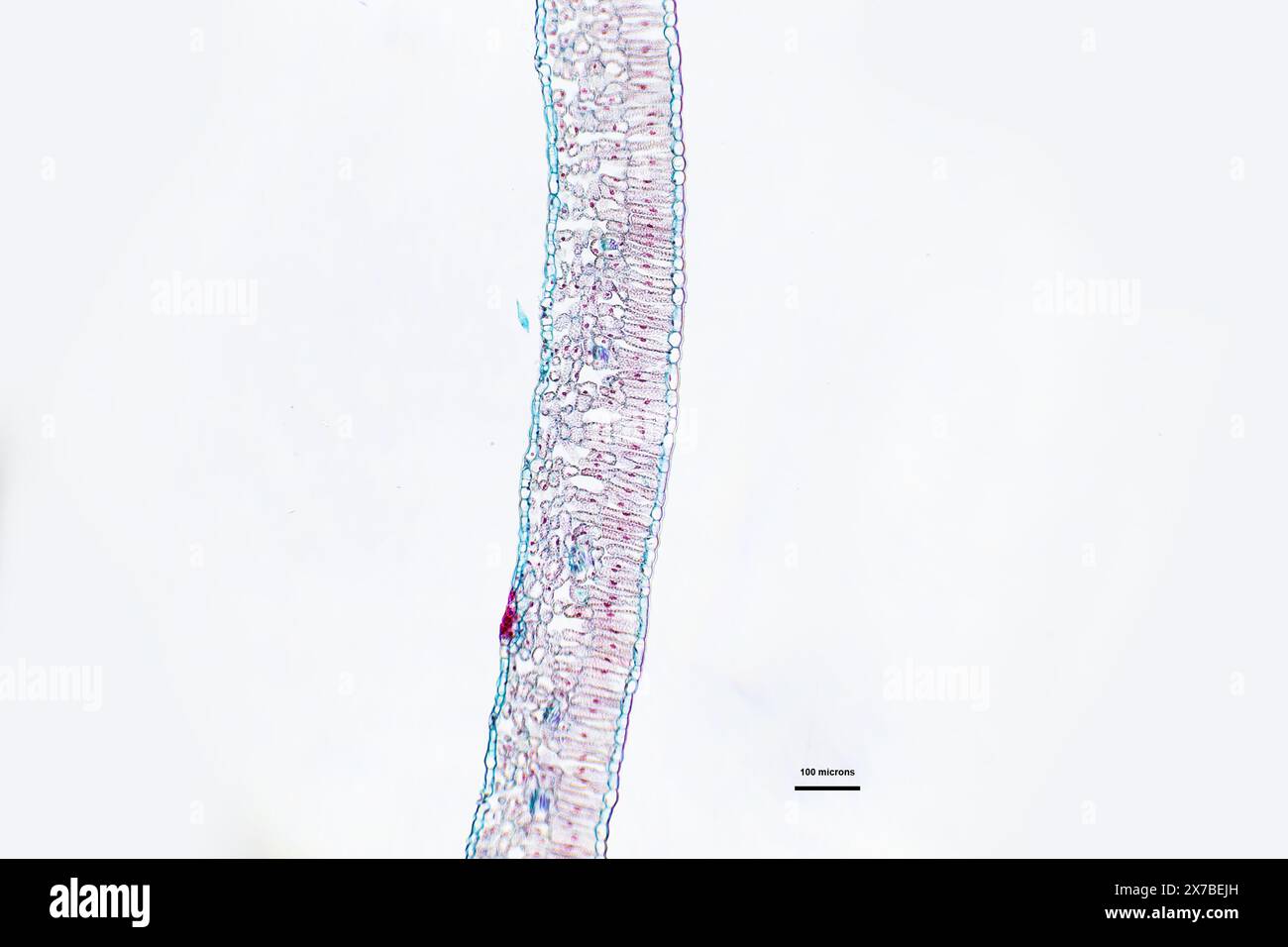 Privet leaf, transverse section under a microscope at 10 times magnification Stock Photohttps://www.alamy.com/image-license-details/?v=1https://www.alamy.com/privet-leaf-transverse-section-under-a-microscope-at-10-times-magnification-image606918441.html
RF2X7BEJH–Privet leaf, transverse section under a microscope at 10 times magnification
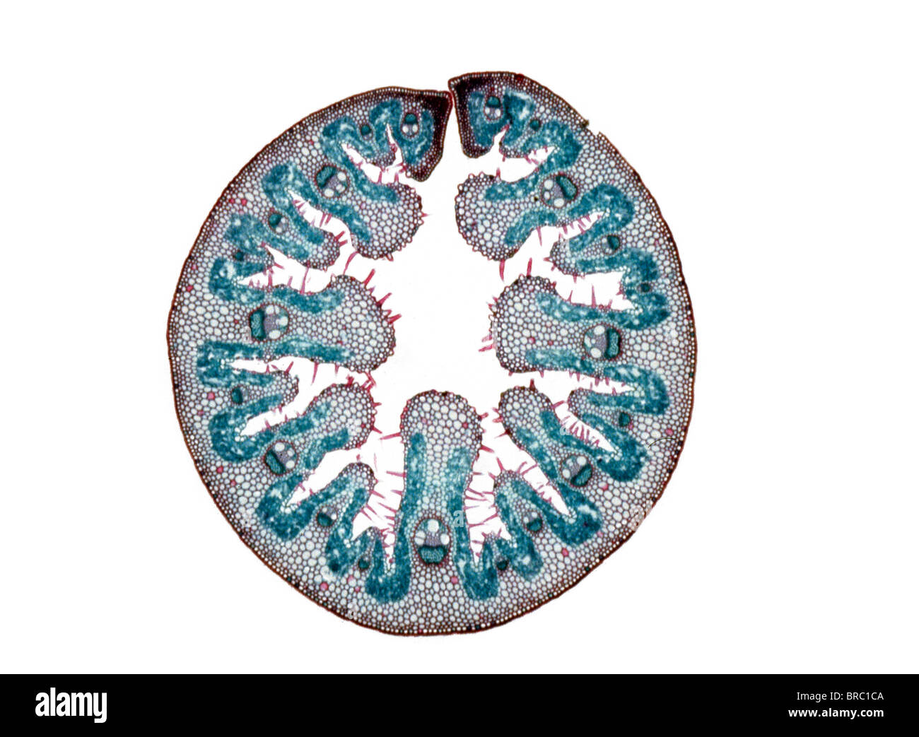 Light Micrograph (LM) of the transverse section of a leaf of Marram Grass (Ammophila sp.), magnification x 15 Stock Photohttps://www.alamy.com/image-license-details/?v=1https://www.alamy.com/stock-photo-light-micrograph-lm-of-the-transverse-section-of-a-leaf-of-marram-31612010.html
RMBRC1CA–Light Micrograph (LM) of the transverse section of a leaf of Marram Grass (Ammophila sp.), magnification x 15
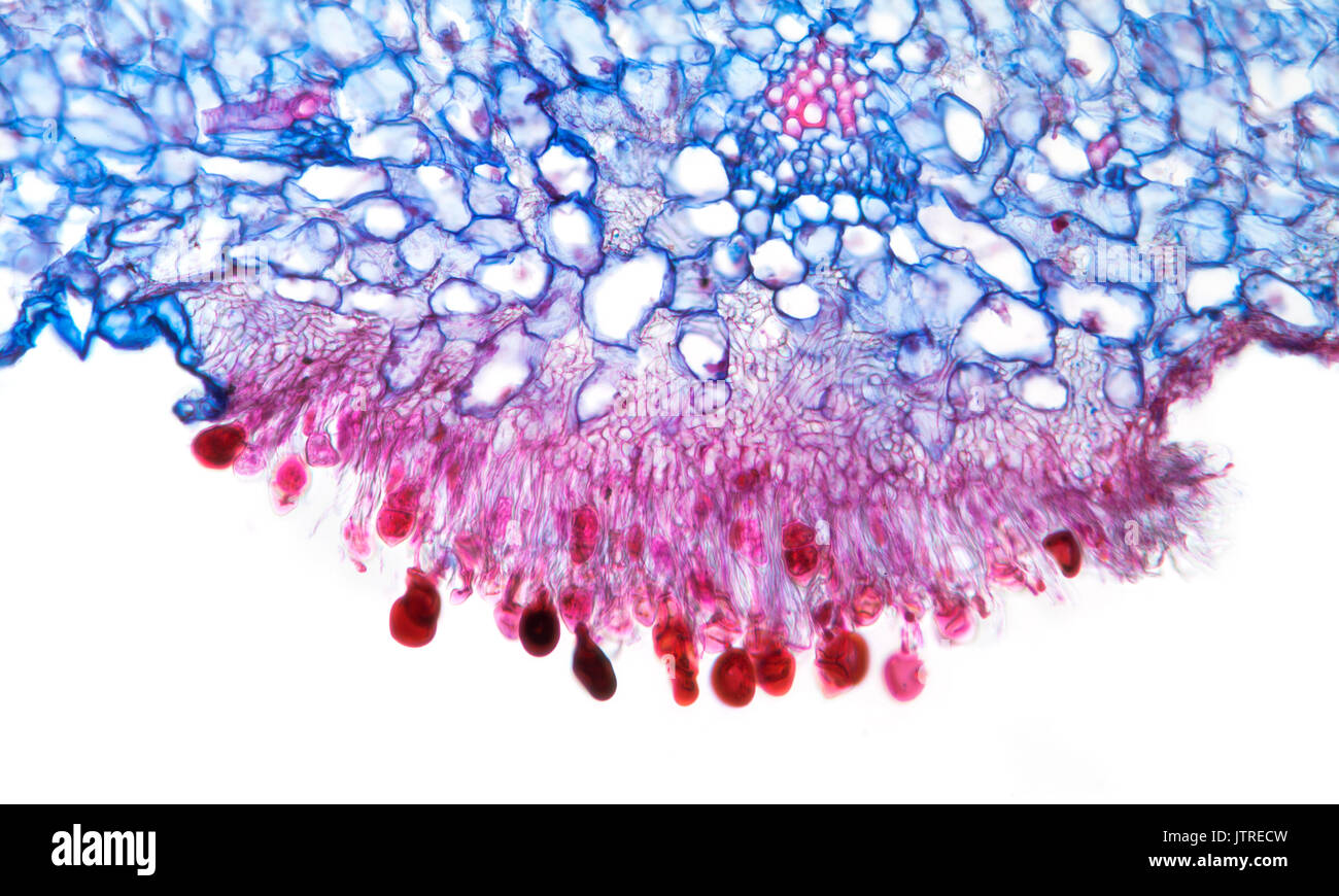 Snapdragon rust, Puccinia antirrhini fungus on antirrhinum leaf, brightfield photomicrograph, TS Stock Photohttps://www.alamy.com/image-license-details/?v=1https://www.alamy.com/snapdragon-rust-puccinia-antirrhini-fungus-on-antirrhinum-leaf-brightfield-image152950921.html
RMJTRECW–Snapdragon rust, Puccinia antirrhini fungus on antirrhinum leaf, brightfield photomicrograph, TS
 biological cell isolated on whithe background microscope 3D Illustration Stock Photohttps://www.alamy.com/image-license-details/?v=1https://www.alamy.com/biological-cell-isolated-on-whithe-background-microscope-3d-illustration-image208953313.html
RFP3XJ29–biological cell isolated on whithe background microscope 3D Illustration
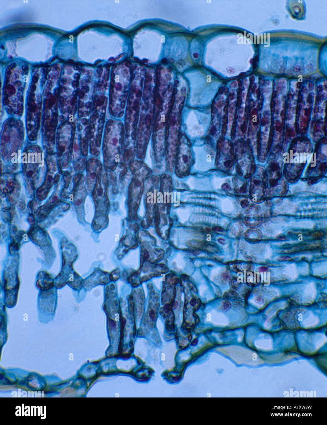 SUNFLOWER LEAF (HELIANTHUS SP.) CROSS SECTION SHOWING UPPER EPIDERMIS, PALASIDE CELLS, SPONGY LAYER, LOWER EPIDERMIS WITH GUARD Stock Photohttps://www.alamy.com/image-license-details/?v=1https://www.alamy.com/stock-photo-sunflower-leaf-helianthus-sp-cross-section-showing-upper-epidermis-10351032.html
RMA1XW8W–SUNFLOWER LEAF (HELIANTHUS SP.) CROSS SECTION SHOWING UPPER EPIDERMIS, PALASIDE CELLS, SPONGY LAYER, LOWER EPIDERMIS WITH GUARD
 Pine Leaf Mesophyll Cells Stock Photohttps://www.alamy.com/image-license-details/?v=1https://www.alamy.com/stock-photo-pine-leaf-mesophyll-cells-134943987.html
RMHRF6C3–Pine Leaf Mesophyll Cells
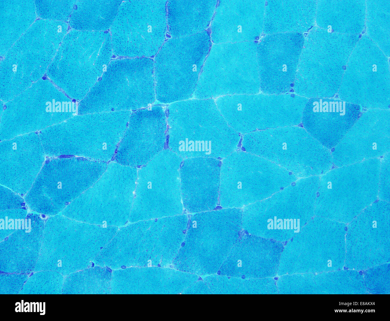 Normal muscle, trichome Stock Photohttps://www.alamy.com/image-license-details/?v=1https://www.alamy.com/stock-photo-normal-muscle-trichome-73993868.html
RFE8AKX4–Normal muscle, trichome
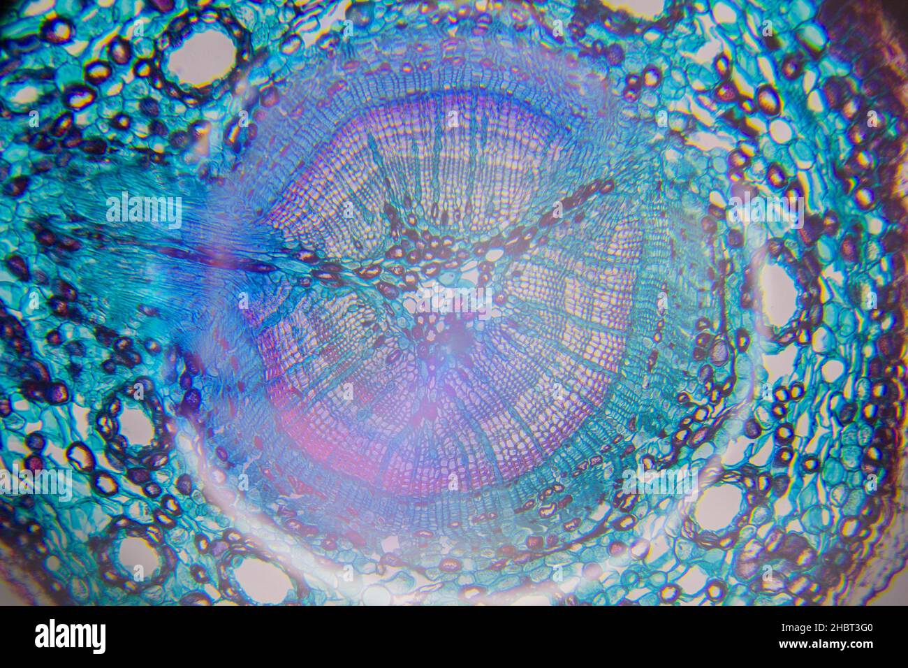 Microscopy of a tree cell Stock Photohttps://www.alamy.com/image-license-details/?v=1https://www.alamy.com/microscopy-of-a-tree-cell-image454760432.html
RF2HBT3G0–Microscopy of a tree cell
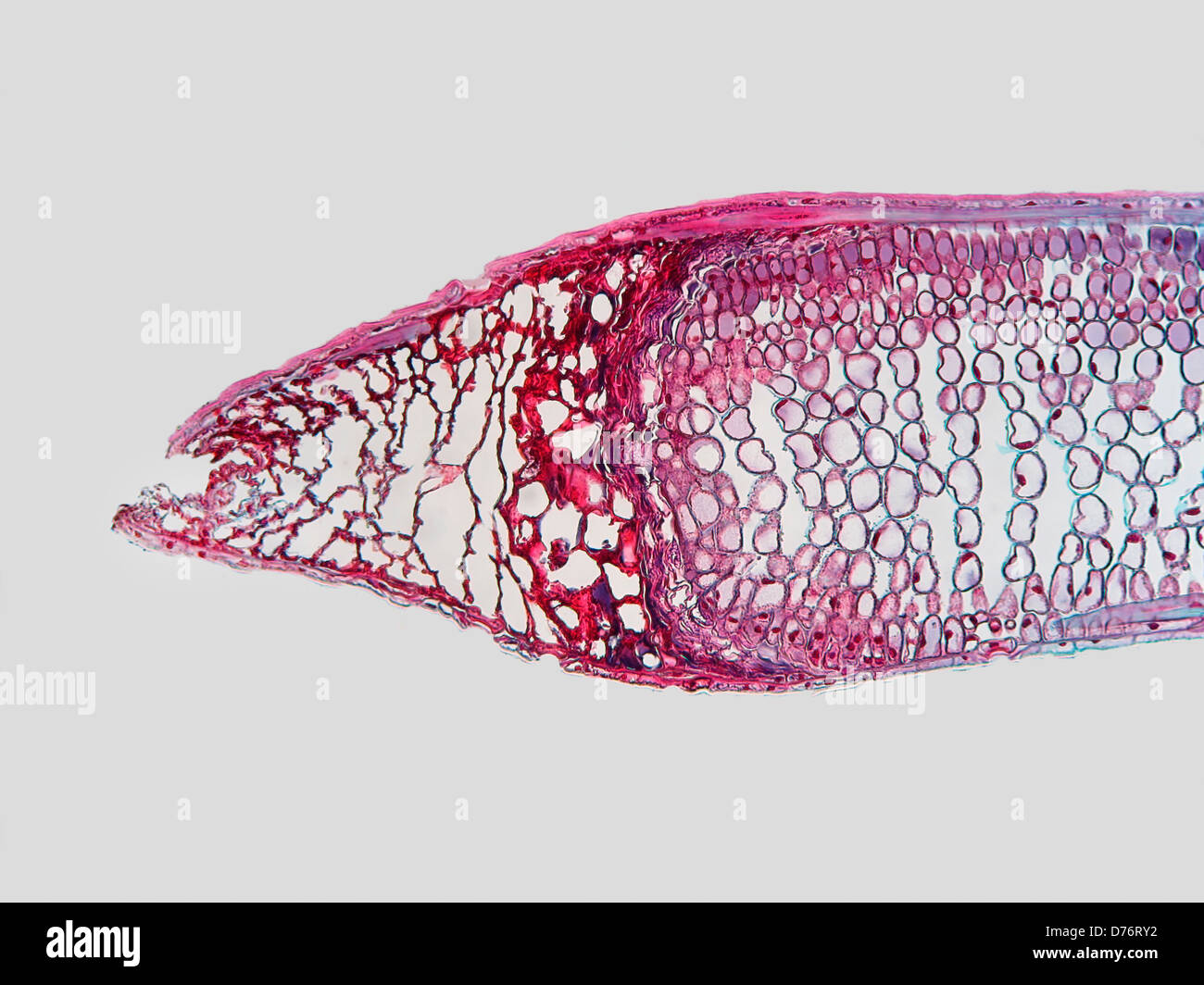 Longitudinal section fir needle that damaged before full growth attained magnification 100x Stock Photohttps://www.alamy.com/image-license-details/?v=1https://www.alamy.com/stock-photo-longitudinal-section-fir-needle-that-damaged-before-full-growth-attained-56084198.html
RMD76RY2–Longitudinal section fir needle that damaged before full growth attained magnification 100x
 Box leaf. Coloured scanning electron micrograph (SEM) of a section through a leaf from the Common Box (Buxus sempervirens). The midrib (midvein) is the continuation of a leaf's stem along the centre of the leaf. At lower centre is a vascular bundle, which consists of xylem and phloem tissues. Xylem transports water and mineral nutrients from the roots throughout the plant and phloem transports carbohydrate and hormones around the plant. The surface (epidermis) of the leaf is covered in a waxy cuticle (top) that helps to prevent water loss. Magnification: x200 when printed 10 centimetres wide. Stock Photohttps://www.alamy.com/image-license-details/?v=1https://www.alamy.com/box-leaf-coloured-scanning-electron-micrograph-sem-of-a-section-through-a-leaf-from-the-common-box-buxus-sempervirens-the-midrib-midvein-is-the-continuation-of-a-leafs-stem-along-the-centre-of-the-leaf-at-lower-centre-is-a-vascular-bundle-which-consists-of-xylem-and-phloem-tissues-xylem-transports-water-and-mineral-nutrients-from-the-roots-throughout-the-plant-and-phloem-transports-carbohydrate-and-hormones-around-the-plant-the-surface-epidermis-of-the-leaf-is-covered-in-a-waxy-cuticle-top-that-helps-to-prevent-water-loss-magnification-x200-when-printed-10-centimetres-wide-image636416261.html
RF2YYB7C5–Box leaf. Coloured scanning electron micrograph (SEM) of a section through a leaf from the Common Box (Buxus sempervirens). The midrib (midvein) is the continuation of a leaf's stem along the centre of the leaf. At lower centre is a vascular bundle, which consists of xylem and phloem tissues. Xylem transports water and mineral nutrients from the roots throughout the plant and phloem transports carbohydrate and hormones around the plant. The surface (epidermis) of the leaf is covered in a waxy cuticle (top) that helps to prevent water loss. Magnification: x200 when printed 10 centimetres wide.
 Stock Photohttps://www.alamy.com/image-license-details/?v=1https://www.alamy.com/-image225982129.html
RFR3JAE9–
 Tea leaf, light micrograph Stock Photohttps://www.alamy.com/image-license-details/?v=1https://www.alamy.com/stock-photo-tea-leaf-light-micrograph-33304993.html
RFBX54T1–Tea leaf, light micrograph
 Microscopy Photography. Flower of Capsella Bursa-pastoris. Stock Photohttps://www.alamy.com/image-license-details/?v=1https://www.alamy.com/microscopy-photography-flower-of-capsella-bursa-pastoris-image182996736.html
RFMHM65M–Microscopy Photography. Flower of Capsella Bursa-pastoris.
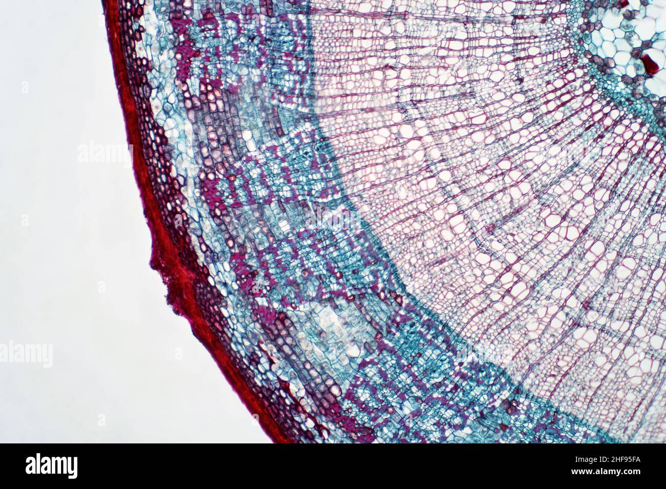 Plant stem, light micrograph Stock Photohttps://www.alamy.com/image-license-details/?v=1https://www.alamy.com/plant-stem-light-micrograph-image456891326.html
RF2HF95FA–Plant stem, light micrograph
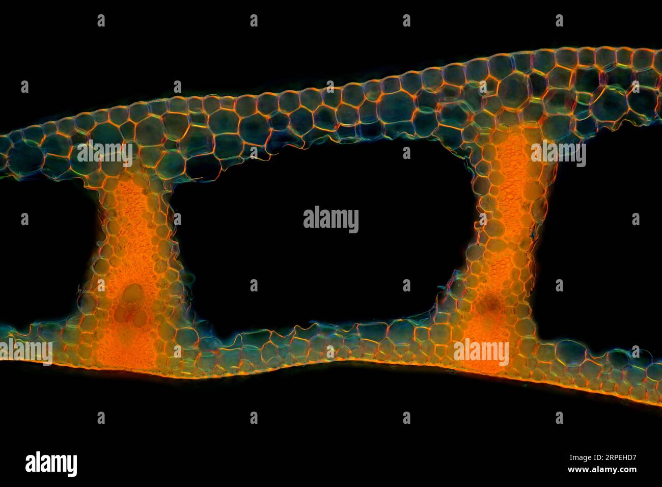 The image presents Carex sp. leaf in transversal cross-section, photographed through the microscope in polarized light at a magnification of 100X Stock Photohttps://www.alamy.com/image-license-details/?v=1https://www.alamy.com/the-image-presents-carex-sp-leaf-in-transversal-cross-section-photographed-through-the-microscope-in-polarized-light-at-a-magnification-of-100x-image564575235.html
RM2RPEHD7–The image presents Carex sp. leaf in transversal cross-section, photographed through the microscope in polarized light at a magnification of 100X
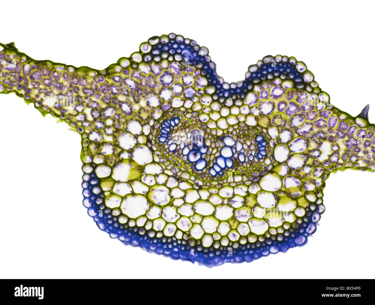 Fern leaf, light micrograph Stock Photohttps://www.alamy.com/image-license-details/?v=1https://www.alamy.com/stock-photo-fern-leaf-light-micrograph-33304936.html
RFBX54P0–Fern leaf, light micrograph
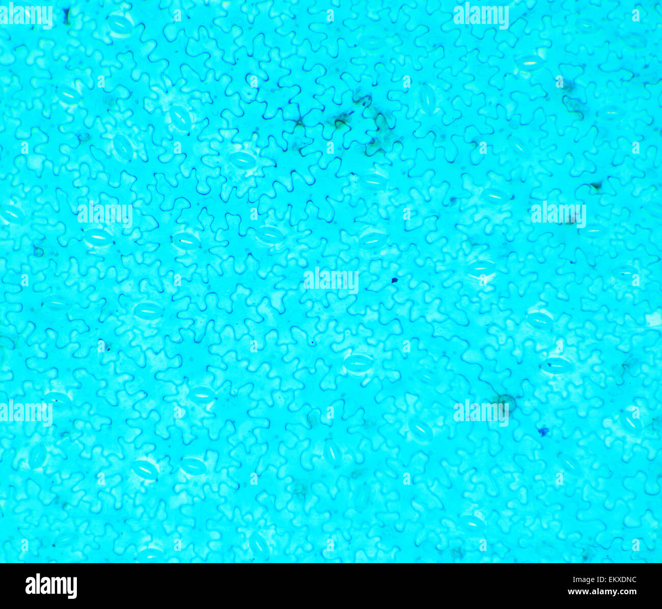 Vicia Dicot leaf under a microscope (Vicia Dicot leaf W.M.), 40x Stock Photohttps://www.alamy.com/image-license-details/?v=1https://www.alamy.com/stock-photo-vicia-dicot-leaf-under-a-microscope-vicia-dicot-leaf-wm-40x-81101480.html
RFEKXDNC–Vicia Dicot leaf under a microscope (Vicia Dicot leaf W.M.), 40x
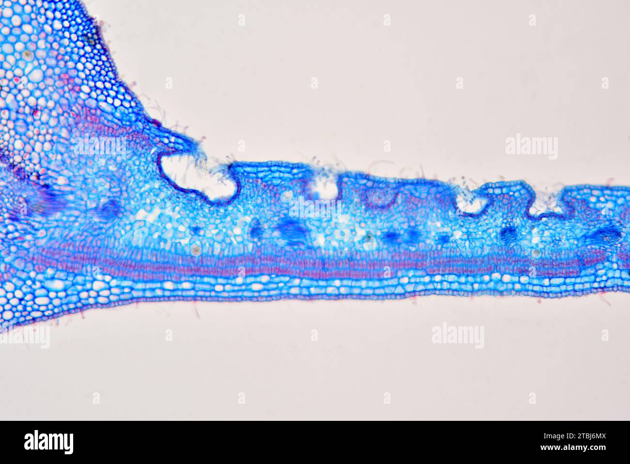 Leaf cross section of Nerium oleander showing epidermis, palisade mesophyll, spongy mesophyll and stomatal crypt. Optical microscope X100. Stock Photohttps://www.alamy.com/image-license-details/?v=1https://www.alamy.com/leaf-cross-section-of-nerium-oleander-showing-epidermis-palisade-mesophyll-spongy-mesophyll-and-stomatal-crypt-optical-microscope-x100-image575103786.html
RF2TBJ6MX–Leaf cross section of Nerium oleander showing epidermis, palisade mesophyll, spongy mesophyll and stomatal crypt. Optical microscope X100.
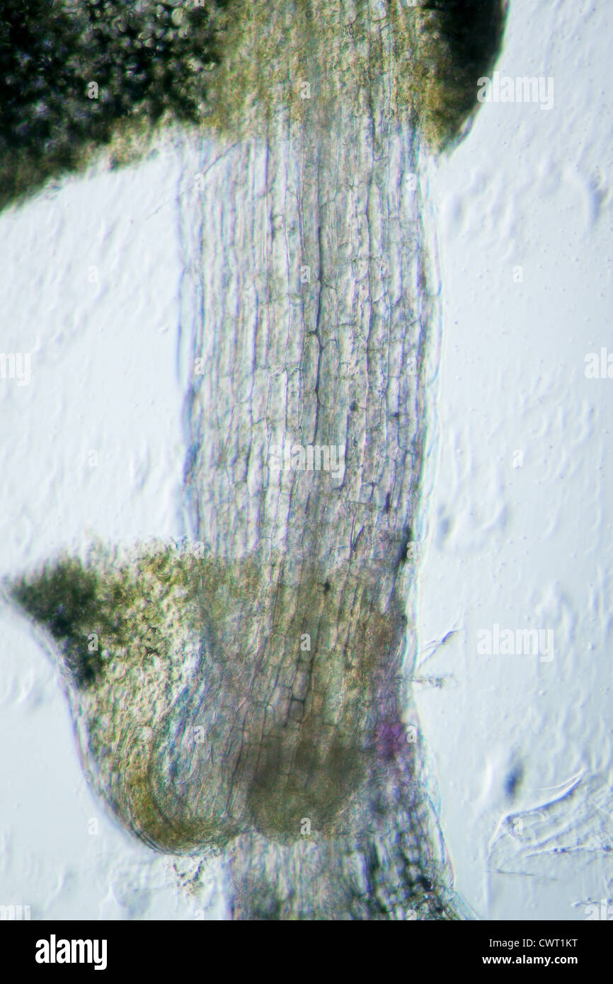 science botany micrograph plant arabidopsis thaliana root tissue micro Stock Photohttps://www.alamy.com/image-license-details/?v=1https://www.alamy.com/stock-photo-science-botany-micrograph-plant-arabidopsis-thaliana-root-tissue-micro-50315324.html
RFCWT1KT–science botany micrograph plant arabidopsis thaliana root tissue micro
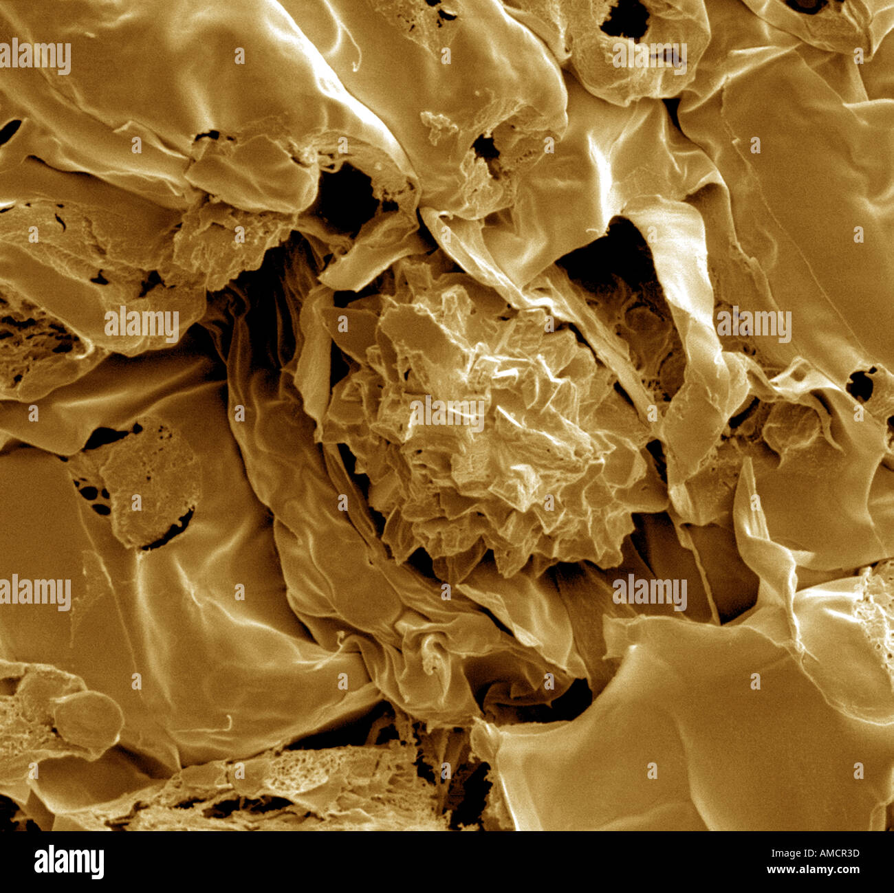 SEM x1000 - Dianthus leaf cross section Stock Photohttps://www.alamy.com/image-license-details/?v=1https://www.alamy.com/sem-x1000-dianthus-leaf-cross-section-image4970300.html
RMAMCR3D–SEM x1000 - Dianthus leaf cross section
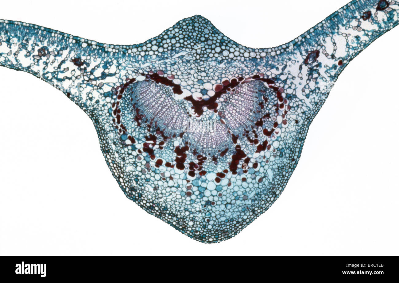 Light Micrograph (LM) of a transverse section of a leaf of a Cherry (Prunus sp.), magnification x 30 Stock Photohttps://www.alamy.com/image-license-details/?v=1https://www.alamy.com/stock-photo-light-micrograph-lm-of-a-transverse-section-of-a-leaf-of-a-cherry-31612067.html
RMBRC1EB–Light Micrograph (LM) of a transverse section of a leaf of a Cherry (Prunus sp.), magnification x 30
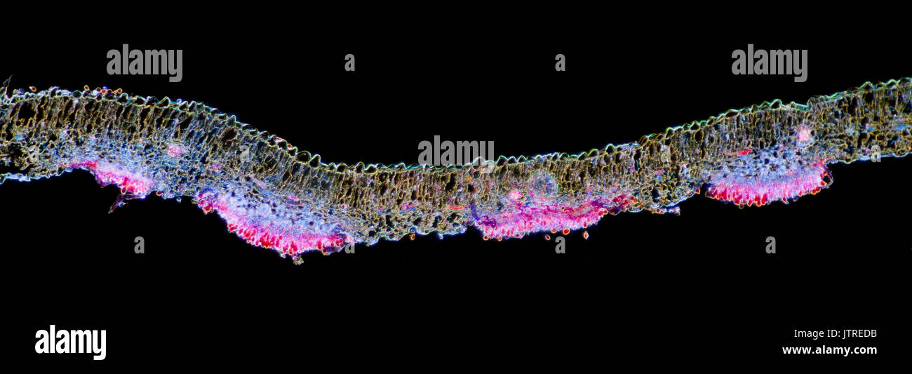 Snapdragon rust, Puccinia antirrhini fungus on antirrhinum leaf, darkfield photomicrograph, TS Stock Photohttps://www.alamy.com/image-license-details/?v=1https://www.alamy.com/snapdragon-rust-puccinia-antirrhini-fungus-on-antirrhinum-leaf-darkfield-image152950935.html
RMJTREDB–Snapdragon rust, Puccinia antirrhini fungus on antirrhinum leaf, darkfield photomicrograph, TS
 biological cell isolated on whithe background microscope 3D Illustration Stock Photohttps://www.alamy.com/image-license-details/?v=1https://www.alamy.com/biological-cell-isolated-on-whithe-background-microscope-3d-illustration-image208953318.html
RFP3XJ2E–biological cell isolated on whithe background microscope 3D Illustration
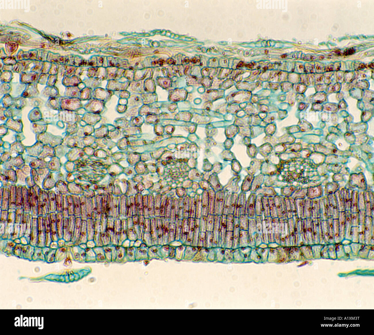 PUMPKIN (CUCURBITA SP.) LEAF CROSS SECTION; UPPER EPIDERMIS, PALISADE LAYER, SPONGY LAYER, VEINS (XYLEM & PHLOEM) LOWER EPIDERMI Stock Photohttps://www.alamy.com/image-license-details/?v=1https://www.alamy.com/stock-photo-pumpkin-cucurbita-sp-leaf-cross-section-upper-epidermis-palisade-layer-10349291.html
RMA1XM3T–PUMPKIN (CUCURBITA SP.) LEAF CROSS SECTION; UPPER EPIDERMIS, PALISADE LAYER, SPONGY LAYER, VEINS (XYLEM & PHLOEM) LOWER EPIDERMI
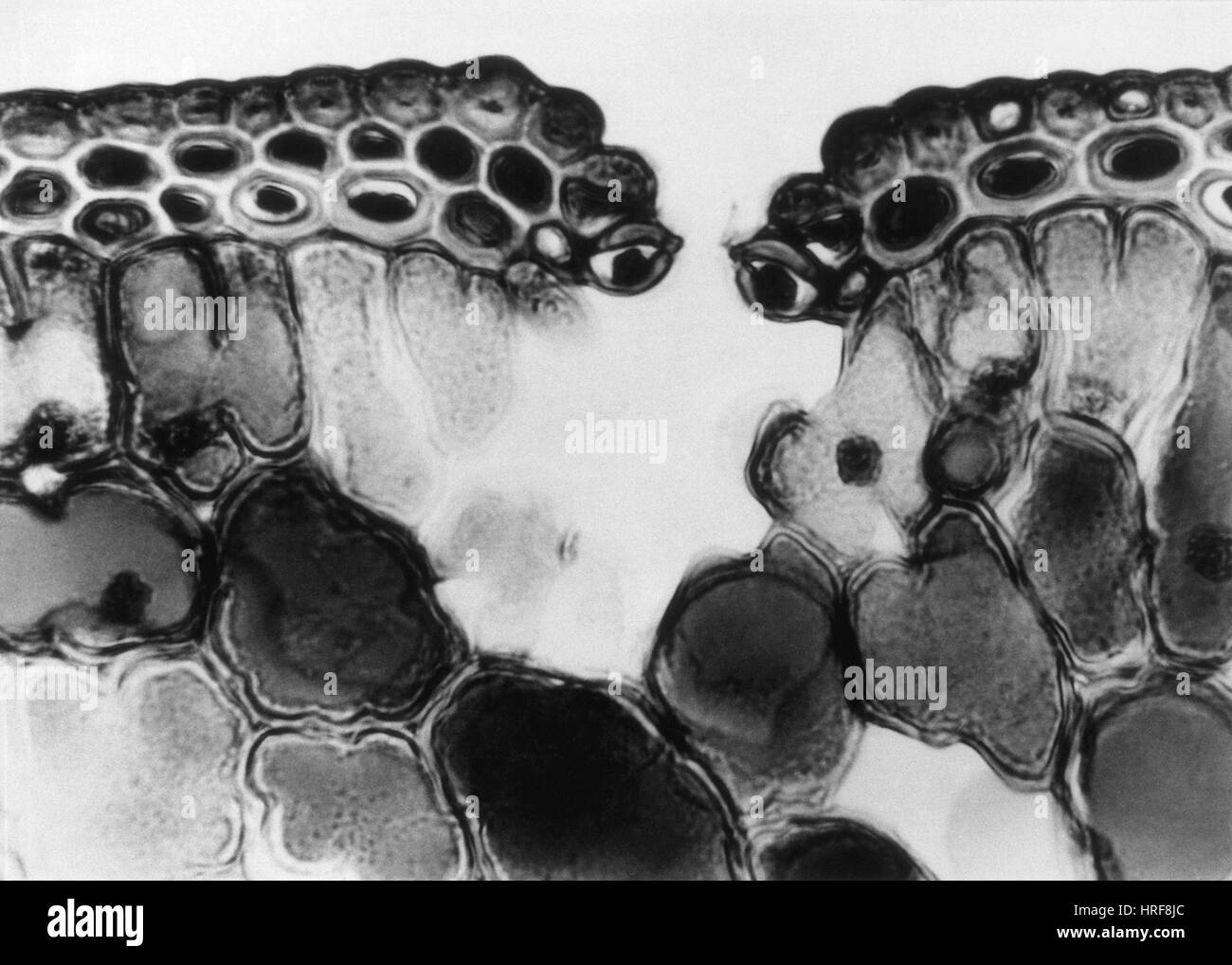 Cuticle and Epidermal Cells in Pine Leaf, LM Stock Photohttps://www.alamy.com/image-license-details/?v=1https://www.alamy.com/stock-photo-cuticle-and-epidermal-cells-in-pine-leaf-lm-134945732.html
RMHRF8JC–Cuticle and Epidermal Cells in Pine Leaf, LM
 Light photomicrograph of Leaf transversal section seen through microscope Stock Photohttps://www.alamy.com/image-license-details/?v=1https://www.alamy.com/stock-photo-light-photomicrograph-of-leaf-transversal-section-seen-through-microscope-75812195.html
RFEB9F6B–Light photomicrograph of Leaf transversal section seen through microscope
 Archive image from page 61 of Cytological methods for the detection,. Cytological methods for the detection, identification, and characterization of orchid viruses and their inclusion bodies cytologicalmetho00kona Year: 1985 -55- Figure 55. Electron micrograph of virus particles distributed within the cytoplasm of CMV- infected Phalaenopsis leaf cells. Section was incubated with CMV antiserum (1/1000 dilution) and labelled with protein A-gold Pd:Plasmodesma. CW:Cell wall. Bar = 350 nm. Figure 56. Same tissue and treatments as in Fig. 55, except that incubation was with normal serum (1/500 Stock Photohttps://www.alamy.com/image-license-details/?v=1https://www.alamy.com/archive-image-from-page-61-of-cytological-methods-for-the-detection-cytological-methods-for-the-detection-identification-and-characterization-of-orchid-viruses-and-their-inclusion-bodies-cytologicalmetho00kona-year-1985-55-figure-55-electron-micrograph-of-virus-particles-distributed-within-the-cytoplasm-of-cmv-infected-phalaenopsis-leaf-cells-section-was-incubated-with-cmv-antiserum-11000-dilution-and-labelled-with-protein-a-gold-pdplasmodesma-cwcell-wall-bar-=-350-nm-figure-56-same-tissue-and-treatments-as-in-fig-55-except-that-incubation-was-with-normal-serum-1500-image259288015.html
RMW1RGE7–Archive image from page 61 of Cytological methods for the detection,. Cytological methods for the detection, identification, and characterization of orchid viruses and their inclusion bodies cytologicalmetho00kona Year: 1985 -55- Figure 55. Electron micrograph of virus particles distributed within the cytoplasm of CMV- infected Phalaenopsis leaf cells. Section was incubated with CMV antiserum (1/1000 dilution) and labelled with protein A-gold Pd:Plasmodesma. CW:Cell wall. Bar = 350 nm. Figure 56. Same tissue and treatments as in Fig. 55, except that incubation was with normal serum (1/500
 Portion longitudinal section pine needle showing stomata on both upper lower surfaces magnification 200x Stock Photohttps://www.alamy.com/image-license-details/?v=1https://www.alamy.com/stock-photo-portion-longitudinal-section-pine-needle-showing-stomata-on-both-upper-56084297.html
RMD76T2H–Portion longitudinal section pine needle showing stomata on both upper lower surfaces magnification 200x
 . Cytological methods for the detection, identification, and characterization of orchid viruses and their inclusion bodies. Orchids; Virus diseases of plants. -53- « * sr >. Figure 51. Electron micrograph of virus aggregations in CyMV-infected Cymbidium leaf cell showing the presence of gold particles on their surface. Section is incubated with CyMV antiserum (1/2000 dilution) and labelled with protein A-gold. Bar = 260 nm, Figure 52. Electron micrograph of virus-like structures in the same antiserum-treated section as in Fig. 51, but showing no specific reaction. Same treatment as in Fig. Stock Photohttps://www.alamy.com/image-license-details/?v=1https://www.alamy.com/cytological-methods-for-the-detection-identification-and-characterization-of-orchid-viruses-and-their-inclusion-bodies-orchids-virus-diseases-of-plants-53-sr-gt-figure-51-electron-micrograph-of-virus-aggregations-in-cymv-infected-cymbidium-leaf-cell-showing-the-presence-of-gold-particles-on-their-surface-section-is-incubated-with-cymv-antiserum-12000-dilution-and-labelled-with-protein-a-gold-bar-=-260-nm-figure-52-electron-micrograph-of-virus-like-structures-in-the-same-antiserum-treated-section-as-in-fig-51-but-showing-no-specific-reaction-same-treatment-as-in-fig-image216162201.html
RMPFK12H–. Cytological methods for the detection, identification, and characterization of orchid viruses and their inclusion bodies. Orchids; Virus diseases of plants. -53- « * sr >. Figure 51. Electron micrograph of virus aggregations in CyMV-infected Cymbidium leaf cell showing the presence of gold particles on their surface. Section is incubated with CyMV antiserum (1/2000 dilution) and labelled with protein A-gold. Bar = 260 nm, Figure 52. Electron micrograph of virus-like structures in the same antiserum-treated section as in Fig. 51, but showing no specific reaction. Same treatment as in Fig.
 Stock Photohttps://www.alamy.com/image-license-details/?v=1https://www.alamy.com/-image225982130.html
RFR3JAEA–
 . Cytological methods for the detection, identification, and characterization of orchid viruses and their inclusion bodies. Orchids; Virus diseases of plants. -55-. Figure 55. Electron micrograph of virus particles distributed within the cytoplasm of CMV- infected Phalaenopsis leaf cells. Section was incubated with CMV antiserum (1/1000 dilution) and labelled with protein A-gold Pd:Plasmodesma. CW:Cell wall. Bar = 350 nm. Figure 56. Same tissue and treatments as in Fig. 55, except that incubation was with normal serum (1/500 dilution). Note the background of nonspecific reaction (arrows). CWrC Stock Photohttps://www.alamy.com/image-license-details/?v=1https://www.alamy.com/cytological-methods-for-the-detection-identification-and-characterization-of-orchid-viruses-and-their-inclusion-bodies-orchids-virus-diseases-of-plants-55-figure-55-electron-micrograph-of-virus-particles-distributed-within-the-cytoplasm-of-cmv-infected-phalaenopsis-leaf-cells-section-was-incubated-with-cmv-antiserum-11000-dilution-and-labelled-with-protein-a-gold-pdplasmodesma-cwcell-wall-bar-=-350-nm-figure-56-same-tissue-and-treatments-as-in-fig-55-except-that-incubation-was-with-normal-serum-1500-dilution-note-the-background-of-nonspecific-reaction-arrows-cwrc-image231779156.html
RMRD2CK0–. Cytological methods for the detection, identification, and characterization of orchid viruses and their inclusion bodies. Orchids; Virus diseases of plants. -55-. Figure 55. Electron micrograph of virus particles distributed within the cytoplasm of CMV- infected Phalaenopsis leaf cells. Section was incubated with CMV antiserum (1/1000 dilution) and labelled with protein A-gold Pd:Plasmodesma. CW:Cell wall. Bar = 350 nm. Figure 56. Same tissue and treatments as in Fig. 55, except that incubation was with normal serum (1/500 dilution). Note the background of nonspecific reaction (arrows). CWrC
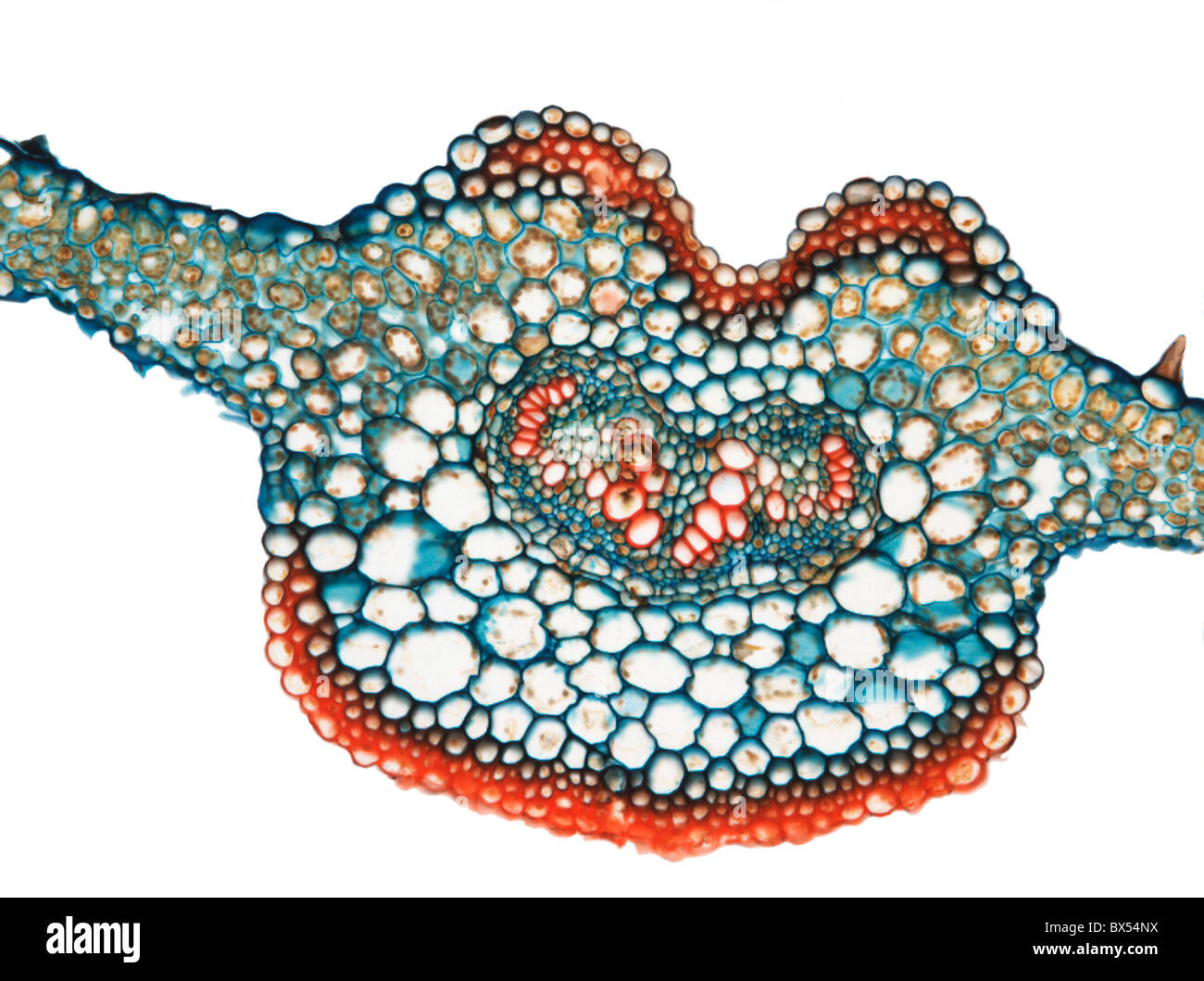 Fern leaf, light micrograph Stock Photohttps://www.alamy.com/image-license-details/?v=1https://www.alamy.com/stock-photo-fern-leaf-light-micrograph-33304934.html
RFBX54NX–Fern leaf, light micrograph
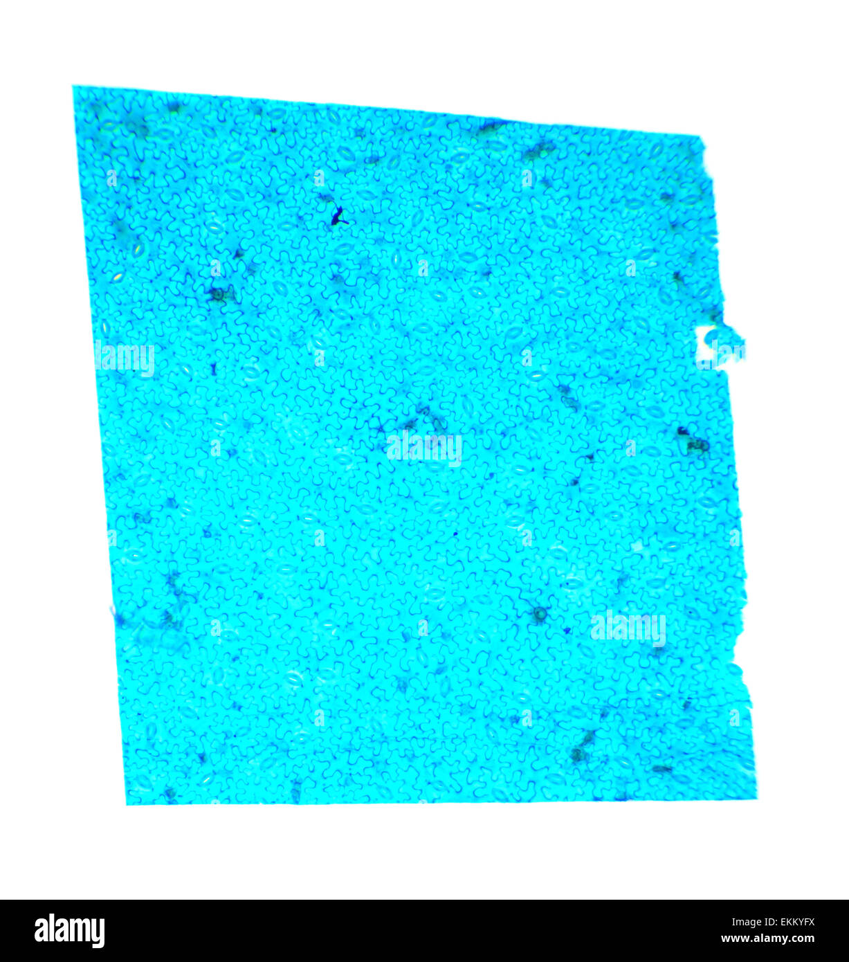 Vicia Dicot leaf under a microscope (Vicia Dicot leaf W.M.), 40x Stock Photohttps://www.alamy.com/image-license-details/?v=1https://www.alamy.com/stock-photo-vicia-dicot-leaf-under-a-microscope-vicia-dicot-leaf-wm-40x-80958638.html
RFEKKYFX–Vicia Dicot leaf under a microscope (Vicia Dicot leaf W.M.), 40x
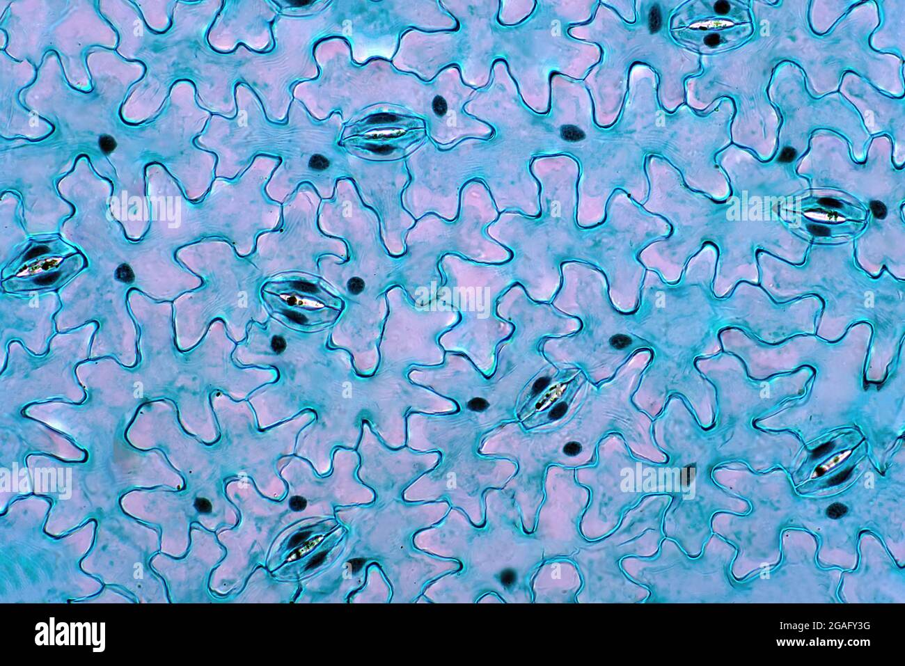 Leaf epidermis, light micrograph Stock Photohttps://www.alamy.com/image-license-details/?v=1https://www.alamy.com/leaf-epidermis-light-micrograph-image436756308.html
RF2GAFY3G–Leaf epidermis, light micrograph
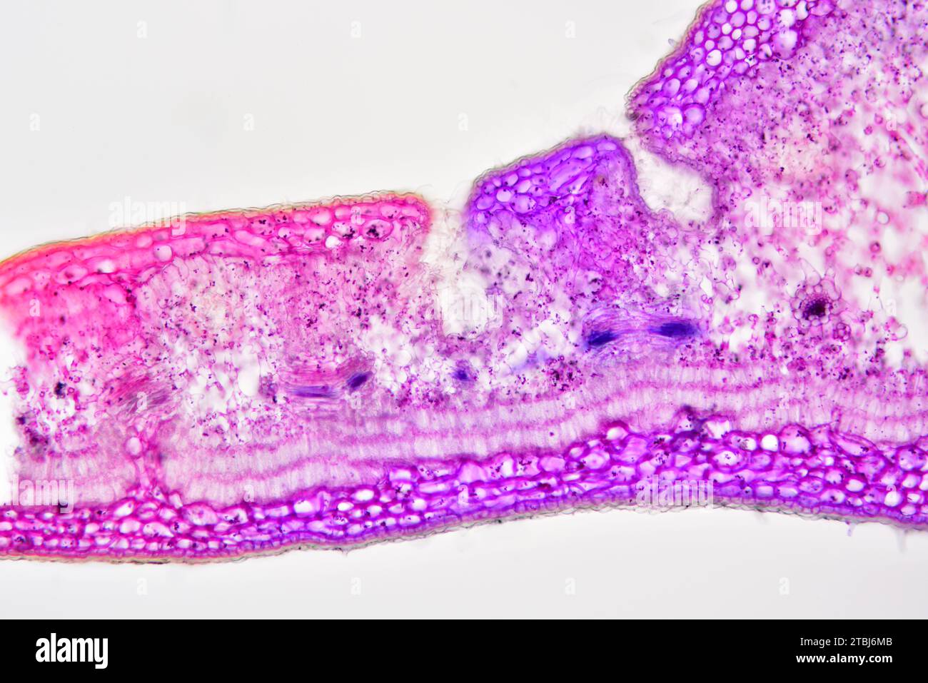 Leaf cross section of Nerium oleander showing epidermis, palisade mesophyll, spongy mesophyll, vascular bundle, phloem, xylem, collenchyma and stomata Stock Photohttps://www.alamy.com/image-license-details/?v=1https://www.alamy.com/leaf-cross-section-of-nerium-oleander-showing-epidermis-palisade-mesophyll-spongy-mesophyll-vascular-bundle-phloem-xylem-collenchyma-and-stomata-image575103771.html
RF2TBJ6MB–Leaf cross section of Nerium oleander showing epidermis, palisade mesophyll, spongy mesophyll, vascular bundle, phloem, xylem, collenchyma and stomata
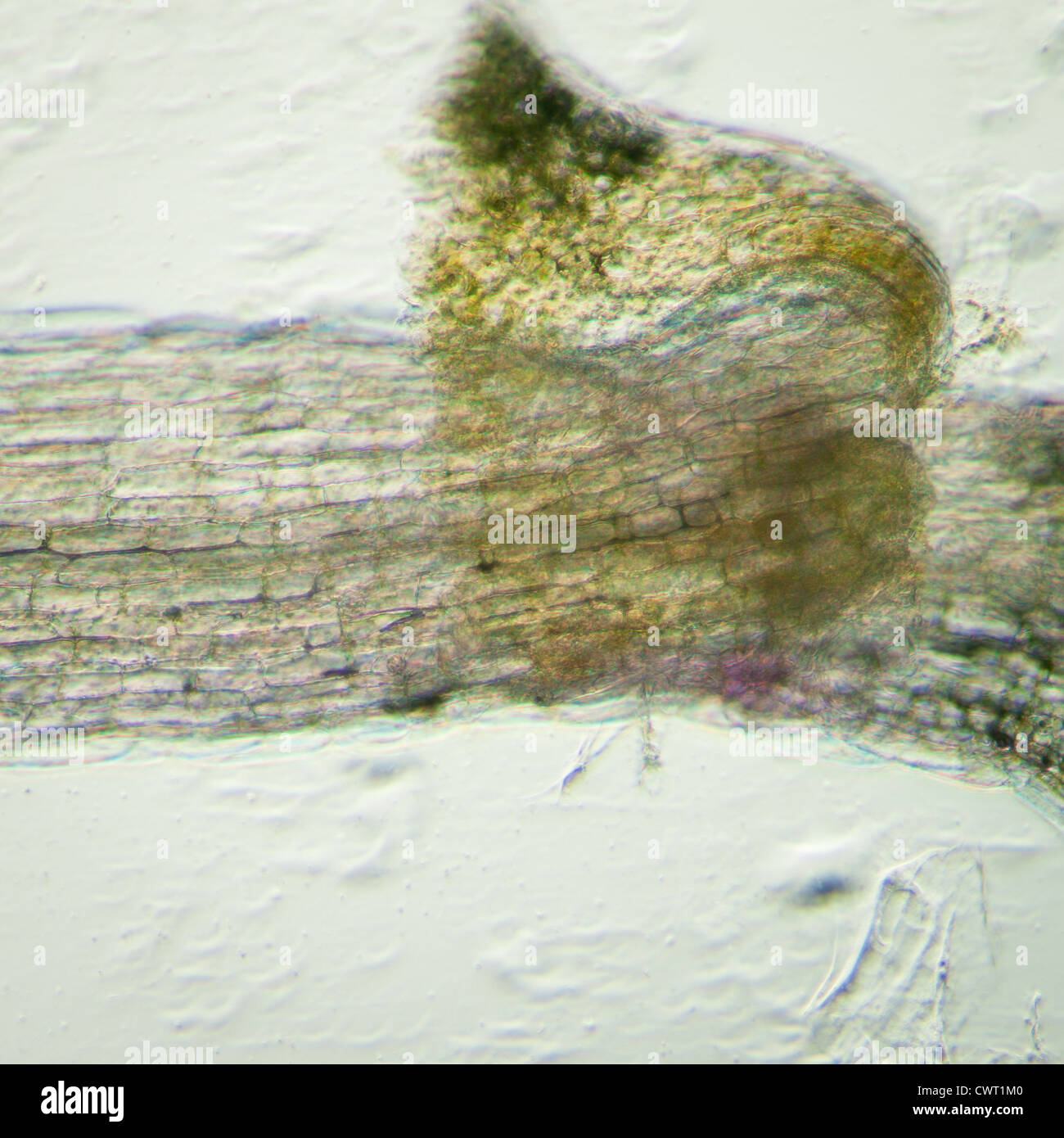 science botany micrograph plant arabidopsis thaliana root tissue micro Stock Photohttps://www.alamy.com/image-license-details/?v=1https://www.alamy.com/stock-photo-science-botany-micrograph-plant-arabidopsis-thaliana-root-tissue-micro-50315328.html
RFCWT1M0–science botany micrograph plant arabidopsis thaliana root tissue micro
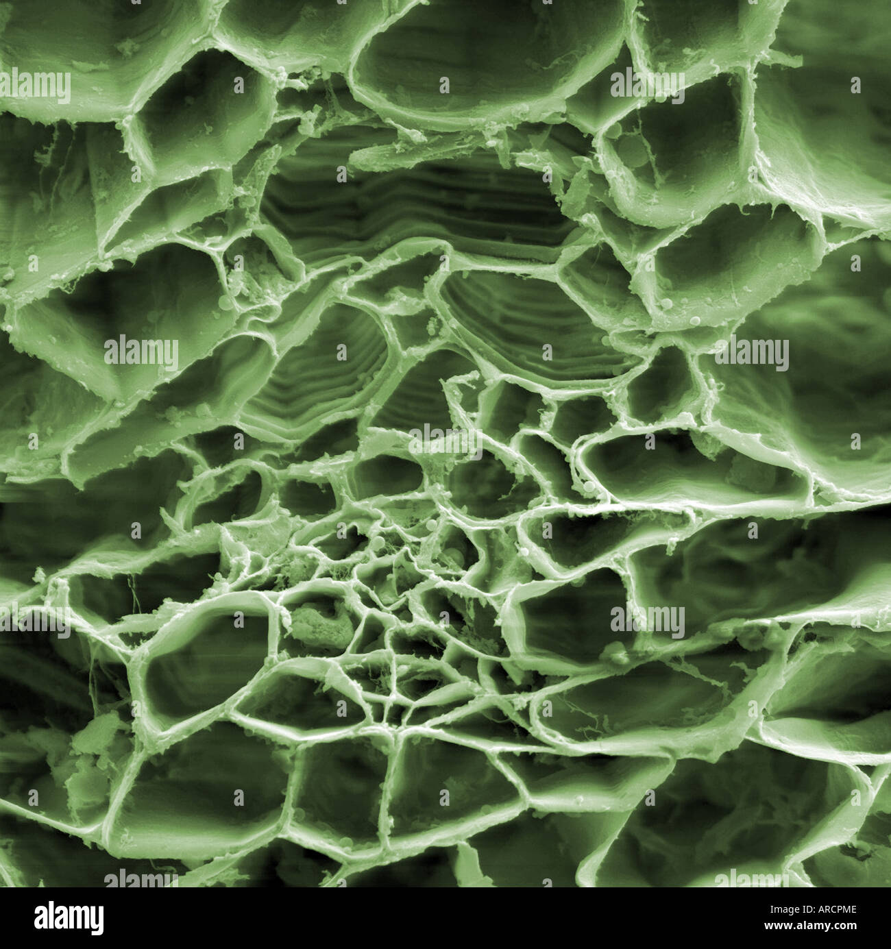 High magnification scanning electron microscope image of a tradescantia (Virginia spiderwort) Stock Photohttps://www.alamy.com/image-license-details/?v=1https://www.alamy.com/high-magnification-scanning-electron-microscope-image-of-a-tradescantia-image9150669.html
RMARCPME–High magnification scanning electron microscope image of a tradescantia (Virginia spiderwort)
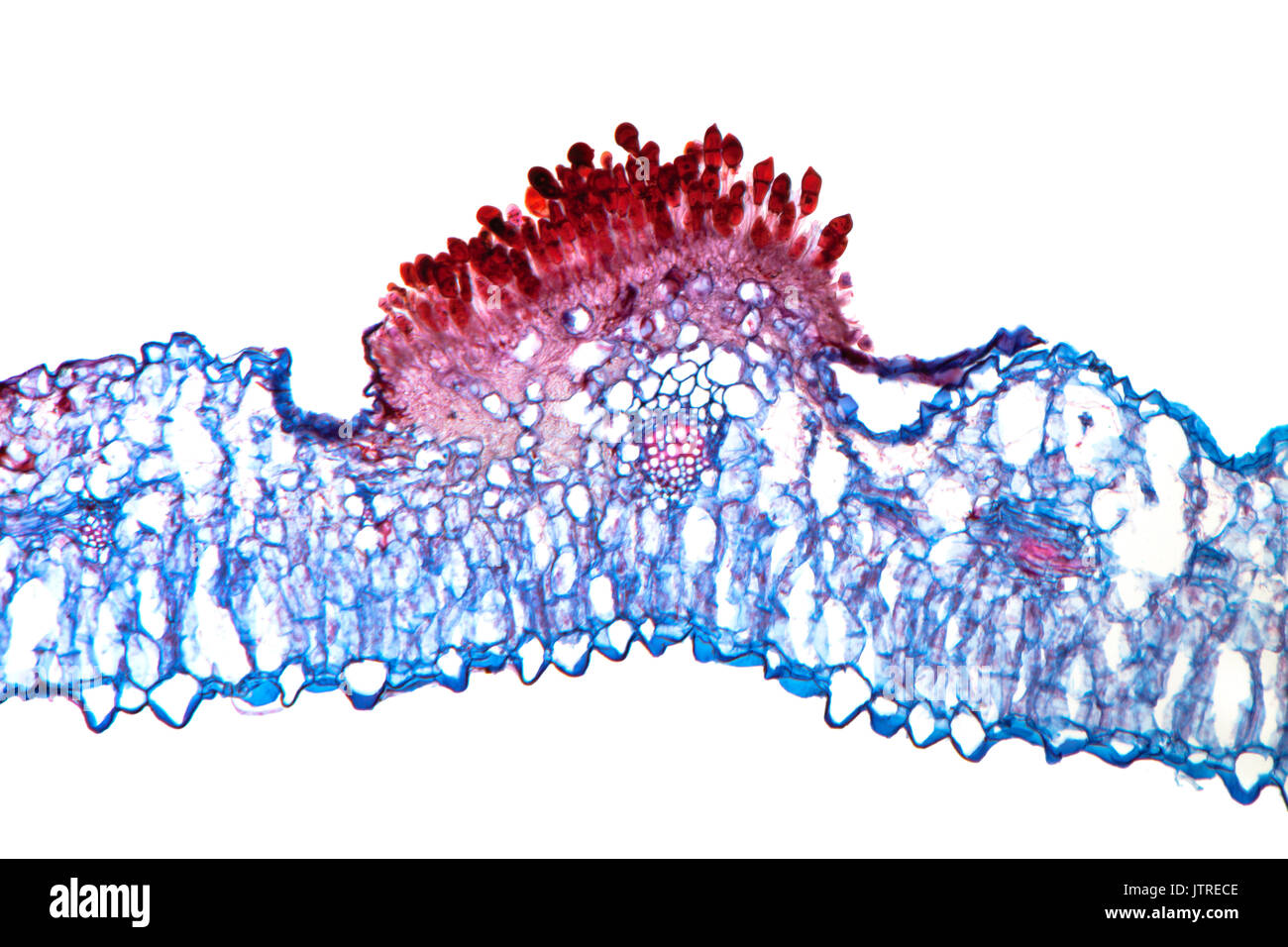 Snapdragon rust, Puccinia antirrhini fungus on antirrhinum leaf, brightfield photomicrograph, TS Stock Photohttps://www.alamy.com/image-license-details/?v=1https://www.alamy.com/snapdragon-rust-puccinia-antirrhini-fungus-on-antirrhinum-leaf-brightfield-image152950910.html
RMJTRECE–Snapdragon rust, Puccinia antirrhini fungus on antirrhinum leaf, brightfield photomicrograph, TS
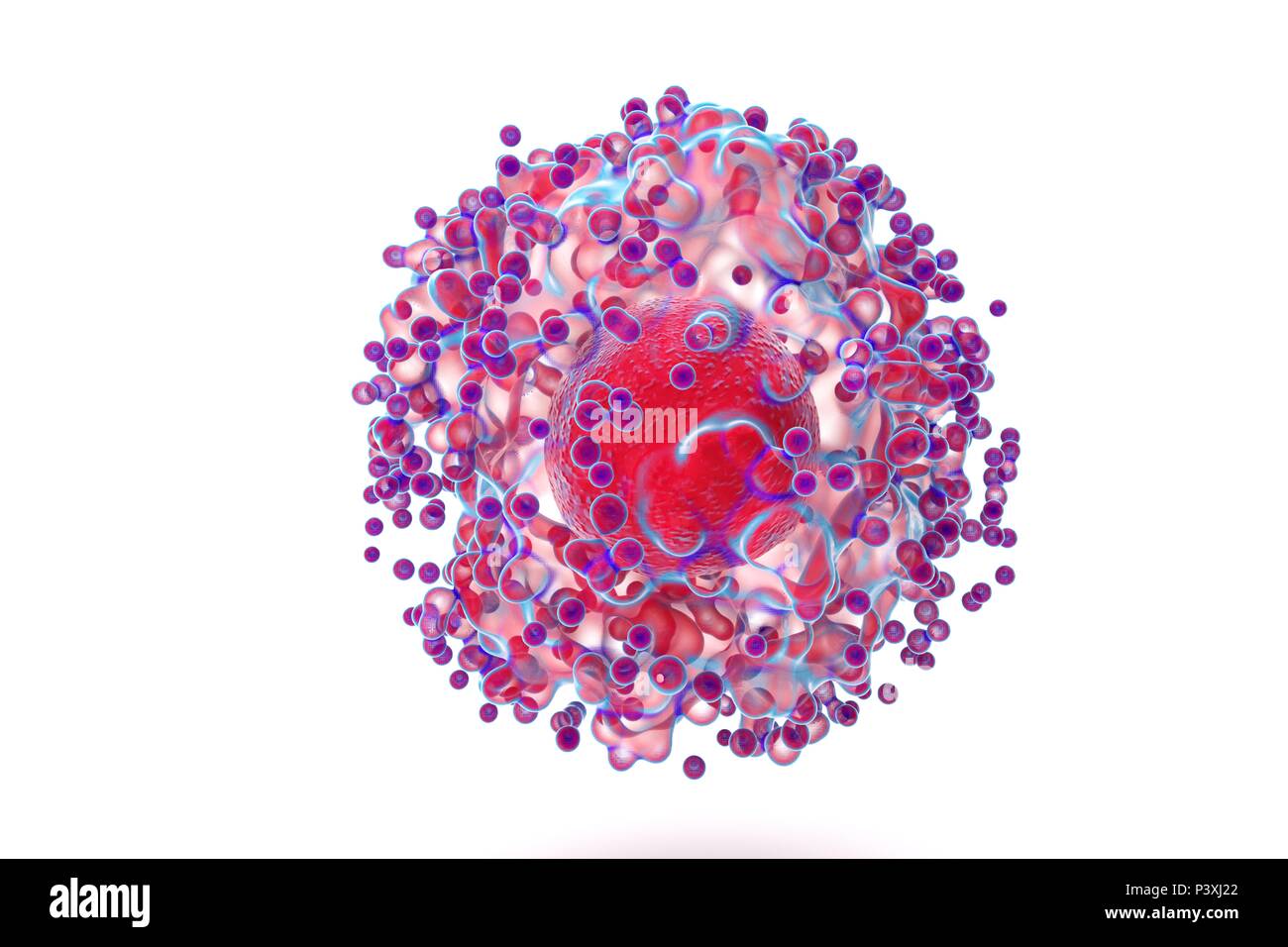 biological cell isolated on whithe background microscope 3D Illustration Stock Photohttps://www.alamy.com/image-license-details/?v=1https://www.alamy.com/biological-cell-isolated-on-whithe-background-microscope-3d-illustration-image208953306.html
RFP3XJ22–biological cell isolated on whithe background microscope 3D Illustration
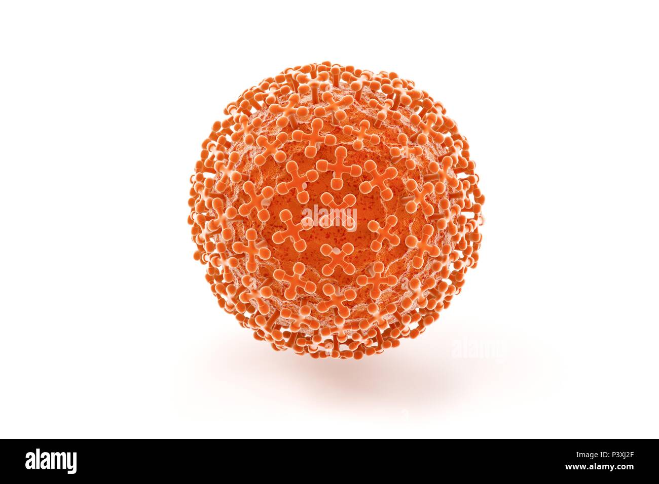 biological cell isolated on whithe background microscope 3D Illustration Stock Photohttps://www.alamy.com/image-license-details/?v=1https://www.alamy.com/biological-cell-isolated-on-whithe-background-microscope-3d-illustration-image208953319.html
RFP3XJ2F–biological cell isolated on whithe background microscope 3D Illustration
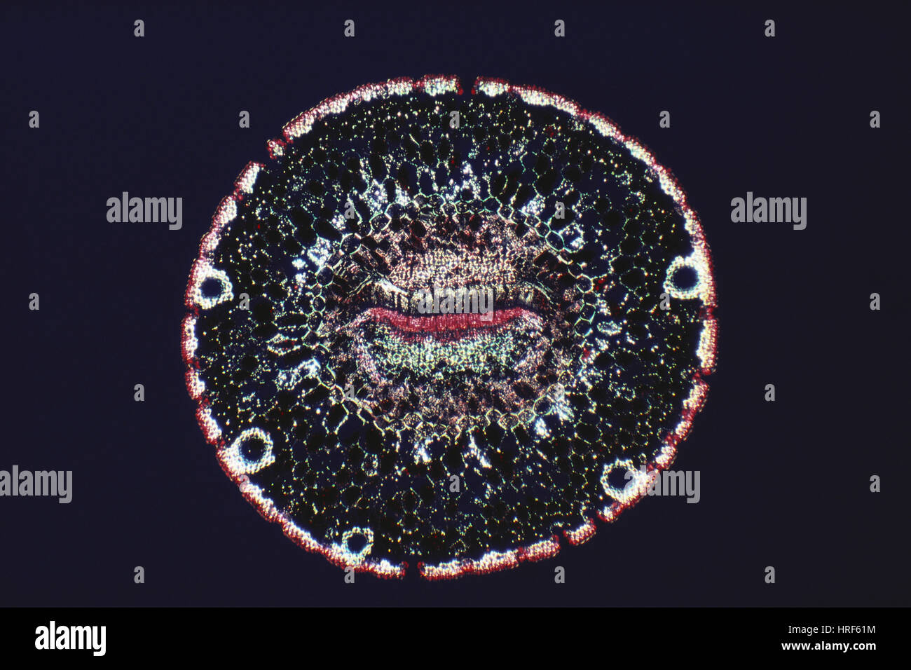 Pine Leaf Stomata Stock Photohttps://www.alamy.com/image-license-details/?v=1https://www.alamy.com/stock-photo-pine-leaf-stomata-134943696.html
RMHRF61M–Pine Leaf Stomata
 Light photomicrograph of Leaf transversal section seen through microscope Stock Photohttps://www.alamy.com/image-license-details/?v=1https://www.alamy.com/light-photomicrograph-of-leaf-transversal-section-seen-through-microscope-image179104200.html
RFMBAW6G–Light photomicrograph of Leaf transversal section seen through microscope
 Longitudinal section portion fir Abies sp needle structural elements especially stomata are shown magnification 100x Stock Photohttps://www.alamy.com/image-license-details/?v=1https://www.alamy.com/stock-photo-longitudinal-section-portion-fir-abies-sp-needle-structural-elements-56084200.html
RMD76RY4–Longitudinal section portion fir Abies sp needle structural elements especially stomata are shown magnification 100x
 Archive image from page 59 of Cytological methods for the detection,. Cytological methods for the detection, identification, and characterization of orchid viruses and their inclusion bodies cytologicalmetho00kona Year: 1985 -53- « sr > Figure 51. Electron micrograph of virus aggregations in CyMV-infected Cymbidium leaf cell showing the presence of gold particles on their surface. Section is incubated with CyMV antiserum (1/2000 dilution) and labelled with protein A-gold. Bar = 260 nm, Figure 52. Electron micrograph of virus-like structures in the same antiserum-treated section as in F Stock Photohttps://www.alamy.com/image-license-details/?v=1https://www.alamy.com/archive-image-from-page-59-of-cytological-methods-for-the-detection-cytological-methods-for-the-detection-identification-and-characterization-of-orchid-viruses-and-their-inclusion-bodies-cytologicalmetho00kona-year-1985-53-sr-gt-figure-51-electron-micrograph-of-virus-aggregations-in-cymv-infected-cymbidium-leaf-cell-showing-the-presence-of-gold-particles-on-their-surface-section-is-incubated-with-cymv-antiserum-12000-dilution-and-labelled-with-protein-a-gold-bar-=-260-nm-figure-52-electron-micrograph-of-virus-like-structures-in-the-same-antiserum-treated-section-as-in-f-image259287450.html
RMW1RFP2–Archive image from page 59 of Cytological methods for the detection,. Cytological methods for the detection, identification, and characterization of orchid viruses and their inclusion bodies cytologicalmetho00kona Year: 1985 -53- « sr > Figure 51. Electron micrograph of virus aggregations in CyMV-infected Cymbidium leaf cell showing the presence of gold particles on their surface. Section is incubated with CyMV antiserum (1/2000 dilution) and labelled with protein A-gold. Bar = 260 nm, Figure 52. Electron micrograph of virus-like structures in the same antiserum-treated section as in F
 Stock Photohttps://www.alamy.com/image-license-details/?v=1https://www.alamy.com/-image225982099.html
RFR3JAD7–
 . Cytological methods for the detection, identification, and characterization of orchid viruses and their inclusion bodies. Orchids; Virus diseases of plants. -55-. Figure 55. Electron micrograph of virus particles distributed within the cytoplasm of CMV- infected Phalaenopsis leaf cells. Section was incubated with CMV antiserum (1/1000 dilution) and labelled with protein A-gold Pd:Plasmodesma. CW:Cell wall. Bar = 350 nm. Figure 56. Same tissue and treatments as in Fig. 55, except that incubation was with normal serum (1/500 dilution). Note the background of nonspecific reaction (arrows). CWrC Stock Photohttps://www.alamy.com/image-license-details/?v=1https://www.alamy.com/cytological-methods-for-the-detection-identification-and-characterization-of-orchid-viruses-and-their-inclusion-bodies-orchids-virus-diseases-of-plants-55-figure-55-electron-micrograph-of-virus-particles-distributed-within-the-cytoplasm-of-cmv-infected-phalaenopsis-leaf-cells-section-was-incubated-with-cmv-antiserum-11000-dilution-and-labelled-with-protein-a-gold-pdplasmodesma-cwcell-wall-bar-=-350-nm-figure-56-same-tissue-and-treatments-as-in-fig-55-except-that-incubation-was-with-normal-serum-1500-dilution-note-the-background-of-nonspecific-reaction-arrows-cwrc-image216162181.html
RMPFK11W–. Cytological methods for the detection, identification, and characterization of orchid viruses and their inclusion bodies. Orchids; Virus diseases of plants. -55-. Figure 55. Electron micrograph of virus particles distributed within the cytoplasm of CMV- infected Phalaenopsis leaf cells. Section was incubated with CMV antiserum (1/1000 dilution) and labelled with protein A-gold Pd:Plasmodesma. CW:Cell wall. Bar = 350 nm. Figure 56. Same tissue and treatments as in Fig. 55, except that incubation was with normal serum (1/500 dilution). Note the background of nonspecific reaction (arrows). CWrC
 . Cytological methods for the detection, identification, and characterization of orchid viruses and their inclusion bodies. Orchids; Virus diseases of plants. -53- « * sr >. Figure 51. Electron micrograph of virus aggregations in CyMV-infected Cymbidium leaf cell showing the presence of gold particles on their surface. Section is incubated with CyMV antiserum (1/2000 dilution) and labelled with protein A-gold. Bar = 260 nm, Figure 52. Electron micrograph of virus-like structures in the same antiserum-treated section as in Fig. 51, but showing no specific reaction. Same treatment as in Fig. Stock Photohttps://www.alamy.com/image-license-details/?v=1https://www.alamy.com/cytological-methods-for-the-detection-identification-and-characterization-of-orchid-viruses-and-their-inclusion-bodies-orchids-virus-diseases-of-plants-53-sr-gt-figure-51-electron-micrograph-of-virus-aggregations-in-cymv-infected-cymbidium-leaf-cell-showing-the-presence-of-gold-particles-on-their-surface-section-is-incubated-with-cymv-antiserum-12000-dilution-and-labelled-with-protein-a-gold-bar-=-260-nm-figure-52-electron-micrograph-of-virus-like-structures-in-the-same-antiserum-treated-section-as-in-fig-51-but-showing-no-specific-reaction-same-treatment-as-in-fig-image231779182.html
RMRD2CKX–. Cytological methods for the detection, identification, and characterization of orchid viruses and their inclusion bodies. Orchids; Virus diseases of plants. -53- « * sr >. Figure 51. Electron micrograph of virus aggregations in CyMV-infected Cymbidium leaf cell showing the presence of gold particles on their surface. Section is incubated with CyMV antiserum (1/2000 dilution) and labelled with protein A-gold. Bar = 260 nm, Figure 52. Electron micrograph of virus-like structures in the same antiserum-treated section as in Fig. 51, but showing no specific reaction. Same treatment as in Fig.
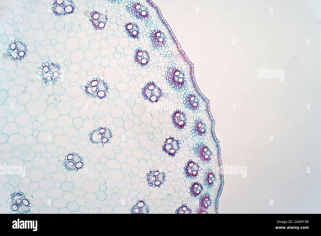 Cross section of monocot stem, light micrograph Stock Photohttps://www.alamy.com/image-license-details/?v=1https://www.alamy.com/cross-section-of-monocot-stem-light-micrograph-image436756303.html
RF2GAFY3B–Cross section of monocot stem, light micrograph
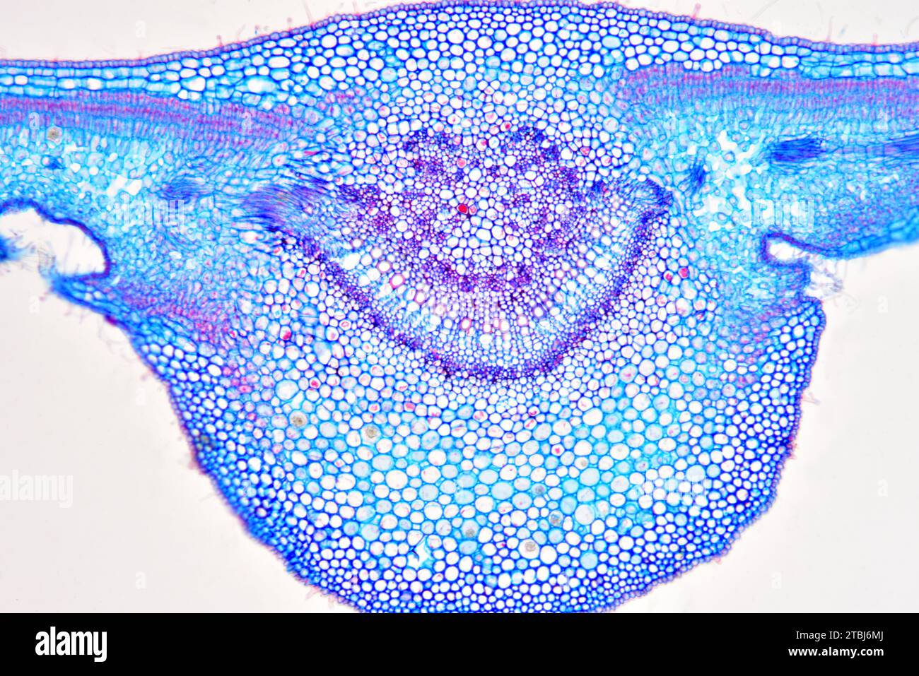 Leaf cross section of Nerium oleander showing epidermis, palisade mesophyll, spongy mesophyll, vascular bundle, phloem, xylem, collenchyma and stomata Stock Photohttps://www.alamy.com/image-license-details/?v=1https://www.alamy.com/leaf-cross-section-of-nerium-oleander-showing-epidermis-palisade-mesophyll-spongy-mesophyll-vascular-bundle-phloem-xylem-collenchyma-and-stomata-image575103778.html
RF2TBJ6MJ–Leaf cross section of Nerium oleander showing epidermis, palisade mesophyll, spongy mesophyll, vascular bundle, phloem, xylem, collenchyma and stomata
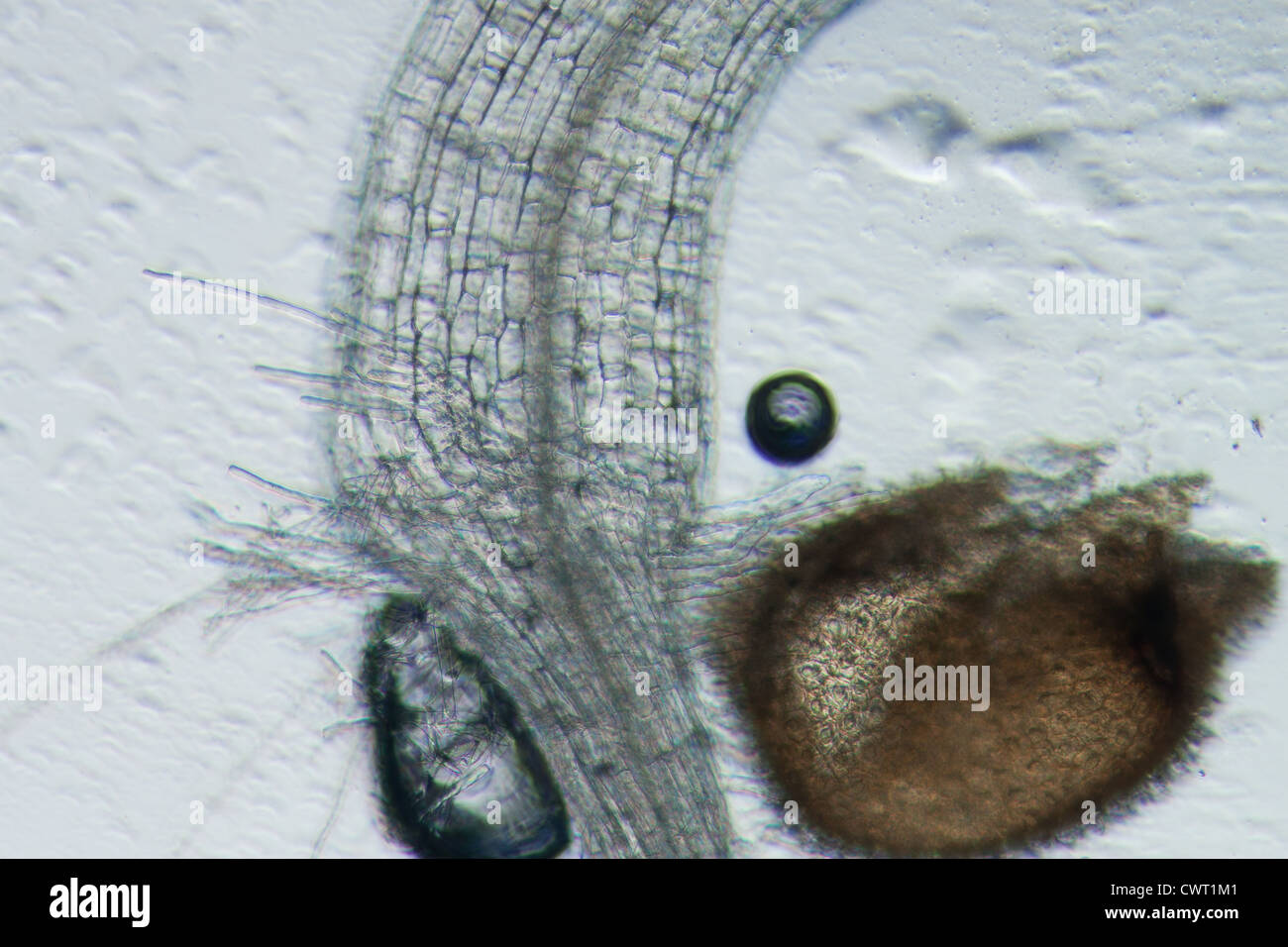 science botany micrograph plant arabidopsis thaliana root tissue micro Stock Photohttps://www.alamy.com/image-license-details/?v=1https://www.alamy.com/stock-photo-science-botany-micrograph-plant-arabidopsis-thaliana-root-tissue-micro-50315329.html
RFCWT1M1–science botany micrograph plant arabidopsis thaliana root tissue micro
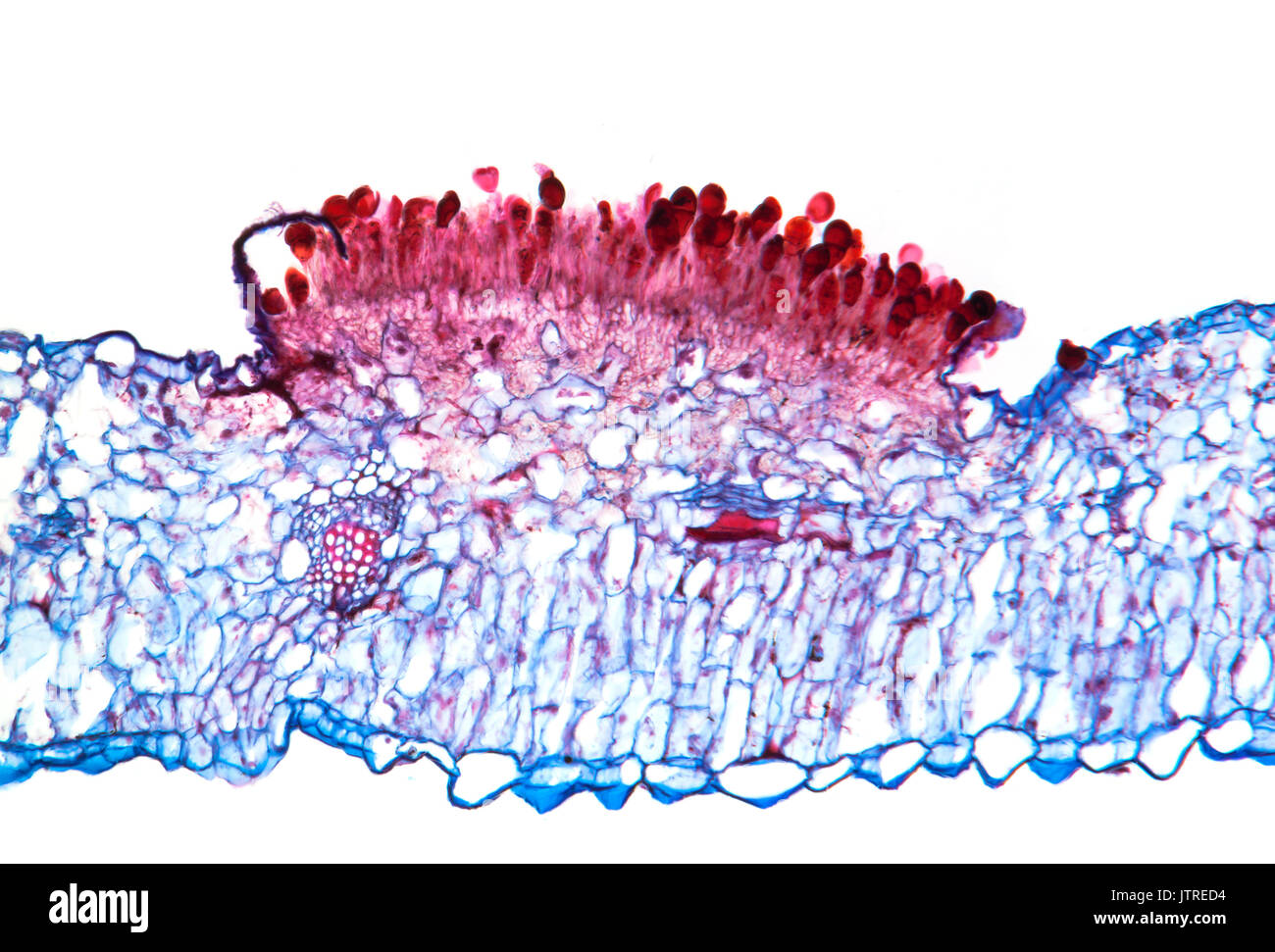 Snapdragon rust, Puccinia antirrhini fungus on antirrhinum leaf, brightfield photomicrograph, TS Stock Photohttps://www.alamy.com/image-license-details/?v=1https://www.alamy.com/snapdragon-rust-puccinia-antirrhini-fungus-on-antirrhinum-leaf-brightfield-image152950928.html
RMJTRED4–Snapdragon rust, Puccinia antirrhini fungus on antirrhinum leaf, brightfield photomicrograph, TS
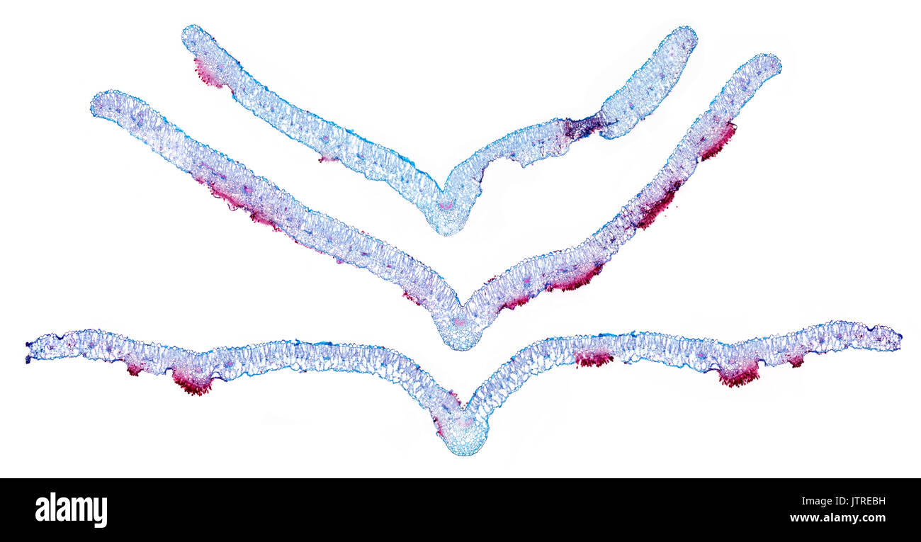 Snapdragon rust, Puccinia antirrhini fungus on antirrhinum leaf, brightfield photomicrograph, TS Stock Photohttps://www.alamy.com/image-license-details/?v=1https://www.alamy.com/snapdragon-rust-puccinia-antirrhini-fungus-on-antirrhinum-leaf-brightfield-image152950885.html
RMJTREBH–Snapdragon rust, Puccinia antirrhini fungus on antirrhinum leaf, brightfield photomicrograph, TS
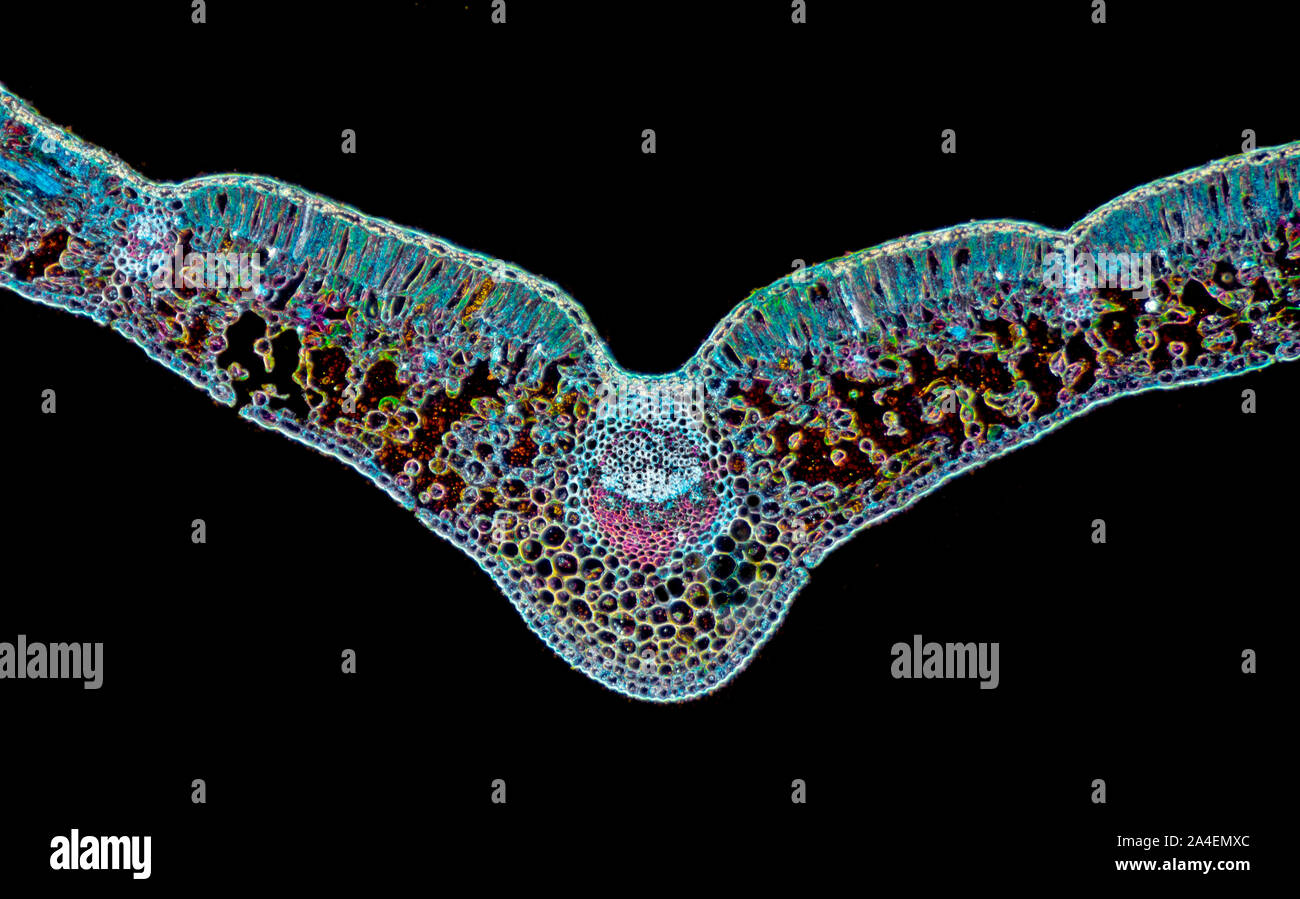 Helleborus niger, Christmas Rose, stained leaf section, TS. Darkfield photomicrograph Stock Photohttps://www.alamy.com/image-license-details/?v=1https://www.alamy.com/helleborus-niger-christmas-rose-stained-leaf-section-ts-darkfield-photomicrograph-image329779364.html
RM2A4EMXC–Helleborus niger, Christmas Rose, stained leaf section, TS. Darkfield photomicrograph
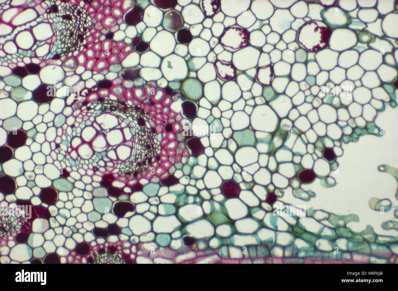 LM of Fig Leaf Stock Photohttps://www.alamy.com/image-license-details/?v=1https://www.alamy.com/stock-photo-lm-of-fig-leaf-134944163.html
RMHRF6JB–LM of Fig Leaf
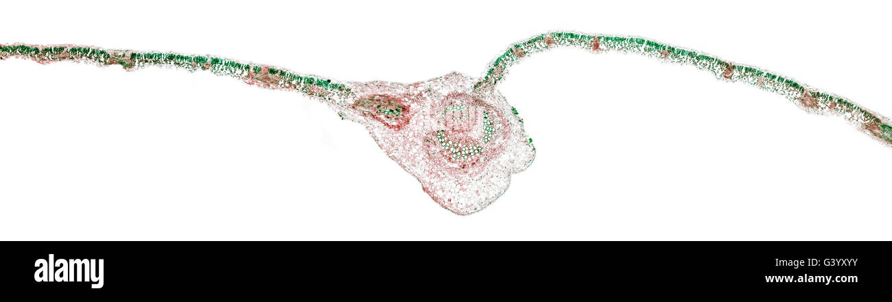 Brightfield photomicrograph, Sycamore leaf section TS showing central vein and cell structure Stock Photohttps://www.alamy.com/image-license-details/?v=1https://www.alamy.com/stock-photo-brightfield-photomicrograph-sycamore-leaf-section-ts-showing-central-105720047.html
RMG3YXYY–Brightfield photomicrograph, Sycamore leaf section TS showing central vein and cell structure
 Chara algae showing stem oogonium round structure antheridiium pointed structures are new leaves magnification 40x Stock Photohttps://www.alamy.com/image-license-details/?v=1https://www.alamy.com/stock-photo-chara-algae-showing-stem-oogonium-round-structure-antheridiium-pointed-56084159.html
RMD76RWK–Chara algae showing stem oogonium round structure antheridiium pointed structures are new leaves magnification 40x
 Datura x candida stem leaf and petiole TS darkfield photomicrograph Stock Photohttps://www.alamy.com/image-license-details/?v=1https://www.alamy.com/datura-x-candida-stem-leaf-and-petiole-ts-darkfield-photomicrograph-image239401045.html
RMRWDJD9–Datura x candida stem leaf and petiole TS darkfield photomicrograph
 Archive image from page 48 of Cytological methods for the detection,. Cytological methods for the detection, identification, and characterization of orchid viruses and their inclusion bodies cytologicalmetho00kona Year: 1985 â yi i Figure 33. Light micrograph of ORSV-infected cells showing a stacked-plate inclusion (SI, side view) in semithin section (0.5 Mm thick). Section is stained with toluidine blue. Bar = 5 jam. Figure 34. Electron micrograph of ORSV stacked- plate inclusion (side view) in Cattleya leaf cells stained with O/G combination. Bar = 600 nm. Stock Photohttps://www.alamy.com/image-license-details/?v=1https://www.alamy.com/archive-image-from-page-48-of-cytological-methods-for-the-detection-cytological-methods-for-the-detection-identification-and-characterization-of-orchid-viruses-and-their-inclusion-bodies-cytologicalmetho00kona-year-1985-yi-i-figure-33-light-micrograph-of-orsv-infected-cells-showing-a-stacked-plate-inclusion-si-side-view-in-semithin-section-05-mm-thick-section-is-stained-with-toluidine-blue-bar-=-5-jam-figure-34-electron-micrograph-of-orsv-stacked-plate-inclusion-side-view-in-cattleya-leaf-cells-stained-with-og-combination-bar-=-600-nm-image259283480.html
RMW1RAM8–Archive image from page 48 of Cytological methods for the detection,. Cytological methods for the detection, identification, and characterization of orchid viruses and their inclusion bodies cytologicalmetho00kona Year: 1985 â yi i Figure 33. Light micrograph of ORSV-infected cells showing a stacked-plate inclusion (SI, side view) in semithin section (0.5 Mm thick). Section is stained with toluidine blue. Bar = 5 jam. Figure 34. Electron micrograph of ORSV stacked- plate inclusion (side view) in Cattleya leaf cells stained with O/G combination. Bar = 600 nm.
 . Cytological methods for the detection, identification, and characterization of orchid viruses and their inclusion bodies. Orchids; Virus diseases of plants. â yi i. Figure 33. Light micrograph of ORSV-infected cells showing a stacked-plate inclusion (SI, side view) in semithin section (0.5 Mm thick). Section is stained with toluidine blue. Bar = 5 jam. Figure 34. Electron micrograph of ORSV stacked- plate inclusion (side view) in Cattleya leaf cells stained with O/G combination. Bar = 600 nm.. Please note that these images are extracted from scanned page images that may have been digitally e Stock Photohttps://www.alamy.com/image-license-details/?v=1https://www.alamy.com/cytological-methods-for-the-detection-identification-and-characterization-of-orchid-viruses-and-their-inclusion-bodies-orchids-virus-diseases-of-plants-yi-i-figure-33-light-micrograph-of-orsv-infected-cells-showing-a-stacked-plate-inclusion-si-side-view-in-semithin-section-05-mm-thick-section-is-stained-with-toluidine-blue-bar-=-5-jam-figure-34-electron-micrograph-of-orsv-stacked-plate-inclusion-side-view-in-cattleya-leaf-cells-stained-with-og-combination-bar-=-600-nm-please-note-that-these-images-are-extracted-from-scanned-page-images-that-may-have-been-digitally-e-image231811256.html
RMRD3WHC–. Cytological methods for the detection, identification, and characterization of orchid viruses and their inclusion bodies. Orchids; Virus diseases of plants. â yi i. Figure 33. Light micrograph of ORSV-infected cells showing a stacked-plate inclusion (SI, side view) in semithin section (0.5 Mm thick). Section is stained with toluidine blue. Bar = 5 jam. Figure 34. Electron micrograph of ORSV stacked- plate inclusion (side view) in Cattleya leaf cells stained with O/G combination. Bar = 600 nm.. Please note that these images are extracted from scanned page images that may have been digitally e
 . Cytological methods for the detection, identification, and characterization of orchid viruses and their inclusion bodies. Orchids; Virus diseases of plants. â yi i. Figure 33. Light micrograph of ORSV-infected cells showing a stacked-plate inclusion (SI, side view) in semithin section (0.5 Mm thick). Section is stained with toluidine blue. Bar = 5 jam. Figure 34. Electron micrograph of ORSV stacked- plate inclusion (side view) in Cattleya leaf cells stained with O/G combination. Bar = 600 nm.. Please note that these images are extracted from scanned page images that may have been digitally e Stock Photohttps://www.alamy.com/image-license-details/?v=1https://www.alamy.com/cytological-methods-for-the-detection-identification-and-characterization-of-orchid-viruses-and-their-inclusion-bodies-orchids-virus-diseases-of-plants-yi-i-figure-33-light-micrograph-of-orsv-infected-cells-showing-a-stacked-plate-inclusion-si-side-view-in-semithin-section-05-mm-thick-section-is-stained-with-toluidine-blue-bar-=-5-jam-figure-34-electron-micrograph-of-orsv-stacked-plate-inclusion-side-view-in-cattleya-leaf-cells-stained-with-og-combination-bar-=-600-nm-please-note-that-these-images-are-extracted-from-scanned-page-images-that-may-have-been-digitally-e-image216162284.html
RMPFK15G–. Cytological methods for the detection, identification, and characterization of orchid viruses and their inclusion bodies. Orchids; Virus diseases of plants. â yi i. Figure 33. Light micrograph of ORSV-infected cells showing a stacked-plate inclusion (SI, side view) in semithin section (0.5 Mm thick). Section is stained with toluidine blue. Bar = 5 jam. Figure 34. Electron micrograph of ORSV stacked- plate inclusion (side view) in Cattleya leaf cells stained with O/G combination. Bar = 600 nm.. Please note that these images are extracted from scanned page images that may have been digitally e
