Quick filters:
Left lung Stock Photos and Images
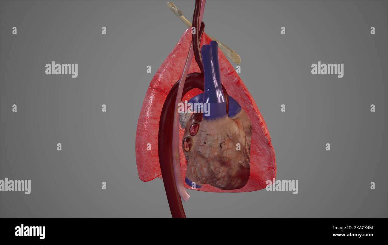 Medial View of Left Lung showing Major structures Related to It Stock Photohttps://www.alamy.com/image-license-details/?v=1https://www.alamy.com/medial-view-of-left-lung-showing-major-structures-related-to-it-image488320804.html
RF2KACX4M–Medial View of Left Lung showing Major structures Related to It
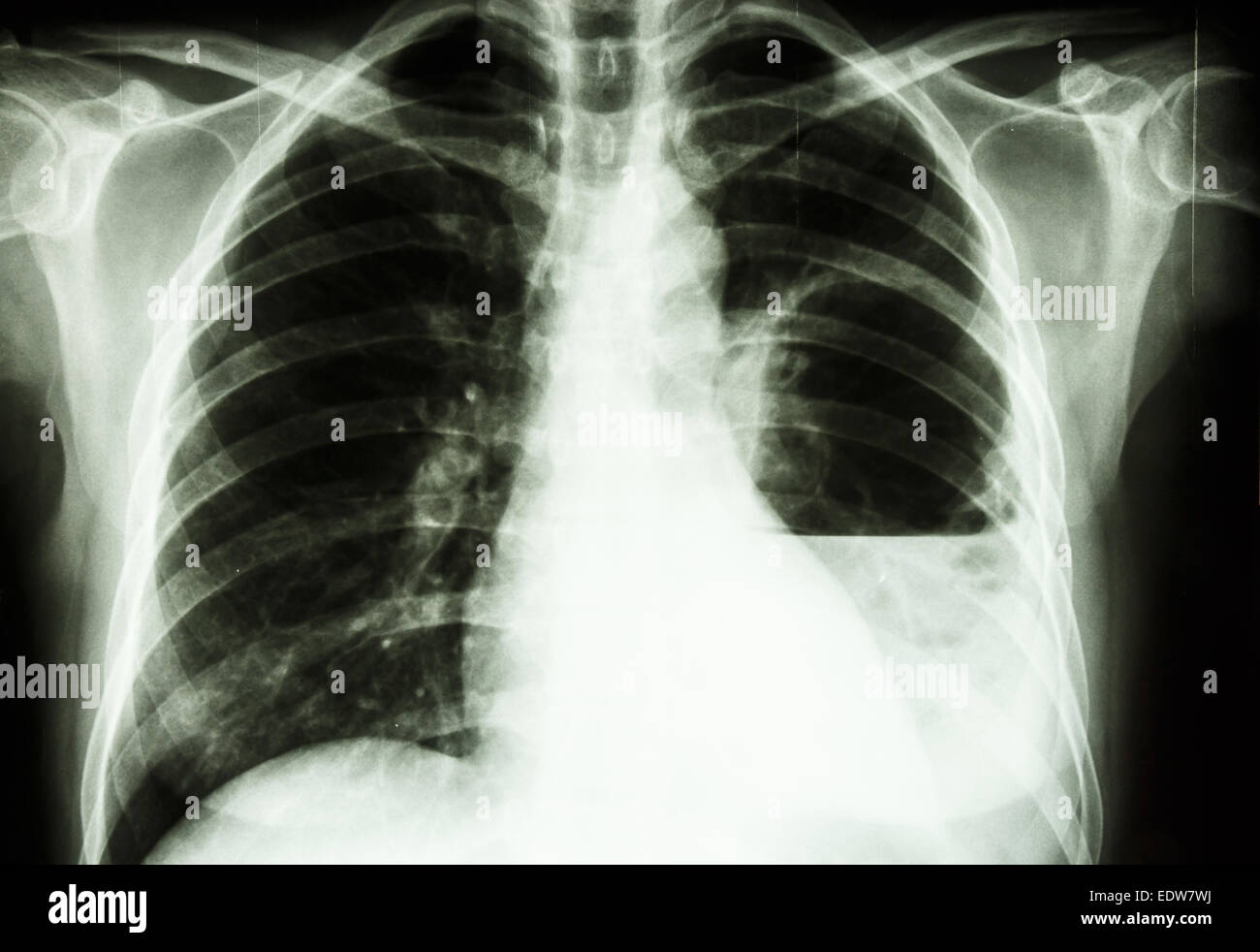 film X-ray show left lung abscess from Burkholderia pseudomallei infection (Mellioidosis) Stock Photohttps://www.alamy.com/image-license-details/?v=1https://www.alamy.com/stock-photo-film-x-ray-show-left-lung-abscess-from-burkholderia-pseudomallei-infection-77387006.html
RFEDW7WJ–film X-ray show left lung abscess from Burkholderia pseudomallei infection (Mellioidosis)
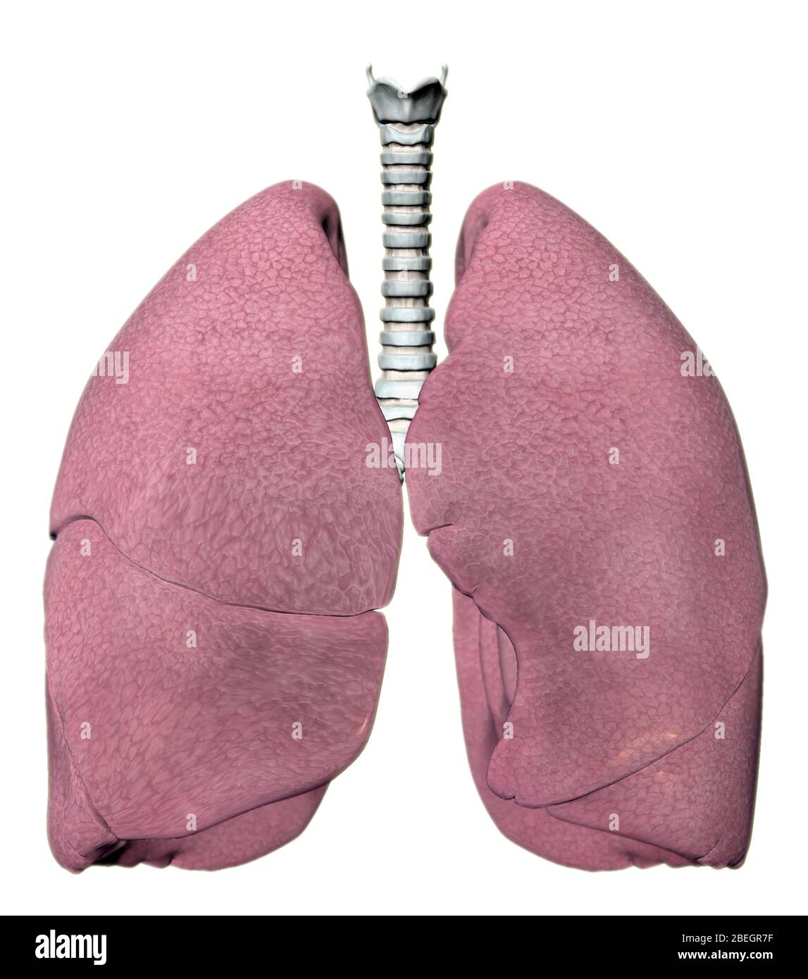 Lungs Stock Photohttps://www.alamy.com/image-license-details/?v=1https://www.alamy.com/lungs-image353182019.html
RM2BEGR7F–Lungs
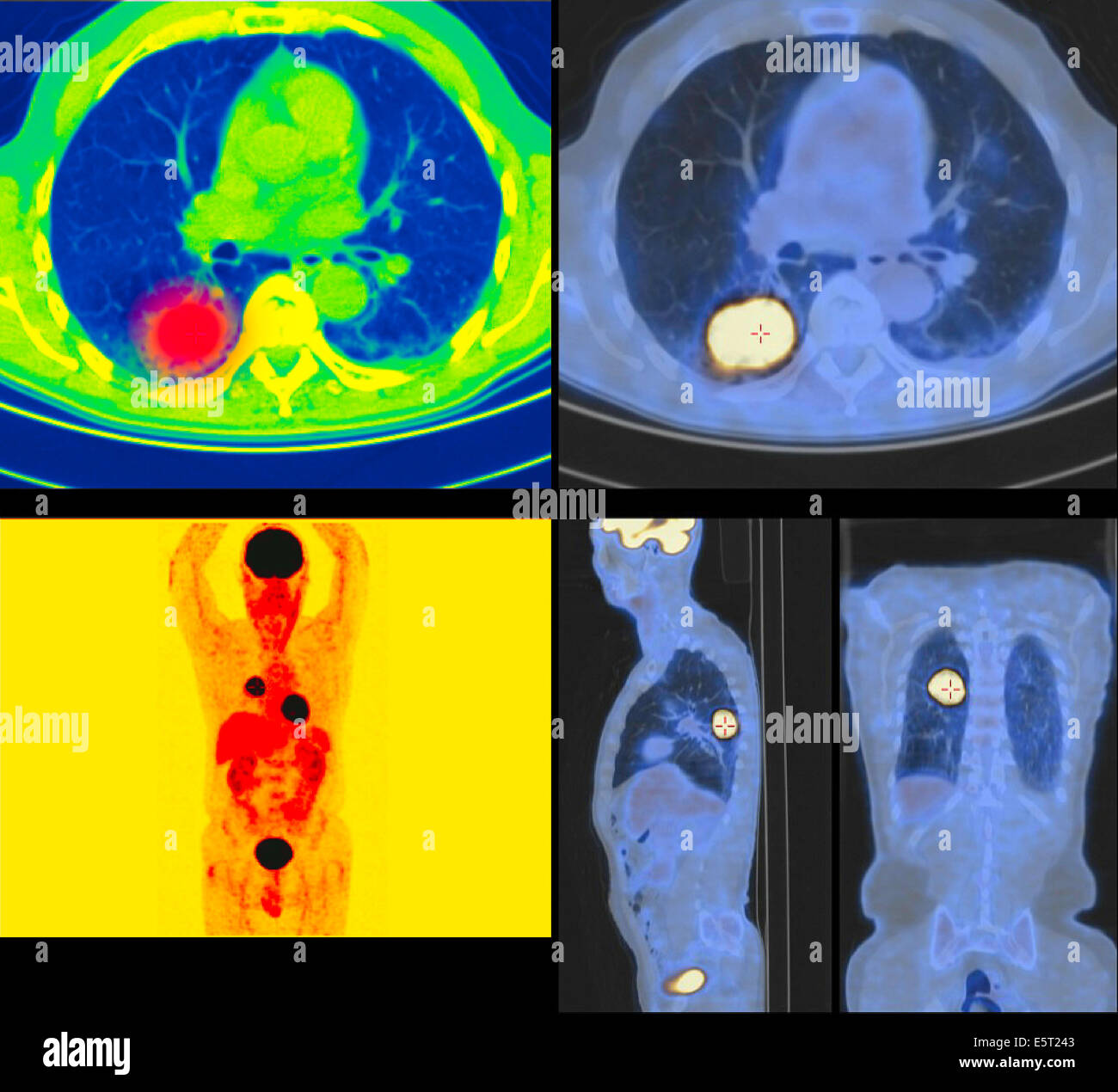 Positron emission tomography (PET) scans of a patient with a tumour the upper lobe of the left lung. Stock Photohttps://www.alamy.com/image-license-details/?v=1https://www.alamy.com/stock-photo-positron-emission-tomography-pet-scans-of-a-patient-with-a-tumour-72443283.html
RME5T243–Positron emission tomography (PET) scans of a patient with a tumour the upper lobe of the left lung.
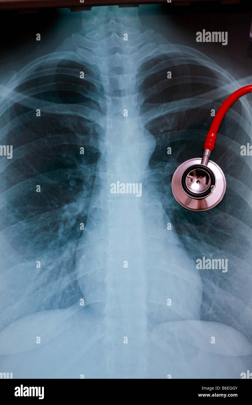 chest x-ray CXR with stethoscope overlying left lung Stock Photohttps://www.alamy.com/image-license-details/?v=1https://www.alamy.com/stock-photo-chest-x-ray-cxr-with-stethoscope-overlying-left-lung-21218651.html
RFB6EGGY–chest x-ray CXR with stethoscope overlying left lung
 Right and left lung cancer (stage 1). Front chest x-ray. Stock Photohttps://www.alamy.com/image-license-details/?v=1https://www.alamy.com/right-and-left-lung-cancer-stage-1-front-chest-x-ray-image627687354.html
RM2YD5HHE–Right and left lung cancer (stage 1). Front chest x-ray.
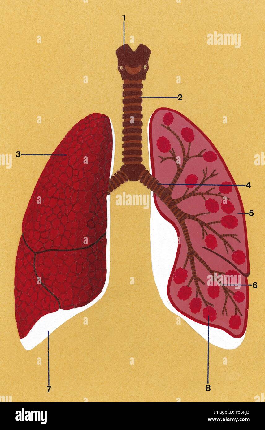 Respiratory system. Schematic drawing of the trachea and lungs. 1. Larynx 2. Trachea 3. Right lung closed 4. Bronchus 5. Left lung opened 6. Bronchioles 7. Pleura 8. Alveoli. Drawing. Color. Stock Photohttps://www.alamy.com/image-license-details/?v=1https://www.alamy.com/respiratory-system-schematic-drawing-of-the-trachea-and-lungs-1-larynx-2-trachea-3-right-lung-closed-4-bronchus-5-left-lung-opened-6-bronchioles-7-pleura-8-alveoli-drawing-color-image209682091.html
RMP53RJ3–Respiratory system. Schematic drawing of the trachea and lungs. 1. Larynx 2. Trachea 3. Right lung closed 4. Bronchus 5. Left lung opened 6. Bronchioles 7. Pleura 8. Alveoli. Drawing. Color.
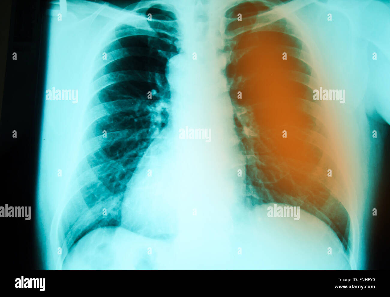 chest x-ray examination for diagnosis Pulmonary tuberculosis infection with left lung Stock Photohttps://www.alamy.com/image-license-details/?v=1https://www.alamy.com/stock-photo-chest-x-ray-examination-for-diagnosis-pulmonary-tuberculosis-infection-99344532.html
RFFNHEY0–chest x-ray examination for diagnosis Pulmonary tuberculosis infection with left lung
 The left lung, vintage engraved illustration. Stock Vectorhttps://www.alamy.com/image-license-details/?v=1https://www.alamy.com/stock-photo-the-left-lung-vintage-engraved-illustration-84301936.html
RFEW47YC–The left lung, vintage engraved illustration.
 Computed tomography (CT) scan of a chest from above showing an azygos lobe in the left lung (segmented area centre-left). An azygos lobe is an anatomical variant in the upper lobe of the lung in which a segment of the lung is separated by a deep groove called an azygos fissure, which consists of the azygos vein and pleura (thin tissue). It is a rare variation, occurring in around 0.3% of people during embryonic development. It causes no symptoms, but may make certain surgical procedures more difficult. Stock Photohttps://www.alamy.com/image-license-details/?v=1https://www.alamy.com/computed-tomography-ct-scan-of-a-chest-from-above-showing-an-azygos-lobe-in-the-left-lung-segmented-area-centre-left-an-azygos-lobe-is-an-anatomical-variant-in-the-upper-lobe-of-the-lung-in-which-a-segment-of-the-lung-is-separated-by-a-deep-groove-called-an-azygos-fissure-which-consists-of-the-azygos-vein-and-pleura-thin-tissue-it-is-a-rare-variation-occurring-in-around-03-of-people-during-embryonic-development-it-causes-no-symptoms-but-may-make-certain-surgical-procedures-more-difficult-image612233850.html
RF2XG1JEJ–Computed tomography (CT) scan of a chest from above showing an azygos lobe in the left lung (segmented area centre-left). An azygos lobe is an anatomical variant in the upper lobe of the lung in which a segment of the lung is separated by a deep groove called an azygos fissure, which consists of the azygos vein and pleura (thin tissue). It is a rare variation, occurring in around 0.3% of people during embryonic development. It causes no symptoms, but may make certain surgical procedures more difficult.
 The Duchess of Cornwall meets Sienna Mason (second left), who suffers heart and lung disease and her sister Seren Mason (left), mother Natalie Pearson and sister Summer Mason (right), at a tea party for children from Ty Hafan hospice in Wales, at Highgrove House in Gloucestershire. Stock Photohttps://www.alamy.com/image-license-details/?v=1https://www.alamy.com/stock-photo-the-duchess-of-cornwall-meets-sienna-mason-second-left-who-suffers-110811315.html
RMGC7TXY–The Duchess of Cornwall meets Sienna Mason (second left), who suffers heart and lung disease and her sister Seren Mason (left), mother Natalie Pearson and sister Summer Mason (right), at a tea party for children from Ty Hafan hospice in Wales, at Highgrove House in Gloucestershire.
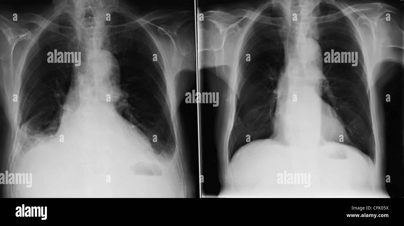 X-rays of the same male chest at different dates with the left showing a fungal infection and the right a normal lung UK Stock Photohttps://www.alamy.com/image-license-details/?v=1https://www.alamy.com/stock-photo-x-rays-of-the-same-male-chest-at-different-dates-with-the-left-showing-44057830.html
RMCFK05X–X-rays of the same male chest at different dates with the left showing a fungal infection and the right a normal lung UK
 Feb. 29, 2012 - The Newly open Skeins of Silk are lung out to dry in the drying cabinet and they are periodicaly examined by Dom Edmund Fatt (left) and Dom Anscar Neilson in their cell-workshop at the Abbey Stock Photohttps://www.alamy.com/image-license-details/?v=1https://www.alamy.com/feb-29-2012-the-newly-open-skeins-of-silk-are-lung-out-to-dry-in-the-image69531535.html
RME13C53–Feb. 29, 2012 - The Newly open Skeins of Silk are lung out to dry in the drying cabinet and they are periodicaly examined by Dom Edmund Fatt (left) and Dom Anscar Neilson in their cell-workshop at the Abbey
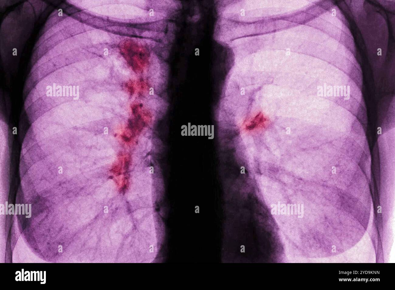 Right and left lung cancer stage 1. Front chest x-ray. Lung cancer 016836 026 Stock Photohttps://www.alamy.com/image-license-details/?v=1https://www.alamy.com/right-and-left-lung-cancer-stage-1-front-chest-x-ray-lung-cancer-016836-026-image627776849.html
RM2YD9KNN–Right and left lung cancer stage 1. Front chest x-ray. Lung cancer 016836 026
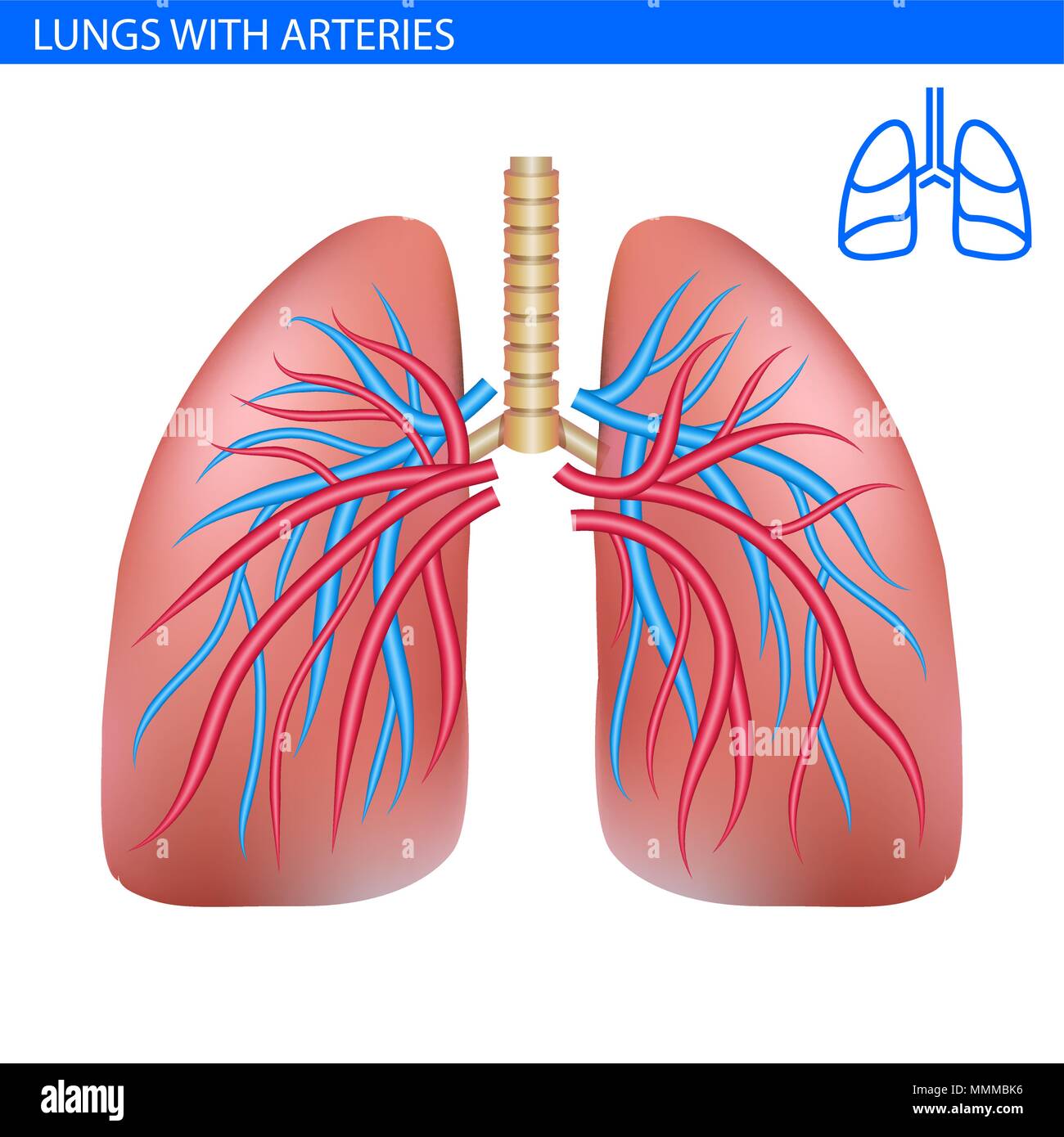 Human lungs anatomy with artery, circulatory system realistic illustration front view in detail. Lunge exercise. Right and left lung with trachea. Hea Stock Vectorhttps://www.alamy.com/image-license-details/?v=1https://www.alamy.com/human-lungs-anatomy-with-artery-circulatory-system-realistic-illustration-front-view-in-detail-lunge-exercise-right-and-left-lung-with-trachea-hea-image184845002.html
RFMMMBK6–Human lungs anatomy with artery, circulatory system realistic illustration front view in detail. Lunge exercise. Right and left lung with trachea. Hea
 The doctor raised his hand, lung, and Corona virus in his left and right hand. Stock Photohttps://www.alamy.com/image-license-details/?v=1https://www.alamy.com/the-doctor-raised-his-hand-lung-and-corona-virus-in-his-left-and-right-hand-image351448271.html
RF2BBNRRY–The doctor raised his hand, lung, and Corona virus in his left and right hand.
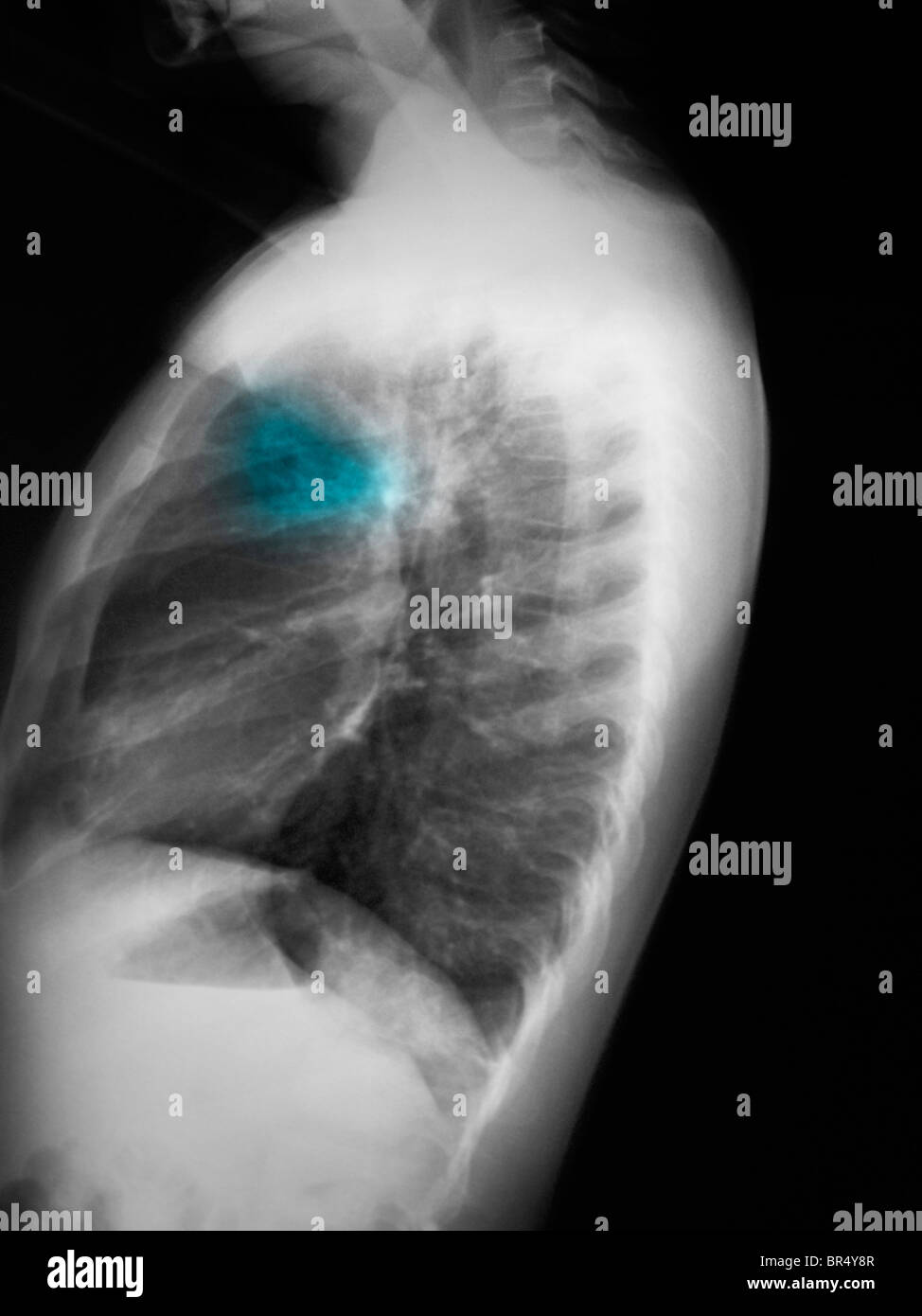 Chest x-ray of a 5 year old boy showing an infiltrate left upper lobe suggestive of pneumonia. Stock Photohttps://www.alamy.com/image-license-details/?v=1https://www.alamy.com/stock-photo-chest-x-ray-of-a-5-year-old-boy-showing-an-infiltrate-left-upper-lobe-31456679.html
RMBR4Y8R–Chest x-ray of a 5 year old boy showing an infiltrate left upper lobe suggestive of pneumonia.
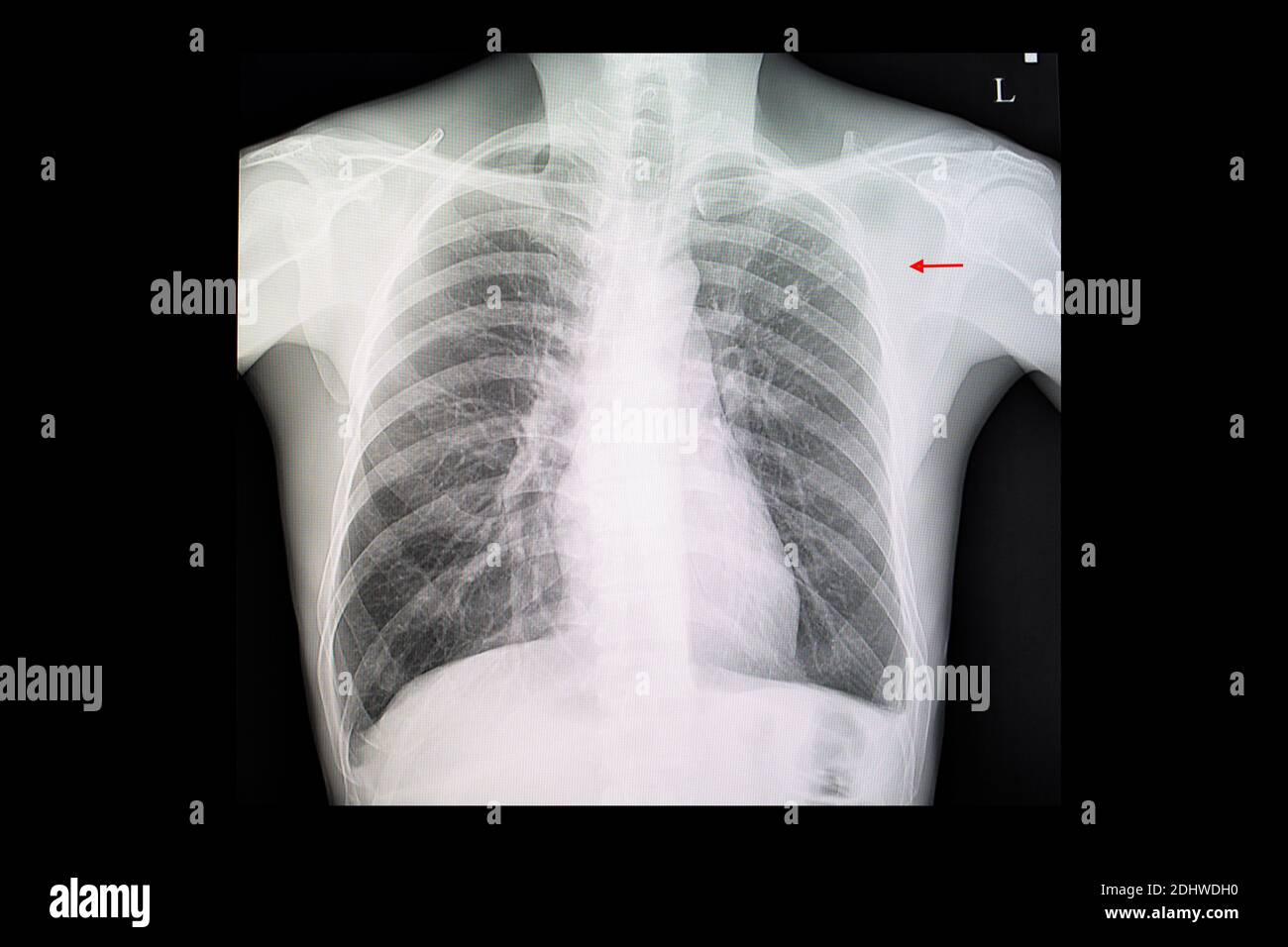 A chest x-ray film of a patient with pulmonary tuberculosis with fibronodular infiltration at left upper lung. Stock Photohttps://www.alamy.com/image-license-details/?v=1https://www.alamy.com/a-chest-x-ray-film-of-a-patient-with-pulmonary-tuberculosis-with-fibronodular-infiltration-at-left-upper-lung-image389636716.html
RF2DHWDH0–A chest x-ray film of a patient with pulmonary tuberculosis with fibronodular infiltration at left upper lung.
 California Gov. Gavin Newsom, left, talks to Los Angeles Mayor Eric Garcetti during a press conference relating to the opening of a state and federal Stock Photohttps://www.alamy.com/image-license-details/?v=1https://www.alamy.com/california-gov-gavin-newsom-left-talks-to-los-angeles-mayor-eric-garcetti-during-a-press-conference-relating-to-the-opening-of-a-state-and-federal-image405209717.html
RF2EF6W3H–California Gov. Gavin Newsom, left, talks to Los Angeles Mayor Eric Garcetti during a press conference relating to the opening of a state and federal
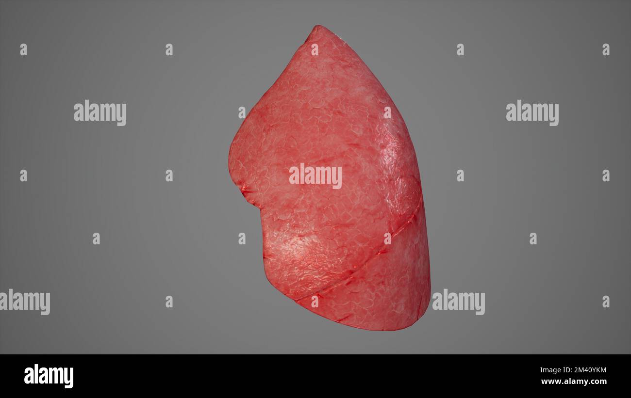 Anatomical Illustration of Left Lung.3d rendering Stock Photohttps://www.alamy.com/image-license-details/?v=1https://www.alamy.com/anatomical-illustration-of-left-lung3d-rendering-image501581016.html
RF2M40YKM–Anatomical Illustration of Left Lung.3d rendering
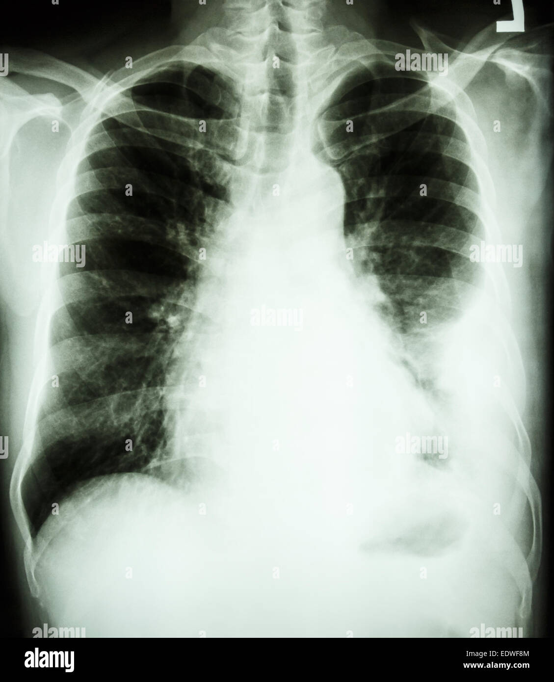 film chest X-ray PA upright : show pleural effusion at left lung due to lung cancer Stock Photohttps://www.alamy.com/image-license-details/?v=1https://www.alamy.com/stock-photo-film-chest-x-ray-pa-upright-show-pleural-effusion-at-left-lung-due-77392804.html
RFEDWF8M–film chest X-ray PA upright : show pleural effusion at left lung due to lung cancer
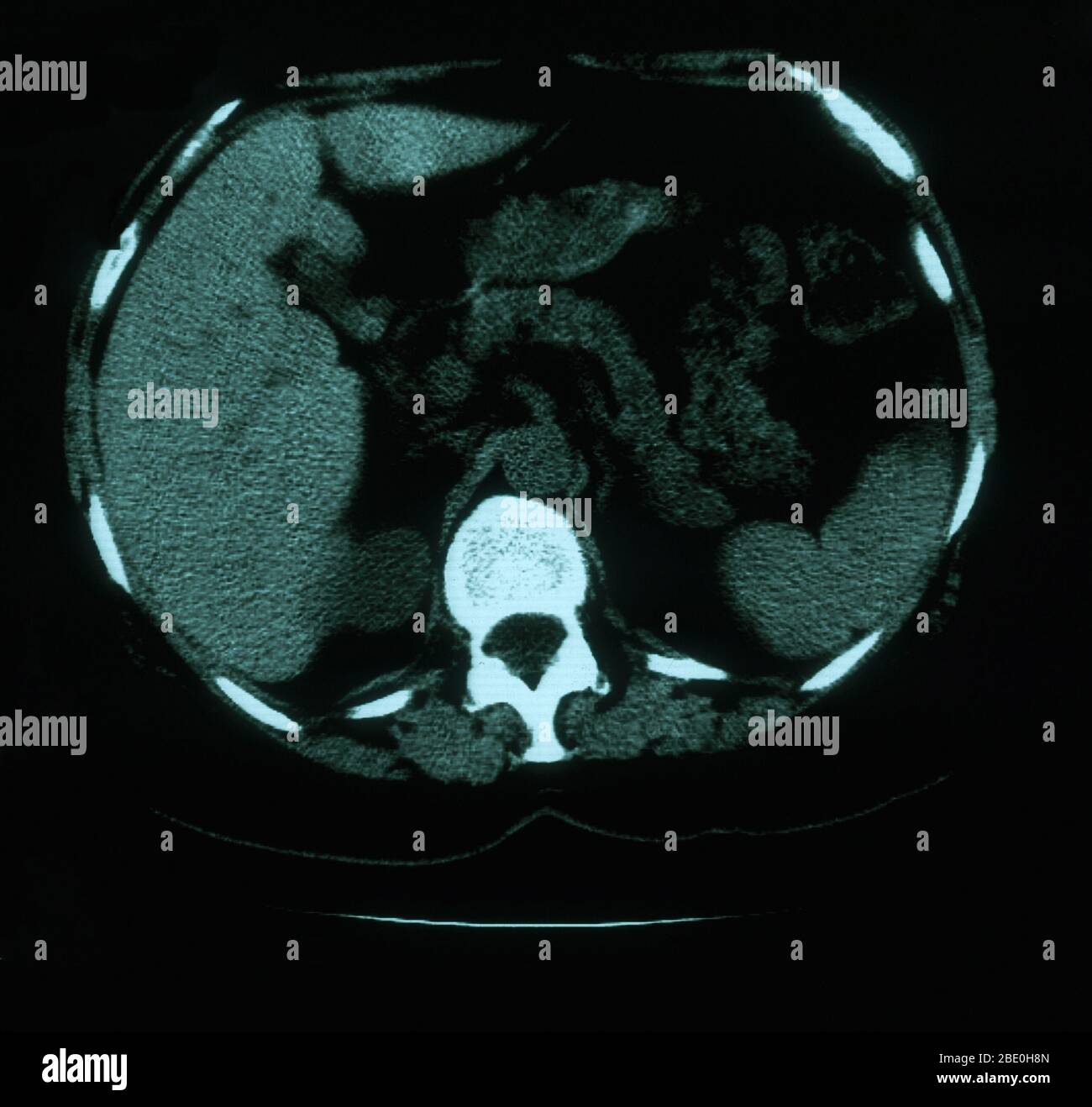 An axial (cross sectional) CT image through the lungs of a 53 year old female. The scan shows tortuosity and prominence of vasculature in the right superior mediastinum. Also present are calcified left hilar nodules and multiple punctate calcifications throughout the spleen which are consistent with granuloma. Stock Photohttps://www.alamy.com/image-license-details/?v=1https://www.alamy.com/an-axial-cross-sectional-ct-image-through-the-lungs-of-a-53-year-old-female-the-scan-shows-tortuosity-and-prominence-of-vasculature-in-the-right-superior-mediastinum-also-present-are-calcified-left-hilar-nodules-and-multiple-punctate-calcifications-throughout-the-spleen-which-are-consistent-with-granuloma-image352826117.html
RM2BE0H8N–An axial (cross sectional) CT image through the lungs of a 53 year old female. The scan shows tortuosity and prominence of vasculature in the right superior mediastinum. Also present are calcified left hilar nodules and multiple punctate calcifications throughout the spleen which are consistent with granuloma.
 Indonesia, Sulawesi, Tana Toraja, Kete Kesu, cigarettes left as offerings amongst exposed bones of dead ancestors Stock Photohttps://www.alamy.com/image-license-details/?v=1https://www.alamy.com/stock-photo-indonesia-sulawesi-tana-toraja-kete-kesu-cigarettes-left-as-offerings-28434167.html
RMBJ781Y–Indonesia, Sulawesi, Tana Toraja, Kete Kesu, cigarettes left as offerings amongst exposed bones of dead ancestors
 ETHEL LUNG, CIAO, 2008 Stock Photohttps://www.alamy.com/image-license-details/?v=1https://www.alamy.com/ethel-lung-ciao-2008-image484849530.html
RM2K4PPEJ–ETHEL LUNG, CIAO, 2008
 Right and left lung cancer (stage 1). Front chest x-ray. Stock Photohttps://www.alamy.com/image-license-details/?v=1https://www.alamy.com/right-and-left-lung-cancer-stage-1-front-chest-x-ray-image627687424.html
RM2YD5HM0–Right and left lung cancer (stage 1). Front chest x-ray.
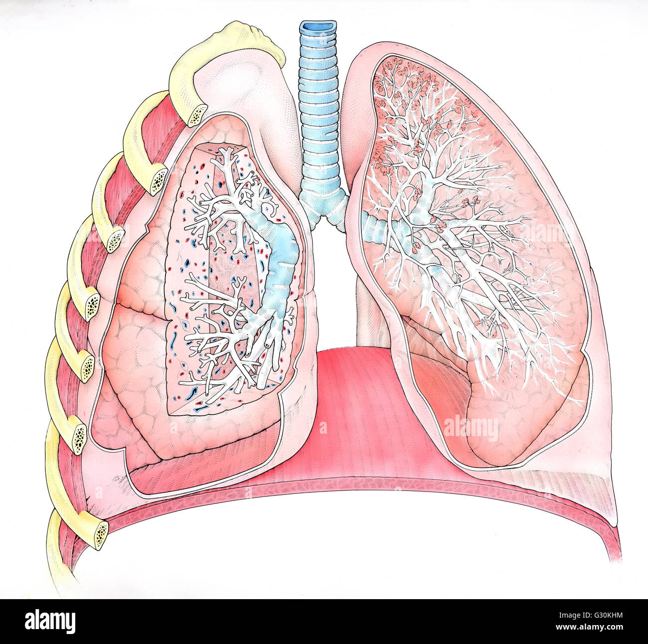 Illustration of right and left human lungs, cross section Stock Photohttps://www.alamy.com/image-license-details/?v=1https://www.alamy.com/stock-photo-illustration-of-right-and-left-human-lungs-cross-section-105121568.html
RMG30KHM–Illustration of right and left human lungs, cross section
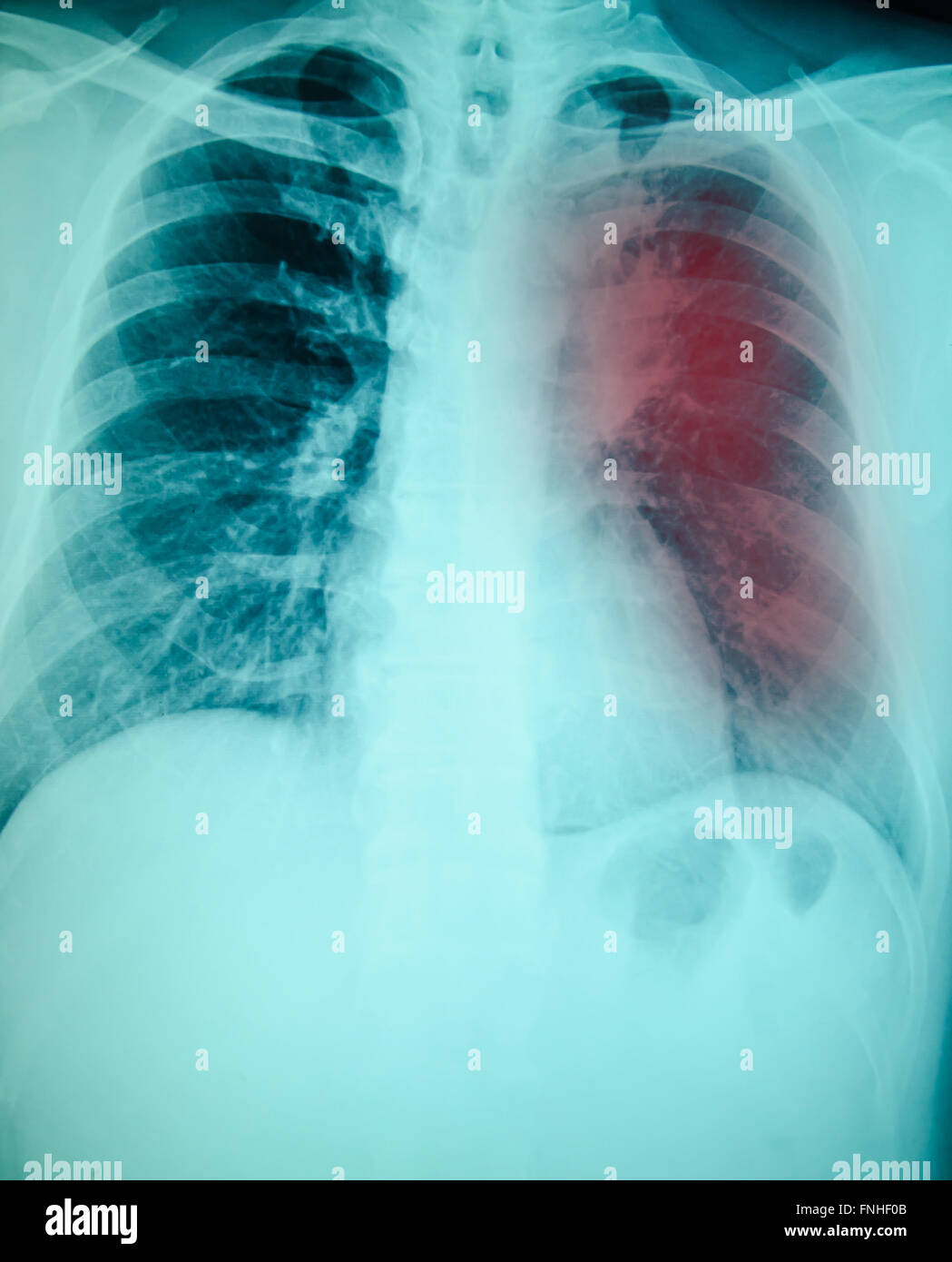 chest x-ray examination for diagnosis Pulmonary tuberculosis infection with left lung Stock Photohttps://www.alamy.com/image-license-details/?v=1https://www.alamy.com/stock-photo-chest-x-ray-examination-for-diagnosis-pulmonary-tuberculosis-infection-99344571.html
RFFNHF0B–chest x-ray examination for diagnosis Pulmonary tuberculosis infection with left lung
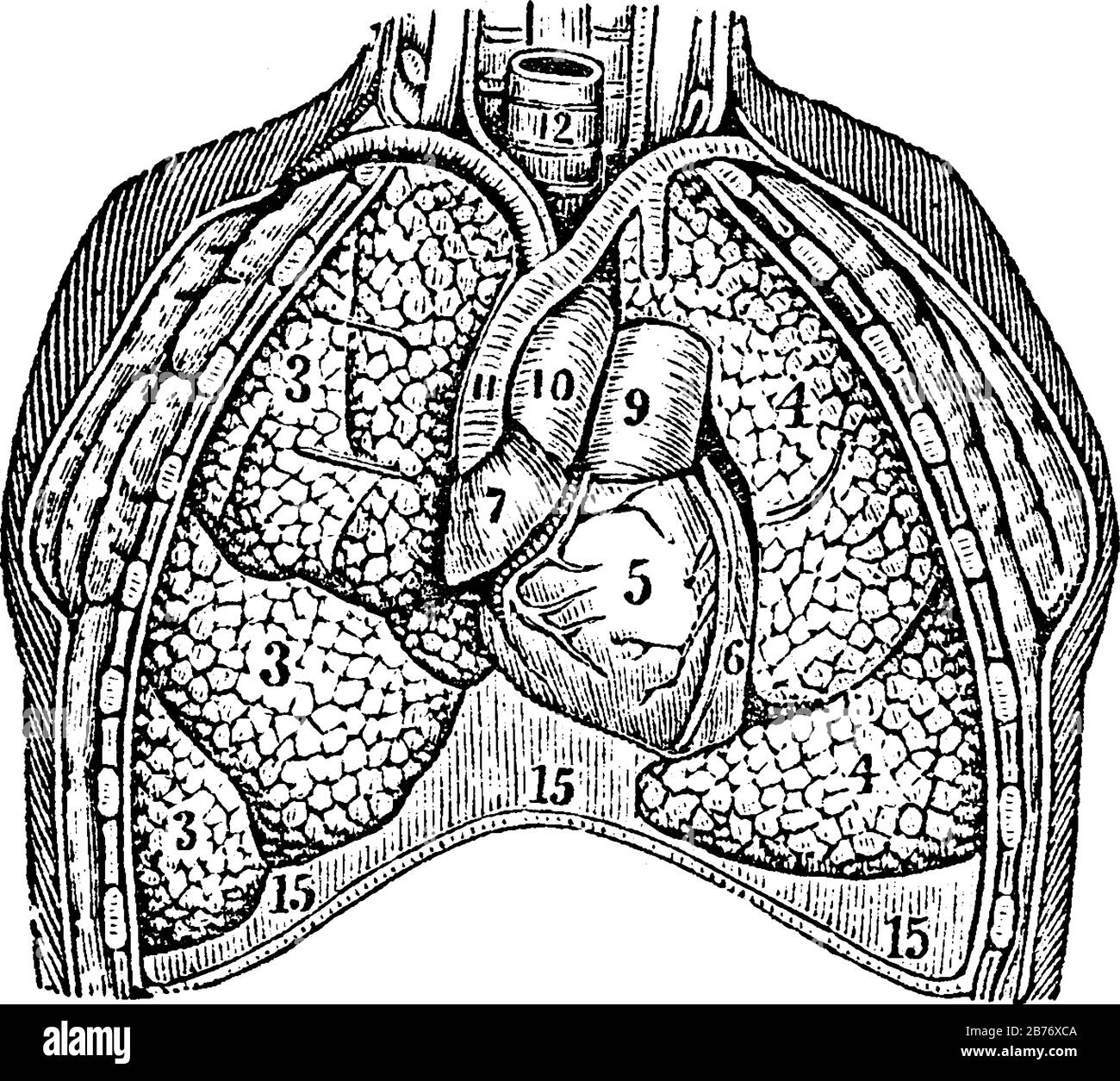 The lungs. Labels: 3; the lobes of the right lung. 4; the lobes of the left lung. 5, 6, 7; the heart. 9, 10, 11; the large blood-vessels; and other; v Stock Vectorhttps://www.alamy.com/image-license-details/?v=1https://www.alamy.com/the-lungs-labels-3-the-lobes-of-the-right-lung-4-the-lobes-of-the-left-lung-5-6-7-the-heart-9-10-11-the-large-blood-vessels-and-other-v-image348662394.html
RF2B76XCA–The lungs. Labels: 3; the lobes of the right lung. 4; the lobes of the left lung. 5, 6, 7; the heart. 9, 10, 11; the large blood-vessels; and other; v
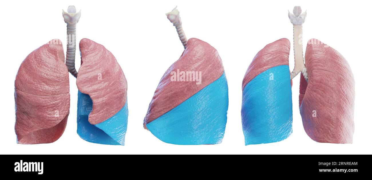 Left lung, illustration Stock Photohttps://www.alamy.com/image-license-details/?v=1https://www.alamy.com/left-lung-illustration-image564155724.html
RF2RNREAM–Left lung, illustration
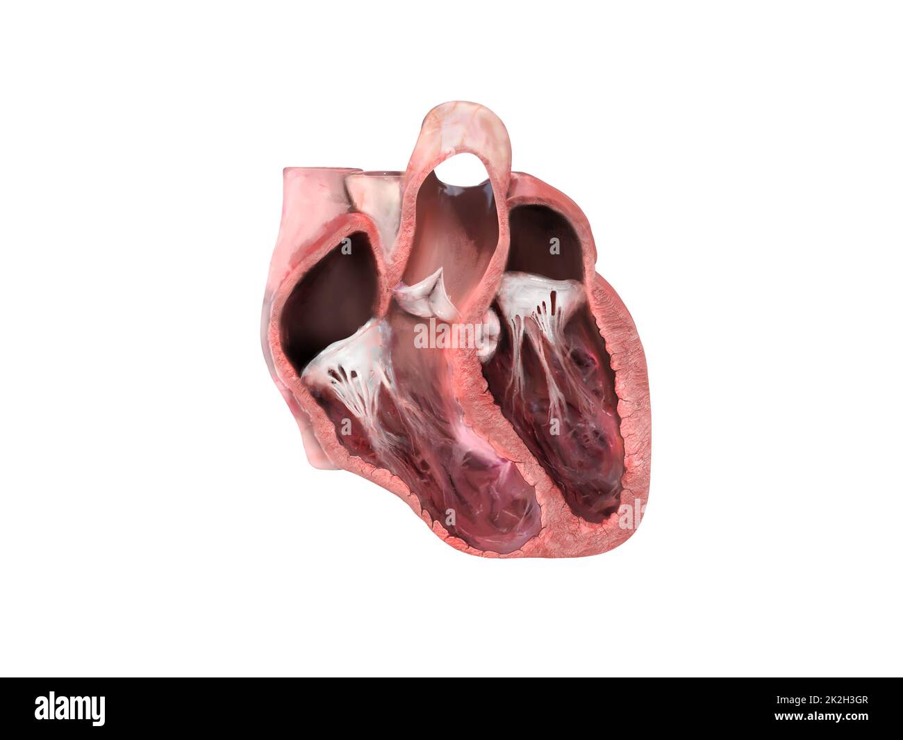 heart anatomy, Left ventricular aneurysm, wall bulge, transmural damage, aneurysm develops, dislocation, aneurysm formation, patient at risk, rupture, thrombus, embolism Stock Photohttps://www.alamy.com/image-license-details/?v=1https://www.alamy.com/heart-anatomy-left-ventricular-aneurysm-wall-bulge-transmural-damage-aneurysm-develops-dislocation-aneurysm-formation-patient-at-risk-rupture-thrombus-embolism-image483495623.html
RF2K2H3GR–heart anatomy, Left ventricular aneurysm, wall bulge, transmural damage, aneurysm develops, dislocation, aneurysm formation, patient at risk, rupture, thrombus, embolism
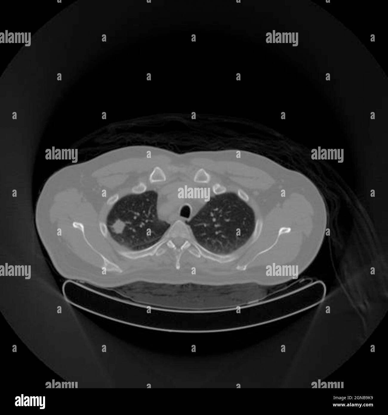 Positron emission tomography (PET) scan of a male, 54 year old patient. A tumour can be seen in the left upper lobe of his lungs Stock Photohttps://www.alamy.com/image-license-details/?v=1https://www.alamy.com/positron-emission-tomography-pet-scan-of-a-male-54-year-old-patient-a-tumour-can-be-seen-in-the-left-upper-lobe-of-his-lungs-image443416045.html
RM2GNB9K9–Positron emission tomography (PET) scan of a male, 54 year old patient. A tumour can be seen in the left upper lobe of his lungs
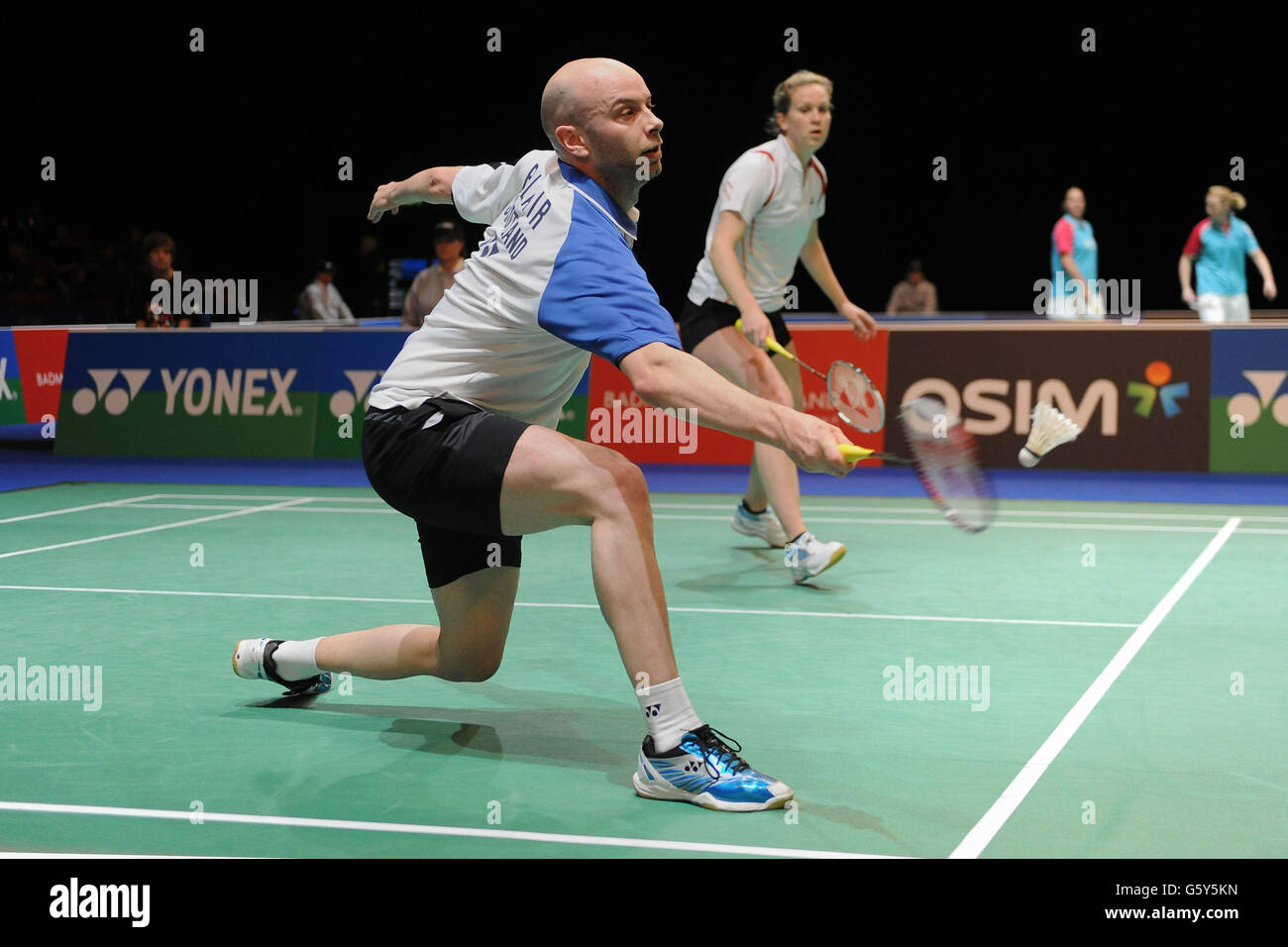 Scotland's Robert Blair (left) and Jilly Cooper in action against Hong Kong's Yun Lung Chan and Ying Suet Tse during day one of the 2013 Yonex All England Badminton Championships at the National Indoor Arena, Birmingham. Stock Photohttps://www.alamy.com/image-license-details/?v=1https://www.alamy.com/stock-photo-scotlands-robert-blair-left-and-jilly-cooper-in-action-against-hong-106932665.html
RMG5Y5KN–Scotland's Robert Blair (left) and Jilly Cooper in action against Hong Kong's Yun Lung Chan and Ying Suet Tse during day one of the 2013 Yonex All England Badminton Championships at the National Indoor Arena, Birmingham.
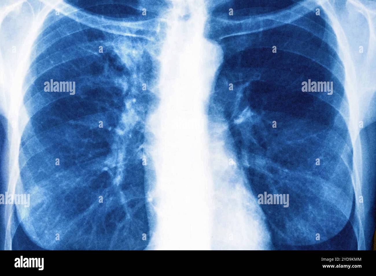 Right and left lung cancer stage 1. Front chest x-ray. Lung cancer 016836 025 Stock Photohttps://www.alamy.com/image-license-details/?v=1https://www.alamy.com/right-and-left-lung-cancer-stage-1-front-chest-x-ray-lung-cancer-016836-025-image627776820.html
RM2YD9KMM–Right and left lung cancer stage 1. Front chest x-ray. Lung cancer 016836 025
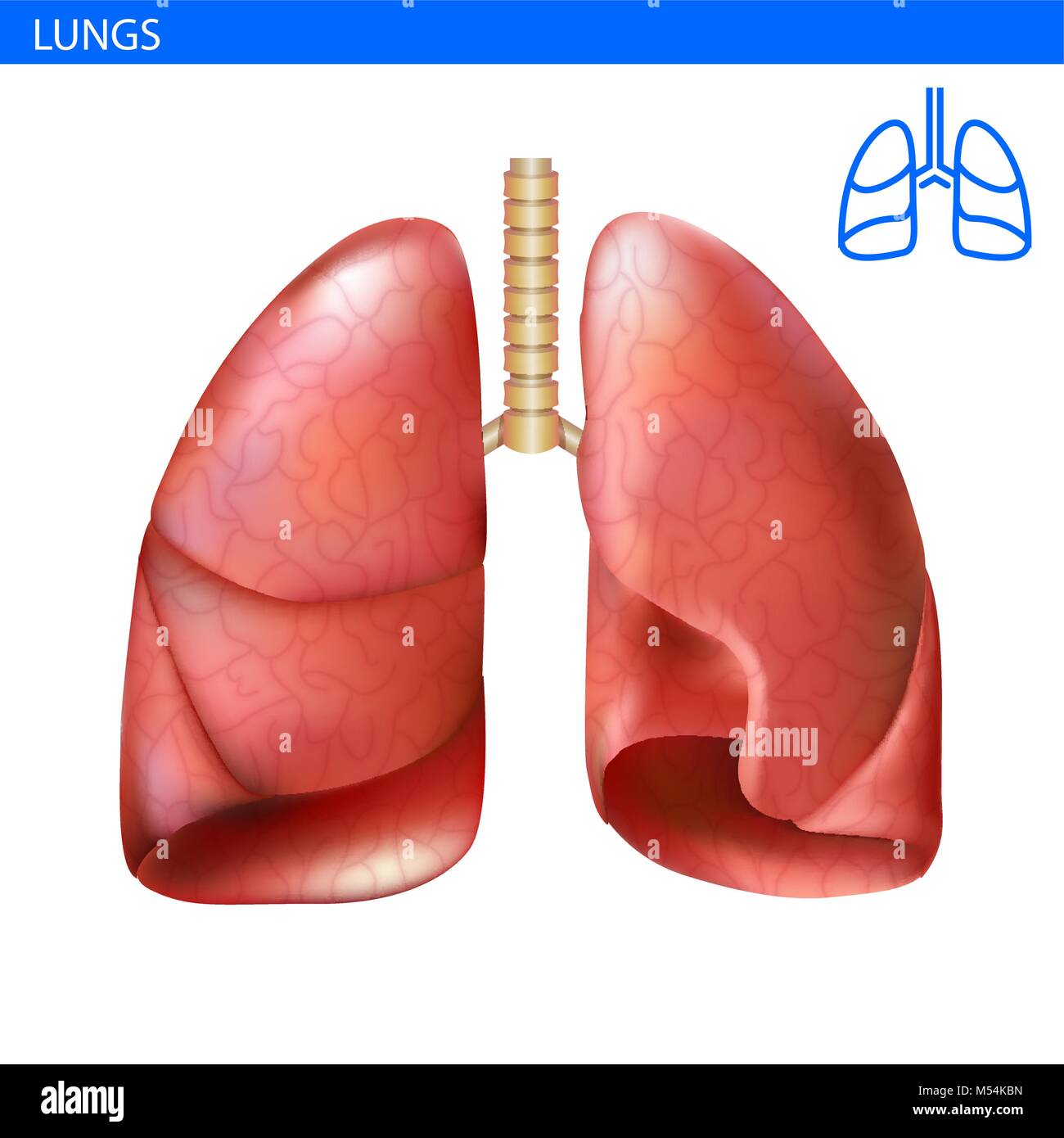 Human lungs anatomy realistic illustration front view in detail. Lunge exercise. Right and left lung with trachea. Healthy lung. Respiratory system Stock Vectorhttps://www.alamy.com/image-license-details/?v=1https://www.alamy.com/stock-photo-human-lungs-anatomy-realistic-illustration-front-view-in-detail-lunge-175279993.html
RFM54KBN–Human lungs anatomy realistic illustration front view in detail. Lunge exercise. Right and left lung with trachea. Healthy lung. Respiratory system
 The doctor raised his hand, lung, and Corona virus in his left and right hand. Stock Photohttps://www.alamy.com/image-license-details/?v=1https://www.alamy.com/the-doctor-raised-his-hand-lung-and-corona-virus-in-his-left-and-right-hand-image351448278.html
RF2BBNRT6–The doctor raised his hand, lung, and Corona virus in his left and right hand.
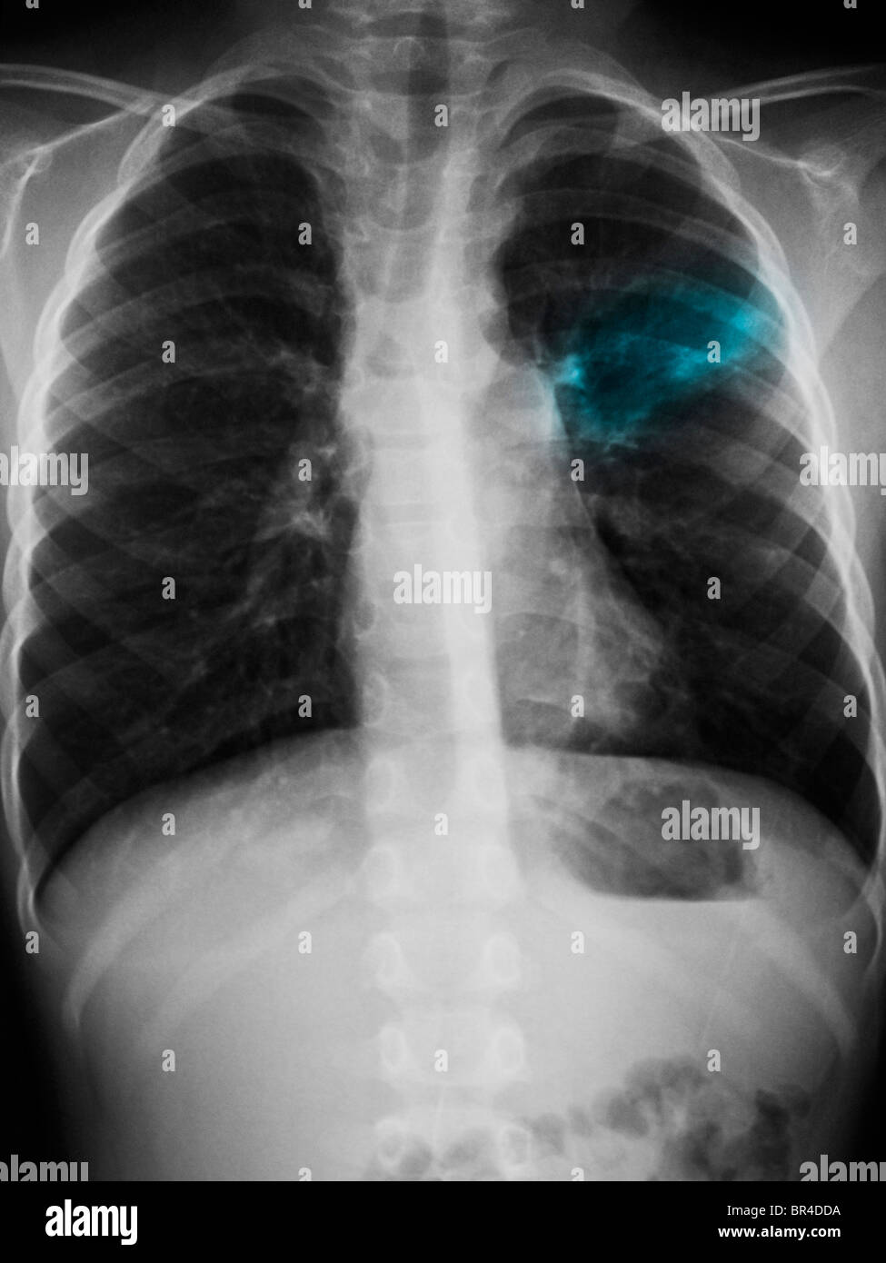 Chest x-ray of a 5 year old boy showing an infiltrate left upper lobe suggestive of pneumonia. Stock Photohttps://www.alamy.com/image-license-details/?v=1https://www.alamy.com/stock-photo-chest-x-ray-of-a-5-year-old-boy-showing-an-infiltrate-left-upper-lobe-31445830.html
RMBR4DDA–Chest x-ray of a 5 year old boy showing an infiltrate left upper lobe suggestive of pneumonia.
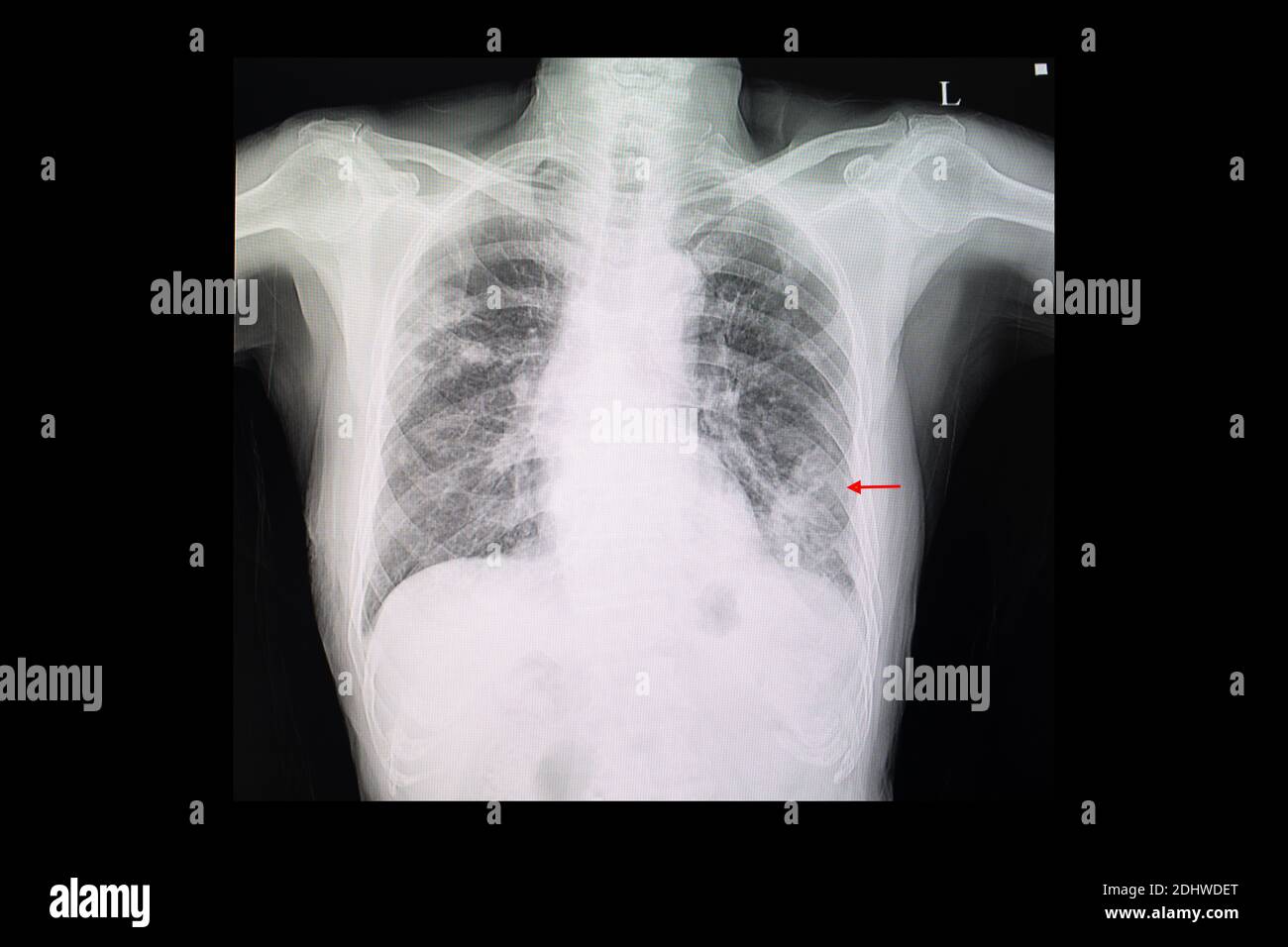 A chest x-ray film of a patient with left lower lung pneumonia and right upper lung nodules Stock Photohttps://www.alamy.com/image-license-details/?v=1https://www.alamy.com/a-chest-x-ray-film-of-a-patient-with-left-lower-lung-pneumonia-and-right-upper-lung-nodules-image389636656.html
RF2DHWDET–A chest x-ray film of a patient with left lower lung pneumonia and right upper lung nodules
 California Gov. Gavin Newsom, left, talks to Los Angeles Mayor Eric Garcetti during a press conference relating to the opening of a state and federal Stock Photohttps://www.alamy.com/image-license-details/?v=1https://www.alamy.com/california-gov-gavin-newsom-left-talks-to-los-angeles-mayor-eric-garcetti-during-a-press-conference-relating-to-the-opening-of-a-state-and-federal-image405209748.html
RF2EF6W4M–California Gov. Gavin Newsom, left, talks to Los Angeles Mayor Eric Garcetti during a press conference relating to the opening of a state and federal
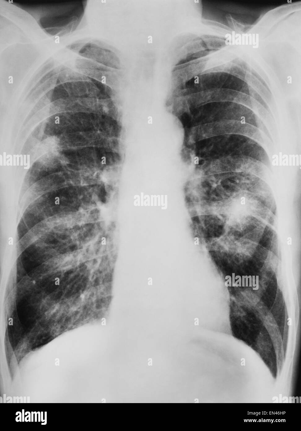 x-ray shadow on the lung engine smoker category b lung cancer of human thoracic chest Stock Photohttps://www.alamy.com/image-license-details/?v=1https://www.alamy.com/stock-photo-x-ray-shadow-on-the-lung-engine-smoker-category-b-lung-cancer-of-human-81842258.html
RMEN46HP–x-ray shadow on the lung engine smoker category b lung cancer of human thoracic chest
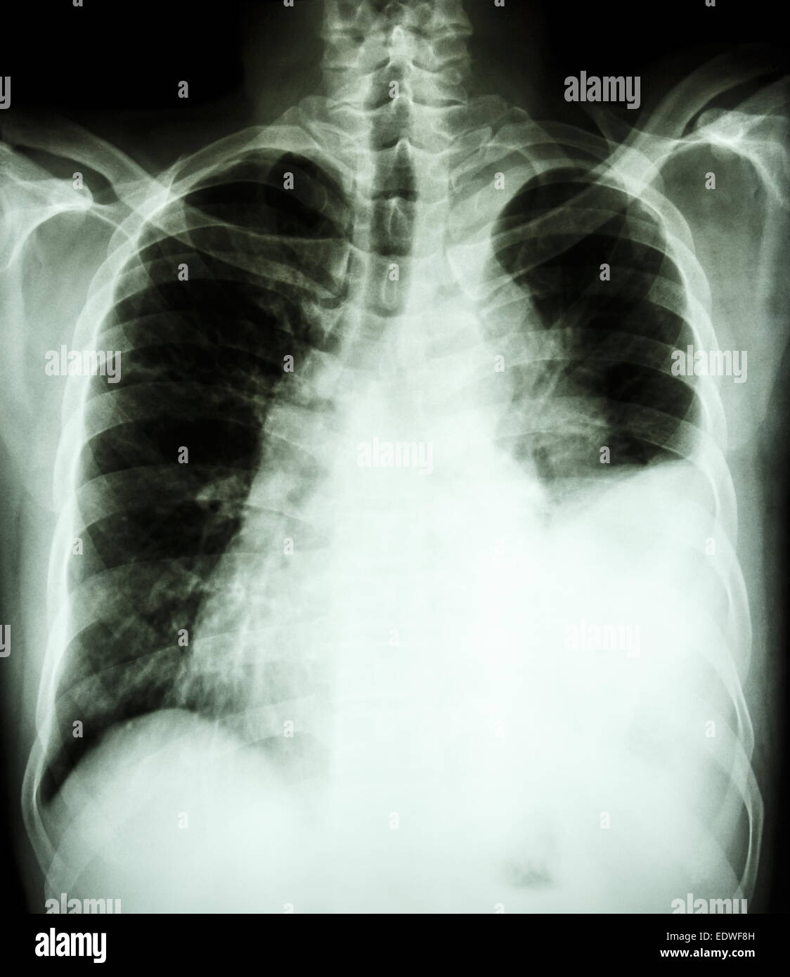 film chest X-ray PA upright : show pleural effusion at left lung due to lung cancer Stock Photohttps://www.alamy.com/image-license-details/?v=1https://www.alamy.com/stock-photo-film-chest-x-ray-pa-upright-show-pleural-effusion-at-left-lung-due-77392801.html
RFEDWF8H–film chest X-ray PA upright : show pleural effusion at left lung due to lung cancer
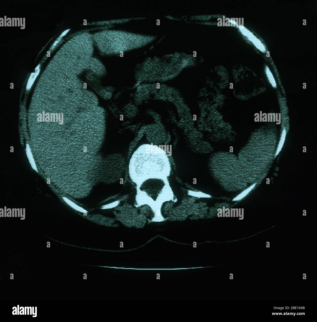 An axial (cross sectional) CT image through the lungs of a 53 year old female. The scan shows tortuosity and prominence of vasculature in the right superior mediastinum. Also present are calcified left hilar nodules and multiple punctate calcifications throughout the spleen which are consistent with granuloma. Stock Photohttps://www.alamy.com/image-license-details/?v=1https://www.alamy.com/an-axial-cross-sectional-ct-image-through-the-lungs-of-a-53-year-old-female-the-scan-shows-tortuosity-and-prominence-of-vasculature-in-the-right-superior-mediastinum-also-present-are-calcified-left-hilar-nodules-and-multiple-punctate-calcifications-throughout-the-spleen-which-are-consistent-with-granuloma-image352834619.html
RM2BE104B–An axial (cross sectional) CT image through the lungs of a 53 year old female. The scan shows tortuosity and prominence of vasculature in the right superior mediastinum. Also present are calcified left hilar nodules and multiple punctate calcifications throughout the spleen which are consistent with granuloma.
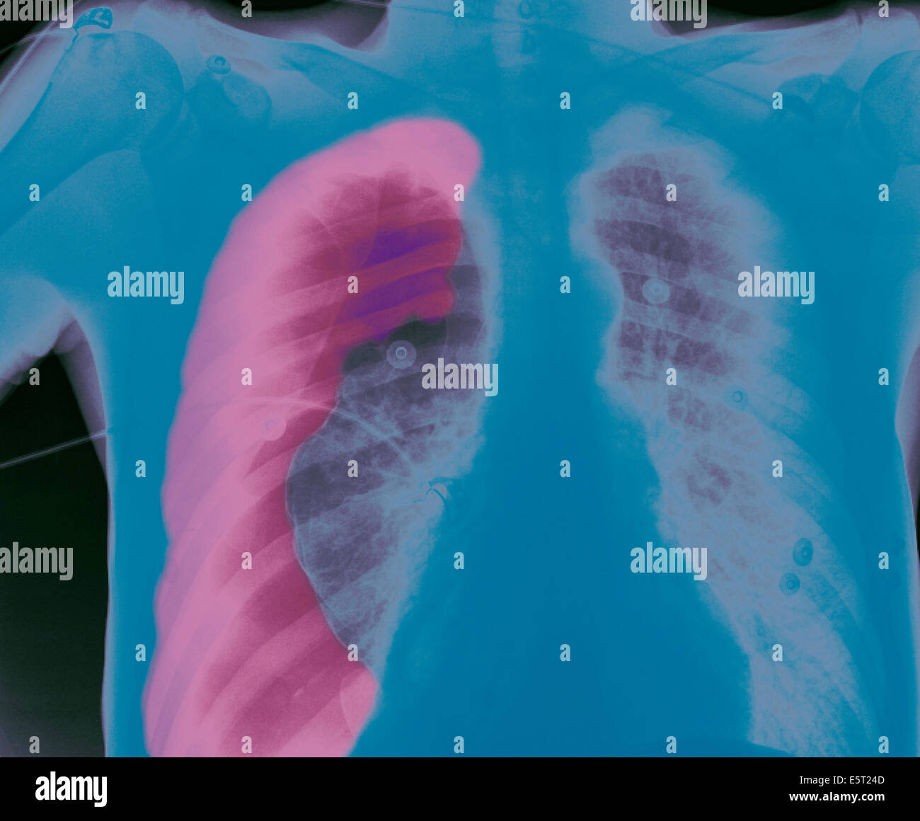 X-ray of the chest of a patient with a pneumothorax, or collapsed lung, The right lung (left on X-ray) has collapsed due to a Stock Photohttps://www.alamy.com/image-license-details/?v=1https://www.alamy.com/stock-photo-x-ray-of-the-chest-of-a-patient-with-a-pneumothorax-or-collapsed-lung-72443293.html
RME5T24D–X-ray of the chest of a patient with a pneumothorax, or collapsed lung, The right lung (left on X-ray) has collapsed due to a
 CHINA WHITE, (aka GWANG TIN LUNG FU WUI), from left: Russell Wong, Steven Vincent Leigh, Billy Drago, 1989, © Imperial Stock Photohttps://www.alamy.com/image-license-details/?v=1https://www.alamy.com/stock-photo-china-white-aka-gwang-tin-lung-fu-wui-from-left-russell-wong-steven-72381651.html
RME5N7EY–CHINA WHITE, (aka GWANG TIN LUNG FU WUI), from left: Russell Wong, Steven Vincent Leigh, Billy Drago, 1989, © Imperial
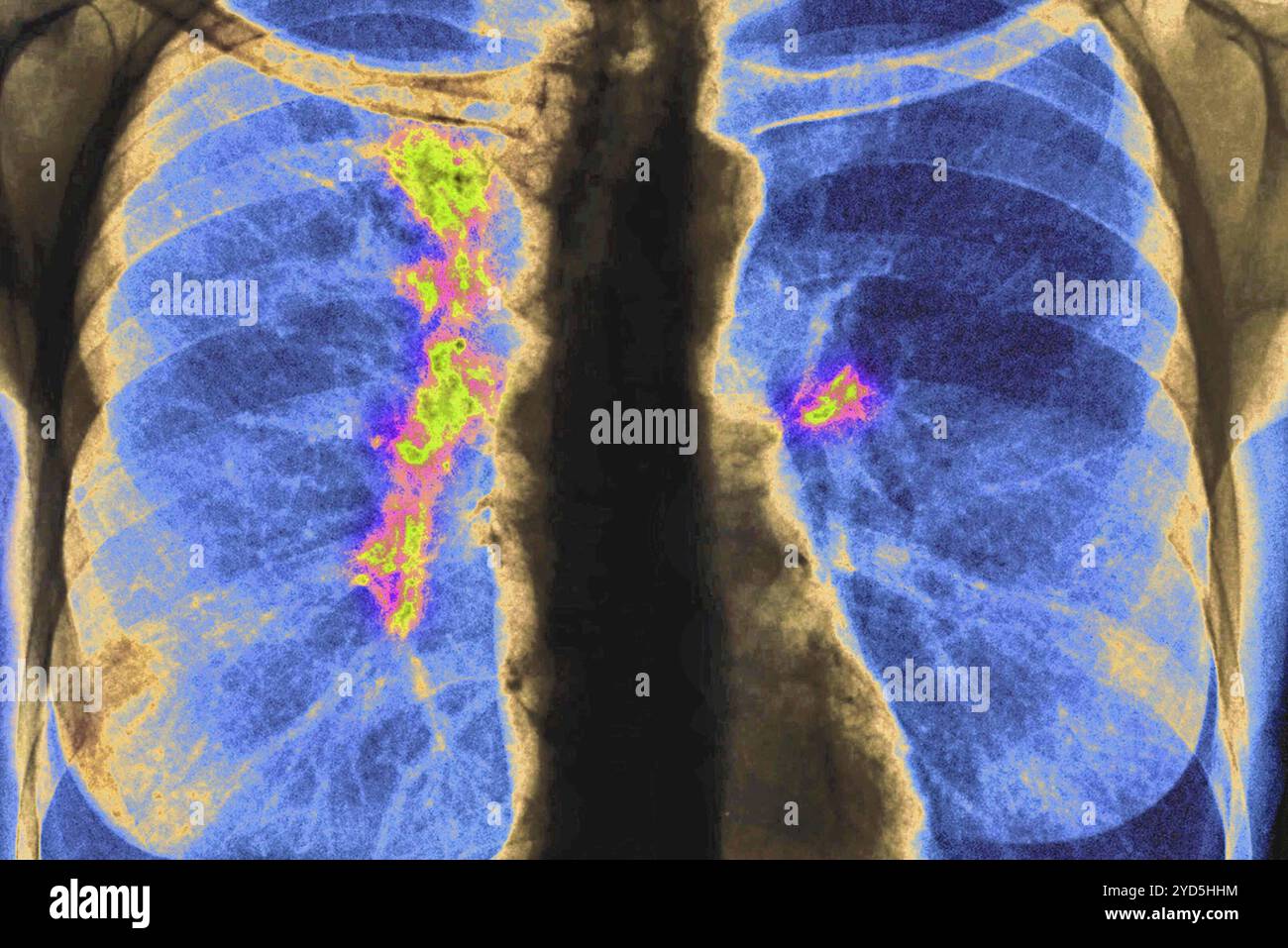 Right and left lung cancer (stage 1). Front chest x-ray. Stock Photohttps://www.alamy.com/image-license-details/?v=1https://www.alamy.com/right-and-left-lung-cancer-stage-1-front-chest-x-ray-image627687360.html
RM2YD5HHM–Right and left lung cancer (stage 1). Front chest x-ray.
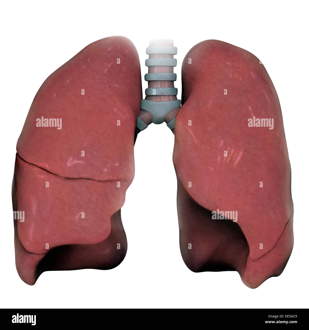 3D model of right and left human lung. Stock Photohttps://www.alamy.com/image-license-details/?v=1https://www.alamy.com/stock-image-3d-model-of-right-and-left-human-lung-163616441.html
RFKE5AC9–3D model of right and left human lung.
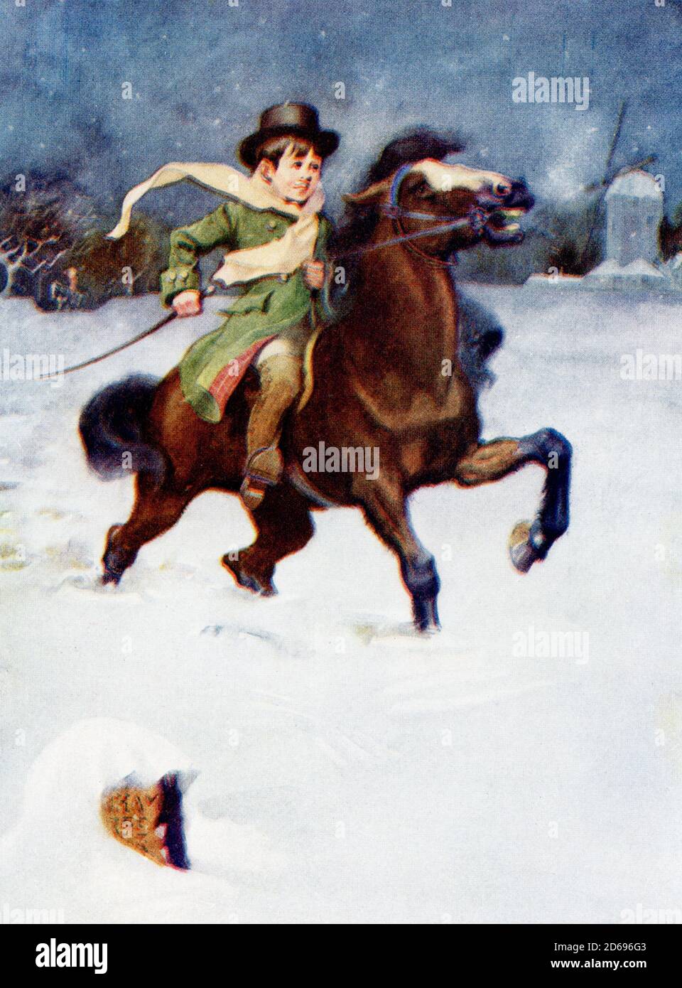 The caption of the 1917 illustration reads: Horatio Nelson and his pony struggled on riding to school. Nelson was born in 1758 in Norfolk, England. At the Battle of Trafalgar in 1805 (part of the Napoleonic Wars), British Admiral Horatio Nelson was aboard the vessel Victory, whose Captain was Thomas Hardy. Nelson struck by a bullet that entered his left shoulder, pierced his lung, and settled at the bottom of his spine. He died soon after. The British won the battle. Stock Photohttps://www.alamy.com/image-license-details/?v=1https://www.alamy.com/the-caption-of-the-1917-illustration-reads-horatio-nelson-and-his-pony-struggled-on-riding-to-school-nelson-was-born-in-1758-in-norfolk-england-at-the-battle-of-trafalgar-in-1805-part-of-the-napoleonic-wars-british-admiral-horatio-nelson-was-aboard-the-vessel-victory-whose-captain-was-thomas-hardy-nelson-struck-by-a-bullet-that-entered-his-left-shoulder-pierced-his-lung-and-settled-at-the-bottom-of-his-spine-he-died-soon-after-the-british-won-the-battle-image382518755.html
RF2D696G3–The caption of the 1917 illustration reads: Horatio Nelson and his pony struggled on riding to school. Nelson was born in 1758 in Norfolk, England. At the Battle of Trafalgar in 1805 (part of the Napoleonic Wars), British Admiral Horatio Nelson was aboard the vessel Victory, whose Captain was Thomas Hardy. Nelson struck by a bullet that entered his left shoulder, pierced his lung, and settled at the bottom of his spine. He died soon after. The British won the battle.
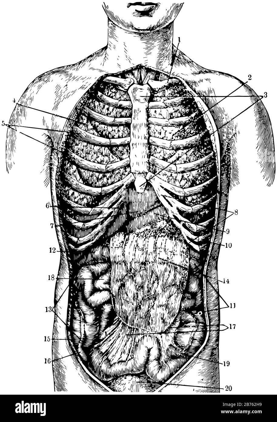 Collar bone and Left Lung, vintage line drawing or engraving illustration. Stock Vectorhttps://www.alamy.com/image-license-details/?v=1https://www.alamy.com/collar-bone-and-left-lung-vintage-line-drawing-or-engraving-illustration-image348643717.html
RF2B762H9–Collar bone and Left Lung, vintage line drawing or engraving illustration.
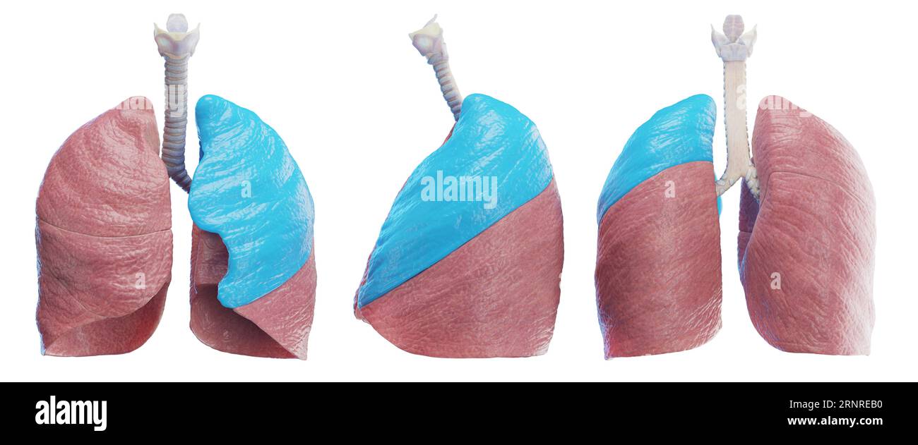 Left lung, illustration Stock Photohttps://www.alamy.com/image-license-details/?v=1https://www.alamy.com/left-lung-illustration-image564155732.html
RF2RNREB0–Left lung, illustration
 (170802) -- JILIN, Aug. 2, 2017 (Xinhua) -- Yue Ruhua (left) introduces treatment of lung disease to 76-year-old Sun Fuxin at Dakouqin Village of Houtuanshan Town in Jilin City, northeast China's Jilin Province, July 31, 2017. 64-year-old Yue Ruhua has been a rural doctor for 37 years. Different from doctors in urban area, rural doctors must make house calls frequently. According to Yue Ruhua's wife Jing Yuhong, he fell down on rugged road and stayed up late to rescue patients countless times. In 2013, Yue Ruhua was diagnosed with acute myocardial infarction and sent to intensive care unit. Vi Stock Photohttps://www.alamy.com/image-license-details/?v=1https://www.alamy.com/170802-jilin-aug-2-2017-xinhua-yue-ruhua-left-introduces-treatment-image151555377.html
RMJPFXC1–(170802) -- JILIN, Aug. 2, 2017 (Xinhua) -- Yue Ruhua (left) introduces treatment of lung disease to 76-year-old Sun Fuxin at Dakouqin Village of Houtuanshan Town in Jilin City, northeast China's Jilin Province, July 31, 2017. 64-year-old Yue Ruhua has been a rural doctor for 37 years. Different from doctors in urban area, rural doctors must make house calls frequently. According to Yue Ruhua's wife Jing Yuhong, he fell down on rugged road and stayed up late to rescue patients countless times. In 2013, Yue Ruhua was diagnosed with acute myocardial infarction and sent to intensive care unit. Vi
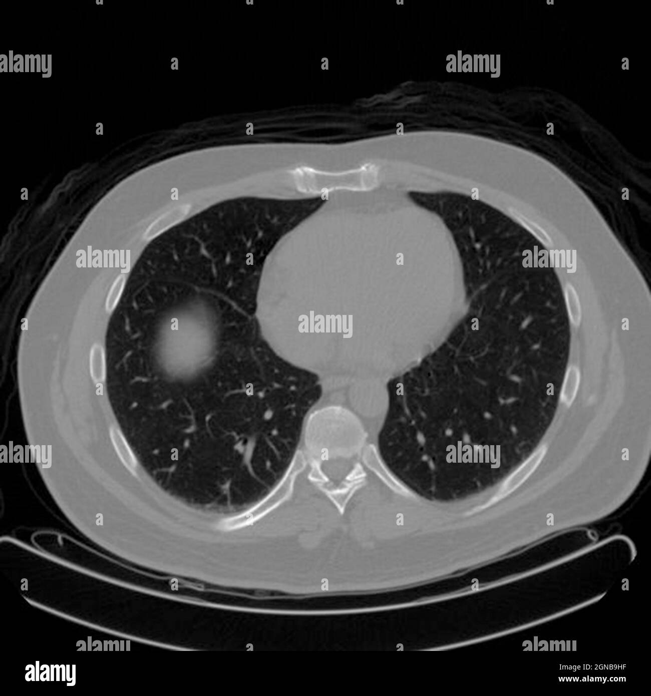 Positron emission tomography (PET) scan of a male, 54 year old patient. A tumour can be seen in the left upper lobe of his lungs Stock Photohttps://www.alamy.com/image-license-details/?v=1https://www.alamy.com/positron-emission-tomography-pet-scan-of-a-male-54-year-old-patient-a-tumour-can-be-seen-in-the-left-upper-lobe-of-his-lungs-image443415995.html
RM2GNB9HF–Positron emission tomography (PET) scan of a male, 54 year old patient. A tumour can be seen in the left upper lobe of his lungs
 Former Nottinghamshire Police officer Timothy Allatt (right) arrives at Mansfield Magistrates Court where he is charged with common assault due to using excessive force during an arrest on July 25, 2010, when the alleged victim was left with a collapsed lung. Stock Photohttps://www.alamy.com/image-license-details/?v=1https://www.alamy.com/stock-photo-former-nottinghamshire-police-officer-timothy-allatt-right-arrives-106064847.html
RMG4FJP7–Former Nottinghamshire Police officer Timothy Allatt (right) arrives at Mansfield Magistrates Court where he is charged with common assault due to using excessive force during an arrest on July 25, 2010, when the alleged victim was left with a collapsed lung.
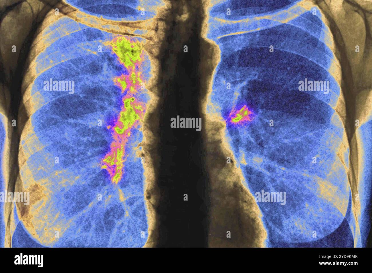 Right and left lung cancer stage 1. Front chest x-ray. Lung cancer 016836 027 Stock Photohttps://www.alamy.com/image-license-details/?v=1https://www.alamy.com/right-and-left-lung-cancer-stage-1-front-chest-x-ray-lung-cancer-016836-027-image627776819.html
RM2YD9KMK–Right and left lung cancer stage 1. Front chest x-ray. Lung cancer 016836 027
 Bad Rothenfelde, Germany. 21st Jan, 2022. Nurse Rüdiger Fels (l) sits in front of a bodyplethysmograph, for examining lung parameters, in a room at the Teutoburger Wald Clinic, a rehab clinic for post-Covid sufferers. Lower Saxony's Health Minister Daniela Behrens (SPD, 2nd from left) visits the facility. Credit: Friso Gentsch/dpa/Alamy Live News Stock Photohttps://www.alamy.com/image-license-details/?v=1https://www.alamy.com/bad-rothenfelde-germany-21st-jan-2022-nurse-rdiger-fels-l-sits-in-front-of-a-bodyplethysmograph-for-examining-lung-parameters-in-a-room-at-the-teutoburger-wald-clinic-a-rehab-clinic-for-post-covid-sufferers-lower-saxonys-health-minister-daniela-behrens-spd-2nd-from-left-visits-the-facility-credit-friso-gentschdpaalamy-live-news-image457729656.html
RM2HGKARM–Bad Rothenfelde, Germany. 21st Jan, 2022. Nurse Rüdiger Fels (l) sits in front of a bodyplethysmograph, for examining lung parameters, in a room at the Teutoburger Wald Clinic, a rehab clinic for post-Covid sufferers. Lower Saxony's Health Minister Daniela Behrens (SPD, 2nd from left) visits the facility. Credit: Friso Gentsch/dpa/Alamy Live News
 Luohu area of Shenzhen City, (left) with Nam Hang Police Post (on hillside, right), New Territories, Hong Kong/Shenzhen Border Area China, March 2021 Stock Photohttps://www.alamy.com/image-license-details/?v=1https://www.alamy.com/luohu-area-of-shenzhen-city-left-with-nam-hang-police-post-on-hillside-right-new-territories-hong-kongshenzhen-border-area-china-march-2021-image417556265.html
RM2F7997N–Luohu area of Shenzhen City, (left) with Nam Hang Police Post (on hillside, right), New Territories, Hong Kong/Shenzhen Border Area China, March 2021
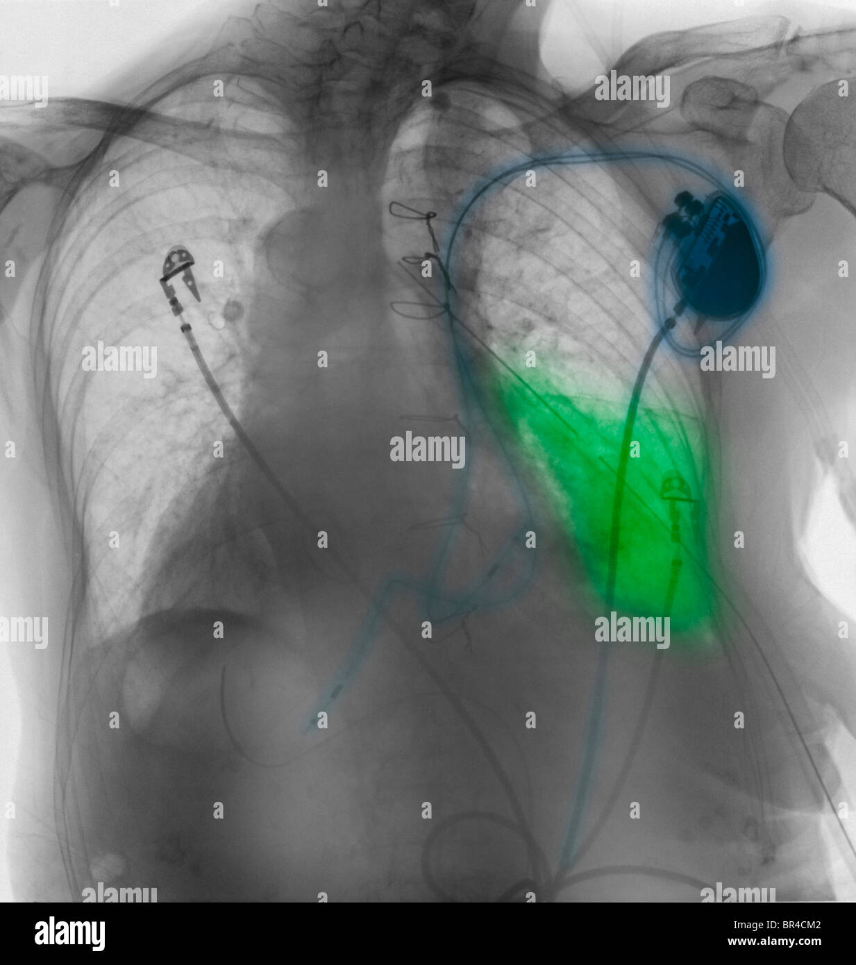 chest x-ray showing a pacemaker and an area of pneumonia in the left lower lobe of a 78 year old woman Stock Photohttps://www.alamy.com/image-license-details/?v=1https://www.alamy.com/stock-photo-chest-x-ray-showing-a-pacemaker-and-an-area-of-pneumonia-in-the-left-31445234.html
RMBR4CM2–chest x-ray showing a pacemaker and an area of pneumonia in the left lower lobe of a 78 year old woman
 Photoalbum Fisherman: Third Karakoru expedition, 1929-1930 Description: Album sheet with four photographs. Top left: trade caravan with dromedaries at Daulat beg öldi; lower left: J.A. Sillem at work at Daulat beg öldi; upper right: expedition members in Ship-Shape valley; lower right: Lung-NAK valley seen south Date: 1929/08/14 Location: India, Karakorum, Pakistan Keywords: mountains , dromedaries, expeditions, caravans, coolies Personal name: Sillem, J.A. Stock Photohttps://www.alamy.com/image-license-details/?v=1https://www.alamy.com/photoalbum-fisherman-third-karakoru-expedition-1929-1930-description-album-sheet-with-four-photographs-top-left-trade-caravan-with-dromedaries-at-daulat-beg-ldi-lower-left-ja-sillem-at-work-at-daulat-beg-ldi-upper-right-expedition-members-in-ship-shape-valley-lower-right-lung-nak-valley-seen-south-date-19290814-location-india-karakorum-pakistan-keywords-mountains-dromedaries-expeditions-caravans-coolies-personal-name-sillem-ja-image340776920.html
RM2APBMC8–Photoalbum Fisherman: Third Karakoru expedition, 1929-1930 Description: Album sheet with four photographs. Top left: trade caravan with dromedaries at Daulat beg öldi; lower left: J.A. Sillem at work at Daulat beg öldi; upper right: expedition members in Ship-Shape valley; lower right: Lung-NAK valley seen south Date: 1929/08/14 Location: India, Karakorum, Pakistan Keywords: mountains , dromedaries, expeditions, caravans, coolies Personal name: Sillem, J.A.
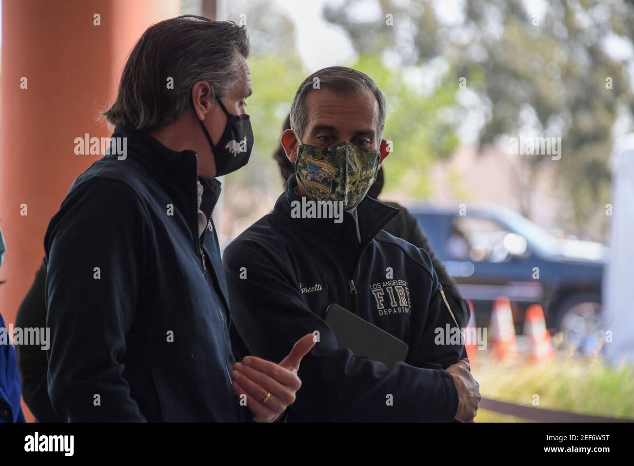 California Gov. Gavin Newsom, left, talks to Los Angeles Mayor Eric Garcetti during a press conference relating to the opening of a state and federal Stock Photohttps://www.alamy.com/image-license-details/?v=1https://www.alamy.com/california-gov-gavin-newsom-left-talks-to-los-angeles-mayor-eric-garcetti-during-a-press-conference-relating-to-the-opening-of-a-state-and-federal-image405209780.html
RF2EF6W5T–California Gov. Gavin Newsom, left, talks to Los Angeles Mayor Eric Garcetti during a press conference relating to the opening of a state and federal
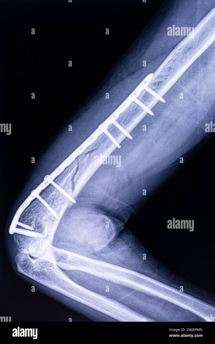 X-ray image of osteosynthesis of the humerus of the left arm of a man close-up Stock Photohttps://www.alamy.com/image-license-details/?v=1https://www.alamy.com/x-ray-image-of-osteosynthesis-of-the-humerus-of-the-left-arm-of-a-man-close-up-image596365861.html
RF2WJ6PMN–X-ray image of osteosynthesis of the humerus of the left arm of a man close-up
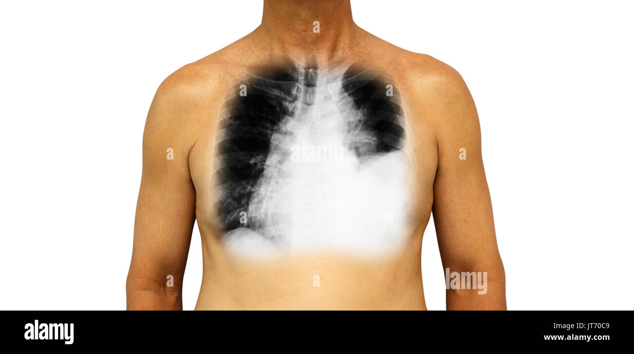 Lung cancer . Human chest and x-ray show pleural effusion left lung due to lung cancer . Stock Photohttps://www.alamy.com/image-license-details/?v=1https://www.alamy.com/lung-cancer-human-chest-and-x-ray-show-pleural-effusion-left-lung-image152588697.html
RFJT70C9–Lung cancer . Human chest and x-ray show pleural effusion left lung due to lung cancer .
 Pulmonary Embolism, Illustration Stock Photohttps://www.alamy.com/image-license-details/?v=1https://www.alamy.com/pulmonary-embolism-illustration-image353190388.html
RM2BEH5XC–Pulmonary Embolism, Illustration
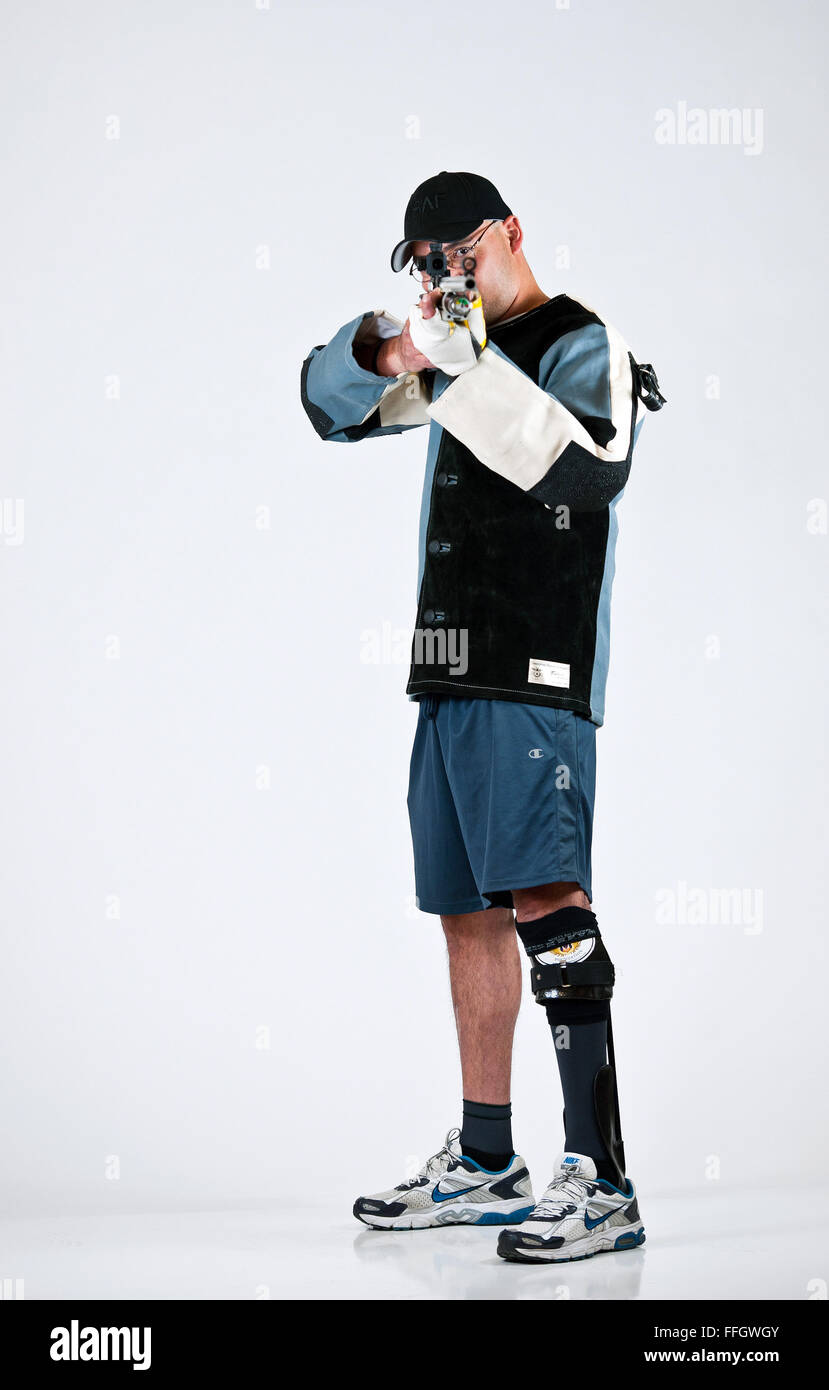 Name: Master Sgt. Christopher Aguilera Age: 37 Hometown: El Paso, Texas Current residence: Las Vegas, Nev. Years in service: 18 Injury/disability: In a 2010 helicopter crash in Afghanistan, he sustained a broken ankle, back, collarbone, hip, jaw, sternum, punctured lung and left upper hamstring, burns over 20 percent of his body, and traumatic brain injury. Sport/sports: Shooting, track and field, wheelchair basketball and sitting volleyball What do the Warrior Games mean to you? To me, it’s the camaraderie between fellow wounded wa Stock Photohttps://www.alamy.com/image-license-details/?v=1https://www.alamy.com/stock-photo-name-master-sgt-christopher-aguilera-age-37-hometown-el-paso-texas-95642987.html
RMFFGWGY–Name: Master Sgt. Christopher Aguilera Age: 37 Hometown: El Paso, Texas Current residence: Las Vegas, Nev. Years in service: 18 Injury/disability: In a 2010 helicopter crash in Afghanistan, he sustained a broken ankle, back, collarbone, hip, jaw, sternum, punctured lung and left upper hamstring, burns over 20 percent of his body, and traumatic brain injury. Sport/sports: Shooting, track and field, wheelchair basketball and sitting volleyball What do the Warrior Games mean to you? To me, it’s the camaraderie between fellow wounded wa
 EAT DRINK MAN WOMAN SIHUNG LUNG left Date: 1994 Stock Photohttps://www.alamy.com/image-license-details/?v=1https://www.alamy.com/eat-drink-man-woman-sihung-lung-left-date-1994-image156875873.html
RMK368NN–EAT DRINK MAN WOMAN SIHUNG LUNG left Date: 1994
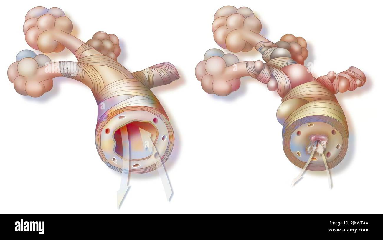 Asthma: healthy bronchiole (left) and asthmatic (right). Stock Photohttps://www.alamy.com/image-license-details/?v=1https://www.alamy.com/asthma-healthy-bronchiole-left-and-asthmatic-right-image476926306.html
RF2JKWTAA–Asthma: healthy bronchiole (left) and asthmatic (right).
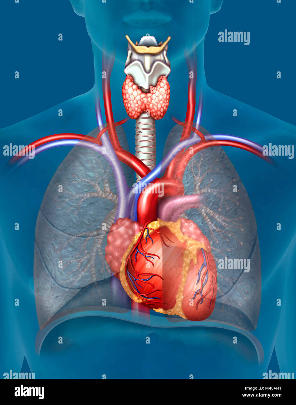 El aparato cardiovascular, capaz de distribuir el oxígeno a través del bombeo de la sangre y canalización de la misma en función de las necesidades me Stock Photohttps://www.alamy.com/image-license-details/?v=1https://www.alamy.com/stock-photo-el-aparato-cardiovascular-capaz-de-distribuir-el-oxgeno-a-travs-del-174566029.html
RFM404N1–El aparato cardiovascular, capaz de distribuir el oxígeno a través del bombeo de la sangre y canalización de la misma en función de las necesidades me
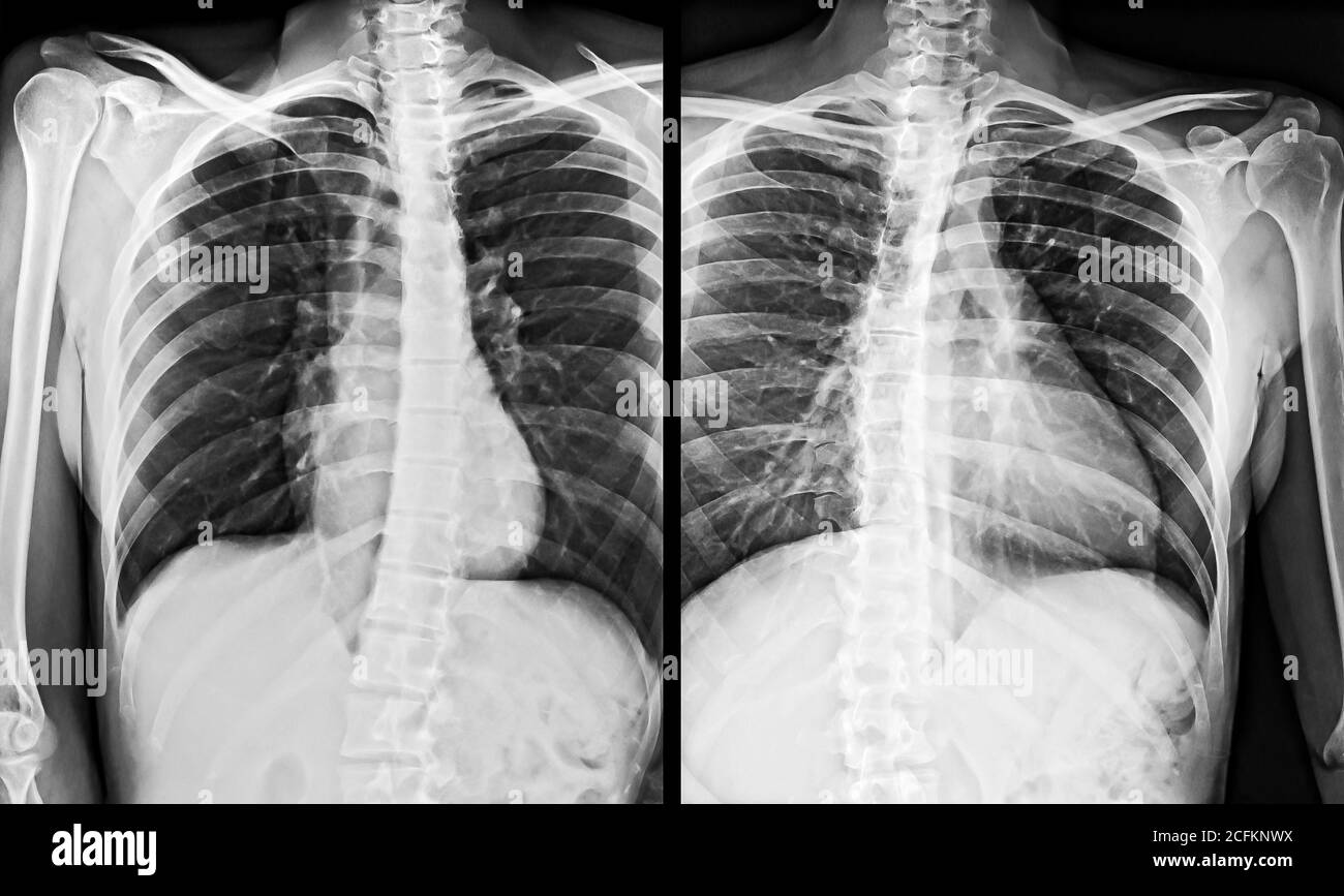 X-Ray Image of the human Chest, left and right side. Stock Photohttps://www.alamy.com/image-license-details/?v=1https://www.alamy.com/x-ray-image-of-the-human-chest-left-and-right-side-image371071846.html
RF2CFKNWX–X-Ray Image of the human Chest, left and right side.
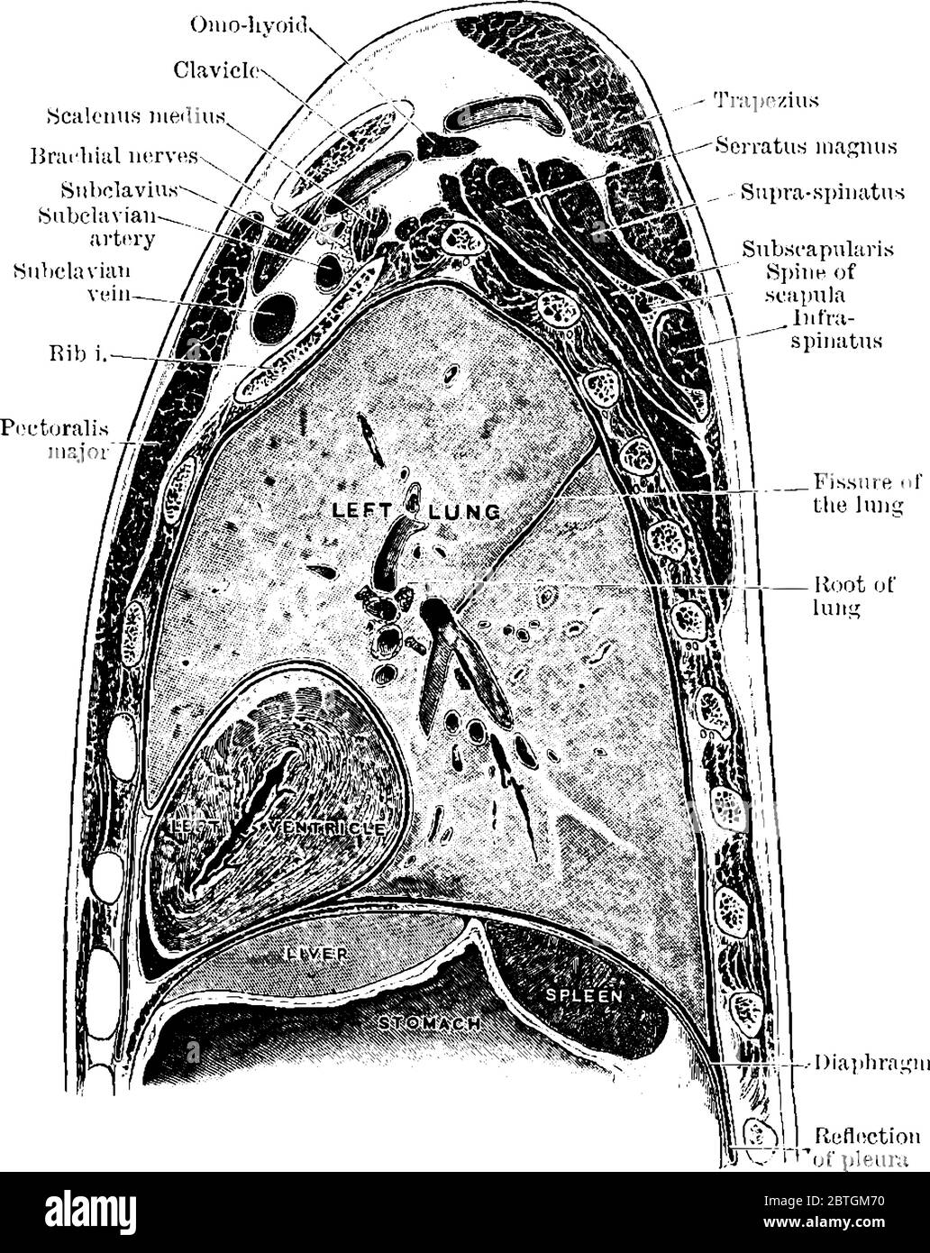 The sagittal section through the left shoulder, lung, and apex of the heart, vintage line drawing or engraving illustration. Stock Vectorhttps://www.alamy.com/image-license-details/?v=1https://www.alamy.com/the-sagittal-section-through-the-left-shoulder-lung-and-apex-of-the-heart-vintage-line-drawing-or-engraving-illustration-image359326212.html
RF2BTGM70–The sagittal section through the left shoulder, lung, and apex of the heart, vintage line drawing or engraving illustration.
 Coloured computed tomography (CT) scan of a chest from above showing an azygos lobe in the left lung (segmented area centre-left). An azygos lobe is an anatomical variant in the upper lobe of the lung in which a segment of the lung is separated by a deep groove called an azygos fissure, which consists of the azygos vein and pleura (thin tissue). It is a rare variation, occurring in around 0.3% of people during embryonic development. It causes no symptoms, but may make certain surgical procedures more difficult. Stock Photohttps://www.alamy.com/image-license-details/?v=1https://www.alamy.com/coloured-computed-tomography-ct-scan-of-a-chest-from-above-showing-an-azygos-lobe-in-the-left-lung-segmented-area-centre-left-an-azygos-lobe-is-an-anatomical-variant-in-the-upper-lobe-of-the-lung-in-which-a-segment-of-the-lung-is-separated-by-a-deep-groove-called-an-azygos-fissure-which-consists-of-the-azygos-vein-and-pleura-thin-tissue-it-is-a-rare-variation-occurring-in-around-03-of-people-during-embryonic-development-it-causes-no-symptoms-but-may-make-certain-surgical-procedures-more-difficult-image612233854.html
RF2XG1JEP–Coloured computed tomography (CT) scan of a chest from above showing an azygos lobe in the left lung (segmented area centre-left). An azygos lobe is an anatomical variant in the upper lobe of the lung in which a segment of the lung is separated by a deep groove called an azygos fissure, which consists of the azygos vein and pleura (thin tissue). It is a rare variation, occurring in around 0.3% of people during embryonic development. It causes no symptoms, but may make certain surgical procedures more difficult.
 (left to right) The Hong Kong Bar Association vice-chairman Jose Antonio Maurellet, chairman Victor Dawes and vice-chairman Derek Chan Ching-lung spoke to the media in Beijing. 12APR23 SCMP/ Kahon Chan Stock Photohttps://www.alamy.com/image-license-details/?v=1https://www.alamy.com/left-to-right-the-hong-kong-bar-association-vice-chairman-jose-antonio-maurellet-chairman-victor-dawes-and-vice-chairman-derek-chan-ching-lung-spoke-to-the-media-in-beijing-12apr23-scmp-kahon-chan-image546317066.html
RM2PMPTYP–(left to right) The Hong Kong Bar Association vice-chairman Jose Antonio Maurellet, chairman Victor Dawes and vice-chairman Derek Chan Ching-lung spoke to the media in Beijing. 12APR23 SCMP/ Kahon Chan
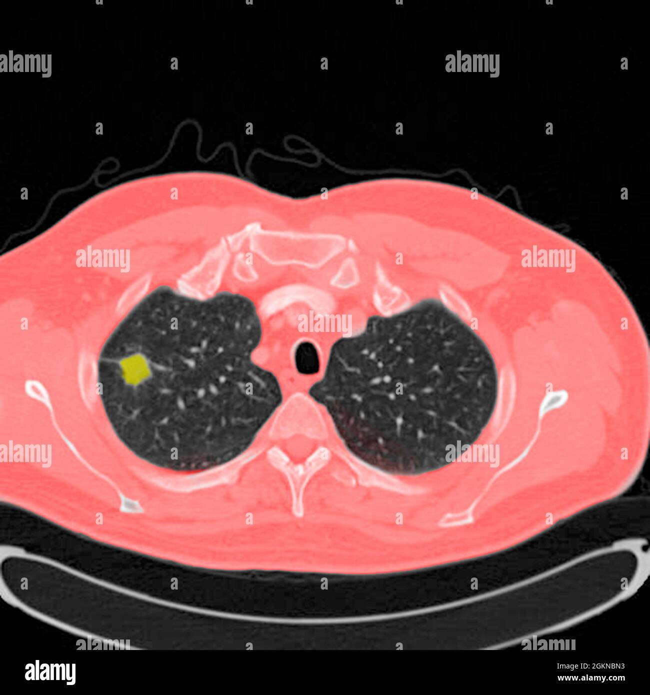 Chest CT scan (X-ray computed tomography) of a male 54 year old patient. A tumour can be seen in the left upper lobe of his lungs Stock Photohttps://www.alamy.com/image-license-details/?v=1https://www.alamy.com/chest-ct-scan-x-ray-computed-tomography-of-a-male-54-year-old-patient-a-tumour-can-be-seen-in-the-left-upper-lobe-of-his-lungs-image442407871.html
RM2GKNBN3–Chest CT scan (X-ray computed tomography) of a male 54 year old patient. A tumour can be seen in the left upper lobe of his lungs
 Health Secretary Andrew Lansley (far right) meets John Price ( left) and Colin Hawkey (centre), patients from the British Lung Foundation who both suffer from COPD (Chronic Obstructive Pulmonary Disease), at the launch of the Government Outcomes Strategy for COPD and Asthma at Richmond House, Whitehall, London. Stock Photohttps://www.alamy.com/image-license-details/?v=1https://www.alamy.com/stock-photo-health-secretary-andrew-lansley-far-right-meets-john-price-left-and-106032713.html
RMG4E5PH–Health Secretary Andrew Lansley (far right) meets John Price ( left) and Colin Hawkey (centre), patients from the British Lung Foundation who both suffer from COPD (Chronic Obstructive Pulmonary Disease), at the launch of the Government Outcomes Strategy for COPD and Asthma at Richmond House, Whitehall, London.
 From left to right, Susan Shurin, deputy director of the National Heart, Lung, and Blood Institute at the National Institute of Health; Thomas Perls, associate professor of medicine and director of the New England Centenarian Study at the Boston University School of Medicine; Alan Rogol, professor of clinical pediatrics at the University of Virginia and Indiana University School of Medicine; and Todd Schlifstein of the Department of Rehabilitation Medicine in the Hospital for Joint Disease, testify before a House Oversight and Government Reform Committee hearing on the myths and facts about hu Stock Photohttps://www.alamy.com/image-license-details/?v=1https://www.alamy.com/from-left-to-right-susan-shurin-deputy-director-of-the-national-heart-lung-and-blood-institute-at-the-national-institute-of-health-thomas-perls-associate-professor-of-medicine-and-director-of-the-new-england-centenarian-study-at-the-boston-university-school-of-medicine-alan-rogol-professor-of-clinical-pediatrics-at-the-university-of-virginia-and-indiana-university-school-of-medicine-and-todd-schlifstein-of-the-department-of-rehabilitation-medicine-in-the-hospital-for-joint-disease-testify-before-a-house-oversight-and-government-reform-committee-hearing-on-the-myths-and-facts-about-hu-image258466742.html
RMW0E4Y2–From left to right, Susan Shurin, deputy director of the National Heart, Lung, and Blood Institute at the National Institute of Health; Thomas Perls, associate professor of medicine and director of the New England Centenarian Study at the Boston University School of Medicine; Alan Rogol, professor of clinical pediatrics at the University of Virginia and Indiana University School of Medicine; and Todd Schlifstein of the Department of Rehabilitation Medicine in the Hospital for Joint Disease, testify before a House Oversight and Government Reform Committee hearing on the myths and facts about hu
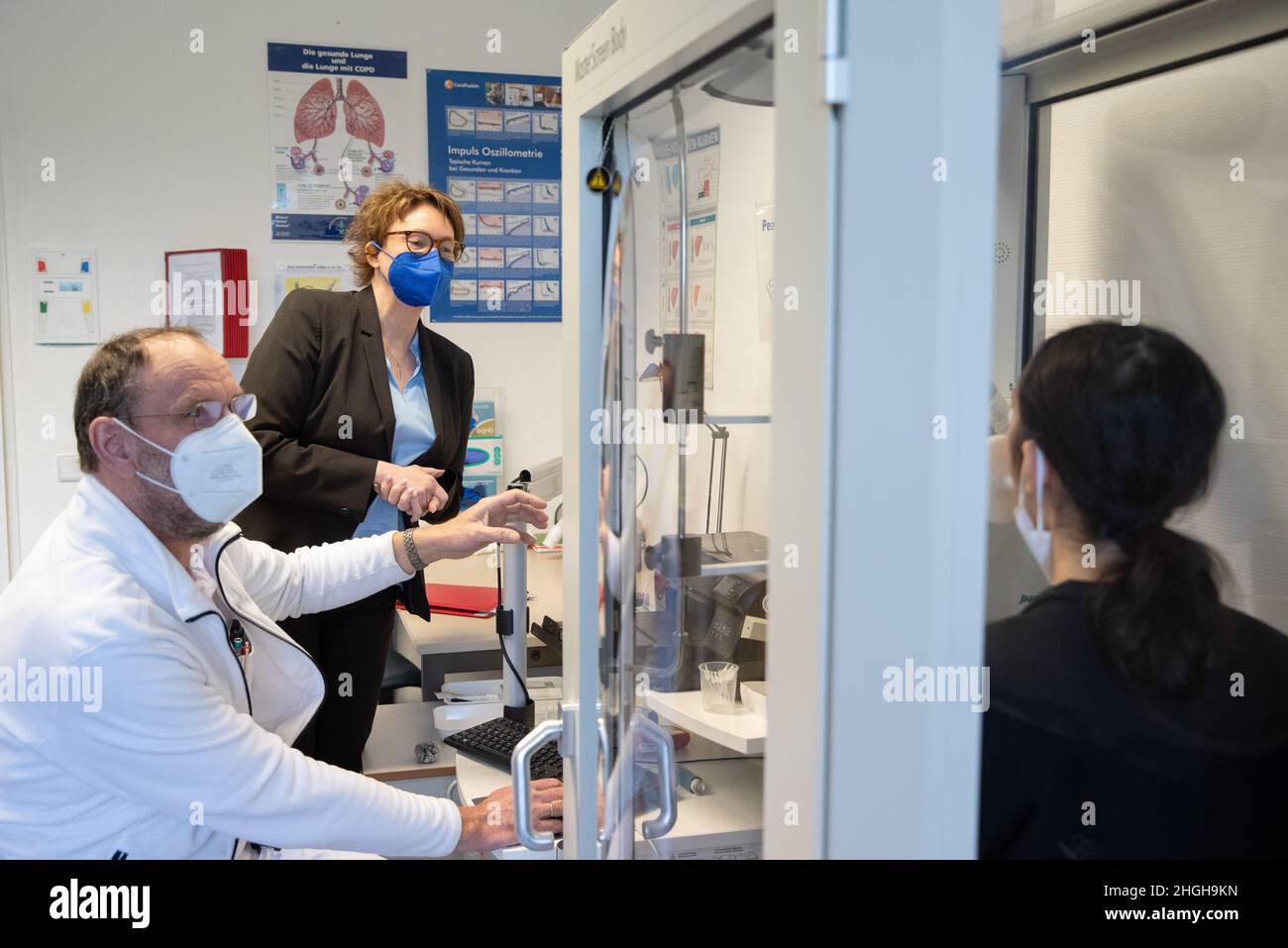 Bad Rothenfelde, Germany. 21st Jan, 2022. Nurse Rüdiger Fels (l) sits in front of a bodyplethysmograph, for examining lung parameters, in a room at the Teutoburger Wald Clinic, a rehab clinic for post-Covid sufferers. Lower Saxony's Health Minister Daniela Behrens (SPD, 2nd from left) visits the facility. Credit: Friso Gentsch/dpa/Alamy Live News Stock Photohttps://www.alamy.com/image-license-details/?v=1https://www.alamy.com/bad-rothenfelde-germany-21st-jan-2022-nurse-rdiger-fels-l-sits-in-front-of-a-bodyplethysmograph-for-examining-lung-parameters-in-a-room-at-the-teutoburger-wald-clinic-a-rehab-clinic-for-post-covid-sufferers-lower-saxonys-health-minister-daniela-behrens-spd-2nd-from-left-visits-the-facility-credit-friso-gentschdpaalamy-live-news-image457684857.html
RM2HGH9KN–Bad Rothenfelde, Germany. 21st Jan, 2022. Nurse Rüdiger Fels (l) sits in front of a bodyplethysmograph, for examining lung parameters, in a room at the Teutoburger Wald Clinic, a rehab clinic for post-Covid sufferers. Lower Saxony's Health Minister Daniela Behrens (SPD, 2nd from left) visits the facility. Credit: Friso Gentsch/dpa/Alamy Live News
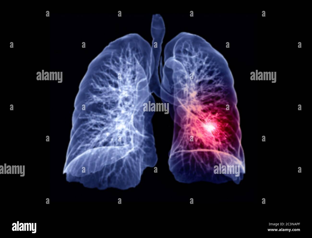 Selective Focus of CT Chest or Lung 3D rendering image showing lesion left area for diagnosis TB,tuberculosis and covid-19 . Stock Photohttps://www.alamy.com/image-license-details/?v=1https://www.alamy.com/selective-focus-of-ct-chest-or-lung-3d-rendering-image-showing-lesion-left-area-for-diagnosis-tbtuberculosis-and-covid-19-image363731159.html
RF2C3NAPF–Selective Focus of CT Chest or Lung 3D rendering image showing lesion left area for diagnosis TB,tuberculosis and covid-19 .
 Chest x-ray of an 84 year old woman with a small right pleural effusion and a moderate left pleural effusion. Stock Photohttps://www.alamy.com/image-license-details/?v=1https://www.alamy.com/stock-photo-chest-x-ray-of-an-84-year-old-woman-with-a-small-right-pleural-effusion-26899457.html
RMBFNAEW–Chest x-ray of an 84 year old woman with a small right pleural effusion and a moderate left pleural effusion.
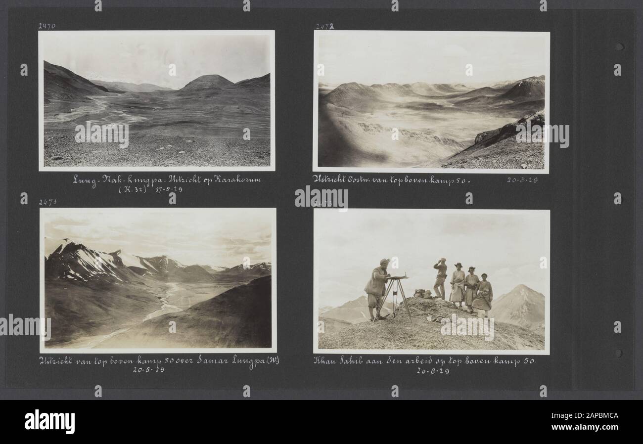 Photoalbum Fisherman: Third Karakoru expedition, 1929-1930 Description: Album sheet with four photographs. Top left: view of the Karakorum (K. 32) from the Lung-Nak-Lungpa; lower left: view from the top above Camp 50 over Samar Lungpa in the west direction; upper right: view from the top above Camp 50 in the east direction; lower right: topographer Khan Sahib working at the top top camp 50 Date: 1929/08/17 Location: India, Karakorum, Pakistan Keywords: mountains, cartography, coolies Stock Photohttps://www.alamy.com/image-license-details/?v=1https://www.alamy.com/photoalbum-fisherman-third-karakoru-expedition-1929-1930-description-album-sheet-with-four-photographs-top-left-view-of-the-karakorum-k-32-from-the-lung-nak-lungpa-lower-left-view-from-the-top-above-camp-50-over-samar-lungpa-in-the-west-direction-upper-right-view-from-the-top-above-camp-50-in-the-east-direction-lower-right-topographer-khan-sahib-working-at-the-top-top-camp-50-date-19290817-location-india-karakorum-pakistan-keywords-mountains-cartography-coolies-image340776922.html
RM2APBMCA–Photoalbum Fisherman: Third Karakoru expedition, 1929-1930 Description: Album sheet with four photographs. Top left: view of the Karakorum (K. 32) from the Lung-Nak-Lungpa; lower left: view from the top above Camp 50 over Samar Lungpa in the west direction; upper right: view from the top above Camp 50 in the east direction; lower right: topographer Khan Sahib working at the top top camp 50 Date: 1929/08/17 Location: India, Karakorum, Pakistan Keywords: mountains, cartography, coolies
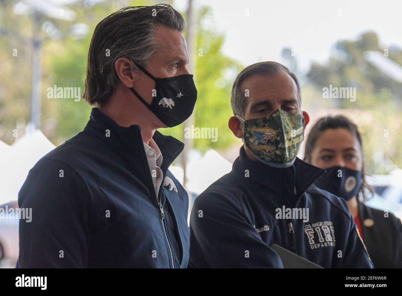 California Gov. Gavin Newsom, left, talks to Los Angeles Mayor Eric Garcetti during a press conference relating to the opening of a state and federal Stock Photohttps://www.alamy.com/image-license-details/?v=1https://www.alamy.com/california-gov-gavin-newsom-left-talks-to-los-angeles-mayor-eric-garcetti-during-a-press-conference-relating-to-the-opening-of-a-state-and-federal-image405209807.html
RF2EF6W6R–California Gov. Gavin Newsom, left, talks to Los Angeles Mayor Eric Garcetti during a press conference relating to the opening of a state and federal
 Hollywood film director John Woo, left, and his wife Annie Woo Ngau Chun-lung arrive for the premiere of the movie The Founding of A Republic, in Beij Stock Photohttps://www.alamy.com/image-license-details/?v=1https://www.alamy.com/hollywood-film-director-john-woo-left-and-his-wife-annie-woo-ngau-chun-lung-arrive-for-the-premiere-of-the-movie-the-founding-of-a-republic-in-beij-image263974821.html
RMW9D2G5–Hollywood film director John Woo, left, and his wife Annie Woo Ngau Chun-lung arrive for the premiere of the movie The Founding of A Republic, in Beij
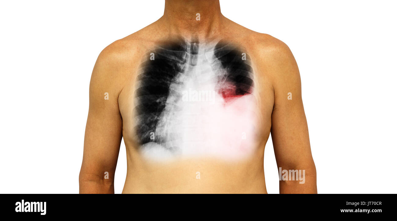 Lung cancer . Human chest and x-ray show pleural effusion left lung due to lung cancer . Stock Photohttps://www.alamy.com/image-license-details/?v=1https://www.alamy.com/lung-cancer-human-chest-and-x-ray-show-pleural-effusion-left-lung-image152588711.html
RFJT70CR–Lung cancer . Human chest and x-ray show pleural effusion left lung due to lung cancer .
 New York Police Department Detective James Zadroga’s father, Joseph, right, and his daughter, Tyler Ann, left, stand after being recognized by United States President Donald J. Trump as he makes remarks prior to signing H.R. 1327, an act to permanently authorize the September 11th victim compensation fund, in the Rose Garden of the White House in Washington, DC on Monday, July 29, 2019. Detective Zadroga spent more than 450 hours serving at Ground Zero where he contracted lung disease, from which he died in 2006.Credit: Ron Sachs / Pool via CNP /MediaPunch Stock Photohttps://www.alamy.com/image-license-details/?v=1https://www.alamy.com/new-york-police-department-detective-james-zadrogas-father-joseph-right-and-his-daughter-tyler-ann-left-stand-after-being-recognized-by-united-states-president-donald-j-trump-as-he-makes-remarks-prior-to-signing-hr-1327-an-act-to-permanently-authorize-the-september-11th-victim-compensation-fund-in-the-rose-garden-of-the-white-house-in-washington-dc-on-monday-july-29-2019-detective-zadroga-spent-more-than-450-hours-serving-at-ground-zero-where-he-contracted-lung-disease-from-which-he-died-in-2006credit-ron-sachs-pool-via-cnp-mediapunch-image261629764.html
RMW5J7C4–New York Police Department Detective James Zadroga’s father, Joseph, right, and his daughter, Tyler Ann, left, stand after being recognized by United States President Donald J. Trump as he makes remarks prior to signing H.R. 1327, an act to permanently authorize the September 11th victim compensation fund, in the Rose Garden of the White House in Washington, DC on Monday, July 29, 2019. Detective Zadroga spent more than 450 hours serving at Ground Zero where he contracted lung disease, from which he died in 2006.Credit: Ron Sachs / Pool via CNP /MediaPunch
 Name: Master Sgt. Christopher Aguilera Age: 37 Hometown: El Paso, Texas Current residence: Las Vegas, Nev. Years in service: 18 Injury/disability: In a 2010 helicopter crash in Afghanistan, he sustained a broken ankle, back, collarbone, hip, jaw, sternum, punctured lung and left upper hamstring, burns over 20 percent of his body, and traumatic brain injury. Sport/sports: Shooting, track and field, wheelchair basketball and sitting volleyball What do the Warrior Games mean to you? To me, it’s the camaraderie between fellow wounded wa Stock Photohttps://www.alamy.com/image-license-details/?v=1https://www.alamy.com/stock-photo-name-master-sgt-christopher-aguilera-age-37-hometown-el-paso-texas-95642988.html
RMFFGWH0–Name: Master Sgt. Christopher Aguilera Age: 37 Hometown: El Paso, Texas Current residence: Las Vegas, Nev. Years in service: 18 Injury/disability: In a 2010 helicopter crash in Afghanistan, he sustained a broken ankle, back, collarbone, hip, jaw, sternum, punctured lung and left upper hamstring, burns over 20 percent of his body, and traumatic brain injury. Sport/sports: Shooting, track and field, wheelchair basketball and sitting volleyball What do the Warrior Games mean to you? To me, it’s the camaraderie between fellow wounded wa
 Cold upper respiratory tract text clearance to the left of 16 to 9 Stock Photohttps://www.alamy.com/image-license-details/?v=1https://www.alamy.com/stock-photo-cold-upper-respiratory-tract-text-clearance-to-the-left-of-16-to-9-139533099.html
RFJ307TY–Cold upper respiratory tract text clearance to the left of 16 to 9
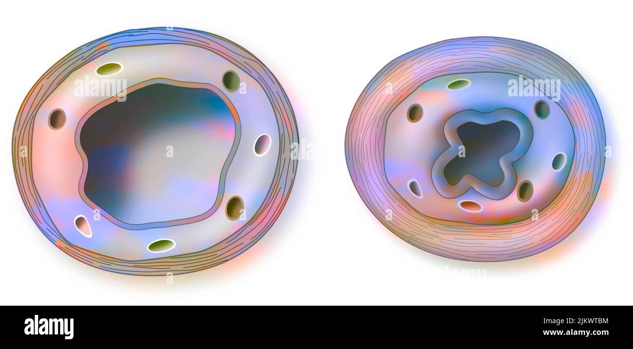 Asthma: healthy (left) and inflamed (right) bronchiole. Stock Photohttps://www.alamy.com/image-license-details/?v=1https://www.alamy.com/asthma-healthy-left-and-inflamed-right-bronchiole-image476926344.html
RF2JKWTBM–Asthma: healthy (left) and inflamed (right) bronchiole.
 Closeup hand of a man with cigarette. People smoking in smoking zone area of the mall and left cigarette in ashtray. Quit smoke or smoking cessation Stock Photohttps://www.alamy.com/image-license-details/?v=1https://www.alamy.com/closeup-hand-of-a-man-with-cigarette-people-smoking-in-smoking-zone-area-of-the-mall-and-left-cigarette-in-ashtray-quit-smoke-or-smoking-cessation-image262563541.html
RFW74PD9–Closeup hand of a man with cigarette. People smoking in smoking zone area of the mall and left cigarette in ashtray. Quit smoke or smoking cessation
 PUSHING HANDS, (aka TUI SHOU), from left: Deb Snyder, Sihung Lung, 1992, © Cinepix Film Properties/courtesy Everett Collection Stock Photohttps://www.alamy.com/image-license-details/?v=1https://www.alamy.com/stock-photo-pushing-hands-aka-tui-shou-from-left-deb-snyder-sihung-lung-1992-cinepix-72392387.html
RME5NN6B–PUSHING HANDS, (aka TUI SHOU), from left: Deb Snyder, Sihung Lung, 1992, © Cinepix Film Properties/courtesy Everett Collection
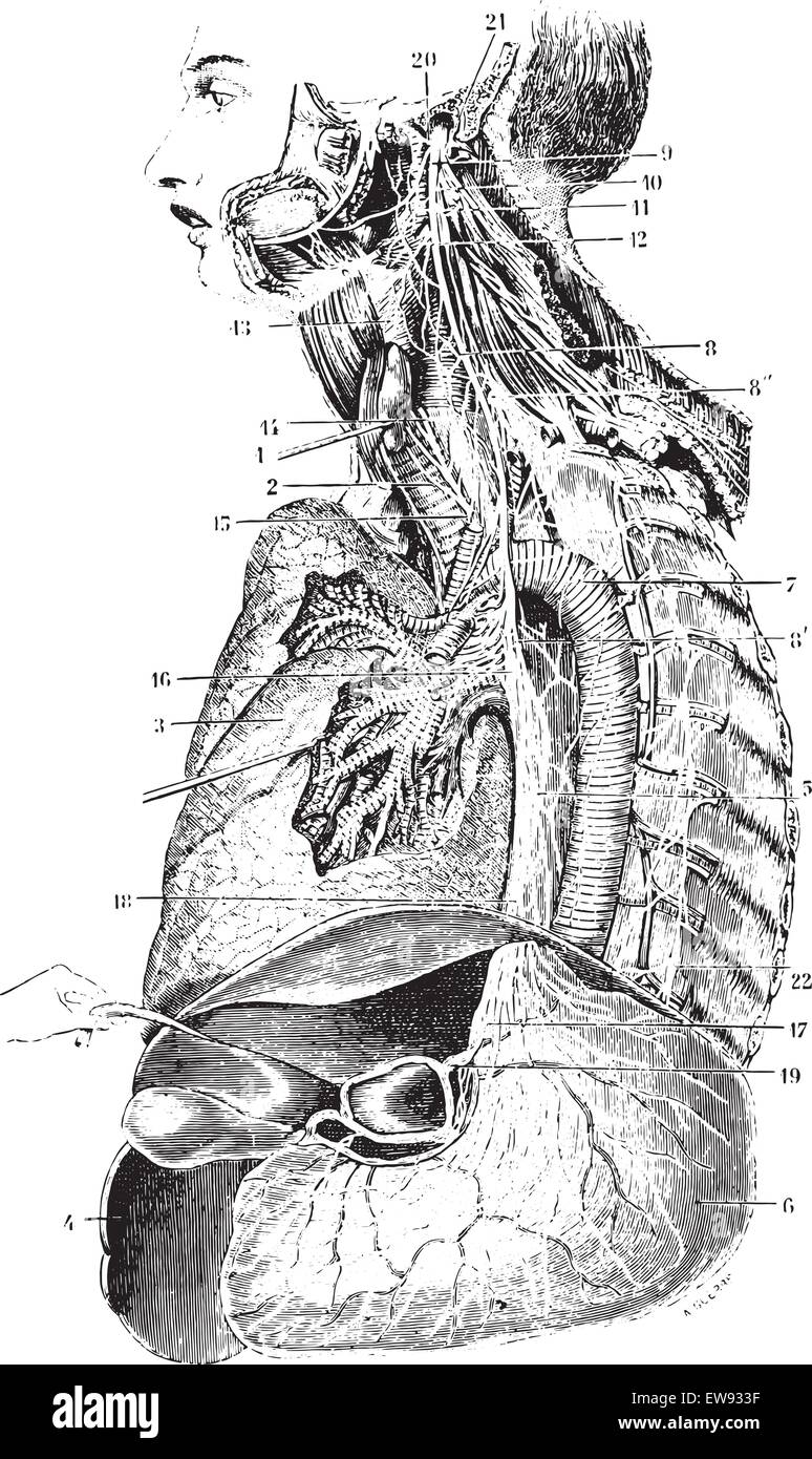 Left Vagus nerve or Pneumogastric nerve or Cranial nerve X, vintage engraved illustration. Usual Medicine Dictionary by Dr Labar Stock Vectorhttps://www.alamy.com/image-license-details/?v=1https://www.alamy.com/stock-photo-left-vagus-nerve-or-pneumogastric-nerve-or-cranial-nerve-x-vintage-84407891.html
RFEW933F–Left Vagus nerve or Pneumogastric nerve or Cranial nerve X, vintage engraved illustration. Usual Medicine Dictionary by Dr Labar
 Coloured computed tomography (CT) scan of a chest from above showing an azygos lobe in the left lung (segmented area centre-left). An azygos lobe is an anatomical variant in the upper lobe of the lung in which a segment of the lung is separated by a deep groove called an azygos fissure, which consists of the azygos vein and pleura (thin tissue). It is a rare variation, occurring in around 0.3% of people during embryonic development. It causes no symptoms, but may make certain surgical procedures more difficult. Stock Photohttps://www.alamy.com/image-license-details/?v=1https://www.alamy.com/coloured-computed-tomography-ct-scan-of-a-chest-from-above-showing-an-azygos-lobe-in-the-left-lung-segmented-area-centre-left-an-azygos-lobe-is-an-anatomical-variant-in-the-upper-lobe-of-the-lung-in-which-a-segment-of-the-lung-is-separated-by-a-deep-groove-called-an-azygos-fissure-which-consists-of-the-azygos-vein-and-pleura-thin-tissue-it-is-a-rare-variation-occurring-in-around-03-of-people-during-embryonic-development-it-causes-no-symptoms-but-may-make-certain-surgical-procedures-more-difficult-image612233858.html
RF2XG1JEX–Coloured computed tomography (CT) scan of a chest from above showing an azygos lobe in the left lung (segmented area centre-left). An azygos lobe is an anatomical variant in the upper lobe of the lung in which a segment of the lung is separated by a deep groove called an azygos fissure, which consists of the azygos vein and pleura (thin tissue). It is a rare variation, occurring in around 0.3% of people during embryonic development. It causes no symptoms, but may make certain surgical procedures more difficult.
 (left to right) The Hong Kong Bar Association vice-chairman Jose Antonio Maurellet, chairman Victor Dawes and vice-chairman Derek Chan Ching-lung spoke to the media in Beijing. 12APR23 SCMP/ Kahon Chan Stock Photohttps://www.alamy.com/image-license-details/?v=1https://www.alamy.com/left-to-right-the-hong-kong-bar-association-vice-chairman-jose-antonio-maurellet-chairman-victor-dawes-and-vice-chairman-derek-chan-ching-lung-spoke-to-the-media-in-beijing-12apr23-scmp-kahon-chan-image546410135.html
RM2PMY3KK–(left to right) The Hong Kong Bar Association vice-chairman Jose Antonio Maurellet, chairman Victor Dawes and vice-chairman Derek Chan Ching-lung spoke to the media in Beijing. 12APR23 SCMP/ Kahon Chan
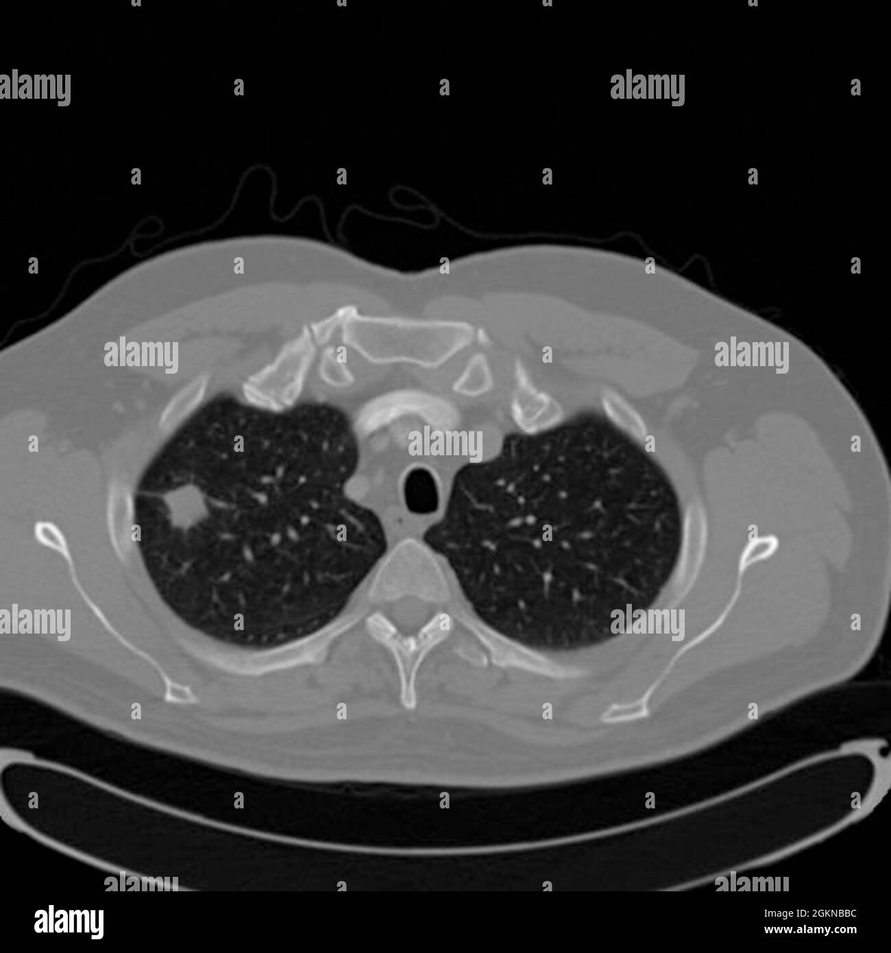 Chest CT scan (X-ray computed tomography) of a male 54 year old patient. A tumour can be seen in the left upper lobe of his lungs Stock Photohttps://www.alamy.com/image-license-details/?v=1https://www.alamy.com/chest-ct-scan-x-ray-computed-tomography-of-a-male-54-year-old-patient-a-tumour-can-be-seen-in-the-left-upper-lobe-of-his-lungs-image442407600.html
RM2GKNBBC–Chest CT scan (X-ray computed tomography) of a male 54 year old patient. A tumour can be seen in the left upper lobe of his lungs
 Health Secretary Andrew Lansley (far right) meets British Lung Foundation Chief Executive Dame Helena Shovelton (left) John Price (second left) and Colin Hawkey (second right), patients from the British Lung Foundation who both suffer from COPD (Chronic Obstructive Pulmonary Disease), at the launch of the Government Outcomes Strategy for COPD and Asthma at Richmond House, Whitehall, London. Stock Photohttps://www.alamy.com/image-license-details/?v=1https://www.alamy.com/stock-photo-health-secretary-andrew-lansley-far-right-meets-british-lung-foundation-106032717.html
RMG4E5PN–Health Secretary Andrew Lansley (far right) meets British Lung Foundation Chief Executive Dame Helena Shovelton (left) John Price (second left) and Colin Hawkey (second right), patients from the British Lung Foundation who both suffer from COPD (Chronic Obstructive Pulmonary Disease), at the launch of the Government Outcomes Strategy for COPD and Asthma at Richmond House, Whitehall, London.
 From left to right, Susan Shurin, deputy director of the National Heart, Lung, and Blood Institute at the National Institute of Health; Thomas Perls, associate professor of medicine and director of the New England Centenarian Study at the Boston University School of Medicine; Alan Rogol, professor of clinical pediatrics at the University of Virginia and Indiana University School of Medicine; and Todd Schlifstein of the Department of Rehabilitation Medicine in the Hospital for Joint Disease, are sworn in prior to testifying before a House Oversight and Government Reform Committee hearing on the Stock Photohttps://www.alamy.com/image-license-details/?v=1https://www.alamy.com/from-left-to-right-susan-shurin-deputy-director-of-the-national-heart-lung-and-blood-institute-at-the-national-institute-of-health-thomas-perls-associate-professor-of-medicine-and-director-of-the-new-england-centenarian-study-at-the-boston-university-school-of-medicine-alan-rogol-professor-of-clinical-pediatrics-at-the-university-of-virginia-and-indiana-university-school-of-medicine-and-todd-schlifstein-of-the-department-of-rehabilitation-medicine-in-the-hospital-for-joint-disease-are-sworn-in-prior-to-testifying-before-a-house-oversight-and-government-reform-committee-hearing-on-the-image258466745.html
RMW0E4Y5–From left to right, Susan Shurin, deputy director of the National Heart, Lung, and Blood Institute at the National Institute of Health; Thomas Perls, associate professor of medicine and director of the New England Centenarian Study at the Boston University School of Medicine; Alan Rogol, professor of clinical pediatrics at the University of Virginia and Indiana University School of Medicine; and Todd Schlifstein of the Department of Rehabilitation Medicine in the Hospital for Joint Disease, are sworn in prior to testifying before a House Oversight and Government Reform Committee hearing on the
 Dresden, Germany. 13th Mar, 2020. An intensive care bed on an intensive care unit at the University Hospital Dresden. On the left side of the bed is a heart-lung machine, on top are the monitors for monitoring vital functions. To the right of the bed is a ventilator and infusion equipment. With this technology, seriously ill patients can be optimally cared for. Credit: Ronald Bonss/dpa-Zentralbild/dpa/Alamy Live News Stock Photohttps://www.alamy.com/image-license-details/?v=1https://www.alamy.com/dresden-germany-13th-mar-2020-an-intensive-care-bed-on-an-intensive-care-unit-at-the-university-hospital-dresden-on-the-left-side-of-the-bed-is-a-heart-lung-machine-on-top-are-the-monitors-for-monitoring-vital-functions-to-the-right-of-the-bed-is-a-ventilator-and-infusion-equipment-with-this-technology-seriously-ill-patients-can-be-optimally-cared-for-credit-ronald-bonssdpa-zentralbilddpaalamy-live-news-image348615241.html
RM2B74P89–Dresden, Germany. 13th Mar, 2020. An intensive care bed on an intensive care unit at the University Hospital Dresden. On the left side of the bed is a heart-lung machine, on top are the monitors for monitoring vital functions. To the right of the bed is a ventilator and infusion equipment. With this technology, seriously ill patients can be optimally cared for. Credit: Ronald Bonss/dpa-Zentralbild/dpa/Alamy Live News
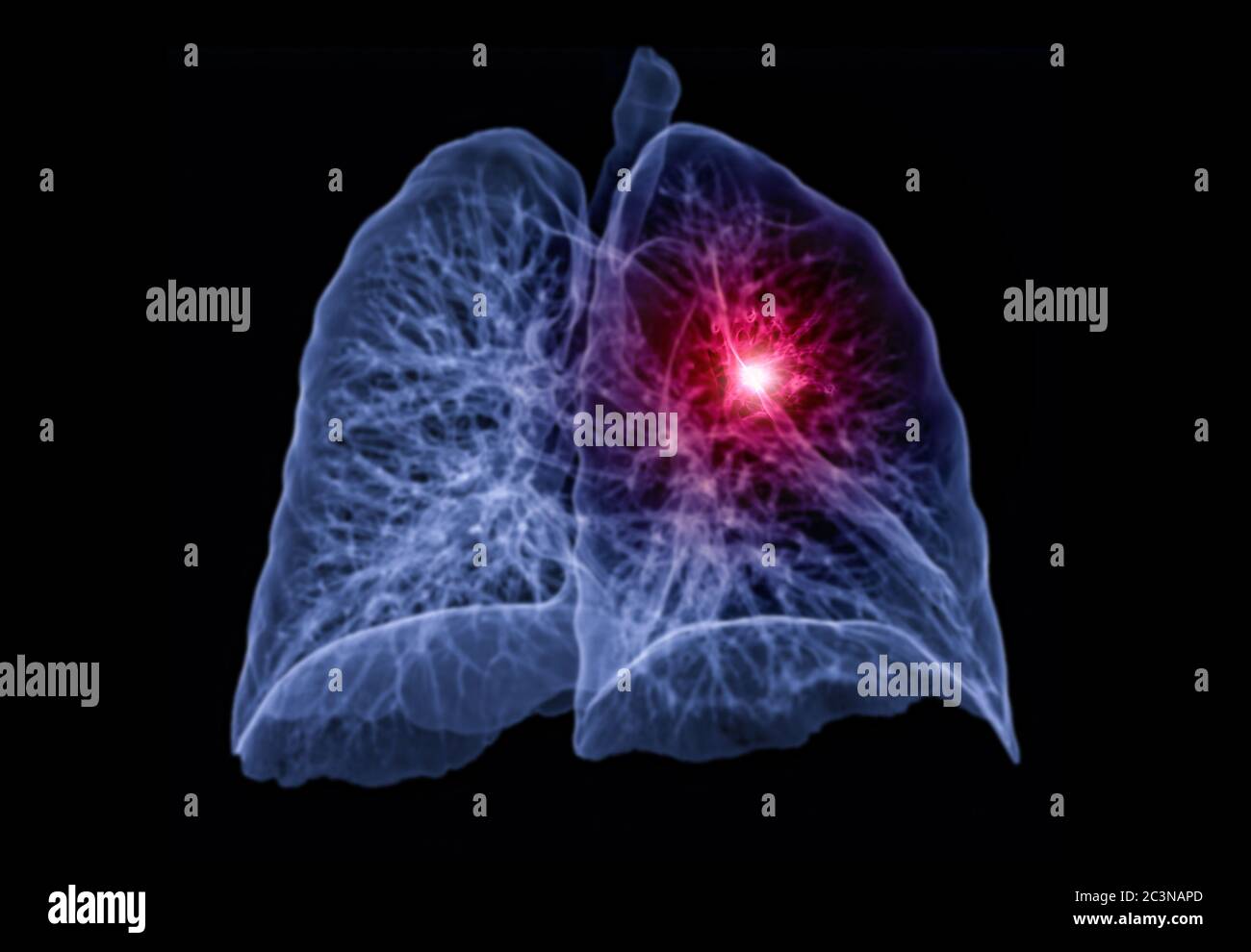 CT Chest or Lung 3D rendering image left oblique view showing lesion left area for diagnosis TB,tuberculosis and covid-19 . Stock Photohttps://www.alamy.com/image-license-details/?v=1https://www.alamy.com/ct-chest-or-lung-3d-rendering-image-left-oblique-view-showing-lesion-left-area-for-diagnosis-tbtuberculosis-and-covid-19-image363731157.html
RF2C3NAPD–CT Chest or Lung 3D rendering image left oblique view showing lesion left area for diagnosis TB,tuberculosis and covid-19 .
 Lateral view of a 3D CT scan showing a cancer in the left lung of a 77 year old woman Stock Photohttps://www.alamy.com/image-license-details/?v=1https://www.alamy.com/stock-photo-lateral-view-of-a-3d-ct-scan-showing-a-cancer-in-the-left-lung-of-30768134.html
RMBP1H1X–Lateral view of a 3D CT scan showing a cancer in the left lung of a 77 year old woman
 Chest radiograph showing a bilateral pulmonary infection experienced by this plague victim with a greater infection in the left lung, 1975. Image courtesy Centers for Disease Control and Prevention (CDC) / Dr Jack Poland. () Stock Photohttps://www.alamy.com/image-license-details/?v=1https://www.alamy.com/chest-radiograph-showing-a-bilateral-pulmonary-infection-experienced-by-this-plague-victim-with-a-greater-infection-in-the-left-lung-1975-image-courtesy-centers-for-disease-control-and-prevention-cdc-dr-jack-poland-image244836153.html
RMT69709–Chest radiograph showing a bilateral pulmonary infection experienced by this plague victim with a greater infection in the left lung, 1975. Image courtesy Centers for Disease Control and Prevention (CDC) / Dr Jack Poland. ()
 California Gov. Gavin Newsom, left, talks to Los Angeles Mayor Eric Garcetti during a press conference relating to the opening of a state and federal Stock Photohttps://www.alamy.com/image-license-details/?v=1https://www.alamy.com/california-gov-gavin-newsom-left-talks-to-los-angeles-mayor-eric-garcetti-during-a-press-conference-relating-to-the-opening-of-a-state-and-federal-image405209671.html
RF2EF6W1Y–California Gov. Gavin Newsom, left, talks to Los Angeles Mayor Eric Garcetti during a press conference relating to the opening of a state and federal
 Hollywood director John Woo, left, and his wife Annie Woo Ngau Chun-lung pose on the red carpet as they arrive at the 50th Golden Horse Awards ceremon Stock Photohttps://www.alamy.com/image-license-details/?v=1https://www.alamy.com/hollywood-director-john-woo-left-and-his-wife-annie-woo-ngau-chun-lung-pose-on-the-red-carpet-as-they-arrive-at-the-50th-golden-horse-awards-ceremon-image263728584.html
RMW91TE0–Hollywood director John Woo, left, and his wife Annie Woo Ngau Chun-lung pose on the red carpet as they arrive at the 50th Golden Horse Awards ceremon
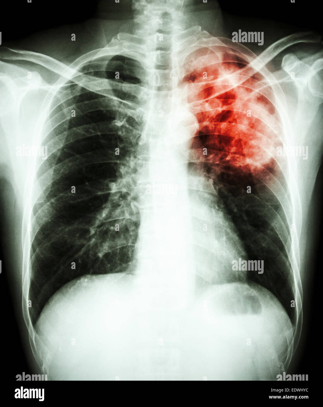 film chest x-ray show alveolar infiltrate at left upper lung due to Mycobacterium tuberculosis infection (Pulmonary Tuberculosis Stock Photohttps://www.alamy.com/image-license-details/?v=1https://www.alamy.com/stock-photo-film-chest-x-ray-show-alveolar-infiltrate-at-left-upper-lung-due-to-77394896.html
RFEDWHYC–film chest x-ray show alveolar infiltrate at left upper lung due to Mycobacterium tuberculosis infection (Pulmonary Tuberculosis
 Prague, Czech Republic. 22nd Nov, 2023. Slovak President Zuzana Caputova, frotn left, during her visit of the Motol teaching Hospital in support of the National Lung Transplantation Programme for the Slovak Republic, in Prague, Czech Republic, on November 22, 2023. On the right side is seen head of the surgical clinic Robert Lischke. Credit: Ondrej Deml/CTK Photo/Alamy Live News Stock Photohttps://www.alamy.com/image-license-details/?v=1https://www.alamy.com/prague-czech-republic-22nd-nov-2023-slovak-president-zuzana-caputova-frotn-left-during-her-visit-of-the-motol-teaching-hospital-in-support-of-the-national-lung-transplantation-programme-for-the-slovak-republic-in-prague-czech-republic-on-november-22-2023-on-the-right-side-is-seen-head-of-the-surgical-clinic-robert-lischke-credit-ondrej-demlctk-photoalamy-live-news-image573566634.html
RM2T9462J–Prague, Czech Republic. 22nd Nov, 2023. Slovak President Zuzana Caputova, frotn left, during her visit of the Motol teaching Hospital in support of the National Lung Transplantation Programme for the Slovak Republic, in Prague, Czech Republic, on November 22, 2023. On the right side is seen head of the surgical clinic Robert Lischke. Credit: Ondrej Deml/CTK Photo/Alamy Live News
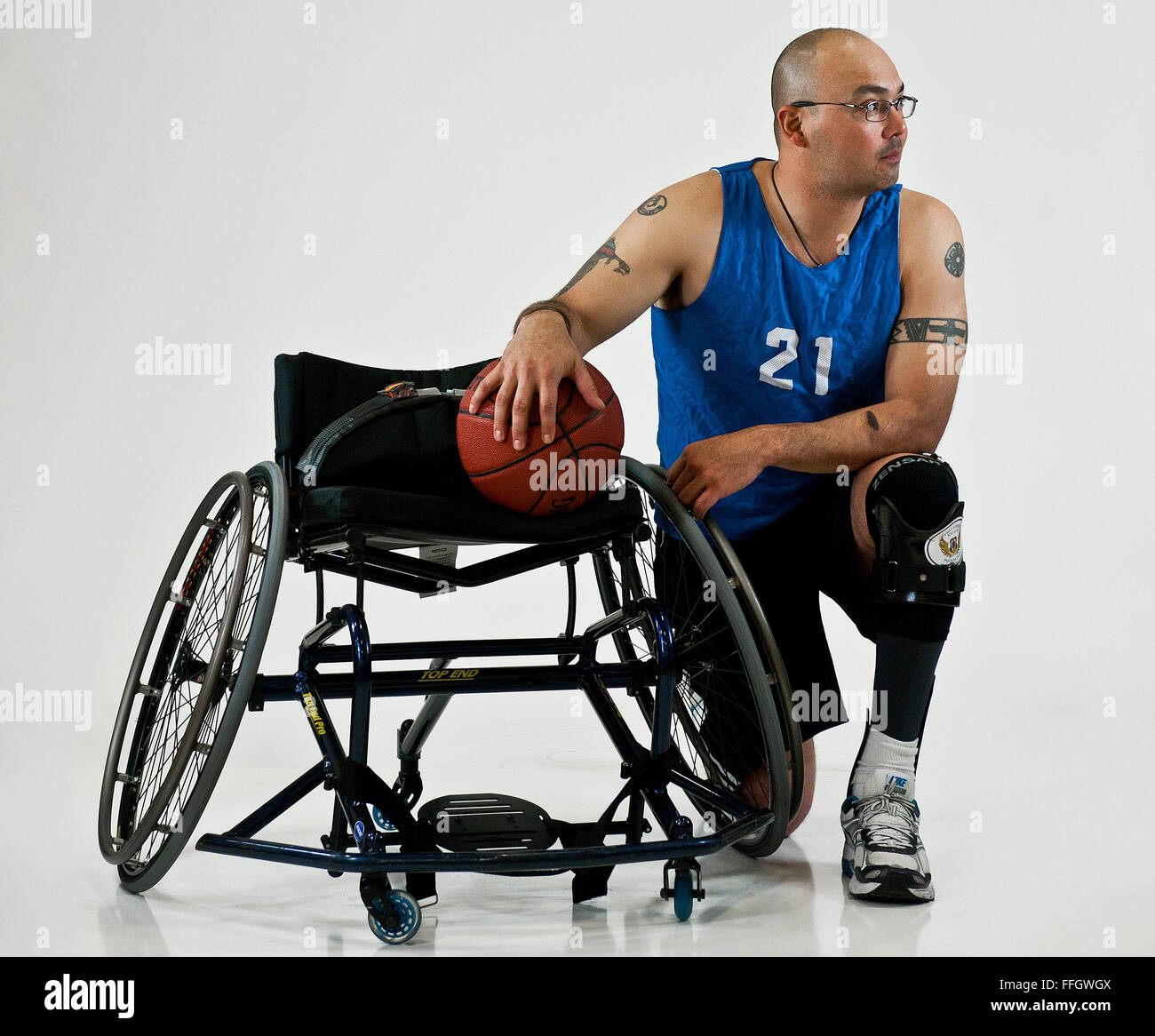 Name: Master Sgt. Christopher Aguilera Age: 37 Hometown: El Paso, Texas Current residence: Las Vegas, Nev. Years in service: 18 Injury/disability: In a 2010 helicopter crash in Afghanistan, he sustained a broken ankle, back, collarbone, hip, jaw, sternum, punctured lung and left upper hamstring, burns over 20 percent of his body, and traumatic brain injury. Sport/sports: Shooting, track and field, wheelchair basketball and sitting volleyball What do the Warrior Games mean to you? To me, it’s the camaraderie between fellow wounded wa Stock Photohttps://www.alamy.com/image-license-details/?v=1https://www.alamy.com/stock-photo-name-master-sgt-christopher-aguilera-age-37-hometown-el-paso-texas-95642986.html
RMFFGWGX–Name: Master Sgt. Christopher Aguilera Age: 37 Hometown: El Paso, Texas Current residence: Las Vegas, Nev. Years in service: 18 Injury/disability: In a 2010 helicopter crash in Afghanistan, he sustained a broken ankle, back, collarbone, hip, jaw, sternum, punctured lung and left upper hamstring, burns over 20 percent of his body, and traumatic brain injury. Sport/sports: Shooting, track and field, wheelchair basketball and sitting volleyball What do the Warrior Games mean to you? To me, it’s the camaraderie between fellow wounded wa
 Clipstone, Nottinghamshire, England, UK. 27th. November 2019. Jonathan Ashworth (Left) Labour's Shadow Secretary for Health and Jerry Hague (Right), Sherwood Labour Party candidate visiting the ex-mining community of Clipstone following Labour’s announcement on specialist lung clinics for retired miners. The only remaining building left standing of the colliery which was closed in 2003 are the grade II listed Headstocks, Europe’s tallest coal mining headstocks at over 200 feet. Credit: Alan Beastall/Alamy Live News Stock Photohttps://www.alamy.com/image-license-details/?v=1https://www.alamy.com/clipstone-nottinghamshire-england-uk-27th-november-2019-jonathan-ashworth-left-labours-shadow-secretary-for-health-and-jerry-hague-right-sherwood-labour-party-candidate-visiting-the-ex-mining-community-of-clipstone-following-labours-announcement-on-specialist-lung-clinics-for-retired-miners-the-only-remaining-building-left-standing-of-the-colliery-which-was-closed-in-2003-are-the-grade-ii-listed-headstocks-europes-tallest-coal-mining-headstocks-at-over-200-feet-credit-alan-beastallalamy-live-news-image334112789.html
RM2ABG47H–Clipstone, Nottinghamshire, England, UK. 27th. November 2019. Jonathan Ashworth (Left) Labour's Shadow Secretary for Health and Jerry Hague (Right), Sherwood Labour Party candidate visiting the ex-mining community of Clipstone following Labour’s announcement on specialist lung clinics for retired miners. The only remaining building left standing of the colliery which was closed in 2003 are the grade II listed Headstocks, Europe’s tallest coal mining headstocks at over 200 feet. Credit: Alan Beastall/Alamy Live News