Life cycle of red blood cell Stock Photos and Images
(14)See life cycle of red blood cell stock video clipsLife cycle of red blood cell Stock Photos and Images
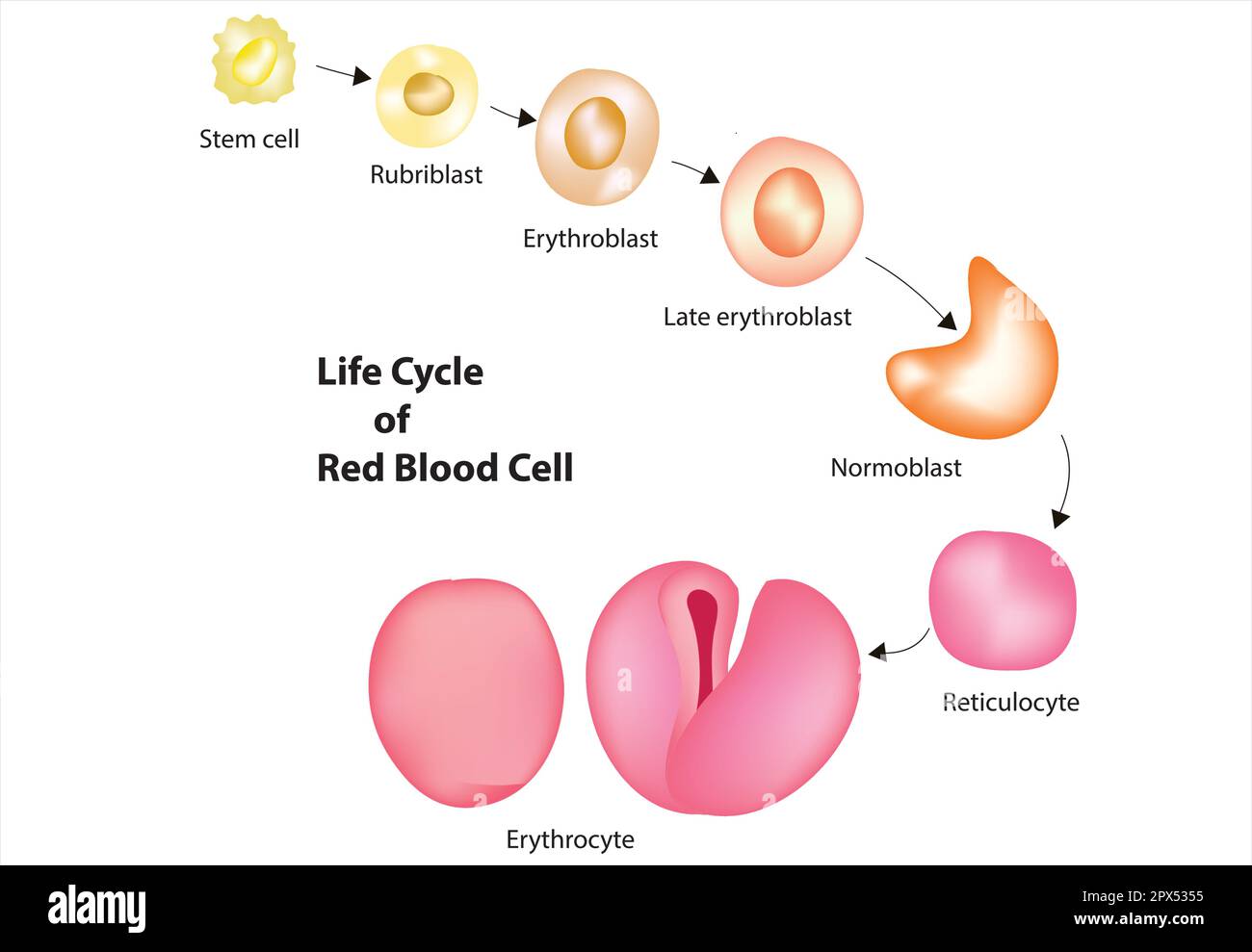 Red blood cell life cycle Stock Vectorhttps://www.alamy.com/image-license-details/?v=1https://www.alamy.com/red-blood-cell-life-cycle-image549614721.html
RF2PX5355–Red blood cell life cycle
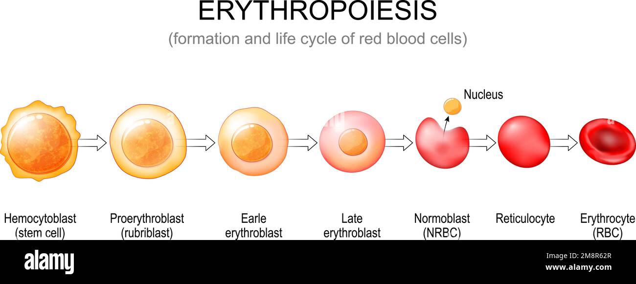 Erythropoiesis. Formation and life cycle of red blood cells from stem cell to Normoblast, Reticulocyte and Erythrocyte. Vector poster Stock Vectorhttps://www.alamy.com/image-license-details/?v=1https://www.alamy.com/erythropoiesis-formation-and-life-cycle-of-red-blood-cells-from-stem-cell-to-normoblast-reticulocyte-and-erythrocyte-vector-poster-image504527599.html
RF2M8R62R–Erythropoiesis. Formation and life cycle of red blood cells from stem cell to Normoblast, Reticulocyte and Erythrocyte. Vector poster
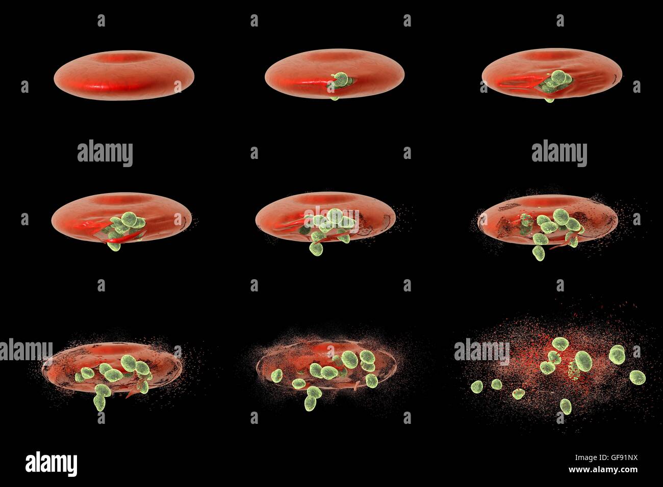 Computer illustration showing different stages of release of malaria merozoites from a red blood cell. Malaria is caused by Plasmodium sp. parasitic protozoans transmitted by female Anopheles sp. mosquitos. When the mosquito bites a person, sporozoites in Stock Photohttps://www.alamy.com/image-license-details/?v=1https://www.alamy.com/stock-photo-computer-illustration-showing-different-stages-of-release-of-malaria-112681014.html
RFGF91NX–Computer illustration showing different stages of release of malaria merozoites from a red blood cell. Malaria is caused by Plasmodium sp. parasitic protozoans transmitted by female Anopheles sp. mosquitos. When the mosquito bites a person, sporozoites in
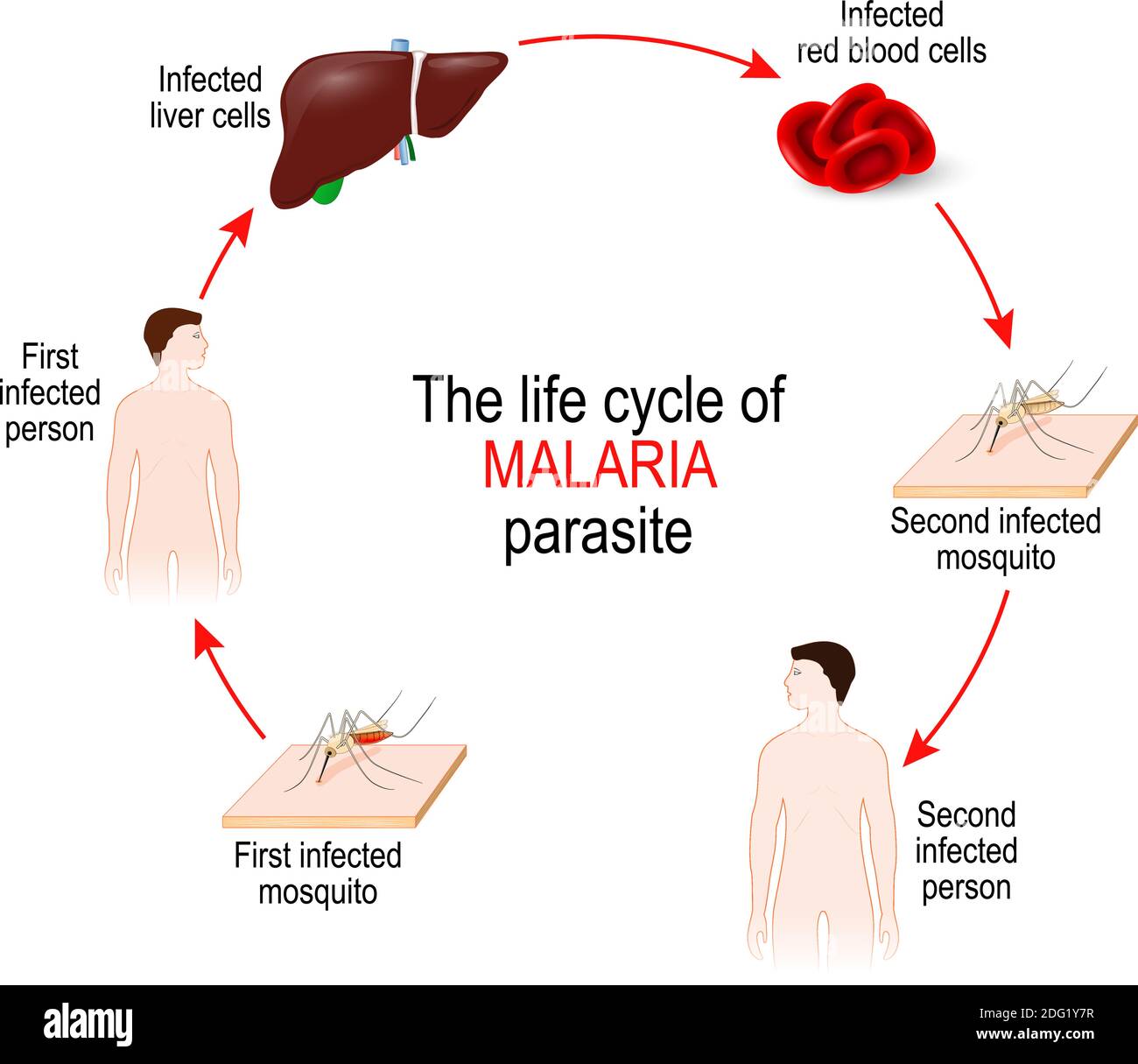 life cycle of a malaria parasite (from First infected mosquito to Second infectedperson). Malaria is a disease caused by a parasite Plasmodium Stock Vectorhttps://www.alamy.com/image-license-details/?v=1https://www.alamy.com/life-cycle-of-a-malaria-parasite-from-first-infected-mosquito-to-second-infectedperson-malaria-is-a-disease-caused-by-a-parasite-plasmodium-image388505931.html
RF2DG1Y7R–life cycle of a malaria parasite (from First infected mosquito to Second infectedperson). Malaria is a disease caused by a parasite Plasmodium
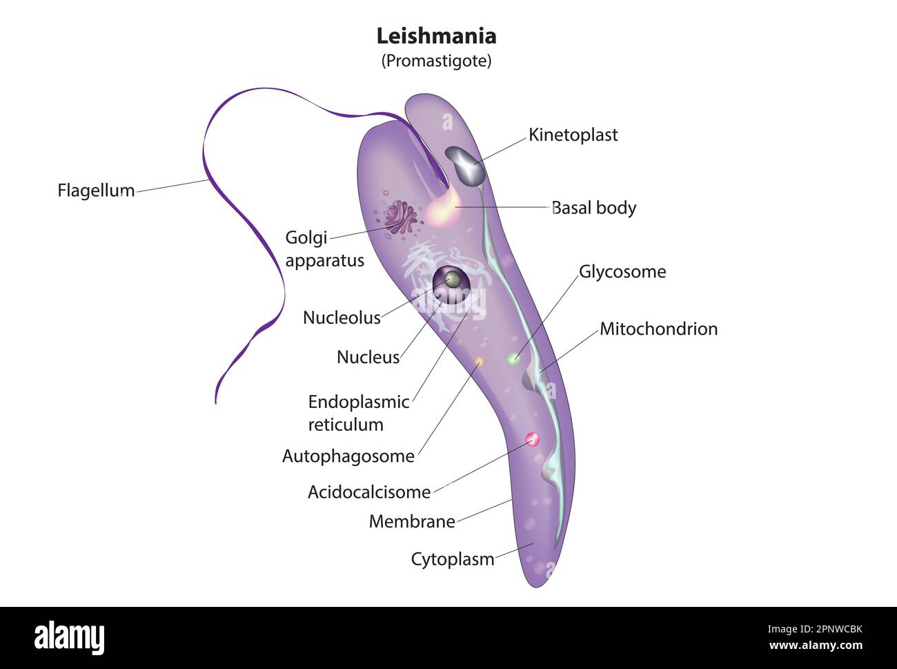 leishmania promastigotes Stock Vectorhttps://www.alamy.com/image-license-details/?v=1https://www.alamy.com/leishmania-promastigotes-image546987719.html
RF2PNWCBK–leishmania promastigotes
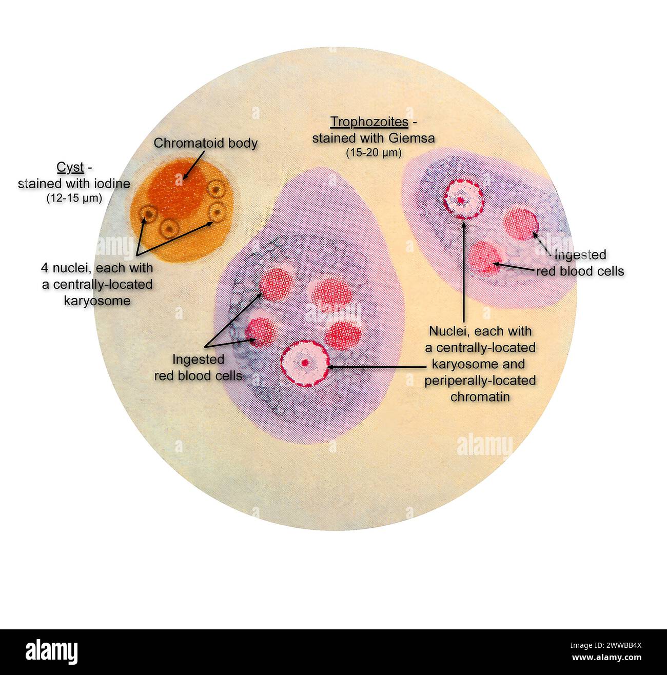 This illustration of a composite photomicrograph reveals the ultrastructural details observed at two stages in the life cycle of the parasite. Stock Photohttps://www.alamy.com/image-license-details/?v=1https://www.alamy.com/this-illustration-of-a-composite-photomicrograph-reveals-the-ultrastructural-details-observed-at-two-stages-in-the-life-cycle-of-the-parasite-image600769146.html
RM2WWBB4X–This illustration of a composite photomicrograph reveals the ultrastructural details observed at two stages in the life cycle of the parasite.
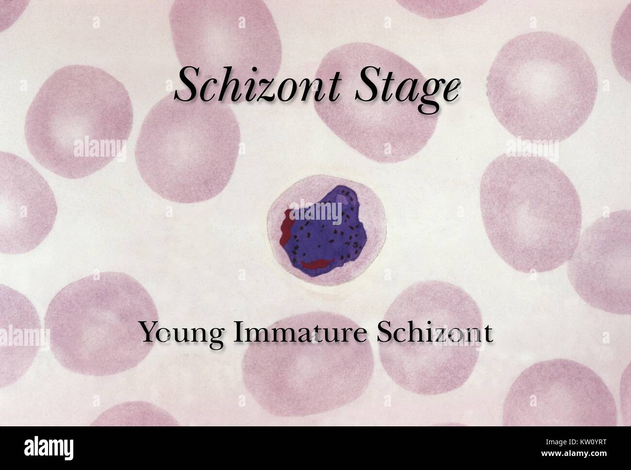 A young, immature schizont common to the Plasmodium spp. life cycle. The schizont stage of the Plasmodium spp. life cycle precedes the release of merozoites, which will go on to further infect more red blood cells. This erythrocytic schizont contains underdeveloped merozoites. Image courtesy CDC. 1975. Stock Photohttps://www.alamy.com/image-license-details/?v=1https://www.alamy.com/stock-photo-a-young-immature-schizont-common-to-the-plasmodium-spp-life-cycle-170281548.html
RMKW0YRT–A young, immature schizont common to the Plasmodium spp. life cycle. The schizont stage of the Plasmodium spp. life cycle precedes the release of merozoites, which will go on to further infect more red blood cells. This erythrocytic schizont contains underdeveloped merozoites. Image courtesy CDC. 1975.
 . Adventures in radioisotope research;. Radioactive tracers; Radiobiology. 868 ADVENTURES IX RADIOISOTOPE RESEARCH other b}^ the fact that the physiological death rate of the mammalian red corpuscle not containing DNA may be accelerated by exposure of the organism to radiation. Irradiation reduces their life-cycle possibly partly through production of agencies promoting haemolysis. Interference with the rate of blood flow is another example where processes independent from cell division promote cell death. Dobs on 1000 o c Q. 10 o c o. u £ o I- x: o 100 c o c « o c o u TD O O > Cf.. 5 After Stock Photohttps://www.alamy.com/image-license-details/?v=1https://www.alamy.com/adventures-in-radioisotope-research-radioactive-tracers-radiobiology-868-adventures-ix-radioisotope-research-other-b-the-fact-that-the-physiological-death-rate-of-the-mammalian-red-corpuscle-not-containing-dna-may-be-accelerated-by-exposure-of-the-organism-to-radiation-irradiation-reduces-their-life-cycle-possibly-partly-through-production-of-agencies-promoting-haemolysis-interference-with-the-rate-of-blood-flow-is-another-example-where-processes-independent-from-cell-division-promote-cell-death-dobs-on-1000-o-c-q-10-o-c-o-u-o-i-x-o-100-c-o-c-o-c-o-u-td-o-o-gt-cf-5-after-image237922750.html
RMRR28W2–. Adventures in radioisotope research;. Radioactive tracers; Radiobiology. 868 ADVENTURES IX RADIOISOTOPE RESEARCH other b}^ the fact that the physiological death rate of the mammalian red corpuscle not containing DNA may be accelerated by exposure of the organism to radiation. Irradiation reduces their life-cycle possibly partly through production of agencies promoting haemolysis. Interference with the rate of blood flow is another example where processes independent from cell division promote cell death. Dobs on 1000 o c Q. 10 o c o. u £ o I- x: o 100 c o c « o c o u TD O O > Cf.. 5 After
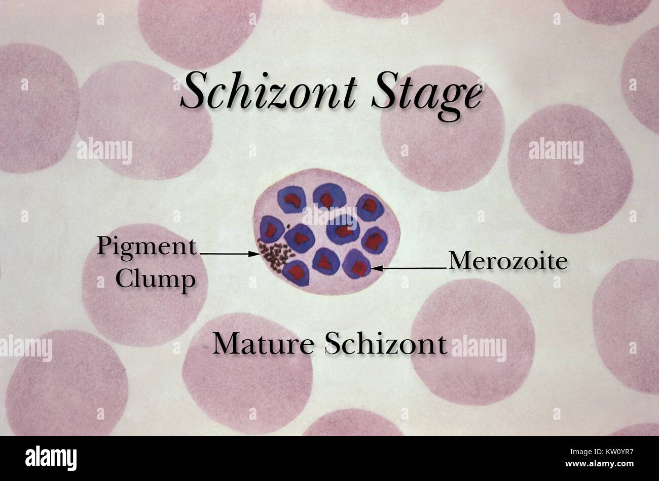 A mature erythrocytic schizont during the Plasmodium spp. life cycle. The schizont stages of Plasmodium spp. development include the formation of merozoites, which in this case, are ready to be released back into the blood to once again begin schizogony, infecting new red blood cells, or grow into gametocytes. Image courtesy CDC, 1971. Stock Photohttps://www.alamy.com/image-license-details/?v=1https://www.alamy.com/stock-photo-a-mature-erythrocytic-schizont-during-the-plasmodium-spp-life-cycle-170281531.html
RMKW0YR7–A mature erythrocytic schizont during the Plasmodium spp. life cycle. The schizont stages of Plasmodium spp. development include the formation of merozoites, which in this case, are ready to be released back into the blood to once again begin schizogony, infecting new red blood cells, or grow into gametocytes. Image courtesy CDC, 1971.
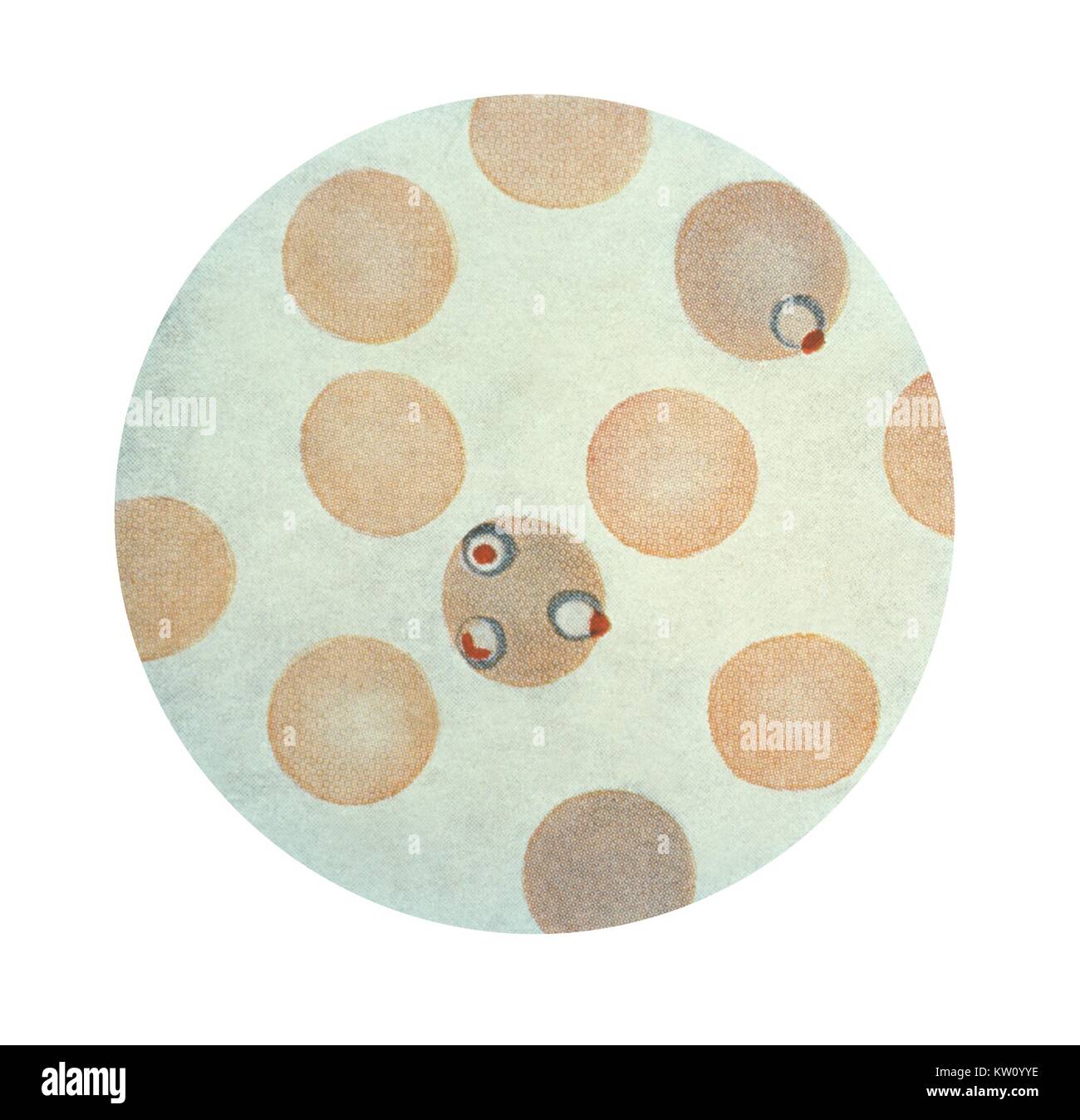 A photomicrograph of Plasmodium falciparum schizonts inside two erythrocytes using Wright's stains. (x1500), The Schizont stage of the life cycle precedes the formation of merozoites, which will be released to go on to infect more red blood cells. This erythrocyte contains a young immature schizont with underdeveloped merozoites. Image courtesy CDC, 1979. Stock Photohttps://www.alamy.com/image-license-details/?v=1https://www.alamy.com/stock-photo-a-photomicrograph-of-plasmodium-falciparum-schizonts-inside-two-erythrocytes-170281650.html
RMKW0YYE–A photomicrograph of Plasmodium falciparum schizonts inside two erythrocytes using Wright's stains. (x1500), The Schizont stage of the life cycle precedes the formation of merozoites, which will be released to go on to infect more red blood cells. This erythrocyte contains a young immature schizont with underdeveloped merozoites. Image courtesy CDC, 1979.
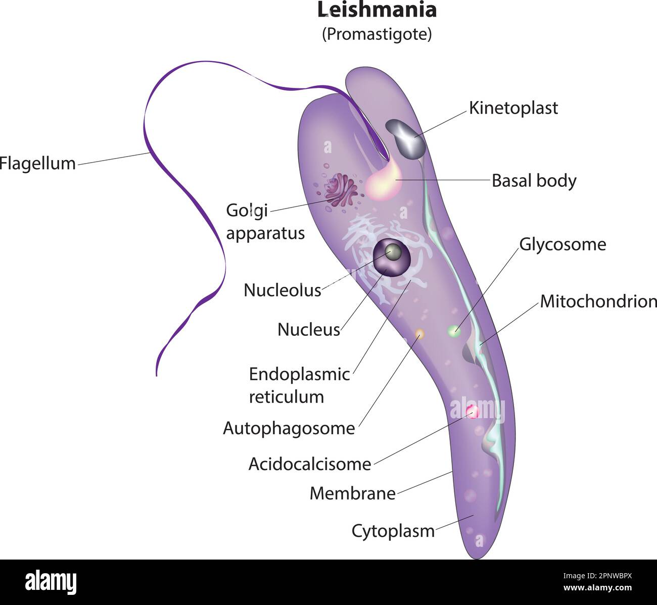 promastigotes stage of leishmania protozoa Stock Vectorhttps://www.alamy.com/image-license-details/?v=1https://www.alamy.com/promastigotes-stage-of-leishmania-protozoa-image546987250.html
RF2PNWBPX–promastigotes stage of leishmania protozoa
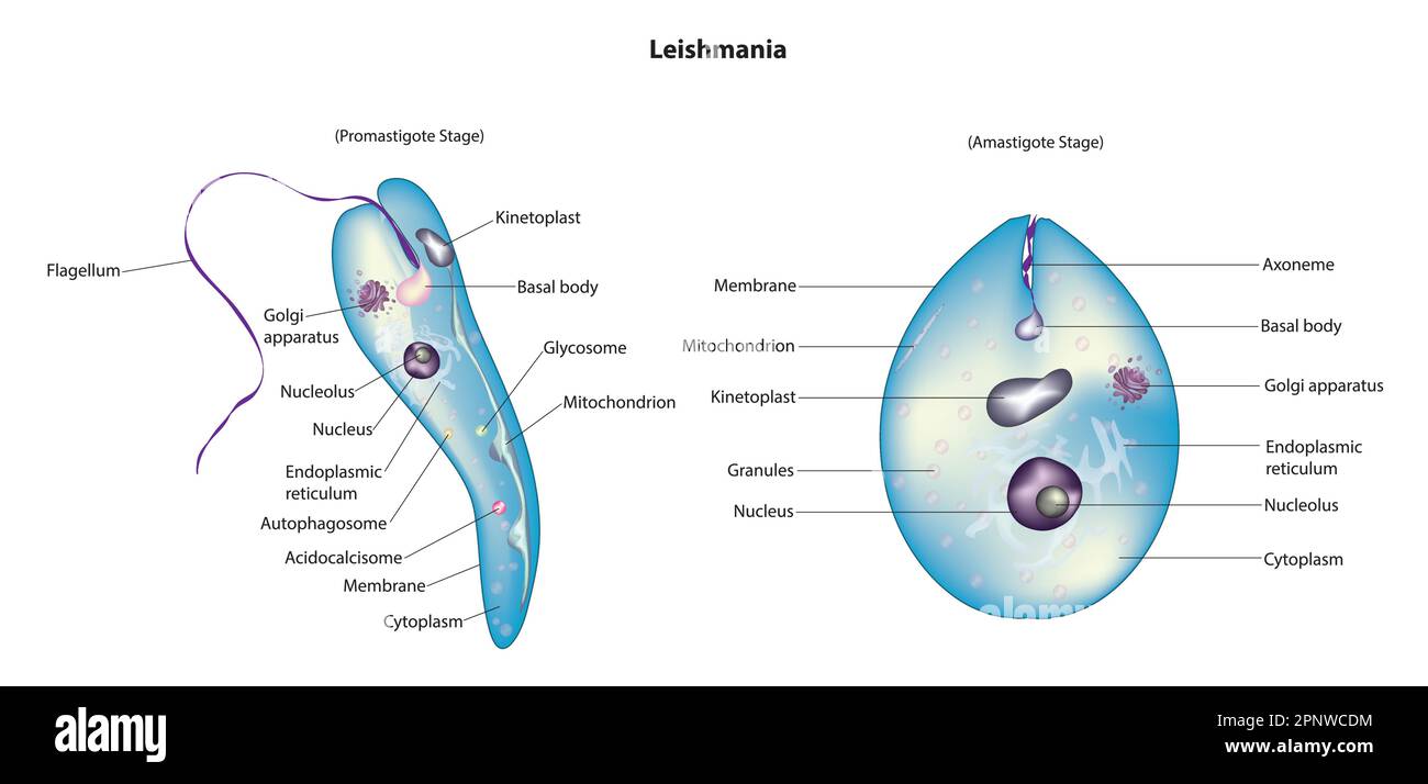 leishmania diagram Stock Vectorhttps://www.alamy.com/image-license-details/?v=1https://www.alamy.com/leishmania-diagram-image546987776.html
RF2PNWCDM–leishmania diagram
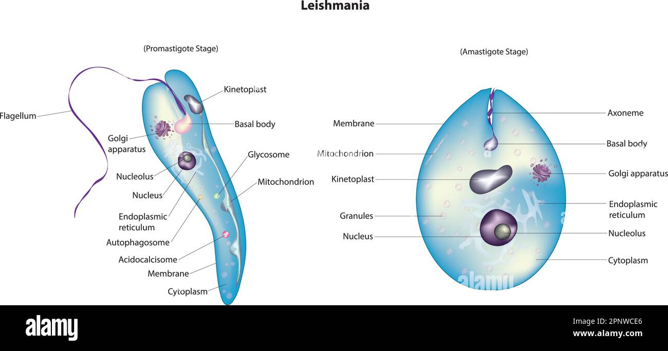 Leishmania stages Stock Vectorhttps://www.alamy.com/image-license-details/?v=1https://www.alamy.com/leishmania-stages-image546987790.html
RF2PNWCE6–Leishmania stages
 Biological anatomy of Entamoeba histolytica, anaerobic parasitic amoebozoan, Detailed diagram and anatomy of Entamoeba histolytica, kingdom Protista Stock Vectorhttps://www.alamy.com/image-license-details/?v=1https://www.alamy.com/biological-anatomy-of-entamoeba-histolytica-anaerobic-parasitic-amoebozoan-detailed-diagram-and-anatomy-of-entamoeba-histolytica-kingdom-protista-image438683990.html
RF2GDKNWA–Biological anatomy of Entamoeba histolytica, anaerobic parasitic amoebozoan, Detailed diagram and anatomy of Entamoeba histolytica, kingdom Protista